cytotoxicity of eupatorium cannabinum l. ethanolic extract against colon cancer cells and...
TRANSCRIPT
Ribeiro-Varandas et al. BMC Complementary and Alternative Medicine 2014, 14:264http://www.biomedcentral.com/1472-6882/14/264
RESEARCH ARTICLE Open Access
Cytotoxicity of Eupatorium cannabinum L.ethanolic extract against colon cancer cells andinteractions with Bisphenol A and DoxorubicinEdna Ribeiro-Varandas1, Filipe Ressurreição1, Wanda Viegas1 and Margarida Delgado1,2*
Abstract
Background: Eupatorium cannabinum L. has long been utilized in traditional medicine, however no information isavailable regarding cellular effects of full extracts. Here we assessed the effects of E. cannabinum ethanolic extract(EcEE) on the colon cancer line HT29. Potential interactions with bisphenol A (BPA) a synthetic phenolic compoundto which humans are generally exposed and a commonly used chemotherapeutic agent, doxorubicin (DOX) werealso evaluated.
Methods: HT29 cells were exposed to different concentrations (0.5 to 50 μg/ml) of EcEE alone or in combinationwith BPA or DOX. Cell viability was analyzed through resazurin assay. Gene transcription levels for NCL, FOS, p21,AURKA and bcl-xl were determined through qRT-PCR. Cytological analysis included evaluation of nuclear and mitoticanomalies after DAPI staining, immunodetection of histone H3 lysine 9 acetylation (H3K9ac) and assessment of DNAdamage by TUNEL assay.
Results: Severe loss of HT29 cell viability was detected for 50 μg/ml EcEE immediately after 24 h exposure whereasthe lower concentrations assayed (0.5, 5 and 25 μg/ml) resulted in significant viability decreases after 96 h. Exposureto 25 μg/ml EcEE for 48 h resulted in irreversible cell damage leading to a drastic decrease in cell viability after 72 hrecovery in EcEE-free medium. 48 h 25 μg/ml EcEE treatment also induced alteration of colony morphology, H3K9hyperacetylation, transcriptional up regulation of p21 and down regulation of NCL, FOS and AURKA, indicatingreduced proliferation capacity. This treatment also resulted in drastic mitotic and nuclear disruption accompaniedby up-regulation of bcl-xl, limited TUNEL labeling and nuclear size increase, suggestive of a non-apoptocic cell deathpathway. EcEE/BPA co-exposure increased mitotic anomalies particularly for the lowest EcEE concentration, althoughwithout major effects on viability. Conversely, EcEE/DOX co-exposure decreased cell viability in relation to DOX forall EcEE concentrations, without affecting the DOX-induced cell cycle arrest.
Conclusions: EcEE has cytotoxic activity on HT29 cancer cells leading to mitotic disruption and non-apoptotic celldeath without severe induction of DNA damage. Interaction experiments showed that EcEE can increase BPA aneugeniceffects and EcEE synergistic effects with DOX supporting a potential use as adjuvant in chemotherapeutic approaches.
BackgroundEupatorium cannabinum L., commonly known as hemp-agrimony is a robust perennial herbaceous plant of theAsteraceae family and the only species of the Eupator-ium genus found in Europe occurring also throughoutNorth Africa and Asia [1]. E. cannabinum has long been
* Correspondence: [email protected] de Botânica Aplicada à Agricultura, Instituto Superior de Agronomia,Universidade de Lisboa, Tapada da Ajuda, 1349-017 Lisboa, Portugal2Faculty of Psychology and Life Sciences, Universidade Lusófona deHumanidades e Tecnologias, Campo Grande 376, 1749-024 Lisboa, Portugal
© 2014 Ribeiro-Varandas et al.; licensee BioMeCreative Commons Attribution License (http:/distribution, and reproduction in any mediumDomain Dedication waiver (http://creativecomarticle, unless otherwise stated.
used for medicinal purposes being referred to by Greeksand Romans as well by the medieval Persian physicianAviccena, for what is also known as Eupatorium of Avic-cena, and later by the Portuguese Renaissance pioneer intropical medicine, Garcia da Orta (1563) [2]. Presently,hemp-agrimnony is used in both Chinese [3] and Indian[4] traditional medicine as well as in natural medicine inwestern countries [5] with very diverse therapeutic indica-tions including influenza-like illnesses [6], hypertension[3,4,6] and as an anti-tumour agent [4]. E. cannabinumextracts has been previously characterized and reveal the
d Central Ltd. This is an Open Access article distributed under the terms of the/creativecommons.org/licenses/by/4.0), which permits unrestricted use,, provided the original work is properly credited. The Creative Commons Publicmons.org/publicdomain/zero/1.0/) applies to the data made available in this

Ribeiro-Varandas et al. BMC Complementary and Alternative Medicine 2014, 14:264 Page 2 of 10http://www.biomedcentral.com/1472-6882/14/264
presence of sesquiterpenes [7], pyrrolizidine alkaloids [3,8]as well as several phenolic compounds [9,10].Sesquiterpenes were found to be a major fraction
(43.3%) of essential oil from E. cannabinum aerial parts[11], being eupatoriopicrin the main component [7].Eupatoriopicrin has been associated with inductionDNA damage in Ehrlich ascites tumour [12] as well aswith cytostatic activity and both in vitro and in vivotumour growth inhibition properties in Lewis lung car-cinoma and FIG 26 fibrosarcoma [13].Pyrrolizidine alkaloids are generally associated with
genotoxicity and tumourigenic activities [14], howeverthe isomers intermedine and lycopsamine indentified inE. cannabinum have low genotoxic potency [15] and ly-copsamine was shown to be non-tumourigenic in rats[16]. Additionally the phenolic compounds identified inthis plant have been described to have anti-inflammatory[9], anti-parasitary [17], as well as anti-proliferative ef-fects in several cell lines [18]. In particular, jaceosidincytotoxic effects have been demonstrated in normal andcancer endometrial cells [19] and hispidulin was shownto efficiently inhibit growth of gastric cancer cells [20]and liver carcinoma cells without significant toxic effectin normal liver cells [21].Although the effects of specific components of Eupa-
torium cannabinum L. extracts have been described, thecellular effects of the full extracts have not, until now,been investigated. Thus, here different concentrations ofEupatorium cannabinum L. ethanolic extract (EcEE)were evaluated on the colon cancer cell line HT29.Moreover we also analyzed its interactions with the syn-thetic phenolic compound bisphenol A (BPA) as well aswith the chemotherapeutic agent Doxorubicin (DOX).Human exposure to BPA is considered generalized in thecommon population and its adverse health effects are thefocus of intense investigation [22,23]. On the other hand,DOX is a commonly used chemotherapeutic agent towhich cell resistance can emerge [24,25]. Plant constitu-ents are a major source of bioactive compounds and sev-eral plants have been investigated aiming to identifypotential synergistic effects with DOX (reviewed in [26]).
MethodsEupatorium cannabinum L. ethanolic extractEupatorium cannabinum L. (Asteraceae) aerial partswere collected in the Rossas fields of Arouca village,Portugal, in August during mass flowering. Formal iden-tification of plant material was performed by A.P. Paesfrom “João de Carvalho e Vasconcellos Herbarium” atInstituto Superior de Agronomia (Lisboa, Portugal). Avoucher specimen was deposited in the same herbariumunder the number LISI 1503/2013. Plant material wasdried and powdered using a grinder and ethanolic ex-tract (EcEE) was obtained by soaking the material in
absolute ethanol for 48 h at room temperature with gen-tle shaking. The extract were filtered and concentratedunder vacuum on a rotary evaporator at 40°C and storedat −20°C for further use.
Cellular cultures, reagents and treatmentsHT29 cells were purchased from European Collection ofCell Cultures (ECACC, UK) and cultivated in RPMImedium under standard conditions as previously de-scribed [27]. Before treatments and experiments HT29,cells were allowed to stabilize for 24 h in standardmedium and further cultivated in EcEE supplementedmedia for 24 h, 48 h or 96 h. Crude ethanolic extractwas dissolved in ethanol to a final work concentration of50 mg/ml before use and added to the culture media atfour different final concentrations (0.5 μg/ml, 5 μg/ml,25 μg/ml and 50 μg/ml). Bisphenol A (Sigma-Aldrich)was freshly diluted in ethanol and added to the culturemedia to the final concentration of 1 μg/ml (4.4 μM)that corresponds to the established Tolerable Daily In-take (TDI) level of 50 ug/kg BW/day [28,29] consideringan average body weight of 70 Kg and daily consumptionof 3 litres of preformed water. Doxorubicin (DOX)(AppliChem) was dissolved in water at stock concentra-tion of 1 mg/ml and added to the culture media to finalconcentration of 2.5 μg/ml (4 μM) which corresponds toa therapeutic dosage [30]. For the combined EcEE/BPAor EcEE/DOX exposures, cells were pre-exposed to EcEEfor 24 h followed by additional 24 h of simultaneous ex-posure to EcEE and BPA or EcEE and DOX. Single 24 hBPA or DOX exposure was carried-out in equivalent cellcultures. For evaluation of cell recovery capacity aftertreatments cells were cultivated for additional 72 h instandard culture medium. Negative controls were per-formed for all experiments using cells grown in standardculture medium as well as cells grown in medium supple-mented with ethanol at final concentration of 170 μM,corresponding to the final concentration of ethanol usedas vehicle for all EcEE concentrations as well as for BPA.
Cell viabilityCell viability was evaluated by CellTiter-Blue assay (Pro-mega) following manufacturer’s instructions. Cells wereplated on 96-well plates at a density of 3.2 × 10⁴ cells/well and after treatments were incubated for 4 h withCellTiter-Blue Reagent. Additional negative controlswere performed in the absence of cells to guarantee thatthe utilized media did not interfere with fluorescencereadings. Experiments were repeated at least three timeswith a minimum of three replicates per experiment.
DAPI staining, TUNEL assay and immunodetectionFor cytological analysis cells were grown over glass cov-erslips coated with 0.2% (v/v) gelatin (Sigma-Aldrich)
Ribeiro-Varandas et al. BMC Complementary and Alternative Medicine 2014, 14:264 Page 3 of 10http://www.biomedcentral.com/1472-6882/14/264
and after treatments fixed in 4% (p/v) formaldehyde inPBS. For evaluation of colony morphology, mitotic indexas well as mitotic and nuclear anomalies cells were DAPIstained and mounted on glass slides with antifade AF1(Citifluor). DNA damage assessment with TUNEL assay(Roche) was performed accordingly to manufacturers’instructions. Immunodetection of H3K9ac and α-tubulinwas performed in fixed cells as previously described [27]using the primary antidodies anti-acetyl-histone H3(Lys 9)(ab10812, Abcam) and anti-α-Tubulin (T9026, Sigma-Aldrich) detected with FITC or Cy3 conjugated secondaryantibodies. Images were captured using the appropriateexcitation and emission filters and recorded using an epi-fluorescence microscope Zeiss Axioskop2 equipped with aZeiss AxioCam MRc5 digital camera. ImageJ software(http://rsbweb.nih.gov/ij/) was used for nuclear area mea-surements. The analysis was performed in the pooled re-sults of at least two independent experiments with at leasttwo replicates.
cDNA isolation and real-time quantitative PCRTranscriptional analysis was performed by quantitativereal-time PCR (qRT-PCR) for the proliferation-associatedgenes nucleolin (NCL), FOS and p21, for the cell cycle re-lated gene AURKA, and the anti-apoptotic gene bcl-xl.The specific primers utilized are listed in Table 1, GAPDHand β-actin were used as control genes [27,31]. TotalRNA was extracted from trypsinized cells with the RNA-queous Kit (Invitrogen) following manufacturers’ instruc-tions. 3 μg of total RNA was utilized for RNase freeDNase digestion (RQ1 RNase free DNase, Promega) andfirst strand cDNA synthesis was completed with randomprimers (DYNAmo cDNA syntesis Kit, Thermo Scientific).The resulting cDNA was utilized for qRT-PCR with Sso-Fast Eva Green Supermix (BioRad) utilizing the followingconditions: 95°C for 3 min, 35 cycles (95°C for 30 s, 55°Cfor 30 s, 72°C for 40 s), and 72°C for 5 min. Control PCRswere also performed with total RNA prior to cDNA syn-thesis as well as for all primer combinations without tem-plate. Experiments were repeated at least three times withat least three replicates per cell treatment/primer combin-ation in each experiment. Since no significant differences
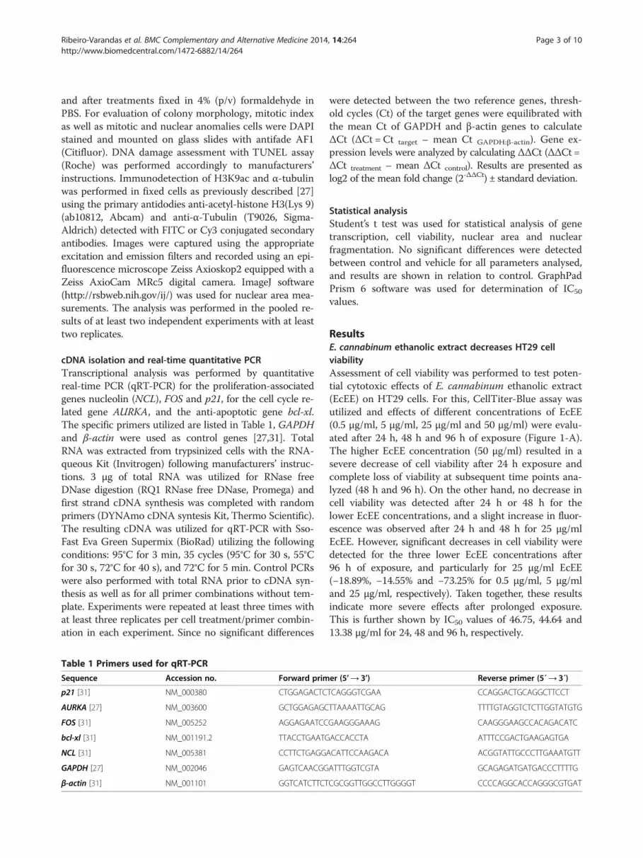
Table 1 Primers used for qRT-PCR
Sequence Accession no. Forward prim
p21 [31] NM_000380 CTGGAGACTC
AURKA [27] NM_003600 GCTGGAGAGC
FOS [31] NM_005252 AGGAGAATCC
bcl-xl [31] NM_001191.2 TTACCTGAATG
NCL [31] NM_005381 CCTTCTGAGGA
GAPDH [27] NM_002046 GAGTCAACGG
β-actin [31] NM_001101 GGTCATCTTCT
were detected between the two reference genes, thresh-old cycles (Ct) of the target genes were equilibrated withthe mean Ct of GAPDH and β-actin genes to calculateΔCt (ΔCt = Ct target – mean Ct GAPDH:β-actin). Gene ex-pression levels were analyzed by calculating ΔΔCt (ΔΔCt =ΔCt treatment – mean ΔCt control). Results are presented aslog2 of the mean fold change (2-ΔΔCt) ± standard deviation.
Statistical analysisStudent’s t test was used for statistical analysis of genetranscription, cell viability, nuclear area and nuclearfragmentation. No significant differences were detectedbetween control and vehicle for all parameters analysed,and results are shown in relation to control. GraphPadPrism 6 software was used for determination of IC50
values.
ResultsE. cannabinum ethanolic extract decreases HT29 cellviabilityAssessment of cell viability was performed to test poten-tial cytotoxic effects of E. cannabinum ethanolic extract(EcEE) on HT29 cells. For this, CellTiter-Blue assay wasutilized and effects of different concentrations of EcEE(0.5 μg/ml, 5 μg/ml, 25 μg/ml and 50 μg/ml) were evalu-ated after 24 h, 48 h and 96 h of exposure (Figure 1-A).The higher EcEE concentration (50 μg/ml) resulted in asevere decrease of cell viability after 24 h exposure andcomplete loss of viability at subsequent time points ana-lyzed (48 h and 96 h). On the other hand, no decrease incell viability was detected after 24 h or 48 h for thelower EcEE concentrations, and a slight increase in fluor-escence was observed after 24 h and 48 h for 25 μg/mlEcEE. However, significant decreases in cell viability weredetected for the three lower EcEE concentrations after96 h of exposure, and particularly for 25 μg/ml EcEE(−18.89%, −14.55% and −73.25% for 0.5 μg/ml, 5 μg/mland 25 μg/ml, respectively). Taken together, these resultsindicate more severe effects after prolonged exposure.This is further shown by IC50 values of 46.75, 44.64 and13.38 μg/ml for 24, 48 and 96 h, respectively.
er (5’→ 3’) Reverse primer (5´→ 3´)
TCAGGGTCGAA CCAGGACTGCAGGCTTCCT
TTAAAATTGCAG TTTTGTAGGTCTCTTGGTATGTG
GAAGGGAAAG CAAGGGAAGCCACAGACATC
ACCACCTA ATTTCCGACTGAAGAGTGA
CATTCCAAGACA ACGGTATTGCCCTTGAAATGTT
ATTTGGTCGTA GCAGAGATGATGACCCTTTTG
CGCGGTTGGCCTTGGGGT CCCCAGGCACCAGGGCGTGAT

Figure 1 EcEE affect cell viability and proliferation. (A) Cell viability after 24 h, 48 h and 96 h of exposure to distinct concentrations of EcEE and(B) after 72 h recovery in EcEE-free medium following 24 h and 48 h treatments. Results are presented as percentage over control, **p < 0.0001 and*p < 0.01. (C) NCL, FOS and p21 differential transcription after 48 h exposure to distinct EcEE concentrations. Results are shown as mean log2 foldchange (2-ΔΔCt) ± standard deviation in relation control, *p < 0.0001. (D) DAPI stained HT29 colonies after 48 h in control medium and mediumsupplemented with EcEE 5 μg/ml or EcEE 25 μg/ml. All images have identical magnification, bar = 50 μm.
Ribeiro-Varandas et al. BMC Complementary and Alternative Medicine 2014, 14:264 Page 4 of 10http://www.biomedcentral.com/1472-6882/14/264
To detect possible deferred effects of the EcEE expos-ure cell viability was also evaluated after 72 h of recoveryin standard culture media (Figure 1-B) and revealed a se-vere decrease exclusively for 48 h exposure to 25 μg/mlEcEE (−75.59%).To better understand the effects of EcEE immediately
after 48 h exposure, gene transcription analysis was car-ried out for three proliferation-associated genes, namelynucleolin (NCL), p21 and FOS (Figure 1-C). Similarly tothe cell viability results, no significant differences intranscription levels were detected after 48 h exposure toEcEE concentrations equal to or lower than 5 μg/ml.Conversely, 25 μg/ml EcEE exposure resulted in signifi-cant differences in mRNA levels of all three genes, cor-responding to down regulation of both NCL and FOS(Log2 fold change = −0.813 ± 0.248 and −0.741 ± 0.078,respectively), and up regulation of p21 (Log2 fold change =1.393 ± 0.128). Evaluation of colony morphology was per-formed immediately after EcEE treatments by DAPI stain-ing. Again, significant alterations in colony morphologywere detected after exposure to 25 μg/ml EcEE for 48 h,evident as cells being more dispersed and showing a flat-tening of cellular aggregates in comparison to controls
with no detectable effect for 5 μg/ml EcEE (Figure 1-D) or0.5 μg/ml EcEE (not shown).
E. cannabinum ethanolic extract induces alterations innuclear structure and mitotic disruptionA detailed cytological analysis was performed for 0.5 μg/ml,5 μg/ml and 25 μg/ml EcEE concentrations after 48 h ofexposure and again significant nuclear alterations wereobserved exclusively for 25 μg/ml EcEE (Figure 2-A). Thiswas obvious as the prominent occurrence of micronucleiand highly condensed nuclei (pyknosis) scattered through-out cell aggregates as well as fragmented nuclei (karyor-rhexis), revealing irreversible nuclear damage. In addition,TUNEL assay showed that induction of DNA breaks alsooccurred after 48 h exposure to 25 μg/ml EcEE treatmentsalthough at a much lower level than nuclear abnormal-ities, as many of the abnormal nuclei were not TUNELpositive and in positive nuclei labeling was sparse(Figure 2-B). For the two lower EcEE concentrations (0.5and 5 μg/ml) no TUNEL positive nuclei were detected(not shown) as observed for control. Importantly, qRT-PCR transcriptional analysis of the anti-apoptotic bcl-xlgene revealed that EcEE exposure induced a significant up

Figure 2 Nuclear organization is disrupted after 48 h of exposure to EcEE 25 μg/ml. (A) DAPI stained HT29 interphase cells. Nuclearanomalies, namely pyknosis (arrow), micronuclei (open arrow head) and karyorrhexis (arrow head) are detectable only for 25 μg/ml EcEE. The lackof effects induced by EcEE lower concentrations is exemplified by 5 μg/ml EcEE. (B) TUNEL positive nuclei (arrow) with sparse labeling aredetectable for 25 μg/ml EcEE. (C) bcl-xl differential expression after 48 h exposure to distinct EcEE concentrations. Results in are shown as themean log2 fold change (2-ΔΔCt) ± standard deviation in relation control, **p < 0.0001 and *p < 0.01. (D) DAPI staining (blue) and α-tubulinimmunodetection (red in merged images at the bottom) of interphase cells after 48 h in control or 25 μg/ml EcEE. (E) Immunodetection ofH3K9ac after 48 h in control or EcEE 25 μg/ml supplemented medium. Within each experiment all images have identical magnification,bar = 5 μm.
Ribeiro-Varandas et al. BMC Complementary and Alternative Medicine 2014, 14:264 Page 5 of 10http://www.biomedcentral.com/1472-6882/14/264
regulation of this gene not only at 25 μg/ml (Log2 foldchange = 0.528 ± 0.243) but also at 5 μg/ml, although to alesser extent (Log2 fold change = 0.158 ± 0.067) (Figure 2-C).Quantification of the nuclear area of non-pyknotic andnon-fragmented DAPI stained nuclei showed a significantincrease in this parameter in relation to control for cellsexposed to 25 μg/ml EcEE but not to the lower EcEE con-centrations (not shown). The increment in nuclear areaafter the 48 h exposure to 25 μg/ml EcEE corresponded inaverage to 48.8% (n > 70 for each growth condition, p <0.0001 for 25 μg/ml EcEE in relation to control) and wasaccompanied by an evident increase in cellular area re-vealed by α-tubulin immunodetection (Figure 2-D). More-over, evident chromatin enrichment in histone H3acetylated on lysine 9 (H3K9ac) was detected also for 48 h25 μg/ml EcEE (Figure 2-E) whereas no alteration was ob-served for either 0.5 μg/ml or 5 μg/ml EcEE (not shown).The effects of exposure to EcEE were further evaluated
on mitotic cells after DAPI staining. No significant vari-ation was observed in the mitotic index between control,
vehicle and EcEE independently of the concentrationassayed (varying between 4.57 and 5.94). On the otherhand, although mitotic anomalies, particularly multi-polar metaphases and anaphases, are a common featureof HT29 cells and therefore observed both in controland vehicle (6.67% and 11.76% after 24 h; 6.38% and10.67% after 48 h, for control and vehicle respectively),the percentage of abnormal mitosis increased after ex-posure to all EcEE concentrations (Figures 3-A and B).Although a slight increase of abnormal mitosis wasalready detectable for 0.5 μg/ml EcEE, this effect wasgreater for 5 μg/ml EcEE (41% and 44% after 24 h or 48 h,respectively). After exposure to 25 μg/ml EcEE, most mi-totic cells presented abnormalities (80% and 63% after 24and 48 h, respectively). Although the frequency of abnor-mal mitosis was greater after 24 h at the higher EcEE con-centration, these results clearly indicate that EcEE inducesmitotic disruption in a dose dependent manner. Interest-ingly, qRT-PCR analysis revealed a significant down regu-lation of AURKA (Log2 fold change = −0.938 ± 0.146), a

Figure 3 EcEE exposure induces mitotic disruption. (A) DAPI stained abnormal mitotic cells after EcEE exposure showing (i) defectivechromosome congression, (ii) tripolar and (iii) tetrapolar metaphases and (iv) tripolar anaphase with chromosome bridges, bar = 5 μm. (B) Percentageof abnormal mitosis after 24 h and 48 h exposure to distinct concentrations of EcEE; the total number of mitotic cells scored and utilized to calculatethe percentage of abnormal mitosis is shown in brackets. (C) AURKA differential transcription after exposure to 48 h of EcEE. Results are shown as themean log2 fold change (2-ΔΔCt) ± standard deviation in relation control, *p < 0.0001.
Ribeiro-Varandas et al. BMC Complementary and Alternative Medicine 2014, 14:264 Page 6 of 10http://www.biomedcentral.com/1472-6882/14/264
gene that encodes a key protein for mitotic chromosomesegregation (Figure 3-C).
E. cannabinum ethanolic extract increases Bisphenol Ainduced mitotic disruptionInteractions between EcEE and the environmental pollu-tant BPA at reference level (1 μg/ml) were evaluated. Co-exposure to EcEE and BPA did not affect cell viability im-mediately after treatments, as no significant differenceswere detected in relation to control (Figure 4-A). After72 h recovery in standard medium, a severe decrease in
Figure 4 EcEE interacts with BPA at reference level. (A) Cell viability aftsubsequent 72 h recovery in standard medium. Results are presented as peafter 48 h culture in standard medium (control) or medium supplementedcombination with BPA. The total number of mitotic cells scored and utilized t
cell viability (−93.48%) was exclusively observed for 25 μg/mlEcEE/BPA (Figure 4-A) which was even greater than thatobserved for 25 μg/ml EcEE alone (Figure 1-B).Cytological evaluation of mitotic disruption after DNA
DAPI staining (Figure 4-B) revealed that BPA exposurealone increased the level of mitotic anomalies to 20.5%.Interestingly, a stronger effect of BPA co-exposure wasobserved for the lowest EcEE concentration assayed(0.5 μg/ml), which resulted in 41% of abnormal mitosis(Figure 4-B) compared to 25% observed for EcEE alone(Figure 3-B). In contrast, no evident effect of BPA was
er co-exposure to BPA (1 μg/ml) and distinct EcEE concentrations andrcentage over control, *p < 0.001. (B) Percentage of mitotic anomalieswith ethanol (vehicle), BPA or EcEE at distinct concentrations ino calculate the percentage of abnormal mitosis is shown in brackets.

Ribeiro-Varandas et al. BMC Complementary and Alternative Medicine 2014, 14:264 Page 7 of 10http://www.biomedcentral.com/1472-6882/14/264
detected for the intermediate EcEE concentration, evi-dent as an identical level of 44% for 5 μg/ml EcEE aloneor in combination with BPA. Co–exposure to the higherEcEE concentration (25 μg/ml) and BPA resulted in aparticular high level of mitotic anomalies (75%), al-though the difference in relation to EcEE alone (63%)was smaller than that observed for the lower EcEEconcentrations.
Cytotoxic effects of Doxorubicin are enhanced byE. cannabinumPotential interactions between different concentrationsof EcEE and the chemotherapeutic drug doxorubicin(DOX) at a therapeutic concentration of 2.5 μg/ml wereinvestigated. Immediately after exposure, DOX aloneresulted in a slight decrease in cell viability (−4.15%)(Figure 5-A). Interestingly, the loss of cell viability wassignificantly more pronounced after co-exposure toEcEE/DOX for all EcEE concentrations (−10.03%, −19.88%and −18.67% for 0.5 μg/ml, 5 μg/ml and 25 μg/ml, respect-ively) (Figure 5-A) contrasting with the lack of effectsobserved for 48 h exposure to EcEE alone (Figure 1-A).Recovery experiments showed that the effects of bothDOX and 25 μg/ml EcEE/DOX have long lasting negativeeffects on viability, apparent as prominent decreases incell viability after 72 h recovery in standard medium in re-lation to what was observed immediately after exposure(Figure 5-A). After recovery, EcEE 25 μg/ml/DOX expos-ure resulted in an even more pronounced loss of cellviability (−93.95%) than that observed for exposure to25 μg/ml EcEE alone (Figure 1-B). Conversely, for thelower EcEE concentrations, no significant differences weredetected between exposure to DOX alone and in combin-ation with EcEE (Figure 1-B).
Figure 5 EcEE has synergistic effects with DOX at a therapeutic dose.concentrations and subsequent 72 h recovery in standard medium. Resultsrelation to DOX alone is indicated by horizontal brackets, **p < 0.0001 andDOX illustrating the occurrence of (i) micronuclei (arrow head) and pyknot(C) Percentage fragmented nuclei after exposure to EcEE 25 μg/ml or DOXshown in brackets, **p < 0.0001 and *p < 0.03 in relation to EcEE 25 μg/ml
Cytological analysis after DAPI staining showed acomplete absence of cells at mitosis after exposure toDOX alone or in combination to EcEE, independently ofthe EcEE concentration. Conversely, both pyknotic cellsand fragmented nuclei were observed after exposure toDOX alone or in combination to EcEE (Figure 5-B).Since identical nuclear alterations were also observedafter single exposure to 25 μg/ml EcEE (Figure 2-E), thelevels of nuclear fragmentation were compared betweensingle exposure to 25 μg/ml EcEE or DOX alone and thecombination of both (Figure 5-C). The results revealedthat the induction of nuclear fragmentation is signifi-cantly higher for 25 μg/ml EcEE/DOX combined expos-ure (20.28%) than for either DOX (7.63%) or 25 μg/mlEcEE (8.89%) alone.
DiscussionEupatorium cannabinum L. is a commonly utilized plantfor alternative and/or complementary medicine treat-ments [6] including as an anticancer agent [4]. Althoughcellular effects of particular phytochemicals known to bepresent in E. cannabinum have been previously de-scribed, to our knowledge this is the first study that eval-uates the cytotoxic potential of E. cannabinum extractson human cancer cells. Here we demonstrated that E.cannabinum ethanolic extract (EcEE) has cytotoxic ef-fects on HT29 colon cancer cells in a time and dosedependent manner. IC50 were similar after 24 and 48 h(46.75 and 44.65 μg/ml, respectively) but considerablylower (13.38 μg/ml) after 96 h of exposure. Cytotoxic ac-tivity has also been demonstrated for extracts from otherEupatorium species. For E. perfoliatum ethanolic extract,IC50 values between 12 and 14 μg/ml were obtained after24 h exposure in three distinct mammalian cell lines
(A) Cell viability after co-exposure to DOX (2.5 μg/ml) and distinct EcEEare presented as percentage over control, the level of significance in*p < 0.01. (B) DAPI stained cell after co-exposure to EcEE 25 μg/ml/ic nuclei (arrow) and (ii) fragmented nuclei (arrows), bar = 5 μm.alone and the combination of both. Total number of cells analyzed isor DOX alone.
Ribeiro-Varandas et al. BMC Complementary and Alternative Medicine 2014, 14:264 Page 8 of 10http://www.biomedcentral.com/1472-6882/14/264
[32]. In MCF7 breast cancer cells a time dependent ef-fect was also observed for E. odoratum ethyl acetateextract (IC50 of 65.72, 83.88 μg/ml and 92.84 μg/ml for 24,48 and 72 h, respectively) while for acetone extract higherIC50 values were obtained but without a direct correlationwith exposure time (133.9, 163.0 and 147.8 μg/ml for 24,48, and 72 h respectively) [33]. The immediate cytotoxicityobserved here for EcEE is lower than that obtained for E.perfoliatum ethanolic extract and higher than that of ethylacetate or acetone extracts from E. odoratum. Interest-ingly the time dependent increase in cytotoxicity of EcEEwas only detected for the longer exposure time (96 h).Moreover, a deferred effect on cell viability was detectedafter 48 h exposure to EcEE at 25 μg/ml. This was also as-sociated with disruption of cell colony three-dimensionalarrangement, a generalized increase in nuclear area andH3K9 hyperacetylation. Relevantly, gene transcriptionanalysis revealed a significant reduction in the mRNAlevels of FOS, which encodes for a nuclear protein fromAP-1 transcription factor complex, and nucleolin (NCL)the most profuse non-ribosomal protein of the nucleolus.Both FOS and nucleolin are involved in the regulation ofcell proliferation [34,35] as their decreased expression hasbeen related with reduced proliferation capacity of cancercells including colon cancer cell lines [36,37]. On theother hand, exposure to EcEE (25 μg/ml, 48 h) also re-sulted in the up regulation of p21, a cyclin-dependent kin-ase inhibitor which is a major regulator of the cell cycle[38]. It was previously shown that histone hyperacetyla-tion induces p21 over expression [39]. In colon cancercells inhibition of histone deacetylation results in both upregulation of p21 [40], and induction of G2/M cell cyclearrest [41]. Relevantly, cell reduction capacity depends onthe cell cycle being higher at G2/M [42]. Considering thatthe cell viability assay used is based on the resazurin re-duction and that overall our results were incompatiblewith EcEE induction of cell proliferation, the slight andtransient augment of fluorescence detected after 24 h and48 h of exposure to 25 μg/ml EcEE was also suggestive ofcell arrest at G2 or M. Moreover, the increase of abnormalmitotic cells after exposure to EcEE is also suggestive of amitotic block. This phenotype was accompanied by a sig-nificant down regulation of Aurora A transcription, whichis consistent with previous results showing that decreasedAurora A levels are associated with mitotic catastropheand consequent cell death [43]. Induction of cell deathafter 48 h exposure to 25 μg/ml was evident by the prom-inent occurrence of pyknotic and fragmented nuclei, char-acteristic of both apoptotic as well as necrotic cells, andsupports the marked loss in cell viability observed after re-covery. This was moreover associated with transcriptionalup regulation of the anti-apoptotic gene bcl-xL suggestinga non-apoptotic cell death pathway [44] which is also sup-ported by limited occurrence of DNA breaks. These
observations together with the increase in cell size is com-patible with a necrotic cell death or necroptosis, a processwhich acts as backup death-inducing mechanism whenapoptosis is inhibited [45].Cytostatic activity was previously described for com-
pounds identified in E. cannabinum extracts, namely thesesquiterpene eupatoriopicrin [7] and the flavonoidscentaureidin, jaceosidin and hispidulin [10]. Severe de-crease of tumour cell survival in vitro was associatedwith eupatoriopicrin concentrations ranging from 1–10 μg/ml [12,13] which was correlated with induction ofDNA damage [12]. Also, anti-proliferative effects on dis-tinct cancer cell lines have been described for centaurei-din concentrations below 1 μg/ml [18] as well as forjaceosidin in the concentration range of 20–50 μg/ml[19] and hispidulin for 4–30 μg/ml [21]. Relevantly, bothjaceosidin [19] and hispidulin [20] effects were associ-ated with increased p21 expression. The results obtainedhere indicate that the anti-proliferative potency of EcEEis similar to that observed for some of its individual con-stituents such as eupatoriopicrin, jaceosidin and hispidu-lin, albeit without marked induction of DNA damageand therefore suggesting a combined action of distinctcompounds.Importantly, EcEE combined exposures with DOX at
therapeutic concentration resulted in a clear enhancementof cytotoxic effects, evident as combined treatments sig-nificantly decreasing HT29 cell viability immediately afterexposure, even for the lower EcEE concentration that perse did not affect cell viability. This was accompanied by in-creased nuclear fragmentation and reduced cell survivalafter recovery resulting in almost total loss of cell viability.DOX is a commonly utilized antineoplastic drug that actsin tumour cells by induction of apoptosis [46]. Neverthe-less different types of cell death can occur simultaneously,independently or through partially common pathways(reviewed in [45]). The severe decrease in cell viability ob-served after combined exposure to DOX and EcEE canthus result from induction of distinct cell death mecha-nisms. On the other hand therapeutic concentrations ofDOX induces cell arrest at G2/M and/or G1/S check-points [47,48]. The results obtained show that EcEE doesnot counteract DOX-induced cell cycle arrest. Consider-ing that DOX acts by induction of apoptosis [46] to whichcell resistance can emerge [24,25] our data substantiatespotential adjuvant EcEE properties in chemotherapeuticapproaches [49].On the other hand, no immediate effect on cell viabil-
ity was associated with co-exposure to EcEE and thesynthetic phenolic compound BPA. However, cell recov-ery capacity after 48 h exposure to 25 μg/ml EcEE de-creased by the presence of BPA. Additionally, EcEE/BPAcombined exposures resulted in increased mitotic anomal-ies in relation to either BPA or EcEE alone for 25 μg/ml

Ribeiro-Varandas et al. BMC Complementary and Alternative Medicine 2014, 14:264 Page 9 of 10http://www.biomedcentral.com/1472-6882/14/264
EcEE but also for 0.5 μg/ml EcEE. BPA is characterized asan aneugenic chemical [50] capable of interfering with celldivision mechanisms even at very low concentrations [27].Nonetheless BPA is widely used in a variety of consumerproducts leading to a generalized human exposure al-though its risks remain highly controversial [23]. Thepresent results raise the possibility that adverse BPA ef-fects could be enhanced by interactions with other chemi-cals, an aspect that remains largely unknown and hasbarely been addressed.
ConclusionsE. cannabinum has been utilized as a medicinal plant foralternative and/or complementary medicine, howeverthe effects or the mode of action of full extracts havenot been evaluated at the cellular level. The presentwork demonstrates that E. cannabinum ethanolic extracthas potent cytotoxic activity against HT29 colon cancercells associated with mitotic disruption and cell deathwithout marked evidences of DNA damage. RelevantlyE. cannabinum extract exhibits synergistic effects withdoxorubicin in the induction of HT29 cell death indicat-ing its potential use in alternative or complementarytherapeutic strategies. On the other hand, the resultsshow also that E. cannabinum can increase aneugeniceffects of the environmental pollutant BPA, drawing at-tention to the possibility that BPA adverse effects maybe potentiated by interaction with other chemicals.
Competing interestsThe authors declare that they have no competing interests.
Authors’ contributionsERV, FR and MD designed the study. ERV performed experiments. ERV andMD prepared the manuscript. WV and MD supervised the project. All authorsread and approved the final manuscript.
AcknowledgementsThis work was funded by Fundação para a Ciência e Tecnologia (Portugal),PTDC/AACAMB/103968/2008, Pest-OE/AGR/UI0240/2014 and grant SFRH/BD/44277/2008 to E. Ribeiro-Varandas.
Received: 18 February 2014 Accepted: 10 July 2014Published: 24 July 2014
References1. Schmidt GJ, Schilling EE: Phylogeny and biogeography of Eupatorium
(Asteraceae: Eupatorieae) based on nuclear ITS sequence data. Am J Bot2000, 87:716–726.
2. da Orta G: Colloquies on the simples & drugs of India; translated with anintroduction and index by Sir Clements Markham. London, United Kingdom:Henry Sotheran; 1913. Available at http://purl.pt/17120.
3. Fu PP, Yang Y, Xia Q, Chou MW, Cui YY, Lin G: Pyrrolizidine Alkaloids -Tumorigenic Components in Chinese Herbal Medicines and DietarySupplements. J Food Drug Anal 2002, 10:198–211.
4. Roeder E, Wiedenfeld H: Plants containing pyrrolizidine alkaloids used inthe traditional Indian medicine-including ayurveda. Pharmazie 2013,68:83–92.
5. Kozel C: Guía de medicina natural Vol II Plantas medicinales. Barcelona, Spain:Ediciones Omedin; 1982.
6. Jaric S, Popovic Z, Macukanovic-Jocic M, Djurdjevic L, Mijatovic M, Karadzic B,Mitrovic M, Pavlovic P: An ethnobotanical study on the usage of wild
medicinal herbs from Kopaonik Mountain (Central Serbia). J Ethnopharmacol2007, 111:160–175.
7. Rucker G, Schenkel EP, Manns D, Mayer R, Hausen BM, Heiden K:Allergenic sesquiterpene lactones from Eupatorium cannabinum L.and Kaunia rufescens (Lund ex de Candolle). Nat Toxins 1997,5:223–227.
8. Boppre M, Colegate SM, Edgar JA, Fischer OW: Hepatotoxicpyrrolizidine alkaloids in pollen and drying-related implications forcommercial processing of bee pollen. J Agric Food Chem 2008,56:5662–5672.
9. Chen JJ, Tsai YC, Hwang TL, Wang TC: Thymol, benzofuranoid, andphenylpropanoid derivatives: anti-inflammatory constituents fromEupatorium cannabinum. J Nat Prod 2011, 74:1021–1027.
10. Zhang ML, Wu M, Zhang JJ, Irwin D, Gu YC, Shi QW: Chemical constituentsof plants from the genus Eupatorium. Chem Biodivers 2008, 5:40–55.
11. Paolini J, Costa J, Bernardini AF: Analysis of the essential oil from aerialparts of Eupatorium cannabinum subsp. corsicum (L.) by gaschromatography with electron impact and chemical ionization massspectrometry. J Chromatogr A 2005, 1076:170–178.
12. Woerdenbag HJ, van der Linde JC, Kampinga HH, Malingre TM, Konings AW:Induction of DNA damage in Ehrlich ascites tumour cells by exposure toeupatoriopicrin. Biochem Pharmacol 1989, 38:2279–2283.
13. Woerdenbag HJ, Lemstra W, Malingre TM, Konings AW: Enhancedcytostatic activity of the sesquiterpene lactone eupatoriopicrin byglutathione depletion. Br J Cancer 1989, 59:68–75.
14. Fu PP, Xia Q, Lin G, Chou MW: Pyrrolizidine alkaloids-genotoxicity,metabolism enzymes, metabolic activation, and mechanisms. Drug MetabRev 2004, 36:1–55.
15. Chen T, Mei N, Fu PP: Genotoxicity of pyrrolizidine alkaloids. J Appl Toxicol2010, 30:183–196.
16. Xia Q, Zhao Y, Von Tungeln LS, Doerge DR, Lin G, Cai L, Fu PP: Pyrrolizidinealkaloid-derived DNA adducts as a common biological biomarker ofpyrrolizidine alkaloid-induced tumorigenicity. Chem Res Toxicol 2013,26:1384–1396.
17. Sulsen VP, Cazorla SI, Frank FM, Redko FC, Anesini CA, Coussio JD, MalchiodiEL, Martino VS, Muschietti LV: Trypanocidal and leishmanicidal activities offlavonoids from Argentine medicinal plants. Am J Trop Med Hyg 2007,77:654–659.
18. Forgo P, Zupko I, Molnar J, Vasas A, Dombi G, Hohmann J: Bioactivity-guided isolation of antiproliferative compounds from Centaurea jacea L.Fitoterapia 2012, 83:921–925.
19. Lee JG, Kim JH, Ahn JH, Lee KT, Baek NI, Choi JH: Jaceosidin, isolated fromdietary mugwort (Artemisia princeps), induces G2/M cell cycle arrest byinactivating cdc25C-cdc2 via ATM-Chk1/2 activation. Food Chem Toxicol2013, 55:214–221.
20. Yu CY, Su KY, Lee PL, Jhan JY, Tsao PH, Chan DC, Chen YL: PotentialTherapeutic Role of Hispidulin in Gastric Cancer through Induction ofApoptosis via NAG-1 Signaling. Evid Based Complement Alternat Med 2013,2013:518301.
21. Gao H, Wang H, Peng J: Hispidulin Induces Apoptosis ThroughMitochondrial Dysfunction and Inhibition of P13k/Akt SignallingPathway in HepG2 Cancer Cells. Cell Biochem Biophys 2014,69:27–34.
22. Vandenberg LN, Maffini MV, Sonnenschein C, Rubin BS, Soto AM:Bisphenol-A and the Great Divide: A Review of Controversies in the Fieldof Endocrine Disruption. Endocr Rev 2009, 30:75–95.
23. Vandenberg LN, Chahoud I, Heindel JJ, Padmanabhan V, Paumgartten FJ,Schoenfelder G: Urinary, circulating, and tissue biomonitoring studiesindicate widespread exposure to bisphenol A. Environ Health Perspect2010, 118:1055–1070.
24. Riganti C, Doublier S, Viarisio D, Miraglia E, Pescarmona G, Ghigo D, Bosia A:Artemisinin induces doxorubicin resistance in human colon cancer cellsvia calcium-dependent activation of HIF-1alpha and P-glycoproteinoverexpression. Br J Pharmacol 2009, 156:1054–1066.
25. Doublier S, Riganti C, Voena C, Costamagna C, Aldieri E, Pescarmona G,Ghigo D, Bosia A: RhoA silencing reverts the resistance to doxorubicin inhuman colon cancer cells. Mol Cancer Res 2008, 6:1607–1620.
26. Kapadia GJ, Rao GS, Ramachandran C, Iida A, Suzuki N, Tokuda H:Synergistic cytotoxicity of red beetroot (Beta vulgaris L.) extract withdoxorubicin in human pancreatic, breast and prostate cancer cell lines.J Complement Integr Med 2013, 1:113–122.
Ribeiro-Varandas et al. BMC Complementary and Alternative Medicine 2014, 14:264 Page 10 of 10http://www.biomedcentral.com/1472-6882/14/264
27. Ribeiro-Varandas E, Viegas W, Pereira HS, Delgado M: Bisphenol A atconcentrations found in human serum induces aneugenic effects inendothelial cells. Mutat Res 2013, 751:27–33.
28. EFSA – European Food Safety Authority: Opinion of the ScientificPanel on Food Additives, Flavourings, Processing Aids and Materialsin Contact with Food on a request from the Commition related to2.2 - BIS(4-HYDROXYPHENYL) PROPANE (bisphenol A). EFSA J 2006,428:1–75.
29. EFSA – European Food Safety Authority: Scientific opinion on Bisphenol A:evaluation on a study investigating its neurodevelopmental toxicity,review of recent scientific literature on its toxicity and advice on theDanish risk assessment of Bisphenol A. EFSA J 2010, 8:1829.
30. Greene RF, Collins JM, Jenkins JF, Speyer JL, Myers CE: Plasmapharmacokinetics of adriamycin and adriamycinol: implications for thedesign of in vitro experiments and treatment protocols. Cancer Res 1983,43:3417–3421.
31. Ribeiro-Varandas E, Pereira HS, Monteiro S, Neves E, Brito L, Ferreira RB,Viegas W, Delgado M: Bisphenol A Disrupts Transcription and DecreasesViability in Aging Vascular Endothelial Cells. Int J Mol Sci 2014, in press.
32. Habtemariam S, Macpherson AM: Cytotoxicity and antibacterial activity ofethanol extract from leaves of a herbal drug, boneset (Eupatoriumperfoliatum). Phytother Res 2000, 14:575–577.
33. Harun FB, Syed Sahil Jamalullail SM, Yin KB, Othman Z, Tilwari A, Balaram P:Autophagic cell death is induced by acetone and ethyl acetate extractsfrom Eupatorium odoratum in vitro: effects on MCF-7 and vero cell lines.ScientificWorldJournal 2012, 2012:439479.
34. Shaulian E, Karin M: AP-1 as a regulator of cell life and death. Nat Cell Biol2002, 4:E131–E136.
35. Mongelard F, Bouvet P: Nucleolin: a multiFACeTed protein. Trends Cell Biol2007, 17:80–86.
36. Pandey MK, Liu G, Cooper TK, Mulder KM: Knockdown of c-Fos suppressesthe growth of human colon carcinoma cells in athymic mice. Int J Cancer2012, 130:213–222.
37. Turck N, Richert S, Gendry P, Stutzmann J, Kedinger M, Leize E, Simon-Assmann P, Van Dorsselaer A, Launay JF: Proteomic analysis of nuclearproteins from proliferative and differentiated human colonic intestinalepithelial cells. Proteomics 2004, 4:93–105.
38. Xiong Y, Hannon GJ, Zhang H, Casso D, Kobayashi R, Beach D: p21 is auniversal inhibitor of cyclin kinases. Nature 1993, 366:701–704.
39. Fang JY, Lu YY: Effects of histone acetylation and DNA methylation onp21(WAF1) regulation. World J Gastroenterol 2002, 8:400–405.
40. Druesne N, Pagniez A, Mayeur C, Thomas M, Cherbuy C, Duee PH, Martel P,Chaumontet C: Diallyl disulfide (DADS) increases histone acetylation andp21(waf1/cip1) expression in human colon tumor cell lines.Carcinogenesis 2004, 25:1227–1236.
41. Robert V, Mouille B, Mayeur C, Michaud M, Blachier F: Effects of the garliccompound diallyl disulfide on the metabolism, adherence and cell cycleof HT-29 colon carcinoma cells: evidence of sensitive and resistantsub-populations. Carcinogenesis 2001, 22:1155–1161.
42. Conour JE, Graham WV, Gaskins HR: A combined in vitro/bioinformaticinvestigation of redox regulatory mechanisms governing cell cycleprogression. Physiol Genomics 2004, 18:196–205.
43. Kimura M, Yoshioka T, Saio M, Banno Y, Nagaoka H, Okano Y: Mitoticcatastrophe and cell death induced by depletion of centrosomalproteins. Cell Death Dis 2013, 4:e603.
44. Michels J, Kepp O, Senovilla L, Lissa D, Castedo M, Kroemer G, Galluzzi L:Functions of BCL-X L at the Interface between Cell Death andMetabolism. Int J Cell Biol 2013, 2013:705294.
45. Cerella C, Teiten MH, Radogna F, Dicato M, Diederich M: From nature tobedside: Pro-survival and cell death mechanisms as therapeutic targetsin cancer treatment. Biotechnol Adv 2014, [Epub ahead of print].
46. Gamen S, Anel A, Perez-Galan P, Lasierra P, Johnson D, Pineiro A, Naval J:Doxorubicin treatment activates a Z-VAD-sensitive caspase, which causesdeltapsim loss, caspase-9 activity, and apoptosis in Jurkat cells. Exp CellRes 2000, 258:223–235.
47. Bar-On O, Shapira M, Hershko DD: Differential effects of doxorubicintreatment on cell cycle arrest and Skp2 expression in breast cancer cells.Anticancer Drugs 2007, 18:1113–1121.
48. Lupertz R, Watjen W, Kahl R, Chovolou Y: Dose- and time-dependenteffects of doxorubicin on cytotoxicity, cell cycle and apoptotic cell deathin human colon cancer cells. Toxicology 2010, 271:115–121.
49. Koehler BC, Jager D, Schulze-Bergkamen H: Targeting cell death signalingin colorectal cancer: current strategies and future perspectives. World JGastroenterol 2014, 20:1923–1934.
50. Johnson GE, Parry EM: Mechanistic investigations of low dose exposuresto the genotoxic compounds bisphenol-A and rotenone. Mutat Res 2008,651:56–63.
doi:10.1186/1472-6882-14-264Cite this article as: Ribeiro-Varandas et al.: Cytotoxicity of Eupatoriumcannabinum L. ethanolic extract against colon cancer cells andinteractions with Bisphenol A and Doxorubicin. BMC Complementary andAlternative Medicine 2014 14:264.
Submit your next manuscript to BioMed Centraland take full advantage of:
• Convenient online submission
• Thorough peer review
• No space constraints or color figure charges
• Immediate publication on acceptance
• Inclusion in PubMed, CAS, Scopus and Google Scholar
• Research which is freely available for redistribution
Submit your manuscript at www.biomedcentral.com/submit