cystic fibrosis f508del patients have apically localized cftr in a reduced number of airway cells
TRANSCRIPT
Cystic Fibrosis F508del Patients Have ApicallyLocalized CFTR in a Reduced Number of Airway Cells
Deborah Penque, Filipa Mendes, Sebastian Beck, Carlos Farinha, Paula Pacheco,Paulo Nogueira, Joao Lavinha, Rui Malho, and Margarida D. Amaral
Centro de Genetica Humana (DP, FM, SB, CF, PP, JL, MDA), Observatorio Nacional de Saude (PN), Instituto
Nacional Saude Dr. Ricardo Jorge, Lisboa; and Departamento de Biologia Vegetal (RM), Departamento Quımica e
Bioquımica (CF, MDA), Faculdade de Ciencias, Universidade de Lisboa, Campo Grande, Lisboa, Portugal
SUMMARY: Present state of knowledge, mostly based on heterologous expression studies, indicates that the cystic fibrosistransmembrane conductance regulator (CFTR) protein bearing the F508del mutation is misprocessed and mislocalized in thecytoplasm, unable to reach the cell surface. Recently, however, it was described that protein levels and localization are similarbetween F508del and wild-type CFTR in airway and intestinal tissues, but not in the sweat glands. In this study, we usedimmunocytochemistry with three different anti-CFTR antibodies to investigate endogenous CFTR expression and localization innasal epithelial cells from F508del homozygous patients, F508del carriers, and non-CF individuals. On average, 300 cells wereobserved per individual. No significant differences were observed for cell type distributions among CF, carrier, and non-CFsamples; epithelial cells made up approximately 80% to 95% of all cells present. CFTR was detected mostly in the apical region(AR) of the tall columnar epithelial (TCE) cells, ciliated or nonciliated. By confocal microscopy analysis, we show that the CFTRapical region-staining does not overlap with either anti-calnexin (endoplasmic reticulum), anti-p58 (Golgi), or anti-tubulin (cilia)stainings. The median from results with three antibodies indicate that the apical localization of CFTR happens in 22% of TCE cellsfrom F508del homozygous patients with CF (n 5 12), in 42% of cells from F508del carriers (n 5 20), and in 56% of cells fromhealthy individuals (n 5 12). Statistical analysis indicates that differences are significant among all groups studied and for thethree antibodies (p , 0.05). These results confirm the presence of CFTR in the apical region of airway cells from F508delhomozygous patients; however, they also reveal that the number of cells in which this occurs is significantly lower than in F508delcarriers and much lower than in healthy individuals. These findings may have an impact on the design of novel pharmacologicalstrategies aimed at circumventing the CF defect caused by the F508del mutation. (Lab Invest 2000, 80:857–868).
C ystic fibrosis (CF) is an autosomal recessivecondition caused by the disruption of biosynthe-
sis or the function of a membrane cAMP-activatedchloride (Cl2) channel, the CF transmembrane con-ductance regulator (CFTR) (Kerem et al, 1989; Riordanet al, 1989; Rommens et al, 1989). The disease ischaracterized by progressive lung disease (the maincause of morbidity and mortality), pancreatic dysfunc-tion, elevated sweat electrolytes, and male infertility(Collins, 1992; Welsh et al, 1995). The predominantmutation is the deletion of a trinucleotide resulting inthe loss of phenylalanine at position 508 (F508del)(Collins, 1992). Several heterologous expression stud-ies provided evidence that F508del-CFTR is mispro-cessed and mislocalized in the cytoplasm, unable toreach its appropriate location as a cAMP-regulatedCl2 channel in the cell membrane (Cheng et al, 1990;
Dalemans et al, 1991; Gregory et al, 1991; Lukacs etal, 1994). These findings were corroborated by a fewimmunocytochemical studies focusing on the sweatglands (Kartner et al, 1992), the bronchial tissues(Engelhardt et al, 1992), or primary cultures of CFairway epithelia (Denning et al, 1992b). Indeed, only afew reports have studied the localization of CFTR infreshly isolated human airway cells. Two of thesestudies, based on biochemical evidence, suggestedthat F508del-CFTR may be appropriately located inCF epithelia (Sarkadi et al, 1992; Zeitlin et al, 1992).Another recent study (Kalin et al, 1999), applying bothimmunohistochemistry and immunoblot analysis toseveral fresh tissues, reported that normal proteinlevels, processing, and localization were observed forF508del-CFTR in the airway and intestinal epithelia,but not in the sweat glands, of F508del homozygouspatients.
Because of its possible therapeutic implications, it isimportant to determine the exact cellular localizationof F508del-CFTR in nonrecombinant systems. In thepresent study, we analyzed cells from the respiratoryepithelium because the airways are the main target ofCF. Culture conditions of airway epithelial cells, theirproliferation, and possibly also their immortalizationcan influence the differentiation state, which in turnaffects CFTR expression levels, traffic, and processing
Received January 18, 2000.This work was supported by PRAXIS XXI P/SAU/55/96 and JNICT(PBIC/C/BIA/2060/95) research grants. FM, SB, and CMF are recipientsof 1/PRODEP/99, BPD/17059/98, and BD/11094/97 fellowships, re-spectively.Address reprint requests to: Dr. D. Penque, Centro de Genetica Humana,Instituto Nacional de Saude Dr. Ricardo Jorge, Av. Padre Cruz, 1649–016 Lisboa, Portugal. Fax: 351 21 759 0441; E-mail: [email protected]
0023-6837/00/8006-857$03.00/0LABORATORY INVESTIGATION Vol. 80, No. 6, p. 857, 2000Copyright © 2000 by The United States and Canadian Academy of Pathology, Inc. Printed in U.S.A.
Laboratory Investigation • June 2000 • Volume 80 • Number 6 857
(Jacquot et al, 1993). For these reasons, we analyzedhere freshly obtained cells (Brezillon et al, 1995; Soodet al, 1992). To determine the intracellular localizationof CFTR protein, we used three different and well-characterized anti-CFTR antibodies (Abs).
Results
Evaluation of Cellular Types Recovered by NasalBrushing
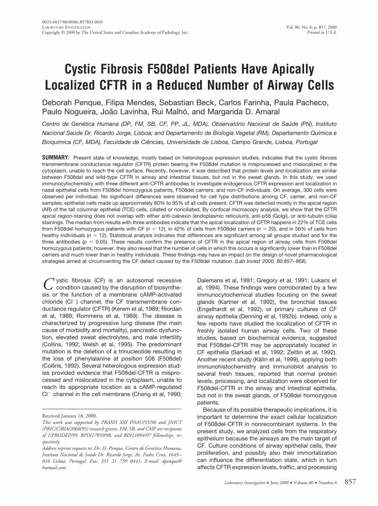
Nasal brushing samples from non–CF individuals (n 55), F508del-carriers (n 5 5), and patients with CF whowere homozygous for F508del (n 5 5) were May-Grunwald-Giemsa (MGG)-stained (see “Materials andMethods” section) to evaluate the cellular types recov-ered. Nasal brushing samples contained three majortypes of epithelial cells: ciliated cells (CC), nonciliatedcells (NC), including secretory, and basal cells (BC), aswell as inflammatory cells (I) and a small group ofnonidentified cells (NI), as shown in Figure 1A for anon–CF subject. Samples from the patients with CFand the carriers (not shown) had similar morphologicappearances.
The epithelial nature of cellular types was confirmedin all cases by immunostaining with anti-cytokeratinAb (Fig. 1B). Epithelial cells accounted for approxi-mately 80% to 95% of all cells (Fig. 1C), and ciliatedcells were the predominant class (37%–42%). None ofthe differences observed for cell type distributionsamong the non–CF, carrier, and CF samples weresignificant.
Specificity of CFTR Antibodies
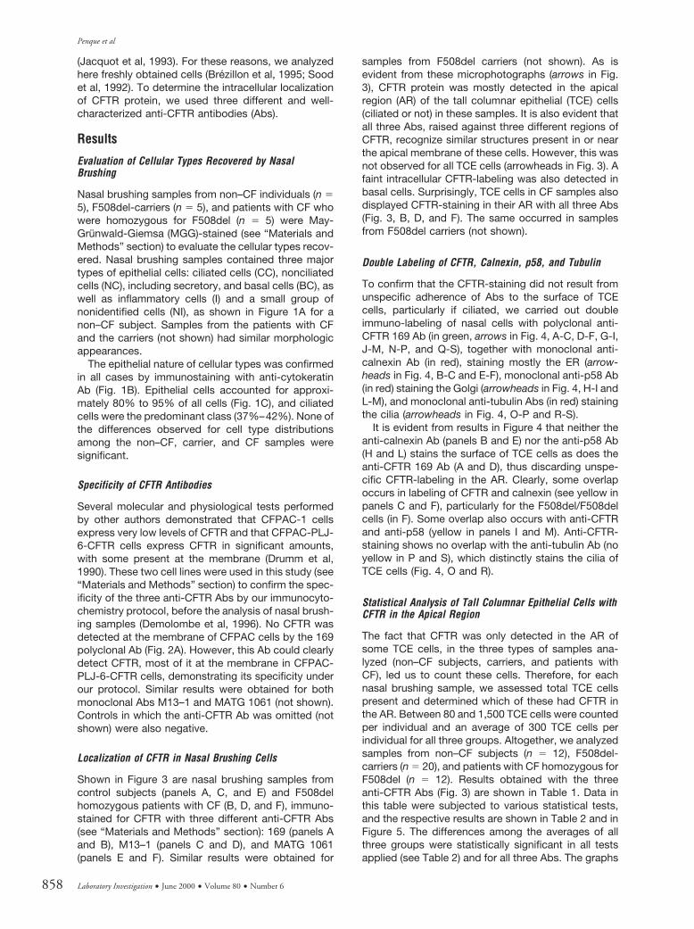
Several molecular and physiological tests performedby other authors demonstrated that CFPAC-1 cellsexpress very low levels of CFTR and that CFPAC-PLJ-6-CFTR cells express CFTR in significant amounts,with some present at the membrane (Drumm et al,1990). These two cell lines were used in this study (see“Materials and Methods” section) to confirm the spec-ificity of the three anti-CFTR Abs by our immunocyto-chemistry protocol, before the analysis of nasal brush-ing samples (Demolombe et al, 1996). No CFTR wasdetected at the membrane of CFPAC cells by the 169polyclonal Ab (Fig. 2A). However, this Ab could clearlydetect CFTR, most of it at the membrane in CFPAC-PLJ-6-CFTR cells, demonstrating its specificity underour protocol. Similar results were obtained for bothmonoclonal Abs M13–1 and MATG 1061 (not shown).Controls in which the anti-CFTR Ab was omitted (notshown) were also negative.
Localization of CFTR in Nasal Brushing Cells
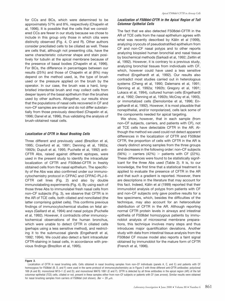
Shown in Figure 3 are nasal brushing samples fromcontrol subjects (panels A, C, and E) and F508delhomozygous patients with CF (B, D, and F), immuno-stained for CFTR with three different anti-CFTR Abs(see “Materials and Methods” section): 169 (panels Aand B), M13–1 (panels C and D), and MATG 1061(panels E and F). Similar results were obtained for
samples from F508del carriers (not shown). As isevident from these microphotographs (arrows in Fig.3), CFTR protein was mostly detected in the apicalregion (AR) of the tall columnar epithelial (TCE) cells(ciliated or not) in these samples. It is also evident thatall three Abs, raised against three different regions ofCFTR, recognize similar structures present in or nearthe apical membrane of these cells. However, this wasnot observed for all TCE cells (arrowheads in Fig. 3). Afaint intracellular CFTR-labeling was also detected inbasal cells. Surprisingly, TCE cells in CF samples alsodisplayed CFTR-staining in their AR with all three Abs(Fig. 3, B, D, and F). The same occurred in samplesfrom F508del carriers (not shown).
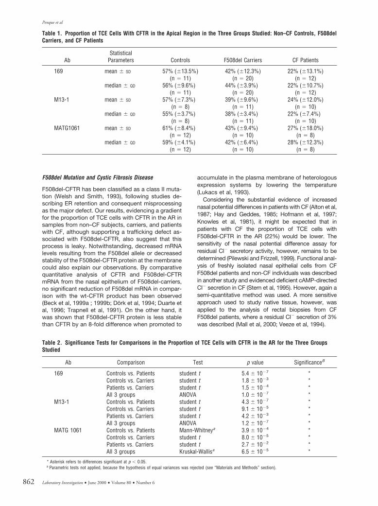
Double Labeling of CFTR, Calnexin, p58, and Tubulin
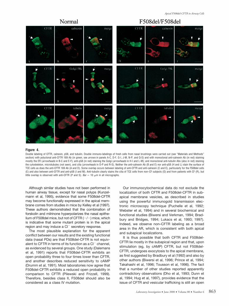
To confirm that the CFTR-staining did not result fromunspecific adherence of Abs to the surface of TCEcells, particularly if ciliated, we carried out doubleimmuno-labeling of nasal cells with polyclonal anti-CFTR 169 Ab (in green, arrows in Fig. 4, A-C, D-F, G-I,J-M, N-P, and Q-S), together with monoclonal anti-calnexin Ab (in red), staining mostly the ER (arrow-heads in Fig. 4, B-C and E-F), monoclonal anti-p58 Ab(in red) staining the Golgi (arrowheads in Fig. 4, H-I andL-M), and monoclonal anti-tubulin Abs (in red) stainingthe cilia (arrowheads in Fig. 4, O-P and R-S).
It is evident from results in Figure 4 that neither theanti-calnexin Ab (panels B and E) nor the anti-p58 Ab(H and L) stains the surface of TCE cells as does theanti-CFTR 169 Ab (A and D), thus discarding unspe-cific CFTR-labeling in the AR. Clearly, some overlapoccurs in labeling of CFTR and calnexin (see yellow inpanels C and F), particularly for the F508del/F508delcells (in F). Some overlap also occurs with anti-CFTRand anti-p58 (yellow in panels I and M). Anti-CFTR-staining shows no overlap with the anti-tubulin Ab (noyellow in P and S), which distinctly stains the cilia ofTCE cells (Fig. 4, O and R).
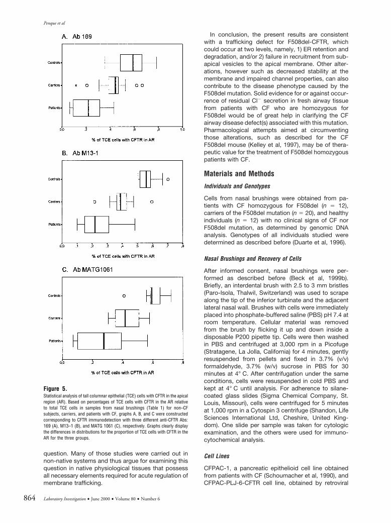
Statistical Analysis of Tall Columnar Epithelial Cells withCFTR in the Apical Region
The fact that CFTR was only detected in the AR ofsome TCE cells, in the three types of samples ana-lyzed (non–CF subjects, carriers, and patients withCF), led us to count these cells. Therefore, for eachnasal brushing sample, we assessed total TCE cellspresent and determined which of these had CFTR inthe AR. Between 80 and 1,500 TCE cells were countedper individual and an average of 300 TCE cells perindividual for all three groups. Altogether, we analyzedsamples from non–CF subjects (n 5 12), F508del-carriers (n 5 20), and patients with CF homozygous forF508del (n 5 12). Results obtained with the threeanti-CFTR Abs (Fig. 3) are shown in Table 1. Data inthis table were subjected to various statistical tests,and the respective results are shown in Table 2 and inFigure 5. The differences among the averages of allthree groups were statistically significant in all testsapplied (see Table 2) and for all three Abs. The graphs
Penque et al
858 Laboratory Investigation • June 2000 • Volume 80 • Number 6

in Figure 5 clearly illustrate distinct distributions for thethree groups. Indeed, a decreasing gradient in theproportion of TCE cells with CFTR in the AR isobserved in the following order: non–CF subjects .F508del carriers . patients with CF.
Discussion
Most of the current knowledge concerning localizationof normal and mutant CFTR results from studiesperformed on heterologous expression systems(Cheng et al, 1990; Dalemans et al, 1991; Denning etal, 1992a; 1992b; Gregory et al, 1991; Lukacs et al,1994), cultured human cells (Denning et al, 1992b;Engelhardt et al, 1992; McCray et al, 1993), or celllines that suffered transformation (Demolombe et al,1996; Engelhardt et al, 1992). Indeed, very few studieshave dealt with this aspect in freshly isolated humanairway cells from patients with CF. This is mainlybecause of the invasiveness and risk of most tech-niques aimed at collecting human cells (eg, biopsies),the small number of cells thus collected, or the limitednumber, poor quality, and nonrepresentative nature ofsamples resulting from surgery (such as nasalpolypectomies or lung transplants). Brushing of therespiratory tract, however, a noninvasive method orig-inally proposed for ciliary studies (Kelsen et al, 1992;Rutland and Cole, 1980; Rutland et al, 1982), allowsthe easy sampling of numerous, representative, well-preserved, and dissociated cells from the superficialmucosa (Bridges et al, 1991; Chapelin et al, 1996;Danel et al, 1996). The present study was thus per-formed on cell samples obtained by nasal brushings ofF508del homozygous patients with CF, F508del carri-ers, and non–CF control subjects.
Characterization of Cell Population in Nasal BrushingSamples
We characterized morphologically the cells recoveredin nasal brushing samples to make sure that ourmethod produced a representative sampling of cells.This analysis revealed that epithelial cells account forapproximately 87% of all cells in the non–CF, carrier,and CF samples, with no significant differencesamong the three groups (Fig. 1C). With results similarto ours, other authors described the percentages ofepithelial cells in nasal brushings of non–CF controlsubjects and patients with CF to be approximately70% to 87% (Bridges et al, 1991), but this decreasedto 65% in patients with chronic rhinitis (Chapelin et al,1996). With regard to specific cell types in nasal brushsamples, our results revealed a similar distributionamong non–CF subjects, carriers, and patients withCF, ie, nonsignificant differences in all comparisons ofspecific cell types among the three groups analyzed(see Fig. 1C). The predominant subtype of epithelialcells was the ciliated cells (CC), comprising on aver-age approximately 39% of all cells, followed by thenonciliated cells (NC) (23%), the basal cells (BC)(25%), and the nonidentified cells (NI) (8%). Otherauthors (Danel et al, 1996) found similar values, except
Figure 1.Evaluation of cellular types recovered by nasal brushing. An example of anMGG-stained nasal brushing sample from a normal individual is shown inpanel A. Three major types of epithelial cells are observed: ciliated (CC),nonciliated, including secretory (NC), and basal cells (BC). Inflammatory cells(I) and a small group of still not identified cells (NI) are also observed. Theepithelial nature of cellular types was confirmed by immunostaining withanti-cytokeratin Ab (see “Materials and Methods” section) and is shown inpanel B. Bar 5 100 mm. The morphologic appearance and cytokeratin-stainingof samples from patients with CF and F508del carriers (not shown) are similarto that shown in panels A and B, respectively. The graph in C summarizescell-type distribution in samples from non–CF subjects, carriers, and patientswith CF.
Apical F508del-CFTR in Airway Cells
Laboratory Investigation • June 2000 • Volume 80 • Number 6 859

Figure 2. TopSpecificity of 169 anti-CFTR Ab. CFPAC-1 (A) and CFPAC-PLJ-6-CFTR (B) cells (see “Materials and Methods” section) were immunostained for CFTR with the 169polyclonal Ab to detect CFTR. No CFTR is observed at the membrane of CFPAC cells (A). In CFPAC-PLJ-6-CFTR cells, however, this Ab clearly detects CFTR at themembrane (see arrows) demonstrating its specificity for CFTR. Similar results were obtained for both monoclonal antibodies M13–1 and MATG 1061 (data notshown). Bar 5 40 mm. Figure 3 legend continued on next page.
Penque et al
860 Laboratory Investigation • June 2000 • Volume 80 • Number 6
for CCs and BCs, which were determined to beapproximately 57% and 8%, respectively (Chapelin etal, 1996). It is possible that the cells that we consid-ered CCs are fewer in our study because we chose toinclude in this group only those in which cilia weredistinctly observed (Fig. 4, O and R). Other authorsconsider preciliated cells to be ciliated as well. Theseare cells that, although not presenting cilia, have thesame characteristic columnar shape and stain posi-tively for tubulin at the apical membrane because ofthe presence of basal bodies (Chapelin et al, 1996).For BCs, the difference in percentages between ourresults (25%) and those of Chapelin et al (8%) maydepend on the method used, ie, the type of brushused or the pressure applied on the brush by theoperator. In our case, the brush was a hard, long-bristled interdental brush and may collect cells fromdeeper layers of the basal epithelium than the brushesused by other authors. Altogether, our results showthat the populations of nasal cells recovered in CF andnon–CF samples are similar and do not differ substan-tially from those previously described (Chapelin et al,1996; Danel et al, 1996), thus validating the analysis ofbrush-obtained nasal cells.
Localization of CFTR in Nasal Brushing Cells
Three different and previously used (Brezillon et al,1995; Crawford et al, 1991; Denning et al, 1992a;1992b; Dupuit et al, 1995; Puchelle et al, 1992) anti-CFTR Abs, raised against different epitopes, wereused in the present study to identify the intracellularlocalization of CFTR and F508del-CFTR in freshlyobtained cells from the nasal epithelium. The specific-ity of the Abs was also confirmed under our immuno-cytochemistry protocol in CFPAC and CFPAC-PLJ-6-CFTR cell lines (Fig. 2) and also by doubleimmunolabeling experiments (Fig. 4). By using each ofthose three Abs to immunolabel fresh nasal cells fromnon–CF subjects (Fig. 3), we observe that CFTR is inthe AR of TCE cells, both ciliated and nonciliated (thelatter comprising goblet cells). This confirms previousfindings of immunocytochemical studies on fetal air-ways (Gaillard et al, 1994) and nasal polyps (Puchelleet al, 1992). However, it contradicts other immunocy-tochemical observations of the human bronchus,which were unable to detect CFTR in ciliated cells(perhaps using a less sensitive method), and restrict-ing it to the submucosal glands (Engelhardt et al,1992; 1994). We could also detect a faint intracellularCFTR-staining in basal cells, in accordance with pre-vious findings (Brezillon et al, 1995).
Localization of F508del-CFTR in the Apical Region of TallColumnar Epithelial Cells
The fact that we also detected F508del-CFTR in theAR of TCE cells from the nasal epithelium agrees withwhat was recently described by Kalin et al (1999),analyzing cryocuts of pseudostratified epithelium fromCF and non-CF nasal polyps and to other reportsanalyzing biopsied human bronchial and nasal tissueby biochemical methods (Sarkadi et al, 1992; Zeitlin etal, 1992). However, it is contrary to a previous study,analyzing bronchial tissues from individuals with CF,which, however could have used a less sensitivemethod (Engelhardt et al, 1992). Our results alsocontradict most studies carried out in heterologoussystems (Cheng et al, 1990; Dalemans et al, 1991;Denning et al, 1992a; 1992b; Gregory et al, 1991;Lukacs et al, 1994), cultured human cells (Engelhardtet al, 1992; Denning et al, 1992b; McCray et al, 1993),or immortalized cells (Demolombe et al, 1996; En-gelhardt et al, 1992). However, it is most plausible thatnonepithelial, and/or nonpolarized, cells lack some ofthe components needed for apical targeting.
We show, however, that in each sample (fromnon–CF subjects, carriers, and patients with CF), notall TCE cells have detectable CFTR in the AR. Al-though the method we used could not detect apparentdifferences in the localization of CFTR and F508delCFTR, the proportion of cells with CFTR in the AR isclearly distinct among samples from the three groupsand decreases in the following order: non–CF subjects(56%) . carriers (42%) . patients with CF (22%).These differences were found to be statistically signif-icant for the three Abs used (Table 2). It is, to ourknowledge, the first time that a statistical treatment isapplied to evaluate the presence of CFTR in the ARand that such a gradient is reported. However, thereare descriptions in the literature that may account forthis fact. Indeed, Kalin et al (1999) reported that theirimmunoblot analysis of polyps from patients with CFand non–CF subjects only gave positive results for afew specimens, which, besides the difficulties of thetechnique, may also account for an heterocellulardistribution of CFTR in the AR. Although reportingnormal CFTR protein levels in airways and intestinalepithelia of F508del homozygous patients by immu-noblot analysis of microsomal membrane prepara-tions, this technique involves many steps and thusintroduces major quantification deviations. Anotherstudy with data from intestinal tissue analysis from theF508del CF mouse model also reports a faint signalobtained by immunoblot for the mature form of CFTR(French et al, 1996).
Figure 3.Localization of CFTR in nasal brushing cells. Cells obtained in nasal brushing samples from non–CF individuals (panels A, C, and E) and patients with CF
homozygous for F508del (B, D, and F) were used in the same protocol of immunocytochemistry as in Figure 2 with three different anti-CFTR antibodies: polyclonal169 (A and B); monoclonal M13–1 (C and D); and monoclonal MATG 1061 (E and F). CFTR is detected by all three antibodies in the apical region (AR) of the tallcolumnar epithelial (TCE) cells, ciliated or not, present in these samples either from non–CF subjects or patients with CF (see arrows). Similar results were obtainedfor nasal brushing samples from carriers of F508del (not shown). Bar 5 20 mm.
Apical F508del-CFTR in Airway Cells
Laboratory Investigation • June 2000 • Volume 80 • Number 6 861
F508del Mutation and Cystic Fibrosis Disease
F508del-CFTR has been classified as a class II muta-tion (Welsh and Smith, 1993), following studies de-scribing ER retention and consequent misprocessingas the major defect. Our results, evidencing a gradientfor the proportion of TCE cells with CFTR in the AR insamples from non–CF subjects, carriers, and patientswith CF, although supporting a trafficking defect as-sociated with F508del-CFTR, also suggest that thisprocess is leaky. Notwithstanding, decreased mRNAlevels resulting from the F508del allele or decreasedstability of the F508del-CFTR protein at the membranecould also explain our observations. By comparativequantitative analysis of CFTR and F508del-CFTRmRNA from the nasal epithelium of F508del-carriers,no significant reduction of F508del mRNA in compar-ison with the wt-CFTR product has been observed(Beck et al, 1999a ; 1999b; Dork et al, 1994; Duarte etal, 1996; Trapnell et al, 1991). On the other hand, itwas shown that F508del-CFTR protein is less stablethan CFTR by an 8-fold difference when promoted to
accumulate in the plasma membrane of heterologousexpression systems by lowering the temperature(Lukacs et al, 1993).
Considering the substantial evidence of increasednasal potential differences in patients with CF (Alton et al,1987; Hay and Geddes, 1985; Hofmann et al, 1997;Knowles et al, 1981), it might be expected that inpatients with CF the proportion of TCE cells withF508del-CFTR in the AR (22%) would be lower. Thesensitivity of the nasal potential difference assay forresidual Cl2 secretory activity, however, remains to bedetermined (Pilewski and Frizzell, 1999). Functional anal-ysis of freshly isolated nasal epithelial cells from CFF508del patients and non-CF individuals was describedin another study and evidenced deficient cAMP-directedCl2 secretion in CF (Stern et al, 1995). However, again asemi-quantitative method was used. A more sensitiveapproach used to study native tissue, however, wasapplied to the analysis of rectal biopsies from CFF508del patients, where a residual Cl2 secretion of 3%was described (Mall et al, 2000; Veeze et al, 1994).
Table 1. Proportion of TCE Cells With CFTR in the Apical Region in the Three Groups Studied: Non–CF Controls, F508delCarriers, and CF Patients
AbStatistical
Parameters Controls F508del Carriers CF Patients
169 mean 6 SD 57% (613.5%) 42% (612.3%) 22% (613.1%)(n 5 11) (n 5 20) (n 5 12)
median 6 QD 56% (69.6%) 44% (63.9%) 22% (610.7%)(n 5 11) (n 5 20) (n 5 12)
M13-1 mean 6 SD 57% (67.3%) 39% (69.6%) 24% (612.0%)(n 5 8) (n 5 11) (n 5 10)
median 6 QD 55% (63.7%) 38% (63.4%) 22% (67.4%)(n 5 8) (n 5 11) (n 5 10)
MATG1061 mean 6 SD 61% (68.4%) 43% (69.4%) 27% (618.0%)(n 5 12) (n 5 10) (n 5 8)
median 6 QD 59% (64.1%) 42% (66.4%) 28% (612.3%)(n 5 12) (n 5 10) (n 5 8)
Table 2. Significance Tests for Comparisons in the Proportion of TCE Cells with CFTR in the AR for the Three GroupsStudied
Ab Comparison Test p value SignificanceB
169 Controls vs. Patients student t 5.4 6 1027 *Controls vs. Carriers student t 1.8 6 1023 *Patients vs. Carriers student t 1.5 6 1024 *All 3 groups ANOVA 1.0 6 1027 *
M13-1 Controls vs. Patients student t 4.3 6 1027 *Controls vs. Carriers student t 9.1 6 1025 *Patients vs. Carriers student t 4.2 6 1023 *All 3 groups ANOVA 1.2 6 1027 *
MATG 1061 Controls vs. Patients Mann-Whitneya 3.9 6 1024 *Controls vs. Carriers student t 8.0 6 1025 *Patients vs. Carriers student t 2.7 6 1022 *All 3 groups Kruskal-Wallisa 6.5 6 1025 *
* Asterisk refers to differences significant at p , 0.05.a Parametric tests not applied, because the hypothesis of equal variances was rejected (see “Materials and Methods” section).
Penque et al
862 Laboratory Investigation • June 2000 • Volume 80 • Number 6
Although similar studies have not been performed inhuman airway tissue, except for nasal polyps (Kunzel-mann et al, 1995), evidence that some F508del-CFTRmay become functionally expressed in the apical mem-brane comes from studies in mice by Kelley et al (1997).These authors demonstrated that the combination offorskolin and milrinone hyperpolarizes the nasal epithe-lium of F508del mice, but not of CFTR (2/2) mice, whichis indicative that some mutant protein is in the apicalregion and may induce a Cl2 secretory response.
The most plausible explanation for the apparentconflict between our results and the existing functionaldata (nasal PDs) are that F508del-CFTR is not equiv-alent to CFTR in terms of its function as a Cl2 channel,as evidenced by several groups. One study (Dalemanset al, 1991) reports that F508del-CFTR exhibits anopen probability three to four times lower than CFTR,and another describes reduced sensitivity to cAMP(Drumm et al, 1991). Most laboratories now agree thatF508del-CFTR exhibits a reduced open probability incomparison to CFTR (Pilewski and Frizzell, 1999).Therefore, besides class II, F508del should also beconsidered as a class IV mutation.
Our immunocytochemical data do not exclude thelocalization of both CFTR and F508del-CFTR in sub-apical membrane vesicles, as described in studiesusing the powerful immunogold transmission elec-tronic microscopy technique (Puchelle et al, 1992;Webster et al, 1994) and in several biochemical andfunctional studies (Biwersi and Verkman, 1994; Brad-bury and Bridges, 1994; Lukacs et al, 1993; 1997).Indeed, we observe non–CFTR labeling as a broadarea in the AR, which is consistent with both apicaland subapical localizations.
It is thus possible that both CFTR and F508del-CFTR lie mostly in the subapical region and that, uponstimulation (eg, by cAMP) CFTR, but not F508del-CFTR, undergoes exocytosis to the apical membrane,as first suggested by Bradbury et al (1992) and also byother authors (Biwersi et al, 1996; Prince et al, 1994;Takahashi et al, 1996; Tousson et al, 1996). The factthat a number of other studies reported apparentlycontradictory observations (Dho et al, 1993; Dunn etal, 1994; Hug et al, 1997), provides evidence that theissue of CFTR and vesicular trafficking is still an open
Figure 4.Double labeling of CFTR, calnexin, p58, and tubulin. Double immuno-labelings of fresh cells from nasal brushings were carried out (see “Materials and Methods”section) with polyclonal anti-CFTR 169 Ab (in green, see arrows in panels A-C, D-F, G-I, J-M, N-P, and Q-S) and with monoclonal anti-calnexin Ab (in red) stainingmostly the ER (arrowheads in B-C and E-F); anti-p58 (in red) staining the Golgi (arrowheads in H-I and L-M); and monoclonal anti-tubulin Abs (also in red) stainingthe cytoskeleton, microtubules (not seen), and cilia (arrowheads in O-P and R-S). Neither the anti-calnexin Ab (B and E) nor anti-p58 (H and L) stain the surface ofTCE cells as does the anti-CFTR 169 Ab (A and D). Some overlap occurs between labeling of anti-CFTR and anti-calnexin (C and E), particularly for the F508del cells(E) and also between anti-CFTR and anti-p58 (I and M). Anti-tubulin clearly stains the cilia of TCE cells from non–CF subjects (O) and from patients with CF (R), butlittle overlap is observed with anti-CFTR (P and S). Bar 5 10 mm in all micrographs.
Apical F508del-CFTR in Airway Cells
Laboratory Investigation • June 2000 • Volume 80 • Number 6 863
question. Many of those studies were carried out innon-native systems and thus argue for examining thisquestion in native physiological tissues that possessall necessary elements required for acute regulation ofmembrane trafficking.
In conclusion, the present results are consistentwith a trafficking defect for F508del-CFTR, whichcould occur at two levels, namely, 1) ER retention anddegradation, and/or 2) failure in recruitment from sub-apical vesicles to the apical membrane. Other alter-ations, however such as decreased stability at themembrane and impaired channel properties, can alsocontribute to the disease phenotype caused by theF508del mutation. Solid evidence for or against occur-rence of residual Cl2 secretion in fresh airway tissuefrom patients with CF who are homozygous forF508del would be of great help in clarifying the CFairway disease defect(s) associated with this mutation.Pharmacological attempts aimed at circumventingthose alterations, such as described for the CFF508del mouse (Kelley et al, 1997), may be of thera-peutic value for the treatment of F508del homozygouspatients with CF.
Materials and Methods
Individuals and Genotypes
Cells from nasal brushings were obtained from pa-tients with CF homozygous for F508del (n 5 12),carriers of the F508del mutation (n 5 20), and healthyindividuals (n 5 12) with no clinical signs of CF norF508del mutation, as determined by genomic DNAanalysis. Genotypes of all individuals studied weredetermined as described before (Duarte et al, 1996).
Nasal Brushings and Recovery of Cells
After informed consent, nasal brushings were per-formed as described before (Beck et al, 1999b).Briefly, an interdental brush with 2.5 to 3 mm bristles(Paro-Isola, Thalwil, Switzerland) was used to scrapealong the tip of the inferior turbinate and the adjacentlateral nasal wall. Brushes with cells were immediatelyplaced into phosphate-buffered saline (PBS) pH 7.4 atroom temperature. Cellular material was removedfrom the brush by flicking it up and down inside adisposable P200 pipette tip. Cells were then washedin PBS and centrifuged at 3,000 rpm in a Picofuge(Stratagene, La Jolla, California) for 4 minutes, gentlyresuspended from pellets and fixed in 3.7% (v/v)formaldehyde, 3.7% (w/v) sucrose in PBS for 30minutes at 4° C. After centrifugation under the sameconditions, cells were resuspended in cold PBS andkept at 4° C until analysis. For adherence to silane-coated glass slides (Sigma Chemical Company, St.Louis, Missouri), cells were centrifuged for 5 minutesat 1,000 rpm in a Cytospin 3 centrifuge (Shandon, LifeSciences International Ltd, Cheshire, United King-dom). One slide per sample was taken for cytologicexamination, and the others were used for immuno-cytochemical analysis.
Cell Lines
CFPAC-1, a pancreatic epithelioid cell line obtainedfrom patients with CF (Schoumacher et al, 1990), andCFPAC-PLJ-6-CFTR cell line, obtained by retroviral
Figure 5.Statistical analysis of tall columnar epithelial (TCE) cells with CFTR in the apicalregion (AR). Based on percentages of TCE cells with CFTR in the AR relativeto total TCE cells in samples from nasal brushings (Table 1) for non–CFsubjects, carriers, and patients with CF, graphs A, B, and C were constructedcorresponding to CFTR immunodetection with three different anti-CFTR Abs:169 (A), M13–1 (B), and MATG 1061 (C), respectively. Graphs clearly displaythe differences in distributions for the proportion of TCE cells with CFTR in theAR for the three groups.
Penque et al
864 Laboratory Investigation • June 2000 • Volume 80 • Number 6
stable transfection of CFPAC-1 with the full-length,wild-type CFTR cDNA (Drumm et al, 1990), were usedas controls of our immunocytochemical protocol. Bothcell lines were cultivated as previously described, afterseeding onto 8-well chamber slides (Nalgenunc Inter-national, Rochester, New York) at a density of 104
cells/well and analyzed 2 days after.
Evaluation of Cellular Types Recovered by NasalBrushing
The freshly isolated human cells recovered from nasalbrushings and spread on silane glass slides werestained by the May-Grunwald-Giemsa (MGG) method(Dacie and Lewis, 1984). After 5 minutes fixing inmethanol, slides were immersed for 5 minutes inMay-Grunwald’s standard stain (Fluka Chemie AG,Buchs, Switzerland), freshly diluted with an equalvolume of phosphate buffer pH 6.8, and then, withoutwashing, immersed for 10 to 15 minutes in Giemsastain (Merck Diagnostica, Darmstadt, Germany) di-luted with nine volumes of phosphate buffer pH 6.8.After 3 to 4 rapid washes in phosphate buffer pH 6.8and 2 to 5 minutes in water, slides were mounted withEntellan (Merck), covered with glass coverslips, anddried for at least 1 hour before analysis. Samples onslides were evaluated for cell differential count andmorphology on a conventional light microscope(Zeiss, Jena, Germany) at a 1003 magnification underoil immersion. Epithelial cells were classified into threemajor categories (ciliated, nonciliated, and basal) onthe basis of described criteria (Danel et al, 1996).Ciliated cells had tall columnar shapes with distinctcilia, nonciliated cells, including secretory goblet cells,had similar shape but no cilia, and basal cells weresmaller, with dense, round nuclei, strongly stainedcytoplasms, and a high nuclear-to-cytoplasmic ratio.Inflammatory cells, ie, lymphocytes, neutrophils, eo-sinophils, and macrophages, were all included in thesame group. Other cells that did not meet any of thesecriteria were called “not identified” (NI).
Antibodies
All Abs were diluted in 0.5% (w/v) BSA in PBS. Thefollowing anti-CFTR Abs, previously shown to recog-nize CFTR, were used: polyclonal 169, raised againstthe peptide comprising amino acids (aa) 724–746, inexon 13 of the R-domain of CFTR (Crawford et al,1991), diluted 1:100; monoclonal M13–1 (Genzyme,Cambridge, Massachusetts), against aa 729–736 ofthe R-domain (Denning et al, 1992a; 1992b; Gregoryet al, 1990), 1:20; and monoclonal MATG 1061 (Trans-gene, Strasbourg, France) (Brezillon et al, 1995; Pu-chelle et al, 1992), against aa 503–515 of NBF1, 1:20.The epithelial nature of cells was determined by usingAb anti-cytokeratin (Clones AE1/AE3; Boehringer,Mannheim, Germany), 1:1500. Ciliary structures weretubulin-stained with a 1:1 mixture of two monoclonalAbs against a- and b-tubulin (Amersham PharmaciaBiotech AB, Uppsala, Sweden), 1:100. Monoclonalanti-calnexin Ab AF8 (Hochstenbach et al, 1992), 1:60,
was used to stain the endoplasmic reticulum. Mono-clonal anti-p58 Ab (Sigma), 1:60, was used to stain theGolgi apparatus (Bloom and Brashear, 1989). Second-ary Abs were FITC-conjugated, anti-rabbit IgG (Amer-sham), 1:50 (for 169 Ab), and FITC-conjugated anti-mouse IgG (Boehringer), 1:80 (for mAbs M13–1 andMATG 1061); TRITC-conjugated, anti-mouse IgG (Sig-ma), 1:600 (for anti-tubulin, anti-calnexin, and anti-p58Abs).
Immunocytochemistry
Unless otherwise indicated, all incubations were keptat room temperature and all solutions in PBS. Afterfixation (see above), cells on slides were rinsed for 5minutes with cold PBS and incubated in methanol for5 minutes at 220° C, followed by two washes withPBS and incubation in 0.25% (v/v) Triton X-100 for 10minutes, for cell permeabilization. Nonspecific stain-ing was prevented by blocking with 1% (w/v) BSA for30 minutes and incubation with one of the anti-CFTRAbs (169, M13–1, or MATG) overnight at 4° C. Afterthree 5-minute washes with PBS, cells were incubatedwith the respective secondary Ab for 30 minutes andwashed as above. For double labeling experiments,fixing, permeabilization, and blocking were performedas above, followed by incubation with the anti-CFTR169 Ab overnight at 4° C, and after three 5-minutewashes, with the other primary Ab (anti-calnexin,anti-tubulins, or anti-p58) for 1 hour at 37° C, followedby the same washing procedure. Reactions with thetwo secondary antibodies were carried out consecu-tively for 45 minutes each, with three 10-minutewashes with PBS in between. Slides were mountedwith Vectashield (Vector Laboratories, Inc., Burlin-game, California), containing DAPI (4, 6-diamino-2-phenylindole, Sigma) for nuclei staining, and coveredwith a glass coverslip. Immunofluorescence stainingwas observed and collected on an Axioskop fluores-cence microscope (Zeiss) with the Power Gene 810/Probe and CGH software system (PSI, Chester, UnitedKingdom). Confocal images were obtained in a MRC-600 confocal imaging system (Bio-Rad, Hercules, Cal-ifornia).
Statistical Analysis
Data are summarised by the mean 6 standard devia-tion (SD) and by median 6 quartile deviation (QD). Thehypothesis of equal variances between the threegroups analyzed (non–CF subjects, F508del-carriers,and patients with CF) was tested using the Levene’stest for each analysis, and there was no evidence toreject it (p . 0.05), except between distributionsrelative to patients and non–CF subjects with AbMATG 1061 (p 5 0.027). Statistical significance com-parisons for samples with equal variances were madeusing the parametric Student’s t test for two unpairedsamples and the parametric one-way ANOVA for threeindependent grouped samples (Sokal and Rohlf,1981). For samples with unequal variances, we usedthe nonparametric Mann-Whitney U test (Wilcoxon) for
Apical F508del-CFTR in Airway Cells
Laboratory Investigation • June 2000 • Volume 80 • Number 6 865
two independent samples and the nonparametricKruskal-Wallis-test, for three independent groupedsamples (Sokal and Rohlf, 1981). Coefficients with a pvalue less than 0.05 were considered to be statisticallysignificant. The SPSS for Windows software (SPSSInc., Chicago, Illinois) was used for all statistical cal-culations.
Acknowledgements
We wish to thank CF doctors, the National CysticFibrosis Association (ANFQ, Lisboa, Portugal), pa-tients with CF and their families for their cooperation,Prof. W Guggino (Baltimore, Maryland) for 169 Ab,Transgene (Strasbourg, France) for MATG1061 Ab, Dr.M. B. Brenner (Boston, Massachusetts) for anti-calnexin AF8 Ab, Dr. D. Escande (Nantes, France) forCFPAC-1 and CFPAC-PLJ-6-CFTR cell lines, andPedro Loureiro and Barbara Marques for technicalassistance.
ReferencesAlton EW, Hay JG, Munro C, and Geddes DM (1987).Measurement of nasal potential difference in adult cysticfibrosis, Young’s syndrome, and bronchiectasis. Thorax 42:815–817.
Beck S, Kuehr J, Schutz VV, Seydewitz HH, Brandis M,Greger R, and Kunzelmann K (1999a). Lack of correlationbetween CFTR expression, CFTR Cl- currents, amiloride-sensitive Na1 conductance, and cystic fibrosis phenotype.Pediatr Pulmonol 27:251–259.
Beck S, Penque D, Garcia S, Gomes A, Farinha C, Mata L,Gulbenkian S, Gil-Ferreira K, Duarte A, Pacheco P, Barreto C,Lopes B, Cavaco J, Lavinha J, and Amaral MD (1999b).Cystic fibrosis patients with the 3272–26A–.G mutationhave mild disease, leaky alternative mRNA splicing, andCFTR protein at the cell membrane. Hum Mutat 14:133–144.
Beck S, Penque D, Lavinha J, and Amaral MD (1999c).Relative quantification of normal CFTR mRNA in nasal epi-thelial cells of patients with the splicing mutation 3272–26A.G and mild clinical phenotype. Pediatr Pulmonol S19:186.
Biwersi J, Emans N, and Verkman AS (1996). Cystic fibrosistransmembrane conductance regulator activation stimulatesendosome fusion in vivo. Proc Natl Acad Sci USA 93:12484–12489.
Biwersi J and Verkman AS (1994). Functional CFTR in endo-somal compartment of CFTR-expressing fibroblasts and T84cells. Am J Physiol 266:C149–C156.
Bloom GS and Brashear TA (1989). A novel 58-kDa proteinassociates with the Golgi apparatus and microtubules. J BiolChem 264:16083–16092.
Bradbury NA and Bridges RJ (1994). Role of membranetrafficking in plasma membrane solute transport. Am JPhysiol 267:C1–C24.
Bradbury NA, Jilling T, Berta G, Sorscher EJ, Bridges RJ, andKirk KL (1992). Regulation of plasma membrane recycling byCFTR. Science 256:530–532.
Brezillon S, Dupuit F, Hinnrasky J, Marchand V, Kalin N,Tummler B, and Puchelle E (1995). Decreased expression of
the CFTR protein in remodeled human nasal epithelium fromnon-cystic fibrosis patients. Lab Invest 72:191–200.
Bridges MA, Walker DC, and Davidson AG (1991). Cysticfibrosis and control nasal epithelial cells harvested by abrushing procedure. In Vitro Cell Dev Biol 27A:684–686.
Chapelin C, Coste A, Gilain L, Poron F, Verra F, and EscudierE (1996). Modified epithelial cell distribution in chronic air-ways inflammation. Eur Respir J 9:2474–2478.
Cheng SH, Gregory RJ, Marshall J, Paul S, Souza DW, WhiteGA, O’Riordan CR, and Smith AE (1990). Defective intracel-lular transport and processing of CFTR is the molecular basisof most cystic fibrosis. Cell 63:827–834.
Collins FS (1992). Cystic fibrosis: Molecular biology andtherapeutic implications. Science 256:774–779.
Crawford I, Maloney PC, Zeitlin PL, Guggino WB, Hyde SC,Turley H, Gatter KC, Harris A, and Higgins CF (1991).Immunocytochemical localization of the cystic fibrosis geneproduct CFTR. Proc Natl Acad Sci USA 88:9262–9266.
Dacie JV and Lewis SM (1984). Practical haematology, 6thed. Edinburgh: Churchill Livingstone, 1–608.
Dalemans W, Barbry P, Champigny G, Jallat S, Dott K, DreyerD, Crystal RG, Pavirani A, Lecocq JP, and Lazdunski M(1991). Altered chloride ion channel kinetics associated withthe delta F508 cystic fibrosis mutation. Nature 354:526–528.
Danel C, Erzurum SC, McElvaney NG, and Crystal RG (1996).Quantitative assessment of the epithelial and inflammatorycell populations in large airways of normals and individualswith cystic fibrosis. Am J Respir Crit Care Med 153:362–368.
Demolombe S, Baro I, Bebok Z, Clancy JP, Sorscher EJ,Thomas-Soumarmon A, Pavirani A, and Escande D (1996). Amethod for the rapid detection of recombinant CFTR duringgene therapy in cystic fibrosis. Gene Ther 3:685–694.
Denning GM, Ostedgaard LS, Cheng SH, Smith AE, andWelsh MJ (1992a). Localization of cystic fibrosis transmem-brane conductance regulator in chloride secretory epithelia.J Clin Invest 89:339–349.
Denning GM, Ostedgaard LS, and Welsh MJ (1992b). Abnor-mal localization of cystic fibrosis transmembrane conduc-tance regulator in primary cultures of cystic fibrosis airwayepithelia. J Cell Biol 118:551–559.
Dho S, Grinstein S, and Foskett JK (1993). Plasma membranerecycling in CFTR-expressing CHO cells. Biochim BiophysActa 1225:78–82.
Dork T, Will K, Grade K, Krawczak M, and Tummler B (1994).A 32-bp deletion (2991del32) in the cystic fibrosis geneassociated with CFTR mRNA reduction. Hum Mutat 4:65–70.
Drumm ML, Pope HA, Cliff WH, Rommens JM, Marvin SA,Tsui LC, Collins FS, Frizzell RA, and Wilson JM (1990).Correction of the cystic fibrosis defect in vitro by retrovirus-mediated gene transfer. Cell 62:1227–1233.
Drumm ML, Wilkinson DJ, Smit LS, Worrell RT, Strong TV,Frizzell RA, Dawson DC, and Collins FS (1991). Chlorideconductance expressed by delta F508 and other mutantCFTRs in Xenopus oocytes. Science 254:1797–1799.
Duarte A, Amaral M, Barreto C, Pacheco P, and Lavinha J(1996). Complex cystic fibrosis allele R334W-R1158X resultsin reduced levels of correctly processed mRNA in a pancre-atic sufficient patient. Hum Mutat 8:134–139.
Penque et al
866 Laboratory Investigation • June 2000 • Volume 80 • Number 6
Dunn KW, Park J, Semrad CE, Gelman DL, Shevell T, andMcGraw TE (1994). Regulation of endocytic trafficking andacidification are independent of the cystic fibrosis transmem-brane regulator. J Biol Chem 269:5336–5345.
Dupuit F, Kalin N, Brezillon S, Hinnrasky J, Tummler B, andPuchelle E (1995). CFTR and differentiation markers expres-sion in non-CF and delta F 508 homozygous CF nasalepithelium. J Clin Invest 96:1601–1611.
Engelhardt JF, Yankaskas JR, Ernst SA, Yang Y, Marino CR,Boucher RC, Cohn JA, and Wilson JM (1992). Submucosalglands are the predominant site of CFTR expression in thehuman bronchus. Nat Genet 2:240–248.
Engelhardt JF, Zepeda M, Cohn JA, Yankaskas JR, andWilson JM (1994). Expression of the cystic fibrosis gene inadult human lung. J Clin Invest 93:737–749.
French PJ, van Doorninck JH, Peters RH, Verbeek E, AmeenNA, Marino CR, de Jonge HR, Bijman J, and Scholte BJ(1996). A delta F508 mutation in mouse cystic fibrosis trans-membrane conductance regulator results in a temperature-sensitive processing defect in vivo. J Clin Invest 98:1304–1312.
Gaillard D, Ruocco S, Lallemand A, Dalemans W, HinnraskyJ, and Puchelle E (1994). Immunohistochemical localizationof cystic fibrosis transmembrane conductance regulator inhuman fetal airway and digestive mucosa. Pediatr Res 36:137–143.
Gregory RJ, Cheng SH, Rich DP, Marshall J, Paul S, Hehir K,Ostedgaard L, Klinger KW, Welsh MJ, and Smith AE (1990).Expression and characterization of the cystic fibrosis trans-membrane conductance regulator. Nature 347:382–386.
Gregory RJ, Rich DP, Cheng SH, Souza DW, Paul S, Man-avalan P, Anderson MP, Welsh MJ, and Smith AE (1991).Maturation and function of cystic fibrosis transmembraneconductance regulator variants bearing mutations in putativenucleotide-binding domains 1 and 2. Mol Cell Biol 11:3886–3893.
Hay JG and Geddes DM (1985). Transepithelial potentialdifference in cystic fibrosis. Thorax 40:493–496.
Hochstenbach F, David V, Watkins S, and Brenner MB(1992). Endoplasmic reticulum resident protein of 90 kilodal-tons associates with the T- and B-cell antigen receptors andmajor histocompatibility complex antigens during their as-sembly. Proc Natl Acad Sci USA 89:4734–4738.
Hofmann T, Bohmer O, Huls G, Terbrack HG, Bittner P,Klingmuller V, Heerd E, and Lindemann H (1997). Conven-tional and modified nasal potential-difference measurementin cystic fibrosis. Am J Respir Crit Care Med 155:1908–1913.
Hug MJ, Thiele IE, and Greger R (1997). The role of exocy-tosis in the activation of the chloride conductance in Chinesehamster ovary cells (CHO) stably expressing CFTR. PflugersArch 434:779–784.
Jacquot J, Puchelle E, Hinnrasky J, Fuchey C, Bettinger C,Spilmont C, Bonnet N, Dieterle A, Dreyer D, and Pavirani A(1993). Localization of the cystic fibrosis transmembraneconductance regulator in airway secretory glands. Eur RespirJ 6:169–176.
Kalin N, Claass A, Sommer M, Puchelle E, and Tummler B(1999). DeltaF508 CFTR protein expression in tissues frompatients with cystic fibrosis. J Clin Invest 103:1379–1389.
Kartner N, Augustinas O, Jensen TJ, Naismith AL, andRiordan JR (1992). Mislocalization of delta F508 CFTR incystic fibrosis sweat gland. Nat Genet 1:321–327.
Kelley TJ, Thomas K, Milgram LJ, and Drumm ML (1997). Invivo activation of the cystic fibrosis transmembrane conduc-tance regulator mutant deltaF508 in murine nasal epithelium.Proc Natl Acad Sci USA 94:2604–2608.
Kelsen SG, Mardini IA, Zhou S, Benovic JL, and Higgins NC(1992). A technique to harvest viable tracheobronchial epi-thelial cells from living human donors. Am J Respir Cell MolBiol 7:66–72.
Kerem B, Rommens JM, Buchanan JA, Markiewicz D, CoxTK, Chakravarti A, Buchwald M, and Tsui LC (1989). Identi-fication of the cystic fibrosis gene: Genetic analysis. Science245:1073–1080.
Knowles M, Gatzy J, and Boucher R (1981). Increasedbioelectric potential difference across respiratory epithelia incystic fibrosis. N Engl J Med 305:1489–1495.
Kunzelmann K, Kathofer S, and Greger R (1995). Na1 and Cl-conductances in airway epithelial cells: Increased Na1 con-ductance in cystic fibrosis. Pflugers Arch 431:1–9.
Lukacs GL, Chang XB, Bear C, Kartner N, Mohamed A,Riordan JR, and Grinstein S (1993). The delta F508 mutationdecreases the stability of cystic fibrosis transmembraneconductance regulator in the plasma membrane. Determina-tion of functional half-lives on transfected cells. J Biol Chem268:21592–21598.
Lukacs GL, Mohamed A, Kartner N, Chang XB, Riordan JR,and Grinstein S (1994). Conformational maturation of CFTRbut not its mutant counterpart (delta F508) occurs in theendoplasmic reticulum and requires ATP. EMBO J 13:6076–6086.
Lukacs GL, Segal G, Kartner N, Grinstein S, and Zhang F(1997). Constitutive internalization of cystic fibrosis trans-membrane conductance regulator occurs via clathrin-dependent endocytosis and is regulated by protein phos-phorylation. Biochem J 328:353–361.
Mall M, Wiessner A, Seydewitz HH, Kuehr J, Brandis M,Greger R, and Kunzelmann K (2000). Defective cholinergicCl2 secretion and detection of K1 secretion in rectal biopsiesfrom cystic fibrosis patients. Am J Physiol 278:617–624.
McCray PB Jr, Bettencourt JD, Bastacky J, Denning GM, andWelsh MJ (1993). Expression of CFTR and a cAMP-stimulated chloride secretory current in cultured human fetalalveolar epithelial cells. Am J Respir Cell Mol Biol 9:578–585.
Pilewski JM and Frizzell RA (1999). Role of CFTR in airwaydisease. Physiol Rev 79:S215–S255.
Prince LS, Workman RBJ, and Marchase RB (1994). Rapidendocytosis of the cystic fibrosis transmembrane conduc-tance regulator chloride channel. Proc Natl Acad Sci USA91:5192–5196.
Puchelle E, Gaillard D, Ploton D, Hinnrasky J, Fuchey C,Boutterin MC, Jacquot J, Dreyer D, Pavirani A, and DalemansW (1992). Differential localization of the cystic fibrosis trans-membrane conductance regulator in normal and cystic fibro-sis airway epithelium. Am J Respir Cell Mol Biol 7:485–491.
Riordan JR, Rommens JM, Kerem B, Alon N, Rozmahel R,Grzelczak Z, Zielenski J, Lok S, Plavsic N, and Chou JL(1989). Identification of the cystic fibrosis gene: Cloning andcharacterization of complementary DNA. Science 245:1066–1073.
Apical F508del-CFTR in Airway Cells
Laboratory Investigation • June 2000 • Volume 80 • Number 6 867
Rommens JM, Iannuzzi MC, Kerem B, Drumm ML, Melmer G,Dean M, Rozmahel R, Cole JL, Kennedy D, Hidaka N, ZsigaM, Buchwald M, Riordan JR, Tsui L-C, and Collins FS (1989).Identification of the cystic fibrosis gene: Chromosome walk-ing and jumping. Science 245:1059–1065.
Rutland J and Cole PJ (1980). Non-invasive sampling of nasalcilia for measurement of beat frequency and study of ultra-structure. Lancet 2:564–565.
Rutland J, Dewar A, Cox T, and Cole P (1982). Nasal brushingfor the study of ciliary ultrastructure. J Clin Pathol 35:357–359.
Sarkadi B, Bauzon D, Huckle WR, Earp HS, Berry A, Such-indran H, Price EM, Olson JC, Boucher RC, and ScarboroughGA (1992). Biochemical characterization of the cystic fibrosistransmembrane conductance regulator in normal and cysticfibrosis epithelial cells. J Biol Chem 267:2087–2095.
Schoumacher RA, Ram J, Iannuzzi MC, Bradbury NA, Wal-lace RW, Hon CT, Kelly DR, Schmid SM, Gelder FB, andRado TA (1990). A cystic fibrosis pancreatic adenocarcinomacell line. Proc Natl Acad Sci USA 87:4012–4016.
Sokal R and Rohlf F (1981). Biometry: The principles andpractice of statistics in biological research, 2nd ed. NewYork: Freeman & Company, 1–829.
Sood R, Bear C, Auerbach W, Reyes E, Jensen T, Kartner N,Riordan JR, and Buchwald M (1992). Regulation of CFTRexpression and function during differentiation of intestinalepithelial cells. EMBO J 11:2487–2494.
Stern M, Munkonge FM, Caplen NJ, Sorgi F, Huang L,Geddes DM, and Alton EW (1995). Quantitative fluorescencemeasurements of chloride secretion in native airway epithe-lium from CF and non-CF subjects. Gene Ther 2:766–774.
Takahashi A, Watkins SC, Howard M, and Frizzell RA (1996).CFTR-dependent membrane insertion is linked to stimulationof the CFTR chloride conductance. Am J Physiol 271:C1887–C1894.
Tousson A, Fuller CM, and Benos DJ (1996). Apical recruit-ment of CFTR in T-84 cells is dependent on cAMP andmicrotubules but not Ca21 or microfilaments. J Cell Sci109:1325–1334.
Trapnell BC, Zeitlin PL, Chu CS, Yoshimura K, Nakamura H,Guggino WB, Bargon J, Banks TC, Dalemans W, and PaviraniA (1991). Down-regulation of cystic fibrosis gene mRNAtranscript levels and induction of the cystic fibrosis chloridesecretory phenotype in epithelial cells by phorbol ester. J BiolChem 266:10319–10323.
Veeze HJ, Halley DJ, Bijman J, de Jongste JC, de Jonge HR,and Sinaasappel M (1994). Determinants of mild clinicalsymptoms in cystic fibrosis patients. Residual chloride se-cretion measured in rectal biopsies in relation to the geno-type. J Clin Invest 93:461–466.
Webster P, Vanacore L, Nairn AC, and Marino CR (1994).Subcellular localization of CFTR to endosomes in a ductalepithelium. Am J Physiol 267:C340–C348.
Welsh M, Tsui L-C, Boat TF, and Beaudet AL (1995). Cysticfibrosis. In: Scriver CR, Beaudet AL, Sly WS, and Valle, D,editors. The metabolic and molecular basis of inheriteddiseases, 7th ed. New York: Mc-Graw-Hill, Inc., 3799–3876.
Welsh MJ and Smith AE (1993). Molecular mechanisms ofCFTR chloride channel dysfunction in cystic fibrosis. Cell73:1251–1254.
Zeitlin PL, Crawford I, Lu L, Woel S, Cohen ME, Donowitz M,Montrose MH, Hamosh A, Cutting GR, and Gruenert D(1992). CFTR protein expression in primary and culturedepithelia. Proc Natl Acad Sci USA 89:344–347.
Penque et al
868 Laboratory Investigation • June 2000 • Volume 80 • Number 6