meconium ileus in cystic fibrosis
TRANSCRIPT
⁎ Corresponding auE-mail address: m
http://dx.doi.org/10.11569-1993© 2017 E
www.elsevier.com/locate/jcfJournal of Cystic Fibrosis 16 (2017) S32–S39
Meconium ileus in Cystic Fibrosis
Meghana Sathe a,⁎, Roderick Houwen b
a UT Southwestern Medical Center, Dallas, TX, United Statesb University Medical Centre of Utrecht, Netherlands
Received 26 April 2017; revised 28 June 2017; accepted 29 June 2017
Abstract
Meconium ileus (MI) is often the first manifestation of cystic fibrosis (CF) and occurs in approximately 20% of patients diagnosed with CF.This article reviews the pathophysiology of MI and its clinical presentation. It focuses on the medical and surgical management emphasizing theimportance of nutrition and a multidisciplinary approach to improve both short-term and long-term outcomes for CF patients with MI.© 2017 European Cystic Fibrosis Society. Published by Elsevier B.V. All rights reserved.
Keywords: Meconium ileus; Meconium peritonitis; Gastrografin; Cystic fibrosis
1. Background
Meconium ileus (MI) is often the first manifestation of cysticfibrosis (CF) and occurs in approximately 20% of patientsdiagnosed with CF. It can present in two forms, simple MI andcomplexMI. In simpleMI, viscid meconium physically obstructsthe terminal ileum and the small intestine proximal to theobstruction, then becomes dilated with additional meconium, gas,and fluid [1]. In complex MI, the meconium-distended segmentsof ileum can give way to complications like prenatal volvulus,ischemic necrosis, intestinal atresia, or perforation and extrusionof the meconium into the peritoneum. The timing of perforationmay contribute to the outcome. If perforation occurs earlier inutero, there is the possibility of reabsorption of some meconiumin the peritoneum before delivery, leaving only a small number ofcalcifications. If necrosis and perforation occur close to delivery,meconium peritonitis is more likely to be seen. Meconium mayalso become (partly) encapsulated: giant cystic meconiumperitonitis (GCMP) [2–3]. This condition may present with a
[email protected] (M. Sathe).
016/j.jcf.2017.06.007uropean Cystic Fibrosis Society. Published by Elsevier B.V. A
palpable mass on exam in a patient with MI or meconiumperitonitis [1]. Both simple and complex MI occur with similarfrequency in patients with CF.
Understanding the pathophysiology of MI has been greatlyadvanced by the development of mouse models as well as thecystic fibrosis transmembrane conductance regulator (CFTR)knock out ferret and pig, which have 100% penetration of MI.Within the small intestine, CFTR is responsible for both Cl− andHCO3– excretion. It is the HCO3– that plays an integral role inchelating Ca2+ associated with the tight matrix of normallyexocytosed mucins within the gut lumen to form normal, loosewell-hydrated mucus [4]. Abnormal CFTR results in abnormalHCO3– secretion, thus decreasing luminal pH. This creates anacidic and dehydrated environment in which the tight matrix ofexocytosed mucins are not disrupted appropriately resulting inthick, dehydrated mucus [4]. The abnormally acidic luminalenvironment also promotes the presence of elevated levels ofstool albumin, increased mineral content, and protein-boundcarbohydrates. These combine with the dense mucus to formviscid meconium that eventually leads to physical obstruction ofthe terminal ileum (TI) [1,4–6]. This process might result in thetwo outcomes described above, either simple or complex MI.
MI is most commonly associated with class I-III CFTRmutations. Specifically, MI is associated with F508del, G542X,W1282X, R553X, and G551D [7]. Based on the United States CF
ll rights reserved.
S33M. Sathe, R. Houwen / Journal of Cystic Fibrosis 16 (2017) S32–S39
Patient Registry 2010 database, a patient with two copies of themost common F508del mutation has a 24.9% risk of presentingwith MI [7]. A patient with a F508del paired with anothermutation has a 16.9% risk of presenting with MI [7]. The risk of apatient with two other CFTR mutations presenting with MI is12.5% [7]. Evaluation for risk of MI in CF monozygotic anddizygotic twins also found an increased concordance in monozy-gotic twins [8]. In addition, it has been noted that in families wherethere is a history of an infant with MI, the chance that a subsequentchild with CF will develop MI is greater than expected [9–10].These findings point to the involvement of modifier genes in thedevelopment of MI. However, although multiple modifier genesthat enhance or reduce the risk of MI have been identified in singlestudies, the ability to replicate these results has been limited [8–13].
With better understanding of CF and earlier recognition of MI,there have been significant improvements in the morbidity andmortality associated with MI. Mortality rates, as high as 33% inthe 1960s for both simple and complex MI, have significantlyimproved [14]. Today, early and late survival rates for bothsimple and complexMI are consistently reported over 80% [1–2].
The first significant improvement came in 1948 when HiattandWilson described a method for intraoperative disimpaction ofMI with saline [15]. This was followed in 1969 with Noblett'sdescription of using hyperosmolar Gastrografin enemas in themanagement of simple MI [16]. At the time, this novel techniqueaided in the minimization of small bowel and colonic resection insimple MI. In addition, the development of a comprehensivemultidisciplinary approach to the care of CF infants by pediatricsurgeons, neonatologists, pulmonologists, respiratory therapists,gastroenterologists, and dieticians has not only led to furtherimprovements in short-term morbidity, but has also allowed fornegligible long-term differences in regards to nutritional status,pulmonary function, and infection status for CF patients withhistory of both simple and complex MI [17–19].
2. Clinical presentation and differential diagnosis
In the modern era of highly sensitive medical technology,MI and meconium peritonitis are often detected prenatally bythe presence of hyperechoic bowel or peritoneal calcificationson ultrasound (US) [20]. However, hyperechoic bowel can alsobe seen inmany other disease processes [20]. A 16 year review ofUS experience in Brittany, France from 1992 to 2007 found thatonly 7.6% of 289 patients who had abnormal bowel detectedby prenatal US actually had CF. Nevertheless, if hyperechoicbowel is detected, it is imperative to assess the fetus's risk of CF[20].
If not identified prenatally, the most common clinicalpresentation of MI is intestinal obstruction, which is oftenseen within hours of birth. When feedings are initiated, biliousemesis occurs with or without abdominal distention. The infantwith meconium peritonitis often presents with additional signs ofabdominal tenderness, fever, and shock [1]. Other infants mayonly display concern for MI with the delayed passage ofmeconium. In all of these cases, the differential diagnosisincludes not only CF withMI, but also other conditions includingmeconium plug (hard stool covered with mucous that is difficult
to pass), Hirschsprung's disease, jejunoileal atresia, volvulus, andbowel perforation.
3. Diagnostic workup
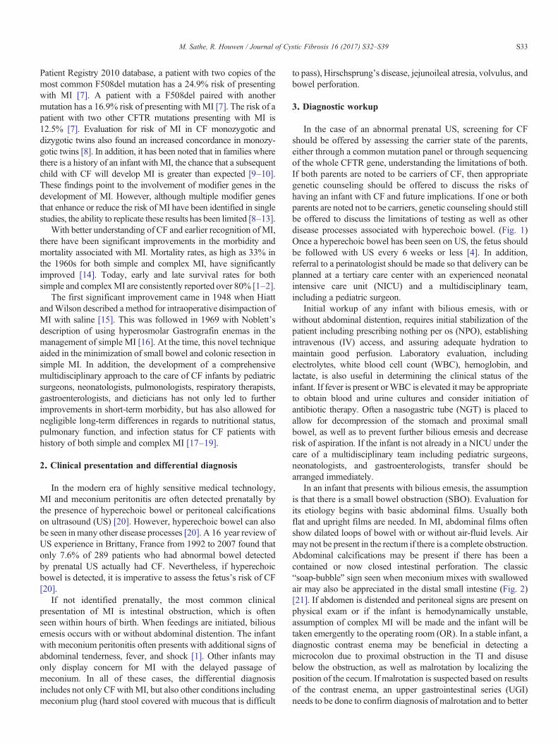
In the case of an abnormal prenatal US, screening for CFshould be offered by assessing the carrier state of the parents,either through a common mutation panel or through sequencingof the whole CFTR gene, understanding the limitations of both.If both parents are noted to be carriers of CF, then appropriategenetic counseling should be offered to discuss the risks ofhaving an infant with CF and future implications. If one or bothparents are noted not to be carriers, genetic counseling should stillbe offered to discuss the limitations of testing as well as otherdisease processes associated with hyperechoic bowel. (Fig. 1)Once a hyperechoic bowel has been seen on US, the fetus shouldbe followed with US every 6 weeks or less [4]. In addition,referral to a perinatologist should be made so that delivery can beplanned at a tertiary care center with an experienced neonatalintensive care unit (NICU) and a multidisciplinary team,including a pediatric surgeon.
Initial workup of any infant with bilious emesis, with orwithout abdominal distention, requires initial stabilization of thepatient including prescribing nothing per os (NPO), establishingintravenous (IV) access, and assuring adequate hydration tomaintain good perfusion. Laboratory evaluation, includingelectrolytes, white blood cell count (WBC), hemoglobin, andlactate, is also useful in determining the clinical status of theinfant. If fever is present orWBC is elevated it may be appropriateto obtain blood and urine cultures and consider initiation ofantibiotic therapy. Often a nasogastric tube (NGT) is placed toallow for decompression of the stomach and proximal smallbowel, as well as to prevent further bilious emesis and decreaserisk of aspiration. If the infant is not already in a NICU under thecare of a multidisciplinary team including pediatric surgeons,neonatologists, and gastroenterologists, transfer should bearranged immediately.
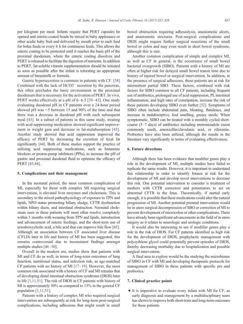
In an infant that presents with bilious emesis, the assumptionis that there is a small bowel obstruction (SBO). Evaluation forits etiology begins with basic abdominal films. Usually bothflat and upright films are needed. In MI, abdominal films oftenshow dilated loops of bowel with or without air-fluid levels. Airmay not be present in the rectum if there is a complete obstruction.Abdominal calcifications may be present if there has been acontained or now closed intestinal perforation. The classic“soap-bubble” sign seen when meconium mixes with swallowedair may also be appreciated in the distal small intestine (Fig. 2)[21]. If abdomen is distended and peritoneal signs are present onphysical exam or if the infant is hemodynamically unstable,assumption of complex MI will be made and the infant will betaken emergently to the operating room (OR). In a stable infant, adiagnostic contrast enema may be beneficial in detecting amicrocolon due to proximal obstruction in the TI and disusebelow the obstruction, as well as malrotation by localizing theposition of the cecum. If malrotation is suspected based on resultsof the contrast enema, an upper gastrointestinal series (UGI)needs to be done to confirm diagnosis of malrotation and to better
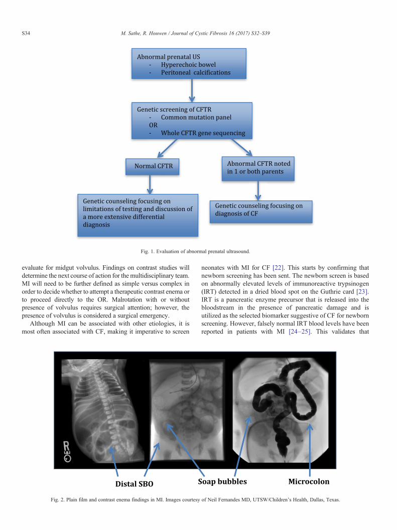
Fig. 1. Evaluation of abnormal prenatal ultrasound.
S34 M. Sathe, R. Houwen / Journal of Cystic Fibrosis 16 (2017) S32–S39
evaluate for midgut volvulus. Findings on contrast studies willdetermine the next course of action for the multidisciplinary team.MI will need to be further defined as simple versus complex inorder to decide whether to attempt a therapeutic contrast enema orto proceed directly to the OR. Malrotation with or withoutpresence of volvulus requires surgical attention; however, thepresence of volvulus is considered a surgical emergency.
Although MI can be associated with other etiologies, it ismost often associated with CF, making it imperative to screen
Fig. 2. Plain film and contrast enema findings in MI. Images courtesy
neonates with MI for CF [22]. This starts by confirming thatnewborn screening has been sent. The newborn screen is basedon abnormally elevated levels of immunoreactive trypsinogen(IRT) detected in a dried blood spot on the Guthrie card [23].IRT is a pancreatic enzyme precursor that is released into thebloodstream in the presence of pancreatic damage and isutilized as the selected biomarker suggestive of CF for newbornscreening. However, falsely normal IRT blood levels have beenreported in patients with MI [24–25]. This validates that
of Neil Fernandes MD, UTSW/Children's Health, Dallas, Texas.
S35M. Sathe, R. Houwen / Journal of Cystic Fibrosis 16 (2017) S32–S39
confirmation of the diagnosis of CF must be made using thegold standard sweat test or genetics [24–25]. A sweat chloridetest can be done as early as 48 h after delivery if the infant isnot edematous and is well hydrated, otherwise, genetic testingcan be sought. However, most infants with MI are too small,malnourished, edematous, or critically ill for immediate sweattesting and will need to have primary genetic testing.
4. Routine management
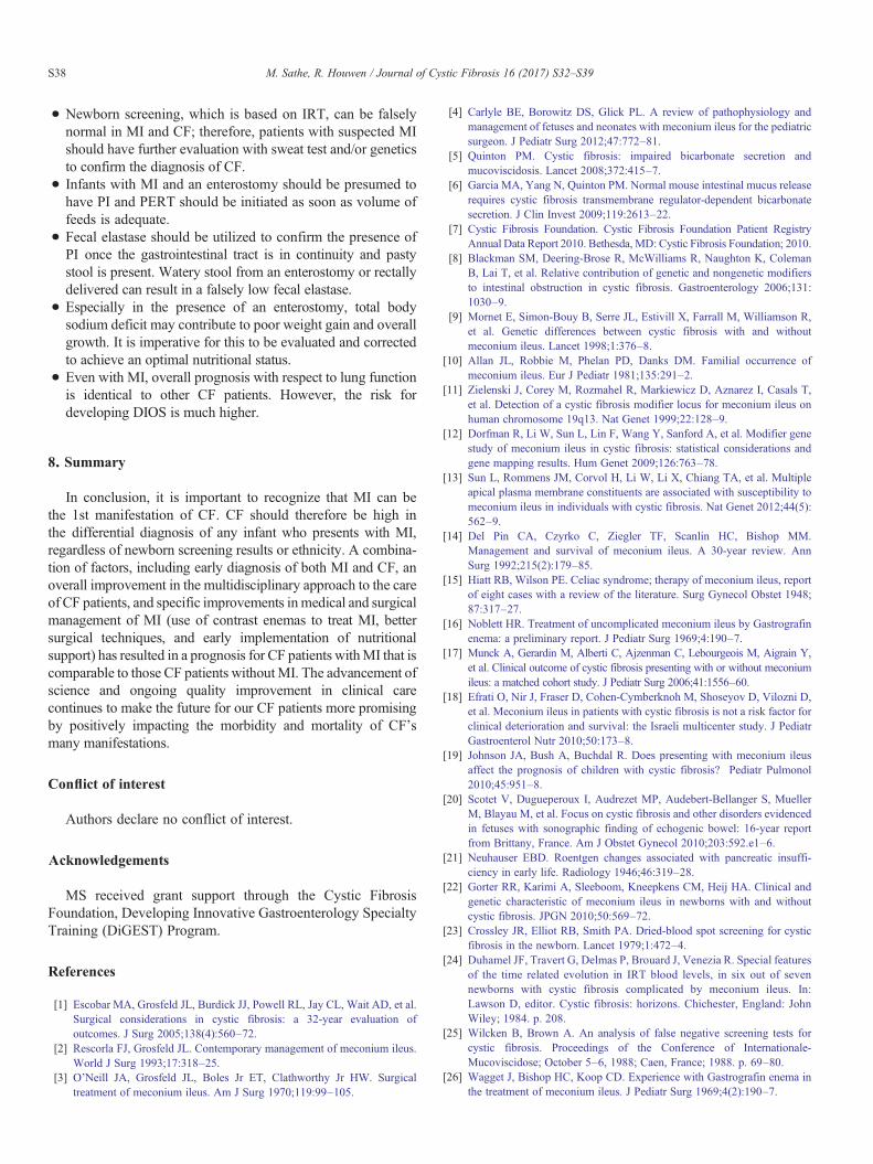
Medical management of simple MI has been developedaround the use of hyperosmolar enemas given under fluoroscopicguidance to ensure that the solution refluxes into/reaches theTI [16]. This technique was first described in 1969 by Noblettutilizing Gastrografin, which contains diatrizoate meglumine,0.1% polysorbate 80 (Tween 80), and 37% organically boundiodine amounting to a solution with 1940 mOsm/L. [16] Themechanism of action is to act as a direct solvent and shift fluidinto the bowel lumen instead of competing with the intracellularspace surrounding the mucosa [26]. When utilizing such ahyperosmolar agent, adequate hydration (150 mL/kg/dayminimum) via an IV line is imperative to avoid hypovolemiathat can lead to shock and end-organ damage includingnecrotizing enterocolitis [1,4,27]. Presence of an IV line isalso essential to respond appropriately to complications suchas need for emergent surgical intervention. Most commonlywith Gastrografin, a ¼–½ dilution with water is infused underlow hydrostatic pressure through a catheter under fluoroscopythrough the rectum until the terminal ileum is reached [4].Perforation risk has been described as low as 2.7% and as highas 23% [28–30]. If hyperosmolar enema is unsuccessful, thensurgical intervention is pursued (Fig. 3).
The primary surgical intervention is disimpaction of themeconium by irrigating the obstructed TI with warm saline orGastrografin in the OR [15,27]. The subsequent creation of acontinuous enterostomy such as the Bishop-Koop is preferredto allow for ongoing irrigation, if necessary, of the TI post-operatively. It also reduces the risk of postoperative complica-tions of a primary enterostomy which can be as high as 30%[33–34]. Risks of creating an enterostomy include high outputlosses, especially of sodium. Bowel resection is reserved for morecomplex cases and is dependent on the extent of bowel injury.It can vary from simple resection with primary anastomosisto simple enterostomy with anastomosis several months laterrequiring minimal resection of small bowel (b10 cm) to moreextensive bowel resection, involving both small bowel and colon,or removal of a meconium cyst.
In the case of simple MI, after the successful disimpaction ofMI with the use of a hyperosmolar enema, Noblett recommendedusing 5 mL of 10% N-acetyl cysteine (NAC) via NGT every 6 hto dissolve meconium proximal to TI [16]. However, due to thepotential risk of aspiration and chemical pneumonitis frominstilling NAC via NGT, currently it is more common torecommend the use of warm saline rectally every 12–24 h forseveral days to encourage further evacuation of meconium. Thisis often done in conjunction with serial abdominal films toevaluate for the presence of remaining meconium.
Although many different solutions have been used throughtime clinically and studied in animal models, Gastrografincontinues to have the most efficacious outcomes of success bothin man and mice [27]. Despite this evidence, Omnipaque (240–350 mOsm/kg water) and Cysto-contray II (400 mOsm/kgwater), which are much less toxic and less hyperosmolar thanGastrografin, are currently the more commonly utilized agents, atleast in the United States [1,27]. Rate of success of contrastenemas rangeswidely from approximately 30–80% [1–2,31–32].Our own anecdotal experience is similar, ranging from 30 to 50%.
Often infants with MI, especially those with complex MIrequiring surgical intervention, will initially require TPN andlipids to support their growth. If possible, the lipid choiceshould favor an anti-inflammatory profile, including mediumchain triglycerides (MCT) and fish oil to minimize risk ofcholestasis. In Europe, SMOF lipid, which meets these criteria,is readily available and used routinely in these infants.
As soon as possible after resolution of MI, the GI tractshould be utilized for enteral nutrition and feeds should beadvanced as tolerated. Often in the presence of complex MI,due to poor perfusion of the intestine and necrosis during theinitial period of small bowel obstruction, peritonitis, and potentialsurgical complications, infants benefit from amino acid-based orprotein hydrolysate formulas rich in MCT oil (ideally 50%).Amino acid-based and protein hydrolysate formulas allow foreasier digestion of proteins, while medium chain triglycerides(MCT) are directly absorbed into portal blood, bypassing thelymphatic system. As the infant continues to recover from theinitial insult of MI, and GI tolerance improves, breastmilk ortraditional formulas can be introduced.
Of note, in the presence of an enterostomy, excess intestinalsodium losses may result in total body sodium deficit [4]. Totalbody sodium deficit will often contribute to metabolic acidosis,poor weight gain, and will negatively impact growth [35–36]. Itcan be evaluated by checking spot urine sodium: creatinineratio with goal for adequate growth being 17–52 mmol/mmol[35] or measuring sodium excretion in a spot urine. If either islow, treatment is supplementation of sodium until values are inthe middle of the normal range. This can be done by increasingsodium in TPN or, if infant is tolerating enteral feeds, increasingsodium supplementation in enteral feeds.
The majority of infants with CF and MI, whether simple orcomplex, have pancreatic insufficiency (PI). Confirmation of PI ismost efficiently and effectively done by obtaining a fecal elastase.However, the fecal elastase sample should be collected fromrectally delivered, formed stools and not from an enterostomy.Watery stools from either an enterostomy or rectally delivered canresult in falsely low fecal elastase values. Therefore, in infantswith MI and an enterostomy, PI should be assumed and fecalelastase collected at a later date after the gastrointestinal tract isback in continuity and stools are at least of a pasty consistency toconfirm diagnosis. Once the infant is able to take a minimalamount of formula or breastmilk by mouth or feeding tube, PERTshould be initiated at 2000–4000 lipase units per 120 mL offormula [37]. PERT contains lipase to digest to lipids, amylase todigest carbohydrates, and protease to digest proteins. PERTdosing is based on lipase units per mL of formula or lipase units
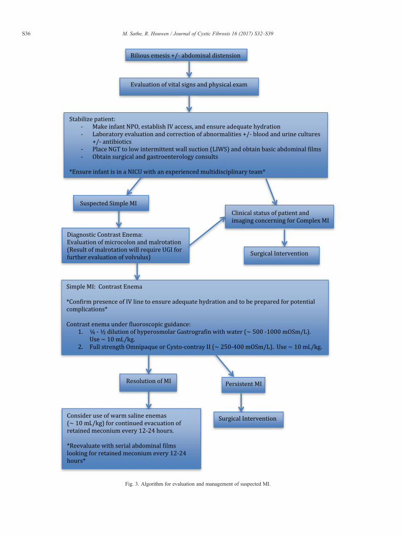
Fig. 3. Algorithm for evaluation and management of suspected MI.
S36 M. Sathe, R. Houwen / Journal of Cystic Fibrosis 16 (2017) S32–S39
S37M. Sathe, R. Houwen / Journal of Cystic Fibrosis 16 (2017) S32–S39
per kilogram per meal. Infants require that PERT capsules beopened and enteric-coated beads be mixed in baby applesauce orother acidic baby fruit and delivered by mouth prior to each feedfor bolus feeds or every 4 h for continuous feeds. This allows theenteric coating to be protected until it reaches the basic pH of theproximal duodenum, where the enteric coating dissolves andPERT is released to facilitate the digestion of nutrients. In additionto PERT, fat-soluble vitamin supplementation should be initiatedas soon as possible after the infant is tolerating an appropriateamount of breastmilk or formula.
Gastric hypersecretion is common in patients with CF. [38]Combined with the lack of HCO3– secretion by the pancreas,this often precludes the basic environment in the proximalduodenum that is necessary for the activation of PERT. In vivo,PERT works effectively at a pH of 6–6.5 [39–42]. One studyevaluating duodenal pH in CF patients over a 24-hour periodshowed pH was b5 between 15 and 90% of the time and thatthere was a decrease in duodenal pH with each subsequentmeal [43]. In a subset of patients in this same study, treatingwith acid suppressing medication showed significant improve-ment in weight gain and decrease in fat-malabsorption [43].Another study showed that acid suppression improved theefficacy of PERT by decreasing the excretion of fecal fatsignificantly [44]. Both of these studies support the practice ofutilizing acid suppressing medications, such as histamineblockers or proton-pump inhibitors (PPIs), to increase the pH ofgastric and proximal duodenal fluid to optimize the efficacy ofPERT [43,44].
5. Complications and their management
In the neonatal period, the most common complication ofMI, especially for those with complex MI requiring surgicalintervention, is elevated liver enzymes and cholestasis. This issecondary to the mixed pathophysiology of exposure to TPN andlipids, NPO status promoting biliary sludge, CFTR dysfunctionwithin biliary ducts, and intestinal obstruction. Neonatal chole-stasis seen in these patients will most often resolve completelywithin 3 months with weaning from TPN and lipids, introductionand advancement of enteric feedings, and the short-term use ofursodeoxycholic acid, a bile acid that can improve bile flow [45].Although an association between CF associated liver disease(CFLD) later in life and history of MI has been suggested, thisremains controversial due to inconsistent findings amongstmultiple studies [46–50].
Overall in the modern era, studies show that patients withMI and CF do as well, in terms of long-term outcomes of lungfunction, nutritional status, and infection risk, as age-matchedCF patients with no history of MI [17–19]. However, the mostcommon risk associated with a history of CF andMI remains thatof developing distal intestinal obstruction syndrome (DIOS) laterin life [1,11,51]. The risk of DIOS in CF patients with history ofMI is approximately 50% as compared to 15% in the general CFpopulation [1,11,51].
Patients with a history of complex MI who required surgicalintervention are subsequently at risk for long-term post-surgicalcomplications, including adhesions that might result in small
bowel obstruction requiring adhesiolysis, anastomotic ulcers,and anastomotic strictures. Post-surgical complications andDIOS might require further surgical resections of the smallbowel or colon and may even result in short bowel syndrome,although this is rare.
Another common complication of simple and complex MI,as well as CF in general, is the occurrence of small bowelbacterial overgrowth (SIBO). Patients with a history of MI areoften at higher risk for delayed small bowel transit time due tohistory of injured bowel or surgical intervention. In addition, inthe presence of surgical adhesions, these patients are at risk forintermittent partial SBO. These factors, combined with riskfactors for SIBO common to all CF patients, including frequentuse of antibiotics, prolonged use of acid suppression, PI, intestinalinflammation, and high rates of constipation, increase the risk ofthese patients developing SIBO even further [52]. Symptoms ofSIBO often include abdominal pain, bloating, flatulence, andincrease in malabsorptive, foul smelling, greasy stools. Whensymptomatic, SIBO can be treated with a monthly cycled short-course (5–7 days) of antibiotics including metronizadole (mostcommonly used), amoxicillin/clavulanic acid, or rifaximin.Probiotics have also been utilized, although the results in theliterature vary significantly in terms of evaluating effectiveness.
6. Future directions
Although there has been evidence that modifier genes play arole in the development of MI, multiple studies have failed toreplicate the same results. However, it is important to understandthis relationship in order to identify fetuses at risk for thedevelopment of MI and develop novel interventions to decreasethis risk. One potential intervention to consider is treatment ofmothers with CFTR correctors and potentiators to act onabnormal CFTR in the fetus. Theoretically, if started earlyenough, it is possible that these medications could alter the naturalprogression of MI. Another potential prenatal intervention wouldbe in utero surgical decompression of SBO or correction of MI toprevent development of microcolon or other complications. Therehave already been significant advancements in the field of in uterosurgery as it relates to neurologic and urologic conditions.
It would also be interesting to see if modifier genes play arole in the risk of DIOS. For CF patients identified as high riskfor the development of DIOS, prophylactic management withpolyeythlene glycol could potentially prevent episodes of DIOS,thereby decreasing morbidity due to hospitalization and possiblesurgical intervention.
A final area to explore would be the studying the microbiomeof SIBO in CF with MI and developing therapeutic protocols formanagement of SIBO in these patients with specific pre andprobiotics.
7. Clinical practice points
• It is imperative to evaluate every infant with MI for CF, asearly diagnosis and management by a multidisciplinary teamhas shown to improve both short-term and long-term outcomesfor these patients.
S38 M. Sathe, R. Houwen / Journal of Cystic Fibrosis 16 (2017) S32–S39
• Newborn screening, which is based on IRT, can be falselynormal in MI and CF; therefore, patients with suspected MIshould have further evaluation with sweat test and/or geneticsto confirm the diagnosis of CF.
• Infants with MI and an enterostomy should be presumed tohave PI and PERT should be initiated as soon as volume offeeds is adequate.
• Fecal elastase should be utilized to confirm the presence ofPI once the gastrointestinal tract is in continuity and pastystool is present. Watery stool from an enterostomy or rectallydelivered can result in a falsely low fecal elastase.
• Especially in the presence of an enterostomy, total bodysodium deficit may contribute to poor weight gain and overallgrowth. It is imperative for this to be evaluated and correctedto achieve an optimal nutritional status.
• Even with MI, overall prognosis with respect to lung functionis identical to other CF patients. However, the risk fordeveloping DIOS is much higher.
8. Summary
In conclusion, it is important to recognize that MI can bethe 1st manifestation of CF. CF should therefore be high inthe differential diagnosis of any infant who presents with MI,regardless of newborn screening results or ethnicity. A combina-tion of factors, including early diagnosis of both MI and CF, anoverall improvement in the multidisciplinary approach to the careof CF patients, and specific improvements inmedical and surgicalmanagement of MI (use of contrast enemas to treat MI, bettersurgical techniques, and early implementation of nutritionalsupport) has resulted in a prognosis for CF patients withMI that iscomparable to those CF patients withoutMI. The advancement ofscience and ongoing quality improvement in clinical carecontinues to make the future for our CF patients more promisingby positively impacting the morbidity and mortality of CF'smany manifestations.
Conflict of interest
Authors declare no conflict of interest.
Acknowledgements
MS received grant support through the Cystic FibrosisFoundation, Developing Innovative Gastroenterology SpecialtyTraining (DiGEST) Program.
References
[1] Escobar MA, Grosfeld JL, Burdick JJ, Powell RL, Jay CL, Wait AD, et al.Surgical considerations in cystic fibrosis: a 32-year evaluation ofoutcomes. J Surg 2005;138(4):560–72.
[2] Rescorla FJ, Grosfeld JL. Contemporary management of meconium ileus.World J Surg 1993;17:318–25.
[3] O'Neill JA, Grosfeld JL, Boles Jr ET, Clathworthy Jr HW. Surgicaltreatment of meconium ileus. Am J Surg 1970;119:99–105.
[4] Carlyle BE, Borowitz DS, Glick PL. A review of pathophysiology andmanagement of fetuses and neonates with meconium ileus for the pediatricsurgeon. J Pediatr Surg 2012;47:772–81.
[5] Quinton PM. Cystic fibrosis: impaired bicarbonate secretion andmucoviscidosis. Lancet 2008;372:415–7.
[6] Garcia MA, Yang N, Quinton PM. Normal mouse intestinal mucus releaserequires cystic fibrosis transmembrane regulator-dependent bicarbonatesecretion. J Clin Invest 2009;119:2613–22.
[7] Cystic Fibrosis Foundation. Cystic Fibrosis Foundation Patient RegistryAnnual Data Report 2010. Bethesda, MD: Cystic Fibrosis Foundation; 2010.
[8] Blackman SM, Deering-Brose R, McWilliams R, Naughton K, ColemanB, Lai T, et al. Relative contribution of genetic and nongenetic modifiersto intestinal obstruction in cystic fibrosis. Gastroenterology 2006;131:1030–9.
[9] Mornet E, Simon-Bouy B, Serre JL, Estivill X, Farrall M, Williamson R,et al. Genetic differences between cystic fibrosis with and withoutmeconium ileus. Lancet 1998;1:376–8.
[10] Allan JL, Robbie M, Phelan PD, Danks DM. Familial occurrence ofmeconium ileus. Eur J Pediatr 1981;135:291–2.
[11] Zielenski J, Corey M, Rozmahel R, Markiewicz D, Aznarez I, Casals T,et al. Detection of a cystic fibrosis modifier locus for meconium ileus onhuman chromosome 19q13. Nat Genet 1999;22:128–9.
[12] Dorfman R, Li W, Sun L, Lin F, Wang Y, Sanford A, et al. Modifier genestudy of meconium ileus in cystic fibrosis: statistical considerations andgene mapping results. Hum Genet 2009;126:763–78.
[13] Sun L, Rommens JM, Corvol H, Li W, Li X, Chiang TA, et al. Multipleapical plasma membrane constituents are associated with susceptibility tomeconium ileus in individuals with cystic fibrosis. Nat Genet 2012;44(5):562–9.
[14] Del Pin CA, Czyrko C, Ziegler TF, Scanlin HC, Bishop MM.Management and survival of meconium ileus. A 30-year review. AnnSurg 1992;215(2):179–85.
[15] Hiatt RB, Wilson PE. Celiac syndrome; therapy of meconium ileus, reportof eight cases with a review of the literature. Surg Gynecol Obstet 1948;87:317–27.
[16] Noblett HR. Treatment of uncomplicated meconium ileus by Gastrografinenema: a preliminary report. J Pediatr Surg 1969;4:190–7.
[17] Munck A, Gerardin M, Alberti C, Ajzenman C, Lebourgeois M, Aigrain Y,et al. Clinical outcome of cystic fibrosis presenting with or without meconiumileus: a matched cohort study. J Pediatr Surg 2006;41:1556–60.
[18] Efrati O, Nir J, Fraser D, Cohen-Cymberknoh M, Shoseyov D, Vilozni D,et al. Meconium ileus in patients with cystic fibrosis is not a risk factor forclinical deterioration and survival: the Israeli multicenter study. J PediatrGastroenterol Nutr 2010;50:173–8.
[19] Johnson JA, Bush A, Buchdal R. Does presenting with meconium ileusaffect the prognosis of children with cystic fibrosis? Pediatr Pulmonol2010;45:951–8.
[20] Scotet V, Dugueperoux I, Audrezet MP, Audebert-Bellanger S, MuellerM, Blayau M, et al. Focus on cystic fibrosis and other disorders evidencedin fetuses with sonographic finding of echogenic bowel: 16-year reportfrom Brittany, France. Am J Obstet Gynecol 2010;203:592.e1–6.
[21] Neuhauser EBD. Roentgen changes associated with pancreatic insuffi-ciency in early life. Radiology 1946;46:319–28.
[22] Gorter RR, Karimi A, Sleeboom, Kneepkens CM, Heij HA. Clinical andgenetic characteristic of meconium ileus in newborns with and withoutcystic fibrosis. JPGN 2010;50:569–72.
[23] Crossley JR, Elliot RB, Smith PA. Dried-blood spot screening for cysticfibrosis in the newborn. Lancet 1979;1:472–4.
[24] Duhamel JF, Travert G, Delmas P, Brouard J, Venezia R. Special featuresof the time related evolution in IRT blood levels, in six out of sevennewborns with cystic fibrosis complicated by meconium ileus. In:Lawson D, editor. Cystic fibrosis: horizons. Chichester, England: JohnWiley; 1984. p. 208.
[25] Wilcken B, Brown A. An analysis of false negative screening tests forcystic fibrosis. Proceedings of the Conference of Internationale-Mucoviscidose; October 5–6, 1988; Caen, France; 1988. p. 69–80.
[26] Wagget J, Bishop HC, Koop CD. Experience with Gastrografin enema inthe treatment of meconium ileus. J Pediatr Surg 1969;4(2):190–7.
S39M. Sathe, R. Houwen / Journal of Cystic Fibrosis 16 (2017) S32–S39
[27] Burke MS, Ragi JM, Karamanoukian HL, Kotter M, Brisseau GF, BorowitzDS, et al. New strategies in nonoperative management of meconium ileus. JPediatr Surg 2002;37(5):760–4.
[28] Copeland DR, St. Peter SD, Sharp SW, Islam S, Cuenca A, Tolleson JS,et al. Diminishing role of contrast enema in simple meconium ileus. JPediatr Surg 2009;44:2130–2.
[29] Ein SH, Shandling B, Reilly BJ, Stephens CA. Bowel perforation withnonoperative treatment of meconium ileus. J Pediatr Surg 1987;22(2):146–7.
[30] Caniano DA, Beaver BL. Meconium ileus: a fifteen-year experience withforty-two neonates. Surgery 1987;102(4):699–703.
[31] Mushtaq I, Wright VM, Drake DP, Mearns MB, Woods CB. Meconiumileus secondary to cystic fibrosis. The East London experience. PediatrSurg Int 1998;13(5–6):365–9.
[32] Rowe MI, Furst AJ, Altman DH, Poole CA. The neonatal response toGastrografin enema. Pediatrics 1971;48:29–35.
[33] Karimi A, Gorter RR, Sleeboom C, Kneepkens CM, Heij HA. Issues inthe management of simple and complex meconium ileus. Pediatr Surg Int2011;27(9):963–8.
[34] Jawaheer J, Khalil B, Plummer T, Bianchi A, Morecraft J, Rakoczy G,et al. Primary resection and anastomosis of complicated meconium ileus: asafe procedure? Pediatr Surg Int 2007;23:1091–3.
[35] Coates AJ, Crofton PM, Marshall T. Evaluation of salt supplementation inCF infants. J Cyst Fibros 2009;8(6):382–5.
[36] Bower TR, Pringle KC, Soper RT. Sodium deficit causing decreasedweight gain and metabolic acidosis in infants with ileostomy. J PediatrSurg 1988;23(5):567–72.
[37] Borowitz DS, Robinson M, Rosenfeld KA, Davis SD, Sabadosa KA.Cystic Fibrosis Foundation evidence-based guidelines for management ofinfants with cystic fibrosis. J Pediatr 2009;155:S73–93.
[38] Hyman PE, Everett SL, Harada T. Gastric acid hypersecretion in shortbowel syndrome infants: association with extent of resection and enteralfeeding. J Pediatr Gastroenterol Nutr 1986;5(2):191–7.
[39] Fieker A, Philpott J, Armand M. Enzyme replacement therapy forpancreatic insufficiency: present and future. Clin Exp Gastroenterol 2011;4:55–73.
[40] Borgstroem B. Influence of bile salt, pH, and time on the action ofpancreatic lipase; physiological implications. J Lipid Res 1964;5:522–31.
[41] Armand M, Borel P, Pasquier B, Dubious C, Senft M, Andre M, et al.Physicochemical characteristics of emulsions during fat digestion inhuman stomach and duodenum. Am J Phys 1996;271(1 Pt 1):G172–83.
[42] Carriere F, Grandval P, Renou C, Palomba A, Prieri F, Giallo J, et al.Quantitative study of digestive enzyme secretion and gastrointestinallipolysis in chronic pancreatitis. Clin Gastroenterol Hepatol 2005;3(1):28–38.
[43] Barraclough M, Taylor CJ. Twenty-four hours ambulatory gastric andduodenal pH profiles in cystic fibrosis: effect of duodenal hyperacidity onpancreatic enzyme function and fat absorption. J Pediatr GastroenterolNutr 1996;23(1):45–50.
[44] Vecht J, Symersky T, Lamers CBHW, Masclee AA. Efficacy of lowerthan standard doses of pancreatic enzyme supplementation therapy duringacid inhibition in patient with pancreatic exocrine insufficiency. J ClinGastroenterol 2006;40(8):721–5.
[45] Flass T, Narkewicz MR. Cirrhosis and other liver disease in cystic fibrosis.J Cyst Fibros 2012;12(2):116–24.
[46] Colombo C, Apostolo MG, Ferrari M, Seia M, Genoni S, Giunta A, et al.Analysis of risk factors for the development of liver disease associatedwith cystic fibrosis. J Pediatr 1994;124(3):393–9.
[47] Colombo C, Battezzati PM, Crosignani A, Morabito A, Constantini D,Padoan R, et al. Liver disease in cystic fibrosis: a prospective study onincidence, risk factors, and outcome. Hepatology 2002;36(6):1374–82.
[48] Corbett K, Kelleher S, Rowland M, Daly L, Drumm B, Cranny G, et al.Cystic fibrosis-associated liver disease: a population-based study. J Pediatr2004;145:327–32.
[49] Lamireau T, Monnereau S, Marcotte JE, Marcotte JE, Winnock M,Alvarez F, et al. Epidemiology of liver disease in cystic fibrosis: alongitudinal study. J Hepatol 2004;41(6):920–5.
[50] Leewen L, Magoffin AK, Fitzgerald DA, Cipolli M, Gaspin KJ.Cholestasis and meconium ileus in infants with cystic fibrosis and theirclinical outcomes. Arch Dis Child 2014;99:443–7.
[51] Bali A, Stableforth DE, Asquith P. Prolonged small-intestinal transit timein cystic fibrosis. Br Med J 1983;287:1011–3.
[52] Miazga A, Osinski M, Cichy W, Zaba R. Current views on theetiopathogenesis, clinical manifestation, diagnostics, treatment andcorrelation with other nosological entities of SIBO. Adv Med Sci 2015;60(1):118–24.