current status and future directions in couple therapy
TRANSCRIPT
Ann
u. R
ev. P
sych
ol. 2
006.
57:3
17-3
44. D
ownl
oade
d fr
om a
rjour
nals
.ann
ualre
view
s.or
g


1702 IEEE TRANSACTIONS ON PLASMA SCIENCE, VOL. 36, NO. 4, AUGUST 2008
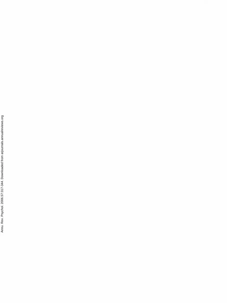
Fig. 4. Electron spots recorded by the beam-profile monitor for laser interac-tion with the 2-mm-diameter gas-jet.
on electron acceleration obtained with the interaction of thefocused laser pulse with 2- and 4-mm He gas-jet, as theseconfigurations gave the most interesting results.
A. Beam-Profile Monitor and Magnetic Spectrometer
The first real-time measurement (useful also for the finetuning of the experiment) was provided by a beam-profilemonitor, made up of a scintillating Lanex plate placed 44 mmbehind the focal point and screened from laser light, X-rays, andlow-energy electrons by a 300-µm Cu foil. With this technique,a pointing stability of less than 100 mrad of the fairly collimatedelectron bunches was revealed by a large number of patterns,some of which are shown in Fig. 4.
It resulted that the mean divergence angle of the centralfeature with single-shot exposition was approximately 30-mradFWHM. Furthermore, some radial structures are shown inFig. 4 (whose patterns varied shot-to-shot), whose nature willbe discussed in Section IV-B.
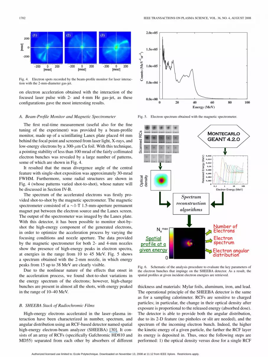
The spectrum of the accelerated electrons was firstly pro-vided shot-to-shot by the magnetic spectrometer. The magneticspectrometer consisted of a ∼1-T 1.5-mm-aperture permanentmagnet put between the electron source and the Lanex screen.The output of the spectrometer was imaged by the Lanex plate.With this detector, it has been possible to monitor shot-by-shot the high-energy component of the generated electrons,in order to optimize the acceleration process by varying thefocusing conditions and nozzle aperture. The data providedby the magnetic spectrometer for both 2- and 4-mm nozzlesshow the presence of high-energy peaks in electron spectra,at energies in the range from 10 to 45 MeV. Fig. 5 showsa spectrum obtained with the 2-mm nozzle, in which energypeaks from 15 up to 38 MeV are clearly visible.
Due to the nonlinear nature of the effects that onset inthe acceleration process, we found shot-to-shot variations inthe energy spectrum of the electrons; however, high-chargebunches are present in almost all the shots, with energy peakedin the range of 10–40 MeV.
B. SHEEBA Stack of Radiochromic Films
High-energy electrons accelerated in the laser–plasma in-teraction have been characterized in number, spectrum, andangular distribution using an RCF-based detector named spatialhigh-energy electron-beam analyzer (SHEEBA) [30]. It con-sists of an array of RCFs (specifically Gafchromic HD810 andMD55) separated from each other by absorbers of different
Fig. 5. Electron spectrum obtained with the magnetic spectrometer.
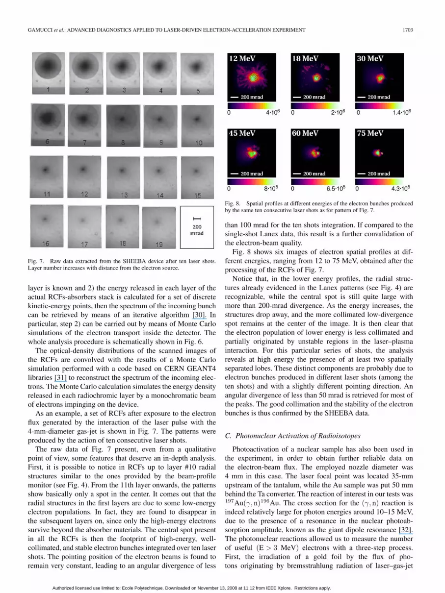
Fig. 6. Schematic of the analysis procedure to evaluate the key parameters ofthe electron bunches that impinge on the SHEEBA detector. As a result, thespatial profiles at given incident electron energies are retrieved.
thickness and materials: Mylar foils, aluminum, iron, and lead.The operational principle of the SHEEBA detector is the sameas for a sampling calorimeter. RCFs are sensitive to chargedparticles; in particular, the change in their optical density afterexposure is proportional to the released energy (absorbed dose).The detector is able to provide both the angular distribution,due to its 2-D feature (no pinholes or slit are needed), and thespectrum of the incoming electron bunch. Indeed, the higherthe kinetic energy of a given particle, the farther the RCF layerits energy is deposited in. Thus, once the following steps areperformed: 1) the optical density versus dose for a single RCF
Authorized licensed use limited to: Ecole Polytechnique. Downloaded on November 13, 2008 at 11:12 from IEEE Xplore. Restrictions apply.
GAMUCCI et al.: ADVANCED DIAGNOSTICS APPLIED TO LASER-DRIVEN ELECTRON-ACCELERATION EXPERIMENT 1703
Fig. 7. Raw data extracted from the SHEEBA device after ten laser shots.Layer number increases with distance from the electron source.
layer is known and 2) the energy released in each layer of theactual RCFs-absorbers stack is calculated for a set of discretekinetic-energy points, then the spectrum of the incoming bunchcan be retrieved by means of an iterative algorithm [30]. Inparticular, step 2) can be carried out by means of Monte Carlosimulations of the electron transport inside the detector. Thewhole analysis procedure is schematically shown in Fig. 6.
The optical-density distributions of the scanned images ofthe RCFs are convolved with the results of a Monte Carlosimulation performed with a code based on CERN GEANT4libraries [31] to reconstruct the spectrum of the incoming elec-trons. The Monte Carlo calculation simulates the energy densityreleased in each radiochromic layer by a monochromatic beamof electrons impinging on the device.
As an example, a set of RCFs after exposure to the electronflux generated by the interaction of the laser pulse with the4-mm-diameter gas-jet is shown in Fig. 7. The patterns wereproduced by the action of ten consecutive laser shots.
The raw data of Fig. 7 present, even from a qualitativepoint of view, some features that deserve an in-depth analysis.First, it is possible to notice in RCFs up to layer #10 radialstructures similar to the ones provided by the beam-profilemonitor (see Fig. 4). From the 11th layer onwards, the patternsshow basically only a spot in the center. It comes out that theradial structures in the first layers are due to some low-energyelectron populations. In fact, they are found to disappear inthe subsequent layers on, since only the high-energy electronssurvive beyond the absorber materials. The central spot presentin all the RCFs is then the footprint of high-energy, well-collimated, and stable electron bunches integrated over ten lasershots. The pointing position of the electron beams is found toremain very constant, leading to an angular divergence of less
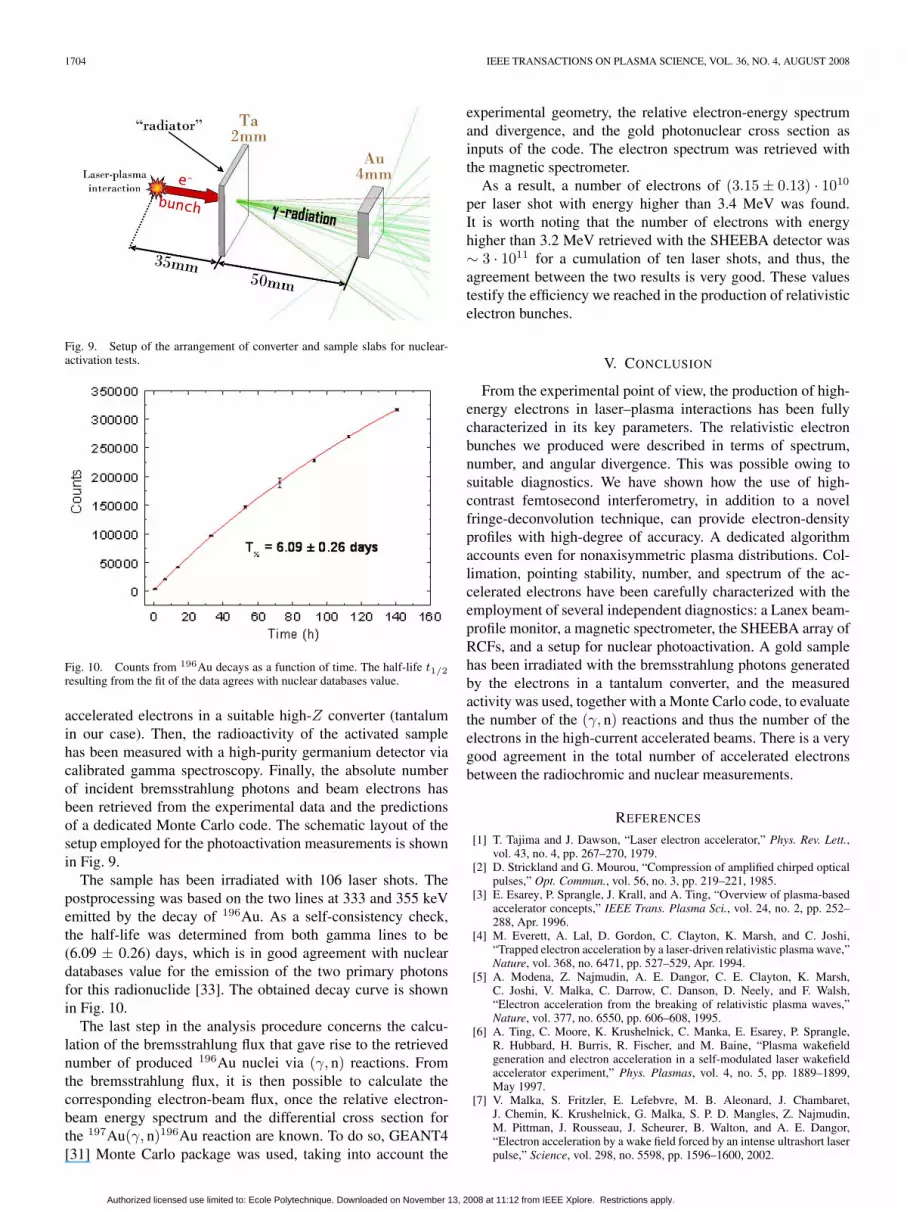
Fig. 8. Spatial profiles at different energies of the electron bunches producedby the same ten consecutive laser shots as for pattern of Fig. 7.
than 100 mrad for the ten shots integration. If compared to thesingle-shot Lanex data, this result is a further convalidation ofthe electron-beam quality.
Fig. 8 shows six images of electron spatial profiles at dif-ferent energies, ranging from 12 to 75 MeV, obtained after theprocessing of the RCFs of Fig. 7.
Notice that, in the lower energy profiles, the radial struc-tures already evidenced in the Lanex patterns (see Fig. 4) arerecognizable, while the central spot is still quite large withmore than 200-mrad divergence. As the energy increases, thestructures drop away, and the more collimated low-divergencespot remains at the center of the image. It is then clear thatthe electron population of lower energy is less collimated andpartially originated by unstable regions in the laser–plasmainteraction. For this particular series of shots, the analysisreveals at high energy the presence of at least two spatiallyseparated lobes. These distinct components are probably due toelectron bunches produced in different laser shots (among theten shots) and with a slightly different pointing direction. Anangular divergence of less than 50 mrad is retrieved for most ofthe peaks. The good collimation and the stability of the electronbunches is thus confirmed by the SHEEBA data.
C. Photonuclear Activation of Radioisotopes
Photoactivation of a nuclear sample has also been used inthe experiment, in order to obtain further reliable data onthe electron-beam flux. The employed nozzle diameter was4 mm in this case. The laser focal point was located 35-mmupstream of the tantalum, while the Au sample was put 50 mmbehind the Ta converter. The reaction of interest in our tests was197Au(γ, n)196Au. The cross section for the (γ, n) reaction isindeed relatively large for photon energies around 10–15 MeV,due to the presence of a resonance in the nuclear photoab-sorption amplitude, known as the giant dipole resonance [32].The photonuclear reactions allowed us to measure the numberof useful (E > 3 MeV) electrons with a three-step process.First, the irradiation of a gold foil by the flux of pho-tons originating by bremsstrahlung radiation of laser–gas-jet
Authorized licensed use limited to: Ecole Polytechnique. Downloaded on November 13, 2008 at 11:12 from IEEE Xplore. Restrictions apply.
1704 IEEE TRANSACTIONS ON PLASMA SCIENCE, VOL. 36, NO. 4, AUGUST 2008
Fig. 9. Setup of the arrangement of converter and sample slabs for nuclear-activation tests.
Fig. 10. Counts from 196Au decays as a function of time. The half-life t1/2resulting from the fit of the data agrees with nuclear databases value.
accelerated electrons in a suitable high-Z converter (tantalumin our case). Then, the radioactivity of the activated samplehas been measured with a high-purity germanium detector viacalibrated gamma spectroscopy. Finally, the absolute numberof incident bremsstrahlung photons and beam electrons hasbeen retrieved from the experimental data and the predictionsof a dedicated Monte Carlo code. The schematic layout of thesetup employed for the photoactivation measurements is shownin Fig. 9.
The sample has been irradiated with 106 laser shots. Thepostprocessing was based on the two lines at 333 and 355 keVemitted by the decay of 196Au. As a self-consistency check,the half-life was determined from both gamma lines to be(6.09 ± 0.26) days, which is in good agreement with nucleardatabases value for the emission of the two primary photonsfor this radionuclide [33]. The obtained decay curve is shownin Fig. 10.
The last step in the analysis procedure concerns the calcu-lation of the bremsstrahlung flux that gave rise to the retrievednumber of produced 196Au nuclei via (γ, n) reactions. Fromthe bremsstrahlung flux, it is then possible to calculate thecorresponding electron-beam flux, once the relative electron-beam energy spectrum and the differential cross section forthe 197Au(γ, n)196Au reaction are known. To do so, GEANT4[31] Monte Carlo package was used, taking into account the
experimental geometry, the relative electron-energy spectrumand divergence, and the gold photonuclear cross section asinputs of the code. The electron spectrum was retrieved withthe magnetic spectrometer.
As a result, a number of electrons of (3.15 ± 0.13) · 1010
per laser shot with energy higher than 3.4 MeV was found.It is worth noting that the number of electrons with energyhigher than 3.2 MeV retrieved with the SHEEBA detector was∼ 3 · 1011 for a cumulation of ten laser shots, and thus, theagreement between the two results is very good. These valuestestify the efficiency we reached in the production of relativisticelectron bunches.
V. CONCLUSION
From the experimental point of view, the production of high-energy electrons in laser–plasma interactions has been fullycharacterized in its key parameters. The relativistic electronbunches we produced were described in terms of spectrum,number, and angular divergence. This was possible owing tosuitable diagnostics. We have shown how the use of high-contrast femtosecond interferometry, in addition to a novelfringe-deconvolution technique, can provide electron-densityprofiles with high-degree of accuracy. A dedicated algorithmaccounts even for nonaxisymmetric plasma distributions. Col-limation, pointing stability, number, and spectrum of the ac-celerated electrons have been carefully characterized with theemployment of several independent diagnostics: a Lanex beam-profile monitor, a magnetic spectrometer, the SHEEBA array ofRCFs, and a setup for nuclear photoactivation. A gold samplehas been irradiated with the bremsstrahlung photons generatedby the electrons in a tantalum converter, and the measuredactivity was used, together with a Monte Carlo code, to evaluatethe number of the (γ, n) reactions and thus the number of theelectrons in the high-current accelerated beams. There is a verygood agreement in the total number of accelerated electronsbetween the radiochromic and nuclear measurements.
REFERENCES
[1] T. Tajima and J. Dawson, “Laser electron accelerator,” Phys. Rev. Lett.,vol. 43, no. 4, pp. 267–270, 1979.
[2] D. Strickland and G. Mourou, “Compression of amplified chirped opticalpulses,” Opt. Commun., vol. 56, no. 3, pp. 219–221, 1985.
[3] E. Esarey, P. Sprangle, J. Krall, and A. Ting, “Overview of plasma-basedaccelerator concepts,” IEEE Trans. Plasma Sci., vol. 24, no. 2, pp. 252–288, Apr. 1996.
[4] M. Everett, A. Lal, D. Gordon, C. Clayton, K. Marsh, and C. Joshi,“Trapped electron acceleration by a laser-driven relativistic plasma wave,”Nature, vol. 368, no. 6471, pp. 527–529, Apr. 1994.
[5] A. Modena, Z. Najmudin, A. E. Dangor, C. E. Clayton, K. Marsh,C. Joshi, V. Malka, C. Darrow, C. Danson, D. Neely, and F. Walsh,“Electron acceleration from the breaking of relativistic plasma waves,”Nature, vol. 377, no. 6550, pp. 606–608, 1995.
[6] A. Ting, C. Moore, K. Krushelnick, C. Manka, E. Esarey, P. Sprangle,R. Hubbard, H. Burris, R. Fischer, and M. Baine, “Plasma wakefieldgeneration and electron acceleration in a self-modulated laser wakefieldaccelerator experiment,” Phys. Plasmas, vol. 4, no. 5, pp. 1889–1899,May 1997.
[7] V. Malka, S. Fritzler, E. Lefebvre, M. B. Aleonard, J. Chambaret,J. Chemin, K. Krushelnick, G. Malka, S. P. D. Mangles, Z. Najmudin,M. Pittman, J. Rousseau, J. Scheurer, B. Walton, and A. E. Dangor,“Electron acceleration by a wake field forced by an intense ultrashort laserpulse,” Science, vol. 298, no. 5598, pp. 1596–1600, 2002.
Authorized licensed use limited to: Ecole Polytechnique. Downloaded on November 13, 2008 at 11:12 from IEEE Xplore. Restrictions apply.

GAMUCCI et al.: ADVANCED DIAGNOSTICS APPLIED TO LASER-DRIVEN ELECTRON-ACCELERATION EXPERIMENT 1705
[8] S. P. D. Mangles, C. Murphy, Z. Najmudin, A. G. R. Thomas, J. Collier,A. E. Dangor, E. Divall, P. Foster, J. Gallacher, C. J. Hooker,D. A. Jaroszynski, A. J. Langley, W. Mori, P. A. Norreys, F. Tsung,R. Viskup, B. Walton, and K. Krushelnick, “Monoenergetic beams ofrelativistic electrons from intense laser–plasma interactions,” Nature,vol. 431, no. 7008, pp. 535–538, 2004.
[9] J. Faure, Y. Glinec, S. Pukhov, A. Kiselev, S. Gordienko, E. Lefebvre,J. Rousseau, F. Burgy, and V. Malka, “A laser–plasma accelerator produc-ing monoenergetic electron beams,” Nature, vol. 431, no. 7008, pp. 541–544, 2004.
[10] C. Geddes, C. Toth, J. van Tilborg, E. Esarey, C. Schroeder,D. Bruhwiler, C. Nieter, J. Cary, and W. Leemans, “High-quality electronbeams from a laser wakefield accelerator using plasma-channel guiding,”Nature, vol. 431, no. 7008, pp. 538–541, 2004.
[11] W. Leemans, B. Nagler, A. Gonsalves, C. Toth, K. Nakamura, C. Geddes,E. Esarey, C. Schroeder, and S. M. Hooker, “GeV electron beams from acentimetre-scale accelerator,” Nat. Phys., vol. 2, pp. 696–699, 2006.
[12] S. Karsch, J. Osterhoff, A. Popp, T. P. Rowlands-Rees, Z. Major,M. Fuchs, B. Marx, R. Horlein, K. Schmid, L. Veisz, S. Becker,U. Schramm, B. Hidding, G. Pretzler, D. Habs, F. Gruner, F. Krausz, andS. M. Hooker, “GeV/scale electron acceleration in a gas-filled capillarydischarge waveguide,” New J. Phys., vol. 9, no. 11, p. 415, 2007.
[13] J. Faure, C. Rechatin, A. Norlin, A. Lifschitz, Y. Glinec, and V. Malka,“Controlled injection and acceleration of electrons in plasma wakefieldsby colliding laser pulses,” Nature, vol. 444, no. 7120, pp. 737–739, 2006.
[14] J. Rosenzweig, “Nonlinear plasma dynamics in the plasma wake-field accelerator,” Phys. Rev. Lett., vol. 58, no. 6, pp. 555–558,Feb. 1987.
[15] P. Sprangle, E. Esarey, and A. Ting, “Nonlinear theory of intenselaser–plasma interactions,” Phys. Rev. Lett., vol. 64, no. 17, pp. 2011–2014, 1990.
[16] P. Sprangle, J. Krall, and E. Esarey, “Hose-modulation instability of laserpulses in plasmas,” Phys. Rev. Lett., vol. 73, no. 26, pp. 3544–3547, 1994.
[17] A. Giulietti, P. Tomassini, M. Galimberti, D. Giulietti, L. A. Gizzi,P. Koester, L. Labate, T. Ceccotti, P. D’Oliveira, T. Auguste, P. Monot, andP. Martin, “Prepulse effect on intense femtosecond laser pulse propagationin gas,” Phys. Plasmas, vol. 13, no. 9, p. 093 103, 2006.
[18] A. Gamucci, M. Galimberti, D. Giulietti, L. A. Gizzi, L. Labate, C. Petcu,P. Tomassini, and A. Giulietti, “Production of hollow cylindrical plasmasfor laser guiding in acceleration experiments,” Appl. Phys. B, Photophys.Laser Chem., vol. 85, no. 4, pp. 611–617, 2006.
[19] S. Deng, C. D. Barnes, C. E. Clayton, C. O’Connell, F. J. Decker,R. A. Fonseca, C. Huang, M. J. Hogan, R. Iverson, D. K. Johnson,C. Joshi, T. Katsouleas, P. Krejcik, W. Lu, W. B. Mori, P. Muggli, E. Oz,F. Tsung, D. Walz, and M. Zhou, “Hose instability and wake generation byan intense electron beam in a self-ionized gas,” Phys. Rev. Lett., vol. 96,no. 4, p. 045 001, 2006.
[20] S. P. D. Mangles, A. G. R. Thomas, O. Lundh, F. Lindau, M. Kaluza,A. Persson, C.-G. Wahlström, K. Krushelnick, and Z. Najmudin, “Onthe stability of laser wakefield electron accelerators in the monoenergeticregime,” Phys. Plasmas, vol. 14, no. 5, p. 056 702, 2007.
[21] N. Hafz, M. Hur, G. Kim, C. Kim, I. Ko, and H. Suk, “Quasimonoener-getic electron beam generation by using a pinholelike collimator in a self-modulated laser wakefield acceleration,” Phys. Rev. E, Stat. Phys. PlasmasFluids Relat. Interdiscip. Top., vol. 73, no. 1, p. 016 405, 2006.
[22] B. Hidding, K. U. Amthor, B. Liesfeld, H. Schwoerer, S. Karsch,M. Geissler, L. Veisz, K. Schmid, J. Gallacher, S. Jamison,D. A. Jaroszynski, G. Pretzler, and R. Sauerbrey, “Generation of quasi-monoenergetic electron bunches with 80-fs laser pulses,” Phys. Rev. Lett.,vol. 96, no. 10, p. 105 004, 2006.
[23] T. Hosokai, K. Kinoshita, T. Ohkubo, A. Maekawa, M. Uesaka,A. Zhidkov, A. Yamazaki, H. Kotaki, M. Kando, K. Nakajima,S. V. Bulanov, P. Tomassini, A. Giulietti, and D. Giulietti, “Observation ofstrong correlation between quasimonoenergetic electron beam generationby laser wakefield and laser guiding inside a preplasma cavity,” Phys.Rev. E, Stat. Phys. Plasmas Fluids Relat. Interdiscip. Top., vol. 73, no. 3,p. 036 407, 2006.
[24] K. Koyama, M. Adachi, E. Miura, S. Kato, S. Masuda, T. Watanabe,A. Ogata, and M. Tanimoto, “Monoenergetic electron beam generationfrom a laser–plasma accelerator,” Laser Part. Beams, vol. 24, no. 1,pp. 95–100, 2006.
[25] S. Semushin and V. Malka, “High density gas jet nozzle design for lasertarget production,” Rev. Sci. Instrum., vol. 72, no. 7, pp. 2961–2965, 2001.
[26] P. Tomassini, A. Giulietti, L. A. Gizzi, M. Galimberti, D. Giulietti,M. Borghesi, and O. Willi, “Analyzing laser plasma interferograms witha continuous wavelet transform ridge extraction technique: The method,”Appl. Opt., vol. 40, no. 35, pp. 6561–6568, 2001.
[27] P. Tomassini and A. Giulietti, “A generalization of Abel inversion to non-axisymmetric density distribution,” Opt. Commun., vol. 199, no. 1–4,pp. 143–148, 2001.
[28] M. Galimberti, “Probe transit effect in interferometry of fast movingsamples,” J. Opt. Soc. Amer. A, Opt. Image Sci., vol. 24, no. 2, pp. 304–310, 2007.
[29] L. A. Gizzi, M. Galimberti, A. Giulietti, D. Giulietti, P. Koester,L. Labate, P. Tomassini, P. Martin, T. Ceccotti, P. D’Oliveira, andP. Monot, “Femtosecond interferometry of propagation of a laminar ion-ization front in a gas,” Phys. Rev. E, Stat. Phys. Plasmas Fluids Relat.Interdiscip. Top., vol. 74, no. 3, p. 036 403, 2006.
[30] M. Galimberti, A. Giulietti, D. Giulietti, and L. A. Gizzi, “SHEEBA: Aspatial high energy electron beam analyzer,” Rev. Sci. Instrum., vol. 76,no. 5, p. 053 303, 2005.
[31] S. Agostinelli et al., “GEANT4—A simulation toolkit,” Nucl. Instrum.Methods Phys. Res. A, Accel. Spectrom. Detect. Assoc. Equip., vol. 506,no. 3, pp. 250–303, 2003.
[32] J. Speth and A. Van Der Woude, “Giant resonances in nuclei,” Rep. Prog.Phys., vol. 44, no. 7, pp. 719–786, 1981.
[33] IAEA Photonuclear Data Library. [Online]. Available: http://www-nds.iaea.org/photonuclear
Andrea Gamucci was born in San Miniato, Pisa,Italy, in 1980. He received the B.S. degree in physicsand the M.S. degree from the University of Pisa,Pisa, in 2003 and 2005, respectively. He is currentlyworking toward the Ph.D. degree in applied physicsat the University of Pisa and in the Intense LaserIrradiation Laboratory, IPCF, CNR, Pisa.
He has been involved in experiments concerningboth the characterization (by means of optical diag-nostics) of plasmas suitable for electron accelerationdriven by ultraintense laser pulses and high-energy
electrons production.
Nicolas Bourgeois, photograph and biography not available at the time ofpublication.
Tiberio Ceccotti, photograph and biography not available at the time ofpublication.
Sandrine Dobosz, photograph and biography not available at the time ofpublication.
Pascal D’Oliveira, photograph and biography not available at the time ofpublication.
Marco Galimberti, photograph and biography not available at the time ofpublication.
Jean Galy received the degree in physics and neu-tron physics in Marseille, France, and the Ph.D.degree on the measurement of the fission productsof 233U in a fast neutron flux in Sweden.
He was with the CEA Cadarache. He joinedthe European Commission-Joint Research Centre atthe Institute for Transuranium Elements, Kalrsruhe,Germany, where he has been a Scientific Officersince 2000, sharing his time between the develop-ment of the software package Nucleonica and laser-induced nuclear-reaction experiments. In 2007, he
was with the “Nuclear Safeguard and Security” Unit.
Antonio Giulietti, photograph and biography not available at the time ofpublication.
Authorized licensed use limited to: Ecole Polytechnique. Downloaded on November 13, 2008 at 11:12 from IEEE Xplore. Restrictions apply.
1706 IEEE TRANSACTIONS ON PLASMA SCIENCE, VOL. 36, NO. 4, AUGUST 2008
Danilo Giulietti received the degree in physics fromPisa University, Pisa, Italy, in 1973 and the Ph.D.degree from Scuola Normale di Pisa, Pisa, in 1979.
Since 1973, he has been continually developingthe scientific and teaching activity at the PhysicsDepartment “E. Fermi,” Pisa University, where heis currently an Associated Professor and Lecturer ofclassical electrodynamics and quantum optics. Theresearch fields in which he has been involved arein the atomic and molecular physics, electron andnuclear magnetic resonance, thermal radiation, phase
transitions, laser-produced plasmas, physics of the inertial confinement fusion,lasers, holography and interferometry, ultrafast electron optics, pulsed X-ray sources and their applications, X-ray spectroscopy, femtosecond lasers,laser–matter interaction at relativistic intensities, and particle acceleration inthe plasmas. He is the Author of about 120 publications in international journalsand more than 220 communications in national and internationals congresses,some of them as invited talks. He has been in charge of the management ofsome scientific international initiatives and the Chair of international congressesdevoted to the topics of his research activity. Due to his activity in thefield of particle acceleration in the plasmas, he is associated to the IstitutoNazionale di Fisica Nucleare (INFN). He is the National Representative of theINFN Strategic Project PLASMONX: plasma acceleration and monochromatictunable X-ray radiation.
Leonida A. Gizzi was born in Telese Terme, Italy,in 1965. He received the Master “Laurea” degree inphysics from the University of Pisa, Pisa, Italy, in1989 and the Ph.D. degree in physics from the Impe-rial College of Science, Technology and Medicine,London, U.K., in 1994.
He is an Experimental Physicist working in thefield of high-power laser–plasmas. In 1995, he wasa Research Staff Member of the experimental astro-physics X-ray group in ITESRE, National ResearchCouncil (CNR), Bologna, Italy. Since 1997, he has
been IFAM-CNR, currently IPCF-CNR, Pisa, where he is currently a SeniorResearcher and Head of the unit of High Field Photonics. Since 2003, he hasalso been an Associate Researcher of the Italian National Institute of NuclearPhysics, Sezione di Pisa, Pisa. He is the coauthor of more than 90 refereedpublications.
David J. Hamilton, photograph and biography not available at the time ofpublication.
Luca Labate received the Ph.D. degree in physicsin 2004.
He is currently a Researcher with the IntenseLaser Irradiation Laboratory, IPCF, CNR, Pisa, Italy.He is an Experimentalist working in the field of(ultra)short and (ultra)intense laser–matter interac-tion, ranging from particle acceleration to ICF.
Jean-Raphaël Marquès, photograph and biography not available at the timeof publication.
Pascal Monot, photograph and biography not available at the time ofpublication.
Horia Popescu, photograph and biography not available at the time ofpublication.
Fabrice Réau, photograph and biography not available at the time ofpublication.
Gianluca Sarri was born in 1983. He received theB.S. degree in physics and the M.S. degree in appliedphysics from the Physics Department “E. Fermi,”Pisa University, Pisa, Italy, in 2004 and 2007, re-spectively. He has been working toward the Ph.D.degree in plasma physics in the Center For PlasmaPhysics, Queen’s University Belfast, Belfast, U.K.,since September 2007.
Paolo Tomassini received the degree in physics from the Università di Pisa,Pisa, Italy, in 1993, with a thesis on quantum gravity, and the Ph.D. degree inphysics from the University of Genoa, Genoa, Italy, in 1997, with a thesis infield theory applied to turbulence.
Since then, he was a Researcher with the Istituto Nazionale di FisicaNucleare (INFN) spin-off and an Associate Researcher with the ILIL-CNRGroup, Pisa. Since 2007, he has been with the Milan Section of INFN. He is alsocurrently with IPCF-CNR, Pisa. Since 2005, he has been the Coordinator of thePLASMONX package “laser–plasma electron acceleration.” His researchinterests include laser–plasma interactions, e-beam acceleration, X-ray produc-tion, and, recently, on FEL physics.
Philippe Martin, photograph and biography not available at the time ofpublication.
Authorized licensed use limited to: Ecole Polytechnique. Downloaded on November 13, 2008 at 11:12 from IEEE Xplore. Restrictions apply.