context-induced reinstatement of methamphetamine seeking is associated with unique molecular...
TRANSCRIPT

Systems/Circuits
Context-Induced Reinstatement of MethamphetamineSeeking Is Associated with Unique Molecular Alterations inFos-Expressing Dorsolateral Striatum Neurons
F. Javier Rubio,1 Qing-Rong Liu,1 Xuan Li,1 Fabio C. Cruz,1 Rodrigo M. Leao,1 Brandon L. Warren,1
Sarita Kambhampati,1 Klil R. Babin,1 Kylie B. McPherson,1 Raffaello Cimbro,2 XJennifer M. Bossert,1 X Yavin Shaham,1
and Bruce T. Hope1
1Behavioral Neuroscience Branch, National Institute on Drug Abuse Intramural Research Program, National Institutes of Health, Department of Health andHuman Services, Baltimore, Maryland 21224, and 2Division of Rheumatology, Johns Hopkins School of Medicine, Baltimore, Maryland 21224
Context-induced reinstatement of drug seeking is a well established animal model for assessing the neural mechanisms underlyingcontext-induced drug relapse, a major factor in human drug addiction. Neural activity in striatum has previously been shown to contrib-ute to context-induced reinstatement of heroin, cocaine, and alcohol seeking, but not yet for methamphetamine seeking. In this study, wefound that context-induced reinstatement of methamphetamine seeking increased expression of the neural activity marker Fos in dorsalbut not ventral striatum. Reversible inactivation of neural activity in dorsolateral but not dorsomedial striatum using the GABA agonistsmuscimol and baclofen decreased context-induced reinstatement. Based on our previous findings that Fos-expressing neurons play acritical role in conditioned drug effects, we assessed whether context-induced reinstatement was associated with molecular alterationsselectively induced within context-activated Fos-expressing neurons. We used fluorescence-activated cell sorting to isolatereinstatement-activated Fos-positive neurons from Fos-negative neurons in dorsal striatum and used quantitative PCR to assess geneexpression within these two populations of neurons. Context-induced reinstatement was associated with increased expression of theimmediate early genes Fos and FosB and the NMDA receptor subunit gene Grin2a in only Fos-positive neurons. RNAscope in situhybridization confirmed that Grin2a, as well as Grin2b, expression were increased in only Fos-positive neurons from dorsolateral, but notdorsomedial, striatum. Our results demonstrate an important role of dorsolateral striatum in context-induced reinstatement of meth-amphetamine seeking and that this reinstatement is associated with unique gene alterations in Fos-expressing neurons.
Key words: extinction; neuroadaptations; neuronal ensembles; renewal; self-administration
IntroductionEnvironmental contexts associated with drug use can provokedrug craving and relapse even after prolonged abstinence(Wikler, 1973; Robins et al., 1974; O’Brien et al., 1986). We de-veloped a rat model of drug relapse called context-induced rein-statement of drug seeking (Crombag and Shaham, 2002;Crombag et al., 2002, 2008) that is based on the ABA renewalprocedure (Bouton and Bolles, 1979; Bouton and Swartzentru-
ber, 1991). In this model, rats are first trained to self-administer adrug in one context (A) and then lever pressing is extinguished ina distinct nondrug context (B). On test day, context-inducedreinstatement (renewal) of drug seeking is assessed by returningrats to the training context A in the absence of drug (Crombagand Shaham, 2002). Lever presses are paired with discrete tone-light cues during all phases. This procedure has been used toidentify neural mechanisms of context-induced reinstatement ofheroin, cocaine, and alcohol seeking (Crombag et al., 2008; Janakand Chaudhri, 2010; Lasseter et al., 2010; Peters et al., 2013;Marchant et al., 2014; McNally, 2014). However, the one studythat examined neural mechanisms in context-induced reinstate-ment of methamphetamine seeking yielded negative results(Widholm et al., 2011).
Studies using the neural activity marker Fos (Morgan andCurran, 1991; Cruz et al., 2014b) indicate that the ventral stria-tum is activated during context-induced reinstatement of co-caine seeking (Cruz et al., 2014a) while pharmacologicalinactivation of neural activity in ventral and dorsal striatum at-tenuates context-induced reinstatement of cocaine (Fuchs et al.,2006, 2008; Cruz et al., 2014a), heroin (Bossert et al., 2007, 2009,2012), and alcohol (Hamlin et al., 2007; Chaudhri et al., 2008,
Received Dec. 8, 2014; revised Feb. 5, 2015; accepted Feb. 17, 2015.Author contributions: F.J.R., F.C.C., J.M.B., Y.S., and B.T.H. designed research; F.J.R., Q.-R.L., X.L., F.C.C., R.M.L.,
B.L.W., S.K., K.R.B., K.B.M., and R.C. performed research; F.J.R., Q.-R.L., X.L., F.C.C., R.M.L., R.C., J.M.B., Y.S., andB.T.H. analyzed data; F.J.R., J.M.B., Y.S., and B.T.H. wrote the paper.
This work was supported by the National Institute on Drug Abuse Intramural Research Program, National Insti-tutes of Health. F.J.R. was supported by an appointment to the National Institute on Drug Abuse Research Partici-pation Program sponsored by the National Institutes of Health and administered by the Oak Ridge Institute forScience and Education, and received additional financial support from Becas-Chile Scholarship managed by theComision Nacional de Investigacion Científica y Tecnologica and the Universidad de los Andes, Santiago, Chile. TheJohns Hopkins FACS Core facility was supported by the National Institute of Arthritis and Musculoskeletal and SkinDiseases of the National Institute of Health under Award Number P30AR053503.
The authors declare no competing financial interests.Correspondence should be addressed to Dr. Bruce Hope at the above address. E-mail: [email protected]:10.1523/JNEUROSCI.4997-14.2015
Copyright © 2015 the authors 0270-6474/15/355625-15$15.00/0
The Journal of Neuroscience, April 8, 2015 • 35(14):5625–5639 • 5625
2009; Perry and McNally, 2013) seeking. Although noncontin-gent methamphetamine administration (Merchant et al., 1994;Ohno et al., 1994; Wang et al., 1995) and contingent metham-phetamine self-administration (Cadet et al., 2014) have beenshown to induce Fos expression in multiple striatal subregions,Fos expression and pharmacological inactivation of these striatalsubregions has not been examined for context-induced reinstate-ment of methamphetamine seeking. We address this question inthe first part of our study.
Context-specific activation of Fos-expressing neurons in ven-tromedial prefrontal cortex and ventral striatum has been shownto play critical roles in context-induced reinstatement of cocaineand heroin seeking (Bossert et al., 2011; Cruz et al., 2014a).Therefore, in the second part of our study, we examined whethercontext-induced reinstatement of methamphetamine seeking in-duces unique molecular alterations within Fos-expressing striatalneurons. We used fluorescence-activated cell sorting (FACS;Guez-Barber et al., 2011; Fanous et al., 2013; Liu et al., 2014) toisolate context-activated Fos-positive neurons from the sur-rounding majority of Fos-negative neurons in dorsal striatumfollowed by targeted cDNA preamplification and quantitativePCR (qPCR) to assess gene expression alterations. We then usedRNAscope in situ hybridization (ISH; Wang et al., 2012) to verifythe FACS-based findings and to obtain better anatomical resolu-tion of the molecular alterations within Fos-expressing neurons.
Materials and MethodsSubjects. We used 130 male Sprague Dawley rats (Charles River) weighing 300–350 g at the time of surgery. Rats were housed individually in the animal facilityunderareverse12hlight/darkcycle(lightsoffat8:00A.M.)withwateradlibitumavailable in their home cages. Food was restricted to 20 g per day of Purina ratchow (given after the daily operant sessions) throughout the experiments. Ex-periments were performed in accordance to the Guide for the Care and Use ofLaboratory Animals (eighth edition, 2011, United States National ResearchCouncil, http://grants.nih.gov/grants/olaw/Guide-for-the-Care-and-Use-of-Laboratory-Animals.pdf). From all experiments, we excluded 35 of the 130 rats:11 rats were excluded either because they did not achieve our pre-establishedcriterion for successful training (�15 infusions/d on the lower methamphet-amine training dose of 0.05 mg/kg/infusion) within the allotted time, or theintravenous catheter leaked during the training phase, or they failed to meetextinction criterion (�25 responses per 3 h over 3 d) after 20 extinction sessions;12 rats were excluded for misplaced cannulae (Experiment 2; rostral to �2.28mm or caudal to �0.36 mm from bregma); four rats were excluded for loss ofhead caps; and eight rats were excluded due to health problems.
Intracranial and intravenous surgery. For all experiments, we anesthe-tized rats with ketamine and xylazine (80 and 20 mg/kg, i.p.) and im-planted catheters into the jugular vein as previously described (Bossert etal., 2004, 2006, 2012; Cruz et al., 2014a). Jugular catheters were guidedunder the skin over the shoulder blades and attached to a modified 22gauge cannula that was mounted to the rat’s skull with dental cement. Asingle injection of buprenorphine (0.1 mg/kg, s.c.) was given immedi-ately after surgery to relieve pain and rats were allowed to recover for7–10 d before starting methamphetamine self-administration training.During the recovery and training phases, catheters were flushed every24 – 48 h with gentamicin (Butler Schein; 5 mg/ml) in sterile saline. ForExperiment 2, we implanted bilateral guide cannulae and jugular cathe-ters during the same surgical session. Guide cannulae (23 gauge; PlasticsOne) were implanted bilaterally 1 mm above the target regions usingstereotaxic coordinates (Paxinos and Watson, 2005): dorsomedial stria-tum [anteroposterior (AP) �1.2 mm, mediolateral (ML) � 2.4 mm (8°angle), and dorsoventral (DV) �4.3 mm] or dorsolateral striatum [AP�1.2 mm, ML �3.4 mm (2° angle), and DV �4.0 mm] with the nose barset at �3.3 mm (Bossert et al., 2009). Catheters were then inserted intothe jugular vein as described above.
Self-administration training, extinction, and context-induced reinstate-ment. Rats were trained and tested in Med Associates self-administration
chambers. Each chamber had two levers placed 9 cm above the floor.Presses on the retractable active lever activated the infusion pumpwhile presses on the nonretractable inactive lever had no pro-grammed consequences. Two different contexts A and B were used forthe self-administration, extinction, and test environments as previ-ously described (Bossert et al., 2011, 2012; Cruz et al., 2014a). The twocontexts contained different auditory (fan turned on or off), visual(red or white house light), tactile (rods of different sizes in floor), andcircadian [morning (onset at 8:00 –9:00 A.M.) or afternoon (onset at2:00 –3:00 P.M.) sessions] cues. The experiments consisted of threephases: self-administration training (12 d), extinction training(12–20 d), and tests for context-induced reinstatement of metha-mphetamine-seeking behavior (1 d). The experimental sequence wasContext A (training)–Context B (extinction)–Contexts A or B (test-ing). Although we counterbalanced Contexts A and B in all of ourexperiments, we refer to the methamphetamine self-administrationtraining context as Context A and the extinction context as Context Bto simplify the description of the experiments.
Rats were trained to self-administer methamphetamine 3 h/d for 12 d.(R)-methamphetamine-HCl (National Institute on Drug Abuse Phar-macy) was dissolved in sterile saline and infused at a volume of 100 �lover 3.5 s at a dose of 0.1 (first six sessions) and 0.05 (last six sessions)mg/kg/infusion. During training, methamphetamine infusions wereearned on a fixed ratio 1 (FR1) 20 s timeout reinforcement schedule; eachdrug infusion was paired with a compound tone-light cue (2900 Hz; 20dB above background, 7.5 W white light) for 2.3 s. During the extinctionphase, responses on the previously active lever led to presentation of thetone-light cue but not methamphetamine. Tests for context-induced re-instatement were conducted under extinction conditions and startedafter a minimum of 12 daily extinction sessions when the rats met extinc-tion criterion of �25 presses per 3 h session on the previously active leverduring the last three extinction sessions.
Experiment 1: context-induced reinstatement of methamphetamine seek-ing and Fos expression. The test group (A-B-A) underwent methamphet-amine self-administration training in Context A, extinction training inContext B, and 90 min reinstatement testing in Context A. The controlgroup (A-B-B) underwent similar training and extinction schedule butwere tested in the Extinction Context B for 90 min. Rats in the controland test groups were matched for their methamphetamine intake andnumber of active lever presses during the training and extinction phases.At the end of the test session, rats were deeply anesthetized with isoflu-rane and perfused with 100 ml of PBS followed by 400 ml of 4% parafor-maldehyde in PBS. The brains were postfixed in paraformaldehyde for 90min and transferred to 30% sucrose in PBS solution at 4°C for 2–3 d.Brains were frozen in powdered dry ice and kept at �80°C until section-ing. Forty micrometer coronal sections were cut between bregma �2.28and �0.36 mm (Paxinos and Watson, 2005). Free-floating sections werewashed three times in PBS, blocked with 3% normal goat serum (NGS) inPBS containing 0.25% Triton X-100 (PBS-Tx) for 1 h, and incubated 24 hat 4°C with anti-Fos antibody (1:4000 dilution; catalog #sc-52, SantaCruz Biotechnology) in the same blocking solution. Sections werewashed again with PBS and incubated for 2 h in biotinylated goat anti-rabbitsecondary antibody (1:600 dilution; Vector Laboratories) in PBS-Tx and 1%NGS. After washing in PBS, sections were incubated for 1 h in avidin-biotin-peroxidase complex (ABC Elite kit, catalog #PK-6100, Vector Laboratories)in PBS containing 0.5% Triton X-100. Finally, sections were washed in PBSand developed in 3,3�-diaminobenzidine for �3 min, transferred into PBS,and mounted onto chromalum-gelatin-coated slides. Once dry, the slideswere dehydrated through a graded series of alcohol (30, 60, 90, 95, 100, 100%ethanol) and cleared with Citrasolv (Fisher Scientific) before coverslippingwith Permount (Sigma-Aldrich).
Bright-field images of immunoreactive (IR) cells in dorsal (medial andlateral) and ventral (accumbens shell and core) striatum were digitallycaptured using an EXi Aqua camera (QImaging, www.qimaging.com)attached to a Zeiss Axioskop 2 microscope at 100� magnification (CarlZeiss Microscopy) and iVision software for Macintosh, version 4.0.15(Biovision). The number of labeled nuclei from two sections (four im-ages) per rat were counted automatically using the iVision software andaveraged for a single n value for each brain area of each rat.
5626 • J. Neurosci., April 8, 2015 • 35(14):5625–5639 Rubio et al. • Unique Neuroadaptations in Fos-Expressing Neurons

Experiment 2: effect of inactivation of dorsolateral and dorsomedial stria-tum on context-induced reinstatement. We used the muscimol plusbaclofen (muscimol�baclofen) inactivation procedure (McFarland andKalivas, 2001) to assess a functional role of dorsolateral or dorsomedialstriatum in context-induced reinstatement. We used two groupsof rats per region (n 15–19 per group) in a 2 (Drug: Vehicle,Muscimol�baclofen; between-subjects factor) � 2 (Test context: A, B;within-subjects factor) mixed factorial design in which each rat wastested in Contexts A and B. After drug self-administration and extinctiontraining (described above), muscimol�baclofen or vehicle was injectedinto dorsolateral or dorsomedial striatum 15 min before placing the ratsinto the test context for 45 min. Muscimol (0.03 nmol/0.5 �l) plusbaclofen (0.3 nmol/0.5 �l; Tocris Bioscience) was dissolved in salinevehicle and bilaterally infused (0.5 �l/side) over 1 min using a syringe
pump (Harvard Apparatus) and 10 �l Hamil-ton syringe (Hamilton) connected to a 30gauge injector (Plastics One). Drug doses werebased on previous studies (Koya et al., 2009;Bossert et al., 2012). The injectors extended 1mm beyond the guide cannula and were leftin place for 1 min after the injections. Eachrat was tested with either vehicle ormuscimol�baclofen infusion using a coun-terbalanced order of testing: once in ContextA (methamphetamine context) and once inContext B (extinction context). The testswere separated by 48 h and rats remained inthe animal housing room between tests. Atthe end of the experiment, the rats were anes-thetized; their brains were removed and keptin 10% formaldehyde for 48 h before slicingto check the cannula placement.
Finally, to rule out that muscimol�baclofeninjections into dorsolateral striatum (whichdecreased context-induced reinstatement)cause nonspecific motor deficits, we trained 18rats from Experiment 2 to lever press (FR1 20 stimeout reinforcement schedule, 1 h/d) for 45mg of food pellets (catalog #1811155, TestDiet)for 5 d. We then tested them on days 6 and 8 for
food-reinforced responding after injections of vehicle or muscimol�baclofen (counterbalanced) into dorsolateral striatum. We used the sameprocedure (concentration and volume injected of drugs) as that de-scribed above for context-induced reinstatement. At the end of the ex-periment, we anesthetized the rats, removed their brains and kept themin 10% formaldehyde for 48 h. We sectioned each brain (50 �m sections)using a cryostat (Leica Microsystems), mounted the sections on gelatin-coated slides, and stained them with cresyl violet. We verified cannulae’splacement using a light microscope.
Experiment 3: FACS sorting of Fos-labeled neurons after context-inducedreinstatement. We used two groups of rats (n 16 –19 per group) in a 2(Test context: A, B; between-subjects factor) � 2 (Fos expression: Fos-negative, Fos-positive; within-subjects factor) mixed factorial design.
Table 1. Primer/probe table for genes analyzed by qPCR
Gene TaqMan probe Forward primer Reverse primer
Pde10a TCCCATCGAGACCGC CGCTGAACCTCCACAACCA CGCAGGCAGTCATCATCAAGNeuN CACTCCAACAGCGTGAC GGCCCCTGGCAGAAAGTAG TTCCCCCTGGTCCTTCTGAFos Rn00487426_g1*Total FosB�FosB CGAGAAGAAACACTTACC CCAGAGCCAGGCCTAGAAGA CTGCGAACCCTTCGTTTTTCFosB AGAGGAAAAGGCAGAGCT CGAGAAGAAACAGAAACGGATCA CCAGGCGTTCCTTCTCTTTTTFosB-2 CCCTTCCCCGTTGTT TGGCCGAGTGAAGTTCAAGTC CGGGCAGGTGAGGACAAAArc Rn00571208_g1*Homer1 Rn00581785_m1*Homer2 Rn00584015_m1*Drd1 Rn00432253_m1*Drd2 Rn00561126_m1*Grin1 Rn01436038_m1*Grin2a Rn00561341_m1*Gria1 Rn00709588_m1*Gria2 Rn00568514_m1*Cb1r TGAGAAGGGGTTCC GTGCCGAGGGAGCTTCTG GACTCAAGGTGACTGAGAAAGATrkb1 CATGAAAGGCCCAGCTT TGGCGAGACATTCCAAGTTTG AGAGTCATCGTCGTTGCTGATGDarpp-32 TCGCCAGAGGAGGAG TTCCGGGTCTCAGAGCATTC TGCCCCTCTCCTGAGGTTCTMap2k6 Rn00586764_m1*Mecp2_A2 ACCAGCTCAAAAAC GTGACAAGCCACCCTGTATTTG CCTTGTCTGTCCCAACCTTAGATG
*TaqMan catalog number for ordering from Applied Biosystems.
Table 2. Fos-positive/Fos-negative ratio gene expression in each context
Gene
Ratio: Fos-positive/Fos-negativea
Main effect of Fos cell type Main effect of Context InteractionExtinction Context B Methamphetamine Context A
Neuronal markerNeuN 1.1, n 9 1.2, n 13 F(1, 20) 8.4, p � 0.01 F(1, 20) 0.1, p 0.91 F(1, 20) 1.3, p 0.26
IEGsFosb 4.7, n 7 2.5, n 8 F(1, 13) 13, p � 0.01 F(1, 13) 0.4, p 0.55 F(1, 13) 0.2, p 0.67Total FosB�FosBb 8.1, n 3 2.6, n 9 F(1, 10) 29, p � 0.01 F(1, 10) 0.61, p 0.45 F(1, 10) 4.1, p 0.07FosB 1.0, n 4 4.3, n 10 F(1,12) 1.2, p 0.29 F(1, 12) 0.1, p 0.82 F(1, 12) 1.2, p 0.29FosB-2 1.7, n 11 0.9, n 13 F(1, 22) 0.9, p 0.36 F(1, 22) 1.6, p 0.21 F(1, 22) 1.2, p 0.29Arc 1.6, n 10 1.2, n 13 F(1, 21) 1.7, p 0.20 F(1, 21) 0.1, p 0.92 F(1, 21) 0.4, p 0.53Homer1b 1.6, n 10 1.3, n 13 F(1, 21) 5.6, p � 0.05 F(1, 21) 0.7, p 0.40 F(1, 21) 0.8, p 0.36Homer2b 4.3, n 7 2.0, n 7 F(1, 12) 14, p � 0.01 F(1, 12) 3.7, p 0.07 F(1, 12) 4.4, p 0.06
ReceptorsDrd1 1.3, n 7 1.0, n 13 F(1, 18) 0.5, p 0.48 F(1, 18) 1.2, p 0.22 F(1, 18) 0.7, p 0.41Drd2 1.0, n 9 1.2, n 13 F(1, 20) 1.6, p 0.22 F(1, 20) 3.9, p 0.06 F(1, 20) 1.9, p 0.18Grin1 (GluN1) 1.0, n 11 1.0, n 13 F(1, 22) 0.2, p 0.63 F(1, 22) 0.6, p 0.44 F(1, 22) 1.9, p 0.19Grin2a (GluN2a)b 1.4, n 12 1.6, n 17 F(1, 27) 13, p � 0.01 F(1, 27) 1.2, p 0.28 F(1, 27) 0.5, p 0.47Gria1 (GluA1) 1.0, n 11 1.1, n 13 F(1, 22) 1.5, p 0.23 F(1, 22) 0.1, p 0.94 F(1, 22) 0.5, p 0.51Gria2 (GluA2) 1.2, n 11 1.0, n 13 F(1, 22) 6.1, p � 0.05 F(1, 22) 0.5, p 0.49 F(1, 22) 0.1, p 0.79Cb1rb 1.7, n 12 1.4, n 14 F(1, 24) 16, p � 0.01 F(1, 24) 0.1, p 0.96 F(1, 24) 1.2, p 0.28Trkb1 1.6, n 6 1.0, n 6 F(1, 10) 3.0, p 0.11 F(1, 10) 0.1, p 0.95 F(1, 10) 1.0, p 0.33
Other genesDarpp-32 1.3, n 11 1.1, n 13 F(1, 22) 1.7, p 0.20 F(1, 22) 0.8, p 0.37 F(1, 22) 0.3, p 0.59Mapk2b 0.8, n 8 1.2, n 13 F(1, 19) 0.1, p 0.72 F(1, 19) 3.3, p 0.08 F(1, 19) 3.9, p 0.06Mecp2_A2 0.9, n 11 1.0, n 13 F(1, 22) 0.4, p 0.55 F(1, 22) 0.1, p 0.86 F(1, 22) 0.5, p 0.49
aUsing 2e(�Ct) values from only matched pairs Fos-positive and Fos-negative within the same animal.bGenes that also passed the criterion of �1.5 for expression in Fos-positive over Fos-negative neurons.
Rubio et al. • Unique Neuroadaptations in Fos-Expressing Neurons J. Neurosci., April 8, 2015 • 35(14):5625–5639 • 5627

Due to the large number of rats in this experi-ment, we had to perform the experiment in twosuccessive sessions and combine the data. Allgroups were equally represented in these twosessions. Rats were decapitated after a 90 mintest session in Context A or B and dorsal stria-tum was dissected from 2-mm-thick coronalsections between approximately bregma �2.28and �0.36 mm (Paxinos and Watson, 2005).Tissue was finely minced with razor blades andplaced on ice in a tube containing 1 ml of Hi-bernate A (catalog #HA-lf, Brain Bits). After-ward, the pieces of tissue were enzymaticallydigested for 30 min at 4°C with 1 ml of Accu-tase (catalog #SCR005, Millipore) with end-over-end mixing. Tissue was centrifuged for 2min at 425 � g and the pellet resuspended in0.6 ml of ice-cold Hibernate A. Each tissuesample was triturated three times in series us-ing fire-polished glass pipettes with succes-sively smaller diameters (1.3, 0.8, and 0.4 mm).Each trituration step consisted of triturating upand down 10 times followed by 2 min on ice tosediment the debris and undissociated cells.The supernatants containing dissociated cellswere collected after each trituration step andcombined for a total volume of 1.8 ml. Twoaliquots of 800 �l per sample were fixed andpermeabilized by adding the same volume of100% cold ethanol for 15 min with occasionalshaking. Cells were centrifuged for 4 min at1000 � g and resuspended in 1 ml of cold PBS.Then, fixed cells were filtered through 100 and40 �m cell strainers (Falcon, BD Biosciences).Cell suspension was split into two tubes foreach sample. One tube (700 �l) was incubatedwith the primary and secondary antibodies forsorting cells. The second tube (rest of the cellsuspension after filtering �250 �l) was usedto set fluorescence thresholds for positiveNeuN-IR and Fos-IR gates as well as to detecttotal cell populations with DAPI.
We used the primary antibodies biotinylatedmouse anti-NeuN antibody (1:1000 dilution;catalog #MAB377B, Millipore) and rabbit anti-Fos antibody (1:1000 dilution of sc-52; SantaCruz Biotechnology) for FACS. The cell sus-pension was rotated end-over-end in primaryantibodies for 30 min at 4°C and then washedby adding 0.8 ml of cold PBS with centrifuga-tion for 3 min at 425 � g. The pellet was resus-pended in 0.7 ml of cold PBS and incubated at4°C for 15 min with streptavidin conjugated toR-phycoerythrin (R-PE Streptavidin, 1:1000dilution; catalog #SA1004-1, Invitrogen, LifeTechnologies) for neuronal-labeling and thedonkey anti-rabbit IgG antibody conjugated toAlexa Fluor 647 (1:300 dilution; catalog #A-31573, Invitrogen, Life Technologies) for Foslabeling. Cells were washed twice consecutivelyin 0.8 and 1 ml of cold PBS followed by centrif-ugation for 3 min at 425 � g. The pellet wasresuspended in 0.5 ml of cold PBS for FACSAria (BD Biosciences)sorting.
Samples were sorted using a FACS Aria I cell sorter (BD Biosci-ences). As we previously reported, cells containing neurons can beidentified based on their unique forward scattering (FSC) and sidescattering (SSC) properties. DAPI staining (labels DNA and nuclei)was used to define the cell and neuron population gates before sorting
the main samples. At least 80 and 95% of the events in the cell andneuron gates, respectively, were DAPI-positive events (cells with nu-clei). A gating strategy based on FSC width and height was used toselect only single cells. We then analyzed and sorted neurons accord-ing to phycoerythrin (PE; NeuN-immunopositive) and Alexa Fluor647 (Fos-immunopositive) fluorescence signal. To set the PE andAlexa Fluor 647 fluorescence thresholds, we used only secondary an-tibodies for the NeuN gate and all antibodies except the Fos primary
Figure 1. A–C, Methamphetamine self-administration training and extinction of drug-reinforced responding for (A) Experi-ment 1 (n 24), (B) Experiment 2 (n 34), and (C) Experiment 3 (n 37). Left, Methamphetamine self-administration dataduring training in Context A: number of infusions, active and inactive lever presses (mean � SEM) over the 12 d of 3 h dailysessions. Right, Extinction data in Context B: number of nonreinforced active and inactive lever presses (mean�SEM) over the first12 d of 3 h daily extinction sessions.
5628 • J. Neurosci., April 8, 2015 • 35(14):5625–5639 Rubio et al. • Unique Neuroadaptations in Fos-Expressing Neurons
antibody for the Fos gate. A maximum of 5000 NeuN-positive andFos-negative events (Fos-negative neuron population) and all NeuN-positive and Fos-positive events (Fos-positive neuron population)were sorted.
RNA isolation and cDNA synthesis from FACS-isolated neurons. Weused FACS with the multiplex preamplification strategy as describedpreviously (Liu et al., 2014) to analyze single rat brain regions. Briefly,sorted cells were collected directly into 50 �l of the extraction buffer fromPicoPure RNA isolation kit (Arcturus Bioscience) and lysed by pipettingup and down 10 times followed by incubation at 42°C for 30 min. Thesuspension was centrifuged at 1000 � g at 4°C for 2 min and the super-natant was collected for RNA isolation. Column filtration, washing, andelution steps of RNA from the columns are described in section C of thePicoPure RNA isolation protocol. RNA integrity numbers were mea-sured using Agilent RNA 600 Pico kit (Agilent Technologies) and foundto be �3– 4 for the FACS samples. Single-strand cDNAs were synthesizedwith the Superscript III first strand cDNA synthesis kit according to themanufacturer’s protocol (Invitrogen, Life Technologies).
TaqMan PreAmp Master Mix Kit was usedfor cDNA preamplification (Applied Biosys-tems, Life Technologies). We used a pooledprimer solution of 0.2� concentration of Taq-Man ABI primer/probes (100 dilution of stock20� TaqMan Gene Expression Assay) and 80nM customer-designed primer sets (Table 1).Each cDNA sample (7.5 �l) was mixed with thepooled primer solution (7.5 �l) and 15 �l of2� TaqMan PreAmp Master Mix. cDNAs werepreamplified in an Applied Biosystems 9700Thermal Cycler using the following program:95°C hold for 10 min; then 14 cycles with de-naturation at 90°C for 15 s; and annealing andextension at 60°C for 4 min. The preamplifica-tion PCR products were immediately dilutedfive times with molecular biology-grade water(5 Prime) and stored at �20°C or immediatelyprocessed for qPCR. Duplex qPCR assays wereperformed on technical duplicates using aFam-labeled probe for each target gene (Table1) and a Vic-labeled probe for the endogenouscontrol gene (Pde10a), along with TaqMan Ad-vanced Fast PCR Master Mix (Life Technolo-gies). Pde10a was used as the control genebecause it was relatively abundant, not alteredby our methamphetamine self-administrationprocedure, and not different between Fos-positive and Fos-negative neurons. We hadalso previously found that this gene was simi-larly expressed in neurons and other brain celltypes (Guez-Barber et al., 2012). Primer Ex-press 3.0 (Applied Biosystems) was used to se-lect primers and probes for preamplificationand qPCR that amplified across exon– exonjunctions for each target gene. qPCRs were runin a 7500 Fast TaqMan instrument using thefollowing program: 95°C hold for 20 s, then 40cycles with denaturation at 95°C for 3 s, andannealing and extension at 60°C for 30 s. Cal-culations of relative expression from Ct data(exponential value) were performed using theformula Ct Ct (target gene) � Ct (Pde10a).For each Ct value, we subtracted the averageCt from Fos-negative samples from ratstested in Context B (ABB-Fos-negative). Thus,we obtained a Ct value that was trans-formed to fold change (linear value) using theformula 2e(�Ct). This provided the foldchange relative to ABB-Fos-negative expres-sion levels for each sorted neuronal popula-tion. Uniformity of the preamplification step
was verified by comparing cDNA templates from the preamplified andunamplified samples. All Ct values were within the range of �1.5 forall target genes between the preamplified and unamplified samples.
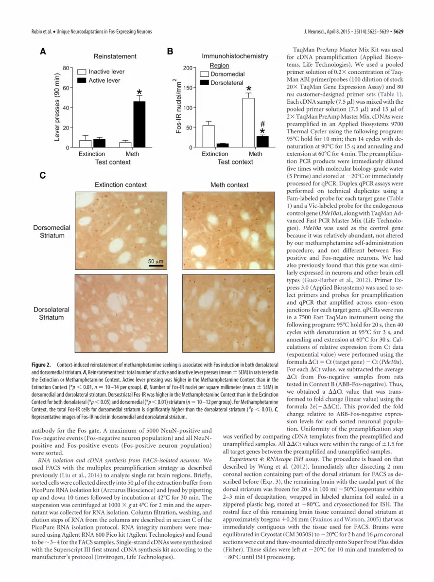
Experiment 4: RNAscope ISH assay. The procedure is based on thatdescribed by Wang et al. (2012). Immediately after dissecting 2 mmcoronal section containing part of the dorsal striatum for FACS as de-scribed before (Exp. 3), the remaining brain with the caudal part of thedorsal striatum was frozen for 20 s in 100 ml �50°C isopentane within2–3 min of decapitation, wrapped in labeled alumina foil sealed in azippered plastic bag, stored at �80°C, and cryosectioned for ISH. Therostral face of this remaining brain tissue contained dorsal striatum atapproximately bregma �0.24 mm (Paxinos and Watson, 2005) that wasimmediately contiguous with the tissue used for FACS. Brains wereequilibrated in Cryostat (CM 3050S) to �20°C for 2 h and 16 �m coronalsections were cut and thaw-mounted directly onto Super Frost Plus slides(Fisher). These slides were left at �20°C for 10 min and transferred to�80°C until ISH processing.
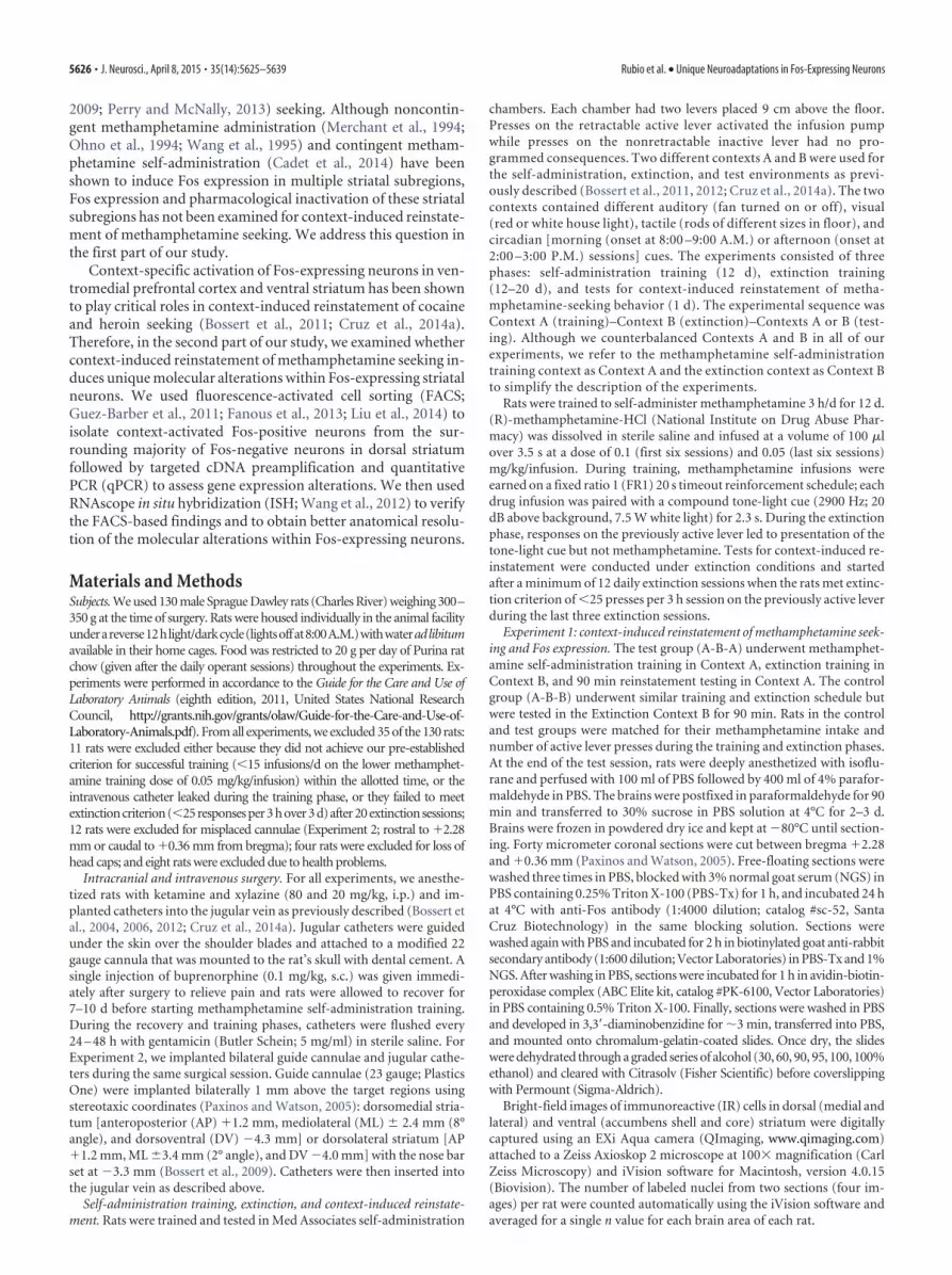
Figure 2. Context-induced reinstatement of methamphetamine seeking is associated with Fos induction in both dorsolateraland dorsomedial striatum. A, Reinstatement test: total number of active and inactive lever presses (mean � SEM) in rats tested inthe Extinction or Methamphetamine Context. Active lever pressing was higher in the Methamphetamine Context than in theExtinction Context (*p � 0.01, n 10 –14 per group). B, Number of Fos-IR nuclei per square millimeter (mean � SEM) indorsomedial and dorsolateral striatum. Dorsostriatal Fos-IR was higher in the Methamphetamine Context than in the ExtinctionContext for both dorsolateral (*p � 0.05) and dorsomedial (*p � 0.01) striatum (n 10 –12 per group). For MethamphetamineContext, the total Fos-IR cells for dorsomedial striatum is significantly higher than the dorsolateral striatum ( #p � 0.01). C,Representative images of Fos-IR nuclei in dorsomedial and dorsolateral striatum.
Rubio et al. • Unique Neuroadaptations in Fos-Expressing Neurons J. Neurosci., April 8, 2015 • 35(14):5625–5639 • 5629

RNA ISH for Fos, Grin2A, and Grin2B mRNAs was performed manu-ally according to the User Manual for Fresh Frozen Tissue using RNAscopeMultiplex Fluorescent Reagent Kit (Advanced Cell Diagnostics). Briefly,the �80°C brain slides were transferred to slide racks and fixed by im-mersion in 10% neutral buffered formalin (Fisher Scientific) for 20 minat 4°C. Slides were rinsed two times in PBS and dehydrated two timeseach in 50 and 70% ethanol and twice in 100% ethanol. Slides weretransferred to a new 100% ethanol container and kept at �20°C over-night. The slides were dried at room temperature (22°C) for 10 min anda hydrophobic pen (ImmEdger Hydrophobic Barrier Pen, Vector Labo-ratories) was used to make a physical barrier surrounding the brain sec-tions to contain RNAscope assay solution. We used the HybEZHybridization System from Advanced Cell Diagnostics. The proteasesolution (Pretreatment 4 solution) was incubated with sections at roomtemperature for 20 min. After washing off the protease solution, 1�target probes for specific RNAs (Fos, Grin2a, and/or Grin2b) were ap-plied to the brain sections and incubated at 40°C for 2 h in the HybEZoven. Each RNAscope target probe contained a mixture of 20 ZZ oligo-nucleotide probes that bound to the target RNA. These probes wereas follows: Fos-C3 probe (accession number NM_022197.2, target nucle-otide region: 473-1497); Grin2a-C1 probe (accession numberNM_012573.3; target nucleotide region: 2812-4265); Grin2b-C2 probe(accession number NM_012574.1; target nucleotide region: 3053-3930);Drd1-C1 probe (accession number NM_012546.2; target nucleotide re-gion: 104-1053); and Drd2-C2 probe (accession number NM_012547.1;target nucleotide region: 445-1531).
Sections were then incubated with preamplifier and amplifier probesby applying AMP1 (40°C for 30 min), AMP2 (40°C for 15 min), andAMP3 (40°C for 30 min). Sections were then incubated with the fluores-cently labeled probes by selecting a specific combination of colors asso-ciated with each channel: green (Alexa 488 nm), orange (Alexa 550 nm),and far-red (Alexa 647 nm). We used AMP4 AltA combination to detectduplex Fos and Grin2a RNAs in far-red and green, respectively; AMP4AltB to detect duplex Fos and Grin2b RNAs in far-red and green, respec-tively; and AMP4 AltB to detect triplex Fos, Grin2a, and Grin2b RNAs infar-red, orange, and green respectively. Finally, we incubated the sectionsfor 20 s with DAPI to stain nuclei (blue). Between each step we washedtwo times with 1� wash buffer supplied with the kit.
The negative control sections received RNase treatment before per-forming the RNAscope assay. After the fixation and protease digestion,we incubated sections with 5 mg/ml RNase A (Qiagen) for 30 min at40°C. The slides were washed three times with distilled water and pro-cessed with target probe hybridization, followed by the steps describedabove. Fluorescent images were captured at 200� magnification using aRolera EM-C 2 (QImaging) on a Nikon Eclipse E800 microscope. Imageswere saved as 16-bit TIFF files, enhanced using Adobe Photoshop CS5software for analysis. Representative images are shown in Figure 8 afterdeconvolving single-channel images using Huygens software (v3.7, Sci-entific Volume Imaging) and then merging the three channels using
iVision software. For each Fos-positive cell, we analyzed two of the sur-rounding Fos-negative cells. For each cell, we counted the total pixels ofthe fluorescent signal (fluorescent “dots”). We assume that each pixel issupposed to represent a single molecule of mRNA. Selection criterion forFos-positive cells was to have �33 total positive pixels after adjustingautomatically the threshold using ImageJ software. Fluorescent dots forGrin2a and Grin2b were counted when their intensity values were greaterthan 23,387 and 24,000, respectively (threshold was set manually).
Statistical analyses. The behavioral and immunohistochemical datawere analyzed by two-way ANOVAs (SPSS version 20, GLM procedure,or GraphPad software). We followed up on significant main or interac-tion effects ( p � 0.05) using Fisher’s PLSD post hoc test. A two-wayANOVA (GraphPad Prism 5 software) was performed to analyze FACS-based qPCR and ISH data. For FACS-sorted samples, we used thebetween-subjects factor of Context (Extinction, Methamphetamine) andthe within-subjects factor of neuron type (Fos-negative, Fos-positive). Ifsignificant differences were found in ANOVAs, then t tests were used toassess significant differences between Fos-positive and Fos-negative neu-rons separately for each context. Our criteria for determining significantalterations in gene expression also included a Fos-positive/Fos-negativeratio of �1.5 for gene expression in each context (Table 2). For ISH weused the between-subjects factor of Region (Dorsolateral, Dorsomedial)and the within-subjects factor of Neuron Type (Fos-negative, Fos-positive). If significant differences were found in ANOVAs, then Bonfer-roni’s post hoc test was used to assess significant differences betweenFos-positive and Fos-negative neurons separately for each context.
ResultsTraining and extinction for Experiments 1–3Rats demonstrated reliable methamphetamine self-administrationin Experiments 1–3, as indicated by the increase in mean number ofinfusions (t(1,94) 22.4) and active lever presses (t(1,94) 18.1, p �0.01) when the methamphetamine dose was decreased from 0.1mg/kg per infusion on training days 1–6 to 0.05 mg/kg per infusionon training days 7–12. Active lever pressing decreased with repeatedextinction sessions (p � 0.01; Fig. 1).
Context-induced reinstatement is associated with increasedFos expression in dorsal but not ventral striatumIn Experiment 1 we determined whether context-induced rein-statement is associated with increased Fos-immunoreactive nu-clei (Fos-IR) in dorsal striatum (dorsolateral, dorsomedial) andventral striatum (accumbens core, shell).
Reinstatement. During testing, exposure to Methamphet-amine Context A, but not Extinction Context B, increased non-reinforced active lever pressing (the operational measure of drugseeking in reinstatement studies; Shaham et al., 2003). Statistical
Figure 3. Context-induced reinstatement of methamphetamine seeking has no effect on Fos-IR in nucleus accumbens. A, Number of Fos-IR nuclei per square millimeter (mean � SEM) inaccumbens core or shell. B, Fos-IR nuclei in accumbens core and accumbens shell captured at 100� magnification. Fos-IR was not different from Extinction Context for the ventral striatum areasquantified (n 10 –12 per group).
5630 • J. Neurosci., April 8, 2015 • 35(14):5625–5639 Rubio et al. • Unique Neuroadaptations in Fos-Expressing Neurons

analysis indicated a significant interaction between Context (A,B) and Lever (Active, Inactive; F(1,44) 21.0, p � 0.01; Fig. 2A).
Fos expression. Exposure to the Methamphetamine Context Aalso increased the number of Fos-IR nuclei in dorsal (Fig. 2B,C)but not ventral (Fig. 3A,B) striatum. Statistical analysis for dorsalstriatum subregions demonstrated a significant interaction be-tween Context (A, B) and Subregions (Dorsomedial, Dorsolat-eral; F(1,40) 8.14, p � 0.01), main effect of Context (F(1,40) 24.1, p � 0.01), and main effect of Subregions (F(1,40) 65.6, p �0.01). As can be seen in Figure 2, Fos-IR levels for both contexts(A and B) were higher in dorsomedial than in dorsolateral stria-tum (main effect of Subregion: F(1,40) 65.6, p � 0.01). Howeverfold induction (Context A/Context B) of Fos expression wasgreater in dorsolateral striatum (3.0-fold) than in dorsomedial
striatum (2.2-fold). Context-induced re-instatement was not associated with in-creased Fos-IR in the accumbens core orshell (p � 0.05).
Inactivation of dorsolateral but notdorsomedial striatum decreasedcontext-induced reinstatementIn Experiment 1, we found that context-induced reinstatement is associated withincreased Fos expression in dorsolateraland dorsomedial striatum but not in ven-tral striatum. In Experiment. 2, we deter-mined the functional role of the dorsalstriatum subregions in context-inducedreinstatement by reversibly inactivatingthese regions with muscimol�baclofen15 min before reinstatement tests in Con-text A or B after extinction. We found thatmuscimol�baclofen injections into dor-solateral but not dorsomedial striatumdecreased context-induced reinstatement(Fig. 4A,B). Statistical analysis of the dor-solateral striatum data indicated a signifi-cant interaction between Context (A, B),Lever (Active, Inactive), and Drug (Vehi-cle, Muscimol�baclofen; F(1,34) 6.3,p � 0.05). Statistical analysis of the dorso-medial striatum data indicated a signifi-cant interaction between Context andLever (F(1,56) 11.52, p � 0.01) but nomain effect of Drug or an interactionbetween Drug and the other two factors( p � 0.05). Post hoc analysis indicatedthat muscimol�baclofen injections intodorsolateral but not dorsomedial stria-tum decreased active lever pressing inthe Methamphetamine Context A ( p �0.01), but not in Extinction Context B.The different experimental manipula-tions had no effect on inactive leverpresses, which were very low (�4 aver-age presses per session). Placements ofthe injector tips within the dorsolateraland dorsomedial striatum are shown inFigure 4C,D.
Finally, to rule out the possibilitythat the effect of muscimol�baclofeninjections into dorsolateral striatum
were due to motor deficits, we trained 18 rats that previouslyparticipated in Experiment 2 (11 rats with cannulae in dorso-lateral striatum and seven rats with cannulae in dorsomedialstriatum) to lever press for food pellets. After stable respond-ing was observed, we determined the effect of muscimol�baclofen or vehicle injections into dorsomedial or dorsolateralstriatum on high-rate operant responding for food. These in-jections had no effect of food-reinforced responding [interac-tion between Drug (Vehicle, Muscimol�baclofen) andSubregions (Dorsomedial, Dorsolateral), F(1,32) 0.04, p �0.1; effect of Drug, F(1,32) � 0.01, p � 0.1; effect of Subregions,F(1,32) � 0.01, p � 0.1], indicating that muscimol�baclofeneffects on context-induced reinstatement were not due to mo-tor deficits (Fig. 5).
Figure 4. Local inactivation of dorsolateral but not dorsomedial striatum with muscimol�baclofen decreased context-inducedreinstatement of methamphetamine seeking. A, B, Bilateral injections of muscimol�baclofen into (B) dorsolateral striatum, butnot (A) dorsomedial striatum, decreased active lever pressing (mean�SEM) during reinstatement tests in the MethamphetamineContext but not in the Extinction Context (*different from the Vehicle Group, *p � 0.01, n 7–12 per group). C, D, Dots indicateapproximate placements (millimeters from bregma) of injector tips into the (C) dorsomedial or (D) dorsolateral striatum (Paxinosand Watson, 2005).
Rubio et al. • Unique Neuroadaptations in Fos-Expressing Neurons J. Neurosci., April 8, 2015 • 35(14):5625–5639 • 5631

Together, the results of Experiment 2 indicate that neuronalactivity in dorsolateral but not dorsomedial region plays an im-portant role in context-induced reinstatement.
FACS sorting of context-activated Fos-expressing neuronsIn Experiment 3, we used FACS to isolate Fos-positive and Fos-negative neurons from dorsal striatum after context-inducedreinstatement of methamphetamine seeking and assessed differ-ences of gene expression in these two neuron populations. Expo-sure to Methamphetamine Context A, but not ExtinctionContext B, increased lever pressing on test day (Fig. 6A). Statisti-cal analysis indicated a significant interaction between the Con-text and Lever (F(1,70) 21.7, p � 0.01).
We then used our modified FACS procedure (Liu et al., 2014)to isolate Fos-positive and Fos-negative neurons from the dorsalstriatum of each rat. Dorsal striatum was dissected from 2-mm-thick sections (Fig. 6B) immediately after the reinstatement testand cells were dissociated and immunolabeled for Fos and thegeneral neuronal marker NeuN. Threshold parameters were setbefore sorting by using small amounts of dissociated cell samplesobtained from rats that were exposed to the Extinction Contextor the Methamphetamine Context. Every particle (cell or debris)that passed through the flow cytometer was an “event” indicatedby a dot in scattergrams that represent the different light charac-teristics of these events. The light scattergram (Fig. 6C, cell pop-ulation) is shown as a density plot of events where FSC indicatesparticle size and SSC indicates granularity of the particle.
The cell population plot indicates a large heterogeneous pop-ulation with different sizes and granularity along with a smallrelatively homogeneous population (�1% of all events). We se-lected or “gated” the smaller population of events as the “cellpopulation” based on previously determined FSC and SSC char-acteristics (Guez-Barber et al., 2011; Liu et al., 2014). From the“cell population” gate, we gated the single-cell population basedon the height and width of the FSC signal to exclude incompletely
dissociated cells (Fig. 6D, doublet exclusion); events with higherFSC width contained cell doublets.
From the “single cell” gate, we gated neurons that were immu-nolabeled with the NeuN antibody fluorescently labeled withR-phycoerythrin (Fig. 6E, neuronal cells). Neurons formed acluster of events with high levels of NeuN labeling and comprised22% of events from the previous single-cell gate. In a separateexperiment, we found that 95% of all neurons were labeled withthe nuclear DNA marker DAPI (Fig. 6F, nuclei). From the “neu-ronal cell” gate, we gated Fos-positive and Fos-negative neuronsbased on their degree of immunolabeling with the Fos antibodyfluorescently labeled with Alexa 647 (Fig. 6G, Fos-positive neu-rons). Neurons with higher levels of Fos labeling were consideredFos-positive neurons (blue dots) while the remaining neuronswere considered Fos-negative (red dots). Dotted lines indicatethe thresholds used to separate the positive from the negativepopulation for NeuN (red line) and Fos (blue line). Off-line anal-ysis indicates that the Methamphetamine Context A inducedmore than twice as many Fos-positive neurons (2.1%) than theExtinction Context B (0.8%; t(1,33) 5.2, p � 0.01). The numberof neurons obtained from dorsal striatum were not different be-tween rats tested in the Methamphetamine Context (15,100 �2433, n 19) and Extinction Context (15,426 � 3140, n 16).
qPCR analyses of gene expression in Fos-positive and Fos-negative neuronsThe mean number of FACS-isolated neurons collected for RNAanalysis was 149 Fos-positive and 4587 Fos-negative neuronsfrom each Methamphetamine Context A rat and 85 Fos-positiveand 4831 Fos-negative neurons from each Extinction Context Brat. Gene expression was assessed using qPCR and the data areexpressed as fold change over expression levels in Fos-negativeneurons from Extinction Context rat brains (Fig. 7). Detailedstatistics for each gene are described in Table 2. Post hoc analysesof Methamphetamine Context A samples alone indicated thatexpression levels of the immediate-early genes (IEGs) Fos andFosB (total) and the glutamate receptor subunit gene (Grin2a)were higher in the Fos-positive neurons than in the Fos-negativeneurons. Expression levels were not different for genes encodingthe dopamine receptors D1dr and D2dr, or for the AMPA recep-tor subunits Gria1 and Gria2. Thus Grin2a, which encodes theGluN2A subunit of the NMDA receptor, was the only receptorgene upregulated by the Methamphetamine Context A in Fos-positive neurons (F(1,27) 13.0, p � 0.01). Analysis of ExtinctionContext B samples alone indicated that expression levels ofHomer-1 (F(1,21) 5.6, p � 0.05), Homer-2 (F(1,12) 14.0, p �0.01), and the cannabinoid receptor gene (Cbr1; F(1,24) 16.0,p � 0.01) were higher in the Fos-positive neurons than in theFos-negative neurons. Expression levels for all other genes werenot different between groups.
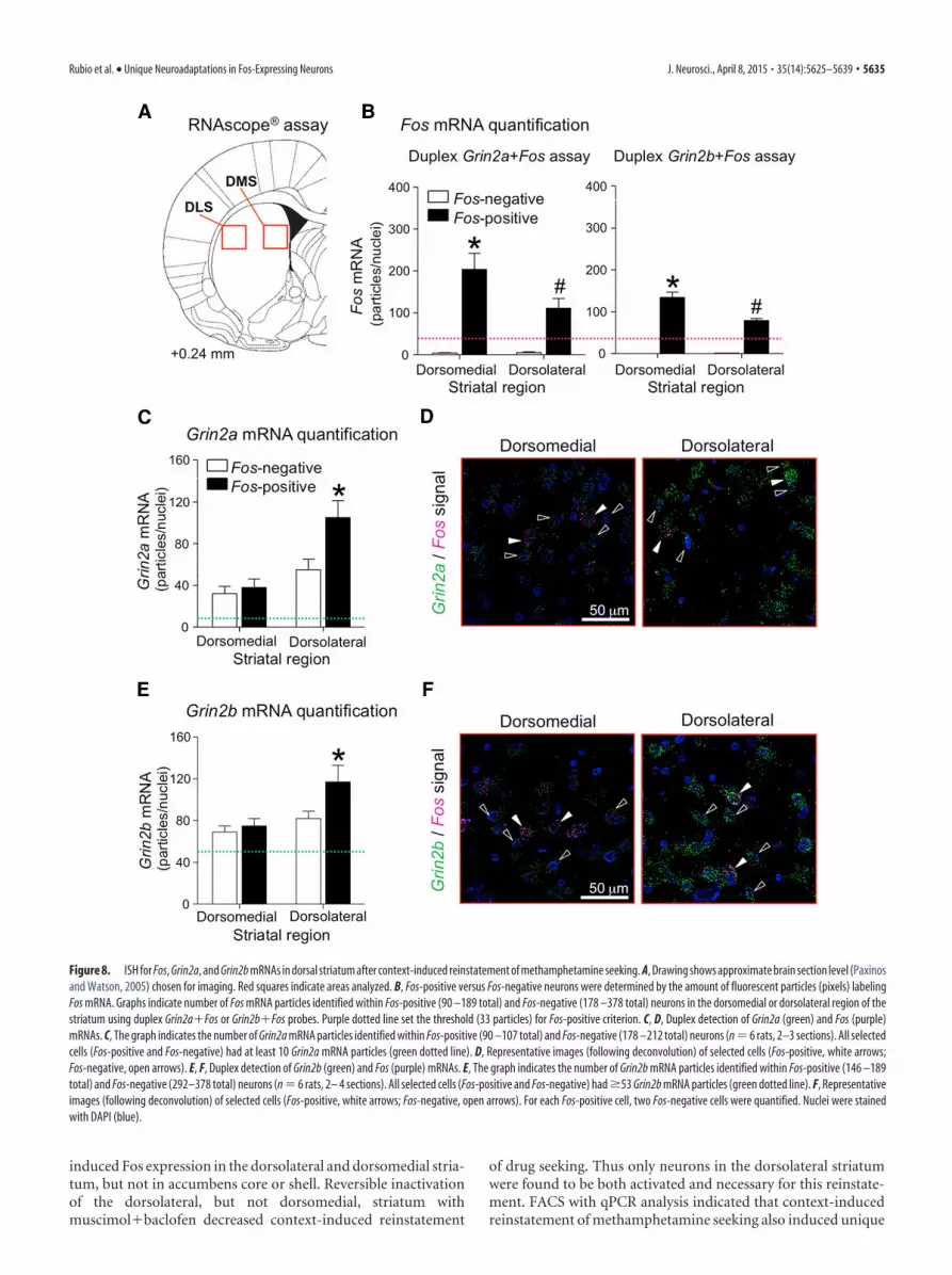
RNAscope ISH analysesThe results from Experiment 2 indicated that the dorsolateral butnot dorsomedial striatum is critical for context-induced rein-statement. Due to detection limitations of the current FACS pro-cedure, we analyzed gene expression in the entire dorsal striatumrather than in the separate dorsolateral and dorsomedial subre-gions. Therefore, we used the recently developed RNAscope ISHprocedure (Wang et al., 2012) to determine whether context-induced reinstatement is associated with a subregion-specificincreases of Grin2a expression in Fos-positive dorsal striatumneurons. This also gave us the opportunity to assesssubregion-specific expression levels of Grin2b, the gene en-
Figure 5. Local inactivation of dorsolateral or dorsomedial striatum with muscimol�baclofen had no effect on high-rate food reinforced responding. Rats were tested after 6 d offood self-administration training (n 7–11 per group).
5632 • J. Neurosci., April 8, 2015 • 35(14):5625–5639 Rubio et al. • Unique Neuroadaptations in Fos-Expressing Neurons

coding the GluN2B subunit of the NMDA receptor, in Fos-positive neurons.
We determined increased expression of Fos, Grin2a, and Grin2bin Fos-positive and Fos-negative neurons in dorsolateral and dorso-medial striatum from rats in the Methamphetamine Context A test
group. We used coronal sections cut from 12 brains that were keptfrozen from Experiment 3 and double-labeled them for Fos andGrin2a mRNAs or Fos and Grin2b mRNAs (Fig. 8A). Fos-positiveneurons were labeled with Grin2a or Grin2b and a minimum of 33Fos-labeled pixels (Fig. 8B) while Fos-negative neurons were selected
Figure 6. FACS of dorsostriatal neurons activated during reinstatement of methamphetamine seeking. A, Reinstatement test: number of active and inactive lever presses (mean � SEM). Activelever pressing was higher in the Methamphetamine Context than in the Extinction Context (*p � 0.01, n 17–20 per group). B, A representative drawing of the caudal face (millimeters frombregma) of a slice used for dissecting dorsal striatum (DS, blue; Paxinos and Watson, 2005). C–G, Representative density plots (C, D, F ) and scattergrams (E, G) used to gate Fos-expressing neuronsfor FACS isolation. C, Cells (red gate): linear plot of forward scatter area (x-axis, cell size) and side scatter area (y-axis, granularity). D, Single cells (red gate): linear plot of forward scatter-height(y-axis) and forward scatter-width (x-axis, width size of the events). E, Neurons (red gate): logarithmic plot of fluorescence for NeuN-positive neurons (R-phycoerythrin on y-axis) indicates neuronsin the upper cluster of events (red gate) and non-neurons in the lower cluster. F, Nuclei (neuronal cell gate): logarithmic plot of DAPI fluorescence (y-axis) indicates 95% of nuclei were found withinthe neuronal population. G, Fos-positive and Fos-negative neurons: logarithmic plots of neurons that were double-labeled for NeuN (R-phycoerythrin on x-axis) and Fos (Alexa 647 on y-axis)immunofluorescence. Selected Fos-positive neurons (upper right quadrants) after exposure to the Extinction Context (mean: 0.8 � 0.1% of all neurons, n 16) or Methamphetamine Context(2.1 � 0.2% of all neurons, n 19). Limited 5000 Fos-negative neurons (lower right quadrant) and all Fos-positive neurons were collected into microtubes for RNA isolation and real-time qPCRanalysis.
Rubio et al. • Unique Neuroadaptations in Fos-Expressing Neurons J. Neurosci., April 8, 2015 • 35(14):5625–5639 • 5633

from cells below 33 Fos-labeled pixels and �10 for Grin2a (Fig. 8C)or 53 labeled pixels for Grin2b (Fig. 8E). Initial analysis indicated thatindependent of the cell type (Fos-positive, Fos-negative), Grin2a andGrin2b levels were higher in the dorsolateral than in the dorsomedialstriatum [Grin2a (F(1,10) 9.3, p � 0.05) and Grin2b (F(1,10) 4.9,p 0.051)].
Higher levels of Grin2a mRNA were found in Fos-positive neu-rons in the dorsolateral but not dorsomedial striatum (Fig. 8C);representative images are shown in Figure 8D. Statistical analysisindicated a significant interaction (F(1,10) 25.0, p � 0.01) betweenthe within-subjects factors of Subregion (Dorsolateral, Dorsome-dial) and Neuron type (Fos-positive, Fos-negative). Post hoc analysisindicated Grin2a levels were higher in Fos-positive neurons than inFos-negative neurons in the dorsolateral striatum (F(1,10) 40.0, p �0.01), but not different in the dorsomedial striatum.
Higher levels of Grin2b mRNA were also found in Fos-positiveneurons in the dorsolateral but not dorsomedial striatum (Fig. 8E);representative images are shown in Figure 8F. Statistical analysisindicated a significant interaction (F(1,10) 5.7, p � 0.05) betweenthe within-subjects factors of Subregion (Dorsolateral, Dorsome-dial) and Neuron type (Fos-positive, Fos-negative). Post hoc analysisindicated Grin2b levels were higher in Fos-positive neurons than inFos-negative neurons in dorsolateral striatum (F(1,10) 12.0, p �0.01), but not different in dorsomedial striatum.
In a separate control experiment, sections were labeled with allthree probes: Fos, Grin2a, and Grin2b (Fig. 9). The pixels forGrin2a and Grin2b labeling did not overlap, which confirmedspecificity of these two probes. No labeling was observed whenthese sections were incubated with RNase before hybridizationwith the specific probes in the RNAscope assay.
Finally, we determined the phenotype of the reinstatement-activated Fos-positive neurons by assessing coexpression of FosmRNA with Drd1 and Drd2 mRNA. We used coronal sections cutfrom nine brains that were kept frozen from Experiment 3 andtriple-labeled them for Fos, Drd1, and Drd2 mRNAs (Fig. 10A).The majority (95–97%) of the Fos-positive cells coexpressed ei-ther Drd1 or Drd2 mRNA with similar percentages for Drd1 ver-sus Drd2 mRNA in the dorsolateral striatum (Fig. 10B). Asignificantly higher percentage of Fos-positive neurons coex-pressed Drd1 than Drd2 in the dorsomedial striatum (F(1,32) 17.4, p � 0.01). Only 3–5% of Fos-positive cells did not coexpressDrd1 or Drd2 mRNA. Representative images are shown in Figure10C.
DiscussionActivation of dorsolateral striatal neurons is important forcontext-induced reinstatement of methamphetamine seeking.Context-induced reinstatement of methamphetamine seeking
A
B
Immediate early genes
Receptors
Fold
indu
ctio
n(r
elat
ive
to F
os-n
eg in
Ext
)
0
1
2
3
4
0
1
2
3
4 Fos-negativeFos-positive
Grin1
0
1
2
3
4Grin2a
*
Cbr1
*
Extinction MethTest context
Extinction MethTest context
Extinction MethTest context
FosB (total)
Extinction Meth0
3
6
9
12
Test context
**
Homer-1
#
Fos
* *
Extinction Meth0
3
6
9
12
Test context
Fos-negativeFos-positive
Fold
indu
ctio
n(r
elat
ive
to F
os-n
eg in
Ext
)
Extinction0
3
6
9
12
Test contextMeth
Figure 7. Gene expression in Fos-positive and Fos-negative neurons that were FACS-isolated from dorsal striatum after context-induced reinstatement of methamphetamine. A, IEGs (Fos, FosB(total ), Homer-1). B, Genes encoding NMDA receptor subunits (Grin1, Grin2a) and cannabinoid receptor (Cbr1). Exposure to the Extinction or Methamphetamine (Meth) Contexts increased geneexpression in Fos-positive neurons relative to the Fos-negative neurons (*p � 0.05; #, main effect for Fos cell type; Extinction Context, n 3–11; Methamphetamine Context, n 7–17). See Table2 summary for all genes analyzed.
5634 • J. Neurosci., April 8, 2015 • 35(14):5625–5639 Rubio et al. • Unique Neuroadaptations in Fos-Expressing Neurons
induced Fos expression in the dorsolateral and dorsomedial stria-tum, but not in accumbens core or shell. Reversible inactivationof the dorsolateral, but not dorsomedial, striatum withmuscimol�baclofen decreased context-induced reinstatement
of drug seeking. Thus only neurons in the dorsolateral striatumwere found to be both activated and necessary for this reinstate-ment. FACS with qPCR analysis indicated that context-inducedreinstatement of methamphetamine seeking also induced unique
Figure 8. ISH for Fos, Grin2a, and Grin2b mRNAs in dorsal striatum after context-induced reinstatement of methamphetamine seeking. A, Drawing shows approximate brain section level (Paxinosand Watson, 2005) chosen for imaging. Red squares indicate areas analyzed. B, Fos-positive versus Fos-negative neurons were determined by the amount of fluorescent particles (pixels) labelingFos mRNA. Graphs indicate number of Fos mRNA particles identified within Fos-positive (90 –189 total) and Fos-negative (178 –378 total) neurons in the dorsomedial or dorsolateral region of thestriatum using duplex Grin2a�Fos or Grin2b�Fos probes. Purple dotted line set the threshold (33 particles) for Fos-positive criterion. C, D, Duplex detection of Grin2a (green) and Fos (purple)mRNAs. C, The graph indicates the number of Grin2a mRNA particles identified within Fos-positive (90 –107 total) and Fos-negative (178 –212 total) neurons (n 6 rats, 2–3 sections). All selectedcells (Fos-positive and Fos-negative) had at least 10 Grin2a mRNA particles (green dotted line). D, Representative images (following deconvolution) of selected cells (Fos-positive, white arrows;Fos-negative, open arrows). E, F, Duplex detection of Grin2b (green) and Fos (purple) mRNAs. E, The graph indicates the number of Grin2b mRNA particles identified within Fos-positive (146 –189total) and Fos-negative (292–378 total) neurons (n 6 rats, 2– 4 sections). All selected cells (Fos-positive and Fos-negative) had �53 Grin2b mRNA particles (green dotted line). F, Representativeimages (following deconvolution) of selected cells (Fos-positive, white arrows; Fos-negative, open arrows). For each Fos-positive cell, two Fos-negative cells were quantified. Nuclei were stainedwith DAPI (blue).
Rubio et al. • Unique Neuroadaptations in Fos-Expressing Neurons J. Neurosci., April 8, 2015 • 35(14):5625–5639 • 5635

gene alterations within the activated Fos-positive neurons but not in Fos-negativeneurons. These alterations included genesinvolved in transcription regulation (Fos,FosB; Nestler, 2001) and synaptic plastic-ity associated with glutamate transmis-sion (Homer-1, Homer-2; Kalivas et al.,2004; Lominac et al., 2005; Cozzoli et al.,2009; Ary et al., 2013), as well as Grin2a,the gene encoding the GluN2A subunit ofthe NMDA receptor (Buller et al., 1994).RNAscope ISH (Wang et al., 2012) indi-cated that expression levels of the genesGrin2a and Grin2b, which encode theGluN2A and GluN2B NMDA receptorsubunits, were selectively increased inFos-positive neurons in the dorsolateral,but not dorsomedial, striatum. Together,our results indicate that neuronal activityin the dorsolateral striatum contributesto context-induced reinstatement ofmethamphetamine seeking and that thecontext-activated Fos-expressing neu-rons in this striatal subregion undergounique neuroadaptations.
Role of dorsal and ventral striatum in context-inducedreinstatement of drug seekingOur results are consistent with those of previous studies on therole of dorsolateral striatum on context-induced and cue-induced reinstatement of drug seeking (Bossert et al., 2013;Marchant et al., 2014). Specifically, reversible inactivation (tetro-dotoxin or muscimol�baclofen) of dorsolateral striatum de-creased cue-induced reinstatement of cocaine and heroin seekingand context-induced reinstatement of cocaine seeking (Fuchs et al.,2006; Rogers et al., 2008). Additionally, blockade of D1-family re-ceptors with SCH23390 in dorsolateral but not dorsomedial stria-tum decreased context-induced reinstatement of heroin seeking(Bossert et al., 2009). Thus, while several studies indicate that differ-ent brain areas mediate relapse induced by discrete cues and contextsassociated with opiate versus psychostimulant drugs (Badiani et al.,2011; Badiani, 2013; Bossert et al., 2013; Peters et al., 2013), thedorsolateral striatum is consistently a critical site for context-induced and cue-induced drug seeking across drug classes (Vander-schuren and Everitt, 2005; Vanderschuren et al., 2005).
In a recent study, we found that context-induced reinstate-ment of cocaine seeking was associated with increased Fos ex-pression in accumbens shell and core and that selective inhibitionof the activated Fos-expressing neurons in accumbens shell (butnot core) using the Daun02 procedure (Cruz et al., 2013) de-creased this reinstatement (Cruz et al., 2014a). Based on theseand related findings on the effect of cocaine cues on accumbensFos expression (Ciccocioppo et al., 2001; Kufahl et al., 2009), itwas surprising to find that context-induced reinstatement wasnot associated with increased Fos expression in either accumbenscore or shell. However, our current finding is consistent withprevious reversible inactivation studies that indicate functionaldissociation between brain areas involved in reinstatement ofmethamphetamine versus cocaine seeking (Bossert et al., 2013).For example, reversible inactivation of ventral medial prefrontalcortex causes reinstatement of cocaine but not methamphet-amine seeking after extinction (Peters et al., 2008; Rocha andKalivas, 2010). Reversible inactivation of the same brain area
decreases cue-induced methamphetamine but not cocaine seek-ing after extinction (McLaughlin and See, 2003; Rocha and Kali-vas, 2010). Finally, reversible activation of the ventral medialprefrontal cortex decreases the time-dependent increases in cue-induced cocaine but not methamphetamine seeking (incubationof drug craving; Koya et al., 2009; Li et al., 2014).
The use of FACS to identify unique molecular changes incontext-activated and cue-activated neuronsIn previous studies, we used the Daun02 inactivation method(Koya et al., 2009) to demonstrate a causal role of behaviorallyactivated Fos-expressing neurons in both context-induced andcue-induced drug seeking (Bossert et al., 2011; Fanous et al.,2012; Cruz et al., 2014a). Based on these findings and the exten-sive literature on the role of drug-induced neuroadaptations indrug reward and relapse (Nestler, 2001; Kalivas, 2009; Van denOever et al., 2010; Wolf and Ferrario, 2010), we proposed thatunique alterations of transcription and synaptic plasticity in theseFos-expressing neurons are likely to play a unique role in drug ad-diction (Cruz et al., 2014b). Unlike the majority of addiction studies,we could not use brain homogenates to assess these alterations, be-cause specific molecular alterations within the small population ofFos-expressing neurons would be diluted or masked within thewhole homogenate (Cruz et al., 2013; Liu et al., 2014).
Thus we used FACS to isolate Fos-expressing neurons for mo-lecular analysis. The use of FACS with adult brain tissue was firstused to purify striatal neurons expressing GFP that were consti-tutively driven by the promoters for D1 and D2 receptors in mice(Lobo et al., 2006; Lobo, 2009). We developed an antibody-basedFACS method to purify activated Fos-expressing neurons fromadult wild-type rats (Guez-Barber et al., 2012) and used this pro-tocol to assess molecular alterations in Fos-expressing neuronsthat were activated during the expression of cocaine sensitizationand cue-induced heroin seeking (Guez-Barber et al., 2011;Fanous et al., 2013). However, this protocol required pooledbrain regions (entire striatum or PFC) and pooled samples (�10rats per sample) that made it difficult to implement in studiesusing complex behavioral models of drug addiction and relapse(Cruz et al., 2013; Liu et al., 2014). Based on these considerations,we added targeted preamplification of cDNA to enhance sen-
Figure 9. A, B, Triplex detection of Grin2a (red), Grin2b (green), and Fos (purple) mRNAs in dorsolateral striatum in the (A)absence or (B) presence of 5 mg/ml RNase treatment. A, Triple labeling for Grin2a, Grin2b, and Fos in the absence of RNasetreatment. Fos-positive (white arrows) and Fos-negative (open arrows) cells contained positive signals for both Grin2a and Grin2bmRNAs. The fluorescence channel for DAPI in this image was removed to more clearly observe the particles for each mRNA. B, Triplelabeling following prior treatment with 5 mg/ml RNase. Only blue DAPI-labeled DNA was observed. Positive signals were notobserved for Grin2a, Grin2b, or Fos.
5636 • J. Neurosci., April 8, 2015 • 35(14):5625–5639 Rubio et al. • Unique Neuroadaptations in Fos-Expressing Neurons

sitivity of our qPCR assay for gene expression from smallnumbers of FACS-isolated neurons in single rat dorsal stria-tum (Liu et al., 2014).
Using FACS�qPCR analysis, we found that exposure to boththe Methamphetamine Context and the Extinction Context in-duced IEGs in only the Fos-expressing dorsal striatal neurons.This finding replicates our previous findings that IEGs, such asFos and FosB, are induced only in Fos-expressing neurons and not
in the less activated Fos-negative majorityof neurons in striatum and medial pre-frontal cortex after exposure to drugs ordrug cues (Guez-Barber et al., 2011;Fanous et al., 2013). Although the func-tional significance of inducing these IEGsis unknown, Fos and FosB encode tran-scription factor proteins that can inducesubsequent waves of gene expression se-lectively with Fos-expressing neurons(Cruz et al., 2014b).
FACS�qPCR analysis and RNAs-cope ISH together indicated thatcontext-induced reinstatement ofmethamphetamine seeking increasedmRNA levels of Grin2a and Grin2b, butnot Grin1, in Fos-positive neurons inthe dorsolateral, but not dorsomedial,striatum. On the other hand, the Gria1and Gria2 genes, which encode theAMPA receptor subunits GluA1 andGluA2, were not different between Fos-positive and Fos-negative neurons. TheGrin2a, Grin2b, and Grin1 genes encodeGluN2A, GluN2B, and GluN1 subunitsof the NMDA receptor, which are ex-pressed at moderate to high levels instriatum (Buller et al., 1994; Standaert etal., 1999). Although the functional sig-nificance of inducing these genes is un-known, increased levels of Grin2a andGrin2b may be related to synaptic alter-ations that were selectively induced inFos-expressing neurons in the accum-bens and medial prefrontal cortex (Ci-fani et al., 2012), or to GluN2B-containing silent synapses induced in allneurons in these brain areas followingcocaine locomotor sensitization(Huang et al., 2009; Brown et al., 2011)or cocaine self-administration (Lee etal., 2013; Ma et al., 2014).
Exposure to the Extinction Context,but not the Methamphetamine Context,induced Homer-1, Homer-2, and Cbr1 geneexpression in Fos-expressing neurons. Thefunctional significance of unique molecularalterations in Fos-expressing neurons in theDrug versus Extinction Contexts is un-known but is in agreement that differentmechanisms control reward learning versusextinction learning (Peters et al., 2009).
Another finding from qPCR assays wasthat the mRNA encoding D1 and D2 re-ceptors did not differ between the Fos-
positive and Fos-negative neurons. The RNAscope dataconfirmed that Fos mRNA was similarly induced in D1 and D2neurons, most likely in medium spiny neurons, which make up�95% of neurons in the dorsal striatum (Tepper et al., 2007).These results extend our previous studies showing that thecontext-activated Fos-expressing neurons are not confined to aparticular cell type in the striatum (Cruz et al., 2014a) or medialprefrontal cortex (Bossert et al., 2011).
C
RNAscope® assay A
+0.24 mm
DMS
DLS
B Drd1+FosDrd2+FosFos alone
DMS DLS0
25
50
75
100
Region
% p
ositi
ve c
ells *
Dor
sola
tera
l stri
atum
Dor
som
edia
l stri
atum
Drd1/Drd2 Fos/DAPI
20µm
Figure 10. Triplex detection of Drd1 (green), Drd2 (red), and Fos (purple) mRNAs in dorsal striatum after context-inducedreinstatement of methamphetamine seeking. A, Drawing shows approximate brain section level (Paxinos and Watson, 2005)chosen for imaging. Red squares indicate areas analyzed. B, Graphs indicate the percentage of Fos-positive neurons (n 9 rats, 1section each) that coexpressed Drd1 (Drd1�Fos) or Drd2 (Drd2�Fos) mRNA in the dorsomedial or dorsolateral region of thestriatum. A small number of Fos-positive neurons did not coexpress either Drd1 or Drd2 mRNA (Fos alone). C, Representative imagesof selected cells (Fos-positive, white arrows). Images on the left show the merged channels for Drd1 (green) and Drd2 (red) signals.Images on the right show the merged channels for Fos (purple) and DAPI (blue) signals from the same brain sections and fields thanthe right side. The fluorescent signals for Fos and Drd2 signals had similar colors; although sufficiently different for quantitation, itis easier to see Fos and DAPI signals separately from those for Drd1 and Drd2 signals.
Rubio et al. • Unique Neuroadaptations in Fos-Expressing Neurons J. Neurosci., April 8, 2015 • 35(14):5625–5639 • 5637

Concluding remarksWe demonstrated that the dorsolateral striatum is a critical brainsubstrate for context-induced reinstatement of methamphet-amine seeking, which together with previous results indicate thatneuronal activity in this brain region is a common substrate forcontext-induced reinstatement of drug seeking for both psycho-stimulant and opiate drugs. Additionally, our FACS�qPCR andRNAscope ISH data indicate that context-induced reinstatementof methamphetamine seeking is associated with unique molecu-lar alterations of NMDA-type glutamate receptor subunit genesin context-activated Fos-expressing neurons in dorsal striatum.It is currently not possible to manipulate gene expression selec-tively within these Fos-expressing neurons. Thus the functionalsignificance of inducing unique molecular alterations within Fos-expressing neurons activated by exposure to the Extinction orMethamphetamine Contexts is currently unknown. Future ex-periments are necessary to determine whether selective regula-tion of these genes in Fos-expressing neurons plays a functionalrole in context-induced reinstatement of drug seeking.
ReferencesAry AW, Lominac KD, Wroten MG, Williams AR, Campbell RR, Ben-Shahar
O, von Jonquieres G, Klugmann M, Szumlinski KK (2013) Imbalancesin prefrontal cortex CC-Homer1 versus CC-Homer2 expression promotecocaine preference. J Neurosci 33:8101– 8113. CrossRef Medline
Badiani A (2013) Substance-specific environmental influences on drug useand drug preference in animals and humans. Curr Opin Neurobiol 23:588 –596. CrossRef Medline
Badiani A, Belin D, Epstein D, Calu D, Shaham Y (2011) Opiate versuspsychostimulant addiction: the differences do matter. Nat Rev Neurosci12:685–700. CrossRef Medline
Bossert JM, Liu SY, Lu L, Shaham Y (2004) A role of ventral tegmental areaglutamate in contextual cue-induced relapse to heroin seeking. J Neurosci24:10726 –10730. CrossRef Medline
Bossert JM, Gray SM, Lu L, Shaham Y (2006) Activation of group II metabo-tropic glutamate receptors in the nucleus accumbens shell attenuatescontext-induced relapse to heroin seeking. Neuropsychopharmacology31:2197–2209. Medline
Bossert JM, Poles GC, Wihbey KA, Koya E, Shaham Y (2007) Differentialeffects of blockade of dopamine D1-family receptors in nucleus accum-bens core or shell on reinstatement of heroin seeking induced by contex-tual and discrete cues. J Neurosci 27:12655–12663. CrossRef Medline
Bossert JM, Wihbey KA, Pickens CL, Nair SG, Shaham Y (2009) Role ofdopamine D(1)-family receptors in dorsolateral striatum in context-induced reinstatement of heroin seeking in rats. Psychopharmacology(Berl) 206:51– 60. CrossRef Medline
Bossert JM, Stern AL, Theberge FR, Cifani C, Koya E, Hope BT, Shaham Y(2011) Ventral medial prefrontal cortex neuronal ensembles mediatecontext-induced relapse to heroin. Nat Neurosci 14:420 – 422. CrossRefMedline
Bossert JM, Stern AL, Theberge FR, Marchant NJ, Wang HL, Morales M,Shaham Y (2012) Role of projections from ventral medial prefrontalcortex to nucleus accumbens shell in context-induced reinstatement ofheroin seeking. J Neurosci 32:4982– 4991. CrossRef Medline
Bossert JM, Marchant NJ, Calu DJ, Shaham Y (2013) The reinstatementmodel of drug relapse: recent neurobiological findings, emerging researchtopics, and translational research. Psychopharmacology (Berl) 229:453–476. CrossRef Medline
Bouton ME, Bolles RC (1979) Role of conditioned contextual stimuli inreinstatement of extinguished fear. J Exp Psychol Anim Behav Process5:368 –378. CrossRef Medline
Bouton ME, Swartzentruber D (1991) Sources of relapse after extinction inPavlovian and instrumental learning. Clin Psychol Rev 11:123–140.
Brown TE, Lee BR, Mu P, Ferguson D, Dietz D, Ohnishi YN, Lin Y, Suska A,Ishikawa M, Huang YH, Shen H, Kalivas PW, Sorg BA, Zukin RS, NestlerEJ, Dong Y, Schluter OM (2011) A silent synapse-based mechanism forcocaine-induced locomotor sensitization. J Neurosci 31:8163– 8174.CrossRef Medline
Buller AL, Larson HC, Schneider BE, Beaton JA, Morrisett RA, Monaghan DT
(1994) The molecular basis of NMDA receptor subtypes: native receptordiversity is predicted by subunit composition. J Neurosci 14:5471–5484.Medline
Cadet JL, Brannock C, Jayanthi S, Krasnova IN (2014) Transcriptional andepigenetic substrates of methamphetamine addiction and withdrawal:evidence from a long-access self-administration model in the rat. MolNeurobiol. Advance online publication. Retrieved Feb. 27, 2015. Medline
Chaudhri N, Sahuque LL, Cone JJ, Janak PH (2008) Reinstated ethanol-seekingin rats is modulated by environmental context and requires the nucleus ac-cumbens core. Eur J Neurosci 28:2288–2298. CrossRef Medline
Chaudhri N, Sahuque LL, Janak PH (2009) Ethanol seeking triggered byenvironmental context is attenuated by blocking dopamine D1 receptorsin the nucleus accumbens core and shell in rats. Psychopharmacology(Berl) 207:303–314. CrossRef Medline
Ciccocioppo R, Sanna PP, Weiss F (2001) Cocaine-predictive stimulus in-duces drug-seeking behavior and neural activation in limbic brain regionsafter multiple months of abstinence: reversal by D(1) antagonists. ProcNatl Acad Sci U S A 98:1976 –1981. CrossRef Medline
Cifani C, Koya E, Navarre BM, Calu DJ, Baumann MH, Marchant NJ, Liu QR,Khuc T, Pickel J, Lupica CR, Shaham Y, Hope BT (2012) Medial pre-frontal cortex neuronal activation and synaptic alterations after stress-induced reinstatement of palatable food seeking: a study using c-fos-GFPtransgenic female rats. J Neurosci 32:8480 – 8490. CrossRef Medline
Cozzoli DK, Goulding SP, Zhang PW, Xiao B, Hu JH, Ary AW, Obara I, RahnA, Abou-Ziab H, Tyrrel B, Marini C, Yoneyama N, Metten P, Snelling C,Dehoff MH, Crabbe JC, Finn DA, Klugmann M, Worley PF, SzumlinskiKK (2009) Binge drinking upregulates accumbens mGluR5-Homer2-PI3K signaling: functional implications for alcoholism. J Neurosci 29:8655– 8668. CrossRef Medline
Crombag HS, Shaham Y (2002) Renewal of drug seeking by contextual cuesafter prolonged extinction in rats. Behav Neurosci 116:169 –173. CrossRefMedline
Crombag HS, Grimm JW, Shaham Y (2002) Effect of dopamine receptorantagonists on renewal of cocaine seeking by reexposure to drug-associated contextual cues. Neuropsychopharmacology 27:1006 –1015.CrossRef Medline
Crombag HS, Bossert JM, Koya E, Shaham Y (2008) Review. Context-induced relapse to drug seeking: a review. Philos Trans R Soc Lond B BiolSci 363:3233–3243. CrossRef Medline
Cruz FC, Koya E, Guez-Barber DH, Bossert JM, Lupica CR, Shaham Y, Hope BT(2013) New technologies for examining the role of neuronal ensembles indrug addiction and fear. Nat Rev Neurosci 14:743–754. CrossRef Medline
Cruz FC, Babin KR, Leao RM, Goldart EM, Bossert JM, Shaham Y, Hope BT(2014a) Role of nucleus accumbens shell neuronal ensembles in context-induced reinstatement of cocaine-seeking. J Neurosci 34:7437–7446.CrossRef Medline
Cruz FC, Rubio FJ, Hope BT (2014b) Using c-fos to study neuronal ensem-bles in corticostriatal circuitry of addiction. Brain Res pii:S0006-8993(14)01552-2. Advance online publication. Retrieved Feb. 27, 2015.CrossRef Medline
Fanous S, Goldart EM, Theberge FR, Bossert JM, Shaham Y, Hope BT (2012)Role of orbitofrontal cortex neuronal ensembles in the expression of incuba-tion of heroin craving. J Neurosci 32:11600–11609. CrossRef Medline
Fanous S, Guez-Barber DH, Goldart EM, Schrama R, Theberge FR, ShahamY, Hope BT (2013) Unique gene alterations are induced in FACS-purified Fos-positive neurons activated during cue-induced relapse toheroin seeking. J Neurochem 124:100 –108. CrossRef Medline
Fuchs RA, Branham RK, See RE (2006) Different neural substrates mediatecocaine seeking after abstinence versus extinction training: a critical role forthe dorsolateral caudate-putamen. J Neurosci 26:3584–3588. CrossRefMedline
Fuchs RA, Ramirez DR, Bell GH (2008) Nucleus accumbens shell and coreinvolvement in drug context-induced reinstatement of cocaine seeking inrats. Psychopharmacology (Berl) 200:545–556. CrossRef Medline
Guez-Barber D, Fanous S, Golden SA, Schrama R, Koya E, Stern AL, BossertJM, Harvey BK, Picciotto MR, Hope BT (2011) FACS identifies uniquecocaine-induced gene regulation in selectively activated adult striatal neu-rons. J Neurosci 31:4251– 4259. CrossRef Medline
Guez-Barber D, Fanous S, Harvey BK, Zhang Y, Lehrmann E, Becker KG, Pic-ciotto MR, Hope BT (2012) FACS purification of immunolabeled cell typesfrom adult rat brain. J Neurosci Methods 203:10–18. CrossRef Medline
Hamlin AS, Newby J, McNally GP (2007) The neural correlates and role of
5638 • J. Neurosci., April 8, 2015 • 35(14):5625–5639 Rubio et al. • Unique Neuroadaptations in Fos-Expressing Neurons

D1 dopamine receptors in renewal of extinguished alcohol-seeking. Neu-roscience 146:525–536. CrossRef Medline
Huang YH, Lin Y, Mu P, Lee BR, Brown TE, Wayman G, Marie H, Liu W, Yan Z,Sorg BA, Schluter OM, Zukin RS, Dong Y (2009) In vivo cocaine experiencegenerates silent synapses. Neuron 63:40–47. CrossRef Medline
Janak PH, Chaudhri N (2010) The potent effect of environmental contexton relapse to alcohol-seeking after extinction. Open Addict J 3:76 – 87.CrossRef Medline
Kalivas PW (2009) The glutamate homeostasis hypothesis of addiction. NatRev Neurosci 10:561–572. CrossRef Medline
Kalivas PW, Szumlinski KK, Worley P (2004) Homer2 gene deletion in miceproduces a phenotype similar to chronic cocaine treated rats. NeurotoxRes 6:385–387. CrossRef Medline
Koya E, Golden SA, Harvey BK, Guez-Barber DH, Berkow A, Simmons DE,Bossert JM, Nair SG, Uejima JL, Marin MT, Mitchell TB, Farquhar D,Ghosh SC, Mattson BJ, Hope BT (2009) Targeted disruption of cocaine-activated nucleus accumbens neurons prevents context-specific sensitiza-tion. Nat Neurosci 12:1069 –1073. CrossRef Medline
Kufahl PR, Zavala AR, Singh A, Thiel KJ, Dickey ED, Joyce JN, NeisewanderJL (2009) c-Fos expression associated with reinstatement of cocaine-seeking behavior by response-contingent conditioned cues. Synapse 63:823– 835. CrossRef Medline
Lasseter HC, Xie X, Ramirez DR, Fuchs RA (2010) Prefrontal cortical regu-lation of drug seeking in animal models of drug relapse. Curr Top BehavNeurosci 3:101–117. CrossRef Medline
Lee BR, Ma YY, Huang YH, Wang X, Otaka M, Ishikawa M, Neumann PA,Graziane NM, Brown TE, Suska A, Guo C, Lobo MK, Sesack SR, Wolf ME,Nestler EJ, Shaham Y, Schluter OM, Dong Y (2013) Maturation of silentsynapses in amygdala-accumbens projection contributes to incubation ofcocaine craving. Nat Neurosci 16:1644 –1651. CrossRef Medline
Li X, Zeric T, Kambhampati S, Bossert JM, Shaham Y (2014) The centralamygdala nucleus is critical for incubation of methamphetamine craving.Neuropsychopharmacology. Advance online publication. Retrieved Feb.28, 2015. CrossRef Medline
Liu QR, Rubio FJ, Bossert JM, Marchant NJ, Fanous S, Hou X, Shaham Y, Hope BT(2014) Detection of molecular alterations in methamphetamine-activated Fos-expressing neurons from a single rat dorsal striatum using fluorescence-activatedcell sorting (FACS). J Neurochem 128:173–185. CrossRef Medline
Lobo MK (2009) Molecular profiling of striatonigral and striatopallidal me-dium spiny neurons past, present, and future. Int Rev Neurobiol 89:1–35.CrossRef Medline
Lobo MK, Karsten SL, Gray M, Geschwind DH, Yang XW (2006) FACS-array profiling of striatal projection neuron subtypes in juvenile and adultmouse brains. Nat Neurosci 9:443– 452. CrossRef Medline
Lominac KD, Oleson EB, Pava M, Klugmann M, Schwarz MK, Seeburg PH,During MJ, Worley PF, Kalivas PW, Szumlinski KK (2005) Distinctroles for different Homer1 isoforms in behaviors and associated prefron-tal cortex function. J Neurosci 25:11586 –11594. CrossRef Medline
Ma YY, Lee BR, Wang X, Guo C, Liu L, Cui R, Lan Y, Balcita-Pedicino JJ, WolfME, Sesack SR, Shaham Y, Schluter OM, Huang YH, Dong Y (2014)Bidirectional modulation of incubation of cocaine craving by silentsynapse-based remodeling of prefrontal cortex to accumbens projections.Neuron 83:1453–1467. CrossRef Medline
Marchant NJ, Kaganovsky K, Shaham Y, Bossert JM (2014) Role of cortico-striatal circuits in context-induced reinstatement of drug seeking. BrainRes pii:S0006-8993(14)01191-3. Advance online publication. RetrievedFeb. 28, 2105. CrossRef Medline
McFarland K, Kalivas PW (2001) The circuitry mediating cocaine-induced re-instatement of drug-seeking behavior. J Neurosci 21:8655–8663. Medline
McLaughlin J, See RE (2003) Selective inactivation of the dorsomedial pre-frontal cortex and the basolateral amygdala attenuates conditioned-cuedreinstatement of extinguished cocaine-seeking behavior in rats. Psycho-pharmacology (Berl) 168:57– 65. CrossRef Medline
McNally GP (2014) Extinction of drug seeking: neural circuits and approachesto augmentation. Neuropharmacology 76:528–532. CrossRef Medline
Merchant KM, Hanson GR, Dorsa DM (1994) Induction of neurotensinand c-fos mRNA in distinct subregions of rat neostriatum after acutemethamphetamine: comparison with acute haloperidol effects. J Pharma-col Exp Ther 269:806 – 812. Medline
Morgan JI, Curran T (1991) Stimulus-transcription coupling in the nervous
system: involvement of the inducible proto-oncogenes fos and jun. AnnuRev Neurosci 14:421– 451. CrossRef Medline
Nestler EJ (2001) Molecular basis of long-term plasticity underlying addic-tion. Nat Rev Neurosci 2:119 –128. CrossRef Medline
O’Brien CP, Ehrman RN, Ternes JW (1986) Classical conditioning in hu-man opioid dependence. In: Behavioral analysis of drug dependence(Goldberg S, Stolerman I, eds), pp 329 –356. Orlando, FL: Academic.
Ohno M, Yoshida H, Watanabe S (1994) NMDA receptor-mediated expres-sion of Fos protein in the rat striatum following methamphetamine ad-ministration: relation to behavioral sensitization. Brain Res 665:135–140.CrossRef Medline
Paxinos G, Watson C (2005) The rat brain in stereotaxic coordinates, fifthedition. Amsterdam: Elsevier Academic.
Perry CJ, McNally GP (2013) mu-Opioid receptors in the nucleus accum-bens shell mediate context-induced reinstatement (renewal) but notprimed reinstatement of extinguished alcohol seeking. Behav Neurosci127:535–543. CrossRef Medline
Peters J, LaLumiere RT, Kalivas PW (2008) Infralimbic prefrontal cortex isresponsible for inhibiting cocaine seeking in extinguished rats. J Neurosci28:6046 – 6053. CrossRef Medline
Peters J, Kalivas PW, Quirk GJ (2009) Extinction circuits for fear and addictionoverlap in prefrontal cortex. Learn Mem 16:279–288. CrossRef Medline
Peters J, Pattij T, De Vries TJ (2013) Targeting cocaine versus heroin mem-ories: divergent roles within ventromedial prefrontal cortex. Trends Phar-macol Sci 34:689 – 695. CrossRef Medline
Robins LN, Davis DH, Goodwin DW (1974) Drug use by U.S. Army enlistedmen in Vietnam: a follow-up on their return home. Am J Epidemiol99:235–249. Medline
Rocha A, Kalivas PW (2010) Role of the prefrontal cortex and nucleus ac-cumbens in reinstating methamphetamine seeking. Eur J Neurosci 31:903–909. CrossRef Medline
Rogers JL, Ghee S, See RE (2008) The neural circuitry underlying reinstate-ment of heroin-seeking behavior in an animal model of relapse. Neuro-science 151:579 –588. CrossRef Medline
Shaham Y, Shalev U, Lu L, De Wit H, Stewart J (2003) The reinstatementmodel of drug relapse: history, methodology and major findings. Psycho-pharmacology (Berl) 168:3–20. CrossRef Medline
Standaert DG, Friberg IK, Landwehrmeyer GB, Young AB, Penney JB Jr(1999) Expression of NMDA glutamate receptor subunit mRNAs in neu-rochemically identified projection and interneurons in the striatum of therat. Brain Res Mol Brain Res 64:11–23. CrossRef Medline
Tepper JM, Abercrombie ED, Bolam JP (2007) Basal ganglia macrocircuits.Prog Brain Res 160:3–7. CrossRef Medline
Van den Oever MC, Spijker S, Smit AB, De Vries TJ (2010) Prefrontal cortexplasticity mechanisms in drug seeking and relapse. Neurosci BiobehavRev 35:276 –284. CrossRef Medline
Vanderschuren LJ, Everitt BJ (2005) Behavioral and neural mechanisms ofcompulsive drug seeking. Eur J Pharmacol 526:77– 88. CrossRef Medline
Vanderschuren LJ, Di Ciano P, Everitt BJ (2005) Involvement of the dorsalstriatum in cue-controlled cocaine seeking. J Neurosci 25:8665– 8670.CrossRef Medline
Wang F, Flanagan J, Su N, Wang LC, Bui S, Nielson A, Wu X, Vo HT, Ma XJ,Luo Y (2012) RNAscope: a novel in situ RNA analysis platform forformalin-fixed, paraffin-embedded tissues. J Mol Diagn 14:22–29.CrossRef Medline
Wang JQ, Smith AJ, McGinty JF (1995) A single injection of amphetamineor methamphetamine induces dynamic alterations in c-fos, zif/268 andpreprodynorphin messenger RNA expression in rat forebrain. Neurosci-ence 68:83–95. CrossRef Medline
Widholm JJ, Gass JT, Cleva RM, Olive MF (2011) The mGluR5 positiveallosteric modulator CDPPB does not alter extinction or contextual rein-statement of methamphetamine-seeking behavior in rats. J Addict ResTher S1:pii:004. Medline
Wikler A (1973) Dynamics of drug dependence. Implications of a condi-tioning theory for research and treatment. Arch Gen Psychiatry 28:611–616. CrossRef Medline
Wolf ME, Ferrario CR (2010) AMPA receptor plasticity in the nucleus ac-cumbens after repeated exposure to cocaine. Neurosci Biobehav Rev 35:185–211. CrossRef Medline
Rubio et al. • Unique Neuroadaptations in Fos-Expressing Neurons J. Neurosci., April 8, 2015 • 35(14):5625–5639 • 5639