computationally designed libraries for rapid enzyme stabilization
TRANSCRIPT
Computationally designed libraries for rapid enzymestabilization
Hein J.Wijma1†, Robert J.Floor1†, Peter A.Jekel1,David Baker2, Siewert J.Marrink1,3 and Dick B. Janssen1,4
1Department of Biochemistry, Groningen Biomolecular Sciences andBiotechnology Institute, University of Groningen, Nijenborgh 4, 9747 AGGroningen, The Netherlands, 2Department of Biochemistry, University ofWashington, Seattle, WA 98105, USA and 3Department of BiophysicalChemistry, Zernike Institute for Advanced Materials, University ofGroningen, Groningen, The Netherlands
4To whom correspondence should be addressed.E-mail: [email protected]
Received August 20, 2013; revised November 28, 2013;
accepted November 30, 2013
Edited by Frances Arnold
The ability to engineer enzymes and other proteins to anydesired stability would have wide-ranging applications.Here, we demonstrate that computational design of alibrary with chemically diverse stabilizing mutationsallows the engineering of drastically stabilized and fullyfunctional variants of the mesostable enzyme limoneneepoxide hydrolase. First, point mutations were selected ifthey significantly improved the predicted free energy ofprotein folding. Disulfide bonds were designed using sam-pling of backbone conformational space, which tripled thenumber of experimentally stabilizing disulfide bridges.Next, orthogonal in silico screening steps were used toremove chemically unreasonable mutations and mutationsthat are predicted to increase protein flexibility. The result-ing library of 64 variants was experimentally screened,which revealed 21 (pairs of) stabilizing mutations locatedboth in relatively rigid and in flexible areas of the enzyme.Finally, combining 10–12 of these confirmed mutationsresulted in multi-site mutants with an increase in apparentmelting temperature from 50 to 8588888C, enhanced cataly-tic activity, preserved regioselectivity and a >250-foldlonger half-life. The developed Framework for RapidEnzyme Stabilization by Computational libraries (FRESCO)requires far less screening than conventional directedevolution.Keywords: enzyme stability/in silico design/in silicoscreening/protein stability engineering/thermostability
Introduction
Metabolic engineering and industrial biocatalysis increasinglyneed protein engineering of enzymes to provide the desiredcatalytic properties, such as regio- and stereospecificity, result-ing in high product yields and low losses to side products. For
applied biocatalysis, an ideal enzyme has a long shelf life andis stable under practical process conditions, which oftenincludes high temperatures that are needed to solubilize sub-strates and prevent microbial contamination. Mutations thatprovide a gain of function often decrease stability, and morethan a few of such mutations in a mesostable enzyme result inthe loss of folding and expression (Bloom et al., 2006;Besenmatter et al., 2007; Tokuriki and Tawfik, 2009). Toimprove protein function by mutagenesis, thermostable start-ing points are preferred, but these are often not available fromnatural biodiversity. For these reasons, methods to improve thestability of enzymes and other proteins are highly relevant(Schmid et al., 2001; Eijsink et al., 2004; Bommarius et al.,2006; Bornscheuer et al., 2012).Unless thermostability is associated with reversible unfold-
ing, it is difficult to stabilize a protein by site-directed muta-genesis. For proteins that do unfold reversibly, there is anequilibrium between the folded and unfolded states and theeffects of mutations on stability can be modeled relatively ac-curately. Computational design can produce highly stabilizedvariants of such model proteins (Malakauskas and Mayo,1998; Borgo and Havranek, 2012). However, for most proteinsinactivation is essentially irreversible, often triggered by aninitial unfolding of a particular region of the protein (Eijsinket al., 2005). Also, due to the kinetically complicated mechan-isms involved, the effects of mutations on stability are hard topredict (Eijsink et al., 2004; Polizzi et al., 2007). For typicalproteins, these complications make it challenging to engineermajor stability increases.Existing protein stabilization strategies normally yield only
2–158C increase in thermostability of enzymes (Williamset al., 1999; Vazquez-Figueroa et al., 2007; Wijma et al.,2013), which is very modest when compared with the rangesof thermostability observed in natural enzymes. Directed evo-lution can be applied to improve the stability of enzymes byintroducing (random) mutations in the coding gene and screen-ing large libraries (typically �104 variants) to find the raremutations that improve thermostability (Giver et al., 1998;Bommarius et al., 2006; Reetz et al., 2006; Turner, 2009). Aserious shortcoming of such a random approach is that it canonly be applied to enzymes for which high-throughput expres-sion and activity screens are available. Other methods to sta-bilize enzymes are consensus design (Lehmann and Wyss,2001; Bommarius et al., 2006), rational protein engineering(Eijsink et al., 2004), the creation of chimeric enzymes(Romero et al., 2013) and computational design (Korkegianet al., 2005; Gribenko et al., 2009; Joo et al., 2011). Thenumber of stabilizing mutations that are introduced is usuallyrather low and currently none of these methods work wellenough to reliably achieve a large stability increase of a targetenzyme.Here, we present a strategy aimed at dramatically improving
the thermostability of an enzyme by computational design.†These two authors contributed equally to this work.
# The Author 2014. Published by Oxford University Press.
This is an Open Access article distributed under the terms of the Creative Commons Attribution Non-Commercial License (http://creativecommons.org/
licenses/by-nc/3.0/), which permits non-commercial re-use, distribution, and reproduction in any medium, provided the original work is properly cited.
For commercial re-use, please contact [email protected] 1
Protein Engineering, Design & Selection pp. 1–10, 2014doi:10.1093/protein/gzt061
PEDS Advance Access published January 8, 2014 by guest on M
arch 17, 2014http://peds.oxfordjournals.org/
Dow
nloaded from

Our idea was that existing protein stabilization methods aremainly limited by their inability to find more than a few stabil-izing mutations. Thus, a more successful stabilization methodshould generate many stabilizing mutations in a short time.The developed stabilization procedure (Scheme 1) employscomputational methods to predict a large number of independ-ent stabilizing mutations. Subsequent in silico screening stepseliminate chemically unreasonable mutations as well as muta-tions which increase protein flexibility. This reduces thenumber of variants that need to be screened in vitro. Theselected mutations are tested experimentally, and the most sta-bilizing mutations are combined to obtain a highly thermo-stable enzyme variant. This strategy is referred to asFramework for Rapid Enzyme Stabilization by Computationallibraries (FRESCO, Scheme 1).To explore this strategy, limonene epoxide hydrolase (LEH)
from Rhodococcus erythropolis DCL14 was selected as it is atarget for protein engineering, aimed at improving its applic-ability in the production of chiral building blocks (Zheng andReetz, 2010). Further efforts to engineer the substrate specificitywould benefit from the availability of a thermostable enzyme(Bloom et al., 2006; Besenmatter et al., 2007; Tokuriki and
Tawfik, 2009), but the TMapp of wild-type (WT) LEH is only
508C. We show that, by applying the described FRESCO strat-egy, it is possible to produce extremely stabilized enzyme var-iants (TM
appþ358C) that are still fully functional.
Methods
Computational
The relative changes in folding free energy DDGFold due topoint mutations and the 3D structures of the correspondingmutant enzymes were predicted with FoldX (foldx.crg.es)(Guerois et al., 2002) and with Rosettaddg (www.rosettacommons.org) (Kellogg et al., 2011) on the basis of theknown LEH X-ray structure 1NWW (Arand et al., 2003). Thepredicted DDGFold equals the DGFold for the protein carryingthe point mutation minus the DGFold for the WT protein. ForFoldX, the standard settings of the software, which had beenoptimized on a large test set, were used, except that the calcu-lation was repeated five times to obtain a better averaging. ForRosettaddg, we used the algorithm described by Kellogg et al.
(2011) which includes repacking within 8 A of the mutatedresidue using a soft-repulsion energy function (options –ddg::local_opt_only true –ddg::opt_radius 8.0 –ddg::weight_filesoft_rep_design -ddg::iterations 50 -ddg::min_cst false -ddg::mean true -ddg::min false -ddg::sc_min_only false -ddg:: ram-p_repulsive false). To avoid mutations that are likely to inter-fere with catalysis or substrate binding, only residues that were.10 A away from the active-site-bound heptamide ligand in1NWW (Arand et al., 2003) were allowed to mutate.Selection of potentially stabilizing mutations was based on
the following two criteria. Any substitution would be selectedif its predicted DDGFold was ,25 kJ mol21, which corre-sponds to the approximate error (3.3 kJ mol21 in DDGFold pre-dictions with FoldX (Guerois et al., 2002). For Rosettaddg, noerror was reported (Kellogg et al., 2011), but since the correl-ation coefficients with experimental data were similar to thosereported for FoldX, we assumed that Rosettaddg has a similarerror. If the mutation had no significant effect, i.e. its predictedDDGFold was in the range of 25 to þ5 kJ mol21, then it wasstill selected if it belonged to one of the following types ofmutations that are often observed to be stabilizing, i.e.XXX!Arg (Mrabet et al., 1992; Kumar et al., 2000;Sokalingam et al., 2012), XXX!Pro, Gly!XXX (Nosoh andSekiguchi, 1991). However, none of these 25 to þ5 kJ mol21
selected variants were experimentally stabilizing (see Fig. 1Aand B).The newly written Dynamic Disulfide Discovery (DDD) al-
gorithm uses for the design of disulfide bonds an ensemble ofstructures that are the snapshots from a molecular dynamics(MD) simulation. For all input structures, the algorithm itera-tively searches for residues that are within 7 A but more than15 positions away in the primary sequence. If such a neighbor-ing residue is found, the algorithm introduces multiple initialgeometries of disulfide bonds with dihedrals u1 for both donorand acceptor cysteine of 2608, 608, and 1808 (thus nine differ-ent combinations). These starting structures are energy mini-mized with fixed backbone atoms, and the resulting structureis analyzed for molecular mechanics energy of the sulfuratoms (to eliminate unnaturally strained disulfide bonds) andgeometry (to eliminate disulfide bonds with uncommon
Scheme 1. FRESCO. In Step 1, stabilizing mutations are generated withmultiple algorithms. The in silico screening Steps 2 and 3 remove falsepositives. In Step 2, variants are eliminated which have properties that areknown to typically decrease thermostability, such as increased hydrophobicsurface exposure to the water phase or an increased number of unsatisfiedH-bond donors and acceptors (for details, see the Materials section and theResults section). Step 3 eliminates variants in increased flexibility (an exampleis shown in Supplementary Fig. S3). An experimental screening (Step 4) isused before combining the most stabilizing mutations in Step 5. Detailsregarding Step 5 are described in the Results section.
H.J.Wijma et al.
2
by guest on March 17, 2014
http://peds.oxfordjournals.org/D
ownloaded from

geometries, criteria described below). If a disulfide bondpasses all these criteria, it is selected.
The geometric criteria for disulfide bonds (SupplementaryFig. S1, Table SI) were selected by us based on the geometriesobserved for a large set of disulfide bonds in the protein databank (Petersen et al., 1999; Pellequer and Chen, 2006). Anenergy criterion for the maximal molecular mechanics energy
of the disulfide bond (10 kJ mol21) was adopted, which in atest set appeared to identify most of the existing disulfidebonds. For the developed algorithm, 12 out of the 14 disulfidebonds in a small test set consisting of X-ray structures 1CC5,1CPO, 1CRN, 1HNF, 1HXN, 1QBA, 1RLR and 2LBP wereacceptable. While adopting more lenient criteria would allowacceptance of all the existing disulfide bonds in the dataset,such relaxed criteria are also expected to result in a higherfraction of false positives.MD simulations were carried out to predict the backbone
flexibility of WT and designed variants after designs withclearly unreasonable features, expected to destabilize theprotein (Nosoh and Sekiguchi, 1991), were filtered out byvisual inspection. The encountered unreasonable features arequantified in the Results section. Simulations were carried outunder Yasara with the Yamber3 force field, which is an Amberff99 (Wang et al., 2000) derivative that has been specificallyparameterized for increased structural accuracy (Krieger et al.,2004). A rectangular simulation box was used (with periodicboundary conditions, extended 7.5 A around the protein fullysolvated in explicit water with sodium chloride counter ionsadded to a concentration of 0.5%). Long-range (.7.86 A)electrostatic interactions were modeled with a particle meshEwald algorithm (Essmann et al., 1995) with fourth degreeB-spline functions. To remove clashes and conformationalstrain, an energy minimization was carried out before eachMD simulation. This energy minimization was continued untilthe total energy decreased by ,0.05 kJ mol21, which wastested every 400 fs. The time step during the simulations was1.25 fs with the electrostatics and Lennard–Jones interac-tions updated once every two time steps. All MD simula-tions started with an energy-minimized structure, which washeated from 5 to 298 K in 30 ps. MD simulations werestarted with the original crystal water present. A Berendsenthermostat was used to control the temperature (Berendsenet al., 1984) under an NPT ensemble (number of particles,pressure, and temperature are constant). The time constant t ofthe temperature coupling was set to 0.1 ps. To improve repro-duction of the canonical ensemble, modifications to theBerendsen thermostat described elsewhere (Krieger et al.,2004) were used.To analyze a large number of variants for structural flexibil-
ity, five MD simulations with different initial atom velocities(Caves et al., 1998) of 100 (for the individual mutants) or1000 ps (for the combined mutants) were carried out. The pre-dicted flexibility of the enzyme by MD simulation depends onthe initial velocities assigned at the start of the MD simulation;if different velocities are assigned initially, a different trajec-tory is observed (Caves et al., 1998). This provides a bettersampling of conformations than a single long MD simulation,even if sub-trajectories are only 100 ps long (Caves et al.,1998). The root mean square fluctuation (RMSF) obtainedfrom 5 of such 100 ps MD simulations correlated well withthose from the X-ray structures [Supplementary Fig. S2A andB, the RMSF of the crystal structures were calculated fromtheir B-factors, with the standard equation RMSF ¼
p
(3�B-factor/8p2)]. However, the changes in flexibility due tomutations were difficult to detect from the RMSF(Supplementary Fig. S2C and D), while they could moreeasily be obtained from structural inspection of the simulatedprotein (Supplementary Fig. S3). All the structural flexibilityeffects were analyzed by inspecting the effect of the mutations
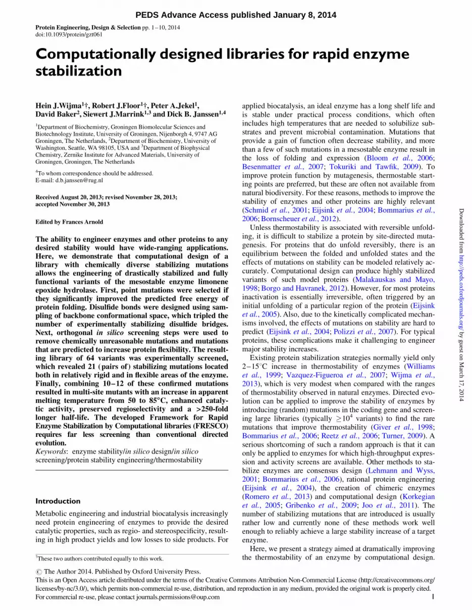
Fig. 1. Experimentally characterized point mutants of LEH. Protein variantsthat are significantly more thermostable are labeled (e.g. T85V). Theabbreviation NE stands for no soluble expression. The gray background isused to distinguish the mutations with a predicted DDGFold that does notsignificantly differ from zero (25 to þ5 kJ/mol). The variants that would notsurvive Steps 2 or 3 are plotted with different symbols as indicated in the inset.(A) The point mutations that were predicted to be stabilizing using Rosettaddgand also survived Steps 2 and 3 in FRESCO (Scheme 1). (B) Idem for thepoint mutations predicted to be stabilizing using FoldX; (C) Librarycharacterized for a control experiment, in which the effects of omitting Steps 2and 3 of the FRESCO protocol (Scheme 1) were tested. The best FoldXvariants were selected, with maximally one mutation per position (thus, onlyT85I with a DDGFold of 220 kJ/mol, not T85V with DDGFold of 214 kJ/mol).
The FRESCO strategy for rapid enzyme stabilization
3
by guest on March 17, 2014
http://peds.oxfordjournals.org/D
ownloaded from

on the averaged structures obtained from the five different tra-jectories per variant (Supplementary Fig. S3). If one out of thefive MD simulations appeared to sample a very different partof the protein conformational space than the other four, it wasignored (Supplementary Fig. S4). Removing these outliersenabled to compare different variants (Supplementary Fig. S3)because otherwise a protein variant that had such an outlierappeared to be much more flexible. For example, initially suchan outlier in the simulation of the WT protein (SupplementaryFig. S4) made it appear during the structural inspection as ifalmost all the mutants were significantly more rigid than theWT.
Experimental
A detailed account of all experimental methods is provided inthe Supplementary data. A plasmid containing the gene for theLEH was kindly provided by Prof. Dr. M. Arand (Universityof Zurich). Mutants of LEH were created by QuikChange mu-tagenesis (Agilent, USA) in a 96-well plate. Most enzyme var-iants, including those with multiple disulfide bonds, wereexpressed in Escherichia coli TOP10 cells (Life Technologies,CA, USA). Only variants with single disulfide bonds wereexpressed in E.coli NEB Shuffle Express (New EnglandBiolabs, USA). The mutations were validated by DNA se-quencing. Purification was carried out on Ni–NTA columnswith a C-terminal hexa-histidine tag using cell lysate preparedfrom cells grown in 1 l of Terrific Broth. If needed, furtherpurification was carried out by gel filtration (Supradex 75, Lifetechnologies, UK). This procedure yielded around 50 mg/lprotein for variants without disulfide bonds, and 5–10 mgprotein per liter culture volume for disulfide variants. Proteinscontaining disulfide bonds are usually less well expressed inthe cytoplasm of E.coli (Marco de, 2009).To determine the TM
app of variants during screening, the ther-mofluor method was carried out essentially as reported else-where (Ericsson et al., 2006). The analyzed samples consistedof either purified protein or cell-free extract for screening ofpoint mutations. For measurements, 5 ml 100� diluted com-mercial Sypro Orange solution (Life Technologies, CA, USA)was added to a 20 ml protein sample. The apparent meltingtemperature (TM
app) was determined by heating the samplesfrom 25 to 908C at 18C/min in a MyiQ real-time PCR machine(Bio-Rad, Hercules, CA, USA) while recording the fluores-cence with a 490 nm excitation filter and a 575 nm emissionfilter (Ericsson et al., 2006). The maximum of the relativefluorescence change with respect to the temperature (dRFU/dT) was taken as the apparent melting temperature (TM
app).The presence of inter-subunit disulfide bonds was analyzed
by examining the migration patterns of the WT and mutantproteins by sodium dodecyl sulfate–polyacrylamide gel elec-trophoresis (SDS–PAGE), both under reducing and non-reducing conditions, since reduction of disulfide bonds shouldcause a shift in migration behavior. To determine both inter-and intramolecular disulfide bonds, the number of freecysteines, which are not engaged in disulfide bonds, was deter-mined using Ellman’s reagent [5,50-dithio-bis-(2-nitrobenzoicacid)] (Ellman, 1959).Catalytic activities were measured in 4.5 ml potassium
phosphate (50 mM, pH 7.1), and the enzyme was pre-incubated at 308C for 5 min before adding (4R)-limonene1,2-epoxide (mixture of (1R,2S,4R) and (1S,2R,4R) isomers)as the substrate. After different incubation times, the reaction
mixtures were extracted with ethyl acetate, centrifuged and theorganic layers were removed and dried by Na2SO4. The pro-duction of diasteromers was analyzed by chiral GC, usinga Hydrodex b-TBDAc column (Aurora Borealis, TheNetherlands), with a temperature program from 40 to 1008C at108C/min, 100–1508C at 58C/min and finally 150–1808C at18C/min. The retention times of (1R,2R,4R)-limonene diol and(1S,2S,4R)-limonene diol under these conditions were 27.5and 29.6 min, respectively. A reference sample with boththese diols was prepared (Wang et al., 2008). The nearlyexclusive production of the (1S,2S,4R)-limonene diol wasconfirmed by 1H-NMR (200 MHz), using the same peakassignments as reported earlier (Blair et al., 2007).To measure residual catalytic activity versus temperature,
enzyme samples (1.0 mg ml21 in 50 mM potassium phos-phate, pH 7.1) were incubated for 15 min at the desired tempera-tures using a peqSTAR gradient polymerase chain reactionheating block (Peqlab Biotechnologie GmbH, Erlangen,Germany). Samples were allowed to cool down to 48C for1 min, and subsequently their catalytic activity was analyzed.
Results
Computational design of enriched libraries
The first step in the FRESCO strategy for engineering proteinstability consists of the computational design of potentiallystabilizing point mutations and disulfide bonds (Scheme 1).Point mutations were selected by computational tools thatpredict the resulting change in DG of folding (DDGFold). TheDDGFold values were calculated with both Rosettaddg andFoldX since the underlying algorithms gave significantly dif-ferent predictions (Supplementary Fig. S5), resulting in differ-ent selected mutations. All residues were allowed to mutate,except those inside or near the active site. Of all 1634 evalu-ated point mutations, 248 were selected either because theywere predicted to decrease the DDGFold
,25 kJ mol21 orbecause they introduced a known type of stabilizing point mu-tation, such as those introducing a proline (see the Methodssection for criteria). Of these 248 point mutations, 48% waspredicted to be stabilizing only by Rosettaddg, 26% only byFoldX and 25% by both algorithms.Disulfide bonds were designed employing the newly
written DDD algorithm, which uses MD simulations to samplebackbone conformational space. Without this sampling ofbackbone conformational space (i.e. using only the X-raystructure), seven disulfide bonds were predicted to be stabiliz-ing (Supplementary Table SII). The sampling of differentbackbone positions by the MD simulation (SupplementaryFig. S6) resulted in the design algorithm recognizing anadditional 21 possible disulfide bonds, providing a total of28 potentially stabilizing disulfide bonds (SupplementaryTable SII).In the second step (Scheme 1), 130 of the point mutants
(52%) were eliminated because structural inspection revealedfeatures that are typically encountered with destabilizing muta-tions (Nosoh and Sekiguchi, 1991). A control experimentdescribed below confirmed that this step enriches for stabiliz-ing mutations. Furthermore, such visual inspection to filter outthe unreasonable variants is commonly employed in computa-tional protein design (Kiss et al., 2013; Wijma and Janssen,2013). Here, the main reasons to eliminate point mutants were
H.J.Wijma et al.
4
by guest on March 17, 2014
http://peds.oxfordjournals.org/D
ownloaded from

that a hydrophobic side chain became surface exposed (70%)or that an unsatisfied H-bond donor or acceptor was created(20%). Furthermore, all 16 point mutations (12%) of Pro23were eliminated because it appeared that the calculations erro-neously predicted Pro23 to be unfavorable for folding. Otherreasons to eliminate variants were because a proline was intro-duced inside an a-helix (4%) or because a hydrophobicprotein cavity was created (2%). The sum is .100% becausefor 8% of the eliminated point mutants multiple eliminationcriteria applied. Of the disulfide bonds, five (18%) were elimi-nated because they created a large hydrophobic cavity. Thisleft 118 point mutations (36% from FoldX, 32% fromRosettaddg, 31% from both) and 23 disulfide bonds.
MD simulations on the surviving point mutations and disul-fide bond variants (Scheme 1, third step) were used to selectagainst variants with increased local flexibility relative to theWT, because regions of increased flexibility in a protein aremore prone to (partial) unfolding leading to inactivation(Vihinen, 1987). A control experiment described below con-firmed that this step eliminated destabilizing mutations. Fromthese MD simulations, it appeared that some of the designedvariants had a significantly lower local flexibility than the WT(Supplementary Fig. S7), which suggests increased stability.With the MD simulations, 54% of the 118 variants were elimi-nated, which reduced the number of variants that were pre-dicted to be stabilized to 64. This included 17 disulfide bondsand 47 point mutants of which 21 originated from FoldX, 12from Rosettaddg and 14 point mutations, which were predictedto be stabilizing by both Rosettaddg and FoldX.
When these 64 variants were experimentally screened(Scheme 1, fourth step; Supplementary Fig. S8, Table SIII),21 variants had an improved TM
app (33%). Of the 17 tested di-sulfide bond variants, 10 had an increased TM
app, ranging fromþ4 to þ158C. Seven out of the 10 stabilizing disulfide bondsoriginated from the additional backbone sampling by MDsimulation (Table I, Fig. 2). Of the 47 point mutations, 11were stabilizing (6 from FoldX, 3 from Rosettaddg, 2 fromboth, Fig. 1A and B). Point mutations with aDDGFold
. 25 kJ/mol had also been tested but none of these15 were experimentally stabilizing (Fig. 1A and B). Of thepoint mutations with a DDGFold
, 25 kJ/mol, 34% was sta-bilizing. The catalytic activity of the thermostabilized variantswas preserved, and most activities differ less than a factor 2from the catalytic activity of the WT LEH (SupplementaryTable SIV).
The discovery of 21 stabilized variants in a library of only64 variants is based on the use of the orthogonal in silico
screening steps (Steps 2 and 3 in Scheme 1) for eliminatingfalse-positive predictions. When using FoldX calculationsonly, a large fraction of the mutations predicted to be stabiliz-ing appeared to be destabilizing or neutral when tested experi-mentally. Selection of the mutations with the best predictedDDGFold using Rosettaddg would only have resulted in theintroduction of highly surface-exposed aromatic groups,which is a known problem in computational design (see theDiscussion section). The best 18 variants at different positionsaccording to FoldX included 6 variants that did pass throughStages 2 and 3, and thus also were included in the FRESCOlibrary. When the discarded 12 variants were experimentallycharacterized, none were stabilizing (Fig. 1C, SupplementaryTable SV) and half were strongly destabilizing. Of the 12false-positive predictions, 9 were eliminated at Step 2 of
FRESCO. Of these nine variants, four were eliminatedbecause a highly surface-exposed hydrophobic group wasintroduced (Q7M, E68L, A48F, S111M), three were elimi-nated because unsatisfied H-bond donors and acceptors werecreated by the mutations (S12M, T22D, G129S) and twowere eliminated because a proline was introduced inside ana-helix (A40P, A41P). The other three false positives wereeliminated at Step 3 of FRESCO because they were predictedto have increased flexibility (E49P, Y96W, R9P). The import-ance of eliminating false-positive predictions through Steps 2and 3 of the screening is also apparent from protein expres-sion levels, where lack of soluble expression of a mutant sug-gests lack of stability. Whereas in the FRESCO library, only2 variants (3%) were not expressed in soluble form, theabsence of soluble expression was observed for 4 of the 12discarded variants. Two of these four variants that could notbe solubly expressed belonged to the three variants that hadbeen eliminated by MD flexibility screening. These resultsconfirm that a framework type of computational approach, inwhich orthogonal screening steps serve to eliminate false-positive predictions, improves the accuracy with which sta-bilizing mutations are predicted and allows the discovery ofmultiple stabilizing mutations with minimal experimentalscreening.
Table I. Experimentally confirmed stabilizing disulfide bonds designed using
crystal structures or conformations from an MD simulation
Protein structurea Cysteine positions3 4 5 40 44 48 112 17 17 89102 82 84 72 68 126 142 92b 94b 91b
1NWW þ þ
1NU3 þ þ þ
500 ps þ þ þ
750 ps þ þ þ þ
1000 ps þ þ þ þ þ
1250 ps þ þ þ
1500 ps þ þ þ
1750 ps þ þ þ þ þ
2000 ps þ þ þ þ þ
2250 ps þ þ þ þ
2500 ps þ þ
aThe first column indicates which X-ray structure (pdb entry) or MDsimulation snapshot (ps after start of simulation) was used for thecomputational design of disulfide bonds.bInter-subunit disulfide bond.
Fig. 2. Overview of the stabilized variants.
The FRESCO strategy for rapid enzyme stabilization
5
by guest on March 17, 2014
http://peds.oxfordjournals.org/D
ownloaded from

Origins of stabilization
The modeled 3D structures of the improved variants were ana-lyzed to investigate the structural basis for the stabilizingeffects. Because of the large number of stabilizing mutations,the effects of individual mutations are described in theSupplementary data. The beneficial mutations appear to intro-duce better H-bonds that stabilize the local protein structure(A19K, N92K), improved surface charge–charge interactions(A19K, E45K, T76K, N92K, N92R), improved hydrophobicinteractions (T85I, T85V, T85L, Y96F, Fig. 3) and entropicstabilization (S15P and all disulfide variants). All of the stabil-izing mutations are located at or near the surface of LEH.Furthermore, the most successful mutations appear to be pre-dominantly localized inside or near the flexible N-terminus(residues 3–24) and to a much lesser extent in Helices 3 and 4(Fig. 4). Many of the mutations are also near to the dimerinterface, with the exception of the highly stabilizingS3C-V102C, K4C-A82C and A5C-E84C disulfide bonds thatare located at the N-terminus but not at the dimer interface.The unsuccessful mutations are more evenly spread over theprotein than the stabilizing mutations. These observations in-dicate that the N-terminal loop is the most critical region for
stability of LEH, and that the successful mutations especiallyintroduced stabilizing interactions in and around this region.
Design of combined variants
Aiming to obtain highly thermostable variants, the most stabil-izing mutations (Fig. 2) were rationally combined withoutconstruction of intermediate variants. If at a single positionmultiple stabilizing mutations were available, then the moststabilizing variant was used; for example, N92K with a DTM
app
of þ78C was preferred over N92R with a DTMapp of þ28C.
Since the stability of LEH can be increased by improving thelocal stability of the N-terminal region (see above), differentcombinations of mutations were screened by MD simulationfor their effect on the flexibility of the N-terminus. Disulfidebonds were included and MD simulations were again used totest their compatibility. To test the usefulness of the flexibilitypredictions by MD simulations (third step of Scheme 1) forvariants in which multiple mutations are combined, twocombinations of disulfide bonds were characterized, of whichone (S3C/I5C/E84C/V102C) was predicted to rigidify theN-terminus and thus be stabilizing, while the other combin-ation (A40C/I44C/E68C/A72C) was predicted to increase the
Fig. 3. Example of the predicted structure for a stabilizing mutation. The substitution T85V (DTmapp¼ þ78C) removes a hydroxyl group in an apolar environment,
and replaces it with a more hydrophobic methyl group. The polar side-chain atoms of Thr97 and Arg99 are .5 A from the hydroxyl oxygen of Thr85, whichexcludes hydrogen bonding of Thr85 with those residues, indicating that Thr85 has an unsatisfied H-bond donor or acceptor.
Fig. 4. Distribution of stabilizing mutations over the protein (crystal structure 1NWW). (A) B-factors of the Ca atoms of 1NWW (thickest traces with red colorcorrespond to the highest B-factors). (B) Location of all the point mutations shown with spheres for which the color reveals the level of stabilization.
H.J.Wijma et al.
6
by guest on March 17, 2014
http://peds.oxfordjournals.org/D
ownloaded from
flexibility of the N-terminal region of the enzyme, and thus bedestabilizing. Indeed, S3C/I5C/E84C/V102C had a higherTMapp than its parents (66.88C, DTM
app¼ þ15.88C versus
þ13.58C for I5C/E84C and þ11.08C for S3C/V102C), whileA40C/I44C/E68C/A72C had a lower TM
app than its parents(54.88C, DTM
app¼ 3.8 versus þ5.08C for A40C/A72C and
þ5.58C for 44C/E68C). These experimental results are inagreement with the predictions from MD simulation, and indi-cate that MD simulations can increase the chance of success-fully combining mutations.
The application of this method to combine multiple muta-tions simultaneously resulted in the final variants F1, with 12mutations, and F2 with 10 mutations (Fig. 2). These twovariants combine the strongest stabilizing mutations that werepredicted by MD to rigidify the N-terminus, whereas combina-tions that enhance local flexibility were discarded. Forexample, S15P was omitted from variant F2 because in com-bination with the other mutations, an increased flexibility ofthe N-terminal loop was predicted by MD simulation.Furthermore, the highly stabilizing mutation N92K wasomitted from variant F2 because it cannot be combined withthe A17C/N92C disulfide bonds. Also, maximally two disul-fide bridges per enzyme were combined to avoid potentialproblems with the kinetics of protein folding.
Catalytic properties of combined variants
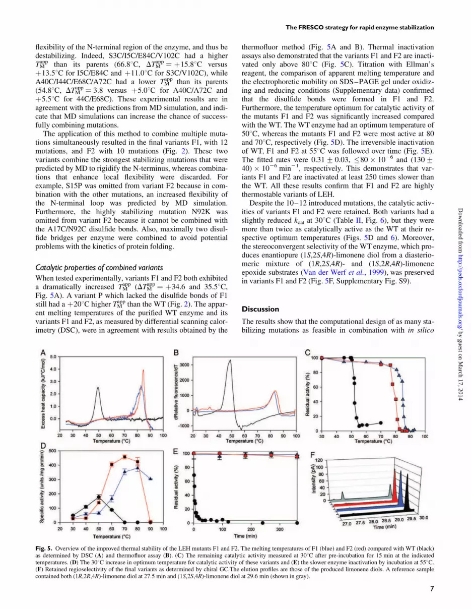
When tested experimentally, variants F1 and F2 both exhibiteda dramatically increased TM
app (DTMapp¼ þ34.6 and 35.58C,
Fig. 5A). A variant P which lacked the disulfide bonds of F1still had a þ208C higher TM
app than the WT (Fig. 2). The appar-ent melting temperatures of the purified WT enzyme and itsvariants F1 and F2, as measured by differential scanning calor-imetry (DSC), were in agreement with results obtained by the
thermofluor method (Fig. 5A and B). Thermal inactivationassays also demonstrated that the variants F1 and F2 are inacti-vated only above 808C (Fig. 5C). Titration with Ellman’sreagent, the comparison of apparent melting temperature andthe electrophoretic mobility on SDS–PAGE gel under oxidiz-ing and reducing conditions (Supplementary data) confirmedthat the disulfide bonds were formed in F1 and F2.Furthermore, the temperature optimum for catalytic activity ofthe mutants F1 and F2 was significantly increased comparedwith the WT. The WT enzyme had an optimum temperature of508C, whereas the mutants F1 and F2 were most active at 80and 708C, respectively (Fig. 5D). The irreversible inactivationof WT, F1 and F2 at 558C was followed over time (Fig. 5E).The fitted rates were 0.31+ 0.03, �80 � 1026 and (130+40) � 1026 min21, respectively. This demonstrates that var-iants F1 and F2 are inactivated at least 250 times slower thanthe WT. All these results confirm that F1 and F2 are highlythermostable variants of LEH.Despite the 10–12 introduced mutations, the catalytic activ-
ities of variants F1 and F2 were retained. Both variants had aslightly reduced kcat at 308C (Table II, Fig. 6), but they weremore than twice as catalytically active as the WT at their re-spective optimum temperatures (Figs. 5D and 6). Moreover,the stereoconvergent selectivity of the WT enzyme, which pro-duces enantiopure (1S,2S,4R)-limonene diol from a diasterio-meric mixture of (1R,2S,4R)- and (1S,2R,4R)-limoneneepoxide substrates (Van der Werf et al., 1999), was preservedin variants F1 and F2 (Fig. 5F, Supplementary Fig. S9).
Discussion
The results show that the computational design of as many sta-bilizing mutations as feasible in combination with in silico
Fig. 5. Overview of the improved thermal stability of the LEH mutants F1 and F2. The melting temperatures of F1 (blue) and F2 (red) compared with WT (black)as determined by DSC (A) and thermofluor assay (B). (C) The remaining catalytic activity measured at 308C after pre-incubation for 15 min at the indicatedtemperatures. (D) The 308C increase in optimum temperature for catalytic activity of these variants and (E) the slower enzyme inactivation by incubation at 558C.(F) Retained regioselectivity of the final variants as determined by chiral GC.The elution profiles are those of the produced limonene diols. A reference samplecontained both (1R,2R,4R)-limonene diol at 27.5 min and (1S,2S,4R)-limonene diol at 29.6 min (shown in gray).
The FRESCO strategy for rapid enzyme stabilization
7
by guest on March 17, 2014
http://peds.oxfordjournals.org/D
ownloaded from

and experimental screening allowed for rapid engineering ofenzyme variants with a dramatically increased thermostability.Essential features of the FRESCO strategy proposed here arethe use of computational design methods to create a library ofpotentially stabilizing mutations, followed by a reduction oflibrary size through orthogonal in silico screening aimed at re-moving false-positive predictions. An experimental screeningof the resulting library is then used to select the most stabiliz-ing variants for combination. The best combined variantsexhibited an increase in TM
app of ≏358C as shown by DSC,thermofluor assays and activity–temperature measurements,while catalytic activity and stereoselectivity were maintained.Natural thermostable enzymes are often far less catalyticallyactive at lower temperatures than their mesostable homologs(Fitter et al., 2001; Cheung et al., 2005). The results here showthat even a 358C increase in TM
app is not necessarily accompan-ied by loss of catalytic activity at lower temperatures.The remarkable stabilization of LEH was achieved by ex-
perimental testing of ,100 variants in just two rounds of mu-tagenesis. This is a very low number when compared withdirected evolution, where often .104 variants need to bescreened to obtain a strong stabilization, since the vast major-ity of the tested mutations are neutral or detrimental for stabil-ity (Bloom and Arnold, 2009). In most cases, the screeningstep is the bottleneck of a directed evolution project (Turner,2009) and limits its application. Often no rapid expressionsystems or stability assays are available. High-throughputscreening can be unfeasible if slow-growing organisms arerequired for protein expression or if assays cannot be scaleddown. Therefore, the protocols presented here will be an at-tractive alternative for many proteins.
The experimental results revealed that a critical region forstabilization of LEH is located in the vicinity of the flexibleN-terminus (Fig. 4B). This includes both the interface of thedimer and the N-terminus itself which is partly more remotefrom the interface. For example, both the G89C/S91C disul-fide bond at the interface and the A5C/E84C disulfide bond atthe N-terminus are highly stabilizing. The residues of the N-terminal S3C-V102C disulfide bond are located .12 A awayfrom the dimer interface, suggesting that its increased thermo-stability can be unrelated to improved stability of this dimerinterface.Recent strategies for protein stabilization often only select
the most flexible residues for mutagenesis (Reetz et al., 2006;Jochens et al., 2010; Joo et al., 2011), with the rationale thatthese should be the most critical residues. However, some ofthe highly stabilizing mutations found here are in a rigid partof the protein. For example, T85V (DTM
appþ78C) is close to
the flexible N-terminus, even though the mutated residue is ina rigid part of the protein as judged by its B-factors (13 A2, inX-ray structure 1NWW), which are lower than average (15 A2
is the average B-factor in 1NWW) and much lower than thoseof the flexible N-terminus (Ile 4, 27 A2). Thus, the computa-tional methods generated stabilizing mutations that wouldhave been missed if only highly flexible residues had beenselected for mutagenesis.An essential element of the FRESCO strategy is the elimin-
ation of mutations that are suggested by the computational pro-tocols, but lack credibility when their predicted flexibility istaken into account or when their predicted structure is exam-ined. Structural inspection showed that about 50% of themutations predicted to be stabilizing by the initial calculations
Table II. Catalytic parameters of WT LEH and variants F1 and F2
Variant WT F1 F2
Temperature (8C) 30 50 30 80 30 70kcat (s
21) 13.9+0.8 63+4 8.9+0.4 135+6 8.2+0.3 160+7KM (mM) 0.3+0.1 0.6+0.1 ,0.25a 0.6+0.1 ,0.25a 0.3+0.1kcat/KM (s21 M21) 4.6 � 104 1.0 � 105 .3.6 � 104 2.1 � 105 .3.3 � 104 4.9 � 105
aKM was below the detection limit of 0.25 mM. The kinetic parameters for the hydrolysis of (4R)-limonene 1,2-epoxide were determined both at 308C and at theoptimum temperature of WT and variants F1 and F2.
Fig. 6. Rate of (4R)-limonene 1,2-epoxide conversion versus its concentration. Wild-type LEH (black circles), variant F1 (blue triangles) and variant F2 (redsquares) are indicated with different symbols. The fit is according to kt ¼ kcat � [S]/([S] þ KM), in which kt is the catalytic turnover rate per enzyme active site and[S] is the substrate concentration. The turnover rate is plotted at (A) 308C and (B) at the optimum temperature for catalytic activity (for WT 508C; for variant F1808C; for variant F2 708C). At 308C, the mutants have a lower catalytic activity compared with the WT, whereas the mutants clearly outperform the WT at theoptimum temperature.
H.J.Wijma et al.
8
by guest on March 17, 2014
http://peds.oxfordjournals.org/D
ownloaded from
(Step 1) are probably false positives. They were discarded inStep 2 (Scheme 1) because they introduce structural featuresthat are expected to destabilize the protein, such as water-exposed hydrophobic side chains. The latter is a knownproblem of computational design algorithms (Jacak et al.,2012), and this justifies the use of rational criteria to removefalse positives that result from imperfect energy functions andsampling in the design algorithms (Kellogg et al., 2011;Leaver-Fay et al., 2013). It is common in computationaldesign to eliminate variants that have clear structural problems(Kiss et al., 2013;Wijma and Janssen, 2013). In the future, thiscould be automated as has been done for finding errors inX-ray structures (Vriend, 1990), or may no longer be necessaryif the energy calculations and sampling methods are furtherimproved. Since local unfolding followed by irreversible ag-gregation may be as important for enzyme inactivation asoverall thermodynamic stability (Polizzi et al., 2006; Reetzet al., 2006; Joo et al., 2011), elimination of false positiveswas also based on MD simulations which predicted effects onlocal flexibility (Step 3). It is well established that high flexi-bility can promote unfolding (Vihinen, 1987). Here, experi-mental characterization of mutants that were eliminated at thethird step of FRESCO because of higher flexibility showedthat the discarded mutations were not stabilizing and oftenwere even strongly destabilizing (Fig. 1C).
The efficiency of FRESCO as a strategy is confirmed by thelarge number of mechanistically different stabilizing muta-tions that were discovered. The point mutations appear to actthrough various effects that can stabilize a protein, includingthe removal of unsatisfied H-bond donors/acceptors, introduc-tion of new H-bonds, better charge distribution (Karshikoffand Ladenstein, 2001; Gribenko et al., 2009), less hydrophobicexposure to solvent and entropic stabilization (Nosoh andSekiguchi, 1991; Eijsink et al., 2004).Multiple disulfide bondsper protein, like in variants F1 and F2, occur naturally in theproteomes of a few thermophiles (Ladenstein and Ren, 2008).A complete list of the mutations and their proposed effects isgiven in Supplementary data. The ability to obtain mechanis-tically diverse types of stabilizing mutations is likely tobecome essential if the goal is to engineer strongly enhancedstability into any target protein.
The developed computational strategy to stabilize anenzyme is reminiscent of directed evolution, in that a libraryof potentially stabilizing mutations is experimentally screenedbefore combining the most successful mutations to final var-iants. A more common approach in computational design ofthermostability is to select the best set of mutations purely in
silico and only characterize the final combined variants(Malakauskas and Mayo, 1998; Korkegian et al., 2005;Gribenko et al., 2009; Diaz et al., 2011; Joo et al., 2011; Borgoand Havranek, 2012;Miklos et al., 2012;Murphy et al., 2012).The results in Fig. 1B show that such an approach would havemissed highly stabilizing mutations (T85V/N92K). Anotherapproach is to use the consensus approach in combination withcomputational design. Using FoldX for the computations,such an approach resulted in a cellobiohydrolase with a 98Cimproved TM
app (Komor et al., 2012). However, in a similarstudy it was reported that FoldX did not correlate well withthermostabilizing mutations (Polizzi et al., 2006), which is inagreement with our results of finding false positives in theabsence of orthogonal screening (Fig. 1C). To allow for largerincreases in thermostability, the FRESCO approach uses an
experimental screening to verify that the mutations indeed sta-bilize the enzyme and spare catalytic activity before creatingvariants in which mutations are combined.The modeling of backbone flexibility is an important
problem in computational protein design. With a rigid back-bone, many beneficial mutations will be sterically excluded.The unusually large number of stabilizing disulfide bonds dis-covered in this study is mainly due to the use of an MD simu-lation that samples the natural backbone flexibility to generatedifferent realistic starting structures for the design of disulfidebonds. Backbone sampling protocols normally do not incorp-orate explicit water molecules (Su and Mayo, 1997; Georgievet al., 2008; Smith and Kortemme, 2008; Havranek and Baker,2009; Babor et al., 2011; Chitsaz and Mayo, 2013). The MDsimulations include the surrounding water hydrogen-bondingnetwork, which should make the sampling of energetically ac-cessible conformations more accurate. This protocol produced7 out of the 10 successful disulfide bonds, which included allthree disulfide bonds that were combined in the final highlythermostable variants (Table I, Fig. 2). We are not aware ofprevious reports describing a similar large number of stabiliz-ing disulfide bonds. With existing methods to stabilizeenzymes, typically one or two stabilizing disulfide bonds arereported (Matsamura et al., 1989; Pikkemaat et al., 2002;Dombkowski, 2003; Chen et al., 2009). Such numbers aresimilar to the finding of three stabilizing disulfide bonds forLEH (Table I) in the absence of backbone conformationalsampling. The significant increase in the number of stabilizingdisulfide bonds due to the conformational sampling experi-mentally shows that MD can generate structures that are accur-ate enough for computational protein design.The backbone sampling allowed to predict stabilizing disul-
fide bonds at positions, where based on the X-ray structure adisulfide bond would not be feasible because the backboneatoms were too far away from each other. For example, in caseof disulfide bonds distances of 3.6–7.2 A occur between theirrespective Ca atoms (Petersen et al., 1999) in natural proteins,whereas the distance between the Ca atoms of residues 4 and82 (where a stabilizing disulfide bond could be formed,Table I, Fig. 2) is at least 8.90 in the available X-ray structures(1NWW, 1NU3). During the MD simulation, the distancebetween the Ca atoms of residues 4 and 82 decreased to6.52 A (results not shown). Without backbone conformationalsampling, the additional disulfide bonds obtained from MDsimulation could only have been discovered if the geometriccriteria would have been relaxed. However, in that case thealgorithms would also have proposed disulfide bonds that areexpected to destabilize the protein because their geometriesare far outside the naturally occurring ranges.The multi-site mutants, which harbored 12 (variant F1) or
10 (variant F2) substitutions, are fully catalytically active,with an increase in kcat at the optimum temperature when com-pared with WT. The only precaution adopted in the FRESCOprotocol was not to introduce mutations at residues close to theactive site. Regioselectivity of water attack on the diastereo-meric substrate is fully retained, allowing enantioconvergentproduction of (1S,2R,4R)-limonene diol. The resulting var-iants are suitable for use in protein engineering aimed at intro-ducing new selectivities.In conclusion, we show that computational library design
can identify many mutations with different stabilizationmechanisms to cumulatively obtain a large increase in enzyme
The FRESCO strategy for rapid enzyme stabilization
9
by guest on March 17, 2014
http://peds.oxfordjournals.org/D
ownloaded from

thermostability. The computational library design enabled alarger jump in enzyme stability while preserving catalytic ac-tivity. The developed FRESCO strategy made it feasible toobtain protein variants with high thermostability in a shorttime with minimal experimental screening.
Supplementary data
Supplementary data are available at PEDS online.
Funding
This work was supported by the European Union 7thFramework via the Kyrobio project (KBBE-2011-5, 289646)and the Metaexplore project (KBBE-2007-3-3-05, 222625)and by NWO (Netherlands Organization for ScientificResearch) through an ECHO grant.
References
Arand,M., Hallberg,B., Zou,J., Bergfors,T., Oesch,F., Van der Werf,M., deBont,J., Jones,T. and Mowbray,S. (2003) EMBO J., 22, 2583–2592.
Babor,M., Mandell,D.J. and Kortemme,T. (2011) Protein Sci., 20,1082–1089.
Berendsen,H.J.C., Postma,J.P.M., Van Gunsteren,W.F., Dinola,A. andHaak,J.R. (1984) J. Chem. Phys., 81, 3684–3690.
Besenmatter,W., Kast,P. and Hilvert,D. (2007) Protein Struct. Funct.
Bioinform., 66, 500–506.Blair,M., Andrews,P.C., Fraser,B.H., Forsyth,C.M., Junk,P.C., Massi,M. andTuck,K.L. (2007) Synthesis, 2007, 1523–1527.
Bloom,J., Labthavikul,S., Otey,C. and Arnold,F. (2006) Proc. Natl Acad. Sci.USA, 103, 5869–5874.
Bloom,J.D. and Arnold,F.H. (2009) Proc. Natl Acad. Sci. USA., 106,9995–10000.
Bommarius,A.S., Broering,J.M., Chaparro-Riggers,J.F. and Polizzi,K.M.(2006) Curr. Opin. Biotechnol., 17, 606–610.
Borgo,B. and Havranek,J.J. (2012) Proc. Natl Acad. Sci. USA, 109,1494–1499.
Bornscheuer,U.T., Huisman,G.W., Kazlauskas,R.J., Lutz,S., Moore,J.C. andRobins,K. (2012) Nature, 485, 185–194.
Caves,L.S.D., Evanseck,J.D. and Karplus,M. (1998) Protein Sci., 7, 649–666.Chen,L., Yu,C., Zhou,X. and Zhang,Y. (2009) J. Microbiol. Biotechnol., 19,1506–1513.
Cheung,Y.Y., Lam,S.Y., Chu,W.K., Allen,M.D., Bycroft,M. and Wong,K.B.(2005) Biochemistry, 44, 4601–4611.
Chitsaz,M. and Mayo,S.L. (2013) J. Comput. Chem., 34, 445–450.Diaz,J.E., Lin,C., Kunishiro,K., Feld,B.K., Avrantinis,S.K., Bronson,J.,Greaves,J., Saven,J.G. and Weiss,G.A. (2011) Protein Sci., 20, 1597–1606.
Dombkowski,A.A. (2003) Bioinformatics, 19, 1852–1853.Eijsink,V., Bjork,A., Gaseidnes,S., Sirevag,R., Synstad,B., Van den Burg,B.and Vriend,G. (2004) J. Biotechnol., 113, 105–120.
Eijsink,V., Gaseidnes,S., Borchert,T. and Van den Burg,B. (2005) Biomol.
Eng., 22, 21–30.Ellman,G.L. (1959) Arch. Biochem. Biophys., 82, 70–77.Ericsson,U.B., Hallberg,B.M., DeTitta,G.T., Dekker,N. and Nordlund,P.(2006) Anal. Biochem., 357, 289–298.
Essmann,U., Perera,L., Berkowitz,M.L., Darden,T., Lee,H. and Pedersen,L.G.(1995) J. Chem. Phys., 103, 8577–8593.
Fitter,J., Herrmann,R., Dencher,N.A., Blume,A. and Hauss,T. (2001)Biochemistry, 40, 10723–10731.
Georgiev,I., Keedy,D., Richardson,J.S., Richardson,D.C. and Donald,B.R.(2008) Bioinformatics, 24, I196–I204.
Giver,L., Gershenson,A., Freskgard,P. and Arnold,F. (1998) Proc. Natl Acad.Sci. USA, 95, 12809–12813.
Gribenko,A.V., Patel,M.M., Liu,J., McCallum,S.A., Wang,C. andMakhatadze,G.I. (2009) Proc. Natl Acad. Sci. USA, 106, 2601–2606.
Guerois,R., Nielsen,J. and Serrano,L. (2002) J. Mol. Biol., 320, 369–387.Havranek,J.J. and Baker,D. (2009) Protein Eng. Des. Sel., 18, 1293–1305.Jacak,R., Leaver-Fay,A. and Kuhlman,B. (2012) Protein Struct. Funct.
Bioinform., 80, 825–838.Jochens,H., Aerts,D. and Bornscheuer,U.T. (2010) Protein Eng. Des. Sel., 23,903–909.
Joo,J.C., Pack,S.P., Kim,Y.H. and Yoo,Y.J. (2011) J. Biotechnol., 151, 56–65.Karshikoff,A. and Ladenstein,R. (2001) Trends Biochem. Sci., 26, 550–556.Kellogg,E.H., Leaver-Fay,A. and Baker,D. (2011) Protein Struct. Funct.
Bioinform., 79, 830–838.Kiss,G., Celebi-Oelcuem,N., Moretti,R., Baker,D. and Houk,K.N. (2013)Angew. Chem. Intl. Ed., 52, 5700–5725.
Komor,R.S., Romero,P.A., Xie,C.B. and Arnold,F.H. (2012) Protein Eng.
Des. Sel., 25, 827–833.Korkegian,A., Black,M., Baker,D. and Stoddard,B. (2005) Science, 308,857–860.
Krieger,E., Darden,T., Nabuurs,S., Finkelstein,A. and Vriend,G. (2004)Proteins, 57, 678–683.
Kumar,S., Tsai,C.J. and Nussinov,R. (2000) Protein Eng., 13, 179–191.Ladenstein,R. and Ren,B. (2008) Extremophiles, 12, 29–38.Leaver-Fay,A., O’Meara,M.J., Tyka,M., et al. (2013) Meth. Prot. Des., 523,109–143.
Lehmann,M. and Wyss,M. (2001) Curr. Opin. Biotechnol., 12, 371–375.Malakauskas,S. and Mayo,S. (1998) Nat. Struct. Biol., 5, 470–475.Marco de,A. (2009)Microb. Cell Factories, 8, e26Matsamura,M., Becktel,W.J., Levitt,M. and Matthes,B.W. (1989) Proc. NatlAcad. Sci. USA, 86, 6562–6566.
Miklos,A.E., Kluwe,C., Der,B.S., et al. (2012) Chem. Biol., 19, 449–455.Mrabet,N.T., Van den Broeck,A., Van den Brande,I., Stanssens,P., Laroche,Y.,Lambeir,A.M., Matthijssens,G., Jenkins,J., Chiadmi,M. and vanTilbeurgh,H. (1992) Biochemistry, 31, 2239–2253.
Murphy,G.S., Mills,J.L., Miley,M.J., Machius,M., Szyperski,T. andKuhlman,B. (2012) Structure, 20, 1086–1096.
Nosoh,Y. and Sekiguchi,T. (1991) Protein Stability and Stabilization Through
Protein Engineering, Horwood, New York.Pellequer,J. and Chen,S.W. (2006) Protein Struct. Funct. Bioinform., 65,192–202.
Petersen,M., Jonson,P. and Petersen,S. (1999) Protein Eng., 12, 535–548.Pikkemaat,M.G., Linssen,A.B., Berendsen,H.J. and Janssen,D.B. (2002)Protein Eng., 15, 185–192.
Polizzi,K.M., Chaparro-Riggers,J.F., Vazquez-Figueroa,E. andBommarius,A.S. (2006) Biotechnol J., 5, 531–536.
Polizzi,K.M., Bommarius,A.S., Broering,J.M. and Chaparro-Riggers,J.F.(2007) Curr. Opin. Chem. Biol., 11, 220–225.
Reetz,M.T., D Carballeira,J. and Vogel,A. (2006) Angew. Chem. Intl. Ed., 45,7745–7751.
Romero,P.A., Krause,A. and Arnold,F.H. (2013) Proc. Natl Acad. Sci. USA,110, E193–E201.
Schmid,A., Dordick,J., Hauer,B., Kiener,A., Wubbolts,M. and Witholt,B.(2001) Nature, 409, 258–268.
Smith,C.A. and Kortemme,T. (2008) J. Mol. Biol., 380, 742–756.Sokalingam,S., Raghunathan,G., Soundrarajan,N. and Lee,S. (2012) Plos One,7, e40410.
Su,A. and Mayo,S.L. (1997) Protein Sci., 6, 1701–1707.Tokuriki,N. and Tawfik,D.S. (2009) Curr. Opin. Struct. Biol., 19, 596–604.Turner,N.J. (2009) Nat. Chem. Biol., 5, 568–574.Van der Werf,M.J., Orru,R.V.A., Overkamp,K.M., Swarts,H.J., Osprian,I.,Steinreiber,A., de Bont,J.A.M. and Faber,K. (1999) Appl. Microbiol.
Biotechnol., 52, 380–385.Vazquez-Figueroa,E., Chaparro-Riggers,J. and Bommarius,A.S. (2007)ChemBioChem, 8, 2295–2301.
Vihinen,M. (1987) Prot. Eng., 1, 477–480.Vriend,G. (1990) J. Mol. Graph., 8, 52–5&.Wang,J., Cieplak,P. and Kollman,P. (2000) J. Comp. Chem., 21, 1049–1074.Wang,Z., Cui,Y., Xu,Z. and Qu,J. (2008) J. Org. Chem., 73, 2270–2274.Wijma,H.J. and Janssen,D.B. (2013) FEBS J., 280, 2948–2960.Wijma,H.J., Floor,R.J. and Janssen,D.B. (2013) Curr. Opin. Struct. Biol., 23,589–594.
Williams,J., Zeelen,J., Neubauer,G., Vriend,G., Backmann,J., Michels,P.,Lambeir,A. and Wierenga,R. (1999) Protein Eng., 12, 243–250.
Zheng,H. and Reetz,M.T. (2010) J. Am. Chem. Soc., 132, 15744–15751.
H.J.Wijma et al.
10
by guest on March 17, 2014
http://peds.oxfordjournals.org/D
ownloaded from