colonisation et biodégradation par la plastisphère
TRANSCRIPT
HAL Id: tel-03381781https://tel.archives-ouvertes.fr/tel-03381781
Submitted on 18 Oct 2021
HAL is a multi-disciplinary open accessarchive for the deposit and dissemination of sci-entific research documents, whether they are pub-lished or not. The documents may come fromteaching and research institutions in France orabroad, or from public or private research centers.
L’archive ouverte pluridisciplinaire HAL, estdestinée au dépôt et à la diffusion de documentsscientifiques de niveau recherche, publiés ou non,émanant des établissements d’enseignement et derecherche français ou étrangers, des laboratoirespublics ou privés.
Ecotoxicologie microbienne des plastiques en mer :colonisation et biodégradation par la plastisphère
Justine Jacquin
To cite this version:Justine Jacquin. Ecotoxicologie microbienne des plastiques en mer : colonisation et biodégradationpar la plastisphère. Ecosystèmes. Sorbonne Université, 2020. Français. �NNT : 2020SORUS104�.�tel-03381781�
Sorbonne Université Ecole doctorale des Sciences de l’Environnement d’Ile-de-France ED 129
Laboratoire d’Océanographie Microbienne UMR 7621
Ecotoxicologie microbienne des plastiques en mer :
Colonisation et biodégradation par la plastisphère
Par Justine JACQUIN
Thèse de doctorat en Microbiologie Marine
Présentée et soutenue publiquement le 16 Octobre 2020
Devant un jury composé de :
Régis GRIMAUD HDR, IPREM, Pau Rapporteur
Gaëtan BURGAUD HDR, LUBEM, Brest Rapporteur
Pierre GALAND DR, LECOB, Banyuls-sur-Mer Président
Stéphane BRUZAUD Professeur, IRDL, Lorient Examinateur
Jean-François BRIAND MCF, MAPIEM, Toulon Examinateur
Valérie BARBE CR, CEA, Paris Co-directrice de thèse
Jean-François GHIGLIONE HDR, LOMIC, Banyuls-sur-Mer Directeur de thèse
Rapporteur
Rapporteur
Président
Examinateur
Examinateur
Co-directrice de thèse
Directeur de thèse

Remerciements
Une thèse ça marque une vie et ces remerciements sonnent la fin de ces trois années
passées au laboratoire de Banyuls. Ce travail n’aurait pu s’achever sans l’aide de toutes les
personnes qui suivent :
En premier lieu j’aimerai remercier le jury de thèse qui a accepté d’évaluer mon travail
en y consacrant une partie de leur temps : Pierre Galand, Stéphane Bruzaud, Jean-François
Briand. Je remercie en particulier les personnes qui ont accepté d’être rapporteurs de cette
thèse : Regis Grimmaud et Gaëtan Burgaud.
Je tiens également à remercier mon directeur de thèse Jean-François Ghiglione et ma
co-directrice Valérie Barbe, m’ayant permis de réaliser cette thèse sur un sujet si passionnant.
Jeff merci de m’avoir laissé en autonomie tout au long de cette thèse et d’avoir eu une relecture
attentive de chacun des chapitres de ce manuscrit. Merci à toi Valérie, d’avoir été là pendant
ces deux années, ton aide a été précieuse, tant au laboratoire que dans l’analyse des résultats et
l’écriture de cette thèse. Merci pour ces moments de détentes et ses discussions si précieuses
pour moi qui m’ont permis d’égayer mes journées au laboratoire.
Mes prochains remerciements sont dédiés à toutes ces personnes m’ayant
accompagnées, guidées et aidées pendant cette thèse. En premier lieu j’aimerai remercier
Nolwenn Callac, qui m’a tant appris malgré son passage éphémère au laboratoire. Merci Nono
pour tout ce que tu as pu m’apporter tant scientifiquement que personnellement. Merci pour ce
soutien sans faille même à des milliers de km, pour ces réunions nocturnes et ces quelques mois
de colocation inoubliables. J’ai conscience que sans toi cette thèse ne serait pas ce qu’elle est
aujourd’hui. J’aimerai également remercier Charlène Odobel, pour son implication et son aide
indispensable pour l’expérience « DNA-SIP ». Merci pour tous ces bons moments partagés,
pour tes conseils et ton expertise scientifique qui ont été d’un grand support pendant ces trois
années.
J’aimerai également remercier toutes ces personnes au laboratoire qui m’ont permis de
d’avancer dans mes travaux de recherches dans de bonnes conditions. Merci à Valérie Bayot,
Laurent Intertaglia, Nyree West, Philippe Catala, Christophe Salmeron, David Pecqueur, et
Olivier Crispi, d’avoir toujours répondu à mes demandes avec gentillesse et rapidité. J’aimerai
également remercier Pascal Conan et Mireille Pujo-Pay, pour leur disponibilité, leurs nombreux
conseils et leur aide dans les expériences. Un merci tout particulier à Valérie Vergé pour
m’avoir toujours sortie de situation de crises avec bonne humeur et enthousiasme, tu as été d’un
réel soutien technique et psychologique tout au long de cette thèse. Je remercie également
Frédérique, Nathalie, Margaux, Carolanne et Clélia, qui m’ont apporté leur soutien ainsi que de
bons moments au laboratoire.
Mes travaux scientifiques ont pu être réalisés aussi grâce à des collaborations. Je
remercie Stéphane Bruzaud, pour nous avoir toujours fourni avec réactivité les plastiques
demandés. Merci également à Yonko Goarant pour avoir passé des journées entières avec moi
au microscope électronique à scruter la surface des polymères.
Je tiens ensuite à remercier mes collègues de travail devenus des amis avec le temps,
qui ont apporté tant de bonne humeur et de joie de vivre au sein de ce laboratoire. First,
Bouchnini merci à toi pour ce soutien de tous les instants, je ne compte plus le nombre de fois
ou ma session R n’a pas aborté grâce à toi ! Merci également à toi Angel, pour tous ces précieux
conseils sur R et pour ta constante bonne humeur qui aura illuminé mes journées. J’ai
conscience d’avoir été dans le bureau des meilleurs ! Je remercie également Pavla, Yann, Ying
and Fernanda, ça a été un bonheur de partager tous ces moments avec vous.
Un immense merci à Jana, pour ce second souffle que tu auras apporté à ma thèse et à
ma motivation, tes conseils et ta bonne humeur communicative m’auront été d’un soutien
essentiel ! Merci pour tout!
Last but not the least important, Jingguang thank you to you, for these three years of
shared PhD, you were a real support on a daily basis. Thank you also for this amazing
roommate, for all these good times, for these improvised aperitifs and for your good humor.
Puis j’aimerai remercier également ces amis qui m’accompagnent dans chaque étape de
ma vie, qui auront été d’un grand soutien et une véritable bouffée d’oxygène à chacune de mes
remontées en Bretagne. P’tit âne toujours là après tant d’années, inutile de te dire à quel points
nos appels (d’accord…souvent en voiture) ont été indispensables pour me faire relativiser et
changer les idées. Juste un grand merci pour ce que tu m’apportes, les fous rires et ce soutien si
important durant ces dernières semaines. Bien sûr je n’oublie pas Ashleigh, Léa, Soso (quel
bonheur de t’avoir retrouvée !),Camille, Margaux et nos soirées si précieuses. Je pense
également à Zoé, qui m’aura permis de trouver cet équilibre qui manquait à ma vie et enrichie
mon quotidien de moments de bonheur intenses en cette période si particulière. Comment ne
pas remercier Pizz’Best ! Sans qui ces études supérieures, ces nombreuses rando infinies, ces
week-ends escapades et ce confinement n’auraient pas eu la même saveur ! Merci d’avoir été
là pour moi et d’avoir partagés tant de bons moments les filles.
Une pensée particulière pour Olivier, Laetitia, Armelle et ces rendez-vous
immanquables du vendredi midi qui auront été une belle échappatoire à ma thèse, merci à vous
de m’avoir nourrie et motivée pendant ces temps de rédaction bretons ! Je remercie également
Mylène, Maryse, Annick, Dédé et Hervé pour ces courses à pieds du dimanche. Un immense
merci également à Malou, d’avoir tenu à merveille ce rôle d’ambulancière en traversant la
France et d’avoir continué à prendre soin de moi lors de cette rédaction.
Enfin mes derniers mots de remerciements iront à ma famille. Merci à Jeremy, Alisson,
Maëlys et Léna pour tous ces moments partagés, ces fous rires et ces discussions vidéo qui ont
pris une place si importante durant ces trois années. Papa, Mama, un immense merci à vous qui
m’avez toujours apporté votre soutien sans faille, toujours été présent, toujours encouragé et ça
peu importe la situation et mes choix. Donc simplement merci du fond du cœur pour tous ce
que vous avez pu faire pour moi.
Résumé
De nos jours, les déchets plastiques ont envahi l’ensemble des écosystèmes marins du
monde en n’épargnant aucune zone. La demande mondiale de plastique ne cesse de croitre
d’année en année, malgré l’impact dramatique qu’il provoque sur l’environnement lorsqu’il est
laissé dans la nature. On estime que chaque année entre 4.8 et 12.7 millions de tonnes de
plastiques finissent dans les océans. Les travaux de cette thèse s’inscrivent dans le cadre de
cette urgence environnementale, en permettant de mieux identifier les biofilms bactériens
attachés aux différents plastiques (« plastisphère) et de mieux caractériser le phénomène de
biodégradation de certains polymères en milieu marin. Dans un premier temps, l’analyse
d’échantillons prélevés pendant deux expéditions (l’expédition Tara-Méditerranée et Tara-
Pacifique) ont permis de caractériser la biogéographie des biofilms bactériens spécifiques des
plastiques. Cette comparaison a permis de mettre en évidence une niche écologique à la surface
des plastiques distincte de l’eau environnante, nettement influencé par la géographie, expliqué
principalement par la température. L’étude taxonomique a permis de mettre en évidence un «
core microbiome », dominé par un genre de cyanobactérie (Rivularia sp.) et des familles
connues (Rhodobacteraceae et Flavobacteraceae) comme étant colonisateurs de plastique en
milieu marin. Dans un second temps, la colonisation bactérienne sur différents polymères a été
étudiée grâce à des aquariums en circuit ouvert sur la baie de Banyuls. Le processus de
biodégradation a été étudié en milieu artificiel sans source de carbone que le polymère, imitant
l’environnement marin et en suivant plusieurs paramètres expérimentaux. Dans notre étude
aucune spécificité des communautés microbiennes liées à la nature des polymères a été
observée pendant la phase de croissance du biofilm. Le processus de biodégradation à pu être
mis en évidence sur certains polymères tels que le PHBV, Bioplast, Mater-Bi et la cellulose
grâce notamment à une activité bactérienne maintenue tout au long de l’incubation. Dans un
troisième temps, une souche provenant du biofilm mature du PHBV a pu être isolée pour étudier
spécifiquement son processus de biodégradation. L’analyse du génome d’Alteromonas sp., a
révélé la présence de 4 dépolymérases dont 3 externes et 1 interne, expliquant sa capacité à
dégrader le PHBV. L’étude du génome a aussi permis de mettre en évidence deux voies de
synthèse des PHA l’une permettant la synthèse de PHASCL et l’autre de PHAMCL. Enfin, l’étude
de la biodégradation du PHBV par un consortium naturel a été approfondi grâce à un marquage
isotopique du polymère. Cette nouvelle expérimentation couplée à une analyse métagénomique
a permis d’étudier des communautés fonctionnelles pouvant assimiler le carbone du polymère.
Les travaux menés durant cette thèse permettent de mieux identifier les biofilms bactériens se
développant à la surface de polymères biodégradables et non dégradables, et également d'affiner
la caractérisation du processus de biodégradation en milieu marin grâce à l'utilisation de
différents paramètres tels que la production bactérienne, la respiration, la perte de poids, le suivi
de l’assimilation du carbone marqué ou l’imagerie en microscopie. Ces études sont primordiales
pour mieux comprendre le processus de biodégradation des plastiques en mer et ainsi proposer
des adaptations aux normes de standardisations régissant l’environnement marin et
actuellement peu représentative.
Mots clés : Déchets plastiques, biofilm, colonisation, biodégradation, génomique
Abstract
Nowadays, plastic waste has invaded all of the world's marine ecosystems, sparing no area. The
global demand for plastic continues to grow year after year, despite its dramatic impact on the
environment when plastic is left in nature. It is estimated that each year between 4.8 and 12.7
million tonnes of plastics end up in the oceans. This PhD aims and works fall within the
framework of this environmental emergency, by making possible to better identify the bacterial
biofilms attached to different plastics (the so called "plastisphere) and to better characterize the
biodegradation process of certain polymers in the marine environment. The first stage, was to
analyse the microbial diversity of samples taken during two expeditions (the Tara-
Mediterranean expedition and Tara-Pacific) in order to characterize the biogeography of
bacterial biofilms specific to plastics. The comparison between samples from the Pacific and
the Mediterranean see allow to highlight an ecological niche on the surface of plastics distinct
from the surrounding water. Niche which is clearly influenced by geography, explained mainly
by temperature. The taxonomic study revealed a "core microbiome" dominated by a genus
affiliated to the cyanobacteria and families (Rhodobacteraceae and flavobacteraceae) known to
be colonizers of plastic in the marine environment. Then, the bacterial colonization on different
polymers was studied in aquarium using uninterrupted circulation of seawater collected
continuously from the Banyuls Bay. The biodegradation process was studied using an artificial
environment without any other carbon source than the polymer in order to mimic the marine
environment, and by following several experimental parameters (Bacterial production,
respiration, loss of mass). In our study we observed during the growth phase of the biofilm no
specific microbial communities related to the nature of the polymers. The biodegradation
process has been demonstrated on certain polymers such as PHBV, Bioplast, Mater-Bi and
cellulose, in particular due to the bacterial activity maintained throughout the incubation. Next,
a strain Alteromonas sp., isolated from the mature biofilm of the PHBV allow us to explore its
biodegradation capabilities. The analysis of the genome of Alteromonas sp. revealed the
presence of 4 depolymerases, with 3 external and 1 internal, explaining its ability to degrade
PHBV. The study of the genome also revealed two pathways for the PHA synthesis, one
allowing the synthesis of PHASCL and the other of PHAMCL. Finally, the study of the
biodegradation of PHBV by a natural consortium was done using isotopic labelling of the
polymer. This experiment, coupled with metagenomic analysis, allowed the study of functional
communities that can assimilate the carbon of the polymer. Thus, this PhD work enhanced the
identification of the bacterial communities inhabiting the biofilms developed on the surface of
polymers (biodegradable and non-degradable), and also to refine the characterization of the
biodegradation process in the marine environment owing the use of various parameters such as
the bacterial production, respiration, weight loss, monitoring of labeled carbon and microscopy.
These studies are essential for a better understanding of the biodegradation process of plastics
at sea and thus to propose adaptations to the standards methods governing the marine
environment and currently not very representative.
Keywords: Plastic waste, biofilm, colonization, biodegradation, genomics
Sommaire
Chapitre 1 Contexte de l’étude _______________________________________________ 15
1. La pollution plastique en mer _______________________________________________ 17
1.1. Production de plastique à travers le monde _______________________________ 18
1.2. Cycle de vie du plastique dans les écosystèmes aquatiques ___________________ 20
1.3. Quantification et focus sur les zones d’études de la thèse ____________________ 22
1.4. Impacts écologiques de la pollution plastique _____________________________ 28
1.4.1. Impact sur les organismes vivants en milieu marin _____________________ 28
1.4.2 Cas des Polluants Organiques Persistants (POPs) _______________________ 31
2. Les différentes classes de polymères _______________________________________ 32
2.1. Polymères conventionnels ____________________________________________ 32
2.2. Polymères Biosourcés________________________________________________ 34
2.2.1. Acide Polylactique (PLA) _________________________________________ 35
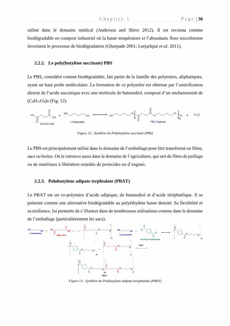
2.2.2. Le poly(butylène succinate) PBS _________________________________ 36
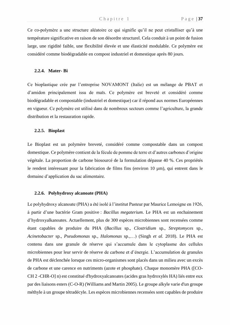
2.2.3. Polubutylene adipate terphtalate (PBAT) _____________________________ 36
2.2.4. Mater- Bi ______________________________________________________ 37
2.2.5. Bioplast _______________________________________________________ 37
2.2.6. Polyhydroxy alcanoate (PHA) _____________________________________ 37
3. La plastisphère _________________________________________________________ 38
3.1. La plastisphère en milieu marin ________________________________________ 39
3.2. Particularité de la plastisphère par comparaison aux micro-organismes de l’eau
environnante ____________________________________________________________ 42
4. Biodégradation des plastiques en mer _______________________________________ 44
4.1. Dégradation abiotique ________________________________________________ 45
4.2. Dégradation biotique ________________________________________________ 45
4.2.1. Les différentes étapes de biodégradation _____________________________ 47
4.2.2. Taux de dégradation plastique ______________________________________ 48
4.2.3. Tests et normes de biodégradabilité en milieu marin : encadrement et loi
Européenne ___________________________________________________________ 49
4.3. Biodégradation en fonction de la composition des polymères ___________________ 52
4.3.1. Plastiques conventionnels : Cas du Polyéthylène (PE) ___________________ 53
4.3.2. Plastiques biosourcés : Cas du PHA _________________________________ 55
5. Cas des PHA : Polymères biosourcés, biodégradables, compostables et recyclables ___ 57
5.1 Généralités ___________________________________________________________ 57
5.2. Intérêt commercial du PHA ___________________________________________ 59
5.3. Biosynthèse des PHA _______________________________________________ 62
5.4 Mécanismes moléculaires impliqués dans la biodégradation des PHA __________ 65
5.5 Limite du succès du PHA _____________________________________________ 68
6. Objectif de la thèse _____________________________________________________ 69
Chapitre 2 Etude de la biogéographie de la plastisphère en milieu marin _______________ 71
1. Préambule ____________________________________________________________ 73
2. Article: Global diversity and core microbiome of the plastisphere compared to organic-
particle attached and free-living planktonic lifestyles from the Tara Oceans expeditions in
the Mediterranean Sea and in the North Pacific gyre. _____________________________ 75
Chapitre 3 Colonisation et biodégradation des plastiques conventionnels et de leurs substituts
biodégradables pour l’usage de bâtonnets ouatés _________________________________ 105
1. Préambule ___________________________________________________________ 107
2. Article : Marine plastisphere activity and diversity during successive colonization and
biodegradation phases of various composition of plastic sticks ____________________ 109
Chapitre 4 Isolement d’une nouvelle souche bactérienne marine capable de synthétiser et de
biodégrader les PHA en utilisant une voie métabolique originale ____________________ 143
1. Préambule ___________________________________________________________ 145
2. Article : A new strain capable of synthesizing and degrading PHBV using atypical
metabolic pathways ________________________________________________________ 147
Chapitre 5 Conclusion et perspectives _________________________________________ 175
1. Conclusion _____________________________________________________________ 177
1.1. Etude de la plastisphère au niveau mondial ______________________________ 177
1.2. Evolution de la plastisphère vers un consortium capable de dégrader les polymères
179
1.3. Etude d’une souche appartenant à ce consortium afin d’identifier les gènes clefs
impliqués dans la biodégradation du PHA ____________________________________ 184
2. Perspectives ____________________________________________________________ 187
2.1. Utilisation de la technique DNA-Stable Isotope Probing (DNA-SIP) comme preuve
ultime de la biodégradation. _______________________________________________ 187
2.2. Enjeux pour une application en biotechnologie _____________________________ 197
2.3. Evolution des normes de biodégradation des plastiques en mer : suivi temporel et
multifactoriel ___________________________________________________________ 198
Références bibliographiques _________________________________________________ 200
Annexes ________________________________________________________________ 221
Annexe 1– Article en premier auteur publié en 2019 dans Frontiers in microbiology ___ 223
Annexe 2– Article en co-auteur en préparation ________________________________ 260
Annexe 3– Article en co-auteur en préparation ________________________________ 262
Annexe 4– Article en co-auteur en préparation ________________________________ 264
Annexe 5– Poster AFEM, Bussang 2019 _____________________________________ 266
Table des figures et des tableaux
Figure 1 : Chronologie de l'histoire du plastique __________________________________ 17
Figure 2 : Production mondiale exprimée en millions de tonnes de plastique ____________ 19
Figure 3: Schéma d'une pièce de plastique qui arrive dans l'océan. ____________________ 21
Figure 4 : Carte présentant les différents gyres océaniques __________________________ 23
Figure 5: Représentation des courants de surfaces régissant la méditerranée _____________ 24
Figure 6 : Carte de la mer méditerranée de 2013 à 2017 ____________________________ 25
Figure 7 : Représentation du gyre du Pacifique Nord à l'Ouest _______________________ 26
Figure 8 : Impacts de la pollution plastique sur l'environnement marin ________________ 28
Figure 9 : Impacts négatifs sur les principaux organismes terrestres et aquatiques. ________ 29
Figure 10 : Proportion de la production mondiale de plastiques biodégradables __________ 35
Figure 11 : Synthèse de l'acide polylactique (PLA) ________________________________ 35
Figure 12 : Synthèse du Polybutylene succinate (PBS) _____________________________ 36
Figure 13 : Synthèse du Polybutylene adipate terephtalate (PBAT) ____________________ 36
Figure 14 : Etapes de la formation du biofilm microbien retrouvés sur les polymères _____ 41
Figure 15 : Diagramme de Venn, montrant le nombre d'OTU et le pourcentage des OTU
unique expliquant la différence entre les fractions vivantes librement (FL) et Particule
attachée (PA) provenant d’une étude menée en méditerranée (Dussud et al. 2018) ________ 43
Figure 16 : Facteurs impliqués dans la dégradation abiotique et biotique des polymères ___ 44
Figure 17 : Différentes étapes de la biodégradation du plastique en mer _______________ 47
Figure 18 : Normes actuelles portant sur la biodégradation des plastiques en mer ________ 50
Figure 19 : Réaction radicalaire retrouvée dans le processus de biodégradation du PE _____ 53
Figure 20: Voie de biodégradation du PE et PHB _________________________________ 54
Figure 21: Conformation d'un carbone asymétrique selon l'ordre de priorité donné au 4
substituants suivant la loi de Cahn classé du plus important (a) au moins important (d) ____ 58
Figure 22 : Nomenclature générale des principaux PHA ____________________________ 58
Figure 23 : Molécule de Poly(β -hydroxybutyrate) _________________________________ 60
Figure 24 : Schéma bilan des interactions entres les protéines impliquées dans la synthèse des
PHA. Figure modifée d'après Pohlmann et al. 2006 ________________________________ 62
Figure 25 : Représentation des 4 PhaC identifiées chez les procaryotes ________________ 63
Figure 26 : Description des trois voies métaboliques de la synthase des PHA ____________ 65
Figure 27 : Présentation des deux types de PhaZ spécifique des PHASCL. _______________ 67
Figure 28 : Vue d'ensemble des études menées pendant cette thèse à différentes échelles __ 70
Figure 29 : Trajet des expéditions Tara Pacifique (A) et Tara Méditerranée (B) __________ 74
Figure 30 : Présentation de la ligne complète de production de tiges des différents polymères
(à gauche) et des tubes à essai ayant servi à l’incubation en milieu minimum (à droite) ___ 108
Figure 31 : Photographies représentant le halo de dégradation de la souche d’Alteromonas sp.
lorsqu’elle est placée en milieu minimum agar en présence de PHBV comme seule source de
carbone (à gauche) ________________________________________________________ 146
Figure 32 : Dendrogramme (méthode Ward), comparant les communautés microbiennes
colonisant les plastiques selon l’endroit géographique _____________________________ 182
Figure 33 : Schéma expérimental de l’expérience DNA-SIP menée sur du 13C PHBV ____ 188
Figure 34 : Consommation de l’oxygène par le consortium bactérien au contact du PHBV 12C
(triplicat) pendant 90 jours. __________________________________________________ 189
Figure 35 : Production bactérienne exprimée en ng C. dm-2.h-1 pour les morceaux de PHBV et
en ng C. L-1.h-1 pour le milieu d’incubation, et le surnageant du milieu artificiel contenant les
pièces de PHBV. __________________________________________________________ 190
Figure 36 : Imagse prisse au microscope électronique du PHBV 12C _________________ 191
Figure 37 : Quantité d’ADN obtenue dans le gradient de chlorure de Cesium __________ 192
Figure 38 : Résultat de l’alpha diversité sur les trois fractions au cours du temps ________ 193
Figure 39 : Comparaison entre les regroupements hiérarchiques basé sur la similitude de Bray
curtis (partie haute) et leurs affiliations taxonomiques comparant les classes en fonction de
leurs abondances relatives (partie inferieure). ____________________________________ 194
Figure 40 : Bubble plot des genres bactériens retrouvés en abondance relative (pourcentage)
sur les différentes fractions en fonction du temps. ________________________________ 195
Figure 41 : Histogramme des 25 gènes différenciant la fraction haute (FH), la fraction légère
(FL), et la fraction contrôle (12C). _____________________________________________ 196
Tableau 1: Comparaison des caractéristiques des deux zones d’études : la méditerranée et le
Pacifique__________________________________________________________________28
Tableau 2 : Les plastiques étudiés lors des expériences menées dans cette thèse. _________ 34
Tableau 3 : Comparaison des différents environnements influençant le taux de biodégradation
des plastiques ______________________________________________________________ 49
Tableau 4 : Présentation des différentes normes qui existent actuellement pour l'ensemble des
milieux aqueux ____________________________________________________________ 50
Tableau 5 :PHA commercialisés dans le monde avec leur caractéristique de production. ___ 61
Tableau 6 : Présentation des différentes classes de PhaC et leurs caractéristiques dans l'opéron
(Mezzolla et al. 2018) _______________________________________________________ 63
Liste des publications
1. Dussud, C., Hudec, C., George, M., Fabre, P., Higgs, P., Bruzaud, S., Delort, A.-M.,
Eyheraguibel, B., Meistertzheim, A.-L., Jacquin, J., Cheng, J., Callac, N., Odobel, C.,
Rabouille, S., Ghiglione, J.-F., 2018. Colonization of Non-biodegradable and
Biodegradable Plastics by Marine Microorganisms.
https://doi.org/10.3389/fmicb.2018.01571
2. Jacquin, J., Cheng, J., Odobel, C., Pandin, C., Conan, P., Pujo-Pay, M., Barbe, V.,
Meistertzheim, A.-L., Ghiglione, J.-F., 2019. Microbial Ecotoxicology of Marine
Plastic Debris: A Review on Colonization and Biodegradation by the “Plastisphere.”
Front. Microbiol. 10. https://doi.org/10.3389/fmicb.2019.00865
3. Jacquin J., Callac N., Cheng J., Giraud C., Gorand Y., Denoual C., Pujo-Pay M., Conan
P., Barbe V., Ter halle A., Meistertzheim A-L., Bruzaud S., Ghiglione J-F. Marine
plastisphere activity and diversity during successive colonization and biodegradation
phases of various composition of plastic sticks (soon submitted)
4. Jacquin J., Cheng J., Conan P., Pujo-Pay M., Bruzaud S., Ghiglione J-F., Barbe V. A
new strain capable of synthesizing and degrading PHBV using atypical metabolic
pathways (In preparation)
5. Jacquin J., Budinich M., Cheng J., Barbe V., Pedrotti M-L., Ghiglione J-F. Global
diversity and core microbiome of the plastisphere compared to organic-particle attached
and free-living planktonic lifestyles from the Tara Oceans expeditions in the
Mediterranean Sea and in the North Pacific gyre (In prepration).
6. Cheng J., Jacquin J, Conan P., Pujo-Pay M., Valérie B., Matthieu G., Pascale F.,
Bruzaud S., Ter Halle A., Meistertzheim A-L and Ghiglione J-F. Relative influence of
plastic debris size and shape, chemical composition and phytoplankton-bacteria
interactions in driving seawater plastisphere abundance, diversity and activity. Frontiers
in microbiology (soon submitted)
7. Cheng J., Eyheraguibel B., Jacquin J., Pujo-Pay M., Conan P., Barbe V., Hoypierres
J., Deligey G., Ter Halle A., Bruzaud S., Ghiglione J-F & Meistertzheim A-L.
Biodegradability under marine conditions of bio-based and petroleum-based polymers
as substitutes of conventional microbeads. Environmental pollution (soon submitted)
8. Cheng J, Meistertzheim A-L, Jacquin J, Valérie B, Escande M-L 4, Bertrand S, Escriva
H. and Ghiglione J-F. Beneficial or detrimental effects of microplastics on the marine
filter-feeder amphioxus (Branchiostoma lanceolatum)? Environmental science &
technology (In prepration).
Liste des communications scientifiques
1. Jacquin J., Callac N., Cheng J., Giraud C., Gorand Y., Denoual C., Pujo-Pay M., Conan
P., Barbe V., Ter halle A., Meistertzheim A-L., Bruzaud S., Ghiglione J-F. Utilisation
des outils “OMICS” pour caractériser la biodégradation des plastiques en mer. Poster
au Colloque de l’AFEM. Bussang, Novembre 2019. 1er Prix Poster
2. Jacquin J., Cheng J., Meistertzheim A-L, Callac C., Lemaire J., Fromageot D., Higgs
P., Eyheraguibel B., Delort A-M., Ghiglione J-F., Nouveau test de biodégradabilité des
plastiques oxodégradables en milieu marin. Poster au workshop Polymère et océans.
Montpellier, janvier 2018.
3. Cheng J., Jacquin J., Pandin C., Odobel C., Catala P., Pecqueur D., Salmeron C., Pujo-
Pay M., Conan P., Eyheraguibel B., Delort A-M., Barbe V., Hoypierres J., Deligey G.,
Bruzaud S., Fabre P., George M., J-F Ghiglione, A-L Meistertzheim Dégradation des
plastiques par les bactéries marines. Poster aux Premières Rencontres du GDR
Polymères et Océans. Créteil, Juillet 2019.

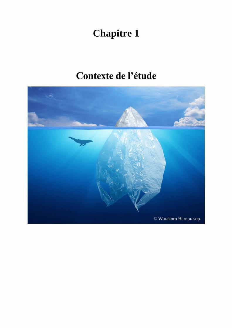
Chapitre 1
Contexte de l’étude
© Warakorn Harnprasop

C h a p i t r e 1 P a g e | 17
« Depuis 1950 ce sont 9 milliards de tonnes de plastique qui se sont accumulés sur Terre, soit
14,5 fois le poids de la population humaine mondiale».
1. La pollution plastique en mer
Le premier plastique synthétique a été fabriqué en 1855 après la découverte de la Parkésine
(Parkes, 1866) qui visait à mettre un terme à la surexploitation de l’ivoire. Alexander Parkes
avait traité de la cellulose avec de l’acide nitrique comme solvant, donnant un polymère
thermodurcissable et ressemblant à l’ivoire en y incorporant des pigments.
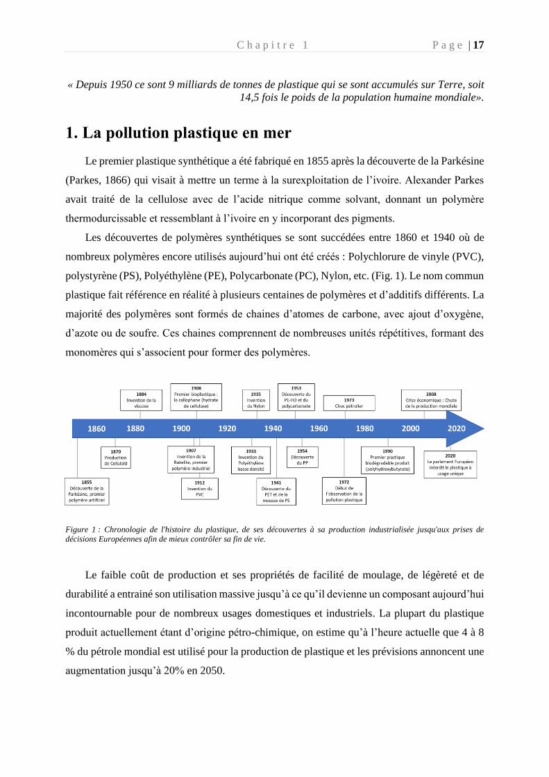
Les découvertes de polymères synthétiques se sont succédées entre 1860 et 1940 où de
nombreux polymères encore utilisés aujourd’hui ont été créés : Polychlorure de vinyle (PVC),
polystyrène (PS), Polyéthylène (PE), Polycarbonate (PC), Nylon, etc. (Fig. 1). Le nom commun
plastique fait référence en réalité à plusieurs centaines de polymères et d’additifs différents. La
majorité des polymères sont formés de chaines d’atomes de carbone, avec ajout d’oxygène,
d’azote ou de soufre. Ces chaines comprennent de nombreuses unités répétitives, formant des
monomères qui s’associent pour former des polymères.
Figure 1 : Chronologie de l'histoire du plastique, de ses découvertes à sa production industrialisée jusqu'aux prises de
décisions Européennes afin de mieux contrôler sa fin de vie.
Le faible coût de production et ses propriétés de facilité de moulage, de légèreté et de
durabilité a entrainé son utilisation massive jusqu’à ce qu’il devienne un composant aujourd’hui
incontournable pour de nombreux usages domestiques et industriels. La plupart du plastique
produit actuellement étant d’origine pétro-chimique, on estime qu’à l’heure actuelle que 4 à 8
% du pétrole mondial est utilisé pour la production de plastique et les prévisions annoncent une
augmentation jusqu’à 20% en 2050.
C h a p i t r e 1 P a g e | 18
De nos jours, un objet du quotidien sur deux est en plastique dans les ménages. On estime
aujourd’hui que 40% de la production plastique est utilisée pour l’emballage, mais on retrouve
du plastique dans les vêtements, véhicules, smartphones, masques, gants, matériaux de
construction, appareils médicaux, etc. L’usage facilité des plastiques a conduit à une
surconsommation de produits à faibles coûts considérés comme jetables ou à usage unique alors
qu’ils gardent leur propriété de durabilité. Sans considérer la gestion des déchets engendrés par
leur usage ni leur potentiel impact sur l’environnement, le plastique est devenu responsable
d’une des plus grandes pollutions de l’environnement engendrée par l’homme et il représente
un fléau pour les écosystèmes marins.
1.1. Production de plastique à travers le monde
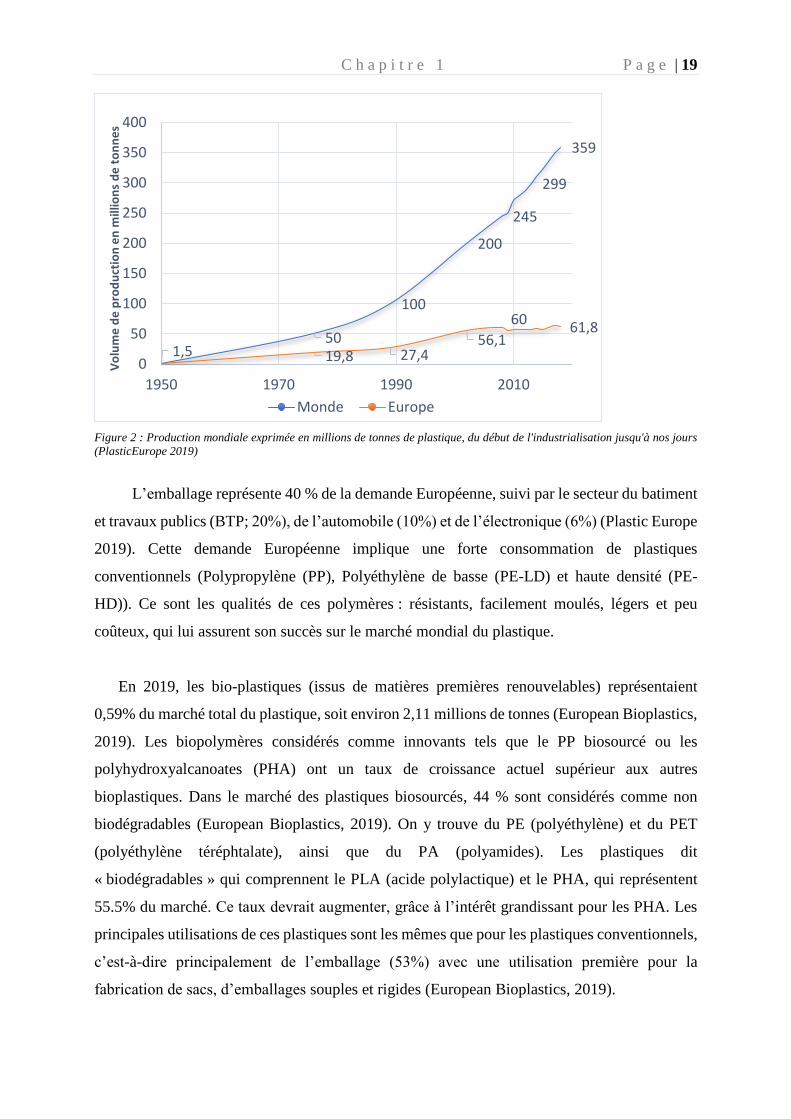
En 2018, avec 359 millions de tonnes de plastiques produits, le record de l’année
précédente a de nouveau été surpassé (Plastic Europe 2019) (Fig. 2) avec une hausse de 3,2 %
(11 millions de tonnes) par rapport à 2017, soit l'équivalent de 11,38 t de plastique produites
par seconde. L’Europe a produit moins de plastique en 2018, avec 61,8 millions de tonnes
contre 64,4 millions de tonnes en 2017, mais a augmenté sa consommation de 0,4 % (Fig. 2).
En 2018, ce sont 51,2 millions de tonnes de plastique qui ont été nécessaire pour répondre à la
demande Européenne et 70 % de cette demande est concentrée dans 6 pays d’Europe
(Allemagne, Italie, France, Espagne, Royaume Unie et Pologne). L’Europe est responsable de
17 % de la production mondiale de plastique, derrière l’Asie (51%) et l’Amérique du Nord
(18%). Dans ce contexte la France a connu en 2018 une baisse de production de plastique de -
5.1 % et de consommation -2.6%, après une année 2017 positive. Ces chiffres provenant de
l’étude de Plastic Europe, ne prennent pas en compte le plastique produit pour l’industrie du
textile (37.2 millions de tonnes) ou le caoutchouc synthétique utilisé dans les pneus (6.4
millions de tonnes) au niveau mondial (Boucher and Friot 2020). Selon Plastic Europe, près de
4 % de la production mondiale de pétrole sert à la fabrication du plastique (Plastic Europe
2019).
C h a p i t r e 1 P a g e | 19
Figure 2 : Production mondiale exprimée en millions de tonnes de plastique, du début de l'industrialisation jusqu'à nos jours
(PlasticEurope 2019)
L’emballage représente 40 % de la demande Européenne, suivi par le secteur du batiment
et travaux publics (BTP; 20%), de l’automobile (10%) et de l’électronique (6%) (Plastic Europe
2019). Cette demande Européenne implique une forte consommation de plastiques
conventionnels (Polypropylène (PP), Polyéthylène de basse (PE-LD) et haute densité (PE-
HD)). Ce sont les qualités de ces polymères : résistants, facilement moulés, légers et peu
coûteux, qui lui assurent son succès sur le marché mondial du plastique.
En 2019, les bio-plastiques (issus de matières premières renouvelables) représentaient
0,59% du marché total du plastique, soit environ 2,11 millions de tonnes (European Bioplastics,
2019). Les biopolymères considérés comme innovants tels que le PP biosourcé ou les
polyhydroxyalcanoates (PHA) ont un taux de croissance actuel supérieur aux autres
bioplastiques. Dans le marché des plastiques biosourcés, 44 % sont considérés comme non
biodégradables (European Bioplastics, 2019). On y trouve du PE (polyéthylène) et du PET
(polyéthylène téréphtalate), ainsi que du PA (polyamides). Les plastiques dit
« biodégradables » qui comprennent le PLA (acide polylactique) et le PHA, qui représentent
55.5% du marché. Ce taux devrait augmenter, grâce à l’intérêt grandissant pour les PHA. Les
principales utilisations de ces plastiques sont les mêmes que pour les plastiques conventionnels,
c’est-à-dire principalement de l’emballage (53%) avec une utilisation première pour la
fabrication de sacs, d’emballages souples et rigides (European Bioplastics, 2019).
50
100
200
245
299
359
1,5 19,8 27,456,1
6061,8
0
50
100
150
200
250
300
350
400
1950 1970 1990 2010
Vo
lum
e d
e p
rod
uct
ion
en
mill
ion
s d
e t
on
ne
s
Monde Europe
C h a p i t r e 1 P a g e | 20
1.2. Cycle de vie du plastique dans les écosystèmes aquatiques
Les océans sont la destination finale des déchets plastiques terrestres non traités. On
estime qu’entre 4,8 à 12,7 millions de tonnes de cette production est retrouvée en mer chaque
année (Jambeck et al. 2015). Différentes voies les mènent vers l’océan comme les rivières, les
eaux de ruissellement, les eaux usées et parfois par des phénomènes météorologiques extrêmes
comme les crues et les tsunamis (Figure3).
Selon leur densité, les plastiques resteront à la surface de l’eau ou couleront dans le fond
des océans. On estime aujourd’hui qu’une grande partie des plastiques coulent dans le fond des
océans, mais les estimations restent aujourd’hui controversées. La flottabilité peut aussi
dépendre de la colonisation du plastique par le biofilm marin. Le biofilm est un ensemble de
communautés microbiennes adhérant entre eux et à une surface, et caractérisé par la sécrétion
d’une matrice extracellulaire. Ce biofilm peut augmenter la densité du polymère, ce qui aura
pour conséquence de transporter des plastiques flottant en surface vers la colonne d’eau ou les
sédiments. De nombreux fonds marins et sédiments ont déjà été contaminés par du plastique
(Matsuguma et al. 2017; Gerigny et al. 2019; Harris 2020). L’exemple le plus connu étant la
pollution de la fosse océanique la plus profonde connu à ce jour : la fosse des Mariannes. Un
sac plastique a été trouvé à près de 11 000 m et plus inquiétant encore : des micro-plastiques
ont été détectés dans les intestins d’amphipodes situés entre 7000 et 10890m (Chiba et al. 2018;
Jamieson et al. 2019).
Une fois introduits dans l’environnement marin, les plastiques vont se fragmenter en
micro-plastiques (<5mm) puis en nano-plastiques (< 0,3 mm) sous l’effet combiné des
ultraviolets (UV), de la température, des vagues ou encore de la dégradation biologique (Arthur
et al. 2009; ter Halle et al. 2016) (Fig.3). On distingue les « micro-plastiques primaires »
directement issus de l’industrie (granulés de plastiques, abrasifs industriels, exfoliants, fibres
synthétiques...) des « micro-plastiques secondaires » qui sont issus de la fragmentation des
produits plastiques de plus grande taille (objets de la vie quotidienne) (Eerkes-Medrano et al.
2015). D’après un rapport publié par l’UICN (Union internationale de la conservation de la
nature), 35% des micro-plastiques proviennent des cycles répétés de lavages de vêtements
synthétiques et 28 % des micro-plastiques provenant de l’usure des pneus se déposant sur les
routes avant d’être lessivés par les pluies (Boucher and Friot 2017). Le transport aérien des
particules de plastiques provenant principalement du trafic routier représenterait une pollution
similaire à celle provenant des différents cours d’eau (Evangeliou et al. 2020). Une récente

C h a p i t r e 1 P a g e | 21
étude a permis de mettre en évidence que près de 140 000 tonnes de plastiques provenant du
trafic routier est déposé dans les océans en provenance de l’atmosphère contre 64 000 tonnes
par les fleuves (van Wijnen et al. 2019).
Figure 3: Schéma illustrant les nombreux cycles de vie potentiels d'une pièce de plastique qui arrive dans l'océan. WWTPs :
Wastewater Treatment Plant désigne la sortie de traitements d'eaux usées. DCM : maximum de chlorophylle profond (Zettler
et al, 2020
Environ 80 % des déchets plastiques trouvés en milieu marin proviennent des activités du
continent (de l’industrie, de l’usage domestique, de l’activité touristique, des rejets des
décharges et des fleuves). Les 20 % restant sont directement jetés en mer par le secteur de la
pêche majoritairement (Li et al. 2016). On estime que 75 % des déchets marins sont du
plastique, de nature et de taille variables (mégots, sacs plastiques, coton-tiges, emballages,
granulés plastiques).
Différentes expéditions ont été menées, principalement dans les zones d’accumulations
(gyres océaniques et méditerranée) et révèlent que la quantité de plastique comptabilisée en
surface ne représente qu’1% des estimations de la pollution plastique (Sebille et al. 2015). Le
plastique manquant (Fig. 3) reste une énigme pour la communauté scientifique. De nombreuses
hypothèses sont envisagées pour expliquer cet écart entre les chiffres théoriques et la quantité
C h a p i t r e 1 P a g e | 22
réellement retrouvée en mer. La première hypothèse est que le plastique ne s’accumule pas
uniquement en surface, mais aussi dans les fonds marins et dans la colonne d’eau (Chiba et al.
2018; Choy et al. 2019). En 2019, Choy et ses collaborateurs ont notamment montré que la
quantité de plastique était plus importante entre deux eaux, étude faite dans la baie de Monterey
(US), alors que d’autres études considèrent que la majorité des plastiques serait dans le fond
des Océans. La seconde hypothèse est que ces déchets plastiques sont ramenés à Terre en étant
largement repoussés vers les côtes, où ils seraient piégés par la végétation (Hardesty and Wilcox
2020). La zone littorale (entre 0 et 8km de distance à la mer) contient 90 % des débris plastiques
marins rejetés vers les côtes. La dernière hypothèse est qu’une quantité non négligeable de
plastique serait ingérée par la faune marine. Plusieurs études ont montré que les animaux de
tous niveaux trophiques avaient une concentration en morceaux de plastique très élevée à
l’intérieur de leur appareil digestif (Jamieson et al. 2019; Rummel et al. 2016; Wilcox et al.
2018; Choy et al. 2019).
A ce jour, il n’existe plus aucun écosystème aqueux non contaminé par le plastique. Cette
pollution est largement trouvée dans l’eau de mer (Barnes et al. 2009; Bergmann et al. 2017;
Sharma and Chatterjee 2017), la fosse des Mariannes (Chiba et al. 2018), la mer arctique
(Lusher et al. 2015; Peeken et al. 2018), l’océan Austral (Isobe et al. 2017), les eaux côtières
du monde (Dussud et al. 2018b; Zhao et al. 2018), les estuaires (Zhao et al. 2014) dans les lacs
(Faure et al. 2015), les rivières (Morritt et al. 2014; Lebreton et al. 2017). Le plastique a infiltré
tous les écosystèmes aqueux, la chaine trophique et les produits dérivés de la mer. On le
retrouve ainsi dans les produits de consommation courante tel que le sel de table (Yang et al.
2015), l’eau potable (Oßmann et al. 2018; Pivokonsky et al. 2018) et dans les bivalves élevées
pour leur consommation (Van Cauwenberghe and Janssen 2014).
1.3. Quantification et focus sur les zones d’études de la thèse
Chaque année 1,15 à 2,41 millions de tonnes de plastique se concentrent dans nos océans, mers,
rivières au gré des courants (Lebreton et al. 2017). Comme plus de la moitié de ces plastiques
est moins dense que l’eau douce, ils resteront en surface à leur entrée dans l’écosystème marin.
Les rejets mondiaux de micro-plastiques primaires représenteraient quant à eux 1,5 millions de
tonnes par an (Boucher and Friot 2017). Une estimation du nombre total de particules de
plastique flottant dans tous les océans, basée sur 24 expéditions océanographiques (2007-2013),
évalue à 5,25 trillions le nombre de morceaux de plastiques. L’ensemble de tous ces morceaux
de plastiques pèserait près de 270 000 tonnes (Eriksen et al. 2014).
C h a p i t r e 1 P a g e | 23
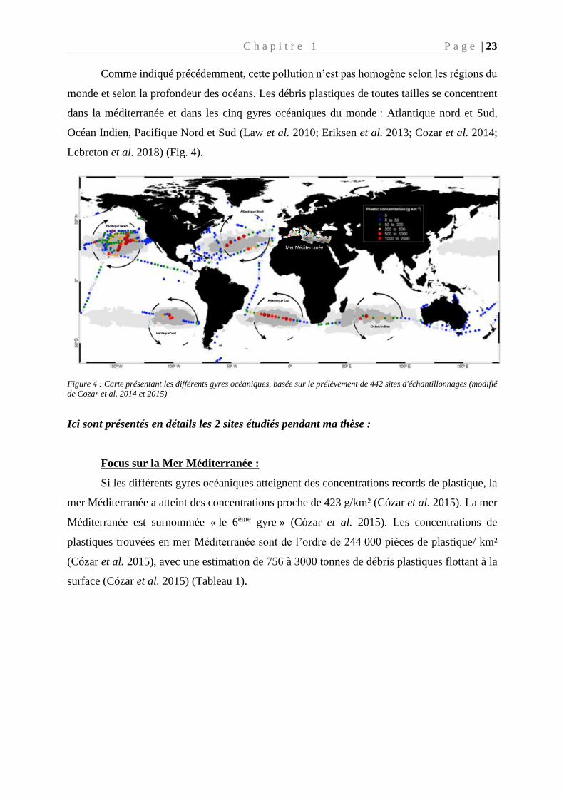
Comme indiqué précédemment, cette pollution n’est pas homogène selon les régions du
monde et selon la profondeur des océans. Les débris plastiques de toutes tailles se concentrent
dans la méditerranée et dans les cinq gyres océaniques du monde : Atlantique nord et Sud,
Océan Indien, Pacifique Nord et Sud (Law et al. 2010; Eriksen et al. 2013; Cozar et al. 2014;
Lebreton et al. 2018) (Fig. 4).
Figure 4 : Carte présentant les différents gyres océaniques, basée sur le prélèvement de 442 sites d'échantillonnages (modifié
de Cozar et al. 2014 et 2015)
Ici sont présentés en détails les 2 sites étudiés pendant ma thèse :
Focus sur la Mer Méditerranée :
Si les différents gyres océaniques atteignent des concentrations records de plastique, la
mer Méditerranée a atteint des concentrations proche de 423 g/km² (Cózar et al. 2015). La mer
Méditerranée est surnommée « le 6ème gyre » (Cózar et al. 2015). Les concentrations de
plastiques trouvées en mer Méditerranée sont de l’ordre de 244 000 pièces de plastique/ km²
(Cózar et al. 2015), avec une estimation de 756 à 3000 tonnes de débris plastiques flottant à la
surface (Cózar et al. 2015) (Tableau 1).
C h a p i t r e 1 P a g e | 24
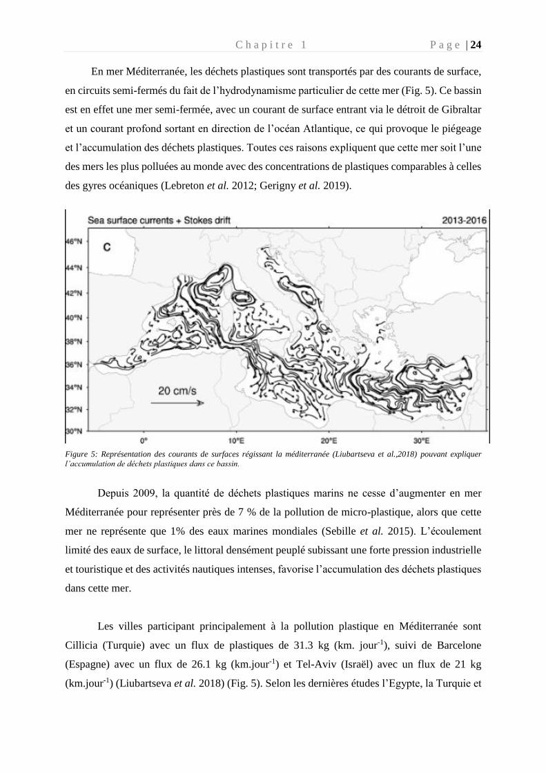
En mer Méditerranée, les déchets plastiques sont transportés par des courants de surface,
en circuits semi-fermés du fait de l’hydrodynamisme particulier de cette mer (Fig. 5). Ce bassin
est en effet une mer semi-fermée, avec un courant de surface entrant via le détroit de Gibraltar
et un courant profond sortant en direction de l’océan Atlantique, ce qui provoque le piégeage
et l’accumulation des déchets plastiques. Toutes ces raisons expliquent que cette mer soit l’une
des mers les plus polluées au monde avec des concentrations de plastiques comparables à celles
des gyres océaniques (Lebreton et al. 2012; Gerigny et al. 2019).
Figure 5: Représentation des courants de surfaces régissant la méditerranée (Liubartseva et al.,2018) pouvant expliquer
l’accumulation de déchets plastiques dans ce bassin.
Depuis 2009, la quantité de déchets plastiques marins ne cesse d’augmenter en mer
Méditerranée pour représenter près de 7 % de la pollution de micro-plastique, alors que cette
mer ne représente que 1% des eaux marines mondiales (Sebille et al. 2015). L’écoulement
limité des eaux de surface, le littoral densément peuplé subissant une forte pression industrielle
et touristique et des activités nautiques intenses, favorise l’accumulation des déchets plastiques
dans cette mer.
Les villes participant principalement à la pollution plastique en Méditerranée sont
Cillicia (Turquie) avec un flux de plastiques de 31.3 kg (km. jour-1), suivi de Barcelone
(Espagne) avec un flux de 26.1 kg (km.jour-1) et Tel-Aviv (Israël) avec un flux de 21 kg
(km.jour-1) (Liubartseva et al. 2018) (Fig. 5). Selon les dernières études l’Egypte, la Turquie et
C h a p i t r e 1 P a g e | 25
l’Italie contribuerait au 2/3 de la pollution plastique de la méditerranée (Jambeck et al. 2015;
Geyer et al. 2017; Liubartseva et al. 2018).
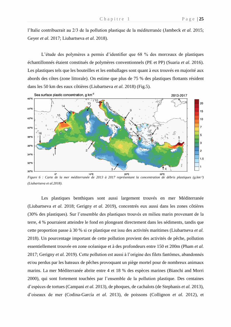
L’étude des polymères a permis d’identifier que 68 % des morceaux de plastiques
échantillonnés étaient constitués de polymères conventionnels (PE et PP) (Suaria et al. 2016).
Les plastiques tels que les bouteilles et les emballages sont quant à eux trouvés en majorité aux
abords des côtes (zone littorale). On estime que plus de 75 % des plastiques flottants résident
dans les 50 km des eaux côtières (Liubartseva et al. 2018) (Fig.5).
Figure 6 : Carte de la mer méditerranée de 2013 à 2017 représentant la concentration de débris plastiques (g.km-²)
(Liubartseva et al.2018).
Les plastiques benthiques sont aussi largement trouvés en mer Méditerranée
(Liubartseva et al. 2018; Gerigny et al. 2019), concentrés eux aussi dans les zones côtières
(30% des plastiques). Sur l’ensemble des plastiques trouvés en milieu marin provenant de la
terre, 4 % pourraient atteindre le fond en plongeant directement dans les sédiments, tandis que
cette proportion passe à 30 % si ce plastique est issu des activités maritimes (Liubartseva et al.
2018). Un pourcentage important de cette pollution provient des activités de pêche, pollution
essentiellement trouvée en zone océanique et à des profondeurs entre 150 et 200m (Pham et al.
2017; Gerigny et al. 2019). Cette pollution est aussi à l’origine des filets fantômes, abandonnés
et/ou perdus par les bateaux de pêches provoquant un piège mortel pour de nombreux animaux
marins. La mer Méditerranée abrite entre 4 et 18 % des espèces marines (Bianchi and Morri
2000), qui sont fortement touchées par l’ensemble de la pollution plastique. Des centaines
d’espèces de tortues (Campani et al. 2013), de phoques, de cachalots (de Stephanis et al. 2013),
d’oiseaux de mer (Codina-García et al. 2013), de poissons (Collignon et al. 2012), et
C h a p i t r e 1 P a g e | 26
d’invertébrés sont tués par ces engins de pêche abandonnés (Green 2020). On estime qu’entre
2 637 et 3 342 tonnes de filets sont perdus chaque année (Golik 1997).
Focus : Le gyre du Pacifique Nord ou « septième continent » :
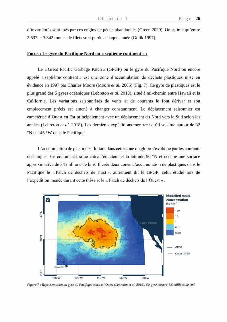
Le « Great Pacific Garbage Patch » (GPGP) ou le gyre du Pacifique Nord ou encore
appelé « septième contient » est une zone d’accumulation de déchets plastiques mise en
évidence en 1997 par Charles Moore (Moore et al. 2005) (Fig. 7). Ce gyre de plastiques est le
plus grand des 5 gyres océaniques (Lebreton et al. 2018), situé à mi-chemin entre Hawaii et la
Californie. Les variations saisonnières de vents et de courants le font dériver et son
emplacement précis est amené à changer constamment. Le déplacement saisonnier est
caractérisé d’Ouest en Est principalement avec un déplacement du Nord vers le Sud selon les
années (Lebreton et al. 2018). Les dernières expéditions montrent qu’il se situe autour de 32
°N et 145 °W dans le Pacifique.
L’accumulation de plastiques flottant dans cette zone du globe s’explique par les courants
océaniques. Ce courant est situé entre l’équateur et la latitude 50 °N et occupe une surface
approximative de 34 millions de km². Il crée deux zones d’accumulation de plastiques dans le
Pacifique le « Patch de déchets de l’Est », autrement dit le GPGP, celui étudié lors de
l’expédition menée durant cette thèse et le « Patch de déchets de l’Ouest » .
Figure 7 : Représentation du gyre du Pacifique Nord à l'Ouest (Lebreton et al. 2016). Ce gyre mesure 1.6 millions de km².
C h a p i t r e 1 P a g e | 27
Depuis quelques années, les études du GPGP se sont multipliées jusqu’à la dernière
grande étude organisée par l’ONG « ocean cleanup » qui ont permis de caractériser plus
précisément ce gyre océanique (Lebreton et al. 2018). En 2015, ils ont entrepris une expédition
avec plusieurs navires pour caractériser les plastiques à la surface de l’eau. Un an plus tard, ils
ont complété ces données avec des images satellite obtenues par avion afin de permettre une
caractérisation plus précise de l’étendue du gyre. Ainsi, leur estimation montre que ce gyre a
une taille de 1,6 millions de km². Le gyre a pour caractéristique principale de concentrer la
masse des plastiques dans les deux premiers mètres sous la surface de l’eau (Reisser et al. 2014;
Kooi et al. 2016). La pollution plastique dans ce gyre est constituée à 94 % de micro-plastiques
situés sous la surface, ce qui rend imprécise l’estimation de son étendue (Reisser et al. 2014;
Kooi et al. 2016).
La totalité des déchets plastiques présents dans cette zone est estimée à 100 000 de tonnes
(Lebreton et al. 2018), avec des concentrations allant jusqu’à 1,3 million de particules par km²
(Egger et al. 2020). Dans cette zone, les déchets plastiques représentent 99,9 % des débris
flottants et 46 % d’entre eux sont des filets de pêche (Lebreton et al. 2018). La concentration
de plastique est estimée à 100 kg.Km-² au centre du gyre et à 10 kg. Km-² à l’extérieur de ce
dernier (Lebreton et al. 2018) (Tableau 1).
Les déchets plastiques du gyre du Pacifique Nord proviendraient principalement de 6
pays : la Chine, l’Indonésie, les Philippines, le Vietnam, le Sri-lanka et la Thaïlande (Jambeck
et al. 2015). Le type de plastique trouvé dans cette zone est similaire à celui de la Méditerranée,
principalement du PE et du PP et des déchets de pêche abandonnés (46%) (Lebreton et al.
2018).
Le plastique présent à la surface du GPGP est 180 fois plus abondant que toutes formes
de vie marine (Gall and Thompson 2015). Cela a pour conséquence que l’alimentation de
certains animaux habitant cette région comme les tortues (Clukey et al. 2017) et les poussins
d’albatros ingèrent en masse ce plastique (Gall and Thompson 2015). Dans cette zone 84 % des
plastiques contenaient un produit chimique toxique (Chen et al. 2018).
C h a p i t r e 1 P a g e | 28
Tableau 1: Comparaison des caractéristiques des deux zones d’études : la méditerranée et le Pacifique
Caractéristiques
Great Pacific
Garbage Patch Mer méditerranée
Taille (millions de km²) 1,6 2,5
Quantité (tonnes) 45 à 129 000 756 à 3000
Concentration (/km²) 1 300 000 13 615
Proportion en micro-plastique
(nombre en %) 94 83
Concentration (kg/km²) 10 à 100 0,022 à 1,9
Composition PE et PP 52 % PE et 16% PP
1.4. Impacts écologiques de la pollution plastique
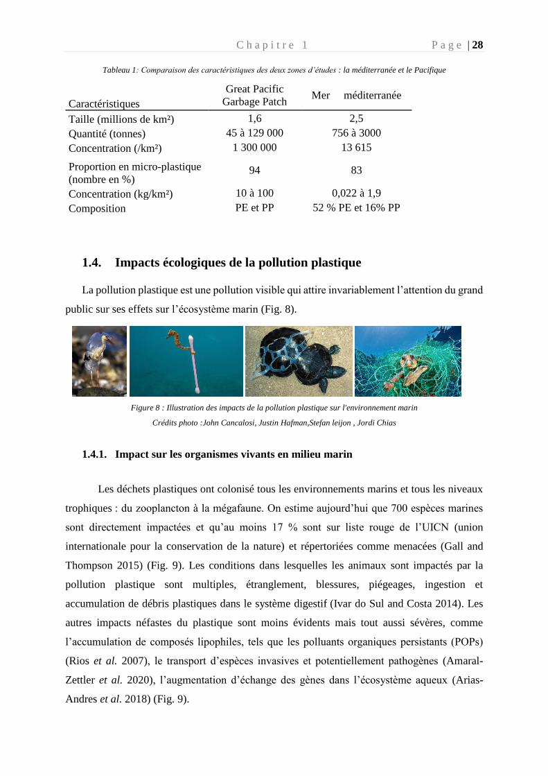
La pollution plastique est une pollution visible qui attire invariablement l’attention du grand
public sur ses effets sur l’écosystème marin (Fig. 8).
Figure 8 : Illustration des impacts de la pollution plastique sur l'environnement marin
Crédits photo :John Cancalosi, Justin Hafman,Stefan leijon , Jordi Chias
1.4.1. Impact sur les organismes vivants en milieu marin
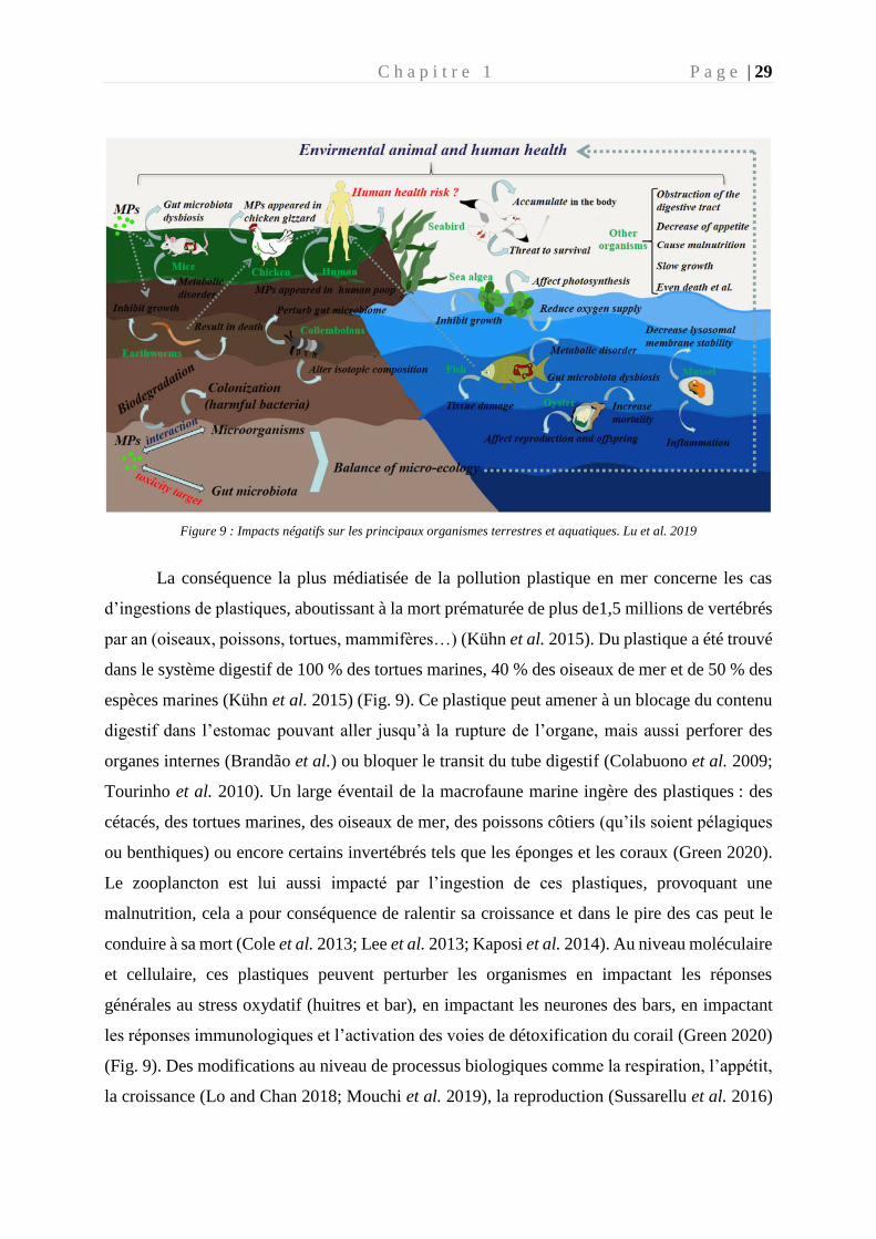
Les déchets plastiques ont colonisé tous les environnements marins et tous les niveaux
trophiques : du zooplancton à la mégafaune. On estime aujourd’hui que 700 espèces marines
sont directement impactées et qu’au moins 17 % sont sur liste rouge de l’UICN (union
internationale pour la conservation de la nature) et répertoriées comme menacées (Gall and
Thompson 2015) (Fig. 9). Les conditions dans lesquelles les animaux sont impactés par la
pollution plastique sont multiples, étranglement, blessures, piégeages, ingestion et
accumulation de débris plastiques dans le système digestif (Ivar do Sul and Costa 2014). Les
autres impacts néfastes du plastique sont moins évidents mais tout aussi sévères, comme
l’accumulation de composés lipophiles, tels que les polluants organiques persistants (POPs)
(Rios et al. 2007), le transport d’espèces invasives et potentiellement pathogènes (Amaral-
Zettler et al. 2020), l’augmentation d’échange des gènes dans l’écosystème aqueux (Arias-
Andres et al. 2018) (Fig. 9).
C h a p i t r e 1 P a g e | 29
Figure 9 : Impacts négatifs sur les principaux organismes terrestres et aquatiques. Lu et al. 2019
La conséquence la plus médiatisée de la pollution plastique en mer concerne les cas
d’ingestions de plastiques, aboutissant à la mort prématurée de plus de1,5 millions de vertébrés
par an (oiseaux, poissons, tortues, mammifères…) (Kühn et al. 2015). Du plastique a été trouvé
dans le système digestif de 100 % des tortues marines, 40 % des oiseaux de mer et de 50 % des
espèces marines (Kühn et al. 2015) (Fig. 9). Ce plastique peut amener à un blocage du contenu
digestif dans l’estomac pouvant aller jusqu’à la rupture de l’organe, mais aussi perforer des
organes internes (Brandão et al.) ou bloquer le transit du tube digestif (Colabuono et al. 2009;
Tourinho et al. 2010). Un large éventail de la macrofaune marine ingère des plastiques : des
cétacés, des tortues marines, des oiseaux de mer, des poissons côtiers (qu’ils soient pélagiques
ou benthiques) ou encore certains invertébrés tels que les éponges et les coraux (Green 2020).
Le zooplancton est lui aussi impacté par l’ingestion de ces plastiques, provoquant une
malnutrition, cela a pour conséquence de ralentir sa croissance et dans le pire des cas peut le
conduire à sa mort (Cole et al. 2013; Lee et al. 2013; Kaposi et al. 2014). Au niveau moléculaire
et cellulaire, ces plastiques peuvent perturber les organismes en impactant les réponses
générales au stress oxydatif (huitres et bar), en impactant les neurones des bars, en impactant
les réponses immunologiques et l’activation des voies de détoxification du corail (Green 2020)
(Fig. 9). Des modifications au niveau de processus biologiques comme la respiration, l’appétit,
la croissance (Lo and Chan 2018; Mouchi et al. 2019), la reproduction (Sussarellu et al. 2016)
C h a p i t r e 1 P a g e | 30
et le comportement (Chapron et al. 2018) ont été mis en évidence chez certains animaux tels
que les huitres, les moules, les copépodes, etc. (Green 2020). Les organismes au sommet de la
chaine trophique ou ayant une grande espérance de vie comme le rorqual ou la baleine sont
régulièrement exposés à cette pollution. Certains travaux ont mis en évidence la présence de
phtalates (additifs provenant notamment du plastique) dans leurs graisses (Fossi et al. 2012).
La pêche fantôme provoquée par les filets de pêches perdus en mer impacte des
centaines d’espèces différentes et représente la plus grande menace anthropique pour certaines
espèces en voies de disparition tels que les phoques et les tortues (Henderson 2001; Boland and
Donohue 2003). Ce piégeage perpétuel entraine une mort rapide par asphyxie (étranglement)
(Cassoff et al. 2011) ou une mort plus lente voire des contraintes physiques importantes. Ces
contraintes sont des lésions cutanées et des mouvements restreints entrainant une diminution de
la capacité à chasser des proies (Kühn et al. 2015) ou à se mouvoir. Les filets fantômes seraient
responsables de 16 à 80 % de la mortalité des pinnipèdes (Butterworth et al. 2012).
Les invertébrés marins sessiles tels que les coraux, les éponges, les bivalves, larves, sont
eux aussi impactés par abrasion des tissus ou étouffement (Green 2020). Cela peut aboutir à
une perte considérable de la taille des récifs coralliens (Richards and Beger 2011).
La flore sous-marine est tout aussi touchée par cette pollution. Les micro-plastiques
peuvent réduire la photosynthèse du phytoplancton, lié à une concentration très élevée des
polymères à la surface de l’eau, obstruant ainsi la lumière du soleil. En impactant le
phytoplancton, c’est la quantité d’oxygène disponible pour la vie marine qui se retrouve
diminuée (Zhao et al. 2014).
L’écosystème microbien est quant à lui aussi affecté par des effets toxiques du plastique
lié à ses additifs. Ces effets ont été décrits dans la littérature comme affectant les bactéries, les
champignons et les levures, provoquant des inhibitions de croissance, l’interruption de la
fonction écologique ou encore l’apparition de réaction de stress, et pouvant aller jusqu’à la mort
du micro-organisme (Nomura et al. 2016; Sun et al. 2018).
C h a p i t r e 1 P a g e | 31
1.4.2 Cas des Polluants Organiques Persistants (POPs)
Les Polluants Organiques Persistants (POP) sont définis selon leur toxicité sur la santé
humaine ou animale, leur persistance dans l’environnement, leur bioaccumulation dans les
tissus vivants et leur transport sur de longue distance. Ces POPs sont classés en deux
catégories : la première regroupe les additifs, les monomères et les oligomères de molécules
constitutives au plastique ; la seconde catégorie regroupe les composés organiques souvent
hydrophobes qui sont adsorbés par le polymère.
Les plastiques contiennent de nombreux additifs dans le but d’améliorer leurs propriétés
mécaniques, esthétiques, la stabilité, et leurs prix de revient. Les phtalates (dérivés de l’acide
phtalique), faisant partie de ces additifs, sont facilement libérés dans le milieu car ils ne sont
pas liés par de fortes liaisons aux polymères.
D’autre part, les plastiques sont généralement imperméables à l’eau et donc
hydrophobes, ce qui attire les POPs qui eux sont lipophiles. Les micro-plastiques ayant un
rapport surface/volume élevé, concentrent les POPs jusqu’à 6 fois plus que l’eau (Hirai et al.
2011). Quelques études se sont concentrées sur l’étude des POPs associés aux micro-plastiques
et ont trouvé qu’un large éventail d’additifs est associé aux particules de plastique
(organophosphate esters (OPE), hydrocarbures aromatiques polycycliques (PAH), pesticides
organochlorés (OCP), …) (Zhang et al. 2020). Le transfert de ces POPs dans l’environnement
ou au sein des organismes, reste peu étudié tant les POPs agglomérés sur le plastique sont
nombreux. Cependant il semblerait que le taux de lixiviation des plusieurs additifs serait plus
important dans l’environnement marin que dans les voies intestinales des micro-organismes
(Koelmans et al. 2014).
Les micro-plastiques sont rapidement accumulés à différents niveaux de la chaine
alimentaire (Carbery et al. 2018), cependant l’effet combiné causé par les micro-plastiques et
les polluants organiques est encore peu documenté et les études se contredisent. Par exemple,
des études toxicologiques se basant sur la mortalité, le comportement alimentaire, l'immunité
et les biomarqueurs du stress oxydatif au niveau des organismes représentatifs de l’écosystème
étudié, ne montrent pas d’effets indésirables des POPs sur les organismes tels que la moule et
certains poissons (Ašmonaitė et al. 2018; Magara et al. 2018). Dans ces études, on ne constatait
C h a p i t r e 1 P a g e | 32
pas d’augmentation de mortalité des animaux testés, ni d’effets inhibiteurs des fonctions
métaboliques, ni de réponses de type stress oxydatif (Oliveira et al. 2013; Magara et al. 2018).
D’autres études avancent aussi que la synergie des POP et du micro-plastique diminue la
toxicité sur certaines micro-algues (Zhu et al. 2018). A l’opposé, d’autres études mettent en
avant un taux de mortalité plus important, une alimentation et une immunité diminuée lorsque
l’organisme marin (arénicole) étudié est mis en contact avec une concentration en micro-
plastique élevée (Browne et al. 2013).
2. Les différentes classes de polymères
Comme précédemment indiqué, le plastique est omniprésent dans nos objets du quotidien.
Leurs différentes applications imposent des qualités bien différentes et donc des polymères très
diversifiés. Les plastiques sont définis comme un mélange contenant une matière de base
(polymère) pouvant être moulé, façonné, à chaud ou sous pression. Ils peuvent être produits à
partir de différentes ressources, d’origine fossile (pétrole brut, gaz, etc.) appelés « plastiques
conventionnels » ou d’origine renouvelable (végétaux, bactéries, lait, amidon, etc.) appelés
« plastiques biosourcés ». Les polymères conventionnels (Polyéthylène-PE, Polypropylène-PP,
Polytéréphtalate d'éthylène -PET, …) constituent la grande majorité des produits utilisés dans
notre quotidien. La nature et l’origine du polymère peut être déterminant pour sa durabilité dans
l’environnement. Un polymère biodégradable, selon les normes actuellement en vigueur est un
polymère dont le carbone organique est capable d’être transformé en CO2 (au moins 60 % par
rapport à la cellulose) ou en biomasse par une communauté microbienne. Les détails des normes
est expliqué dans la partie 4.3.
2.1. Polymères conventionnels
Les Plastiques conventionnels, d’origine fossiles sont classés en 2 catégories :
- Les thermoplastiques, qui peuvent fondre s’ils sont chauffés et durcissent lors du
processus de refroidissement. Ces propriétés sont réversibles et le plastique peut être
remodelé plusieurs fois. C’est le cas du PE, PP, PET, par exemple.
- Les thermodurcissables ne peuvent pas être refondus après avoir été modelés, leur
point de fusion élevé empêche le polymère de redevenir souple et son recyclage est
impossible. Cela concerne le Polyurethane, le Vinyl ester, le Silicone…
C h a p i t r e 1 P a g e | 33
Les thermoplastiques peuvent être retrouvés à l’état physique cristallin, c’est-à-dire que les
chaines de carbones sont linéaires, ordonnées et liées entre elles par de fortes liaisons
intermoléculaires donnant une matière rigide (PE haute densité, HDPE), ou sous forme
amorphe avec des chaines ramifiées, désordonnées avec des liaisons faibles (PE basse densité,
LDPE) (Tableau 2). Parmi les polymères conventionnels, les plus abondants sont les
polyoléfines avec une majorité de polypropylène (PP) et de Polyéthylène à faible ou haute
densité (LD-PE et HD-PE) (Tableau 2). Ces polymères sont hydrophobes, inertes à beaucoup
de produits chimiques (aux solvants, acides, bases…), ils ont une faible densité, ils sont
résistants et modifiables, expliquant le succès mondial qu’ils rencontrent depuis des dizaines
d’années.
Le Polyéthylène téréphtalate (PET), thermoplastique, formé d’un alcool, d’un éthylène
glycol et de l’acide téréphtalique est un plastique conventionnel qui nécessite presque deux
kilos de pétrole brut pour fabriquer un kilo de PET. Le PET est amorphe, transparent, résistant
à des pressions élevées (environ 10 bars), donnant des films d’une très grande solidité et
stabilité. Ces caractéristiques font de lui le 6ème polymère le plus demandé en Europe avec un
besoin de près de 4 millions de tonnes en 2018 (Plastic Europe 2019). C’est le polymère le plus
abondant dans le monde avec près de 70 millions de tonnes fabriquées chaque année (Tournier
et al. 2020). On le trouve habituellement dans les bouteilles recyclables (étanches au CO2), dans
de nombreuses cartes (carte de crédit, fidélité), fibres textiles, emballages, films transparents,
etc. Pourtant ces caractéristiques font de lui un polymère qui limite fortement le recyclage. En
effet la molécule de poly (téréphtalate d’éthylène) s’hydrolyse à température élevée (240°C),
provoquant la chute de son poids moléculaire et le rendant inutilisable pour un usage habituel.
Cependant une nouvelle étude parue récemment montre le potentiel recyclable du PET, grâce
à une nouvelle enzyme (PET depolymerase) capable de dépolymériser par voie biologique les
déchets plastiques en PET (Tournier et al. 2020). Cette enzyme permet d’obtenir un PET
recyclé biologiquement ayant les mêmes propriétés que le PET issu de la pétrochimie.
C h a p i t r e 1 P a g e | 34
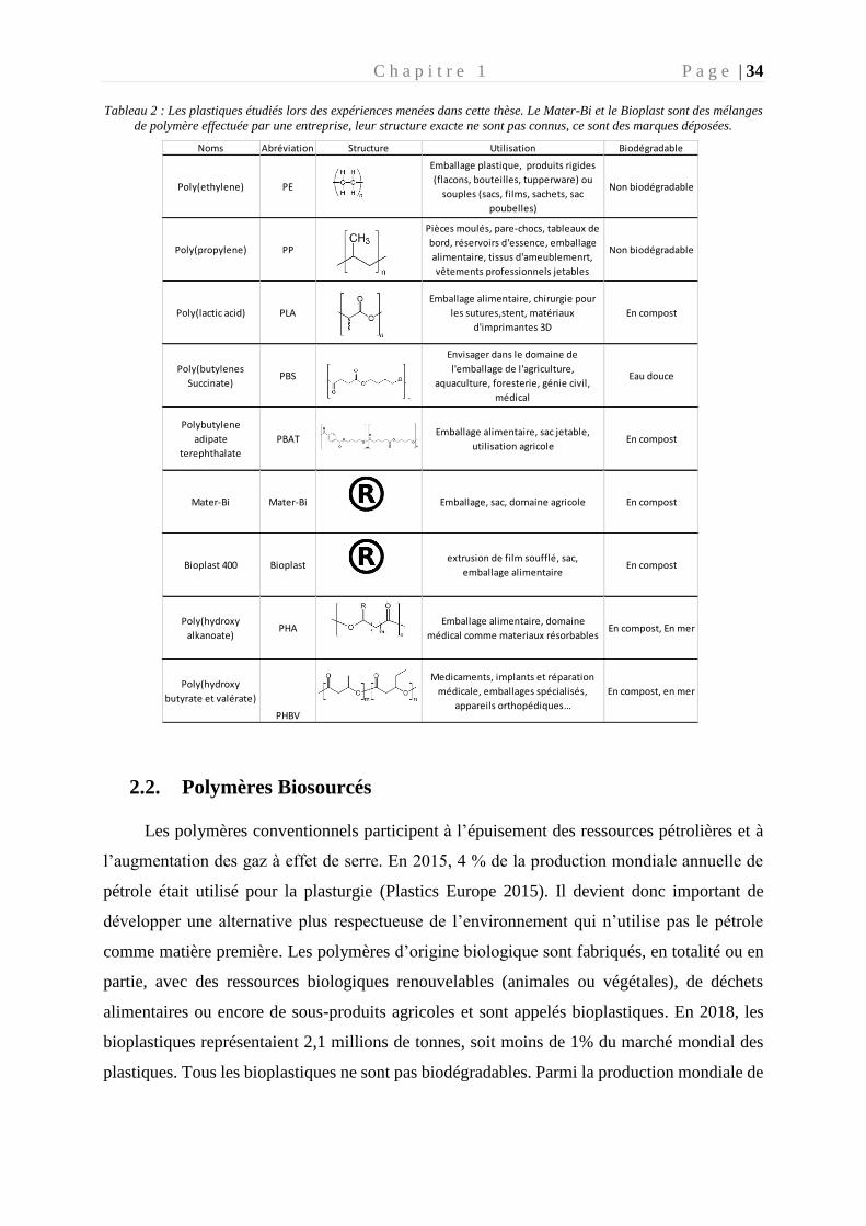
Tableau 2 : Les plastiques étudiés lors des expériences menées dans cette thèse. Le Mater-Bi et le Bioplast sont des mélanges
de polymère effectuée par une entreprise, leur structure exacte ne sont pas connus, ce sont des marques déposées.
2.2. Polymères Biosourcés
Les polymères conventionnels participent à l’épuisement des ressources pétrolières et à
l’augmentation des gaz à effet de serre. En 2015, 4 % de la production mondiale annuelle de
pétrole était utilisé pour la plasturgie (Plastics Europe 2015). Il devient donc important de
développer une alternative plus respectueuse de l’environnement qui n’utilise pas le pétrole
comme matière première. Les polymères d’origine biologique sont fabriqués, en totalité ou en
partie, avec des ressources biologiques renouvelables (animales ou végétales), de déchets
alimentaires ou encore de sous-produits agricoles et sont appelés bioplastiques. En 2018, les
bioplastiques représentaient 2,1 millions de tonnes, soit moins de 1% du marché mondial des
plastiques. Tous les bioplastiques ne sont pas biodégradables. Parmi la production mondiale de
Noms Abréviation Structure Utilisation Biodégradable
Poly(ethylene) PE
Emballage plastique, produits rigides
(flacons, bouteilles, tupperware) ou
souples (sacs, films, sachets, sac
poubelles)
Non biodégradable
Poly(propylene) PP
Pièces moulés, pare-chocs, tableaux de
bord, réservoirs d'essence, emballage
alimentaire, tissus d'ameublemenrt,
vêtements professionnels jetables
Non biodégradable
Poly(lactic acid) PLA
Emballage alimentaire, chirurgie pour
les sutures,stent, matériaux
d'imprimantes 3D
En compost
Poly(butylenes
Succinate)PBS
Envisager dans le domaine de
l'emballage de l'agriculture,
aquaculture, foresterie, génie civil,
médical
Eau douce
Polybutylene
adipate
terephthalate
PBATEmballage alimentaire, sac jetable,
utilisation agricoleEn compost
Mater-Bi Mater-Bi Emballage, sac, domaine agricole En compost
Bioplast 400 Bioplastextrusion de film soufflé, sac,
emballage alimentaireEn compost
Poly(hydroxy
alkanoate)PHA
Emballage alimentaire, domaine
médical comme materiaux résorbablesEn compost, En mer
Poly(hydroxy
butyrate et valérate)
PHBV
Medicaments, implants et réparation
médicale, emballages spécialisés,
appareils orthopédiques…
En compost, en mer
C h a p i t r e 1 P a g e | 35
plastiques biodégradables, ce sont deux polymères biosourcés qui dominent cette production :
ceux à base d’amidon et le PLA (Fig.10).
Figure 10 : Proportion de la production mondiale de plastiques biodégradables (PlasticEurope, 2018)
La liste des polymères biosourcés ci-dessous n’est pas une liste exhaustive. Ils ont été
sélectionnés parce qu’ils ont été utilisés dans la cadre de la thèse.
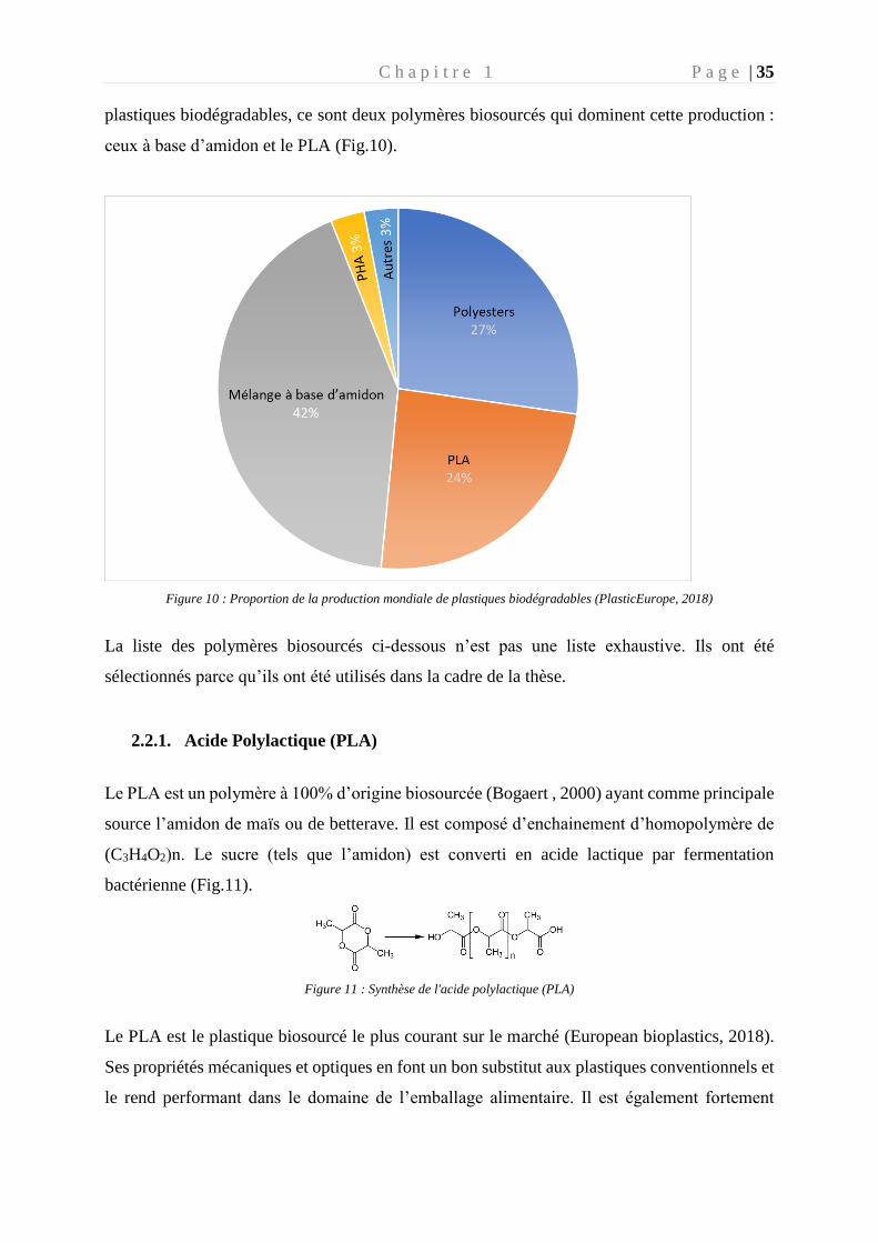
2.2.1. Acide Polylactique (PLA)
Le PLA est un polymère à 100% d’origine biosourcée (Bogaert , 2000) ayant comme principale
source l’amidon de maïs ou de betterave. Il est composé d’enchainement d’homopolymère de
(C3H4O2)n. Le sucre (tels que l’amidon) est converti en acide lactique par fermentation
bactérienne (Fig.11).
Figure 11 : Synthèse de l'acide polylactique (PLA)
Le PLA est le plastique biosourcé le plus courant sur le marché (European bioplastics, 2018).
Ses propriétés mécaniques et optiques en font un bon substitut aux plastiques conventionnels et
le rend performant dans le domaine de l’emballage alimentaire. Il est également fortement
C h a p i t r e 1 P a g e | 36
utilisé dans le domaine médical (Anderson and Shive 2012). Il est reconnu comme
biodégradable en compost industriel où la haute température et l’abondante flore microbienne
favorisent le processus de biodégradation (Ghorpade 2001; Leejarkpai et al. 2011).
2.2.2. Le poly(butylène succinate) PBS
Le PBS, considéré comme biodégradable, fait partie de la famille des polyesters, aliphatiques,
ayant un haut poids moléculaire. La formation de ce polymère est obtenue par l’estérification
directe de l’acide succinique avec une molécule de butanediol, composé d’un enchainement de
(C8H12O4)n (Fig. 12).
Figure 12 : Synthèse du Polybutylene succinate (PBS)
Le PBS est principalement utilisé dans le domaine de l’emballage pour être transformé en films,
sacs ou boites. On le retrouve aussi dans le domaine de l’agriculture, qui sert de films de paillage
ou de matériaux à libération retardée de pesticides ou d’engrais.
2.2.3. Polubutylene adipate terphtalate (PBAT)
Le PBAT est un co-polymère d’acide adipique, de butanediol et d’acide téréphatlique. Il se
présente comme une alternative biodégradable au polyéthylène basse densité. Sa flexibilité et
sa résilience, lui permette de s’illustrer dans de nombreuses utilisations comme dans le domaine
de l’emballage (particulièrement les sacs).
Figure 13 : Synthèse du Polybutylene adipate terephtalate (PBAT)
C h a p i t r e 1 P a g e | 37
Ce co-polymère a une structure aléatoire ce qui signifie qu’il ne peut cristalliser qu’à une
température significative en raison de son désordre structurel. Cela conduit à un point de fusion
large, une rigidité faible, une flexibilité élevée et une élasticité modulable. Ce polymère est
considéré comme biodégradable en compost industriel et domestique après 80 jours.
2.2.4. Mater- Bi
Ce bioplastique crée par l’entreprise NOVAMONT (Italie) est un mélange de PBAT et
d’amidon principalement issu de maïs. Ce polymère est breveté et considéré comme
biodégradable et compostable (industriel et domestique) car il répond aux normes Européennes
en vigueur. Ce polymère est utilisé dans de nombreux secteurs comme l’agriculture, la grande
distribution et la restauration rapide.
2.2.5. Bioplast
Le Bioplast est un polymère breveté, considéré comme compostable dans un compost
domestique. Ce polymère contient de la fécule de pomme de terre et d’autres carbones d’origine
végétale. La proportion de carbone biosourcé de la formulation dépasse 40 %. Ces propriétés
le rendent intéressant pour la fabrication de films fins (environ 10 µm), qui entrent dans le
domaine d’application du sac alimentaire.
2.2.6. Polyhydroxy alcanoate (PHA)
Le polyhydroxy alcanoate (PHA) a été isolé à l’institut Pasteur par Maurice Lemoigne en 1926,
à partir d’une bactérie Gram positive : Bacillus megaterium. Le PHA est un enchainement
d’hydroxyalkanoates. Actuellement, plus de 300 espèces microbiennes sont recensées comme
étant capables de produire du PHA (Bacillus sp., Clostridium sp., Streptomyces sp.,
Acinetobacter sp., Pseudomonas sp., Halomonas sp.,…) (Singh et al. 2018). Le PHA est
contenu dans une granule de réserve qui s’accumule dans le cytoplasme des cellules
microbiennes pour leur servir de réserve de carbone et d’énergie. L’accumulation de granules
de PHA est déclenchée lorsque ces micro-organismes sont placés dans un milieu avec un excès
de carbone et une carence en nutriments (azote et phosphate). Chaque monomère PHA ([CO-
CH 2 -CHR-O] n) est constitué d'hydroxyalcanoates (acides gras hydroxylés HA) liés entre eux
par des liaisons esters (C-O-R) (Williams and Martin 2005). Le groupe alkyle varie d'un groupe
méthyle à un groupe tétradécyle. Les espèces microbiennes recensées sont capables de produire
C h a p i t r e 1 P a g e | 38
150 monomères d'hydroxyalcanoate différents, laissant supposer une diversité de propriétés
différentes et une multifonctionnalité (Castro-Sowinski et al. 2010).
3. La plastisphère
Le terme de « plastisphère » est apparu récemment dans la littérature, avec l’étude menée
par l’équipe de Zettler et al. (2013), grâce à l’apparition des techniques modernes de
séquençage. La « plastisphère » décrit la vie microbienne qui se développe sur les plastiques.
Leurs travaux ont montré une vie microbienne diversifiée, abondante et distincte des micro-
organismes vivant à l’état libre dans l’eau de mer environnante. Depuis lors, plusieurs
expéditions ont été menées dans divers environnements marins, tels que le gyre du Pacifique
Nord (Debroas et al. 2017; Lebreton et al. 2018), la mer Méditerranée (Dussud et al. 2018b),
les régions polaires (Peeken et al. 2018), dans des zones éloignées de la civilisation comme des
plages (Claessens et al. 2011) dont certaines proches de l’équateur (Ivar do Sul et al. 2009) et
en mer profonde jusqu’à 10 890 m (Jamieson et al. 2019).
Les débris plastiques trouvés lors des différentes expéditions sont principalement composés
de polyéthylène (PE), suivi de polypropylène (PP) et de polystyrène (PS) (Auta et al. 2017;
Lebreton et al. 2018). L’origine des objets est rarement identifiable de par leur fragmentation
importante, cependant lorsque cela est possible, on trouve majoritairement des plastiques à
usages uniques ou des plastiques dits ‘jetables’ tels que des bouteilles, des emballages et du
matériel de pêche (Lebreton et al. 2018).
Dès qu’un plastique est introduit dans l’environnement marin, il subit une colonisation
microbienne également appelée ‘biofouling’. Suite à l’adsorption rapide de molécules
organiques dissoutes présentent dans la colonne d’eau, on assiste à une succession de phases de
croissance du biofilm qui débute par la fixation de cellules bactériennes (phase d’adhésion ou
primo-colonisation), la sécrétion d’une matrice extracellulaire et la croissance de cellules dont
l’adhésion est plus importante (phase de croissance) et le recrutement d’autres organismes tels
que d’autres micro-organismes ou des larves et des spores (phase de maturation) (Dobretsov
2010). Quel que soit le type de polymère, la diversité du biofilm mature est très différente de
celle des bactéries libres (Debroas et al. 2017) ou des bactéries attachées aux particules
organiques qui vivent dans l'eau de mer environnante (Oberbeckmann et al. 2018; Dussud et
C h a p i t r e 1 P a g e | 39
al. 2018b). Des observations similaires ont été faites pour les communautés fongiques (Kettner
et al. 2017).
3.1. La plastisphère en milieu marin
L’existence de niches écologiques des communautés bactériennes dans l’environnement
marin est souvent attribuée aux facteurs environnementaux tels que la salinité, la température,
la profondeur ou l’attachement aux particules organiques (Ghiglione et al. 2012). L’originalité
du biofilm vivant sur les plastiques, confère au plastique la notion de niche écologique pour les
communautés microbiennes au même titre que les micro-organismes vivant dans l’eau de mer
environnante (sous forme libres ou attachées aux particules organiques (Dussud et al. 2018b))
ou vivant dans les sédiments (Delacuvellerie et al. 2019) ou sur des particules de bois ou de
verre (Oberbeckmann et al. 2018).
Les communautés microbiennes formant la plastisphère vont être influencées par leurs
localisations géographiques, les variations saisonnières et éventuellement par le type de
polymère (Basili et al. 2020). Plusieurs travaux ont démontré que la richesse microbienne sur
les plastiques est moins importante que dans le milieu environnant, mais une plus grande
équitabilité entre les espèces a été détectée (Delacuvellerie et al. 2019; Amaral-Zettler et al.
2020).
En mer, certaines bactéries ont une préférence pour un mode de vie attaché plutôt qu’un
mode de vie libre, ce qui laisse à penser que nous pouvons retrouver beaucoup de colonisateurs
opportunistes sur la plastisphère. Ce phénomène n’est pas spécifique des micro-plastiques ; il a
déjà été mis en évidence dans les océans pour les particules en suspension (Witt et al. 2011).
La famille des Rhodobacteraceae est connue pour sa capacité à coloniser la surface de
nombreuses particules (Elifantz et al. 2013; Dang and Lovell 2016; Mata et al. 2017; Moura et
al. 2018), et le plastique ne fait pas exception (Dussud et al. 2018b; Oberbeckmann and Labrenz
2020). Cependant ce sont les diatomées qui colonisent en abondance la surface des polymères
(Amaral-Zettler et al. 2020) et leur rôle crucial pendant la formation du biofilm a déjà été
démontré (Patil and Anil 2005). Après deux semaines de colonisation, les diatomées se
multiplient sur le polymère et peuvent atteindre des concentrations 10 fois plus élevées par
comparaison au milieu environnant (Eich et al. 2015). Des familles bactériennes ont aussi
montré leur importance dans la colonisation primaire du plastique lors de la formation du
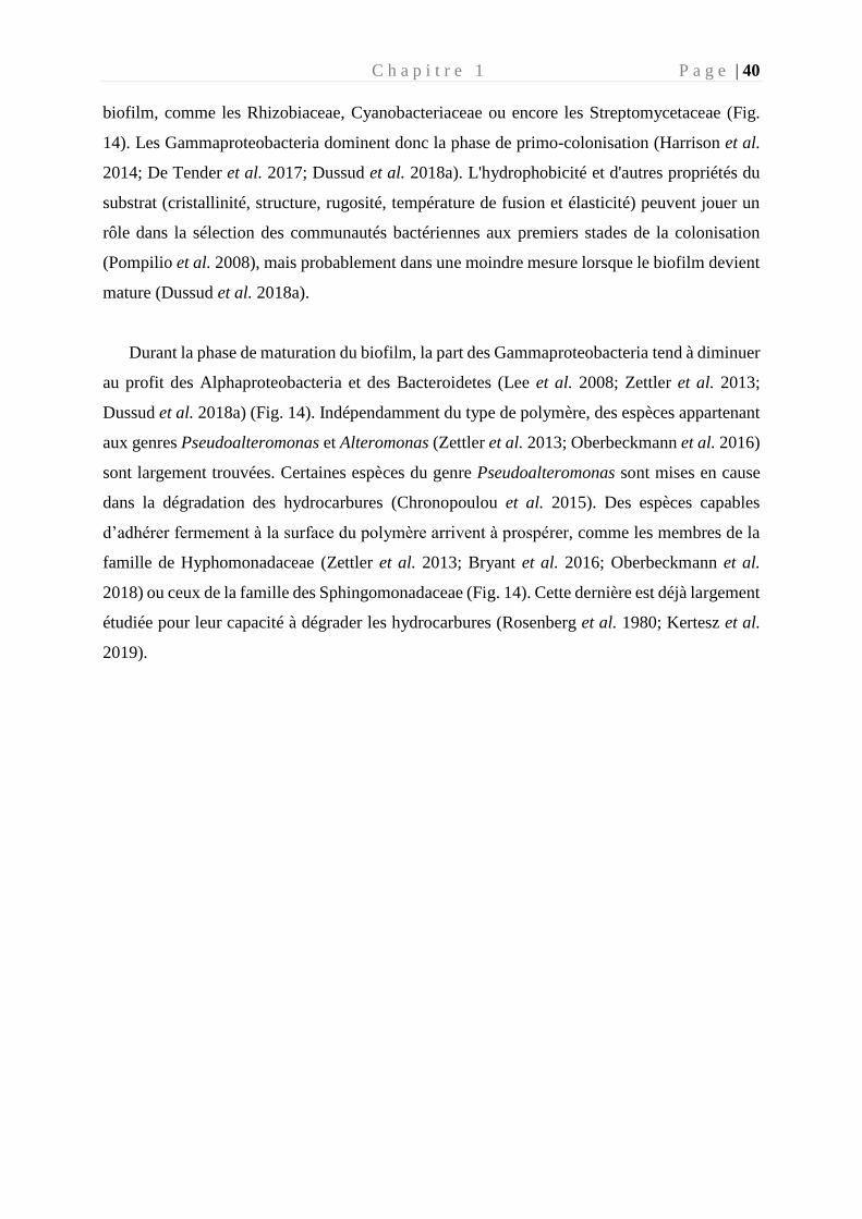
C h a p i t r e 1 P a g e | 40
biofilm, comme les Rhizobiaceae, Cyanobacteriaceae ou encore les Streptomycetaceae (Fig.
14). Les Gammaproteobacteria dominent donc la phase de primo-colonisation (Harrison et al.
2014; De Tender et al. 2017; Dussud et al. 2018a). L'hydrophobicité et d'autres propriétés du
substrat (cristallinité, structure, rugosité, température de fusion et élasticité) peuvent jouer un
rôle dans la sélection des communautés bactériennes aux premiers stades de la colonisation
(Pompilio et al. 2008), mais probablement dans une moindre mesure lorsque le biofilm devient
mature (Dussud et al. 2018a).
Durant la phase de maturation du biofilm, la part des Gammaproteobacteria tend à diminuer
au profit des Alphaproteobacteria et des Bacteroidetes (Lee et al. 2008; Zettler et al. 2013;
Dussud et al. 2018a) (Fig. 14). Indépendamment du type de polymère, des espèces appartenant
aux genres Pseudoalteromonas et Alteromonas (Zettler et al. 2013; Oberbeckmann et al. 2016)
sont largement trouvées. Certaines espèces du genre Pseudoalteromonas sont mises en cause
dans la dégradation des hydrocarbures (Chronopoulou et al. 2015). Des espèces capables
d’adhérer fermement à la surface du polymère arrivent à prospérer, comme les membres de la
famille de Hyphomonadaceae (Zettler et al. 2013; Bryant et al. 2016; Oberbeckmann et al.
2018) ou ceux de la famille des Sphingomonadaceae (Fig. 14). Cette dernière est déjà largement
étudiée pour leur capacité à dégrader les hydrocarbures (Rosenberg et al. 1980; Kertesz et al.
2019).
C h a p i t r e 1 P a g e | 41
Figure 14 : Différentes étapes de la formation du biofilm microbien retrouvés sur les polymères (modifié de Douarche et al.
2018). Image SEM : Zettler et al. 2020
La plastisphère étudiée lors d’expéditions, ou de prélèvement in situ, permet l’étude du biofilm
mature uniquement, présent depuis parfois plusieurs années sur le polymère et soumis aux
facteurs environnementaux.
Colonisateur primaire :
Diatomées, Gamma-proteobacteria Rhodobacteraceae,
rhizobiaceae, Cyanobacteriaceae et Streptomycetaceae
Colonisateur du biofilm mature :
Gamma et Alpha-proteobacteria, Bactéroidetes, Phycisphaerae,
Hyphomonadaceae, Sphingomononadaceae, Planctomycetia,
C h a p i t r e 1 P a g e | 42
3.2. Particularité de la plastisphère par comparaison aux micro-
organismes de l’eau environnante
Les communautés microbiennes de la plastisphère vont généralement partager plus de
similarité avec celles attachées aux particules que celles vivant librement dans l’eau
environnante. Les communautés vivantes sur les particules organiques sont dominées par les
Alphaproteobacteria (principalement Erythrobacter sp., lignée abondante sur les plastiques) et
les Gammaproteobacteria (principalement Alteromonas sp., lignée abondante également sur les
plastiques) (Crespo et al. 2013; Mohit et al. 2014; Dussud et al. 2018b). L’étude des gènes
fonctionnels de la plastisphère en utilisant des analyses métagénomiques a permis de distinguer
les communautés de la plastisphère de celle de l’environnement marin. Les communautés
détectées sur les polymères expriment des gènes impliqués dans les systèmes de sécrétion, dans
des activités de biodégradation xénobiotiques (Bryant et al. 2016), dans les fonctions de la
porphyrine et de la chlorophylle contrairement aux génomes des bactéries vivant librement ou
attachées aux particules de l’environnement (Dussud et al. 2018b).
Fait intéressant, la plupart des études caractérisant la plastisphère ont démontré que les
cyanobactéries étaient surreprésentées (en termes d’abondance relative issue de séquençage
haut débit) sur les plastiques par rapport aux fractions environnantes libres et liées aux
particules organiques (Zettler et al. 2013; Bryant et al. 2016; Dussud et al. 2018b). On trouve
des taxa affiliés aux Leptolyngbya sp., Pleurocapsa sp., Calothrix sp., Scytonema sp. et
Rivularia sp., ce qui explique souvent la différence de communauté entre les différentes parties
de l’environnement marin (fraction libre, attachée aux particules ou sur les plastiques marins)
(Fig.15) (Zettler et al. 2013; Dussud et al. 2018b; Basili et al. 2020).
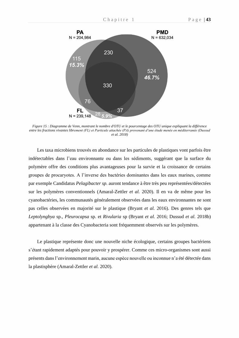
C h a p i t r e 1 P a g e | 43
Figure 15 : Diagramme de Venn, montrant le nombre d'OTU et le pourcentage des OTU unique expliquant la différence
entre les fractions vivantes librement (FL) et Particule attachée (PA) provenant d’une étude menée en méditerranée (Dussud
et al. 2018)
Les taxa microbiens trouvés en abondance sur les particules de plastiques vont parfois être
indétectables dans l’eau environnante ou dans les sédiments, suggérant que la surface du
polymère offre des conditions plus avantageuses pour la survie et la croissance de certains
groupes de procaryotes. A l’inverse des bactéries dominantes dans les eaux marines, comme
par exemple Candidatus Pelagibacter sp. auront tendance à être très peu représentées/détectées
sur les polymères conventionnels (Amaral-Zettler et al. 2020). Il en va de même pour les
cyanobactéries, les communautés généralement observées dans les eaux environnantes ne sont
pas celles observées en majorité sur le plastique (Bryant et al. 2016). Des genres tels que
Leptolyngbya sp., Pleurocapsa sp. et Rivularia sp (Bryant et al. 2016; Dussud et al. 2018b)
appartenant à la classe des Cyanobacteria sont fréquemment observés sur les polymères.
Le plastique représente donc une nouvelle niche écologique, certains groupes bactériens
s’étant rapidement adaptés pour pouvoir y prospérer. Comme ces micro-organismes sont aussi
présents dans l’environnement marin, aucune espèce nouvelle ou inconnue n’a été détectée dans
la plastisphère (Amaral-Zettler et al. 2020).
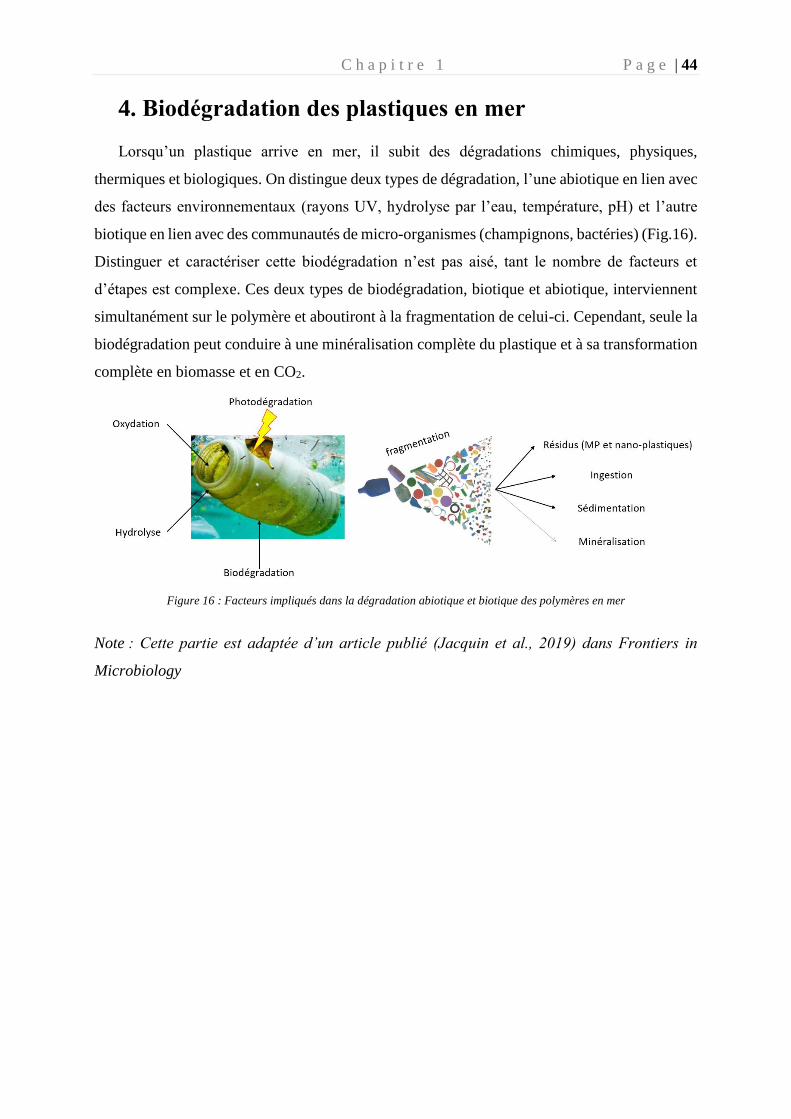
C h a p i t r e 1 P a g e | 44
4. Biodégradation des plastiques en mer
Lorsqu’un plastique arrive en mer, il subit des dégradations chimiques, physiques,
thermiques et biologiques. On distingue deux types de dégradation, l’une abiotique en lien avec
des facteurs environnementaux (rayons UV, hydrolyse par l’eau, température, pH) et l’autre
biotique en lien avec des communautés de micro-organismes (champignons, bactéries) (Fig.16).
Distinguer et caractériser cette biodégradation n’est pas aisé, tant le nombre de facteurs et
d’étapes est complexe. Ces deux types de biodégradation, biotique et abiotique, interviennent
simultanément sur le polymère et aboutiront à la fragmentation de celui-ci. Cependant, seule la
biodégradation peut conduire à une minéralisation complète du plastique et à sa transformation
complète en biomasse et en CO2.
Figure 16 : Facteurs impliqués dans la dégradation abiotique et biotique des polymères en mer
Note : Cette partie est adaptée d’un article publié (Jacquin et al., 2019) dans Frontiers in
Microbiology
C h a p i t r e 1 P a g e | 45
4.1. Dégradation abiotique
La dégradation abiotique intervient dès l’entrée du polymère dans le milieu marin et
généralement avant la dégradation biotique. Plusieurs formes de dégradation abiotiques sont
observées :
- La dégradation oxydative : ce facteur est le plus important. En effet, les plastiques se
situant dans la zone euphotique de l’environnement marin vont subir l’action des
ultraviolets (UV) qui entrainent des modifications chimiques du polymère en oxydant
les chaines carbonées grâce à la réaction de Norrish (Sudhakar et al. 2008). Cette
réaction permet le clivage photochimique des aldéhydes et des cétones en deux radicaux
libres intermédiaires.
- La dégradation mécanique : crée par les vagues, les courants, fragilisant le polymère
et accélérant son vieillissement au niveau moléculaire (Briassoulis 2007).
- La dégradation thermique : le changement de température peut fragiliser la structure
du polymère. Cependant ce paramètre a beaucoup moins d’importance que dans
d’autres environnements tel que le compost par exemple. En effet, bien que les
polymères aient un point de fusion relativement bas, la température des océans reste
largement en dessous de cette température de fusion.
- La dégradation par hydrolyse : agit principalement sur les liaisons ester (Gu and Gu
2005) grâce à l’eau environnante.
L'altération des propriétés plastiques due à la dégradation abiotique est appelée
«vieillissement» et augmente le taux de dégradation en oxydant ou en perturbant la longueur
de la chaîne polymère. Cette dégradation abiotique agit dans un second temps en synergie avec
la biodégradation.
4.2. Dégradation biotique
La biodégradation est un processus complexe, qui même dans sa définition divise au sein
de la communauté scientifique et des instituts de normalisation. Toutefois un consensus a été
admis, pour définir la biodégradation du plastique en mer comme étant un processus qui se
traduit par une conversion (totale ou partielle) du carbone organique en biogaz (CO2 , CH4 ,
H2S, NH4 et H2 ) (Mohee et al. 2008) et en biomasse associés à l'activité d'une communauté de
micro-organismes (bactéries, champignons) capables d'utiliser le plastique comme source de
carbone (Shah et al. 2008). Dans cette définition, il n’y a pas de notion de temps, ni de taux de
C h a p i t r e 1 P a g e | 46
biodégradation, ce qui laisse place à de nombreux débats. Dans une revue récente, l’équipe de
Zettler a proposé de mettre l’accent sur une biodégradation totale (en CO2, H2O et biomasse) et
non partielle, dans un délai raisonnable (plus de 90 % de carbone doit est converti en CO2 en
compost et 30% du carbone doit est converti en CO2 en mer (à 30°C) en 180 jours) (Amaral-
Zettler et al. 2020). La plastisphère est de plus en plus étudiée pour ses capacités de
biodégradation du polymère en milieu marin, et peut être envisagée comme une solution pour
endiguer cette pollution.
On considère que la biodégradation intervient dans un second temps. Elle est facilitée par
la dégradation abiotique qui fragilise la structure des polymères en révélant la rugosité, les
fissures et les changements moléculaires (İpekogˇlu et al. 2007).
La capacité des micro-organismes à dégrader le plastique a été observée pour de
nombreuses souches bactériennes (Shah et al. 2008; Krueger et al. 2015; Jacquin et al. 2019).
Les champignons ont également la capacité de dégrader les plastiques, mais la plupart des
études ont été menées en conditions terrestres (Cosgrove et al. 2007; Koitabashi et al. 2012;
Gajendiran et al. 2016; Magnin et al. 2018). Très peu d'études existent à ce jour en conditions
marines (Gonda et al. 2000; R. Pramila 2011; Sánchez 2020).
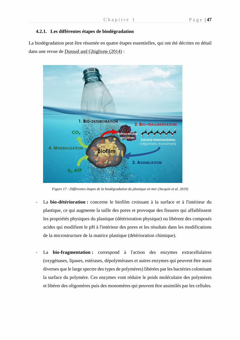
C h a p i t r e 1 P a g e | 47
4.2.1. Les différentes étapes de biodégradation
La biodégradation peut être résumée en quatre étapes essentielles, qui ont été décrites en détail
dans une revue de Dussud and Ghiglione (2014) :
Figure 17 : Différentes étapes de la biodégradation du plastique en mer (Jacquin et al. 2019)
- La bio-détérioration : concerne le biofilm croissant à la surface et à l'intérieur du
plastique, ce qui augmente la taille des pores et provoque des fissures qui affaiblissent
les propriétés physiques du plastique (détérioration physique) ou libèrent des composés
acides qui modifient le pH à l'intérieur des pores et les résultats dans les modifications
de la microstructure de la matrice plastique (détérioration chimique).
- La bio-fragmentation : correspond à l'action des enzymes extracellulaires
(oxygénases, lipases, estérases, dépolymérases et autres enzymes qui peuvent être aussi
diverses que le large spectre des types de polymères) libérées par les bactéries colonisant
la surface du polymère. Ces enzymes vont réduire le poids moléculaire des polymères
et libérer des oligomères puis des monomères qui peuvent être assimilés par les cellules.
C h a p i t r e 1 P a g e | 48
- L'assimilation : permet aux oligomères de moins de 600 Daltons d'être intégrés à
l'intérieur des cellules pour être utilisés comme source de carbone, augmentant ainsi la
biomasse microbienne.
- La minéralisation : est l'étape ultime de la biodégradation d'un polymère plastique et
entraîne l'excrétion de métabolites complètement oxydés (CO2, N2, CH4 et H2O).
4.2.2. Taux de dégradation plastique
La biodégradation n’est pas un processus limité dans le temps. Cependant, les polymères
continuent de s’accumuler en mer, liés au rythme d’introduction des déchets plastiques dans
l’environnement qui est bien plus rapide que celui de la biodégradation (Tableau 2).
L’environnement marin n’est pas un milieu agressif et propice à une forte biodégradation
comparé au compost (industriel ou domestique) ou à la digestion anaérobie (Tableau 2). Les
taux de dégradation des plastiques conventionnels par les micro-organismes sont extrêmement
faibles, même dans des conditions de laboratoire optimisées. La plupart des plastiques
conventionnels sont récalcitrants à la biodégradation dans les environnements marins et
terrestres, entraînant des durées de vie des polymères de plusieurs dizaines d’années voir
plusieurs siècles (Krueger et al. 2015).
Les plastiques présentent une faible biodisponibilité car ils sont généralement solides, liés
entre eux par des liaisons fortes offrant une faible accessibilité aux micro-organismes et aux
enzymes, circonscrits à la couche la plus externe des morceaux de plastiques. Dans l'écosystème
pélagique, les plastiques sont biodégradés par le métabolisme aérobie des micro-organismes,
c'est-à-dire que le produit final de la réaction sera la biomasse microbienne, le CO2 et le H2O.
La voie de biodégradation anaérobie serait plus fréquemment rencontrée dans les sédiments et
devrait être encore plus lente que dans la zone pélagique (Ishigaki et al. 2004). Le rapport C /
N défavorable est un facteur clé pour la biodégradation d'autres produits à base d'hydrocarbures
dans les océans (Sauret et al. 2016) et peut potentiellement limiter la biodégradation plastique.
Les données actuellement disponibles reposent largement sur des approches culturales dans des
conditions de laboratoire, bien que les bactéries pouvant être cultivées représentent moins de
1% de la diversité totale retrouvée dans l’environnement (Hugenholtz et al. 2009). À ce jour,
les données sur le taux de minéralisation plastique dans les océans sont encore pratiquement
inexistantes.
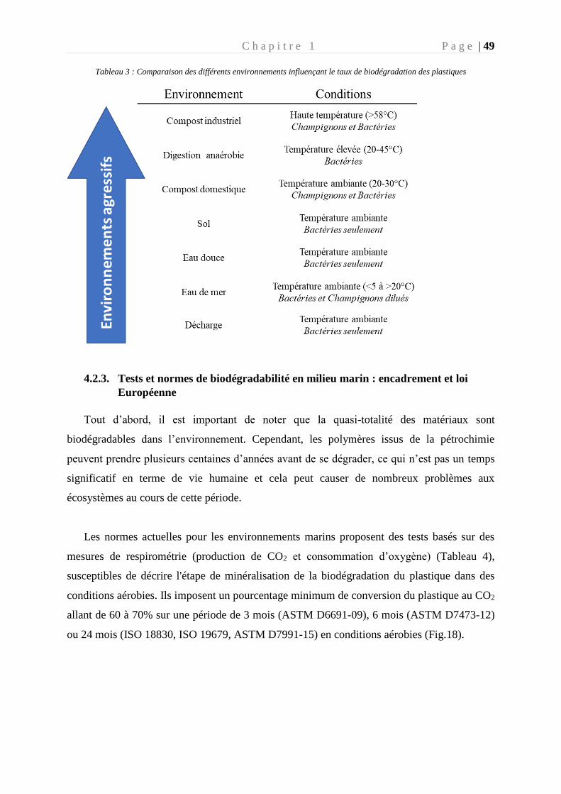
C h a p i t r e 1 P a g e | 49
Tableau 3 : Comparaison des différents environnements influençant le taux de biodégradation des plastiques
4.2.3. Tests et normes de biodégradabilité en milieu marin : encadrement et loi
Européenne
Tout d’abord, il est important de noter que la quasi-totalité des matériaux sont
biodégradables dans l’environnement. Cependant, les polymères issus de la pétrochimie
peuvent prendre plusieurs centaines d’années avant de se dégrader, ce qui n’est pas un temps
significatif en terme de vie humaine et cela peut causer de nombreux problèmes aux
écosystèmes au cours de cette période.
Les normes actuelles pour les environnements marins proposent des tests basés sur des
mesures de respirométrie (production de CO2 et consommation d’oxygène) (Tableau 4),
susceptibles de décrire l'étape de minéralisation de la biodégradation du plastique dans des
conditions aérobies. Ils imposent un pourcentage minimum de conversion du plastique au CO2
allant de 60 à 70% sur une période de 3 mois (ASTM D6691-09), 6 mois (ASTM D7473-12)
ou 24 mois (ISO 18830, ISO 19679, ASTM D7991-15) en conditions aérobies (Fig.18).
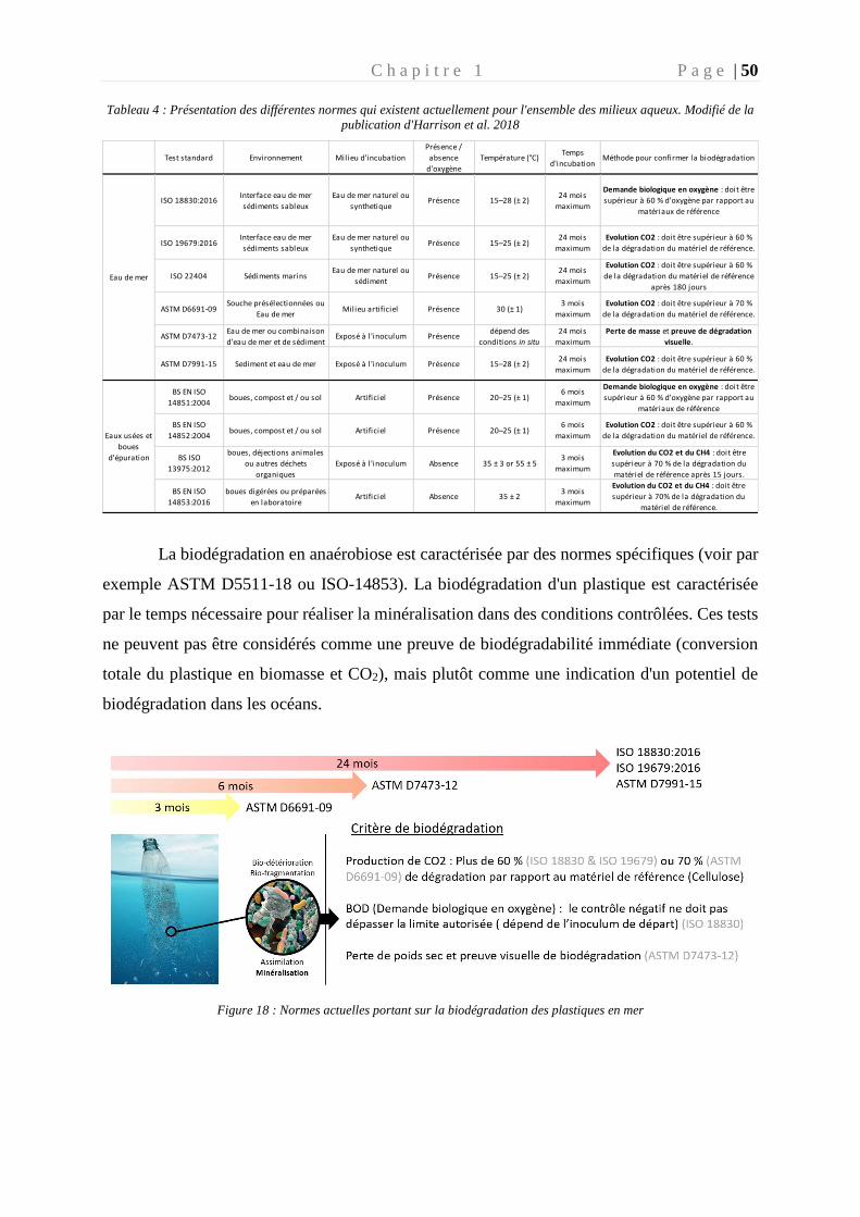
C h a p i t r e 1 P a g e | 50
Tableau 4 : Présentation des différentes normes qui existent actuellement pour l'ensemble des milieux aqueux. Modifié de la
publication d'Harrison et al. 2018
La biodégradation en anaérobiose est caractérisée par des normes spécifiques (voir par
exemple ASTM D5511-18 ou ISO-14853). La biodégradation d'un plastique est caractérisée
par le temps nécessaire pour réaliser la minéralisation dans des conditions contrôlées. Ces tests
ne peuvent pas être considérés comme une preuve de biodégradabilité immédiate (conversion
totale du plastique en biomasse et CO2), mais plutôt comme une indication d'un potentiel de
biodégradation dans les océans.
Figure 18 : Normes actuelles portant sur la biodégradation des plastiques en mer
Test standard Environnement Milieu d'incubation
Présence /
absence
d'oxygène
Température (°C)Temps
d'incubationMéthode pour confirmer la biodégradation
ISO 18830:2016 Interface eau de mer
sédiments sableux
Eau de mer naturel ou
synthetiquePrésence 15–28 (± 2)
24 mois
maximum
Demande biologique en oxygène : doit être
supérieur à 60 % d'oxygène par rapport au
matériaux de référence
ISO 19679:2016Interface eau de mer
sédiments sableux
Eau de mer naturel ou
synthetiquePrésence 15–25 (± 2)
24 mois
maximum
Evolution CO2 : doit être supérieur à 60 %
de la dégradation du matériel de référence.
ISO 22404 Sédiments marinsEau de mer naturel ou
sédimentPrésence 15–25 (± 2)
24 mois
maximum
Evolution CO2 : doit être supérieur à 60 %
de la dégradation du matériel de référence
après 180 jours
ASTM D6691-09 Souche présélectionnées ou
Eau de merMilieu artificiel Présence 30 (± 1)
3 mois
maximum
Evolution CO2 : doit être supérieur à 70 %
de la dégradation du matériel de référence.
ASTM D7473-12 Eau de mer ou combinaison
d'eau de mer et de sédimentExposé à l 'inoculum Présence
dépend des
conditions in situ
24 mois
maximum
Perte de masse et preuve de dégradation
visuelle.
ASTM D7991-15 Sediment et eau de mer Exposé à l 'inoculum Présence 15–28 (± 2)24 mois
maximum
Evolution CO2 : doit être supérieur à 60 %
de la dégradation du matériel de référence.
BS EN ISO
14851:2004 boues, compost et / ou sol Artificiel Présence 20–25 (± 1)
6 mois
maximum
Demande biologique en oxygène : doit être
supérieur à 60 % d'oxygène par rapport au
matériaux de référence
BS EN ISO
14852:2004boues, compost et / ou sol Artificiel Présence 20–25 (± 1)
6 mois
maximum
Evolution CO2 : doit être supérieur à 60 %
de la dégradation du matériel de référence.
BS ISO
13975:2012
boues, déjections animales
ou autres déchets
organiques
Exposé à l 'inoculum Absence 35 ± 3 or 55 ± 53 mois
maximum
Evolution du CO2 et du CH4 : doit être
supérieur à 70 % de la dégradation du
matériel de référence après 15 jours.
BS EN ISO
14853:2016
boues digérées ou préparées
en laboratoireArtificiel Absence 35 ± 2
3 mois
maximum
Evolution du CO2 et du CH4 : doit être
supérieur à 70% de la dégradation du
matériel de référence.
Eau de mer
Eaux usées et
boues
d'épuration
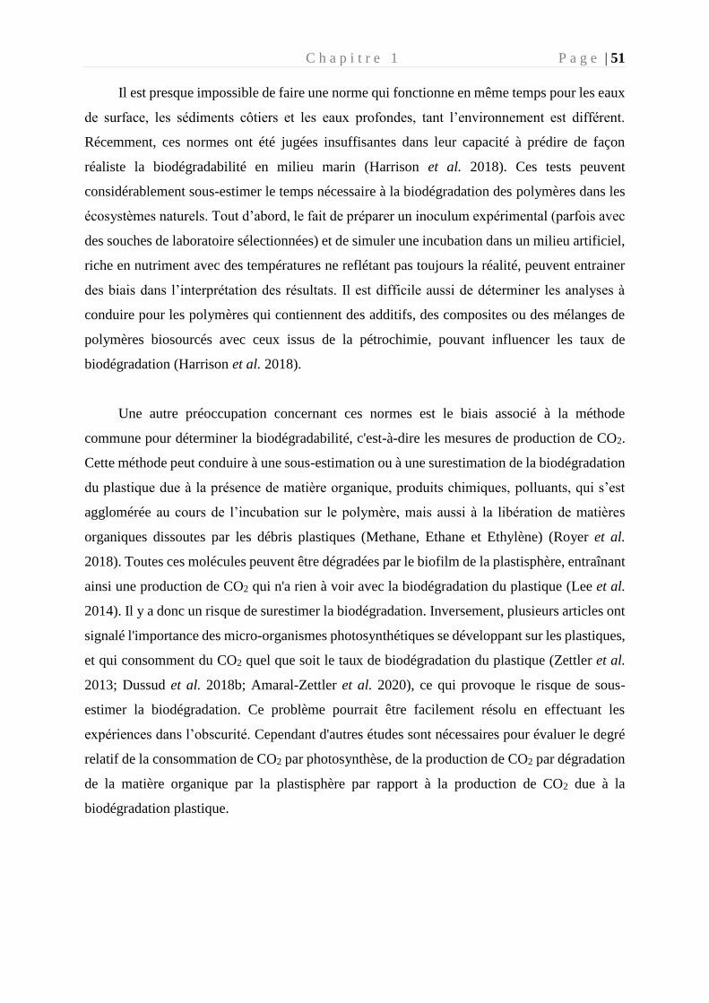
C h a p i t r e 1 P a g e | 51
Il est presque impossible de faire une norme qui fonctionne en même temps pour les eaux
de surface, les sédiments côtiers et les eaux profondes, tant l’environnement est différent.
Récemment, ces normes ont été jugées insuffisantes dans leur capacité à prédire de façon
réaliste la biodégradabilité en milieu marin (Harrison et al. 2018). Ces tests peuvent
considérablement sous-estimer le temps nécessaire à la biodégradation des polymères dans les
écosystèmes naturels. Tout d’abord, le fait de préparer un inoculum expérimental (parfois avec
des souches de laboratoire sélectionnées) et de simuler une incubation dans un milieu artificiel,
riche en nutriment avec des températures ne reflétant pas toujours la réalité, peuvent entrainer
des biais dans l’interprétation des résultats. Il est difficile aussi de déterminer les analyses à
conduire pour les polymères qui contiennent des additifs, des composites ou des mélanges de
polymères biosourcés avec ceux issus de la pétrochimie, pouvant influencer les taux de
biodégradation (Harrison et al. 2018).
Une autre préoccupation concernant ces normes est le biais associé à la méthode
commune pour déterminer la biodégradabilité, c'est-à-dire les mesures de production de CO2.
Cette méthode peut conduire à une sous-estimation ou à une surestimation de la biodégradation
du plastique due à la présence de matière organique, produits chimiques, polluants, qui s’est
agglomérée au cours de l’incubation sur le polymère, mais aussi à la libération de matières
organiques dissoutes par les débris plastiques (Methane, Ethane et Ethylène) (Royer et al.
2018). Toutes ces molécules peuvent être dégradées par le biofilm de la plastisphère, entraînant
ainsi une production de CO2 qui n'a rien à voir avec la biodégradation du plastique (Lee et al.
2014). Il y a donc un risque de surestimer la biodégradation. Inversement, plusieurs articles ont
signalé l'importance des micro-organismes photosynthétiques se développant sur les plastiques,
et qui consomment du CO2 quel que soit le taux de biodégradation du plastique (Zettler et al.
2013; Dussud et al. 2018b; Amaral-Zettler et al. 2020), ce qui provoque le risque de sous-
estimer la biodégradation. Ce problème pourrait être facilement résolu en effectuant les
expériences dans l’obscurité. Cependant d'autres études sont nécessaires pour évaluer le degré
relatif de la consommation de CO2 par photosynthèse, de la production de CO2 par dégradation
de la matière organique par la plastisphère par rapport à la production de CO2 due à la
biodégradation plastique.
C h a p i t r e 1 P a g e | 52
Les limites des méthodes respiratoires décrites ci-dessus peuvent être surmontées par
d'autres techniques et approches analytiques supplémentaires, pour confirmer les changements
dans les propriétés physiques et la structure chimique des polymères pendant la biodégradation.
Les altérations de l'aspect visuel et de la masse ou les changements de propriétés mécaniques
sont des méthodes relativement faciles et peu coûteuses à effectuer pour évaluer des
changements physiques pendant la biodégradation. D’autres méthodes pourraient être
combinées comme le marquage isotopique, qui permet d’évaluer l’assimilation microbienne du
polymère. En effet, une étude récente a utilisé du poly (butylène adipate-téréphtalate) (PBAT)
marqué, associé à de la spectrométrie de masse (Nanosims) pour prouver l’assimilation
microbienne du carbone marqué du polymère par les communautés microbiennes (Zumstein et
al. 2018). Une précédente version de norme ASTM (D6692-01) utilisait cette technique de
marquage au 14C lors de suivi de production de CO2. D’autres techniques peuvent confirmer les
changements dans la structure moléculaire des polymères comme les mesures d'hydrolyse de
surface et d'autres mesures chromatographiques (chromatographie en phase gazeuse,
chromatographie liquide, chromatographie de perméation de gel) couplées ou non à des
techniques spectrométriques (spectrométrie de masse, spectroscopie de résonance magnétique
nucléaire, spectroscopie infrarouge à transformer de Fourier). La microscopie à force atomique
et à balayage électronique peuvent également évaluer la biodégradation de la surface du
polymère due à l’activité microbienne. Chacune de ces méthodes a ses limites et ses biais
(description en détails dans les revues de Koutny et al. 2006; Ho et al. 2018; Harrison et al.
2018. Les normes actuelles proposent parfois d'utiliser de telles techniques pour corroborer le
test principal basé sur la mesure de la respirométrie, mais aucune directive claire sur la façon
d'utiliser ces tests n'est fournie.
4.3. Biodégradation en fonction de la composition des polymères
Il existe actuellement plus de 5300 types de polymères dans le commerce (Wagner and
Lambert 2018). Ils sont généralement produits avec une gamme d'additifs chimiques tels que
des plastifiants, des ignifugeants, des antioxydants et d'autres stabilisants, des pro-oxydants,
des surfactants, des charges inorganiques ou des pigments (Wagner and Lambert 2018). Leurs
propriétés physico-chimiques hétérogènes entraîneront probablement des voies métaboliques
de biodégradation très diverses, en particulier si l'on considère la grande variété de micro-
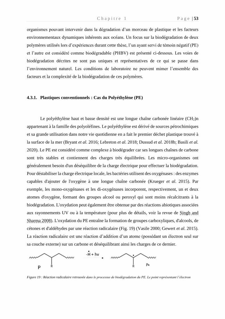
C h a p i t r e 1 P a g e | 53
organismes pouvant intervenir dans la dégradation d’un morceau de plastique et les facteurs
environnementaux dynamiques inhérents aux océans. Un focus sur la biodégradation de deux
polymères utilisés lors d’expériences durant cette thèse, l’un ayant servi de témoin négatif (PE)
et l’autre est considéré comme biodégradable (PHBV) est présenté ci-dessous. Les voies de
biodégradation décrites ne sont pas uniques et représentatives de ce qui se passe dans
l’environnement naturel. Les conditions de laboratoire ne peuvent mimer l’ensemble des
facteurs et la complexité de la biodégradation de ces polymères.
4.3.1. Plastiques conventionnels : Cas du Polyéthylène (PE)
Le polyéthylène haut et basse densité est une longue chaîne carbonée linéaire (CH2)n
appartenant à la famille des polyoléfines. Le polyéthylène est dérivé de sources pétrochimiques
et sa grande utilisation dans notre vie quotidienne en a fait le premier déchet plastique trouvé à
la surface de la mer (Bryant et al. 2016; Lebreton et al. 2018; Dussud et al. 2018b; Basili et al.
2020). Le PE est considéré comme complexe à biodégrader car ses longues chaînes de carbone
sont très stables et contiennent des charges très équilibrées. Les micro-organismes ont
généralement besoin d'un déséquilibre de la charge électrique pour effectuer la biodégradation.
Pour déstabiliser la charge électrique locale, les bactéries utilisent des oxygénases : des enzymes
capables d'ajouter de l'oxygène à une longue chaîne carbonée (Krueger et al. 2015). Par
exemple, les mono-oxygénases et les di-oxygénases incorporent, respectivement, un et deux
atomes d'oxygène, formant des groupes alcool ou peroxyl qui sont moins récalcitrants à la
biodégradation. L'oxydation peut également être obtenue par des réactions abiotiques associées
aux rayonnements UV ou à la température (pour plus de détails, voir la revue de Singh and
Sharma 2008). L'oxydation du PE entraîne la formation de groupes carboxyliques, d'alcools, de
cétones et d'aldéhydes par une réaction radicalaire (Fig. 19) (Vasile 2000; Gewert et al. 2015).
La réaction radicalaire est une réaction d’addition d’un atome (possédant un électron seul sur
sa couche externe) sur un carbone et déséquilibrant ainsi les charges de ce dernier.
Figure 19 : Réaction radicalaire retrouvée dans le processus de biodégradation du PE. Le point représentant l’électron
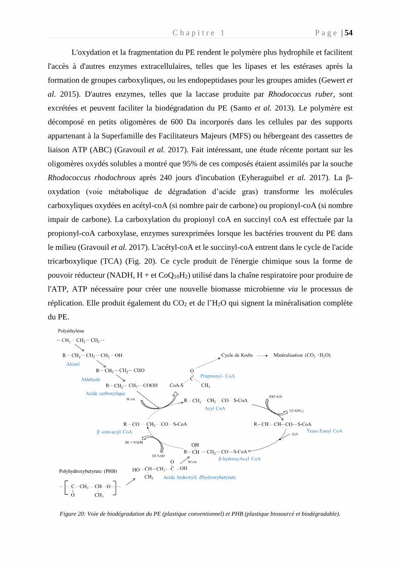
C h a p i t r e 1 P a g e | 54
L'oxydation et la fragmentation du PE rendent le polymère plus hydrophile et facilitent
l'accès à d'autres enzymes extracellulaires, telles que les lipases et les estérases après la
formation de groupes carboxyliques, ou les endopeptidases pour les groupes amides (Gewert et
al. 2015). D'autres enzymes, telles que la laccase produite par Rhodococcus ruber, sont
excrétées et peuvent faciliter la biodégradation du PE (Santo et al. 2013). Le polymère est
décomposé en petits oligomères de 600 Da incorporés dans les cellules par des supports
appartenant à la Superfamille des Facilitateurs Majeurs (MFS) ou hébergeant des cassettes de
liaison ATP (ABC) (Gravouil et al. 2017). Fait intéressant, une étude récente portant sur les
oligomères oxydés solubles a montré que 95% de ces composés étaient assimilés par la souche
Rhodococcus rhodochrous après 240 jours d'incubation (Eyheraguibel et al. 2017). La β-
oxydation (voie métabolique de dégradation d’acide gras) transforme les molécules
carboxyliques oxydées en acétyl-coA (si nombre pair de carbone) ou propionyl-coA (si nombre
impair de carbone). La carboxylation du propionyl coA en succinyl coA est effectuée par la
propionyl-coA carboxylase, enzymes surexprimées lorsque les bactéries trouvent du PE dans
le milieu (Gravouil et al. 2017). L'acétyl-coA et le succinyl-coA entrent dans le cycle de l'acide
tricarboxylique (TCA) (Fig. 20). Ce cycle produit de l'énergie chimique sous la forme de
pouvoir réducteur (NADH, H + et CoQ10H2) utilisé dans la chaîne respiratoire pour produire de
l'ATP, ATP nécessaire pour créer une nouvelle biomasse microbienne via le processus de
réplication. Elle produit également du CO2 et de l’H2O qui signent la minéralisation complète
du PE.
Figure 20: Voie de biodégradation du PE (plastique conventionnel) et PHB (plastique biosourcé et biodégradable).
C h a p i t r e 1 P a g e | 55
Depuis 20 ans maintenant, les scientifiques s'intéressent à la biodégradation du PE par
les communautés microbiennes. Les souches bactériennes et fongiques présentant des capacités
de biodégradation du PE sont répertoriées dans la revue publiée par Jacquin et al. 2019.
Les processus génétiques et métaboliques impliqués dans la biodégradation du PE
restent peu décrits dans la littérature. Cependant quelques travaux préliminaires ont mis en
évidence des enzymes, des transporteurs ou des gènes pouvant être impliqués dans ce processus
(Gravouil et al. 2017). Les gènes de l'alcane hydroxylase se sont avérés jouer un rôle central
dans la biodégradation du PE. Pseudomonas sp. E4 est capable de minéraliser 28,6% du carbone
organique du polymère en 80 jours. Le gène alkB a ensuite été introduit dans la souche
Escherichia coli BL21, qui a ensuite pu minéraliser 19,3% du carbone organique du polymère
(Yoon et al. 2012). Seules deux autres études ont utilisé l'analyse génétique pour prouver
l'importance de la laccase dans la biodégradation du PE par Rhodococcus ruber (Sivan 2011;
Santo et al. 2013; Gravouil et al. 2017).
4.3.2. Plastiques biosourcés : Cas du PHA
En raison de leur origine microbienne, les PHA se sont révélés biodégradables dans de
nombreux environnements tels que le sol, les écosystèmes marins ou les boues de stations
d'épurations (Eubeler et al. 2009). La biodégradation du PHB (un des nombreux polymères de
type PHA) a été prouvée avec des taux comparables à ceux de la cellulose, avec une dégradation
plus rapide trouvée dans des conditions aérobies (85 jours) par rapport à des conditions
anaérobies (6 mois) (Wang et al. 2018).
Sa biodégradation permet la scission des chaines de polymères en monomères ou
oligomère par rupture des liaisons esters. Ces liaisons sont clivées par une hydrolyse passive
ou une réaction enzymatique active (Göpferich 1996). La dégradation abiotique intervient
quand l’énergie externe dépasse le seuil de résistance des liaisons esters. Néanmoins, ce
phénomène reste rare et s’applique dans des conditions bien précises (rotation ou chaleur
excessives) (Göpferich 1996; Hablot et al. 2008).
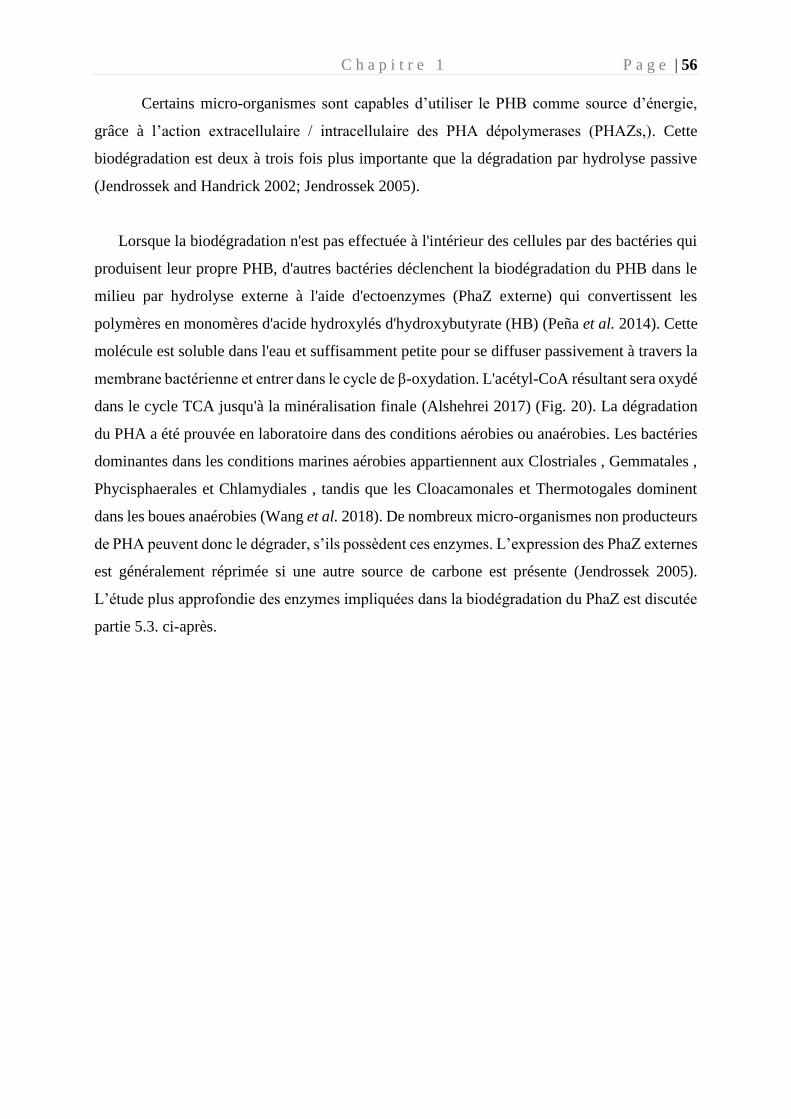
C h a p i t r e 1 P a g e | 56
Certains micro-organismes sont capables d’utiliser le PHB comme source d’énergie,
grâce à l’action extracellulaire / intracellulaire des PHA dépolymerases (PHAZs,). Cette
biodégradation est deux à trois fois plus importante que la dégradation par hydrolyse passive
(Jendrossek and Handrick 2002; Jendrossek 2005).
Lorsque la biodégradation n'est pas effectuée à l'intérieur des cellules par des bactéries qui
produisent leur propre PHB, d'autres bactéries déclenchent la biodégradation du PHB dans le
milieu par hydrolyse externe à l'aide d'ectoenzymes (PhaZ externe) qui convertissent les
polymères en monomères d'acide hydroxylés d'hydroxybutyrate (HB) (Peña et al. 2014). Cette
molécule est soluble dans l'eau et suffisamment petite pour se diffuser passivement à travers la
membrane bactérienne et entrer dans le cycle de β-oxydation. L'acétyl-CoA résultant sera oxydé
dans le cycle TCA jusqu'à la minéralisation finale (Alshehrei 2017) (Fig. 20). La dégradation
du PHA a été prouvée en laboratoire dans des conditions aérobies ou anaérobies. Les bactéries
dominantes dans les conditions marines aérobies appartiennent aux Clostriales , Gemmatales ,
Phycisphaerales et Chlamydiales , tandis que les Cloacamonales et Thermotogales dominent
dans les boues anaérobies (Wang et al. 2018). De nombreux micro-organismes non producteurs
de PHA peuvent donc le dégrader, s’ils possèdent ces enzymes. L’expression des PhaZ externes
est généralement réprimée si une autre source de carbone est présente (Jendrossek 2005).
L’étude plus approfondie des enzymes impliquées dans la biodégradation du PhaZ est discutée
partie 5.3. ci-après.
C h a p i t r e 1 P a g e | 57
5. Cas des PHA : Polymères biosourcés, biodégradables,
compostables et recyclables
Une attention particulière a été portée lors de cette thèse sur les PHA (voir chapitres 3 et 4),
qui sont actuellement décrits comme des polymères ayant des propriétés très prometteuses pour
se substituer dans certaines applications à des polymères conventionnels. Nous avons choisi
d’approfondir à la fois les intérêts commerciaux associés à leurs propriétés remarquables, tout
en décrivant l’état des connaissances actuelles sur leur voies de synthèse et de biodégradation.
5.1 Généralités
Dans ce contexte actuel de développement de plastique alternatif en limitant les
composés issus de la pétrochimie, les PHA semblent s’imposer comme une solution durable.
Ce sont des biopolymères considérés comme biodégradable, non toxique et biocompatible.
Depuis leur découverte en 1926, de nombreuses études ont montrés que le PHA est un polymère
synthétisé par de nombreuses bactéries, archées et eucaryotes (Anderson and Dawes 1990).
Les granules de PHA ont été longtemps considérés comme uniquement une molécule
de stockage d’énergie pour l’organisme producteur. En effet, cette molécule est produite lorsque
le micro-organisme se trouve dans un milieu riche en carbone (glucidique ou lipidique) et
subissant un stress environnemental (carence en azote et ou phosphore, oxygène) (Lee and Choi
1999). Le PHA est stocké sous forme de granules de réserve, exerçant une faible pression
osmotique sur les parois cellulaires et n’affectant pas le métabolisme du micro-organisme
(Jendrossek and Pfeiffer 2014). La synthèse de PHA est régulée et forme un équilibre entre le
stockage de sources carbonées et la dégradation en oligomères pour y récupérer les nutriments
nécessaires lors d’un stress nutritif de l’environnement (Kadouri et al. 2005). Certaines
bactéries peuvent accumuler jusqu'à 90% de leur poids total en polymère dans des conditions
très spécifiques (Peña et al. 2014). Les PHA sous forme de granules ont un diamètre compris
entre 0,2 et 0,7 µm et sont constitués à 97,7 % de PHA, 1,8 % de protéines et 0,5 % de lipides
(Koller et al. 2010). Tous les carbones des monomères du PHA ont une configuration R (Rectus,
carbone asymétrique, les atomes prioritaires sont classés dans le sens horaire) (Fig. 21), en
raison de la stéréospécificité de la Pha synthase (PhaC) (Verlinden et al. 2007; Singh et al.
2018), ceci a une grande importance sur la biodégradabilité du polymère (Voir partie 5.3).
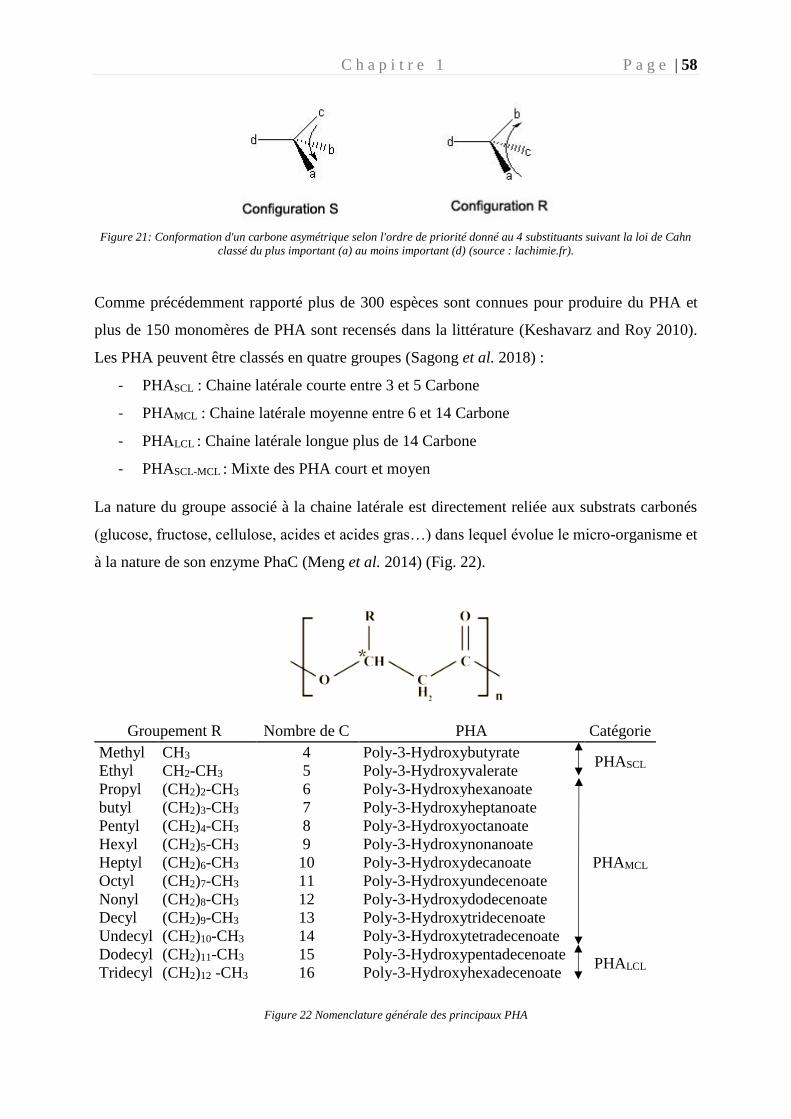
C h a p i t r e 1 P a g e | 58
Figure 21: Conformation d'un carbone asymétrique selon l'ordre de priorité donné au 4 substituants suivant la loi de Cahn
classé du plus important (a) au moins important (d) (source : lachimie.fr).
Comme précédemment rapporté plus de 300 espèces sont connues pour produire du PHA et
plus de 150 monomères de PHA sont recensés dans la littérature (Keshavarz and Roy 2010).
Les PHA peuvent être classés en quatre groupes (Sagong et al. 2018) :
- PHASCL : Chaine latérale courte entre 3 et 5 Carbone
- PHAMCL : Chaine latérale moyenne entre 6 et 14 Carbone
- PHALCL : Chaine latérale longue plus de 14 Carbone
- PHASCL-MCL : Mixte des PHA court et moyen
La nature du groupe associé à la chaine latérale est directement reliée aux substrats carbonés
(glucose, fructose, cellulose, acides et acides gras…) dans lequel évolue le micro-organisme et
à la nature de son enzyme PhaC (Meng et al. 2014) (Fig. 22).
Groupement R Nombre de C PHA Catégorie
Methyl CH3 4 Poly-3-Hydroxybutyrate PHASCL
Ethyl CH2-CH3 5 Poly-3-Hydroxyvalerate
Propyl (CH2)2-CH3 6 Poly-3-Hydroxyhexanoate
PHAMCL
butyl (CH2)3-CH3 7 Poly-3-Hydroxyheptanoate
Pentyl (CH2)4-CH3 8 Poly-3-Hydroxyoctanoate
Hexyl (CH2)5-CH3 9 Poly-3-Hydroxynonanoate
Heptyl (CH2)6-CH3 10 Poly-3-Hydroxydecanoate
Octyl (CH2)7-CH3 11 Poly-3-Hydroxyundecenoate
Nonyl (CH2)8-CH3 12 Poly-3-Hydroxydodecenoate
Decyl (CH2)9-CH3 13 Poly-3-Hydroxytridecenoate
Undecyl (CH2)10-CH3 14 Poly-3-Hydroxytetradecenoate
Dodecyl (CH2)11-CH3 15 Poly-3-Hydroxypentadecenoate PHALCL
Tridecyl (CH2)12 -CH3 16 Poly-3-Hydroxyhexadecenoate
Figure 22 Nomenclature générale des principaux PHA
C h a p i t r e 1 P a g e | 59
Ce stockage de granule de PHA permet :
- d’augmenter la survie du micro-organisme producteur de PHA (Kadouri et al. 2005).
- d’augmenter la résistance au stress oxydatif (Goh et al. 2014) en surexprimant les gènes
codant les protéines de stress, menant à la production de molécules anti-oxydantes (Ruiz
et al. 2004; Ayub et al. 2009).
- d’augmenter sa résistance au choc osmotique chez les micro-organismes marins (Yin et
al. 2015).
- d’augmenter la résistance aux températures basses ou hautes (Wu et al. 2011; Pärnänen
et al. 2015).
- d’augmenter la résistance au stress salin et thermique en limitant le phénomène
d’agrégation de protéines (Soto et al. 2012).
5.2. Intérêt commercial du PHA
Les propriétés physiques et chimiques du PHA vont principalement dépendre du
monomère qui le compose. Le type de monomère est contrôlé par les micro-organismes
producteurs, le milieu, les conditions de fermentations et la méthode de récupération des PHA
(Keshavarz and Roy 2010; Amirul et al. 2015).
Les PHA sont envisagés dans de nombreux domaines tels que l’industrie plastique
(produits extrudés, moulés, films d’emballage), l’usage médical (implants, sutures, appareils
médicaux corporels), support de biodégradation pour molécules d’intérêts (hormones,
médicaments, biocides), domaines agricoles, domaine de la peinture (Scholz 2001).
Les PHA montre une perméabilité à l’eau et l’oxygène peu importante, ce qui représente
un avantage considérable dans le domaine de l’emballage. Généralement considérée comme
hydrophobe, cette propriété est variable en fonction des groupements fonctionnels composant
le monomère de PHA. En effet, l’hydrophobicité peut être modifiée par des groupements
hydroxyl, amine ou carboxylique compose le monomère ou par copolymérisation intégrants des
composés hydrophiles.
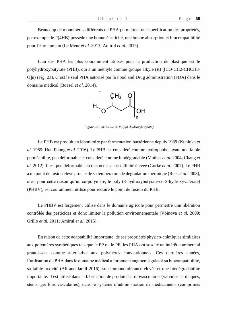
C h a p i t r e 1 P a g e | 60
Beaucoup de monomères différents de PHA permettent une spécification des propriétés,
par exemple le P(4HB) possède une bonne élasticité, une bonne absorption et biocompatibilité
pour l’être humain (Le Meur et al. 2013; Amirul et al. 2015).
L'un des PHA les plus couramment utilisés pour la production de plastique est le
polyhydroxybutyrate (PHB), qui a un méthyle comme groupe alkyle (R) ([CO-CH2-CHCH3-
O]n) (Fig. 23). C’est le seul PHA autorisé par la Food and Drug administration (FDA) dans le
domaine médical (Boesel et al. 2014).
Figure 23 : Molécule de Poly(β -hydroxybutyrate)
Le PHB est produit en laboratoire par fermentation bactérienne depuis 1989 (Kunioka et
al. 1989; Huu Phong et al. 2016). Le PHB est considéré comme hydrophobe, ayant une faible
perméabilité, peu déformable et considéré comme biodégradable (Mothes et al. 2004; Chang et
al. 2012). Il est peu déformable en raison de sa cristallinité élevée (Gorke et al. 2007). Le PHB
a un point de fusion élevé proche de sa température de dégradation thermique (Reis et al. 2003),
c’est pour cette raison qu’un co-polymère, le poly (3-hydroxybutyrate-co-3-hydroxyvalérate)
(PHBV), est couramment utilisé pour réduire le point de fusion du PHB.
Le PHBV est largement utilisé dans le domaine agricole pour permettre une libération
contrôlée des pesticides et donc limiter la pollution environnementale (Voinova et al. 2009;
Grillo et al. 2011; Amirul et al. 2015).
En raison de cette adaptabilité importante, de ses propriétés physico-chimiques similaires
aux polymères synthétiques tels que le PP ou le PE, les PHA ont suscité un intérêt commercial
grandissant comme alternative aux polymères conventionnels. Ces dernières années,
l’utilisation du PHA dans le domaine médical a fortement augmenté grâce à sa biocompatibilité,
sa faible toxicité (Ali and Jamil 2016), son immunotolérance élevée et une biodégradabilité
importante. Il est utilisé dans la fabrication de produits cardiovasculaires (valvules cardiaques,
stents, greffons vasculaires), dans le système d’administration de médicaments (comprimés
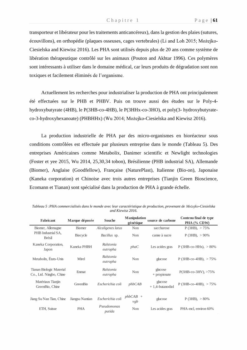
C h a p i t r e 1 P a g e | 61
transporteur et libérateur pour les traitements anticancéreux), dans la gestion des plaies (sutures,
écouvillons), en orthopédie (plaques osseuses, cages vertebrales) (Li and Loh 2015; Możejko-
Ciesielska and Kiewisz 2016). Les PHA sont utilisés depuis plus de 20 ans comme système de
libération thérapeutique contrôlé sur les animaux (Pouton and Akhtar 1996). Ces polymères
sont intéressants à utiliser dans le domaine médical, car leurs produits de dégradation sont non
toxiques et facilement éliminés de l’organisme.
Actuellement les recherches pour industrialiser la production de PHA ont principalement
été effectuées sur le PHB et PHBV. Puis on trouve aussi des études sur le Poly-4-
hydroxybutyrate (4HB), le P(3HB-co-4HB), le P(3HHx-co-3HO), et poly(3- hydroxybutyrate-
co-3-hydroxyhexanoate) (PHBHHx) (Wu 2014; Możejko-Ciesielska and Kiewisz 2016).
La production industrielle de PHA par des micro-organismes en bioréacteur sous
conditions contrôlées est effectuée par plusieurs entreprise dans le monde (Tableau 5). Des
entreprises Américaines comme Metabolix, Danimer scientific et Newlight technologies
(Foster et yee 2015, Wu 2014, 25,30,34 tobon), Brésilienne (PHB industrial SA), Allemande
(Biomer), Anglaise (Goodfellow), Française (NaturePlast), Italienne (Bio-on), Japonaise
(Kaneka corporation) et Chinoise avec trois autres entreprises (Tianjin Green Bioscience,
Ecomann et Tianan) sont spécialisé dans la production de PHA à grande échelle.
Tableau 5 :PHA commercialisés dans le monde avec leur caractéristique de production, provenant de Możejko-Ciesielska
and Kiewisz 2016.
Fabricant Marque déposée SoucheManipulation
génétiquesource de carbone
Contenu final de type
PHA (% CDW)
Biomer, Allemagne Biomer Alcaligenes latus Non saccharose P (3HB), > 75%
PHB Industrial SA,
BrésilBiocycle Bacillus sp. Non canne à sucre P (3HB), > 90%
Kaneka Corporation,
JaponKaneka PHBH
Ralstonia
eutrophaphaC Les acides gras P (3HB-co-HHx), > 80%
Metabolix, États-Unis MirelRalstonia
eutrophaNon glucose P (3HB-co-4HB), > 75%
Tianan Biologic Material
Co., Ltd. Ningbo, ChineEnmat
Ralstonia
eutrophaNon
glucose
+ propionateP(3HB-co-3HV), >75%
Matériaux Tianjin
GreenBio, ChineGreenBio Escherichia coli phbCAB
glucose
+ 1,4-butanodiolP (3HB-co-4HB), > 75%
Jiang Su Nan Tian, Chine Jiangsu Nantian Escherichia coliphbCAB +
vgbglucose P (3HB), > 80%
ETH, Suisse PHAPseudomonas
putidaNon Les acides gras PHA-mcl, environ 60%
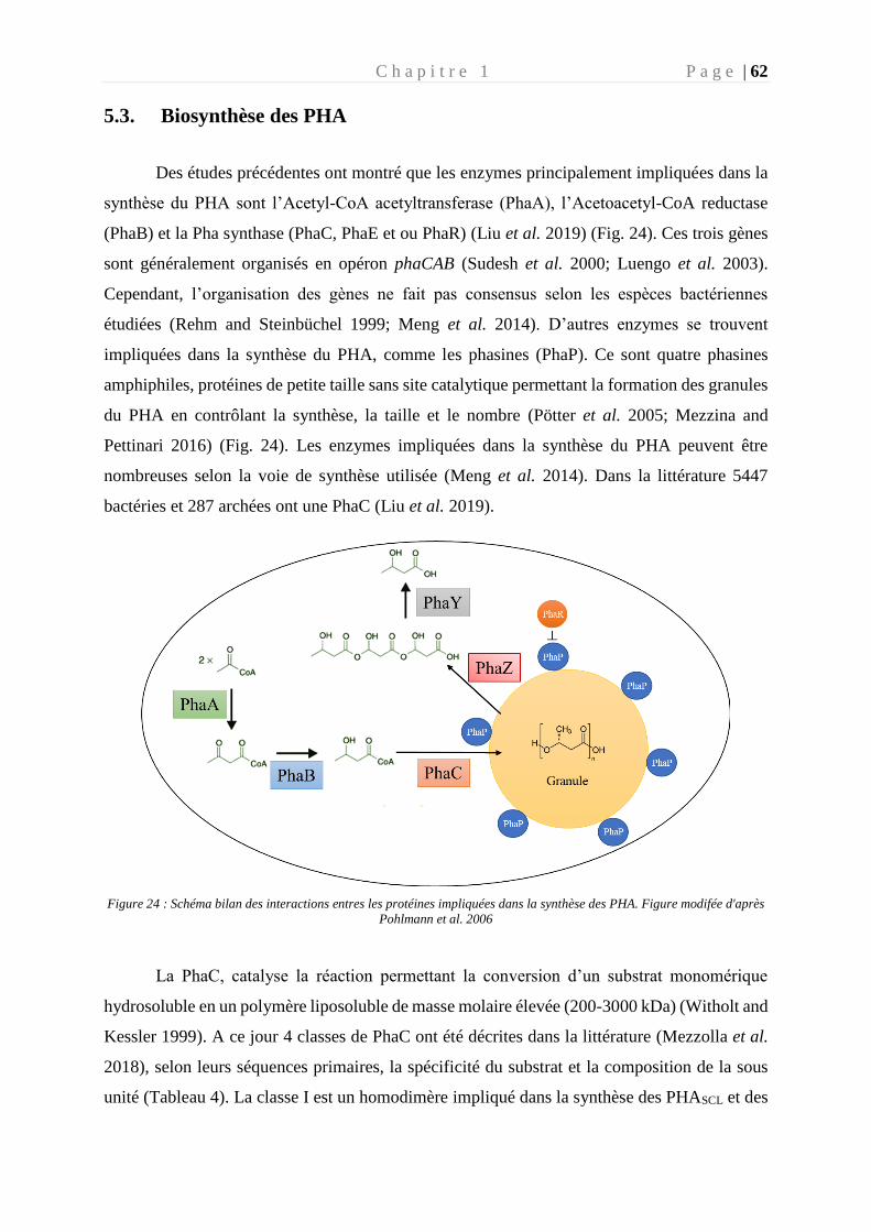
C h a p i t r e 1 P a g e | 62
5.3. Biosynthèse des PHA
Des études précédentes ont montré que les enzymes principalement impliquées dans la
synthèse du PHA sont l’Acetyl-CoA acetyltransferase (PhaA), l’Acetoacetyl-CoA reductase
(PhaB) et la Pha synthase (PhaC, PhaE et ou PhaR) (Liu et al. 2019) (Fig. 24). Ces trois gènes
sont généralement organisés en opéron phaCAB (Sudesh et al. 2000; Luengo et al. 2003).
Cependant, l’organisation des gènes ne fait pas consensus selon les espèces bactériennes
étudiées (Rehm and Steinbüchel 1999; Meng et al. 2014). D’autres enzymes se trouvent
impliquées dans la synthèse du PHA, comme les phasines (PhaP). Ce sont quatre phasines
amphiphiles, protéines de petite taille sans site catalytique permettant la formation des granules
du PHA en contrôlant la synthèse, la taille et le nombre (Pötter et al. 2005; Mezzina and
Pettinari 2016) (Fig. 24). Les enzymes impliquées dans la synthèse du PHA peuvent être
nombreuses selon la voie de synthèse utilisée (Meng et al. 2014). Dans la littérature 5447
bactéries et 287 archées ont une PhaC (Liu et al. 2019).
Figure 24 : Schéma bilan des interactions entres les protéines impliquées dans la synthèse des PHA. Figure modifée d'après
Pohlmann et al. 2006
La PhaC, catalyse la réaction permettant la conversion d’un substrat monomérique
hydrosoluble en un polymère liposoluble de masse molaire élevée (200-3000 kDa) (Witholt and
Kessler 1999). A ce jour 4 classes de PhaC ont été décrites dans la littérature (Mezzolla et al.
2018), selon leurs séquences primaires, la spécificité du substrat et la composition de la sous
unité (Tableau 4). La classe I est un homodimère impliqué dans la synthèse des PHASCL et des
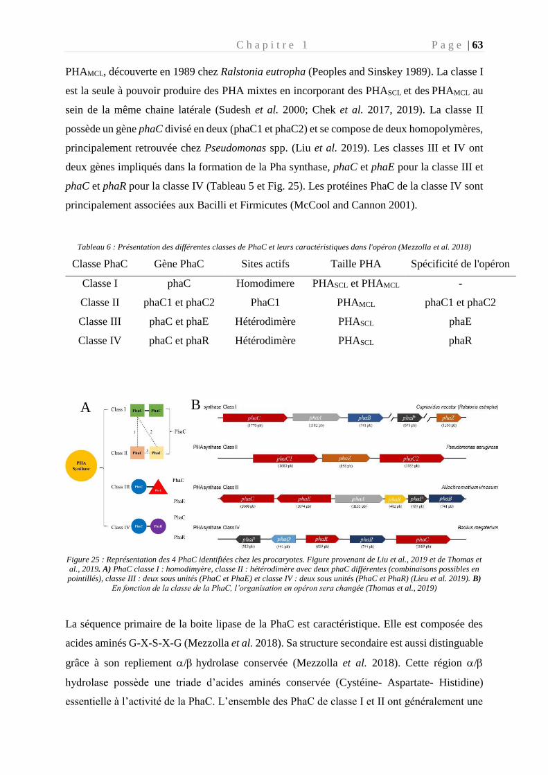
C h a p i t r e 1 P a g e | 63
PHAMCL, découverte en 1989 chez Ralstonia eutropha (Peoples and Sinskey 1989). La classe I
est la seule à pouvoir produire des PHA mixtes en incorporant des PHASCL et des PHAMCL au
sein de la même chaine latérale (Sudesh et al. 2000; Chek et al. 2017, 2019). La classe II
possède un gène phaC divisé en deux (phaC1 et phaC2) et se compose de deux homopolymères,
principalement retrouvée chez Pseudomonas spp. (Liu et al. 2019). Les classes III et IV ont
deux gènes impliqués dans la formation de la Pha synthase, phaC et phaE pour la classe III et
phaC et phaR pour la classe IV (Tableau 5 et Fig. 25). Les protéines PhaC de la classe IV sont
principalement associées aux Bacilli et Firmicutes (McCool and Cannon 2001).
Tableau 6 : Présentation des différentes classes de PhaC et leurs caractéristiques dans l'opéron (Mezzolla et al. 2018)
Classe PhaC Gène PhaC Sites actifs Taille PHA Spécificité de l'opéron
Classe I phaC Homodimere PHASCL et PHAMCL -
Classe II phaC1 et phaC2 PhaC1 PHAMCL phaC1 et phaC2
Classe III phaC et phaE Hétérodimère PHASCL phaE
Classe IV phaC et phaR Hétérodimère PHASCL phaR
Figure 25 : Représentation des 4 PhaC identifiées chez les procaryotes. Figure provenant de Liu et al., 2019 et de Thomas et
al., 2019. A) PhaC classe I : homodimyère, classe II : hétérodimère avec deux phaC différentes (combinaisons possibles en
pointillés), classe III : deux sous unités (PhaC et PhaE) et classe IV : deux sous unités (PhaC et PhaR) (Lieu et al. 2019). B)
En fonction de la classe de la PhaC, l’organisation en opéron sera changée (Thomas et al., 2019)
La séquence primaire de la boite lipase de la PhaC est caractéristique. Elle est composée des
acides aminés G-X-S-X-G (Mezzolla et al. 2018). Sa structure secondaire est aussi distinguable
grâce à son repliement / hydrolase conservée (Mezzolla et al. 2018). Cette région /
hydrolase possède une triade d’acides aminés conservée (Cystéine- Aspartate- Histidine)
essentielle à l’activité de la PhaC. L’ensemble des PhaC de classe I et II ont généralement une
A B
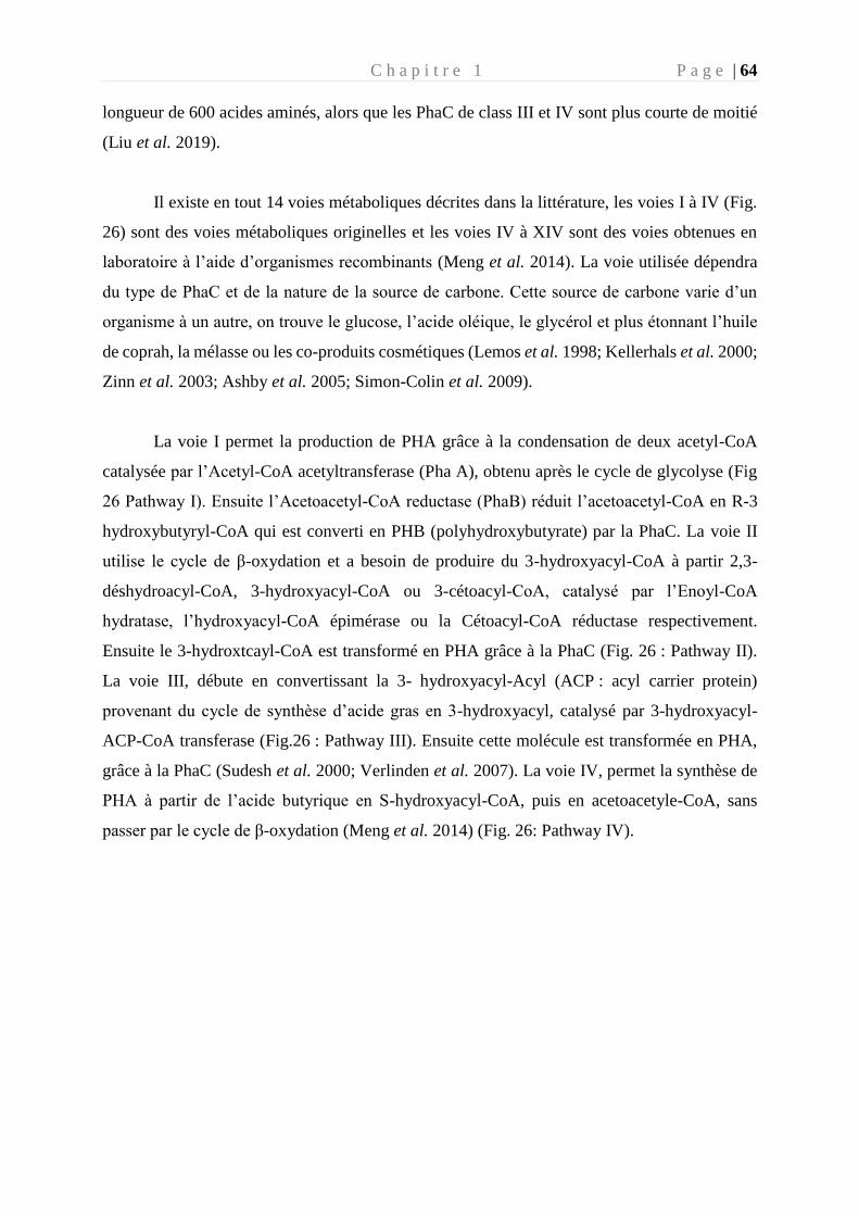
C h a p i t r e 1 P a g e | 64
longueur de 600 acides aminés, alors que les PhaC de class III et IV sont plus courte de moitié
(Liu et al. 2019).
Il existe en tout 14 voies métaboliques décrites dans la littérature, les voies I à IV (Fig.
26) sont des voies métaboliques originelles et les voies IV à XIV sont des voies obtenues en
laboratoire à l’aide d’organismes recombinants (Meng et al. 2014). La voie utilisée dépendra
du type de PhaC et de la nature de la source de carbone. Cette source de carbone varie d’un
organisme à un autre, on trouve le glucose, l’acide oléique, le glycérol et plus étonnant l’huile
de coprah, la mélasse ou les co-produits cosmétiques (Lemos et al. 1998; Kellerhals et al. 2000;
Zinn et al. 2003; Ashby et al. 2005; Simon-Colin et al. 2009).
La voie I permet la production de PHA grâce à la condensation de deux acetyl-CoA
catalysée par l’Acetyl-CoA acetyltransferase (Pha A), obtenu après le cycle de glycolyse (Fig
26 Pathway I). Ensuite l’Acetoacetyl-CoA reductase (PhaB) réduit l’acetoacetyl-CoA en R-3
hydroxybutyryl-CoA qui est converti en PHB (polyhydroxybutyrate) par la PhaC. La voie II
utilise le cycle de β-oxydation et a besoin de produire du 3-hydroxyacyl-CoA à partir 2,3-
déshydroacyl-CoA, 3-hydroxyacyl-CoA ou 3-cétoacyl-CoA, catalysé par l’Enoyl-CoA
hydratase, l’hydroxyacyl-CoA épimérase ou la Cétoacyl-CoA réductase respectivement.
Ensuite le 3-hydroxtcayl-CoA est transformé en PHA grâce à la PhaC (Fig. 26 : Pathway II).
La voie III, débute en convertissant la 3- hydroxyacyl-Acyl (ACP : acyl carrier protein)
provenant du cycle de synthèse d’acide gras en 3-hydroxyacyl, catalysé par 3-hydroxyacyl-
ACP-CoA transferase (Fig.26 : Pathway III). Ensuite cette molécule est transformée en PHA,
grâce à la PhaC (Sudesh et al. 2000; Verlinden et al. 2007). La voie IV, permet la synthèse de
PHA à partir de l’acide butyrique en S-hydroxyacyl-CoA, puis en acetoacetyle-CoA, sans
passer par le cycle de β-oxydation (Meng et al. 2014) (Fig. 26: Pathway IV).
C h a p i t r e 1 P a g e | 65
Figure 26 : Description des trois voies métaboliques de la synthase des PHA (modifié de Thomas et al. 2019). PhaA : Acetyl-
CoA acetyltransferase, PhaB : Acetoacetyl-CoA reductase, PhaC : Pha synthase, PhaJ : enoyl-coenzyme A hydratase, FabD
: malonyl-CoA-ACP transacylase, FabG : ketoacyl-ACP, PhaG : 3-hydroxydecanoyl-ACP-CoA transacylase
Ces 4 voies sont régulées au niveau enzymatique et transcriptionnel, les concentrations
en coenzyme A, acétyl-CoA, NADPH et le rapport NADPH / NADP peuvent agir pour réguler
la biosynthèse de PHA (Castro-Sowinski et al. 2010). L’étude de la PhaC est essentielle pour
optimiser les propriétés du PHA et sa production.
5.4 Mécanismes moléculaires impliqués dans la biodégradation des PHA
Certains micro-organismes sont capables d’utiliser le PHA comme source de carbone
sous l’action des PHA dépolymérases (PhaZ) intra- ou extracellulaires (Knoll et al. 2009; Meng
et al. 2014; Mezzolla et al. 2018).
PHA intracellulaire
La PHA intracellulaire (i-PHAZ) permet l’hydrolyse active des granules du PHB
accumulées. Cette hydrolyse permet aux micro-organismes de récupérer du carbone et de
l’énergie afin de pallier à une carence nutritionnelle de l’environnement (Doi et al. 1990). Les
i-PhaZ peuvent dégrader le PHB amorphe natif mais pas semi-cristallin, contrairement aux
PhaZ extracellulaire (Sznajder and Jendrossek 2011). En utilisant le PHB comme source de
C h a p i t r e 1 P a g e | 66
carbone, certaines bactéries augmentent considérablement leur temps de survie dans un
environnement hostile (jusqu’à 600 jours pour Legionella pneumophila) (James et al. 1999).
Certaines bactéries appartenant aux genre Rhizobium sp., Spirillium sp. et Pseudomonas sp. ont
été identifiées comme pouvant synthétiser et dégrader le PHB intracellulaire pendant une
culture au laboratoire (Hayward et al. 1959). La dégradation des granules de PHB est permise
par une protéine activatrice, qui rend le PHB amorphe et disponible pour la i-PHAZ (Merrick
and Doudoroff 1964; Griebel and Merrick 1971). Le PHB est hydrolysé en monomères
d’hydroxy-butyrate, puis oxydés en acéto-acétate par la 3 hydroxy-butyrate déshydrogénase.
Puis l’acétyl co-A est estérifié par la 3-cetoacide-coA transférase ou l’acétoacétyl-coA synthase
et la synthèse du PHB recommence (Numata et al. 2007).
PHA extracellulaire
La synthèse intracellulaire du PHB n’est pas une condition sine qua non à la dégradation
du PHB. De nombreux micro-organismes non producteurs de PHB sont capables de l’utiliser
comme source de carbone grâce à leur PhaZ extracellulaire (e-PhaZ) (Jendrossek 2007). Les e-
PhaZ peuvent hydrolyser le PHB-semi-cristallin, afin de le transformer en monomères solubles
dans l’eau et pouvant être incorporés dans une cellule bactérienne (Sudesh and Abe 2010).
Les PhaZ contiennent plusieurs domaines (Fig. 27) : un signal peptidique (SP), un grand
domaine catalytique N-terminal (Cat), une courte région de liaison (linker) qui permet de faire
la liaison entre le domaine catalytique et le substrat terminal (SBD, substrate binding domain)
(Jendrossek and Handrick 2002; Jendrossek 2005). Le signal peptidique est composé de 22 à
58 acides aminés, il permet la sécrétion du PhaZ mature à travers le cytoplasme (Jendrossek
and Handrick 2002; Jendrossek 2005). La sécrétion de la PhaZ n’est permise qu’après excision
du SP par les peptidases. Le domaine catalytique est constitué d’un site actif avec trois acides
aminés strictement conservés, la sérine (S), l’aspartate (D) et l’histidine (H), indispensable à
l’activité catalytique (fig). Deux types de domaines peuvent être différenciés par la disposition
des acides aminés du site actif au niveau de la séquence primaire d’acides aminés (Jendrossek
and Handrick 2002) :
- Type I : Histidine- Serine- Aspartate- Histidine (H S D H)
- Type II : L’histidine est place en C terminale du site actif (S D H H)
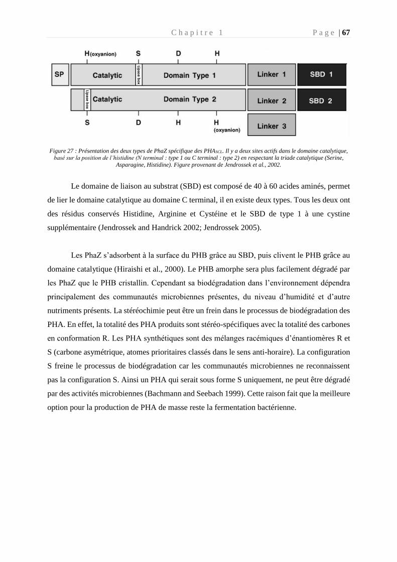
C h a p i t r e 1 P a g e | 67
Figure 27 : Présentation des deux types de PhaZ spécifique des PHASCL. Il y a deux sites actifs dans le domaine catalytique,
basé sur la position de l’histidine (N terminal : type 1 ou C terminal : type 2) en respectant la triade catalytique (Serine,
Asparagine, Histidine). Figure provenant de Jendrossek et al., 2002.
Le domaine de liaison au substrat (SBD) est composé de 40 à 60 acides aminés, permet
de lier le domaine catalytique au domaine C terminal, il en existe deux types. Tous les deux ont
des résidus conservés Histidine, Arginine et Cystéine et le SBD de type 1 à une cystine
supplémentaire (Jendrossek and Handrick 2002; Jendrossek 2005).
Les PhaZ s’adsorbent à la surface du PHB grâce au SBD, puis clivent le PHB grâce au
domaine catalytique (Hiraishi et al., 2000). Le PHB amorphe sera plus facilement dégradé par
les PhaZ que le PHB cristallin. Cependant sa biodégradation dans l’environnement dépendra
principalement des communautés microbiennes présentes, du niveau d’humidité et d’autre
nutriments présents. La stéréochimie peut être un frein dans le processus de biodégradation des
PHA. En effet, la totalité des PHA produits sont stéréo-spécifiques avec la totalité des carbones
en conformation R. Les PHA synthétiques sont des mélanges racémiques d’énantiomères R et
S (carbone asymétrique, atomes prioritaires classés dans le sens anti-horaire). La configuration
S freine le processus de biodégradation car les communautés microbiennes ne reconnaissent
pas la configuration S. Ainsi un PHA qui serait sous forme S uniquement, ne peut être dégradé
par des activités microbiennes (Bachmann and Seebach 1999). Cette raison fait que la meilleure
option pour la production de PHA de masse reste la fermentation bactérienne.
C h a p i t r e 1 P a g e | 68
5.5 Limite du succès du PHA
L’inconvénient majeur pour la commercialisation des PHA à grande échelle est leur
coût final (7 à 10 €/kg), qui est 5 à 15 fois plus élevé que le plastique conventionnel (Singh and
Mallick 2009; Rehm 2010) et son ingénierie biologique.
Pour réduire les coûts de production du PHA, des voies de synthèse alternatives utilisant
des substrats carbonés moins couteux sont nécessaires, tels que le glycérol (sous-produit de la
fabrication du biodiesel) (Ciesielski et al. 2015). Les coûts des sources de carbone
représenteraient environ 50 % du coût de la production final de PHA. Une étude menée en 2010
a montré que si la source de carbone était issus de déchets, le coût de production de PHA
pourrait atteindre 3.51 €/kg alors que le coût de production des polymères conventionnels (PP,
PE) coûte entre 1.15 € et 1.47 €/kg (Obruca 2010).
Le coût de production du PHA est fortement impacté par l’extraction du PHA après
fermentation bactérienne, il représenterait 30 % du prix global (Sun et al. 2007). Actuellement
plusieurs stratégies sont mises en œuvre pour l’extraction des PHA, la plus favorisée et celle
utilisant comme solvant le chloroforme ou l’acétone, entrainant un cout supplémentaire pour
l’élimination des déchets et créant des déchets nocifs pour l’environnement (Lee et Na, 2013).
Les méthodes plus respectueuses de l’environnement comme la digestion enzymatique ou
chimique par exemple, reste à ce jour trop coûteuse.
Les conditions de croissance bactérienne pour obtenir une production optimale de PHA
en utilisant un minimum de carbone tout en évitant l’utilisation de la source carbonée pour la
croissance bactérienne est un processus complexe. Le processus d’accumulation de PHA
intervient très tôt dans la courbe de croissance du micro-organisme qui affecte de ce fait la
productivité de PHA. Car l’accumulation de grande quantité de PHA inhibe la croissance
cellulaire et affecte négativement le métabolisme de l’organisme. Il y a donc un équilibre à
avoir, entre ne pas le synthétiser trop tôt pour optimiser la quantité de PHA accumulée et ni trop
tard car une grande partie de la source carbonée sera gaspillée pour la croissance cellulaire. Ce
juste équilibre est à déterminer pour chaque micro-organisme producteur de PHA, dans des
conditions bien précises, ce qui demande une étude poussée entrainant un surcoût de
production.
C h a p i t r e 1 P a g e | 69
Actuellement le devenir des PHA dépend principalement de son coût de production, c’est pour
cela qu’il est important de développer les recherches en étudiant les voies métaboliques de
production de PHA, les souches capables de produire le PHA à vitesse optimale et les procédés
d’extractions simples et respectueux de l’environnement.
6. Objectif de la thèse
Depuis la prise de conscience de l’envergure de la pollution plastique dans les océans, les
études se sont multipliées au cours de ces dernières années. Comprendre d’où vient cette
pollution, comment elle interagit avec son environnement et comment elle peut créer un nouvel
habitat propice au développement de certains micro-organismes est un sujet d’actualité. Le
fonctionnement des communautés microbiennes et les interactions entre les micro-organismes
et les polymères demeurent un enjeu environnemental et économique majeur à ce jour. Les
travaux de cette thèse ont été motivés pour répondre aux questions suivantes :
- Peut-on définir des paramètres qui influencent la biogéographie de la plastisphère ?
Existe-t-il un microbiome commun à tous les plastiques (« core microbiome ») ?
(Chapitre 2)
- Les communautés microbiennes colonisent-t-elles de la même façon les polymères
biodégradables et non biodégradables ? Quelle est la part des micro-organismes de la
plastisphère potentiellement impliqués dans la biodégradation ? (Chapitre 3)
- Comment l’analyse des génomes peut-elle nous aider à mieux comprendre la
biodégradation des polymères ? (Chapitre 4)
Afin de répondre à ces questions, une approche pluridisciplinaire impliquant les domaines
de l’écotoxicologie microbienne, de la physique et de la chimie, de la génétique et de la
bioinformatique a été mise en place (Fig. 28). La plastisphère a été abordée à différents
niveaux in situ et/ou in vitro :
- A l’échelle de l’écosystème : à partir de plusieurs prélèvements dans les zones les plus
polluées en micro-plastiques (gyre du Pacifique Nord et Méditerranée Nord
occidentale), pour lesquels la diversité microbienne, le type de polymère et les
conditions environnementales ont été analysés.
- A l’échelle de la communauté microbienne in situ et in vitro: à partir du suivi de la
formation de biofilms microbiens naturels sélectionnés sur différents types de
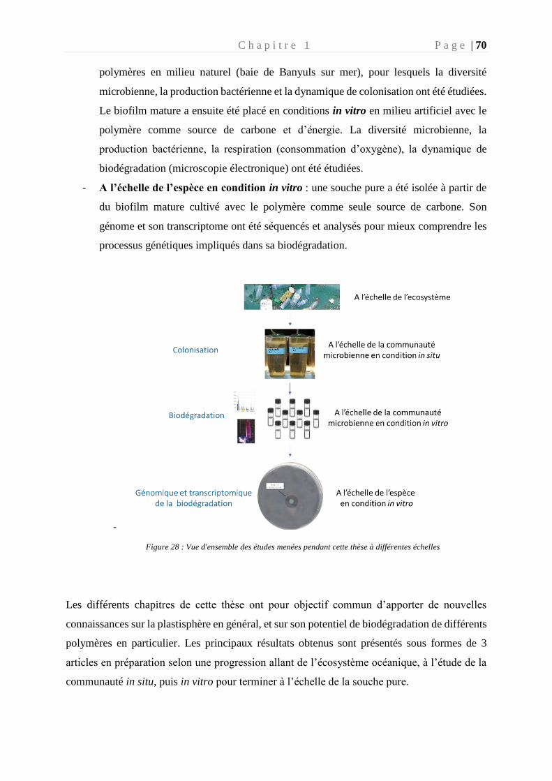
C h a p i t r e 1 P a g e | 70
polymères en milieu naturel (baie de Banyuls sur mer), pour lesquels la diversité
microbienne, la production bactérienne et la dynamique de colonisation ont été étudiées.
Le biofilm mature a ensuite été placé en conditions in vitro en milieu artificiel avec le
polymère comme source de carbone et d’énergie. La diversité microbienne, la
production bactérienne, la respiration (consommation d’oxygène), la dynamique de
biodégradation (microscopie électronique) ont été étudiées.
- A l’échelle de l’espèce en condition in vitro : une souche pure a été isolée à partir de
du biofilm mature cultivé avec le polymère comme seule source de carbone. Son
génome et son transcriptome ont été séquencés et analysés pour mieux comprendre les
processus génétiques impliqués dans sa biodégradation.
-
Figure 28 : Vue d'ensemble des études menées pendant cette thèse à différentes échelles
Les différents chapitres de cette thèse ont pour objectif commun d’apporter de nouvelles
connaissances sur la plastisphère en général, et sur son potentiel de biodégradation de différents
polymères en particulier. Les principaux résultats obtenus sont présentés sous formes de 3
articles en préparation selon une progression allant de l’écosystème océanique, à l’étude de la
communauté in situ, puis in vitro pour terminer à l’échelle de la souche pure.
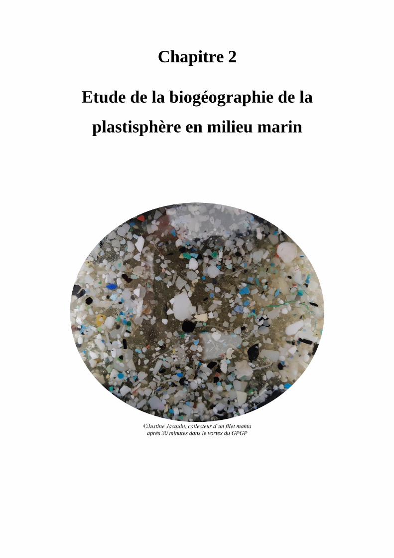
Chapitre 2
Etude de la biogéographie de la
plastisphère en milieu marin
©Justine Jacquin, collecteur d’un filet manta
après 30 minutes dans le vortex du GPGP

C h a p i t r e 2 P a g e | 73
Chapitre 2
« If you weighed all the living organisms in the ocean, 90 percent of that weight would be
from micro-organisms” Danielle Hall
Préambule
Les expéditions Tara Pacifique et Tara Méditerranée :
Découvert à la fin des années 1990, la pollution plastique du gyre du Pacifique Nord ou
« vortex de déchet du Pacique Nord » (littéralement traduit de « Great Pacific garbage patch »)
a conduit à une médiatisation sans précédent de cette pollution largement rependue à travers les
océans du monde. On ne présente plus le « septième continent », cette zone située dans l’océan
Pacifique, vortex de plastique situé à mi-chemin entre Hawaï et la Californie.
L’expédition Tara Pacifique (2016-2018) avait pour objectif d’explorer les potentialités
de résistance, d’adaptation et de résiliences des récifs coralliens face au changement global. En
quittant le port de Lorient le 28 mai 2016, cette goélette a sondé pendant près de 2 ans sur
100 000 km les eaux du Pacifique. Dans son trajet, Tara a traversé le gyre du Pacifique Nord et
a pu mesurer cette pollution plastique. J’ai eu la chance de participer à cette traversée entre
Honolulu (Hawaii) et Portland (Orgeon) qui a duré 17 jours et où 7 stations ont été
échantillonnées. En plein milieu du Pacifique, au cœur du vortex de déchets, les micro
plastiques ont colmaté le filet Manta habituellement remplis de planctons et d’algues (fig. 29).
Les morceaux de plastiques au cœur du gyre Pacifique Nord étaient si nombreux, que l’on avait
dû doubler la cadence de prélèvement, 4 filets Manta mis à l’eau par jour, pendant une demi-
heure à 3 nœuds. C’est ainsi que le filet a remonté des milliers de morceaux plastiques dont une
petite partie (200) ont été analysés au laboratoire.
Notre équipe avait également participé à la mission Tara Méditerranée en 2014, qui
navigua sur la mer Méditerranée durant 7 mois d’expédition. Elle a permis de montrer que la
mer Méditerranée présentait en moyenne les densités de plastique les plus importantes au
monde, avec 250 milliards de microplastiques. Nous avions également réalisé le séquençage de
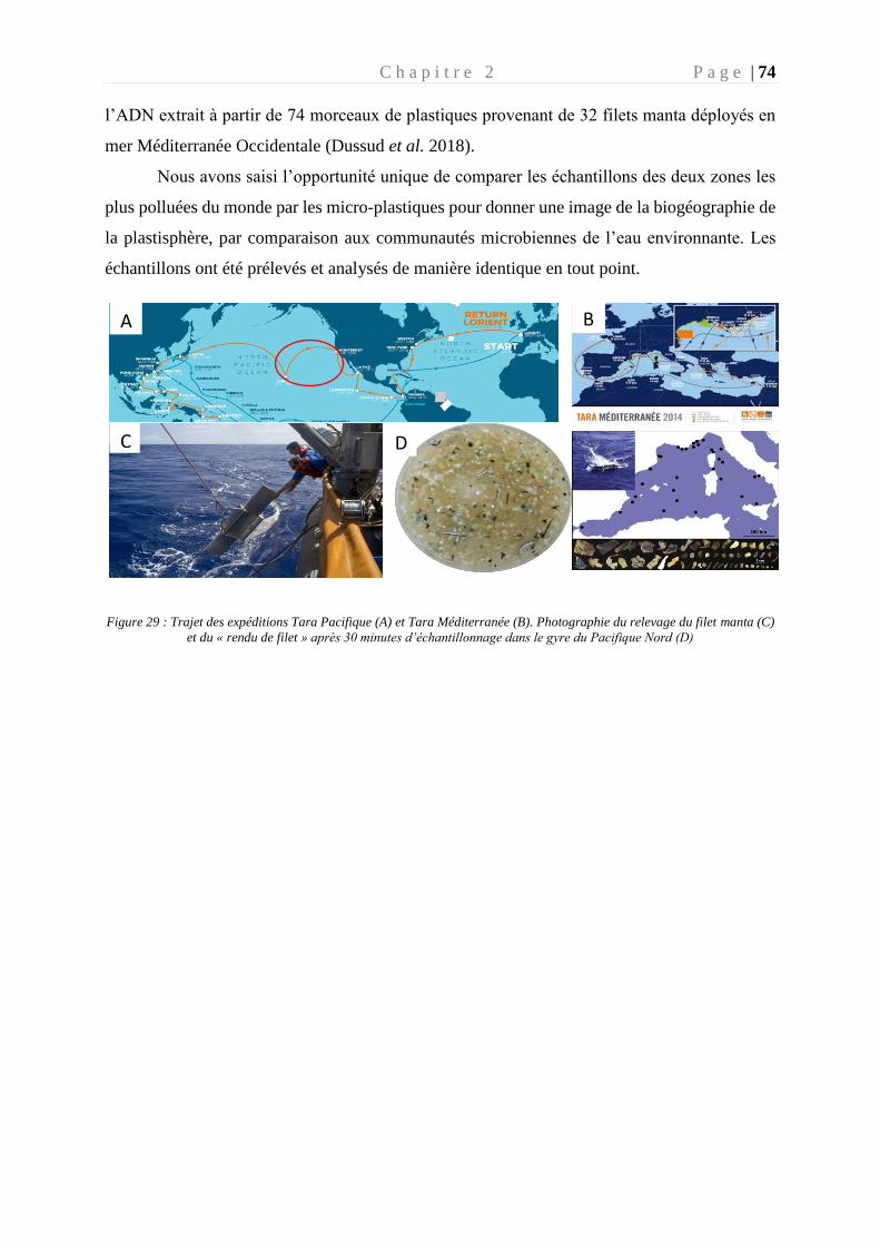
C h a p i t r e 2 P a g e | 74
l’ADN extrait à partir de 74 morceaux de plastiques provenant de 32 filets manta déployés en
mer Méditerranée Occidentale (Dussud et al. 2018).
Nous avons saisi l’opportunité unique de comparer les échantillons des deux zones les
plus polluées du monde par les micro-plastiques pour donner une image de la biogéographie de
la plastisphère, par comparaison aux communautés microbiennes de l’eau environnante. Les
échantillons ont été prélevés et analysés de manière identique en tout point.
Figure 29 : Trajet des expéditions Tara Pacifique (A) et Tara Méditerranée (B). Photographie du relevage du filet manta (C)
et du « rendu de filet » après 30 minutes d’échantillonnage dans le gyre du Pacifique Nord (D)
A B
C D
C h a p i t r e 2 P a g e | 75
Global diversity and core microbiome of the plastisphere compared to
organic-particle attached and free-living planktonic lifestyles from the Tara
Oceans expeditions in the Mediterranean Sea and in the North Pacific gyre.
List of authors : JACQUIN Justine1, Marko Budinich2,3, CHENG Jingguang1, BARBE
Valérie4, PEDROTTI Maria-Luiza5, GHIGLIONE Jean-François1*.
Affiliations :
1- CNRS, Sorbonne Universités, UMR 7621, Laboratoire d’Océanographie
Microbienne, Observatoire Océanologique de Banyuls, France
2- Research Federation (FR2022) Tara Océan GO-SEE, Paris, France.
3- Sorbonne Université, UPMC Université Paris 06, CNRS, Laboratoire Adaptation et
Diversité en Milieu Marin, Station Biologique de Roscoff, 29680 Roscoff, France.
4- Génomique Métabolique, Genoscope, Institut François Jacob, CEA, CNRS, Univ
Evry, Université Paris-Saclay, Evry, France
5- Sorbonne Université, UPMC Paris 06, CNRS UMR 7076, Laboratoire
d’Oceanographie de Villefranche, Villefranche-sur-mer, France.
Abstract The North Pacific and the Mediterranean are two areas known to be heavily polluted by plastic
debris. Plastics thus provide a habitat distinct from the surrounding marine environment. A
previous study carried out in the Mediterranean showed a clear partitioning between free life
(FL), attached organic particles (PA) and plastic marine debris (PMD). This study made it
possible to compare the diversity of the plastisphere found on the different fractions (PMD, PA
and FL) between the Pacific and the Mediterranean. The plastics communities at the two sites
are different from those found in the surrounding seawater (PA and FL). Microbial communities
were mainly separated by region and not by polymer, suggesting that environmental factors are
more important than polymer type for the composition of the plastisphere. Cyanobacteria are
overrepresented in all PMDs. The PMD fractions were not enriched in hydrocarbonoclasts but
potentially in pathogenic microorganisms found in abundance. However, the study of the V4-
V5 region of microorganisms does not make it possible to conclude on the effective
pathogenicity of OTUs, this deserves further study by another molecular method.
C h a p i t r e 2 P a g e | 76
Introduction
The oceans represent the final destination of untreated terrestrial plastic waste, receiving
around 8 million tonnes of plastic each year (Jambeck et al. 2015). This pollution affects all the
aqueous ecosystems of the world, oceans, seas (Barnes et al. 2009) and the most remote oceans
(Cozar et al. 2014; Peeken et al. 2018) and rivers (Di and Wang 2018). Once integrated into
the marine system, the plastics pieces fragment into microplastics (size <5mm) under the action
of abiotic (UV, temperature, oxidation) and or biotic (microorganisms) factors (Shim et al.
2016). The microplastics represent the majority of pollution in the accumulation zones
(Lebreton et al. 2018; Pan et al. 2019), such as the Gyre. Plastics will remain on the surface if
they have a low density, but can become heavy over time through the accumulation of
microorganisms forming a biofilm on its surface (Amaral-Zettler et al. 2020). This is how
plastics have been identified in the water column (Choy et al. 2019), in the depths of the oceans
(Chiba et al. 2018) and in sediments (Matsuguma et al. 2017; Dobaradaran et al. 2018).
This pollution is not homogeneous in the marine ecosystem, indeed there are areas of
accumulation of plastic waste in the world (Cozar et al. 2014). These areas are often defined
according to sea currents, because the plastics floating on the surface are trapped and then
accumulate in this so called, accumulated areas (Mediterranean sea, off southern Corsica and
northern Italy) or gyres (North/South Pacific, North/South Atlantic, Indian ocean) (Cozar et al.
2014). In these accumulation zones, we can distinguish the Great Pacific Garbage Patch
(GPGP) which alone accumulates between 45 and 129 miles tonnes of plastic, in an area of 1.6
million km² (Lebreton et al. 2018). This corresponds to a concentration within the GPGP of 10
to 100 kg per km². The Mediterranean is considered to be the most polluted sea in the world,
with an estimate between 756 and 3000 tonnes of plastic on (Cózar et al. 2015). A recent study
has shown a very high plastic concentration compared to other regions of the world, with an
average concentration of 0.4 kg / km² and up to 1.9 kg / km² depending on site studied (Cózar
et al. 2015; Ruiz-Orejón et al. 2016). Areas like northwest Italy, north of Sicily, around the
Ionian Islands, have particularly large plastic accumulations (Ruiz-Orejon et al. 2016).
This strong accumulation is mainly due to the hydrodynamic characteristics of the
Mediterranean Sea as it is a semi-closed basin with a residence time up to 70 years and with
currents enhancing the trapping of plastics (Millot and Taupier-Letage 2005; Durrieu de
Madron et al. 2011).
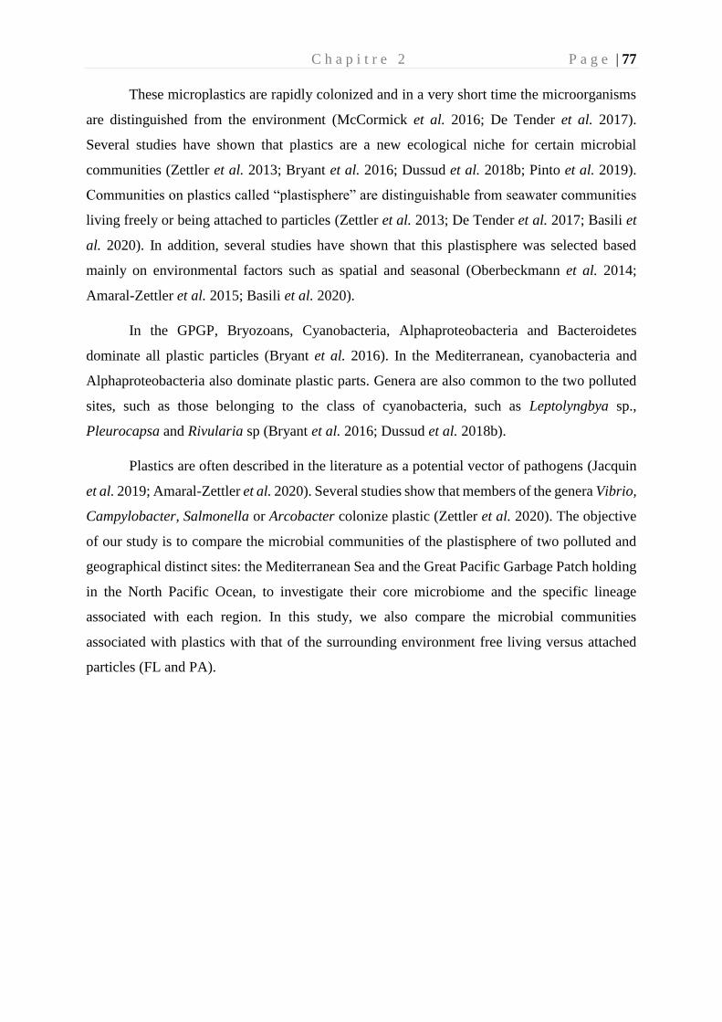
C h a p i t r e 2 P a g e | 77
These microplastics are rapidly colonized and in a very short time the microorganisms
are distinguished from the environment (McCormick et al. 2016; De Tender et al. 2017).
Several studies have shown that plastics are a new ecological niche for certain microbial
communities (Zettler et al. 2013; Bryant et al. 2016; Dussud et al. 2018b; Pinto et al. 2019).
Communities on plastics called “plastisphere” are distinguishable from seawater communities
living freely or being attached to particles (Zettler et al. 2013; De Tender et al. 2017; Basili et
al. 2020). In addition, several studies have shown that this plastisphere was selected based
mainly on environmental factors such as spatial and seasonal (Oberbeckmann et al. 2014;
Amaral-Zettler et al. 2015; Basili et al. 2020).
In the GPGP, Bryozoans, Cyanobacteria, Alphaproteobacteria and Bacteroidetes
dominate all plastic particles (Bryant et al. 2016). In the Mediterranean, cyanobacteria and
Alphaproteobacteria also dominate plastic parts. Genera are also common to the two polluted
sites, such as those belonging to the class of cyanobacteria, such as Leptolyngbya sp.,
Pleurocapsa and Rivularia sp (Bryant et al. 2016; Dussud et al. 2018b).
Plastics are often described in the literature as a potential vector of pathogens (Jacquin
et al. 2019; Amaral-Zettler et al. 2020). Several studies show that members of the genera Vibrio,
Campylobacter, Salmonella or Arcobacter colonize plastic (Zettler et al. 2020). The objective
of our study is to compare the microbial communities of the plastisphere of two polluted and
geographical distinct sites: the Mediterranean Sea and the Great Pacific Garbage Patch holding
in the North Pacific Ocean, to investigate their core microbiome and the specific lineage
associated with each region. In this study, we also compare the microbial communities
associated with plastics with that of the surrounding environment free living versus attached
particles (FL and PA).
C h a p i t r e 2 P a g e | 78
Materials and Methods
1. Sampling of plastic and surrounding sea water
Samples were taken during two expeditions: Tara-Mediterranean (Tara-Méditerrannée) (April-
November 2014) and Tara-Pacific (June-July 2018) aboard the research vessel Tara. The plastic
particles, in both sites, were collected using a manta net with a 333mm mesh. The sampling
map for the Mediterranean and the Pacific is shown in Fig. 1. The expedition in the North
Pacific gyre, was a transect of 7 sampling points all carried out during the day. The expedition
in Mediterranean sea contain 27 sampling points all carried out during the day.
Figure 1: Sampling map of the two Tara expeditions (Mediterranean, Pacific), showing the
locations of each sampling station (black point on the map).
For each sampling point, a subset of polymer was randomly sorted with sterilized forceps and
rinsed with sterilized seawater and immediately stored at -20°C for future microbial diversity
analysis. At each station, a seawater sample was taken, 2L were pre-filtered on 3 µm pore size
to collect the particle attached fraction (named PA) and then filtered on 0.2 µm-pore size to
collect the free-living fraction (FL fraction). All the membranes used were made of
polycarbonate filters using a peristaltic pump (pressure <100 mbar). Filters were stored at -20
° C until further DNA extraction for microbial diversity analyses.
C h a p i t r e 2 P a g e | 79
2. Chemical characterization of the PMD
For Mediterranean samples, the plastic particles were characterized by Fourier Transform
Infrared Spectroscopy (FTIR) and was performed using the individual PMD using Spectrum
100 equipped with an attenuated total reflectance (ATR) (Perkin-Elmer). The spectra were
compared to references to identify the nature of the polymer.
3. DNA extraction and Next-Generation Sequencing
DNA extraction was performed on each piece of separation plastic, 75 pieces of plastic from
the Mediterranean, and 104 pieces from the Pacific. These plastics, taken randomly, represent
each sampling point. Regarding the 3 µm and 0.2 µm filters, they were extracted using the same
method as plastics, all separately. Prior to the DNA extraction done using the phenol chloroform
method (Dussud et al. 2018), an ultrasonic bath (3*1 minutes) step was done in order to help
the biofilm breakdown and dispersion of the biofilm. The target area for amplification is the
V4-V5 region of 16S rDNA using the Furhman primers the 515-F (5'-GTG YCA GCM GCC
GCG GTA A-3' and 926-R (5'-CCG YCA ATT YMT TTR AGT TT-3') (Fuhrman et al. 1989),
which are particularly adapted to the marine environment (Parada et al. 2016). The
Mediterranean samples were sequenced at Research and Testing Laboratories (Lubbock, TX),
the Pacific plastics at MrDNA (Shallowater, USA) and the surrounding water (PA and FL) at
the Genoscope (Paris, France).
The sequences were analysed using FROGS pipeline host in the Galaxy platform (Escudié et
al. 2018), following the guidelines given in the publication. Briefly, the forward and reverse
reads were assembled, sequences were clustered using the SWARM algorithm (Mahé et al.
2014) using the aggregation distance of 3 and the chimeras were removed using the FROGS
Vsearch tool (Rognes et al. 2016). An additional filter step was used to select the OTU clusters
with an abundance at 5.10-5 threshold to select the most relevant OTUs sequences. The
taxonomic affiliation was assigned by BLASTn + (Camacho et al. 2009) using the SILVA 138
database 16S (Quast et al. 2013).
C h a p i t r e 2 P a g e | 80
4. Statistical analysis, diversity assessment and predictive metagenome analysis
All the statistical analyzes were carried out with R software (https://www.r-project.org/). The
alpha diversity was determined by 3 indices (chao, Shannon and Evenness). The beta diversity
is based on a Bray-Curtis distance and allowed to realize the NMDS and the network. The
statistical difference was made with the non-parametric Kruskall-Wallis test and the
correlations with the spearman test. To identify the OTUs having a major role in the
differentiation of bacterial biofilms, we performed SIMPR tests using the Primer 6 software.
Results
1. Chemical characterization of the plastic composition This study was based on the comparison of 75 plastic marine debris (PMD) in the Western
Mediterranean Sea and 104 PMD in the North Pacific gyre. Our selection criteria was to get
between 3 to 5 PMD of each Manta net after DNA sequencing, whereas only 2 PMD was finally
selected for some sampling station after resampling because of not enough sequences (see
below). Visually, we covered similar variety of morphological shapes between the samples
taken in the Western Mediterranean Sea and in the North Pacific gyre, including filaments,
granules, pellets, sometimes transparent, white or coloured PMD. FTIR and ATR analysis
showed that polyethylene (PE) dominated the composition of the 75 pieces of plastic, with
73.3% and 71.1% of the PMD found in the Western Mediterranean sea compared to the North
Pacific gyre, respectively. In the former, PE was followed by propylene (16%), polystyrene
(5.3%) and composite (5.3%), whereas EVOH (ethylene vinyl alcohol) (7.7%), propylene
(3.8%), polystyrene (2.9%) and PET (1%) were found in the later.
2. Diversity indexes (α-diversity) of bacteria attached to PMD or attached to organic
particles (PA) compared to the free-living planktonic fraction (FL).
At the end of sequencing procedure, a set of 29,353,155 merged sequences was obtained. After
a first cleaning (expected length, primer present), chimera checking and abundance filter
(0.005%), we obtained 22,545,882 sequences that represented 1,285 OTUs for the analysis with
97.7% of OTUs that could be affiliated. Chloroplastic, mitochondrial and eukaryotic sequences
were removed from the analysis. Only the bacterial reads were treated in this study, due to the
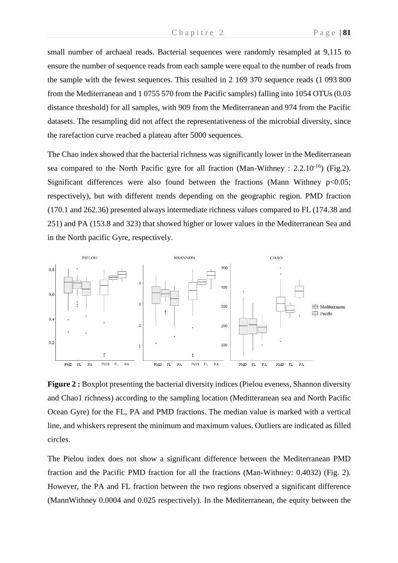
C h a p i t r e 2 P a g e | 81
small number of archaeal reads. Bacterial sequences were randomly resampled at 9,115 to
ensure the number of sequence reads from each sample were equal to the number of reads from
the sample with the fewest sequences. This resulted in 2 169 370 sequence reads (1 093 800
from the Mediterranean and 1 0755 570 from the Pacific samples) falling into 1054 OTUs (0.03
distance threshold) for all samples, with 909 from the Mediterranean and 974 from the Pacific
datasets. The resampling did not affect the representativeness of the microbial diversity, since
the rarefaction curve reached a plateau after 5000 sequences.
The Chao index showed that the bacterial richness was significantly lower in the Mediterranean
sea compared to the North Pacific gyre for all fraction (Man-Withney : 2.2.10-16) (Fig.2).
Significant differences were also found between the fractions (Mann Withney p<0.05;
respectively), but with different trends depending on the geographic region. PMD fraction
(170.1 and 262.36) presented always intermediate richness values compared to FL (174.38 and
251) and PA (153.8 and 323) that showed higher or lower values in the Mediterranean Sea and
in the North pacific Gyre, respectively.
Figure 2 : Boxplot presenting the bacterial diversity indices (Pielou eveness, Shannon diversity
and Chao1 richness) according to the sampling location (Meditteranean sea and North Pacific
Ocean Gyre) for the FL, PA and PMD fractions. The median value is marked with a vertical
line, and whiskers represent the minimum and maximum values. Outliers are indicated as filled
circles.
The Pielou index does not show a significant difference between the Mediterranean PMD
fraction and the Pacific PMD fraction for all the fractions (Man-Withney: 0.4032) (Fig. 2).
However, the PA and FL fraction between the two regions observed a significant difference
(MannWithney 0.0004 and 0.025 respectively). In the Mediterranean, the equity between the
C h a p i t r e 2 P a g e | 82
three fractions is not significantly different (Kruskal and Wallis, p = 0.239) with a Pielou index
on average of 0.62 for PA, 0.68 for FL and 0.67 for PMD. For the Pacific, equity is different
(Kruskal and Wallis, p = 7.21.10-5), a higher value is found on the PA (0.76), then the FL (0.73)
and lastly the PMD (0.64).
The Shannon index confirmed that the FL (4.03 and 3.42) and PA (4.33 and 3.14) fractions
presented a higher microbial diversity in the Pacific than in the Mediterranean samples,
respectively (Kruskal et Wallis, p= 7.56.10-5). However, we could not found a significant
difference for the PMD diversity between the two regions (Mann-Withney p=0.1334).
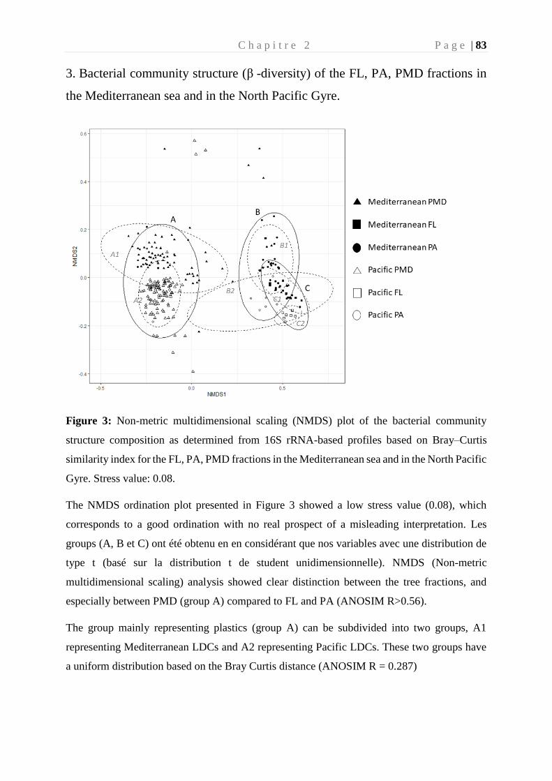
C h a p i t r e 2 P a g e | 83
3. Bacterial community structure (β -diversity) of the FL, PA, PMD fractions in
the Mediterranean sea and in the North Pacific Gyre.
Figure 3: Non-metric multidimensional scaling (NMDS) plot of the bacterial community
structure composition as determined from 16S rRNA-based profiles based on Bray–Curtis
similarity index for the FL, PA, PMD fractions in the Mediterranean sea and in the North Pacific
Gyre. Stress value: 0.08.
The NMDS ordination plot presented in Figure 3 showed a low stress value (0.08), which
corresponds to a good ordination with no real prospect of a misleading interpretation. Les
groups (A, B et C) ont été obtenu en en considérant que nos variables avec une distribution de
type t (basé sur la distribution t de student unidimensionnelle). NMDS (Non-metric
multidimensional scaling) analysis showed clear distinction between the tree fractions, and
especially between PMD (group A) compared to FL and PA (ANOSIM R>0.56).
The group mainly representing plastics (group A) can be subdivided into two groups, A1
representing Mediterranean LDCs and A2 representing Pacific LDCs. These two groups have
a uniform distribution based on the Bray Curtis distance (ANOSIM R = 0.287)
C h a p i t r e 2 P a g e | 84
The group predominantly representing the PA fraction (B) can be divided into two subgroups,
one represented mainly by the Mediterranean PA (B1) and the other by the Pacific PA (B2).
These two subgroups have similarities with an ANOSIM R = 0.32. Like the FL fraction (group
C), divided into subgroups with a uniform distribution (ANOSIM, R = 0.257), C1 is
predominantly dominated by the Mediterranean and C2 predominantly dominated by the
Pacific.
In the Mediterranean the PMD fraction shows more similarity with the PA fraction (ANOSIM,
R = 0.567) than with the FL fraction (ANOSIM, R = 0.66). The FL and PA fraction shows more
similarity according to the bray-Curtis distance, than with the PMD fraction (ANOSIM, R =
0.544).
This same pattern is found in the Pacific, the PMD fraction shows slightly more resemblance
with the PA fraction (ANOSIM, R = 0.8) than with the FL fraction (ANOSIM, R = 0.89). The
FL and PA fraction does not show as strong a dissimilarity as with the PMD fraction (ANOSIM,
R = 0.723)
The samples from the Pacific, having the most interactions are those from station 199 (Middle
of GPGP) and 201 (towards the outside of GPGP) and 199 with those station M60 (NE of
Sardinia), M42 (N of Corsica), M66 (Middle of the Mediterranean between Italy and Sardinia)
and M173 (near of Algeria) of the Mediterranean. Pacific Station 192 (off Hawaii) appears to
be completely isolated from other stations. This particularity is also found in the Mediterranean
for the M19 station (Station near the south coast of France).
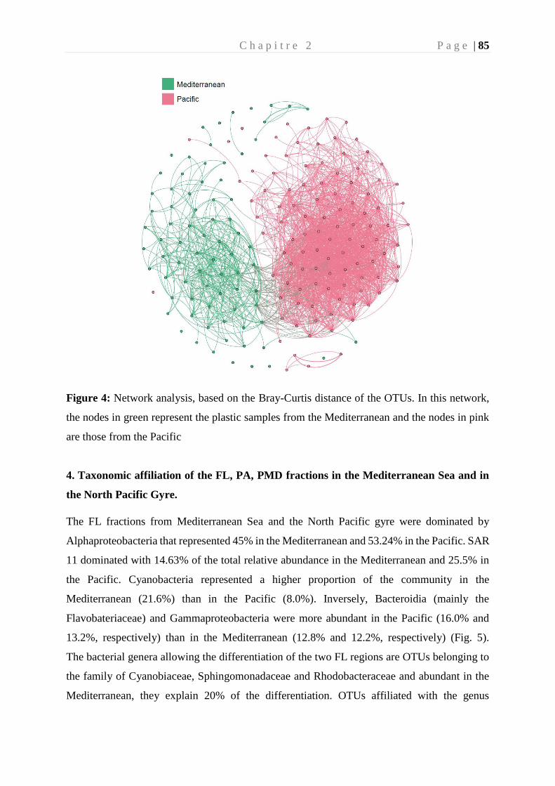
C h a p i t r e 2 P a g e | 85
Figure 4: Network analysis, based on the Bray-Curtis distance of the OTUs. In this network,
the nodes in green represent the plastic samples from the Mediterranean and the nodes in pink
are those from the Pacific
4. Taxonomic affiliation of the FL, PA, PMD fractions in the Mediterranean Sea and in
the North Pacific Gyre.
The FL fractions from Mediterranean Sea and the North Pacific gyre were dominated by
Alphaproteobacteria that represented 45% in the Mediterranean and 53.24% in the Pacific. SAR
11 dominated with 14.63% of the total relative abundance in the Mediterranean and 25.5% in
the Pacific. Cyanobacteria represented a higher proportion of the community in the
Mediterranean (21.6%) than in the Pacific (8.0%). Inversely, Bacteroidia (mainly the
Flavobateriaceae) and Gammaproteobacteria were more abundant in the Pacific (16.0% and
13.2%, respectively) than in the Mediterranean (12.8% and 12.2%, respectively) (Fig. 5).
The bacterial genera allowing the differentiation of the two FL regions are OTUs belonging to
the family of Cyanobiaceae, Sphingomonadaceae and Rhodobacteraceae and abundant in the
Mediterranean, they explain 20% of the differentiation. OTUs affiliated with the genus
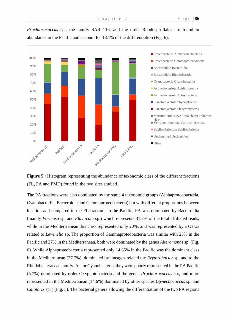
C h a p i t r e 2 P a g e | 86
Prochlorococcus sp., the family SAR 116, and the order Rhodospirillales are found in
abundance in the Pacific and account for 18.1% of the differentiation (Fig. 6).
Figure 5 : Histogram representing the abundance of taxonomic class of the different fractions
(FL, PA and PMD) found in the two sites studied.
The PA fractions were also dominated by the same 4 taxonomic groups (Alphaproteobacteria,
Cyanobacteriia, Bacteroidia and Gammaproteobacteria) but with different proportions between
location and compared to the FL fraction. In the Pacific, PA was dominated by Bacteroidia
(mainly Formosa sp. and Fluviicola sp.) which represents 31.7% of the total affiliated reads,
while in the Mediterrranean this class represented only 20%, and was represented by a OTUs
related to Lewinella sp. The proportion of Gammaproteobacteria was similar with 25% in the
Pacific and 27% in the Mediterranean, both were dominated by the genus Alteromonas sp. (Fig.
6). While Alphaproteobacteria represented only 14.35% in the Pacific was the dominant class
in the Mediterranean (27.7%), dominated by lineages related the Erythrobacter sp. and to the
Rhodobacteraceae family. As for Cyanobacteria, they were poorly represented in the PA Pacific
(5.7%) dominated by order Oxyphotobacteria and the genus Prochlorococcus sp., and more
represented in the Mediterranean (14.6%) dominated by other species (Synechococcus sp. and
Calothrix sp. ) (Fig. 5). The bacterial genera allowing the differentiation of the two PA regions
0%
10%
20%
30%
40%
50%
60%
70%
80%
90%
100%Proteobacteria Alphaproteobacteria
Proteobacteria Gammaproteobacteria
Bacteroidota Bacteroidia
Bacteroidota Rhodothermia
Cyanobacteria Cyanobacteriia
Actinobacteriota Acidimicrobiia
Actinobacteriota Actinobacteria
Planctomycetota Phycisphaerae
Planctomycetota Planctomycetes
Marinimicrobia (SAR406 clade) unknown
classVerrucomicrobiota Verrucomicrobiae
Bdellovibrionota Bdellovibrionia
Unclassified Unclassified
Other
C h a p i t r e 2 P a g e | 87
are OTUs belonging to the family of Alteromonadacae, Sphingomonadaceae and the genus
Lewinella sp., abundant in the Mediterranean and explaining 15.5% of the differentiation (Fig.
6). OTUs affiliated with the genus Formosa sp., Fluviicola sp. and Aesturiibacter sp. are found
abundantly in the Pacific and account for 6.2% of the differentiation according to SIMPR
analyzes.
The PMD fractions were dominated by 3 taxonomic groups including Cyanobacteria (39.2%
and 15%), the Alphaproteobacteria (35.8% and 54.5%) and the Bacteroidetes (13.9% and
15.0%) in the Mediterranean and in the Pacific, respectively (Fig. 5). Within Cyanobacteria,
Rivularia sp. affiliated with the Calothrix sp. lineage dominated PMD fractions in both regions,
the Mediterranean (9.3%) and in the Pacific (9%) respectively. Cyanobacteria are also
represented by the genus Phormidesmis sp. (16.4% and 6.1%) and Pleurocaspa sp. representing
(6.2% and 0.08%) in the Mediterranean and the Pacific respectively. Alphaproteobacteria are
dominated by the Rhodobacteraceae family (19.8 and 30.5%), the genus Pseudahrensia sp.
(2.9% and 7.3%), Tateyamaria sp. (2.34 and 4.1%) and Erythrobacter sp. (3.3% and 1.4%) in
the Mediterranean and the Pacific respectively. Bacteroidetes are dominated by the family
Flavobacteriaceae sp. (9.8% and 8.7%), Saprospiraceae (2.84% and 7.5%) and
Rhodothermaceae (0.4%, 1.84%), including the genus Muricauda sp. (1.2%, 1.7%), Lewinella
sp. (1.9%, 5.4%). The OTUs explaining the difference in community between the
Mediterranean and the Pacific on PMDs are the genera Phormidesmis sp., Pleurocapsa sp.,
Erythrobacter sp., Acrophormium sp., Dokdonia sp. and Sagittula sp. present in abundance in
the Mediterranean and explaining 20.32% of the differentiation, according to SIMPR analyzes.
The genera present in abundance on the PMDs of the Pacific are Pseudahrensia sp., Lewinella
sp., Psychrobacter sp., Tenacibaculum sp., Roseovarius sp. and Cobetia sp., explaining 14.3%
of the differentiation of the communities (Fig. 6).
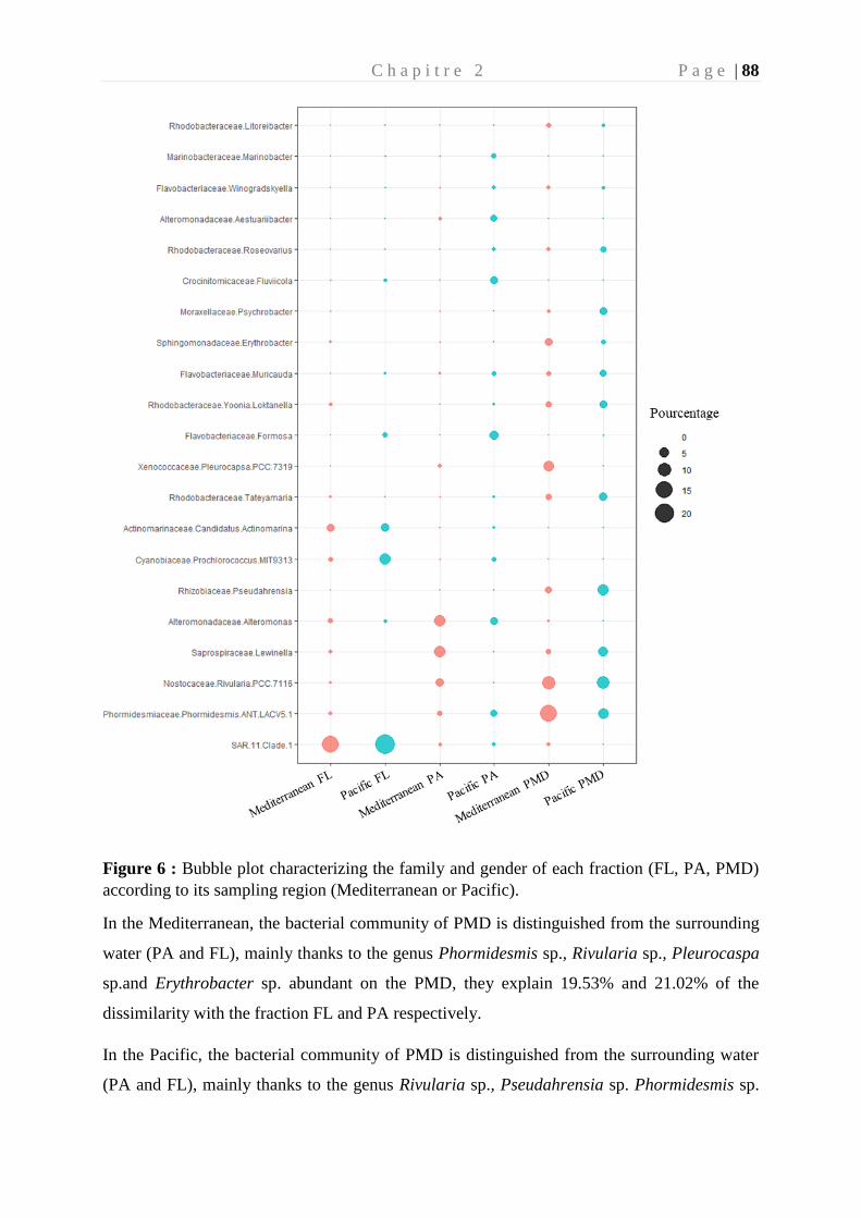
C h a p i t r e 2 P a g e | 88
Figure 6 : Bubble plot characterizing the family and gender of each fraction (FL, PA, PMD)
according to its sampling region (Mediterranean or Pacific).
In the Mediterranean, the bacterial community of PMD is distinguished from the surrounding
water (PA and FL), mainly thanks to the genus Phormidesmis sp., Rivularia sp., Pleurocaspa
sp.and Erythrobacter sp. abundant on the PMD, they explain 19.53% and 21.02% of the
dissimilarity with the fraction FL and PA respectively.
In the Pacific, the bacterial community of PMD is distinguished from the surrounding water
(PA and FL), mainly thanks to the genus Rivularia sp., Pseudahrensia sp. Phormidesmis sp.
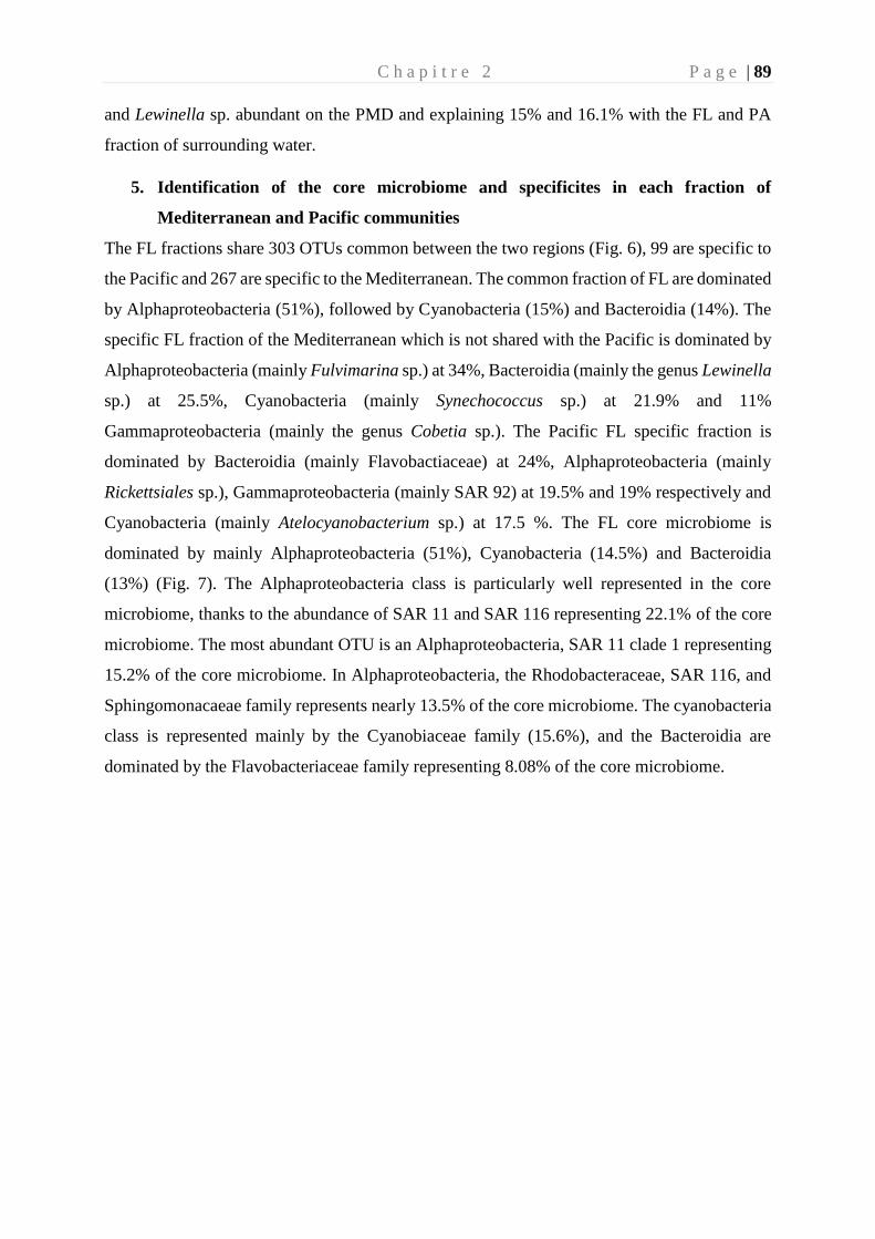
C h a p i t r e 2 P a g e | 89
and Lewinella sp. abundant on the PMD and explaining 15% and 16.1% with the FL and PA
fraction of surrounding water.
5. Identification of the core microbiome and specificites in each fraction of
Mediterranean and Pacific communities
The FL fractions share 303 OTUs common between the two regions (Fig. 6), 99 are specific to
the Pacific and 267 are specific to the Mediterranean. The common fraction of FL are dominated
by Alphaproteobacteria (51%), followed by Cyanobacteria (15%) and Bacteroidia (14%). The
specific FL fraction of the Mediterranean which is not shared with the Pacific is dominated by
Alphaproteobacteria (mainly Fulvimarina sp.) at 34%, Bacteroidia (mainly the genus Lewinella
sp.) at 25.5%, Cyanobacteria (mainly Synechococcus sp.) at 21.9% and 11%
Gammaproteobacteria (mainly the genus Cobetia sp.). The Pacific FL specific fraction is
dominated by Bacteroidia (mainly Flavobactiaceae) at 24%, Alphaproteobacteria (mainly
Rickettsiales sp.), Gammaproteobacteria (mainly SAR 92) at 19.5% and 19% respectively and
Cyanobacteria (mainly Atelocyanobacterium sp.) at 17.5 %. The FL core microbiome is
dominated by mainly Alphaproteobacteria (51%), Cyanobacteria (14.5%) and Bacteroidia
(13%) (Fig. 7). The Alphaproteobacteria class is particularly well represented in the core
microbiome, thanks to the abundance of SAR 11 and SAR 116 representing 22.1% of the core
microbiome. The most abundant OTU is an Alphaproteobacteria, SAR 11 clade 1 representing
15.2% of the core microbiome. In Alphaproteobacteria, the Rhodobacteraceae, SAR 116, and
Sphingomonacaeae family represents nearly 13.5% of the core microbiome. The cyanobacteria
class is represented mainly by the Cyanobiaceae family (15.6%), and the Bacteroidia are
dominated by the Flavobacteriaceae family representing 8.08% of the core microbiome.
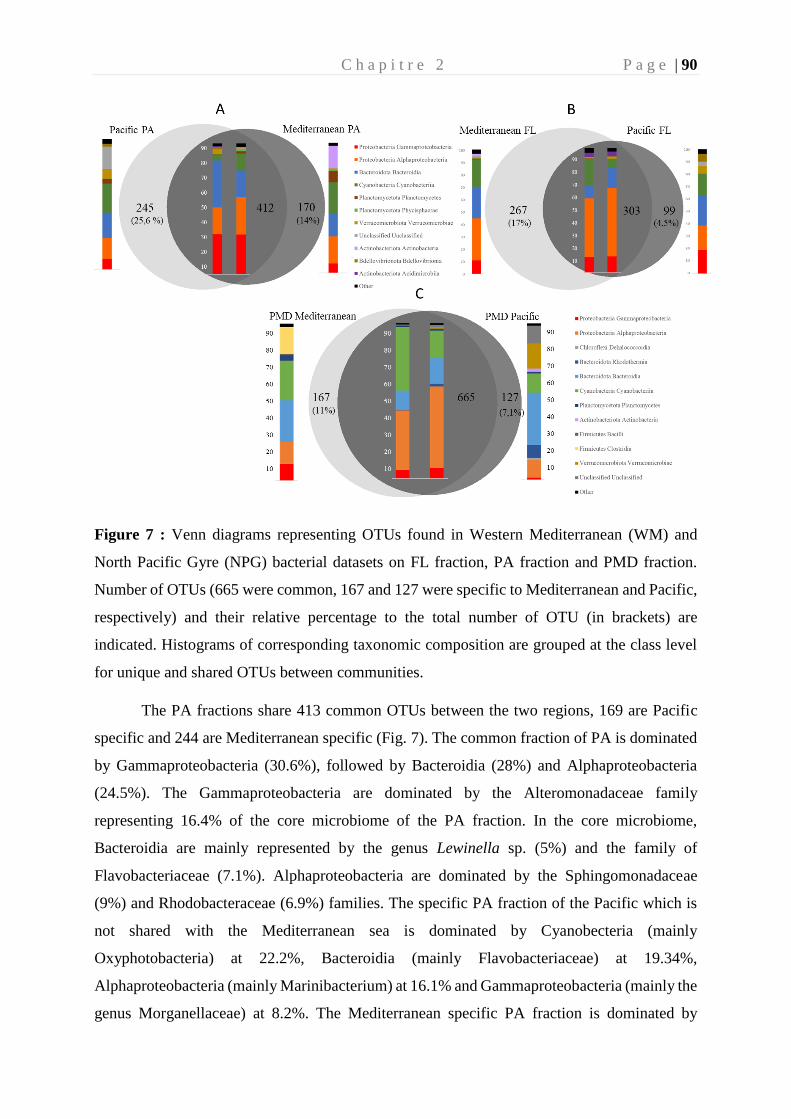
C h a p i t r e 2 P a g e | 90
Figure 7 : Venn diagrams representing OTUs found in Western Mediterranean (WM) and
North Pacific Gyre (NPG) bacterial datasets on FL fraction, PA fraction and PMD fraction.
Number of OTUs (665 were common, 167 and 127 were specific to Mediterranean and Pacific,
respectively) and their relative percentage to the total number of OTU (in brackets) are
indicated. Histograms of corresponding taxonomic composition are grouped at the class level
for unique and shared OTUs between communities.
The PA fractions share 413 common OTUs between the two regions, 169 are Pacific
specific and 244 are Mediterranean specific (Fig. 7). The common fraction of PA is dominated
by Gammaproteobacteria (30.6%), followed by Bacteroidia (28%) and Alphaproteobacteria
(24.5%). The Gammaproteobacteria are dominated by the Alteromonadaceae family
representing 16.4% of the core microbiome of the PA fraction. In the core microbiome,
Bacteroidia are mainly represented by the genus Lewinella sp. (5%) and the family of
Flavobacteriaceae (7.1%). Alphaproteobacteria are dominated by the Sphingomonadaceae
(9%) and Rhodobacteraceae (6.9%) families. The specific PA fraction of the Pacific which is
not shared with the Mediterranean sea is dominated by Cyanobecteria (mainly
Oxyphotobacteria) at 22.2%, Bacteroidia (mainly Flavobacteriaceae) at 19.34%,
Alphaproteobacteria (mainly Marinibacterium) at 16.1% and Gammaproteobacteria (mainly the
genus Morganellaceae) at 8.2%. The Mediterranean specific PA fraction is dominated by
C h a p i t r e 2 P a g e | 91
Cyanobacteria (mainly Rivularia sp.) at 24.5%, Alphaproteobacteria (mainly
Sphingomonadaceae) at 20.8%, Actinobacteria (mainly Pseudonocardia sp.) at 17.4% and
Bacteroidia (mainly Flavobacteriaceae) at 17.1%.
The comparison of PMD fraction between OTU compositions in Western
Mediterranean or North Pacific Gyre showed that relatively fewer OTUs were specific to the
geographical location (167 in the Mediterranean and 127 in the Pacific) compared to the
common OTUs that constituted a core microbiome on our samples (665 OTUs). The common
OTUs found in both Western Mediterranean Sea and North Pacific Gyre were composed of
three phyla, including Alphaproteobacteria (46.8%, Rhodobacteraceae, Rhizobiaceae),
Cyanobacteria (26.8%, Phormidesmiaceae and Rivulariaceae) and Bacteroidia (15.4%,
Flavobacteriaceae and Saprospiraceae) (Fig.7). Among the PMD core microbiome, the most
abundant OTU is Calothrix parasitica accounting for nearly 7.3% of the total relative
abundance. The most represented family are the Rhodobacteraceae (mainly Tateyamaria sp.
and Yoonia-Loktanella sp.) 25.5%, followed by Phormidsmiaceae (mainly Phormidesmisis sp.)
11.3% and Rhizobiaceae (mainly Pseudahrensia sp.) 9.4%. Among the PMD core microbiome,
the most abundant OTU is Calothrix parasitica accounting for nearly 7.3% of the total relative
abundance. The most represented family are the Rhodobacteraceae (mainly Tateyamaria sp.
and Yoonia-Loktanella sp.) 25.5%, followed by Phormidsmiaceae (mainly Phormidesmisis sp.)
11.3% and Rhizobiaceae (mainly Pseudahrensia sp.) 9.4%.
In order to better characterize the core microbiome, we looked at the OTUs shared at
90% by all the plastic parts sampled. In the Western Mediterranean Sea, we identified 832
OTUs on all PMD fraction. None of the 832 OTUs are shared by all the plastic parts making
up the PMD fraction, and only 6 OTUs were shared by 90% of PMD. These common OTUs
were all belonging to the Alphaproteobacteria, including Rhodobacteraceae (81%, mainly
Litoreibacter sp., Yoonia-Loktanella sp., Tateyamaria sp.) and Sphingomonadaceae (19%,
mainly Erythrobacter sp.). In the North Pacific Gyre, we identified 792 OTUs on all PMD
fraction, from which only 6 OTUs were shared by all of the PMD. These common OTUs were
all belonging to the Alphaproteobacteria with 3 Rhizobiaceae (Pseudahrensia sp.), 2
Rhodobacteraceae (Roseovarius sp. ) and 1 Hyphomonadaceae undetermined. When looking at
common OTUs shared by 90% of all the PMD fractions of the Pacific, the resulting 36 OTUs
were mainly represented by the Rhodobacteraceae (41%, mainly Roseovarius sp.),
Rhizobiaceae (19%, mainly Pseudahrensia sp.) and Rivulariaceae (17%, mainly Calothrix sp.).
C h a p i t r e 2 P a g e | 92
The unique OTUs in PMD found only in the Mediterranean Sea belonged mainly (28%) to
Dokdonia sp., Acinetobacter sp., Phormidesmis sp. and unidentified Oscillospiraceae sp. In the
Pacific, they belonged mainly (20%) to Haloferula sp., Aureispira sp. and Phormidesmis sp.
All unique OTUs from Mediterranean or Pacific were from the so-called “rare biosphere”
(<1%).
6. Putative hydrocarbonoclastic bacteria in PMD, PA and FL
Putative hydrocarbonoclastic bacteria represented 14.1% of the OTUs in the PMD from the
Western Mediterranean Sea. They were dominated by Erythrobacter sp., Lewinella sp.,
Muricauda sp., Roseovarius sp. and Winogradskyella sp. that represented 59.6% of putative
hydrocarbonoclatic OTUs. They represented up to 20.3% in the Pacific, dominated by
Lewinella sp., Psychrobacter sp., Muricauda sp., Roseovarius sp., and Tenacibaculum sp. that
represented 74.3% of the putative hydrocarbonoclastic OTUs.
On the 665 OTUs from the core microbiome commonly found in Mediterranean and Pacific
PMD, we observed that 17.7% were putative hydrocarbonoclastic (108 OTU). Most of them
(70%) belonged to Bacteroidia (33% represented by Muricauda sp. and Lewinella sp.) 48 %,
Gammaproteobacteria represented 23 % (18.3% is represented by Psychrobacter sp. and
Cobetia sp.) and Alphaproteobacteria represented 28;5 % (23.52 % represented by Roseovarius
sp. and Erythrobacter sp.).
It is noteworthy that putative hydrocarbonoclastic bacteria represented 10% and 26.3% of the
compiled Mediterranean and Pacific FL and PA fractions, respectively. In the Western
Mediteranean Sea, they belonged to Balneola sp. and Alteromonas sp. in the FL fraction and to
Alteromonas sp. and Sphingobium sp. in the PA fraction. In the North Pacific Gyre, they
belonged to Winogradskyella sp., Muricauda sp. and Tenacibaculum sp.in the FL fraction and
to Aesturribacter sp., Oleibacter sp. and Marinobacter sp. in the PA fraction. Interestingly,
Lewinella sp. and Erythrobacter sp. were two genera found in abundance (from 1.16 to 11.8%)
in all FL, PA and PMD fractions and in the two sites studied.
7. Putative pathogens in PMD, PA and FL
Putative pathogenic OTUs were found on all the different fractions (PMD, PA and FL) in the
Pacific and the Mediterranean, but mainly in the PMD of the 2 geographical regions (71% of
all putative pathogenic OTU numbers) compared to PA (25%) and FL (4%). Vibrio was the
C h a p i t r e 2 P a g e | 93
major putative pathogen detected in 158 samples. Vibrio sequences were found with high
homology (query cover >97%) in Blastn with human (V. alginolyticus, V. parahaemolyticus)
or animal pathogens (V. spendidus, V. crassostreae), but the V4-V5 primers used in this study
was not sufficient to give clear assignation at the species level. Vibrio were found in 67 and 53
PMD, 16 and 6 PA and in 19 and 6 FL samples from the Mediterranean and Pacific regions,
respectively. In the Mediterranean PMD Fraction, Vibrio sp. accounts for 0.5% of the total
abundance, while they represent only 0.078% in the Pacific. One of the polypropylene sample
(from manta M165, 43 km of city Palerme) in the Mediterranean sea hosted 14% of the total
sequences affiliated to Vibrio sp., but this sample was rather an exception than a rule since this
genera generally did not exceed 0.5%. We also found a large proportion (24%) of the putative
human pathogen Acinetobacter in the 2 geographic. This bacterium was mainly found on PMD
(25 PMD in the Mediterranean and 7 in the Pacific). On the PMD fractions of the
Mediterranean, this bacterial genus represents on average 0.8% against 0.003% in the Pacific.
Acinetobacter was also found on the FL fractions in the Mediterranean (16) and rarely in the
Pacific (1), but also in PA (7 in the Mediterranean and 1 in the Pacific).
C h a p i t r e 2 P a g e | 94
Discussion
1. Niche partitioning between PMD, FL and PA fractions
The results presented here demonstrated niche partitioning in sea surface prokaryotes as
reflected in clear differences in the compositions of free-living, organic particle-attached and
PMD-attached bacteria.
Bacterioplankton has been generally classified into two types of communities depending on
their relationship with the particulate matter, e.g. free-living (FL) and particle-attached (PA)
lifestyles (Crump et al. 1999). FL and PA marine microbial communities have repeatedly been
shown to differ in their diversity (alpha-diversity) and composition (beta-diversity) in different
locations worldwide (Ghiglione et al. 2005, 2007; Grossart 2010; Ortega-Retuerta et al. 2013).
More recently, another type of marine bacterial communities living on PMD were shown to
differ from the FL fraction (Zettler et al. 2013; Debroas et al. 2017) and also from the PA
fraction (Oberbeckmann et al. 2018; Dussud et al. 2018b). In this study, we observed that the
spatially isolated Western Mediterranean and North Pacific Gyre hosted FL, PA and PMD
communities that consistently differed in beta-diversity, with these differences accounting for
a considerable proportion of the variance in community composition. This result is in
accordance with a previous studies which showing that differences between FL and PA
fractions exceeded the global-scale geographical variation (Acinas et al. 1997, Salazar et al.
2015). Here, we show that it is not only the case for FL and PA fractions, but also for the PMD
fraction, which confirms the strong niche partitioning already observed in different locations
but without exploring their biogeography between different ocean basins (Oberbeckmann et al.
2018; Xu et al. 2019).
We also observed that the communities within any of the FL, PA and PMD fractions were
clearly influenced by their geographical origin. Such biogeographical patterns can result from
different ecological processes as a consequence of two main processes (Hanson et al. 2012) :
(a) the ‘environmental selection’, build on the principle that environmental pressure exert
different pressure on each fraction or (b) the ‘dispersal effect’, where dispersal capabilities of
microbes from each fraction is considered. We estimated the relative contribution of both
processes by relating community composition to a set of environmental variables, and to the
geographical distance between sampling locations. The multiple regression on matrices analysis
explained a total of 26.7% (FL fraction), 17.6% (PA fraction) and 16.8% (PMD fraction) of the
total variance in the Bray–Curtis dissimilarity matrix. The effect of environmental variables on
C h a p i t r e 2 P a g e | 95
bacterial community structure was statistically significant (p<0.05) for the FL (explaining
14.3% of the variance), PA (explaining 7.6% of the variance) and PMD fractions (explaining
6.5% of the variance). We found that temperature was the main environmental driver of
bacterial communities in all fractions, which is in accordance with another study based on Tara
expeditions on global planktonic ocean microbiome in the epipelagic zone (Sunagawa et al.
2015). On another hand, the effect of geographical distance was significant between the two
ocean basins for the three fractions (p<0,05), but not within the same basin. Thus, both
environmental selection and dispersal effects appear to shape the biogeography of all the FL,
PA and PMD fractions, although spurious distance effects may arise as a result of unmeasured
environmental variables (Hanson et al. 2012).
In the specific case of PMD, we also evaluated the influence of the polymer composition,
morphology, size and age in shaping this specific community. As in many other places in the
world surface oceans (Erni-Cassola et al. 2019), PE was the most abundant polymer (> 70%)
in both location, followed by PP, PS together or not with PET in the North Pacific gyre or in
western Mediterranean Sea, respectively. By comparing the total of 179 plastic pieces of
different polymer composition, we could not find any significant link between the bacterial
community structure and the polymer type. The only other study evaluating the biogeography
of the plastisphere from North Pacific and North Atlantic gyres showed also a main difference
between ocean basins and to a lesser extent between polymer types (Amaral-Zettler et al. 2015).
Our study gives another proof that the environmental conditions rather than the polymer type
are driving the plastisphere composition at both global (Amaral-Zettler et al. 2015) and regional
scales (Basili et al. 2020). This result may appeared in contradiction with other studies showing
clear distinction between biofilms growing on different polymer type (Oberbeckmann et al.
2018; Dussud et al. 2018a; Pinto et al. 2019). Such discrepancy may be explained by the fact
that these studies were focusing on the formation of the biofilm when pristine new plastic were
immerged for several weeks or months in seawater, which is different from our study that
sampled microplastics directly in the environment and from unknown origin. As mentioned
above, the plastisphere community is mainly driven by the environmental conditions and the
dispersion of PMD rather than their composition. PMD are long-lived substrates that can
transport microbial communities across ocean basins and our results, together with other global
comparison between the North Pacific and North Atlantic gyres (Amaral-Zettler et al. 2015)
are indicating that the environment selection is even more important in shaping the specific
community growing on plastics. However, further studies will be needed to find the other
C h a p i t r e 2 P a g e | 96
parameters that may be specifically applied to PMD in order to explain the clear niche
partitioning of the plastisphere compared to FL and PA communities.
The alpha diversity indices also make it possible to distinguish the two area of studies. The
richness (chao index) is greater on all fractions of the Pacific compared to that of the
Mediterranean. The richness is not significantly different between the fractions FL, PA and
PMD of the Mediterranean and PMD and FL of the Pacific. These results are correlated by
previous studies, which show greater richness in the surrounding water (Zettler et al. 2013) or
on PMD fractions (De Tender et al. 2015; Bryant et al. 2016).
In the Mediterranean, the evenness is homogeneous over the three fractions, which is not the
case in the Pacific, which sees a lower equity on the PMD fraction (Pielou: 0.64). PMDs
promote the emergence of certain species in the Pacific. The diversity found in the FL and PA
fractions is greater in the Pacific than in the Mediterranean. Unlike the Mediterranean, the
diversity on the PA fraction is greater than the rest of the other fractions, this is correlated with
a previous study conducted in the North Pacific area (Suzuki et al. 2017).
2. Original OTUs in the plastisphere compared to PA and FL in the Western
Mediterranean Sea or in the North Pacific gyre and common OTUs found in both
locations.
The Mediterranean and Pacific FL fractions are mainly characterized with the genus SAR 11.
This clade has already been identified as abundant in these two regions of the world (Morris et
al. 2002; Suzuki et al. 2017), although its biological role remains poorly known. Cyanobacteria
are 2.7 times more important in the Mediterranean than in the Pacific. The Mediterranean Sea
is known as one of the most nutrient-poor seas (Siokou-Frangou et al. 2010). In the
Mediterranean, a phosphate is observed with an N: P ratio of 20: 1 (Ribera d’Alcalà 2003). The
north pacific subtropical gyre is also known to have a low phosphate concentration (Moutin et
al. 2008). Cyanobacteria are influenced by water temperature, excess nitrogen, phosphorus and
the N / P ratio (Dolman et al. 2012), which may explain this difference between the Pacific and
the Mediterranean. FL fractions are characterized also by the genus Synechococcus sp. in the
Mediterranean and Prochlorococcus sp. in the Pacific, two phototrophic bacterial genera most
widespread in the oceans (Suzuki et al. 2017; Sunagawa et al. 2020). SAR 11, Synechococcus
sp., Prochlorococcus sp. are found in abundance in nutrient poor ocean environments.
The PA fractions observed a strong abundance of the genera Alteromonas sp. in the two regions,
of the genera Lewinella sp. and Muricauda sp. in the Mediterranean and of the genera
C h a p i t r e 2 P a g e | 97
Marinobacter sp., Winogradskyella sp. in the Pacific. The attachment of bacteria depends on
many factors, however the microorganism must be provided with organelles (flagella, pili),
with a load and hydrophobicity of the outer membrane conducive to the attachment of a surface
(Donlan 2002; Palmer et al. 2007). This may explain why bacterial genera living preferentially
attached to particles like Alteromonas sp. are also found in the biofilm of the plastisphere
(Suzuki et al. 2017; Pinto et al. 2019).
This study allows a more global observation of the communities composing the plastisphere on
a global scale. the two oceanographic regions that present the highest sea surface pollution by
microplastic worldwide (Cozar et al. 2014; Lebreton et al. 2018).
PMD fractions in the Pacific are dominated by primary colonizers (Alphaproteobacteria and
Cyanobacteria) and secondary colonizers (Bacteroidetes) (Dang and Lovell 2000; Lee et al.
2008; Amaral-Zettler et al. 2020) allow us to deduce that the biofilms formed on the surface of
the plastic are at an advanced stage of maturation. The PMD fraction in the Mediterranean is
largely dominated by the primary colonizer (Cyanobacteria and Alphaproteobacteria) and to a
lesser extent by the secondary colonizers, possibly suggesting that the bacterial biofilm is more
recent in the Mediterranean than in the Pacific.The bacterial genera Phormidesmis sp. and
Rivularia sp. are preferentially associated with the PMD fraction in the Mediterranean and the
Pacific. Rivularia sp. is an already isolated genus known to colonize and degrade hydrocarbons
and identified on the North Atlantic plastisphere (Abed et al.2014; Zettler 2013). Generally
speaking, prokaryotic phototrophs like cyanobacteria are strongly represented on plastics
floating on the surface of seawater (Zettler et al. 2013; Amaral-Zettler et al. 2020).
In the Mediterranean, it is the genera Erythrobacter sp. and Pleurocaspa sp. which
preferentially colonized the PMD fraction. The genus Erythrobacter sp. has been isolated from
plastics in cold water, this genus being known to use polycyclic aromatic hydrocarbons (PAHs)
(Wang et al. 2016). The genus Erythobacter sp. has already been found on microplastics
samples that have been incubated for 1 year in the Pacific (Xu et al. 2019).
In the Pacific we find the genera Pseudahrensia sp. and Lewinella sp. on the PMD fraction.
Lewinella sp. has previously been on floating PET in the North Sea near the United Kindgom
shores (Oberbeckmann et al. 2016). The genus Psychrobacter sp. has also been isolated in deep
sediments (5000 m) of Japan colonizing pieces of PCL (Sekiguchi et al. 2011). Muricauda sp.,
a Roseovarius sp. abundant on genus microplastics abundant in the Pacific near Fujian Bay in
China (Lu et al. 2019).
C h a p i t r e 2 P a g e | 98
On PMDs, we have identified a core microbiome essentially composed of Alphaproteobacteria,
cyanobacteria and Bacteroidia. The genus Rivularia sp. is the most abundant in this core
microbiome, it has already been identified in numerous studies on the plastisphere (Zettler et
al. 2013; Bryant et al. 2016; Rogers et al. 2020). The Rhodobacteraceae family is particularly
well represented in this biofilm with the genus Tateyamaria sp., already isolated in several
regions of the world such as the North Atlantic, the North Pacific, the North Sea, in estuaries
or in the Baltic Sea (Rogers et al. 2020). The Rhodobacteraceae family has a general quorum
sensing detection system (Gram et al. 2002) and are identified as essential for biofilm formation
(Elifantz et al. 2013). Quorum sensing allows for inter-bacterial communication through self-
inducing molecules released into the environment. This communication allows the regulation
of numerous physiological and biochemical reactions, which can give it a consequent advantage
in the formation of the biofilm and the colonization of the plastic (McDougald et al. 2007)). As
a result, it is regularly identified as abundant on the plastisphere of many plastics (Dang and
Lovell 2002; Jones et al. 2007; Bryant et al. 2016; Xu et al. 2019; Amaral-Zettler et al. 2020).
This phenomenon may explain why the Rhodobacteraceae are mainly found on the PMD
fraction in our study (25% on average in the two areas) than on the PA and FL fraction (12.21%
on average).
Other genera have already been demonstrated as an abundant component of the plastisphere, it
is the case for the genus Erythrobacter sp., Roseovarius sp., Calothrix sp. (Rogers et al. 2020).
More generally, this core microbiome has the families most commonly identified on the
plastisphere (Rhodobacteracae, Rhizobiaceae, Rivulariaceae, Flavobacteriaceae) (Rogers et al.
2020; Amaral-Zettler et al. 2020). The core microbiome identified during this study is
composed of almost 17% hydrocabonoclasts, mainly dominated by the genus Lewinella sp.,
Erytrhobacter sp. and Psychrobacter sp. representing 47.5% of the hydrocarbonoclasts. The
core microbiome is not enriched in hydrocarbonoclast compared to the PA fraction from the
environment.
Hydrocarbonoclasts are found in all of the world's oceans and are not restricted to areas
contaminated with hydrocarbons (Gutierrez 2019). This is consistent with the level of
hydrocarbonoclasts discovered on the FL and PA fractions of the Mediterranean and the Pacific.
Hydrocarbonoclasts represent 20.3% of the entire microbial community of PMDs in the Pacific
and 14.2% in the Mediterranean. In the identified core microbiome, the hydrocarbonoclasts
represent 17.7% of the community. We find mostly hydrocarbonoclasts that can degrade linear
C h a p i t r e 2 P a g e | 99
or branched hydrocarbons such as Alcalinovorax sp., Marinobacter sp. The genus Alcanivorax
sp. has already been identified as a key genus in the degradation of PE and other hydrocarbons
such as petroleum (Delacuvellerie et al. 2019; Zadjelovic et al. 2020). Other genus like
Roseovarius sp. have already been identified in the PE plastisphere (Delacuvellerie et al. 2019).
In the species identified as potentially pathogenic, the distribution is not homogeneous and is
mainly concentrated on the PMD fraction of the Mediterranean and the Pacific (69%), then on
the PA fraction (27%). In these OTU we find potential animal or human pathogens, such as V.
parahaemolyticus which is found in the PA, FL and PMD fractions of the Pacific and the
Mediterranean, always belonging to the rare biosphere. This species has also been found on
PMD in the North and Baltic Sea (Kirstein et al. 2016). OTUs belonging to the genus Vibrio
are 15.6 times higher on PMD compared to FL fraction of meditereranean and tree times more
compare to the PA fraction. In the Pacific vibrio sp. are thirty-two times more abundant on
PMD compared to FL and three times more important compared to PA. In all the samples only
one plastic, located 43 km from the coast near the city of Palermo with an abundance of Vibrio
which exceeds that of the rare biosphere. The genus vibrio, endemic to the marine environment,
has shown strong hydrophobicity in the hydrocarbon adhesion test (BATH) and has shown a
strong ability to form a biofilm (Lamari et al. 2018). Sampling in the Mediterranean and the
Pacific took place during the summer period, corresponding to an increase in seawater
temperatures and therefore favoring the development of the genus Vibrio (Amaral-Zettler et al.
2020). The persistence of plastic in the marine environment, and its movement with the
currents, may explain this potentially pathogenic "accumulation" of OTU. All these reasons can
explain this potentially pathogenic distribution of OTU mainly on the PMD fraction of the
Pacific and the Mediterranean. However, these results do not prove that these OTUs are indeed
pathogenic. The sequencing method (illumina 16S V4 -V5) identifies only the bacterial genus
and not the species. Further, more in-depth studies are needed to assess the pathogenicity of the
OTUs identified in this study.
C h a p i t r e 2 P a g e | 100
References
Abed EH, Abbas AH, Abed AG, and Haloob AA. Biodegradation of low-density polyethylene by
bacteria spp. isolated from oil basins.
Acinas SG, Rodríguez-Valera F, and Pedrós-Alió C. 1997. Spatial and temporal variation in marine
bacterioplankton diversity as shown by RFLP fingerprinting of PCR amplified 16S rDNA.
FEMS Microbiol Ecol 24: 27–40.
Amaral-Zettler LA, Zettler ER, and Mincer TJ. 2020. Ecology of the plastisphere. Nat Rev Microbiol
18: 139–51.
Amaral-Zettler LA, Zettler ER, Slikas B, et al. 2015. The biogeography of the Plastisphere:
implications for policy. Frontiers in Ecology and the Environment 13: 541–6.
Barnes DKA, Galgani F, Thompson RC, and Barlaz M. 2009. Accumulation and fragmentation of
plastic debris in global environments. Philosophical Transactions of the Royal Society B:
Biological Sciences 364: 1985–98.
Basili M, Quero GM, Giovannelli D, et al. 2020. Major Role of Surrounding Environment in Shaping
Biofilm Community Composition on Marine Plastic Debris. Front Mar Sci 7.
Bryant JA, Clemente TM, Viviani DA, et al. 2016. Diversity and Activity of Communities Inhabiting
Plastic Debris in the North Pacific Gyre. mSystems 1.
Camacho C, Coulouris G, Avagyan V, et al. 2009. BLAST+: architecture and applications. BMC
Bioinformatics 10: 421.
Chiba S, Saito H, Fletcher R, et al. 2018. Human footprint in the abyss: 30 year records of deep-sea
plastic debris. Marine Policy 96: 204–12.
Choy CA, Robison BH, Gagne TO, et al. 2019. The vertical distribution and biological transport of
marine microplastics across the epipelagic and mesopelagic water column. Scientific Reports
9: 7843.
Cozar A, Echevarria F, Gonzalez-Gordillo JI, et al. 2014. Plastic debris in the open ocean.
Proceedings of the National Academy of Sciences 111: 10239–44.
Cózar A, Sanz-Martín M, Martí E, et al. 2015. Plastic Accumulation in the Mediterranean Sea. PLOS
ONE 10: e0121762.
Crump BC, Armbrust EV, and Baross JA. 1999. Phylogenetic Analysis of Particle-Attached and Free-
Living Bacterial Communities in the Columbia River, Its Estuary, and the Adjacent Coastal
Ocean. Appl Environ Microbiol 65: 3192–204.
Dang H and Lovell CR. 2000. Bacterial Primary Colonization and Early Succession on Surfaces in
Marine Waters as Determined by Amplified rRNA Gene Restriction Analysis and Sequence
Analysis of 16S rRNA Genes. Appl Environ Microbiol 66: 467–75.
Dang H and Lovell CR. 2002. Numerical dominance and phylotype diversity of marine Rhodobacter
species during early colonization of submerged surfaces in coastal marine waters as
determined by 16S ribosomal DNA sequence analysis and fluorescence in situ hybridization.
Appl Environ Microbiol 68: 496–504.
C h a p i t r e 2 P a g e | 101
De Tender CA, Devriese LI, Haegeman A, et al. 2015. Bacterial Community Profiling of Plastic Litter
in the Belgian Part of the North Sea. Environ Sci Technol 49: 9629–38.
De Tender C, Devriese LI, Haegeman A, et al. 2017. Temporal Dynamics of Bacterial and Fungal
Colonization on Plastic Debris in the North Sea. Environ Sci Technol 51: 7350–60.
Debroas D, Mone A, and Ter Halle A. 2017. Plastics in the North Atlantic garbage patch: A boat-
microbe for hitchhikers and plastic degraders. Sci Total Environ 599–600: 1222–32.
Delacuvellerie A, Cyriaque V, Gobert S, et al. 2019. The plastisphere in marine ecosystem hosts
potential specific microbial degraders including Alcanivorax borkumensis as a key player for
the low-density polyethylene degradation. Journal of Hazardous Materials 380: 120899.
Di M and Wang J. 2018. Microplastics in surface waters and sediments of the Three Gorges Reservoir,
China. Science of The Total Environment 616–617: 1620–7.
Dobaradaran S, Schmidt TC, Nabipour I, et al. 2018. Characterization of plastic debris and association
of metals with microplastics in coastline sediment along the Persian Gulf. Waste Management
78: 649–58.
Dolman A, Rücker J, Pick F, et al. 2012. Cyanobacteria and Cyanotoxins: The Influence of Nitrogen
versus Phosphorus. PloS one 7: e38757.
Donlan RM. 2002. Biofilms: microbial life on surfaces. Emerging Infect Dis 8: 881–90.
Durrieu de Madron X, Guieu C, Sempéré R, et al. 2011. Marine ecosystems’ responses to climatic and
anthropogenic forcings in the Mediterranean. Progress in Oceanography 91: 97–166.
Dussud C, Hudec C, George M, et al. 2018a. Colonization of Non-biodegradable and Biodegradable
Plastics by Marine Microorganisms. Front Microbiol 9.
Dussud C, Meistertzheim AL, Conan P, et al. 2018b. Evidence of niche partitioning among bacteria
living on plastics, organic particles and surrounding seawaters. Environmental Pollution 236:
807–16.
Elifantz H, Horn G, Ayon M, et al. 2013. Rhodobacteraceae are the key members of the microbial
community of the initial biofilm formed in Eastern Mediterranean coastal seawater. FEMS
Microbiol Ecol 85: 348–57.
Erni-Cassola G, Zadjelovic V, Gibson MI, and Christie-Oleza JA. 2019. Distribution of plastic
polymer types in the marine environment; A meta-analysis. Journal of Hazardous Materials
369: 691–8.
Escudié F, Auer L, Bernard M, et al. 2018. FROGS: Find, Rapidly, OTUs with Galaxy Solution.
Bioinformatics 34: 1287–94.
Fuhrman JA, Sleeter TD, Carlson CA, and Proctor LM. 1989. Dominance of bacterial biomass in the
Sargasso Sea and its ecological implications. Marine Ecology Progress Series 57: 207–17.
Ghiglione J-F, Larcher M, and Lebaron P. 2005. Spatial and temporal scales of variation in
bacterioplankton community structure in the NW Mediterranean Sea. Aquatic Microbial
Ecology 40: 229–40.
Ghiglione JF, Mevel G, Pujo-Pay M, et al. 2007. Diel and Seasonal Variations in Abundance,
Activity, and Community Structure of Particle-Attached and Free-Living Bacteria in NW
Mediterranean Sea. Microb Ecol 54: 217–31.
C h a p i t r e 2 P a g e | 102
Gram L, Grossart H-P, Schlingloff A, and Kiørboe T. 2002. Possible Quorum Sensing in Marine Snow
Bacteria: Production of Acylated Homoserine Lactones by Roseobacter Strains Isolated from
Marine Snow. Appl Environ Microbiol 68: 4111–6.
Grossart H-P. 2010. Ecological consequences of bacterioplankton lifestyles: changes in concepts are
needed. Environmental Microbiology Reports 2: 706–14.
Gutierrez T. 2019. Occurrence and Roles of the Obligate Hydrocarbonoclastic Bacteria in the Ocean
When There Is No Obvious Hydrocarbon Contamination. In: McGenity TJ (Ed). Taxonomy,
Genomics and Ecophysiology of Hydrocarbon-Degrading Microbes. Cham: Springer
International Publishing.
Hanson CA, Fuhrman JA, Horner-Devine MC, and Martiny JBH. 2012. Beyond biogeographic
patterns: processes shaping the microbial landscape. Nature Reviews Microbiology 10: 497–
506.
Jacquin J, Cheng J, Odobel C, et al. 2019. Microbial Ecotoxicology of Marine Plastic Debris: A
Review on Colonization and Biodegradation by the “Plastisphere.” Front Microbiol 10.
Jambeck JR, Geyer R, Wilcox C, et al. 2015. Marine pollution. Plastic waste inputs from land into the
ocean. Science 347: 768–71.
Jones PR, Cottrell MT, Kirchman DL, and Dexter SC. 2007. Bacterial community structure of
biofilms on artificial surfaces in an estuary. Microb Ecol 53: 153–62.
Kirstein IV, Kirmizi S, Wichels A, et al. 2016. Dangerous hitchhikers? Evidence for potentially
pathogenic Vibrio spp. on microplastic particles. Marine Environmental Research 120: 1–8.
Lamari F, Khouadja S, and Rtimi S. 2018. Interaction of Vibrio to Biotic and Abiotic Surfaces:
Relationship between Hydrophobicity, Cell Adherence, Biofilm Production and Cytotoxic
Activity. Surfaces 1.
Lamontagne M and Holden P. 2003. Comparison of free-living and particle-associated bacterial
communities in a coastal Lagoon. Microbial ecology 46: 228–37.
Lebreton L, Slat B, Ferrari F, et al. 2018. Evidence that the Great Pacific Garbage Patch is rapidly
accumulating plastic. Scientific Reports 8: 4666.
Lee J-W, Nam J-H, Kim Y-H, et al. 2008. Bacterial communities in the initial stage of marine biofilm
formation on artificial surfaces. J Microbiol 46: 174–82.
Lu L, Luo T, Zhao Y, et al. 2019. Interaction between microplastics and microorganism as well as gut
microbiota: A consideration on environmental animal and human health. Science of The Total
Environment 667: 94–100.
Mahé F, Rognes T, Quince C, et al. 2014. Swarm: robust and fast clustering method for amplicon-
based studies. PeerJ 2: e593.
Matsuguma Y, Takada H, Kumata H, et al. 2017. Microplastics in Sediment Cores from Asia and
Africa as Indicators of Temporal Trends in Plastic Pollution. Arch Environ Contam Toxicol
73: 230–9.
McCormick AR, Hoellein TJ, London MG, et al. 2016. Microplastic in surface waters of urban rivers:
concentration, sources, and associated bacterial assemblages. Ecosphere 7: e01556.
C h a p i t r e 2 P a g e | 103
McDougald D, Rice SA, and Kjelleberg S. 2007. Bacterial quorum sensing and interference by
naturally occurring biomimics. Anal Bioanal Chem 387: 445–53.
Millot C and Taupier-Letage I. 2005. Circulation in the Mediterranean Sea. In: Saliot A (Ed). The
Mediterranean Sea. Springer.
Morris RM, Rappé MS, Connon SA, et al. 2002. SAR11 clade dominates ocean surface
bacterioplankton communities. Nature 420: 806–10.
Moutin T, Karl DM, Duhamel S, et al. 2008. Phosphate availability and the ultimate control of new
nitrogen input by nitrogen fixation in the tropical Pacific Ocean. Biogeosciences 5: 95–109.
Oberbeckmann S, Kreikemeyer B, and Labrenz M. 2018. Environmental Factors Support the
Formation of Specific Bacterial Assemblages on Microplastics. Front Microbiol 8.
Oberbeckmann S, Loeder MGJ, Gerdts G, and Osborn AM. 2014. Spatial and seasonal variation in
diversity and structure of microbial biofilms on marine plastics in Northern European waters.
FEMS Microbiol Ecol 90: 478–92.
Oberbeckmann S, Osborn AM, and Duhaime MB. 2016. Microbes on a Bottle: Substrate, Season and
Geography Influence Community Composition of Microbes Colonizing Marine Plastic Debris.
PLoS One 11.
Ortega-Retuerta E, Joux F, Jeffrey WH, and Ghiglione JF. 2013. Spatial variability of particle-attached
and free-living bacterial diversity in surface waters from the Mackenzie River to the Beaufort
Sea (Canadian Arctic).
Palmer J, Flint S, and Brooks J. 2007. Bacterial cell attachment, the beginning of a biofilm. J Ind
Microbiol Biotechnol 34: 577–88.
Pan Z, Guo H, Chen H, et al. 2019. Microplastics in the Northwestern Pacific: Abundance,
distribution, and characteristics. Science of The Total Environment 650: 1913–22.
Parada AE, Needham DM, and Fuhrman JA. 2016. Every base matters: assessing small subunit rRNA
primers for marine microbiomes with mock communities, time series and global field samples.
Environmental Microbiology 18: 1403–14.
Peeken I, Primpke S, Beyer B, et al. 2018. Arctic sea ice is an important temporal sink and means of
transport for microplastic. Nat Commun 9: 1505.
Pinto M, Langer TM, Hüffer T, et al. 2019. The composition of bacterial communities associated with
plastic biofilms differs between different polymers and stages of biofilm succession. PLOS
ONE 14: e0217165.
Quast C, Pruesse E, Yilmaz P, et al. 2013. The SILVA ribosomal RNA gene database project:
improved data processing and web-based tools. Nucleic Acids Res 41: D590–6.
Ribera d’Alcalà M. 2003. Nutrient ratios and fluxes hint at overlooked processes in the Mediterranean
Sea. J Geophys Res 108: 8106.
Rogers K, Carreres-Calabuig JA, Gorokhova E, and Posth N. 2020. Micro‐by‐micro interactions: How
microorganisms influence the fate of marine microplastics. Limnology and Oceanography
Letters 5.
Rognes T, Flouri T, Nichols B, et al. 2016. VSEARCH: a versatile open source tool for
metagenomics. PeerJ 4: e2584.
C h a p i t r e 2 P a g e | 104
Ruiz-Orejón LF, Sardá R, and Ramis-Pujol J. 2016. Floating plastic debris in the Central and Western
Mediterranean Sea. Marine Environmental Research 120: 136–44.
Sekiguchi T, Sato T, Enoki M, et al. 2011. Isolation and characterization of biodegradable plastic
degrading bacteria from deep-sea environments. JAMSTEC Report of Research and
Development 11: 33–41.
Shim W, Hong S, and Eo S. 2016. Identification methods in microplastic analysis: A review. Anal
Methods 9.
Siokou-Frangou I, Christaki U, Mazzocchi MG, et al. 2010. Plankton in the open Mediterranean Sea: a
review. Biogeosciences 7: 1543–86.
Sunagawa S, Acinas SG, Bork P, et al. 2020. Tara Oceans: towards global ocean ecosystems biology.
Nat Rev Microbiol 18: 428–45.
Sunagawa S, Coelho LP, Chaffron S, et al. 2015. Structure and function of the global ocean
microbiome. Science 348.
Suzuki S, Kaneko R, Kodama T, et al. 2017. Comparison of community structures between particle-
associated and free-living prokaryotes in tropical and subtropical Pacific Ocean surface
waters. J Oceanogr 73: 383–95.
Wang H, Wang B, Dong W, and Hu X. 2016. Co-acclimation of bacterial communities under stresses
of hydrocarbons with different structures. Sci Rep 6: 34588.
Xu X, Wang S, Gao F, et al. 2019. Marine microplastic-associated bacterial community succession in
response to geography, exposure time, and plastic type in China’s coastal seawaters. Marine
Pollution Bulletin 145: 278–86.
Zadjelovic V, Chhun A, Quareshy M, et al. 2020. Beyond oil degradation: enzymatic potential of
Alcanivorax to degrade natural and synthetic polyesters. Environmental Microbiology 22:
1356–69.
Zettler ER, Mincer TJ, and Amaral-Zettler LA. 2013. Life in the “Plastisphere”: Microbial
Communities on Plastic Marine Debris. Environmental Science & Technology 47: 7137–46.
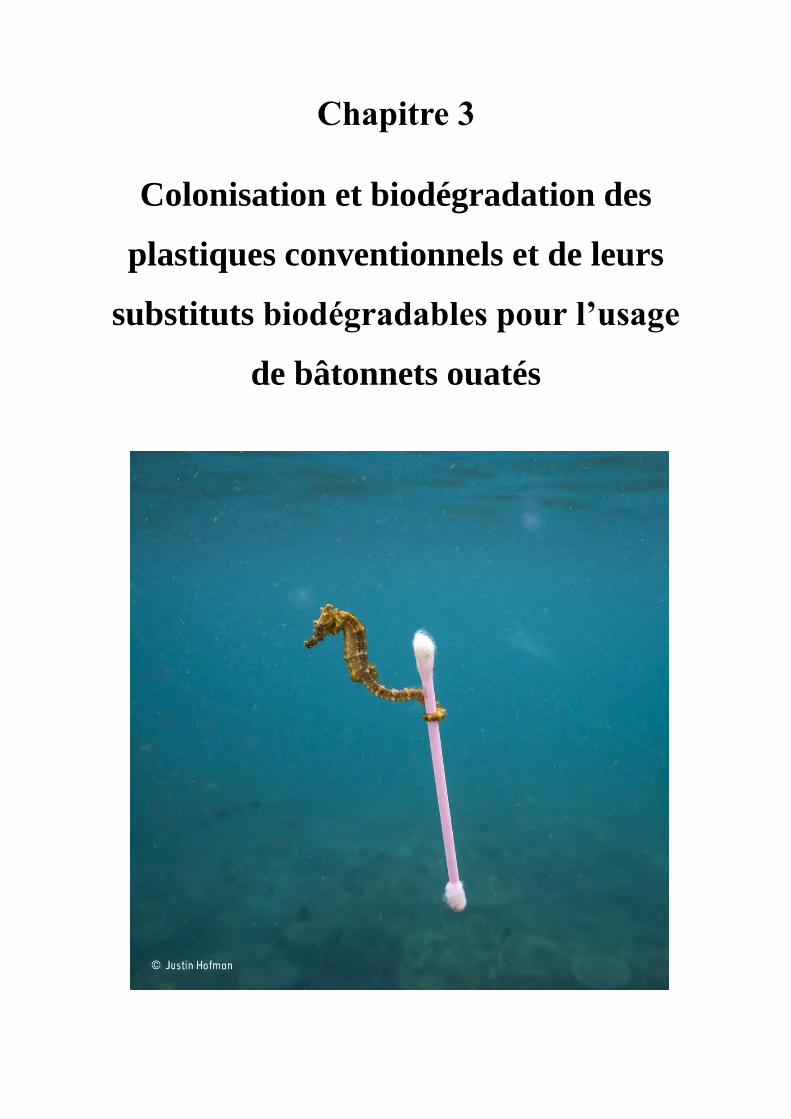
Chapitre 3
Colonisation et biodégradation des
plastiques conventionnels et de leurs
substituts biodégradables pour l’usage
de bâtonnets ouatés

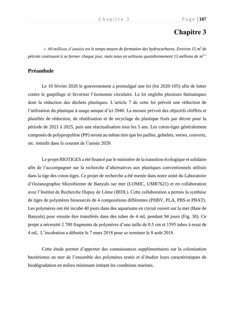
C h a p i t r e 3 P a g e | 107
Chapitre 3
« 60 millions d’années est le temps moyen de formation des hydrocarbures. Environ 15 m3 de
pétrole continuent à se former chaque jour, mais nous en utilisons quotidiennement 15 millions de m3“
Préambule
Le 10 février 2020 le gouvernement a promulgué une loi (loi 2020-105) afin de lutter
contre le gaspillage et favoriser l’économie circulaire. La loi englobe plusieurs thématiques
dont la réduction des déchets plastiques. L’article 7 de cette loi prévoit une réduction de
l’utilisation du plastique à usage unique d’ici 2040. La mesure prévoit des objectifs chiffrés et
planifiés de réduction, de réutilisation et de recyclage du plastique fixés par décret pour la
période de 2021 à 2025, puis une réactualisation tous les 5 ans. Les coton-tiges généralement
composés de polypropylène (PP) seront au même titre que les pailles, gobelets, verres, couverts,
etc. interdit dans le courant de l’année 2020.
Le projet BIOTIGES a été financé par le ministère de la transition écologique et solidaire
afin de l’accompagner sur la recherche d’alternatives aux plastiques conventionnels utilisés
dans la tige des coton-tiges. Ce projet de recherche a été menée dans notre unité du Laboratoire
d’Océanographie Microbienne de Banyuls sur mer (LOMIC, UMR7621) et en collaboration
avec l’Institut de Recherche Dupuy de Lôme (IRDL). Cette collaboration a permis la synthèse
de tiges de polymères biosourcés de 4 compositions différentes (PHBV, PLA, PBS et PBAT).
Les polymères ont été incubé 40 jours dans des aquariums en circuit ouvert sur la mer (Baie de
Banyuls) pour ensuite être transférés dans des tubes de 4 mL pendant 94 jours (Fig. 30). Ce
projet a nécessité 2 700 fragments de polymères d’une taille de 0.5 cm et 1595 tubes à essai de
4 mL. L’incubation a débutée le 7 mars 2018 pour se terminer le 8 août 2018.
Cette étude permet d’apporter des connaissances supplémentaires sur la colonisation
bactérienne en mer de l’ensemble des polymères testés et d’étudier leurs caractéristiques de
biodégradation en milieu minimum imitant les conditions marines.
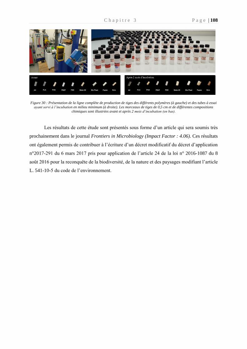
C h a p i t r e 3 P a g e | 108
Figure 30 : Présentation de la ligne complète de production de tiges des différents polymères (à gauche) et des tubes à essai
ayant servi à l’incubation en milieu minimum (à droite). Les morceaux de tiges de 0,5 cm et de différentes compositions
chimiques sont illustrées avant et après 2 mois d’incubation (en bas).
Les résultats de cette étude sont présentés sous forme d’un article qui sera soumis très
prochainement dans le journal Frontiers in Microbiology (Impact Factor : 4.06). Ces résultats
ont également permis de contribuer à l’écriture d’un décret modificatif du décret d’application
n°2017-291 du 6 mars 2017 pris pour application de l’article 24 de la loi n° 2016-1087 du 8
août 2016 pour la reconquête de la biodiversité, de la nature et des paysages modifiant l’article
L. 541-10-5 du code de l’environnement.
C h a p i t r e 3 P a g e | 109
Title: Marine plastisphere activity and diversity during successive
colonization and biodegradation phases of various composition of plastic
sticks
List of authors : JACQUIN Justine1, CALLAC Nolwenn1,2, CHENG Jingguang1, GIRAUD
Carolane1,2, GORAND Yonko3, DENOUAL Clément4, PUJO-PAY Mireille1, CONAN
Pascal1, BARBE Valérie5, TER HALLE Alexandra6, MEISTERTZHEIM Anne-Leila7,
BRUZAUD Stéphane4, GHIGLIONE Jean-François1*.
Affiliations :
1. CNRS, Sorbonne Universités, UMR 7621, Laboratoire d’Océanographie Microbienne,
Observatoire Océanologique de Banyuls, France
2. IFREMER Unité de Recherche Lagons, Ecosystèmes et Aquaculture Durable (LEAD-
NC), Nouvelle Calédonie
3. Plateforme EnRMAT, Laboratoire PROMES, Rambla de la Thermodynamique,
tecnosud, Perpignan
4. Institut de Recherche Dupuy de Lôme (IRDL), Université de Bretagne-Sud, UMR
CNRS 6027, Rue Saint Maudé, Lorient, France
5. Génomique Métabolique, Genoscope, Institut François Jacob, CEA, CNRS, Univ Evry,
Université Paris-Saclay, Evry, France
6. Laboratoire IMRCP, Université de Toulouse, CNRS UMR 5623, Université Paul
Sabatier, Toulouse, France
7. MEISTERTZHEIM Anne-Leila7SAS Plastic@Sea, Observatoire Océanologique de
Banyuls, France
* Correspondence:
Jean-François Ghiglione
C h a p i t r e 3 P a g e | 110
Abstract
The European parliament recently approved a new law banning single-use plastic items in 2021
such as plates, cutlery, straws, cotton buds sticks and plastic balloon sticks. Transition to
bioeconomy include the substitution of these banned products by biodegradable polymers.
Several polymers such as PLA, PBAT, PBS, PHBV, Bioplast, mater-Bi could be good
candidates to substitute for conventional plastics sticks. However, the biodegradability of this
polymers need to be tested in marine condition. Here, we describe first the 40 days colonisation
of sticks made of the 6 putative biodegradable polymer types compared to controls made of
non-biodegradable PP or biodegradable cellulose. Clear changes in bacterial diversity (16S
rRNA Illumina sequencing) and heterotrophic activity (3H-Leucine incorporation)
accompanied a classical succession of primo-colonisation, growth and maturation phases of the
biofilm. Second, biodegradability of the polymers was tested by transferring the sticks along
with their mature biofilm for another 94 days in strict diet condition with the polymers as sole
carbon source. The drastic decrease of bacterial activity on PP, PLA and PBS suggested no
bacterial attack of these polymers under our experimental conditions, whereas bacterial activity
in PBAT, Bioplast, mater-Bi and PHBV presented similar response as the cellulose positive
control. Different trends were also observed in term of bacterial diversity for biodegradable
versus non-biodegradable polymers. For example, rapid changes were found for the
biodegradable PHA, with Marinobacter sp., Lewinella sp. and Alteromonas sp. becoming
abundant and remaining stable during the three months of incubation in minimum medium with
this polymer as sole carbon source, whereas the diversity of the mature biofilm on PP remained
stable during the same period. This original study underlined the importance of microbial
ecotoxicology when evaluating the relevance of biodegradable substitutes for conventional
plastic products.
C h a p i t r e 3 P a g e | 111
1. Introduction
The growing use of plastic worldwide together with waste mismanagement resulted in an
estimated of more than 5 trillion plastic particles weighing over 250,000 tons afloat in the global
oceans, with dramatic toxicological effects along the marine trophic chain (Eriksen et al. 2014;
Wang et al. 2019). A large majority of plastic items found at sea were made of polyolefins,
mainly polyethylene (PE) and polypropylene (PP) widely used in the single-use disposable
products rapidly ending up in waste and litter stream (Poulain et al. 2019).
Government regulations have been recently installed to restrict the usage of single-use plastics
and promote the biodegradable plastic industry. At least 127 countries have made regulations
against plastic bags since 2018 (UNEP, 2018). The European Parliament voted to ban many
other single-use plastics by 2021, including plates, cutlery, straws, food containers, expanded
polystyrene cups and sticks used for cotton bud and plastic balloon (Europarl, 2019).
Biodegradable plastics have become the focus of recent research to replace conventional
polymers made of polyethylene (PE), polypropylene (PP), polystyrene (PS), polyvinyl chloride
(PVC) and other high molecular compounds. Today, commercially available biodegradable
polymers are of petrochemical origin, such as poly (butylene succinate) (PBS) and polybutylene
adipate terephthalate (PBAT, brand name Ecoflex®), or bio-based origin such as polylactic
acid (PLA) and polyhydroxybutyrate-valerate (PHBV). Natural starch is also frequently
proposed, coupled or not with petrochemical-based (mainly potato starch for Bioplast©) or bio-
based polymers (corn starch with PBAT for Mater Bi©). Biodegradable polymers are supposed
to be biodegraded without any harmful effects caused by their persistence (Haider et al. 2018).
The biodegradability of a polymer is defined according to several standards reflecting disposal
condition in soil, compost or aquatic environments. Tests are generally based on respirometry
(biological oxygen demand and CO2 production) attributed to the biodegradation of the polymer
by the community of microorganisms, without any other source of carbon in artificial medium,
in comparison with cellulose which serves as a reference control (standards ISO 18830:2016,
ISO 19679:2016, ASTM D6691-09). Several drawbacks were pointed out by a recent paper on
the ability of current standard protocols to realistically predict the biodegradability in marine
environment (Harrison et al.,2018). For example, the large diversity of microorganisms
colonizing plastics in natural conditions also known as the “plastisphere”(Zettler et al., 2013)
was generally not taken into consideration in these biodegradability tests.
When accidently released in seawater, plastics are rapidly colonized by bacteria. This process
has been extensively studied, showing successive phases of biofilm formation (Dang and Lovell
C h a p i t r e 3 P a g e | 112
2000; Lobelle and Cunliffe 2011; Eich et al. 2015; Erni-Cassola et al. 2019). During the first
days at sea, a “conditioning film” made of inorganic and organic matter support the first
colonizers, which are mainly composed of Gammaproteobacteria and Alphaproteobacteria
regardless of the polymer type (Oberbeckmann et al. 2015). A second phase of rapidly growing
bacteria is generally characterized by a succession of Bacteroidetes, Acidobacteria,
Actinobacteria, Cyanobacteria, Firmicutes and Planctomytes (Salta et al. 2013). The last phase
corresponds to the formation of a mature biofilm that generally appear after 15 to 30 days at
sea and remained stable afterward for several months, where clear differences in bacterial
abundance, diversity and activity were found between non-biodegradable and biodegradable
plastics (Eich et al. 2015; Dussud et al. 2018a). Hydrocarbonoclastic bacteria (HCB) such as
Alcanivorax sp., Aestuariicella hydrocarbonica, Marinobacter sp. became abundant in the
mature biofilm, and they were proposed to be potentially involved in the degradation of the
petroleum-based polymers. However, no study has prolonged the experience by incubating the
mature biofilm in a minimal medium with the polymer as the sole carbon source in order to
prove its biodegradation capabilities.
Here, we used plastic sticks made of different conventional (PP) and ‘biodegradable’ polymers
as potential candidates for substitutes to conventional single-use plastics sticks (PLA, PBS,
PBAT, Mater-Bi, Bioplast, PHBV and cellulose). We hypothesized that the properties of the
distinct polymers would select different marine bacterial communities with specific
biodegradation capabilities. We elaborated an original two-steps experimental design based on
a first colonization step in natural condition (40 days) followed by a second step of incubation
in minimum medium with each polymer types as sole source of carbon and energy (94 days).
Temporal dynamics of the biofilm was followed by measuring bacterial activity (3H-leucine
incorporation), morphology (scanning electron microscopy) and diversity (Illumina sequencing
of 16S rRNA genes) during the successive colonization and biodegradation phases of the
various composition of plastic sticks.
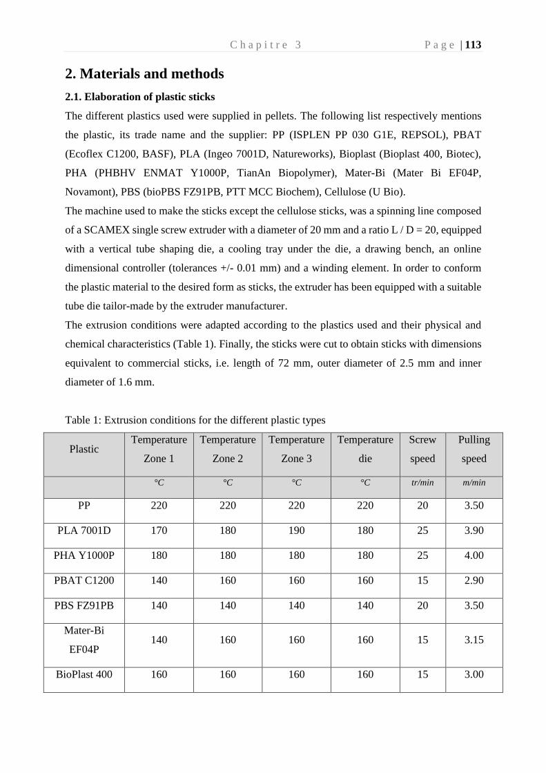
C h a p i t r e 3 P a g e | 113
2. Materials and methods
2.1. Elaboration of plastic sticks
The different plastics used were supplied in pellets. The following list respectively mentions
the plastic, its trade name and the supplier: PP (ISPLEN PP 030 G1E, REPSOL), PBAT
(Ecoflex C1200, BASF), PLA (Ingeo 7001D, Natureworks), Bioplast (Bioplast 400, Biotec),
PHA (PHBHV ENMAT Y1000P, TianAn Biopolymer), Mater-Bi (Mater Bi EF04P,
Novamont), PBS (bioPBS FZ91PB, PTT MCC Biochem), Cellulose (U Bio).
The machine used to make the sticks except the cellulose sticks, was a spinning line composed
of a SCAMEX single screw extruder with a diameter of 20 mm and a ratio L / D = 20, equipped
with a vertical tube shaping die, a cooling tray under the die, a drawing bench, an online
dimensional controller (tolerances +/- 0.01 mm) and a winding element. In order to conform
the plastic material to the desired form as sticks, the extruder has been equipped with a suitable
tube die tailor-made by the extruder manufacturer.
The extrusion conditions were adapted according to the plastics used and their physical and
chemical characteristics (Table 1). Finally, the sticks were cut to obtain sticks with dimensions
equivalent to commercial sticks, i.e. length of 72 mm, outer diameter of 2.5 mm and inner
diameter of 1.6 mm.
Table 1: Extrusion conditions for the different plastic types
Plastic Temperature
Zone 1
Temperature
Zone 2
Temperature
Zone 3
Temperature
die
Screw
speed
Pulling
speed
°C °C °C °C tr/min m/min
PP 220 220 220 220 20 3.50
PLA 7001D 170 180 190 180 25 3.90
PHA Y1000P 180 180 180 180 25 4.00
PBAT C1200 140 160 160 160 15 2.90
PBS FZ91PB 140 140 140 140 20 3.50
Mater-Bi
EF04P 140 160 160 160 15 3.15
BioPlast 400 160 160 160 160 15 3.00
C h a p i t r e 3 P a g e | 114
2.2. Incubation under natural marine conditions (40 days).
Each polymer type was incubated in aquariums with direct circulation to the sea, as previously
described (Dussud et al. 2018a). Briefly, we used 9 identical aquariums consisting in trays with
a 1.8 L capacity (Sodispan, Spain), in which 1.5 L of 200-microns filtered seawater was
continually renewed by direct pumping at 14 m depth in the Banyuls bay, close to the SOLA
marine observatory station (42°29’300 N – 03°08’700 E, France). A flow rate of 50 mL min-1
was chosen to ensure a sufficient renewal of natural bacteria (every 30 min) and a homogeneous
distribution of the plastic pieces in the aquariums during the entire experiment.
Each aquarium contained 0.5 cm polymer pieces of a single polymer (PP, PLA, PBS, PBAT,
Bioplast, Mater-Bi, PHBV or cellulose), with the exception of the “control aquarium” that only
contained the circulating seawater. On the 7th March 2018, pieces of each polymer incubated in
the aquariums were sampled after 7 (D7), 15 (D15), 30 (D30) and 40 days (D40). Aquariums
were kept in the dark to avoid UV-driven degradation of the polymers. Throughout the
incubation, seawater temperature (between 12.5 and 13.5°C) and salinity (38.5) were monitored
in the aquariums and they were similar to the data found at the SOLA observatory in the Banyuls
bay during this period (https://www.somlit.fr/).
2.3. Incubation in minimum medium with polymer sticks as sole carbon source (94 days).
After 40 days under natural marine conditions, the remaining individual pieces of each of the 8
polymer types were carefully collected with sterilized tweezers and placed in 4 ml flasks (ref.
294330, Interchim) containing 2 ml of minimum medium with no other carbon source than the
polymer. The composition of the minimum medium was modified from Fegatella et al., 1998
(Fegatella et al. 1998): NaCl, 420 mM ; Na2SO4, 28.8 mM; KCl, 9.39 mM ; NaBr, 0.84 mM ;
H3BO3, 0.485 mM ; MgCl2.6H2O, 0.546 mM ; CaCl2, 0.105 mM ; NH4Cl 9.35 mM ;
SrCl2.6H2O, 0.0638 mM ; NaF, 0.0714 mM ; NaNO3, 0.88 mM ; NaH2PO4.H2O, 0.036 mM ;
KH2PO4, 0.106 mM ; CuSO4·5H2O, 0.04 μM ; ZnSO4·7H2O 0.08 µM ; CoCl2·6H2O, 0.04 μM;
MnCl2·4H2O, 0.9 μM ; Na2MoO4·2H2O, 0.03 μM ; FeCl3 1.85 μM ; thiamine, 0.3 μM ; biotin,
2.1 nM ; and B12 vitamin, 0.317 nM. Pieces of each polymer type were sampled after 3
(D40+3), 7 (D40+7), 15 (D40+15) and 94 days (D40+94).
2.4. Scanning electron microscopy
Three pieces of each of the 8 polymer types were fixed at the beginning (D0) and at the end of
the experiment (D40+94) with the addition of 3% (v/v) glutaraldehyde (final concentration) and
C h a p i t r e 3 P a g e | 115
stored for at least one night at 4°C. Samples were then washed three times during 15 min in the
following solution: sodium cacodylate buffer, 0.4 M diluted to 0.2 M in minimum medium.
Then, fixation with osmium tetroxide (OsO4) at 1% was carried out for one hour at room
temperature.
Progressive dehydration in successive ethanol baths (70%, 95% and 100%) was applied during
30 minutes before analysis by scanning electron microscope. Samples were immersed in 100%
ethanol in a critical point desiccator tank (Balzers), and liquid CO2 was then gradually
introduced to replace ethanol. Samples were then heated and pressurized to allow the CO2 to
pass into the gaseous state bypassing its critical point (Temperature = 31.1 ° C; P = 73.8 bars)
and to avoid surface tensions and deformation. The CO2 gas was then removed, and dry samples
were collected. After dehydration, a thin conductive layer of Au-Pd (about 10nm thick) was
deposited by sputtering on the surface of the samples to allow electron microscope observation
(Hitachi SEM FEG S-4500 high vacuum).
2.5. Heterotrophic Bacterial Production
Bacterial production (BP) was measured on each type of polymer at each sampling time during
the colonization and biodegradation phases, using a modified protocol from van Wambeke et
al. (2009). Briefly, a cell detachment pre-treatment was applied using 3 cycles of 3 min
sonication bath (Deltasonic, France) followed by 3 min vortex at maximum speed (Skyline,
Elmi Ltd, Russia) as previously described (Dussud et al. 2018a). Immediately after cell-
detachment, 3H-leucine (specific activity 123.8 Ci mmol-1; Perkin Elmer) was added for each
sample, which consisted of 1.5 mL of sterilized seawater containing the plastic piece and it’s
the detached cells. Final leucine concentrations were 36 nmol L-1 (3H-leucine at 4 nmol L-1)
during the colonization phase and 150 nmol L-1 after the transfer to the minimum medium (3H-
leucine at 1 nmol L-1). Higher final leucine concentration was required to adapt to the increase
of BP in the minimum medium for some of the polymers, according to preliminary results (data
not shown). Final 3H-leucine concentration was 4.3 nmol.L-1 for seawater samples from the
control aquarium. All samples were treated in triplicate and incubated in the dark at in
situ temperature for 3 hours. We used the empirical conversion factor of 1.55 ng C.pmol−1 of
incorporated leucine to calculate BP (Simon and Azam 1989). Because exact surface of plastic
sticks was difficult to measure, each piece was weighted with precision balance of sensitivity
0,1 mg (Precisa 125A, Swiss Quality) and results were expressed in ng C.g-1.h-1.
C h a p i t r e 3 P a g e | 116
2.6. DNA Extraction, PCR and Sequencing
For each polymer type, individual pieces were taken at all sampling times and stored at -80°C
until DNA extraction. In parallel, 1 L seawater was sampled in the control aquarium and
successively filtered onto 3 and 0.2 μm pore size polycarbonate filters (47 mm diameter,
Nucleopore) and stored at -80°C until analysis. DNA extractions were performed on polymers
or filters using a classical phenol-chloroform method (Sauret et al. 2016) with slight
modifications of the method for polymer samples (Debeljak et al. 2016). Briefly, the same cell
detachment pre-treatment was used as for BP (see above) before enzymatic and chemical cell
lysis (incubation with 1 mg.mL-1 lysozyme at 37°C for 45 min followed by 1 h at 50°C with
0.2 mg.mL- 1 proteinase K and 1% SDS). DNA was quantified by spectrophotometry (Quant-
iT™ PicoGreen™ dsDNA Assay Kit, Invitrogen).
PCR amplification of the V4-V5 region was performed using the primers 515-F (5'-GTG YCA
GCM GCC GCG GTA A-3') and 926-R (5'-CCG YCA ATT YMT TTR AGT TT-3') (Fuhrman
et al. 1989), which are particularly well suited for marine samples (Parada et al. 2016).
Sequencing was performed on Illumina MiSeq by MrDNA (Molecular Research LP)
(Shallowater, TX, USA) and Integrated Microbiome Resource (IMR) (Dalhousie University,
Canada). The sequences were analysed using FROGS pipeline hosted in the Galaxy platform
(Escudié et al. 2018), following the guidelines given in the publication. Briefly, the forward
and reverse reads were assembled and sequences were clustered using the SWARM algorithm
(Mahé et al. 2014) using the aggregation distance of 3. Then, the chimeras were removed using
the FROGS Vsearch tool (Rognes et al. 2016). An additional filter step was used on the
abundance at 5.10-5 threshold to select the most relevant OTUs sequences. The taxonomic
affiliation was assigned by standard nucleotid BLAST (Camacho et al. 2009) using the SILVA
132 database 16S (Quast et al. 2013). Chloroplast, mitochondrial and eukaryotic sequences
were removed. All the 16S rRNA data are available in the NCBI SRA repository (accession
number PRJNA649857).
2.7. Statistical Analysis
The bacterial sequences of each sample were randomly resampled in the OTU file to allow
comparison and normalization based on the sample with the fewest sequences (n = 9,698).
These data sets were used to realize all the analyses performed using Rstudio software
(https://www.r-project.org/). The alpha-diversity indices (Chao1, Shannon, Evenness,
Simpson) and the Beta-diversity analysis (Bray-Curtis similarity) were calculated with the
phyloseq package (McMurdie and Holmes 2013).
C h a p i t r e 3 P a g e | 117
Differences between polymer types were tested using Anova test and confirmed by a post hoc
Tuckey test. The test was interpreted as significant if the p value was less than 0.05.
Dendrogram was obtained based on the dissimilarity matrix of Bray-Curtis and applying the
Ward algorithm (Ward 1963).
OTUs responsible for dissimilarities between pairs of clusters from the start and the end of
incubation in minimum medium were identified using similarity percentage (SIMPER,
PRIMER 6).
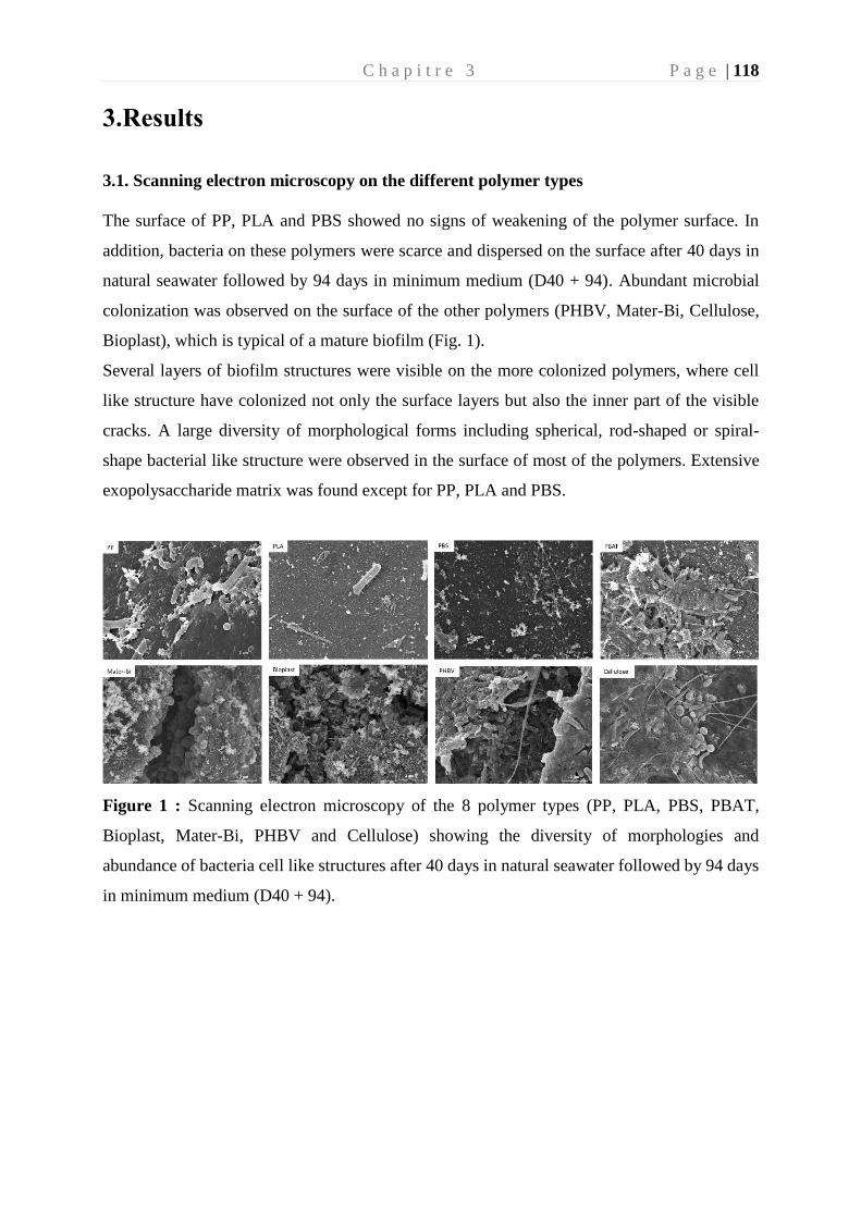
C h a p i t r e 3 P a g e | 118
3.Results
3.1. Scanning electron microscopy on the different polymer types
The surface of PP, PLA and PBS showed no signs of weakening of the polymer surface. In
addition, bacteria on these polymers were scarce and dispersed on the surface after 40 days in
natural seawater followed by 94 days in minimum medium (D40 + 94). Abundant microbial
colonization was observed on the surface of the other polymers (PHBV, Mater-Bi, Cellulose,
Bioplast), which is typical of a mature biofilm (Fig. 1).
Several layers of biofilm structures were visible on the more colonized polymers, where cell
like structure have colonized not only the surface layers but also the inner part of the visible
cracks. A large diversity of morphological forms including spherical, rod-shaped or spiral-
shape bacterial like structure were observed in the surface of most of the polymers. Extensive
exopolysaccharide matrix was found except for PP, PLA and PBS.
Figure 1 : Scanning electron microscopy of the 8 polymer types (PP, PLA, PBS, PBAT,
Bioplast, Mater-Bi, PHBV and Cellulose) showing the diversity of morphologies and
abundance of bacteria cell like structures after 40 days in natural seawater followed by 94 days
in minimum medium (D40 + 94).
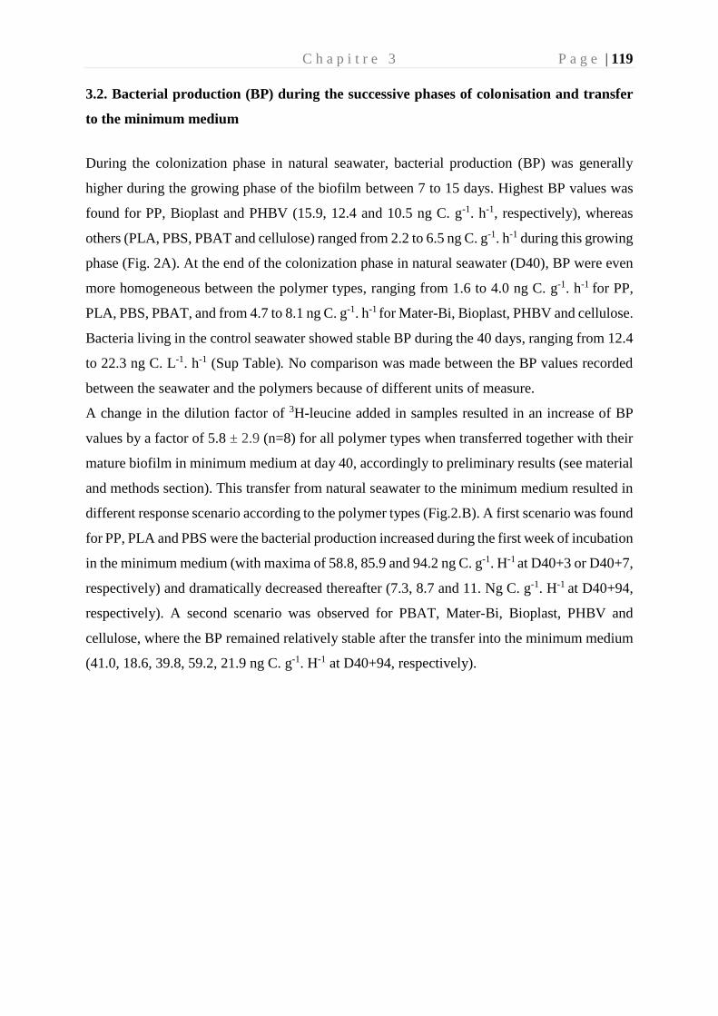
C h a p i t r e 3 P a g e | 119
3.2. Bacterial production (BP) during the successive phases of colonisation and transfer
to the minimum medium
During the colonization phase in natural seawater, bacterial production (BP) was generally
higher during the growing phase of the biofilm between 7 to 15 days. Highest BP values was
found for PP, Bioplast and PHBV (15.9, 12.4 and 10.5 ng C. g-1. h-1, respectively), whereas
others (PLA, PBS, PBAT and cellulose) ranged from 2.2 to 6.5 ng C. g-1. h-1 during this growing
phase (Fig. 2A). At the end of the colonization phase in natural seawater (D40), BP were even
more homogeneous between the polymer types, ranging from 1.6 to 4.0 ng C. g-1. h-1 for PP,
PLA, PBS, PBAT, and from 4.7 to 8.1 ng C. g-1. h-1 for Mater-Bi, Bioplast, PHBV and cellulose.
Bacteria living in the control seawater showed stable BP during the 40 days, ranging from 12.4
to 22.3 ng C. L-1. h-1 (Sup Table). No comparison was made between the BP values recorded
between the seawater and the polymers because of different units of measure.
A change in the dilution factor of 3H-leucine added in samples resulted in an increase of BP
values by a factor of 5.8 ± 2.9 (n=8) for all polymer types when transferred together with their
mature biofilm in minimum medium at day 40, accordingly to preliminary results (see material
and methods section). This transfer from natural seawater to the minimum medium resulted in
different response scenario according to the polymer types (Fig.2.B). A first scenario was found
for PP, PLA and PBS were the bacterial production increased during the first week of incubation
in the minimum medium (with maxima of 58.8, 85.9 and 94.2 ng C. g-1. H-1 at D40+3 or D40+7,
respectively) and dramatically decreased thereafter (7.3, 8.7 and 11. Ng C. g-1. H-1 at D40+94,
respectively). A second scenario was observed for PBAT, Mater-Bi, Bioplast, PHBV and
cellulose, where the BP remained relatively stable after the transfer into the minimum medium
(41.0, 18.6, 39.8, 59.2, 21.9 ng C. g-1. H-1 at D40+94, respectively).
C h a p i t r e 3 P a g e | 120
Figure 2 : Bacterial production (in ng C. g-1. H-1) (A) during the colonization phase in natural
seawater at days 7 (D7), 15 (D15) and 40 (D40) and (B) after the transfer to minimum medium
during 3 (D40+3), 7 (D40+7), 15 (D40+15), 30 (D40+30) and 94 days (D40+94). The vertical
bars represent the average of the bacterial production for each polymer (n=3) ± standard
deviation. Changes in BP values at D40 between A and B correspond to changes on dilution
factor of the 3H-leucine added (see material and methods section).
C h a p i t r e 3 P a g e | 121
3.3. Bacterial community structure, diversity indexes and taxonomy during the successive
phases of colonisation and transfer to the minimum medium
A total of 5,427,894 paired reads were acquired after sequencing all of the samples. After
normalisation based on the sample with the fewest merged sequences (n = 9,698), 3,772,255
sequence tags were selected belonging to 978 OTUs (0.03 distance threshold). Several distinct
sequencing of the seawater control at D40 failed for unknown reasons.
3.3.1. Changes in -diversity indices
-diversity was studied using Chao1 richness, Pielou eveness and Shannon diversity indexes.
Regardless the polymer types, the richness rapidly increased during the first days of
colonization until 15 days to reach between 120 and 384 estimated OTUs after 40 days (Table
S1). The transfer to the minimum medium did not influence the richness (+/- 50 estimated
species) for some polymer types including PP, PLA, PBS, Bioplast, but resulted in a decrease
for PBAT, Mater-Bi, PHBV and cellulose when comparing D40 and D40+94. We observed
that the Chao1 index was statistically linked to the polymer type after the transfer into the
minimum medium (Anova, pvalue = 0.0147).
Pielou eveness index showed relatively homogeneous diversity during the colonization phase,
with evenness > 0.5 for all polymers after 40 days (Table S1). A slight increase was found after
the transfer in the minimum medium for the PP, Bioplast, PHBV and cellulose, as correlated
with the Chao1 index (p<0.05), whereas it slightly decreased for PLA, PBS, PBAT and Mater-
Bi at the end of the incubation.
The resulting Shannon diversity was consistent with the Pielou and Chao1 indices. During the
colonization phase, a decrease in Shannon diversity index was found for the PP (2.4) due to its
decrease in evenness (Table 1) and an increase in diversity for all the other polymers at D40
(between 2.64 and 4.40). The transfer to the minimum medium resulted in an increase in
diversity until the end of the experiment (between 3.67 and 3.9 at D40+94) for PP, PHBV and
Cellulose according to the increase in evenness. Conversely, a decrease of Shannon diversity
(between 2.1 and 3.64) was caused by a decrease in richness for PBS, PBAT and Mater-Bi or
by a decrease in evenness for PLA and Bioplast.
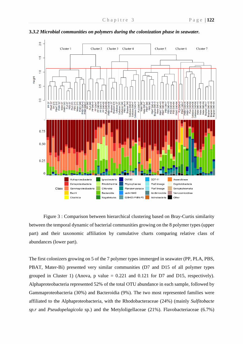
C h a p i t r e 3 P a g e | 122
3.3.2 Microbial communities on polymers during the colonization phase in seawater.
Figure 3 : Comparison between hierarchical clustering based on Bray-Curtis similarity
between the temporal dynamic of bacterial communities growing on the 8 polymer types (upper
part) and their taxonomic affiliation by cumulative charts comparing relative class of
abundances (lower part).
The first colonizers growing on 5 of the 7 polymer types immerged in seawater (PP, PLA, PBS,
PBAT, Mater-Bi) presented very similar communities (D7 and D15 of all polymer types
grouped in Cluster 1) (Anova, p value = 0.221 and 0.121 for D7 and D15, respectively).
Alphaproteobacteria represented 52% of the total OTU abundance in each sample, followed by
Gammaproteobacteria (30%) and Bacteroidia (9%). The two most represented families were
affiliated to the Alphaproteobacteria, with the Rhodobacteraceae (24%) (mainly Sulfitobacte
sp.r and Pseudopelagicola sp.) and the Metyloligellaceae (21%). Flavobacteriaceae (6.7%)
C h a p i t r e 3 P a g e | 123
(mainly Croceibacter sp. and Aquibacter sp.) and Nitrinocolaceae (mainly Neptunibacter sp.)
(5.5%) were also abundant during this stage. Bacteria growing on Bioplast and cellulose
grouped in a separated cluster (Cluster 3, Fig. 3), with a lower dominance of
Alphaproteobacteria (32%) and higher presence of Bacteroidia (25%), together with similar
dominance of Gammaproteobacteria (30%) as compared to the other polymer types (see above).
Alteromonadaceae family dominated (24.5%), represented mainly by the Alteromonas sp. and
Marinobacter sp. Flavobacteriaceae family represents 22% of the biofilm community, mainly
represented by the Wenyingzhuangia sp. Finally, like the other polymers, the Rhodobacteraceae
family was very abundant (21%), mainly represented by the genus Sulfitobacter sp.
Bacterial community structure generally diverged after the growing phase of the biofilm, were
distinct mature biofilm appeared between polymer types after 40 days in seawater (D40). PP
and PLA biofilm (cluster 2) were dominated by Alphaproteobacteria (Rhodobacteraceae) and
Bacteroidia (Flavobacteriaceae). PBS and PBAT (Cluster 5) were dominated by
Alphaproteobacteria (mainly Rhodobacteraceae) and Gammaproteobacteria (mainly
Solimonadaceae). Bioplast and cellulose (Cluster 4) were dominated by Alphaproteobacteria
(mainly Hyphomonadaceae) and Gammaproteobacteria (mainly Alteromonadaceae).
PHBV was dominated by Alphaproteobacteria (Rhodobacteraceae) followed by
Gammaproteobacteria (Cellvibrionaceae) and finally Bacteroidia (Flavobacteriaceae). As for
Mater-Bi, its mature biofilm was dominated by Alphaproteobacteria (Rhodobacteraceae),
followed by Gammaproteobacteria (Spongiibacteraceae) and bacteroidia (Flavobacteriaceae).
3.3.3. Biofilm specialization after transfer to minimum medium
The transfer from natural seawater to the minimum medium resulted in bacterial
community changes following three different scenarii according to the polymer types. First,
bacterial community structure of PP (cluster 2), PBAT and PBS (cluster 5) remained stable and
distinct during the 94 days of incubation. The second scenario was shared between most of the
polymers, where the initial biofilm established during the colonization phase remained stable
for one week after the transfer in minimal medium (Day 40+7) and then change to another
community that remained stable until the end of the experiment (Day 40+94). This was the case
for Bioplast, MaterBi, PHBV and Cellulose that (from clusters 1, 4, 5 to cluster 7), even if each
of them grouped in distinct sub-clusters. A third scenario was found for PLA which presented
a chaotic dispersion of the bacterial communities among all other clusters, even if the
community at the end of the experiment clustered together in the cluster 7, with three sub-
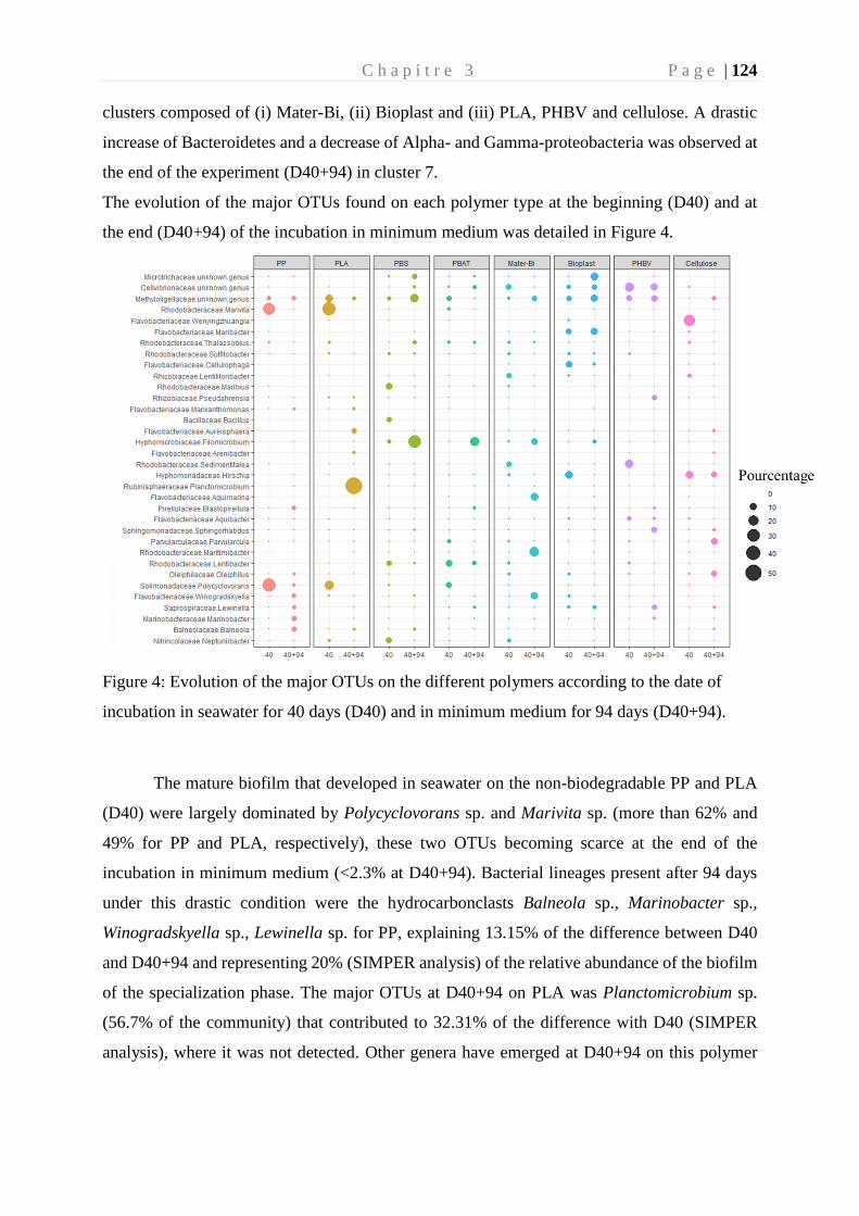
C h a p i t r e 3 P a g e | 124
clusters composed of (i) Mater-Bi, (ii) Bioplast and (iii) PLA, PHBV and cellulose. A drastic
increase of Bacteroidetes and a decrease of Alpha- and Gamma-proteobacteria was observed at
the end of the experiment (D40+94) in cluster 7.
The evolution of the major OTUs found on each polymer type at the beginning (D40) and at
the end (D40+94) of the incubation in minimum medium was detailed in Figure 4.
Figure 4: Evolution of the major OTUs on the different polymers according to the date of
incubation in seawater for 40 days (D40) and in minimum medium for 94 days (D40+94).
The mature biofilm that developed in seawater on the non-biodegradable PP and PLA
(D40) were largely dominated by Polycyclovorans sp. and Marivita sp. (more than 62% and
49% for PP and PLA, respectively), these two OTUs becoming scarce at the end of the
incubation in minimum medium (<2.3% at D40+94). Bacterial lineages present after 94 days
under this drastic condition were the hydrocarbonclasts Balneola sp., Marinobacter sp.,
Winogradskyella sp., Lewinella sp. for PP, explaining 13.15% of the difference between D40
and D40+94 and representing 20% (SIMPER analysis) of the relative abundance of the biofilm
of the specialization phase. The major OTUs at D40+94 on PLA was Planctomicrobium sp.
(56.7% of the community) that contributed to 32.31% of the difference with D40 (SIMPER
analysis), where it was not detected. Other genera have emerged at D40+94 on this polymer
C h a p i t r e 3 P a g e | 125
such as Arenibacter sp., Aureisphaera sp. and Marixanthomonas sp. representing almost 11%
of the relative abundance at the end of incubation.
The mature biofilm on PBS (D40) was dominated by Maribius sp., Neptuniibacter sp.
and Bacillus sp. representing nearly 21% of the total OTUs and these 3 genera represented only
0.64% at D40+94. At the end of the experiment (D40+94), the biofilm was dominated by
Filomicrobium sp. (31%), Thalassobius sp. (3%) and Bythopirellula sp. (2%) explaining about
23% of the disparity with the J40. These three bacterial genera were scarce until the transfer
into the minimum medium (3%).
The dominant OTUs found in the mature biofilm (D40) on PBAT were
Rhodobacteraceae at 20.37% (Marivita sp., Lentibacter sp.), Solimonadaceae at 7.1 %
(Lewinella sp.) and Methylologellaceae at 5.6 % and then moved to Saprospiraceae (42.5%)
and Filomicrobium sp. (15.8%) at D40+94. These two OTUs alone explained 34.6% of the
difference between the 2 dates. As already found for PP and PLA, Polycyclovorans sp. was
very abundant at D40 (7.1%) and were no longer detected at D40+94.
Incubation in minimum medium with Mater-Bi as sole carbon source resulted in the
selection of Aquimarina sp., Winogradskyella sp., Maritimibacter sp. and Filomicrobium sp.
that represented nearly 48% of the total OTUs at D40+94, but less than 2% at D40. These four
genera explained 27.8% of the difference between the 2 dates based on SIMPER analysis.
The OTUs selected at D40+94 for Bioplast were dominated by Microtrichaceae (12%), while
it belonged to the rare biosphere at D40 (<0.05%). Other genera emerged (>9%) including
Aliiglaciecola sp., Filomicrobium sp., Flexitrhrix sp., while representing low abundance at D40
(<0.4%). In the opposite, Alteromonas sp. and Oleibacter sp. were abundant (8.8%) at D40 and
became rare at D40+94 (<0.05%). Selection on minimum medium with PHBV as sole carbon
source showed the emergence of Sphingorhabdus sp., Lewinella sp., Pseudahrensia sp. and
Marinobacter sp. (>21% after D40+94 and <0.6% at D40), which explained 17.26% of the
difference between D40 and D40+94 according to SIMPER analysis (Fig.4), whereas the
dominant Sedimentitalea sp., Thalassotalea sp. and Lentibacter sp. at D40 (20% of the total
OTUs) represented less than 0.1% at D40+94.
Cellulose selected Parvularcula sp., Oleiphilus sp., Pirellula sp. and Phycisphaera sp.
at D40+94 (21.5 % of the total OTUs at D40+94 compared to <1.5% at D40), which explained
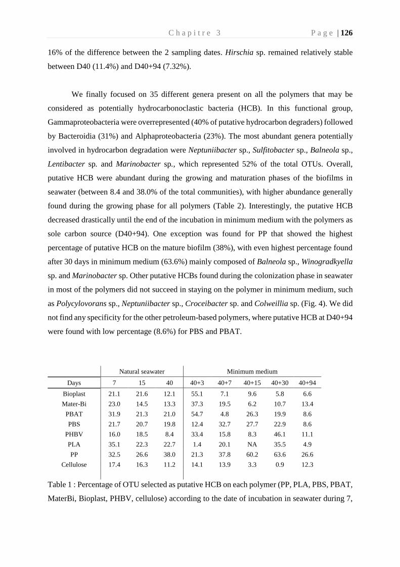
C h a p i t r e 3 P a g e | 126
16% of the difference between the 2 sampling dates. Hirschia sp. remained relatively stable
between D40 (11.4%) and D40+94 (7.32%).
We finally focused on 35 different genera present on all the polymers that may be
considered as potentially hydrocarbonoclastic bacteria (HCB). In this functional group,
Gammaproteobacteria were overrepresented (40% of putative hydrocarbon degraders) followed
by Bacteroidia (31%) and Alphaproteobacteria (23%). The most abundant genera potentially
involved in hydrocarbon degradation were Neptuniibacter sp., Sulfitobacter sp., Balneola sp.,
Lentibacter sp. and Marinobacter sp., which represented 52% of the total OTUs. Overall,
putative HCB were abundant during the growing and maturation phases of the biofilms in
seawater (between 8.4 and 38.0% of the total communities), with higher abundance generally
found during the growing phase for all polymers (Table 2). Interestingly, the putative HCB
decreased drastically until the end of the incubation in minimum medium with the polymers as
sole carbon source (D40+94). One exception was found for PP that showed the highest
percentage of putative HCB on the mature biofilm (38%), with even highest percentage found
after 30 days in minimum medium (63.6%) mainly composed of Balneola sp., Winogradkyella
sp. and Marinobacter sp. Other putative HCBs found during the colonization phase in seawater
in most of the polymers did not succeed in staying on the polymer in minimum medium, such
as Polycylovorans sp., Neptuniibacter sp., Croceibacter sp. and Colweillia sp. (Fig. 4). We did
not find any specificity for the other petroleum-based polymers, where putative HCB at D40+94
were found with low percentage (8.6%) for PBS and PBAT.
Natural seawater Minimum medium
Days 7 15 40 40+3 40+7 40+15 40+30 40+94
Bioplast 21.1 21.6 12.1 55.1 7.1 9.6 5.8 6.6
Mater-Bi 23.0 14.5 13.3 37.3 19.5 6.2 10.7 13.4
PBAT 31.9 21.3 21.0 54.7 4.8 26.3 19.9 8.6
PBS 21.7 20.7 19.8 12.4 32.7 27.7 22.9 8.6
PHBV 16.0 18.5 8.4 33.4 15.8 8.3 46.1 11.1
PLA 35.1 22.3 22.7 1.4 20.1 NA 35.5 4.9
PP 32.5 26.6 38.0 21.3 37.8 60.2 63.6 26.6
Cellulose 17.4 16.3 11.2 14.1 13.9 3.3 0.9 12.3
Table 1 : Percentage of OTU selected as putative HCB on each polymer (PP, PLA, PBS, PBAT,
MaterBi, Bioplast, PHBV, cellulose) according to the date of incubation in seawater during 7,
C h a p i t r e 3 P a g e | 127
15 and 40 days (D40) followed by the transfer in minimum medium during 3 (40+3), 7 (40+7),
15 (40+15), 30 (40+30) and 94 days (40+94).
C h a p i t r e 3 P a g e | 128
4.Discussion
4.1. Formation of mature biofilms on all polymer types in seawater
4.1.1. Aquarium with direct circulation to the sea mimics the natural conditions of the North
Western Mediterranean Sea.
The colonization of the different polymer types was feasible thanks to distinct
incubation in aquarium with every 30 min renewal of seawater, which presented similar
hydrological conditions found at the SOLA observatory in the Banyuls Bay (NW
Mediterranean Sea, France). During the first 40 days of incubation in seawater, temperature
(13°C ± 0.5°C), chlorophyll a and nutrients (data not shown) were classical to generally
observed at the long-term SOLA observatory station (Ghiglione et al. 2005; Lambert et al.
2019). Bacterial heterotrophic activity in the seawater, measured by 3H-leucine incorporation
reached 22.3 ng.C.L-1.h-1, which was similar to what was generally reported in situ in the NW
Mediterranean Sea (Pulido-Villena et al. 2012). These results showed that the incubation in our
aquarium with direct circulation to the sea allowed to reproduce the natural conditions during
our incubation, thus making extrapolation of our results to natural seawater possible.
4.1.2 Growing and maturation phases of the biofilms on all the polymer types in seawater
In agreement with patterns observed in other study (Salta et al. 2013), we found a succession
of growing and maturation phases of the biofilms for all the polymer types during the 40 days
of incubation in natural seawater. Each phase was linked with specific bacterial communities
that were similar during the growing phase but became specific to the polymer types when the
mature biofilm formed. This result is in agreement with previous studies conducted in the same
region (Dussud et al. 2018).
Bacterial heterotrophic activity peaked during the growing phase of the biofilm in all polymer
types (either after 7 or 15 days). The growing bacterial communities were very similar
regardless polymer types, except for Bioplast and Cellulose that clustered in another group.
This result was surprising considering the distinct chemical polymer compositions and surface
C h a p i t r e 3 P a g e | 129
properties of the plastics used in our study. Early stages of biofilm formation have been depicted
as strongly dependent on substrate properties of polymers, including
hydrophobicity/hydrophilicity, structure and roughness (Lorite et al. 2011). This was not
obvious for immerged plastics in seawater, since our results were similar with other studies
which did not find distinct communities during the first weeks of biofilm formation on different
plastic types (Harrison et al. 2014; De Tender et al. 2017; Dussud et al. 2018a). This growing
phase was mainly dominated by Proteobacteria (84% of the total abundance on all polymer),
followed by Bacteroidetes (12.5%). The dominance of Proteobacteria during the first stage of
colonization has already been described for the first stage of colonization on the plastisphere
(Jacquin et al. 2019). The main contributors of the Proteobacteria were Alphaproteobacteria
(47.14%, mainly Methyloligellaceae and Rhodobacteraceae) and Gammaproteobacteria
(31.46%). The Methyloligellaceae has already been isolated on other types of plastic such as
polyamide, polycarbonate or PP (Feng et al. 2020). Polymers were mainly colonized by
members of the Rhodobacteraceae during this colonization phase and specialization phase of
incubation. This family has been depicted as a key taxonomic group in marine biofilm
formation (Elifantz et al. 2013; Kviatkovski and Minz 2015; Roager and Sonnenschein 2019),
which explained its abundant presence on the tested polymers. During the colonization phase,
the family of Flavobacteriaceae and particularly the genus Neptuniibacter sp., generally able to
degrade hydrocarbons, was found in abundance on PLA, PBS and Mater-Bi (Gutierrez 2019).
This result is consistent with a previous study which showed its presence only during the phase
of primary colonization of polymers in similar conditions (Dussud et al. 2018a).
In all polymer types, the mature biofilm formed after 40 days of incubation in seawater showed
lower values of bacterial heterotrophic activity compared to the growing phase. This is
consistent with observations made in the unique study measuring the bacterial heterotrophic
activities on plastisphere (Dussud et al. 2018a). It can be explained by decreasing availability
of labile inorganic and organic matter when the biofilm become mature, and also by the
selection of new communities developing over slower-growing metabolic strategies (Lorite et
al. 2011). Diversity in mature biofilms became specific for each polymer, which is consistent
with observations made in the natural environment (Webb et al. 2012; De Tender et al. 2017;
Pinto et al. 2019). The evenness at the microbial community level increased for 40 days for all
polymers except PP and PBAT. This decline in evenness is mainly explained by a decrease in
diversity (Shannon) and to a lesser extent by the decrease in wealth (Chao1).
C h a p i t r e 3 P a g e | 130
4.2. Convergent signs of biodegradability for some polymers
As recommended in several standard tests, the biodegradability of the polymers was tested after
transferring each polymer type into a minimum medium with no other source of carbon and
energy than the polymer (ISO 18830:2016, ISO 19679:2016, ASTM D6691-09). As suggested
in these tests, we used the cellulose as positive control and we considered PP as negative control
since its biodegradation was previously depicted to take years or decades in nature (Barnes et
al. 2009).
When standards authorized various types of bacterial inoculum from unique or multiple
environmental sources (Harrison et al. 2018; Jacquin et al. 2019), we used the mature biofilm
that developed in each polymer types during 40 days incubation in natural seawater in order to
be as close as possible to the natural conditions. After 94 days in minimum medium, SEM
observation showed no surface modifications for PP, PLA and PBS, whereas PBAT, Bioplast,
Mater-Bi, PHBV and cellulose exhibited observable morphological alterations, with clear
evidences of swelling and erosion. Visible alterations of the plastic surfaces are considered to
be suitable signs of biodegradation, even if they are not sufficient by themselves (Zettler 2013).
Plastics, such as PHBV, are known to undergo erosion mechanism by numerous exo-enzymatic
actions (amylases, proteases, nucleases), leading to weakening of the external structure of
polymers (Deroiné et al. 2014, 2015). In a previous study, it has been shown that PHAs degrade
faster in seawater than other polymers such as PBS, PBAT or PLA (Sashiwa et al. 2018).
In parallel, SEM observations at the end of the experiment showed dispersed and isolated
bacteria for PP, PLA and PBS, whereas dense biofilms with several layers and important
extracellular matrix were found for all the other polymers. This is another sign of biofilm
capability to use polymers as sole carbon source for growth. PLA was found to degrade very
slowly in seawater and remained stable at room temperature (Deroiné et al. 2014). PBS
degradation was found to increase with temperature, whereas high salinity slowed down its
degradation (da Silva Dutra et al. 2019).
The same conclusions were also found when using radio-labelled leucine incorporation as a
proxy of bacterial biodegradability in our conditions. This technique has been used for decades
as the most sensitive methodology to measure microbial heterotrophic activity and growth in
seawaters (Kirchman et al. 1985). It has been successfully used to prove the biodegradability
of several substrates, such as antifouling biocides (Maraldo and Dahllöf 2004), hydrocarbons
(Peng et al. 2020), plasticizers and flame retardants (Vila-Costa et al. 2019). This technique has
never been used so far to prove polymer biodegradability in seawater, probably because of the
C h a p i t r e 3 P a g e | 131
lack of studies in marine environments. It is noteworthy that respirometry analysis generally
made by titrimetric methods for O2 (Montgomery et al. 1964) or CO2 measurements (Bartha
and Pramer 1965) generally limit the number of samples analyzed since they are particularly
time consuming compared to measurement of bacterial production by 3H-leucine incorporation.
(Kirchman 2001). Following the kinetics of microbial activity is also an important aspect that
is not necessarily taken into consideration in standard tests and can result in misinterpretation.
We showed here that bacteria living in the biofilm can continue to be active for several days
after transfer under minimum medium with no other carbon source, as it is the case for PP, PLA
and PBS that showed bacterial heterotrophic activities during one or two weeks before falling-
off. Such activity could be misinterpreted as polymer biodegradation if integrative
measurements would be taken only at the end of the experiment, as it is authorized in current
standard tests. For PBAT, Bioplast, Mater-Bi, PHBV and cellulose, the kinetics of bacterial
heterotrophic activity remained rather stable after 15 days in minimum medium and until the
end of the experiment (94 days). Taken together with congruent results from SEM observations,
we consider that these latter polymers were exposed to microbial attack that participated to their
biodegradation. It has already been shown on PBAT (Weng et al. 2013; Saadi et al. 2013),
Mater-Bi (Kim et al. 2000), Bioplast (https://fr.biotec.de/), PHBV (Weng et al. 2010; Arcos-
Hernandez et al. 2015) and Cellulose (Salehpour et al. 2018), which were all found to be
biodegradable in soil or compost environments, but this is the first study to give indication of
their biodegradability by natural complex communities in seawater. It is interesting to note that
PLA, a polymer considered to be compostable and biodegradable in soil and compost (Ren et
al. 2007), did not show sign of biodegradation under our marine experimental conditions.
4.3. Identification of putative biodegraders of the set of polymers.
According to the SEM and heterotrophic bacterial activity measurements, we focused on the
OTUs selected at the end of the incubation in minimum medium on the polymers that showed
clear signs of biodegradability under our experimental conditions (PBAT, Bioplast, Mater-Bi,
PHBV and cellulose). The presence of complex communities living on such strict conditions
strongly suggest that they were member of putative biodegraders that may interact to each other
to perform all or some part of the degradation until the ultimate mineralization in CO2 (Dussud
and Ghiglione 2014). We are aware that part of the identified OTUs may not be involved in the
polymer degradation but rather opportunists that may survive in the biofilm (Sauret et al. 2014).
Our original procedure allowed to present here a list of putative OTUs selected from a mature
C h a p i t r e 3 P a g e | 132
biofilm grown under natural seawater conditions, which may be involved in biodegradability
of the polymers.
On the PBAT, we found Thalassobius sp. as dominant OTUs after 94 days in minimum medium
with the mature biofilm and its polymer as sole carbon source. This genus has been already
shown as potentially capable of degrading the aromatic hydrocarbons in seawater (Rodrigo-
Torres et al. 2017) and found in the oil polluted waters of the Gulf of Mexico (Liu et al. 2017).
Filomicrobium sp. were also dominant at the end of the experiment, already observed in other
marine environment (Wu et al. 2009), as well as Prevotella sp. and Streptococcus sp. known
as potential pathogenic genera (Fittipaldi et al. 2012; Larsen et al. 2017). None of them were
previously identified as potential PBAT degraders.
The bacteria growing with Mater-Bi as sole carbon source was dominated by Filomicrobium
sp., already identified on the plastisphere of PE and PET (Muthukrishnan et al. 2018). Other
groups were Winogradskyella sp., Maritimibacter sp., Aquimarina sp. and Halomonas sp.,
which were previously described as putative HCB (Liu et al. 2017, Mnif et al. 2009). None of
them were previously identified as potential Mater-Bi degraders.
Bacteria selected on Bioplast at the end of the experiment included Lewinella sp., already
identified on the plastisphere of petroleum-based PP, PE and PET, as well as on biobased PHBH
films (Morohoshi et al. 2018; Roager and Sonnenschein 2019). Other OTUs belonging to
putative HCB were Croceitalea sp., Hirschia sp., Maribacter sp. and Cellulophaga sp. None
of them were previously identified as potential Bioplast degraders.
Microbial communities selected after 94 days on PHBV as sole carbon source such as
Marinobacter sp. and Alteromonas sp. were already shown for their ability to degrade the
PHBV (Mergaert et al. 1995; Martinez Tobon 2019). It is noteworthy that Marinobacter sp.
genera contains well-known HCB capable of degrading Benzene, Toluene, Ethylbenzene and
Xylene (BTEX) (Li et al. 2012), long-chain alkans (Grimaud et al. 2012; Grimaud et al. 2012),
polycyclic aromatic hydrocarbons (Gao et al. 2013; Cui et al. 2016), chlorinated solvents (Inoue
et al. 2020), phenol (Zhou et al. 2020) and PHA (Kasuya et al. 2000; Martinez Tobon 2019).
Finally, it is noteworthy that putative HCB were very abundant during the formation of the
biofilm in seawater (from 8 to 35% of the total OTUs) and diminished after the selection in
minimum medium with the polymer as sole carbon source (from 6 to 12% of the total OTUs).
One exception was found for the PP that always present high percentage of putative HCB (38%
during the biofilm formation and 27% at the end of the experiment). We could have expected
an increase in this functional group during the selection under minimum medium, since several
authors proposed that the HCB are certainly involved in plastic biodegradation (Bryant et al.
C h a p i t r e 3 P a g e | 133
2016; Pollet et al. 2018; Amaral-Zettler et al. 2020). Here we showed that some of HCB may
be effectively involved in biodegradation, but they were not a majority for polymer that showed
biodegradation under our experimental conditions. If the HCB were dominant on PP, it was not
the case on PBS and PBAT, thus suggesting that the presence of HCB on the plastisphere is not
particularly linked to petroleum-based polymers. HCB generally present a high capability to
attached to particles and to live in a biofilm (Grimaud et al. 2012), two factors that may be more
important than their ability to degrade either the hydrocarbon than can attached to plastics or
the plastic itself.
Concluding remarks
Decisions taken by the government to reduce the use of single-use plastics and promote
biodegradable polymers lead to finding substitutes for conventional plastics. Here we present
that polymers such as PHBV, Mater-BI, Bioplast, PBAT and cellulose are good candidates for
alternatives to non-biodegradable plastics. It is noteworthy that the bio-based polymer PLA
showed no signs of biodegradation under our marine conditions, whereas the petroleum-based
polymer PBAT showed significant evidence of biodegradation. The current biodegradation
standards to prove biodegradability of plastics in the marine environment are mainly based on
respirometry methods, considered to be insufficient by the scientific community for a good
characterization of this complex process (Harrison et al. 2018). Here, we propose an innovative
experimental design using a natural inoculum for biodegradation tests, which takes into account
the formation of a natural mature biofilm on plastics as inoculum that is crucial to mimic natural
conditions. We also recommend the use of the 3H-Leucine incorporation method that is
classically used in marine microbial ecology (Kirchman et al. 1985) as a powerful tool to
supplement the material- and time-consuming respirometry tests (O2 and CO2). We are aware
that this study only takes into account the polymer and not include the additives that are
classically part of the composition of the plastic items found in the environment. Further studies
are needed to evaluate the toxicity of the polymers and their degradation products, as well as
the release of additives generally supplemented by manufacturers in the microplastic
composition.
C h a p i t r e 3 P a g e | 134
Reference
Amaral-Zettler LA, Zettler ER, and Mincer TJ. 2020. Ecology of the plastisphere. Nat Rev
Microbiol 18: 139–51.
Arcos-Hernandez M, Montano-Herrera L, Janarthanan OM, et al. 2015. Value-added
bioplastics from services of wastewater treatment. Water Practice & Technology 10:
546.
Barnes DKA, Galgani F, Thompson RC, and Barlaz M. 2009. Accumulation and
fragmentation of plastic debris in global environments. Philosophical Transactions of
the Royal Society B: Biological Sciences 364: 1985–98.
Bartha R and Pramer D. 1965. Features of a Flask and Method for Measuring the Persistence
and Biological Effects of Pesticides in Soil. Soil Science 100: 68–70.
Bryant JA, Clemente TM, Viviani DA, et al. 2016. Diversity and Activity of Communities
Inhabiting Plastic Debris in the North Pacific Gyre. mSystems 1.
Camacho C, Coulouris G, Avagyan V, et al. 2009. BLAST+: architecture and applications.
BMC Bioinformatics 10: 421.
Cui Z, Gao W, Xu G, et al. 2016. Marinobacter aromaticivorans sp. nov., a polycyclic
aromatic hydrocarbon-degrading bacterium isolated from sea sediment. International
Journal of Systematic and Evolutionary Microbiology, 66: 353–9.
Dang H and Lovell CR. 2000. Bacterial Primary Colonization and Early Succession on
Surfaces in Marine Waters as Determined by Amplified rRNA Gene Restriction
Analysis and Sequence Analysis of 16S rRNA Genes. Appl Environ Microbiol 66:
467–75.
De Tender C, Devriese LI, Haegeman A, et al. 2017. Temporal Dynamics of Bacterial and
Fungal Colonization on Plastic Debris in the North Sea. Environ Sci Technol 51:
7350–60.
Debeljak P, Pinto M, Proietti M, et al. 2016. Extracting DNA from ocean microplastics: A
method comparison study. Analytical methods 9.
Deroiné M, César G, Le Duigou A, et al. 2015. Natural Degradation and Biodegradation of
Poly(3-Hydroxybutyrate-co-3-Hydroxyvalerate) in Liquid and Solid Marine
Environments. J Polym Environ 23: 493–505.
Deroiné M, Le Duigou A, Corre Y-M, et al. 2014a. Accelerated ageing and lifetime
prediction of poly(3-hydroxybutyrate-co-3-hydroxyvalerate) in distilled water.
Polymer Testing 39: 70–8.
Deroiné M, Le Duigou A, Corre Y-M, et al. 2014b. Accelerated ageing of polylactide in
aqueous environments: Comparative study between distilled water and seawater.
Polymer Degradation and Stability 108: 319–29.
C h a p i t r e 3 P a g e | 135
Dussud C, Hudec C, George M, et al. 2018a. Colonization of Non-biodegradable and
Biodegradable Plastics by Marine Microorganisms. Front Microbiol 9.
Dussud C, Meistertzheim AL, Conan P, et al. 2018b. Evidence of niche partitioning among
bacteria living on plastics, organic particles and surrounding seawaters. Environmental
Pollution 236: 807–16.
Eich A, Mildenberger T, Laforsch C, and Weber M. 2015. Biofilm and Diatom Succession on
Polyethylene (PE) and Biodegradable Plastic Bags in Two Marine Habitats: Early
Signs of Degradation in the Pelagic and Benthic Zone? PLoS One 10.
Elifantz H, Horn G, Ayon M, et al. 2013. Rhodobacteraceae are the key members of the
microbial community of the initial biofilm formed in Eastern Mediterranean coastal
seawater. FEMS Microbiol Ecol 85: 348–57.
Eriksen M, Lebreton LCM, Carson HS, et al. 2014. Plastic Pollution in the World’s Oceans:
More than 5 Trillion Plastic Pieces Weighing over 250,000 Tons Afloat at Sea. PLOS
ONE 9: e111913.
Erni-Cassola G, Wright RJ, Gibson MI, and Christie-Oleza JA. 2019. Early Colonization of
Weathered Polyethylene by Distinct Bacteria in Marine Coastal Seawater. Microb
Ecol.
Escudié F, Auer L, Bernard M, et al. 2018. FROGS: Find, Rapidly, OTUs with Galaxy
Solution. Bioinformatics 34: 1287–94.
Fegatella F, Lim J, Kjelleberg S, and Cavicchioli R. 1998. Implications of rRNA operon copy
number and ribosome content in the marine oligotrophic ultramicrobacterium
Sphingomonas sp. strain RB2256. Appl Environ Microbiol 64: 4433–8.
Feng L, He L, Jiang S, et al. 2020. Investigating the composition and distribution of
microplastics surface biofilms in coral areas. Chemosphere 252: 126565.
Fittipaldi N, Segura M, Grenier D, and Gottschalk M. 2012. Virulence factors involved in the
pathogenesis of the infection caused by the swine pathogen and zoonotic agent
Streptococcus suis. Future Microbiology 7: 259–79.
Fuhrman JA, Sleeter TD, Carlson CA, and Proctor LM. 1989. Dominance of bacterial
biomass in the Sargasso Sea and its ecological implications. Marine Ecology Progress
Series 57: 207–17.
Gao W, Cui Z, Li Q, et al. 2013. Marinobacter nanhaiticus sp. nov., polycyclic aromatic
hydrocarbon-degrading bacterium isolated from the sediment of the South China Sea.
Antonie van Leeuwenhoek 103: 485–91.
Ghiglione J-F, Larcher M, and Lebaron P. 2005. Spatial and temporal scales of variation in
bacterioplankton community structure in the NW Mediterranean Sea. Aquatic
Microbial Ecology 40: 229–40.
Grimaud R, Ghiglione J-F, Cagnon C, et al. 2012. Genome sequence of the marine bacterium
Marinobacter hydrocarbonoclasticus SP17, which forms biofilms on hydrophobic
organic compounds. Journal of Bacteriology 194: 3539–40.
C h a p i t r e 3 P a g e | 136
Gutierrez T. 2019. Occurrence and Roles of the Obligate Hydrocarbonoclastic Bacteria in the
Ocean When There Is No Obvious Hydrocarbon Contamination. In: McGenity TJ
(Ed). Taxonomy, Genomics and Ecophysiology of Hydrocarbon-Degrading Microbes.
Cham: Springer International Publishing.
Haider T, Völker C, Kramm J, et al. 2018. Plastics of the Future? The Impact of
Biodegradable Polymers on the Environment and on Society. Angewandte Chemie
International Edition 58.
Harrison JP, Boardman C, O’Callaghan K, et al. 2018. Biodegradability standards for carrier
bags and plastic films in aquatic environments: a critical review. Royal Society Open
Science 5: 171792.
Harrison JP, Schratzberger M, Sapp M, and Osborn AM. 2014. Rapid bacterial colonization
of low-density polyethylene microplastics in coastal sediment microcosms. BMC
Microbiology 14: 232.
Inoue Y, Fukunaga Y, Katsumata H, et al. 2020. Aerobic degradation of cis-dichloroethene
by the marine bacterium Marinobacter salsuginis strain 5N-3. The Journal of General
and Applied Microbiology advpub.
Jacquin J, Cheng J, Odobel C, et al. 2019. Microbial Ecotoxicology of Marine Plastic Debris:
A Review on Colonization and Biodegradation by the “Plastisphere.” Front Microbiol
10.
Kang H, Kim H, Joung Y, and Joh K. 2017. Winogradskyella maritima sp. nov., isolated from
seawater. International Journal of Systematic and Evolutionary Microbiology, 67:
3840–5.
Kasuya K, Mitomo H, Nakahara M, et al. 2000. Identification of a marine benthic P(3HB)-
degrading bacterium isolate and characterization of its P(3HB) depolymerase.
Biomacromolecules 1: 194–201.
Kim M-N, Lee A-R, Yoon J-S, and Chin I-J. 2000. Biodegradation of poly(3-
hydroxybutyrate), Sky-Green® and Mater-Bi® by fungi isolated from soils. European
Polymer Journal 36: 1677–85.
Kirchman D. 2001. Measuring bacterial biomass production and growth rates from leucine
incorporation in natural aquatic environments. In: Methods in Microbiology.
Academic Press.
Kirchman D, K’nees E, and Hodson R. 1985. Leucine incorporation and its potential as a
measure of protein synthesis by bacteria in natural aquatic systems. Appl Environ
Microbiol 49: 599–607.
Kviatkovski I and Minz D. 2015. A member of the Rhodobacteraceae promotes initial biofilm
formation via the secretion of extracellular factor(s). Aquatic Microbial Ecology 75:
155–67.
Lambert S, Tragin M, Lozano J-C, et al. 2019. Rhythmicity of coastal marine picoeukaryotes,
bacteria and archaea despite irregular environmental perturbations. ISME J 13: 388–
401.
C h a p i t r e 3 P a g e | 137
Larsen JM. 2017. The immune response to Prevotella bacteria in chronic inflammatory
disease. Immunology 151: 363–74.
Lebaron P, Ghiglione J-F, Fajon C, et al. 1998. Phenotypic and genetic diversity within a
colony morphotype. FEMS Microbiol Lett 160: 137–43.
Li H, Zhang Q, Wang X-L, et al. 2012. Biodegradation of benzene homologues in
contaminated sediment of the East China Sea. Bioresource Technology 124: 129–36.
Liu J, Bacosa HP, and Liu Z. 2017. Potential Environmental Factors Affecting Oil-Degrading
Bacterial Populations in Deep and Surface Waters of the Northern Gulf of Mexico.
Front Microbiol 7.
Lobelle D and Cunliffe M. 2011. Early microbial biofilm formation on marine plastic debris.
Marine Pollution Bulletin 62: 197–200.
Lorite GS, Rodrigues CM, Souza AA de, et al. 2011. The role of conditioning film formation
and surface chemical changes on Xylella fastidiosa adhesion and biofilm evolution. J
Colloid Interface Sci 359: 289–95.
Mahé F, Rognes T, Quince C, et al. 2014. Swarm: robust and fast clustering method for
amplicon-based studies. PeerJ 2: e593.
Maraldo K and Dahllöf I. 2004. Indirect estimation of degradation time for zinc pyrithione
and copper pyrithione in seawater. Mar Pollut Bull 48: 894–901.
Martinez Tobon DI. 2019. Biodegradation of Polyhydroxybutyrate by Bacterial Strains,
Native Extracellular PHB Depolymerases, and Structural Variants.
McMurdie PJ and Holmes S. 2013. phyloseq: An R Package for Reproducible Interactive
Analysis and Graphics of Microbiome Census Data. PLOS ONE 8: e61217.
Mergaert J, Wouters A, Swings J, and Anderson C. 1995. In situ biodegradation of poly(3-
hydroxybutyrate) and poly(3-hydroxybutyrate-co-3-hydroxyvalerate) in natural
waters. Can J Microbiol 41: 154–9.
Mnif S, Chamkha M, and Sayadi S. 2009. Isolation and characterization of Halomonas sp.
strain C2SS100, a hydrocarbon-degrading bacterium under hypersaline conditions.
Journal of Applied Microbiology 107: 785–94.
Montgomery H a. C, Thom NS, and Cockburn A. 1964. Determination of dissolved oxygen
by the winkler method and the solubility of oxygen in pure water and sea water.
Journal of Applied Chemistry 14: 280–96.
Morohoshi T, Ogata K, Okura T, and Sato S. 2018. Molecular Characterization of the
Bacterial Community in Biofilms for Degradation of Poly(3-Hydroxybutyrate-co-3-
Hydroxyhexanoate) Films in Seawater. Microbes and environments 33: 19–25.
Muthukrishnan T, Al Khaburi M, and Abed RMM. 2018. Fouling Microbial Communities on
Plastics Compared with Wood and Steel: Are They Substrate- or Location-Specific?
Microbial Ecology.
C h a p i t r e 3 P a g e | 138
Oberbeckmann S, Löder MGJ, and Labrenz M. 2015. Marine microplastic-associated biofilms
– a review. Environ Chem 12: 551–62.
Parada AE, Needham DM, and Fuhrman JA. 2016. Every base matters: assessing small
subunit rRNA primers for marine microbiomes with mock communities, time series
and global field samples. Environmental Microbiology 18: 1403–14.
Peng C, Tang Y, Yang H, et al. 2020. Time- and compound-dependent microbial community
compositions and oil hydrocarbon degrading activities in seawater near the Chinese
Zhoushan Archipelago. Marine Pollution Bulletin 152: 110907.
Pinto M, Langer TM, Hüffer T, et al. 2019. The composition of bacterial communities
associated with plastic biofilms differs between different polymers and stages of
biofilm succession. PLOS ONE 14: e0217165.
Pollet T, Berdjeb L, Garnier C, et al. 2018. Prokaryotic community successions and
interactions in marine biofilms: the key role of Flavobacteriia. FEMS Microbiol Ecol
94.
Poulain M, Mercier MJ, Brach L, et al. 2019. Small Microplastics As a Main Contributor to
Plastic Mass Balance in the North Atlantic Subtropical Gyre. Environ Sci Technol 53:
1157–64.
Pulido-Villena E, Ghiglione J-F, Ortega-Retuerta E, et al. 2012. Heterotrophic Bacteria in the
Pelagic Realm of the Mediterranean Sea. Life in the Mediterranean Sea: A Look at
Habitat Changes.
Quast C, Pruesse E, Yilmaz P, et al. 2013. The SILVA ribosomal RNA gene database project:
improved data processing and web-based tools. Nucleic Acids Res 41: D590–6.
Ren J, Liu Z, and Ren T. 2007. Mechanical and Thermal Properties of Poly(Lactic
Acid)/Starch/Montmorillonite Biodegradable Blends. Polymers and Polymer
Composites 15: 633–8.
Roager L and Sonnenschein EC. 2019. Bacterial Candidates for Colonization and
Degradation of Marine Plastic Debris. Environ Sci Technol 53: 11636–43.
Rodrigo-Torres L, Pujalte MJ, and Arahal DR. 2017. Draft genome sequence of Thalassobius
gelatinovorus CECT 4357T, a roseobacter with the potential ability to degrade
polycyclic aromatic hydrocarbons. Gene Reports 9: 32–6.
Rognes T, Flouri T, Nichols B, et al. 2016. VSEARCH: a versatile open source tool for
metagenomics. PeerJ 4: e2584.
Saadi Z, Cesar G, Bewa H, and Benguigui L. 2013. Fungal Degradation of Poly(Butylene
Adipate-Co-Terephthalate) in Soil and in Compost. J Polym Environ 21: 893–901.
Salehpour S, Jonoobi M, Ahmadzadeh M, et al. 2018. Biodegradation and ecotoxicological
impact of cellulose nanocomposites in municipal solid waste composting.
International Journal of Biological Macromolecules 111: 264–70.
C h a p i t r e 3 P a g e | 139
Salta M, Wharton JA, Blache Y, et al. 2013. Marine biofilms on artificial surfaces: structure
and dynamics: Marine biofilms: structure and dynamics. Environ Microbiol: n/a-n/a.
Sashiwa H, Fukuda R, Okura T, et al. 2018. Microbial Degradation Behavior in Seawater of
Polyester Blends Containing Poly(3-hydroxybutyrate-co-3-hydroxyhexanoate)
(PHBHHx). Marine Drugs 16: 34.
Silva Dutra L da, Souza Belan Costa T de, Lobo VTV, et al. 2019. Preparation of Polymer
Microparticles Through Non-aqueous Suspension Polycondensations: Part III—
Degradation of PBS Microparticles in Different Aqueous Environments. J Polym
Environ 27: 176–88.
Simon M and Azam F. 1989. Protein content and protein synthesis rates of planktonic marine
bacteria. Marine Ecology Progress Series 51: 201–13.
Vila-Costa M, Sebastián M, Pizarro M, et al. 2019. Microbial consumption of
organophosphate esters in seawater under phosphorus limited conditions. Scientific
Reports 9: 233.
Wambeke FV, Ghiglione J-F, Nedoma J, et al. 2009. Bottom up effects on bacterioplankton
growth and composition during summer-autumn transition in the open NW
Mediterranean Sea. Biogeosciences 6: 705–20.
Wang W, Gao H, Jin S, et al. 2019. The ecotoxicological effects of microplastics on aquatic
food web, from primary producer to human: A review. Ecotoxicology and
Environmental Safety 173: 110–7.
Ward JH. 1963. Hierarchical Grouping to Optimize an Objective Function. Journal of the
American Statistical Association 58: 236–44.
Webb H, Arnott J, Crawford R, and Ivanova E. 2012. Plastic Degradation and Its
Environmental Implications with Special Reference to Poly(ethylene terephthalate).
Polymers 5: 1–18.
Weng Y-X, Jin Y-J, Meng Q-Y, et al. 2013. Biodegradation behavior of poly(butylene
adipate-co-terephthalate) (PBAT), poly(lactic acid) (PLA), and their blend under soil
conditions. Polymer Testing 32: 918–26.
Weng Y-X, Wang Y, Wang X-L, and Wang Y-Z. 2010. Biodegradation behavior of PHBV
films in a pilot-scale composting condition. Polymer Testing 29: 579–87.
Wu X-L, Yu S-L, Gu J, et al. 2009. Filomicrobium insigne sp. nov., isolated from an oil-
polluted saline soil. International Journal of Systematic and Evolutionary
Microbiology, 59: 300–5.
Zettler ER. 2013. Life in the “Plastisphere”: Microbial Communities on Plastic Marine
Debris. Environmental Science & Technology 47: 7137–46.
Zhou M, Dong B, and Shao Z. 2020. Complete genome sequence of Marinobacter sp. LQ44,
a haloalkaliphilic phenol-degrading bacterium isolated from a deep-sea hydrothermal
vent. Marine Genomics 50: 100697.
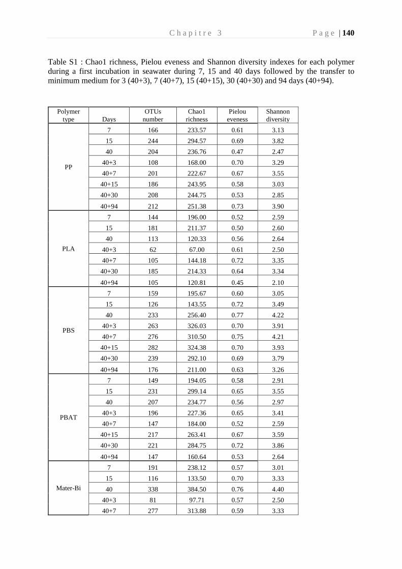
C h a p i t r e 3 P a g e | 140
Table S1 : Chao1 richness, Pielou eveness and Shannon diversity indexes for each polymer
during a first incubation in seawater during 7, 15 and 40 days followed by the transfer to
minimum medium for 3 (40+3), 7 (40+7), 15 (40+15), 30 (40+30) and 94 days (40+94).
Polymer
type Days
OTUs
number
Chao1
richness
Pielou
eveness
Shannon
diversity
PP
7 166 233.57 0.61 3.13
15 244 294.57 0.69 3.82
40 204 236.76 0.47 2.47
40+3 108 168.00 0.70 3.29
40+7 201 222.67 0.67 3.55
40+15 186 243.95 0.58 3.03
40+30 208 244.75 0.53 2.85
40+94 212 251.38 0.73 3.90
PLA
7 144 196.00 0.52 2.59
15 181 211.37 0.50 2.60
40 113 120.33 0.56 2.64
40+3 62 67.00 0.61 2.50
40+7 105 144.18 0.72 3.35
40+30 185 214.33 0.64 3.34
40+94 105 120.81 0.45 2.10
PBS
7 159 195.67 0.60 3.05
15 126 143.55 0.72 3.49
40 233 256.40 0.77 4.22
40+3 263 326.03 0.70 3.91
40+7 276 310.50 0.75 4.21
40+15 282 324.38 0.70 3.93
40+30 239 292.10 0.69 3.79
40+94 176 211.00 0.63 3.26
PBAT
7 149 194.05 0.58 2.91
15 231 299.14 0.65 3.55
40 207 234.77 0.56 2.97
40+3 196 227.36 0.65 3.41
40+7 147 184.00 0.52 2.59
40+15 217 263.41 0.67 3.59
40+30 221 284.75 0.72 3.86
40+94 147 160.64 0.53 2.64
Mater-Bi
7 191 238.12 0.57 3.01
15 116 133.50 0.70 3.33
40 338 384.50 0.76 4.40
40+3 81 97.71 0.57 2.50
40+7 277 313.88 0.59 3.33
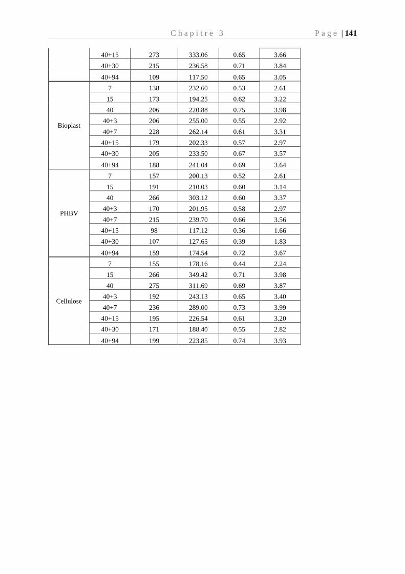
C h a p i t r e 3 P a g e | 141
40+15 273 333.06 0.65 3.66
40+30 215 236.58 0.71 3.84
40+94 109 117.50 0.65 3.05
Bioplast
7 138 232.60 0.53 2.61
15 173 194.25 0.62 3.22
40 206 220.88 0.75 3.98
40+3 206 255.00 0.55 2.92
40+7 228 262.14 0.61 3.31
40+15 179 202.33 0.57 2.97
40+30 205 233.50 0.67 3.57
40+94 188 241.04 0.69 3.64
PHBV
7 157 200.13 0.52 2.61
15 191 210.03 0.60 3.14
40 266 303.12 0.60 3.37
40+3 170 201.95 0.58 2.97
40+7 215 239.70 0.66 3.56
40+15 98 117.12 0.36 1.66
40+30 107 127.65 0.39 1.83
40+94 159 174.54 0.72 3.67
Cellulose
7 155 178.16 0.44 2.24
15 266 349.42 0.71 3.98
40 275 311.69 0.69 3.87
40+3 192 243.13 0.65 3.40
40+7 236 289.00 0.73 3.99
40+15 195 226.54 0.61 3.20
40+30 171 188.40 0.55 2.82
40+94 199 223.85 0.74 3.93
C h a p i t r e 3 P a g e | 142
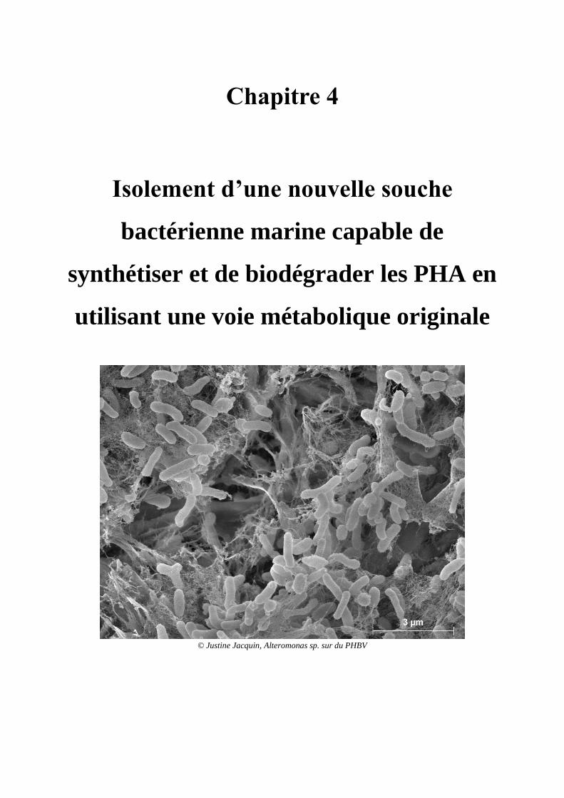
Chapitre 4
Isolement d’une nouvelle souche
bactérienne marine capable de
synthétiser et de biodégrader les PHA en
utilisant une voie métabolique originale
© Justine Jacquin, Alteromonas sp. sur du PHBV

C h a p i t r e 4 P a g e | 145
Chapitre 4
"Anything found to be true of E. coli must also be true of elephants." Jacques Monod
Préambule
Suite aux résultats de biodégradation observés sur les tiges de PHBV (voir chapitre 3),
nous avons étudié les bactéries encore actives après 154 jours d’incubation. Le but de cette
expérience était d’isoler une nouvelle souche capable de dégrader ce polymère et d’en étudier
le génome pour mieux comprendre le processus de biodégradation du PHBV.
Ce travail a mené à l’isolement de 15 souches différentes, dont une souche identifiée
comme appartenant au genre Alteromonas sp. qui formait un halo de dégradation sur boite de
Pétri en condition de milieu minimum + PHBV) (Fig. 31). Nous avons retenu cette souche
d’Alteromonas sp. pour sa vitesse de croissance en milieu minimum et pour son taux de
dégradation plus rapide que les autres souches. L’incubation s’est déroulée sur 40 jours, dans
des tubes de 20 mL comportant 10 mL de milieu artificiel d’eau de mer. Avant l’incubation,
Alteromonas sp. a été mis pendant 14 h en milieu minimum pour s’assurer que le PHBV était
bien l’unique source de carbone durant l’incubation. La biodégradation a été caractérisée par
de multiple paramètres tels que la mesure d’oxygène, la perte de poids, la production
bactérienne et l’observation de la surface du polymère par des images en microscopie
électronique. Ces différents paramètres, tous convergents, ont montré qu’Alteromonas sp. était
capable d’utiliser le PHBV comme seule source de carbone et d’énergie. L’analyse de son
génome nous a ensuite permis de poser de nouvelles hypothèses sur les gènes impliqués dans
la biodégradation du PHBV. L’étude de son unique chromosome a permis de mettre en évidence
une voie de biodégradation originale par rapport aux connaissances de la littérature.
Afin de confirmer ces premiers résultats, des incubations de la souche ont été effectuées
dans du milieu minimum (imitant l’environnement marin) et ayant comme unique source de
carbone du glucose ou du PHBV, afin d’en extraire l’ARN. L’analyse transcriptomique
permettra de valider ou d’infirmer les hypothèses émises. Le séquençage des ARN est
actuellement en cours au Génoscope (Evry, France).
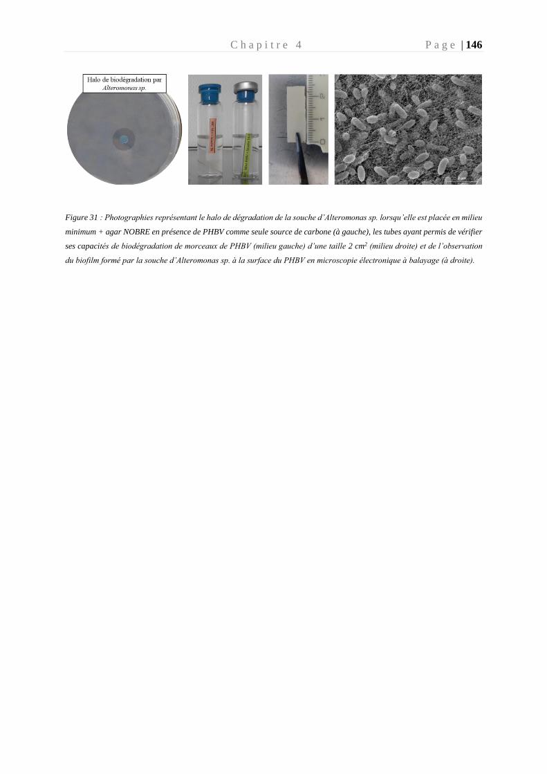
C h a p i t r e 4 P a g e | 146
Figure 31 : Photographies représentant le halo de dégradation de la souche d’Alteromonas sp. lorsqu’elle est placée en milieu
minimum + agar NOBRE en présence de PHBV comme seule source de carbone (à gauche), les tubes ayant permis de vérifier
ses capacités de biodégradation de morceaux de PHBV (milieu gauche) d’une taille 2 cm2 (milieu droite) et de l’observation
du biofilm formé par la souche d’Alteromonas sp. à la surface du PHBV en microscopie électronique à balayage (à droite).
C h a p i t r e 4 P a g e | 147
A new strain capable of synthesizing and degrading PHBV
using atypical metabolic pathways
List of authors : Justine Jacquin1, Jingguang Cheng1, Pascal Conan1, Mireille Pujo-Pay1,
Meistertzheim Anne-Leila2, Stéphane Bruzaud3, Jean-François Ghiglione1*, Barbe Valérie4.
Affiliations :
1. CNRS, Sorbonne Université, UMR 7621, Laboratoire d’Océanographie Microbienne,
Observatoire Océanologique de Banyuls, France
2. SAS Plastic@Sea, Observatoire Océanologique de Banyuls, France
3. Institut de Recherche Dupuy de Lôme (IRDL), Université de Bretagne-Sud, UMR
CNRS 6027, Rue Saint Maudé, Lorient, France
4. Génomique Métabolique, Genoscope, Institut François Jacob, CEA, CNRS, Univ Evry,
Université Paris-Saclay, Evry, France
Abstract
Plastics from petrochemicals represent a major problem, due to their low biodegradability and
their accumulation in many environments. PHAs presented as biodegradable seems to be a good
alternative for this issue environmental. PHBV is a PHA with strong commercial appeal for its
good ability to replace conventional plastic with conventional plastic in many areas. In this
study we have identified a strain belonging to Alteromonas sp. capable of synthesizing and
degrading. This strain was isolated from a consortium of bacteria that developed on PHBV as
only carbon source. Different parameters such as degradation halo on an agar plate, oxygen
consumption, bacterial production, weight loss and scanning electron microscopy were
performed to confirm the ability of this strain to degrade PHBV. Analysis of the complete
genome of the strain revealed three external depolymerases explaining this ability to degrade
PHBV. In addition, genome analysis revealed two pathways of PHA synthesis, allowing this
genome to synthetize PHASCL and PHAMCL. Contrary to what is usually observed, here the
genes involved in the biosynthesis of PHASCL are not organized in operon. Experimental
validations have to be carried out to confirm that Alteromonas sp. possess atypical pathways of
degradation and synthesis of PHAs.
C h a p i t r e 4 P a g e | 148
Introduction
Plastic pollution has become a global problem since its impacts on the environment is
causing irreversible damage to fauna and flora all over the world (Jambeck et al. 2015). Once
introduced in the marine environment, the plastics are rapidly colonized by microorganisms in
marine microorganisms (Oberbeckmann et al. 2015). Bacterial colonizers form a biofilm which
will modify and weaken the surface of the polymer (Donlan 2002). This bacterial biofilm is
often dominated by Proteobacteria and more particularly by Alphaproteobacteria (such as
Rhodobacteraceae) and Gammaproteobacteria (such as the Alteromonadaceae) (Amaral-Zettler
et al. 2020).
Conventional plastics derived from petrochemicals are generally categorized as non-
biodegradable and accumulate in the oceans. Some bio-based plastics have been developed for
their ability to degrade under the action of microorganisms, such as polyhydroxyalkanoates
(PHA) (Eubeler et al. 2009). PHAs have been shown to be biodegradable in many ecosystems
like soils, marine, compost and sewage sludge (Eubeler et al. 2009).
PHAs are polyesters synthesized by a wide range of microorganisms (bacteria and
fungi) from various renewable carbon sources (sugar, acids, vegetable oils)(Peña et al. 2014;
Ali and Jamil 2016; Tebaldi et al. 2019). PHAs are stored into the cytoplasm in granules
proposed to be called “carbonosomes” by Mezzola et al. (Mezzolla et al. 2018). The polymers
are a form of reserve molecules allowing microorganisms to grow in environment with limited
essential nutrients (including nitrogen, magnesium, phosphorus or oxygen) but with a surplus
of carbon supply (Martinez Tobon 2019).
PHA can be classified into four groups (Sagong et al., 2018): PHA-SCL having a short
side chain (between 3 and 5 carbons), PHA-MCL medium side chain (between 6 and 14 carbons),
PHA-LCL long side chain (more than 14 Carbon) and PHAscl-mcl having a mixed length of PHA
chains (short and medium). The nature of the group associated with the side chain is directly
linked to carbon substrates (glucose, fructose, cellulose, acids and fatty acids, etc.) in which the
microorganism evolves and to the nature of the enzyme(s) involved in the last step of the PHA
synthesis: the PHA synthase(s) (Meng et al. 2014).
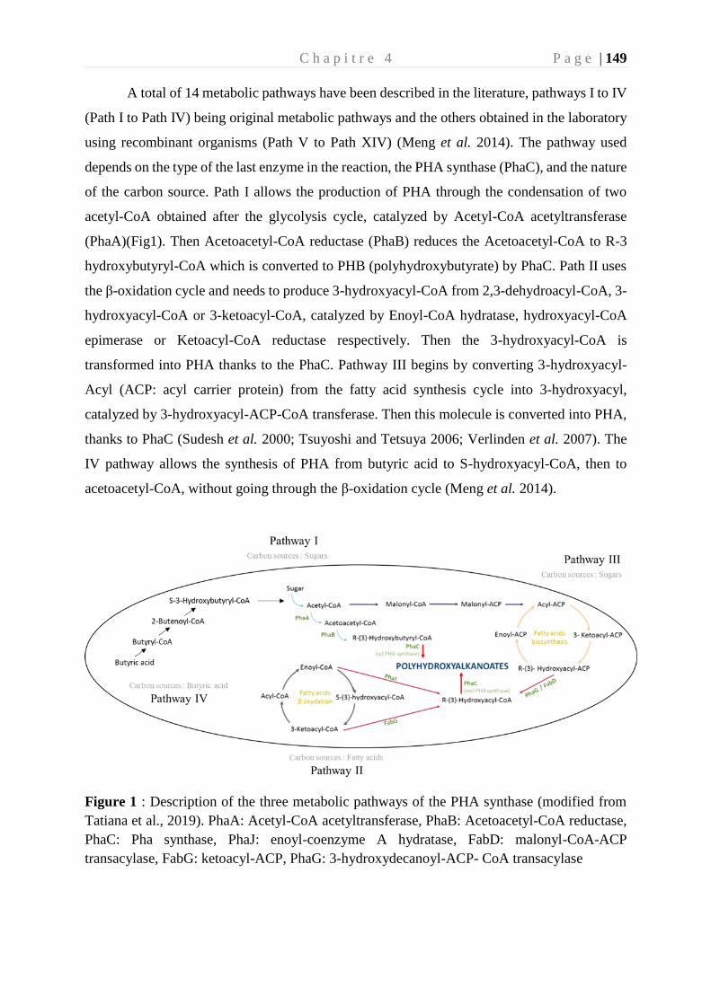
C h a p i t r e 4 P a g e | 149
A total of 14 metabolic pathways have been described in the literature, pathways I to IV
(Path I to Path IV) being original metabolic pathways and the others obtained in the laboratory
using recombinant organisms (Path V to Path XIV) (Meng et al. 2014). The pathway used
depends on the type of the last enzyme in the reaction, the PHA synthase (PhaC), and the nature
of the carbon source. Path I allows the production of PHA through the condensation of two
acetyl-CoA obtained after the glycolysis cycle, catalyzed by Acetyl-CoA acetyltransferase
(PhaA)(Fig1). Then Acetoacetyl-CoA reductase (PhaB) reduces the Acetoacetyl-CoA to R-3
hydroxybutyryl-CoA which is converted to PHB (polyhydroxybutyrate) by PhaC. Path II uses
the β-oxidation cycle and needs to produce 3-hydroxyacyl-CoA from 2,3-dehydroacyl-CoA, 3-
hydroxyacyl-CoA or 3-ketoacyl-CoA, catalyzed by Enoyl-CoA hydratase, hydroxyacyl-CoA
epimerase or Ketoacyl-CoA reductase respectively. Then the 3-hydroxyacyl-CoA is
transformed into PHA thanks to the PhaC. Pathway III begins by converting 3-hydroxyacyl-
Acyl (ACP: acyl carrier protein) from the fatty acid synthesis cycle into 3-hydroxyacyl,
catalyzed by 3-hydroxyacyl-ACP-CoA transferase. Then this molecule is converted into PHA,
thanks to PhaC (Sudesh et al. 2000; Tsuyoshi and Tetsuya 2006; Verlinden et al. 2007). The
IV pathway allows the synthesis of PHA from butyric acid to S-hydroxyacyl-CoA, then to
acetoacetyl-CoA, without going through the β-oxidation cycle (Meng et al. 2014).
Figure 1 : Description of the three metabolic pathways of the PHA synthase (modified from
Tatiana et al., 2019). PhaA: Acetyl-CoA acetyltransferase, PhaB: Acetoacetyl-CoA reductase,
PhaC: Pha synthase, PhaJ: enoyl-coenzyme A hydratase, FabD: malonyl-CoA-ACP
transacylase, FabG: ketoacyl-ACP, PhaG: 3-hydroxydecanoyl-ACP- CoA transacylase
C h a p i t r e 4 P a g e | 150
In recent years, PHA has developed and presents an increasing commercial and
industrial interest as a biodegradable substitute for conventional plastics derived from
petrochemicals. One of the most commonly used PHAs for plastic production is
polyhydroxybutyrate (PHB, which has methyl as the alkyl group (R) ([CO-CH2-CHCH3-O]
n). PHB is considered to be hydrophobic, having low permeability, little deformability and
considered to be biodegradable (Mothes et al. 2004; Chang et al. 2012). Unfortunately, this
polymer is not very deformable due to its high crystallinity (Gorke et al. 2007). PHB has a high
melting point close to its thermal degradation temperature (Reis et al. 2003), it is for this reason
that a co-polymer, poly (3-hydroxybutyrate-co-3-hydroxyvalerate) (PHBV), is commonly used
to reduce the melting point of PHB. The PHBV is a mix of short and medium side chain (PHA-
SCL-MCL) and can be synthetized by many species of microorganisms (Wang et al. 2013; Tebaldi
et al. 2019).
The PHAs are degraded by many microorganisms in the marine environment (Jacquin
et al. 2019). This degradation is enabled by a key enzyme called depolymerase, PhaZ
(Jendrossek and Handrick 2002; Jendrossek 2005). Two principal types of PhaZ (intracellular
(i-PhaZ) and extracellular (e-PhaZ) were identified in different microorganisms (Jendrossek
2007). The i-PhaZ is able to degrade the native PHA granules (nPHA), composed by scl and/or
mcl chains. These PHAs are generally amorphous polymers which are higher elasticity and
flexibility as compared to crystalline PHA polymers. In contrast, the excreted depolymerase (e-
PhaZ) degrades the extracellular amorphous or christallin PHAs (called denatured (dPHA))
(Jendrossek and Handrick 2002). A peculiarity was highlighted for Rhodospirillum rubrum in
which a periplasmic depolymerase has been identified (Handrick et al. 2004).
The PHA depolymerases are carboxylesterases belonging to the α / β hydrolase family
members (Jendrossek and Handrick 2002). The comparison between several depolymerase
reveals a catalytic triad (serine - histidine - aspartic acid) present in the vast majority of PHA
depolymerases. The catalytic serine site belongs to a lipase box (GxSxG sequence) (Jendrossek
and Handrick 2002). The catalytic domain can be divided into two groups depending on the
location of the lipase box, relative to the oxyanion hole also belonging to the catalytic domain.
If the oxyanion hole is located at the N-terminal of the lipase box, then the catalytic domain is
type 1, if it is located at C-ter of the lipase box then the type 2 catalytic domain. the catalytic
triad is not found in all intracellular PHA depolymerases. In this case, these native
C h a p i t r e 4 P a g e | 151
depolymerase SCL have a catalytic triad consisting of cysteine, histidine and aspartic acid
(Handrick et al. 2000).
This study aims to characterize the biodegradation of PHBV by the selected strain and
to better understand the biodegradation process by studying its genome. The study of its
complete genome makes it possible to identify the genes involved in the metabolism of PHA
(synthesis and degradation). These results may allow a better understanding of the use of PHBV
by the strain and of its industrial potential.
Materials and Methods
1. Selection and identification of the bacterial strain
The bacterial strain was isolated in this study during experiments showing the biodegradation
of PHBV by marine microbial communities (Jacquin et al., submitted). Briefly, after leaving
pieces of PHBV for 40 days in an open circuit aquarium with direct circulation to seawater from
the bay of Banyuls-sur-mer (42°29’300 N – 03°08’700 E, NW Mediterranean sea), the
fragments of PHBV together with mature biofilm were transferred into an artificial medium for
94 days. Biodegradation being observed at the end of the experiment, the consortium resulting
from the biofilm formed on the pieces of PHBV was detached. Cell detachment was using 3
cycles of sonication bath during 3 min (deltasonic, France) followed by 3 min of vortexing at
maximum speed (Skyline, Elmi Ltd, Russia). To isolate the bacterial clones, successive
dilutions were carried out (107 to 102 cells/mL) and 100 µL of each dilution was spread on
Marine Agar. The dishes were stored at 18°C for 3 days. The resulting colonies were picked
and resuspended into 10 mL of Marine Broth. A part of culture strain (100µL) was loaded onto
an artificial seawater medium with noble agar and molten PHBV as sole carbon source. The
plates were incubated 10 days at 18 °C. The clones having the ability to use PHBV as carbon
source, characterized by the presence of degradation halo, were all isolated as previously
described (Rodriguez et al. 2010). The strain with the largest halo was selected for this study
(Fig S1). This strain, named “strain of interest” was cultured in Marine Broth for 24 h at 18 ° C
and stored at -80 ° C in glycerol (30%).
C h a p i t r e 4 P a g e | 152
2. Culture of the strain
A preculture of the strain of interest in 15 mL of Marine Broth was transferred to 200 mL of
fresh artificial seawater medium (ASM) supplemented with 10 mM glucose. The composition
of this artificial medium was modified from Fegatella (Fegatella et al. 1998) : NaCl, 420 mM ;
Tris, 50 mM ; Na2SO4, 28.8 mM; KCl, 9.39 mM ; NaBr, 0.84 mM ; H3BO3, 0.485 mM ; MgCl2-
6H2O, 0.546 mM ; CaCl2, 0.105 mM ; NH4Cl 9.35 mM ; SrCl2-6H2O, 0.0638 mM ; NaF,
0.0714 mM ; NaNO3, 0.88 mM ; NaH2PO4-H2O, 0.036 mM ; KH2PO4, 0.106 mM ;
CuSO4·5H2O, 0.04 μM ; ZnSO4·7H2O 0.08 µM ; CoCl2·6H2O, 0.04 μM; MnCl2·4H2O, 0.9 μM
; Na2MoO4·2H2O, 0.03 μM ; FeCl3 1.85 μM ; thiamine, 0.3 μM ; biotin, 2.1 nM ; and B12
vitamin, 0.317 nM (Fegatella et al., 1998).
After 72 h culture in this artificial sea water medium at 18 °C with 2 mM of glucose, cells were
centrifuged during 10 min at a relative force of 7500 rpm and resuspended in 1 000mL of
artificial seawater minimum medium for 14 hours without any source of carbon. The inoculum
was then diluted in ASM until a concentration of 107 bacteria. mL-1. Four culture conditions
were carried out for this study containing 10 mL of artificial medium supplemented or not with
a fragment of PHBV, and in presence or not of the strain (at a final concentration of 107 cell.mL-
1). The PHBV fragments of around 1cm2 were previously washed using ethanol 70% over 48
hours then sterilized by UV during 15 minutes. Tubes containing only artificial seawater
medium with or without glucose, PHBV or strain only were used as negative controls. The
culture of all the samples was performed in 20 mL tubes (Serum flask, Dutscher) closed with a
Butyl stopper and a capsule (Dutscher). Six times (D0, D5, D11, D16, D32 and D40 ) were
chosen for the experiment corresponding to the start, 5, 11, 16, 32 and 40 days of incubation
respectively.. During the entire incubation period the nutrients (PO4, NO2 and NO3) were
measured to ensure that they were not in limiting conditions (Table S1).
C h a p i t r e 4 P a g e | 153
3. Oxygen measurements
Throughout the experiment, oxygen monitoring was carried out on 4 samples. There were
2 negative controls, one containing the artificial medium alone, and the other the artificial
medium with the PHBV, and two samples, one containing the artificial medium with the strain
and the other containing the artificial medium, with the PHBV and the strain.
The 4 tubes were fitted with an oxygen sensor (Presens, SP-PSt3-YAU) and a luminescent
optical fiber. The measurement was carried out by a Fiber Optic Oxygen Meter box (Fibox 4,
Presens). Oxygen was recorded every hour during the 40 days of incubation in an artificial
environment. To avoid having anoxic incubation conditions, the tubes containing the PHBV
with the strain were flushed with air filtered at 0.2 μm under a laminar flow hood on days
5,11,16 and 24. The concentration in oxygen has always been maintained above 50 μM.
Bacterial oxygen demand (BOD) was calculated by taking oxygen consumption as a function
of time.
4. Bacterial production
Bacterial production (BP) was measured in triplicate at each sampling time for all sample
(PHBV, supernantant of PHBV, PHBV + strain, Supernatant of PHBV+strain and the negative
control artificial sea water). H-leucine incorporation into proteins was measured, using a
modified protocol from Van Wambeke et al. (Wambeke et al. 2009). Briefly, the cell
detachment pre-treatment was applied for PHBV+strain, using 3 cycles of sonication bath
during 3 min (deltasonic, France) followed by vortexing at maximum speed for 3 min (Skyline,
Elmi Ltd, Russia) in order to improve the BP signal (Dussud et al. 2018). Immediately after
cell-detachment, 3H-leucine (specific activity 123.8 Ci mmol-1; Perkin Elmer) was added for
each sample, which consisted of 1.5 mL of sterilized seawater containing the piece of plastic
together with the detached cells. Final leucine concentrations were 150 nmol L-1 (3H-leucine at
1 nmol L-1). The samples containing a piece of PHBV with or without strain, the supernatant of
both conditions and the negative control were treated in triplicate and incubated in the dark at in
situ temperature for 3 h. We used the empirical conversion factor of 1.55 ng C pmol−1 of
incorporated leucine to calculate BP (Simon and Azam 1989).. The exact surface area of the
plastic was calculated with image J software (Abràmoff et al. 2004) and the results are reported
in ng C.m-2. h-1.
C h a p i t r e 4 P a g e | 154
5. Scanning electron microscopy
At each point of the experiment, one sample containing PHBV with the strain or not
(control) were fixed in solution A (0.4M Na Cacodylate buffer; 3% of Glutaraldehyde; Qsp
artificial medium) and stored at 4°C until analysis under an electron microscope. Before
analysis, the samples were washed three times for 15 minutes with solution B (Sodium
Cacodylate buffer, 0.4 M diluted to 0.2 M in artificial medium) in order to remove all traces of
glutaraldehyde. Then the samples were fixed with osmium tetroxide (OsO4) at 1%, for one hour
at room temperature. Progressive dehydration in ethanol baths (70%, 95% and 100%) was
applied during 30 minutes for each bath. Samples were immersed in 100% ethanol in a critical
point desiccator tank (Balzers), and liquid CO2 was then gradually introduced to replace
ethanol. Samples were then heated and pressurized to allow the CO2 to pass to the gaseous state
bypassing its critical point (Temperature = 31.1 ° C; P = 73.8 bars) and to avoid surface tensions
and deformation. The CO2 gas was then removed, and dry samples were collected. After
dehydration, a thin conductive layer of Au-Pd (about 10nm thick) was deposited by sputtering
on the surface of the samples to allow electron microscope observation (Hitachi SEM FEG S-
4500 high vacuum).
6. Weight lost
A total of 8 plastics were sterile weighed on the precision balance (Precisa 125 A, Swiss
quality) prior to incubation. Four of them were incubated in artificial seawater medium
containing the strain of interest and the rest without the strain. The incubations were maintained
until the end of the experiment (T40). At the final stage, the 8 plastics were rinsed with distilled
water and stored at -20 °C for 48 hours. In order to remove any presence of the artificial
medium, a step of drying in a freeze-dryer was performed. Finally, the plastics were weighed
using the same precision balance. The results are expressed in mg and the weight loss is
expressed as a percentage.
C h a p i t r e 4 P a g e | 155
7. DNA extraction, sequencing, assembly and annotation of the genome of the strain
of interest
Genomic DNA was obtained after a pure culture of the strain of interest incubated in marine
broth at 18 ° C for 48 hours. The culture was centrifuged at 7000 rpm for 10 minutes. DNA
extraction was performed using a classical phenol-chloroform method for seawater samples
(Sauret et al. 2016) with slight modification. Briefly, we used the enzymatic and chemical cell
lysis (incubation with 1 mg.mL-1 lysozyme at 37°C for 45 min followed by 1 h at 50°C with
0.2 mg.mL- 1 proteinase K and 1% SDS) before the DNA extraction with phenol and
purification with chloroform. All the steps were carried out carefully, without vortexing, with
very gentle shaking by inverting the tube to reduce DNA fragmentation. DNA was quantified
by spectrophotometry (Quant-iT™ PicoGreen™ dsDNA Assay Kit, Invitrogen).
The sequencing strategy to obtain the complete sequence of the interest strain was based on the
complementarity of two methods mixing short (Illumina, www.illumina.com) and long reads
(Oxford Nanopore Technologies, https://nanoporetech.com/). Short reads allow obtaining
quality consensus generated after assembly while long reads provide scaffold on which short
reads are anchored.
To prepare Illumina library, 100 ng of genomic DNA was fragmented to an average size
of 500-600 bp using a Covaris E220. The ends of the DNA fragments were repaired, dATP was
added and the adapters (NEXTflex HT Barcodes) were ligated using the NEBNext DNA
modules. After being purified twice consecutively with AMPure XP (1 volume), the DNA
fragments were amplified by PCR (12 cycles) with the Kapa Hifi Hotstart NGS library
amplification kit, and then purified again with AMPure XP (0.7 volume). After being quantified
by qPCR, the library was sequenced on a MiSeq device in “paired end” (2x250bp). A total of
1,551,185 (466 Mb) read pairs were obtained and used for assembly.
To complete this data set, a first Nanopore library was prepared from 825ng of genomic
DNA according to the “Genomic DNA by Ligation” protocol (SQK-LSK109) recommended
by Oxford Nanopore. This library was loaded on a Flongle R9.4.1 flowcell leading to 76,547
sequences (126 Mb) with an N50 of 10,801bp. To improve these data, a second library was
prepared according to the same protocol from 700ng of genomic DNA and loaded on a MinION
R9.4.1 flowcell, leading to 641,816 sequences (1.130Gb) with an N50 of 14,665bp.
Unicycler software v.0.4.6 (Wick et al. 2017) was run using default options with all 3 datasets.
The final hybrid assembly resulted in a circular molecule of 5.33 Mb length and with an average
C h a p i t r e 4 P a g e | 156
GC content of 48.79%. The automatic annotation was performed via the MicroScope platform
(https://mage.genoscope.cns.fr/microscope/home/index.php).
8. Phylogenetic study and classification
Before establishing the phylogenetic tree, all the 16S rDNA sequences obtained after
assembly were compared with each other to verify that there were completely identical. Then
this sequence was used to perform a nucleotide blast to NCBI with the nt database
(https://blast.ncbi.nlm.nih.gov/Blast.cgi). All the results with percentage identity greater 97%
were kept and the sequences of the selected matches were extracted to create a database. In
addition, the 16S rDNA sequence of Marinobacter hydrocarbonoclasticus ATCC 49840 has
been added to the database due to this genus has been identified already on PHBV and
hydrophobic organic compounds (Grimaud et al. 2012; Kong et al. 2018) . Finally, a total of 17
strains (all belonging to Alteromonadaceae family) composed the database and the neighbor-
joining method with the MEGA7 software (Kumar et al. 2008) was used to produce a
phylogenetic tree.
9. Genome analysis
Genome of the strain of interest has been studied using the MicroScope annotation and
analysis platform (https://mage.genoscope.cns.fr/microscope/home/index.php). To identify the
coding sequences (CDS) possibly having depolymerase function, the protein sequences of all
the CDS were extracted. A protein comparison using blastp (with parameters: evalue 1.0,
word_size 5 and threshold 10 (https://www.ncbi.nlm.nih.gov/books/NBK279684/)) was
performed comparing the MicroScope data and the DED database (Knoll et al. 2009, http:
//www.ded.uni -stuttgart.de/). Knoll and its collaborators built a PHA depolymerase database
using the DWARF system (an automated retrieval tool that extracts various sequence
information, such as structure or function). First, this database was constructed from 28 seed
sequences, with laboratory-validated depolymerase activity. In addition to this 28 protein
sequences, other sequences annotated as PHA depolymerase in GenBank came to complete the
database. Currently the DED database contains 587 sequences of 8 super families of PHA
depolymerases and 38 homologous families. Sequences with more than 98% identity from the
C h a p i t r e 4 P a g e | 157
same source organism have been assigned to a single protein. A summarize of this database is
presented in Sup Table 1 (Table S2).
Results and discussion
Criteria for assessing biodegradation
The strain of this study was selected by its capacity to degrade PHBV. Indeed, after culture of
this strain on Marine Broth and deposited on an artificial seawater medium with noble agar and
molten PHBV as carbon source, a degradation halo was observed (Fig S1). Different parameters
were monitoring to validate or invalidate this degradation.
1. Oxygen consumption
A total of 4 tubes containing artificial seawater medium only (ASM), complemented with
strain (ASM-str) or PHBV (ASM-PHBV), or with the both (ASM-str-PHBV), are monitored
throughout the experiment (Fig 2). The evolution of the dissolved oxygen concentration in the
ASM tube is considered as an experiment blank and the small drift measured was subtracted
from the other 3 tubes. Indeed, the oxygen consumption is estimated as the difference between
oxygen concentration in the sample and this blank :
O2 (µmolO2.L-1.d-1) = Conc O2 ech - Conc O2 ASM
Throughout the experiment, oxygen consumption in the ASM-PHBV tube was
negligible, with the slope of -0.09 µmolO2.L-1.d-1 not significantly different from 0 (p<0.05).
For the tube ASM-str, a weak but significant oxygen consumption is observed (-0.22 µmolO2.L-
1.d-1) probably due to the use of remaining carbon source from the inoculum or reserves of the
microorganism.
On the other hand, the tube containing the strain and the PHBV (Fig 2) showed very
rapid and strong daily oxygen consumption. At the beginning of the experiment, the
consumption was close to -40 µmolO2.L-1.d-1. To prevent the environment from becoming
anoxic, oxygen was added in the form of sterile filtered air on days 5, 10 and 18. Between each
of these additions, daily oxygen consumption was relatively constant and high and varies
between 13.21 and 38.54 µmolO2.L-1.d-1. A slowing of this consumption was observed from
day 25, and then remained stable until the end of the experiment (~ -13.21 µmolO2.L-1.d-1).
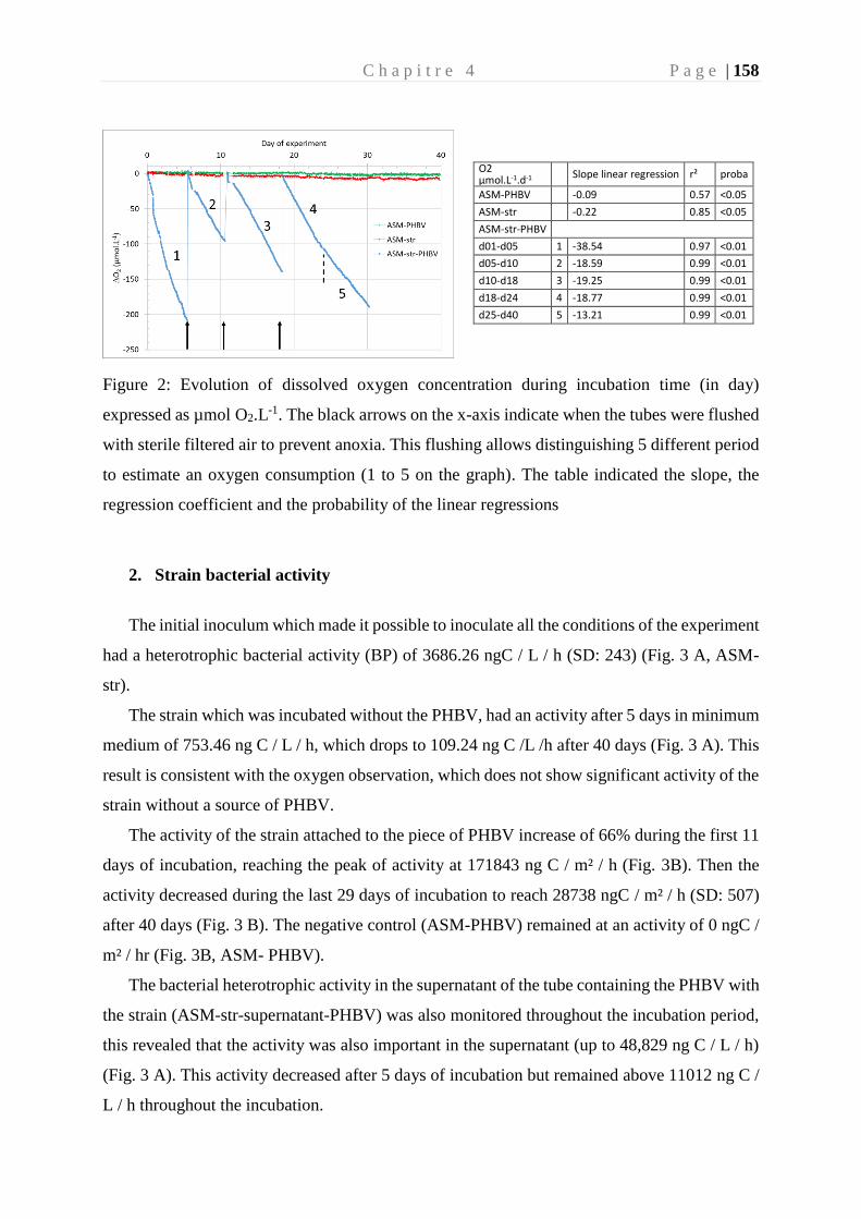
C h a p i t r e 4 P a g e | 158
O2 µmol.L-1.d-1
Slope linear regression r² proba
ASM-PHBV -0.09 0.57 <0.05
ASM-str -0.22 0.85 <0.05
ASM-str-PHBV
d01-d05 1 -38.54 0.97 <0.01
d05-d10 2 -18.59 0.99 <0.01
d10-d18 3 -19.25 0.99 <0.01
d18-d24 4 -18.77 0.99 <0.01
d25-d40 5 -13.21 0.99 <0.01
Figure 2: Evolution of dissolved oxygen concentration during incubation time (in day)
expressed as µmol O2.L-1. The black arrows on the x-axis indicate when the tubes were flushed
with sterile filtered air to prevent anoxia. This flushing allows distinguishing 5 different period
to estimate an oxygen consumption (1 to 5 on the graph). The table indicated the slope, the
regression coefficient and the probability of the linear regressions
2. Strain bacterial activity
The initial inoculum which made it possible to inoculate all the conditions of the experiment
had a heterotrophic bacterial activity (BP) of 3686.26 ngC / L / h (SD: 243) (Fig. 3 A, ASM-
str).
The strain which was incubated without the PHBV, had an activity after 5 days in minimum
medium of 753.46 ng C / L / h, which drops to 109.24 ng C /L /h after 40 days (Fig. 3 A). This
result is consistent with the oxygen observation, which does not show significant activity of the
strain without a source of PHBV.
The activity of the strain attached to the piece of PHBV increase of 66% during the first 11
days of incubation, reaching the peak of activity at 171843 ng C / m² / h (Fig. 3B). Then the
activity decreased during the last 29 days of incubation to reach 28738 ngC / m² / h (SD: 507)
after 40 days (Fig. 3 B). The negative control (ASM-PHBV) remained at an activity of 0 ngC /
m² / hr (Fig. 3B, ASM- PHBV).
The bacterial heterotrophic activity in the supernatant of the tube containing the PHBV with
the strain (ASM-str-supernatant-PHBV) was also monitored throughout the incubation period,
this revealed that the activity was also important in the supernatant (up to 48,829 ng C / L / h)
(Fig. 3 A). This activity decreased after 5 days of incubation but remained above 11012 ng C /
L / h throughout the incubation.
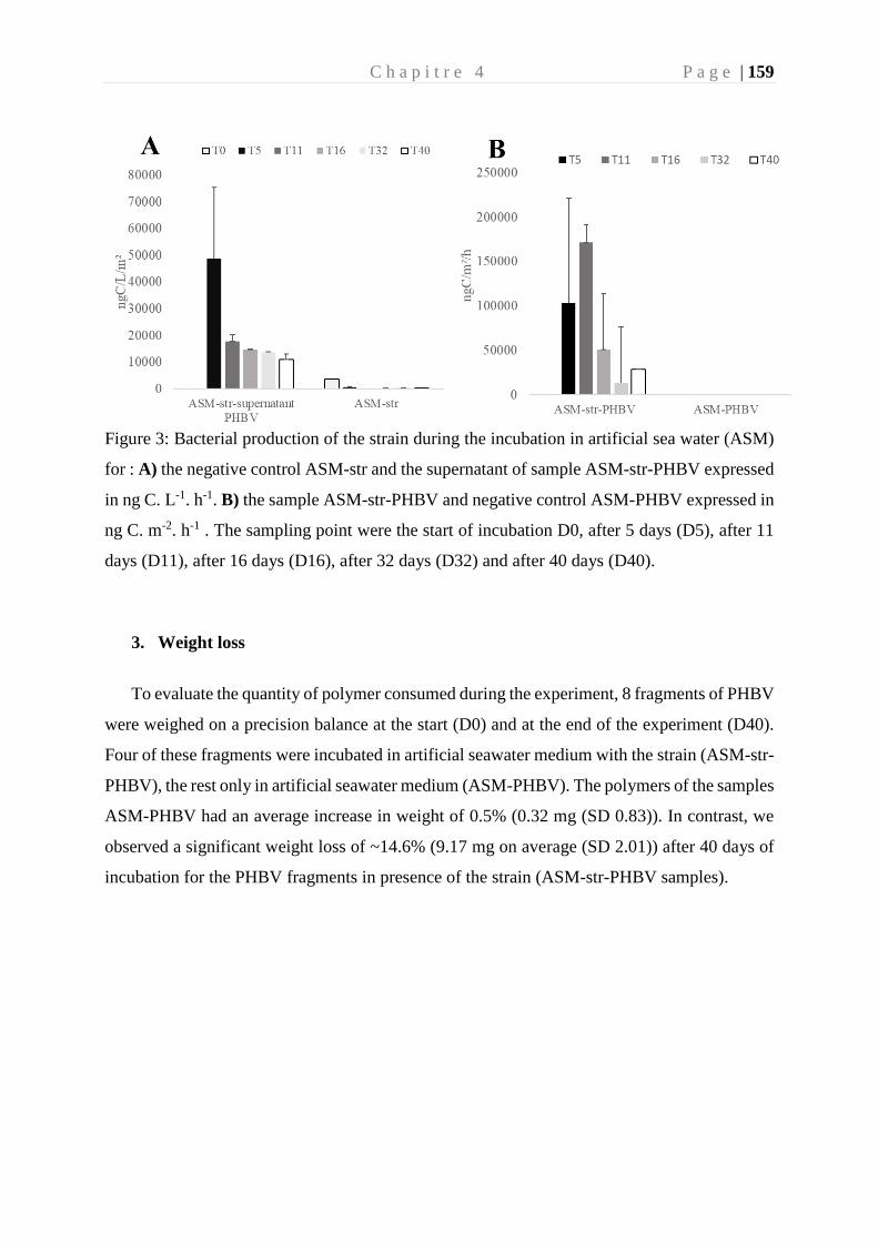
C h a p i t r e 4 P a g e | 159
Figure 3: Bacterial production of the strain during the incubation in artificial sea water (ASM)
for : A) the negative control ASM-str and the supernatant of sample ASM-str-PHBV expressed
in ng C. L-1. h-1. B) the sample ASM-str-PHBV and negative control ASM-PHBV expressed in
ng C. m-2. h-1 . The sampling point were the start of incubation D0, after 5 days (D5), after 11
days (D11), after 16 days (D16), after 32 days (D32) and after 40 days (D40).
3. Weight loss
To evaluate the quantity of polymer consumed during the experiment, 8 fragments of PHBV
were weighed on a precision balance at the start (D0) and at the end of the experiment (D40).
Four of these fragments were incubated in artificial seawater medium with the strain (ASM-str-
PHBV), the rest only in artificial seawater medium (ASM-PHBV). The polymers of the samples
ASM-PHBV had an average increase in weight of 0.5% (0.32 mg (SD 0.83)). In contrast, we
observed a significant weight loss of ~14.6% (9.17 mg on average (SD 2.01)) after 40 days of
incubation for the PHBV fragments in presence of the strain (ASM-str-PHBV samples).
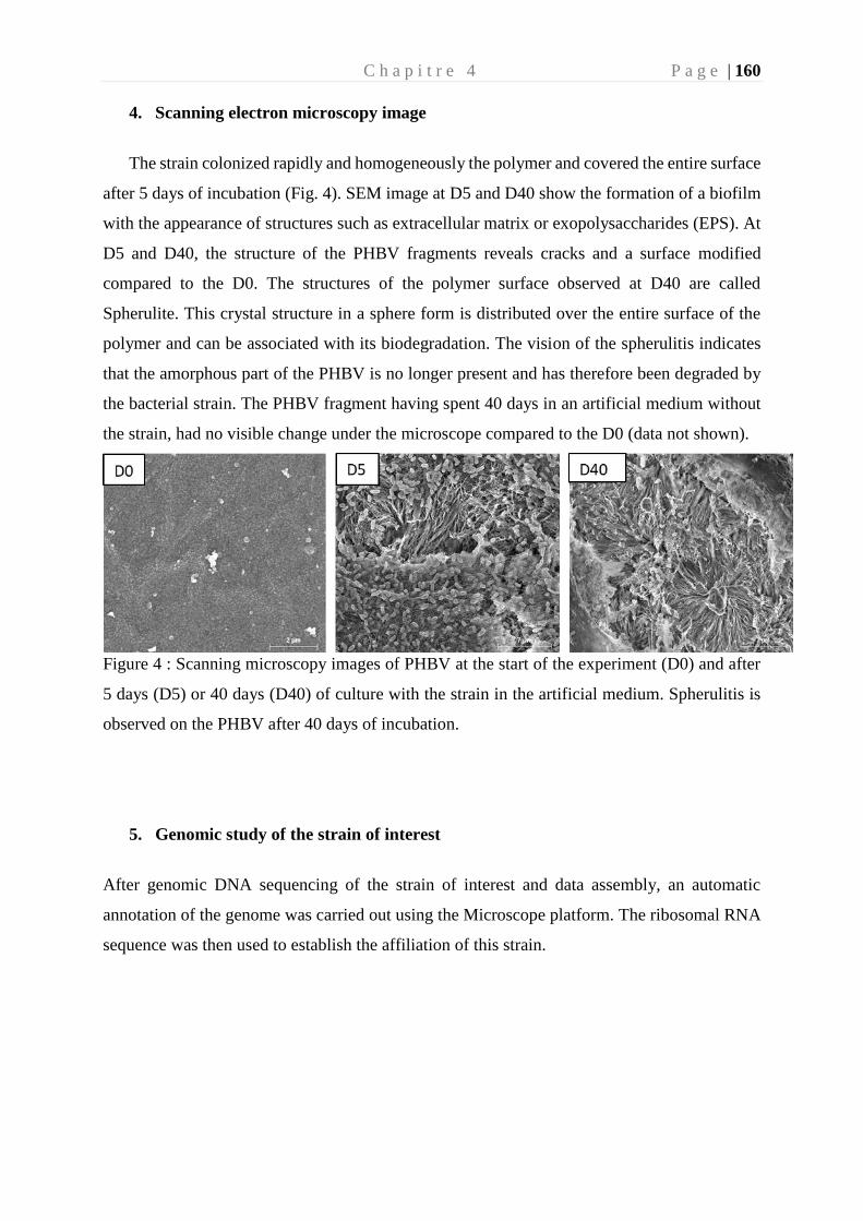
C h a p i t r e 4 P a g e | 160
4. Scanning electron microscopy image
The strain colonized rapidly and homogeneously the polymer and covered the entire surface
after 5 days of incubation (Fig. 4). SEM image at D5 and D40 show the formation of a biofilm
with the appearance of structures such as extracellular matrix or exopolysaccharides (EPS). At
D5 and D40, the structure of the PHBV fragments reveals cracks and a surface modified
compared to the D0. The structures of the polymer surface observed at D40 are called
Spherulite. This crystal structure in a sphere form is distributed over the entire surface of the
polymer and can be associated with its biodegradation. The vision of the spherulitis indicates
that the amorphous part of the PHBV is no longer present and has therefore been degraded by
the bacterial strain. The PHBV fragment having spent 40 days in an artificial medium without
the strain, had no visible change under the microscope compared to the D0 (data not shown).
Figure 4 : Scanning microscopy images of PHBV at the start of the experiment (D0) and after
5 days (D5) or 40 days (D40) of culture with the strain in the artificial medium. Spherulitis is
observed on the PHBV after 40 days of incubation.
5. Genomic study of the strain of interest
After genomic DNA sequencing of the strain of interest and data assembly, an automatic
annotation of the genome was carried out using the Microscope platform. The ribosomal RNA
sequence was then used to establish the affiliation of this strain.
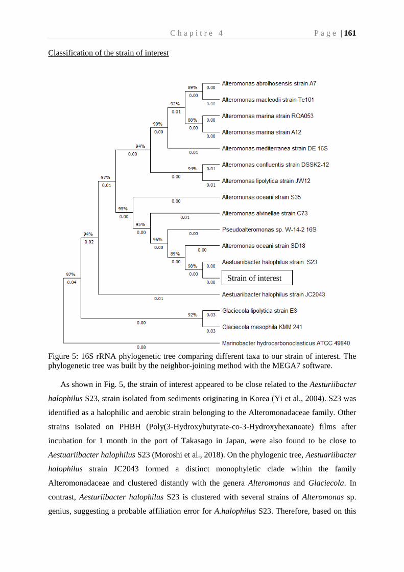
C h a p i t r e 4 P a g e | 161
Classification of the strain of interest
Figure 5: 16S rRNA phylogenetic tree comparing different taxa to our strain of interest. The
phylogenetic tree was built by the neighbor-joining method with the MEGA7 software.
As shown in Fig. 5, the strain of interest appeared to be close related to the Aesturiibacter
halophilus S23, strain isolated from sediments originating in Korea (Yi et al., 2004). S23 was
identified as a halophilic and aerobic strain belonging to the Alteromonadaceae family. Other
strains isolated on PHBH (Poly(3-Hydroxybutyrate-co-3-Hydroxyhexanoate) films after
incubation for 1 month in the port of Takasago in Japan, were also found to be close to
Aestuariibacter halophilus S23 (Moroshi et al., 2018). On the phylogenic tree, Aestuariibacter
halophilus strain JC2043 formed a distinct monophyletic clade within the family
Alteromonadaceae and clustered distantly with the genera Alteromonas and Glaciecola. In
contrast, Aesturiibacter halophilus S23 is clustered with several strains of Alteromonas sp.
genius, suggesting a probable affiliation error for A.halophilus S23. Therefore, based on this
Strain of interest
C h a p i t r e 4 P a g e | 162
tree, it might be conceivable that the strain of interest is an Alteromonas. This strain will be
named here after: Alteromonas sp.
5.1. General features
The genome of Alteromonas sp. consisted in a circular chromosome of 5,332,999bp
with a GC content of 48.8%. All the information is summarized in Fig 6, Table 1 and
Supplementary Table 4.
Table 1: General features of the Alteromonas sp. genome
Attribute Values
Genome size (bp) 5,332,999
DNA G+C (%) 48.8
Plasmid 0
DNA scaffolds 1
rRNA genes 12
tRNA genes 58
tmRNA 1
misc_RNA 34
Protein 4810
Predicted genes 4805
Pseudo genes 8
Automatic annotation revealed that the chromosome contains 4805 coding sequences
(CDS) (including 8 pseudogenes) with an average size of 994 bp. The coding density was
89.34% and the repetition rate estimated at nearly 3%. This strain had a larger genome and a
higher GC percent compared to the majority of the sequences of other Alteromonas and
Pseudoalteromonas species available in the Microscope platform (Table S3). Only
Alteromonas sp. 190 had comparable genome size and a GC percent. Alteromonas sp. shared
3802 CDSs in synteny with Alteromonas sp. 190 have (data not shown). Fifty-eight tRNAs,
representing 21 amino acids including 20 standards, have been identified in the genome (Table
1). It can be noted that there was a bias in coding usage since the tRNAs Alanine, Arginine,
Leucine, Methionine, and Serine that were on average more present in the genome compared
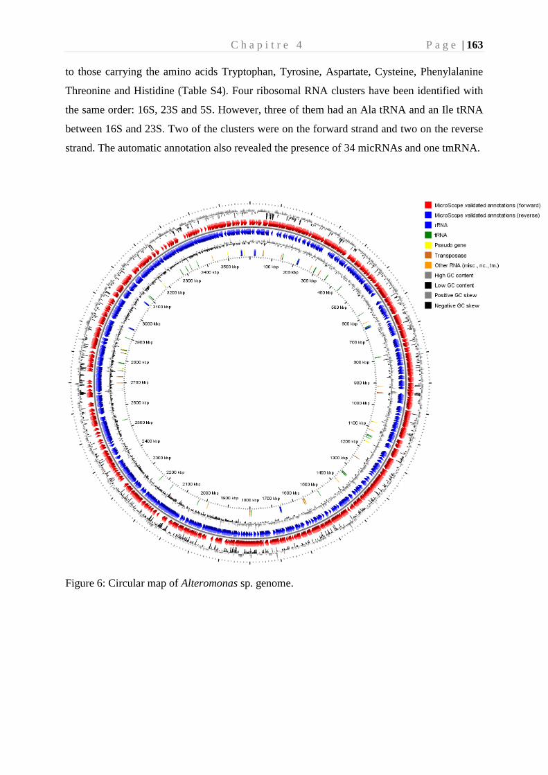
C h a p i t r e 4 P a g e | 163
to those carrying the amino acids Tryptophan, Tyrosine, Aspartate, Cysteine, Phenylalanine
Threonine and Histidine (Table S4). Four ribosomal RNA clusters have been identified with
the same order: 16S, 23S and 5S. However, three of them had an Ala tRNA and an Ile tRNA
between 16S and 23S. Two of the clusters were on the forward strand and two on the reverse
strand. The automatic annotation also revealed the presence of 34 micRNAs and one tmRNA.
Figure 6: Circular map of Alteromonas sp. genome.
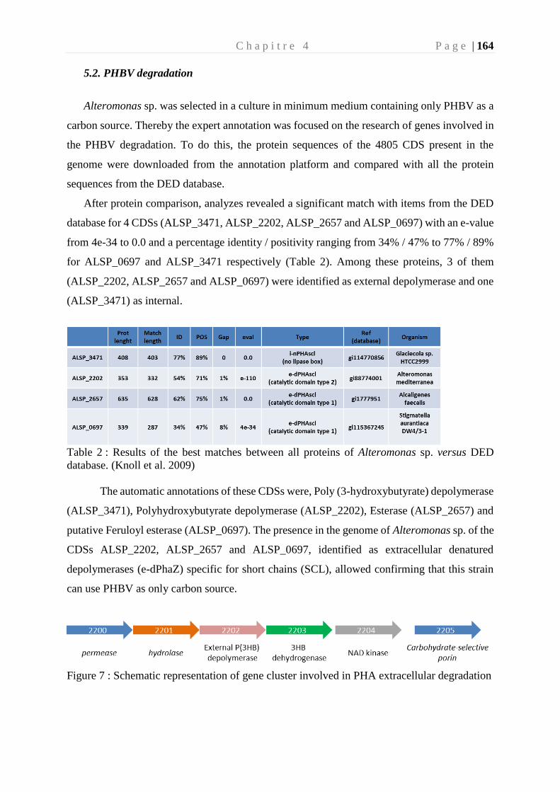
C h a p i t r e 4 P a g e | 164
5.2. PHBV degradation
Alteromonas sp. was selected in a culture in minimum medium containing only PHBV as a
carbon source. Thereby the expert annotation was focused on the research of genes involved in
the PHBV degradation. To do this, the protein sequences of the 4805 CDS present in the
genome were downloaded from the annotation platform and compared with all the protein
sequences from the DED database.
After protein comparison, analyzes revealed a significant match with items from the DED
database for 4 CDSs (ALSP_3471, ALSP_2202, ALSP_2657 and ALSP_0697) with an e-value
from 4e-34 to 0.0 and a percentage identity / positivity ranging from 34% / 47% to 77% / 89%
for ALSP_0697 and ALSP_3471 respectively (Table 2). Among these proteins, 3 of them
(ALSP_2202, ALSP_2657 and ALSP_0697) were identified as external depolymerase and one
(ALSP_3471) as internal.
Table 2 : Results of the best matches between all proteins of Alteromonas sp. versus DED
database. (Knoll et al. 2009)
The automatic annotations of these CDSs were, Poly (3-hydroxybutyrate) depolymerase
(ALSP_3471), Polyhydroxybutyrate depolymerase (ALSP_2202), Esterase (ALSP_2657) and
putative Feruloyl esterase (ALSP_0697). The presence in the genome of Alteromonas sp. of the
CDSs ALSP_2202, ALSP_2657 and ALSP_0697, identified as extracellular denatured
depolymerases (e-dPhaZ) specific for short chains (SCL), allowed confirming that this strain
can use PHBV as only carbon source.
Figure 7 : Schematic representation of gene cluster involved in PHA extracellular degradation
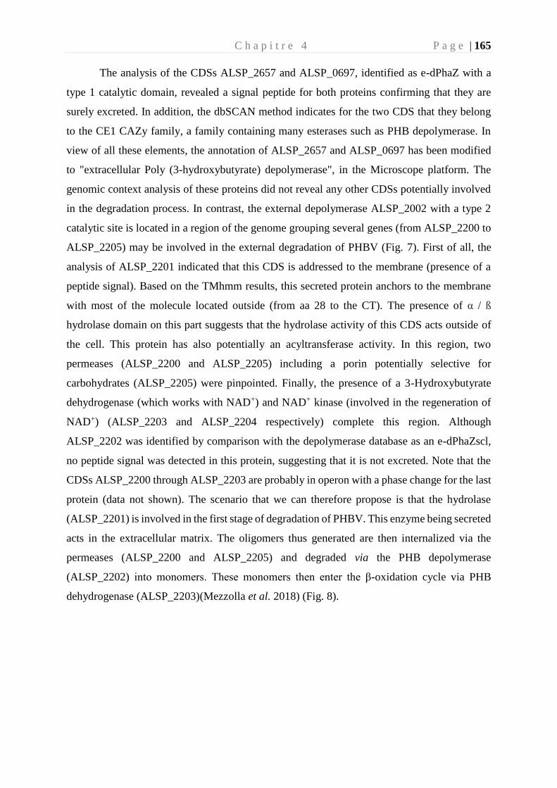
C h a p i t r e 4 P a g e | 165
The analysis of the CDSs ALSP_2657 and ALSP_0697, identified as e-dPhaZ with a
type 1 catalytic domain, revealed a signal peptide for both proteins confirming that they are
surely excreted. In addition, the dbSCAN method indicates for the two CDS that they belong
to the CE1 CAZy family, a family containing many esterases such as PHB depolymerase. In
view of all these elements, the annotation of ALSP_2657 and ALSP_0697 has been modified
to "extracellular Poly (3-hydroxybutyrate) depolymerase", in the Microscope platform. The
genomic context analysis of these proteins did not reveal any other CDSs potentially involved
in the degradation process. In contrast, the external depolymerase ALSP_2002 with a type 2
catalytic site is located in a region of the genome grouping several genes (from ALSP_2200 to
ALSP_2205) may be involved in the external degradation of PHBV (Fig. 7). First of all, the
analysis of ALSP_2201 indicated that this CDS is addressed to the membrane (presence of a
peptide signal). Based on the TMhmm results, this secreted protein anchors to the membrane
with most of the molecule located outside (from aa 28 to the CT). The presence of α / ß
hydrolase domain on this part suggests that the hydrolase activity of this CDS acts outside of
the cell. This protein has also potentially an acyltransferase activity. In this region, two
permeases (ALSP_2200 and ALSP_2205) including a porin potentially selective for
carbohydrates (ALSP_2205) were pinpointed. Finally, the presence of a 3-Hydroxybutyrate
dehydrogenase (which works with NAD+) and NAD+ kinase (involved in the regeneration of
NAD+) (ALSP_2203 and ALSP_2204 respectively) complete this region. Although
ALSP_2202 was identified by comparison with the depolymerase database as an e-dPhaZscl,
no peptide signal was detected in this protein, suggesting that it is not excreted. Note that the
CDSs ALSP_2200 through ALSP_2203 are probably in operon with a phase change for the last
protein (data not shown). The scenario that we can therefore propose is that the hydrolase
(ALSP_2201) is involved in the first stage of degradation of PHBV. This enzyme being secreted
acts in the extracellular matrix. The oligomers thus generated are then internalized via the
permeases (ALSP_2200 and ALSP_2205) and degraded via the PHB depolymerase
(ALSP_2202) into monomers. These monomers then enter the β-oxidation cycle via PHB
dehydrogenase (ALSP_2203)(Mezzolla et al. 2018) (Fig. 8).
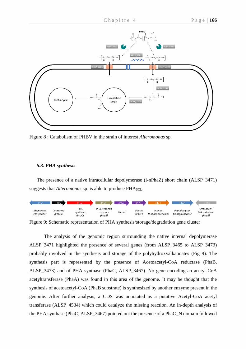
C h a p i t r e 4 P a g e | 166
Figure 8 : Catabolism of PHBV in the strain of interest Alteromonas sp.
5.3. PHA synthesis
The presence of a native intracellular depolymerase (i-nPhaZ) short chain (ALSP_3471)
suggests that Alteromonas sp. is able to produce PHASCL.
Figure 9: Schematic representation of PHA synthesis/storage/degradation gene cluster
The analysis of the genomic region surrounding the native internal depolymerase
ALSP_3471 highlighted the presence of several genes (from ALSP_3465 to ALSP_3473)
probably involved in the synthesis and storage of the polyhydroxyalkanoates (Fig 9). The
synthesis part is represented by the presence of Acetoacetyl-CoA reductase (PhaB,
ALSP_3473) and of PHA synthase (PhaC, ALSP_3467). No gene encoding an acetyl-CoA
acetyltransferase (PhaA) was found in this area of the genome. It may be thought that the
synthesis of acetoacetyl-CoA (PhaB substrate) is synthesized by another enzyme present in the
genome. After further analysis, a CDS was annotated as a putative Acetyl-CoA acetyl
transferase (ALSP_4534) which could catalyze the missing reaction. An in-depth analysis of
the PHA synthase (PhaC, ALSP_3467) pointed out the presence of a PhaC_N domain followed
C h a p i t r e 4 P a g e | 167
by an A / B hydrolase domain involved in the secondary structure of the PHA synthase. These
two domains are signatures of a Type 3 PHA synthase (Liu et al. 2019). As a single CDS appears
to encode a PHA synthase in this region, the activity is probably linked to a dimerization of
ALSP_3467 leading to a PhaC of Class I (Mezzolla et al. 2018). Close to PhaC, a transcriptional
regulator of phasin synthesis (PhaR, ALSP_3468) was identified. The Phasins are the major
components of PHA granules (Maestro and Sanz 2017). These proteins have an essential
contribution in the physical stabilization of granules (also called carbonosomes) in the cell, and
they ensure the proper distribution of polyester during cell division and assist other proteins
(synthases and depolymerases) in the metabolism of PHA. Two phasins (ALSP_3469 and
ALSP_3470) are located in this gene cluster. These two CDS have a signature in the Pfam
database (PF09361) which is one of the 4 phasin Pfam families (Maestro and Sanz 2017). To
complete this genomic region, the proteins ALSP_3465 and ALSP_3472, coding for a
membrane component and for a peptidoglycan transglycosylase respectively, are potentially
components of the granules. Finally, although no information is available to assign an
annotation to it, CDS ALSP_3466, preserved in a large number of Alteromonas and in operon
with ALSP_3467, must certainly have a role in the metabolism of PHA (data not shown).
Further analysis of the genome revealed a region containing another PHA synthase
(ALSP_3115). Like ALSP_3467, this PHA synthase has a PhaC_N domain followed by an A /
B hydrolase domain, defining it as a Type 3 PHA synthase. This PhaC is colocalized with a
bifunctional protein including an (R) -specific enoyl-CoA hydratase (PhaJ) and a phosphate
acetyltransferase (Pta) (ALSP_3116) followed by an acetate kinase (AckA) (ALSP_3117). We
can hypothesize that AckA and Pta leads the synthesis of acetyl-CoA from acetate which then
enter into cell metabolism, while PhaJ and PhaC have a role in the synthesis of medium chain
of PHA from Enoyl-CoA (β-oxidation of fatty acids). As observed for ALSP_3467, the
presence of a unique PhaC in the area suggests that this PHA synthase belongs to a Class I. As
mentioned by Liu et al., there are some rare cases of Class I PHA synthase that can produce
mixed PHAs composed of mixtures of SCL and MCL hydroxyalkanoates (Liu et al. 2019)
PHBV is a copolymer mixing short and medium carbon chains. Based on the annotation
analysis, we can assume that Alteromonas sp. is probably capable of producing PHBV.
C h a p i t r e 4 P a g e | 168
Conclusion
Here, we report a novel marine strain isolated from a microbial consortium degrading Poly(3-
hydroxybutyrate-co-3-hydroxyvalerate) (PHBV). This strain was affiliate to the Alteromonas
genus and was named Alteromonas sp. Different parameters such as degradation halo on an
agar plate, oxygen consumption, bacterial production, weight loss and scanning electron
microscopy were performed to confirm the ability of this strain to degrade PHBV. In addition,
the genome sequencing of this strain reveals three proteins potentially involved in the external
depolymerisation, two of which are isolated in the genome (ALSP_2657 and ALSP_0697).
These two depolymerases have all the characteristics to be excreted and involved in catabolism
of the carbohydrates. Regarding the third external depolymerase (ALSP_2202), no signal
peptide was detected. This last protein is located in a cluster of genes potentially involved in
PHA degradation (Fig 7). A hypothetical pathway is shown Fig 8. The genomic overview also
revealed an internal depolymerase (ALSP_3471) located in a cluster of nine genes (ALSP_3465
to ALSP_3473) possibly involved in the synthesis, storage and degradation of short carbon
chain PHA (Fig 9). Although genes coding for the key enzymes of the pathway I of the PHA
biosynthesis (PhaA, PhaB and PhaC) are frequently organized in one operon, such as
Cupriavidus nectator (Sudesh et al., 2000), no PHA operon was identified in this genome. To
complete the PHA synthesis analysis, an enoyl-CoA hydratase (PhaJ, ALSP_3116) and a PHA
synthase (PhaC, ALSP_3115) were identified which suggests that Alteromonas sp. can also
synthetize medium carbon chain PHA. Experimental validations have to be carried out to
confirm that Alteromonas sp. possesses, to the best of our knowledge, atypical pathways of
degradation and synthesis of PHAs.
C h a p i t r e 4 P a g e | 169
References
Abràmoff M, Magalhães P, and Ram S. 2004. Image processing with ImageJ. biophotonics international
11: 36–42.
Ali I and Jamil N. 2016. Polyhydroxyalkanoates: Current applications in the medical field. Front Biol
11: 19–27.
Amaral-Zettler LA, Zettler ER, and Mincer TJ. 2020. Ecology of the plastisphere. Nat Rev Microbiol
18: 139–51.
Chang H-F, Chang W-C, and Tsai C-Y. 2012. Synthesis of poly(3-hydroxybutyrate/3-hydroxyvalerate)
from propionate-fed activated sludge under various carbon sources. Bioresour Technol 113: 51–
7.
Donlan RM. 2002. Biofilms: microbial life on surfaces. Emerging Infect Dis 8: 881–90.
Dussud C, Hudec C, George M, et al. 2018. Colonization of Non-biodegradable and Biodegradable
Plastics by Marine Microorganisms. Front Microbiol 9.
Eubeler JP, Zok S, Bernhard M, and Knepper TP. 2009. Environmental biodegradation of synthetic
polymers I. Test methodologies and procedures. TrAC Trends in Analytical Chemistry 28:
1057–72.
Fegatella F, Lim J, Kjelleberg S, and Cavicchioli R. 1998. Implications of rRNA operon copy number
and ribosome content in the marine oligotrophic ultramicrobacterium Sphingomonas sp. strain
RB2256. Appl Environ Microbiol 64: 4433–8.
Gorke JT, Okrasa K, Louwagie A, et al. 2007. Enzymatic synthesis of poly(hydroxyalkanoates) in ionic
liquids. Journal of Biotechnology 132: 306–13.
Grimaud R, Ghiglione J-F, Cagnon C, et al. 2012. Genome sequence of the marine bacterium
Marinobacter hydrocarbonoclasticus SP17, which forms biofilms on hydrophobic organic
compounds. Journal of Bacteriology 194: 3539–40.
Handrick R, Reinhardt S, and Jendrossek D. 2000. Mobilization of poly(3-hydroxybutyrate) in Ralstonia
eutropha. J Bacteriol 182: 5916–8.
Handrick R, Reinhardt S, Kimmig P, and Jendrossek D. 2004. The “intracellular” poly(3-
hydroxybutyrate) (PHB) depolymerase of Rhodospirillum rubrum is a periplasm-located
protein with specificity for native PHB and with structural similarity to extracellular PHB
depolymerases. J Bacteriol 186: 7243–53.
Jacquin J, Cheng J, Odobel C, et al. 2019. Microbial Ecotoxicology of Marine Plastic Debris: A Review
on Colonization and Biodegradation by the “Plastisphere.” Front Microbiol 10.
Jambeck JR, Geyer R, Wilcox C, et al. 2015. Marine pollution. Plastic waste inputs from land into the
ocean. Science 347: 768–71.
Jendrossek D. 2005. Extracellular Polyhydroxyalkanoate (PHA) Depolymerases: The Key Enzymes of
PHA Degradation. In: Biopolymers Online. American Cancer Society.
C h a p i t r e 4 P a g e | 170
Jendrossek D. 2007. Peculiarities of PHA granules preparation and PHA depolymerase activity
determination. Appl Microbiol Biotechnol 74: 1186–96.
Jendrossek D and Handrick R. 2002. Microbial degradation of polyhydroxyalkanoates. Annu Rev
Microbiol 56: 403–32.
Knoll M, Hamm TM, Wagner F, et al. 2009. The PHA Depolymerase Engineering Database: A
systematic analysis tool for the diverse family of polyhydroxyalkanoate (PHA) depolymerases.
BMC Bioinformatics 10: 89.
Kong D, Li W, Deng Y, et al. 2018. Denitrification-Potential Evaluation and Nitrate-Removal-Pathway
Analysis of Aerobic Denitrifier Strain Marinobacter hydrocarbonoclasticus RAD-2. Water 10:
1298.
Kumar S, Nei M, Dudley J, and Tamura K. 2008. MEGA: A biologist-centric software for evolutionary
analysis of DNA and protein sequences. Brief Bioinform 9: 299–306.
Lebaron P, Ghiglione J-F, Fajon C, et al. 1998. Phenotypic and genetic diversity within a colony
morphotype. FEMS Microbiol Lett 160: 137–43.
Liu Z, Zhu Z, Yang J, et al. 2019. Domain-centric dissection and classification of prokaryotic poly(3-
hydroxyalkanoate) synthases. bioRxiv: 693432.
Maestro B and Sanz JM. 2017. Polyhydroxyalkanoate-associated phasins as phylogenetically
heterogeneous, multipurpose proteins. Microbial Biotechnology 10: 1323–37.
Martinez Tobon DI. 2019. Biodegradation of Polyhydroxybutyrate by Bacterial Strains, Native
Extracellular PHB Depolymerases, and Structural Variants.
Meng D-C, Shen R, Yao H, et al. 2014. Engineering the diversity of polyesters. Curr Opin Biotechnol
29: 24–33.
Mezzolla V, D’Urso OF, and Poltronieri P. 2018. Role of PhaC Type I and Type II Enzymes during
PHA Biosynthesis. Polymers (Basel) 10.
Mothes G, Ackermann J-U, and Babel W. 2004. Mole Fraction Control of Poly([R]-3-hydroxybutyrate-
co-3-hydroxyvalerate) (PHB/HV) Synthesized by Paracoccus denitrificans. Engineering in Life
Sciences 4: 247–51.
Oberbeckmann S, Löder MGJ, and Labrenz M. 2015. Marine microplastic-associated biofilms – a
review. Environ Chem 12: 551–62.
Ong SY, Chee JY, School of Biological Sciences Universiti Sains Malaysia, et al. 2017. Degradation of
Polyhydroxyalkanoate (PHA): a Review. J Sib Fed Univ, Biol 10: 21–225.
Peña C, López S, García A, et al. 2014. Biosynthesis of poly-β-hydroxybutyrate (PHB) with a high
molecular mass by a mutant strain of Azotobacter vinelandii (OPN). Ann Microbiol 64: 39–47.
Reis MAM, Serafim LS, Lemos PC, et al. 2003. Production of polyhydroxyalkanoates by mixed
microbial cultures. Bioprocess Biosyst Eng 25: 377–85.
Simon M and Azam F. 1989. Protein content and protein synthesis rates of planktonic marine bacteria.
Marine Ecology Progress Series 51: 201–13.
Sudesh K, Abe H, and Doi Y. 2000. Synthesis, structure and properties of polyhydroxyalkanoates:
biological polyesters. Progress in Polymer Science 25: 1503–55.
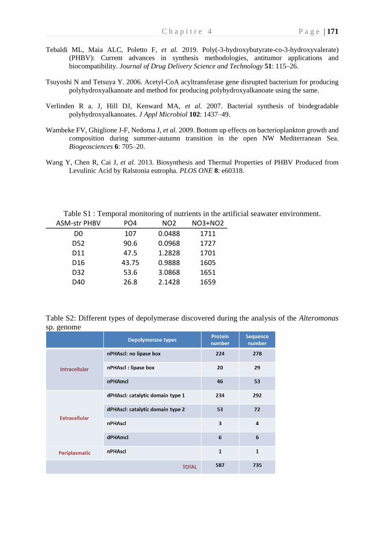
C h a p i t r e 4 P a g e | 171
Tebaldi ML, Maia ALC, Poletto F, et al. 2019. Poly(-3-hydroxybutyrate-co-3-hydroxyvalerate)
(PHBV): Current advances in synthesis methodologies, antitumor applications and
biocompatibility. Journal of Drug Delivery Science and Technology 51: 115–26.
Tsuyoshi N and Tetsuya Y. 2006. Acetyl-CoA acyltransferase gene disrupted bacterium for producing
polyhydroxyalkanoate and method for producing polyhydroxyalkanoate using the same.
Verlinden R a. J, Hill DJ, Kenward MA, et al. 2007. Bacterial synthesis of biodegradable
polyhydroxyalkanoates. J Appl Microbiol 102: 1437–49.
Wambeke FV, Ghiglione J-F, Nedoma J, et al. 2009. Bottom up effects on bacterioplankton growth and
composition during summer-autumn transition in the open NW Mediterranean Sea.
Biogeosciences 6: 705–20.
Wang Y, Chen R, Cai J, et al. 2013. Biosynthesis and Thermal Properties of PHBV Produced from
Levulinic Acid by Ralstonia eutropha. PLOS ONE 8: e60318.
Table S1 : Temporal monitoring of nutrients in the artificial seawater environment.
ASM-str PHBV PO4 NO2 NO3+NO2
D0 107 0.0488 1711 D52 90.6 0.0968 1727 D11 47.5 1.2828 1701 D16 43.75 0.9888 1605 D32 53.6 3.0868 1651 D40 26.8 2.1428 1659
Table S2: Different types of depolymerase discovered during the analysis of the Alteromonas
sp. genome
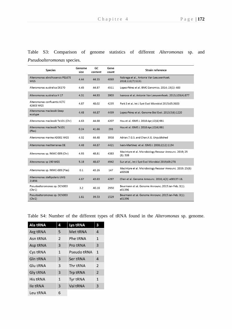
C h a p i t r e 4 P a g e | 172
Table S3: Comparison of genome statistics of different Alteromonas sp. and
Pseudoalteromonas species.
Table S4: Number of the different types of tRNA found in the Alteromonas sp. genome.
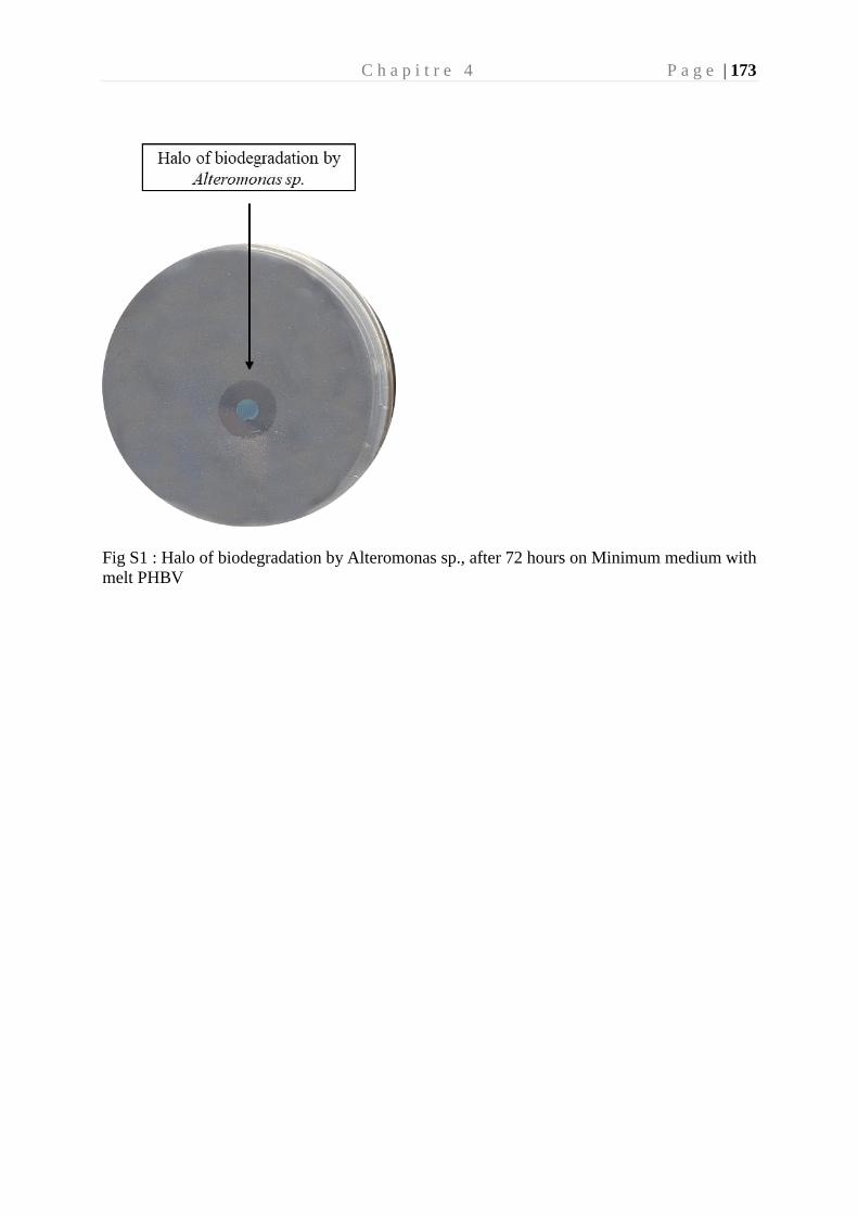
C h a p i t r e 4 P a g e | 173
Fig S1 : Halo of biodegradation by Alteromonas sp., after 72 hours on Minimum medium with
melt PHBV


Chapitre 5
Conclusions et Perspectives
© Hedenberg Frej

C h a p i t r e 5 P a g e | 177
“Insanity is doing the same thing over and over again and expecting different results”
Einstein
1. Conclusion
Une des originalités de cette thèse a été de travailler à différents niveaux d’intégrations,
allant de la comparaison d’écosystèmes à l’échelle planétaire (expéditions Tara-Méditerranée
2014 et Tara-Pacifique 2018), de travaux menés à l’échelle locale (aquariums en circulation
ouverte sur la Baie de Banyuls sur mer) jusqu’à l’échelle de l’individu et de son génome
(isolement d’une souche bactérienne en laboratoire). Ces différents niveaux d’intégrations
m’ont permis d’aborder différentes facettes de l’écotoxicologie microbienne marine, en
étudiant l’impact de la pollution plastique sur les bactéries marines (la plastisphère) et
inversement, le rôle des bactéries sur le devenir des plastiques en mer (la biodégradation).
1.1. Etude de la plastisphère au niveau mondial
L’étude de la plastisphère mondiale a été permise grâce à deux expéditions menées par
la fondation Tara Océan. La première a été menée en 2014 en mer Méditerranée et la seconde
à laquelle j’ai eu l’opportunité de participer en 2018 dans le gyre du Pacifique Nord. Ces deux
expéditions d’une envergure exceptionnelle ont permis l’étude de plusieurs centaines
d’échantillons à travers deux régions du monde fortement touchées par cette pollution. Le
plastique est devenu en quelques années un paramètre traduisant la santé des océans, au même
titre que d’autres sources tels que le changement climatique, l’acidification des océans, la
surpêche ou la destruction de l’habitat.
Jusqu’à présent, la biogéographie de la plastisphère restait peu étudiée et les facteurs
influençant les communautés microbiennes à travers les océans étaient encore un sujet de débat.
Quelques études s’étaient attachées à comparer la plastisphère dans différents bassins
océaniques à différentes échelles, comme celle de Amaral-Zettler et al. (2015) entre le Pacifique
et l’Atlantique, de Basili et al. (2020) dans le bassin méditerranéen à deux endroits distincts, de
Xu et al. (2019) entre la mer jaune et la mer de Chine méridionale ou de Oberbeckmann et al.
(2016) entre deux zones de la mer du Nord à deux endroits distincts également. Dans le monde
scientifique, deux hypothèses s’affrontent, la première étant que les communautés retrouvées
sur les plastiques devraient être homogènes car fortement dépendant de la composition
chimique de ce dernier et la seconde étant que les communautés microbiennes sont fortement

C h a p i t r e 5 P a g e | 178
liées aux facteurs environnementaux et dans une moindre mesure à la composition chimique du
polymère. Si les communautés varient effectivement en fonction des paramètres
environnementaux et donc de leur géographie, alors il y aura une variation régionale importante
dans le transport d’espèces invasives, nuisibles ou pathogènes à travers les différents bassins
océaniques (Zettler 2013; Amaral-Zettler et al. 2015).
Une partie des travaux de cette thèse ont été concentrés sur les communautés
microbiennes des plastiques trouvés à la surface de l’eau de mer de deux régions distinctes du
globe. La première conclusion a été que les communautés microbiennes vivant sur les plastiques
(PMD) sont très différentes par rapport au milieu environnant à l’état libre (FL) ou attachées
aux particules organiques (PA) dans le Pacifique, comme en méditerranée. Ce résultat vient
conforter de nombreuses études qui font état de cette distinction (Oberbeckmann et al. 2014;
De Tender et al. 2015; Bryant et al. 2016; Basili et al. 2020). Dans cette étude, nous avons aussi
observé un cloisonnement net dû à la géographie sur les fractions PMD, FL et PA entre le
Pacifique et la Méditerranée. Ces résultats semblent converger avec les premières observations
faites à travers différents endroits du globe (Amaral-Zettler et al. 2015; Oberbeckmann et al.
2016; Xu et al. 2019; Basili et al. 2020). Le principal facteur favorisant le cloisonnement
géographique dans notre étude est la température.
Lorsque nous avons étudié les différents types des polymères de la fraction PMD, nous
n’avons constaté aucun lien entre la nature du polymère et la communauté bactérienne de la
plastisphère. Cela contredit différentes études menées sur des plastiques vierges incubés en eau
de mer (Oberbeckmann et al. 2018; Dussud et al. 2018; Pinto et al. 2019). Cela peut s’expliquer
par le temps de résidence des différents plastiques prélevés. En effet, la composition du
polymère et surtout ses propriétés de surface (cristallinité, hydrophobicité, rugosité, …),
peuvent influencer l’attachement de certains genres bactériens à court terme. Cependant l’effet
à long terme des facteurs environnementaux semble prédominer sur les plastiques retrouvés
dans l’environnement et provoquera une convergence des communautés microbiennes sur le
biofilm mature, expliquant cette différence avec les études précédemment citées.
Les deux zones géographiques ne sont pas reliées entre elles directement, mais pourtant
un microbiome commun (ou « core microbiome ») a pu être identifié sur la fraction PMD. Ce
core microbiome est principalement composé d’Alphaproteobacteries, de Cyanobacteries et de
Bacteroidia. Ces classes sont également retrouvées dans l’eau environnante, ce qui suggère un
échange avec les fractions libres et/ou attachées aux particules organiques. Nous avons révélé
C h a p i t r e 5 P a g e | 179
que la cyanobactérie Rivularia sp. appartenant à la famille des Rivulariaceae (connue également
sous Calothrix sp.) était le genre le plus abondant du « core microbiome ». D’autres taxons ont
été identifiés comme composants principaux du core microbiome, comme Tateyamaria sp.
(Rhodobacteraceae ), Muricauda sp. ( Flavobacteriaceae), ou encore Pseudahrensia sp.
(Rhizobiaceae ). Ces genres sont connus dans la littérature pour se développer sur des
polymères et être impliqués dans des biofilms marins (Lu et al. 2019; Rogers et al. 2020). La
présence de ce « core microbiome » est explicable par des facteurs abiotiques, principalement
température, salinité, nutriments, et dans une moindre mesure par des facteurs biotiques
(Amaral-Zettler et al. 2020). Les bactéries sessiles, ayant des membranes hydrophobes, pouvant
produire des EPS et favorisant un mode de vie attaché vont être avantagées dans l’adhésion à
la surface des plastiques.
La colonisation des plastiques par ces genres bactériens n’est pas le signe d’une
biodégradation du polymère par cette communauté. L’analyse de la capacité des
microorganismes à biodégrader les plastiques requiert généralement une étude en laboratoire.
Dans la partie suivante, nous nous sommes attachés à conserver un consortium bactérien naturel
pour en évaluer la capacité à biodégrader différents polymères en milieu marin.
1.2. Evolution de la plastisphère vers un consortium capable de dégrader
les polymères
Le laboratoire d’océanographie microbienne (LOMIC UMR7621) a développé depuis
quelques années des incubations en milieu ouvert sur la baie de Banyuls pour mimer le milieu
naturel en conditions contrôlées. Ces aquariums permettent de suivre la colonisation des
plastiques conventionnels et/ou biodégradables. Les premières expériences menées dans ces
microcosmes ont montré une bonne représentativité des communautés microbiennes vivant sur
les plastiques dans ces aquariums par rapport à celle de l’environnement (Dussud et al. 2018).
Cet avantage a été mis au service du projet BIOTIGES financé par le ministère de la
transition écologique et solidaire qui avait pour objectif d’accompagner le gouvernement
français sur la recherche d’alternatives aux plastiques conventionnels utilisés dans la tige des
coton-tiges. Cette étude nous a permis de participer à l’écriture d’un décret modificatif du décret
d’application n°2017-291 du 6 mars 2017 pris pour application de l’article 24 de la loi n° 2016-
C h a p i t r e 5 P a g e | 180
1087 du 8 août 2016 pour la reconquête de la biodiversité, de la nature et des paysages modifiant
l’article L. 541-10-5 du code de l’environnement. Ce texte réglementaire est relatif aux
conditions de mise en œuvre de l’interdiction de mise sur le marché des produits cosmétiques
rincés à usage d’exfoliation ou de nettoyage comportant des particules plastiques solides et des
bâtonnets ouatés à usage domestique dont la tige est en plastique. La loi du 10 février 2020 (loi
2020-105) a été promulguée pour lutter contre le gaspillage, favoriser l’économie circulaire afin
de réduire les déchets et préserver les ressources naturelles. La loi s’articule autour de plusieurs
thématiques dont la réduction des déchets plastiques (Article 7). Cet Article prévoit la fin de la
mise sur le marché de plastique à usage unique d’ici à 2040. Des objectifs de réduction d’usage
de plastique sont fixés par décret pour la période 2021-2025, puis tous les 5 ans jusqu’en 2040.
L’interdiction des cotons-tiges en plastique est également soumise à la loi biodiversité du 8 aout
2016 et interviendra dès l’année 2020. Notre rôle a été d’étudier les solutions alternatives aux
plastiques conventionnels classiquement utilisés pour les tiges de coton-tiges (généralement
fabriqués en polypropylène, PP).
Actuellement de nombreux polymères considérés comme biodégradables sont mis sur
le marché. Certains issus de la pétrochimie (PBS and PBAT), d’autre d’origine biosourcés (PLA
et PHBV) et d’autres provenant d’amidon (Bioplast et Matert-Bi). Cependant, la colonisation
de ces polymères par les communautés microbiennes et l’impact sur la biodégradabilité de ces
polymères en milieu marin étaient jusqu’alors inconnus dans la littérature.
Nous avons mis en évidence que quel que soit le type de polymère, la colonisation
microbienne suivait une succession de phases de croissance puis de maturation. Ces résultats
corroborent les précédentes études réalisées par notre équipe sur d’autres plastiques sous forme
de films composés de polymères oxo-dégradables et de PHBV (Dussud et al. 2018). La phase
de croissance était caractérisée par une activité bactérienne maximale entre 7 et 15 jours pour
l’ensemble des polymères. La phase de maturation du biofilm a été caractérisée par une baisse
de la production bactérienne, comme l’avait déjà démontré la précédente étude faite dans les
mêmes conditions (Dussud et al. 2018).
Aucune spécificité des communautés microbiennes liée à la nature des polymères n’a
été observée pendant la phase de croissance du biofilm. Bien que surprenant au premier abord
compte tenu de la diversité des polymères testés dans cette étude, ces résultats sont concordants
avec d’autres études menées sur la formation de biofilm sur des plastiques vierges (Harrison et
al. 2014; De Tender et al. 2017). Fait intéressant, lorsque l’on compare les communautés
C h a p i t r e 5 P a g e | 181
microbiennes identifiées sur nos plastiques vierges incubés avec celles des plastiques in situ
trouvés en Méditerranée ou dans le Pacifique, il s’avère que les premières étapes de colonisation
du papier et du Bioplast présentent des similitudes avec les plastiques échantillonnés en
Méditerranée, au large de Nice (Fig. 32). Cela confirme la représentativité de notre procédé
expérimental qui mime les conditions naturelles en conditions contrôlées. Cela montre
également que le biofilm constitué sur les polymères au bout de 40 jours correspondent bien à
un biofilm mature qui peut être retrouvé dans l’environnement.
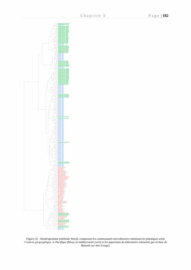
C h a p i t r e 5 P a g e | 182
Figure 32 : Dendrogramme (méthode Ward), comparant les communautés microbiennes colonisant les plastiques selon
l’endroit géographique, le Pacifique (bleu), la méditerranée (vert) et les aquariums du laboratoire alimentés par la baie de
Banyuls sur mer (rouge).
wa
rd.D
2 lin
ka
ge
clu
ste
rin
g t
ree
mediterranean M10
mediterranean M238
mediterranean M238
mediterranean M238
mediterranean M263
mediterranean M263
mediterranean M19
mediterranean M266
mediterranean M266
mediterranean M165
mediterranean M269
mediterranean M269
mediterranean M19
mediterranean M277
mediterranean M19
mediterranean M280
mediterranean M280
mediterranean M280
mediterranean M283mediterranean M283
mediterranean M19
mediterranean M285
mediterranean M285mediterranean M285
mediterranean M20
mediterranean M20
mediterranean M20
mediterranean M10
mediterranean M20
mediterranean M32
mediterranean M32
mediterranean M32
mediterranean M32
mediterranean M10
mediterranean M38
mediterranean M42
mediterranean M42
mediterranean M42
mediterranean M47
mediterranean M47
mediterranean M47mediterranean M51
mediterranean M51
mediterranean M56
mediterranean M60
mediterranean M60
mediterranean M60
mediterranean M15
mediterranean M66
mediterranean M66
mediterranean M66
mediterranean M165mediterranean M165
mediterranean M165
mediterranean M15
mediterranean M173
mediterranean M173
mediterranean M173
mediterranean M173
mediterranean M178
mediterranean M178
mediterranean M178
mediterranean M15
mediterranean M189
mediterranean M189
mediterranean M189
mediterranean M193
mediterranean M193
mediterranean M193
mediterranean M193
mediterranean M202
mediterranean M202
mediterranean M202
mediterranean M234
mediterranean M234
pacific 192
pacific 192pacific 192
pacific 192
pacific 194
pacific 194
pacific 197
pacific 197
pacific 198
pacific 198
pacific 198
pacific 198
pacific 198
pacific 198
pacific 198
pacific 198
pacific 198
pacific 198
pacific 198
pacific 198
pacific 198
pacific 198
pacific 198
pacific 198
pacific 198
pacific 198pacific 198
pacific 198
pacific 198
pacific 198
pacific 198
pacific 198
pacific 198
pacific 198
pacific 198
pacific 198
pacific 198
pacific 198
pacific 198
pacific 198
pacific 198
pacific 198
pacific 198
pacific 198
pacific 198
pacific 198
pacific 199
pacific 199
pacific 199pacific 199
pacific 199
pacific 199
pacific 199
pacific 199
pacific 199pacific 199
pacific 199
pacific 199
pacific 199
pacific 199
pacific 199
pacific 199
pacific 199
pacific 199
pacific 199
pacific 199
pacific 199
pacific 199
pacific 199
pacific 199
pacific 199pacific 199
pacific 199
pacific 199
pacific 199
pacific 199
pacific 200
pacific 200
pacific 200
pacific 200
pacific 200
pacific 200
pacific 200pacific 200
pacific 200
pacific 200
pacific 200
pacific 201
pacific 201
pacific 201
pacific 201pacific 201
pacific 201
pacific 201
pacific 192
pacific 194
pacific 197
pacific 201
pacific 201
pacific 201pacific 201
pacific 201
pacific 201pacific 201
BIOPLAST D15
BIOPLAST D40+15
BIOPLAST D40+30
BIOPLAST D40
BIOPLAST D7
BIOPLAST D40+7
BIOPLAST D40+94
BIOPLAST D40+3
MATER D15
MATER D40+3
MATER D40+15MATER D40+30
MATER D40
MATER D7
MATER D40+7
MATER D40+94
PBAT D15
PBAT D40+15
PBAT D40+30
PBAT D40+3
PBAT D40
PBAT D7
PBAT D40+7
PBAT D40+94
PBS D15
PBS D40+15
PBS D40+30
PBS D40+3
PBS D40
PBS D7
PBS D40+7
PBS D40+94
PHA D15
PHA D40+15
PHA D40+30
PHA D40+3
PHA D40
PHA D7
PHA D40+7
PHA D40+94
PLA D40+7
PLA D15
PLA D40+30
PLA D40+3
PLA D40
PLA D7
PLA D40+94
PP D40+3
PP D40+15
PP D15
PP D40+30
PP D40
PP D7
PP D40+7PP D40+94
PAPER D15
PAPER D40+15
PAPER D40+30
PAPER D40+3
PAPER D40
PAPER D7
PAPER D40+7
PAPER D40+94
Bio
tige
Mediterranée
Pacifi
c
C h a p i t r e 5 P a g e | 183
L’étude de la biodégradation a impliqué le transfert de ces polymères et de leur biofilm
mature dans un milieu artificiel d’eau de mer sans aucune autre source de carbone. L’étude de
la biodégradation des plastiques en milieu minimum imitant les conditions marines n’avait
encore jamais été réalisée en laboratoire, ce qui dénote d’une autre originalité de ce travail de
thèse en terme cette fois de procédé expérimental pour mettre en évidence la biodégradabilité
des polymères en milieu marin. Une enveloppe SOLEAU a été déposée en Décembre 2017
(DSO2017012612 : Protocole pour l’évaluation de la biodégradabilité des plastiques en milieu
marin) par JF Ghiglione pour protéger ce procédé expérimental original.
La caractérisation de la biodégradation a été réalisée de manière originale par le suivi
dynamique de la production bactérienne tout au long de l’incubation et par une confirmation
visuelle de changement de morphologie du polymère par microscopie électronique, qui sont des
paramètres non pris en compte dans les normes actuelles. L’étude de la dynamique de l’activité
bactérienne a permis de montrer que les communautés continuaient d’avoir une activité
plusieurs jours après le transfert en milieu minimum comme sur le PP, témoin négatif de
l’expérience. Cette activité est certainement liée à la matière organique déposée sur le polymère
lors de la colonisation par le biofilm en milieu naturel et non à une biodégradation du polymère
lui-même. L’activité bactérienne n’a pas été maintenue au-delà des 15 jours sur des polymères
non-biodégradables tels que le PP, le PLA et le PBS. Les observations en microscopie
électronique ont montré des bactéries isolées et une surface des polymères inchangée. Tous ces
signaux convergent pour indiquer que ces polymères n’ont pas été dégradés dans nos conditions
expérimentales. Il est à souligner que le PLA est pourtant considéré comme biodégradable en
sol et en compost (Ren et al. 2007), mais nos travaux n’ont pu montrer sa biodégradabilité en
milieu marin dans nos conditions expérimentales. Les communautés microbiennes sur les
polymères PHBV, Bioplast, Mater-bi, et la cellulose ont continué à être actives et leur impact
sur la surface du polymère a pu être caractérisé au microscope électronique, suggérant que ces
polymères sont biodégradables en milieu marin.
L’incubation en milieu minimum a eu pour effet de faire évoluer les communautés
microbiennes qui sont devenues spécifiques de chaque polymère. Une attention particulière a
été portée sur les communautés microbiennes qui étaient encore actives après 3 mois avec le
polymère comme seule source de carbone, que l’on considère comme capable de
biodégradation du polymère. Les genres devenus majoritaires en fin d’incubation (D40+94)
C h a p i t r e 5 P a g e | 184
appartenaient tous à la biosphère rare (<1% de la communauté totale) du biofilm mature naturel
(D40). Nos résultats suggèrent que nous avons découvert des activités de biodégradation des
polymères insoupçonnés pour ces genres bactériens. En effet la majorité des genres sélectionnés
ne sont pas connus dans la littérature pour dégrader ces polymères, même s’ils ont en général
déjà été identifiés comme capables de dégrader des hydrocarbures ou comme faisant partie de
la plastisphère en milieu naturel. Etonnamment, les genres sélectionnés ne sont pas
majoritairement des hydrocarbonoclastes, bien que plusieurs études ont fait un lien entre les
hydrocarbonoclastes et leurs activités potentielles de biodégradation (Bryant et al. 2016; Pollet
et al. 2018; Amaral-Zettler et al. 2020).
Les normes actuelles se contentent généralement de mesurer la respiration des
communautés microbiennes (inoculum pouvant être artificiel et non représentatif du milieu
d’étude) et impose un pourcentage de conversion en CO2 du carbone organique du polymère
(entre 60 et 70 %) entre 3 et 24 mois d’incubation. Cependant, notre étude a révélé deux points
faibles à ces normes. La première est que la matière organique provenant de l’environnement
marin est suffisante pour permettre une activité bactérienne mesurable et non négligeable sur le
PP (polymère non biodégradable) et la seconde est que les communautés microbiennes sont
composées de cyanobactéries (autotrophe) pouvant interférer sur les résultats de respirométrie
analysés par les normes. Ces résultats suggèrent que les protocoles utilisés actuellement dans
les normes peuvent conduire à des faux-positifs. Ils soulignent que les tests de respirométrie ne
sont pas suffisants et montrent l’importance d’approches pluridisciplinaires qui permettent de
tenir compte des différentes étapes de la biodégradation.
1.3. Etude d’une souche appartenant à ce consortium afin d’identifier les
gènes clefs impliqués dans la biodégradation du PHA
La souche Alteromonas sp. a été isolée à partir d’un fragment de PHBV ayant séjourné
dans un aquarium ouvert sur la baie de Banyuls sur Mer pendant 40 jours puis transféré 94 jours
en milieu minimum (Expérience : chapitre 3).
Le genre Alteromonas sp. est déjà connu dans la littérature pour dégrader le PHB en eau
de mer (Mergaert et al. 1995). Alteromonas sp. est aussi connu comme étant hydrocarbonoclaste
pouvant dégrader le naphtalène, le phénantrène, l’anthracène et le pyrène (Jin et al. 2012) et
ayant un rôle clef dans la biodégradation de ces hydrocarbures. Le genre Alteromonas sp. est
C h a p i t r e 5 P a g e | 185
présent en abondance en Méditerranée occidentale en surface ou en profondeur jusqu’à 400m
(Acinas et al. 1999). Ce genre bactérien était abondant pendant la phase de colonisation en
aquarium sur le Bioplast, le Mater, le PHBV et la cellulose (entre 0.21 % et 57% d’abondance
relative). Ensuite son abondance relative a fortement chutée lorsque l’incubation a été transférée
en milieu minimum (moyenne de 0.25 %) à l’exception du PHBV qui a vu le genre Alteromonas
sp. devenir fortement abondant (30%) après 30 jours en milieu minimum. Ce genre n’est que
très peu abondant sur les plastiques issus de la pétrochimie (PP, PBAT et PBS) dans les
conditions de nos aquariums en circulation ouverte sur la baie de Banyuls sur mer (en moyenne
0.8%) ou sur les plastiques in situ prélevés en Méditerranée (en moyenne 0.4% de l’abondance
relative) et dans le Pacifique (en moyenne 0.07% de l’abondance relative) . Notons d’ailleurs
que la composition des échantillons issus des missions sont essentiellement composés de PE
(Polyéthylène) et PP.
Le genre Alteromonas sp. a montré des signes de biodégradation du PHBV très
significatifs en laboratoire, avec une forte consommation d’oxygène pendant 40 jours (22.45
µmol/jour), une activité bactérienne maintenue tout au long de l’incubation ( 28738 ng C/m²/h
après 40 jours) et une fragilisation de la surface du PHBV (fissures, trous et apparition de
structures caractéristiques de la biodégradation).
Après séquençage de l’ADN génomique de cette souche l’étude de son unique
chromosome a permis d’identifier des gènes impliqués dans la biodégradation du PHBV ainsi
que ceux intervenants dans la synthèse des PHA. L’exploration du génome d’Alteromonas sp.
a révélé la présence de 4 gènes identifiés comme étant des dépolymérases, dont 3 externes et 1
interne. L’analyse plus approfondie de l’annotation de ces gènes a permis d’identifier que les
CDSs codant deux des dépolymérases externes (ALSP_2657 et ALSP_0697) possédaient un
signal peptide laissant suggérer une excrétion de ces protéines. Une autre dépolymérase externe
(ALSP_2202) semble être maintenue dans le cytoplasme. En revanche, une hydrolase
(ALSP_2201), non identifiée comme dépolymérase, possède tous les domaines nécessaires
pour être adressée à la membrane. Cette hydrolase agit certainement dans le milieu
extracellulaire. Les deux gènes codant ces protéines font partie d’un cluster de gènes
probablement impliqués dans la biodégradation du PHBV. En effet, l’hypothèse qui est émise
est que le PHBV est internalisé après hydrolyse grâce aux perméases présentes dans le cluster.
Ensuite, les oligomères sont dégradés en monomère grâce à la dépolymérase. Pour finir, ces
monomères sont intégrés au cycle de β-oxydation afin d’être assimilés par la bactérie (Mezzolla
C h a p i t r e 5 P a g e | 186
et al. 2018). La présence de l’ensemble de ces dépolymérases dans le génome d’Alteromonas
sp. explique la capacité de la souche à dégrader le PHBV externe (Jendrossek and Handrick
2002; Knoll et al. 2009), ce qui est cohérant avec les premiers résultats de métagénomique
effectué sur du PHA (Pinnell and Turner 2019). Ces résultats faisaient état d’un enrichissement
de gènes codant des dépolymérases (multiplié par 20 par rapport aux autres polymères) dans
une communauté microbienne attachée aux PHA. De plus cette étude rapporte également qu’il
existe dans le métagénome un grand nombre de gènes codant des dépolymérases différentes,
signifiant une distribution hétérogène à travers le consortium microbien mixte.
L’étude du génome d’Alteromonas sp. a permis d’identifier également deux voies de
biosynthèse de PHA, l’une permettant la synthèse de PHASCL et l’autre de PHAMCL. L’activité
de synthèse du PHA a été mise en évidence grâce à la présence de deux régions du génome dont
une regroupant entre autres une dépolymérase interne (i-nPhaZ) une Acetoacetyl-CoA
reductase (PhaB), une PHA synthase (PhaC) et des phasines. Dans l’autre région, une autre
PHA synthase et une (R)-specific enoyl-CoA hydratase (phaJ) ont été mise en évidence.
Ces analyses sont basées sur l’étude du génome complet d’Alteromonas sp. Afin de
confirmer ces hypothèses, une seconde expérience a été réalisée pour observer l’expression des
gènes lorsqu’Alteromonas sp. n’utilise que du PHBV comme source de carbone (analyse
transcriptomique). Pour se faire, Alteromonas sp. a été cultivée dans un milieu minimum
contenant du glucose (2mM) (pendant 48h), ou du PHBV (pendant 11 jours) comme source de
carbone. L’extraction des ARNs a été réalisée et les échantillons sont en cours de séquençage
au Génoscope (Evry, France). Cette expérience aura pour but de confirmer ou d’infirmer nos
observations sur l’implications des gènes dans le processus de dépolymérisation du PHBV.
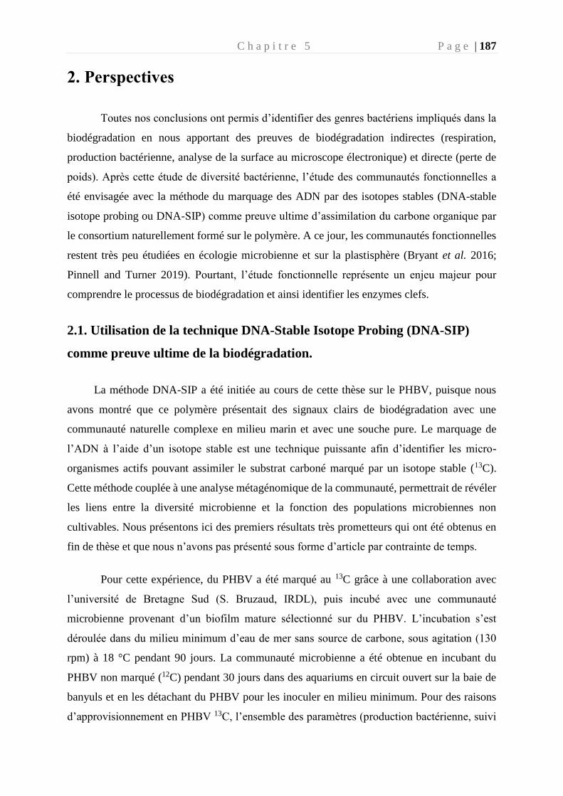
C h a p i t r e 5 P a g e | 187
2. Perspectives
Toutes nos conclusions ont permis d’identifier des genres bactériens impliqués dans la
biodégradation en nous apportant des preuves de biodégradation indirectes (respiration,
production bactérienne, analyse de la surface au microscope électronique) et directe (perte de
poids). Après cette étude de diversité bactérienne, l’étude des communautés fonctionnelles a
été envisagée avec la méthode du marquage des ADN par des isotopes stables (DNA-stable
isotope probing ou DNA-SIP) comme preuve ultime d’assimilation du carbone organique par
le consortium naturellement formé sur le polymère. A ce jour, les communautés fonctionnelles
restent très peu étudiées en écologie microbienne et sur la plastisphère (Bryant et al. 2016;
Pinnell and Turner 2019). Pourtant, l’étude fonctionnelle représente un enjeu majeur pour
comprendre le processus de biodégradation et ainsi identifier les enzymes clefs.
2.1. Utilisation de la technique DNA-Stable Isotope Probing (DNA-SIP)
comme preuve ultime de la biodégradation.
La méthode DNA-SIP a été initiée au cours de cette thèse sur le PHBV, puisque nous
avons montré que ce polymère présentait des signaux clairs de biodégradation avec une
communauté naturelle complexe en milieu marin et avec une souche pure. Le marquage de
l’ADN à l’aide d’un isotope stable est une technique puissante afin d’identifier les micro-
organismes actifs pouvant assimiler le substrat carboné marqué par un isotope stable (13C).
Cette méthode couplée à une analyse métagénomique de la communauté, permettrait de révéler
les liens entre la diversité microbienne et la fonction des populations microbiennes non
cultivables. Nous présentons ici des premiers résultats très prometteurs qui ont été obtenus en
fin de thèse et que nous n’avons pas présenté sous forme d’article par contrainte de temps.
Pour cette expérience, du PHBV a été marqué au 13C grâce à une collaboration avec
l’université de Bretagne Sud (S. Bruzaud, IRDL), puis incubé avec une communauté
microbienne provenant d’un biofilm mature sélectionné sur du PHBV. L’incubation s’est
déroulée dans du milieu minimum d’eau de mer sans source de carbone, sous agitation (130
rpm) à 18 °C pendant 90 jours. La communauté microbienne a été obtenue en incubant du
PHBV non marqué (12C) pendant 30 jours dans des aquariums en circuit ouvert sur la baie de
banyuls et en les détachant du PHBV pour les inoculer en milieu minimum. Pour des raisons
d’approvisionnement en PHBV 13C, l’ensemble des paramètres (production bactérienne, suivi
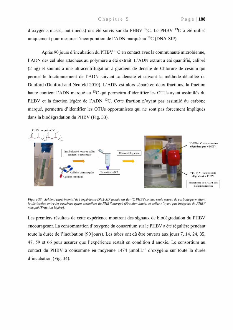
C h a p i t r e 5 P a g e | 188
d’oxygène, masse, nutriments) ont été suivis sur du PHBV 12C. Le PHBV 13C a été utilisé
uniquement pour mesurer l’incorporation de l’ADN marqué au 13C (DNA-SIP).
Après 90 jours d’incubation du PHBV 13C en contact avec la communauté microbienne,
l’ADN des cellules attachées au polymère a été extrait. L’ADN extrait a été quantifié, calibré
(2 ng) et soumis à une ultracentrifugation à gradient de densité de Chlorure de césium qui
permet le fractionnement de l’ADN suivant sa densité et suivant la méthode détaillée de
Dunford (Dunford and Neufeld 2010). L’ADN est alors séparé en deux fractions, la fraction
haute contient l’ADN marqué au 13C qui permettra d’identifier les OTUs ayant assimilés du
PHBV et la fraction légère de l’ADN 12C. Cette fraction n’ayant pas assimilé du carbone
marqué, permettra d’identifier les OTUs opportunistes qui ne sont pas forcément impliqués
dans la biodégradation du PHBV (Fig. 33).
Figure 33 : Schéma expérimental de l’expérience DNA-SIP menée sur du 13C PHBV comme seule source de carbone permettant
la distinction entre les bactéries ayant assimilées du PHBV marqué (Fraction haute) et celles n’ayant pas intégrées du PHBV
marqué (Fraction légère).
Les premiers résultats de cette expérience montrent des signaux de biodégradation du PHBV
encourageant. La consommation d’oxygène du consortium sur le PHBV a été régulière pendant
toute la durée de l’incubation (90 jours). Les tubes ont dû être ouverts aux jours 7, 14, 24, 35,
47, 59 et 66 pour assurer que l’expérience restait en condition d’anoxie. Le consortium au
contact du PHBV a consommé en moyenne 1474 µmol.L-1 d’oxygène sur toute la durée
d’incubation (Fig. 34).
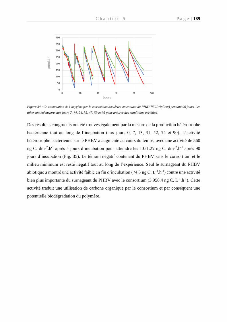
C h a p i t r e 5 P a g e | 189
Figure 34 : Consommation de l’oxygène par le consortium bactérien au contact du PHBV 12C (triplicat) pendant 90 jours. Les
tubes ont été ouverts aux jours 7, 14, 24, 35, 47, 59 et 66 pour assurer des conditions aérobies.
Des résultats congruents ont été trouvés également par la mesure de la production hétérotrophe
bactérienne tout au long de l’incubation (aux jours 0, 7, 13, 31, 52, 74 et 90). L’activité
hétérotrophe bactérienne sur le PHBV a augmenté au cours du temps, avec une activité de 560
ng C. dm-2.h-1 après 5 jours d’incubation pour atteindre les 1351.27 ng C. dm-2.h-1 après 90
jours d’incubation (Fig. 35). Le témoin négatif contenant du PHBV sans le consortium et le
milieu minimum est resté négatif tout au long de l’expérience. Seul le surnageant du PHBV
abiotique a montré une activité faible en fin d’incubation (74.3 ng C. L-1.h-1) contre une activité
bien plus importante du surnageant du PHBV avec le consortium (3 958.4 ng C. L-1.h-1). Cette
activité traduit une utilisation de carbone organique par le consortium et par conséquent une
potentielle biodégradation du polymère.
µm
ol.L
-1
Jours
C h a p i t r e 5 P a g e | 190
Figure 35 : Production bactérienne exprimée en ng C. dm-2.h-1 pour les morceaux de PHBV et en ng C. L-1.h-1 pour le milieu
d’incubation, et le surnageant du milieu artificiel contenant les pièces de PHBV.
Cette activité de biodégradation a été confirmée par la mesure de la masse de deux
morceaux de polymères qui ont perdu en moyenne 16 % de la masse initiale après avoir passé
94 jours en en milieu minimum en contact avec le consortium bactérien. Dans ce même laps de
temps, le PHBV contenu dans du milieu minimum sans le consortium a un poids identique à
celui du début d’incubation.
Les images au microscope électronique ont permis de caractériser l’impact du
consortium sur la surface du polymère (Fig. 6). Après 5 jours, les bactéries sont attachées aux
polymères et commencent à coloniser de façon homogène la surface. Des éléments typiques
d’EPS ont été détectés. Après 90 jours, le biofilm a adopté une structure en 3 dimensions,
recouvrant la totalité du polymère. On a pu constater sous la surface du biofilm des fissures et
des trous de la taille d’une bactérie, fragilisant sa structure.
0
500
1000
1500
2000
2500
3000
3500
4000
4500
PHBV abiotic PHBV
ng
C/d
m2
/h
T0
T7
T13
T31
T52
T74
T90
0
500
1000
1500
2000
2500
3000
3500
4000
4500
Minimummedium
Supernatantabiotic
Supernatant
ng
C/L
/h
T0
T7
T13
T31
T52
T74
T90
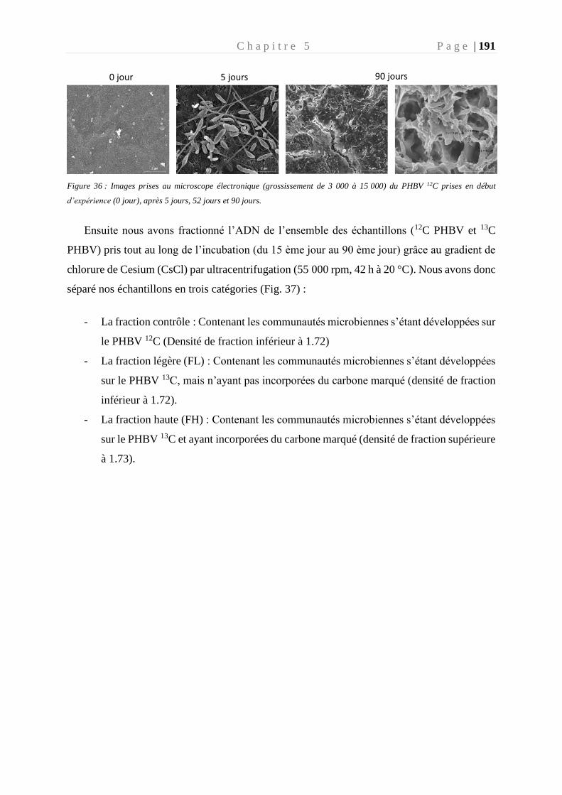
C h a p i t r e 5 P a g e | 191
Figure 36 : Images prises au microscope électronique (grossissement de 3 000 à 15 000) du PHBV 12C prises en début
d’expérience (0 jour), après 5 jours, 52 jours et 90 jours.
Ensuite nous avons fractionné l’ADN de l’ensemble des échantillons (12C PHBV et 13C
PHBV) pris tout au long de l’incubation (du 15 ème jour au 90 ème jour) grâce au gradient de
chlorure de Cesium (CsCl) par ultracentrifugation (55 000 rpm, 42 h à 20 °C). Nous avons donc
séparé nos échantillons en trois catégories (Fig. 37) :
- La fraction contrôle : Contenant les communautés microbiennes s’étant développées sur
le PHBV 12C (Densité de fraction inférieur à 1.72)
- La fraction légère (FL) : Contenant les communautés microbiennes s’étant développées
sur le PHBV 13C, mais n’ayant pas incorporées du carbone marqué (densité de fraction
inférieur à 1.72).
- La fraction haute (FH) : Contenant les communautés microbiennes s’étant développées
sur le PHBV 13C et ayant incorporées du carbone marqué (densité de fraction supérieure
à 1.73).
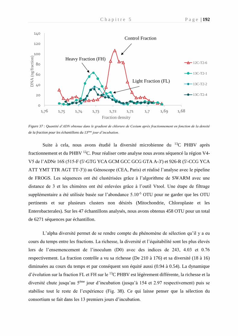
C h a p i t r e 5 P a g e | 192
Figure 37 : Quantité d’ADN obtenue dans le gradient de chlorure de Cesium après fractionnement en fonction de la densité
de la fraction pour les échantillons du 13ème jour d’incubation.
Suite à cela, nous avons étudié la diversité microbienne du 13C PHBV après
fractionnement et du PHBV 12C. Pour réaliser cette analyse nous avons séquencé la région V4-
V5 de l’ADNr 16S (515-F (5'-GTG YCA GCM GCC GCG GTA A-3') et 926-R (5'-CCG YCA
ATT YMT TTR AGT TT-3')) au Génoscope (CEA, Paris) et réalisé l’analyse avec le pipeline
de FROGS. Les séquences ont été clustérisées grâce à l’algorithme de SWARM avec une
distance de 3 et les chimères ont été enlevées grâce à l’outil Vtool. Une étape de filtrage
supplémentaire a été utilisée basée sur l’abondance 5.10-5 OTU pour ne garder que les OTU
pertinents et sur plusieurs clusters non désirés (Mitochondrie, Chloroplaste et les
Enterobacterales). Sur les 47 échantillons analysés, nous avons obtenus 458 OTU pour un total
de 6271 séquences par échantillon.
L’alpha diversité permet de se rendre compte du phénomène de sélection qu’il y a eu
cours du temps entre les fractions. La richesse, la diversité et l’équitabilité sont les plus élevés
lors de l’ensemencement de l’inoculum (D0) avec des indices de 243, 4.03 et 0.76
respectivement. La fraction contrôle a vu sa richesse (De 210 à 176) et sa diversité (18 à 16)
diminuées au cours du temps et par conséquent son équité aussi (0.94 à 0.54). La dynamique
d’évolution sur la fraction FL et FH sur le 13C PHBV est légèrement différente, la richesse et la
diversité chute jusqu’au 5ème jour d’incubation (jusqu’à 154 et 2.97 respectivement) puis se
stabilise tout le reste de l’expérience (Fig. 38). Ce qui laisse penser que la sélection du
consortium se fait dans les 13 premiers jours d’incubation.
0
20
40
60
80
100
120
140
1,681,691,71,711,721,731,741,751,76
DN
A (
ng
/fra
ctio
n)
Fraction density
12C-T2-6
13C-T2-1
13C-T2-2
13C-T2-4
Heavy Fraction (FH)
Control Fraction
Light Fraction (FL)
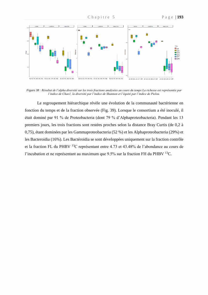
C h a p i t r e 5 P a g e | 193
Figure 38 : Résultat de l’alpha diversité sur les trois fractions analysées au cours du temps La richesse est représentée par
l’indice de Chao1, la diversité par l’indice de Shannon et l’équité par l’indice de Pielou.
Le regroupement hiérarchique révèle une évolution de la communauté bactérienne en
fonction du temps et de la fraction observée (Fig. 39). Lorsque le consortium a été inoculé, il
était dominé par 91 % de Proteobacteria (dont 79 % d’Alphaproteobacteria). Pendant les 13
premiers jours, les trois fractions sont restées proches selon la distance Bray Curtis (de 0,2 à
0,75), étant dominées par les Gammaproteobacteria (52 %) et les Alphaproteobacteria (29%) et
les Bacteroidia (16%). Les Bactéroidia se sont développées uniquement sur la fraction contrôle
et la fraction FL du PHBV 13C représentant entre 4.73 et 43.48% de l’abondance au cours de
l’incubation et ne représentant au maximum que 9.5% sur la fraction FH du PHBV 13C.
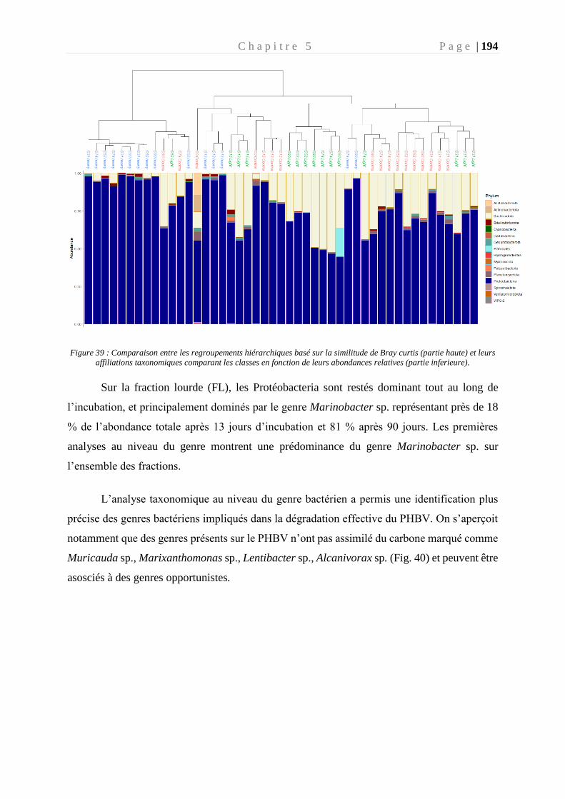
C h a p i t r e 5 P a g e | 194
Figure 39 : Comparaison entre les regroupements hiérarchiques basé sur la similitude de Bray curtis (partie haute) et leurs
affiliations taxonomiques comparant les classes en fonction de leurs abondances relatives (partie inferieure).
Sur la fraction lourde (FL), les Protéobacteria sont restés dominant tout au long de
l’incubation, et principalement dominés par le genre Marinobacter sp. représentant près de 18
% de l’abondance totale après 13 jours d’incubation et 81 % après 90 jours. Les premières
analyses au niveau du genre montrent une prédominance du genre Marinobacter sp. sur
l’ensemble des fractions.
L’analyse taxonomique au niveau du genre bactérien a permis une identification plus
précise des genres bactériens impliqués dans la dégradation effective du PHBV. On s’aperçoit
notamment que des genres présents sur le PHBV n’ont pas assimilé du carbone marqué comme
Muricauda sp., Marixanthomonas sp., Lentibacter sp., Alcanivorax sp. (Fig. 40) et peuvent être
asosciés à des genres opportunistes.
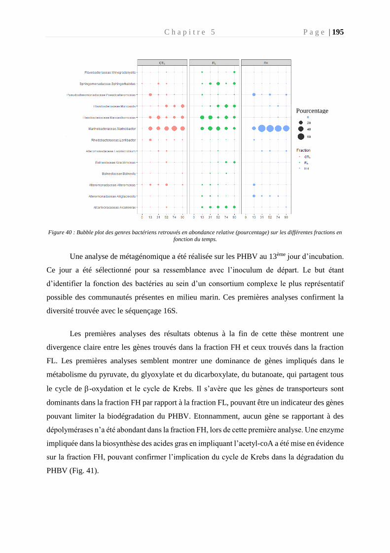
C h a p i t r e 5 P a g e | 195
Figure 40 : Bubble plot des genres bactériens retrouvés en abondance relative (pourcentage) sur les différentes fractions en
fonction du temps.
Une analyse de métagénomique a été réalisée sur les PHBV au 13ème jour d’incubation.
Ce jour a été sélectionné pour sa ressemblance avec l’inoculum de départ. Le but étant
d’identifier la fonction des bactéries au sein d’un consortium complexe le plus représentatif
possible des communautés présentes en milieu marin. Ces premières analyses confirment la
diversité trouvée avec le séquençage 16S.
Les premières analyses des résultats obtenus à la fin de cette thèse montrent une
divergence claire entre les gènes trouvés dans la fraction FH et ceux trouvés dans la fraction
FL. Les premières analyses semblent montrer une dominance de gènes impliqués dans le
métabolisme du pyruvate, du glyoxylate et du dicarboxylate, du butanoate, qui partagent tous
le cycle de -oxydation et le cycle de Krebs. Il s’avère que les gènes de transporteurs sont
dominants dans la fraction FH par rapport à la fraction FL, pouvant être un indicateur des gènes
pouvant limiter la biodégradation du PHBV. Etonnamment, aucun gène se rapportant à des
dépolymérases n’a été abondant dans la fraction FH, lors de cette première analyse. Une enzyme
impliquée dans la biosynthèse des acides gras en impliquant l’acetyl-coA a été mise en évidence
sur la fraction FH, pouvant confirmer l’implication du cycle de Krebs dans la dégradation du
PHBV (Fig. 41).
Pourcentage
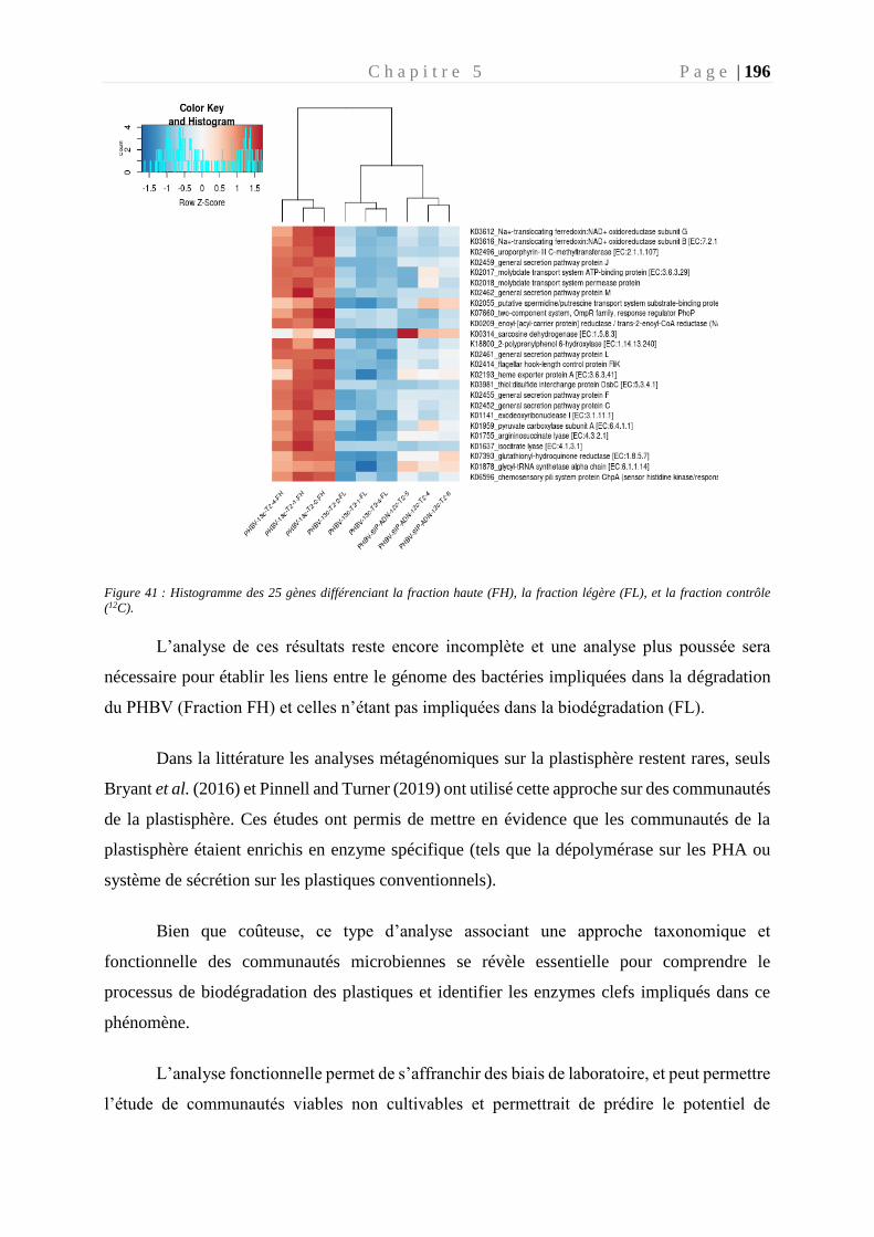
C h a p i t r e 5 P a g e | 196
Figure 41 : Histogramme des 25 gènes différenciant la fraction haute (FH), la fraction légère (FL), et la fraction contrôle
(12C).
L’analyse de ces résultats reste encore incomplète et une analyse plus poussée sera
nécessaire pour établir les liens entre le génome des bactéries impliquées dans la dégradation
du PHBV (Fraction FH) et celles n’étant pas impliquées dans la biodégradation (FL).
Dans la littérature les analyses métagénomiques sur la plastisphère restent rares, seuls
Bryant et al. (2016) et Pinnell and Turner (2019) ont utilisé cette approche sur des communautés
de la plastisphère. Ces études ont permis de mettre en évidence que les communautés de la
plastisphère étaient enrichis en enzyme spécifique (tels que la dépolymérase sur les PHA ou
système de sécrétion sur les plastiques conventionnels).
Bien que coûteuse, ce type d’analyse associant une approche taxonomique et
fonctionnelle des communautés microbiennes se révèle essentielle pour comprendre le
processus de biodégradation des plastiques et identifier les enzymes clefs impliqués dans ce
phénomène.
L’analyse fonctionnelle permet de s’affranchir des biais de laboratoire, et peut permettre
l’étude de communautés viables non cultivables et permettrait de prédire le potentiel de
C h a p i t r e 5 P a g e | 197
biodégradation d’un polymère in situ. Elle permet un cadre puissant d’étude des communautés
fonctionnelles qui devraient permettre d’ouvrir de nouvelles directions d’études dans le
domaine des biotechnologies.
2.2. Enjeux pour une application en biotechnologie
Les microorganismes sont les principales sources de biocatalyseurs qui sont essentiels
pour des applications industrielles. L’augmentation des connaissances dans le domaine de la
métagénomique est une aubaine pour des futures solutions biotechnologiques.
Cette thèse a exploré le potentiel génétique de la souche Alteromonas sp. sur sa
biodégradation du PHBV (grâce à la présence de PhaZ) et sa potentielle activité de biosynthèse
(grâce à la présence de la PhaC). Cela a permis de générer de nouvelles connaissances sur cette
souche et ses voies métaboliques impliquées le métabolisme du PHBV. A présent, il faudrait
envisager une validation expérimentale en laboratoire des prédictions menées sur la base de
l’étude du génome. Cela implique de caractériser l’expression génétique de phaZ, l’activité de
la PhaZ, son substrat et son produit de réaction.
L’ingénierie des protéines pourrait permettre une modification des protéines en
augmentant l’activité, en modifiant sa spécificité au substrat ou en augmentant la stabilité de
l’enzyme dans des conditions bien définies (température, pH, solvants). Cependant, la
modification génétique d’enzymes, la purification et la production à grande échelle demandent
un développement de méthode pouvant être fastidieux. Un bel exemple de cette ingénierie de
protéine concerne la PET- dépolymérase présente naturellement chez Ideonella sakaiensis.
L’étude menée en 2020, a transformé génétiquement l’enzyme en modifiant le site de liaison
de l’enzyme au substrat afin d’augmenter sa stabilité thermique et a permis d’obtenir une
dépolymérisation du PET de 90 % au lieu de 31 % naturellement (Tournier et al. 2020). Cela
ouvre donc de nouvelles perspectives de recyclage à l’infini pour le PET, plastique jusqu’alors
limité dans les cycles de recyclage.
C h a p i t r e 5 P a g e | 198
2.3. Evolution des normes de biodégradation des plastiques en mer : suivi
temporel et multifactoriel
Actuellement, les normes de biodégradation en vigueur ne permettent actuellement pas
de caractériser la biodégradation des plastiques en mer. Nos résultats ont montré qu’en se basant
uniquement sur la respirométrie, il y a un risque d’une mauvaise interprétation des résultats.
Comme nous l’avons observé lors de nos expériences, les différents consortia continuent à avoir
une activité bactérienne après 10 jours après transfert en milieu minimum sans autre source de
carbone que le polymère, même lorsqu’il ne présente pas d’activité de biodégradation (cas du
PP). Cette activité a été attribuée à l’utilisation de la matière organique par le biofilm et non au
carbone du polymère. De plus, nos analyses de diversité microbienne ont mis en évidence une
dominance des cyanobactéries sur les plastiques conventionnels in situ. Ces deux observations
remettent en question les normes actuelles qui se basent uniquement que les paramètres de
respirométrie. Plusieurs revues scientifiques ont déjà fait état de cette faiblesse dans les normes
actuelles (Harrison et al. 2018; Jacquin et al. 2019).
Les travaux de cette thèse s’appuient sur plusieurs paramètres pour caractériser la
biodégradation des polymères étudiés. Tout d’abord, l’incubation en milieu contrôlé sans autre
source de carbone sur une période d’incubation longue a permis de s’affranchir de l’utilisation
de la matière organique par le consortium (Experience : chapitre 3). Après avoir constaté une
consommation de la matière organique pendant près de 15 jours, nous avons fait le choix
d’inoculer un consortium bactérien (provenant du biofilm mature détaché sur polymère) sur des
plastiques vierges et stériles n’ayant pas séjournés en milieu ouvert auparavant (Expérience :
perspective). Ensuite, nous avons multiplié les paramètres permettant de suivre le processus de
biodégradation : tout d’abord nous avons suivi l’activité bactérienne grâce à l’incorporation de
leucine tritiée et à sa consommation d’oxygène au cours du temps. Puis dans un second temps
en fin d’incubation nous avons observé l’impact de l’activité bactérienne sur le polymère grâce
à une mesure de perte de poids (en mg) et une observation de sa surface en microscopie
électronique. Enfin la méthode du marquage isotopique (13C PHBV) semble être une preuve
ultime pour observer l’assimilation du carbone du polymère par les microorganismes.
Nous avons aussi testé en laboratoire d’autres suivis de paramètres tels que la mesure
de l’ATP contenu dans le cytoplasme bactérien tout au long de nos incubations en milieu
minimum, comme cela a été utilisé dans la norme AFNOR AC T 51-808 (Fontanella et al.
C h a p i t r e 5 P a g e | 199
2010). Cette mesure s’est révélée peu fiable et non concordante avec les autres paramètres de
suivi (production bactérienne et oxygène) tout au long de notre incubation en milieu minimum.
Les tentatives d’amélioration de protocole n’ont pas permis d’utiliser cette méthode comme un
indicatrice de la biodégradation.
Il serait donc intéressant de faire évoluer les normes actuelles régissant l’environnement
marin pour prendre en compte tous les paramètres de l’environnement pouvant influencer la
biodégradation des plastiques en mer. L’utilisation d’un consortium marin et non artificiel de
laboratoire, d’un milieu contrôlé sans source de carbone et la multiplication de suivi de
paramètres pluridisciplinaires sont des pistes de réflexion à mener.
R é f é r e n c e s P a g e | 200
Références
-A-
Acinas SG, Antón J, and Rodríguez-Valera F. 1999. Diversity of Free-Living and Attached
Bacteria in Offshore Western Mediterranean Waters as Depicted by Analysis of Genes
Encoding 16S rRNA. Appl Environ Microbiol 65: 514–22.
Ali I and Jamil N. 2016. Polyhydroxyalkanoates: Current applications in the medical field.
Front Biol 11: 19–27.
Alshehrei F. 2017. Biodegradation of Synthetic and Natural Plastic by Microorganisms.
Journal of Applied & Environmental Microbiology, Journal of Applied &
Environmental Microbiology 5: 8–19.
Amaral-Zettler LA, Zettler ER, and Mincer TJ. 2020. Ecology of the plastisphere. Nat Rev
Microbiol 18: 139–51.
Amaral-Zettler LA, Zettler ER, Slikas B, et al. 2015. The biogeography of the Plastisphere:
implications for policy. Frontiers in Ecology and the Environment 13: 541–6.
Amirul AA, Ramachandran H, Huong K-H, and Kannusamy S. 2015. Microbial-based
polyhydroxyalkanoates: upstream and downstream processing.
Anderson AJ and Dawes EA. 1990. Occurrence, metabolism, metabolic role, and industrial uses
of bacterial polyhydroxyalkanoates. Microbiol Rev 54: 450–72.
Anderson JM and Shive MS. 2012. Biodegradation and biocompatibility of PLA and PLGA
microspheres. Advanced Drug Delivery Reviews 64: 72–82.
Arias-Andres M, Klümper U, Rojas-Jimenez K, and Grossart H-P. 2018. Microplastic pollution
increases gene exchange in aquatic ecosystems. Environ Pollut 237: 253–61.
Arthur C, baker J, and Bamford H. 2009. Proceedings of the International Research Workshop
on the Occurrence, Effects, and Fate of Microplastic Marine Debris.
Ashby RD, Solaiman DKY, and Foglia TA. 2005. Synthesis of short-/medium-chain-length
poly(hydroxyalkanoate) blends by mixed culture fermentation of glycerol.
Biomacromolecules 6: 2106–12.
Ašmonaitė G, Larsson K, Undeland I, et al. 2018. Size Matters: Ingestion of Relatively Large
Microplastics Contaminated with Environmental Pollutants Posed Little Risk for Fish
Health and Fillet Quality. Environ Sci Technol 52: 14381–91.
Auta H, Emenike C, and Fauziah SH. 2017. Distribution and importance of microplastics in the
marine environment: A review of the sources, fate, effects, and potential solutions.
Environment international 102.
R é f é r e n c e s P a g e | 201
Ayub ND, Tribelli PM, and López NI. 2009. Polyhydroxyalkanoates are essential for
maintenance of redox state in the Antarctic bacterium Pseudomonas sp. 14-3 during low
temperature adaptation. Extremophiles 13: 59–66.
-B-
Bachmann BM and Seebach D. 1999. Investigation of the Enzymatic Cleavage of
Diastereomeric Oligo(3-hydroxybutanoates) Containing Two to Eight HB Units. A
Model for the Stereoselectivity of PHB Depolymerase from Alcaligenes faecalis T1.
Macromolecules 32: 1777–84.
Barnes DKA, Galgani F, Thompson RC, and Barlaz M. 2009. Accumulation and fragmentation
of plastic debris in global environments. Philosophical Transactions of the Royal
Society B: Biological Sciences 364: 1985–98.
Basili M, Quero GM, Giovannelli D, et al. 2020. Major Role of Surrounding Environment in
Shaping Biofilm Community Composition on Marine Plastic Debris. Front Mar Sci 7.
Bergmann M, Tekman MB, and Gutow L. 2017. Sea change for plastic pollution. Nature 544:
297–297.
Bianchi CN and Morri C. 2000. Marine Biodiversity of the Mediterranean Sea: Situation,
Problems and Prospects for Future Research. Marine Pollution Bulletin 40: 367–76.
Boesel LF, Meur SL, Thöny-Meyer L, and Ren Q. 2014. The effect of molecular weight on the
material properties of biosynthesized poly(4-hydroxybutyrate). International Journal of
Biological Macromolecules 71: 124–30.
Bogaert JC CP. 2000. Poly(lactic acids): A potential solution to plastic waste dilemma. 153:
287–303.
Boland RC and Donohue MJ. 2003. Marine debris accumulation in the nearshore marine habitat
of the endangered Hawaiian monk seal, Monachus schauinslandi 1999-2001. Mar
Pollut Bull 46: 1385–94.
Boucher J and Friot D. 2017. Primary microplastics in the oceans: A global evaluation of
sources. IUCN International Union for Conservation of Nature.
Boucher J and Friot D. 2020. Microplastiques primaires dans les océans. IUCN, International
Union for Conservation of Nature.
Brandão ML, Braga KM, and Luque JL. Marine debris ingestion by Magellanic penguins,
Spheniscus magellanicus (Aves: Sphenisciformes), from the Brazilian coastal zone.
Marine Pollution Bulletin 62: 2246–9.
Briassoulis D. 2007. Analysis of the mechanical and degradation performances of optimised
agricultural biodegradable films. Polymer Degradation and Stability 92: 1115–32.
R é f é r e n c e s P a g e | 202
Browne MA, Niven SJ, Galloway TS, et al. 2013. Microplastic Moves Pollutants and Additives
to Worms, Reducing Functions Linked to Health and Biodiversity. Current Biology 23:
2388–92.
Bryant JA, Clemente TM, Viviani DA, et al. 2016. Diversity and Activity of Communities
Inhabiting Plastic Debris in the North Pacific Gyre. mSystems 1.
Buchholz O. New market data 2019: Bioplastics industry shows dynamic growth. European
Bioplastics eV.
Butterworth A, C B, and Clegg I. 2012. Marine debris: a global picture of the impact on animal
welfare and of animal-focused solutions.
-C-
Campani T, Baini M, Giannetti M, et al. 2013. Presence of plastic debris in loggerhead turtle
stranded along the Tuscany coasts of the Pelagos Sanctuary for Mediterranean Marine
Mammals (Italy). Marine Pollution Bulletin 74: 225–30.
Carbery M, O’Connor W, and Palanisami T. 2018. Trophic transfer of microplastics and mixed
contaminants in the marine food web and implications for human health. Environ Int
115: 400–9.
Cassoff R, Moore K, McLellan W, et al. 2011. Lethal entanglement in baleen whales. Diseases
of aquatic organisms 96: 175–85.
Castro-Sowinski S, Burdman S, Matan O, and Okon Y. 2010. Natural Functions of Bacterial
Polyhydroxyalkanoates. In: Chen GG-Q (Ed). Plastics from Bacteria. Berlin,
Heidelberg: Springer Berlin Heidelberg.
Chang H-F, Chang W-C, and Tsai C-Y. 2012. Synthesis of poly(3-hydroxybutyrate/3-
hydroxyvalerate) from propionate-fed activated sludge under various carbon sources.
Bioresour Technol 113: 51–7.
Chapron L, Peru E, Engler A, et al. 2018. Macro- and microplastics affect cold-water corals
growth, feeding and behaviour. Scientific Reports 8: 1–8.
Chek MF, Hiroe A, Hakoshima T, et al. 2019. PHA synthase (PhaC): interpreting the functions
of bioplastic-producing enzyme from a structural perspective. Applied Microbiology
and Biotechnology 103.
Chek MF, Kim S-Y, Mori T, et al. 2017. Structure of polyhydroxyalkanoate (PHA) synthase
PhaC from Chromobacterium sp. USM2, producing biodegradable plastics. Sci Rep 7.
Chen Q, Reisser J, Cunsolo S, et al. 2018. Pollutants in Plastics within the North Pacific
Subtropical Gyre. Environ Sci Technol 52: 446–56.
Chiba S, Saito H, Fletcher R, et al. 2018. Human footprint in the abyss: 30 year records of deep-
sea plastic debris. Marine Policy 96: 204–12.
R é f é r e n c e s P a g e | 203
Choy CA, Robison BH, Gagne TO, et al. 2019. The vertical distribution and biological transport
of marine microplastics across the epipelagic and mesopelagic water column. Scientific
Reports 9: 7843.
Chronopoulou P-M, Sanni GO, Silas‐Olu DI, et al. 2015. Generalist hydrocarbon-degrading
bacterial communities in the oil-polluted water column of the North Sea. Microbial
Biotechnology 8: 434–47.
Ciesielski S, Możejko J, and Pisutpaisal N. 2015. Plant oils as promising substrates for
polyhydroxyalkanoates production. Journal of Cleaner Production 106: 408–21.
Claessens M, De Meester S, Van Landuyt L, et al. 2011. Occurrence and distribution of
microplastics in marine sediments along the Belgian coast. Mar Pollut Bull 62: 2199–
204.
Clukey KE, Lepczyk CA, Balazs GH, et al. 2017. Investigation of plastic debris ingestion by
four species of sea turtles collected as bycatch in pelagic Pacific longline fisheries.
Marine Pollution Bulletin 120: 117–25.
Codina-García M, Militão T, Moreno J, and González-Solís J. 2013. Plastic debris in
Mediterranean seabirds. Marine Pollution Bulletin 77: 220–6.
Colabuono FI, Barquete V, Domingues BS, and Montone RC. 2009. Plastic ingestion by
Procellariiformes in Southern Brazil. Marine Pollution Bulletin 58: 93–6.
Cole M, Lindeque P, Fileman E, et al. 2013. Microplastic Ingestion by Zooplankton. Environ
Sci Technol 47: 6646–55.
Collignon A, Hecq J-H, Glagani F, et al. 2012. Neustonic microplastic and zooplankton in the
North Western Mediterranean Sea. Mar Pollut Bull 64: 861–4.
Cosgrove L, McGeechan PL, Robson GD, and Handley PS. 2007. Fungal Communities
Associated with Degradation of Polyester Polyurethane in Soil. Applied and
Environmental Microbiology 73: 5817–24.
Cozar A, Echevarria F, Gonzalez-Gordillo JI, et al. 2014. Plastic debris in the open ocean.
Proceedings of the National Academy of Sciences 111: 10239–44.
Cózar A, Sanz-Martín M, Martí E, et al. 2015. Plastic Accumulation in the Mediterranean Sea.
PLOS ONE 10: e0121762.
Crespo BG, Pommier T, Fernández-Gómez B, and Pedrós-Alió C. 2013. Taxonomic
composition of the particle-attached and free-living bacterial assemblages in the
Northwest Mediterranean Sea analyzed by pyrosequencing of the 16S rRNA.
Microbiologyopen 2: 541–52.
-D-
R é f é r e n c e s P a g e | 204
Dang H and Lovell CR. 2016. Microbial Surface Colonization and Biofilm Development in
Marine Environments. Microbiol Mol Biol Rev 80: 91–138.
De Tender CA, Devriese LI, Haegeman A, et al. 2015. Bacterial Community Profiling of Plastic
Litter in the Belgian Part of the North Sea. Environ Sci Technol 49: 9629–38.
De Tender C, Devriese LI, Haegeman A, et al. 2017. Temporal Dynamics of Bacterial and
Fungal Colonization on Plastic Debris in the North Sea. Environ Sci Technol 51: 7350–
60.
Debroas D, Mone A, and Ter Halle A. 2017. Plastics in the North Atlantic garbage patch: A
boat-microbe for hitchhikers and plastic degraders. Sci Total Environ 599–600: 1222–
32.
Delacuvellerie A, Cyriaque V, Gobert S, et al. 2019. The plastisphere in marine ecosystem
hosts potential specific microbial degraders including Alcanivorax borkumensis as a
key player for the low-density polyethylene degradation. Journal of Hazardous
Materials 380: 120899.
Dobretsov S. 2010. Marine Biofilms. In: Biofouling.
Doi Y, Kanesawa Y, Kunioka M, and Saito T. 1990. Biodegradation of microbial copolyesters:
poly(3-hydroxybutyrate-co-3-hydroxyvalerate) and poly(3-hydroxybutyrate-co-4-
hydroxybutyrate). Macromolecules 23: 26–31.
Douarche C, Bailleux V, Even C, et al. 2018. La mécanique des biofilms à la surface de liquides.
Reflets phys: 20–4.
Dunford EA and Neufeld JD. 2010. DNA Stable-Isotope Probing (DNA-SIP). J Vis Exp.
Dussud C and Ghiglione J-F. 2014. Bacterial degradation of synthetic plastics. Marine litter in
the Mediterranean and Black Seas CIESM workshop Monographs: 43–8.
Dussud C, Hudec C, George M, et al. 2018a. Colonization of Non-biodegradable and
Biodegradable Plastics by Marine Microorganisms. Front Microbiol 9.
Dussud C, Meistertzheim AL, Conan P, et al. 2018b. Evidence of niche partitioning among
bacteria living on plastics, organic particles and surrounding seawaters. Environmental
Pollution 236: 807–16.
-E-
Eerkes-Medrano D, Thompson RC, and Aldridge DC. 2015. Microplastics in freshwater
systems: A review of the emerging threats, identification of knowledge gaps and
prioritisation of research needs. Water Research 75: 63–82.
Egger M, Sulu-Gambari F, and Lebreton L. 2020. First evidence of plastic fallout from the
North Pacific Garbage Patch. Scientific Reports 10: 7495.
R é f é r e n c e s P a g e | 205
Eich A, Mildenberger T, Laforsch C, and Weber M. 2015. Biofilm and Diatom Succession on
Polyethylene (PE) and Biodegradable Plastic Bags in Two Marine Habitats: Early Signs
of Degradation in the Pelagic and Benthic Zone? PLoS One 10.
Elifantz H, Horn G, Ayon M, et al. 2013. Rhodobacteraceae are the key members of the
microbial community of the initial biofilm formed in Eastern Mediterranean coastal
seawater. FEMS Microbiol Ecol 85: 348–57.
Eriksen M, Lebreton LCM, Carson HS, et al. 2014. Plastic Pollution in the World’s Oceans:
More than 5 Trillion Plastic Pieces Weighing over 250,000 Tons Afloat at Sea. PLOS
ONE 9: e111913.
Eriksen M, Maximenko N, Thiel M, et al. 2013. Plastic pollution in the South Pacific
subtropical gyre. Marine Pollution Bulletin 68: 71–6.
Eubeler JP, Zok S, Bernhard M, and Knepper TP. 2009. Environmental biodegradation of
synthetic polymers I. Test methodologies and procedures. TrAC Trends in Analytical
Chemistry 28: 1057–72.
Evangeliou N, Grythe H, Klimont Z, et al. 2020. Atmospheric transport is a major pathway of
microplastics to remote regions. Nature Communications 11: 3381.
Eyheraguibel B, Traikia M, Fontanella S, et al. 2017. Characterization of oxidized oligomers
from polyethylene films by mass spectrometry and NMR spectroscopy before and after
biodegradation by a Rhodococcus rhodochrous strain. Chemosphere 184: 366–74.
-F-
Faure F, Demars C, Wieser O, et al. 2015. Plastic pollution in Swiss surface waters: Nature and
concentrations, interaction with pollutants. Environmental Chemistry 12.
Fontanella S, Bonhomme S, Koutny M, et al. 2010. Comparison of the biodegradability of
various polyethylene films containing pro-oxidant additives. Polymer Degradation and
Stability 95: 1011–21.
Fossi MC, Panti C, Guerranti C, et al. 2012. Are baleen whales exposed to the threat of
microplastics? A case study of the Mediterranean fin whale (Balaenoptera physalus).
Mar Pollut Bull 64: 2374–9.
-G-
Gajendiran A, Krishnamoorthy S, and Abraham J. 2016. Microbial degradation of low-density
polyethylene (LDPE) by Aspergillus clavatus strain JASK1 isolated from landfill soil.
3 Biotech 6.
Gall SC and Thompson RC. 2015. The impact of debris on marine life. Marine Pollution
Bulletin 92: 170–9.
R é f é r e n c e s P a g e | 206
Gerigny O, Brun M, Fabri MC, et al. 2019. Seafloor litter from the continental shelf and
canyons in French Mediterranean Water: Distribution, typologies and trends. Marine
Pollution Bulletin 146: 653–66.
Gewert B, M. Plassmann M, and MacLeod M. 2015. Pathways for degradation of plastic
polymers floating in the marine environment. Environmental Science: Processes &
Impacts 17: 1513–21.
Geyer R, Jambeck JR, and Law KL. 2017. Production, use, and fate of all plastics ever made.
Science Advances 3: e1700782.
Ghiglione J-F, Galand PE, Pommier T, et al. 2012. Pole-to-pole biogeography of surface and
deep marine bacterial communities. PNAS 109: 17633–8.
Ghorpade VM. 2001. Laboratory composting of extruded poly(lactic acid) sheets. Bioresour
Technol 76: 57–61.
Goh L-K, Purama RK, and Sudesh K. 2014. Enhancement of stress tolerance in the
polyhydroxyalkanoate producers without mobilization of the accumulated granules.
Appl Biochem Biotechnol 172: 1585–98.
Golik A. 1997. Debris in The Mediterranean Sea: Types, Quantities, and Behavior. In: Coe JM,
Rogers DB (Eds). Marine Debris. New York, NY: Springer New York.
Gonda KE, Jendrossek D, and Molitoris HP. 2000. Fungal degradation of the thermoplastic
polymer poly-ß-hydroxybutyric acid (PHB) under simulated deep sea pressure. In:
Liebezeit G, Dittmann S, Kröncke I (Eds). Life at Interfaces and Under Extreme
Conditions. Dordrecht: Springer Netherlands.
Göpferich A. 1996. Mechanisms of polymer degradation and erosion. Biomaterials 17: 103–
14.
Gorke JT, Okrasa K, Louwagie A, et al. 2007. Enzymatic synthesis of poly(hydroxyalkanoates)
in ionic liquids. Journal of Biotechnology 132: 306–13.
Gravouil K, Ferru-Clément R, Colas S, et al. 2017. Transcriptomics and Lipidomics of the
Environmental Strain Rhodococcus ruber Point out Consumption Pathways and
Potential Metabolic Bottlenecks for Polyethylene Degradation. Environ Sci Technol 51:
5172–81.
Green DS. 2020. Biological and Ecological Impacts of Plastic Debris in Aquatic Ecosystems.
In: Berlin, Heidelberg: Springer Berlin Heidelberg.
Griebel RJ and Merrick JM. 1971. Metabolism of Poly-β-Hydroxybutyrate: Effect of Mild
Alkaline Extraction on Native Poly-β-Hydroxybutyrate Granules1. J Bacteriol 108:
782–9.
Grillo R, Pereira A do ES, Melo NFS de, et al. 2011. Controlled release system for ametryn
using polymer microspheres: Preparation, characterization and release kinetics in water.
Journal of Hazardous Materials 186: 1645–51.
R é f é r e n c e s P a g e | 207
Gu J-G and Gu J-D. 2005. Methods Currently Used in Testing Microbiological Degradation
and Deterioration of a Wide Range of Polymeric Materials with Various Degree of
Degradability: A Review. J Polym Environ 13: 65–74.
-H-
Hablot E, Bordes P, Pollet E, and Avérous L. 2008. Thermal and thermo-mechanical
degradation of poly(3-hydroxybutyrate)-based multiphase systems. Polymer
Degradation and Stability 93: 413–21.
Halle A ter, Ladirat L, Gendre X, et al. 2016. Understanding the Fragmentation Pattern of
Marine Plastic Debris. Environ Sci Technol 50: 5668–75.
Hardesty BD and Wilcox C. 2020. For decades, scientists puzzled over the plastic ‘missing’
from our oceans – but now it’s been found.
Harris PT. 2020. The fate of microplastic in marine sedimentary environments: A review and
synthesis. Marine Pollution Bulletin 158: 111398.
Harrison JP, Boardman C, O’Callaghan K, et al. 2018. Biodegradability standards for carrier
bags and plastic films in aquatic environments: a critical review. Royal Society Open
Science 5: 171792.
Harrison JP, Schratzberger M, Sapp M, and Osborn AM. 2014. Rapid bacterial colonization of
low-density polyethylene microplastics in coastal sediment microcosms. BMC
Microbiology 14: 232.
Hayward Ac, Forsyth WGC and Roberts JB. 1959. Synthesis and Breakdown of Poly-β-
Hydroxybutyric Acid by Bacteria. Microbiology, 20: 510–8.
Henderson JR. 2001. A Pre- and Post-MARPOL Annex V Summary of Hawaiian Monk Seal
Entanglements and Marine Debris Accumulation in the Northwestern Hawaiian Islands,
1982–1998. Marine Pollution Bulletin 42: 584–9.
Hirai H, Takada H, Ogata Y, et al. 2011. Organic micropollutants in marine plastics debris from
the open ocean and remote and urban beaches. Mar Pollut Bull 62: 1683–92.
Ho BT, Roberts TK, and Lucas S. 2018. An overview on biodegradation of polystyrene and
modified polystyrene: the microbial approach. Critical Reviews in Biotechnology 38:
308–20.
Hugenholtz P, Hooper SD, and Kyrpides NC. 2009. Focus: Synergistetes. Environ Microbiol
11: 1327–9.
Huu Phong T, Van Thuoc D, and Sudesh K. 2016. Biosynthesis of poly(3-hydroxybutyrate)
and its copolymers by Yangia sp. ND199 from different carbon sources. International
Journal of Biological Macromolecules 84: 361–6.
R é f é r e n c e s P a g e | 208
-I-
İpekogˇlu B, Böke H, and Çizer Ö. 2007. Assessment of material use in relation to climate in
historical buildings. Building and Environment 42: 970–8.
Ishigaki T, Sugano W, Nakanishi A, et al. 2004. The degradability of biodegradable plastics in
aerobic and anaerobic waste landfill model reactors. Chemosphere 54: 225–33.
Isobe A, Uchiyama-Matsumoto K, Uchida K, and Tokai T. 2017. Microplastics in the Southern
Ocean. Marine Pollution Bulletin 114: 623–6.
Ivar do Sul JA and Costa MF. 2014. The present and future of microplastic pollution in the
marine environment. Environmental Pollution 185: 352–64.
Ivar do Sul JA, Spengler Â, and Costa MF. 2009. Here, there and everywhere. Small plastic
fragments and pellets on beaches of Fernando de Noronha (Equatorial Western
Atlantic). Mar Pollut Bull 58: 1236–8.
-J-
Jacquin J, Cheng J, Odobel C, et al. 2019. Microbial Ecotoxicology of Marine Plastic Debris:
A Review on Colonization and Biodegradation by the “Plastisphere.” Front Microbiol
10.
Jambeck JR, Geyer R, Wilcox C, et al. 2015. Marine pollution. Plastic waste inputs from land
into the ocean. Science 347: 768–71.
James BW, Mauchline WS, Dennis PJ, et al. 1999. Poly-3-Hydroxybutyrate in Legionella
pneumophila, an Energy Source for Survival in Low-Nutrient Environments. Appl
Environ Microbiol 65: 822–7.
Jamieson AJ, Brooks LSR, Reid WDK, et al. 2019. Microplastics and synthetic particles
ingested by deep-sea amphipods in six of the deepest marine ecosystems on Earth. R
Soc open sci 6: 180667.
Jamieson AJ, Brooks LSR, Reid WDK, et al. Microplastics and synthetic particles ingested by
deep-sea amphipods in six of the deepest marine ecosystems on Earth. Royal Society
Open Science 6: 180667.
Jendrossek D. 2005. Extracellular Polyhydroxyalkanoate (PHA) Depolymerases: The Key
Enzymes of PHA Degradation. In: Biopolymers Online. American Cancer Society.
Jendrossek D. 2007. Peculiarities of PHA granules preparation and PHA depolymerase activity
determination. Appl Microbiol Biotechnol 74: 1186–96.
Jendrossek D and Handrick R. 2002. Microbial degradation of polyhydroxyalkanoates. Annu
Rev Microbiol 56: 403–32.
R é f é r e n c e s P a g e | 209
Jendrossek D and Pfeiffer D. 2014. New insights in the formation of polyhydroxyalkanoate
granules (carbonosomes) and novel functions of poly(3-hydroxybutyrate). Environ
Microbiol 16: 2357–73.
Jin HM, Kim JM, Lee HJ, et al. 2012. Alteromonas As a Key Agent of Polycyclic Aromatic
Hydrocarbon Biodegradation in Crude Oil-Contaminated Coastal Sediment. Environ
Sci Technol 46: 7731–40.
-K-
Kadouri D, Jurkevitch E, Okon Y, and Castro-Sowinski S. 2005. Ecological and Agricultural
Significance of Bacterial Polyhydroxyalkanoates. Critical reviews in microbiology 31:
55–67.
Kaposi KL, Mos B, Kelaher BP, and Dworjanyn SA. 2014. Ingestion of Microplastic Has
Limited Impact on a Marine Larva. Environ Sci Technol 48: 1638–45.
Kellerhals MB, Kessler B, Witholt B, et al. 2000. Renewable Long-Chain Fatty Acids for
Production of Biodegradable Medium-Chain-Length Polyhydroxyalkanoates (mcl-
PHAs) at Laboratory and Pilot Plant Scales. Macromolecules 33: 4690–8.
Kertesz MA, Kawasaki A, and Stolz A. 2019. Aerobic Hydrocarbon-Degrading
Alphaproteobacteria: Sphingomonadales. In: McGenity TJ (Ed). Taxonomy, Genomics
and Ecophysiology of Hydrocarbon-Degrading Microbes. Cham: Springer International
Publishing.
Keshavarz T and Roy I. 2010. Polyhydroxyalkanoates: bioplastics with a green agenda. Current
Opinion in Microbiology 13: 321–6.
Kettner MT, Rojas‐Jimenez K, Oberbeckmann S, et al. 2017. Microplastics alter composition
of fungal communities in aquatic ecosystems. Environmental Microbiology 19: 4447–
59.
Knoll M, Hamm TM, Wagner F, et al. 2009. The PHA Depolymerase Engineering Database:
A systematic analysis tool for the diverse family of polyhydroxyalkanoate (PHA)
depolymerases. BMC Bioinformatics 10: 89.
Koelmans AA, Besseling E, and Foekema EM. 2014. Leaching of plastic additives to marine
organisms. Environmental Pollution 187: 49–54.
Koitabashi M, Noguchi MT, Sameshima-Yamashita Y, et al. 2012. Degradation of
biodegradable plastic mulch films in soil environment by phylloplane fungi isolated
from gramineous plants. AMB Express 2: 40.
Koller M, Atlić A, Dias M, et al. 2010. Microbial PHA Production from Waste Raw Materials.
In: Chen GG-Q (Ed). Plastics from Bacteria: Natural Functions and Applications.
Berlin, Heidelberg: Springer.
R é f é r e n c e s P a g e | 210
Kooi M, Reisser J, Slat B, et al. 2016. The effect of particle properties on the depth profile of
buoyant plastics in the ocean. Scientific Reports 6: 33882.
Koutny M, Lemaire J, and Delort A-M. 2006. Biodegradation of polyethylene films with
prooxidant additives. Chemosphere 64: 1243–52.
Krueger MC, Harms H, and Schlosser D. 2015. Prospects for microbiological solutions to
environmental pollution with plastics. Appl Microbiol Biotechnol 99: 8857–74.
Kühn S, Bravo Rebolledo EL, and Franeker JA van. 2015. Deleterious Effects of Litter on
Marine Life. In: Bergmann M, Gutow L, Klages M (Eds). Marine Anthropogenic Litter.
Cham: Springer International Publishing.
Kunioka M, Kawaguchi Y, and Doi Y. 1989. Production of biodegradable copolyesters of 3-
hydroxybutyrate and 4-hydroxybutyrate by Alcaligenes eutrophus. Appl Microbiol
Biotechnol 30: 569–73.
-L-
Law KL, Morét-Ferguson S, Maximenko NA, et al. 2010. Plastic Accumulation in the North
Atlantic Subtropical Gyre. Science 329: 1185–8.
Le Meur S, Zinn M, Egli T, et al. 2013. Poly(4-hydroxybutyrate) (P4HB) production in
recombinant Escherichia coli: P4HB synthesis is uncoupled with cell growth. Microbial
Cell Factories 12: 123.
Lebreton LC-M, Greer SD, and Borrero JC. 2012. Numerical modelling of floating debris in
the world’s oceans. Mar Pollut Bull 64: 653–61.
Lebreton L, Slat B, Ferrari F, et al. 2018. Evidence that the Great Pacific Garbage Patch is
rapidly accumulating plastic. Scientific Reports 8: 4666.
Lebreton LCM, Zwet J van der, Damsteeg J-W, et al. 2017. River plastic emissions to the
world’s oceans. Nature Communications 8: 1–10.
Lee SY and Choi J. 1999. Production and degradation of polyhydroxyalkanoates in waste
environment. Waste Management 19: 133–9.
Lee J-W, Nam J-H, Kim Y-H, et al. 2008. Bacterial communities in the initial stage of marine
biofilm formation on artificial surfaces. J Microbiol 46: 174–82.
Lee H, Shim WJ, and Kwon J-H. 2014. Sorption capacity of plastic debris for hydrophobic
organic chemicals. Science of The Total Environment 470–471: 1545–52.
Lee K-W, Shim WJ, Kwon OY, and Kang J-H. 2013. Size-Dependent Effects of Micro
Polystyrene Particles in the Marine Copepod Tigriopus japonicus. Environ Sci Technol
47: 11278–83.
Leejarkpai T, Suwanmanee U, Rudeekit Y, and Mungcharoen T. 2011. Biodegradable kinetics
of plastics under controlled composting conditions. Waste Management 31: 1153–61.
R é f é r e n c e s P a g e | 211
Lemos PC, Viana C, Salgueiro EN, et al. 1998. Effect of carbon source on the formation of
polyhydroxyalkanoates (PHA) by a phosphate-accumulating mixed culture. Enzyme
and Microbial Technology 22: 662–71.
Li Z and Loh XJ. 2015. Water soluble polyhydroxyalkanoates: future materials for therapeutic
applications. Chem Soc Rev 44: 2865–79.
Li WC, Tse HF, and Fok L. 2016. Plastic waste in the marine environment: A review of sources,
occurrence and effects. Science of The Total Environment 566–567: 333–49.
Liu Z, Zhu Z, Yang J, et al. 2019. Domain-centric dissection and classification of prokaryotic
poly(3-hydroxyalkanoate) synthases. bioRxiv: 693432.
Liubartseva S, Coppini G, Lecci R, and Clementi E. 2018. Tracking plastics in the
Mediterranean: 2D Lagrangian model. Marine Pollution Bulletin 129: 151–62.
Lo HKA and Chan KYK. 2018. Negative effects of microplastic exposure on growth and
development of Crepidula onyx. Environmental Pollution 233: 588–95.
Lu L, Luo T, Zhao Y, et al. 2019. Interaction between microplastics and microorganism as well
as gut microbiota: A consideration on environmental animal and human health. Science
of The Total Environment 667: 94–100.
Luengo JM, Garcı́a B, Sandoval A, et al. 2003. Bioplastics from microorganisms. Current
Opinion in Microbiology 6: 251–60.
Lusher AL, Tirelli V, O’Connor I, and Officer R. 2015. Microplastics in Arctic polar waters:
the first reported values of particles in surface and sub-surface samples. Scientific
Reports 5: 1–9.
-M-
Magara G, Elia AC, Syberg K, and Khan FR. 2018. Single contaminant and combined
exposures of polyethylene microplastics and fluoranthene: accumulation and oxidative
stress response in the blue mussel, Mytilus edulis. J Toxicol Environ Health Part A 81:
761–73.
Magnin A, Hoornaert L, Pollet E, et al. 2018. Isolation and characterization of different
promising fungi for biological waste management of polyurethanes. Microbial
Biotechnology.
Mata MT, Luza MF, and Riquelme CE. 2017. Production of diatom–bacteria biofilm isolated
from Seriola lalandi cultures for aquaculture application. Aquaculture Research 48:
4308–20.
Matsuguma Y, Takada H, Kumata H, et al. 2017. Microplastics in Sediment Cores from Asia
and Africa as Indicators of Temporal Trends in Plastic Pollution. Arch Environ Contam
Toxicol 73: 230–9.
McCool GJ and Cannon MC. 2001. PhaC and PhaR Are Required for Polyhydroxyalkanoic
Acid Synthase Activity in Bacillus megaterium. Journal of Bacteriology 183: 4235–43.
R é f é r e n c e s P a g e | 212
Meng D-C, Shen R, Yao H, et al. 2014. Engineering the diversity of polyesters. Curr Opin
Biotechnol 29: 24–33.
Mergaert J, Wouters A, Anderson C, and Swings J. 1995. In situ biodegradation of poly(3-
hydroxybutyrate) and poly(3-hydroxybutyrate-co-3-hydroxyvalerate) in natural waters.
Can J Microbiol 41 Suppl 1: 154–9.
Merrick JM and Doudoroff M. 1964. Depolymerization of poly β-hydroxybutyrate by an
intracellular enzyme system. J Bacteriol 88: 60–71.
Mezzina MP and Pettinari MJ. 2016. Phasins, Multifaceted Polyhydroxyalkanoate Granule-
Associated Proteins. Appl Environ Microbiol 82: 5060–7.
Mezzolla V, D’Urso OF, and Poltronieri P. 2018. Role of PhaC Type I and Type II Enzymes
during PHA Biosynthesis. Polymers (Basel) 10.
Mohee R, Unmar GD, Mudhoo A, and Khadoo P. 2008. Biodegradability of
biodegradable/degradable plastic materials under aerobic and anaerobic conditions.
Waste Manag 28: 1624–9.
Mohit V, Archambault P, Toupoint N, and Lovejoy C. 2014. Phylogenetic Differences in
Attached and Free-Living Bacterial Communities in a Temperate Coastal Lagoon
during Summer, Revealed via High-Throughput 16S rRNA Gene Sequencing. Appl
Environ Microbiol 80: 2071–83.
Moore C, Lattin G, and Zellers A. 2005. Density of Plastic Particles found in zooplankton trawls
from Coastal Waters of California to the North Pacific Central Gyre.
Morritt D, Stefanoudis PV, Pearce D, et al. 2014. Plastic in the Thames: A river runs through
it. Marine Pollution Bulletin 78: 196–200.
Mothes G, Ackermann J-U, and Babel W. 2004. Mole Fraction Control of Poly([R]-3-
hydroxybutyrate-co-3-hydroxyvalerate) (PHB/HV) Synthesized by Paracoccus
denitrificans. Engineering in Life Sciences 4: 247–51.
Mouchi V, Chapron L, Peru E, et al. 2019. Long-term aquaria study suggests species-specific
responses of two cold-water corals to macro-and microplastics exposure. Environmental
Pollution 253: 322–9.
Moura V, Ribeiro I, Moriggi P, et al. 2018. The influence of surface microbial diversity and
succession on microbiologically influenced corrosion of steel in a simulated marine
environment. Arch Microbiol 200: 1447–56.
Możejko-Ciesielska J and Kiewisz R. 2016. Bacterial polyhydroxyalkanoates: Still fabulous?
Microbiological Research 192: 271–82.
R é f é r e n c e s P a g e | 213
-N-
Nomura T, Tani S, Yamamoto M, et al. 2016. Cytotoxicity and colloidal behavior of
polystyrene latex nanoparticles toward filamentous fungi in isotonic solutions.
Chemosphere 149: 84–90.
Numata K, Yamashita K, Fujita M, et al. 2007. Adsorption and hydrolysis reactions of
poly(hydroxybutyric acid) depolymerases secreted from Ralstonia pickettii T1 and
Penicillium funiculosum onto poly[(R)-3-hydroxybutyric acid]. Biomacromolecules 8:
2276–81.
-O-
Oberbeckmann S, Kreikemeyer B, and Labrenz M. 2018. Environmental Factors Support the
Formation of Specific Bacterial Assemblages on Microplastics. Front Microbiol 8.
Oberbeckmann S and Labrenz M. 2020. Marine Microbial Assemblages on Microplastics:
Diversity, Adaptation, and Role in Degradation. Annual Review of Marine Science 12:
209–32.
Oberbeckmann S, Osborn AM, and Duhaime MB. 2016. Microbes on a Bottle: Substrate,
Season and Geography Influence Community Composition of Microbes Colonizing
Marine Plastic Debris. PLoS One 11.
Oberbeckmann S, Loeder MGJ, Gerdts G, and Osborn AM. 2014. Spatial and seasonal variation
in diversity and structure of microbial biofilms on marine plastics in Northern European
waters. FEMS Microbiol Ecol 90: 478–92.
Obruca IS. 2010. controlled production and degradation of selected biomaterials. PhD: 42.
Oliveira M, Ribeiro A, Hylland K, and Guilhermino L. 2013. Single and combined effects of
microplastics and pyrene on juveniles (0+ group) of the common goby Pomatoschistus
microps (Teleostei, Gobiidae). Ecological Indicators 34: 641–7.
Oßmann BE, Sarau G, Holtmannspötter H, et al. 2018. Small-sized microplastics and
pigmented particles in bottled mineral water. Water Research 141: 307–16.
-P-
Parkes Alex. 1866. On the properties of parkesine and its application to the arts and and
manufactures. Journal of the Franklin Institute 81: 384–8.
Pärnänen K, Karkman A, Virta M, et al. 2015. Discovery of bacterial polyhydroxyalkanoate
synthase (PhaC)-encoding genes from seasonal Baltic Sea ice and cold estuarine waters.
Extremophiles 19: 197–206.
Patil J and Anil A. 2005. Biofilm diatom community structure: Influence of temporal and
substratum variability. Biofouling 21: 189–206.
R é f é r e n c e s P a g e | 214
Peeken I, Primpke S, Beyer B, et al. 2018. Arctic sea ice is an important temporal sink and
means of transport for microplastic. Nat Commun 9: 1505.
Peña C, López S, García A, et al. 2014. Biosynthesis of poly-β-hydroxybutyrate (PHB) with a
high molecular mass by a mutant strain of Azotobacter vinelandii (OPN). Ann Microbiol
64: 39–47.
Peoples OP and Sinskey AJ. 1989. Poly-beta-hydroxybutyrate (PHB) biosynthesis in
Alcaligenes eutrophus H16. Identification and characterization of the PHB polymerase
gene (phbC). J Biol Chem 264: 15298–303.
Pham CK, Rodríguez Y, Dauphin A, et al. 2017. Plastic ingestion in oceanic-stage loggerhead
sea turtles (Caretta caretta) off the North Atlantic subtropical gyre. Marine Pollution
Bulletin 121: 222–9.
Pinnell LJ and Turner JW. 2019. Shotgun Metagenomics Reveals the Benthic Microbial
Community Response to Plastic and Bioplastic in a Coastal Marine Environment. Front
Microbiol 10.
Pinto M, Langer TM, Hüffer T, et al. 2019. The composition of bacterial communities
associated with plastic biofilms differs between different polymers and stages of biofilm
succession. PLOS ONE 14: e0217165.
Pivokonsky M, Cermakova L, Novotna K, et al. 2018. Occurrence of microplastics in raw and
treated drinking water. Science of The Total Environment 643: 1644–51.
Plastic Europe. 2019. Données du marché : PlasticsEurope
https://www.plasticseurope.org/fr/resources/market-data. Viewed 17 Aug 2020.
PlasticsEurope. 2015. An analysis of European plastics, demand and waste data.
Pollet T, Berdjeb L, Garnier C, et al. 2018. Prokaryotic community successions and interactions
in marine biofilms: the key role of Flavobacteriia. FEMS Microbiol Ecol 94.
Pompilio A, Piccolomini R, Picciani C, et al. 2008. Factors associated with adherence to and
biofilm formation on polystyrene by Stenotrophomonas maltophilia: the role of cell
surface hydrophobicity and motility. FEMS Microbiol Lett 287: 41–7.
Pötter M, Müller H, and Steinbüchel A. 2005. Influence of homologous phasins (PhaP) on PHA
accumulation and regulation of their expression by the transcriptional repressor PhaR
in Ralstonia eutropha H16. Microbiology (Reading, Engl) 151: 825–33.
Pouton CW and Akhtar S. 1996. Biosynthetic polyhydroxyalkanoates and their potential in drug
delivery. Advanced Drug Delivery Reviews 18: 133–62.
Pramila. 2011. Biodegradation of low density polyethylene (LDPE) by fungi isolated from
marine water– a SEM analysis. African Journal of Microbiology Research 5.
R é f é r e n c e s P a g e | 215
-R-
Rehm BHA. 2010. Bacterial polymers: biosynthesis, modifications and applications. Nature
Reviews Microbiology 8: 578–92.
Rehm BHA and Steinbüchel A. 1999. Biochemical and genetic analysis of PHA synthases and
other proteins required for PHA synthesis. International Journal of Biological
Macromolecules 25: 3–19.
Reis MAM, Serafim LS, Lemos PC, et al. 2003. Production of polyhydroxyalkanoates by
mixed microbial cultures. Bioprocess Biosyst Eng 25: 377–85.
Reisser J, Slat B, Noble K, et al. 2014. The vertical distribution of buoyant plastics at sea.
Biogeosciences Discussions 11.
Ren J, Liu Z, and Ren T. 2007. Mechanical and Thermal Properties of Poly(Lactic
Acid)/Starch/Montmorillonite Biodegradable Blends. Polymers and Polymer
Composites 15: 633–8.
Richards ZT and Beger M. 2011. A quantification of the standing stock of macro-debris in
Majuro lagoon and its effect on hard coral communities. Marine Pollution Bulletin 62:
1693–701.
Rios LM, Moore C, and Jones PR. 2007. Persistent organic pollutants carried by synthetic
polymers in the ocean environment. Mar Pollut Bull 54: 1230–7.
Rogers K, Carreres-Calabuig JA, Gorokhova E, and Posth N. 2020. Micro‐by‐micro
interactions: How microorganisms influence the fate of marine microplastics.
Limnology and Oceanography Letters 5.
Rosenberg M, Gutnick D, and Rosenberg E. 1980. Adherence of bacteria to hydrocarbons: A
simple method for measuring cell-surface hydrophobicity. FEMS Microbiology Letters
9: 29–33.
Royer S-J, Ferrón S, Wilson ST, and Karl DM. 2018. Production of methane and ethylene from
plastic in the environment. PLOS ONE 13: e0200574.
Ruiz JA, López NI, and Méndez BS. 2004. rpoS gene expression in carbon-starved cultures of
the Polyhydroxyalkanoate-accumulating species Pseudomonas oleovorans. Curr
Microbiol 48: 396–400.
Rummel CD, Löder MGJ, Fricke NF, et al. 2016. Plastic ingestion by pelagic and demersal fish
from the North Sea and Baltic Sea. Marine Pollution Bulletin 102: 134–41.
R é f é r e n c e s P a g e | 216
-S-
Sagong H-Y, Son HF, Choi SY, et al. 2018. Structural Insights into Polyhydroxyalkanoates
Biosynthesis. Trends in Biochemical Sciences 43: 790–805.
Sánchez C. 2020. Fungal potential for the degradation of petroleum-based polymers: An
overview of macro- and microplastics biodegradation. Biotechnology Advances 40:
107501.
Santo M, Weitsman R, and Sivan A. 2013. The role of the copper-binding enzyme – laccase –
in the biodegradation of polyethylene by the actinomycete Rhodococcus ruber.
International Biodeterioration & Biodegradation 84: 204–10.
Sauret C, TEDETTI M, GUIGUE C, et al. 2016. Influence of PAHs among other coastal
environmental variables on total and PAH-degrading bacterial communities.
Environmental Science and Pollution Research 23: 4242–56.
Scholz C. 2001. Poly(β-hydroxyalkanoates) as Potential Biomedical Materials: An Overview.
In: Polymers from Renewable Resources. American Chemical Society.
Sebille E van, Wilcox C, Lebreton L, et al. 2015. A global inventory of small floating plastic
debris. Environ Res Lett 10: 124006.
Shah AA, Hasan F, Hameed A, and Ahmed S. 2008. Biological degradation of plastics: A
comprehensive review. Biotechnology Advances 26: 246–65.
Sharma S and Chatterjee S. 2017. Microplastic pollution, a threat to marine ecosystem and
human health: a short review. Environ Sci Pollut Res 24: 21530–47.
Simon-Colin C, Alain K, Raguénès G, et al. 2009. Biosynthesis of medium chain length poly(3-
hydroxyalkanoates) (mcl PHAs) from cosmetic co-products by Pseudomonas raguenesii
sp. nov., isolated from Tetiaroa, French Polynesia. Bioresource Technology 100: 6033–
9.
Singh AK and Mallick N. 2009. Exploitation of inexpensive substrates for production of a novel
SCL–LCL-PHA co-polymer by Pseudomonas aeruginosa MTCC 7925. J Ind Microbiol
Biotechnol 36: 347–54.
Singh B and Sharma N. 2008. Mechanistic implications of plastic degradation. Polymer
Degradation and Stability 93: 561–84.
Singh AK, Sharma L, Srivastava JK, et al. 2018. Microbially Originated Polyhydroxyalkanoate
(PHA) Biopolymers: An Insight into the Molecular Mechanism and Biogenesis of PHA
Granules. In: Singh OV, Chandel AK (Eds). Sustainable Biotechnology- Enzymatic
Resources of Renewable Energy. Cham: Springer International Publishing.
Sivan A. 2011. New perspectives in plastic biodegradation. Current Opinion in Biotechnology
22: 422–6.
R é f é r e n c e s P a g e | 217
Soto G, Setten L, Lisi C, et al. 2012. Hydroxybutyrate prevents protein aggregation in the
halotolerant bacterium Pseudomonas sp. CT13 under abiotic stress. Extremophiles 16:
455–62.
Stephanis R de, Giménez J, Carpinelli E, et al. 2013. As main meal for sperm whales: Plastics
debris. Marine Pollution Bulletin 69: 206–14.
Suaria G, Avio CG, Mineo A, et al. 2016. The Mediterranean Plastic Soup: synthetic polymers
in Mediterranean surface waters. Scientific Reports 6: 37551.
Sudesh K and Abe H. 2010. Practical Guide to Microbial Polyhydroxyalkanoates.
Sudesh K, Abe H, and Doi Y. 2000. Synthesis, structure and properties of
polyhydroxyalkanoates: biological polyesters. Progress in Polymer Science 25: 1503–
55.
Sudhakar M, Doble M, Murthy PS, and Venkatesan R. 2008. Marine microbe-mediated
biodegradation of low- and high-density polyethylenes. International Biodeterioration
& Biodegradation 61: 203–13.
Sun X, Chen B, Li Q, et al. 2018. Toxicities of polystyrene nano- and microplastics toward
marine bacterium Halomonas alkaliphila. Science of The Total Environment 642: 1378–
85.
Sunagawa S, Acinas SG, Bork P, et al. 2020. Tara Oceans: towards global ocean ecosystems
biology. Nat Rev Microbiol 18: 428–45.
Sussarellu R, Suquet M, Thomas Y, et al. 2016. Oyster reproduction is affected by exposure to
polystyrene microplastics. Proceedings of the National Academy of Sciences 113:
201519019.
Sznajder A and Jendrossek D. 2011. Biochemical characterization of a new type of intracellular
PHB depolymerase from Rhodospirillum rubrum with high hydrolytic activity on native
PHB granules. Appl Microbiol Biotechnol 89: 1487–95.
-T-
Thomas T, Bazire A, Anne E, and Bruzaud S. 2019. Complete genome sequence of the
halophilic PHA-producing bacterium Halomonas sp. SF2003: insights into its
biotechnological potential. World Journal of Microbiology and Biotechnology 35.
Tourinho PS, Ivar do Sul JA, and Fillmann G. 2010. Is marine debris ingestion still a problem
for the coastal marine biota of southern Brazil? Mar Pollut Bull 60: 396–401.
Tournier V, Topham CM, Gilles A, et al. 2020. An engineered PET depolymerase to break
down and recycle plastic bottles. Nature 580: 216–9.
R é f é r e n c e s P a g e | 218
-V-
Van Cauwenberghe L and Janssen CR. 2014. Microplastics in bivalves cultured for human
consumption. Environmental Pollution 193: 65–70.
Vasile C. 2000. Handbook of polyolefins. New Yrok.
Verlinden R a. J, Hill DJ, Kenward MA, et al. 2007. Bacterial synthesis of biodegradable
polyhydroxyalkanoates. J Appl Microbiol 102: 1437–49.
Voinova ON, Kalacheva GS, Grodnitskaya ID, and Volova TG. 2009. Microbial polymers as a
degradable carrier for pesticide delivery. Appl Biochem Microbiol 45: 384–8.
-W-
Wagner M and Lambert S (Eds). 2018. Freshwater Microplastics : Emerging Environmental
Contaminants? Springer International Publishing.
Wang S, Lydon KA, White EM, et al. 2018. Biodegradation of Poly(3-hydroxybutyrate- co -3-
hydroxyhexanoate) Plastic under Anaerobic Sludge and Aerobic Seawater Conditions:
Gas Evolution and Microbial Diversity. Environ Sci Technol 52: 5700–9.
Wijnen J van, Ragas AMJ, and Kroeze C. 2019. Modelling global river export of microplastics
to the marine environment: Sources and future trends. Science of The Total Environment
673: 392–401.
Wilcox C, Puckridge M, Schuyler QA, et al. 2018. A quantitative analysis linking sea turtle
mortality and plastic debris ingestion. Scientific Reports 8: 1–11.
Williams S and Martin D. 2005. Applications of Polyhydroxyalkanoates (PHA) in Medicine
and Pharmacy. In: Biolymers Online.
Witholt and Kessler . 1999. Perspectives of medium chain length poly(hydroxyalkanoates), a
versatile set of bacterial bioplastics. Curr Opin Biotechnol 10: 279–85.
Witt V, Wild C, Anthony K, et al. 2011. Witt V, Wild C, Anthony KRN, Diaz-Pulido G,
Uthicke S.. Effects of ocean acidification on microbial community composition of, and
oxygen fluxes through, biofilms from the Great Barrier Reef. Environ Microbiol 13:
2976-2989. Environmental microbiology 13: 2976–89.
Wu L-P. 2014. Polyhydroxyalkanoates (PHAs): Biosynthesis, Industrial Production and
Applications in Medicine. Nova Science Publishers.
Wu D, He J, Gong Y, et al. 2011. Proteomic analysis reveals the strategies of Bacillus
thuringiensis YBT-1520 for survival under long-term heat stress. Proteomics 11: 2580–
91.
-X-
R é f é r e n c e s P a g e | 219
Xu X, Wang S, Gao F, et al. 2019. Marine microplastic-associated bacterial community
succession in response to geography, exposure time, and plastic type in China’s coastal
seawaters. Marine Pollution Bulletin 145: 278–86.
-Y-
Yang D, Shi H, Li L, et al. 2015. Microplastic Pollution in Table Salts from China. Environ Sci
Technol 49: 13622–7.
Yin J, Chen J-C, Wu Q, and Chen G-Q. 2015. Halophiles, coming stars for industrial
biotechnology. Biotechnol Adv 33: 1433–42.
Yoon MG, Jeon HJ, and Kim MN. 2012. Biodegradation of Polyethylene by a Soil Bacterium
and AlkB Cloned Recombinant Cell. Journal of Bioremediation & Biodegradation 3:
1–8.
-Z-
Zettler ER, Mincer TJ, and Amaral-Zettler LA. 2013. Life in the “Plastisphere”: Microbial
Communities on Plastic Marine Debris. Environmental Science & Technology 47:
7137–46.
Zhang H, Fei Y, Wang H, et al. 2020. Interaction of Microplastics and Organic Pollutants:
Quantification, Environmental Fates, and Ecological Consequences. : 1–24.
Zhao J, Ran W, Teng J, et al. 2018. Microplastic pollution in sediments from the Bohai Sea and
the Yellow Sea, China. Science of The Total Environment 640–641: 637–45.
Zhao S, Zhu L, Wang T, and Li D. 2014. Suspended microplastics in the surface water of the
Yangtze Estuary System, China: First observations on occurrence, distribution. Marine
Pollution Bulletin 86: 562–8.
Zhu Z, Wang S, Zhao F, et al. 2018. Joint toxicity of microplastics with triclosan to marine
microalgae Skeletonema costatum. Environmental Pollution 246: 509–17.
Zinn M, Weilenmann H-U, Hany R, et al. 2003. Tailored Synthesis of Poly([R]-3-
hydroxybutyrate-co-3-hydroxyvalerate) (PHB/HV) in Ralstonia eutropha DSM 428.
Acta Biotechnologica 23: 309–16.
Zumstein MT, Schintlmeister A, Nelson TF, et al. 2018. Biodegradation of synthetic polymers
in soils: Tracking carbon into CO2 and microbial biomass. Science Advances 4:
eaas9024.

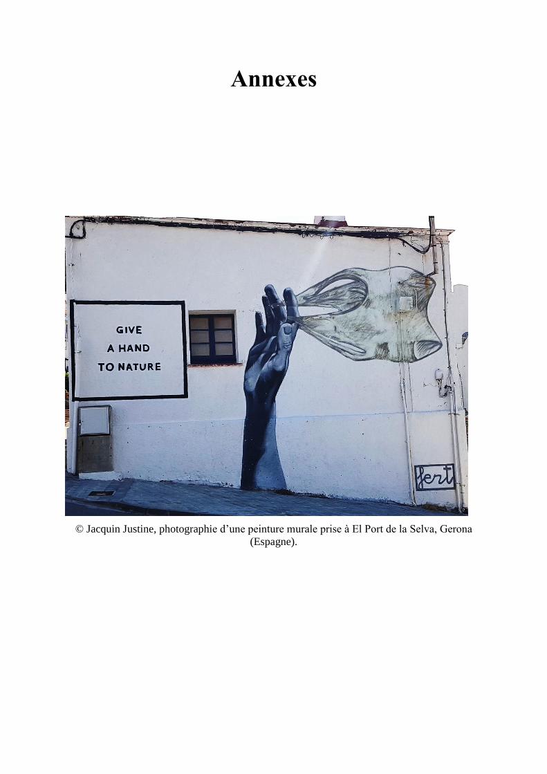
Annexes
© Jacquin Justine, photographie d’une peinture murale prise à El Port de la Selva, Gerona
(Espagne).

A n n e x e s P a g e | 223
223
Annexe 1– Article en premier auteur publié en 2019 dans Frontiers in microbiology
Title: Microbial ecotoxicology of marine plastic debris: a review on colonization and
biodegradation by the ‘plastisphere’
Authors: Jacquin J1, Cheng J1, Odobel C1, Pandin C1, Conan P1, Pujo-Pay M1, Barbe V1,2,
Meistertzheim AL1 and Ghiglione JF1*
Affiliations :
1. CNRS, Sorbonne Université, Laboratoire d’Océanographie Microbienne LOMIC,
UMR 7621, Observatoire Océanologique de Banyuls, 66650 Banyuls sur mer, France.
2. Commissariat à l′Energie Atomique et aux Energies Alternatives, Genoscope, Institut
de biologie François-Jacob, 2 Rue Gaston Crémieux, 91000 Evry, France
*Corresponding author:
JF Ghiglione, CNRS, Sorbonne Université, Laboratoire d’Océanographie Microbienne
LOMIC, UMR 7621, Observatoire Océanologique de Banyuls, 66650 Banyuls sur mer, France.
Tel.: + 33 (0) 4 68 88 73 16. E-mail: [email protected]
Keywords: Bacteria; marine plastics debris; colonization; biodegradation; metabolic pathways
A n n e x e s P a g e | 224
224
ABSTRACT
Over the last decades, it has become clear that plastic pollution presents a global societal and
environmental challenge given its increasing presence in the oceans. A growing literature has
focused on the microbial life growing on the surfaces of these polluants, called the
‘plastisphere’, but the general concepts of microbial ecotoxicology have only rarely been
integrated. Microbial ecotoxicology deals with (i) the impact of pollutants on microbial
communities and inversely (ii) how much microbes can influence their biodegradation. The
goal of this review is to enlighten the growing literature of the last 15 years on microbial
ecotoxicology related to plastic pollution in the oceans. First, we focus on the impact of plastic
on marine microbial life and on the various functions it ensures in the ecosystems. In this part,
we also discuss the driving factors influencing biofilm development on plastic surfaces and the
potential role of plastic debris as vector for dispersal of harmful pathogen species. Second, we
give a critical view of the extent to which marine microorganisms can participate in the
decomposition of plastic in the oceans and of the relevance of current standard tests for plastic
biodegradability at sea. We highlight some examples of metabolic pathways of polymer
biodegradation. We conclude with several questions regarding gaps in current knowledge of
plastic biodegration by marine microorganisms and the identification of possible directions for
future research.
A n n e x e s P a g e | 225
225
1. Introduction
The amount of land-based plastic debris entering the ocean is estimated at 4.8 to 12.7
million tons per years (Jambeck et al., 2015). It is so important that plastic is regarded as a
marker of the Anthropocene (Duis and Coors, 2016; Zalasiewicz et al., 2016). A growing body
of research has investigated plastic distribution (Willis et al., 2017; Worm et al., 2017) and
toxicity for marine fauna (Bakir et al., 2014; Gewert et al., 2015). A comparatively smaller but
growing literature has been devoted to the microbial ecotoxicology of marine plastic debris, i.e.
(1) the impact of plastic on marine microbial life together with the various ecosystem services
that marine microbial life ensures and inversely, (2) the role of microorganisms in the
degradation of ocean plastic (Ghiglione et al., 2014, 2016). Both aspects will be successively
explored by this review, which covers the last 15 years of literature.
The investigation of microorganisms colonizing plastic surfaces using modern
techniques of massive DNA sequencing (Zettler et al., 2013) was introduced only recently. The
authors introduced the world ‘plastisphere’ to describe the microbial life growing on these
surfaces. They also detected members of the potentially pathogenic genus Vibrio, which may
be dispersed over long distances by floating persistent plastics. Since then, several studies
investigated various marine environments, such as the North Pacific gyre (Debroas et al., 2017)
or the Mediterranean sea (Dussud et al., 2018a). In parallel, a growing literature described the
first steps of colonization of new plastic until the formation of a mature biofilm (Dussud et al.,
2018a; Lobelle and Cunliffe, 2011; Oberbeckmann et al., 2015) .
Such knowledge is of great interest to better understand the impact of plastic on marine
microbial life and ecosystem functions. Only one study so far used shotgun metagenomics,
showing that plastic-inhabiting microbes present an enriched gene repertoire compared to
microbes living in the surrounding waters (Bryant et al., 2016). In this review, we argue that
current knowledge is insufficient to draw a clear picture of the impact of plastic on marine
A n n e x e s P a g e | 226
226
microbial life and ecosystem functions, and we propose several directions for further studies in
this field (see section 2: microorganisms colonizing plastic at sea).
The role of microbes on plastic degradation in the ocean is a second subject of concern
Very recently, an excellent comprehensive review concluded that “current international
standards and regional test methods are insufficient in their ability to realistically predict the
biodegradability of carrier bags in marine environment, due to several shortcomings in
experimental procedures and a paucity of information in the scientific literature” (Harrison et
al., 2018). The capability of microorganisms to biodegrade plastic was reported for numerous
bacterial strains (Krueger et al., 2015). Fungi also have the capability to biodegrade plastics,
but most of the studies were conducted in terrestrial conditions (Magnin et al., 2018, Gajendiran
et al., 2016, Koitabashi et al., 2012, Cosgrove et al., 2007) whereas very few studies so far exist
in marine conditions (Gonda et al., 2000; Pramila et al., 2011). Moreover, most of these studies
were based on the selection and testing of single strains in laboratory conditions, which is very
far from environmental conditions. In this review, we underscore the knowledge gaps on plastic
biodegradation by marine microorganisms and we attempt to identify possible directions for
future research in this area (see section 3: how much can microorganisms participate in plastic
degradation at sea?).
2. Microorganisms colonizing plastic at sea
2.1. A new niche for marine microorganisms. I
t was not until recently that the first work using modern techniques of massive DNA
sequencing provided a detailed picture of the microbial life on plastic and introduced the term
‘plastisphere’ (Zettler et al., 2013). Bacteria, Archaea, Fungi and microbial Eukaryotes were
detected in several studies, starting from plastics sampled at sea or from new plastics
A n n e x e s P a g e | 227
227
experimentally incubated in marine conditions (Table 1). Plastic debris are mainly composed
of polyethylene (PE) at sea surface, followed by polypropylene (PP) and polystyrene (PS) (Auta
et al., 2017). Whatever the polymer type, recent studies emphasized the difference between the
bacteria living on plastics and the bacteria living in free-living state (Debroas et al., 2017) or
on organic particles in the surrounding seawater (Dussud et al., 2018a; Oberbeckmann et al.,
2018). Similar observations have been made for fungal communities (Kettner et al., 2017)
Another aspect that received much less attention is the plastisphere living in the water
column other than the surface layer. Because of methodological constrains, most of the studies
so far have been limited to sampling surface seawater using manta trawls, which represents less
than 1% of the global load of plastic in the open ocean (Cózar et al., 2014). Only certain types
of plastics made of PE and PP with high surface-to-volume ratios, such as rigid plastics and
bundled fishing nets and ropes, have the capability to remain for a very long time at the surface
of the oceans (Lebreton et al., 2018). Most other buoyant plastic such as films or smaller pieces,
tend to sink to the sediment owing to biofouling (Fazey and Ryan, 2016, Kalogerakis et al.,
2017). Very limited information is available concerning the composition of microbial
communities on plastic items sampled from the seafloor (De Tender et al., 2015). If
photoautotrophic bacteria such as the cyanobacteria Phormidium and Rivularia dominate the
sub-surface plastisphere communities (Zettler et al. 2013; Bryant et al. 2016; Dussud et al.
2018a), the core microbiome of the seafloor and sub-surface plastisphere seems to share some
taxa: Bacteroidetes (Flavobacteriaceae) and Proteobacteria (Rhodobacteraceae and
Alcanivoracaceae) (Zettler et al. 2013; Bryant et al. 2016; De Tender et al. 2017; Dussud et al.
2018a).
A n n e x e s P a g e | 228
228
2.2. Successive colonization stages of new plastics incubated in marine conditions. In parallel
to studies on plastic directly sampled at sea, other studies focused on the successive colonization
steps of new plastics incubated in marine conditions (Table 1). At sea, plastics are rapidly
covered by the ‘conditioning film’ made of inorganic and organic matter, which is then rapidly
colonized by bacteria (mainly Gammaproteobacteria and Alphaproteobacteria)
(Oberbeckmann et al., 2015). With time, members of Bacteroidetes become increasingly
abundant (Lee et al., 2008). Hydrophobicity and other substratum properties (crystallinity and
crystal structure, roughness, glass transition temperature, melting temperature, modulus of
elasticity) may play a role in the selection of bacterial community in the early stages of
colonization (Pompilio et al., 2008), but probably do so to a lesser extent when the biofilm
becomes mature (Dussud et al., 2018a). The successive growing and maturation phases of
biofilm formation, already described for other surfaces such as glass, acryl, steel or rocks and
algae (Salta et al., 2013), were also observed for plastics of different compositions
(Oberbeckmann et al., 2015). Biofilm developments were followed during several weeks in
seawater on PE-based plastic bags (Lobelle and Cunliffe, 2011), polyethylene terephthalate
(PET)-based plastic bottles (Oberbeckmann et al., 2014), polyvinyl chloride (PVC) (Dang et
al. 2008) or polystyrene (PS) coupons (Briand et al., 2012). PE-based plastics were also rapidly
colonized by microorganisms in marine sediments (Harrison et al., 2014). Clear differences in
bacterial abundance, diversity and activity were found between non-biodegradable and
biodegradable plastics (Dussud et al. 2018b, Eich et al. 2015). Higher colonization by active
and specific bacteria were found after 6 weeks on poly(3-hydroxybutyrate-co-3-
hydroxyvalerate) (PHBV) and pre-oxidized PE-based oxodegradable polymers (OXO) in
comparison to non-biodegradable PE polymers (Dussud et al. 2018b, Eich et al. 2015). Longer-
term studies carried out over a 6-month to 1 year period also showed differences in biofilm
formation and maturation according to the polymer type, i.e. PE, PP, PET, or polycarbonate
A n n e x e s P a g e | 229
229
(PC) (De Tender et al., 2017; Webb et al., 2009). Not only bacteria but also fungi were shown
to form biofilms on plastic surfaces (Pramila et al., 2011), mainly dominated by
Chytridiomycota, Cryptomycota (Kettner et al., 2018) and Ascomycota (De Tender et al., 2017;
Kettner et. al., 2018; Oberbeckmann et al., 2016).
2.3. Potential impact of plastic on the microbial role in regulation of biogeochemical cycles.
The quantity of plastic in the oceans can no longer be considered a limited ecological problem,
since small pieces of plastic called “microplastics” (<5mm) found at sea could cover 4,2 million
km2 of the sea surface (Charette and Smith, 2010; Eriksen et al., 2014; Hidalgo-Ruz et al.,
2012). Marine microorganisms that compose the plastisphere are known to play a key role in
the biogeochemical cycles in the oceans (Pomeroy et al., 2007). One-half of oceanic primary
production on average is channelled via heterotrophic bacterioplankton into the microbial loop,
thus contributing significantly to food web structure and carbon biogeochemical cycling in the
ocean (Fenchel, 2008) (Figure 1). Only one recent study compared the heterotrophic production
of bacteria living on plastic and in seawater. Heterotrophic bacteria living on plastics were
particularly active, the cell-specific activity measured by 3H-leucine incorporation into proteins
being 43- to 88-fold higher than that of the free-living fraction (Dussud et al., 2018a).
Unfortunately, these results were obtained in the frame of a study on colonization of new
plastics incubated at sea for a relatively short period (45 days). Similar methodologies applied
to plastics that had spent several years at sea would be necessary to evaluate how much the
large amount of plastic and the accompanying plastisphere influence the biogeochemical
carbon cycle in the oceans.
Interestingly, most of the studies aiming to characterize the plastisphere mentioned that
Cyanobacteria were overrepresented on plastics compared to the surrounding free-living and
organic particle-attached fractions. The relative importance of photosynthetic activities that
Cyanobacteria living on plastic have on global pelagic primary production is still unknown.
A n n e x e s P a g e | 230
230
Coupling primary production and heterotrophic production measurements over large temporal
and spatial scales will be necessary to obtain a better view of the role of the plastisphere on
carbon cycling in the oceans. Microorganisms are not only involved in the carbon cycle, but
basically in all other biogeochemical cycles including nitrogen, sulfur, iron, manganese,
chromium, phosphorus, calcium and silicate cycles, which may also be impacted by the
presence of plastic at sea (Hutchins and Fu, 2017).
2.4 Potential dispersion of pathogen species
Interest has been raised about opportunist pathogen dispersal on plastics, such as animal or
human pathogenic Vibrio sp. (Zettler et al., 2013). Marine plastic debris as vector of harmful
species was first suggested by Masó et al. (2003), who identified potential harmful
dinoflagellates such as Ostreopsis sp. and Coolia sp. Putative pathogen of fish (Tenacibaculum
sp.) and of invertebrates (Phormidium sp. and Leptolyngbya sp.) were found to be more
common on plastic compared to surrounding seawater (Dussud et al. 2018a). Some bacterial
taxa considered as putative pathogens for human, coral and fish were also found in the intertidal
zone of the Yangtze Estuary, at relatively low abundance (<1.6%) (Jiang et al., 2018). A
putative pathogen for coral Halofolliculina spp. was found to be abundant on some western
Pacific plastic debris (Goldstein et al., 2014). Some toxic eukaryotic species were also
mentioned by Debroas et al. (2017) at low abundance (<0.04%), but might be regarded as
hitchhiker organisms. Nevertheless, caution should be taken since the 16S rRNA
metabarcoding approach used in all these studies was not an appropriate method for describing
bacterial virulence. The recent coupling of the 16S rRNA metabarcoding technique with the
detection of virulence-associated genes may be an interesting option to address this question
(Kirstein et al., 2016). Pathogenicity evidence on marine animals in relation to the plastisphere
has never been proven, and further research will be required before publicizing alarmist
conclusions on the possible responsibility of plastic debris as vector for the spread of disease-
A n n e x e s P a g e | 231
231
causing organisms. Apart from those results, microplastics colonized by pathogens may also
pose threats to humans who are exposed to contaminated beach and bathing environments
(Keswani et al., 2016). Evidence is still missing to determine whether plastic debris could lead
to the spread and prolonged persistence of pathogenic species in the oceans.
2.5. Factors driving the plastisphere composition and activities.
Factors driving the plastisphere composition are complex, mainly spatial and seasonal, but are
also influenced by the polymer type, surface properties and size. Plastisphere communities
studied in different polymer types floating in the North Pacific and North Atlantic reflected first
their biogeographic origins, and to a lesser extent the plastic type (Amaral-Zettler et al., 2015).
Similar conclusions were found for bacterial communities colonizing plastics along an
environmental gradient. These communities are shaped firstly by the freshwater to marine
environmental conditions and secondarily by the plastic type (PS and PE) (Oberbeckmann et
al., 2018). Inversely, another study based on a large number of microplastics sampled in the
western Mediterranean sea showed no effect of geographical location (including coastal and
open ocean samples) or plastic type (mainly PE, PP and PS) on the bacterial community
composition. The growing number of studies on the plastisphere are giving a better view of the
microbial biofilm community on plastics in the oceans, but the complex network of influences
is still the subject of ongoing debate. A clearer picture will hopefully emerge from more
extensive investigations with widespread and numerous samples, together with better
descriptions of the physical and chemical properties of the polymers.
The physical properties of plastic offer a unique habitat that contribute to the long-
distance transport of diverse microbial hitchhikers attached to its surface (Harrison et al., 2011;
Zettler et al., 2013). A vast range of other phyla, including Arthropoda, Annelida, Mollusca,
A n n e x e s P a g e | 232
232
Bryozoa and Cnidaria have conferred on plastics the role of vector for the transfer of organisms,
some of them being catalogued as invasive alien species (Oberbeckmann et al., 2015). For
instance, plastic debris with tropical biota including corals was detected in the Netherlands
(Hoeksema, 2012), and southern ocean bryozoans were observed in Antarctica (Barnes and
Fraser, 2003). Interactions between micro- and macro-organisms, their substratum and their
surroundings are needed to better predict the ecological consequences of microplastics
transported through the global oceans.
3. How much can microorganisms participate in plastic degradation at sea?
3.1. Definition and main processes involved in plastic biodegradation.
Biodegradation of plastic is a process that results in total or partial conversion of organic carbon
into biogas and biomass associated with the activity of a community of microorganisms
(bacteria, fungi and actinomycetes) capable of using plastic as a carbon source (Shah et al.,
2008). Depending on the respiratory conditions (aerobic / anaerobic) and the microorganisms
involved, the biogas will be different (CO2, CH4, H2S, NH4, H2) (Mohee et al., 2008).
Microorganisms, including bacteria and fungi, present the capabilities to degrade or deteriorate
plastics and several review papers updated the list of plastic-degraders (Bhardwaj et al., 2013;
Kale et al., 2015; Pathak, 2017; Shah et al., 2008). Arthrobacter, Corynebacterium,
Micrococcus, Pseudomonas, Rhodococcus and Streptomyces were the prominent microbial
taxa able to use plastic as sole carbon source and energy in laboratory conditions. Table 2
proposes an update of the current list of microorganisms proven to present biodegradation
capabilities under laboratory conditions.
Biodegradation is considered to occur after or concomitant with physical and chemical
degradation (abiotic degradation), which weakens the structure of polymers as revealed by
A n n e x e s P a g e | 233
233
roughness, cracks and molecular changes (İpekoglu et al., 2007). Alteration of plastic properties
due to abiotic degradation is called ‘aging’ and in nature depends on several factors such as
temperature, solar light and chemicals that enhance the rate of degradation by oxidizing or
disrupting the length of the polymer chain.
Biodegradation can be summarized in 4 essential steps, which have been described in detail
in a review by Dussud and Ghiglione (2014):
- Bio-deterioration relates to the biofilm growing on the surface and inside the plastic,
which increases the pore size and provokes cracks that weaken the physical properties
of the plastic (physical deterioration) or releases acid compounds that modify the pH
inside the pores and results in changes in the microstructure of the plastic matrix
(chemical deterioration).
- Bio-fragmentation corresponds to the action of extracellular enzymes (oxygenases,
lipases, esterases, depolymerases and other enzymes that may be as diverse as the large
spectrum of polymer types) released by bacteria colonizing the polymer surface. These
enzymes will reduce the molecular weight of polymers and release oligomers and then
monomers that can be assimilated by cells.
- Assimilation allows oligomers of less than 600 Daltons to be integrated inside the cells
to be used as a carbon source, thus increasing the microbial biomass.
- Mineralization is the ultimate step in the biodegradation of a plastic polymer and results
in the excretion of completely oxidized metabolites (CO2, N2, CH4, H2O).
3.2. Rates of plastic degradation.
Rates of degradation of conventional plastics by microorganisms are extremely low, even
in optimized laboratory conditions (Krueger et al., 2015). Most of the conventional plastics are
recalcitrant to biodegradation in marine and terrestrial environments, resulting in lifetimes of
decades or even centuries (Krueger et al. 2015). Plastics present low bioavailability since they
A n n e x e s P a g e | 234
234
are generally solid and made of densely cross-linked polymers that provide low accessibility
for microbes and enzymes circumscribed to the outermost layer of the items. In the pelagic
ecosystem, plastics are biodegraded by the aerobic route of microorganisms, i.e. the end product
of the reaction will be microbial biomass, CO2 and H2O. The anaerobic biodegradation pathway
would be more frequently encountered in sediment and is supposed to be even slower than in
the pelagic zone (Ishigaki et al., 2004). Unfavorable C/N ratio is a key factor for biodegradation
of other hydrocarbon-based products in the oceans (Sauret et al., 2016) and may potentially
also limit plastic biodegration.
Data currently available rely heavily on culture-based approaches in laboratory conditions,
although bacteria that can be cultured represent less than 1% of the number of bacteria in nature
(the so-called ‘great plate count anomaly’) and a very small proportion of its very large diversity
(Hugenholtz et al., 2009). To date, data on the rate of plastic mineralization in the oceans are
still virtually non-existent. Congruent descriptions of the plastisphere that forms an abundant
biofilm characterized by very diverse and active plastic-specific characteristics are available
(Debroas et al. 2017; Dussud et al. 2018b). Evidence of pits visualized in the plastic debris that
conform to bacterial shapes directly found in the marine environment (Zettler et al., 2013)
together with a number of putative xenobiotic degradation genes likely involved in plastic
degradation that were found to be significantly more abundant in the plastic-specific
communities (Bryant et al. 2016; Debroas et al. 2017; Dussud et al. 2018b) are thus of great
interest. A recent study underlined the need of cometabolic pathways on PE biodegradation,
thus confirming that complex microbial communities rather than single species are necessary
to degrade recalcitrant plastic (Syranidou et al., 2017). So far, the timescales of degradation and
the characterization and the fate of the degradation products, are fundamental, yet still
unanswered questions.
3.3. Standard tests for plastic biodegradability at sea.
A n n e x e s P a g e | 235
235
The current standards for marine environments propose tests based on respirometry
measurements, susceptible to describe the mineralization step of plastic biodegradation in
aerobic conditions (see Figure 2). They impose a minimum percentage of conversion from
plastic to CO2 ranging from 60% to 70% over a period of 3 months (ASTM D6691-09), 6
months (ASTM D7473-12) or 24 months (ISO 18830, ISO 19679, ASTM D7991-15) under
aerobic conditions (see Figure 3). Anaerobic biodegradation is characterized by specific
standards (see for example ASTM D5511-18), but to our knowledge none of these standards
applies to the marine environment. Biodegradation of a plastic is characterized by the time
required to achieve mineralization under controlled conditions. These tests cannot be
considered as a proof of ready biodegradability (total conversion of plastic into biomass and
CO2), but rather an indication about a potential for biodegradation in the oceans.
Recently, these standards were considered insufficient in their ability to realistically predict
the biodegradability in marine environment (Harrison et al., 2018). These tests can significantly
underestimate the time required for polymer biodegradation within natural ecosystems. First,
the authors underlined “biases associated with the preparation of experimental inocula and the
test conditions themselves, including the use of preselected and/or pre-conditioned strains,
artificially modified inocula, powdered test materials, nutrient-rich synthetic media and test
temperatures that are frequently higher than those encountered within the environment”. The
authors also pointed out “the lack of clear guidelines for the analysis of different polymer types,
including composite materials and plastics that contain additives”, which can considerably
influence the rates of biodegradation. “There is also a paucity of guidelines for materials of
varying shapes and sizes and, in certain cases, the test procedures lack a sufficient level of
statistical replication”. Another concern, not raised by Harrison et al. (2018), is the biases
associated with the common method for determining biodegradability, i.e. measurements of
CO2 evolution. This method may lead to either underestimation or overestimation of the plastic
A n n e x e s P a g e | 236
236
biodegradation due to other processes. It is noteworthy that plastic generally presents high
sorption capability of organic matter (especially hydrophobic organic chemicals including
pollutants) that can be biodegraded by the plastisphere biofilm, thus resulting in a CO2
production that has nothing to do with plastic biodegradation (Lee et al., 2014). Inversely,
several papers reported the importance of photosynthetic microorganisms growing on plastics,
which consume CO2 regardless of plastic biodegradation (Zettler et al. 2013; Bryant et al. 2016;
Dussud et al. 2018b). Further studies are needed to evaluate the relative degree of CO2
consumption by photosynthesis, CO2 production by organic matter degradation by the
plastisphere as compared to CO2 production due to plastic biodegradation.
The limitations of the respiratory methods described above can be overcome by other
additional analytical techniques and approaches to confirm changes in the physical properties
and the chemical structure of polymers during biodegradation. Alterations in the visual
appearance and in the mass or changes in mechanical properties are relatively easy and low-
cost methods for the evaluation of physical changes during biodegradation. Other methods
could be combined to confirm changes in the molecular structure of polymers, such as
measurements of surface hydrolysis and other chromatographic (gas chromatography with or
without flame ionization detection, liquid chromatography, gel-permeation chromatography)
measurements coupled or not with spectrometric techniques (mass spectrometry, nuclear
magnetic resonance spectroscopy, Fourier-transform infrared spectroscopy). Optical, atomic
force and scanning electron microscopy can also be used to assess the biodeterioration of the
surface due to microbial activity or biofilm formation. Any of these techniques are enough to
prove biodegradation by its own, and each of them has limitations that have been previously
detailed for example in the excellent reviews of (Harrison et al., 2018; Ho et al., 2018; Koutny
et al., 2006). The current standards sometimes propose to use such techniques to corroborate
A n n e x e s P a g e | 237
237
the main test based on respirometry measurement, but no clear guidelines on how to use these
tests is provided.
3.4. Examples of metabolic pathways of polymer biodegradation.
There are currently more than 5,300 grades of synthetic polymers for plastics in commerce
(Wagner and Lambert (eds) 2018). They are generally produced with a range of chemical
additives such as plasticizers, flame retardants, antioxidants and other stabilizers, pro-oxidants,
surfactants, inorganic fillers or pigments (Wagner and Lambert (eds) 2018). Their
heterogeneous physical-chemical properties will likely result in very heterogeneous metabolic
pathways of biodegradation, especially when considering the large variety of microorganisms
that may interact for the degradation of a single piece of plastic, together with the environmental
factors of very dynamic oceanic conditions. We are aware that treating plastic as a single
compound does not make sense and providing details on the metabolic pathways of plastic
biodegradation would necessarily be unrepresentative of the complexity of the various
processes that occur in the environment. We have chosen to focus on the metabolic pathways
associated with the biodegradation of model compounds used in the formulation of
conventional (PE, PET, PS) and so called “biodegradable” plastics (PHA) that are the most
popular and the most extensively studied in the literature. Moreover, it should be noted that
because of the difficulty of dealing with long-term experiments and complex communities
under natural conditions, all the following studies describing the metabolic pathways of plastic
biodegradation were done using a culture-based approach.
3.4.1. Metabolic pathways of polyethylene (PE) biodegradation. High- and low-density
polyethylene is a long linear carbon chain (CH2) belonging to the family of polyolefins.
Polyethylene is derived from petroleum sources and its large use in our daily life made it the
first plastic waste found at sea surface. PE is considered difficult to biodegrade because the
A n n e x e s P a g e | 238
238
long chains of carbons and hydrogens are very stable and contain very balanced charges.
Microorganisms generally need imbalance of electric charge to perform biodegradation. To
destabilize the local electric charge, bacteria use oxygenases: enzymes able to add oxygen to a
long carbon chain (Krueger et al., 2015). For instance, mono-oxygenases and di-oxygenases
incorporate, respectively, one and two oxygen atoms, forming alcohol or peroxyl groups that
are less recalcitrant for biodegradation. Oxidation may also be processed by abiotic reactions
associated with UV radiation or temperature (for more details, see the review by Singh and
Sharma (2008). Oxidation of PE results in the formation of carboxylic groups, alcohols, ketones
and aldehydes by a radical reaction (Gewert et al. 2015; Vasile, 2000). The oxidation and
fragmentation of PE make the polymer more hydrophilic and facilitates access to other
extracellular enzymes, such as lipases and esterases after the formation of carboxylic groups,
or endopeptidases for amide groups (Gewert et al., 2015). Other enzymes such as laccase in
Rhodococcus ruber are excreted and can facilitate the biodegradation of PE (Santo et al.,2013).
Interestingly, a recent study focused on soluble oxidized oligomers showed that 95% of these
compounds were assimilated by a strain of Rhodococcus rhodocchrous after 240 days of
incubation (Eyheraguibel et al., 2017). The polymer is broken down into small oligomers of
600 Da incorporated in the cells by carriers belonging to the Major Facilitor Superfamily (MFS)
or harboring ATP binding cassettes (ABC) (Gravouil et al., 2017). -oxidation transforms
oxidized carboxylic molecules (having an even number of carbon atoms) into acetyl coA or
propionyl coA (if odd number of carbons). Carboxylation of propionyl coA into succinyl coA
is performed by propionyl-coA carboxylase. Gravouil and al (2017), propose identification of
an overexpressed enzyme, when the bacteria find PE in the medium (Gravouil et al., 2017).
Acetyl coA and succinyl coA enter the tricarboxylic acid (TCA) cycle (Figure 4). This cycle
produces chemical energy in the form of a reducing power (NADH, H + and CoQ10H2) used in
the respiratory chain to produce ATP, which is necessary to create new microbial biomass via
A n n e x e s P a g e | 239
239
replication processes. It also produces CO2 and H2O that sign the complete mineralisation of
PE.
For twenty years now, scientists have been interested in the biodegradation of
polyethylene by the microbial community. Bacterial and fungal strains presenting
biodegradation capabilities of PE are listed in Table 2 and Supplemental Table 1.
Genetic evidence of PE biodegradation remains scarce in the literature, but preliminary
work highlighted enzymes, transporters or genes that may be involved in this process (Gravouil
et al., 2017). Alkane hydroxylase genes were found to play a central role in PE biodegradation
for Pseudomonas sp. E4 strain, which was capable of mineralizing 28.6% of the organic carbon
of the polymer in 80 days. The alkB gene was then introduced in Escherichia coli BL21 strain,
which was then able to mineralize 19.3% of the organic carbon of the polymer (Yoon et al.,
2012). Only two other studies used genetic analysis to provide evidence for the importance of
laccase in PE biodegradation by Rhodococcus ruber (Gravouil et al., 2017; Santo et al., 2013;
Sivan, 2011).
3.4.2. Metabolic pathways of polyethylene terephthalate (PET) biodegradation. PET is part of
the polyester family and it is widely used in the design of bottles and synthetic fibres. It is
considered as persistent plastic in the environment because of its long carbon chains containing
aromatic rings that are difficult to biodegrade (Marten et al., 2005). In recent years, studies have
shown that some bacterial strains were able to degrade PET as sole carbon source and energy,
such as Idonella sakaiensis (Yoshida et al., 2016), Nocardia sp. (Sharon and Sharon, 2017)
Pseudomonas mendocina (Ronkvist et al., 2009), Thermobifida fusca (Müller et al., 2005).
Some fungal communities are also known to biodegrade PET, such as Humicola insolens,
several Fusarium species, and Penicillium citrinum (Liebminger et al., 2007; Nimchua et al.,
2007; Ronkvist et al., 2009; Silva et al., 2005). Cutinases or hydrolases play key roles in PET
biodegradation (Danso et al., 2018). For example, Ideonella sakaiensis 201-F6 adhered to the
A n n e x e s P a g e | 240
240
PET surface and first secreted two enzymes involved in the biodegradation process of PET:
PETase (hydrolase) and MHETase. PETase is an extracellular enzyme capable of hydrolysing
PET to mono-(2-hydroxyethyl) terephthalate (MHET), terephthalic acid (TPA) and bis (2-
hydroxyethyl) terephthalate (BHET). Fungi seem to have the same biodegradation strategy and
are able to degrade PET into BHET and MHET (Liebminger et al., 2007). The MHETase
hydrolyzes MHET to TPA and ethylene glycol (EG). The terephthalic acid molecule is then
internalized in the bacterial cells by the TPA transporter (Hosaka et al., 2013) and then
catabolized by TPA 1,2-dioxyenase (TPADO) and 1,2-dihydroxy-3,5-cyclohexadiene-1,4-
dicarboxylate dehydrogenase (DCDDH) to give protocatechuic acid (PCA) as the final
molecule (Yoshida et al., 2016). This PCA is cleaved by PCA 3,4 dioxygenase (PCA34) to give
the hemiacetal form of 4-carboxy-2-hydroxymuconic. The latter becomes the substrate of a
dehydrogenase to form 2-pyrone-4-6-dicarboxylic acid that enters the TCA cycle and initially
transformed into pyruvate and oxaloacetate, then assimilated as CO2 and H2O (Figure 4).
3.4.3. Metabolic pathways of polystyrene (PS) biodegradation.
Polystyrene is a polymer composed of styrene monomer (CH2 = CH2-Ph). The polymer is
highly hydrophobic and presents a high molecular weight. Like other conventional plastics,
partial biodegradation in the laboratory has been observed while it continues to accumulate in
the oceans (Auta et al., 2017) thus inciting increasing interest in PS biodegradation (see
Supplemental Table 1 and Asmita et al., 2015; Atiq et al., 2010; Mor and Sivan, 2008; Oikawa
et al., 2003; Yang et al., 2015; Tang et al., 2017).
Several biodegradation pathways may be considered, depending on the microorganism
involved. The predominant pathway is the oxidation pathway of the styrene side chain
presented in Figure 4. The styrene is directly oxidized with a styrene monooxygenase to form
a styrene epoxide which will then be oxidized to phenylacetaldehyde by styrene oxide. This
A n n e x e s P a g e | 241
241
molecule is then catabolized into phenylacetic acid. This conversion of styrene to phenylacetic
acid is called the upper pathway of styrene metabolism. Phenylacetic acid is converted to
phenylacetyl-CoA (acetyl coenzyme A) by the so-called lower pathway (Luu et al., 2013) then
subjected to several enzymatic reactions (Figure 4) to finally enter the tricarboxylic acid (TCA)
cycle. The biodegradation products enter the TCA cycle through the final formation of acetyl-
Co A and succinyl-CoA (succinyl-CoenzymeA) (Luu et al., 2013).
Interestingly, Pseudomonas putida CA-3 can accumulate polyhydroxyalkanoates (PHA
at medium chain length) when growing on styrene, thus using an original biodegradation
pathway. A catabolic operon has been identified as responsible for this bioconversion; this path
is called the PACoA (Phenylacetyl-CoA) catabolon. It involves oxidation of the aromatic ring,
followed by entry into the -oxidation cycle and the conversion to acetyl-CoA (O’Leary et al.,
2005). This acetyl-CoA can follow different metabolic pathways, either entering the TCA cycle
or following the de novo fatty acid biosynthesis path which will give as final product medium-
chain-length polyhydroxyalkanoates (mcl-PHAs) (O’Leary et al., 2005). This study shows the
complexity of studying the biodegradation pathways of these polymers and indicates the great
range of possibilities when considering the large diversity of microorganisms found in the
plastisphere.
3.4.4. Metabolic pathways of polyhydroxyalkanoate (PHA) biodegradation. The current global
production of PHA is increasing, reaching 49,200 tons per year that represents 2.4% of the
production of bioplastics (https://www.european-bioplastics.org). PHAs are biopolymers of
hydroxylated fatty acids produced within a bacteria in granular form. Each PHA monomer
([CO-CH2-CHR-O]n) consists of hydroxyalkanoates linked together by ester bonds. The alkyl
group (R) varies from a methyl group to a tetradecyl group. When bacteria are placed in a
medium with an excess carbon source and low nutrient content, they accumulate storage
granules. Over 300 bacterial species are capable of producing 80 different hydroxyalkanoate
A n n e x e s P a g e | 242
242
monomers, and some bacteria can accumulate up to 90% of their total weight of polymer in
very specific conditions (Pena et al., 2014). One of the most commonly used PHA for plastic
production is polyhydroxybutyrate (PHB), which has a methyl as an alkyl group (R) ([CO-
CH2-CHCH3-O]n). PHB is one of the homopolymers with high commercial power because it
has thermoplastic, hydrophobic, low oxygen permeability and is considered biodegradable
(Chang et al., 2012; Mothes et al., 2004). It is not very deformable, because of its high
crystallinity (Gorke et al., 2007) and it has a high melting point close to its thermal degradation
temperature (Reis et al., 2003). A copolymer made of Poly(3-hydroxybutyrate-co-3-
hydroxyvalerate) (PHBV) that reduces the melting point of PHB is seen to emerge in PHA
production. The advantage of using PHA is that it is stable over time, as long as the conditions
governing its biodegradation are not met (Jaffredo et al., 2013)
Due to their microbial origin, PHAs were found to be biodegradable in many environments
such as soil, marine ecosystems or sewage sludge (Eubeler et al., 2010). Biodegradation of Poly
(3-hydroxybutyrate-co-3-hydroxyhexanoate) has been proven with comparable rates to that of
cellulose, with faster degradation found under aerobic (85 days) compared to anaerobic (6
months) conditions (Wang et al., 2018). The biodegradation scheme in Figure 4 shows the
different steps of PHB biodegradation. When the biodegradation is not carried out inside the
cells by bacteria that produce their own PHB, other bacteria initiate the biodegradation of PHB
in the medium by external hydrolysis using ectoenzymes that convert the polymers into
hydroxylated acid monomers of hydroxybutyrate (HB) (Pena et al., 2014). This molecule is
water soluble and small enough to passively diffuse across the bacterial membrane and enter
the -oxidation cycle. The resulting acetyl-CoA will be oxidized in the TCA cycle until final
mineralisation (Alshehrei, 2017). PHA-degradation has been proven in the laboratory under
aerobic or anaerobic conditions (see a non-exhaustive list in Supplemental Table 1). The
dominant bacteria in aerobic marine conditions belong to Clostriales, Gemmatales,
A n n e x e s P a g e | 243
243
Phycisphaerales and Chlamydiales, whereas Cloacamonales and Thermotogales dominate in
anaerobic sludge (Wang et al., 2018).
4. Concluding remarks:
In this review, we have presented both aspects of microbial ecotoxicology on marine plastic
debris, namely the impact of plastic on marine microbial life and inversely how microbes can
play a role in plastic biodegradation. An increasing number of studies either describe the
different steps of biofilm formation under marine conditions, or give new insights on bacteria
colonizing the aged plastics directly sampled at sea. The very diverse and active bacteria living
on plastics as compared to the surrounding waters suggest a potential impact on the global
biogeochemical cycles associated with the relatively recent introduction of plastic in the oceans,
impact that remains to be determined. Plastic released in the oceans is also accused to be a raft
for invasive species including pathogenic bacteria, but no proof of pathogenicity on marine
animals or humans in relation to plastic ingestion has emerged so far.
A better knowledge of the plastisphere is also a critical issue in understanding the role
played by bacteria in plastic biodegradation. Several studies have underlined that current
standards are failing to prove biodegradability at sea for several reasons that have been
highlighted in this review. Biodegradation of a polymer at sea depends on many factors related
to its own composition, but also on the various ecosystems and environmental conditions
encountered during its very long lifetime. It is for these reasons that plastic polymers continue
to accumulate at sea and that biodegradation rates reported in the laboratory are never reached
in the environment. Thus, a complete study of the biodegradation of a polymer at sea must
combine several monitoring parameters, and especially be confirmed in the field with
experiments in situ. Given the complexity of the plastic problem, research network initiatives
such as ‘Polymers & Oceans’ (https://po2018.wixsite.com/po2018) that bring together
A n n e x e s P a g e | 244
244
physicists, chemists and biologists are required to answer the wishes and needs of many
scientists to face this environmental problem and its resonance in the society.
5. Acknowledgments
The work was made possible thanks to support granted to ANR by the Programme
OXOMAR (funded by the French Ministry for Education and Research, http://lomic.obs-
banyuls.fr/fr/axe_4_ecotoxicologie_et_ingenierie_metabolique_microbienne/oxomar.html)
and the company Plastic@Sea (http://plasticatsea.com). We wish to thank V. Domien, L. Hesse,
L. Intertaglia, R. Gendron/ E-marinlab, Fondation TaraExpeditions and Soixante seize for their
help in figure design, as well as Guigui PA and VJPJS for their insightful comments on the
manuscript. We are also grateful to our colleagues of the office of the international research
network ‘EcotoxicoMic’ on Microbial Ecotoxicology (https://ecotoxicomic.org) and of the
French research network ‘Polymers & Oceans’ (https://po2018.wixsite.com/po2018) supported
by CNRS.
All of the reported work is original, and authors have seen and approved the final version
submitted. The material has not been submitted for publication elsewhere while under
consideration for Frontiers in Microbiology. The authors declare that there are no conflicts of
interest. Likewise, consent is given for publication in Frontiers in Microbiology, if accepted.
A n n e x e s P a g e | 245
245
6. References
Abou-Zeid, D., Müller, R., and Deckwer, W. (2001). Anaerobic biodegradation of natural and synthetic polyesters.
Germany.
Alshehrei, F. (2017). Biodegradation of synthetic and natural plastic by microorganisms. J. Appl. Environ.
Microbiol. J. Appl. Environ. Microbiol. 5, 8–19. doi:10.12691/jaem-5-1-2.
Amaral-Zettler, L. A., Zettler, E. R., Slikas, B., Boyd, G. D., Melvin, D. W., Morrall, C. E., et al. (2015). The
biogeography of the plastisphere: implications for policy. Front. Ecol. Environ. 13, 541–546.
doi:10.1890/150017.
Ansari, S., and Fatma, T. (2016). Cyanobacterial polyhydroxybutyrate (PHB): screening, optimization and
characterization. PLoS One 11, e0158168. doi:10.1371/journal.pone.0158168.
Asmita, K., Shubhamsingh, T., and Tejashree, S. (2015). Isolation of plastic degrading micro-organisms from soil
samples collected at various locations in Mumbai, India. 4, 9.
Atiq, N., Ahmed, S., Ali, M. I., Saadia, leeb, Ahmad, B., et al. (2010). Isolation and identification of polystyrene
biodegrading bacteria from soil. African J. Microbiol. Res. 4, 1537–1541.
Auta, H. S., Emenike, C. U., and Fauziah, S. H. (2017). Distribution and importance of microplastics in the marine
environment: A review of the sources, fate, effects, and potential solutions. Environ. Int. 102, 165–176.
doi:10.1016/j.envint.2017.02.013.
Bakir, A., Rowland, S. J., and Thompson, R. C. (2014). Enhanced desorption of persistent organic pollutants from
microplastics under simulated physiological conditions. Environ. Pollut. 185, 16–23.
doi:10.1016/j.envpol.2013.10.007.
Barnes, D. K. A., and Fraser, K. P. P. (2003). Rafting by five phyla on man-made flotsam in the Southern Ocean.
Mar. Ecol. Prog. Ser. 262, 289–291. doi:10.3354/meps262289.
Bhardwaj, H., Gupta, R., and Tiwari, A. (2013). Communities of microbial enzymes associated with
biodegradation of plastics. J. Polym. Environ. 21, 575–579. doi:10.1007/s10924-012-0456-z.
Briand, J.-F., Djeridi, I., Jamet, D., Coupé, S., Bressy, C., Molmeret, M., et al. (2012). Pioneer marine biofilms on
artificial surfaces including antifouling coatings immersed in two contrasting French Mediterranean coast
sites. Biofouling 28, 453–463. doi:10.1080/08927014.2012.688957.
Brunner, I., Fischer, M., Rüthi, J., Stierli, B., and Frey, B. (2018). Ability of fungi isolated from plastic debris
floating in the shoreline of a lake to degrade plastics. PLOS ONE 13, e0202047.
doi:10.1371/journal.pone.0202047.
Bryant, J. A., Clemente, T. M., Viviani, D. A., Fong, A. A., Thomas, K. A., Kemp, P., et al. (2016). Diversity and
activity of aommunities inhabiting plastic debris in the North Pacific gyre. mSystems 1.
doi:10.1128/mSystems.00024-16.
Carlton, J. T., Chapman, J. W., Geller, J. B., Miller, J. A., Carlton, D. A., McCuller, M. I., et al. (2017). Tsunami-
driven rafting: Transoceanic species dispersal and implications for marine biogeography. Science 357, 1402–
1406. doi:10.1126/science.aao1498.
Chang, H.-F., Chang, W.-C., and Tsai, C.-Y. (2012). Synthesis of poly(3-hydroxybutyrate/3-hydroxyvalerate)
from propionate-fed activated sludge under various carbon sources. Bioresour. Technol. 113, 51–57.
doi:10.1016/j.biortech.2011.12.138.
Charette, M., and Smith, W. (2010). The volume of Earth’s Ocean. Oceanogr. (washingt. D.C.) 23.
doi:10.5670/oceanog.2010.51.
Cosgrove, L., McGeechan, P. L., Robson, G. D., and Handley, P. S. (2007). Fungal Communities Associated with
Degradation of Polyester Polyurethane in Soil. Applied and Environmental Microbiology 73, 5817–5824.
doi:10.1128/AEM.01083-07.
Cózar, A., Echevarria, F., Gonzalez-Gordillo, J. I., Irigoien, X., Ubeda, B., Hernandez-Leon, S., et al. (2014).
Plastic debris in the open ocean. Proc. Natl. Acad. Sci. 111, 10239–10244. doi:10.1073/pnas.1314705111.
Dang, H., Li, T., Chen, M., Huang, G. (2008) Cross-ocean distribution of Rhodobacterales bacteria as primary
surface colonizers in temperate coastal marine waters. Appl. Environ. Microbiol. 74:52–60.
Danso, D., Schmeisser, C., Chow, J., Zimmermann, W., Wei, R., Leggewie, C., et al. (2018). New insights into
A n n e x e s P a g e | 246
246
the function and global distribution of polyethylene terephthalate (PET)- degrading bacteria and enzymes in
marine and terrestrial metagenomes. Appl. Environ. Microbiol. 84. doi:10.1128/AEM.02773-17.
Das, M. P., and Kumar, S. (2015). An approach to low-density polyethylene biodegradation by Bacillus
amyloliquefaciens. 3 Biotech 5, 81–86. doi:10.1007/s13205-014-0205-1.
De Tender, C. A., Devriese, L. I., Haegeman, A., Maes, S., Ruttink, T., and Dawyndt, P. (2015). Bacterial
community profiling of plastic litter in the Belgian part of the North sea. Environ. Sci. Technol.
doi:10.1021/acs.est.5b01093.
De Tender, C., Devriese, L. I., Haegeman, A., Maes, S., Vangeyte, J., Cattrijsse, A., et al. (2017). Temporal
dynamics of bacterial and fungal colonization on plastic debris in the North sea. Environ. Sci. Technol. 51,
7350–7360. doi:10.1021/acs.est.7b00697.
Debroas, D., Mone, A., and Ter Halle, A. (2017). Plastics in the North Atlantic garbage patch: a boat-microbe for
hitchhikers and plastic degraders. Sci. Total Environ. 599, 1222–1232. doi:10.1016/j.scitotenv.2017.05.059.
Duis, K., and Coors, A. (2016). Microplastics in the aquatic and terrestrial environment: sources (with a specific
focus on personal care products), fate and effects. Environ. Sci. Eur. 28, 2. doi:10.1186/s12302-015-0069-
y.
Dussud, C., and Ghiglione, J.-F. (2014). “Bacterial degradation of synthetic plastics,” in Marine litter in the
Mediterranean and Black Seas, ed. F. Briand (CIESM Publisher), 180.
Dussud, C., Hudec, C., George, M., Fabre, P., Higgs, P., Bruzaud, S., et al. (2018a). Colonization of Non-
biodegradable and Biodegradable plastics by marine microorganisms. Front. Microbiol. 9, 1571.
Dussud, C., Meistertzheim, A.-L., Conan, P., Pujo-Pay, M., George, M., Fabre, P., et al. (2018b). Evidence of
niche partitioning among bacteria living on plastics, organic particles and surrounding seawaters. Environ.
Pollut. 236, 807–816.
Eich, A., Mildenberger, T., Laforsch, C., and Weber, M. (2015). Biofilm and diatom succession on polyethylene
(PE) and biodegradable plastic bags in two marine habitats: Early signs of degradation in the pelagic and
benthic zone? PLoS One 10, 1–16. doi:10.1371/journal.pone.0137201.
Eriksen, M., Lebreton, L. C. M., Carson, H. S., Thiel, M., Moore, C. J., Borerro, J. C., et al. (2014). Plastic pollution
in the world’s Oceans: More than 5 trillion plastic pieces weighing over 250,000 Tons Afloat at Sea. PLoS
One 9, 1–15. doi:10.1371/journal.pone.0111913.
Esmaeili, A., Pourbabaee, A. A., Alikhani, H. A., Shabani, F., and Esmaeili, E. (2013). Biodegradation of Low-
Density Polyethylene (LDPE) by Mixed Culture of Lysinibacillus xylanilyticus and Aspergillus niger in Soil.
PLoS One 8, e71720. doi:10.1371/journal.pone.0071720.
Eubeler, J. P., Bernhard, M., Knepper, T.P. (2010). Environmental biodegradation of synthetic polymers II.
Biodegradation of different polymer groups. Trends Anal. Chem. 29, 84-100.
Eyheraguibel, B., Traikia, M., Fontanella, S., Sancelme, M., Bonhomme, S., Fromageot, D., et al. (2017).
Characterization of oxidized oligomers from polyethylene films by mass spectrometry and NMR
spectroscopy before and after biodegradation by a Rhodococcus rhodochrous strain. Chemosphere 184, 366–
374. doi:10.1016/j.chemosphere.2017.05.137.
Fazey, F. M. C., and Ryan, P. G. (2016). Biofouling on buoyant marine plastics: An experimental study into the
effect of size on surface longevity. Environ. Pollut. 210, 354–360.
doi:https://doi.org/10.1016/j.envpol.2016.01.026.
Fenchel, T. (2008) The microbial loop – 25 years later. J. Exp. Mar. Biol. Ecol. 366, 99-103.
Gajendiran, A., Krishnamoorthy, S., and Abraham, J. (2016). Microbial degradation of low-density polyethylene
(LDPE) by Aspergillus clavatus strain JASK1 isolated from landfill soil. 3 Biotech 6.
doi:10.1007/s13205-016-0394-x.
Gewert, B., Plassmann, M. M., and Macleod, M. (2015). Pathways for degradation of plastic polymers floating in
the marine environment. Environ. Sci. Process. Impacts 17, 1513–1521. doi:10.1039/c5em00207a.
Ghiglione, J.-F., Martin-Laurent, F., and Pesce, S. (2016). Microbial ecotoxicology: an emerging discipline facing
contemporary environmental threats. Environ. Sci. Pollut. Res. 23, 3981–3983. doi:10.1007/s11356-015-
5763-1.
Ghiglione, J. F., Martin-Laurent, F., Stachowski-Haberkorn, S., Pesce, S., and Vuilleumier, S. (2014). The coming
of age of microbial ecotoxicology: report on the first two meetings in France. Environ. Sci. Pollut. Res. Int.
A n n e x e s P a g e | 247
247
21, 14241–14245. doi:10.1007/s11356-014-3390-x.
Gilan, I., and Sivan, A. (2013). Effect of proteases on biofilm formation of the plastic-degrading actinomycete
Rhodococcus ruber C208. FEMS Microbiol. Lett. 342, 18–23. doi:10.1111/1574-6968.12114.
Goldstein, M. C., Carson, H. S., and Eriksen, M. (2014). Relationship of diversity and habitat area in North Pacific
plastic-associated rafting communities. Mar. Biol. 161, 1441–1453. doi:10.1007/s00227-014-2432-8.
Gonda, K. E., Jendrossek, D., and Molitoris, H. P. (2000). Fungal degradation of the thermoplastic polymer poly-
ß-hydroxybutyric acid (PHB) under simulated deep sea pressure BT - Life at Interfaces and Under Extreme
Conditions. in, eds. G. Liebezeit, S. Dittmann, and I. Kröncke (Dordrecht: Springer Netherlands), 173–183.
Gorke, J. T., Okrasa, K., Louwagie, A., Kazlauskas, R. J., and Srienc, F. (2007). Enzymatic synthesis of
poly(hydroxyalkanoates) in ionic liquids. J. Biotechnol. 132, 306–313. doi:10.1016/j.jbiotec.2007.04.001.
Gravouil, K., Ferru-Clément, R., Colas, S., Helye, R., Kadri, L., Bourdeau, L., et al. (2017). Transcriptomics and
lipidomics of the environmental strain Rhodococcus ruber point out consumption pathways and potential
metabolic bottlenecks for polyethylene degradation. Environ. Sci. Technol. 51, 5172–5181.
doi:10.1021/acs.est.7b00846.
Hadad, D., Geresh, S., and Sivan, A. (2005). Biodegradation of polyethylene by the thermophilic bacterium
Brevibacillus borstelensis. J. Appl. Microbiol. 98, 1093–1100. doi:10.1111/j.1365-2672.2005.02553.x.
Harrison, J. P., Boardman, C., O’Callaghan, K., Delort, A.-M., and Song, J. (2018). Biodegradability standards
for carrier bags and plastic films in aquatic environments: a critical review. R. Soc. Open Sci. 5. Available
at: http://rsos.royalsocietypublishing.org/content/5/5/171792.abstract.
Harrison, J. P., Sapp, M., Schratzberger, M., and Osborn, A. M. (2011). Interactions between Microorganisms and
marine microplastics: A Call for Research. Mar. Technol. Soc. J. 45, 12–20. doi:10.4031/MTSJ.45.2.2.
Harrison, J. P., Schratzberger, M., Sapp, M., and Osborn, A. M. (2014). Rapid bacterial colonization of low-density
polyethylene microplastics in coastal sediment microcosms. BMC Microbiol. 14. doi:10.1186/s12866-014-
0232-4.
Harshvardhan, K., and Jha, B. (2013). Biodegradation of low-density polyethylene by marine bacteria from pelagic
waters, Arabian Sea, India. Mar. Pollut. Bull. 77, 100–106. doi:10.1016/j.marpolbul.2013.10.025.
Hidalgo-Ruz, V., Gutow, L., Thompson, R. C., and Thiel, M. (2012). Microplastics in the marine environment: A
Review of the methods used for identification and quantification. Environ. Sci. Technol. 46, 3060–3075.
doi:10.1021/es2031505.
Ho, B. T., Roberts, T. K., and Lucas, S. (2018). An overview on biodegradation of polystyrene and modified
polystyrene: the microbial approach. Crit. Rev. Biotechnol. 38, 308–320.
doi:10.1080/07388551.2017.1355293.
Hoeksema, B. W. (2012). Evolutionary trends in onshore-offshore distribution patterns of mushroom coral species
(Scleractinia: Fungiidae). Contrib Zool 81.
Hosaka, M., Kamimura, N., Toribami, S., Mori, K., Kasai, D., Fukuda, M., et al. (2013). Novel tripartite aromatic
acid transporter essential for terephthalate uptake in Comamonas sp. strain E6. Appl. Environ. Microbiol.
79, 6148–6155. doi:10.1128/AEM.01600-13.
Hugenholtz, P., Hooper, S. D., and Kyrpides, N. C. (2009). Focus: Synergistetes. Environ. Microbiol. 11, 1327–
1329. doi:10.1111/j.1462-2920.2009.01949.x.
Hutchins, D. A., and Fu, F. (2017). Microorganisms and ocean global change. Nat. Microbiol. 2, 17058. Available
at: http://dx.doi.org/10.1038/nmicrobiol.2017.58.
Ibiene, A. A., Stanley, H. O., and Immanuel, O. M. (2013). Biodegradation of polyethylene by Bacillus sp.
indigenous to the Niger delta mangrove swamp. Niger. J. Biotechnol. 26, 68–78. Available at:
https://www.ajol.info/index.php/njb/article/view/103387.
İpekoglu, B., Böke, H., and Çizer, Ö. (2007). Assessment of material use in relation to climate in historical
buildings. Build. Environ. 42, 970–978.
Ingavale, R. ., and Raut, P. D. (2018). Comparative biodegradation studies of LDPE and HDPE using Bacillus
weihenstephanensis isolated from garbage soil. Nat. Environ. Pollut. Technol. 17, 649–655.
Ishigaki, T., Sugano, W., Nakanishi, A., Tateda, M., Ike, M., and Fujita, M. (2004). The degradability of
biodegradable plastics in aerobic and anaerobic waste landfill model reactors. Chemosphere 54, 225–233.
A n n e x e s P a g e | 248
248
doi:10.1016/S0045-6535(03)00750-1.
Jaffredo, C. G., Carpentier, J.-F., and Guillaume, S. M. (2013). Organocatalyzed controlled ROP of β-lactones
towards poly(hydroxyalkanoate)s: from β-butyrolactone to benzyl β-malolactone polymers. Polym. Chem.
4, 3837–3850. doi:10.1039/C3PY00401E.
Jambeck, J. R., Geyer, R., Wilcox, C., Siegler, T. R., Perryman, M., Andrady, A., et al. (2015). Plastic waste inputs
from land into the ocean. Science (80-. ). 347, 768–771. doi:10.1126/science.1260352.
Jendrossek, D., Knoke, I., Habibian, R. B., Steinbüchel, A., and Schlegel, H. G. (1993). Degradation of poly(3-
hydroxybutyrate), PHB, by bacteria and purification of a novel PHB depolymerase from Comamonas sp. J.
Environ. Polym. Degrad. 1, 53–63. doi:10.1007/BF01457653.
Jiang, P., Zhao, S., Zhu, L., and Li, D. (2018). Microplastic-associated bacterial assemblages in the intertidal zone
of the Yangtze Estuary. Sci. Total Environ. 624, 48–54. doi:https://doi.org/10.1016/j.scitotenv.2017.12.105.
Kale, S. K., Deshmukh, A. G., Dudhare, M. S., and Patil, V. B. (2015). Microbial degradation of plastic: a review.
J. Biochem. Technol. 6, 952–961.
Kalogerakis, N., Karkanorachaki, K., Kalogerakis, G. C., Triantafyllidi, E. I., Gotsis, A. D., Partsinevelos, P., et
al. (2017). Microplastics generation: onset of fragmentation of polyethylene Films in Marine Environment
Mesocosms. Front. Mar. Sci. 4, 84. Available at:
https://www.frontiersin.org/article/10.3389/fmars.2017.00084.
Kaplan, D. L., Hartenstein, R., and Sutter, J. (1979). Biodegradation of polystyrene, poly(metnyl methacrylate),
and phenol formaldehyde. Appl. Environ. Microbiol. 38, 551–553. Available at:
https://www.ncbi.nlm.nih.gov/pmc/articles/PMC243531/.
Kasuya, K., Inoue, Y., Tanaka, T., Akehata, T., Iwata, T., Fukui, T., et al. (1997). Biochemical and molecular
characterization of the polyhydroxybutyrate depolymerase of Comamonas acidovorans YM1609, isolated
from freshwater. Appl. Environ. Microbiol. 63, 4844–4852. Available at:
https://aem.asm.org/content/63/12/4844.
Kathiresan, K. (2003). Polythene and plastic-degrading microbes in an Indian mangrove soil. Rev. Biol. Trop. 51,
629–633.
Keswani, A., Oliver, D. M., Gutierrez, T., and Quilliam, R. S. (2016). Microbial hitchhikers on marine plastic
debris: Human exposure risks at bathing waters and beach environments. Mar. Environ. Res. 118, 10–19.
doi:https://doi.org/10.1016/j.marenvres.2016.04.006.
Kettner, M. T., Rojas-Jimenez, K., Oberbeckmann, S., Labrenz, M., and Grossart, H.-P. (2017). Microplastics alter
composition of fungal communities in aquatic ecosystems. Environ. Microbiol. 19, 4447–4459.
doi:10.1111/1462-2920.13891.
Kirstein, I. V, Kirmizi, S., Wichels, A., Garin-Fernandez, A., Erler, R., Löder, M., et al. (2016). Dangerous
hitchhikers? Evidence for potentially pathogenic Vibrio spp. on microplastic particles. Mar. Environ. Res.
120, 1–8. doi:https://doi.org/10.1016/j.marenvres.2016.07.004.
Kita, K., Ishimaru, K., Teraoka, M., Yanase, H., and Kato, N. (1995). Properties of poly(3-hydroxybutyrate)
depolymerase from a marine bacterium, Alcaligenes faecalis AE122. Appl. Environ. Microbiol. 61, 1727–
1730. Available at: https://www.ncbi.nlm.nih.gov/pmc/articles/PMC167434/.
Kobayashi, T., Sugiyama, A., Kawase, Y., Saito, T., Mergaert, J., and Swings, J. (1999). Biochemical and Genetic
Characterization of an Extracellular Poly(3-Hydroxybutyrate) Depolymerase from Acidovorax sp. Strain
TP4. J. Polym. Environ. 7, 9–18. doi:10.1023/A:1021885901119.
Koitabashi, M., Noguchi, M. T., Sameshima-Yamashita, Y., Hiradate, S., Suzuki, K., Yoshida, S., et al. (2012).
Degradation of biodegradable plastic mulch films in soil environment by phylloplane fungi isolated from
gramineous plants. AMB Express 2, 40. doi:10.1186/2191-0855-2-40.
Koutny, M., Lemaire, J., and Delort, A.-M. (2006). Biodegradation of polyethylene films with prooxidant
additives. Chemosphere 64, 1243–1252. doi:10.1016/j.chemosphere.2005.12.060.
Kowalczyk, A., Chyc, M., Ryszka, P., and Latowski, D. (2016). Achromobacter xylosoxidans as a new
microorganism strain colonizing high-density polyethylene as a key step to its biodegradation. Environ. Sci.
Pollut. Res. 23, 11349–11356. doi:10.1007/s11356-016-6563-y.
Krueger, M. C., Harms, H., and Schlosser, D. (2015). Prospects for microbiological solutions to environmental
pollution with plastics. Appl. Microbiol. Biotechnol. 99, 8857–8874. doi:10.1007/s00253-015-6879-4.
A n n e x e s P a g e | 249
249
Klindworth, A.; Pruesse, E.; Schweer, T.; Peplies, J.; Quast, C.; Horn, M.; Glöckner, F.O. Evaluation of general
16S ribosomal RNA gene PCR primers for classical and next-generation sequencing-based diversity
studies. Nucleic Acids Res, 2013, 41, 1-11.
Kubanek, J., Jensen, P. R., Keifer, P. A., Sullards, M. C., Collins, D. O., and Fenical, W. (2003). Seaweed
resistance to microbial attack: A targeted chemical defense against marine fungi. PNAS 100, 6916–6921.
doi:10.1073/pnas.1131855100.
Kyaw, B. M., Champakalakshmi, R., Sakharkar, M. K., Lim, C. S., and Sakharkar, K. R. (2012). Biodegradation
of Low Density Polythene (LDPE) by Pseudomonas Species. Indian J. Microbiol. 52, 411–419.
doi:10.1007/s12088-012-0250-6.
Lebreton, L., Slat, B., Ferrari, F., Sainte-Rose, B., Aitken, J., Marthouse, R., et al. (2018). Evidence that the Great
Pacific Garbage Patch is rapidly accumulating plastic. Sci. Rep. 8, 4666. doi:10.1038/s41598-018-22939-w.
Lee, H., Shim, W. J., and Kwon, J.-H. (2014). Sorption capacity of plastic debris for hydrophobic organic
chemicals. Sci. Total Environ. 470–471, 1545–1552. doi:10.1016/j.scitotenv.2013.08.023.
Lee, J.-W., Nam, J.-H., Kim, Y.-H., Lee, K.-H., and Lee, D.-H. (2008). Bacterial communities in the initial stage
of marine biofilm formation on artificial surfaces. J. Microbiol. 46, 174–182. doi:10.1007/s12275-008-0032-
3.
Liebminger, S., Eberl, A., Sousa, F., Heumann, S., Fischer-Colbrie, G., Cavaco-Paulo, A., et al. (2007). Hydrolysis
of PET and bis-(benzoyloxyethyl) terephthalate with a new polyesterase from Penicillium citrinum.
Biocatal. Biotransformation 25, 171–177. doi:10.1080/10242420701379734.
Lobelle, D., and Cunliffe, M. (2011). Early microbial biofilm formation on marine plastic debris. Mar. Pollut.
Bull. 62, 197–200. doi:https://doi.org/10.1016/j.marpolbul.2010.10.013.
Luu, R. A., Schneider, B. J., Ho, C. C., Nesteryuk, V., Ngwesse, S. E., Liu, X., et al. (2013). Taxis of Pseudomonas
putida F1 toward phenylacetic acid is mediated by the energy taxis receptor Aer2. Appl. Environ. Microbiol.
79, 2416–2423. doi:10.1128/AEM.03895-12.
Luzier, W. D. (1992). Materials derived from biomass/biodegradable materials. Proc. Natl. Acad. Sci. U. S. A. 89,
839–842.
Mabrouk, M. M., and Sabry, S. A. (2001). Degradation of poly (3-hydroxybutyrate) and its copolymer poly (3-
hydroxybutyrate-co-3-hydroxyvalerate) by a marine Streptomyces sp. SNG9. Microbiol. Res. 156, 323–335.
doi:10.1078/0944-5013-00115
Magnin, A., Hoornaert, L., Pollet, E., Laurichesse, S., Phalip, V., and Avérous, L. (2018). Isolation and
characterization of different promising fungi for biological waste management of polyurethanes.
Microbial Biotechnology. doi:10.1111/1751-7915.13346.
Manzur, A., Limón‐González, M., and Favela‐Torres. (2004).Biodegradation of physicochemically treated LDPE
by a consortium of filamentous fungi. Journal of Applied Polymer Science 92, 265–271.
doi:10.1002/app.13644.
Marten, E., Müller, R.-J., and Deckwer, W.-D. (2005). Studies on the enzymatic hydrolysis of polyesters. II.
Aliphatic–aromatic copolyesters. Polym. Degrad. Stab. 88, 371–381.
doi:https://doi.org/10.1016/j.polymdegradstab.2004.12.001.
Martínez-Tobón, D. I., Gul, M., Elias, A. L., and Sauvageau, D. (2018). Polyhydroxybutyrate (PHB)
biodegradation using bacterial strains with demonstrated and predicted PHB depolymerase activity. Appl.
Microbiol. Biotechnol. 102, 8049–8067. doi:10.1007/s00253-018-9153-8.
Masó, M., Garcés, E., Pagès, F., and Camp, J. (2003). Drifting plastic debris as a potential vector for dispersing
Harmful Algal Bloom (HAB) species. Sci. Mar. 67, 107–111.
Mohanrasu, K., Premnath, N., Siva Prakash, G., Sudhakar, M., Boobalan, T., and Arun, A. (2018). Exploring multi
potential uses of marine bacteria; an integrated approach for PHB production, PAHs and polyethylene
biodegradation. J. Photochem. Photobiol. B Biol. 185, 55–65. doi:10.1016/j.jphotobiol.2018.05.014.
Mohee R, Unmar GD, Mudhoo A, and Khadoo P. 2008. Biodegradability of
biodegradable/degradable plastic materials under aerobic and anaerobic conditions. Waste Manag 28:
1624–9.
Mor, R., and Sivan, A. (2008). Biofilm formation and partial biodegradation of polystyrene by the
actinomycete Rhodococcus ruber. Biodegradation 19, 851–858. doi:10.1007/s10532-008-9188-0.
Mothes, G., Ackermann, J.-U., and Babel, W. (2004). Mole Fraction Control of Poly([R]-3-hydroxybutyrate-co-
3-hydroxyvalerate) (PHB/HV) Synthesized by Paracoccus denitrificans. Eng. Life Sci. 4, 247–251.
A n n e x e s P a g e | 250
250
doi:10.1002/elsc.200320029.
Mukai, K., Yamada, K., and Doi, Y. (1994). Efficient hydrolysis of polyhydroxyalkanoates by Pseudomonas
stutzeri YM1414 isolated from lake water. Polym. Degrad. Stab. 43, 319–327. doi:10.1016/0141-
3910(94)90002-7.
Müller, R.-J., Schrader, H., Profe, J., Dresler, K., and Deckwer, W.-D. (2005). Enzymatic degradation of
Poly(ethylene terephthalate): rapid hydrolyse using a hydrolase from T. fusca. Macromol. Rapid Commun.
26, 1400–1405. doi:10.1002/marc.200500410.
Muthukrishnan, T., Al Khaburi, M., and Abed, R. M. M. (2018). Fouling microbial communities on plastics
compared with wood and steel: Are they substrate- or location-specific? Microbial Ecology.
doi:10.1007/s00248-018-1303-0.
Nimchua, T., Punnapayak, H., and Zimmermann, W. (2007). Comparison of the hydrolysis of polyethylene
terephthalate fibers by a hydrolase from Fusarium oxysporum LCH I and Fusarium solani f. sp. pisi.
Biotechnol. J. 2, 361–364. doi:10.1002/biot.200600095.
Novotný, Č., Malachová, K., Adamus, G., Kwiecień, M., Lotti, N., Soccio, M., et al. (2018). Deterioration of
irradiation/high-temperature pretreated, linear low-density polyethylene (LLDPE) by Bacillus
amyloliquefaciens. Int. Biodeterior. Biodegradation. doi:10.1016/j.ibiod.2018.04.014.
O’Leary, N. D., O’Connor, K. E., Ward, P., Goff, M., and Dobson, A. D. W. (2005). Genetic characterization of
accumulation of polyhydroxyalkanoate from styrene in Pseudomonas putida CA-3. Appl. Environ.
Microbiol. 71, 4380–4387. doi:10.1128/AEM.71.8.4380-4387.2005.
Oberbeckmann, S., Kreikemeyer, B., and Labrenz, M. (2018). Environmental Factors Support the formation of
specific bacterial assemblages on microplastics. Front. Microbiol. 8, 2709. Available at:
https://www.frontiersin.org/article/10.3389/fmicb.2017.02709.
Oberbeckmann, S., Loeder, M. G. J., Gerdts, G., and Mark Osborn, A. (2014). Spatial and seasonal variation in
diversity and structure of microbial biofilms on marine plastics in Northern European waters. FEMS
Microbiol Ecol 49, 478–492. doi:10.1111/1574-6941.12409.
Oberbeckmann, S., Loeder, M. G. J., and Labrenz, M. (2015). Marine microplastic- associated biofilms - a review.
Environ. Chem. 12, 551–562. doi:10.1071/EN15069.
Oikawa, E., Linn, K. T., Endo, T., Oikawa, T., and Ishibashi, Y. (2003). Isolation and characterization of
polystyrene degrading microorganisms for zero emission treatment of expanded polystyrene. Environ. Eng.
Res. 40, 373–379. doi:10.11532/proes1992.40.373.
Pathak, V. M. (2017). Review on the current status of polymer degradation: a microbial approach. Bioresour.
Bioprocess. 4, 15. doi:10.1186/s40643-017-0145-9.
Peixoto, J., Silva, L. P., and Krüger, R. H. (2017). Brazilian Cerrado soil reveals an untapped microbial potential
for unpretreated polyethylene biodegradation. J. Hazard. Mater. 324, 634–644.
doi:10.1016/j.jhazmat.2016.11.037.
Peña, C., Castillo, T., García, A., Millán, M., Segura, D. (2014) Biotechnological strategies to improve production
of microbial poly‐(3‐hydroxybutyrate): a review of recent research work. Microb. Biotech. 7, 278-293.
Pomeroy, L. R., LeB. Williams, P. J., Azam, F., and Hobbie, J. E. (2007). The microbial loop. Oceanography 20,
28–33. Available at: http://www.jstor.org/stable/24860040.
Pompilio, A., Piccolomini, R., Picciani, C., D’Antonio, D., Savini, V., and Di Bonaventura, G. (2008). Factors
associated with adherence to and biofilm formation on polystyrene by Stenotrophomonas maltophilia: The
role of cell surface hydrophobicity and motility. FEMS Microbiol. Lett. 287, 41–47. doi:10.1111/j.1574-
6968.2008.01292.x.
Pramila, R. (2012). Brevibacillus parabrevis, Acinetobacter baumannii and Pseudomonas citronellolis - Potential
candidates for biodegradation of low density polyethylene (LDPE). J. Bacteriol. Res. 4.
doi:10.5897/JBR12.003.
Pramila, R., and Ramesh, K. V. (2011). Biodegradation of low density polyethylene (LDPE) by fungi isolated
from marine water a SEM analysis. AJMR 5, 5013–5018. doi:10.5897/AJMR11.670.
Reis, M. A. M., Serafim, L. S., Lemos, P. C., Ramos, A. M., Aguiar, F. R., and Van Loosdrecht, M. C. M. (2003).
Production of polyhydroxyalkanoates by mixed microbial cultures. Bioprocess Biosyst. Eng. 25, 377–385.
doi:10.1007/s00449-003-0322-4.
Romen, F., Reinhardt, S., and Jendrossek, D. (2004). Thermotolerant poly(3-hydroxybutyrate)-degrading bacteria
A n n e x e s P a g e | 251
251
from hot compost and characterization of the PHB depolymerase of Schlegelella sp. KB1a. Arch. Microbiol.
182, 157–164. doi:10.1007/s00203-004-0684-2.
Ronkvist, Å. M., Xie, W., Lu, W., and Gross, R. A. (2009). Cutinase-catalyzed hydrolysis of poly(ethylene
terephthalate). Macromolecules 42, 5128–5138. doi:10.1021/ma9005318.
Salta, M., Wharton, J. A., Blache, Y., Stokes, K. R., and Briand, J. F. (2013). Marine biofilms on artificial surfaces:
Structure and dynamics. Environ. Microbiol. 15, 2879–2893. doi:10.1111/1462-2920.12186.
Santo, M., Weitsman, R., and Sivan, A. (2013). The role of the copper-binding enzyme – laccase – in the
biodegradation of polyethylene by the actinomycete Rhodococcus ruber. Int. Biodeterior. Biodegradation
84, 204–210. doi:10.1016/j.ibiod.2012.03.001.
Sauret, C., Tedetti, M., Guigue, C., Dumas, C., Lami, R., Pujo-Pay, M., et al. (2016). Influence of PAHs among
other coastal environmental variables on total and PAH-degrading bacterial communities. Environ. Sci.
Pollut. Res. 23, 4242–4256. doi:10.1007/s11356-015-4768-0.
Shah, A. A., Hasan, F., Hameed, A., and Ahmed, S. (2008). Biological degradation of plastics: A comprehensive
review. Biotechnol. Adv. 26, 246–265. doi:10.1016/j.biotechadv.2007.12.005.
Sharon, C., and Sharon, M. (2017). Studies on biodegradation of polyethylene terephthalate: A synthetic polymer.
J. Microbiol. Biotechnol. Res. 2, 248–257.
Sielicki, M., Focht, D. D., and Martin, J. P. (1978). Microbial degradation of [C14C]polystyrene and 1,3-
diphenylbutane. Can. J. Microbiol. 24, 798–803. Available at: http://www.ncbi.nlm.nih.gov/pubmed/98222.
Silva, C. M., Carneiro, F., O’Neill, A., Fonseca, L. P., Cabral, J. S. M., Guebitz, G., et al. (2005). Cutinase? A new
tool for biomodification of synthetic fibers. J. Polym. Sci. Part A Polym. Chem. 43, 2448–2450.
doi:10.1002/pola.20684.
Singh, B., and Sharma, N. (2008). Mechanistic implications of plastic degradation. Polym. Degrad. Stab. 93, 561–
584. doi:10.1016/j.polymdegradstab.2007.11.008.
Sivan, A. (2011). New perspectives in plastic biodegradation. Curr. Opin. Biotechnol. 22, 422–426.
doi:10.1016/j.copbio.2011.01.013.
Syranidou, E., Karkanorachaki, K., Amorotti, F., Franchini, M., Repouskou, E., Kaliva, M., et al. (2017).
Biodegradation of weathered polystyrene films in seawater microcosms. Sci. Rep. 7, 17991.
doi:10.1038/s41598-017-18366-y.
Takeda, M., Koizumi, J.-I., Yabe, K., and Adachi, K. (1998). Thermostable poly(3-hydroxybutyrate)
depolymerase of a thermophilic strain of Leptothrix sp. isolated from a hot spring. J. Ferment. Bioeng. 85,
375–380. doi:10.1016/S0922-338X(98)80080-9.
Tang, Z.-L., Kuo, T.-A., and Liu, H.-H. (2017). The study of the microbes degraded polystyrene. Adv. Technol.
Innov. 2, 13–17. Available at: http://ojs.imeti.org/index.php/AITI/article/view/204.
Tanio, T., Fukui, T., Shirakura, Y., Saito, T., Tomita, K., Kaiho, T., et al. (1982). An extracellular poly(3-
hydroxybutyrate) depolymerase from Alcaligenes faecalis. Eur. J. Biochem. 124, 71–77. Available at:
http://www.ncbi.nlm.nih.gov/pubmed/7084231.
Uefuji, M., Kasuya, K., and Doi, Y. (1997). Enzymatic degradation of poly[(R)-3-hydroxybutyrate]: secretion and
properties of PHB depolymerase from Pseudomonas stutzeri. Polym. Degrad. Stab. 58, 275–281.
doi:10.1016/S0141-3910(97)00058-X.
Volke-Sepúlveda, T., Saucedo-Castañeda, G., Gutiérrez-Rojas, M., Manzur, A., and Favela-Torres, E. (2002).
Thermally treated low density polyethylene biodegradation by Penicillium pinophilum and Aspergillus
niger. J. Appl. Polym. Sci. 83, 305–314. doi:10.1002/app.2245.
Wagner, M., Lambert, S., and Eds (2018). Freshwater microplastics : emerging environmental contaminants ?
Handb. Environ. Chem. 58.
Wang S, Lydon KA, White EM, et al. 2018. Biodegradation of Poly(3-hydroxybutyrate-co-3
hydroxyhexanoate) Plastic under Anaerobic Sludge and Aerobic Seawater Conditions: Gas Evolution and
Microbial Diversity. Environ Sci Technol 52: 5700–9.
Ward, P. G., Goff, M., Donner, M., Kaminsky, W., and O’Connor, K. E. (2006). A two step chemo-
biotechnological conversion of polystyrene to a biodegradable thermoplastic. Environ. Sci. Technol. 40,
2433–2437. Available at: http://www.ncbi.nlm.nih.gov/pubmed/16649270.
A n n e x e s P a g e | 252
252
Webb, H. K., Crawford, R. J., Sawabe, T., and Ivanova, E. P. (2009). Poly(ethylene terephthalate) polymer
surfaces as a substrate for bacterial attachment and biofilm formation. Microbes Environ. 24, 39–42.
doi:10.1264/jsme2.ME08538.
Willis, K., Denise Hardesty, B., Kriwoken, L., and Wilcox, C. (2017). Differentiating littering, urban runoff and
marine transport as sources of marine debris in coastal and estuarine environments. Sci. Rep. 7, 44479.
Available at: http://dx.doi.org/10.1038/srep44479.
Wommack, K. E., and Colwell, R. R. (2000). Virioplankton: viruses in aquatic ecosystems. Microbiol. Mol. Biol.
Rev. 64, 69–114. doi:10.1128/MMBR.64.1.69-114.2000.
Worm, B., Lotze, H. K., Jubinville, I., Wilcox, C., and Jambeck, J. (2017). Plastic as a persistent marine pollutant.
Annu. Rev. Environ. Resour. 42, 1–26. doi:10.1146/annurev-environ-102016-060700.
Yamada, K., Mukai, K., and Doi, Y. (1993). Enzymatic degradation of poly(hydroxyalkanoates) by Pseudomonas
pickettii. Int. J. Biol. Macromol. 15, 215–220. doi:10.1016/0141-8130(93)90040-S.
Yang, J., Yang, Y., Wu, W.-M., Zhao, J., and Jiang, L. (2014). Evidence of Polyethylene Biodegradation by
Bacterial Strains from the Guts of Plastic-Eating Waxworms. Environ. Sci. Technol. 48, 13776–13784.
doi:10.1021/es504038a.
Yang, Y., Yang, J., Wu, W.-M., Zhao, J., Song, Y., Gao, L., et al. (2015). Biodegradation and mineralization of
polystyrene by plastic-eating mealworms: Part 1. Chemical and Physical Characterization and Isotopic Tests.
Environ. Sci. Technol. 49, 12080–12086. doi:10.1021/acs.est.5b02661.
Yoon, M. G., Jeon, H. J., and Kim, M. N. (2012). Biodegradation of polyethylene by a soil bacterium and AlkB
cloned recombinant cell. J. Bioremediation Biodegrad. 3, 1–8. doi:10.4172/2155-6199.1000145.
Yoshida, S., Hiraga, K., Takehana, T., Taniguchi, I., Yamaji, H., Maeda, Y., et al. (2016). A bacterium that
degrades and assimilates poly(ethylene terephthalate). Science 351, 1196–1199.
doi:10.1126/science.aad6359.
Zalasiewicz, J., Waters, C. N., do Sul, J. A. I., Corcoran, P. L., Barnosky, A. D., Cearreta, A., et al. (2016). The
geological cycle of plastics and their use as a stratigraphic indicator of the Anthropocene. Anthropocene 13,
4–17. doi:https://doi.org/10.1016/j.ancene.2016.01.002.
Zettler, E. R., Mincer, T. J., and Amaral-Zettler, L. A. (2013). Life in the “Plastisphere”: Microbial communities
on plastic marine debris. Environ. Sci. Technol. 47, 7137–7146. doi:10.1021/es401288x.
Zumstein, M. T., Schintlmeister, A., Nelson, T. F., Baumgartner, R., Woebken, D., Wagner, M., et al. (2018).
Biodegradation of synthetic polymers in soils: Tracking carbon into CO2 and microbial biomass. Sci. Adv.
4. doi:10.1126/sciadv.aas9024.
A n n e x e s P a g e | 253
253
7. Figure legends
Figure 1: Illustration of the potential impact of plastic in the regulatory role of carbon and nutrient
cycles played by bacteria via the microbial loop. Dissolve (DOM) and particulate (POM) organic matter
originated from the linear trophic chain is returned to higher trophic levels via its incorporation in
bacterial biomass.
Figure 2: The different steps of plastic biodegradation at sea (modified from Dussud and Ghiglione,
2014).
Figure 3: Current standards on biodegradability of plastics at sea. ISO 18830: Plastics-determination of
aerobic biodegradation of non-floating plastic materials in a seawater/ sandy sediment interface —
Method by measuring the oxygen demand in closed. ISO 19679: Determination of aerobic
biodegradation of non-floating plastic materials in a seawater/sediment interface — method by analysis
of evolved carbon dioxide respirometer. ASTMD7991: Determining aerobic biodegradation of plastics
buried in sandy marine sediment under controlled laboratory conditions. ASTM D7473: Standard test
method for weight attrition of plastic materials in the marine environment by open system aquarium
incubations. ASTM: Standard test method for determining aerobic biodegradation of plastic materials
in the marine environment by a defined microbial consortium or natural sea water inoculum.
Figure 4: Biodegradation pathways under aerobic conditions of three conventional plastic
(polyethylene, polyethylene terephthalate and polystyrene) and one biodegradable plastic
(polyhydroxybutyrate). See explanation in the text indicating that degradation rates may be very
different between polymer types. Complete mineralisation into CO2 and H2O occurred after several steps
of transformation of the initial molecule involving several microbial enzymes. The common stage of
transformation through the TCA cycle produce also ATP, which is a key component for bacterial growth
and biomass production. Enzyme commission numbers (EC numbers) were given for each enzyme-
catalyzed reactions. EC 3.1.1.76: poly(3-hydroxyoctanoate) depolymerase; EC 2.3.1.16: acetyl-CoA C-
acyltransferase; EC 1.1.1.35 : 3-hydroxyacyl-CoA dehydrogenase; EC 1.3.8.7 : medium-chain acyl-
CoA dehydrogenase; EC 4.2.1.17 : enoyl-CoA hydratase ; EC 3.1.1.101 : poly(ethylene terephthalate)
A n n e x e s P a g e | 254
254
hydrolase; EC 3.1.1.102 : Mono(2-hydroxyethyl) terephthalate hydrolase; EC 1.14.12.15 : terephthalate
1,2-dioxygenase ; EC 1.3.1.53 3,4-dihydroxycyclohexa-1,5-diene-1,4-dicarboxylate dehydrogenase ;
EC 1.13.11.8 : protocatechuate 4,5-dioxygenase ; EC 1 : Oxidoreductase ; EC 3 : Hydrolase; EC
1.14.14. 11 styrene monooxygenase ; EC 5.3.99.7 : styrene-oxide isomerase; EC 1.2.1.39 :
phenylacetaldehyde dehydrogenase; EC 6.2.1.30 : phenylacetylCoA ligase : ; EC 1.14.13.149 :
phenylacetyl-CoA 1,2-epoxidase ; EC 1.14.13.- : ring 1,2-epoxyphenylacetyl-CoA isomerase ; EC
1.2.1.91 : 3-oxo-5,6-dehydrosuberyl-CoA semialdehyde dehydrogenase ; EC 2.3.1.174 : 3-oxoadipyl-
CoA/3-oxo-5,6-dehydrosuberyl-CoA thiolase ; EC 4.2.1.17 : 2,3-dehydroadipyl-CoA hydratase ; EC
1.1.1.35 : 3-hydroxyadipyl-CoA dehydrogenase.
Supplemental Table 1: Detailed information on the origin and methodology used to prove the
biodegradation of various polymer types (PE, PET, PHB, PHBV, PS) by microorganisms cited in Table
1.
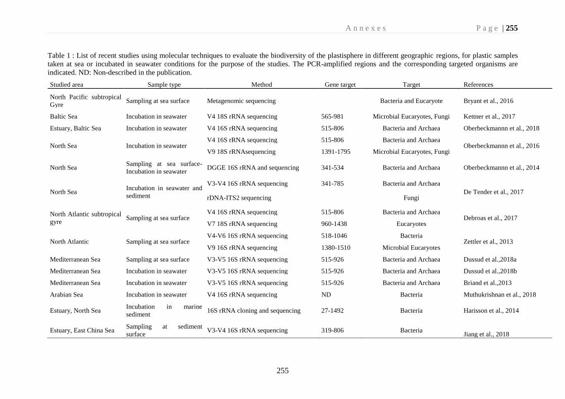
A n n e x e s P a g e | 255
255
Table 1 : List of recent studies using molecular techniques to evaluate the biodiversity of the plastisphere in different geographic regions, for plastic samples
taken at sea or incubated in seawater conditions for the purpose of the studies. The PCR-amplified regions and the corresponding targeted organisms are
indicated. ND: Non-described in the publication.
Studied area Sample type Method Gene target Target References
North Pacific subtropical
Gyre Sampling at sea surface Metagenomic sequencing
Bacteria and Eucaryote Bryant et al., 2016
Baltic Sea Incubation in seawater V4 18S rRNA sequencing 565-981 Microbial Eucaryotes, Fungi Kettner et al., 2017
Estuary, Baltic Sea Incubation in seawater V4 16S rRNA sequencing 515-806 Bacteria and Archaea Oberbeckmannn et al., 2018
North Sea Incubation in seawater V4 16S rRNA sequencing 515-806 Bacteria and Archaea
Oberbeckmannn et al., 2016 V9 18S rRNAsequencing 1391-1795 Microbial Eucaryotes, Fungi
North Sea Sampling at sea surface-
Incubation in seawater DGGE 16S rRNA and sequencing 341-534 Bacteria and Archaea Oberbeckmannn et al., 2014
North Sea Incubation in seawater and
sediment
V3-V4 16S rRNA sequencing 341-785 Bacteria and Archaea
De Tender et al., 2017 rDNA-ITS2 sequencing
Fungi
North Atlantic subtropical
gyre Sampling at sea surface
V4 16S rRNA sequencing 515-806 Bacteria and Archaea Debroas et al., 2017
V7 18S rRNA sequencing 960-1438 Eucaryotes
North Atlantic Sampling at sea surface V4-V6 16S rRNA sequencing 518-1046 Bacteria
Zettler et al., 2013 V9 16S rRNA sequencing 1380-1510 Microbial Eucaryotes
Mediterranean Sea Sampling at sea surface V3-V5 16S rRNA sequencing 515-926 Bacteria and Archaea Dussud et al.,2018a
Mediterranean Sea Incubation in seawater V3-V5 16S rRNA sequencing 515-926 Bacteria and Archaea Dussud et al.,2018b
Mediterranean Sea Incubation in seawater V3-V5 16S rRNA sequencing 515-926 Bacteria and Archaea Briand et al.,2013
Arabian Sea Incubation in seawater V4 16S rRNA sequencing ND Bacteria Muthukrishnan et al., 2018
Estuary, North Sea Incubation in marine
sediment 16S rRNA cloning and sequencing 27-1492 Bacteria Harisson et al., 2014
Estuary, East China Sea Sampling at sediment
surface V3-V4 16S rRNA sequencing 319-806 Bacteria
Jiang et al., 2018
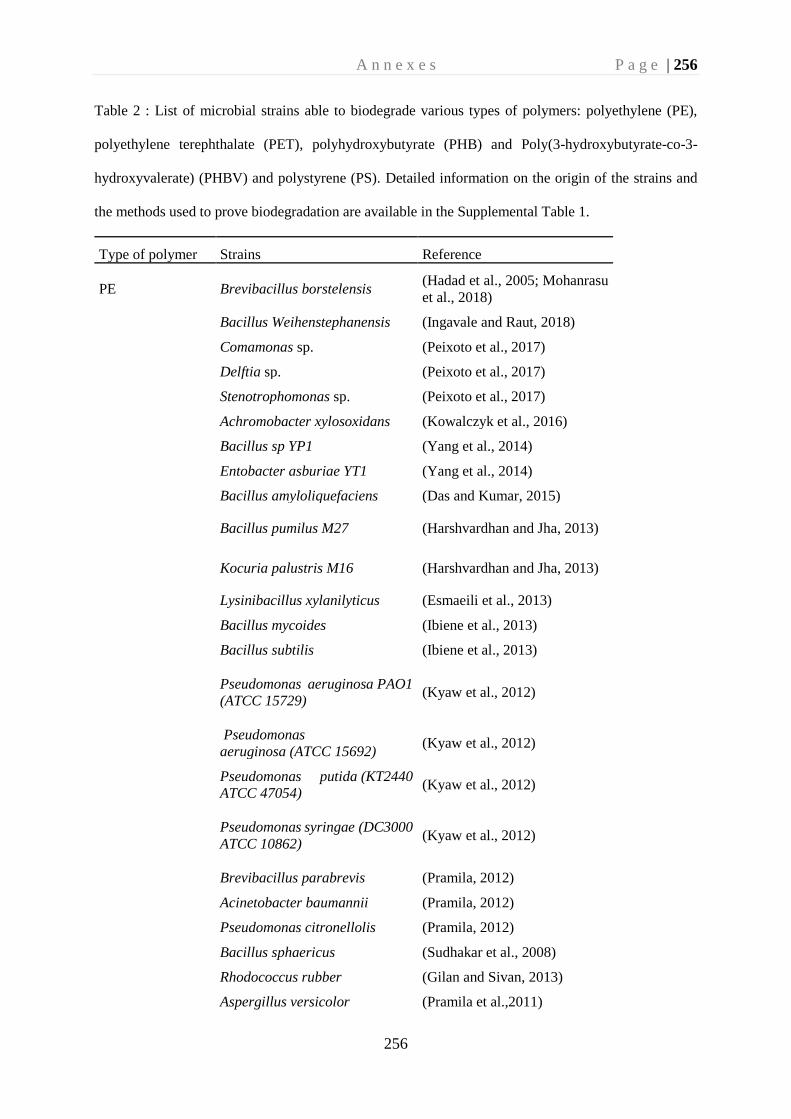
A n n e x e s P a g e | 256
256
Table 2 : List of microbial strains able to biodegrade various types of polymers: polyethylene (PE),
polyethylene terephthalate (PET), polyhydroxybutyrate (PHB) and Poly(3-hydroxybutyrate-co-3-
hydroxyvalerate) (PHBV) and polystyrene (PS). Detailed information on the origin of the strains and
the methods used to prove biodegradation are available in the Supplemental Table 1.
Type of polymer Strains Reference
PE Brevibacillus borstelensis (Hadad et al., 2005; Mohanrasu
et al., 2018)
Bacillus Weihenstephanensis (Ingavale and Raut, 2018)
Comamonas sp. (Peixoto et al., 2017)
Delftia sp. (Peixoto et al., 2017)
Stenotrophomonas sp. (Peixoto et al., 2017)
Achromobacter xylosoxidans (Kowalczyk et al., 2016)
Bacillus sp YP1 (Yang et al., 2014)
Entobacter asburiae YT1 (Yang et al., 2014)
Bacillus amyloliquefaciens (Das and Kumar, 2015)
Bacillus pumilus M27 (Harshvardhan and Jha, 2013)
Kocuria palustris M16 (Harshvardhan and Jha, 2013)
Lysinibacillus xylanilyticus (Esmaeili et al., 2013)
Bacillus mycoides (Ibiene et al., 2013)
Bacillus subtilis (Ibiene et al., 2013)
Pseudomonas aeruginosa PAO1
(ATCC 15729) (Kyaw et al., 2012)
Pseudomonas
aeruginosa (ATCC 15692) (Kyaw et al., 2012)
Pseudomonas putida (KT2440
ATCC 47054) (Kyaw et al., 2012)
Pseudomonas syringae (DC3000
ATCC 10862) (Kyaw et al., 2012)
Brevibacillus parabrevis (Pramila, 2012)
Acinetobacter baumannii (Pramila, 2012)
Pseudomonas citronellolis (Pramila, 2012)
Bacillus sphaericus (Sudhakar et al., 2008)
Rhodococcus rubber (Gilan and Sivan, 2013)
Aspergillus versicolor (Pramila et al.,2011)
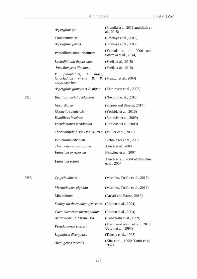
A n n e x e s P a g e | 257
257
Aspergillus sp.
(Pramila et al.,2011 and sheik et
al., 2015)
Chaetomium sp. (Sowmya et al., 2012)
Aspergillus flavus (Sowmya et al., 2012)
Penicillium simplicissimum
(Yamada et al., 2000 and
Sowmya et al., 2014)
Lasiodiplodia theobromae (Sheik et al., 2015)
Paecilomyces lilacinus, (Sheik et al., 2015)
P. pinophilum, A niger,
Gliocladium virens & P.
chrysosporium
(Manzur et al., 2004)
Aspergillus glaucus et A. niger (Kathiresan et al., 2003)
PET Bacillus amyloliquefaciens (Novotný et al., 2018)
Nocardia sp. (Sharon and Sharon, 2017)
ideonella sakaiensis (Yoshida et al., 2016)
Humilicas insolens (Ronkvist et al., 2009)
Pseudomonas mendocina (Ronkvist et al., 2009)
Thermobifida fusca DSM 43793 (Müller et al., 2005)
Penicillium citrinum Liebminger et al., 2007
Thermomonospora fusca Alisch et al., 2004
Fusarium oxysporum Nimchua et al., 2007
Fusarium solani
Alisch et al., 2004 et Nimchua
et al., 2007
PHB Crupriavidus sp. (Martínez-Tobón et al., 2018)
Marinobacter algicola (Martínez-Tobón et al., 2018)
Mix cultures (Ansari and Fatma, 2016)
Schlegella thermodepolymerans (Romen et al., 2004)
Caenibacterium thermophilum (Romen et al., 2004)
Acidovorax Sp. Strain TP4 (Kobayashi et al., 1999)
Pseudomonas stutzeri (Martínez-Tobón et al., 2018;
Uefuji et al., 1997)
Leptothrix discophora (Takeda et al., 1998)
Alcaligenes faecalis (Kita et al., 1995; Tanio et al.,
1982)
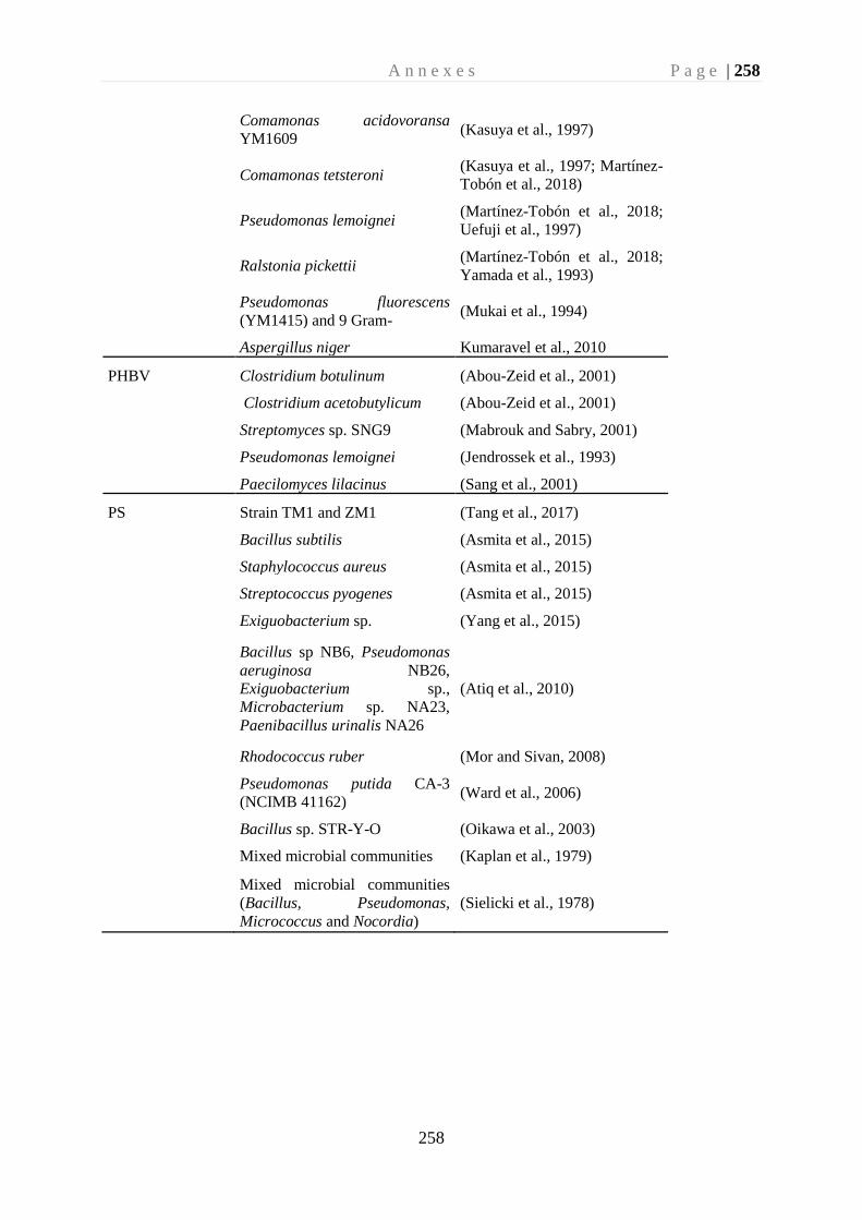
A n n e x e s P a g e | 258
258
Comamonas acidovoransa
YM1609 (Kasuya et al., 1997)
Comamonas tetsteroni (Kasuya et al., 1997; Martínez-
Tobón et al., 2018)
Pseudomonas lemoignei (Martínez-Tobón et al., 2018;
Uefuji et al., 1997)
Ralstonia pickettii (Martínez-Tobón et al., 2018;
Yamada et al., 1993)
Pseudomonas fluorescens
(YM1415) and 9 Gram- (Mukai et al., 1994)
Aspergillus niger Kumaravel et al., 2010
PHBV Clostridium botulinum (Abou-Zeid et al., 2001)
Clostridium acetobutylicum (Abou-Zeid et al., 2001)
Streptomyces sp. SNG9 (Mabrouk and Sabry, 2001)
Pseudomonas lemoignei (Jendrossek et al., 1993)
Paecilomyces lilacinus (Sang et al., 2001)
PS Strain TM1 and ZM1 (Tang et al., 2017)
Bacillus subtilis (Asmita et al., 2015)
Staphylococcus aureus (Asmita et al., 2015)
Streptococcus pyogenes (Asmita et al., 2015)
Exiguobacterium sp. (Yang et al., 2015)
Bacillus sp NB6, Pseudomonas
aeruginosa NB26,
Exiguobacterium sp.,
Microbacterium sp. NA23,
Paenibacillus urinalis NA26
(Atiq et al., 2010)
Rhodococcus ruber (Mor and Sivan, 2008)
Pseudomonas putida CA-3
(NCIMB 41162) (Ward et al., 2006)
Bacillus sp. STR-Y-O (Oikawa et al., 2003)
Mixed microbial communities (Kaplan et al., 1979)
Mixed microbial communities
(Bacillus, Pseudomonas,
Micrococcus and Nocordia)
(Sielicki et al., 1978)

A n n e x e s P a g e | 260
260
Annexe 2– Article en co-auteur en préparation
Title: Biodegradability under marine conditions of bio-based and petroleum-
based polymers as substitutes of conventional microbeads
Authors: Cheng Jingguang1#, Eyheraguibel Boris2#, Jacquin Justine1, Pujo-Pay Mireille1,
Conan Pascal1, Barbe Valérie3, Hoypierres Julia4, Deligey Gaëlle4, Alexandra Ter Halle5,
Bruzaud Stéphane4, Ghiglione Jean-François1 & Meistertzheim Anne-Leila6*
Affiliations :
1 CNRS, Sorbonne Universités, UMR 7621, Laboratoire d’Océanographie Microbienne,
Observatoire Océanologique de Banyuls, France
2 CNRS, Univ. Clermont Auvergne, UMR 6296, Institut de Chimie de Clermont-Ferrand,
France
3 Génomique Métabolique, Genoscope, Institut François Jacob, CEA, CNRS, Univ Evry,
Université Paris-Saclay, 91057 Evry, France
4 UMR CNRS 6027, Institut de Recherche Dupuy de Lôme (IRDL), Univ. Bretagne Sud,
Lorient-Pontivy, France
5 Laboratoire des IMRCP, Université de Toulouse, CNRS UMR 5623, Université Paul Sabatier,
118 route de Narbonne 31062 Toulouse Cedex 9, France
6 SAS Plastic@Sea, Observatoire Océanologique de Banyuls, France
(#) Jingguang Cheng and Boris Eyheraguibel shared co-first authorship
(*) Corresponding author:
Anne-Leila Meistertzheim, SAS Plastic@Sea, Observatoire Océanologique de Banyuls Email :
Running title: Microplastics biodegradation at sea
Keywords: microbeads, cosmetics, biosourced and biodegradable polymer, microbial
ecotoxicology
A n n e x e s P a g e | 261
261
Abstract:
Plastic microbeads have been produced at very large scales since several decades for cosmetics
and personal care products, a large amount of which being directly transported in the oceans as
‘primary microplastics’ and emerging pollutants. To better understand their biodegradability in
marine environment and evaluate their possible replacement by biodegradable polymers, seven
polymer types were studied including three conventional petroleum-based polymers (PE,
PMMA and PCL), two bio-based polymers (PLA and PHBV) and two natural products (rice
seeds and apricot kernel). We used several innovative approaches by both the experimental
design and the set of multidisciplinary techniques that allowed us to follow the successive steps
of biodegradation together with abiotic degradation under seawater conditions. We found that
microbeads made of PHBV or rice and in a lesser extent PCL and apricot are good candidates
for substitution of conventional microplastics, classically made of PE or PMMA that were not
biodegraded under our conditions. Congruent signs of biodegradability were first observed by
oxygen measurement and weight loss. Mass spectrometry and 1H NMR confirmed the
biodegradation by identifying ester bound cleavage for PHBV, with 3-hydroxybutyric acid, 3-
hydroxyvaleric acid and its dimer and heterotrimer as degradation products. Similarly, ε-
caprolactone and glucose were identified as the corresponding oligomers of PCL and rice. This
study provides the first time arguments for the use of some bio-based polymers, but not all, as
substitutes of conventional microbeads to support the recent legislative rules aiming to reduce
the pollution by primary microplastics in the oceans.
L'article complet est présenté dans la thèse de Jingguang Cheng : « Microplastics in the marine
environment: An ecotoxicological perspective »
A n n e x e s P a g e | 262
262
Annexe 3– Article en co-auteur en préparation
Title: Relative influence of plastic debris size and shape, chemical composition
and phytoplankton-bacteria interactions in driving seawater plastisphere
abundance, diversity and activity
Authors: Cheng Jingguang1, Jacquin Justine1, Conan Pascal1, Pujo-Pay Mireille1, Valérie
Barbe2, Matthieu George3, Pascale Fabre3, Bruzaud Stéphane4, Alexandra Ter Halle5,
Meistertzheim Anne-Leila6 and Ghiglione Jean-François1*
Affiliations :
1 CNRS, Sorbonne Universités, UMR 7621, Laboratoire d’Océanographie Microbienne,
Observatoire Océanologique de Banyuls, 66650 Banyuls sur mer, France
2 Génomique Métabolique, Genoscope, Institut François Jacob, CEA, CNRS, Univ Evry,
Université Paris-Saclay, 91057 Evry, France
3 Laboratoire Charles Coulomb (L2C), UMR 5221 CNRS/UM, Place Eugène Bataillon, 34095,
Montpellier, France
4 Institut de Recherche Dupuy de Lôme (IRDL), Université de Bretagne-Sud, UMR CNRS
6027, Rue Saint Maudé, 56321 Lorient
5 IMRCP, Univ. Toulouse, CNRS, Toulouse, France
6 SAS Plastic@Sea, Observatoire Océanologique de Banyuls, France
(*) Corresponding author:
Jean-François Ghiglione, CNRS, Sorbonne Universités, UMR 7621, Laboratoire
d’Océanographie Microbienne, Observatoire Océanologique de Banyuls, France Email :
Keywords: plastic litters, plastisphere, biofilm, biofouling, microbial ecotoxicology
A n n e x e s P a g e | 263
263
Abstract:
The thin film of life that inhabits all plastics in the oceans, so-called “plastisphere”, has multiple
effects on the fate and impacts of plastic in the marine environment. Its composition was shown
to be influenced by plastic type, geographical distance and environmental changes between
seasons. Here, we hypothesized that the large spectrum of plastic sizes encountered in the
environment may be also a critical driver of the plastisphere. Polyethylene (PE) and polylactide
acid (PLA) together with glass controls in the forms of meso-debris (18mm diameter) and large-
microplastics (LMP; 3mm diameter), as well as small-microplastics (SMP) of 100 m diameter
with spherical or irregular shapes were immerged during 2 months in seawater. Results of
bacterial abundance (confocal microscopy), diversity (16S rRNA Illumina sequencing)
indicated that the three classical colonization phases, including primo-colonization (after 3
days), growing phase (after 10 days) and maturation phase of the biofilm (after 30 days), were
not influenced by the size and the shape of the materials, even when a diatom bloom (Pseudo-
nitzschia sp.) occurred after the first month of incubation. Influence of plastic size and shape
was only visible on bacterial activity (3H leucine incorporation), where SML showed higher
activity than the rest material sizes, irregular 100m particles showed higher activity than the
regular one. A mature biofilm was visible after 30 days for all material types, with significantly
higher abundance in the plastics (PE and PLA) compared to glass, with distinct bacterial
assemblages found on each material type. The diatom bloom event had a great impact on the
plastisphere of all materials, resulting in a drastic change in diversity and activity. Our research
reveals that the plastic chemical composition, the successive phases of biofilm formation and
the phytoplankton-bacteria interactions are more important factors than the material size and
shape in shaping the abundance, diversity and activity of the plastisphere.
L'article complet est présenté dans la thèse de Jingguang Cheng : « Microplastics in the marine
environment: An ecotoxicological perspective »
A n n e x e s P a g e | 264
264
Annexe 4– Article en co-auteur en préparation
Title: Beneficial or detrimental effects of microplastics on the marine
filter-feeder amphioxus (Branchiostoma lanceolatum)
Authors: Cheng Jingguang1, Meistertzheim Anne-Leila1,2, Jacquin Justine1, Valérie Barbe3,
Escande Marie-Line 4, Bertrand Stephanie4, Escriva Hector4 and Ghiglione Jean-François1*
Affiliations:
1 CNRS, Sorbonne Universités, UMR 7621, Laboratoire d’Océanographie Microbienne,
Observatoire Océanologique de Banyuls, France
2 SAS Plastic@Sea, Observatoire Océanologique de Banyuls, France
3 Génomique Métabolique, Genoscope, Institut François Jacob, CEA, CNRS, Univ Evry,
Université Paris-Saclay, 91057 Evry, France
4 CNRS, Sorbonne Universités, UMR 7232, Biologie Intégrative des Organismes Marins,
Observatoire Océanologique de Banyuls, France
(*) Corresponding author:
Jean-François Ghiglione, CNRS, Sorbonne Universités, UMR 7621, Laboratoire
d’Océanographie Microbienne, Observatoire Océanologique de Banyuls, France Email :
Keywords: microplastics, toxicity, gut microbiome, plastisphere, oxidative stress, amphioxus
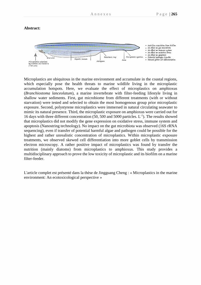
A n n e x e s P a g e | 265
Abstract:
Microplastics are ubiquitous in the marine environment and accumulate in the coastal regions,
which especially pose the health threats to marine wildlife living in the microplastic
accumulation hotspots. Here, we evaluate the effect of microplastics on amphioxus
(Branchiostoma lanceolatum), a marine invertebrate with filter-feeding lifestyle living in
shallow water sediments. First, gut microbiome from different treatments (with or without
starvation) were tested and selected to obtain the most homogenous group prior microplastic
exposure. Second, polystyrene microplastics were immersed in natural circulating seawater to
mimic its natural presence. Third, the microplastic exposure on amphioxus were carried out for
16 days with three different concentration (50, 500 and 5000 particles. L-1). The results showed
that microplastics did not modify the gene expression on oxidative stress, immune system and
apoptosis (Nanostring technology). No impact on the gut microbiota was observed (16S rRNA
sequencing), even if transfer of potential harmful algae and pathogen could be possible for the
highest and rather unrealistic concentration of microplastics. Within microplastic exposure
treatments, we observed skewed cell differentiation into more goblet cells by transmission
electron microscopy. A rather positive impact of microplastics was found by transfer the
nutrition (mainly diatoms) from microplastics to amphioxus. This study provides a
multidisciplinary approach to prove the low toxicity of microplastic and its biofilm on a marine
filter-feeder.
L'article complet est présenté dans la thèse de Jingguang Cheng : « Microplastics in the marine
environment: An ecotoxicological perspective »
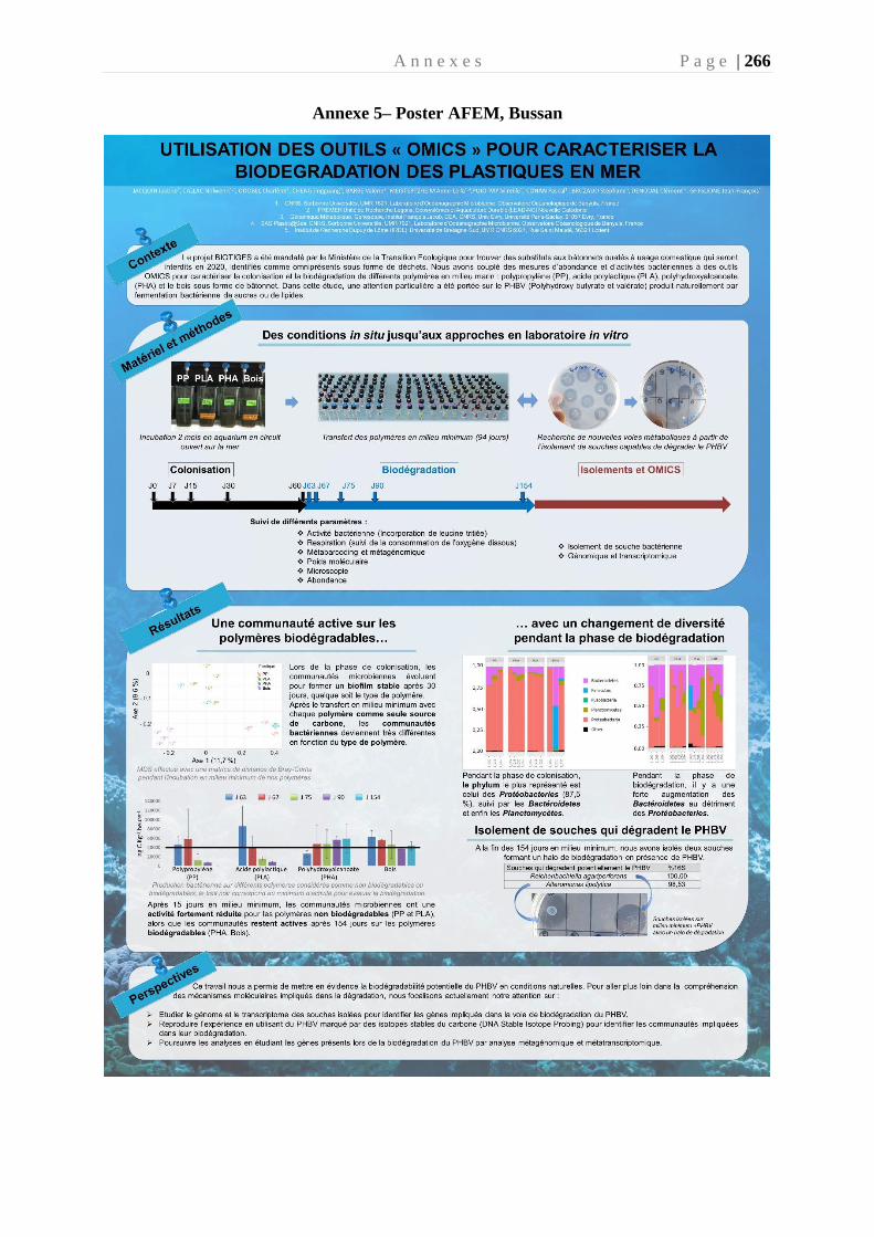
A n n e x e s P a g e | 266
Annexe 5– Poster AFEM, Bussan