coexpression of the cd45ra and cd45ro antigens on t lymphocytes in chronic arthritis
TRANSCRIPT
Clin Exp Immunol 1994; 97:39-44
Co-expression of the CD45RA and CD45RO antigens on T lymphocytes inchronic arthritis
K. L. SUMMERS. J. L. O 'DONNELL & D. N. J, HART Haematology/hmnunology Research Group.Christchurch Hospital, Christchurch. New Zealand
(Accepted for publication 3 March 1994)
SUMMARY
The site of T lymphocyte activation in chronic arthritis is unknown. Peripheral blood (PB)lymphocytes from chronic arthritis patients are in a "naive" or non-activated state, as defined byexpression ofthe CD45RA antigen and lack of HLA class II expression. In contrast, most synovialfluid (SF) T lymphocytes express a "memory* or activated phenotype. as defined by the CD45ROantigen and high HLA class II expression. Following stimulation, naive cells lose CD45RA andgain CD45RO expression to become memory cells with a transitional stage of dual CD45RA,CD45RO antigen expression. To localize where this change in phenotype occurs we used dualcolour immunofluorescence labelling to compare the percentage of dual CD45RA, CD45RO-positive T lymphocytes in PB and SF from chronic arthritic patients and from normal PB,assuming this population would be increased at the primary site of T lymphocyte activation.Expression ofthe intermediate and late activation marker. HLA-DR, was also analysed using dualcolour immunofiuorescence labelling. The percentage of dual positive T lymphocytes was similarbetween arthritic PB, SF. and normal PB, as was the density of both CD45RA and CD45ROantigens. Thus, CD45 isoform expression did not indicate where T lymphocytes were activated.However, we identified a previously unreported population of CD45RA^ CD45RO^ HLA-DR"T lymphocytes in arthritic and normal PB. In SF, this population was absent, but a substantialnumber of dual CD45RA, CD45RO-positive HLA-DR' T lymphocytes were identitied. Thispopulation would not be predicted by the current model of T lymphocyte activation. Division of Tlymphocytes into functional groups on the basis of CD45 isoform expression is likely to be morecomplicated than previously thought. Based on our findings we propose an alternative model of Tlymphocyte differentiation.
Keywords CD45 chronic arthritis T lymphocytes
INTRODUCTION
It is not clear whether the T lymphocyte response associatedwith chronic arthritis is initiated locally within the joint, orwithin the central lymphoid system. As T lymphocyte matura-tion and difierentiation can now be examined using differentisoforms of the leucocyte common antigen (T200. CD45), thepresence of these and other phenotypie markers on T lympho-cytes from both sites is of interest. Cells expressing the highmolecular weight isoform, CD45RA. demonstrate functionalcharacteristics of naive T lymphocytes which do not respond torecall antigens [I]. T lymphocytes expressing the low molecularweight isoform, CD45RO. demonstrate functional character-istics of memory cells which do respond to recall antigens [2].Followmg mitogenic or alloantigenic stimulation in vitro, aseries of phenotypie changes occur in naive T lymphocytes
Correspondence: Dr J, L, O'Donnell, Department of Immunology,Christchurch Hospital. Private Bag, Christchurch. New Zealand.
whereby they lose expression of the CD45RA isoform andacquire the CD45RO isoform. During this differentiation andactivation process, a transitional stage occurs where bothCD45RA and CD45RO antigens are co-expressed [3-5],These dual positive cells display functional properties whichlie between those of naive and memory cells. Both dual positiveand naive T lymphocytes suppress immunoglobulin productionby B lymphocytes, unlike memory cells which help with thisprocess [6.7], However, they resemble memory T lymphocytesin their ability to express mRNA [8] and to produce interferon-gamma (IFN-7) [6],
In rheumatoid arthritis (RA), T lymphocytes located inperipheral blood {PB) primarily express the CD45RA isoform.whereas most T lymphocytes found in the synovium [9,10] andthe synoviai fluid (SF) of rheumatoid joints express theCD45RO isoform [11-14]. Expression of the MHC class ilantigen, HLA-DR. is also markedly increased on T lymphocyteslocated in arthritic joints [9,11,13.15], This antigen is first
39
40 K. L. Summers. J. L. O'Donnell & D. N. J. Hart
expressed during an intermediate stage of activation [16].Although these results suggest a predominance of activated Tlymphocytes in rheumatoid joints, they do not define the sitewhere this activation process is initiated. The presence of anexpanded dual CD45RA, CD45RO"^ T lymphocyte populationwithin the joint may support the hypothesis that T lymphoeytesare activated locally.
In this study, T lymphocytes were isolated from PB and SFof patients with various chronic arthritic diseases, and from PBof normal healthy controls. Using two-colour immunoHuores-cence labelling, cells were analysed for dual expression ofCD45RA and CD45RO antigens, and for the presence of theactivation marker, HLA-DR, to detennine whether T lympho-cytes were being activated within the joint or external to it.
PATIENTS AND M E T H O D S
Patients and controlsPaired samples of PB and SF were obtained from 10 patientswith chronic arthritic diseases following informed consent: RA(« = 5), psoriatic arthritis (/; = 3), reactive arthritis {n = l)iankylosing spondylitis (n = I), ln addition. PB samples weretaken as controls from nine age- {±2 years) and sex-matchednormal healthy volunteers. All samples were collected intoEDTA tubes.
Monoclonal antibodiesThe following MoAbs were used. Those produced and char-acterized in this laboratory were the CD45RA MoAb, CMRF-II and biotin-conjugated CMRF-ll [17]; the CD14 MoAb,CMRF-31: and the green control MoAb, 16.4.4 (IgGl, mouseanti-rat class I MHC) [18]. The anti-HLA-DR (HB55) MoAbwas produced from the L243 hybridoma obtained from theAmerican Tissue Culture Collection (Rockville, MD) andgrown in this laboratory. PE-conjugated MoAbs usedincluded Leu-1 (CD5). Leu-45RO {CD45RO) and mouseIgGl (red control), all of which were obtained from BectonDickinson (Mountain View, CA). Other MoAbs were kindlydonated from colleagues, including the CD16 MoAb, HuNK2(Professor I. McKetizie, Melbourne. Australia); the CD 19
MoAb. FMC63 (Professor H. Zola, Adelaide. Australia) [19];and the CD45RO MoAb, UCHLI (Dr P. Beverley, London,UK) [20].
Isolation of T lymphocytesMononuclear cells (MNC) were isolated from PB and SF bydensity gradient centrifugation over Ficoll-Hypaque (Phar-macia Fine Chemicals, Uppsala, Sweden). An enriched popula-tion of T lymphocytes was obtained by a negative selectionprocedure using immunomagnetic beads. This involved label-ling MNC with CDI4, CD16 and CD19 MoAbs at 4 ^ for30min to select for monocytes/macrophages. natural killer(NK) cells and B lymphocytes. Cells were then washed twiceand resuspended in 0 5% bovine serum albumin (BSA)/PBS.Either rat anti-mouse immunoglobulin or sheep anti-mouseimmunoglobulin-coated Dynabeads (Dynal, Oslo, Norway)were added to the cell suspension in the ratio of one cell tofive magnetic beads, and incubated for 30 min at room tem-perature with frequent gentle mixing. The cells were subjectedto a Dynal magnetic particle concentrator for 2 min, afterwhich time the supernatant containing non-magnetized orunlabelled cells was collected, washed and resuspended in0-5% BSA/PBS. These cells constituted the purified T lym-phocyte population. Purity was assessed by CD5 staining(Table I),
Dual-colour imtnunojiuorescencePurified T lymphocytes (10^/test) were incubated with saturat-ing amounts of primary MoAb for 30 min on ice. washed andthen stained with 30/ig/ml of fluorescein-conjugated F(ab')2sheep anti-mouse IgG (FITC-SAM; Silenus Laboratories.Melbourne, Australia) for 30 min at 4 C. After washing, Tlymphocytes were incubated in 10% normal mouse serum forlOmin on ice, followed by a 30-min incubation with either aPE-conjugated secondary MoAb. or a biotin-conjugated sec-ondary MoAb with PE-conjugated streptavidin (CaltagLaboratories, San Francisco, CA) as the second layer. Cellswere washed, resuspended in 0 5% BSA/PBS. and analysed ona fluorescence activated cell analyser (Epics Profile 1, CoulterElectronics, Hialeah, FL).
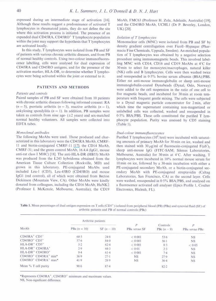
Table 1. Mean percentages of dual antigen expression on T cells (CD5''') isolated from peripheral blood (PB) (PBa) and synovial fluid (SF) ofarthritic patients and PB of normal controls (PBc)
MoAb
CD45RA+ CD5^CD45RO+ CD5+HLA-DR^ CD5'HLA-DR+ CD45RA +HLA-DR+ CD45RO+CD45RO' CD45RA^ min*CD45RO' CD45RA' max*
Mean % T cell purity
PBa (« =
60 357-6
5-2299 4
36941 9
90-6
Arthritic patients
= 10) S F ( « = 10)
24-884062444-361-427128-9
874
PPBa versus SF
< 0 001< 0-001<O00I<OOI<0001
NSNS
ControlsPBc {n = 9)
53-656-18-32-37 4
27'9364
82-2
PPBa versus PBc
NSNS
<0-05NSNSNSNS
*Represents CD45RA+. CD45RO^NS, Non-significant difference.
minimum and maximum values.
CD45 T cell subsets in chronic arthritis 41
( c ) b) lgG-PE
Log fluorescence
LUQ.1
(5
( c i
1644
Log fluorescence
LJa.
CEinQu
l e )
• • . : • : • : ' ' ' •
1644
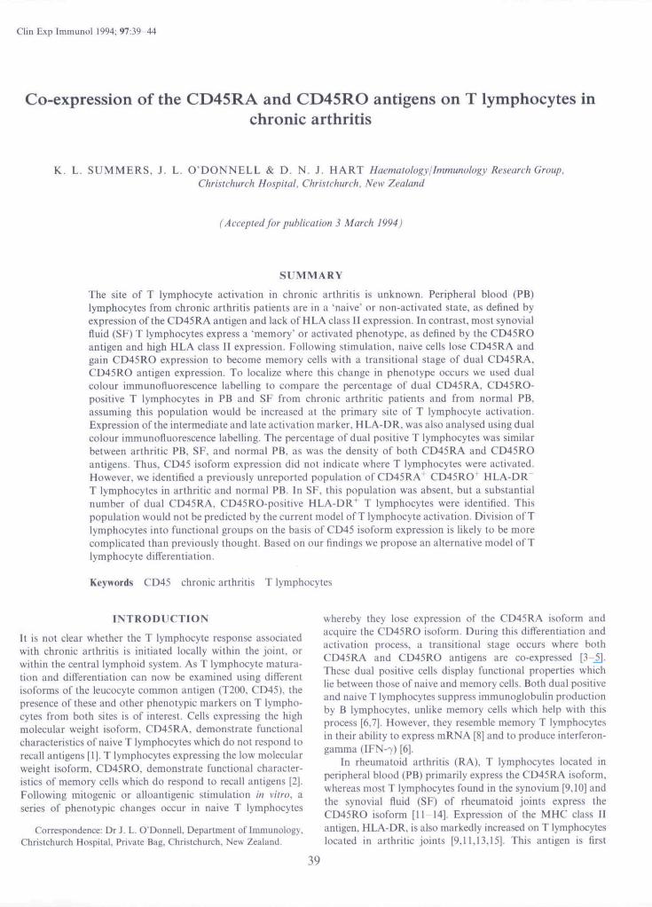
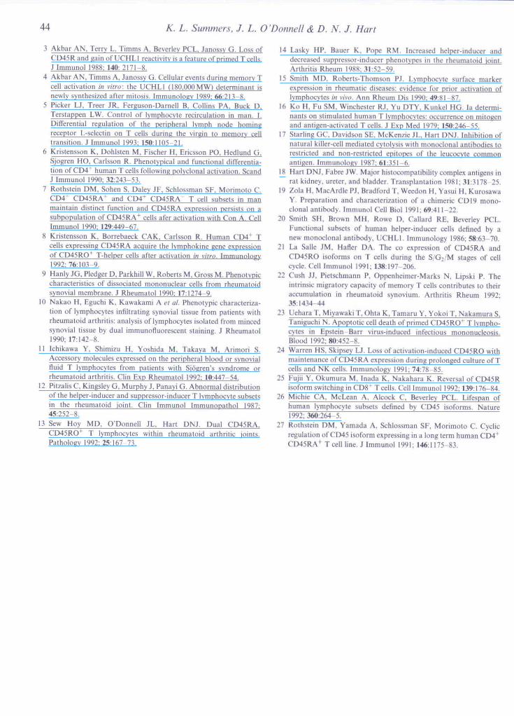
Fig. 1. Dual-colour immunofluoreseence staining of T lymphocytespurified from peripheral blood fPB) of one representative chronicarthritic patient. The upper two histograms show staining profilesused to determine T cell purity, (a) Percentage of direct PE-IabelledCD5' T lymphocytes, (b) Direct PE-Iabelled irrelevant MoAb used todetermine non-specific binding. Parameters were set to allow no greaterthan 2% of eells non-specifically labelling. The lower three dot-plotsshow staining patterns used to calculate the percentage of dual-positiveT lymphocytes, (c) Dual-colour control using direct PE-labelledirrelevant MoAb as in b, and indirect FITC-labelled irrelevantMoAb, 16.4.4. (d) Dual labelling using direct PE-Iabelled CD45RAMoAb and indirect EITC-labelled CD45RO MoAb. (e) Non-specificdual labelling of direct PE-labelled CD45RA" cells with indirect FJTC-labelled irrelevant MoAb, 16.4.4.
Statistical analysisIn order to calculate the percentage of dual CD45RA,CD45RO-positive T lymphocytes in each sample, the contam-inating non-T lymphocyte population remaining after magneticbead separation had to be considered. Consequently twocalculations were used to determine the percentage of dual-positive cells present. The minimum calculation assumedthat all contaminating non-T lymphocytes labelled dualpositive, whereas the maximum calculation assumed thatno contaminating cells labelled dual positive. The formulaeused to calculate these percentages of CD45RA'. CD45RO*T lymphocytes within the total CD5^ T cell populationare shown below. Refer to Fig. 1 for definitions of a, b.c.dand e.
{i) T cell purity = a — b
{ii) Maximum % CD45RA+, CD45RO+ T cells =
(iii) Minimum % CD45RA\ CD45RO^
{d-c)-{e-c)
d-ca-b
T cells =a-b
_d-e
Data were analysed for statistical significance using the paired/-test program from the SAS computer statistical package.Reproducibility was determined by repeating duplicate tubesand simultaneously reading them on the flow cytometer. Nosignificant duplicate error was detected {data not shown).
RESULTS
Expression of CD45RA and CD45RO antigens on CD5^ TlymphocytesThe percentage of CDS"*̂ T lymphocytes {henceforth referred toas T lymphocytes) separately expressing either CD45RA orCD45RO was analysed {Table 1). There was no difference inthe expression of CD45RA or CD45RO on PB T lymphocytesbetween arthritic patients and normal controls. In PB from ninenormal controls, the CD45RA antigen and the CD45ROantigen were expressed on 53-6yo and 56-1%, respectively, ofT lymphocytes. Similarly, there was no significant difference inthe density (mean fluorescence intensity) of expression of eitherantigen on PB T lymphocytes between arthritic patients andcontrols {Table 2).
Within 10 chronic arthritic patients, 60-3% of PB Tlymphocytes expressed the CD45RA antigen, compared with24 8% of those in SF. In contrast, the CD45RO antigen wasexpressed on 57-6% of PB T lymphocytes, compared with 84%of SF T lymphocytes. Both the level of expression and thedensity of these antigens differed significantly between thearthritic PB and SF compartments. The density of expressionof the CD45RO antigen was significantly higher on SF cells andthe density of CD45RA significantly lower on these cellscompared with PB from the same individual (Table 2).
Dual expression of CD45RA and CD45RO on T lymphocytesThere was no signifieant difference in the percentage of dualCD45RA *. CD45RO' (henceforth referred to as dual positive)PB T lymphocytes between arthritic patients and controlsbased on either minimum or ma.ximum values {Table I). The
Table 2. Mean fluorescence intensity of theCD45RA and CD45RO antigens onT lymphocytes isolated from peripheral blood{PB) {PBa) and synovial fiuid (SF) of arthritic patients and PB of normal controls {PBc)
MoAb
CD45RACD45RO
Arthritic patients
P B a { n - 10) SF{n = 10)
79-6 35-533 9 49 0
PPBa versus SF
<ao5< 0 05
ControlsPBc (H - 9)
79425-8
PPBa versus PBc
NSNS
NS, Non-significant diflference.
42 K. L. Summers. J. L. O'Donnell & D. N. J. Hart
surface density of the CD45RA antigen on these cells wassimilar, whereas the CD45RO antigen density was slightlyhigher in the control PB population (data not shown).
When the arthritic SF was examined, there was no signifi-cant increase in the dual-positive population compared witharthritic PB using either minimum or maximum values. Thedensity of expression of individual antigens did not differbetween the arthritic PB and joint compartments.
HLA-DR class II expression on T IvmphoevtesThe HLA-DR antigen was expressed on 52% of PB Tlymphocytes from arthritic patients, and on a greater per-centage of PB T lymphocytes from eontrol subjects (8-3%)(Table I). Expression of the HLA-DR antigen was predomi-nantly associated with expression of the CD45RO antigen,although in PB of both arthritic patients and controls thepercentage of CD45RO' T lymphocytes co-expressing HLA-DR was low (9 4% and 7-4%, respectively). Dual expression ofthe HLA-DR and CD45RA antigens was even lower (2-9% and2-3%, respectively, above background) and at a level that wasvery close to the analytical sensitivity of our assay.
In SF, 62 4% of the T lymphocytes expressed HLA-DR.Since the same percentage of CD45RO^ cells expressed thisdass II antigen (61 4%), HLA-DR expression seemed to beentirely associated with CD45RO expression. Interestingly.44 3% of CD45RA+ cells in SF co-expressed HLA-DR. Thismay all be accounted for by the presence of dual-positive cellsalso expressing the HLA-DR antigen,
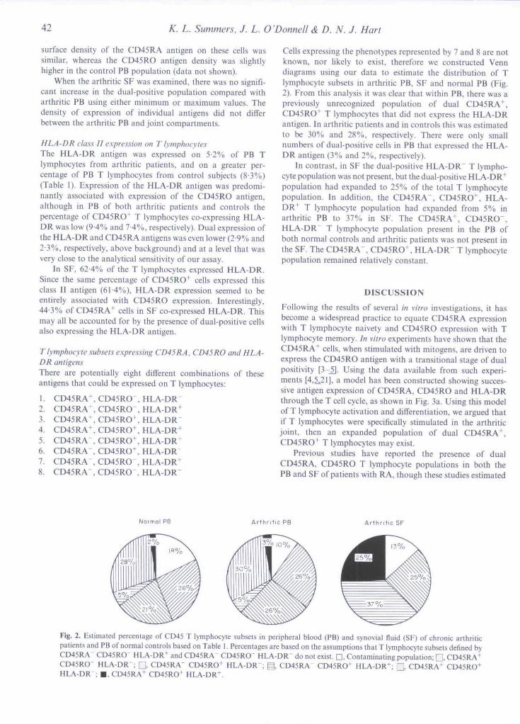
Tlymphocyte subsets expressing CD45RA. CD45RO and HLA-DR atitigetisThere are potentially eight different combinations of theseantigens that could be expressed on T lymphocytes:
1. CD45RA+,2. CD45RA+.3. CD45RA ,4. CD45RA^,5. CD45RA ,6. CD45RA .7. CD45RA ,8. CD45RA ,
CD45ROCD45ROCD45RO'CD45RO'CD45RO'CD45RO'CD45RO"CD45RO"
HLA-DRHLA-DR^HLA-DRHLA-DR'HLA-DR'HLA-DRHLA-DR'HLA-DR"
Cells expressing the phenotypes represented by 7 and 8 are notknown, nor likely to exist, therefore we constructed Venndiagrams using our data to estimate the distribution of Tlymphocyte subsets in arthritic PB, SF and normal PB (Fig,2). From this analysis it was clear that within PB, there was apreviously unrecognized population of dual CD45RA'^.CD45RO' T lymphocytes that did not express the HLA-DRantigen. In arthritic patients and in controls this was estimatedto be 30% and 28%, respectively. There were only smallnumbers of dual-positive cells in PB that expressed the HLA-DR antigen (3% and 2%, respectively).
In contrast, in SF the dual-positive HLA-DR" T lympho-cyte population was not present, but the dual-positive HLA-DR"*"population had expanded to 25% of the total T lymphocytepopulation. In addition, the CD45RA", CD45RO^. HLA-DR' T lymphocyte population had expanded from 5% inarthritic PB to 37% in SF. The CD45RA\ CD45RO",HLA-DR" T lymphocyte population present in the PB ofboth normal controls and arthritic patients was not present inthe SF. The CD45RA . CD45RO', HLA-DR" T lymphocytepopulation remained relatively constant.
DISCUSSION
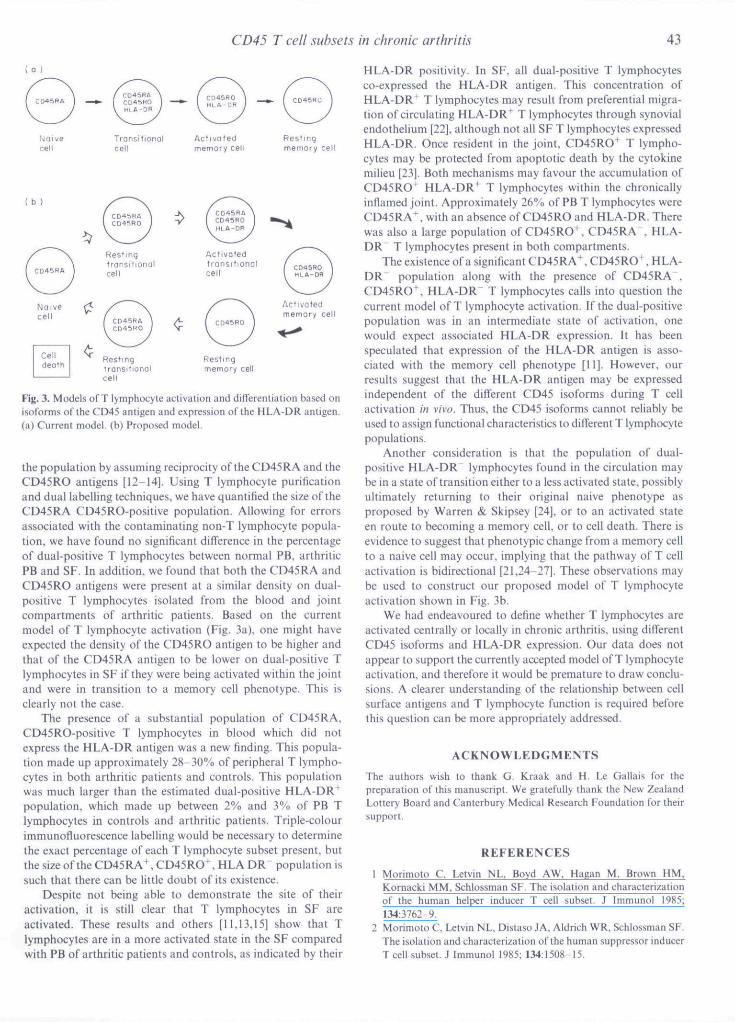
Following the results of several in vitro investigations, it hasbecome a widespread practice to equate CD45RA expressionwith T lymphocyte naivety and CD45RO expression with Tlymphocyte memory. In vitro experiments have shown that theCD45RA^ cells, when stitnulated with mitogens, are driven toexpress the CD45RO antigen with a transitional stage of dualpositivity [3-5], Using the data available from such experi-ments [4,5,21], a model has been constructed showing succes-sive antigen expression of CD45RA, CD45RO and HLA-DRthrough the T cell cycle, as shown in Fig. 3a. Using this modelof T lymphocyte aetivation and differentiation, we argued thatif T lymphocytes were specifically stimulated in the arthriticjoint, then an expanded population of dual CD45RA^,CD45RO^ T lymphocytes may exist.
Previous studies have reported the presence of dualCD45RA. CD45RO T lymphocyte populations in both thePB and SF of patients with RA, though these studies estimated
Arthr i t ic PB Arthr i t ic SF
Fig. 2. Estimated percentage of CD45 T lymphocyte subsets in peripheral blood (PB) and synovial fluid (SF) of chronic arthriticpatients and PB of normal controls based on Table 1, Percentages are based on the assumptions that T lymphocyte subsets defined byCD45RA CD45RO" HLA-DR+ and CD45RA" CD45RO" HLA-DR do noi exist, Q. Contaminating population; Q CD45RA^CD45RO" HLA-DR": Q CD45RA- CD45RO+ HLA-DR^ g . CD45RA- CD45RO^ HLA-DR+; C CD45RA* CD45RO^HLA-DR"; • , CD45RA+ CD45RO+ HLA-DR+.
CD45 T cell subsets in chronic arthritis 43
( b }
transitionalcell
Restingmemory cell
Fig. 3. Models of T lymphocyte activation and differentiation based onisoforms of the CD45 antigen and expression of the HLA-DR antigen.(a) Current model, (b) Proposed model.
the population by assuming reciprocity of the CD45RA and theCD45RO antigens [12-14]. Using T lymphocyte purificationand dual labelling techniques, we have quantified the size of theCD45RA CD45RO-positive population. Allowing for errorsassociated with the contaminating non-T lymphocyte popula-tion, we have found no significant difference in the percentageof dual-positive T lymphocytes between normal PB, arthriticPB and SF. In addition, we found that both the CD45RA andCD45RO antigens were present at a similar density on dual-positive T lymphocytes isolated from the blood and jointcompartments of arthritic patients. Based on the currentmodel of T lymphocyte activation (Fig, 3a), one might haveexpected the density of the CD45RO antigen to be higher andihat of the CD45RA antigen to be lower on dual-positive Tlymphocytes in SF if they were being activated within the jointand were in transition to a memory cell phenotype. This isclearly not the case.
The presence of a substantial population of CD45RA.CD45RO-positive T lymphoeytes in blood which did note.xpress the HLA-DR antigen was a new finding. This popula-tion made up approximately 28 30% of peripheral T lympho-cytes in both arthritic patients and controls. This populationwas much larger than the estimated dual-positive HLA-DR^population, which made up between 2% and 3% of PB Tlymphocytes in controls and arthritic patients. Triple-colourimmunofluorescence labelling would be necessary to determinethe exact percentage of each T lymphoeyte subset present, butthe size of the CD45RA^, CD45RO", HLA DR population issuch that there ean be little doubt of its existence.
Despite not being able to demonstrate the site of theiractivation, it is still clear that T lymphocytes in SF areactivated. These results and others [11.13,15] show that Tlymphocytes are in a more activated state in the SF comparedwilh PB of arthritic patients and controls, as indicated by their
HLA-DR positivity. In SF. all dual-positive T lymphocytesco-expressed the HLA-DR antigen. This concentration ofHLA-DR"^ T lymphocytes may result from preferential migra-tion of circulating HLA-DR' T lymphocytes through synovialendothelium [22], although not all SF T lymphocytes expressedHLA-DR, Once resident in the joint, CD45RO^ T lympho-cytes may be protected from apoptotie death by the cytokinemilieu [23]. Both mechanisms may favour ihe accumulation ofCD45RO* HLA-DR+ T lymphocytes within the chronicallyinflamed joint, Approximately 26% of PB T lymphocytes wereCD45RA+, with an absenee of CD45RO and HLA-DR. Therewas also a large population of CD45RO^, CD45RA". HLA-DR T lymphocytes present in both compartments.
The existence of a significant CD45RA^ CD45RO\ HLA-DR" population along with the presence of CD45RA",CD45RO , HLA-DR" T lymphocytes calls into question thecurrent tnodel of T lymphocyte activation. If the dual-positivepopulation was in an intermediate state of activation, onewould expect associated HLA-DR expression. It has beenspeculated that expression of the HLA-DR antigen is asso-ciated with the memory cell phenotype [II]. However, ourresults suggest that the HLA-DR antigen may be expressedindependent of the different CD45 isoforms during T cellactivation in vivo. Thus, the CD45 isoforms cannot reliably beused to assign functional characteristics to different T lymphocytepopulations.
Another consideration is that the population of dual-positive HLA-DR lymphocytes found in the circulation maybe in a state of transition either to a less activated state, possiblyultimately returning to their original naive phenotype asproposed by Warren & Skipsey [24], or to an activated stateen route to becoming a memory cell, or to cell death. There isevidence to suggest thai phenolypic change from a memory cellto a naive cell may occur, implying that the pathway of T cellactivation is bidirectional [21.24-27]. These observations maybe used to construct our proposed model of T lymphocyteactivation shown in Fig. 3b.
We had endeavoured lo define whether T lymphocytes areactivated centrally or locally in chronic arthritis, using differentCD45 isoforms and HLA-DR expression. Our data does notappear to support the currently accepted model of T lymphocyteactivation, and therefore it would be premature to draw conclu-sions. A clearer understanding of the relationship between cellsurface antigens and T lymphocyte function is required beforethis question can be more appropriately addressed.
ACKNOWLEDGMENTS
The authors wish to thank G. Kraak and H, Le Gallais for thepreparation of this manuscript. We gratefully thank the New ZealandLottery Bourd and Canterbury Medical Research Foundation for theirsupport.
REFERENCES
1 Morimoto C, Letvin NL, Boyd AW, Hagan M, Brown HM,Kornacki MM, Schlossman SF. The isolation and characterizationof the human helper inducer T cell subset. J Immunol 1985;134:3762-9.
2 Modmolo C. Letvin NL, Distaso JA, Aldrich WR. Schlossman SF.The isolation and characlerization of the human suppressor inducerTcell subset. J Immunol 1985; 134:1508-15.
44 K. L. Summers. 7. L. O 'Donnell & D. N, J. Hart
3 Akbar AN, Terry L, Timms A, Beverley PCL, Janossy G. Loss ofCD45R atid gain of UCHL1 reactivity is a feature of primed T celLs.J Immunol 1988: 140: 2171-8.
4 Akbar AN, Timms A, Janossy G. Cellular events during memory Tcell activation i/i viiro: the UCHLI (180,000MW) determinant isnewly synthesized after mitosis. Immunology 1989; 66:213-8.
5 Picker LJ, Treer JR, Ferguson-Darnell B. Collins PA, Buck D.Terstappen LW. Control of lymphocyte recirculation in man, 1.Differential regulation of the peripheral lymph node homingreceptor L-selectin on T cells during the virgin to memory celltransition. J Immunol 1993; 150:1105 21.
6 Kristensson K, Dohlsten M, Fischer H, Ericsson PO. Hedlund G,Sjogren HO, Carlsson R. Phenotypical and functional diflerentia-tlon of CD4^ human T cells following polyclonal activation. SeandJ Immunol 1990; 32:243-53.
7 Rothstein DM, Sohen S, Daley JF. Sehlossman SF, Morimoto C.CD4+ CD45RA^ and CD4" CD45RA T cell subsets in manmaintain distinct function and CD45RA expression persists on asubpopulation of CD45RA"^ cells afer activation with Con A. CellImmunol 1990: 129:449-67.
8 Knstensson K. Borrebaeck CAK, Carlsson R. Human CD4* Teells expressing CD45RA acqtjire the lymphokine gene expressionof CD45RO"^ T-helper cells after activation in viiro. Immunology1992; 76:103-9.
9 Hanly JG, Pledger D, Parkhill W. Roberts M, Gross M. Phenotypiccharacteristics of dissociated mononuclear cells from rheumatoidsynovial membrane. J Rheumatol 1990: 17:1274 9.
10 Nakao H, Eguchi K, Kawakami A el at. Phenotypic characteriza-tion of lymphocytes mliltrating synovial tissue from patients withrheumatoid arthritis: analysis of lymphocytes isolated from mincedsynovial tissue by dual immunofluorescent staining. J Rheumato!1990; 17:142-8.
11 lchikawa Y, Shimizu H, Yoshida M, Takaya M, Arimori S.Accessory molecules expressed on the peripheral blood or synovialfluid T lymphocytes from patients with Sjogren's syndrome orrheumatoid arthritis. Clin Exp Rheumatol 1992: 10:447-54.
12 PitzalisC, KingsleyG, Murphy J, Panayi G. Abnormal distributionof the helper-inducer and suppressor-inducer T lymphocyte subsetsin the rheumatoid joint. Clin Immunol Immunopathol 1987;45:252-8.
13 Sew Hoy MO. O'Donnell JL. Hart DNJ. Dual CD45RA,CD45RO*^ T lymphocytes within rheumatoid arthritic joints.Pathology 1992; 25:167-73.
14 Lasky HP, Bauer K, Pope RM. Increased helper-inducer anddecreased suppressor-inducer phenotypes in the rheumatoid joint.Arthritis Rheum 1988:31:52-59.
15 Smith MD, Roberts-Thomson PJ. Lymphocyte surface markerexpression in rheumatic diseases: evidence for prior activation oflymphocytes in vivo. Ann Rheum Dis 1990; 49:81-87.
16 Ko H. Fu SM, Winchester RJ. Yu DTY, Kunkel HG. la determi-nants on stimulated human T lymphocytes: occurrence on mitogenand antigen-activated T cells. J Exp Med 1979: 150:246-55.
17 Starling GC. Davidson SE, McKenzie JL. Hart DNJ. Inhibition ofnatural killer-cell mediated cytolysis with monoclonal antibodies torestricted and non-restricted epitopes of the leucocyte commonantigen. Immunology 1987; 61:351 6,
18 Hart DNJ. Fabre JW, Major hi.stocompatibility complex antigens inrat kidney, ureter, and bladder. Transplantation 1981; 31:3178-25.
19 Zola H. MacArdle PJ, Bradford T, Weedon H, Yasui H, KurosawaY. Preparation and characterization of a chimeric CDI9 mono-clonal antibody. Immunol Cell Biol 1991; 69:411-22,
20 Smith SH, Brown MH, Rowe D. Callard RE, Beverley PCL.Functional subsets of human helper-inducer cells defined by anew monoclonal antibody. UCHLI, Immunology 1986; 58:63-70.
21 La Salle JM. Hafler DA. The co expression of CD45RA andCD45RO isoforms on T cells during the S/G2/M stages of cellcycle. Cell Immunol 1991; 138:197-206,
22 Cush JJ, Pietschmann P, Oppenheimer-Marks N, Lipski P, Theintrinsic migratory capacity of memory T cells contributes to theiraccumulation in rheumatoid synovium. Arthritis Rheum 1992;35:1434-44
23 Uehara T, Miyawaki T, Ohta K, Tamaru Y, Yokoi T, Nakamura S,Taniguchi N. Apoptotic cell death of primed CD45RO^ T lympho-cytes in Epstein-Barr virus-mduced infectious mononucleosis.Blood 1992; 80:452-8.
24 Warren HS, Skipsey LJ. Loss of activation-induced CD45RO withmaintenance orCD45RA expression during prolonged culture of Tcells and NK cells. Immunology 1991; 74:78-85.
25 Eujii Y. Okumura M, Inada K, Nakahara K, Reversal of CD45Risoform switching in CD8"' T cells. Cell Immunol 1992; 139:176-84.
26 Michie CA. McLean A, Alcock C, Beverley PCL. Lifespan ofhuman lymphocyte subsets defined by CD45 isoforms. Nature1992; 360:264-5.
27 Rothstein DM, Yamada A, Sehlossman SF. Morimoto C. Cyclicregulation of CD45 isoform expressing in a long term human CD4^CD45RA+ Teell line. J Immunol 1991; 146:1175-83.