inhibition of cell spreading by expression of the c-terminal domain of focal adhesion kinase (fak)...
TRANSCRIPT
1997, 17(12):6906. Mol. Cell. Biol.
A Richardson, R K Malik, J D Hildebrand and J T Parsons tyrosine phosphorylation.catalytically inactive FAK: a role for paxillin (FAK) is rescued by coexpression of Src orC-terminal domain of focal adhesion kinase Inhibition of cell spreading by expression of the
http://mcb.asm.org/content/17/12/6906Updated information and services can be found at:
These include:
CONTENT ALERTS more»cite this article),
Receive: RSS Feeds, eTOCs, free email alerts (when new articles
http://journals.asm.org/site/misc/reprints.xhtmlInformation about commercial reprint orders: http://journals.asm.org/site/subscriptions/To subscribe to to another ASM Journal go to:
on March 14, 2014 by guest
http://mcb.asm
.org/D
ownloaded from
on M
arch 14, 2014 by guesthttp://m
cb.asm.org/
Dow
nloaded from
MOLECULAR AND CELLULAR BIOLOGY,0270-7306/97/$04.0010
Dec. 1997, p. 6906–6914 Vol. 17, No. 12
Copyright © 1997, American Society for Microbiology
Inhibition of Cell Spreading by Expression of the C-TerminalDomain of Focal Adhesion Kinase (FAK) Is Rescued by
Coexpression of Src or Catalytically Inactive FAK:a Role for Paxillin Tyrosine Phosphorylation
ALAN RICHARDSON,1† RAJESH K. MALIK,2 JEFFREY D. HILDEBRAND,1‡ AND
J. THOMAS PARSONS1*
Departments of Microbiology1 and Pediatrics,2 Health Sciences Center, University of Virginia,Charlottesville, Virginia 22908
Received 5 June 1997/Returned for modification 7 August 1997/Accepted 19 September 1997
pp125FAK is a tyrosine kinase that appears to regulate the assembly of focal adhesions and thereby promotescell spreading on the extracellular matrix. In some cells, the C terminus of pp125FAK is expressed as a separateprotein, pp41/43FRNK. We have previously shown that overexpression of pp41/43FRNK inhibits tyrosine phos-phorylation of pp125FAK and paxillin and, in addition, delays cell spreading and focal adhesion assembly.Thus, pp41/43FRNK functions as a negative inhibitor of adhesion signaling and provides a tool to dissect themechanism by which pp125FAK promotes cell spreading. We report here that the inhibitory effects of pp41/43FRNK expression can be rescued by the co-overexpression of wild-type pp125FAK and partially rescued bycatalytically inactive variants of pp125FAK. However, coexpression of an autophosphorylation site mutant ofpp125FAK, which fails to bind the SH2 domain of pp60c-Src, or a mutant that fails to bind paxillin did notpromote cell spreading. In contrast, expression of pp41/43FRNK and pp60c-Src reconstituted cell spreading andtyrosine phosphorylation of paxillin but did so without inducing tyrosine phosphorylation of pp125FAK. Thesedata provide additional support for a model whereby pp125FAK acts as a “switchable adaptor” that recruitspp60c-Src to phosphorylate paxillin, promoting cell spreading. In addition, these data point to tyrosine phos-phorylation of paxillin as being a critical step in focal adhesion assembly.
The ability of cells to migrate on the extracellular matrix(ECM) requires the formation of transient adhesive links be-tween integrins and the components of the ECM, therebyallowing the cells to move by exerting force against the sub-strate (20, 26). Cultured cells form specialized adhesive struc-tures termed focal adhesions (5, 6). The assembly of focaladhesions and the concomitant recruitment of actin stress fi-bers and cytoskeletal proteins appear to play an important rolein cell migration in vivo (26). The integrins constitute a familyof transmembrane heterodimeric proteins whose specificitiesfor different ECM molecules are determined by the composi-tions of a and b subunits (21). Whereas binding to the ECM ismediated by the integrin extracellular domain, cytoplasmic do-mains interact with a number of cytoskeletal molecules thusproviding a link to the actin cytoskeleton (5, 6). Migrating cellsmust continually remodel this actin cytoskeleton and form newadhesive links with the substratum (4, 20), suggesting thatintegrins may play a dual role, both as adhesive molecules andas receptors that transduce signals from the ECM that directreorganization of the actin cytoskeleton. Thus, an understand-ing of the molecular events underlying cell motility requiresanalysis of the mechanisms by which integrins transduce signalsfrom the ECM.
As integrins lack intrinsic catalytic activity, they recruit and
activate other signaling molecules. One such signaling mole-cule is focal adhesion kinase (pp125FAK), a protein tyrosinekinase that is enriched in focal adhesions (15, 37, 40, 44).Attachment and spreading of cells on a variety of ECM pro-teins lead to an increase in pp125FAK phosphorylation on ty-rosine and the concomitant activation of pp125FAK catalyticactivity, indicating a role for pp125FAK in integrin signaling(27, 44). The importance of pp125FAK in this pathway is fur-ther underscored by the observation that cells isolated frommice genetically deficient in focal adhesion kinase (FAK) showreduced rates of migration (22, 23). However, the forced over-expression of pp125FAK in CHO cells results in an increase incell migration (8). Furthermore, the increased expression ofpp125FAK noted in melanoma cell lines correlates with in-creased cell motility (1), and the elevated pp125FAK expressionobserved in colonic and breast tumors appears to be restrictedto invasive as opposed to noninvasive tumors (32, 49). Thus,considerable evidence points to a role for pp125FAK in regu-lating the motility of both normal and malignant cells.
The structure of pp125FAK has provided clues to howpp125FAK transduces signals from integrins. pp125FAK com-prises a central catalytic domain flanked by N- and C-terminaldomains (40). Clustering of cell surface integrins by usingbeads coated with noninhibitory monoclonal antibodies(MAbs) to integrin receptors induces intracellular accumula-tion of tyrosine-phosphorylated proteins, including pp125FAK
(29, 30). In vitro, the N-terminal domain of FAK binds directlyto peptides corresponding to the cytoplasmic domain of inte-grin b subunits which have been covalently linked to beads(43). These data suggest that integrin clustering may provide ameans of both recruiting and activating pp125FAK in responseto extracellular ligand binding.
* Corresponding author. Mailing address: Department of Microbi-ology, Box 441, Health Sciences Center, University of Virginia, Char-lottesville, VA 22908. Phone: (804) 924-5395. Fax: (804) 982-1071.E-mail: [email protected].
† Present address: Janssen Pharmaceutica, Beerse, Belgium B-2340.‡ Present address: Fred Hutchison Cancer Research Center, Seattle,
WA 98104.
6906
on March 14, 2014 by guest
http://mcb.asm
.org/D
ownloaded from
Integrin binding to the ECM results in activation ofpp125FAK and phosphorylation of Tyr-397 (44). The residuessurrounding Tyr-397 constitute a sequence that efficientlybinds to the SH2 domain of another protein tyrosine kinase,pp60Src (12). The binding of phosphorylated Tyr-397 (P-Tyr-397) in pp125FAK to the SH2 domain of pp60Src activatespp60Src catalytic activity, presumably by displacing the site ofregulatory phosphorylation in pp60Src, Tyr-527 (42). Thismodel is supported by the observation that pp60Src is activatedfollowing cell spreading on fibronectin (46, 47) as well as by theidentification of activated Src present in FAK immune com-plexes (data not shown). Thus, integrin binding to the ECMcreates and activates a bipartite kinase complex.
The C-terminal domain of pp125FAK contains binding sitesfor a number of signaling molecules, including phosphoinosi-tide 3-kinase (2, 9, 11, 14), the adapter proteins p130Cas (16, 34,35) and Grb2 (46), GTPase-activating protein GRAF (19), andthe two cytoskeletal proteins paxillin and talin (10, 18). TheC-terminal domain also contains a focal adhesion targetingsequence that is necessary and sufficient for recruitingpp125FAK to focal adhesions (17). In some cells, the C-termi-nal domain of pp125FAK is expressed as a separate protein,pp41/43FRNK (FAK-related nonkinase) (41). We have previ-ously shown that overexpression of pp41/43FRNK functions toinhibit cell spreading and that ectopic expression of pp41/43FRNK inhibits integrin-stimulated tyrosine phosphorylationof pp125FAK as well as tyrosine phosphorylation of the focaladhesion-associated proteins, paxillin and tensin (38). Thesedata support those from previous experiments implicating pax-illin and tensin as downstream targets for either pp125FAK ora kinase activated by pp125FAK (25). Microinjection of a fusionprotein corresponding to pp41/43FRNK has been shown to re-duce cell motility (13), and expression of pp41/43FRNK delaysthe formation of focal adhesions and chicken embryo (CE) cellspreading on fibronectin (38), suggesting that pp125FAK playsa direct role in promoting the assembly of focal adhesions.
Cell spreading is the process by which a cell in suspensionadopts a flattened morphology when allowed to adhere to theECM. This provides a convenient model for examining theformation of focal adhesions and provides a partial model ofcell motility that is amenable to biochemical analysis. We haveused overexpression of pp41/43FRNK as a tool to examine themechanism by which pp125FAK promotes the cytoskeletalchanges that are necessary for cell spreading on the ECM.Cells overexpressing pp41/43FRNK exhibit an inhibition of thekinetics of cell spreading that can be “rescued” by co-overex-pression of wild-type pp125FAK. Using this assay we have ec-topically expressed variants of pp125FAK and pp60Src that lackcatalytic activity or binding sites for other proteins and havedetermined the abilities to promote cell spreading and tyrosinephosphorylation of paxillin and tensin in the presence or ab-sence of the expression of pp41/43FRNK. The data presentedbelow indicate that the recruitment of pp60Src and paxillin bypp125FAK and the phosphorylation of paxillin may be limitingevents in cell spreading. Dominant-negative inhibitors ofpp125FAK and pp60Src also reduce cell migration, emphasizingthe importance of pp125FAK and pp60Src in mediating molec-ular events that govern cell motility.
MATERIALS AND METHODS
Molecular and cell biology. The pp125FAK cDNA was subcloned into pALTER,and site-directed mutagenesis was performed with the ALTERED SITES mu-tagenesis system (Promega). All mutations were confirmed by subsequent DNAsequencing. The mutated cDNA was subcloned into replication-competent ret-roviral vector RCAS (17, 38). RCAS constructs encoding pp60Src variants werea kind gift from Joan Brugge (ARIAD, Inc.). CE cells were prepared as de-scribed previously (36), and pp125FAK, pp41/43FRNK, and pp60Src variants were
expressed by transfection with RCAS DNA. To coexpress two separate proteins,the individual coding sequences were inserted into RCAS viruses of differentsubgroups (either A or B). Cells were transfected with either RCAS (A) orRCAS (B) DNA and cultured for 4 to 6 days. The cells were then mixed andcultured for another 7 days, resulting in dual infection of the majority of the cells(38).
Measurement of cell spreading. Cell spreading was measured as describedpreviously (38). Briefly, CE cells were collected by trypsinization and washedwith 1 mg of soybean trypsin inhibitor per ml. Then, 106 cells were added to a60-mm-diameter dish coated with fibronectin (1 mg/cm2) and containing 2 ml ofprewarmed L15 medium (Gibco). The cells were allowed to spread for 20 min at37°C. For each experiment, five random fields were photographed. Between fiveand eight independent experiments, using at least four separately preparedpopulations of CE cells, were performed. A total of at least 500 cells werecounted for each experimental condition. The photographs were coded, and theextent of cell spreading was assessed by three individuals. Unspread cells weredefined as round phase-bright cells; spread cells were defined as those that hadextended processes, that lacked a rounded morphology, and that were not phasebright. Fewer than 5% of the cells could not be categorized in either of these twogroups.
To measure cell migration, BIOCOAT cell culture inserts (8-mm pore size)were coated uniformly with fibronectin (1 mg/cm2) for 2 h at 37°C. CE cells weregrown to confluence and collected by trypsinization, and 200 3 103 were platedin the insert in conditioned media. After 12 h, the cells on both sides of the insertwere collected by trypsinization and counted on a Coulter Counter. Under theseconditions, the number of cells that migrated through the insert was linear fortimes up to 12 h. Additionally, staining with crystal violet indicated that anegligible fraction of the cells remained attached after trypsinization, andtrypsinizing the cells immediately after allowing them to attach confirmed that anegligible proportion of the cells passed through the insert during trypsinization.
Immunoprecipitation and immunoblot analysis. Confluent CE cells werelysed in a supplemented radioimmunoprecipitation assay (S-RIPA) buffer con-taining 50 mM HEPES, 150 mM NaCl, 2 mM EDTA, 0.5% sodium deoxy-cholate, 1% Nonidet P-40 (pH 7.0), 100 mM leupeptin, 10 mM pepstatin, 0.05TIU of aprotinin per ml, 1 mM phenylmethylsulfonyl fluoride, 1 mM Na3VO4, 1mM benzamidine, 20 mg of soybean trypsin inhibitor per ml, 10 mM sodiumpyrophosphate, 40 mM sodium-p-nitrophenylphosphate, and 40 mM NaF. Thelysates were cleared by centrifugation at 15,000 3 g for 10 min at 4°C. Proteinswere immunoprecipitated, separated by sodium dodecyl sulfate-polyacrylamidegel electrophoresis (SDS-PAGE), and transferred to nitrocellulose. Paxillin wasprecipitated from 0.25 mg of lysate protein with 0.5 mg of MAb P13520 (Trans-duction Labs) and detected by immunoblotting with the same MAb (25 ng/ml).Tensin was immunoprecipitated from 0.8 mg of lysate protein with 5 mg of MAb3C4 (24) and detected with the same MAb (1 mg/ml). Endogenous pp125FAK wasimmunoprecipitated from 1.5 mg of lysate protein with 20 ml of polyclonal serumBC2 (an antibody to the catalytic domain of pp125FAK [40]) and detected withserum BC3 (1:1,000 dilution) (40). Phosphotyrosine was detected with recombi-nant MAb RC-20 (1:2,500 dilution; Transduction Labs). All immunoblots werevisualized by enhanced chemiluminescence (Amersham).
Immune complex kinase assays. pp125FAK was immunoprecipitated (with 2 mgof MAb 2A7) (24, 40) from 0.2 mg of lysate prepared from CE cells infected withretroviruses encoding individual FAK variants. The immunoprecipitates werecollected with 20 ml of protein A-Sepharose and were washed twice with S-RIPA,once with Tris-buffered saline, and once with kinase buffer (10 mM HEPES, 3mM MnCl2 [pH 7.0]). The immune precipitates were incubated with 1 mg ofpurified glutathione S-transferase (GST)–paxillin (N-term) in solution, 10 mCi of[g-32P]ATP, and 0.1 mM (unlabeled) ATP in a total volume of 20 ml. Thereaction was terminated by the addition of sample buffer. The labeled GST-paxillin was separated by SDS-PAGE, and the gel was dried prior to analysis ona phosphorimager.
RESULTS
Rescue of FRNK-mediated inhibition of cell spreading bystructural variants of FAK. We have previously shown (38)that overexpression of pp41/43FRNK inhibits cell spreading onfibronectin and that this inhibition of spreading can be re-versed by coexpression of wild-type pp125FAK. To determinewhether the catalytic activity or autophosphorylation ofpp125FAK was necessary for the reversal of the pp41/43FRNK
inhibition of spreading, variants of pp125FAK that lacked theautophosphorylation site (Tyr-397 to Phe; pp125FAK (Y397F))were engineered or were catalytically inactivated by the muta-tion of residues predicted to function during catalysis[pp125FAK (D564A)] or binding of ATP [pp125FAK (K454R)]. Toassess the ability of these FAK variants to rescue pp41/43FRNK-induced inhibition of cell spreading, CE cells coexpressingpp41/43FRNK and each of the individual pp125FAK variants
VOL. 17, 1997 pp41/43FRNK INHIBITION OF CELL SPREADING 6907
on March 14, 2014 by guest
http://mcb.asm
.org/D
ownloaded from
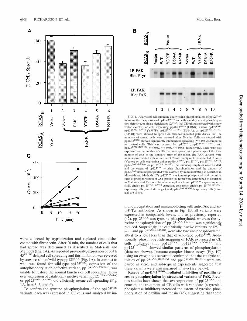
were collected by trypsinization and replated onto dishescoated with fibronectin. After 20 min, the number of cells thathad spread was determined as described in Materials andMethods (Fig. 1A). As reported previously, expression of pp41/43FRNK delayed cell spreading and this inhibition was reversedby coexpression of wild-type pp125FAK (Fig. 1A). In contrast towhat was found for wild-type pp125FAK, expression of theautophosphorylation-defective variant, pp125FAK (Y397F), wasunable to restore the normal kinetics of cell spreading. How-ever, expression of catalytically inactive variant pp125FAK (D564A)
or pp125FAK (K454R) did efficiently rescue cell spreading (Fig.1A, bars 3, 5, and 6).
To confirm the tyrosine phosphorylation of the pp125FAK
variants, each was expressed in CE cells and analyzed by im-
munoprecipitation and immunoblotting with anti-FAK and an-ti-P-Tyr antibodies. As shown in Fig. 1B, all variants wereexpressed at comparable levels, and as previously reported(42), pp125FAK was tyrosine phosphorylated, whereas the ty-rosine phosphorylation of pp125FAK (Y397F) was significantlyreduced. Surprisingly, the catalytically inactive variants, pp125
FAK
(D564A) and pp125FAK (K454R), were also tyrosine phosphorylated,albeit to a level less than that of wild-type pp125FAK. Addi-tionally, phosphopeptide mapping of FAK expressed in CEcells indicated that pp125FAK, pp125FAK (D564A), andpp125
FAK (K454R)showed similar patterns of phosphorylation
(data not shown). Immune complex kinase assays (Fig. 1C)using an exogenous substrate confirmed that the catalytic ac-tivities of pp125FAK (D564A) and pp125FAK (K454R) were im-paired in vitro, and subsequent experiments suggested thatthese variants were also impaired in vivo (see below).
Rescue of pp41/43FRNK-mediated inhibition of paxillin ty-rosine phosphorylation by structural variants of FAK. Previ-ous studies have shown that overexpression of pp125FAK andconcomitant treatment of CE cells with vanadate (a tyrosinephosphatase inhibitor) increased the extent of tyrosine phos-phorylation of paxillin and tensin (45), suggesting that these
FIG. 1. Analysis of cell spreading and tyrosine phosphorylation of pp125FAK
following the coexpression of pp41/43FRNK and either wild-type, autophosphoryla-tion-defective, or kinase-deficient pp125FAK. (A) CE cells transfected with emptyvector (Vector) or cells expressing pp41/43FRNK (FRNK) and/or pp125FAK,pp125FAK (Y397F) (Y397F), pp125FAK (D564A) (D564A), or pp125FAK (K454R)
(K454R) were allowed to spread on fibronectin-coated petri dishes, and thenumbers of spread cells were assessed after 20 min. Cells transfected withpp41/43FRNK showed significantly inhibited cell spreading (P , 0.002) comparedto control cells. This was reversed by pp125FAK, pp125FAK (D564A), andpp125FAK (K454R) (P , 0.02, P , 0.05, P , 0.005, respectively). Each result wasexpressed as the number of cells that were spread as a percentage of the totalnumber of cells 6 the standard error of the mean. (B) FAK variants wereimmunoprecipitated with antiserum BC2 from empty vector-transfected CE cells(Vector) or cells expressing either pp41/43FRNK, pp125FAK, pp125FAK (Y397F),pp125FAK (D564A), or pp125FAK (K454R). The immunoprecipitates were divided,and the extent of pp125FAK tyrosine phosphorylation and the amount ofpp125FAK immunoprecipitated were assessed by immunoblotting as described inMaterials and Methods. (C) pp125FAK was immunoprecipitated, and the initialrates of phosphorylation of GST-paxillin (N-term) were determined as describedin Materials and Methods. Immune complexes from pp125FAK-expressing cells(solid circle), pp125FAK (Y397F)-expressing cells (open circle), pp125FAK (D564A)-expressing cells (inverted triangle), and pp125FAK (K454R)-expressing cells (trian-gle) are shown.
6908 RICHARDSON ET AL. MOL. CELL. BIOL.
on March 14, 2014 by guest
http://mcb.asm
.org/D
ownloaded from
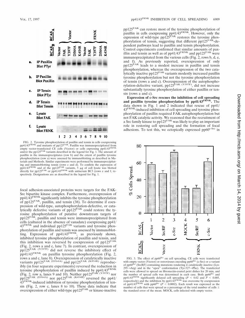
focal adhesion-associated proteins were targets for the FAK-Src bipartite kinase complex. Furthermore, overexpression ofpp41/43FRNK significantly inhibits the tyrosine phosphorylationof pp125FAK, paxillin, and tensin (38). To determine if coex-pression of wild-type, autophosphorylation-defective, or cata-lytically defective variants of pp125FAK could restore the ty-rosine phosphorylation of putative downstream targets ofpp125FAK, paxillin and tensin were immunoprecipitated fromcells (cultured in the absence of vanadate) coexpressing pp41/43FRNK and individual pp125FAK variants and tyrosine phos-phorylation of paxillin and tensin was assessed by immunoblot-ting. Expression of pp41/43FRNK, as previously shown,inhibited tyrosine phosphorylation of paxillin and tensin, andthis inhibition was reversed by coexpression of pp125FAK
(Fig. 2, rows a and c, lane 7). In contrast, overexpression ofpp125FAK (Y397F) did not reverse the inhibitory effect ofpp41/43FRNK on paxillin tyrosine phosphorylation (Fig. 2,rows a and c, lane 8). Overexpression of catalytically inactivevariants pp125FAK (D564A) and pp125FAK (K454R) reproduc-ibly (in four separate experiments) reversed the reduction intyrosine phosphorylation of paxillin induced by pp41/43FRNK
(Fig. 2, row a, lanes 9 and 10). Neither pp125FAK (Y397F) norpp125FAK (D564A) nor pp125FAK (K454R) reversed the pp41/43FRNK-induced inhibition of tyrosine phosphorylation of ten-sin (Fig. 2, row c, lanes 8 to 10). These data indicate thatoverexpression of either wild-type pp125FAK or kinase-inactive
pp125FAK can restore most of the tyrosine phosphorylation ofpaxillin in cells coexpressing pp41/43FRNK. However, only theexpression of wild-type pp125FAK restores the tyrosine phos-phorylation of tensin, suggesting that different pp125FAK-de-pendent pathways lead to paxillin and tensin phosphorylation.Control experiments confirmed that similar amounts of pax-illin and tensin as well as of pp41/43FRNK and pp125FAK wereimmunoprecipitated from the various cells (Fig. 2, rows b, d, e,and f). As previously reported, overexpression of onlypp125FAK leads to a modest increase in paxillin and tensinphosphorylation, whereas the overexpression of the two cata-lytically inactive pp125FAK variants modestly increased paxillintyrosine phosphorylation but not the tyrosine phosphorylationof tensin (rows a and c). Overexpression of the autophospho-rylation-defective variant, pp125FAK (Y397F), did not increasesubstantially tyrosine phosphorylation of either paxillin or ten-sin (rows a and c).
Expression of c-Src rescues the inhibition of cell spreadingand paxillin tyrosine phosphorylation by pp41/43FRNK. Thedata shown in Fig. 1 and 2 indicated that rescue of pp41/43FRNK-induced inhibition of cell spreading and tyrosine phos-phorylation of paxillin required FAK autophosphorylation butnot FAK catalytic activity. We reasoned that the recruitment ofa Src family kinase to pp125FAK was likely to play an importantrole in restoring cell spreading and the formation of focaladhesions. To test this, we ectopically expressed pp60c-Src in
FIG. 2. Tyrosine phosphorylation of paxillin and tensin in cells coexpressingpp41/43FRNK and mutants of pp125FAK. Paxillin was immunoprecipitated fromempty vector-transfected CE cells (Vector) or cells expressing pp41/43FRNK
and/or the pp125FAK variants described in the legend for Fig. 1. The amount ofpaxillin in the immunoprecipitates (row b) and the extent of paxillin tyrosinephosphorylation (row a) were assessed by immunoblotting as described in Ma-terials and Methods. Similar experiments were performed by immunoprecipitat-ing and immunoblotting tensin (rows c and d). To confirm the expression ofpp41/43FRNK and of the pp125FAK variants, 5 mg of cell lysate was blotteddirectly for pp125FAK or pp41/43FRNK with antiserum BC3 (rows e and f, re-spectively. Designations are as described in the legend for Fig. 1.
FIG. 3. The effect of pp60Src on cell spreading. CE cells were transfectedwith empty vector (Vector) or retroviruses encoding pp60Src (c-Src) or a variantof pp60Src (SrcRF) containing mutations rendering it catalytically inactive (Lys-2953Arg) and in the “open” conformation (Tyr-5273Phe). The transfectedcells were allowed to spread on fibronectin-coated petri dishes for 20 min, andthe number of spread cells was determined in each case. Both pp60Src andpp41/43FRNK significantly delayed cell spreading (P , 0.02 and P , 0.005,respectively) and the inhibition by pp41/43FRNK was overcome by coexpressionof pp41/43FRNK with pp60Src (P , 0.0002). Each result was expressed as thenumber of cells that were spread as a percentage of the total number of cells 6the standard error of the mean. MOCK, cells infected with empty vector.
VOL. 17, 1997 pp41/43FRNK INHIBITION OF CELL SPREADING 6909
on March 14, 2014 by guest
http://mcb.asm
.org/D
ownloaded from
CE cells alone or in CE cells coexpressing pp41/43FRNK (Fig.3). Overexpression of pp60c-Src alone accelerated cell spread-ing. This increase in the apparent rate of cell spreading re-quired the catalytic activity of pp60c-Src, as a kinase-inactivevariant of pp60c-Src [pp60Src (RF), containing mutations Lys-2953Arg and Tyr-5273Phe] failed to accelerate cell spread-ing (Fig. 3). As shown in Fig. 3, coexpression of pp60c-Src andpp41/43FRNK reversed the inhibitory effects of pp41/43FRNK oncell spreading, whereas the expression of pp60Src (RF) failed torescue pp41/43FRNK-induced inhibition of cell spreading,clearly showing that the catalytic activity of pp60c-Src was re-quired. A pp60c-Src variant which contained only the Lys-2953Arg mutation, behaved similarly to pp60Src (RF) (data notshown) and was unable to rescue cell spreading and paxillinphosphorylation (see below).
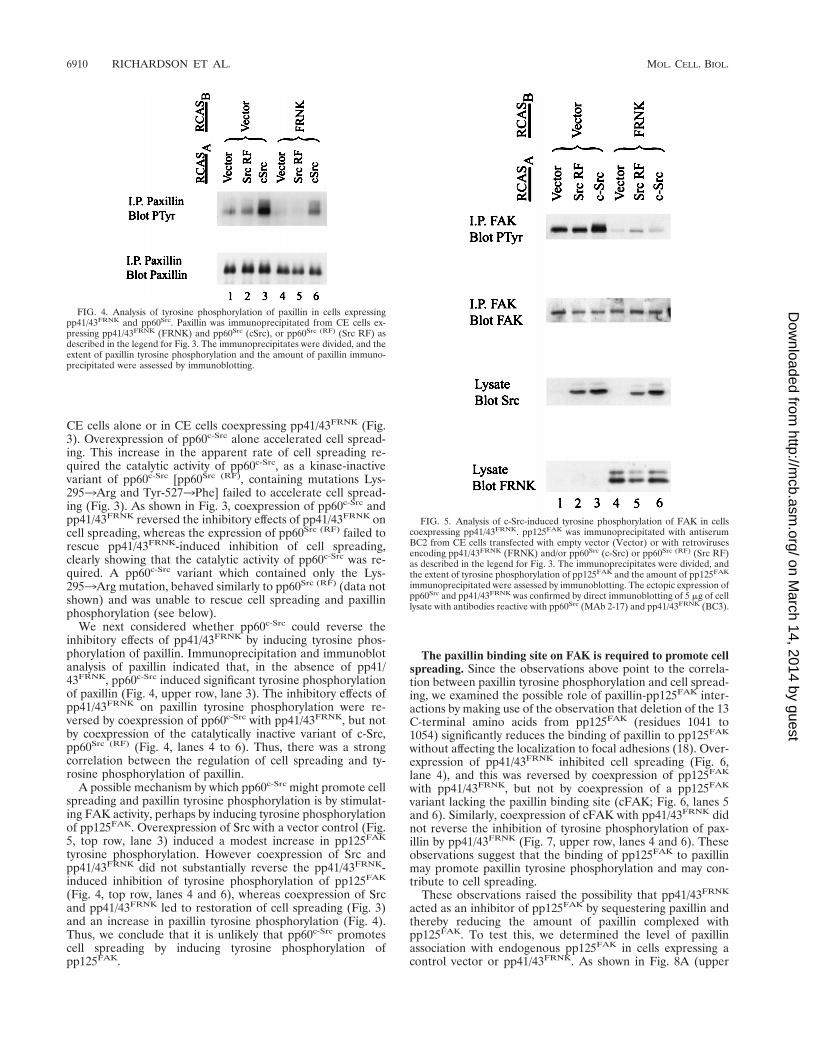
We next considered whether pp60c-Src could reverse theinhibitory effects of pp41/43FRNK by inducing tyrosine phos-phorylation of paxillin. Immunoprecipitation and immunoblotanalysis of paxillin indicated that, in the absence of pp41/43FRNK, pp60c-Src induced significant tyrosine phosphorylationof paxillin (Fig. 4, upper row, lane 3). The inhibitory effects ofpp41/43FRNK on paxillin tyrosine phosphorylation were re-versed by coexpression of pp60c-Src with pp41/43FRNK, but notby coexpression of the catalytically inactive variant of c-Src,pp60Src (RF) (Fig. 4, lanes 4 to 6). Thus, there was a strongcorrelation between the regulation of cell spreading and ty-rosine phosphorylation of paxillin.
A possible mechanism by which pp60c-Src might promote cellspreading and paxillin tyrosine phosphorylation is by stimulat-ing FAK activity, perhaps by inducing tyrosine phosphorylationof pp125FAK. Overexpression of Src with a vector control (Fig.5, top row, lane 3) induced a modest increase in pp125FAK
tyrosine phosphorylation. However coexpression of Src andpp41/43FRNK did not substantially reverse the pp41/43FRNK-induced inhibition of tyrosine phosphorylation of pp125FAK
(Fig. 4, top row, lanes 4 and 6), whereas coexpression of Srcand pp41/43FRNK led to restoration of cell spreading (Fig. 3)and an increase in paxillin tyrosine phosphorylation (Fig. 4).Thus, we conclude that it is unlikely that pp60c-Src promotescell spreading by inducing tyrosine phosphorylation ofpp125FAK.
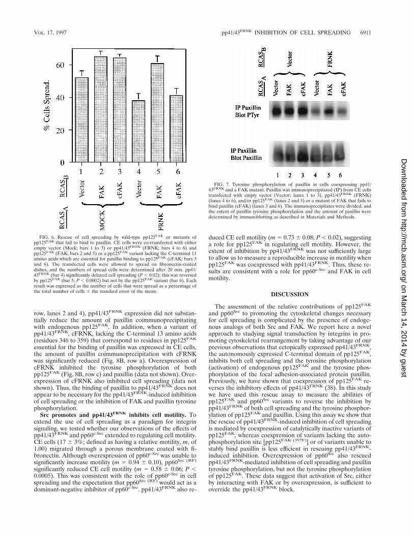
The paxillin binding site on FAK is required to promote cellspreading. Since the observations above point to the correla-tion between paxillin tyrosine phosphorylation and cell spread-ing, we examined the possible role of paxillin-pp125FAK inter-actions by making use of the observation that deletion of the 13C-terminal amino acids from pp125FAK (residues 1041 to1054) significantly reduces the binding of paxillin to pp125FAK
without affecting the localization to focal adhesions (18). Over-expression of pp41/43FRNK inhibited cell spreading (Fig. 6,lane 4), and this was reversed by coexpression of pp125FAK
with pp41/43FRNK, but not by coexpression of a pp125FAK
variant lacking the paxillin binding site (cFAK; Fig. 6, lanes 5and 6). Similarly, coexpression of cFAK with pp41/43FRNK didnot reverse the inhibition of tyrosine phosphorylation of pax-illin by pp41/43FRNK (Fig. 7, upper row, lanes 4 and 6). Theseobservations suggest that the binding of pp125FAK to paxillinmay promote paxillin tyrosine phosphorylation and may con-tribute to cell spreading.
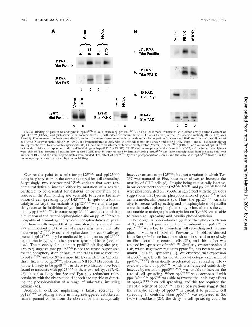
These observations raised the possibility that pp41/43FRNK
acted as an inhibitor of pp125FAK by sequestering paxillin andthereby reducing the amount of paxillin complexed withpp125FAK. To test this, we determined the level of paxillinassociation with endogenous pp125FAK in cells expressing acontrol vector or pp41/43FRNK. As shown in Fig. 8A (upper
FIG. 4. Analysis of tyrosine phosphorylation of paxillin in cells expressingpp41/43FRNK and pp60Src. Paxillin was immunoprecipitated from CE cells ex-pressing pp41/43FRNK (FRNK) and pp60Src (cSrc), or pp60Src (RF) (Src RF) asdescribed in the legend for Fig. 3. The immunoprecipitates were divided, and theextent of paxillin tyrosine phosphorylation and the amount of paxillin immuno-precipitated were assessed by immunoblotting.
FIG. 5. Analysis of c-Src-induced tyrosine phosphorylation of FAK in cellscoexpressing pp41/43FRNK. pp125FAK was immunoprecipitated with antiserumBC2 from CE cells transfected with empty vector (Vector) or with retrovirusesencoding pp41/43FRNK (FRNK) and/or pp60Src (c-Src) or pp60Src (RF) (Src RF)as described in the legend for Fig. 3. The immunoprecipitates were divided, andthe extent of tyrosine phosphorylation of pp125FAK and the amount of pp125FAK
immunoprecipitated were assessed by immunoblotting. The ectopic expression ofpp60Src and pp41/43FRNK was confirmed by direct immunoblotting of 5 mg of celllysate with antibodies reactive with pp60Src (MAb 2-17) and pp41/43FRNK (BC3).
6910 RICHARDSON ET AL. MOL. CELL. BIOL.
on March 14, 2014 by guest
http://mcb.asm
.org/D
ownloaded from
row, lanes 2 and 4), pp41/43FRNK expression did not substan-tially reduce the amount of paxillin coimmunoprecipitatingwith endogenous pp125FAK. In addition, when a variant ofpp41/43FRNK, cFRNK, lacking the C-terminal 13 amino acids(residues 346 to 359) that correspond to residues in pp125FAK
essential for the binding of paxillin was expressed in CE cells,the amount of paxillin coimmunoprecipitation with cFRNKwas significantly reduced (Fig. 8B, row a). Overexpression ofcFRNK inhibited the tyrosine phosphorylation of bothpp125FAK (Fig. 8B, row c) and paxillin (data not shown). Over-expression of cFRNK also inhibited cell spreading (data notshown). Thus, the binding of paxillin to pp41/43FRNK does notappear to be necessary for the pp41/43FRNK-induced inhibitionof cell spreading or the inhibition of FAK and paxillin tyrosinephosphorylation.
Src promotes and pp41/43FRNK inhibits cell motility. Toextend the use of cell spreading as a paradigm for integrinsignaling, we tested whether our observations of the effects ofpp41/43FRNK and pp60c-Src extended to regulating cell motility.CE cells (17 6 3%; defined as having a relative motility, m, of1.00) migrated through a porous membrane coated with fi-bronectin. Although overexpression of pp60c-Src was unable tosignificantly increase motility (m 5 0.94 6 0.10), pp60Src (RF)
significantly reduced CE cell motility (m 5 0.58 6 0.06; P ,0.0005). This was consistent with the role of pp60c-Src in cellspreading and the expectation that pp60Src (RF) would act as adominant-negative inhibitor of pp60c-Src. pp41/43FRNK also re-
duced CE cell motility (m 5 0.73 6 0.08; P , 0.02), suggestinga role for pp125FAK in regulating cell motility. However, theextent of inhibition by pp41/43FRNK was not sufficiently largeto allow us to measure a reproducible increase in motility whenpp125FAK was coexpressed with pp41/43FRNK. Thus, these re-sults are consistent with a role for pp60c-Src and FAK in cellmotility.
DISCUSSION
The assessment of the relative contributions of pp125FAK
and pp60Src to promoting the cytoskeletal changes necessaryfor cell spreading is complicated by the presence of endoge-nous analogs of both Src and FAK. We report here a novelapproach to studying signal transduction by integrins in pro-moting cytoskeletal rearrangement by taking advantage of ourprevious observations that ectopically expressed pp41/43FRNK,the autonomously expressed C-terminal domain of pp125FAK,inhibits both cell spreading and the tyrosine phosphorylation(activation) of endogenous pp125FAK and the tyrosine phos-phorylation of the focal adhesion-associated protein paxillin.Previously, we have shown that coexpression of pp125FAK re-verses the inhibitory effects of pp41/43FRNK (38). In this studywe have used this rescue assay to measure the abilities ofpp125FAK and pp60Src variants to reverse the inhibition bypp41/43FRNK of both cell spreading and the tyrosine phosphor-ylation of pp125FAK and paxillin. Using this assay we show thatthe rescue of pp41/43FRNK-induced inhibition of cell spreadingis mediated by coexpression of catalytically inactive variants ofpp125FAK, whereas coexpression of variants lacking the auto-phosphorylation site [pp125FAK (397F)] or of variants unable tostably bind paxillin is less efficient in rescuing pp41/43FRNK-induced inhibition. Overexpression of pp60Src also rescuedpp41/43FRNK-mediated inhibition of cell spreading and paxillintyrosine phosphorylation, but not the tyrosine phosphorylationof pp125FAK. These data suggest that activation of Src, eitherby interacting with FAK or by overexpression, is sufficient tooverride the pp41/43FRNK block.
FIG. 6. Rescue of cell spreading by wild-type pp125FAK or mutants ofpp125FAK that fail to bind to paxillin. CE cells were co-transfected with eitherempty vector (Mock; bars 1 to 3) or pp41/43FRNK (FRNK; bars 4 to 6) andpp125FAK (FAK; bars 2 and 5) or a pp125FAK variant lacking the C-terminal 11amino acids which are essential for paxillin binding to pp125FAK (cFAK; bars 3and 6). The transfected cells were allowed to spread on fibronectin-coateddishes, and the numbers of spread cells were determined after 20 min. pp41/43FRNK (bar 4) significantly delayed cell spreading (P , 0.02); this was reversedby pp125FAK (bar 5; P , 0.0002) but not by the pp125FAK variant (bar 6). Eachresult was expressed as the number of cells that were spread as a percentage ofthe total number of cells 6 the standard error of the mean.
FIG. 7. Tyrosine phosphorylation of paxillin in cells coexpressing pp41/43FRNK and a FAK mutant. Paxillin was immunoprecipitated (IP) from CE cellstransfected with empty vector (Vector; lanes 1 to 3), pp41/43FRNK (FRNK)(lanes 4 to 6), and/or pp125FAK (lanes 2 and 5) or a mutant of FAK that fails tobind paxillin (cFAK) (lanes 3 and 6). The immunoprecipitates were divided, andthe extent of paxillin tyrosine phosphorylation and the amount of paxillin weredetermined by immunoblotting as described in Materials and Methods.
VOL. 17, 1997 pp41/43FRNK INHIBITION OF CELL SPREADING 6911
on March 14, 2014 by guest
http://mcb.asm
.org/D
ownloaded from
Our results point to a role for pp125FAK and pp125FAK
autophosphorylation in the events required for cell spreading.Surprisingly, two separate pp125FAK variants that were ren-dered catalytically inactive either by mutation of a residuepredicted to be essential for catalysis or by mutation of aresidue in the ATP binding site were able to reverse the inhi-bition of cell spreading by pp41/43FRNK. In spite of a loss incatalytic activity these mutants of pp125FAK were able to par-tially reverse the inhibition of tyrosine phosphorylation of pax-illin by pp41/43FRNK. In contrast pp125FAK variants containinga mutation of the autophosphorylation site on pp125FAK wereincapable of promoting the tyrosine phosphorylation of paxil-lin. These results suggest that tyrosine phosphorylation of Tyr-397 is important and that in cells expressing the catalyticallyinactive pp125FAK, tyrosine phosphorylation of ectopically ex-pressed pp125FAK may be mediated by endogenous pp125FAK
or, alternatively, by another protein tyrosine kinase (see be-low). The necessity for an intact pp60Src binding site (e.g.,Tyr-397) suggests that pp125FAK is not the kinase responsiblefor the phosphorylation of paxillin and that a kinase recruitedto pp125FAK via Tyr-397 is a more likely candidate. In CE cells,this is likely to be pp59Fyn, whereas in NIH 3T3 fibroblasts thekinase is likely to be pp60c-Src, as these two kinases have beenfound to associate with pp125FAK in these two cell types (7, 42,46). It is also likely that Src and Fyn play redundant roles,consistent with the observation that both are capable of direct-ing the phosphorylation of a range of substrates, includingpaxillin (48).
Additional evidence implicating a kinase recruited topp125FAK as playing a role in integrin-triggered cytoskeletalrearrangement comes from the observation that catalytically
inactive variants of pp125FAK, but not a variant in which Tyr-397 was mutated to Phe, have been shown to increase themotility of CHO cells (8). Despite being catalytically inactive,in our experiments both pp125FAK (K454R) and pp125FAK (D564A)
were phosphorylated on Tyr-397, in agreement with the previoussuggestions that tyrosine phosphorylation of pp125FAK is notan intramolecular process (7). Thus, the pp125FAK variantsable to rescue cell spreading and phosphorylation of paxillinwere themselves phosphorylated on tyrosine, whereas the vari-ant unable to undergo phosphorylation on Tyr-397 was unableto rescue cell spreading and paxillin phosphorylation.
The foregoing observations suggested that phosphorylationof Tyr-397 and presumably the recruitment of pp60Src topp125FAK were key to promoting cell spreading and tyrosinephosphorylation of paxillin. Previously, fibroblasts derivedfrom Src (2/2) mice have been shown to spread more slowlyon fibronectin than control cells (25), and this defect wasrescued by expression of pp60c-Src. Similarly, overexpression ofCsk, which negatively regulates pp60c-Src, has been shown toinhibit HeLa cell spreading (3). We observed that expressionof pp60Src in CE cells (in the absence of ectopic expression ofpp41/43FRNK) dramatically accelerated cell spreading. How-ever, a variant of pp60c-Src which was rendered catalyticallyinactive by mutation [pp60Src (RF)] was unable to increase therate of cell spreading. When pp60c-Src was coexpressed withpp41/43FRNK, pp60Src was able to reverse the inhibitory effectsof pp41/43FRNK on cell spreading, and this too required thecatalytic activity of pp60c-Src. These observations suggest thatthe catalytic activity of pp60Src is essential to promote cellspreading. In contrast, when pp60c-Src was expressed in Src(2/2) fibroblasts (25), the delay in cell spreading could be
FIG. 8. Binding of paxillin to endogenous pp125FAK in cells expressing pp41/43FRNK. (A) CE cells were transfected with either empty vector (Vector) orpp41/43FRNK (FRNK), and lysates were immunoprecipitated (IP) with either preimmune serum (P.I.; lanes 1 and 3) or the FAK-specific antibody, BC2 (BC2; lanes2 and 4). The immune complexes were divided, and equal amounts were immunoblotted with antibodies to paxillin (top row) and FAK (middle row). An aliquot ofcell lysate (5 mg) was subjected to SDS-PAGE and immunoblotted directly with an antibody to paxillin (lanes 5 and 6) or FRNK (lanes 7 and 8). The results shownare representative of four separate experiments. (B) CE cells were transfected with either empty vector (Vector), pp41/43FRNK (FRNK), or a variant of pp41/43FRNK
lacking the residues corresponding to the paxillin binding site in pp125FAK (cFRNK). FRNK was immunoprecipitated with antiserum BC3, and the immunoprecipitateswere divided. The amounts of paxillin (row a) and FRNK (row b) were assessed by immunoblotting. pp125FAK was immunoprecipitated from the same cells withantiserum BC2, and the immunoprecipitates were divided. The extent of pp125FAK tyrosine phosphorylation (row c) and the amount of pp125FAK (row d) in theimmunoprecipitates were assessed by immunoblotting.
6912 RICHARDSON ET AL. MOL. CELL. BIOL.
on March 14, 2014 by guest
http://mcb.asm
.org/D
ownloaded from
rescued by catalytically inactive variants of pp60c-Src. One pos-sible explanation for the discrepancy with our results, in whichthe catalytic activity of pp60Src is necessary to promote cellspreading, may be the different cellular backgrounds in whichspreading is assessed. Thus, in cells expressing pp41/43FRNK,the reduced tyrosine phosphorylation of pp125FAK may alsoprevent activation of endogenous Src, thereby requiring theoverexpression of catalytically active Src to bypass the effects ofpp41/43FRNK. In Src (2/2) cells, the catalytic activity ofpp125FAK may be sufficient to compensate for the lack ofpp60Src catalytic activity, as long as other domains of pp60Src
are expressed.Our results point to a critical role for pp60c-Src in controlling
cell spreading. Expression of pp60Src was able to promote cellspreading in the presence of ectopically expressed pp41/43FRNK. In addition, pp60Src did not appear to induce tyrosinephosphorylation of pp125FAK. Thus, the inhibitory effects ofpp41/43FRNK can be bypassed if pp60Src is expressed at suffi-ciently high levels. As pp125FAK is more abundant thanpp60Src, it is possible that the small amount of pp125FAK thatremains tyrosine phosphorylated in the presence of pp41/43FRNK is sufficient to recruit and activate pp60Src or thatoverexpression of pp60Src leads to a higher level of basalactivity of Src. An alternative explanation for the ability ofpp60c-Src to promote and rescue cell spreading is that whenpp60c-Src is expressed at high levels, it can bind directly to focaladhesion proteins and directly phosphorylate them. For ex-ample, pp60c-Src is reported to bind to paxillin (39). Expres-sion of pp60c-Src induced strong tyrosine phosphorylation ofpaxillin. pp60c-Src also induced tyrosine phosphorylation ofpaxillin in cells in which pp41/43FRNK was ectopically ex-pressed, and in these cells pp60c-Src also reversed the inhibitionof cell spreading by pp41/43FRNK. This observation, along withour earlier observation that those pp125FAK variants that werecapable of overcoming the inhibition of cell spreading by pp41/43FRNK were also capable of rescuing tyrosine phosphorylationof paxillin, makes paxillin an obvious candidate substrate forpp60c-Src. Further evidence that pp60c-Src can phosphorylatepaxillin comes from the observation that deregulation ofpp60c-Src by rendering cells deficient in Csk leads to hyper-phosphorylation of paxillin (48). This is partially reversed incells which lack either Src or Fyn in addition to Csk (48). Thus,our data support the phosphorylation of paxillin by pp60Src asbeing an important event during cell spreading.
A pp125FAK variant lacking the binding site for paxillinfailed to reverse the pp41/43FRNK-mediated inhibition of bothcell spreading and paxillin tyrosine phosphorylation. Thesedata and the requirement for Tyr-397 phosphorylation provideadditional support for a model whereby pp125FAK functions asa “switchable adaptor”. The C terminus of pp125FAK bindspaxillin, whereas activation of pp125FAK by integrins inducestyrosine phosphorylation of pp125FAK and leads to the recruit-ment of pp60Src. Thus, pp125FAK promotes the phosphoryla-tion of paxillin by pp60c-Src by efficiently localizing both thesubstrate and the kinase.
We speculate that both the binding of pp125FAK to paxillinand tyrosine phosphorylation of paxillin play important roles incell spreading. A likely role for paxillin is the recruitment ofadditional proteins to the “early focal complex.” Recruitmentof additional paxillin binding proteins will likely be dependenton the tyrosine phosphorylation of paxillin, since the pp125FAK
variant unable to bind paxillin is unable to induce the tyrosinephosphorylation of paxillin and is unable to rescue cell spread-ing. However, it remains possible that signaling molecules thatpromote cell spreading bind to paxillin in a phosphotyrosine-independent fashion. In this case, pp60Src would promote cell
spreading by some mechanism other than by inducing tyrosinephosphorylation of paxillin. The foregoing observations sug-gest a central role for paxillin in promoting cell spreading.However, in addition to paxillin, tensin has also been suggestedto be a substrate of pp125FAK. Both the catalytically inactiveand autophosphorylation-defective forms of pp125FAK failedto reverse the pp41/43FRNK-dependent inhibition of tyrosinephosphorylation of tensin. Thus, we suggest that tyrosine phos-phorylation of tensin may be less critical for cell spreading.
Cell migration requires both the formation of new adhesivecontacts at the leading edge and the disassembly of contacts atthe trailing edge (26). When cells in suspension are allowed tospread on fibronectin, they initially form small focal complexes(31) which become progressively larger as the cells spread (38).This suggests that cells in suspension lack macromolecularfocal complexes, so measuring cell spreading is likely to bebiased towards reflecting events during the assembly ratherthan the disassembly of adhesive complexes. Therefore, wealso examined the effects of pp60c-Src and pp41/43FRNK on cellmotility and found that pp60Src (RF) acted as an inhibitor ofmotility. The failure of this variant to display a measurablephenotype during cell spreading suggests that pp60Src may playroles during motility in addition to those it plays during cellspreading. These roles need not be limited to the formationand disassembly of adhesive complexes, as cell motility involvesnumerous other events (28). In parallel to its effects on cellspreading, pp41/43FRNK also functions to inhibit cell motility.Coexpression of pp60c-Src with pp41/43FRNK restores cell mo-tility and mirrors the ability of pp60c-Src to promote cell spread-ing. The effect of pp41/43FRNK on motility was less pronouncedthan the inhibition of spreading by pp41/43FRNK. Whetherprocesses other than the formation of new adhesive contacts,which we presume are inhibited by pp41/43FRNK, limit the rateof migration remain to be analyzed. In addition, interpretationof these results is complicated because the relationship be-tween adhesive strength and motility is not simple—cells whichadhere either too strongly or too weakly migrate more slowly(33).
We speculate that pp125FAK promotes cell motility by cat-alyzing the formation of focal adhesions which act as anchorpoints against which the actin-myosin system can generateforce. The activation of pp125FAK, the recruitment and acti-vation of pp60Src, and the phosphorylation of paxillin appear tobe the early key events during this process. It will be importantto determine which molecules are subsequently recruited bypaxillin and how this ultimately leads to reorganization of theactin cytoskeleton.
ACKNOWLEDGMENTS
A. Richardson and R. K. Malik contributed equally to this work.We thank Richard Roof and Yun Rui Du for helpful suggestions.
We thank Cheryl Borgman and Marlene Macklem for technical assis-tance.
These studies were supported by grants from the DHHS-NationalCancer Institute (CA29243 and CA40042) and the Council for To-bacco Research USA, Inc. (no. 4491). R.K.M. was supported by grantK08 CA72662 from the National Cancer Institute.
REFERENCES
1. Akasaka, T., R. L. van Leeuwen, I. G. Yoshinaga, M. C. J. Mihm, and H. R.Byers. 1995. Focal adhesion kinase (p125FAK) expression correlates withmotility of human melanoma cell lines. J. Invest. Dermatol. 105:104–108.
2. Bachelot, C., L. Rameh, T. Parsons, and L. C. Cantley. 1996. Association ofphosphatidylinositol 3-kinase, via the SH2 domains of p85, with focal adhe-sion kinase in polyoma middle T-transformed fibroblasts. Biochim. Biophys.Acta 1311:45–52.
3. Bergman, M., V. Joukov, I. Virtanen, and K. Alitalo. 1995. Overexpressed
VOL. 17, 1997 pp41/43FRNK INHIBITION OF CELL SPREADING 6913
on March 14, 2014 by guest
http://mcb.asm
.org/D
ownloaded from
Csk tyrosine kinase is localized in focal adhesions, causes reorganization ofaVb5 integrin, and interferes with HeLa cell spreading. Mol. Cell. Biol.15:711–722.
4. Bretscher, M. S. 1996. Getting membrane flow and the cytoskeleton tocooperate in moving cells. Cell 87:601–606.
5. Burridge, K., and M. Chrzanowska-Wodnicka. 1996. Focal adhesions, con-tractility, and signaling. Annu. Rev. Cell Dev. Biol. 12:463–519.
6. Burridge, K., K. Fath, T. Kelly, G. Nuckolls, and C. Turner. 1988. Focaladhesions: transmembrane junctions between the extracellular matrix andthe cytoskeleton. Annu. Rev. Cell Biol. 4:487–525.
7. Calalb, M. B., T. R. Polte, and S. K. Hanks. 1995. Tyrosine phosphorylationof focal adhesion kinase at sites in the catalytic domain regulates kinaseactivity: a role for Src family kinases. Mol. Cell. Biol. 15:954–963.
8. Carey, L. A., J. F. Chang, and J.-L. Guan. 1996. Stimulation of cell migrationby overexpression of focal adhesion kinase and its association with Src andFyn. J. Cell. Sci. 109:1787–1794.
9. Chen, H.-C., P. A. Appeddu, H. Isoda, and J.-L. Guan. 1996. Phosphorylationof tyrosine 397 in focal adhesion kinase is required for binding phosphati-dylinositol 3-kinase. J. Biol. Chem. 271:26329–26334.
10. Chen, H.-C., P. A. Appeddu, J. T. Parsons, J. D. Hildebrand, M. D. Schaller,and J.-L. Guan. 1995. Interaction of focal adhesion kinase with cytoskeletalprotein talin. J. Biol. Chem. 270:16995–16999.
11. Chen, H.-C., and J.-L. Guan. 1994. Association of focal adhesion kinase withits potential substrate phosphatidylinositol 3-kinase. Proc. Natl. Acad. Sci.USA 91:10148–10152.
12. Cobb, B. S., M. D. Schaller, T. H. Leu, and J. T. Parsons. 1994. Stableassociation of pp60src and pp59fyn with the focal adhesion-associated proteintyrosine kinase, pp125FAK. Mol. Cell. Biol. 14:147–155.
13. Gilmore, A. P., and L. H. Romer. 1996. Inhibition of focal adhesion kinase(FAK) signaling in focal adhesions decreases cell motility and proliferation.Mol. Biol. Cell 7:1209–1224.
14. Guinebault, C., B. Payrastre, C. Racaaud-Sultan, H. Mazarguil, M. Breton,G. Mauco, M. Plantavid, and H. Chap. 1995. Integrin-dependent transloca-tion of phosphoinositide 3-kinase to the cytoskeleton of thrombin-activatedplatelets involves specific interactions of p85-alpha with actin filaments andfocal adhesion kinase. J. Cell Biol. 129:831–842.
15. Hanks, S. K., M. B. Calalb, M. C. Harper, and S. K. Patel. 1992. Focaladhesion protein-tyrosine kinase phosphorylated in response to cell attach-ment to fibronectin. Proc. Natl. Acad. Sci. USA 89:8487–8491.
16. Harte, M. T., J. D. Hildebrand, M. R. Burnham, A. H. Bouton, and J. T.Parsons. 1996. p130Cas, a substrate associated with v-Src and v-Crk, localizesto focal adhesions and binds to focal adhesion kinase. J. Biol. Chem. 271:13649–13655.
17. Hildebrand, J. D., M. D. Schaller, and J. T. Parsons. 1993. Identification ofsequences required for the efficient localization of the focal adhesion kinase,pp125FAK, to cellular focal adhesions. J. Cell Biol. 123:993–1005.
18. Hildebrand, J. D., M. D. Schaller, and J. T. Parsons. 1995. Paxillin, atyrosine phosphorylated focal adhesion-associated protein, binds to the car-boxyl terminal domain of focal adhesion kinase. Mol. Biol. Cell 6:637–647.
19. Hildebrand, J. D., J. M. Taylor, and J. T. Parsons. 1996. An SH3 domain-containing GTPase-activating protein for Rho and Cdc42 associates withfocal adhesion kinase. Mol. Cell. Biol. 16:3169–3178.
20. Huttenlocher, A., R. Sandborg, and A. F. Horwitz. 1995. Adhesion in cellmigration. Curr. Biol. 7:697–706.
21. Hynes, R. O. 1992. Integrins: versatility, modulation, and signaling in celladhesion. Cell 69:11–25.
22. Ilic, D., S. Kanazawa, Y. Furuta, T. Yamamoto, and S. Aizawa. 1996. Im-pairment of mobility in endodermal cells by FAK deficiency. Exp. Cell Res.222:298–303.
23. Ilic, D., Y. Furuta, S. Kanazawa, N. Takeda, K. Sobue, N. Nakatsu, S.Nomura, J. Fujimoto, M. Okada, T. Yamamoto, and S. Aizawa. 1995. Re-duced cell motility and enhanced focal adhesion contact formation in cellsfrom FAK-deficient mice. Nature 377:539–543.
24. Kanner, S. B., A. B. Reynolds, R. R. Vines, and J. T. Parsons. 1990. Mono-clonal antibodies to individual tyrosine-phosphorylated protein substrates ofoncogene-encoded tyrosine kinases. Proc. Natl. Acad. Sci. USA 87:3328–3332.
25. Kaplan, K. B., J. R. Swedlow, D. O. Morgan, and H. E. Varmus. 1995. c-Srcenhances the spreading of src2/2 fibroblasts on fibronectin by a kinase-independent mechanism. Genes Dev. 9:1505–1517.
26. Lauffenburger, D. A., and A. F. Horwitz. 1996. Cell migration: a physicallyintegrated molecular process. Cell 84:359–369.
27. Lipfert, L., B. Haimovich, M. D. Schaller, B. S. Cobb, J. T. Parsons, and J. S.Brugge. 1992. Integrin-dependent phosphorylation and activation of theprotein tyrosine kinase pp125FAK in platelets. J. Cell Biol. 119:905–912.
28. Mitchison, T. J., and L. P. Cramer. 1996. Actin-based cell motility and celllocomotion. Cell 84:371–379.
29. Miyamoto, S., S. K. Akiyama, and K. M. Yamada. 1995. Synergistic roles forreceptor occupancy and aggregation in integrin transmembrane function.Science 267:883–885.
30. Miyamoto, S., H. Teramoto, O. A. Coso, J. S. Gutkind, P. D. Burbelo, S. K.Akiyama, and K. M. Yamada. 1995. Integrin function: molecular hierarchiesof cytoskeletal and signaling molecules. J. Cell Biol. 131:791–805.
31. Nobes, C. D., and A. Hall. 1995. Rho, Rac, and Cdc42 GTPases regulate theassembly of multimolecular focal complexes associated with actin stressfibers, lamellipodia, and filopodia. Cell 81:53–62.
32. Owens, L. V., L. Xu, R. J. Craven, G. A. Dent, T. M. Weiner, L. Kornberg,E. T. Liu, and W. G. Cance. 1995. Overexpression of the focal adhesionkinase (p125FAK) in invasive human tumors. Cancer Res. 55:2752–2755.
33. Palecek, S. P., J. C. Loftus, M. H. Ginsberg, D. A. Lauffenburger, and A. F.Horwitz. 1997. Integrin-ligand binding properties govern cell migrationspeed through cell-substratum adhesiveness. Nature 385:537–540.
34. Polte, T. R., and S. K. Hanks. 1995. Interaction between focal adhesionkinase and Crk-associated tyrosine kinase substrate p130Cas. Proc. Natl.Acad. Sci. USA 92:10678–10682.
35. Polte, T. R., and S. K. Hanks. 1997. Complexes of focal adhesion kinase(FAK) and Crk-associated substrate (p130Cas) are elevated in cytoskeleton-associated fractions following adhesion and Src transformation. J. Biol.Chem. 272:5501–5509.
36. Reynolds, A. B., D. J. Roesel, S. B. Kanner, and J. T. Parsons. 1989. Trans-formation-specific tyrosine phosphorylation of a novel cellular protein inchicken cells expressing oncogenic variants of the avian cellular src gene.Mol. Cell. Biol. 9:629–638.
37. Richardson, A., and J. T. Parsons. 1995. Signal transduction through inte-grins: a central role for focal adhesion kinase? Bioessays 17:229–236.
38. Richardson, A., and J. T. Parsons. 1996. A mechanism for regulation of theadhesion-associated protein tyrosine kinase pp125FAK. Nature 380:538–540.
39. Sabe, H., A. Hata, M. Okada, H. Nakagawa, and H. Hanafusa. 1994. Analysisof the binding of the Src homology 2 domain of Csk to tyrosine phosphor-ylated proteins in the suppression and mitotic activation of c-Src. Proc. Natl.Acad. Sci. USA 91:3984–3988.
40. Schaller, M. D., C. A. Borgman, B. C. Cobb, A. B. Reynolds, and J. T.Parsons. 1992. pp125FAK, a structurally distinctive protein-tyrosine kinaseassociated with focal adhesions. Proc. Natl. Acad. Sci. USA 89:5192–5196.
41. Schaller, M. D., C. A. Borgman, and J. T. Parsons. 1993. Autonomousexpression of a noncatalytic domain of the focal adhesion-associated proteintyrosine kinase pp125FAK. Mol. Cell. Biol. 13:785–791.
42. Schaller, M. D., J. D. Hildebrand, J. D. Shannon, J. W. Fox, R. R. Vines, andJ. T. Parsons. 1994. Autophosphorylation of the focal adhesion kinase,pp125FAK, directs SH2-dependent binding of pp60src. Mol. Cell. Biol. 14:1680–1688.
43. Schaller, M. D., C. A. Otey, J. D. Hildebrand, and J. T. Parsons. 1995. Focaladhesion kinase and paxillin bind to peptides mimicking beta integrin cyto-plasmic domains. J. Cell Biol. 130:1181–1187.
44. Schaller, M. D., and J. T. Parsons. 1994. Focal adhesion kinase and associ-ated proteins. Curr. Opin. Cell Biol. 6:705–710.
45. Schaller, M. D., and J. T. Parsons. 1995. pp125FAK-dependent tyrosinephosphorylation of paxillin creates a high-affinity binding site for Crk. Mol.Cell. Biol. 15:2635–2645.
46. Schlaepfer, D. D., S. K. Hanks, T. Hunter, and P. van der Geer. 1994.Integrin-mediated signal transduction linked to Ras pathway by GRB2 bind-ing to focal adhesion kinase. Nature 372:786–791.
47. Schlaepfer, D. D., and T. Hunter. 1996. Evidence for in vivo phosphorylationof the Grb2 SH2-domain binding site on focal adhesion kinase by Src-familyprotein-tyrosine kinases. Mol. Cell. Biol. 16:5623–5633.
48. Thomas, S. M., P. Soriano, and A. Imamoto. 1995. Specific and redundantroles of Src and Fyn in organizing the cytoskeleton. Nature 376:267–271.
49. Weiner, T. M., E. T. Liu, R. J. Craven, and W. G. Cance. 1993. Expressionof focal adhesion kinase gene in invasive cancer. Lancet 342:1024–1025.
6914 RICHARDSON ET AL. MOL. CELL. BIOL.
on March 14, 2014 by guest
http://mcb.asm
.org/D
ownloaded from