alternative splicing controls the mechanisms of fak autophosphorylation
TRANSCRIPT

MOLECULAR AND CELLULAR BIOLOGY, Nov. 2002, p. 7731–7743 Vol. 22, No. 220270-7306/02/$04.00�0 DOI: 10.1128/MCB.22.22.7731–7743.2002Copyright © 2002, American Society for Microbiology. All Rights Reserved.
Alternative Splicing Controls the Mechanisms ofFAK Autophosphorylation
Madeleine Toutant, Alicia Costa, Jeanne-Marie Studler,† Gress Kadare,Michele Carnaud, and Jean-Antoine Girault*
INSERM/UPMC U536, Institut du Fer a Moulin, 75005 Paris, France
Received 24 January 2002/Returned for modification 28 February 2002/Accepted 1 August 2002
Focal adhesion kinase (FAK) is activated following integrin engagement or stimulation of transmembranereceptors. Autophosphorylation of FAK on Tyr-397 is a critical event, allowing binding of Src family kinasesand activation of signal transduction pathways. Tissue-specific alternative splicing generates several isoformsof FAK with different autophosphorylation rates. Despite its importance, the mechanisms of FAK autophos-phorylation and the basis for differences between isoforms are not known. We addressed these questions usingisoforms of FAK expressed in brain. Autophosphorylation of FAK�, which is identical to that of “standard”FAK, was intermolecular in transfected cells, although it did not involve the formation of stable multimericcomplexes. Coumermycin-induced dimerization of gyrase B-FAK� chimeras triggered autophosphorylation ofTyr-397. This was independent of cell adhesion but required the C terminus of the protein. In contrast, theelevated autophosphorylation of FAK�6,7, the major neuronal splice isoform, was not accounted for bytransphosphorylation. Specifically designed immune precipitate kinase assays confirmed that autophosphor-ylation of FAK� was intermolecular, whereas autophosphorylation of FAK�6,7 or FAK�7 was predominantlyintramolecular and insensitive to the inhibitory effects of the N-terminal domain. Our results clarify themechanisms of FAK activation and show how alternative splicing can dramatically alter the mechanism ofautophosphorylation of a protein kinase.
The nonreceptor tyrosine kinase focal adhesion kinase(FAK) plays a key role at focal adhesion sites in promotingspreading, migration, and transmission of anchorage-depen-dent antiapoptotic signals (50). The important physiologicalrole of FAK is demonstrated by the lethality of its null muta-tion around midgestation, with severe developmental defects(33). FAK is phosphorylated on tyrosine in response to inte-grin engagement, G protein-coupled receptor activation, andgrowth factor receptor stimulation (20, 57). In cultured cells,integrin engagement promotes FAK recruitment to focal ad-hesions and its phosphorylation on multiple tyrosine residues(26, 46). Tyrosine phosphorylation of FAK results from auto-phosphorylation and from phosphorylation by other tyrosinekinases, including Src family kinases (4, 11) and insulin recep-tor (1). In contrast to many other kinases (see reference 32),autophosphorylation of FAK does not occur in the activationloop of the catalytic domain (A-loop), but on a tyrosine locatedat position 397, in the linker region between the N-terminaldomain and the catalytic domain.
Autophosphorylation at Tyr-397 is a key event for FAKbiological function, since it creates a high-affinity binding sitefor proteins with SH2 domains, including Src family kinases(c-Src, Fyn) (11) that play a major role in integrin signaling (6).Following their binding to phospho-Tyr-397, Src family kinasesphosphorylate FAK on other tyrosine residues (4, 5, 48). Phos-phorylation of Tyr-576 and 577, located in the A-loop, in-creases FAK activity (4), whereas phosphorylation of Tyr-925
provides a binding site for the SH2 domain of Grb2, which canmediate ERK 1/2 activation through the Ras pathway (48).Phospho-Tyr-397 is also a docking site for phosphatidylinosit-ide 3�-OH-kinase (8), which activates the antiapoptotic Aktpathway (53). Thus, the available evidence indicates that FAKfunctions essentially as an adapter protein regulated by auto-phosphorylation that triggers the activation of various signaltransduction pathways.
Alternative RNA splicing results in the formation of severalFAK isoforms, which are highly conserved among vertebrates(2, 3, 28). The “standard” isoform, here referred to as FAK°, isdevoid of additional exons, whereas FAK molecules expressedin neurons and also in some other cell types, including astro-cytes, contain a 3-amino-acid insertion in the C-terminal re-gion and are referred to as FAK� (3, 14, 37). (FAK isoformnomenclature: FAK° refers to the standard isoform withoutadditional peptides. FAK� corresponds to the presence ofthree additional residues inserted after residue 903; FAK6
corresponds to six additional residues inserted after residue392; FAK7 corresponds to seven additional residues after po-sition 411 and a frameshift mutation that causes a Thr to Alaresidue substitution. FAK�6,7 corresponds to the isoform withthese three insertions. FAK�6,7,28 contains an additional 28residues immediately N-terminal to box 6. To avoid confusion,in this report we always use the numbering of residues corre-sponding to the sequence of rat FAK°, not taking into accountthe change in numbering resulting from the presence of addi-tional exons or additional polypeptides in fusion proteins.)
FAK�6,7, the major isoform of FAK expressed in neurons,contains two additional short peptides (boxes 6 and 7) oneither side of Tyr-397 (3, 15). A minor brain isoform,FAK�6,7,28, contains an additional 28-residue peptide imme-
* Corresponding author. Mailing address: INSERM U536, Institutdu Fer a Moulin, 17 rue du Fer a Moulin, 75005 Paris, France. Phone:33 1 45 87 61 52. Fax: 33 1 45 87 61 59. E-mail: [email protected].
† Present address: INSERM U114, College de France, 75005 Paris,France.
7731
on January 12, 2016 by guesthttp://m
cb.asm.org/
Dow
nloaded from

diately C-terminal to box 6 (3). All isoforms undergo auto-phosphorylation in cells and in vitro and are able to recruit Srcfamily kinases when Tyr-397 is phosphorylated (3, 55). How-ever, alternative splicing dramatically alters the autophosphor-ylation rate: FAK� has the same low autophosphorylationcapacity as FAK°, whereas FAK�6,7 and FAK�6,7,28 display anincreased autophosphorylation in transfected cells and in im-mune precipitate kinase assays (3, 55). Boxes 6 and 7 bothcontribute to this enhanced autophosphorylation (55). Inser-tion of boxes 6 and 7 in chicken FAK replicates the enhancedautophosphorylation (19). In rodent hippocampus, the auto-phosphorylation of FAK�6,7 is stimulated by several neuro-transmitters and neuromodulators (13–15, 52), and this iso-form of FAK may play an important role in neuronaldevelopment and synaptic plasticity (see reference 20 for adiscussion).
Thus, autophosphorylation is a critical aspect of FAK func-tion and is facilitated by alternative RNA splicing in neurons.However, in spite of its functional importance, the precisemechanisms leading to FAK autophosphorylation on Tyr-397and the basis for the differences between isoforms are notknown. Previous studies with CD2-FAK fusion proteins haveshown that constitutive recruitment of FAK to the plasmamembrane results in its permanent tyrosine phosphorylation(7), but the mechanism of this effect was not established. In-terestingly, with mutants of the ATP-binding site, it has beenshown that transphosphorylation of FAK (16) and of CD2-FAK chimeras is possible (7). Therefore, by analogy with whatis known about the activation of receptor tyrosine kinases (56),it is generally assumed without further evidence that phosphor-ylation of FAK on Tyr-397 may result from intermolecularautophosphorylation (50).
The present study was designed to examine the mechanismsof autophosphorylation of FAK and to elucidate the basis forthe differences between isoforms. We used the isoforms ex-pressed in brain, which can be specifically distinguished fromCOS-7 endogenous FAK° by their 3-residue C-terminal insert,which is recognized by specific antibodies. Our results showthat the autophosphorylation of FAK� on Tyr-397 results froman intermolecular reaction and that FAK� dimerization issufficient to induce Tyr-397 phosphorylation. FAK�6,7 has theadditional capacity to undergo intramolecular autophosphor-ylation and is less sensitive to inhibition by the N-terminaldomain. These results provide mechanistic insights into themechanism of FAK activation and explanations for the highcapacity of autophosphorylation of the neuronal isoform.
MATERIALS AND METHODS
Materials. Antiserum SL 41 was raised against a synthetic peptide encompass-ing residues 901 to 911 of FAK� (52). Anti-phospho-Tyr-397 antibodies wereeither affinity-purified SL625857 (55) or commercially available antibodies fromBiosource Inc. (1:2,000 for immunoblotting). These antibodies have been fullycharacterized, react specifically with FAK phosphorylated on Tyr-397, and do notcross-react with the unphosphorylated form or with other phosphorylated resi-dues of FAK (44, 55). Antibodies reacting specifically with FAK phosphorylatedon Tyr-577 or Tyr-925 were from Biosource Inc. (1:2,000 for immunoblotting).Anti-FAK polyclonal antibodies directed against residues 2 to 18 (1:250 forimmunoblotting), were from Santa Cruz Biotechnology Inc. (A-17) or producedby Agro-Bio (La Ferte, Saint Aubin, France). Antiphosphotyrosine monoclonalantibody 4G10 (1:4,000 for immunoblotting) was from Upstate BiotechnologiesInc. Anti-vesicular stomatitis virus (VSV) antibodies (1:250 for immunoblotting)were raised in a rabbit, and antihemagglutinin (HA) mouse monoclonal antibody
(1:500 for immunoblotting) was from Boehringer. Coumermycin, novobiocin,and dimethyl sulfoxide were from Sigma, and PP2 [4-amino-5-(chlorophenyl)-7-(t-butyl)pyrazolol(3,4-d)pyrimidine)] was from Calbiochem.
Subcloning and construction of FAK� mutants and of Gyr-FAK�. In allexperiments, we used rat FAK� (2) or isoforms FAK�6,7, FAK�7, andFAK�6,7,28 (3, 55), which have in common a 3-amino-acid insertion (Pro-Trp-Arg) in the C-terminal region of the protein. The presence of this insertion doesnot alter the localization or autophosphorylation of FAK and allows its identi-fication with specific antibodies without the necessity for peptide tagging (55).The kinase-dead mutants (K454R) and the autophosphorylation site mutants(Y397F) were generated with the Quick-Change site-directed mutagenesis kit(Stratagene). The double mutant (K454R, Y397F) was created by introducingthe fragment containing Y397F in the kinase-dead mutant at the SphI and ClaIrestriction sites.
HA-FAK� was obtained by placing the FAK� sequence 3� of the HA tag in apECE expression vector, with PvuII and XbaI restriction sites. To create VSV-FAK�, the SacI site was eliminated from pBlueScript.SK� (pBS.SK�) (Strat-agene), and the VSV tag was introduced by SmaI and BamHI digestion. A FAKPCR fragment produced with the primers MF9 (5�-TCTGCAGCATCAGCAGCC-3�) and FAKSTOPMOD1 (5�-TCAACCCGGGCCGTGTCTGCCCTAGCAT-3�), which contains an XbaI site placed just before the stop codon, wasintroduced in pBS.SK�, 5� of the VSV tag by XbaI-PstI digestion. The SacI-XbaIfragment was then transferred into FAK� sequence.
For expression in COS-7 cells, the various constructs were placed in pBK-CMV2, derived from pBK-CMV (Stratagene) by deletion of an NheI-SalI frag-ment corresponding to the bacterial promoter. The chimeric protein Gyr-FAK�
was generated with the 24-kDa N-terminal fragment of the B subunit of bacterialDNA gyrase (gyrase B) (17), kindly given by M. Farrar. The correspondingsequence was introduced in an SpeI site created in rat FAK� at the position ofamino acid 18 with the Quick-Change site-directed mutagenesis kit (Stratagene).
C-terminal deletion mutations (841 to 1054, with the numbering of residues inFAK°) of FAK� and FAK�6,7,28, were obtained by transferring the XbaI-XhoIfragment of rat FAK cDNA (3) from pBS.SK� into an SpeI- and XhoI-digestedpBK-CMV2 vector. To construct Gyr-FAK��841-1054, the 1.8-kb XhoI (FAK2950)-MluI (vector 463) fragment of Gyr-FAK� was replaced by a 0.6-kb XhoI-MluI fragment from FAK�841-1054 bearing a stop codon immediately after theXhoI site. The 1 to 386 deletion of FAK� and FAK�6,7,28 was created throughmultiple steps: elimination of the SacI restriction site in the pBS.SK� vector;insertion of the XbaI-EcoRI PCR fragment containing an ATG in a Kozaksequence identical to that of the natural FAK initiation codon; insertion of theEcoRI-XhoI fragment from pBK-FAK� (805 to 2950) or pBK FAK�6,7,28 (805 to3040); elimination of the SacI-SphI (linker 1583) fragment; and insertion of theXbaI-XhoI (linker 2950 or 3040) fragments into SpeI- and XhoI-digested (linker2950) pBK-CMV2 FAK�. All mutated forms were verified by sequencing.
Cell culture and transfection. COS-7 cells were grown on 100-mm-diameterculture dishes at 37°C in a humidified atmosphere with 5% CO2. The transfec-tions were carried out in the presence of polyethyleneimine, as described previ-ously (55), with a final amount of 8 �g of DNA per dish. For experiments withGyr-FAK�, cells were serum starved in Dulbecco’s modified Eagle’s medium(DMEM) for 18 h the day after transfection and treated with 10 �M coumer-mycin, 10 �M novobiocin, or vehicle (dimethyl sulfoxide) for the indicated times.PP2 (10 �M) was applied 20 min before and during coumermycin or novobiocintreatment. All drugs were diluted in dimethyl sulfoxide and mixed with DMEMbefore addition to the culture dishes. Suspended cells were prepared by incu-bating the cultures in 2 ml of HEPES containing trypsin (2.5 mg/ml) and EDTA(1 mM; Gibco-BRL). When cells were detached, soybean trypsin inhibitor (Sig-ma) was added to a final concentration of 0.5 mg/ml in DMEM containing 2%(wt/vol) bovine serum albumin, and cells were washed once in DMEM–2%bovine serum albumin. Cells were gently resuspended in DMEM–2% bovineserum albumin, and 14 ml of the suspension was incubated in 15-ml polypro-pylene tubes on a shaking platform at 37°C.
Immunoprecipitation and immunoblotting. Cells were used 48 h after trans-fection. For immunoprecipitation, COS-7 cells were homogenized in modifiedradioimmunoprecipitation assay (RIPA) buffer (1% Triton X-100, 0.5% [wt/vol]deoxycholate, 0.1% [wt/vol] SDS, 50 mM Tris [pH 7.4], 150 mM NaCl, 1 mMsodium orthovanadate, and complete proteases inhibitors [Boehringer]), as de-scribed previously (55). The homogenates were precleared with 120 �l ofSephacryl beads, and immunoprecipitation was carried out with 30 �l of rabbitantiserum SL41 coupled to protein A-Sepharose, as described previously (38).
For direct immunoblot analysis, cells were lysed in 1% sodium dodecyl sulfate(SDS) and 1 mM sodium orthovanadate. Immunoblotting after electrophoresisin a 7% (wt/vol) polyacrylamide gel in the presence of SDS and peroxidasechemiluminescence detection of antibodies were carried out as described previ-
7732 TOUTANT ET AL. MOL. CELL. BIOL.
on January 12, 2016 by guesthttp://m
cb.asm.org/
Dow
nloaded from

ously (55). Quantification was achieved by computer-assisted densitometry scan-ning of autoradiograms.
In vitro kinase assays. For in vitro kinase assays, COS-7 cells were homoge-nized in ice-cold nondenaturing buffer (20 mM Tris-HCl [pH 7.4], 2 mM EDTA)and protease inhibitors (Boehringer) in the absence of sodium orthovanadate.Prior to immunoprecipitation, homogenates were incubated for 10 min with thecatalytic domain of receptor-like protein tyrosine phosphatase � (RPTP-�), areceptor-like protein tyrosine phosphatase, in fusion with glutathione S-trans-ferase (GST–RPTP-�; a generous gift of Janine Ragab, INSERM, Toulouse,France), coupled to glutathione-Sepharose beads. This treatment permitted re-moval of endogenous phosphate from tyrosine residues. Beads were removed bycentrifugation, and FAK� was immunoprecipitated from the supernatants. Im-mune precipitate kinase assays were carried out for 5 min at 25°C in 50 �l ofbuffer containing 50 mM HEPES (pH 7.4), 10 mM MnCl2, 1 �M ATP, and 5 �Ciof [�-32P]ATP (3,000 Ci/mmol; NEN Life Science Products).
To study the kinase activity of FAK, 50 �g of poly(Glu, Tyr) 4:1 (Sigma) wasadded, and samples were incubated for 4 min at 25°C prior to the addition ofATP. In kinase assays designed to test the inter- or intramolecular nature of FAKautophosphorylation (cis/trans immunoprecipitate kinase [CITIK] assay; see Re-sults), the lysate from one 100-mm-diameter culture dish of transfected COS-7cells was diluted, dispatched in seven tubes, and used for immunoprecipitationwith the same amount of protein A but increasing amounts of serum. Theimmune precipitates were further treated for in vitro kinase assays as describedabove. Samples were resolved by sodium dodecyl sulfate-polyacrylamide gelelectrophoresis (SDS-PAGE), 7% (wt/vol) acrylamide or 10% for truncatedforms, and transferred to nitrocellulose. Quantification of 32P incorporation wasachieved by direct measurement of the radioactivity with an Instant Imager(Packard). The levels of FAK on the membranes were determined by immuno-blotting.
RESULTS
FAK autophosphorylation occurs in trans in intact cells. Todetermine whether phosphorylation of Tyr-397 can be inter-molecular, COS-7 cells were transfected with wild-type or ki-nase-dead (FAK� K454R) and autophosphorylation site
point-mutated (FAK� Y397F) forms of FAK�, alone or incombination. The phosphorylation of Tyr-397 was measured byimmunoblotting with a phosphorylation state-specific anti-body, which reacts specifically with FAK phosphorylated onTyr-397 (55). As expected, wild-type FAK� was phosphory-lated on Tyr-397 in adherent COS-7 cells (Fig. 1A, lane 1),whereas neither mutated form transfected alone was phos-phorylated (Fig. 1A, lanes 2, 3). In contrast, when the twomutated forms of FAK� were transfected together, a signifi-cant phosphorylation of FAK� K454R on Tyr-397 was ob-served (Fig. 1A, lane 4). No transphosphorylation of FAK�
K454R by FAK� Y397F was observed in suspended cells (datanot shown) demonstrating that the intermolecular phosphory-lation observed in transiently transfected cells corresponded tothe normal, adhesion-dependent mechanism of phosphoryla-tion of FAK.
To determine the contribution of an intermolecular mecha-nism in the autophosphorylation of wild-type FAK, we trans-fected COS-7 cells with FAK� and increasing amounts of theFAK� Y397F/K454R double mutant (Fig. 1B). We observed adose-dependent inhibition of FAK� phosphorylation on Tyr-397, as expected if the double mutant was competing in theintermolecular autophosphorylation reaction.
If FAK autophosphorylation occurs as an intermolecularreaction in intact cells, it could result from the oligomerizationof two or more FAK molecules by themselves or in combina-tion with other proteins. To test whether complexes containingseveral molecules of FAK could exist, we cotransfected COS-7cells with two differently tagged FAK� molecules (HA-FAK�
and FAK�-VSV) and examined whether they coimmunopre-
FIG. 1. Transphosphorylation of FAK� Tyr-397 in COS-7 cells. (A) COS-7 cells were transfected with 4 �g of plasmids coding for FAK�,FAK� Y397F, FAK� K454R (kinase dead), or empty pBK-CMV2 vector (vector), as indicated. Immunoblotting of cell lysates was carried out withantibodies recognizing specifically FAK phosphorylated on Tyr-397 (SL625857, blot P-Tyr 397) or independently of its phosphorylation state (A-17,blot FAK). Results are representative of three independent experiments. (B) COS-7 cells were transfected with 2 �g of plasmid coding forwild-type FAK� and increasing amounts of the FAK� K454R/Y397F double mutant. The total amount of DNA was kept constant by the additionof vector. The phospho-Tyr 397 immunoreactivity (blot P-Tyr-397) of transfected FAK� and the total immunoreactivity of FAK� and FAK�
K454R/Y397F (blot FAK) were quantified from immunoblots by densitometry scanning of autoradiograms and analysis with NIH Image software.Plotted data are the means of two independent experiments. (C) To determine whether stable complexes comprising several molecules of FAKwere formed in cells, COS-7 cells were cotransfected with HA-FAK� and FAK�-VSV. After 48 h, cells were lysed, and the lysates were subjectedto immunoprecipitation with anti-HA (IP HA) or anti-VSV (IP VSV) antibodies. The samples were immunoblotted with anti-HA (blot HA) oranti-VSV (blot VSV) antibodies.
VOL. 22, 2002 MECHANISMS OF FAK AUTOPHOSPHORYLATION 7733
on January 12, 2016 by guesthttp://m
cb.asm.org/
Dow
nloaded from

cipitated. Neither anti-HA nor anti-VSV immunoprecipitationpulled down the other tagged molecule (Fig. 1C). As a control,we verified that p130-Cas, a protein known to be associatedwith FAK (27), coimmunoprecipitated with FAK� in the sameexperimental conditions (data not shown). Other approachesattempted to detect possible stable intermolecular interactionsbetween FAK domains (coimmunoprecipitation experimentswith truncated mutants of FAK and yeast two-hybrid assays)were also negative (data not shown). Altogether, these resultsshow that FAK autophosphorylation occurs as an intermolec-ular reaction in intact cells but that FAK molecules do notform stable complexes.
Induced dimerization of Gyr-FAK� fusion proteins stronglyincreases Tyr-397 phosphorylation in intact cells. The resultsreported above confirm and extend previous reports (7, 16)
and indicate that FAK autophosphorylation occurs as an in-termolecular reaction in cells. To determine whether bringingtogether two molecules of FAK is sufficient to induce phos-phorylation of Tyr-397, we used the procedure described byFarrar et al. (17), in which fusion of the protein of interest tothe N-terminal domain of the B subunit of bacterial DNAgyrase (gyrase B) allows its drug-induced dimerization in intactcells, as depicted in Fig. 2A. Coumermycin, a small, divalent,cell-permeating molecule, binds two gyrase B moieties andinduces the dimerization of the fusion protein. The use ofnovobiocin, a related monovalent molecule that binds to thesame site but is unable to induce dimerization, provides anoptimal control condition ensuring that coumermycin effectsare due to its ability to promote dimerization. Gyr-FAK� mol-ecules migrated as a 150-kDa band and were easily distin-
FIG. 2. Coumermycin-induced dimerization enhances phosphorylation of gyrase B-FAK� fusion proteins (Gyr-FAK�) on Tyr-397. (A) Sche-matic representation of the gyrase B-inducible dimerization system (17) applied to FAK�. The positions of the dimerization domain of the Bsubunit of bacterial gyrase (gyrase B), the central catalytic domain of FAK� (kinase), and Tyr-397 are indicated. Addition of coumermycin, adivalent ligand of gyrase B, leads to the formation of Gyr-FAK� homodimers. Novobiocin, a monovalent molecule also able to bind to gyrase B,is unable to induce its dimerization and was used as a negative control. (B) COS-7 cells transfected with Gyr-FAK� were treated for 15 min withdimethyl sulfoxide (DMSO, vehicle), 10 �M novobiocin, or 10 �M coumermycin. Cell lysates were analyzed directly by immunoblotting with anantibody reacting with all isoforms of FAK (A-17, blot FAK). The apparent molecular weights of endogenous FAK° and Gyr-FAK� wereapproximately 125,000 and 150,000, respectively. Phosphorylation of Tyr-397 was assessed with specific antibodies (SL625857, blot P-Tyr 397).(C) Time course of coumermycin-induced phosphorylation of Tyr-397. COS-7 cells transfected with Gyr-FAK� were incubated in the presence of10 �M coumermycin for the indicated times. Gyr-FAK� immune precipitates were subjected to immunoblotting with anti-phospho-Tyr-397(SL625857, blot P-Tyr 397) or anti-FAK (A17, blot FAK). Results are representative of three independent experiments. (D) The effects ofcoumermycin treatment on Gyr-FAK� were examined in attached or suspended transfected COS-7 cells. After incubation in the presence of 10�M novobiocin or coumermycin, the cells were lysed in RIPA buffer. An aliquot of the lysate was used for immunoblotting with antibodies specificfor phospho-Tyr 397 (Biosource, blot P-Tyr 397), and the total amount of Gyr-FAK� was determined by immunoblotting with an antibody reactingwith FAK independently of its level of phosphorylation (A-17, blot FAK). Gyr-FAK� Tyr-397 phosphorylation was: novobiocin/attached, 7 � 1;coumermycin/attached, 75 � 10; novobiocin/suspended, 3 � 1; coumermycin/suspended, 94 � 5 (arbitrary units, mean � standard error of themean of three independent experiments) (analysis of variance: P � 0.05).
7734 TOUTANT ET AL. MOL. CELL. BIOL.
on January 12, 2016 by guesthttp://m
cb.asm.org/
Dow
nloaded from

guished from endogenous FAK, which has an apparent molec-ular mass of 125 kDa (Fig. 2B, lanes 1 to 3).
With this chimeric protein, we were able to test the effects ofdimerization on FAK� phosphorylation independently of anyother stimulus. Phosphorylation of Tyr-397 was measured witha specific antibody (Fig. 2B, lanes 4 to 6). Treatment of trans-fected COS-7 cells with coumermycin markedly increased thephosphorylation of Gyr-FAK� on Tyr-397 (Fig. 2B, lane 6). Incontrast, novobiocin did not alter Gyr-FAK� phosphorylation(Fig. 2B, lane 5), demonstrating that the effects of coumermy-cin were due to drug-induced dimerization. Neither coumer-mycin nor novobiocin altered the total levels of Gyr-FAK�
(Fig. 2B, lanes 1 to 3) or the phosphorylation of endogenousFAK° (Fig. 2B, lanes 4 to 6). The increase in the phospho-Tyr-397 signal of Gyr-FAK� observed at 15 min was stable at latertime points (Fig. 2C). These results demonstrate that drug-induced dimerization of FAK� markedly and specifically stim-ulates its phosphorylation on Tyr-397.
Coumermycin-induced phosphorylation of Gyr-FAK� onTyr-397 is independent of cell attachment and of Src familykinases but requires the C-terminal region of the protein.FAK phosphorylation is strongly dependent on cell attachmentand, more precisely, on the integrin-mediated adhesion of cellsto the extracellular matrix (50, 51). We examined the role ofcell attachment in the dimerization-induced autophosphoryla-tion of Gyr-FAK�. As described above, coumermycin induceda robust phosphorylation of Gyr-FAK� on Tyr-397 in attachedcells (Fig. 2D, lane 2). In suspended cells, although phosphor-ylation of endogenous FAK was undetectable (Fig. 2D, lanes 3and 4), coumermycin stimulated phosphorylation of Gyr-FAK� on Tyr-397 to the same levels as in attached cells (Fig.2D, lane 4). These results demonstrate that drug-induceddimerization of Gyr-FAK� increases its phosphorylation onTyr-397 by a mechanism that bypasses the normal requirementfor cell attachment.
Autophosphorylation of FAK on Tyr-397 allows the recruit-ment of Src family kinases and, thus, phosphorylation of sev-eral other tyrosine residues in the FAK sequence (4, 5, 47, 48).Src family kinases appear to be critical for full phosphorylationof Tyr-397 in response to integrin engagement, presumably
through an amplification mechanism involving phosphoryla-tion of the activation loop residues (42). We examined the roleof Src family kinases in coumermycin-induced activation ofGyr-FAK� with PP2, an inhibitor of these enzymes (25, 45).
We immunoprecipitated Gyr-FAK� with antibodies specificfor the FAK� isoform, which do not react with the FAK°isoform endogenously expressed in COS-7 cells (14), and mea-sured its total tyrosine phosphorylation with anti-phospho-Tyrantibodies (Fig. 3A). The overall tyrosine phosphorylation ofGyr-FAK� was increased by coumermycin compared to novo-biocin (Fig. 3A, lanes 1, 2). Although PP2 markedly decreasedthe total tyrosine phosphorylation of Gyr-FAK� in both novo-biocin- and coumermycin-treated cells, a residual stimulatoryeffect of coumermycin was still present (Fig. 3A, lanes 3, 4).
To determine precisely the contribution of Src family ki-nases, we examined simultaneously the phosphorylation levelsof Tyr-397, the autophosphorylation site, and of two residuesphosphorylated by Src, Tyr-577 in the activation loop and Tyr-925 in the C-terminal focal adhesion targeting (FAT) domain(Fig. 3B). Although coumermycin markedly increased thephosphorylation of Tyr-397, it had no effect on phosphoryla-tion of Tyr-577 and Tyr-925 (Fig. 3B, lanes 1, 2). In the pres-ence of PP2, coumermycin-induced phosphorylation of Tyr-397 was unaltered, whereas phosphorylation of both Tyr-577and Tyr-925 was dramatically decreased (Fig. 3B, lanes 3, 4).These results are important because they demonstrate thatinduced dimerization of Gyr-FAK� promotes its phosphory-lation on Tyr-397 independently of Src family kinases. In ad-dition, they suggest that, although Gyr-FAK� is phosphory-lated by Src family kinases as well as endogenous FAK in theabsence of coumermycin (data not shown), the formation of astable Gyr-FAK� dimer in the presence of coumermycin pre-vents access of phospho-Tyr-397 to Src family kinases.
The C-terminal region of FAK encompasses a focal adhe-sion targeting domain (FAT) that is necessary and sufficient fortargeting the protein to focal adhesions (29). Because of thecritical role of its C terminus in FAK activation (51), we ex-amined the consequences of the 841 to 1054 deletion oncoumermycin-induced phosphorylation of Gyr-FAK� (Fig. 4).Since coumermycin-induced phosphorylation of Gyr-FAK�
FIG. 3. Dimerization-induced phosphorylation of Tyr-397 in Gyr-FAK� is independent of Src family kinases. COS-7 cells transfected withGyr-FAK� were treated for 15 min with 10 �M novobiocin or 10 �M coumermycin. A specific Src family kinase inhibitor, PP2 (10 �M), or vehicle(dimethyl sulfoxide, DMSO) was added 20 min prior to coumermycin or novobiocin. (A) Gyr-FAK� was immunoprecipitated with antibodiesspecific for FAK�, and its total phosphorylation was assessed with anti-phospho-Tyr antibodies (IP-FAK� blot P-Tyr). and with antibodies reactingwith FAK independently of its state of phosphorylation (IP-FAK� blot FAK). (B) Total lysates from the same cells were immunoblotted withantibodies specific for phospho-Tyr-397, phospho-Tyr-577, phospho-Tyr-925, or total FAK. Results are representative of at least three experiments.
VOL. 22, 2002 MECHANISMS OF FAK AUTOPHOSPHORYLATION 7735
on January 12, 2016 by guesthttp://m
cb.asm.org/
Dow
nloaded from

occurred independently of cell adhesion, we expected that itwould not be altered by the C-terminal deletion.
Contrary to that prediction, the effects of coumermycin onGyr-FAK��841-1054 were dramatically reduced (Fig. 4, lanes3, 4) compared to full-length Gyr-FAK� (Fig. 5, lanes 1, 2),although the two proteins were expressed at similar levels intransfected COS-7 cells. No further increase in Gyr-FAK��841-1054 Tyr-397 phosphorylation was observed whencoumermycin was applied for longer durations, up to 60 min(data not shown). The dramatic reduction of autophosphory-lation of Gyr-FAK��841-1054 was not due to a deficient ki-nase activity, since the full-length and truncated forms of Gyr-FAK� had similar activities in vitro; the autophosphorylation
activities of Gyr-FAK� and Gyr-FAK��841-1054 were, re-spectively, 205 � 25 and 230 � 20 (arbitrary units, mean �standard error of the mean, n 3). These observations revealthat the efficient phosphorylation of Gyr-FAK� on Tyr-397 inresponse to induced dimerization in intact cells requires thepresence of its C-terminal region.
Alternative splicing alters FAK autophosphorylation mech-anism in transfected cells. The neuronal splice variants ofFAK that include boxes 6 and 7 (see Fig. 5A) have a dramat-ically increased autophosphorylation in immune precipitatekinase assays and in transfected cells (3, 55). Since the exper-iments with Gyr-FAK� revealed the importance of the C-terminal region of FAK for dimerization-induced autophos-phorylation, we compared the role of this region in theautophosphorylation of FAK� and FAK�6,7,28 with truncatedforms of either isoform (Fig. 5B). In transfected COS-7 cells,FAK��841-1054 was not phosphorylated on Tyr-397 (Fig. 5B),confirming the importance of the C-terminal region of FAK�
for its autophosphorylation in intact cells. In contrast, theC-terminal truncation did not alter the phosphorylation ofFAK�6,7,28 on Tyr-397 (Fig. 5B).
We verified that the C-terminal truncation did not alter theability of FAK� to autophosphorylate in immune precipitatekinase assays the autophosphorylation activity (arbitrary units,mean � standard error of the mean, n 4) was 363 � 39 forFAK� and 398 � 52 for FAK��841-1054. The truncation didnot alter the ability of either of these kinases to phosphorylatean exogenous substrate, poly(Glu,Tyr), in immune precipitatekinase assays; the phosphorylation activity (arbitrary units,mean � standard error of the mean, n 3) was 1,665 � 179 forFAK� and 1,651 � 204 for FAK��841-1054. Thus, the differ-ent sensitivities of FAK isoforms to C-terminal truncation ob-served in transfected cells did not result from an alteration of
FIG. 4. Dimerization-induced autophosphorylation of Gyr-FAK�
requires its C-terminal region. COS-7 cells were transfected with Gyr-FAK� or Gyr-FAK��841-1054 and incubated for 15 min in the pres-ence of novobiocin or coumermycin. Cells were lysed, and phosphor-ylation of Tyr-397 and the total amount of FAK were determined byimmunoblotting with specific antibodies. Note that endogenous FAKwas not apparent in total FAK immunoblotting at the short exposuretime used to allow comparison of Gyr-FAK� and Gyr-FAK��841-1054 levels. Results are representative of at least three experiments.
FIG. 5. Differences in autophosphorylation between FAK splice isoforms in transfected cells. (A) The positions of the N-terminal FERM/JEFdomain, the catalytic domain (Tyr kinase), the focal adhesion targeting (FAT) domain, and the peptides (boxes 28, 6, 7, and 3) coded byalternatively spliced exons are indicated. Tyr-397 is the autophosphorylation site. (B) Deletion of the C-terminal region (�841-1054) abolishedautophosphorylation of FAK� in transfected COS-7 cells, whereas it did not alter the autophosphorylation of FAK�6,7,28. The level of phosphor-ylation of Tyr-397 in intact cells was measured by immunoblotting with specific antibodies for phospho-Tyr-397 (blot P-Tyr-397). The total amountof FAK was measured with antibodies reacting with FAK independently of its state of phosphorylation (blot FAK). (C) Transphosphorylation inintact cells did not fully restore the increased autophosphorylation of FAK�6,7. COS-7 cells were transfected with 4 �g of plasmids coding for thewild-type, Y397F, or K454R forms of FAK� or FAK�6,7, as indicated. Empty pBK-CMV2 vector was added when necessary to keep the totalamount of transfected DNA constant. Cells were lysed on the culture dish. Immunoblotting was carried out with antibodies recognizing FAKphosphorylated on Tyr-397 (SL625857, blot P-Tyr 397) or independently of its phosphorylation state (A-17, blot FAK). Results are representativeof three independent experiments which gave similar results.
7736 TOUTANT ET AL. MOL. CELL. BIOL.
on January 12, 2016 by guesthttp://m
cb.asm.org/
Dow
nloaded from

the intrinsic kinase activity but more likely from a modificationof the mechanism of autophosphorylation.
Since FAK�6,7 is the most abundant isoform expressed inneurons and has the same autophosphorylation characteristicsas FAK�6,7,28 (3), further studies on the mechanism of auto-phosphorylation were carried out with FAK�6,7. To determinewhether FAK�6,7 was able to undergo transautophosphoryla-tion in intact cells, we used the approach described above forFAK� (see Fig. 1). We transiently expressed wild-type andK454R and Y397F mutants of FAK�6,7, alone and in combi-nation (Fig. 5C, lanes 5 to 8). For comparison, the same ex-periment was carried out in parallel with FAK� (Fig. 5C, lanes1 to 4). Although FAK�6,7 Y397F was able to transphospho-rylate FAK�6,7 K454R (Fig. 5C, lane 8), this transphosphory-lation did not reproduce the strong autophosphorylation ob-served with wild-type FAK�6,7 (Fig. 5C, compare lanes 5 and8). These results suggest that although FAK�6,7 can undergointermolecular phosphorylation on Tyr-397, this mechanismdoes not account for the increased level of autophosphoryla-tion observed in intact cells.
Alternative splicing alters FAK autophosphorylation mech-anism in vitro. The experiments reported above provide strongevidence that FAK� autophosphorylation is an intermolecularreaction in intact cells. Since the markedly increased autophos-phorylation of FAK�6,7 appears to be independent of its C-terminal region, which is necessary for dimerization-inducedautophosphorylation and is not reproduced by transphospho-rylation in transfected cells (see above), we hypothesized thatit might result from an intramolecular mechanism. To test thishypothesis, we investigated directly the inter- or intramolecularnature of the autophosphorylation reaction of FAK� andFAK�6,7 in vitro.
Intermolecular autophosphorylation reactions are depen-dent on the kinase concentration, whereas intramolecular re-actions are not. FAK autophosphorylation is usually measuredin immune precipitate kinase assays, in which binding to anti-bodies may mimic the interaction of FAK with focal adhesionproteins. Since we have shown that FAK does not immuno-precipitate as oligomers (see Fig. 1C), it can be hypothesizedthat divalent antibodies artificially promote intermolecularphosphorylation. We designed simple experimental conditionsto test this hypothesis and to study the mechanism of FAKautophosphorylation in immune precipitate kinase assays (re-ferred to as CITIK assays).
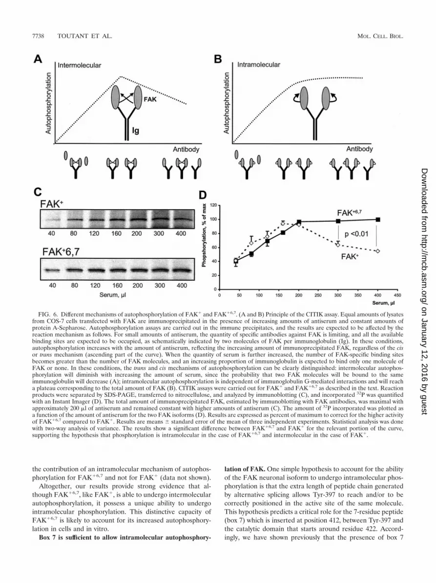
For CITIK assays, constant amounts of FAK were subjectedto immunoprecipitation with increasing amounts of antiserum,with a fixed amount of protein A-Sepharose in a large excess inbinding capacity, and an in vitro autophosphorylation reactionwas carried out in the immune precipitate in the presence of[�-32P]ATP. The principle of the assays is schematically illus-trated in Fig. 6A and B. As the amount of serum is increased,two phases can be distinguished. With small amounts of serum,the available sites are saturated by the antigen and theamounts of precipitated FAK increase linearly with theamount of antibody. Above a certain threshold of serum, allthe available antigen is precipitated, and further increasing theantibody level results in an excess of available binding sites forFAK, of which an increasing proportion are unoccupied. Studyof the rate of phosphorylation in this second phase allows us todistinguish trans and cis mechanisms of autophosphorylation:
intermolecular reactions, whose rate depends on the kinaseconcentration, will diminish when the amount of serum is in-creased above this threshold, since the probability that twomolecules of FAK are close enough to transphosphorylate(i.e., presumably on the same immunoglobulin molecule) willdecrease (Fig. 6A); in contrast, intramolecular reactions,whose rate is independent of the kinase concentration, willreach a plateau corresponding to the maximal amount of im-munoprecipitated kinase (Fig. 6B). (In this model, the phos-phorylation rate is proportional to the number of antibodies towhich two molecules of FAK are bound. Assuming high affin-ities of antibodies for FAK, when the total number of antibod-ies [NAB] is greater than the number of antigens [NAG], i.e.,FAK, the probability that the two sites of one antibody mole-cule are occupied varies as (NAG/NAB)2. Thus, when NAB �NAG, the phosphorylation rate varies as a function of 1/NAB
2
when phosphorylation is intermolecular, whereas it is constantwhen it is intramolecular.)
We used CITIK assays to compare the mechanism of auto-phosphorylation of FAK� and FAK�6,7. Immunoprecipitationwas carried out with fixed amounts of COS-7 lysates and pro-tein A-Sepharose and increasing amounts of an antiserum thatspecifically recognized FAK� and FAK�6,7 but did not reactwith COS-7 cell endogenous FAK° (14). For each immunopre-cipitation condition, a phosphorylation reaction was carriedout in the presence of [�-32P]ATP, and the phosphorylatedproteins were separated by SDS-PAGE and transferred tonitrocellulose, allowing both immunoblotting to determine thetotal amount of FAK precipitated (Fig. 6C) and quantitativemeasurement of incorporated 32P with an Instant Imager (Fig.6D).
As expected, the quantity of immunoprecipitated FAK in-creased progressively with the amount of antiserum andreached a plateau corresponding to complete immunoprecipi-tation at about 200 �l of antiserum (Fig. 6C). The differencesbetween the phosphorylation of FAK� and FAK�6,7 werereadily apparent and statistically significant above this thresh-old of 200 �l (Fig. 6D): in the case of FAK�, the amount of 32Pincorporated decreased progressively, as expected for an inter-molecular autophosphorylation, whereas in the case ofFAK�6,7, incorporation of 32P reached a plateau and did notdecrease with increasing amounts of antibodies, showing thatautophosphorylation was an intramolecular reaction.
Several control experiments were carried out to validate thisconclusion. First, in both cases, the results obtained by immu-noblotting for phospho-Tyr-397 were similar to those obtainedby measurement of 32P incorporation, although they could notbe quantified as accurately (data not shown). Second, we ver-ified that the apparent intramolecular phosphorylation ofFAK�6,7 was not due to its ability to form stable multiproteincomplexes containing several molecules of FAK�6,7. To do so,experiments similar to those described for FAK� in Fig. 1Cwere carried out with FAK�6,7, with identical results (data notshown). Finally, we used a completely different approach, inwhich constant amounts of either FAK� or FAK�6,7 wereimmunoprecipitated with fixed amounts of antiserum but inthe presence of increasing amounts of the FAK� Y397F/K454R double mutant, resulting in dilution of the active kinasein the immune precipitate. These experiments also supported
VOL. 22, 2002 MECHANISMS OF FAK AUTOPHOSPHORYLATION 7737
on January 12, 2016 by guesthttp://m
cb.asm.org/
Dow
nloaded from
the contribution of an intramolecular mechanism of autophos-phorylation for FAK�6,7 and not for FAK� (data not shown).
Altogether, our results provide strong evidence that al-though FAK�6,7, like FAK�, is able to undergo intermolecularautophosphorylation, it possess a unique ability to undergointramolecular phosphorylation. This distinctive capacity ofFAK�6,7 is likely to account for its increased autophosphory-lation in cells and in vitro.
Box 7 is sufficient to allow intramolecular autophosphory-
lation of FAK. One simple hypothesis to account for the abilityof the FAK neuronal isoform to undergo intramolecular phos-phorylation is that the extra length of peptide chain generatedby alternative splicing allows Tyr-397 to reach and/or to becorrectly positioned in the active site of the same molecule.This hypothesis predicts a critical role for the 7-residue peptide(box 7) which is inserted at position 412, between Tyr-397 andthe catalytic domain that starts around residue 422. Accord-ingly, we have shown previously that the presence of box 7
FIG. 6. Different mechanisms of autophosphorylation of FAK� and FAK�6,7. (A and B) Principle of the CITIK assay. Equal amounts of lysatesfrom COS-7 cells transfected with FAK are immunoprecipitated in the presence of increasing amounts of antiserum and constant amounts ofprotein A-Sepharose. Autophosphorylation assays are carried out in the immune precipitates, and the results are expected to be affected by thereaction mechanism as follows. For small amounts of antiserum, the quantity of specific antibodies against FAK is limiting, and all the availablebinding sites are expected to be occupied, as schematically indicated by two molecules of FAK per immunoglobulin (Ig). In these conditions,autophosphorylation increases with the amount of antiserum, reflecting the increasing amount of immunoprecipitated FAK, regardless of the cisor trans mechanism (ascending part of the curve). When the quantity of serum is further increased, the number of FAK-specific binding sitesbecomes greater than the number of FAK molecules, and an increasing proportion of immunoglobulin is expected to bind only one molecule ofFAK or none. In these conditions, the trans and cis mechanisms of autophosphorylation can be clearly distinguished: intermolecular autophos-phorylation will diminish with increasing the amount of serum, since the probability that two FAK molecules will be bound to the sameimmunoglobulin will decrease (A); intramolecular autophosphorylation is independent of immunoglobulin G-mediated interactions and will reacha plateau corresponding to the total amount of FAK (B). CITIK assays were carried out for FAK� and FAK�6,7 as described in the text. Reactionproducts were separated by SDS-PAGE, transferred to nitrocellulose, and analyzed by immunoblotting (C), and incorporated 32P was quantifiedwith an Instant Imager (D). The total amount of immunoprecipitated FAK, estimated by immunoblotting with FAK antibodies, was maximal withapproximately 200 �l of antiserum and remained constant with higher amounts of antiserum (C). The amount of 32P incorporated was plotted asa function of the amount of antiserum for the two FAK isoforms (D). Results are expressed as percent of maximum to correct for the higher activityof FAK�6,7 compared to FAK�. Results are means � standard error of the mean of three independent experiments. Statistical analysis was donewith two-way analysis of variance. The results show a significant difference between FAK�6,7 and FAK� for the relevant portion of the curve,supporting the hypothesis that phosphorylation is intramolecular in the case of FAK�6,7 and intermolecular in the case of FAK�.
7738 TOUTANT ET AL. MOL. CELL. BIOL.
on January 12, 2016 by guesthttp://m
cb.asm.org/
Dow
nloaded from
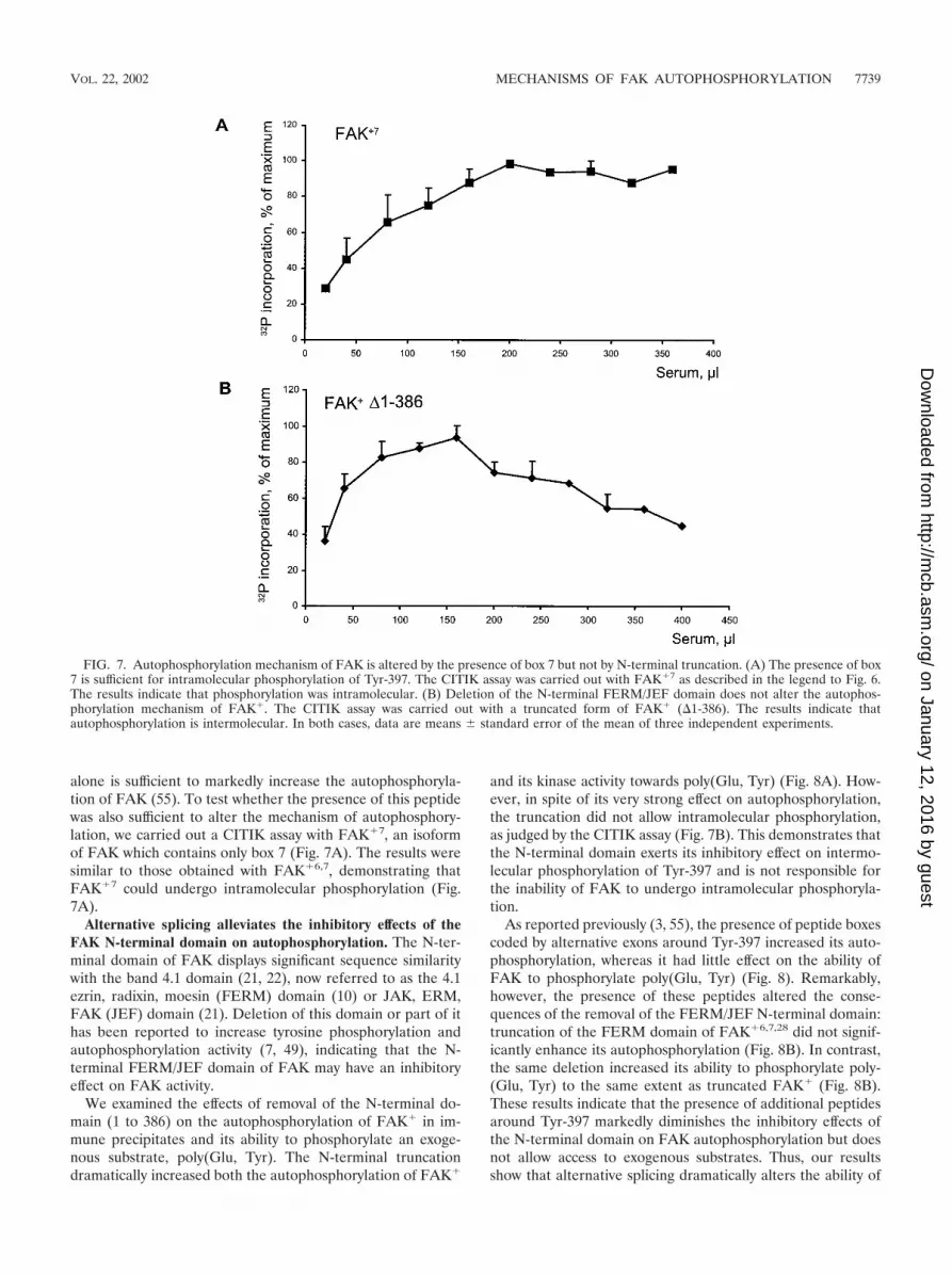
alone is sufficient to markedly increase the autophosphoryla-tion of FAK (55). To test whether the presence of this peptidewas also sufficient to alter the mechanism of autophosphory-lation, we carried out a CITIK assay with FAK�7, an isoformof FAK which contains only box 7 (Fig. 7A). The results weresimilar to those obtained with FAK�6,7, demonstrating thatFAK�7 could undergo intramolecular phosphorylation (Fig.7A).
Alternative splicing alleviates the inhibitory effects of theFAK N-terminal domain on autophosphorylation. The N-ter-minal domain of FAK displays significant sequence similaritywith the band 4.1 domain (21, 22), now referred to as the 4.1ezrin, radixin, moesin (FERM) domain (10) or JAK, ERM,FAK (JEF) domain (21). Deletion of this domain or part of ithas been reported to increase tyrosine phosphorylation andautophosphorylation activity (7, 49), indicating that the N-terminal FERM/JEF domain of FAK may have an inhibitoryeffect on FAK activity.
We examined the effects of removal of the N-terminal do-main (1 to 386) on the autophosphorylation of FAK� in im-mune precipitates and its ability to phosphorylate an exoge-nous substrate, poly(Glu, Tyr). The N-terminal truncationdramatically increased both the autophosphorylation of FAK�
and its kinase activity towards poly(Glu, Tyr) (Fig. 8A). How-ever, in spite of its very strong effect on autophosphorylation,the truncation did not allow intramolecular phosphorylation,as judged by the CITIK assay (Fig. 7B). This demonstrates thatthe N-terminal domain exerts its inhibitory effect on intermo-lecular phosphorylation of Tyr-397 and is not responsible forthe inability of FAK to undergo intramolecular phosphoryla-tion.
As reported previously (3, 55), the presence of peptide boxescoded by alternative exons around Tyr-397 increased its auto-phosphorylation, whereas it had little effect on the ability ofFAK to phosphorylate poly(Glu, Tyr) (Fig. 8). Remarkably,however, the presence of these peptides altered the conse-quences of the removal of the FERM/JEF N-terminal domain:truncation of the FERM domain of FAK�6,7,28 did not signif-icantly enhance its autophosphorylation (Fig. 8B). In contrast,the same deletion increased its ability to phosphorylate poly-(Glu, Tyr) to the same extent as truncated FAK� (Fig. 8B).These results indicate that the presence of additional peptidesaround Tyr-397 markedly diminishes the inhibitory effects ofthe N-terminal domain on FAK autophosphorylation but doesnot allow access to exogenous substrates. Thus, our resultsshow that alternative splicing dramatically alters the ability of
FIG. 7. Autophosphorylation mechanism of FAK is altered by the presence of box 7 but not by N-terminal truncation. (A) The presence of box7 is sufficient for intramolecular phosphorylation of Tyr-397. The CITIK assay was carried out with FAK�7 as described in the legend to Fig. 6.The results indicate that phosphorylation was intramolecular. (B) Deletion of the N-terminal FERM/JEF domain does not alter the autophos-phorylation mechanism of FAK�. The CITIK assay was carried out with a truncated form of FAK� (�1-386). The results indicate thatautophosphorylation is intermolecular. In both cases, data are means � standard error of the mean of three independent experiments.
VOL. 22, 2002 MECHANISMS OF FAK AUTOPHOSPHORYLATION 7739
on January 12, 2016 by guesthttp://m
cb.asm.org/
Dow
nloaded from

FAK to undergo autophosphorylation by two distinct mecha-nisms: by allowing intramolecular autophosphorylation and byalleviating the inhibitory effect of the N-terminal FERM/JEFdomain.
DISCUSSION
Autophosphorylation of FAK on Tyr-397 plays a critical rolein its functional activation in response to various stimuli (seereferences 20 and 50 for reviews). In contrast to many othertyrosine kinases (32), autophosphorylation of FAK takes placenot in the A-loop, but on a linker region between the N-terminal FERM/JEF domain and the central kinase domain.
The present study clarifies the mechanism of FAK autophos-phorylation and demonstrates that this mechanism differsmarkedly between alternatively spliced isoforms. Our resultswith Tyr-3973Phe and kinase-dead mutants show that FAK�
undergoes intermolecular phosphorylation in intact cells. SinceFAK� and FAK° (the isoform without additional alternativelyspliced exons) have similar autophosphorylation properties(3), the results reported here with FAK� can be generalized tothe FAK° isoform. The observation that increasing amounts ofFAK� double mutant (Y397F, K454R) decreased the phos-phorylation on Tyr-397 of cotransfected FAK� strongly sug-
gests that intermolecular phosphorylation is the major mech-anism of autophosphorylation in intact cells. However, we wereunable to isolate protein complexes containing several mole-cules of FAK, indicating that, in spite of its ability to bindvarious proteins of focal adhesions and signaling molecules,FAK does not form stable complexes with itself.
Interestingly, it has been reported that PYK2, a tyrosinekinase related to FAK (45% overall sequence identity), wasable to transphosphorylate FAK on Tyr-397 in transfectedcells, although the converse reaction did not occur (36). In thatstudy, FAK and PYK2 did not coimmunoprecipitate (36). Thefact that FAK molecules do not form stable homo-oligomersmay have functional importance, allowing separation of thepolypeptide chains once they are autophosphorylated and theiraction on other FAK molecules, a condition that seems to bea prerequisite for amplification by Src family kinases (see be-low).
Since autophosphorylation of FAK appears to be intermo-lecular in intact cells, we examined whether bringing two mol-ecules of FAK into close proximity was sufficient to induce itsautophosphorylation. To do so, we used fusion proteins ofFAK� with gyrase B, which dimerizes in the presence ofcoumermycin (17). This system has been used successfully toinduce the dimerization of protein kinases (17, 35, 39) andother types of proteins (34, 43) in intact cells. Treatment oftransfected cells with coumermycin was sufficient to increasetyrosine phosphorylation of Gyr-FAK�. Tyrosine phosphory-lation occurred on Tyr-397, as demonstrated by immunoblot-ting with a specific antibody. Importantly, PP2, a Src familykinase inhibitor (25), had no effect on phosphorylation of Tyr-397 in response to coumermycin, demonstrating that dimeriza-tion-induced autophosphorylation of FAK is independent ofthese kinases.
Coumermycin-induced dimerization of Gyr-FAK� did nottrigger the phosphorylation of Tyr-577 or Tyr-925, presumablybecause Src family kinases could not access the autophospho-rylated Tyr-397 in stable Gyr-FAK� dimers. This observationunderscores the importance of the reversibility of the interac-tions between FAK molecules, discussed above. Dimerization-induced autophosphorylation of Gyr-FAK� was independentof cell adhesion, showing that dimerization of FAK induces itsautophosphorylation independently of any other stimulus.However, we found that deletion of the C-terminal region ofGyr-FAK� (residues 841 to 1054) dramatically decreased thelevel of coumermycin-induced autophosphorylation. This un-expected finding was not accounted for by an alteration ofintrinsic kinase activity. It also seems unlikely that the phos-phorylation deficit was due to an increased sensitivity to ty-rosine phosphatases, since C-terminal deletions have been re-ported to provide resistance to dephosphorylation (51).
The C-terminal region of FAK contains binding sites fortalin (9), paxillin (30, 54), Graf (31), and Hic-5 (18). It alsoencompasses the FAT sequence (29), which mediates targetingof FAK to focal adhesions, at least in part through its interac-tion with the focal adhesion proteins talin and paxillin (12, 30).One possible explanation for the effects of the C-terminaldeletion on coumermycin’s ability to induce Gyr-FAK� auto-phosphorylation is the loss of such protein-protein interac-tions. These interactions could facilitate the effects of coumer-mycin by locally enriching the concentration of Gyr-FAK�, for
FIG. 8. Effects of deletion of the N-terminal FERM/JEF domainon autophosphorylation of FAK isoforms and their ability to phos-phorylate an exogenous substrate. Wild-type (wt) FAK�, its N-termi-nally truncated form (FAK��1-386) (left panel), and wild-type (wt)FAK�6,7,28 and its truncated form (FAK�6,7,28�1-386) (right panel)were expressed in COS-7 cells, dephosphorylated in vitro, and immu-noprecipitated, and their autophosphorylation was assayed in the pres-ence of [�-32P]ATP (Autophos). In the same conditions, the ability ofeach immunoprecipitated isoform of FAK to phosphorylate an exog-enous substrate, poly(Glu, Tyr), was examined. The amount of radio-activity incorporated into FAK or poly(Glu, Tyr) was measured withan Instant Imager after SDS-PAGE, and the counts per minute werenormalized to the total amount of immunoprecipitated FAK, mea-sured by immunoblotting. The autophosphorylation of FAK�6,7,28 washigher than that of FAK� (E; P � 0.01, two-tailed Student’s t test).N-terminal truncation increased the ability of FAK� to autophospho-rylate (�; P � 0.005, two-tailed Student’s t test), whereas it did notsignificantly alter the autophosphorylation of FAK�6,7,28 (left panel).In contrast, N-terminal truncation dramatically increased the ability ofboth isoforms to phosphorylate poly(Glu, Tyr) (�, P � 0.05, two-tailedStudent’s t test). Values are means � standard error of the mean forfour (autophosphorylation) or three [phosphorylation of poly(Glu,Tyr)] experiments.
7740 TOUTANT ET AL. MOL. CELL. BIOL.
on January 12, 2016 by guesthttp://m
cb.asm.org/
Dow
nloaded from

example, in the vicinity of the membrane and/or cytoskeletalelements. Additional experiments will be required to deter-mine whether any of these proteins exerts a more specific effecton FAK activation in intact cells.
When we examined the role of the C-terminal region in thephosphorylation of Tyr-397 of wild-type FAK in intact cells, wedisclosed a dramatic difference between splicing isoforms, al-though this deletion had no effect on the intrinsic kinase ac-tivity of FAK in immune precipitate kinase assays. In trans-fected COS-7 cells, the C-terminal deletion prevented Tyr-397phosphorylation in FAK�, whereas it had no effect onFAK�6,7,28. The lack of effect of the C-terminal deletion onisoforms with boxes 6 and 7 strongly suggested that the pres-ence of additional peptide sequences surrounding Tyr-397 al-tered the mechanism of autophosphorylation. In addition, al-though FAK�6,7 was able to undergo intermolecularphosphorylation in intact cells, the cotransfection of Y397Fand kinase-dead mutants of FAK�6,7 did not restore the highlevel of autophosphorylation of wild-type FAK�6,7. This ob-servation also pointed at a possible difference in the autophos-phorylation mechanism.
To address the intra- or intermolecular mechanism of auto-phosphorylation of FAK isoforms, we used a modified versionof the immune precipitate kinase assays, referred to as theCITIK assay. This procedure has the advantage that it mimicsto some extent the conditions in which FAK undergoes auto-phosphorylation in intact cells, since binding to antibodies islikely to mimic binding to cytoskeletal and/or membrane pro-teins. Moreover, CITIK assays allow us to address the mech-anism of the reaction in the conditions of the widely usedimmune precipitate kinase assays. The results indicated thatautophosphorylation of FAK� was predominantly intermolec-ular, whereas autophosphorylation of FAK�6,7 was intramo-lecular. It should be emphasized that FAK�6,7 is able to un-dergo intermolecular phosphorylation on Tyr-397, as shown bytransphosphorylation of Y397F and kinase-dead mutants intransfected cells (see above), as well as in vitro (data notshown). However, this isoform has the additional ability toundergo intramolecular phosphorylation, a process that seemspredominant. The ability to undergo intramolecular phosphor-ylation accounts for the lack of sensitivity of FAK�6,7 to C-terminal truncation, which was shown to dramatically reducedimerization-induced autophosphorylation.
Our results provide important clues about the possiblemechanisms of FAK autophosphorylation. A simple hypothe-sis explaining the ability of FAK�6,7 to undergo cis autophos-phorylation is that the presence of additional residues on ei-ther sides of Tyr-397 provides additional length and flexibilityto the peptide chain, allowing it access to the catalytic site. Thishypothesis would predict that the peptide (box 7) located be-tween Tyr-397 and the catalytic site plays a major role inallowing intramolecular autophosphorylation. Interestingly,the presence of box 7 alone has a greater effect on autophos-phorylation than box 6 alone (located between Tyr-397 and theN-terminal side of FAK) (55). The CITIK assay results show-ing that the presence of box 7 is sufficient to allow intramolec-ular phosphorylation support this hypothesis.
Other mechanisms are also likely to be involved in the con-trol of FAK autophosphorylation. The present study, confirm-ing previous suggestions (7, 49), demonstrates that the N-
terminal FERM/JEF domain of FAK inhibits its ability toautophosphorylate as well as its ability to phosphorylate anexogenous substrate. Since deletion of the FERM/JEF domainof FAK� did not alter the mechanism of autophosphorylationitself, which was still intermolecular, it can be concluded thatthe N-terminal domain hinders intermolecular autophosphor-ylation. The N-terminal domain may prevent the correct posi-tioning of the two molecules and/or the access of Tyr-397 fromone peptide chain to the active site of the other peptide chain.The presence of alternatively spliced peptides flanking Tyr-397alleviated the inhibitory effects of the FERM/JEF domain onautophosphorylation, whereas it did not prevent the inhibitionexerted by this domain on the phosphorylation of poly(Glu,Tyr). A possible explanation of these effects (diagrammed inFig. 9) is that alternative splicing allows the positioning ofTyr-397 in the active site of the same molecule, in a positionthat is insensitive to the effects of the N-terminal domain.
Previous observations and the results of the present studysupport a model for FAK activation in which recruitment ofthis kinase to focal adhesions and its interaction with otherproteins at these sites induce its transautophosphorylationwhen integrins are engaged. This clustering step, dependent oncell adhesion, is likely to be the one mimicked by coumermy-cin-induced dimerization of Gyr-FAK� molecules. Thus,transphosphorylation of FAK may result from a mere increasein its local concentration or involve more directed interactionswith specific proteins. It does not implicate stable dimers oroligomers of FAK, but rather transient interactions betweenmultiple FAK molecules.
This model accounts well for the role of Src family kinases inFAK activation (40). Phosphorylation of FAK by Src is greatlyenhanced by its binding to phospho-Tyr-397 (47). In turn,phosphorylation of A-loop residues Tyr-576 and Tyr-577 by Srcincreases FAK kinase activity (4) and may thus enhance fur-ther phosphorylation of Tyr-397 (40). Our data support amodel in which autophosphorylation of Tyr-397 would be thestarting point resulting from a transient intermolecular inter-action between FAK molecules. Src could then be involved ina positive feed-back loop, as suggested previously (40), inwhich autophosphorylated FAK recruits Src, which phosphor-ylates Tyr-576 and -577 in the A-loop, increasing FAK catalyticactivity and its ability to transphosphorylate additional mole-cules of FAK. It is noteworthy that such a model implies thatinteractions between FAK molecules are dynamic and revers-ible, in keeping with the lack of stable complexes found in thepresent study. Interestingly, the Src inhibitor PP2 had no effecton coumermycin-induced phosphorylation of Tyr-397 in Gyr-FAK�. This is consistent with the fact that coumermycin-bound dimers of Gyr-FAK� are stable and do not allow theamplification mechanism mediated by Src.
In contrast to FAK° or FAK�, the major neuronal isoformFAK�6,7 is able to undergo intramolecular phosphorylation ofTyr-397, a property that accounts to a large extent for itsincreased autophosphorylation. The specific features ofFAK�6,7 suggest that its regulation in neurons involves mech-anisms different from integrin-mediated clustering, which mayinclude relief of inhibitory influences on the cis-autophospho-rylation reaction and/or control of protein tyrosine phospha-tases. Thus, our results illustrate how alternative RNA splicing,a common process particularly frequent in neurons (23), has
VOL. 22, 2002 MECHANISMS OF FAK AUTOPHOSPHORYLATION 7741
on January 12, 2016 by guesthttp://m
cb.asm.org/
Dow
nloaded from

dramatic consequences on the properties of a ubiquitous andimportant protein kinase: the presence of short peptides oneither side of the autophosphorylated tyrosine enhances therate of autophosphorylation by switching it from an intermo-lecular to an intramolecular reaction.
ACKNOWLEDGMENTS
We thank Pascal Ezan and Sylvie Clain for valuable assistance andHerve Enslen for critical reading of the manuscript. Michael Ferrarand Leon Perlmutter are gratefully acknowledged for providing thegyrase B construct, and Janine Ragab is gratefully acknowledged forproviding RPTP-�.
A.C. was the recipient of a fellowship from the French Ministry ofForeign Affairs and the French Embassy in Uruguay. The work wassupported in part by grants from the European Community (Bio4CT98 0526), the Human Frontier Science Program, the Fondationpour la Recherche Medicale, and the Fondation Schlumberger pourl’Enseignement et la Recherche (Dotation Annette Gruner Schlum-berger) to J.A.G.
The first two authors contributed equally to this work.
REFERENCES
1. Baron, V., V. Calleja, P. Ferrari, F. Alengrin, and E. Van Obberghen. 1998.p125Fak focal adhesion kinase is a substrate for the insulin and insulin-likegrowth factor-I tyrosine kinase receptors. J. Biol. Chem. 273:7162–7168.
2. Burgaya, F., and J. A. Girault. 1996. Cloning of focal adhesion kinase fromrat stiatum reveals multiple transcripts. Mol. Brain Res. 37:63–73.
3. Burgaya, F., M. Toutant, J. M. Studler, A. Costa, M. Le Bert, M. Gelman,and J. A. Girault. 1997. Alternatively spliced focal adhesion kinase in ratbrain with increased autophosphorylation activity. J. Biol. Chem. 272:28720–28725.
4. Calalb, M. B., T. R. Polte, and S. K. Hanks. 1995. Tyrosine phosphorylationof focal adhesion kinase at sites in the catalytic domain regulates kinaseactivity: A role for Src family kinases. Mol. Cell. Biol. 15:954–963.
5. Calalb, M. B., X. E. Zhang, T. R. Polte, and S. K. Hanks. 1996. Focaladhesion kinase tyrosine-861 is a major site of phosphorylation by Src.Biochem. Biophys. Res. Commun. 228:662–668.
6. Cary, L. A., R. A. Klinghoffer, C. Sachsenmaier, and J. A. Cooper. 2002. SRCcatalytic but not scaffolding function is needed for integrin-regulated ty-rosine phosphorylation, cell migration, and cell spreading. Mol. Cell. Biol.22:2427–2440.
7. Chan, P.-Y., S. B. Kanner, G. Whitney, and A. Aruffo. 1994. A transmem-brane-anchored chimeric focal adhesion kinase is constitutively activatedand phosphorylated at tyrosine residues identical to pp125FAK. J. Biol.Chem. 269:20567–20574.
8. Chen, H. C., P. A. Appeddu, H. Isoda, and J. L. Guan. 1996. Phosphorylation
FIG. 9. Schematic representation of FAK autophosphorylation mechanisms and differences between isoforms, based on the results of thepresent study. (A) FAK� (as well as FAK°, which has the same low autophosphorylation) is in a poorly active state in basal conditions, presumablydue to inhibition by the N-terminal domain. The FERM/JEF domain is indicated here as a trilobate structure, on the basis of the recentlydetermined structure of several ERM domains (24, 41). The C-terminal region, including the FAT domain, is necessary for phosphorylation inintact cells. Cell attachment or artificial dimerization (as shown in the present study) promotes intermolecular phosphorylation of Tyr-397.(B) Deletion of the FERM/JEF domain of FAK� increases intermolecular autophosphorylation in vitro by removing the inhibitory effect of theN-terminal FERM/JEF and facilitating the access of substrates to the active site [arrow, poly(Glu, Tyr)]. (C) The neuronal isoform, FAK�6,7 (aswell as FAK�6,7,28, which has the same high autophosphorylation), has an increased autophosphorylation because of its ability to undergointramolecular phosphorylation. The presence of box 7 is sufficient to allow intramolecular phosphorylation, presumably by allowing access ofTyr-397 to the active site located on the same peptide chain. However, in FAK�6,7, the N-terminal FERM/JEF domain still hampers the accessof external substrates. (D) The deletion of the FERM/JEF domain has little effect on FAK�6,7 autophosphorylation but increases the accessibilityof substrates to the active site [arrow, poly(Glu, Tyr)]. Whereas the C-terminal FAT domain is essential for the attachment- or artificialdimerization-induced autophosphorylation of FAK�, this does not appear to be the case for FAK�6,7. This is symbolized by the ordered openposition of the C terminus in A and B and its loosely closed position in C and D.
7742 TOUTANT ET AL. MOL. CELL. BIOL.
on January 12, 2016 by guesthttp://m
cb.asm.org/
Dow
nloaded from

of tyrosine 397 in focal adhesion kinase is required for binding phosphati-dylinositol 3-kinase. J. Biol. Chem. 271:26329–26334.
9. Chen, H.-C., P. A. Appeddu, J. T. Parsons, J. D. Hildebrand, M. D. Schaller,and J.-L. Guan. 1995. Interaction of focal adhesion kinase with cytoskeletalprotein talin. J. Biol. Chem. 270:16995–16999.
10. Chishti, A. H., A. C. Kim, S. M. Marfatia, M. Lutchman, M. Hanspal, H.Jindal, S. C. Liu, P. S. Low, G. A. Rouleau, et al. 1998. The FERM domain:a unique module involved in the linkage of cytoplasmic proteins to themembrane. Trends Biochem. Sci. 23:281–282.
11. Cobb, B. S., M. D. Schaller, T.-H. Leu, and J. T. Parsons. 1994. Stableassociation of pp60src and pp59fyn with the focal adhesion-associated proteintyrosine kinase, pp125FAK. Mol. Cell. Biol. 14:147–155.
12. Cooley, M. A., J. M. Broome, C. Ohngemach, L. H. Romer, and M. D.Schaller. 2000. Paxillin binding is not the sole determinant of focal adhesionlocalization or dominant-negative activity of focal adhesion kinase/focal ad-hesion kinase-related nonkinase. Mol. Biol. Cell 11:3247–3263.
13. Derkinderen, P., J. Siciliano, M. Toutant, and J. A. Girault. 1998. Differ-ential regulation of FAK� and PYK2/Cak�, two related tyrosine kinases, inrat hippocampal slices: effects of LPA, carbachol, depolarization and hyper-osmolarity. Eur. J. Neurosci. 10:1667–1675.
14. Derkinderen, P., M. Toutant, F. Burgaya, M. Le Bert, J. C. Siciliano, V. DeFranciscis, M. Gelman, and J. A. Girault. 1996. Regulation of a neuronalform of focal adhesion kinase by anandamide. Science 273:1719–1722.
15. Derkinderen, P., M. Toutant, G. Kadare, C. Ledent, M. Parmentier, andJ. A. Girault. 2001. Dual role of Fyn in the regulation of FAK�6,7 bycannabinoids in hippocampus. J. Biol. Chem. 276:38289–38296.
16. Eide, B. L., C. W. Turck, and J. A. Escobedo. 1995. Identification of Tyr-397as the primary site of tyrosine phosphorylation and pp60src association in thefocal adhesion kinase, pp125FAK. Mol. Cell. Biol. 15:2819–2827.
17. Farrar, M. A., J. Alberola-Ila, and R. M. Perlmutter. 1996. Activation of theRaf-1 kinase cascade by coumermycin-induced dimerization. Nature 383:178–181.
18. Fujita, H., K. Kamiguchi, D. Cho, M. Shibanuma, C. Morimoto, and K.Tachibana. 1998. Interaction of Hic-5, a senescence-related protein, withfocal adhesion kinase. J. Biol. Chem. 273:26516–26521.
19. Gabarra-Niecko, V., P. J. Keely, and M. D. Schaller. 2002. Characterizationof an activated mutant of focal adhesion kinase: superFAK. Biochem. J.365:591–603.
20. Girault, J. A., A. Costa, P. Derkinderen, J. M. Studler, and M. Toutant. 1999.FAK and PYK2/CAK in the nervous system, a link between neuronal activ-ity, plasticity and survival? Trends Neurosci. 22:257–263.
21. Girault, J. A., G. Labesse, J.-P. Mornon, and I. Callebaut. 1998. The FAKsand JAKs play in the 4.1 band: a superfamily of band 4.1 domains importantfor cell structure and signal transduction. Mol. Medicine. 4:751–769.
22. Girault, J. A., G. Labesse, J.-P. Mornon, and I. Callebaut. 1999. The N-termini of FAK and JAKs contain divergent band 4.1 domains. TrendsBiochem. Sci. 24:54–57.
23. Grabowski, P. J., and D. L. Black. 2001. Alternative RNA splicing in thenervous system. Prog. Neurobiol. 65:289–308.
24. Hamada, K., T. Shimizu, T. Matsui, S. Tsukita, and T. Hakoshima. 2000.Structural basis of the membrane-targeting and unmasking mechanisms ofthe radixin FERM domain. EMBO J. 19:4449–4462.
25. Hanke, J. H., J. P. Gardner, R. L. Dow, P. S. Changelian, W. H. Brissette,E. J. Weringer, K. Pollok, and P. A. Connelly. 1996. Discovery of a novel,potent, and Src family-selective tyrosine kinase inhibitor — study of Lck- andFynT-dependent T cell activation. J. Biol. Chem. 271:695–701.
26. Hanks, S. K., M. B. Calalb, M. C. Harper, and S. K. Patel. 1992. Focaladhesion protein-tyrosine kinase phosphorylated in response to cell attach-ment to fibronectin. Proc. Natl. Acad. Sci. USA 89:8487–8491.
27. Harte, M. T., J. D. Hildebrand, M. R. Burnham, A. H. Bouton, and J. T.Parsons. 1996. p130Cas, a substrate associated with v-Src and v-Crk, localizestofocal adhesions and binds to focal adhesion kinase. J. Biol. Chem. 271:13649–13655.
28. Hens, M. D., and D. W. DeSimone. 1995. Molecular analysis and develop-mental expression of the focal adhesion kinase pp125FAK in Xenopus laevis.Dev. Biol. 170:274–288.
29. Hildebrand, J. D., M. D. Schaller, and J. T. Parsons. 1993. Identification ofsequences required for the efficient localization of the focal adhesion kinase,pp125FAK, to cellular focal adhesions. J. Cell Biol. 123:993–1005.
30. Hildebrand, J. D., M. D. Schaller, and J. T. Parsons. 1995. Paxillin, atyrosine phosphorylated focal adhesion-associated protein binds to the car-boxyl terminal domain of focal adhesion kinase. Mol. Biol. Cell 6:637–647.
31. Hildebrand, J. D., J. M. Taylor, and J. T. Parsons. 1996. An SH3 domain-containing GTPase-activating protein for Rho and Cdc42 associates withfocal adhesion kinase. Mol. Cell. Biol. 16:3169–3178.
32. Hubbard, S. R., M. Mohammadi, and J. Schlessinger. 1998. Autoregulatorymechanisms in protein-tyrosine kinases. J. Biol. Chem. 273:11987–11990.
33. Ilic, D., Y. Furuta, S. Kanazawa, N. Takeda, K. Sobue, N. Nakatsuji, S.Nomura, J. Fujimoto, M. Okada, T. Yamamoto, and S. Aizawa. 1995. Re-duced cell motility and enhanced focal adhesion contact formation in cellsfrom FAK-deficient mice. Nature 377:539–544.
34. Inouye, K., S. Mizutani, H. Koide, and Y. Kaziro. 2000. Formation of the Rasdimer is essential for Raf-1 activation. J. Biol. Chem. 275:3737–3740.
35. Knight, E. L., A. J. Warner, A. Maxwell, and S. A. Prigent. 2000. ChimericVEGFRs are activated by a small-molecule dimerizer and mediate down-stream signalling cascades in endothelial cells. Oncogene 19:5398–5405.
36. Li, X., R. C. Dy, W. G. Cance, L. M. Graves, and H. S. Earp. 1999. Inter-actions between two cytoskeleton-associated tyrosine kinases: calcium-de-pendent tyrosine kinase and focal adhesion tyrosine kinase. J. Biol. Chem.274:8917–8924.
37. Menegon, A., F. Burgaya, P. Baudot, D. D. Dunlap, J. A. Girault, and F.Valtorta. 1999. FAK� and PYK2/CAK�, two related tyrosine kinases highlyexpressed in the central nervous system: Similarities and differences in theexpression pattern. Eur. J. Neurosci. 11:3777–3788.
38. Menegoz, M., P. Gaspar, M. Le Bert, T. Galvez, F. Burgaya, C. Palfrey, P.Ezan, F. Amos, and J. A. Girault. 1997. Paranodin, a glycoprotein of neu-ronal paranodal membranes. Neuron 19:319–331.
39. Mohi, M. G., K. Arai, and S. Watanabe. 1998. Activation and functionalanalysis of Janus kinase 2 in BA/F3 cells with the coumermycin/gyrase Bsystem. Mol. Biol. Cell 9:3299–3308.
40. Owen, J. D., P. J. Ruest, D. W. Fry, and S. K. Hanks. 1999. Induced focaladhesion kinase (FAK) expression in FAK-null cells enhance cell spreadingand migration requiring both auto- and activation loop phosphorylation sitesand inhibits adhesion-dependent tyrosine phosphorylation of Pyk2. Mol.Cell. Biol. 19:4806–4818.
41. Pearson, M. A., D. Reczek, A. Bretscher, and P. A. Karplus. 2000. Structureof the ERM protein moesin reveals the FERM domain fold masked by anextended actin binding tail domain. Cell 101:259–270.
42. Perez Salazar, E., and E. Rozengurt. 2001. Src family kinases are requiredfor integrin-mediated but not for G protein-coupled receptor stimulation ofFAK autophosphorylation at Tyr-397. J. Biol. Chem. 276:17788–17795.
43. Prodromou, C., B. Panaretou, S. Chohan, G. Siligardi, R. O’Brien, J. E.Ladbury, S. M. Roe, P. W. Piper, and L. H. Pearl. 2000. The ATPase cycleof Hsp90 drives a molecular �clamp’ via transient dimerization of the N-terminal domains. EMBO J. 19:4383–4392.
44. Ruest, P. J., S. Roy, E. Shi, R. L. Mernaugh, and S. K. Hanks. 2000.Phosphospecific antibodies reveal focal adhesion kinase activation loopphosphorylation in nascent and mature focal adhesions and requirement forthe autophosphorylation site. Cell Growth Differ. 11:41–48.
45. Salazar, E. P., and E. Rozengurt. 1999. Bombesin and platelet-derivedgrowth factor induce association of endogenous focal adhesion kinase withSrc in intact Swiss 3T3 cells. J. Biol. Chem. 274:28371–28378.
46. Schaller, M. D., C. A. Borgman, B. S. Cobb, R. R. Vines, A. B. Reynolds, andJ. T. Parsons. 1992. pp125FAK, A structurally distinctive protein-tyrosinekinase associated with focal adhesions. Proc. Natl. Acad. Sci. USA 89:5192–5196.
47. Schaller, M. D., J. D. Hildebrand, J. D. Shannon, J. W. Fox, R. R. Vines, andJ. T. Parsons. 1994. Autophosphorylation of the focal adhesion kinase,pp125FAK, directs SH2-dependent binding of pp60src. Mol. Cell. Biol. 14:1680–1688.
48. Schlaepfer, D. D., S. K. Hanks, T. Hunter, and P. Van der Geer. 1994.Integrin-mediated signal transduction linked to Ras pathway by GRB2 bind-ing to focal adhesion kinase. Nature 372:786–791.
49. Schlaepfer, D. D., and T. Hunter. 1996. Evidence for in vivo phosphorylationof the Grb2 SH2-domain binding site on focal adhesion kinase by Src familyprotein-tyrosine kinases. Mol. Cell. Biol. 16:5623–5633.
50. Schlaepfer, D. D., and T. Hunter. 1998. Integrin signalling and tyrosinephosphorylation: just the FAKs? Trends Cell Biol. 8:151–157.
51. Shen, Y., and M. D. Schaller. 1999. Focal adhesion targeting: the criticaldeterminant of FAK regulation and substrate phosphorylation. Mol. Biol.Cell 10:2507–2518.
52. Siciliano, J. C., M. Toutant, P. Derkinderen, T. Sasaki, and J. A. Girault.1996. Differential regulation of proline-rich tyrosine kinase 2 cell adhesionkinase � (PYK2/CAK�) and pp125FAK by glutamate and depolarization inrat hippocampus. J. Biol. Chem. 271:28942–28946.
53. Sonoda, Y., Y. Matsumoto, M. Funakoshi, D. Yamamoto, S. K. Hanks, andT. Kasahara. 2000. Anti-apoptotic role of focal adhesion kinase (FAK).Induction of inhibitor-of-apoptosis proteins and apoptosis suppression bythe overexpression of FAK in a human leukemic cell line, HL-60. J. Biol.Chem. 275:16309–16315.
54. Tachibana, K., T. Sato, N. D’Avirro, and C. Morimoto. 1995. Direct associ-ation of pp125FAK with paxillin, the focal adhesion-targeting mechanism ofpp125FAK. J. Exp. Med. 182:1089–1099.
55. Toutant, M., J. M. Studler, F. Burgaya, A. Costa, P. Ezan, M. Gelman, andJ. A. Girault. 2000. Molecular characterization of FAK neuronal isoforms.Biochem. J. 348:119–128.
56. Weiss, A., and J. Schlessinger. 1998. Switching signals on or off by receptordimerization. Cell 94:277–280.
57. Zachary, I., and E. Rozengurt. 1992. Focal adhesion kinase (p125FAK): Apoint of convergence in the action of neuropeptides, integrins, and onco-genes. Cell 71:891–894.
VOL. 22, 2002 MECHANISMS OF FAK AUTOPHOSPHORYLATION 7743
on January 12, 2016 by guesthttp://m
cb.asm.org/
Dow
nloaded from