differential localization of d glutamate receptors in the rat cerebellum: coexpression with ampa...
TRANSCRIPT
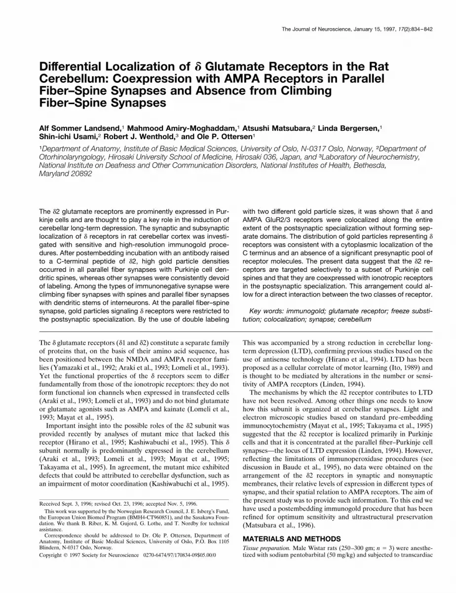
Differential Localization of d Glutamate Receptors in the RatCerebellum: Coexpression with AMPA Receptors in ParallelFiber–Spine Synapses and Absence from ClimbingFiber–Spine Synapses
Alf Sommer Landsend,1 Mahmood Amiry-Moghaddam,1 Atsushi Matsubara,2 Linda Bergersen,1Shin-ichi Usami,2 Robert J. Wenthold,3 and Ole P. Ottersen1
1Department of Anatomy, Institute of Basic Medical Sciences, University of Oslo, N-0317 Oslo, Norway, 2Department ofOtorhinolaryngology, Hirosaki University School of Medicine, Hirosaki 036, Japan, and 3Laboratory of Neurochemistry,National Institute on Deafness and Other Communication Disorders, National Institutes of Health, Bethesda,Maryland 20892
The d2 glutamate receptors are prominently expressed in Pur-kinje cells and are thought to play a key role in the induction ofcerebellar long-term depression. The synaptic and subsynapticlocalization of d receptors in rat cerebellar cortex was investi-gated with sensitive and high-resolution immunogold proce-dures. After postembedding incubation with an antibody raisedto a C-terminal peptide of d2, high gold particle densitiesoccurred in all parallel fiber synapses with Purkinje cell den-dritic spines, whereas other synapses were consistently devoidof labeling. Among the types of immunonegative synapse wereclimbing fiber synapses with spines and parallel fiber synapseswith dendritic stems of interneurons. At the parallel fiber–spinesynapse, gold particles signaling d receptors were restricted tothe postsynaptic specialization. By the use of double labeling
with two different gold particle sizes, it was shown that d andAMPA GluR2/3 receptors were colocalized along the entireextent of the postsynaptic specialization without forming sep-arate domains. The distribution of gold particles representing dreceptors was consistent with a cytoplasmic localization of theC terminus and an absence of a significant presynaptic pool ofreceptor molecules. The present data suggest that the d2 re-ceptors are targeted selectively to a subset of Purkinje cellspines and that they are coexpressed with ionotropic receptorsin the postsynaptic specialization. This arrangement could al-low for a direct interaction between the two classes of receptor.
Key words: immunogold; glutamate receptor; freeze substi-tution; colocalization; synapse; cerebellum
The d glutamate receptors (d1 and d2) constitute a separate familyof proteins that, on the basis of their amino acid sequence, hasbeen positioned between the NMDA and AMPA receptor fami-lies (Yamazaki et al., 1992; Araki et al., 1993; Lomeli et al., 1993).Yet the functional properties of the d receptors seem to differfundamentally from those of the ionotropic receptors: they do notform functional ion channels when expressed in transfected cells(Araki et al., 1993; Lomeli et al., 1993) and do not bind glutamateor glutamate agonists such as AMPA and kainate (Lomeli et al.,1993; Mayat et al., 1995).Important insight into the possible roles of the d2 subunit was
provided recently by analyses of mutant mice that lacked thisreceptor (Hirano et al., 1995; Kashiwabuchi et al., 1995). This dsubunit normally is predominantly expressed in the cerebellum(Araki et al., 1993; Lomeli et al., 1993; Mayat et al., 1995;Takayama et al., 1995). In agreement, the mutant mice exhibiteddefects that could be attributed to cerebellar dysfunction, such asan impairment of motor coordination (Kashiwabuchi et al., 1995).
This was accompanied by a strong reduction in cerebellar long-term depression (LTD), confirming previous studies based on theuse of antisense technology (Hirano et al., 1994). LTD has beenproposed as a cellular correlate of motor learning (Ito, 1989) andis thought to be mediated by alterations in the number or sensi-tivity of AMPA receptors (Linden, 1994).The mechanisms by which the d2 receptor contributes to LTD
have not been resolved. Among other things one needs to knowhow this subunit is organized at cerebellar synapses. Light andelectron microscopic studies based on standard pre-embeddingimmunocytochemistry (Mayat et al., 1995; Takayama et al., 1995)suggested that the d2 receptor is localized primarily in Purkinjecells and that it is concentrated at the parallel fiber–Purkinje cellsynapses—the locus of LTD expression (Linden, 1994). However,reflecting the limitations of immunoperoxidase procedures (seediscussion in Baude et al., 1995), no data were obtained on thearrangement of the d2 receptors in synaptic and nonsynapticmembranes, their relative levels of expression in different types ofsynapse, and their spatial relation to AMPA receptors. The aim ofthe present study was to provide such information. To this end wehave used a postembedding immunogold procedure that has beenrefined for optimum sensitivity and ultrastructural preservation(Matsubara et al., 1996).
MATERIALS AND METHODSTissue preparation. Male Wistar rats (250–300 gm; n 5 3) were anesthe-tized with sodium pentobarbital (50 mg/kg) and subjected to transcardiac
Received Sept. 3, 1996; revised Oct. 23, 1996; accepted Nov. 5, 1996.This work was supported by the Norwegian Research Council, J. E. Isberg’s Fund,
the European Union Biomed Program (BMH4-CT960851), and the Sasakawa Foun-dation. We thank B. Riber, K. M. Gujord, G. Lothe, and T. Nordby for technicalassistance.Correspondence should be addressed to Dr. Ole P. Ottersen, Department of
Anatomy, Institute of Basic Medical Sciences, University of Oslo, P.O. Box 1105Blindern, N-0317 Oslo, Norway.Copyright q 1997 Society for Neuroscience 0270-6474/97/170834-09$05.00/0
The Journal of Neuroscience, January 15, 1997, 17(2):834–842
perfusion with 2% dextran (MW 70,000) in 0.1 M sodium phosphatebuffer (PB; pH 7.4, 48C, 15 sec), followed by a mixture of glutaraldehyde(0.1%) and formaldehyde (4%; freshly depolymerized from paraformal-dehyde) in the same buffer (room temperature, 50 ml/min for 20 min).The brain was left in situ overnight (48C). Specimens from the cerebellum(lobule VI) and hippocampus (CA1) were isolated, cryoprotected ingraded concentrations of phosphate-buffered glycerol, and rapidly frozenin liquid propane (21708C) in a cryofixation unit (Reichert KF80, Vi-enna, Austria). The specimens were transferred to 0.5% uranyl acetatedissolved in anhydrous methanol (2908C) in a cryosubstitution unit(AFS; Reichert). The temperature was raised stepwise to 2458C. Thesamples were infiltrated with Lowicryl HM20 resin (Lowi, Waldkraiburg,Germany), and polymerization was induced by UV light for 48 hr. Adetailed description of the procedure has been published (Hjelle et al.,1994; Chaudhry et al., 1995) (also see van Lookeren Campagne etal., 1991).Immunoincubation. Ultrathin sections were mounted on nickel grids or
gold-coated grids and processed for immunogold cytochemistry as de-scribed by Matsubara et al. (1996). Briefly, the sections were treated witha saturated solution of NaOH in absolute ethanol (2–3 sec), rinsed, andincubated sequentially in (1) 0.1% sodium borohydride and 50 mMglycine in Tris buffer containing 0.05 M NaCl and 0.1% Triton X-100(TBNT); (2) 2% human serum albumin (HSA) in TBNT; (3) antibodiesto d or AMPA receptors (1 mg/ml and 2 mg/ml, respectively) in TBNT and2% HSA; (4) 2% HSA in TBNT; and (5) goat anti-rabbit immunoglobu-lins coupled to 5, 10, 15, or 30 nm gold particles (Amersham, ArlingtonHeights, IL) and diluted 1:20 in TBNT with 2% HSA or with 2% HSAand 5 mg/ml polyethyleneglycol. Finally, the sections were counterstainedand examined in a Philips CM10 transmission electron microscope.Double labeling was performed as described by Ottersen et al. (1992),
using formaldehyde vapor (Wang and Larsson, 1985) to prevent interfer-ence between the sequential incubations. GluR2/3 and d receptors weredistinguished by means of different gold particle sizes (10 or 15 nm forGluR2/3 and 30 or 5 nm for d).Antisera. The d receptor antibody was raised against a synthetic peptide
(QPTPTLGLNLGNDPDRGTSI) corresponding to the C terminus of therat glutamate d2 receptor subunit (Mayat et al., 1995). This antibody alsorecognizes the d1 receptor subunit but does not label other glutamatereceptors, including AMPA and NMDA receptors (Mayat et al., 1995).The AMPA receptor antibody (Ab 25; Wenthold et al., 1992) reacts withGluR2 as well as GluR3. Both antibodies were affinity-purified.Control experiments. The d receptor antibody (1 mg/ml) was substituted
with 1 mg/ml nonimmune IgG or preadsorbed with 50 mg/ml of one of thefollowing peptides: (1) peptide corresponding to the C-terminal 20 aminoacids of the d2 receptor (i.e., the peptide used for immunization), (2)peptide corresponding to amino acids 850–862 of AMPA GluR2 (used togenerate the AMPA GluR2/3 antibody), or (3) peptide corresponding toamino acids 877–889 or 868–881 of AMPA GluR1 or GluR4, respec-tively (Wenthold et al., 1992). Controls for double labeling includedomission of the first or the second primary antibody and reversal of theantibody sequence.Quantitative analysis. Different types of synapse were identified on
morphological criteria (Palay and Chan-Palay, 1974) in electron micro-graphs sampled throughout the thickness of the cerebellar cortex. Foreach transversely cut synaptic profile with well defined postsynapticdensity, the length of the postsynaptic specialization was measured, andthe number of associated gold particles was counted. The immunolabel-ing density was expressed as the number of gold particles per micrometerof postsynaptic specialization.The distribution of labeling along the axis perpendicular to the postsyn-
aptic density was assessed at high magnification by recording the distancebetween the midpoint of the postsynaptic membrane and the centers ofthe gold particles.The lengths of the postsynaptic membranes relative to the total plasma
membrane lengths were estimated in micrographs according to the pro-cedure described by Gundersen et al. (1988), using a transparent overlaywith lines arranged in a regular tessellation. Intersections with profileboundaries were recorded only where the membranes were distinct, thusexcluding any membrane that had been obliquely cut by the plane ofsection. The number of gold particles associated with synaptic and non-synaptic membranes was counted.The cross-sectional area of spines relative to the total area of the
analyzed tissue was calculated by point counting, using an overlay similarto that described above (Gundersen et al., 1988).
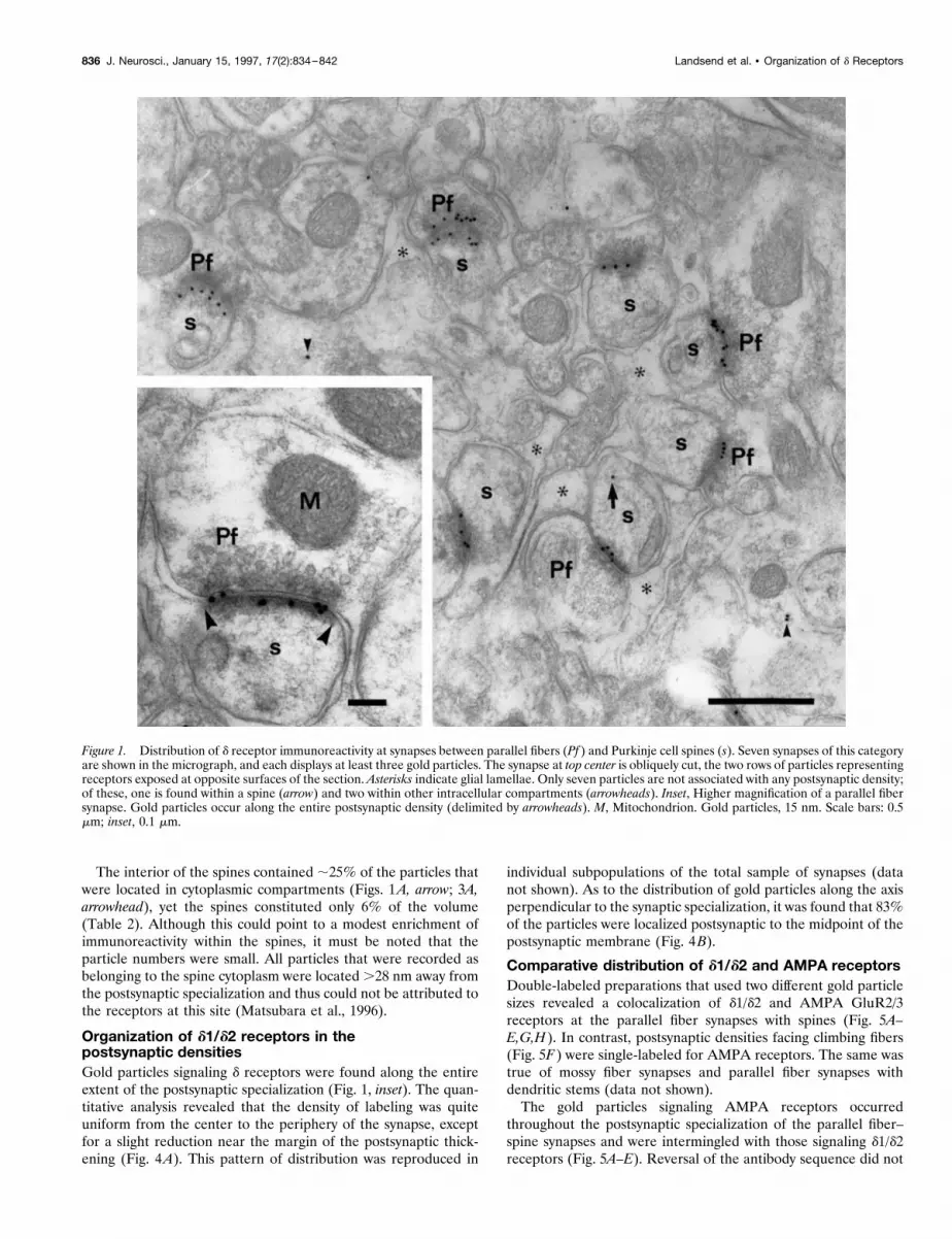
RESULTSDifferential expression of d1/d2 receptors atcerebellar synapsesAs noted in Materials and Methods, the d receptor antibodyrecognizes the d1 as well as the d2 subunit. Among the differentsynapses in the cerebellar cortex only those between parallel fibersand Purkinje cell spines exhibited significant immunolabeling ford1/d2 receptors (Table 1; Figs. 1-3). The linear density of goldparticles at this type of synapse was 20 per micrometer, while noparticles were recorded at other synapses in the sample (Table 1).Immunonegative synapses included those between climbing fibersand Purkinje cell spines (Fig. 2A), between mossy fiber terminalsand granule cell dendritic digits (Fig. 2B), and between parallelfibers and dendritic stems (Fig. 3A,B). Densities postsynaptic toterminals of stellate cells (Fig. 3B) or basket cells (not illustrated)also were devoid of labeling. Similarly, no or, in some cases, asingle gold particle was found to be associated with asymmetricspine synapses in the stratum oriens and radiatum of the hip-pocampus (Fig. 3B, inset). The hippocampal sections had beenincubated together with those from the cerebellum to ensureidentical incubation conditions. The same pattern of labeling wasobtained in each of the three animals subjected to investigation.Replacement of the primary antibody with nonimmune IgG or
preadsorption with the peptide used for immunization (Fig. 2B,right inset) abolished the labeling, whereas preadsorption withAMPA receptor peptides had no effect (Fig. 2B, left inset).
Synaptic versus nonsynaptic expression ofd1/d2 receptorsIn preparations based on the use of 15 nm gold particles (whichgave negligible background labeling; see legend to Table 2);90%of the total number of particles in the molecular layer was asso-ciated with the postsynaptic specializations of parallel fiber syn-apses with spines (Table 2; Figs. 1, 2, 3A). The postsynapticspecializations constitute 3% of the total boundary length in thesampled area (Table 2), implying an ;800-fold enrichment of thelabeling at these sites. Approximately one-third (32%) of theparticles that were situated outside of postsynaptic densities couldbe attributed to nonsynaptic membranes, while the remainingparticles occurred over cytoplasmic compartments (Table 2).The few particles that were associated with nonsynaptic mem-
branes did not seem to show any preference for specific mem-brane compartments. Notably, there was no enrichment of immu-noreactivity in the glial membranes that surround the parallelfiber–spine synapses (Figs. 1, 2) or in the spine plasma membraneslateral to the postsynaptic densities.
Table 1. Immunoreactivity for the d2 receptor at different types ofsynapse
Category of synapseNumberof PSDs
Total lengthof PSDs(mm)
Number of goldparticles per mmPSDs (6 SD)
Parallel fiber–spine 53 15.5 19.7 (6 6.3)Parallel fiber–dendritic stem 5 1.1 0Basket /stellate cell synapses 5 1.5 0Mossy fiber synapses 8 2.2 0Climbing fiber synapses 19 4.3 0
Quantitative assessment of d receptor immunogold labeling. The data were obtainedfrom a single section (represented in Figs. 1, 2; 15 nm gold particles) to ensureidentical incubation conditions. Note selective labeling of postsynaptic densities(PSDs) of synapses between parallel fibers and Purkinje cell spines. The number andtotal length of the postsynaptic densities are indicated.
Landsend et al. • Organization of d Receptors J. Neurosci., January 15, 1997, 17(2):834–842 835
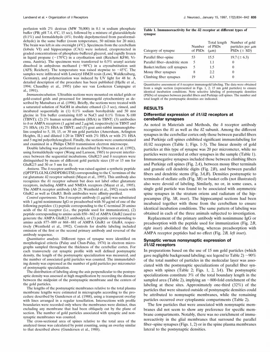
The interior of the spines contained ;25% of the particles thatwere located in cytoplasmic compartments (Figs. 1A, arrow; 3A,arrowhead), yet the spines constituted only 6% of the volume(Table 2). Although this could point to a modest enrichment ofimmunoreactivity within the spines, it must be noted that theparticle numbers were small. All particles that were recorded asbelonging to the spine cytoplasm were located .28 nm away fromthe postsynaptic specialization and thus could not be attributed tothe receptors at this site (Matsubara et al., 1996).
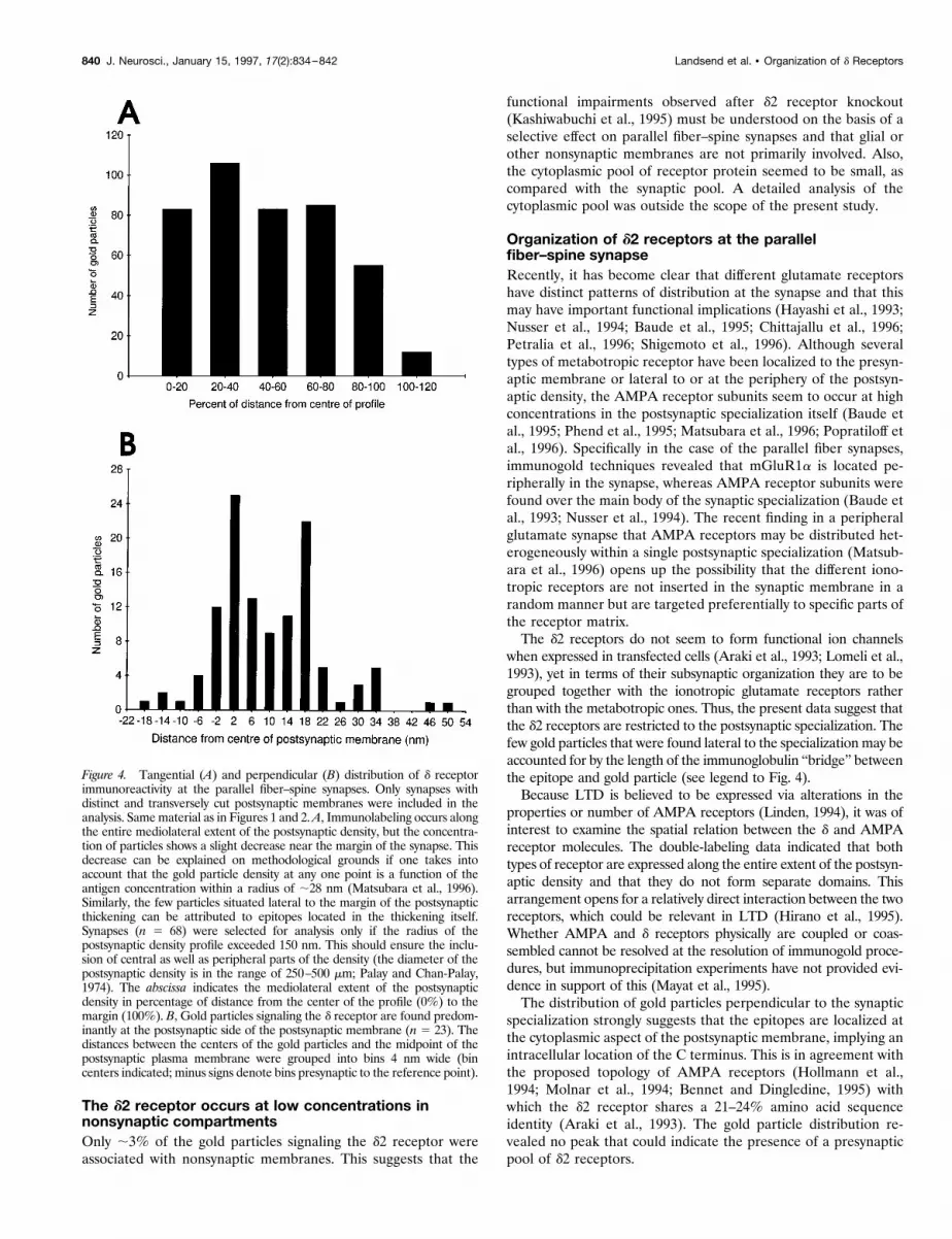
Organization of d1/d2 receptors in thepostsynaptic densitiesGold particles signaling d receptors were found along the entireextent of the postsynaptic specialization (Fig. 1, inset). The quan-titative analysis revealed that the density of labeling was quiteuniform from the center to the periphery of the synapse, exceptfor a slight reduction near the margin of the postsynaptic thick-ening (Fig. 4A). This pattern of distribution was reproduced in
individual subpopulations of the total sample of synapses (datanot shown). As to the distribution of gold particles along the axisperpendicular to the synaptic specialization, it was found that 83%of the particles were localized postsynaptic to the midpoint of thepostsynaptic membrane (Fig. 4B).
Comparative distribution of d1/d2 and AMPA receptorsDouble-labeled preparations that used two different gold particlesizes revealed a colocalization of d1/d2 and AMPA GluR2/3receptors at the parallel fiber synapses with spines (Fig. 5A–E,G,H). In contrast, postsynaptic densities facing climbing fibers(Fig. 5F) were single-labeled for AMPA receptors. The same wastrue of mossy fiber synapses and parallel fiber synapses withdendritic stems (data not shown).The gold particles signaling AMPA receptors occurred
throughout the postsynaptic specialization of the parallel fiber–spine synapses and were intermingled with those signaling d1/d2receptors (Fig. 5A–E). Reversal of the antibody sequence did not
Figure 1. Distribution of d receptor immunoreactivity at synapses between parallel fibers (Pf ) and Purkinje cell spines (s). Seven synapses of this categoryare shown in the micrograph, and each displays at least three gold particles. The synapse at top center is obliquely cut, the two rows of particles representingreceptors exposed at opposite surfaces of the section. Asterisks indicate glial lamellae. Only seven particles are not associated with any postsynaptic density;of these, one is found within a spine (arrow) and two within other intracellular compartments (arrowheads). Inset, Higher magnification of a parallel fibersynapse. Gold particles occur along the entire postsynaptic density (delimited by arrowheads). M, Mitochondrion. Gold particles, 15 nm. Scale bars: 0.5mm; inset, 0.1 mm.
836 J. Neurosci., January 15, 1997, 17(2):834–842 Landsend et al. • Organization of d Receptors

Figure 2. Immunolabeling for the d receptor is absent from synapses between climbing fibers (Cf ) and Purkinje cell spines (asterisks) and between mossyfibers (Mf ) and granule cell dendritic digits (d). Arrowheads in B delimit postsynaptic densities. A and B are from the same section as Figure 1. B, Insets,Immunolabeling for the d receptor is abolished after preadsorption with the peptide used for immunization (right; arrows show negative postsynapticdensities) but remains after preadsorption with the peptide used to generate the GluR2/3 antibody (left). Pf, Parallel fibers; s, Purkinje cell spines. Goldparticles, 15 nm. Scale bars: A, B, 0.5 mm; insets, 0.25 mm.
Landsend et al. • Organization of d Receptors J. Neurosci., January 15, 1997, 17(2):834–842 837
Figure 3. No d receptor immunolabeling occurs at synapses between parallel fibers (Pf ) and dendritic stems (d ) or postsynaptic to stellate cell terminals(St), whereas adjacent parallel fiber synapses with spines (s) show dense labeling. Long arrows indicate negative synapses. Arrowhead in A shows goldparticle in spine. The stellate cell synapse in B is obliquely cut but can be identified by the presence of flattened vesicles (short arrows). B, Inset, Asymmetricsynapse (arrows) established by nerve terminal (T ) in stratum oriens in the CA1 of hippocampus. The section was incubated together with the cerebellarsection in A. The sizes of gold particles were 15 nm (A and inset in B) or 10 nm (B), the latter giving a slightly higher background labeling than the former.Scale bars: A, B, 0.5 mm; inset, 0.25 mm.
838 J. Neurosci., January 15, 1997, 17(2):834–842 Landsend et al. • Organization of d Receptors
cause any alterations in the pattern of labeling, whereas omissionof the first or second antibody selectively removed the respectivegold particle size (data not shown).Postsynaptic membranes were identified that had been cut
more or less tangentially, thus exposing a two-dimensional ratherthan a linear receptor matrix (Fig. 5G,H). Because of the curva-ture of the synaptic specialization, only part of it would be acces-sible to the immunoreagents even if tangentially cut. At such sitesit could be seen that the two receptor types did not occupyseparate domains, although spots were found in which one or theother gold particle size was in clear excess (Fig. 5H). At thetangentially cut surfaces the density of 5 nm gold particles repre-senting d receptors was typically higher than 10 particles per 104
nm2 (e.g., Fig. 5H, right). A similar labeling intensity was observedin tangentially cut postsynaptic membranes labeled with 15 nmgold particles (data not shown). As was the case for the larger goldparticle sizes, the 5 nm gold particles were restricted to parallelfiber synapses with spines.
DISCUSSIONThe d2 receptor is expressed selectively at parallelfiber–Purkinje cell synapsesThe d receptors belong to the superfamily of glutamate receptors(Yamazaki et al., 1992; Araki et al., 1993; Lomeli et al., 1993).Although the role of the d1 subunit remains obscure, it was shownrecently that knockout of the d2 subunit produces severe deficitsin motor coordination and cerebellar long-term depression(Kashiwabuchi et al., 1995). In the same study, electrophysiolog-ical evidence was obtained of multiple climbing fiber innervation.To translate these findings into a better understanding of cere-bellar function, we needed to identify the precise loci of d2receptor expression. Although previous evidence has pointed toits localization in Purkinje cells and at parallel fiber–spine syn-apses (Araki et al., 1993; Lomeli et al., 1993; Hirano et al., 1994,1995; Mayat et al., 1995; Petralia et al., 1995; Takayama et al.,1995), it must be resolved whether the d2 receptor also occurs atother synaptic sites, whether it is expressed presynaptically or innonsynaptic membranes, and how it is associated with AMPAreceptors at the synaptic and subsynaptic levels. With immunocy-tochemistry such issues can be settled by use of particulate mark-ers, which are nondiffusible and thus provide a spatial resolutionlimited mainly by the molecular sizes of the immunoglobulins orimmunoglobulin fragments (Ottersen, 1989; Kellenberger andHayat, 1991).Because postembedding immunogold labeling is restricted to
those epitopes that are exposed at the surface of the section, a lowlabeling intensity is a recurrent problem when this approach isapplied to glutamate receptors. However, by optimizing the con-ditions for tissue preparation and immunoincubation (Matsubara
et al., 1996), it has been possible to achieve a sensitivity thatpermits a quantitative analysis of the issues mentioned above.The present study revealed a remarkable selectivity in the
synaptic expression of d receptors in the cerebellar cortex. Allsynapses between parallel fibers and Purkinje cell spines appearedto be labeled, although no significant labeling was associated withother types of synapse. The same pattern was reproduced withthree different gold particle sizes. Nevertheless, absence of label-ing in a particular type of synapse must be interpreted withcaution. Masking of antigens could pose a problem, but theoccurrence of AMPA immunogold labeling in synapses devoid ofd receptor immunoreactivity (climbing fiber synapses with spinesand parallel fiber synapses with dendritic stems) shows that thereceptor matrix in these synapses is accessible to the immunore-agents. Further, the sensitivity remains an issue to be considered,despite recent improvements. Although the labeling efficiencycannot be defined in absolute terms, it must be quite high, asjudged by the close spacing of gold particles at tangentially cutsynaptic specializations. Even a very small pool of receptorsshould be picked up by the procedure in the absence of significantbackground labeling. Thus it would seem reasonable to concludethat the d receptor density must be orders of magnitude higher inthe parallel fiber–spine synapses than in other types of synapse inthe cerebellar cortex.The antibody used here recognizes the d1 receptor as well as
the d2 receptor (Mayat et al., 1995). To assess the extent towhich the d1 receptor had contributed to the present pattern ofimmunolabeling, we processed the cerebellar sections togetherwith hippocampal sections from the same animals. In situhybridization analyses have shown that, although the hip-pocampus does not contain detectable levels of d2 subunitmRNA (Araki et al., 1993; Lomeli et al., 1993), it is the brainstructure that is richest in d1 mRNA (Lomeli et al., 1993). Thevery weak labeling obtained by the d receptor antibody insynapses of coprocessed hippocampal sections suggests that thelabeling in the cerebellum is attributable primarily to thepresence of the d2 receptor. This would agree with previousobservations based on other approaches (Mayat et al., 1995). Inthe remaining Discussion, the labeling will be considered toreflect the d2 receptor distribution.The absence of detectable d2 receptor expression postsynaptic
to climbing fibers implies that the Purkinje cell can target thisreceptor subunit to a specific subpopulation of its spines. It is alsoclear that d2 receptor expression is not induced indiscriminativelyby parallel fibers, because such receptors were not found atparallel fiber synapses with stem dendrites of interneurons. Theresults suggest that the expression of the d2 receptor is contingenton a unique combination of pre- and postsynaptic elements.
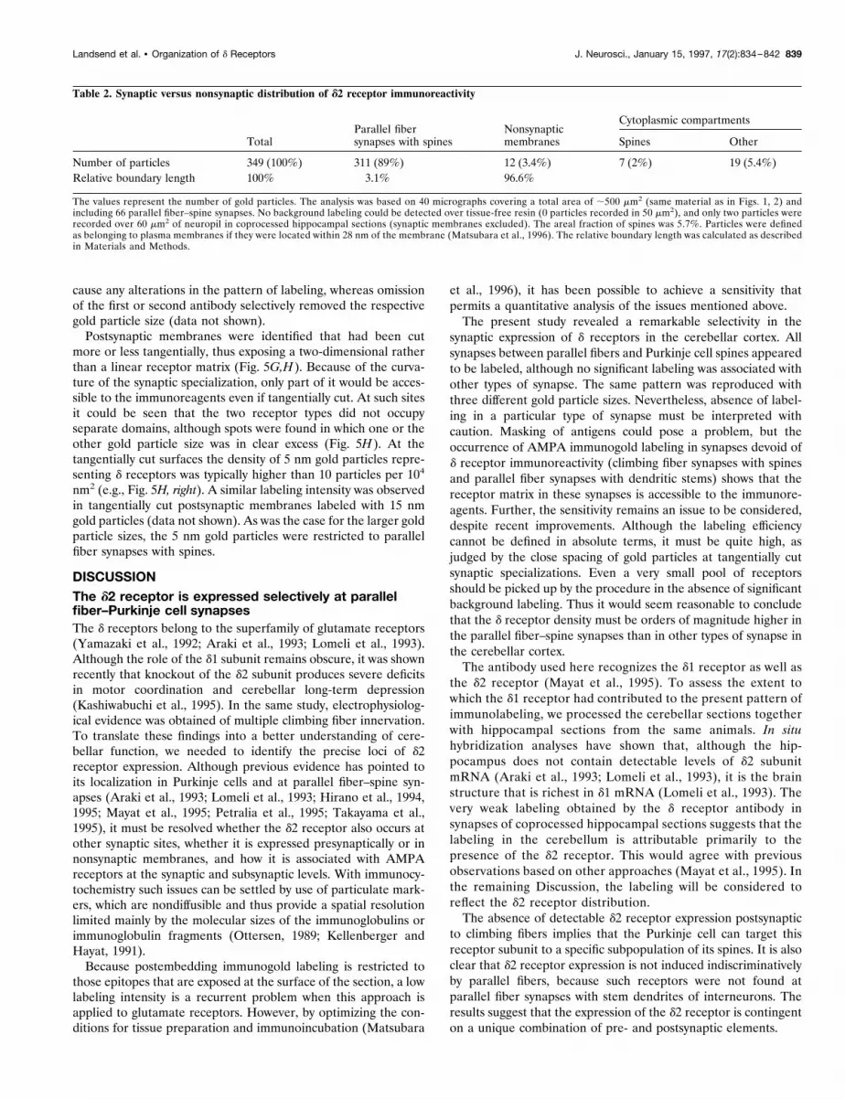
Table 2. Synaptic versus nonsynaptic distribution of d2 receptor immunoreactivity
TotalParallel fibersynapses with spines
Nonsynapticmembranes
Cytoplasmic compartments
Spines Other
Number of particles 349 (100%) 311 (89%) 12 (3.4%) 7 (2%) 19 (5.4%)Relative boundary length 100% 3.1% 96.6%
The values represent the number of gold particles. The analysis was based on 40 micrographs covering a total area of ;500 mm2 (same material as in Figs. 1, 2) andincluding 66 parallel fiber–spine synapses. No background labeling could be detected over tissue-free resin (0 particles recorded in 50 mm2), and only two particles wererecorded over 60 mm2 of neuropil in coprocessed hippocampal sections (synaptic membranes excluded). The areal fraction of spines was 5.7%. Particles were definedas belonging to plasma membranes if they were located within 28 nm of the membrane (Matsubara et al., 1996). The relative boundary length was calculated as describedin Materials and Methods.
Landsend et al. • Organization of d Receptors J. Neurosci., January 15, 1997, 17(2):834–842 839
The d2 receptor occurs at low concentrations innonsynaptic compartmentsOnly ;3% of the gold particles signaling the d2 receptor wereassociated with nonsynaptic membranes. This suggests that the
functional impairments observed after d2 receptor knockout(Kashiwabuchi et al., 1995) must be understood on the basis of aselective effect on parallel fiber–spine synapses and that glial orother nonsynaptic membranes are not primarily involved. Also,the cytoplasmic pool of receptor protein seemed to be small, ascompared with the synaptic pool. A detailed analysis of thecytoplasmic pool was outside the scope of the present study.
Organization of d2 receptors at the parallelfiber–spine synapseRecently, it has become clear that different glutamate receptorshave distinct patterns of distribution at the synapse and that thismay have important functional implications (Hayashi et al., 1993;Nusser et al., 1994; Baude et al., 1995; Chittajallu et al., 1996;Petralia et al., 1996; Shigemoto et al., 1996). Although severaltypes of metabotropic receptor have been localized to the presyn-aptic membrane or lateral to or at the periphery of the postsyn-aptic density, the AMPA receptor subunits seem to occur at highconcentrations in the postsynaptic specialization itself (Baude etal., 1995; Phend et al., 1995; Matsubara et al., 1996; Popratiloff etal., 1996). Specifically in the case of the parallel fiber synapses,immunogold techniques revealed that mGluR1a is located pe-ripherally in the synapse, whereas AMPA receptor subunits werefound over the main body of the synaptic specialization (Baude etal., 1993; Nusser et al., 1994). The recent finding in a peripheralglutamate synapse that AMPA receptors may be distributed het-erogeneously within a single postsynaptic specialization (Matsub-ara et al., 1996) opens up the possibility that the different iono-tropic receptors are not inserted in the synaptic membrane in arandom manner but are targeted preferentially to specific parts ofthe receptor matrix.The d2 receptors do not seem to form functional ion channels
when expressed in transfected cells (Araki et al., 1993; Lomeli et al.,1993), yet in terms of their subsynaptic organization they are to begrouped together with the ionotropic glutamate receptors ratherthan with the metabotropic ones. Thus, the present data suggest thatthe d2 receptors are restricted to the postsynaptic specialization. Thefew gold particles that were found lateral to the specialization may beaccounted for by the length of the immunoglobulin “bridge” betweenthe epitope and gold particle (see legend to Fig. 4).Because LTD is believed to be expressed via alterations in the
properties or number of AMPA receptors (Linden, 1994), it was ofinterest to examine the spatial relation between the d and AMPAreceptor molecules. The double-labeling data indicated that bothtypes of receptor are expressed along the entire extent of the postsyn-aptic density and that they do not form separate domains. Thisarrangement opens for a relatively direct interaction between the tworeceptors, which could be relevant in LTD (Hirano et al., 1995).Whether AMPA and d receptors physically are coupled or coas-sembled cannot be resolved at the resolution of immunogold proce-dures, but immunoprecipitation experiments have not provided evi-dence in support of this (Mayat et al., 1995).The distribution of gold particles perpendicular to the synaptic
specialization strongly suggests that the epitopes are localized atthe cytoplasmic aspect of the postsynaptic membrane, implying anintracellular location of the C terminus. This is in agreement withthe proposed topology of AMPA receptors (Hollmann et al.,1994; Molnar et al., 1994; Bennet and Dingledine, 1995) withwhich the d2 receptor shares a 21–24% amino acid sequenceidentity (Araki et al., 1993). The gold particle distribution re-vealed no peak that could indicate the presence of a presynapticpool of d2 receptors.
Figure 4. Tangential (A) and perpendicular (B) distribution of d receptorimmunoreactivity at the parallel fiber–spine synapses. Only synapses withdistinct and transversely cut postsynaptic membranes were included in theanalysis. Samematerial as in Figures 1 and 2.A, Immunolabeling occurs alongthe entire mediolateral extent of the postsynaptic density, but the concentra-tion of particles shows a slight decrease near the margin of the synapse. Thisdecrease can be explained on methodological grounds if one takes intoaccount that the gold particle density at any one point is a function of theantigen concentration within a radius of ;28 nm (Matsubara et al., 1996).Similarly, the few particles situated lateral to the margin of the postsynapticthickening can be attributed to epitopes located in the thickening itself.Synapses (n 5 68) were selected for analysis only if the radius of thepostsynaptic density profile exceeded 150 nm. This should ensure the inclu-sion of central as well as peripheral parts of the density (the diameter of thepostsynaptic density is in the range of 250–500 mm; Palay and Chan-Palay,1974). The abscissa indicates the mediolateral extent of the postsynapticdensity in percentage of distance from the center of the profile (0%) to themargin (100%). B, Gold particles signaling the d receptor are found predom-inantly at the postsynaptic side of the postsynaptic membrane (n 5 23). Thedistances between the centers of the gold particles and the midpoint of thepostsynaptic plasma membrane were grouped into bins 4 nm wide (bincenters indicated; minus signs denote bins presynaptic to the reference point).
840 J. Neurosci., January 15, 1997, 17(2):834–842 Landsend et al. • Organization of d Receptors

ConclusionsTo our knowledge this is the first high-resolution immunogoldanalysis of a member of the d glutamate receptor family. The datasuggest that the d2 subunit is targeted to the spines that are
postsynaptic to parallel fibers, but not to those that are postsyn-aptic to the climbing fibers, which form the other main excitatoryinput to the Purkinje cells. In contrast, AMPA receptors areexpressed in both types of synapse. This shows that postsynaptic
Figure 5. Double labeling with antisera to the d receptor (30 nm particles in A–F, 5 nm particles inG,H) and GluR2/3 (10 nm in A–F, 15 nm inG,H). Doublelabeling is found at synapses between parallel fibers (Pf) and Purkinje cell spines (s), whereas climbing fibers (Cf) establish synapses that are single-labeled forGluR2/3 (F). Synapses in G and H are obliquely cut so that parts of the postsynaptic membranes are viewed en face. Some of the 5 nm particles are indicatedby arrows. The distance between these particles is typically in the range of 15–30 nm (H, right part). Scale bars: A–F, 0.25 mm; G, H, 0.1 mm.
Landsend et al. • Organization of d Receptors J. Neurosci., January 15, 1997, 17(2):834–842 841
neurons may compose distinct glutamate receptor profiles depen-dent on the identity of the presynaptic element. The organizationof the d2 receptors in the postsynaptic membrane mimics that ofAMPA receptors in regard to tangential distribution (highly con-centrated in postsynaptic specialization) and membrane topology(intracellular C terminus). The two types of receptor show a closespatial association that could underlie their ability to interact inconditions of synaptic plasticity.
REFERENCESAraki K, Meguro H, Kushiya E, Takayama C, Inoue Y, Mishina M (1993)Selective expression of the glutamate receptor channel delta 2 subunitin cerebellar Purkinje cells. Biochem Biophys Res Commun197:1267–1276.
Baude A, Nusser Z, Roberts JDB, Mulvihill E, McIlhinney RAJ, SomogyiP (1993) The metabotropic glutamate receptor (mGluR1a) is concen-trated at perisynaptic membrane of neuronal subpopulations as de-tected by immunogold reaction. Neuron 11:771–787.
Baude A, Nusser Z, Molnar E, McIlhinney RAJ, Somogyi P (1995)High-resolution immunogold localization of AMPA-type glutamate re-ceptor subunits at synaptic and nonsynaptic sites in the rat hippocam-pus. Neuroscience 69:1031–1055.
Bennet JA, Dingledine R (1995) Topology profile for a glutamate recep-tor: three transmembrane domains and a channel-lining reentrant mem-brane loop. Neuron 14:373–384.
Chaudhry FA, Lehre KP, van Lookeren Campagne M, Ottersen OP,Danbolt NC, Storm-Mathisen J (1995) Glutamate transporters in glialplasma membranes: highly differentiated localizations revealed by quan-titative ultrastructural immunocytochemistry. Neuron 15:711–720.
Chittajallu R, Vignes M, Dev KK, Barnes JM, Collingridge GL, HenleyJM (1996) Regulation of glutamate release by presynaptic kainatereceptors in the hippocampus. Nature 379:79–81.
Gundersen HJG, Bendtsen TF, Korbo L, Marcussen N, Møller A, NielsenK, Nyengaard JR, Pakkenberg B, Sørensen FB, Vesterby A, West MJ(1988) Some new, simple, and efficient stereological methods and theiruse in pathological research and diagnosis. APMIS 96:379–394.
Hayashi Y, Momiyama A, Takahashi T, Ohishi H, Ogawa-Meguro R,Shigemoto R, Mizuno N, Nakanishi S (1993) Role of metabotropicglutamate receptor in synaptic modulation in the accessory olfactorybulb. Nature 366:687–690.
Hirano T, Kasono K, Araki K, Shinozuka K, Mishina M (1994) Involve-ment of the glutamate receptor delta 2 subunit in the long-term depres-sion of glutamate responsiveness in cultured rat Purkinje cells. NeurosciLett 182:172–176.
Hirano T, Kasono K, Araki K, Mishina M (1995) Suppression of LTD incultured Purkinje cells deficient in the glutamate receptor delta 2subunit. NeuroReport 6:524–526.
Hjelle OP, Chaudhry FA, Ottersen OP (1994) Antisera to glutathione:characterization and immunocytochemical application to the rat cere-bellum. Eur J Neurosci 6:794–804.
Hollmann M, Maron C, Heinemann S (1994) N-Glycosylation site tag-ging suggests a three transmembrane domain topology for the gluta-mate receptor GluR1. Neuron 13:1331–1343.
Ito M (1989) Long-term depression. Annu Rev Neurosci 12:85–102.Kashiwabuchi N, Ikeda K, Araki K, Hirano T, Shibuki K, Takayama C,Inoue Y, Kutsuwada T, Yagi T, Kang Y, Aizawa S, Mishina M (1995)Impairment of motor coordination, Purkinje cell synapse formation,and cerebellar long-term depression in GluRd2 mutant mice. Cell81:245–252.
Kellenberger E, Hayat MA (1991) Some basic concepts for the choice ofmethods. In: Colloidal gold: principles, methods, and applications(Hayat MA, ed), pp 1–30. San Diego: Academic.
Linden DJ (1994) Long-term synaptic depression in the mammalianbrain. Neuron 12:457–472.
Lomeli H, Sprengel R, Laurie DJ, Kohr G, Herb A, Seeburg PH, WisdenW (1993) The rat delta-1 and delta-2 subunits extend the excitatoryamino acid receptor family. FEBS Lett 315:318–322.
Matsubara A, Laake JH, Davanger S, Usami S-i, Ottersen OP (1996)Organization of AMPA receptor subunits at a glutamate synapse: aquantitative immunogold analysis of hair cell synapses in the rat organof Corti. J Neurosci 16:4457–4467.
Mayat E, Petralia RS, Wang YX, Wenthold RJ (1995) Immunoprecipi-tation, immunoblotting, and immunocytochemistry studies suggest thatglutamate receptor delta subunits form novel postsynaptic receptorcomplexes. J Neurosci 15:2533–2546.
Molnar E, McIlhinney RA, Baude A, Nusser Z, Somogyi P (1994) Mem-brane topology of the GluR1 glutamate receptor subunit: epitope map-ping by site-directed anti-peptide antibodies. J Neurochem 63:683–693.
Nusser Z, Mulvihill E, Streit P, Somogyi P (1994) Subsynaptic segrega-tion of metabotropic and ionotropic glutamate receptors as revealed byimmunogold localization. Neuroscience 61:421–427.
Ottersen OP (1989) Quantitative electron microscopic immunocyto-chemistry of amino acids. Anat Embryol (Berl) 180:1–15.
Ottersen OP, Zhang N, Walberg F (1992) Metabolic compartmentationof glutamate and glutamine: morphological evidence obtained by quan-titative immunocytochemistry in rat cerebellum. Neuroscience46:519–534.
Palay SL, Chan-Palay V (1974) Cerebellar cortex: cytology and organi-zation. Heidelberg: Springer.
Petralia RS, Zhao HM, Wang YX, Wenthold RJ (1995) Immunocyto-chemical localization of glutamate receptor subunits in Purkinje cells indeveloping and adult rats. Soc Neurosci Abstr 21:838.
Petralia RS, Wang YX, Niedzielski AS, Wenthold RJ (1996) Themetabotropic glutamate receptors, mGluR2 and mGluR3, show uniquepostsynaptic, presynaptic, and glial localizations. Neuroscience71:949–976.
Phend KD, Rustioni A, Weinberg RJ (1995) An osmium-free method ofepon embedment that preserves both ultrastructure and antigenicity forpost-embedding immunocytochemistry. J Histochem Cytochem43:283–292.
Popratiloff A, Weinberg RJ, Rustioni A (1996) AMPA receptor subunitsunderlying terminals of fine-caliber primary afferent fibers. J Neurosci16:3363–3372.
Shigemoto R, Kulik A, Roberts JDB, Ohishi H, Nusser Z, Kaneko T,Somogyi P (1996) Target-cell-specific concentration of metabotropicglutamate receptor in the presynaptic active zone. Nature 381:523–525.
Takayama C, Nakagawa S, Watanabe M, Mishina M, Inoue Y (1995)Light and electron microscopic localization of the glutamate receptorchannel delta 2 subunit in the mouse Purkinje cell. Neurosci Lett188:89–92.
van Lookeren Campagne M, Oestreicher AB, van der Krift TP, GispenWH, Verkleij AJ (1991) Freeze-substitution and Lowicryl HM20 em-bedding of fixed rat brain: suitability for immunogold ultrastructurallocalization of neural antigens. J Histochem Cytochem 39:1267–1279.
Wang B-L, Larsson L-I (1985) Simultaneous demonstration of multipleantigens by indirect immunofluorescence or immunogold staining: novellight and electron microscopical double and triple staining methodemploying primary antibodies from the same species. Histochemistry83:47–56.
Wenthold RJ, Yokotani N, Doi K, Wada K (1992) Immunocytochemicalcharacterization of the non-NMDA glutamate receptor using subunitspecific antibodies. J Biol Chem 267:501–507.
Yamazaki M, Araki K, Shibata A, Mishina M (1992) Molecular cloningof a cDNA encoding a novel member of the mouse glutamate receptorchannel family. Biochem Biophys Res Commun 183:886–892.
842 J. Neurosci., January 15, 1997, 17(2):834–842 Landsend et al. • Organization of d Receptors