hair cell ribbon synapses
TRANSCRIPT
REVIEW
Hair cell ribbon synapses
Tobias Moser & Andreas Brandt & Anna Lysakowski
Received: 11 May 2006 /Accepted: 8 June 2006 / Published online: 31 August 2006# Springer-Verlag 2006
Abstract Hearing and balance rely on the faithful synapticcoding of mechanical input by the auditory and vestibularhair cells of the inner ear. Mechanical deflection of theirstereocilia causes the opening of mechanosensitive chan-nels, resulting in hair cell depolarization, which controls therelease of glutamate at ribbon-type synapses. Hair cellshave a compact shape with strong polarity. Mechanoelec-trical transduction and active membrane turnover associatedwith stereociliar renewal dominate the apical compartment.Transmitter release occurs at several active zones along thebasolateral membrane. The astonishing capability of thehair cell ribbon synapse for temporally precise and reliablesensory coding has been the subject of intense investigationover the past few years. This research has been facilitatedby the excellent experimental accessibility of the hair cell.For the same reason, the hair cell serves as an important
model for studying presynaptic Ca2+ signaling and stimu-lus-secretion coupling. In addition to common principles,hair cell synapses differ in their anatomical and functionalproperties among species, among the auditory and vestib-ular organs, and among hair cell positions within the organ.Here, we briefly review synaptic morphology and connec-tivity and then focus on stimulus-secretion coupling at haircell synapses.
Keywords Hair cell . Ribbon synapse . Cochlear .
Vestibular . Inner ear
Introduction
The coding of information with respect to sounds and headmovements by the hair cell afferent synapse requiressynaptic transmission to be both reliable and temporallyprecise. Even in the absence of stimulation, the hair cellribbon-type active zone drives “spontaneous” spiking in theafferent neuron at rates of up to or even beyond 100 Hz(Kiang et al. 1965; Liberman 1982; Brichta and Goldberg2000). To ensure such remarkable temporal fidelity andhigh throughput, the hair cell afferent synapse relies onunique molecular and structural specializations, some ofwhich have been elucidated only recently.
Mature afferent hair cell synapses usually display onesingle synaptic ribbon or synaptic body facing onepostsynaptic density (Fig. 1). Ribbons tether a monolayerof synaptic vesicles, with a high packing density (Lenzi etal. 1999). A fraction of these ribbon-associated vesicles“docks” onto the presynaptic membrane. In addition, somedocked vesicles are not associated with the ribbon, andsome cytosolic vesicles neither touch the presynapticmembrane nor bind to the ribbon. The synaptic vesicles
Cell Tissue Res (2006) 326:347–359DOI 10.1007/s00441-006-0276-3
Research at the Moser laboratory was supported by grants fromthe DFG (SFB406 and CMPB), the European Commission (throughthe integrated project EuroHear), the Human Frontiers ScienceProgram (HFSP), and the Federal Goverment (through the BernsteinCenter for Computational Neuroscience, Göttingen). Research atthe Lysakowski laboratory was supported by grants from NIH (R01DC02521, R01 DC02290, and R01 DC002358) and the AmericanHearing Research Foundation.
T. Moser (*) :A. BrandtDepartment of Otolaryngology and Center for MolecularPhysiology of the Brain,University of Göttingen,Robert-Koch-Strasse 40,37075 Göttingen, Germanye-mail: [email protected]
A. LysakowskiDepartment of Anatomy & Cell Biology,University of Illinois at Chicago,Chicago IL 60612, USA

348 Cell Tissue Res (2006) 326:347–359
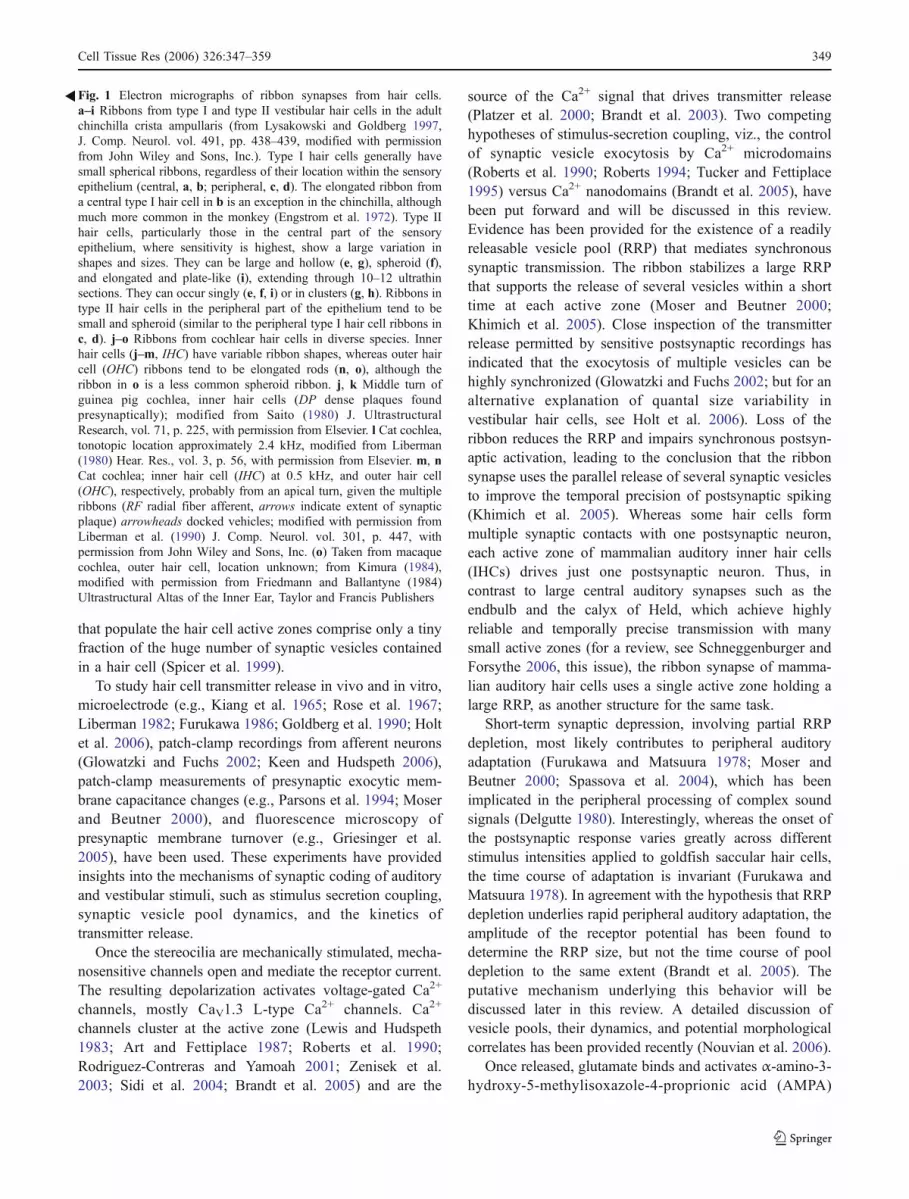
that populate the hair cell active zones comprise only a tinyfraction of the huge number of synaptic vesicles containedin a hair cell (Spicer et al. 1999).
To study hair cell transmitter release in vivo and in vitro,microelectrode (e.g., Kiang et al. 1965; Rose et al. 1967;Liberman 1982; Furukawa 1986; Goldberg et al. 1990; Holtet al. 2006), patch-clamp recordings from afferent neurons(Glowatzki and Fuchs 2002; Keen and Hudspeth 2006),patch-clamp measurements of presynaptic exocytic mem-brane capacitance changes (e.g., Parsons et al. 1994; Moserand Beutner 2000), and fluorescence microscopy ofpresynaptic membrane turnover (e.g., Griesinger et al.2005), have been used. These experiments have providedinsights into the mechanisms of synaptic coding of auditoryand vestibular stimuli, such as stimulus secretion coupling,synaptic vesicle pool dynamics, and the kinetics oftransmitter release.
Once the stereocilia are mechanically stimulated, mecha-nosensitive channels open and mediate the receptor current.The resulting depolarization activates voltage-gated Ca2+
channels, mostly CaV1.3 L-type Ca2+ channels. Ca2+
channels cluster at the active zone (Lewis and Hudspeth1983; Art and Fettiplace 1987; Roberts et al. 1990;Rodriguez-Contreras and Yamoah 2001; Zenisek et al.2003; Sidi et al. 2004; Brandt et al. 2005) and are the
source of the Ca2+ signal that drives transmitter release(Platzer et al. 2000; Brandt et al. 2003). Two competinghypotheses of stimulus-secretion coupling, viz., the controlof synaptic vesicle exocytosis by Ca2+ microdomains(Roberts et al. 1990; Roberts 1994; Tucker and Fettiplace1995) versus Ca2+ nanodomains (Brandt et al. 2005), havebeen put forward and will be discussed in this review.Evidence has been provided for the existence of a readilyreleasable vesicle pool (RRP) that mediates synchronoussynaptic transmission. The ribbon stabilizes a large RRPthat supports the release of several vesicles within a shorttime at each active zone (Moser and Beutner 2000;Khimich et al. 2005). Close inspection of the transmitterrelease permitted by sensitive postsynaptic recordings hasindicated that the exocytosis of multiple vesicles can behighly synchronized (Glowatzki and Fuchs 2002; but for analternative explanation of quantal size variability investibular hair cells, see Holt et al. 2006). Loss of theribbon reduces the RRP and impairs synchronous postsyn-aptic activation, leading to the conclusion that the ribbonsynapse uses the parallel release of several synaptic vesiclesto improve the temporal precision of postsynaptic spiking(Khimich et al. 2005). Whereas some hair cells formmultiple synaptic contacts with one postsynaptic neuron,each active zone of mammalian auditory inner hair cells(IHCs) drives just one postsynaptic neuron. Thus, incontrast to large central auditory synapses such as theendbulb and the calyx of Held, which achieve highlyreliable and temporally precise transmission with manysmall active zones (for a review, see Schneggenburger andForsythe 2006, this issue), the ribbon synapse of mamma-lian auditory hair cells uses a single active zone holding alarge RRP, as another structure for the same task.
Short-term synaptic depression, involving partial RRPdepletion, most likely contributes to peripheral auditoryadaptation (Furukawa and Matsuura 1978; Moser andBeutner 2000; Spassova et al. 2004), which has beenimplicated in the peripheral processing of complex soundsignals (Delgutte 1980). Interestingly, whereas the onset ofthe postsynaptic response varies greatly across differentstimulus intensities applied to goldfish saccular hair cells,the time course of adaptation is invariant (Furukawa andMatsuura 1978). In agreement with the hypothesis that RRPdepletion underlies rapid peripheral auditory adaptation, theamplitude of the receptor potential has been found todetermine the RRP size, but not the time course of pooldepletion to the same extent (Brandt et al. 2005). Theputative mechanism underlying this behavior will bediscussed later in this review. A detailed discussion ofvesicle pools, their dynamics, and potential morphologicalcorrelates has been provided recently (Nouvian et al. 2006).
Once released, glutamate binds and activates α-amino-3-hydroxy-5-methylisoxazole-4-proprionic acid (AMPA)
Fig. 1 Electron micrographs of ribbon synapses from hair cells.a–i Ribbons from type I and type II vestibular hair cells in the adultchinchilla crista ampullaris (from Lysakowski and Goldberg 1997,J. Comp. Neurol. vol. 491, pp. 438–439, modified with permissionfrom John Wiley and Sons, Inc.). Type I hair cells generally havesmall spherical ribbons, regardless of their location within the sensoryepithelium (central, a, b; peripheral, c, d). The elongated ribbon froma central type I hair cell in b is an exception in the chinchilla, althoughmuch more common in the monkey (Engstrom et al. 1972). Type IIhair cells, particularly those in the central part of the sensoryepithelium, where sensitivity is highest, show a large variation inshapes and sizes. They can be large and hollow (e, g), spheroid (f),and elongated and plate-like (i), extending through 10–12 ultrathinsections. They can occur singly (e, f, i) or in clusters (g, h). Ribbons intype II hair cells in the peripheral part of the epithelium tend to besmall and spheroid (similar to the peripheral type I hair cell ribbons inc, d). j–o Ribbons from cochlear hair cells in diverse species. Innerhair cells (j–m, IHC) have variable ribbon shapes, whereas outer haircell (OHC) ribbons tend to be elongated rods (n, o), although theribbon in o is a less common spheroid ribbon. j, k Middle turn ofguinea pig cochlea, inner hair cells (DP dense plaques foundpresynaptically); modified from Saito (1980) J. UltrastructuralResearch, vol. 71, p. 225, with permission from Elsevier. l Cat cochlea,tonotopic location approximately 2.4 kHz, modified from Liberman(1980) Hear. Res., vol. 3, p. 56, with permission from Elsevier. m, nCat cochlea; inner hair cell (IHC) at 0.5 kHz, and outer hair cell(OHC), respectively, probably from an apical turn, given the multipleribbons (RF radial fiber afferent, arrows indicate extent of synapticplaque) arrowheads docked vehicles; modified with permission fromLiberman et al. (1990) J. Comp. Neurol. vol. 301, p. 447, withpermission from John Wiley and Sons, Inc. (o) Taken from macaquecochlea, outer hair cell, location unknown; from Kimura (1984),modified with permission from Friedmann and Ballantyne (1984)Ultrastructural Altas of the Inner Ear, Taylor and Francis Publishers
�
Cell Tissue Res (2006) 326:347–359 349
receptors (Glowatzki and Fuchs 2002) that are clustered inthe postsynaptic density (Matsubara et al. 1996). Finally,glutamate is taken up by glutamate transporters (GLAST)of glia-like supporting cells (Furness and Lehre 1997;Furness and Lawton 2003) avoiding glutamate accumula-tion and hence limiting excitotoxic damage in the case ofstrong stimulation (Hakuba et al. 2000). The type Ivestibular hair cell-calyx synapse isolates the hair cell fromthe supporting cells.
Progress in understanding the molecular anatomy ofhair cell synapses (Safieddine and Wenthold 1999;Eybalin et al. 2002; Khimich et al. 2005) has been slow,mostly because of the limited amount of material (a fewthousand hair cells per ear). Although we are far from adetailed description (for a recent review, see Nouvian et al.2006), we can state that the molecular composition issimilar to that of retinal synapses (for a review, see tomDieck and Brandstätter 2006, this issue). Important differ-ences include the dominance of CaV1.3 channels and thepresence of the putative Ca2+ sensor of exocytosisotoferlin (Yasunaga et al. 1999).
Here, we will first review the common features anddifferent aspects of active zone morphology and synapticconnectivity in auditory and vestibular hair cells. We willthen discuss the general properties of presynaptic functionin hair cells. We will also focus on ion channels and Ca2+
signaling related to transmitter release.
Structure of hair cell ribbon synapses and synapticconnectivity
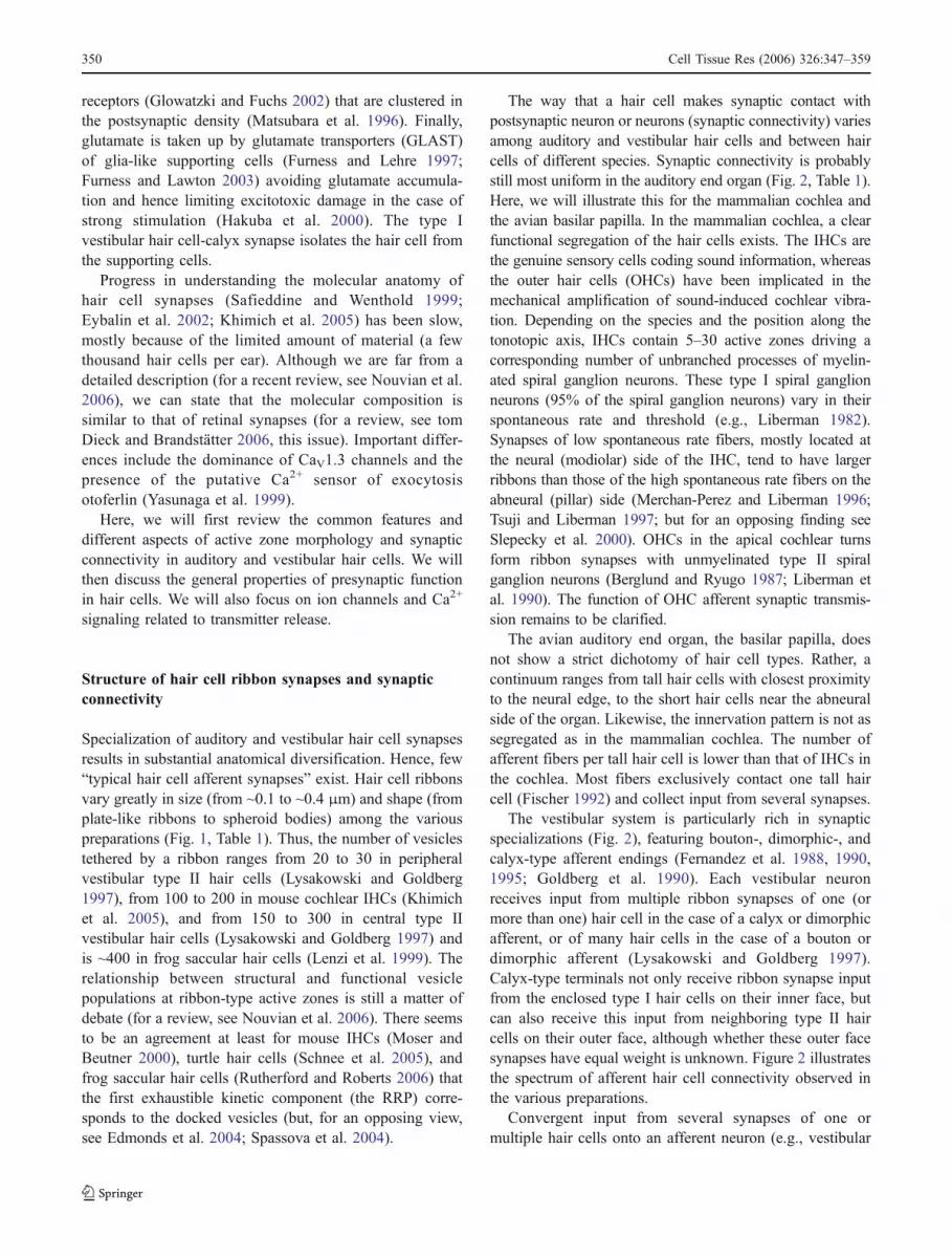
Specialization of auditory and vestibular hair cell synapsesresults in substantial anatomical diversification. Hence, few“typical hair cell afferent synapses” exist. Hair cell ribbonsvary greatly in size (from ~0.1 to ~0.4 μm) and shape (fromplate-like ribbons to spheroid bodies) among the variouspreparations (Fig. 1, Table 1). Thus, the number of vesiclestethered by a ribbon ranges from 20 to 30 in peripheralvestibular type II hair cells (Lysakowski and Goldberg1997), from 100 to 200 in mouse cochlear IHCs (Khimichet al. 2005), and from 150 to 300 in central type IIvestibular hair cells (Lysakowski and Goldberg 1997) andis ~400 in frog saccular hair cells (Lenzi et al. 1999). Therelationship between structural and functional vesiclepopulations at ribbon-type active zones is still a matter ofdebate (for a review, see Nouvian et al. 2006). There seemsto be an agreement at least for mouse IHCs (Moser andBeutner 2000), turtle hair cells (Schnee et al. 2005), andfrog saccular hair cells (Rutherford and Roberts 2006) thatthe first exhaustible kinetic component (the RRP) corre-sponds to the docked vesicles (but, for an opposing view,see Edmonds et al. 2004; Spassova et al. 2004).
The way that a hair cell makes synaptic contact withpostsynaptic neuron or neurons (synaptic connectivity) variesamong auditory and vestibular hair cells and between haircells of different species. Synaptic connectivity is probablystill most uniform in the auditory end organ (Fig. 2, Table 1).Here, we will illustrate this for the mammalian cochlea andthe avian basilar papilla. In the mammalian cochlea, a clearfunctional segregation of the hair cells exists. The IHCs arethe genuine sensory cells coding sound information, whereasthe outer hair cells (OHCs) have been implicated in themechanical amplification of sound-induced cochlear vibra-tion. Depending on the species and the position along thetonotopic axis, IHCs contain 5–30 active zones driving acorresponding number of unbranched processes of myelin-ated spiral ganglion neurons. These type I spiral ganglionneurons (95% of the spiral ganglion neurons) vary in theirspontaneous rate and threshold (e.g., Liberman 1982).Synapses of low spontaneous rate fibers, mostly located atthe neural (modiolar) side of the IHC, tend to have largerribbons than those of the high spontaneous rate fibers on theabneural (pillar) side (Merchan-Perez and Liberman 1996;Tsuji and Liberman 1997; but for an opposing finding seeSlepecky et al. 2000). OHCs in the apical cochlear turnsform ribbon synapses with unmyelinated type II spiralganglion neurons (Berglund and Ryugo 1987; Liberman etal. 1990). The function of OHC afferent synaptic transmis-sion remains to be clarified.
The avian auditory end organ, the basilar papilla, doesnot show a strict dichotomy of hair cell types. Rather, acontinuum ranges from tall hair cells with closest proximityto the neural edge, to the short hair cells near the abneuralside of the organ. Likewise, the innervation pattern is not assegregated as in the mammalian cochlea. The number ofafferent fibers per tall hair cell is lower than that of IHCs inthe cochlea. Most fibers exclusively contact one tall haircell (Fischer 1992) and collect input from several synapses.
The vestibular system is particularly rich in synapticspecializations (Fig. 2), featuring bouton-, dimorphic-, andcalyx-type afferent endings (Fernandez et al. 1988, 1990,1995; Goldberg et al. 1990). Each vestibular neuronreceives input from multiple ribbon synapses of one (ormore than one) hair cell in the case of a calyx or dimorphicafferent, or of many hair cells in the case of a bouton ordimorphic afferent (Lysakowski and Goldberg 1997).Calyx-type terminals not only receive ribbon synapse inputfrom the enclosed type I hair cells on their inner face, butcan also receive this input from neighboring type II haircells on their outer face, although whether these outer facesynapses have equal weight is unknown. Figure 2 illustratesthe spectrum of afferent hair cell connectivity observed inthe various preparations.
Convergent input from several synapses of one ormultiple hair cells onto an afferent neuron (e.g., vestibular
350 Cell Tissue Res (2006) 326:347–359
system, avian basilar papilla) is likely to enhance theevoked activity over background. Detection of coincidentsynaptic inputs by the action-potential-generating elementwould help to suppress uncorrelated spontaneous synapticinput. On the other hand, the number of statisticallyindependent channels carrying sensory information tosecond order neurons will be lower than, for example, inthe mammalian cochlea with potential consequences forprocessing in the brainstem. In the vestibular system, withits large variation in synaptic ribbons, afferent fibers, andterminal endings, it would be surprising if these did notpermit a wide latitude in coding mechanisms relevant tovestibular function (Goldberg 2000). Future work will beneeded to clarify the exact way that the different connec-tivities relate to coding strategies of auditory and vestibularstimuli.
Ion channels affecting hair cell presynaptic function
Hair cells display a resting membrane conductance basedon basolateral potassium channels (KCNQ and erg families)and stereociliar mechanoelectrical transduction channels.Mutation of the gene coding for KCNQ4 causes autosomal
dominant hearing impairment DFNA2 in humans (Kubischet al. 1999). The hearing loss of mouse models of DFNA2can mostly be explained by the dysfunction/degeneration ofmechanically active OHCs (Kharkovets et al. 2006), as hadbeen suggested before in a pharmacological model ofDFNA2 (Nouvian et al. 2003). The DFNA2 mouse modelsshow no major change of IHCs synaptic function and noobvious vestibular dysfunction. In addition to KCNQchannels, vestibular type I hair cells express erg channels(Eatock et al. 2002; Wong et al. 2004), which maycontribute to a distinctive current found in type I hair cells,IK,L (K.M. Hurley , S. Gaboyard, J.R. Wooltorton, J.L.Garcia, S.D. Price, A. Lysakowski, R.A. Eatock, submittedfor publication).
CaV1.3 Ca2+ channels of hair cells (see below) becomeactivated at the resting potential of the hair cell even in theabsence of sensory stimuli (Koschak et al. 2001; Xu andLipscombe 2001; Brandt et al. 2005; Johnson et al. 2005).Afferent neuron “spontaneous” activity seems to relyentirely on transmitter release from hair cells (Libermanand Kiang 1978; Sewell 1984). Once mechanoelectricaltransduction occurs, the apical potassium influx drivesreceptor potential generation, which further activatesvoltage-gated K+ and Ca2+ channels. BK (large conduc-
Table 1 Synaptic morphology and synaptic connectivity (HC hair cell, Fib fiber, LF low frequency, HF high frequency, Syn synapse,x unknown). The table summarizes part of the available information obtained in the various preparations. The references are examples, fromwhich information was obtained but do not represent a complete list. Data were obtained by electron microscopy except for byimmunofluorescence (Khimich et al. 2005)
Species/Organ Hair cell Ribbon: shape, size (nm),and number
Afferent connectivity References
Mammaliancochlea
Inner hair cells Ellipsoid; 3 axes: 55,100–200, 200–600;5–30
1 HC : 5–30 Fib; 1 Fib : 1 HC;1 Fib : 1 Syn
Liberman 1980; Francis et al.2004; Khimich et al. 2005
Outer hair cell(apex)
Elongate; 100–200;8–10
Liberman et al. 1990
Avian papilla Tall hair cells: Round; 120 (apex),210 (base); 15
1 HC : 1–3 Fib; 1 Fib : 1(−3)HC*; 1 Fib : several Syn
Fischer 1992; Martinez-Dunstet al. 1997
Short hair cells 0 (HF) −10 (LF)Mammaliancrista ampullaris
Type I hair cell Spheroid: 90, Rod:200–300. ~17
1 HC : 1 Fib; 1 Fib : 1–5 HC;1 Fib : 1–5 Calyces with 15 Syneach
Goldberg et al. 1990
Type II hair cell Barrel: 200 Elongate150–300. ~18
1 HC : 3–6 Fib; 1 Fib (bouton) : −30 HC;1 Fib (bouton) : 50–80 Syn
Lysakowski and Goldberg 1997Fernandez et al. 1988
Avian cristaampullaris
Type I hair celltype II hair cell
Spheroid; rod ND 1 Fib : up to 12 HCs Lysakowski 1996
Turtle cristaampullaris
Type I hair celltype II hair cell
Spheroid; ND 1 Fib : up to 8 HCs Lysakowski 1996; Brichtaand Peterson 1994
Frog cristaampullaris
Type II hair cell Spheroid; ND Lysakowski 1996
Fish cristaampullaris
Type II hair cell Spheroid 15–25central;4–5 peripheral
Wegner 1982; Chang et al.1992; Lysakowski 1996
Table 1 Synaptic morphology and synaptic connectivity (HC haircell, Fib fiber, LF low frequency, HF high frequency, Syn synapse,ND not determined). The table summarizes part of the availableinformation obtained in the various preparations. The references are
examples from which information was obtained, but which do notrepresent a complete list. Data were obtained by electron microscopyexcept for those obtained by immunofluorescence (Khimich et al.2005)
Cell Tissue Res (2006) 326:347–359 351
tance) currents are activated within hundreds of micro-seconds and have been implicated in shortening the haircell membrane time constant required for high temporalprecision sound coding, in limiting of the receptor potential,and in electrical frequency tuning (in non-mammalian haircells). Electrical hair cell tuning to a certain stimulusfrequency builds on a intimate interplay of BK and Ca2+
channels, which require close spatial colocalization, mostlikely at the active zones (Lewis and Hudspeth 1983; Art et
al. 1986; Fuchs et al. 1988; Roberts et al. 1990; Tucker andFettiplace 1995). This has fostered the use of BK channelsas a readout of active zone Ca2+ signaling of hair cells (seenext section). Surprisingly, BK channels of mammaliancochlear IHCs cluster remotely from the active zones at theapical part of the cell (neck; Pyott et al. 2004; Hafidi et al.2005) and are largely unaffected by manipulations of Ca2+
channels (Marcotti et al. 2004; Thurm et al. 2005). Thisfinding might be related to the evolution of other efficienttuning mechanisms in mammalian cochlea. Deletion of theKcnma1 gene in mice has surprisingly little primary impacton hearing as revealed by auditory brainstem responses(Ruttiger et al. 2004), which reflect the sound-evokedsynchronized activation of neuronal populations along theauditory pathway. However, single hair cell and singlespiral ganglion neuron analysis has revealed defects in thetiming of receptor potentials and subsequently of postsyn-aptic action potential generation (Oliver et al. 2006).Activation of KV-type channels causes a millisecond timecourse decline of the hair cell receptor potential and maythereby contribute to rapid auditory adaptation (Kros 1996;Moser and Beutner 2000).
Evidence for an essential role of L-type Ca2+ channelsfor hair cell transmitter release is well documented (Lewisand Hudspeth 1983; Art and Fettiplace 1987; Fuchs et al.1990; Roberts et al. 1990; Zhang et al. 1999; Moser andBeutner 2000; Platzer et al. 2000; Spassova et al. 2001;Robertson and Paki 2002; Brandt et al. 2003; Dou et al.2004; Schnee et al. 2005). Two independently generatedCaV1.3 knockout mice show complete deafness (lack ofauditory brainstem responses) and more than a 90%reduction of hair cell Ca2+ currents (Platzer et al. 2000;Brandt et al. 2003; Michna et al. 2003; Dou et al. 2004), butno obvious balance problems. Interestingly, Ca2+ uncaging,bypassing the defect in Ca2+ influx, elicits robust exocyticcapacitance changes (Brandt et al. 2003). This resulttogether with the finding of a largely unchanged molecularsynaptic anatomy in young mice (Nemzou et al. 2006)argues that the presynaptic machinery is assembled, despitethe lack of Ca2+ influx. The remaining Ca2+ current ofcochlear hair cells is at least partially mediated by L-typeCa2+ channels and is insensitive to blockers of N, P/Q, andR-type Ca2+ channels. The Yamoah group has presentedsingle channel evidence for the presence of non-L, possiblyN-type, channels in frog saccular hair cells (Rodriguez-Contreras and Yamoah 2001; Rodriguez-Contreras et al.2002). In frog crista hair cells, R-type Ca2+ channels havebeen suggested by pharmacological analysis of whole-cellCa2+ currents (Martini et al. 2000). This, together withnormal performance of CaV1.3-deficient mice in screeningtests of vestibular function (Dou et al. 2004), has led theauthors to conclude that non-L-type channel can mediatetransmitter release in vestibular hair cells.
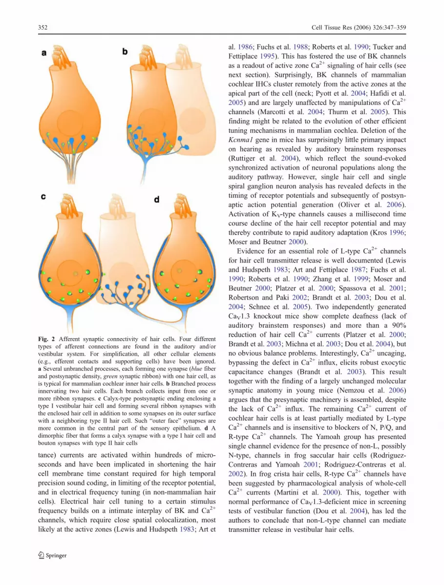
Fig. 2 Afferent synaptic connectivity of hair cells. Four differenttypes of afferent connections are found in the auditory and/orvestibular system. For simplification, all other cellular elements(e.g., efferent contacts and supporting cells) have been ignored.a Several unbranched processes, each forming one synapse (blue fiberand postsynaptic density, green synaptic ribbon) with one hair cell, asis typical for mammalian cochlear inner hair cells. b Branched processinnervating two hair cells. Each branch collects input from one ormore ribbon synapses. c Calyx-type postsynaptic ending enclosing atype I vestibular hair cell and forming several ribbon synapses withthe enclosed hair cell in addition to some synapses on its outer surfacewith a neighboring type II hair cell. Such “outer face” synapses aremore common in the central part of the sensory epithelium. d Adimorphic fiber that forms a calyx synapse with a type I hair cell andbouton synapses with type II hair cells
352 Cell Tissue Res (2006) 326:347–359
Stimulus-secretion coupling at the hair cell ribbonsynapse
Many aspects of presynaptic Ca2+ signaling are inaccessiblein small presynaptic boutons of the CNS but can be readilystudied and manipulated in hair cells. The classical work ofRoberts and Fettiplace and their colleagues on Ca2+ channelnumber and distribution and on cytosolic Ca2+ buffers haslargely contributed to making the hair cell a valuable modelsystem for presynaptic Ca2+ signaling (for previousreviews, see Lenzi and Roberts 1994; Tucker and Fettiplace1996; Augustine 2001; Augustine et al. 2003). Moreover,the characterization of stimulus-secretion coupling in haircells is the key to understanding the coding of informationregarding sound or vestibular stimuli.
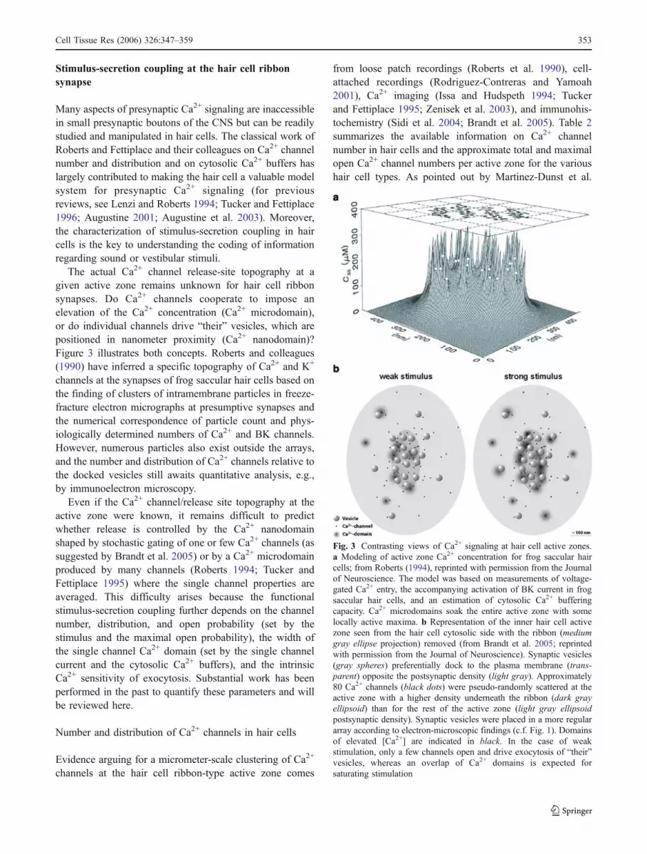
The actual Ca2+ channel release-site topography at agiven active zone remains unknown for hair cell ribbonsynapses. Do Ca2+ channels cooperate to impose anelevation of the Ca2+ concentration (Ca2+ microdomain),or do individual channels drive “their” vesicles, which arepositioned in nanometer proximity (Ca2+ nanodomain)?Figure 3 illustrates both concepts. Roberts and colleagues(1990) have inferred a specific topography of Ca2+ and K+
channels at the synapses of frog saccular hair cells based onthe finding of clusters of intramembrane particles in freeze-fracture electron micrographs at presumptive synapses andthe numerical correspondence of particle count and phys-iologically determined numbers of Ca2+ and BK channels.However, numerous particles also exist outside the arrays,and the number and distribution of Ca2+ channels relative tothe docked vesicles still awaits quantitative analysis, e.g.,by immunoelectron microscopy.
Even if the Ca2+ channel/release site topography at theactive zone were known, it remains difficult to predictwhether release is controlled by the Ca2+ nanodomainshaped by stochastic gating of one or few Ca2+ channels (assuggested by Brandt et al. 2005) or by a Ca2+ microdomainproduced by many channels (Roberts 1994; Tucker andFettiplace 1995) where the single channel properties areaveraged. This difficulty arises because the functionalstimulus-secretion coupling further depends on the channelnumber, distribution, and open probability (set by thestimulus and the maximal open probability), the width ofthe single channel Ca2+ domain (set by the single channelcurrent and the cytosolic Ca2+ buffers), and the intrinsicCa2+ sensitivity of exocytosis. Substantial work has beenperformed in the past to quantify these parameters and willbe reviewed here.
Number and distribution of Ca2+ channels in hair cells
Evidence arguing for a micrometer-scale clustering of Ca2+
channels at the hair cell ribbon-type active zone comes
from loose patch recordings (Roberts et al. 1990), cell-attached recordings (Rodriguez-Contreras and Yamoah2001), Ca2+ imaging (Issa and Hudspeth 1994; Tuckerand Fettiplace 1995; Zenisek et al. 2003), and immunohis-tochemistry (Sidi et al. 2004; Brandt et al. 2005). Table 2summarizes the available information on Ca2+ channelnumber in hair cells and the approximate total and maximalopen Ca2+ channel numbers per active zone for the varioushair cell types. As pointed out by Martinez-Dunst et al.
Fig. 3 Contrasting views of Ca2+ signaling at hair cell active zones.a Modeling of active zone Ca2+ concentration for frog saccular haircells; from Roberts (1994), reprinted with permission from the Journalof Neuroscience. The model was based on measurements of voltage-gated Ca2+ entry, the accompanying activation of BK current in frogsaccular hair cells, and an estimation of cytosolic Ca2+ bufferingcapacity. Ca2+ microdomains soak the entire active zone with somelocally active maxima. b Representation of the inner hair cell activezone seen from the hair cell cytosolic side with the ribbon (mediumgray ellipse projection) removed (from Brandt et al. 2005; reprintedwith permission from the Journal of Neuroscience). Synaptic vesicles(gray spheres) preferentially dock to the plasma membrane (trans-parent) opposite the postsynaptic density (light gray). Approximately80 Ca2+ channels (black dots) were pseudo-randomly scattered at theactive zone with a higher density underneath the ribbon (dark grayellipsoid) than for the rest of the active zone (light gray ellipsoidpostsynaptic density). Synaptic vesicles were placed in a more regulararray according to electron-microscopic findings (c.f. Fig. 1). Domainsof elevated [Ca2+] are indicated in black. In the case of weakstimulation, only a few channels open and drive exocytosis of “their”vesicles, whereas an overlap of Ca2+ domains is expected forsaturating stimulation
Cell Tissue Res (2006) 326:347–359 353

(1997), hair cell Ca2+ current seems to scale not only withthe number of synapses, but also with the size of the activezone. Hair cell Ca2+ channel numbers have been obtainedby (1) non-stationary fluctuation analysis, (2) dividing thewhole cell Ca2+ current (tail or pulse current) by the singlechannel current. Approximations of the number of Ca2+
channels per active zone either assume a purely synapticdistribution (Roberts et al. 1990; Tucker and Fettiplace1995; Martinez-Dunst et al. 1997) or consider a certainextrasynaptic density of Ca2+ channels in hair cells(Brandt et al. 2005).
The assumption of a purely synaptic localization has beenmotivated by the co-variation of Ca2+ current and active zonenumber (e.g., Martinez-Dunst et al. 1997; Wu et al. 1995;Schnee et al. 2005). However, a substantial, albeit muchlower, abundance of L-type Ca2+ channels has been foundoutside the observed dense clusters (Rodriguez-Contreras andYamoah 2001) in cell-attached measurements of Ca2+ channeldistribution in frog saccular hair cell. Imaging of Ca2+ entry(low affinity dyes and excess non-fluorescent high-affinityexogenous Ca2+ buffers) and of immunostained Ca2+
channels is not capable of detecting extrasynaptic channels,which probably occur at low density. Hence, further experi-ments, such as immunoelectron microscopy and cell-attachedrecordings of Ca2+ channel distribution in hair cells with aknown relationship to the synapse position, are needed.
Ca2+ buffering in hair cells
Work by the groups of Hudspeth and Roberts and Fettiplaceand colleagues has indicated a millimolar concentration offast mobile Ca2+-binding proteins in frog (Roberts et al.1990; Edmonds et al. 2000; Heller et al. 2002) and turtle(Tucker and Fettiplace 1995) hair cells (see Table 3). The
presence of the mobile proteinaceous Ca2+ buffers calreti-nin, calbindin, and parvalbumin has now been documentedin a variety of cochlear and vestibular hair cells (Edmondset al. 2000; Heller et al. 2002; Hackney et al. 2003, 2005;Desai et al. 2005a,b). Their relevance for local hair cell Ca2+ signaling has been inferred from recordings of Ca2+-activated large conductance K+ channels (Fettiplace 1992;Roberts 1993; Tucker and Fettiplace 1996; Edmonds et al.2000), from investigations of the adaptation of mechano-electrical transduction current (Ricci et al. 1998), fromsimulations (Roberts 1994), and from capacitance measure-ments (Moser and Beutner 2000; Spassova et al. 2004).Most likely, they spatiotemporally restrict the presynapticCa2+ domains and hence improve the timing of synaptictransmission. Table 3 summarizes currently availableinformation on Ca2+ buffers in hair cells of various species.Not only are the concentrations of the buffers differentbetween the various hair cells, but these proteins also differin their kinetics of Ca2+ binding. Calbindin and calretininbind Ca2+ rapidly and are therefore often compared with thefast Ca2+ chelator BAPTA, whereas the two slow Ca2+-binding sites of parvalbumin are mostly considered to beequivalent to two molecules of EGTA. These comparisonsare, however, oversimplifications, not taking into consider-ation distinct properties of each of the four or five bindingsites in calbindin D-28k and calretinin, respectively norcooperativity between binding sites (Schwaller et al. 2002).
The major differences in the amount of buffers in thevarious hair cells suggest that our understanding of the roleof calcium buffers for stimulus-secretion coupling at thehair cell active zone synapse is far from complete. Withinthe mammalian cochlea, immunohistochemistry and phys-iology suggest that IHCs contain submillimolar concen-trations of Ca2+-binding sites. Surprisingly, OHCs of thecochlea, whose primary function is mechanical amplifica-
Table 2 Number of Ca2+ channel at hair cell synapses. This table summarizes part of the available information obtained in diverse preparations.The analysis is complicated by the low open probability of hair cell L-type Ca2+ channels in the absence of the dihydropyridine agonist of L-typeCa2+ channels, BayK8644. The numbers provided are rough approximations relying on several assumptions (e.g., extrasynaptic Ca2+ channeldensity) and should be taken with caution (LF low frequency, HF high frequency)
Species/organ Hair cell Total numberof channels
Channels per active zone(total/ maximal open)
References
Frog sacculus ~1,800 ~90/~20 Roberts et al. 1990a, assuming an exclusivelysynaptic Ca2+ channel localization
Turtle basilar papilla ~2,240 (HF) ~40 Wu et al. 1995b; Sneary 1988, assuming anexclusively synaptic Ca2+ channel localization~400 (LF) ~20
Mammalian cochlea Inner hair cells ~1,700 ~80/~30 Brandt et al. 2005,a assuming an extrasynapticchannel density of 1 channel/ μm2
Avian basilar papilla Tall hair cells ~350 (HF) ~15 Martinez-Dunst et al. 1997b, assuming anexclusively synaptic Ca2+ channel localization~200 (LF) ~23
Short hair cells ~100
aEstimates obtained by non-stationary fluctuation analysis on Ca2+ tail currents estimatesbApproximations provided by the cited reference based on whole-cell Ca2+ or Ba2+ current measurements and single channel currents
Table 2 Number of Ca2+ channels at hair cell synapses. This tablesummarizes part of the available information obtained in diversepreparations. The analysis is complicated by the low open probabilityof hair cell L-type Ca2+ channels in the absence of the dihydropyridine
agonist of L-type Ca2+ channels, BayK8644. The numbers providedare rough approximations relying on several assumptions (e.g.,extrasynaptic Ca2+ channel density) and should be taken with caution(LF low frequency, HF high frequency)
354 Cell Tissue Res (2006) 326:347–359

Tab
le3
Presenceandconcentrations
ofcalcium-binding
proteins
inhaircells.The
tablesummarizes
partof
theavailableinform
ationon
calcium
bind
ingobtained
from
variou
spreparations.
Haircells
vary
inthetype
andam
ount
ofcalcium-binding
proteins,as
determ
ined
bythevariou
sapproaches.The
numberof
bind
ingsitesperproteinalso
varies
(for
additio
naldiscussion
,see
Hackney
etal.2
005;
Desaietal.2005b),e.g.,m
ammaliancalbindinD-28k
hasfour
andcalretinin
five
functio
nalcalcium
-binding
sites,whileparvalbumin
bind
son
lytwocalcium
ions.T
heCa2
+
affinity
underph
ysiologicalcond
ition
sis
rather
similarforallthreeproteins
(intherang
eof
0.3–1.5μM),while
kinetic
prop
ertiesdiffer
considerably:calbindinD-28k
andcalretinin
are
considered
“fast”bu
ffers,whileparvalbu
min
hasprop
ertiessimilarto
EGTA
(Schwalleretal.2
002)
(LFlowfrequency,HFhigh
frequency,IH
Cinnerhaircell,
OHCou
terhaircell,
+po
sitiv
e,−negativ
e)
Species/organ
Calbind
in-D
28k
(μM)
Calretin
in(μM)
Parvalbum
in-α
(μM)
Parvalbum
in-β
(μM)
Parvalbum
in–3
(μM)
Ca-bind
ingsites
(mM)
App
roach
References
Tallfrog
saccular
−1,20
0±40
06
Western
blot,
patch-clam
pEdm
onds
etal.20
00
700–
3000
Western
blot
Helleret
al.20
02Turtle
auditory
627±
151(H
F)
9±3(H
F)
256±
72(H
F)
3,0(H
F)
Calibratedim
mun
ogold
Hackn
eyet
al.
2003
129±
95(LF)
11±2
(LF)
223±
51(LF)
1,0(LF)
1Patch-clamp
Tuckerand
Fettip
lace
1996
0.1–
0.4
Patch-clamp
Ricci
etal.19
98Matureratcochlear
IHC
−~3
5(H
F)
~160
(HF)
−0.49
(HF)
Calibratedim
mun
ogold
Hackn
eyet
al.20
05~4
0(LF)
~170
(LF)
0.53
(LF)
Matureratcochlear
OHC
15±1
1(H
F)
~70(H
F)
~100
(HF)
2892
±120
(HF)
6.0(H
F)
Calibratedim
mun
ogold
Hackn
eyet
al.20
0523
0±38
(LF)
~40(LF)
~290
(LF)
1954
±151
(LF)
5.4(LF)
Mou
secochlear
IHC
+++
++
Immun
ofluorescence
Sendinet
al.,
unpu
blished
Sub
millim
olar
Patch-clamp
Moser
andBeutner
2000
Matureratandmou
setype
IIvestibular
hair
cells
++(70&
–80%
oftype
IIhaircells)
Immun
operox
idase
Desai
etal.20
05a,b
+:medium
staining
++:strong
staining
Cell Tissue Res (2006) 326:347–359 355
tion, but who have little if any role in sound coding, containmuch higher concentrations of Ca2+ buffers (Hackney et al.2005). A high buffer concentration might be important forstereociliar Ca2+ signaling and hair-bundle-based amplifi-cation in these cells (Kennedy et al. 2005). Examination ofhair cells from mutant mice lacking the major proteinaceousCa2+ buffers promises further insights into the role andimpact of fast Ca2+ buffering in amplification and synapticsound coding in the mammalian cochlea.
Ca2+ sensitivity of release
Transmitter release depends on cooperative binding of morethan one incoming Ca2+ ion to the fusion apparatus in mostpresynaptic cells (Augustine 2001). The cooperativity ofincoming Ca2+ ions in regulating release (“apparent” Ca2+
cooperativity) depends on several factors: (1) the bindingproperties of the Ca2+ sensor of exocytosis (“intrinsic” Ca2+
cooperativity), (2) the topography of the vesicle release siteand the Ca2+ channels triggering release at the active zone,and (3) the way that Ca2+ influx is experimentallymanipulated. When transmitter release is evoked bydepolarization to varying levels, a low Ca2+ cooperativity(close to unity) is observed in presynaptic capacitancemeasurements (Moser and Beutner 2000; Brandt et al.2005; Johnson et al. 2005; Schnee et al. 2005) and in pairedpre- and postsynaptic recordings (Keen and Hudspeth2006). This is in sharp contrast to the high intrinsic Ca2+
cooperativity of transmitter release elicited by Ca2+ uncag-ing (Beutner et al. 2001) in the low micromolar [Ca2+]range (cooperative binding of five ions). In these experi-ments, the Ca2+-dependent kinetics of exocytosis displaysaturation at [Ca2+] exceeding 50 μM.
Brandt and colleagues (2005) have compared the effectsof changes in open Ca2+ channel number and in single Ca2+
channel current on exocytosis of the RRP in mouse IHCs.A high apparent Ca2+ cooperativity of RRP exocytosis,approaching the intrinsic Ca2+ cooperativity, has beenobserved during changes of single channel current (changesof extracellular Ca2+) and rapid flicker block (Zn2+) in therange of small Ca2+ currents. On the other hand, near linearchanges of RRP exocytosis with the Ca2+ current have beenfound during pharmacological (dihydropyridine) modula-tion of Ca2+ channel open probability, mimicking the Ca2+
cooperativity of RRP exocytosis observed upon depolariza-tion to different levels. In contrast to their findings, a Ca2+
microdomain control of exocytosis should have revealeda high Ca2+ cooperativity, irrespective of the way that theCa2+ influx was manipulated (Augustine et al. 1991; Mintzet al. 1995). Hence, stimulus-secretion coupling in mouseIHCs has been interpreted within the framework of a Ca2+
nanodomain control of synaptic vesicle exocytosis. Thishypothesis has further been supported by the small number
of open Ca2+ channels per active zone (maximal 30 atsaturating stimuli), the low sensitivity to the exogenouslyadded, slow-binding, Ca2+ chelator EGTA (Moser andBeutner 2000), the evidence for nanodomain overlap forstrong depolarization (Augustine et al. 1991), and the lowCa2+ affinity of the hair cell Ca2+ sensor (Beutner et al.2001). The depolarization level has been concluded todetermine the number of active channel-release site units,i.e. the RRP, whereas the kinetics of pool depletion aremuch less dependent on the stimulus (Brandt et al. 2005).This Ca2+ nanodomain control of exocytosis has beenpostulated to enable the hair cell to be involved in soundcoding with high temporal precision even at low soundintensities. The temporal precision of synaptic transmissionwould be further improved with stronger stimuli, becausethen more vesicles could be released in parallel, therebyreducing the jitter of postsynaptic spiking.
Concluding remarks
The various hair cell types share common presynapticproperties. These include a high abundance of potassiumchannels shortening the membrane time constant and thepresence of ribbons providing a large RRP for theexocytosis of glutamate driven by Ca2+ influx throughCaV1.3 L-type Ca2+ channels. However, they differextensively in synaptic morphology and connectivity. Moreresearch is required to relate the specific molecular anatomyand physiology of a particular hair cell synapse to theirspecific coding properties.
Acknowledgements We thank Regis Nouvian, Alexander Meyerand Beat Schwaller for comments on the manuscript, William Robertsfor providing figures of his microdomain modeling, Ruth Anne Eatockfor discussion on Ca2+ channels in mammalian vestibular hair cells,and Gerhard Hoch and Steven D. Price for excellent technicalassistance.
References
Art JJ, Fettiplace R (1987) Variation of membrane properties inhair cells isolated from the turtle cochlea. J Physiol (Lond)385:207–242
Art JJ, Crawford AC, Fettiplace R (1986) Electrical resonance andmembrane currents in turtle cochlear hair cells. Hear Res 22:31–36
Augustine GJ (2001) How does calcium trigger neurotransmitterrelease? Curr Opin Neurobiol 11:320–326
Augustine GJ, Adler EM, Charlton MP (1991) The calcium signal fortransmitter secretion from presynaptic nerve terminals. Ann N YAcad Sci 635:365–381
Augustine GJ, Santamaria F, Tanaka K (2003) Local calcium signalingin neurons. Neuron 40:331–346
Berglund AM, Ryugo DK (1987) Hair cell innervation by spiralganglion neurons in the mouse. J Comp Neurol 255:560–570
356 Cell Tissue Res (2006) 326:347–359

Beutner D, Voets T, Neher E, Moser T (2001) Calcium dependence ofexocytosis and endocytosis at the cochlear inner hair cell afferentsynapse. Neuron 29:681–690
Brandt A, Striessnig J, Moser T (2003) CaV1.3 channels are essentialfor development and presynaptic activity of cochlear inner haircells. J Neurosci 23:10832–10840
Brandt A, Khimich D, Moser T (2005) Few CaV1.3 channels regulatethe exocytosis of a synaptic vesicle at the hair cell ribbonsynapse. J Neurosci 25:11577–11585
Brichta AM, Peterson EH (1994) Functional architecture of vestibularprimary afferent from the posterior semicircular canal of the turtle,Pseudemys (Trachemys) scripta. J Comp Neurol 344:481–507
Brichta AM, Goldberg JM (2000) Responses to efferent activation andexcitatory response-intensity relations of turtle posterior-cristaafferents. J Neurophysiol 83:1224–1242
Chang JS, Popper AN, Saidel WM (1992) Heterogeneity of sensoryhair cells in a fish ear. J Comp Neurol 324:621–640
Delgutte B (1980) Representation of speech-like sounds in thedischarge patterns of auditory-nerve fibers. J Acoust Soc Am68:843–857
Desai SS, Ali H, Lysakowski A (2005a) Comparative morphology ofrodent vestibular periphery. II. Cristae ampullares. J Neurophysiol93:267–280
Desai SS, Zeh C, Lysakowski A (2005b) Comparative morphology ofrodent vestibular periphery. I. Saccular and utricular maculae.J Neurophysiol 93:251–266
Dieck S tom, Brandstätter JH (2006) Retinal ribbon synapses. CellTissue Res (this issue, DOI 10.1007/s00441-006-0234-0)
Dou H, Vazquez AE, Namkung Y, Chu H, Cardell EL, Nie L, ParsonS, Shin HS, Yamoah EN (2004) Null mutation of alpha1D Ca2+
channel gene results in deafness but no vestibular defect in mice.J Assoc Res Otolaryngol 5:215–226
Eatock RA, Hurley KM, Vollrath MA (2002) Mechanoelectrical andvoltage-gated ion channels in mammalian vestibular hair cells.Audiol Neurootol 7:31–35
Edmonds B, Reyes R, Schwaller B, Roberts WM (2000) Calretininmodifies presynaptic calcium signaling in frog saccular hair cells.Nat Neurosci 3:786–790
Edmonds BW, Gregory FD, Schweizer FE (2004) Evidence that fastexocytosis can be predominantly mediated by vesicles notdocked at active zones in frog saccular hair cells. J Physiol(Lond) 560:439–450
Engstrom H, Bergstrom B, Ades HW (1972) Macula utriculi andmacula sacculi in the squirrel monkey. Acta Otolaryngol Suppl301:75–126
Eybalin M, Renard N, Aure F, Safieddine S (2002) Cysteine-stringprotein in inner hair cells of the organ of Corti: synapticexpression and upregulation at the onset of hearing. Eur JNeurosci 15:1409–1420
Fernandez C, Baird RA, Goldberg JM (1988) The vestibular nerve ofthe chinchilla. I. Peripheral innervation patterns in the horizontaland superior semicircular canals. J Neurophysiol 60:167–181
Fernandez C, Goldberg JM, Baird RA (1990) The vestibular nerve ofthe chinchilla. III. Peripheral innervation patterns in the utricularmacula. J Neurophysiol 63:767–780
Fernandez C, Lysakowski A, Goldberg JM (1995) Hair-cell counts andafferent innervation patterns in the cristae ampullares of the squirrelmonkey with a comparison to the chinchilla. J Neurophysiol73:1253–1269
Fettiplace R (1992) The role of calcium in hair cell transduction. SocGen Physiol Ser 47:343–356
Fischer FP (1992) Quantitative analysis of the innervation of thechicken basilar papilla. Hear Res 61:167–178
Francis HW, Rivas A, Lehar M, Ryugo DK (2004) Two types ofafferent terminals innervate cochlear inner hair cells in C57BL/6Jmice. Brain Res 1016:182–194
Fuchs PA, Nagai T, Evans MG (1988) Electrical tuning in hair cellsisolated from the chick cochlea. J Neurosci 8:2460–2467
Fuchs PA, Evans MG, Murrow BW (1990) Calcium currents in haircells isolated from the cochlea of the chick. J Physiol (Lond)429:553–568
Furness DN, Lawton DM (2003) Comparative distribution ofglutamate transporters and receptors in relation to afferentinnervation density in the mammalian cochlea. J Neurosci23:11296–11304
Furness DN, Lehre KP (1997) Immunocytochemical localization of ahigh-affinity glutamate-aspartate transporter, GLAST, in the ratand guinea-pig cochlea. Eur J Neurosci 9:1961–1969
Furukawa T (1986) Sound reception and synaptic transmission ingoldfish hair cells. Jpn J Physiol 36:1059–1077
Furukawa T, Matsuura S (1978) Adaptive rundown of excitatory post-synaptic potentials at synapses between hair cells and eight nervefibres in the goldfish. J Physiol (Lond) 276:193–209
Glowatzki E, Fuchs PA (2002) Transmitter release at the hair cellribbon synapse. Nat Neurosci 5:147–154
Goldberg JM, Lysakowski A, Fernandez C (1990) Morphophysio-logical and ultrastructural studies in the mammalian cristaeampullares. Hear Res 49:89–102
Goldberg JM (2000) Afferent diversity and the organization of centralvestibular pathways. Exp Brain Res 130:277–297
Griesinger CB, Richards CD, Ashmore JF (2005) Fast vesiclereplenishment allows indefatigable signalling at the first auditorysynapse. Nature 435:212–215
Hackney CM, Mahendrasingam S, Jones EM, Fettiplace R (2003) Thedistribution of calcium buffering proteins in the turtle cochlea.J Neurosci 23:4577–4589
Hackney CM, Mahendrasingam S, Penn A, Fettiplace R (2005) Theconcentrations of calcium buffering proteins in mammaliancochlear hair cells. J Neurosci 25:7867–7875
Hafidi A, Beurg M, Dulon D (2005) Localization and developmentalexpression of BK channels in mammalian cochlear hair cells.Neuroscience 130:475–484
Hakuba N, Koga K, Shudou M, Watanabe F, Mitani A, Gyo K(2000) Hearing loss and glutamate efflux in the perilymphfollowing transient hindbrain ischemia in gerbils. J CompNeurol 418:217–226
Heller S, Bell AM, Denis CS, Choe Y, Hudspeth AJ (2002)Parvalbumin 3 is an abundant Ca2+ buffer in hair cells. J AssocRes Otolaryngol 3:488–498
Holt JC, Xue JT, Brichta AM, Goldberg JM (2006) Transmissionbetween type II hair cells and bouton afferents in the turtleposterior crista. J Neurophysiol 95:428–452
Issa NP, Hudspeth AJ (1994) Clustering of Ca2+ channels andCa2+-activated K+ channels at fluorescently labeled presynapticactive zones of hair cells. Proc Natl Acad Sci USA 91:7578–7582
Johnson SL, Marcotti W, Kros CJ (2005) Increase in efficiency andreduction in Ca2+ dependence of exocytosis during developmentof mouse inner hair cells. J Physiol (Lond) 563:177–191
Keen EC, Hudspeth AJ (2006) Transfer characteristics of the haircell’s afferent synapse. Proc Natl Acad Sci USA 103:5537–5542
Kennedy HJ, Crawford AC, Fettiplace R (2005) Force generation bymammalian hair bundles supports a role in cochlear amplifica-tion. Nature 433:880–883
Kharkovets T, Dedek K, Maier H, Schweizer M, Khimich D,Nouvian R, Vardanyan V, Leuwer R, Moser T, Jentsch TJ(2006) Mice with altered KCNQ4 K+ channels implicatesensory outer hair cells in human progressive deafness. EMBOJ 25:642–652
Khimich D, Nouvian R, Pujol R, Tom Dieck S, Egner A, GundelfingerED, Moser T (2005) Hair cell synaptic ribbons are essential forsynchronous auditory signalling. Nature 434:889–894
Cell Tissue Res (2006) 326:347–359 357
Kiang NY-S, Watanabe T, Thomas EC, Clark LF (1965) Dischargepattern of single fibers in the cat’s auditory nerve. MIT Press,Cambridge
Kimura RS (1984) Sensory and accessory epithelia of the cochlea. In:Friedemann I, Ballantyne J (eds) Ultrastructural atlas of the innerear. Butterworths, London, pp 101–132
Koschak A, Reimer D, Huber I, Grabner M, Glossmann H, Engel J,Striessnig J (2001) Alpha 1D (Cav1.3) subunits can form L-typeCa2+ channels activating at negative voltages. J Biol Chem276:22100–22106
Kros CJ (1996) Physiology of mammalian cochlea hair cells. In:Dallos P, et al (eds) The cochlea. Springer, Berlin HeidelbergNew York, pp 318–385
Kubisch C, Schroeder BC, Friedrich T, Lutjohann B, El-Amraoui A,Marlin, S, Petit C, Jentsch TJ (1999) KCNQ4, a novel potassiumchannel expressed in sensory outer hair cells, is mutated indominant deafness. Cell 96:437–446
Lenzi D, Roberts WM (1994) Calcium signalling in hair cells:multiple roles in a compact cell. Curr Opin Neurobiol 4:496–502
Lenzi D, Runyeon JW, Crum J, Ellisman MH, Roberts WM (1999)Synaptic vesicle populations in saccular hair cells reconstructedby electron tomography. J Neurosci 19:119–132
Lewis RS, Hudspeth AJ (1983) Voltage- and ion-dependent con-ductances in solitary vertebrate hair cells. Nature 304:538–541
Liberman MC (1980) Morphological differences among radial afferentfibers in the cat cochlea: an electron-microscopic study of serialsections. Hear Res 3:45–63
Liberman MC (1982) Single-neuron labeling in cat auditory nerve.Science 216:1239–1241
Liberman MC, Kiang NY (1978) Acoustic trauma in cats. Cochlearpathology and auditory-nerve activity. Acta Otolaryngol Suppl358:1–63
Liberman MC, Dodds LW, Pierce S (1990) Afferent and efferentinnervation of the cat cochlea: quantitative analysis with light andelectron microscopy [erratum in J Comp Neurol (1991) 304:341].J Comp Neurol 301:443–460
Lysakowski A (1996) Synaptic organization of the crista ampullaris invertebrates. Ann N Y Acad Sci 781:164–182
Lysakowski A, Goldberg JM (1997) A regional ultrastructural analysisof the cellular and synaptic architecture in the chinchilla cristaeampullares. J Comp Neurol 389:419–443
Marcotti W, Johnson SL, Kros CJ (2004) Effects of intracellular storesand extracellular Ca(2+) on Ca(2+)-activated K(+) currents inmature mouse inner hair cells. J Physiol (Lond) 557:613–633
Martinez-Dunst C, Michaels RL, Fuchs PA (1997) Release sites andcalcium channels in hair cells of the chick’s cochlea. J Neurosci17:9133–9144
Martini M, Rossi ML, Rubbini G, Rispoli G (2000) Calcium currentsin hair cells isolated from semicircular canals of the frog.Biophys J 78:1240–1254
Matsubara A, Laake JH, Davanger S, Usami S, Ottersen OP (1996)Organization of AMPA receptor subunits at a glutamate synapse:a quantitative immunogold analysis of hair cell synapses in therat organ of Corti. J Neurosci 16:4457–4467
Merchan-Perez A, Liberman MC (1996) Ultrastructural differencesamong afferent synapses on cochlear hair cells: correlations withspontaneous discharge rate. J Comp Neurol 371:208–221
Michna M, Knirsch M, Hoda JC, Muenkner S, Langer P, Platzer J,Striessnig J, Engel J (2003) Cav1.3 (alpha1D) Ca2+ currents inneonatal outer hair cells of mice. J Physiol (Lond) 553:747–758
Mintz IM, Sabatini BL, Regehr WG (1995) Calcium control oftransmitter release at a cerebellar synapse. Neuron 15:675–688
Moser T, Beutner D (2000) Kinetics of exocytosis and endocytosis atthe cochlear inner hair cell afferent synapse of the mouse. ProcNatl Acad Sci USA 97:883–888
Nemzou RM, Bulankina AV, Khimich D, Giese A, Moser T (2006)Synaptic organization in CaV1.3 Ca2+ channel deficient cochlearhair cells. Neuroscience (in press) DOI 10.1016/j.neuroscience.2006.05.057
Nouvian R, Ruel J, Wang J, Guitton MJ, Pujol R, Puel JL (2003)Degeneration of sensory outer hair cells following pharmacolog-ical blockade of cochlear KCNQ channels in the adult guinea pig.Eur J Neurosci 17:2553–2562
Nouvian R, Beutner D, Parsons TD, Moser T (2006) Structure andfunction of the hair cell ribbon synapse. J Membr Biol209:153–165
Oliver D, Taberner AM, Thurm H, Sausbier M, Arntz C, Ruth P,Fakler B, Liberman MC (2006) The role of BKCa channels inelectrical signal encoding in the mammalian auditory periphery.J Neurosci 26:6181–6189
Parsons TD, Lenzi D, Almers W, Roberts WM (1994) Calcium-triggered exocytosis and endocytosis in an isolated presynapticcell: capacitance measurements in saccular hair cells. Neuron13:875–883
Platzer J, Engel J, Schrott-Fischer A, Stephan K, Bova S, Chen H,Zheng H, Striessnig J (2000) Congenital deafness and sinoatrialnode dysfunction in mice lacking class D L-type Ca2+ channels.Cell 102:89–97
Pyott SJ, Glowatzki E, Trimmer JS, Aldrich RW (2004) Extrasynapticlocalization of inactivating calcium-activated potassium channelsin mouse inner hair cells. J Neurosci 24:9469–9474
Ricci AJ, Wu YC, Fettiplace R (1998) The endogenous calcium bufferand the time course of transducer adaptation in auditory haircells. J Neurosci 18:8261–8277
Roberts WM (1993) Spatial calcium buffering in saccular hair cells.Nature 363:74–76
Roberts WM (1994) Localization of calcium signals by a mobilecalcium buffer in frog saccular hair cells. J Neurosci 14:3246–3262
Roberts WM, Jacobs RA, Hudspeth AJ (1990) Colocalization of ionchannels involved in frequency selectivity and synaptic trans-mission at presynaptic active zones of hair cells. J Neurosci10:3664–3684
Robertson D, Paki B (2002) Role of L-type Ca2+ channels intransmitter release from mammalian inner hair cells. II. Single-neuron activity. J Neurophysiol 87:2734–2740
Rodriguez-Contreras A, Yamoah EN (2001) Direct measurement ofsingle-channel Ca(2+) currents in bullfrog hair cells reveals twodistinct channel subtypes. J Physiol (Lond) 534:669–689
Rodriguez-Contreras A, Nonner W, Yamoah EN (2002) Ca2+ transportproperties and determinants of anomalous mole fraction effects ofsingle voltage-gated Ca2+ channels in hair cells from bullfrogsaccule. J Physiol (Lond) 538:729–745
Rose JE, Brugge JF, Anderson DJ, Hind JE (1967) Phase-lockedresponse to low frequency tones in single auditory nerve fibers ofthe squirrel monkey. J Neurophysiol 30:769–793
Rutherford MA, Roberts WM (2006) Frequency selectivity ofsynaptic exocytosis in frog saccular hair cells. Proc Natl AcadSci USA 103:2898–2903
Ruttiger L, Sausbier M, Zimmermann U, Winter H, Braig C,Engel J, Knirsch M, Arntz C, Langer P, Hirt B, Muller M,Kopschall I, Pfister M, Munkner S, Rohbock K, Pfaff I,Rusch A, Ruth P, Knipper M (2004) Deletion of the Ca2+-activated potassium (BK) alpha-subunit but not the BKbeta1-subunit leads to progressive hearing loss. Proc Natl Acad SciUSA 101:12922–12927
Safieddine S, Wenthold RJ (1999) SNARE complex at the ribbonsynapses of cochlear hair cells: analysis of synaptic vesicle-and synaptic membrane-associated proteins. Eur J Neurosci11:803–812
358 Cell Tissue Res (2006) 326:347–359
Saito K (1980) Fine structure of the sensory epithelium of the guineapig organ of Corti: afferent and efferent synapses of hair cells.J Ultrastruc Res 71:222–232
Schnee ME, Lawton DM, Furness DN, Benke TA, Ricci AJ (2005)Auditory hair cell-afferent fiber synapses are specialized tooperate at their best frequencies. Neuron 47:243–254
Schneggenburger R, Forsythe ID (2006) The calyx of Held. CellTissue Res (this issue, 10.1007/s00441-006-0272-7)
Schwaller B, Meyer M, Schiffmann S (2002) “New” functions for“old” proteins: the role of the calcium-binding proteins calbindinD-28k, calretinin and parvalbumin, in cerebellar physiology.Studies with knockout mice. Cerebellum 1:241–258
Sewell WF (1984) The relation between the endocochlear potentialand spontaneous activity in auditory nerve fibres of the cat.J Physiol (Lond) 347:685–696
Sidi S, Busch-Nentwich E, Friedrich R, Schoenberger U, Nicolson T(2004) Gemini encodes a zebrafish L-type calcium channel thatlocalizes at sensory hair cell ribbon synapses. J Neurosci24:4213–4223
Slepecky NB, Galsky MD, Swartzentruber-Martin H, Savage J (2000)Study of afferent nerve terminals and fibers in gerbil cochlea:distribution by size. Hear Res 144:124–134
Sneary MG (1988) Auditory receptor of the red-eared turtle. II.Afferent and efferent synapses and innervation patterns. J CompNeurol 276:588–606
Spassova M, Eisen MD, Saunders JC, Parsons TD (2001) Chickcochlear hair cell exocytosis mediated by dihydropyridine-sensitive calcium channels. J Physiol (Lond) 535:689–696
Spassova MA, Avissar M, Furman AC, Crumling MA, Saunders JC,Parsons TD (2004) Evidence that rapid vesicle replenishment ofthe synaptic ribbon mediates recovery from short-term adapta-tion at the hair cell afferent synapse. J Assoc Res Otolaryngol5:376–390
Spicer SS, Salvi RJ, Schulte BA (1999) Ablation of inner hair cells bycarboplatin alters cells in the medial K(+) flow route and disruptstectorial membrane. Hear Res 136:139–150
Thurm H, Fakler B, Oliver D (2005) Ca2+-independent activation ofBKCa channels at negative potentials in mammalian inner haircells. J Physiol (Lond) 569:137–151
Tsuji J, Liberman MC (1997) Intracellular labeling of auditory nervefibers in guinea pig: central and peripheral projections. J CompNeurol 381:188–202
Tucker T, Fettiplace R (1995) Confocal imaging of calcium micro-domains and calcium extrusion in turtle hair cells. Neuron15:1323–1335
Tucker TR, Fettiplace R (1996) Monitoring calcium in turtle hair cellswith a calcium-activated potassium channel. J Physiol (Lond)494:613–626
Wegner N (1982) A qualitative and quantitative study of a sensoryepithelium in the inner ear of a fish (Colisa labiosa; Anabanti-dae). Acta Zoologica 63:133–146
Wong WH, Hurley KM, Eatock RA (2004) Differences between thenegatively activating potassium conductances of mammalian cochlearand vestibular hair cells. J Assoc Res Otolaryngol 5:270–284
Wu YC, Art JJ, Goodman MB, Fettiplace R (1995) A kinetic descriptionof the calcium-activated potassium channel and its application toelectrical tuning of hair cells. Prog Biophys Mol Biol 63:131–158
Xu W, Lipscombe D (2001) Neuronal Ca(V)1.3alpha(1) L-typechannels activate at relatively hyperpolarized membrane potentialsand are incompletely inhibited by dihydropyridines. J Neurosci21:5944–5951
Yasunaga S, Grati M, Cohen-Salmon M, El-Amraoui A, Mustapha M,Salem N, El-Zir E, Loiselet J, Petit C (1999) A mutation inOTOF, encoding otoferlin, a FER-1-like protein, causes DFNB9,a nonsyndromic form of deafness. Nat Genet 21:363–369
Zenisek D, Davila V, Wan L, Almers W (2003) Imaging calcium entrysites and ribbon structures in two presynaptic cells. J Neurosci23:2538–2548
Zhang SY, Robertson D, Yates G, Everett A (1999) Role of L-typeCa(2+) channels in transmitter release from mammalian innerhair cells. I. Gross sound-evoked potentials. J Neurophysiol82:3307–3315
Cell Tissue Res (2006) 326:347–359 359