cloning and characterisation of mtdbp, a dna-binding protein which binds two distinct regions of sea...
TRANSCRIPT
1999 Oxford University Press1890–1899 Nucleic Acids Research, 1999, Vol. 27, No. 8
Cloning and characterisation of mtDBP, a DNA-bindingprotein which binds two distinct regions of sea urchinmitochondrial DNAPaola L oguercio Polosa , Marina Roberti , Clara Mu sicco , Maria Nicola Gad aleta , Ernesto Quag liariello and Palmiro Cantatore*
Dipartimento di Biochimica e Biologia Molecolare, Università di Bari and the Centro Studi sui Mitocondri e MetabolismoEnergetico, CNR, Via Orabona 4, 70125 Bari, Italy
Received December 21, 1998; Revised and Accepted February 24, 1999 DDBJ/EMBL/GenBank accession no. AJ011076
ABSTRACT
The cDNA for the sea urchin mitochondrial D-loop-binding protein (mtDBP), a 40 kDa protein which bindstwo homologous regions of mitochondrial DNA (theD-loop region and the boundary between the oppositelytranscribed ND5 and ND6 genes), has been cloned.Four different 3 ′-untranslated regions have beendetected that are related to each other in pairs and donot contain the canonical polyadenylation signal. Thein vitro synthesised mature protein (348 amino acids),deprived of the putative signal sequence, bindsspecifically to its DNA target sequence and producesa DNase I footprint identical to that given by the naturalprotein. mtDBP contains two leucine zippers, one ofwhich is bipartite, and two small N- and C-terminalbasic domains. A deletion mutation analysis of therecombinant protein has shown that the N-terminalregion and the two leucine zippers are necessary forthe binding. Furthermore, evidence was provided thatmtDBP binds DNA as a monomer. This rules out adimerization role for the leucine zippers and rathersuggests that intramolecular interactions betweenleucine zippers take place. A database search hasrevealed as the most significative homology a matchwith the human mitochondrial transcription terminationfactor (mTERF), a protein that also binds DNA as amonomer and contains three leucine zippers formingintramolecular interactions. These similarities, and theobservation that mtDBP-binding sites contain the3′-ends of mtRNAs coded by opposite strands and the3′-end of the D-loop structure, point to a dual functionof the protein in modulating sea urchin mitochondrialDNA transcription and replication.
INTRODUCTION
Despite the large amount of knowledge on biogenesis ofmammalian mitochondria, little is known of mitochondrial(mt)DNA metabolism of invertebrates. Sea urchin is an excellent
subject for investigation as it is one of the most developedinvertebrates and is used as a model system for studyingmitochondrial biogenesis during development. Sea urchinmtDNA is a circular, double-stranded molecule of ∼15.7 kbwhose sequence has been determined in several species (1–3). Itcontains the same genes found in vertebrate mitochondrialgenomes (4,5), but the gene order and the distribution between thetwo strands are strikingly different. Major differences concern theseparation of the two rRNA genes, the clustering of 15 tRNAgenes and the reduced size of the main non-coding region (NCR),which is ∼130 bp long. An H-strand replication origin wasmapped in the NCR of Strongylocentrotus purpuratus mtDNA(6) that was associated with a D-loop triplex of ∼80 nt, with thenascent H-strand consisting mostly of RNA. The L-strandreplication origin has not formally been mapped; the laggingstrand probably initiates from multiple points, one of whichappears to be located near the main H-strand replication pausesite, at the junction between the genes for ATPase 6 and COIII (7).The sea urchin mitochondrial transcription mechanism appears todiffer from that existing in vertebrates: studies in Paracentrotuslividus and S.purpuratus supported a mechanism based onmultiple and probably overlapping transcription units in whichpost-transcriptional processing events play a relevant role (8,9).In particular, it is intriguing that the 3′-ends of 12S and 16SrRNAs were both located a few bases inside the adjacentdownstream gene. Nevertheless, contrary to what occurs inmammals, the termination of the two rRNAs seems not to dependon a protein factor (10). These peculiar mechanisms suggest thelikely involvement of regulatory factors different from thosedescribed in vertebrates. Two sequence-specific DNA bindingproteins, mtPBP1 and mtPBP2, were identified in S.purpuratus,that interact with two sequences located in the region of the majorH-strand replication pause site, between the ATPase 6 and COIIIgenes (11,12). By fractionating a mitochondrial lysate fromP.lividus eggs we identified and purified a 40 kDa protein whichbinds tightly and specifically a sequence of ∼25 bp located in theNCR corresponding to the 3′-end of the D-loop structure (13,14).This suggested that the protein, which was named mtDBP formitochondrial D-loop binding protein, may serve as a regulatoryelement in the mtDNA replication process. The same protein also
*To whom correspondence should be addressed at: Dipartimento di Biochimica e Biologia Molecolare, Università di Bari, Via Orabona 4, 70125 Bari, Italy.Tel: +39 080 5443378; Fax: +39 080 5443317; Email: [email protected]

1891
Nucleic Acids Research, 1994, Vol. 22, No. 1Nucleic Acids Research, 1999, Vol. 27, No. 81891
recognises, though with lower affinity, another sequence highlyhomologous to the NCR binding site and encompassing theadjacent 3′-ends of the oppositely transcribed ND5 and ND6genes. Recent dimethylsulphate footprinting studies confirmedthat the binding to both regions has a physiological significance,as it also occurs in vivo in unfertilised eggs and in embryos (10).
Here we describe the cloning, sequencing and characterisationof the cDNA encoding for mtDBP. Structure–function analysis ofthe recombinant protein shows that it exhibits the expectedspecific DNA-binding capacity, binds DNA as a monomer andcontains two leucine zipper domains that probably act bypromoting intramolecular interactions. Moreover, mtDBP displaysa significant sequence homology with the human mitochondrialtranscription termination factor mTERF (15), a protein that alsobinds DNA as a monomer and contains three leucine zippers (16).These data, together with the above reported features of the seaurchin mitochondrial genetic system (1–3,6–14), point to a dualrole of mtDBP in regulating both mtDNA replication andtranscription.
MATERIALS AND METHODS
Purification of mtDBP and protein sequence analysis
mtDBP was purified from ∼900 g of P.lividus eggs (2.5–3.0 g ofmitochondrial proteins) as already reported (14). The DNAaffinity chromatography eluates (0.9–1.2 mg of proteins) werecombined, TCA precipitated, and the pellet was electrophoresedon a 12% SDS–polyacrylamide minigel according to Laemmli (17).The proteins were electrotransferred to polyvynilidene difluoride(PVDF) membrane and stained with 0.1% Amido Black. Theprotein-containing membrane was excised, rinsed with HPLC gradewater and in situ digested with trypsin (W. Lane, HarvardMicrochemistry Facility). The resulting peptides were fractionatedby reverse phase HPLC. Five tryptic peptides were sequenced,yielding five continuous stretches of 6–15 amino acids. Peptidesequences were: EAAFLR (peptide 1); EFGYHGEDL(V + I)(peptide 2); SVYELVEYLK (peptide 3); FFSTPETVMDNI(peptide 4); SLGLENADIINIIYK (peptide 5).
Isolation of RNA and cDNA synthesis
Total RNA was extracted from sea urchin eggs (18) and poly(A)+
RNA was prepared from it using Dynabeads (Dynal AS, Oslo),following the manufacturer’s instructions. Single-stranded (ss)cDNA was synthesised by reverse transcription of ∼1 µg ofpoly(A)+ RNA with oligo(dT) primer using a cDNA synthesis kitfrom Amersham. The reaction mixture, containing ∼500 ng of sscDNA, was stored at +4�C. Prior to use, ss cDNA was heated for5 min at 95�C and quenched on ice.
Amplification, cloning and sequencing of mtDBP cDNA
Two degenerate primers, 5-For (underlined sequence) derivedfrom peptide 5 (SLGLENADIINIIYK ) and 4-Rev (underlinedsequence) derived from peptide 4 (FFSTPETVMDNI ), weredesigned and used to amplify by PCR ∼10 ng of the ss cDNApool. The reaction was carried out in a 100 µl volume, in thepresence of 200 µM dNTPs and 1 µM each primer. After heatingat 94�C for 90 s, 3 U of Taq DNA polymerase (Boehringer) wereadded, then the reaction was cycled 30 times (95�C for 1 min,45�C for 2 min, 72�C for 2 min), with a final incubation at 72�C
for 7 min. A 15 µl sample of the PCR mixture was used astemplate in a second amplification step performed in the sameconditions as above except that primer concentration was raisedto 1.4 µM. A series of bands including a fragment of 173 bp wereobtained (Fig. 1A). A 15 µl sample of the above PCR mixture wasthen subjected to a third round of PCR in the presence of the samesense primer 5-For and of the antisense primer 4-Rev A, selectedfrom peptide 4 (bold sequence above). A single 158 bp band wasobtained, thus confirming the specificity of the 173 bp fragmentas expected on the basis of the antisense priming site (Fig. 1A).The second round PCR products were electrophoresed through a2% agarose gel; the 173 bp fragment was purified, inserted intovector pCRII (Invitrogen) and sequenced according to LoguercioPolosa and Cantatore (19). Rapid amplification of cDNA ends(RACE) was performed to obtain the 5′- and 3′-ends of themtDBP cDNA (20). The 5′-end of the clone was obtained byusing a Marathon cDNA amplification kit (Clontech) followingthe supplier’s recommendations. The 5′-end double-strandedcDNA was made by priming egg poly(A)+ RNA with the specificprimer SP1-Rev (nt 584–564, Fig. 1B). The products were ligatedto Marathon cDNA adaptors and then subjected to PCRamplification with primers SP1-Rev and AP1 (Clontech). Onetwo-hundredth of the reaction was further amplified with nestedprimers SP2-Rev (nt 540–518, Fig. 1B) and AP2 (Clontech). Theproduct (Fig. 1A) was cloned into vector pCRII and sequenced.To obtain the 3′-end of the clone, 1 µg of egg poly(A)+ RNA wasreverse transcribed with a 36mer oligonucleotide containing(dT)15 and a 21-base linker. The ss cDNA pool was used astemplate in a first PCR reaction with primers SP1-For(nt 518–540, Fig. 1B) and (dT)15-linker. One-thousandth of thePCR mixture was subjected to further PCR with nested primerSP2-For (nt 564–584, Fig. 1B) and primer (dT)15-linker.3′-RACE products were shown to be specific by Southern blothybridisation using an open reading frame (ORF)-containing PCRfragment. The specific 3′-RACE products were gel eluted, clonedinto vector pCRII and sequenced. Finally, two ORF-containing PCRproducts (nt 15–540 and 564–1075, Fig. 1B) were labelled byrandom priming with [α-32P]dATP (21) and used as probes toscreen, according to standard procedure (22), 1 × 10⁶ plaquesfrom a λ Uni-Zap cDNA library prepared from P.lividus embryosat the stage of four blastomeres (23). One positive plaque wasobtained that was converted into recombinant plasmid pBluescriptaccording to the manufacturer’s protocol (Stratagene) and wassequenced on both strands (Fig. 1B).
Plasmid constructs and in vitro translation of wild-type andmutated versions of mtDBP
Plasmid constructs of mtDBP suitable for in vitro translationwere obtained by amplifying the appropriate fragments from thelibrary-derived cDNA clone (λ mtDBP). To generate constructsmtDBPr1, mtDBPr2 and ∆N, the forward primers wereDBPr1-For (5′-ACACGAATTCACCATGGTGTCCTCGGAA-TTAACATG-3′), DBPr2-For (5′-ACACGAATTCACCATGGC-AAACTTCCGGGAATGCTCGACT-3′) and ∆N-For (5′-ACAC-GAATTCACCATGGCACCTACGGTCCTGAAACAGAAC-3′),containing an EcoRI restriction site and the initiation codon(underlined). As reverse primer, the M13 20mer was used. Togenerate ∆C, oligonucleotides DBPr2-For and ∆C-Rev (5′-GTGT-CTCGAGCTATAGCACTATCAGTTCATGTTT-3′), containingthe stop codon (underlined) and a XhoI restriction site, were used.

Nucleic Acids Research, 1999, Vol. 27, No. 81892
To obtain ∆L1, primers DBPr2-For and ∆L1-Rev (5′-GTGTGG-ATCCACGAGTTTTCTGCAGCCTGTC-3′), containing a BamHIrestriction site, and ∆L1-For (5′-ACACGGATCCGGTCTGAA-GGATGGTGAGGTA-3′), containing a BamHI restriction site, andM13 20mer were used. To generate ∆L2, primers were DBPr2-Forand ∆L2-Rev (5′-GTGTGGATCCACAGTACCTGATGATTGA-TAT-3′), containing a BamHI restriction site, and ∆L2-For(5′-ACACGGATCCGGTTTTACGAAAGAGGAGATG-3′), con-taining a BamHI restriction site, and M13 20mer. PCR productsmtDBPr1, mtDBPr2, ∆N and ∆C were digested with EcoRI andXhoI, purified and cloned into vector pBluescript II (SK+)(Stratagene). PCR fragments for generating ∆L1 and ∆L2 wereBamHI digested, purified and ligated. The constructs were thengel eluted, digested with XhoI and EcoRI and cloned intopBluescript. The correct nucleotide sequence of all constructswas verified. Proteins were synthesised in reticulocyte lysate byusing the coupled transcription–translation system (TNT) fromPromega. A 0.8 or 1.5 µg amount of recombinant plasmid wasadded to a 25 or 50 µl reaction volume and reactions were performedaccording to the manufacturer’s protocols.
DNA binding assays
Gel mobility shift assays were carried out in a 20 µl reaction mixturecontaining 20 mM HEPES pH 7.9, 5 mM MgCl2, 75 mM KCl,0.1 mM EDTA, 1 mM DTT, 2 µg of poly (dI·dC), 2 µg of BSA,20 fmol of the appropriate labelled probe, and the protein fraction asspecified in figure legends. After incubation at room temperature for30 min, samples were loaded on a native 6 or 10% polyacrylamidegel and run at 4�C in 0.5× TBE at 300 V. To quantify theDNA-binding activity of mtDBP mutants relative to the wild-typeversion, differences in the efficiency of translation of the variousconstructs were determined. A densitometric analysis of the[35S]methionine-labelled products electrophoresed on a SDS–polyacrylamide gel was performed. Densitometric values were thencorrected for the number of methionines of each mutant version.
DNase I footprinting
DNase I footprinting was carried out as reported previously (13)with some modifications. The probes used were as alreadyreported (13); binding reactions were as for the gel shift assayexcept that the sample was 3.5 times the sample for gel shift assay.After incubation at room temperature for 30 min, the mixture wasadded to an equal volume of 5 mM CaCl2, 10 mM MgCl2,followed by the addition of 5–50 ng/ml of DNase I (Boehringer),and incubated at room temperature for 60 s. Reactions werestopped by the addition of 20 mM EGTA and processed asdescribed elsewhere (13).
RESULTS
Cloning and sequencing of mtDBP
In order to clone the cDNA for P.lividus mtDBP a RT–PCRstrategy was used (Fig. 1A). Approximately 1 µg of affinity-purifiedmtDBP was separated on a SDS–polyacrylamide gel andtransferred to PVDF membrane as detailed in Materials andMethods. Following HPLC fractionation of the tryptic peptides,amino acid information was obtained for five of them (Materialsand Methods). Because the relative position of the peptides withinmtDBP was not known, two pairs of sense and antisenseoligonucleotide guessmers were selected from the two longestpeptides (peptides 4 and 5) and both primer combinations weretested in PCRs. A particular combination of oligonucleotideguessmers (5-For and 4-Rev) produced, after two rounds ofamplification, a series of bands including a fragment of 173 bp(Fig. 1A). This product was inserted into vector pCRII andsequenced. Its deduced amino acid sequence was compared withthat of peptides 4 and 5 and shown to encode the known aminoacid sequence. Furthermore, inspection of the DNA sequencerevealed that it contained peptides 1 and 3 (Fig. 1B). Thisconfirmed that a short unique cDNA sequence of mtDBP wasproduced. The sequence information was then used to design twospecific primers that were employed for RACE using eggpoly(A)+ RNA as template for reverse transcription (20). In

1893
Nucleic Acids Research, 1994, Vol. 22, No. 1Nucleic Acids Research, 1999, Vol. 27, No. 81893
Figure 1. (Above and opposite) Cloning and sequence of mtDBP cDNA. (A) Schematic representation of cDNA clones isolated by RT–PCR and library screening.In the upper part of the figure, the entire mtDBP clone obtained by combining the PCR clones is represented. Black bars indicate the protein coding region, open barsthe 5′- and 3′-UTR regions. The dashed bar denotes the heterogeneity of the 3′-UTR region. Arrows pointing left or right show the position and direction of syntheticoligonucleotide primers used in PCR. (B) Nucleic acid and deduced protein sequences of λ mtDBP cDNA. The two potential starting methionine residues are boxed.The putative N-terminal presequence is indicated with negative numbering. The cleavage sequence (in bold) and the boxed first amino acid of the mature protein weresuggested from sequence analysis of mtDBP protein with the PSORTII algorithm. Thick underlines indicate tryptic peptides derived from the purified mtDBP. Thepositions of the putative leucine zippers are shown by a thin underline with the residue at the d position boxed. The asterisk represents the first stop codon. A potentialpolyadenylation signal is underlined.

Nucleic Acids Research, 1999, Vol. 27, No. 81894
Figure 2. Schematic representation of mtDBP 3′-UTRs. Numbers in parenthesesindicate the length of 3′-UTRs. Black bars indicate nucleotide sequencecommon to the four 3′-UTRs; striped bars indicate the 16 nt stretch common toUTR-a and UTR-c; spotted bars represent the 36 nt stretch common to UTR-band UTR-λ. Open bars indicate the remaining stretches which are unique.
further experiments two PCR fragments containing part of theORF were used as probes to screen a λ Uni-Zap cDNA library ofP.lividus embryos at the 4 cell stage (23). The screening of about1 × 106 plaques yielded one positive clone (λ mtDBP) having aninsert of ∼1.5 kb. The λ clone was converted to plasmid DNA byphage rescue excision and then sequenced on both strands. Thenucleotide sequence of mtDBP cDNA and the deduced aminoacid sequence are displayed in Figure 1B. The cDNA clone,which is 1475 bp long, begins with a short 5′-untranslated region(5′-UTR) of 14 bases. There follows an ORF of 1308 basesencoding 435 amino acids and a 3′-untranslated region (3′-UTR)of 135 bp (UTR-λ). In the 3′-UTR a non-canonical polyadenylationsequence AAUAAC (24) is located 96 bp downstream of theTAG termination codon, followed after 33 more bases by an 18 ntpoly(A) tail. The ORF includes all the five peptide sequencesobtained by tryptic digestion of the purified P.lividus mtDBP.Whereas the 5′-RACE generated one single product of 569 bp, the3′-RACE produced three cDNA fragments of 1541, 1158 and915 bp (Fig. 1A). They shared the coding region and only part ofthe 3′-UTR. Therefore four 3′-UTRs named UTR-a, UTR-b,UTR-c and UTR-λ were detected, having lengths of 759, 374,131 and 135 nt, respectively (Fig. 2). The four 3′-UTRs endedwith an 18–25 nt poly(A) tail; however, a canonical poly(A)addition signal could not be detected.
Structural features of recombinant mtDBP
The ORF of mtDBP cDNA was expected to specify the precursorof mtDBP, including the mitochondrial targeting sequence. Thefirst 100 amino acids encoded in the ORF contain two methionineresidues, one of which could be the initiator amino acid of themtDBP precursor. When a construct containing the entire ORF of435 amino acids was used in a coupled transcription–translationsystem a product of ∼43 kDa was obtained, having the size of apolypeptide starting at the first methionine (Fig. 3A and B). Useof this AUG as the initiator codon is supported by the observationthat the adjacent sequence has a better match to a Kozakconsensus initiation sequence than does the second methionine(25). According to an analysis with the PSORT II algorithm, themtDBP polypeptide sequence is characterised by an N-terminalportion that has the typical features of a mitochondrial targetingsequence (26). The potential cleavage site should be placedbetween residues S(–1) and N(+1) according to the ‘R(–2) rule’(27). Based on this assignment the mature mtDBP should be348 amino acids long and have a calculated molecular mass of
40.67 kDa, which is actually consistent with the apparent size of40 kDa estimated by SDS–polyacrylamide gel electrophoresis.
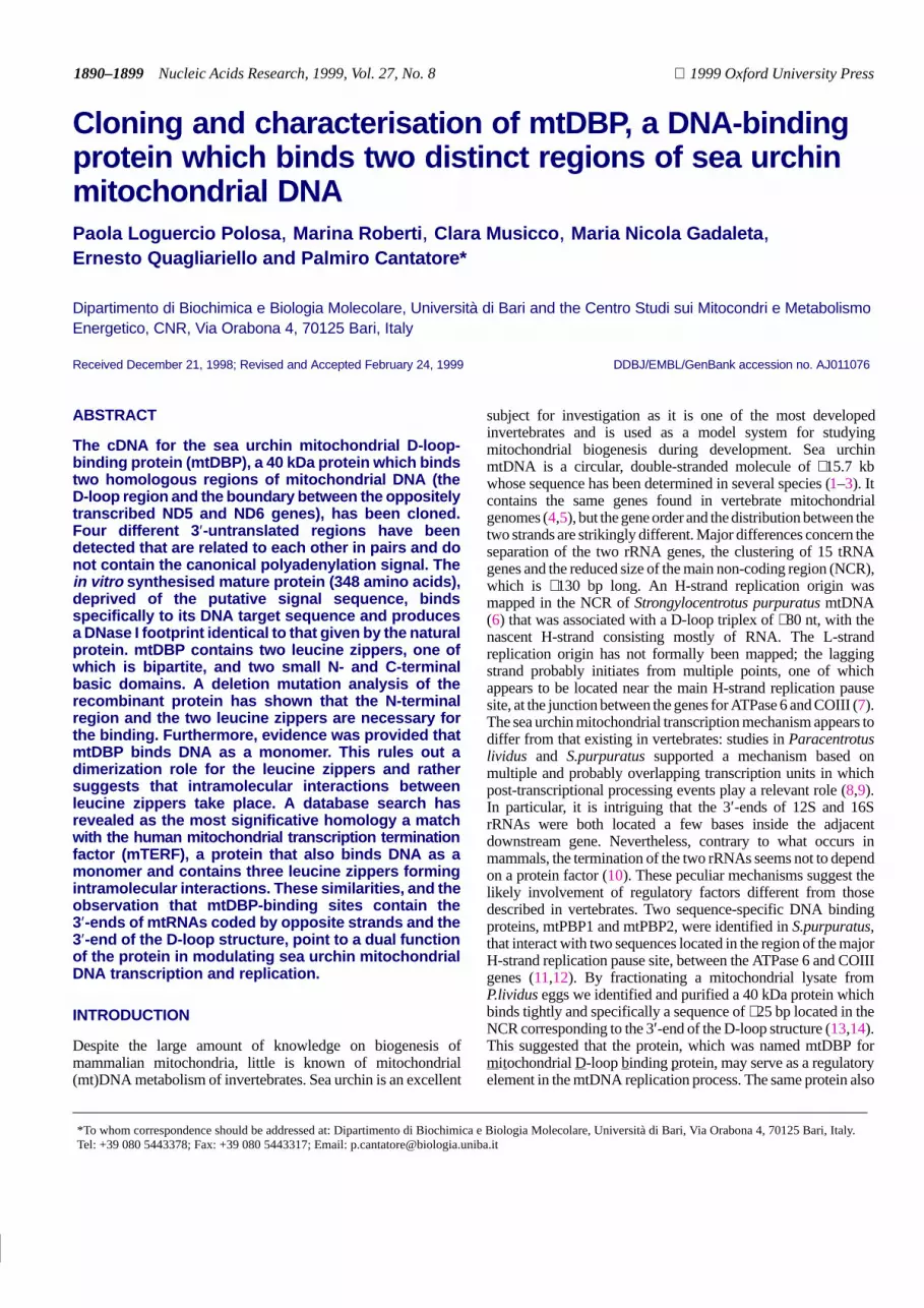
In order to analyse the functional capacity of the recombinantmtDBP (mtDBPr), the wild-type cDNA construct and two otherconstructs, mtDBPr1 and mtDBPr2, having N-terminal deletionsof different sizes, were in vitro translated (Fig. 3A and B). TheMtDBPr1 construct lacked part of the putative signal peptide(deleted residues were from –87 to –17) whereas mtDBPr2 lackedthe whole signal peptide and started from amino acid +1. Thebinding capacity of mtDBPr was initially determined by gelelectrophoretic mobility shift assay. As shown in Figure 3C, theprecursor version of the protein was not able to form a complexwith the labelled double-stranded oligonucleotide containing theNCR binding site. This feature seems to be common to all theprecursors of the mtDNA-binding proteins identified to date, asit has been reported also for the human transcription factorsmTERF (16) and mtTFA (28). A tentative explanation for thisphenomenon is that the structural fold of the signal sequencecould somehow mask the DNA-binding domain and prevent theprotein from contacting the corresponding site. Partial(mtDBPr1) and complete (mtDBPr2) removal of the N-terminusfrom the precursor relieved the apparent inhibition of DNAbinding, producing a single, specific retarded band. The bindingcapacity displayed by mtDBPr2 was much stronger than that ofmtDBPr1, which retains part of the presequence. Moreover, thecomplex formed by mtDBPr2 had the same mobility as thatproduced by the affinity-purified protein. These data let usassume that mtDBPr2 represents the active and probably matureversion of the protein. Increasing the amount of the proteinresulted in an increase in the intensity of the retarded band withoutthe appearance of any more slowly moving secondary band. Thisfinding argues against the formation of different complexes ofmtDBP with the probe. Finally, no complex formation was detectedusing a probe with a deletion in the NCR-binding site (data notshown), indicating that the interaction was specific. The specificityof mtDBPr interaction with the DNA was further confirmed byDNase I footprinting analysis. As shown in Figure 4, the pattern ofDNase I protection at the NCR (nt 1098–1126) and at the ND5/ND6boundary (nt 14 028–14 053) binding site produced by mtDBPr2 isessentially the same as that obtained with the affinity-purifiedmtDBP. This result conclusively confirms that the cDNA clone weisolated codes for P.lividus mtDBP.
A BLASTP analysis of the amino acid sequence of mtDBP withthe available protein databases revealed as statistically significanta match with the human mitochondrial transcription terminationfactor mTERF (accession no. Y09615; P = 1.7 × 10⁻¹⁴) and amatch with an unknown protein from Arabidopsis thaliana(accession no. AC000375; P = 1 × 10⁻¹⁰). This is a 462 aminoacid long polypeptide (J.Schwartz, personal communication) thatis predicted to be a mitochondrial protein by PSORTII analysis.When the comparison was performed with the mature versions ofthe proteins, 22% amino acid identity and 61% amino acidsimilarity were obtained for the pair mtDBP/mTERF (Fig. 5); acomparison between mtDBP and the unknown A.thaliana protein(not shown) gave 18% amino acid identity and 59.8% amino acidsimilarity. Concerning the pair mtDBP/mTERF, the homologyseems to be uniformly distributed along the molecule; however,regions of higher similarity (residues 99–113, 202–221 and310–325) can be detected. Almost 40% of the common residuesbetween mtDBP and mTERF were conserved among the threeproteins.

1895
Nucleic Acids Research, 1994, Vol. 22, No. 1Nucleic Acids Research, 1999, Vol. 27, No. 81895
Figure 3. Analysis of mtDBP recombinant forms. (A) Schematic representation of the precursor and shortened versions of mtDBP. The numbers represent amino acidpositions according to the numbering system used in Figure 1B. (B) SDS–polyacrylamide gel analysis. Precursor and shortened versions of mtDBP shown in (A) werein vitro expressed in the presence of [35S]methionine, separated on a SDS–polyacrylamide gel and subjected to autoradiography. The position of molecular massmarkers (in kDa) are shown to the left. (C) Mobility shift assays. Three increasing amounts of in vitro transcription–translation reaction mixtures (2, 4 and 8 µl)programmed with the templates shown in (A) were incubated with [α-32P]-labelled double-stranded 44mer oligonucleotide probe containing the NCR binding site(14). The protein–DNA complexes were resolved on a native polyacrylamide gel. The affinity-purified mtDBP (Aff. fraction), a minus protein (–) and anon-programmed reticulocyte lysate reaction (Retic. lys.) were used as controls.
Sequence analysis of mtDBP cDNA revealed, as the mostevident feature, the presence of multiple leucine zipper (LZ)motifs (underlined in Fig. 1B). The LZ motif consists of arepetition of leucines, or similar hydrophobic amino acids, whichare spaced seven residues apart (29,30). The most typical LZmotif found in mtDBP is LZ1, situated near the N-terminus of themature protein between residues 40 and 95. It is composed ofeight repeats of the heptad X3LX3 with asparagine, isoleucine andvaline substituting for leucine at the d position in the fourth, fifth andseventh heptads, respectively (the residues in the heptad beingdesigned a–g). The repeats exhibit the expected preponderance ofhydrophobic residues at the a (7/8) and d (7/8) positions of theheptads. A second potential leucine zipper can be localised nearthe C-terminus, between positions 221 and 277. In this case, themotif is formed by two repeats of three heptads located betweenresidues 221–241 and 257–277 and separated by a 15 amino acidloop. These heptads also have a high preponderance of hydro-phobic residues at a (4/6) and d (6/6) positions, with two valinesand one isoleucine substituting for leucine at the d position of thefirst, sixth and fifth heptads, respectively.
The roles of the different regions of mtDBP in its DNA-bindingactivity were investigated in gel shift experiments employingdeletion mutants. Four mutated versions of the protein werederived from mtDBPr2, since this has been shown to have thesame binding activity as the natural mtDBP. Two deletion
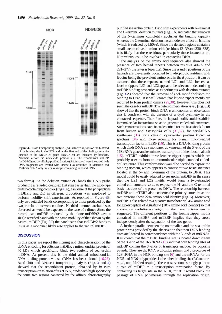
constructs were designed to produce N- and C-terminal truncatedversions of the protein (∆N and ∆C) lacking 39 and 19 aminoacids, respectively. The LZ1 region was deleted in the construct∆L1 eliminating amino acids from 40 to 95. To test the effect ofspecifically disrupting the bipartite leucine zipper domain LZ2,another construct, ∆L2 (lacking amino acids 221–277), was made(Fig. 6A). Of these mutated versions, only ∆C (Fig. 6A) retainssome binding activity (∼50%). When the N-terminus or the twoleucine zipper motifs were deleted separately, no binding activitywas observed. This result could be explained either by theremoval of a domain that interacts with DNA or by a deletion-induced change in the protein conformation which prevents theprotein from binding DNA.
Band shift assays using mtDBPr2 and a DNA probe containingthe specific binding site revealed a single retarded band evenwhen large amounts of the protein were used (Fig. 3C). Thisresult, together with the absence of a dyad symmetry in themtDBP binding site suggests that the protein binds DNA as amonomer. In order to obtain conclusive evidence about this pointthe heterodimerisation assay was used. This assay requires thattwo versions of mtDBPr differing in size form DNA–proteincomplexes with different mobility in a gel shift assay. If dimersof mtDBPr bind the DNA target, then using both protein forms inthe assay, three retarded bands should appear (homodimers of thelarge form, homodimers of the small form and heterodimers of the
Nucleic Acids Research, 1999, Vol. 27, No. 81896
Figure 4. DNase I footprinting analysis. (A) Protected regions on the L-strandof the binding site in the NCR and on the H-strand of the binding site at thejunction of the ND5/ND6 genes (ND5/ND6) are indicated by brackets.Numbers denote the nucleotide position (1). The recombinant mtDBP(mtDBPr2) and the affinity-purified fraction (Aff. fraction) were incubated withDNA fragments and treated with DNase I as described in Materials andMethods. ‘DNA only’ refers to sample containing unbound DNA.
two forms). As the deletion mutant ∆C binds the DNA probeproducing a retarded complex that runs faster than the wild-typeprotein-containing complex (Fig. 6A), a mixture of the polypeptidesmtDBPr2 and ∆C in different proportions was employed toperform mobility shift experiments. As reported in Figure 6B,only two retarded bands corresponding to those produced by thetwo proteins alone were obtained. No third intermediate band wasobserved, as would be expected in the case of a dimer. Since therecombinant mtDBP produced by the clone mtDBPr2 gave asingle retarded band with the same mobility of that shown by thenatural mtDBP (Fig. 3C) the conclusion that mtDBPr2 binds toDNA as a monomer likely also applies to the natural mtDBP.
DISCUSSION
In this paper we report the cloning and characterisation of thecDNA encoding for P.lividus mtDBP, a mitochondrial protein of40 kDa which specifically binds two regions of sea urchinmtDNA. At present this is the third animal mitochondrialDNA-binding protein whose cDNA has been cloned (16,28).Band shift and DNase I footprinting analysis (Figs 3 and 4)showed that the recombinant protein, obtained by in vitrotranscription–translation of its cDNA, binds with high specificitythe same two regions contacted by the affinity chromatography
purified sea urchin protein. Band shift experiments with N-terminaland C-terminal deletion mutants (Fig. 6A) indicated that removalof the N-terminus completely abolishes the binding capacitywhereas the C-terminal deletion has a moderate effect on binding(which is reduced by ∼50%). Since the deleted regions contain asmall stretch of basic amino acids (residues 12–39 and 330–338),it is likely that these residues, particularly those located at theN-terminus, could be involved in contacting DNA.
The analysis of the amino acid sequence also showed thepresence of two heptad repeats between residues 40–95 and221–277 (the latter is bipartite). Since the a and d positions of theheptads are prevalently occupied by hydrophobic residues, withleucine being the prevalent amino acid in the d position, it can beassumed that these repeats, named LZ1 and LZ2, behave asleucine zippers. LZ1 and LZ2 appear to be relevant in determiningmtDBP binding properties as experiments with deletion mutants(Fig. 6A) showed that the removal of each motif abolishes thebinding to DNA. It is well known that leucine zipper motifs arerequired to form protein dimers (29,30); however, this does notseem the case for mtDBP. The heterodimerisation assay (Fig. 6B)showed that the protein binds DNA as a monomer, an observationthat is consistent with the absence of a dyad symmetry in thecontacted sequence. Therefore, the heptad motifs could establishintramolecular interactions so as to generate coiled-coil structures.Such conformations have been described for the heat shock factorfrom human and Drosophila cells (31,32), for seryl-tRNAsynthetase (33), for a class of cytoskeleton proteins known asspectrins (34) and, more recently, for human mitochondrialtranscription factor mTERF (16). This is a DNA-binding proteinwhich binds DNA as a monomer downstream of the 3′-end of the16S rRNA gene and terminates transcription of the ribosomal unit(15). mTERF exhibits three leucine zipper heptads which areprobably used to form an intramolecular triple-stranded coiled-coil structure. This conformation would be needed to expose thebinding domain, which appears to consist of two basic stretcheslocated at the N- and C-termini of the protein, to DNA. Thismodel could be easily adapted to sea urchin mtDBP in the sensethat the LZ1 and LZ2 motifs would form a two-strandedcoiled-coil structure so as to expose the N- and the C-terminalbasic residues of the protein to DNA. The relationship betweenmtDBP and mTERF also concerns the primary structure as thetwo proteins show 22% amino acid identity (Fig. 5). Moreover,mtDBP is also related to a putative mitochondrial 462 amino acidlong polypeptide of A.thaliana (18% amino acid identity) so thata common evolutionary origin for the three proteins can besuggested. The different positions of the leucine zipper motifscontained in mtDBP and mTERF implies that they aroseindependently after the separation of the two genes.
A further parallel between the mammalian and the sea urchinprotein was provided by the observation that their DNA bindingsites are located in correspondence with the 3′-ends of mtRNAs.It is known that the mTERF binding site is located downstreamof the 3′-end of the 16S rRNA (15) and that both binding sites ofmtDBP contain the 3′-ends of transcripts encoded by oppositestrands. They are the RNA replication primer and a precursor of12S rRNA in the NCR binding site (6) and the mRNAs for theND5 and ND6 polypeptides in the other binding site (P.Cantatoreet al., unpublished results). These observations strongly point toa role of mtDBP as a transcription termination factor. Bycontacting its target site in the NCR, mtDBP would block thepassage of RNA polymerase through the replication origin,

1897
Nucleic Acids Research, 1994, Vol. 22, No. 1Nucleic Acids Research, 1999, Vol. 27, No. 81897
Figure 5. Sequence alignment of mtDBP and mTERF mature forms. Asterisks indicate identical nucleotides; colons and dots indicate very similar and similar residuesaccording to Thompson et al. (43).
avoiding the read-through of this region which might disturbprimer–template base pairing; on the other hand, transcriptionarrest at the boundary between the ND5 and ND6 genes wouldprevent head-on collision between the H- and L-strand transcriptionmachinery. The proposed role for mtDBP as a bidirectionaltranscription terminator provides new insights into the mechanismof mitochondrial transcription termination in sea urchins ascompared to vertebrates. In mammals one termination event forthe ribosomal transcription unit depending on mTERF has beendescribed (15). In sea urchins transcription arrest would occur incorrespondence with the two mtDBP binding sites, whereas the3′-ends of the two rRNAs will be generated by post-transcriptionalprocessing events as no protein-mediated termination event takesplace in these regions (10).
Based on the observation that the mtDBP-binding site in theNCR contains the 3′-end of the D-loop structure, it waspreviously inferred that mtDBP might have a role in regulatingmtDNA replication (14). By binding to its target site in the NCR,the protein could function as a negative regulator of H-strandelongation, thereby leading to D-loop formation. Relaxation ofthis interaction would favour H-strand extension thus resulting inproductive replication of the mitochondrial genome. A protein of∼48 kDa, the TAS-binding factor, has been shown to serve thisfunction in mammals (35). Therefore, mtDBP is likely to play adual function in regulating both mitochondrial DNA replication
and transcription. The use of the same protein to perform a roleboth in replication and transcription in sea urchins is justified bythe compact organisation of the sea urchin D-loop (∼130 nt asopposed to ∼1000 nt in mammals) and by the observation that the3′-end of the RNA primer is very close (20–30 bp) to the 3′-endof the newly synthesised DNA (6). Two other sequence-specificDNA-binding proteins (mtPBP-1 and mtPBP-2) from sea urchinmitochondria have been characterised (11,12). They bind to themain pause region of sea urchin mtDNA, which is located at theboundary between the COIII and ATPase 6 genes where the mainorigin for lagging strand replication was mapped. In this case anaction through blocking of leading strand replication andprogression of the RNA polymerase at this site has also beenproposed. The observation that the same protein factor is able toarrest both replication and transcription has been reported inmany prokaryotic and eukaryotic systems. In particular, inEscherichia coli and in Bacillus subtilis it was shown that thesame protein factor (ter protein in E.coli and RTP in B.subtilis) isable to block both progression of the replication fork at specificsites and RNA chain elongation (36). In mammals it has recentlybeen described that the RNA polymerase I transcription terminationfactor TTF-1 also causes polar arrest of rDNA replication,preventing head-on collision between the DNA replicationapparatus and the transcription machinery (37).
Nucleic Acids Research, 1999, Vol. 27, No. 81898
Figure 6. DNA-binding properties of recombinant mtDBP. (A) Mobility shift assay using different deletion mutants. (Upper) Schematic representation of wild-typeand deleted constructs used as templates in the in vitro expression system. The putative leucine zipper domains (LZ1 and LZ2) are indicated by black boxes. Thenumbers represent the amino acid positions according to the numbering system used in Figure 1B. (Lower) Mobility shift assays using two different amounts (2 and4 µl) of the expression reaction mixtures containing equivalent amounts of the constructs shown in the upper part as templates. The mature recombinant protein(mtDBPr2) was used as control. The probe was the 44mer double-stranded oligonucleotide. (B) mtDBP binds mtDNA as a monomer. Mobility shift analysis wasperformed by incubating the mature version (mtDBPr2) and the C-terminal truncated (∆C) version of mtDBP with the labelled 44mer probe. Different amounts ofthe expression reaction mixtures were employed in the mobility shift assays, as reported at the top of the figure. The protein–DNA complexes were resolved on a 10%native polyacrylamide gel.
In light of all the observations reported here, it seems thatmtDBP and mTERF are two proteins having a commonevolutionary origin that diverged to accomplish different roles,according to the variation in gene organisation and expressionbetween sea urchin and mammalian mitochondrial genomes.
In the course of mtDBP cDNA cloning, we identified fouridentical ORFs which display an unusual organisation of their3′-UTRs (Fig. 2). All four UTRs share the first 99 bp sequence;however, while UTR-λ appears to be a shortened version ofUTR-b, UTR-a and UTR-c overlap each other for only a total of115 bp. Therefore, the four UTRs appear to be related to eachother in pairs. No other sequence similarity, such as AU-richsequence motifs (38), are exhibited by the four UTRs. Concerningthe polyadenylation signal, no canonical AAUAAA sequence(39) was detected in any of the four UTRs. However, UTR-λexhibits the signal AAUAAC, which has been found as apolyadenylation signal in the sea urchin CS H1 cDNA (24).UTR-a, UTR-b and UTR-c display other variants of the signal(AAGAAA, AGUAAA and AAUACA, respectively). The four3′-UTRs detected so far are probably only some of the existing3′-UTRs of mtDBP. A RT–PCR experiment (not shown)employing a set of primers designed to detect differences in the3′-UTR region of RNAs indeed revealed the existence of acomplex pattern of RNA species for mtDBP which appeared tobe developmentally regulated. These multiple mRNA speciesmight be involved in determining the translational efficiency
and/or the stability of the message during development bypromoting the rapid degradation and removal of the message orby conferring an increase in translation at critical developmentalstages (40–42).
ACKNOWLEDGEMENTS
We are very grateful to R. Fiore for collaboration in the late stagesof this investigation. The help of W. Lane (Harvard MicrochemistryFacility, Cambridge, MA) in the protein sequencing analysis isgratefully acknowledged. We thank F. Aniello for kindlyproviding the λ Uni-Zap cDNA library of P.lividus embryos. Thetechnical assistance of F. Milella and V. Cataldo is gratefullyacknowledged. This work was supported in part by Ministerodell’Università e della Ricerca Scientifica, project ‘Protein–nucleicacid interactions’, from Consiglio Nazionale delle Ricerche, contractno. 96.03726.CT14, and from Telethon Italy (grant no. 863).
REFERENCES
1 Cantatore,P., Roberti,M., Rainaldi,G., Gadaleta,M.N. and Saccone,C.(1989) J. Biol. Chem., 264, 10965–10975.
2 Jacobs,H.T., Elliott,D.J., Math,V.B. and Farquharson,A. (1988)J. Mol. Biol., 202, 185–217.
3 De Giorgi,C., Martiradonna,A., Lanave,C. and Saccone,C. (1996)Mol. Phylogenet. Evol., 5, 323–332.
4 Cantatore,P. and Saccone,C. (1987) Int. Rev. Cytol., 108, 149–208.5 Attardi,G. and Schatz,G. (1988) Annu. Rev. Cell. Biol., 4, 289–333.

1899
Nucleic Acids Research, 1994, Vol. 22, No. 1Nucleic Acids Research, 1999, Vol. 27, No. 81899
6 Jacobs,H.T., Herbert,E.R. and Rankine,J. (1989) Nucleic Acids Res., 17,8949–8965.
7 Mayhook,A.G., Rinaldi,A.M. and Jacobs,H.T. (1992) Proc. R. Soc. Lond. B,248, 85–94.
8 Cantatore,P., Roberti,M., Loguercio Polosa,P., Mustich,A. andGadaleta,M.N. (1990) Curr. Genet., 17, 235–245.
9 Elliott,D.J. and Jacobs,H.T. (1989) Mol. Cell. Biol., 9, 1069–1082.10 Roberti,M., Loguercio Polosa,P., Musicco,C., Milella,F., Qureshi,S.,
Gadaleta,M.N., Jacobs,H.T. and Cantatore,P. (1999) Curr. Genet., 34,449–458.
11 Qureshi,S.A. and Jacobs,H.T. (1993) Nucleic Acids Res., 21, 811–816.12 Qureshi,S.A. and Jacobs,H.T. (1993) Nucleic Acids Res., 21, 2801–2808.13 Roberti,M., Mustich,A., Gadaleta,M.N. and Cantatore,P. (1991)
Nucleic Acids Res., 19, 6249–6254.14 Loguercio Polosa,P., Roberti,M., Mustich,A., Gadaleta,M.N. and
Cantatore,P. (1994) Curr. Genet., 25, 350–356.15 Kruse,B., Narasimhan,N. and Attardi,G. (1989) Cell, 58, 391–397.16 Fernandez-Silva,P., Martinez-Azorin,F., Micol,V. and Attardi,G. (1997)
EMBO J., 16, 1066–1079.17 Laemmli,U.K. (1970) Nature, 227, 680–685.18 MacDonald,R.J., Swift,G.H., Przybyla,A.E. and Chirgwin,J.M. (1987)
Methods Enzymol., 152, 223–224.19 Loguercio Polosa,P. and Cantatore,P. (1997) Comments Amers. Life Sci.,
23, 10–11.20 Frohman,M.A. (1994) PCR Methods Applicat., 4, S40–S58.21 Feinberg,A.P. and Vogelstein,B. (1984) Anal. Biochem., 137, 266–267.22 Sambrook,J., Fritsch,E.F. and Maniatis,T. (1989) Molecular Cloning:
A Laboratory Manual, 2nd Edn. Cold Spring Harbor Laboratory Press,Cold Spring Harbor, NY.
23 Fucci,L., Piscopo,A., Aniello,F., Branno,M., Di Gregorio,A., Calogero,R.and Geraci,G. (1995) Gene, 152, 205–208.
24 Mandl,B., Brandt,W.F., Superti-Furga,G., Graninger,P.G., Birnstiel,M.L.and Busslinger,M. (1997) Mol. Cell. Biol., 17, 1189–1200.
25 Kozak,M. (1986) Cell, 44, 283–292.26 Nakai,K. and Kanehisa,M. (1992) Genomics, 14, 897–911.27 von Heijne,G., Steppuhn,J. and Herrmann,R.G. (1989) Eur. J. Biochem.,
180, 535–545.28 Parisi,M. and Clayton,D.A. (1991) Science, 252, 965–969.29 Landshultz,W.H., Johnson,P.F. and McKnight,S.L. (1988) Science, 240,
1759–1764.30 Hurst,H. (1995) In Sheterline,P. (ed.), Protein Profile. Academic Press,
London, UK, Vol. 2, pp. 105–168.31 Zuo,J., Baler,R., Dahl,G. and Voellmy,R. (1994) Mol. Cell. Biol., 14,
7557–7568.32 Westwood,T. and Wu,C. (1993) Mol. Cell. Biol., 13, 3481–3486.33 Cusack,S., Berthet-Colominas,C., Härtlein,M., Nassar,N. and Leberman,R.
(1990) Nature, 347, 249–255.34 Yan,Y., Winograd,E., Viel,A., Cronin,T., Harrison,S.C. and Branton,D.
(1993) Science, 262, 2027–2030.35 Madsen,C.S., Ghivizzani,S.C. and Hauswirth,W.W. (1993) Mol. Cell. Biol.,
13, 2162–2171.36 Mohanty,B.K., Sahoo,T. and Bastia,D. (1996) EMBO J., 15, 2530–2539.37 Gerber,J.K., Gögel,E., Berger,C., Wallisch,M., Müller,F., Grummt,I. and
Grummt,F. (1997) Cell, 90, 559–567.38 Chen,C.-Y. and Shyu,A.-B. (1995) Trends Biochem. Sci., 20, 465–470.39 Humphrey,T. and Proudfoot,N.J. (1988) Trends Genet., 4, 243–245.40 Sheets,M.D., Ogg,S.C. and Wickens,M.P. (1990) Nucleic Acids Res., 18,
5799–5805.41 Jackson,R.J. (1993) Cell, 74, 9–14.42 Wilhelm,J.E. and Vale,R.D. (1993) J. Cell Biol., 123, 269–274.43 Thompson,J.D., Higgins,D.G. and Gibson,T.J. (1994) Nucleic Acids Res.,
22, 4673–4680.