chronic ketamine administration impairs mitochondrial complex i in the rat liver
TRANSCRIPT
Life Sciences 93 (2013) 464–470
Contents lists available at ScienceDirect
Life Sciences
j ourna l homepage: www.e lsev ie r .com/ locate / l i fesc ie
Chronic ketamine administration impairs mitochondrial complex I in therat liver
Carlos Venâncio a,b,⁎, Luís Antunes a,c, Luís Félix a, Paula Rodrigues b,c, Teresa Summavielle d, Francisco Peixoto e
a Laboratory Animal Science, Instituto de Biologia Molecular e Celular, Universidade do Porto, Rua do Campo Alegre 823, 4150-180 Porto, Portugalb Veterinary and Animal Research Centre, Universidade de Trás-os-Montes e Alto Douro, Apartado 1013, 5001-801 Vila Real, Portugalc Department of Veterinary Sciences, Escola de Ciências Agrárias e Veterinárias, Universidade de Trás-os-Montes e Alto Douro, Apartado 1013, 5001-801 Vila Real, Portugald Department of Functional Sciences, Escola Superior de Tecnologia da Saúde do Porto, Instítuto politécnico do Porto, Vila Nova de Gaia, Portugale Centre for Research and Technology of Agro-Environment and Biological Sciences, Universidade de Trás-os-Montes e Alto Douro, Apartado 1013, 5001-801 Vila Real, Portugal
⁎ Corresponding author at: Quinta de Prados, ApartaPortugal. Tel.: +351 259 350 403; fax: +351 259 350 32
E-mail address: [email protected] (C. Venâncio).
0024-3205/$ – see front matter © 2013 Elsevier Inc. All rihttp://dx.doi.org/10.1016/j.lfs.2013.08.001
a b s t r a c t
a r t i c l e i n f oArticle history:
Received 6 April 2013Accepted 2 August 2013Keywords:KetamineMitochondriaOxidative stressLiverBioenergeticsGlycogen
Aim: Ketamine can induce hepatotoxicity which has been suggested to be dependent on mitochondrial impair-ment. This study investigated the long-term effects of chronic low-dose ketamine on liver mitochondrial func-tion, oxidative stress parameters, liver histology and glycogen content.Main methods: Adult rats were administeredwith saline or ketamine (5 or 10 mg/kg) twice a day for a fourteen-day period in order tomimic chronic treatments. Effects between groupswere compared ten days after the treat-ment had ended. Liver mitochondrial function was monitored in isolated mitochondrial extracts through evalu-ation of respiration parameters and activity of respiratory complexes, as well as oxidative stress, through lipidperoxidation, protein oxidation and superoxide dismutase activity. The hepatic histology and liver glycogen con-tent were also evaluated.Key findings: Ketamine groups showed a decreased evolution in body weight gains during the treatment period.
Ketamine had no effect either on serum liver enzymes or on the oxidative stress parameters of liver mitochon-dria. Ketamine decreased the hepatic glycogen content, inhibitedmitochondrial complex I and oxygen consump-tion when glutamate–malate substrate was used.Significance: These findings reflect a long-term mitochondrial bioenergetic deterioration induced by ketamine,which may explain the increased susceptibility of some patients to its prolonged or repeated use.© 2013 Elsevier Inc. All rights reserved.
Introduction
Ketamine has gained a renewed interest for depression and chronicpain management, since it has an immediate and persistent effect on ahigh percentage of cases in which the classic therapeutic is not effective(Correll et al., 2004; Zarate et al., 2006).Moreover, ketamine is being in-creasingly used as a recreational drug (Morgan et al., 2010). However,hepatotoxic side effects have been frequently described after repeatedexposure to it (Correll et al., 2004; Noppers et al., 2011). Ketamine'sexact biochemicalmechanism for hepatotoxicity is not fully understood,although mitochondrial dysfunction implications have been suggestedas a possible explanation (Noppers et al., 2011). The liver is a very activemetabolic organ with a high number of mitochondria and very suscep-tible to drugs acting on mitochondria, which may interfere with bioen-ergetic function and redox homeostasis (Boelsterli and Lim, 2007;Szewczyk and Wojtczak, 2002).
Ketamine is metabolised in the liver by the cytochrome P-450micro-somal system (Chan et al., 2005). In human patients, hepatic injuries
do 1013, 5001-801 Vila Real,7.
ghts reserved.
have been reported after ketamine anaesthetic infusions (Blunnie et al.,1981; Kiefer et al., 2008) and in the course of long chronic pain treat-ments (Correll et al., 2004; Noppers et al., 2011). In vitro studies suggestthat mitochondrial dysfunction has an important role on ketamine liverinduced injuries (Chang et al., 2009; Lee et al., 2009; Markham et al.,1981). When exposed to ketamine, hepatocytes showed an inhibitionof oxidative phosphorylation (Chang et al., 2009), increased apoptotic in-sults (Lee et al., 2009) and reactive oxygen species (ROS) production(Reinke et al., 1998). Contrarily, ketamine also showed hepatoprotectiveeffectswhich are attributed to its anti-inflammatory properties (Suliburket al., 2005).
Despite several in vitro studies (Chang et al., 2009; Lee et al., 2009;Markham et al., 1981), there is still little information on ketaminechronic effects on liver mitochondria and oxidative stress responseswhen in vivo models are used. Very recently, Kalkan et al. (2013) havereferred hepatic pathologic changes with mitochondrial degenerationin rats which had been administered high doses of ketamine for twoweeks (Kalkan et al., 2013). In addition, we had previously reportedthat rats subject to a similar period of chronic low doses of ketamineshowed persistent behavioural alterations and loss of weight gain(Venancio et al., 2011). Thus, this study was designed to look into thelong-term effects on the liver mitochondrial bioenergetic function,
465C. Venâncio et al. / Life Sciences 93 (2013) 464–470
oxidative stress parameters and liver glycogen content following achronic low dose ketamine treatment in adult rats.
Material and methods
Animals and drug treatment
All procedures used were approved by the local ethical committeeand by the PortugueseAgency for AnimalWelfare, General Board of Vet-erinaryMedicine, in compliancewith the European Community CouncilDirective of September 22, 2010 (2010/63/UE). All efforts weremade toensure minimal animal stress and discomfort.
Twenty-one adult male Wistar rats (90–110 days) acquired fromCharles River (Barcelona, Spain) were randomised in three groups.Animals were kept under controlled environmental conditions of tem-perature (20 ± 1 °C), relative air humidity (45–55%), maintained in a12-h light/dark cycle and housed in groups of two or three per cagewith standard rodent food and water supplied ad libitum.
Ketamine (Imalgene1000®Merial, Portugal, 100 mg/ml) was fresh-ly diluted before subcutaneous injection in a volume of 1 ml/kg of bodyweight. Groups were administered with saline solution (K0), 5 mg/kgketamine (K5) or 10 mg/kg ketamine (K10). Doses were establishedin accordance with previous studies for analgesic (Wang et al., 2000)or antidepressant properties in rats (Li et al., 2010). Injections weregiven twice daily (9:00/21:00 h) for 14 consecutive days to achieve achronic effect (Garcia et al., 2008). The body weight gains were moni-tored daily throughout the experimental period. Rats were killed10 days after the last administration (24th day of experiment) by de-capitation and blood samples were obtained. The liver was rapidly re-moved for mitochondrial isolation, histology analysis and glycogendetermination.
Reagents
Substrates, enzymes and standard chemical reagents were of thehighest grade commercially available and obtained from Sigma (Sigma-Aldrich, Steinheim, Germany). All solutions were prepared with ultra-pure water purified by a Milli-Q Gradient system (Millipore, Bedford,USA).
Liver enzyme activity measurements
The measurement of the hepatocellular enzymes alanine amino-transferase, aspartate aminotransferase and gamma-glutamyl transfer-ase in the blood stream was used as an index of hepatic injury. Bloodsampleswere stored at 4 °C for 1 h; after clotting, theywere centrifugedfor 10 min at 2800 g. Serum levels of enzyme activity weremeasured at37 °C by standard clinical chemical methods using an automatic analyz-er type Architect ci8200 (Abbott Laboratories Ltd., USA).
Liver histology
Liver samples were processed by standard techniques and submit-ted to histologic examination (light microscopy). Briefly, sampleswere immersed in 10% buffered formalin and embedded in paraffinwax prior to sectioning. For each sample, three series of three sections(3 μm thick) were cut and stained with haematoxylin and eosin or pe-riodic acid-Schiff (PAS) reagent method to detect glycogen as previousdescribed (Bancroft and Stevens, 2002). Diastase for glycogen digestionwas used as control. Morphologic analysis was made by a pathologistwhowas blinded to the experimental protocol. The hepatic morpholog-ic characteristics and increase of areas with glycogen (PAS+) sensitiveto digestion with diastase were assessed. The selection of hepatic areasfor glycogen content evaluation in hepatocytes was done according topreviously describedmethods (Stadler et al., 2005). Digital photomicro-graphs of each section were taken (Nikon 4500 Coolpix, USA). Each
sample section was graded as follows for PAS+ areas: mild (+) — allareas showing slight PAS+; moderate (++) — a combination of areaswith slight PAS+ and others with intense PAS+ staining; high(+++) — extensive and marked areas with intense PAS+ staining.
Glycogen analysis
Liver homogenate was used for glycogen concentration determina-tion using phenol–sulphuric method as described previously (Bennettet al., 2007). This was read at 490 nm, 30 min after incubation (Cary100 Bio, Varian Analytical Instruments, USA).
Isolation of liver mitochondria
The isolation was performed as described previously (Peixoto et al.,2002). The homogenization medium contained 225 mM mannitol,75 mM sucrose, 10 mM Hepes (pH 7.4), 2 mM ethylene glycol-bis(β-aminoethyl ether)-N,N,N′,N′-tetraacetic acid (EGTA) and 0.1% fattyacid-free bovine serum albumin (BSA). EGTA and BSA were omittedfrom the final washingmedium. The final concentration of mitochondri-al protein was determined by the Biuret method, using BSA as standard.
Liver mitochondrial respiratory activity
Oxygen consumption of isolated mitochondria was monitored po-larographically with a Clark-type oxygen electrode, using a HansatechOxygraph measurement system (Hansatech, Norfolk, UK) at 25 °C.Mitochondria (0.5 mg) were incubated, for 2 min in 1 ml of re-action medium (100 mM sucrose, 100 mM KCl, 2 mM KH2PO4, 5 mMHepes, 10 μM EGTA, pH 7.4). The mitochondria were energised withglutamate–malate (complex I substrate) or succinate (complex II sub-strate). Oxygen consumption was measured in the absence (state4) or presence of 100 μM ADP (state 3). Respiratory rates wereexpressed in nmol O2/citrate synthase activity. Respiratory controlratio (RCR = state 3/state 4) was calculated (Vilela et al., 2009).
Liver mitochondrial enzymatic activity assays
Before the enzyme assays, the mitochondrial frozen samples (at−70 °C) were freeze-thawed three times to disrupt their mem-branes. All the enzymatic assays were performed on a temperaturecontrolled chamber with stirring and were performed in triplicatein a microplate apparatus (Power Wave XS2, Biotek, USA).
Citrate synthase activity was determined as described previously at30 °C (Shepherd and Garland, 1969) with slight modifications. The re-action mixture contained 100 mM Tris–HCl buffer (pH 8.0), 5 mMMgCl2, 0.5 mM 5,5-dithio-bis-2-nitrobenzoic acid, 0.2 mM acetyl CoA,1 mM oxaloacetic acid and broken mitochondria (15–25 μg). The de-crease of acetyl CoA absorption was monitored at 412 nm and specificactivity, as units of citrate synthase activity/min ∙ mg of protein, wascalculated and used to normalise the other mitochondrial functionalparameters.
The electron transport chain complexes I, II, III and IV were quanti-fied by spectrophotometric assay as previously described (Kiebishet al., 2008). Complex V (ATPase) activity was determined by monitor-ing the pH change associated with ATP hydrolysis (Peixoto et al., 2002).
Liver mitochondrial oxidative stress parameters
Lipid peroxidation was determined measuring malondialdehydeequivalents, using the thiobarbituric acid assay, as previously described(Alves et al., 2007). Protein carbonylswere quantified through the spec-trophotometric carbonyl assay, as recently described (Hawkins et al.,2009), using 2,4-dinitrophenylhydrazine. Superoxide dismutase activi-ty was determined as described previously (Paya et al., 1992).
466 C. Venâncio et al. / Life Sciences 93 (2013) 464–470
Statistical analysis
Comparisonsweremade by one-wayANOVA followed,when appro-priated, by Tukey's post hoc test. Repeated-measures ANOVA was usedforweight gain analysis. Results are expressed asmean ± SEM. The sta-tistical level of significant was considered at p b 0.05, using theGraphPad Prism 6.0 (GraphPad Software, San Diego, CA, USA).
Results
Body weight gain
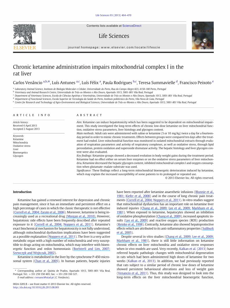
The chronic ketamine administrations induced a decrease in bodyweight gain during the experimental period F(2,17) = 32.44,p b 0.0001, (Fig. 1). Nomortalitywas observed between groups; howev-er, one rat was excluded for displaying aggressive behaviour. The resultsrevealed decreased body weight gain among the K0 and the ketaminegroups after the secondday of treatment (p b 0.05). Theweight recoverystarted on day ten for both ketamine groups. The K5 group showed lessaccentuated differences and after the twenty-second day no significantdifferences were observed by comparison with K0 group (p = 0.79).Both groups failed to reach the control values. Moreover, except forday two, the K10 group showed a significant reduction in body weightgain compared to the K5 group (p b 0.05).
Blood evaluation
The activities of biochemical markers of liver damage, such as ala-nine aminotransferase and aspartate aminotransferase, were not signif-icantly different between groups. The levels of gamma-glutamyltransferase were below the detection limits (b4 IU/l) in all samples.
Fig. 1. Effect of chronic ketamine administration onweight gain throughout the experimental pmean ± SEM for each group of body weight in a given day. Saline solution (K0, n = 7); 5 mgmarked as ⁎p b 0.05, ⁎⁎p b 0.01 and ⁎⁎⁎p b 0.001 for K0; #p b 0.05, ##p b 0.01 and ###p b 0.0
Liver histology and glycogen concentration
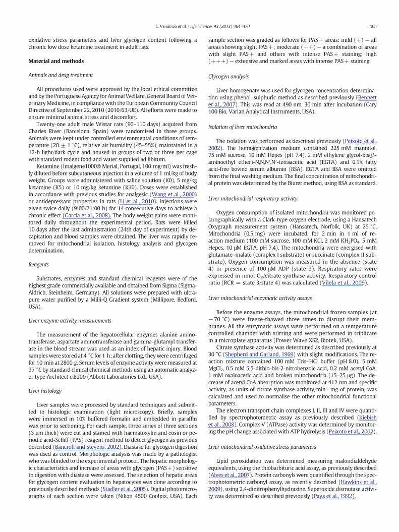
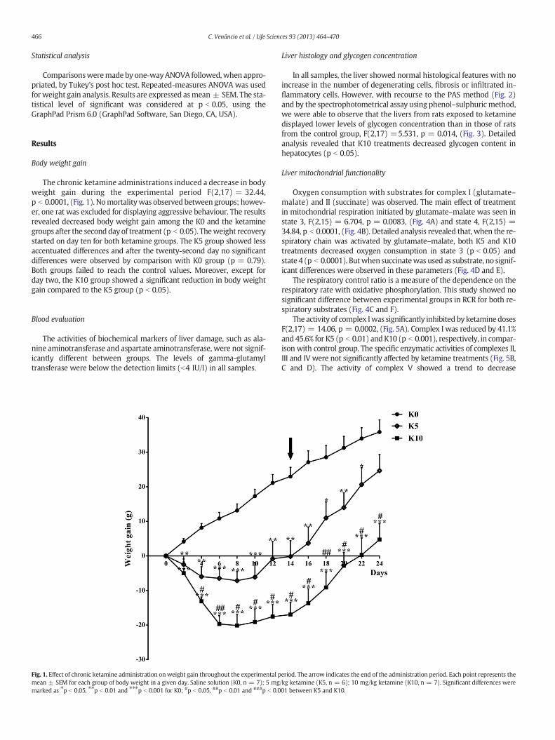
In all samples, the liver showed normal histological features with noincrease in the number of degenerating cells, fibrosis or infiltrated in-flammatory cells. However, with recourse to the PAS method (Fig. 2)and by the spectrophotometrical assay using phenol–sulphuricmethod,we were able to observe that the livers from rats exposed to ketaminedisplayed lower levels of glycogen concentration than in those of ratsfrom the control group, F(2,17) =5.531, p = 0.014, (Fig. 3). Detailedanalysis revealed that K10 treatments decreased glycogen content inhepatocytes (p b 0.05).
Liver mitochondrial functionality
Oxygen consumption with substrates for complex I (glutamate–malate) and II (succinate) was observed. The main effect of treatmentin mitochondrial respiration initiated by glutamate–malate was seen instate 3, F(2,15) = 6.704, p = 0.0083, (Fig. 4A) and state 4, F(2,15) =34.84, p b 0.0001, (Fig. 4B). Detailed analysis revealed that, when the re-spiratory chain was activated by glutamate–malate, both K5 and K10treatments decreased oxygen consumption in state 3 (p b 0.05) andstate 4 (p b 0.0001). Butwhen succinatewas used as substrate, no signif-icant differences were observed in these parameters (Fig. 4D and E).
The respiratory control ratio is a measure of the dependence on therespiratory rate with oxidative phosphorylation. This study showed nosignificant difference between experimental groups in RCR for both re-spiratory substrates (Fig. 4C and F).
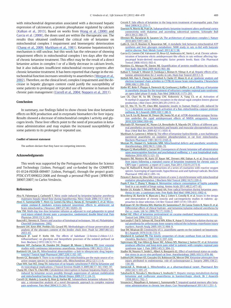
The activity of complex Iwas significantly inhibited by ketamine dosesF(2,17) = 14.06, p = 0.0002, (Fig. 5A). Complex I was reduced by 41.1%and 45.6% for K5 (p b 0.01) andK10 (p b 0.001), respectively, in compar-ison with control group. The specific enzymatic activities of complexes II,III and IV were not significantly affected by ketamine treatments (Fig. 5B,C and D). The activity of complex V showed a trend to decrease
eriod. The arrow indicates the end of the administration period. Each point represents the/kg ketamine (K5, n = 6); 10 mg/kg ketamine (K10, n = 7). Significant differences were01 between K5 and K10.
Fig. 2. Effects of chronic ketamine in liver glycogen content. Histopathologic findings showed a reduction of glycogen in hepatocytes for 5 and 10 mg/kg ketamine compared to control.Representative photomicrographs of liver sections stained with PAS (objective, 20×): (A) saline solution; (B) 5 mg/kg ketamine and (C) 10 mg/kg ketamine group.
467C. Venâncio et al. / Life Sciences 93 (2013) 464–470
F(2,17) = 3.133, p = 0.0695, (Fig. 5E), with a reduction of 45.0% and28.3% in groups K5 and K10, respectively, in comparison with the controlgroup.
Liver mitochondrial oxidative stress
The lipid peroxidation, protein carbonyls and superoxide dismutaseactivity evaluated in liver mitochondria were not significantly differentbetween groups (data not shown).
Discussion
This work shows that chronic ketamine treatment with a lower dosein rats impairs liver mitochondrial complex I and decreases liver glyco-gen content as well as body weight gain. Nevertheless, neither other he-patic histological changes nor significant hepatotoxicity effects wereobserved.
The body weight gain is a parameter usually used as a health indica-tor in chronic rodent toxicology studies (Rhomberg et al., 2007). Chroniclow ketamine doses have induced a significant loss of body weight gain.These results are consistent with other previous observations (Venancioet al., 2011). At the end of the experimental period, only the higher ke-tamine group showed a significant weight reduction. A possible expla-nation for the decrease of body weight gain could be related to the factthat ketamine induces nausea and vomitingwith consequent loss of ap-petite as observed in humans (Cvrcek, 2008). Moreover, nausea andvomiting are associated with liver injury (Verma and Kaplowitz,2009). However, we did not observe alterations in enzymatic bio-markers for liver injury. Similarly, in humans, after chronic pain man-agement with ketamine, no significant alterations of liver enzymeswere shown (Amr, 2010; Cvrcek, 2008; Sigtermans et al., 2009). This
Fig. 3. Effects of chronic ketamine in liver glycogen content. Results are expressed asmeans ± SEM (n = 6–7) for each group. K0 (saline solution), K5 and K10 (5 or 10 mg/kgketamine, respectively). Significant differences for control are marked as *p b 0.05.
could also be the result of biochemical analysis having been performedten days after the last ketamine administration; also, as it happenedwith humans, an increase of liver enzymes was observed but it wentback to normal values between ten days and two months after thepain treatment had ended (Kiefer et al., 2008; Noppers et al., 2011). An-other likely explanation for the decrease of bodyweight gainmay be re-lated to the fact that, being an N-methyl-D-aspartate (NMDA) receptorantagonist, ketamine may have brain interference in food intake(Popik et al., 2011;Wu et al., 2012) and also hepatic metabolism regula-tion implications (Lam et al., 2010).
This study showed a decrease of hepatic glycogen concentration inthe higher ketamine group, whichwas also corroborated by histologicalobservations. Otherwise, no hepatic histological changes were ob-served. Reduced glycogen store in hepatocytes is related to an increasedsusceptibility to hepatotoxicity (Stadler et al., 2005). In mice, chronicketamine treatment also decreases glycogen in liver, reflecting either afailure of glycogen synthesis or an increase of glycogenolysis (Wonget al., 2012). Other studies also reported that ketamine can promote he-patic bioenergetic deteriorationwith concomitant reduction of ATP pro-duction (Takahashi et al., 1998), gluconeogenesis (Sear and McGivan,1979) and increased glycogenolysis (Machado et al., 2009) which maybe associated with an increase of blood glucose levels (Dawson et al.,2013; Rodrigues et al., 2006).
Hepatic bioenergetic deterioration reflected by the decrease in gly-cogen is usually coupled with reduction of ATP production (Galliset al., 2011). Ketamine decreased the oxygen consumption in state 3and state 4 when complex I was energised. This may result from theobserved inhibition of complex I (NADH ubiquinone oxidoreductase)activity. Similar effects of ketamine in hepatic cell cultures have been re-cently observed such as time-dependent interruption of microtubularcytoskeleton remodelling, decreased intracellular levels of calcium con-centrations and concurrent reduction of ATP production due to down-regulation of the mitochondrial membrane potential and complex I ac-tivity (Chang et al., 2009). When succinate was used as substrate, therewere neither significant alterations, nor significant change in the activ-ity of other electron transport chain complexes which confirms that theprevious reported decrease in oxygen consumption is due to a direct ef-fect on complex I. In line with these findings, it has been reported thatketamine induces mitochondrial complex I impairment on isolated ratliver mitochondria (Markham et al., 1981). However, a normal valueof RCR in ketamine groups for the two substrates used also indicatesthat the structural integrity of mitochondria was not affected. ComplexI is the first enzyme of the respiratory chain and plays a central role inoxidative phosphorylation (Efremov et al., 2010). Its inhibition promot-ed by ketamine and by other anaestheticsmay be related to the interfer-ence in glucose metabolism (Biebuyck et al., 1972).
Complex I inhibition can exacerbate mitochondrial ROS generation.Nevertheless, due to its antioxidant capacity, mitochondria can rapidlydegrade ROS (Brown and Borutaite, 2012). This can account for the
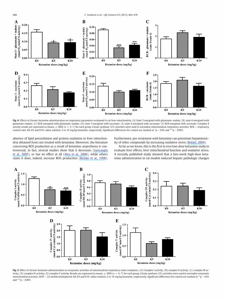
Fig. 4. Effects of chronic ketamine administration on respiratory parameters evaluated in rat liver mitochondria. (A) State 3 energised with glutamate–malate; (B) state 4 energised withglutamate–malate; (C) RCR energised with glutamate–malate; (D) state 3 energised with succinate; (E) state 4 energised with succinate; (F) RCR energised with succinate. Complex IIactivity results are expressed as means ± SEM (n = 5–7) for each group. Citrate synthase (CS) activities were used to normalise mitochondrial respiratory activities. RCR — respiratorycontrol ratio. K0, K5 and K10: saline solution, 5 or 10 mg/kg ketamine, respectively. Significant differences for control are marked as *p b 0.05 and ***p b 0.001.
468 C. Venâncio et al. / Life Sciences 93 (2013) 464–470
absence of lipid peroxidation and protein oxidation in liver mitochon-dria obtained from rats treated with ketamine. Moreover, the literatureconcerning ROS production as a result of ketamine anaesthesia is con-troversial; in fact, several studies show that it decreases (Saricaogluet al., 2005), or has no effect at all (Alva et al., 2006), while othersclaim it does, indeed, increase ROS production (Reinke et al., 1998).
Fig. 5. Effect of chronic ketamine administration on enzymatic activities of mitochondrial respitivity; (D) complex IV activity; (E) complex V activity. Results are expressed asmeans ± SEM (nmitochondrial activities. DCIP— 2,6-dichloroindophenol. K0, K5 and K10: saline solution, 5 or 10and ***p b 0.001.
Furthermore, pre-treatment with ketamine can potentiate hepatotoxic-ity of other compounds by increasing oxidative stress (Rofael, 2004).
As far aswe know, this is thefirst in vivo low-dose ketamine study toevaluate liver effects, liver mitochondrial function and oxidative stress.A recently published study showed that a two-week high-dose keta-mine administration in rat models induced hepatic pathologic changes
ratory chain complexes. (A) Complex I activity; (B) complex II activity; (C) complex III ac-= 6–7) for each group. Citrate synthase (CS) activities were used to normalise enzymaticmg/kg ketamine, respectively. Significant differences for control aremarked as **p b 0.01
469C. Venâncio et al. / Life Sciences 93 (2013) 464–470
with mitochondrial degeneration associated with a decreased hepaticexpression of calcineurin, a protein phosphatase regulated by calcium(Kalkan et al., 2013). Based on works from Wang et al. (2000) andGarcia et al. (2008), the doses used are within the therapeutic use. Theresults thus obtained confirmed the critical role of ketamine onmitochondrial complex I impairment and bioenergetic deterioration(Chang et al., 2009; Markham et al., 1981). Ketamine hepatotoxicity'smechanism is still unclear, but this work has the relevance of showingimpairment effects in mitochondrial complex I ten days after the endof chronic ketamine treatment. This effect may be the result of a directketamine action in complex I or of a likely decrease in calcium levels,but it also indicates insufficient mitochondrial recovery systems afterchronic ketamine treatment. It has been reported that change in themi-tochondrial function increases sensitivity to anaesthetics (Morgan et al.,2002). Therefore, on the clinical level, complex I impairment and thede-crease in hepatic glycogen content could justify the susceptibility ofsome patients to prolonged or repeated use of ketamine in humans forchronic pain management (Correll et al., 2004; Noppers et al., 2011).
Conclusion
In summary, our findings failed to show chronic low dose ketamineeffects on ROS production and in enzymatic biomarkers for liver injury.Results showed a decrease of mitochondrial complex I activity and gly-cogen levels. These liver effects point to the need of precaution in keta-mine administration and may explain the increased susceptibility ofsome patients to its prolonged or repeated use.
Conflict of interest statement
The authors declare that they have no competing interests.
Acknowledgments
This work was supported by the Portuguese Foundation for Scienceand Technology (Lisbon, Portugal) and co-funded by the COMPETE:01-0124-FEDER-009497 (Lisbon, Portugal), through the project grant:PTDC/CVT/099022/2008 and through a personal PhD grant (SFRH/BD/38907/2007) to Carlos Venâncio.
References
Alva N, Palomeque J, Carbonell T. Nitric oxide induced by ketamine/xylazine anesthesiamaintains hepatic blood flow during hypothermia. Nitric Oxide 2006;15(1):64–9.
Alves E, Summavielle T, Alves CJ, Gomes-Da-Silva J, Barata JC, Fernandes E, et al. Mono-amine oxidase-B mediates ecstasy-induced neurotoxic effects to adolescent ratbrain mitochondria. J Neurosci 2007;27(38):10203–10.
Amr YM. Multi-day low dose ketamine infusion as adjuvant to oral gabapentin in spinalcord injury related chronic pain: a prospective, randomized, double blind trial. PainPhysician 2010;13(3):245–9.
Bancroft J, Stevens A. Theory and practice of histological techniques. 5th ed. Philadelphia:Churchill Livingstone; 2002.
Bennett LW, Keirs RW, Peebles ED, Gerard PD. Methodologies of tissue preservation andanalysis of the glycogen content of the broiler chick liver. Poult Sci 2007;86(12):2653–65.
Biebuyck JF, Lund P, Krebs HA. The effects of halothane (2-bromo-2-chloro-1,1,1-trifluoroethane) on glycolysis and biosynthetic processes of the isolated perfused ratliver. Biochem J 1972;128(3):711–20.
Blunnie WP, Zacharias M, Dundee JW, Doggart JR, Moore J, Mcilroy PD. Liver enzymestudies with continuous intravenous anaesthesia. Anaesthesia 1981;36(2):152–6.
Boelsterli UA, Lim PL. Mitochondrial abnormalities—a link to idiosyncratic drug hepato-toxicity? Toxicol Appl Pharmacol 2007;220(1):92–107.
Brown GC, Borutaite V. There is no evidence that mitochondria are the main source of re-active oxygen species in mammalian cells. Mitochondrion 2012;12(1):1–4.
ChanWH, SunWZ, Ueng TH. Induction of rat hepatic cytochrome P-450 by ketamine andits toxicological implications. J Toxicol Environ Health A 2005;68(17–18):1581–97.
Chang HC, Chen TL, Chen RM. Cytoskeleton interruption in human hepatoma HepG2 cellsinduced by ketamine occurs possibly through suppression of calcium mobilizationand mitochondrial function. Drug Metab Dispos 2009;37(1):24–31.
Correll GE, Maleki J, Gracely EJ, Muir JJ, Harbut RE. Subanesthetic ketamine infusion ther-apy: a retrospective analysis of a novel therapeutic approach to complex regionalpain syndrome. Pain Med 2004;5(3):263–75.
Cvrcek P. Side effects of ketamine in the long-term treatment of neuropathic pain. PainMed 2008;9(2):253–7.
Dawson N, Morris BJ, Pratt JA. Subanaesthetic ketamine treatment alters prefrontal cortexconnectivity with thalamus and ascending subcortical systems. Schizophr Bull2013;39(2):366–77.
Efremov RG, Baradaran R, Sazanov LA. The architecture of respiratory complex I. Nature2010;465(7297):441–5.
Gallis JL, Gin H, Roumes H, Beauvieux MC. A metabolic link between mitochondrial ATPsynthesis and liver glycogen metabolism: NMR study in rats re-fed with butyrateand/or glucose. Nutr Metab (Lond) 2011;8(1):38.
Garcia LS, Comim CM, Valvassori SS, Reus GZ, Andreazza AC, Stertz L, et al. Chronic admin-istration of ketamine elicits antidepressant-like effects in rats without affecting hip-pocampal brain-derived neurotrophic factor protein levels. Basic Clin PharmacolToxicol 2008;103(6):502–6.
Hawkins CL, Morgan PE, Davies MJ. Quantification of protein modification by oxidants.Free Radic Biol Med 2009;46(8):965–88.
Kalkan Y, Tomak Y, Altuner D, Tumkaya L, Bostan H, Yilmaz A, et al. Hepatic effects of ke-tamine administration for 2 weeks in rats. Hum Exp Toxicol 2013:1–9.
Kiebish MA, Han X, Cheng H, Lunceford A, Clarke CF, Moon H, et al. Lipidomic analysis andelectron transport chain activities in C57BL/6J mouse brain mitochondria. J Neurochem2008;106(1):299–312.
Kiefer RT, Rohr P, Ploppa A, Dieterich HJ, Grothusen J, Koffler S, et al. Efficacy of ketaminein anesthetic dosage for the treatment of refractory complex regional pain syndrome:an open-label phase II study. Pain Med 2008;9(8):1173–201.
Lam CK, Chari M, Su BB, Cheung GW, Kokorovic A, Yang CS, et al. Activation ofN-methyl-D-aspartate (NMDA) receptors in the dorsal vagal complex lowers glucoseproduction. J Biol Chem 2010;285(29):21913–21.
Lee ST, Wu TT, Yu PY, Chen RM. Apoptotic insults to human HepG2 cells induced byS-(+)-ketamine occurs through activation of a Bax–mitochondria–caspase proteasepathway. Br J Anaesth 2009;102(1):80–9.
Li N, Lee B, Liu RJ, Banasr M, Dwyer JM, Iwata M, et al. mTOR-dependent synapse forma-tion underlies the rapid antidepressant effects of NMDA antagonists. Science2010;329(5994):959–64.
Machado EF, Normand AC, Nunes LA, Brenzikofer R, Macedo DV. Effects of different gen-eral anesthetics on serum hemolysis and hepatic and muscular glycogenolysis in rats.Braz J Med Biol Res 2009;42(11):1035–8.
Markham A, Cameron I, White SJ. The effect of ketamine hydrochloride, a non-barbiturateparenteral anaesthetic on oxidative phosphorylation in rat liver mitochondria.Biochem Pharmacol 1981;30(15):2165–8.
Morgan PG, Hoppel CL, Sedensky MM. Mitochondrial defects and anesthetic sensitivity.Anesthesiology 2002;96(5):1268–70.
Morgan CJ,Muetzelfeldt L, CurranHV. Consequences of chronic ketamine self-administrationupon neurocognitive function and psychological wellbeing: a 1-year longitudinal study.Addiction 2010;105(1):121–33.
Noppers IM, Niesters M, Aarts LP, Bauer MC, Drewes AM, Dahan A, et al. Drug-inducedliver injury following a repeated course of ketamine treatment for chronic pain inCRPS type 1 patients: a report of 3 cases. Pain 2011;152(9):2173–8.
Paya M, Halliwell B, Hoult JR. Interactions of a series of coumarins with reactive oxygenspecies. Scavenging of superoxide, hypochlorous acid and hydroxyl radicals. BiochemPharmacol 1992;44(2):205–14.
Peixoto F, Barros AI, Silva AM. Interactions of a new 2-styrylchromonewithmitochondrialoxidative phosphorylation. J Biochem Mol Toxicol 2002;16(5):220–6.
Popik P, Kos T, Zhang Y, Bisaga A. Memantine reduces consumption of highly palatablefood in a rat model of binge eating. Amino Acids 2011;40(2):477–85.
Reinke LA, Kotake Y, Moore DR, Nanji AA. Free radical formation during ketamine anes-thesia in rats: a cautionary note. Free Radic Biol Med 1998;24(6):1002–6.
Rhomberg LR, Baetcke K, Blancato J, Bus J, Cohen S, Conolly R, et al. Issues in the designand interpretation of chronic toxicity and carcinogenicity studies in rodents: ap-proaches to dose selection. Crit Rev Toxicol 2007;37(9):729–837.
Rodrigues SF, De Oliveira MA, Martins JO, Sannomiya P, De Cassia Tostes R, Nigro D, et al.Differential effects of chloral hydrate- and ketamine/xylazine-induced anesthesia bythe s.c. route. Life Sci 2006;79(17):1630–7.
Rofael HZ. Effect of ketamine pretreatment on cocaine-mediated hepatotoxicity in rats.Toxicol Lett 2004;152(3):213–22.
Saricaoglu F, Dal D, Salman AE, Doral MN, Kilinc K, Aypar U. Ketamine sedation during spi-nal anesthesia for arthroscopic knee surgery reduced the ischemia–reperfusion injurymarkers. Anesth Analg 2005;101(3):904–9.
Sear JW, Mcgivan JD. Cytotoxicity of i.v. anaesthetic agents on the isolated rat hepatocyte.Br J Anaesth 1979;51(8):733–9.
Shepherd D, Garland PB. The kinetic properties of citrate synthase from rat liver mito-chondria. Biochem J 1969;114(3):597–610.
Sigtermans MJ, Van Hilten JJ, Bauer MC, Arbous MS, Marinus J, Sarton EY, et al. Ketamineproduces effective and long-term pain relief in patients with complex regional painsyndrome type 1. Pain 2009;145(3):304–11.
Stadler M, Nuyens V, Seidel L, Albert A, Boogaerts JG. Effect of nutritional status on oxida-tive stress in an ex vivo perfused rat liver. Anesthesiology 2005;103(5):978–86.
Suliburk JW, Helmer KS, Gonzalez EA, Robinson EK,Mercer DW. Ketamine attenuates liverinjury attributed to endotoxemia: role of cyclooxygenase-2. Surgery 2005;138(2):134–40.
Szewczyk A, Wojtczak L. Mitochondria as a pharmacological target. Pharmacol Rev2002;54(1):101–27.
Takahashi K, Nosaka S, Morikawa S, Inubushi T. Hepatic energy metabolism duringketamine and isoflurane anaesthesia in haemorrhagic shock. Br J Anaesth1998;80(6):782–7.
Venancio C, Magalhaes A, Antunes L, Summavielle T. Impaired spatial memory after keta-mine administration in chronic low doses. Curr Neuropharmacol 2011;9(1):251–5.

470 C. Venâncio et al. / Life Sciences 93 (2013) 464–470
Verma S, Kaplowitz N. Diagnosis, management and prevention of drug-induced liverinjury. Gut 2009;58(11):1555–64.
Vilela SM, Santos DJ, Felix L, Almeida JM, Antunes L, Peixoto F. Are fentanyl andremifentanil safe opioids for rat brain mitochondrial bioenergetics? Mitochon-drion 2009;9(4):247–53.
Wang Y, Huang C, Cao Y, Han JS. Repeated administration of low dose ketaminefor the treatment of monoarthritic pain in the rat. Life Sci 2000;67(3):261–7.
Wong YW, Lam LH, Tang HC, Liang Y, Tan S, Yew DT. Intestinal and liver changes afterchronic ketamine and ketamine plus alcohol treatment. Microsc Res Tech 2012;75(9):1170–5.
Wu Q, Clark MS, Palmiter RD. Deciphering a neuronal circuit that mediates appetite.Nature 2012;483(7391):594–7.
Zarate Jr CA, Singh JB, Carlson PJ, Brutsche NE, Ameli R, Luckenbaugh DA, et al. A ran-domized trial of an N-methyl-D-aspartate antagonist in treatment-resistantmajor depression. Arch Gen Psychiatry 2006;63(8):856–64.