chibby, an antagonist of the wnt/beta-catenin pathway, facilitates cardiomyocyte differentiation of...
TRANSCRIPT
Chibby, an Antagonist of the Wnt/β-Catenin Pathway, FacilitatesCardiomyocyte Differentiation of Murine Embryonic Stem Cells
Amar M. Singh, PhD, Feng-Qian Li, PhD, Takashi Hamazaki, MD, Hideko Kasahara, MD,PhD, Ken-Ichi Takemaru, PhD, and Naohiro Terada, MD, PhDDepartments of Pathology (A.M.S., T.H., N.T.) and Physiology (H.K.), University of Florida Collegeof Medicine, Gainesville; and Department of Pharmacology, SUNY at Stony Brook, Stony Brook,NY (F.Q., K.-I.T.).
AbstractBackground—Embryonic stem cell (ESC)–derived cardiomyocytes are anticipated to serve as auseful source for future cell-based cardiovascular disease therapies. Research emphasis is currentlyfocused on determining methods to direct the differentiation of ESCs to a large population ofcardiomyocytes with high purity. To this aim, understanding the molecular mechanisms that controlESC-to-cardiomyocyte differentiation should play a critical role in the development of thismethodology. The Wnt/β-catenin signaling pathway has been implicated in both embryonic cardiacdevelopment and in vitro ESC differentiation into cardiomyocytes. Chibby is a recently identifiednuclear protein that directly binds to β-catenin and antagonizes its transcriptional activity.
Methods and Results—Chibby was ubiquitously expressed in early stages of ESC differentiationbut upregulated during cardiomyocyte specification. Of interest, the Chibby gene promoter hasmultiple binding sites for the cardiac-specific homeodomain protein Nkx2.5, and its promoter activitywas indeed positively regulated by Nkx2.5. Furthermore, overexpression of Chibby increased cardiacdifferentiation of ESCs, whereas loss of Chibby by RNAi impaired cardiomyocyte differentiation.
Conclusions—These data illustrate the regulation and function of Chibby in facilitatingcardiomyocyte differentiation from ESCs. By revealing molecular mechanisms that control ESC-to-cardiomyocyte differentiation, this study will allow for the future development of technologies toimprove cardiomyocyte differentiation from ESCs.
Keywordsmyocytes; signal transduction; stem cells
Correspondence to Naohiro Terada, MD, PhD, Department of Pathology, University of Florida College of Medicine, Box 100275,Gainesville, FL 32610. E-mail [email protected] PERSPECTIVE Cell-based therapeutics for the treatment and repair of damaged myocardium, after injury resulting fromcardiovascular disease, is one of most promising avenues in stem cell research. Embryonic stem cell–derived cardiomyocytes areconsidered one of the cell types with the highest potential to be useful for these therapies. However, many obstacles need to be overcomebefore we can take these cells to the clinical-trials stage. One such obstacle is the heterogeneity of cell types formed during thedifferentiation of embryonic stem cells. We believe that by understanding the molecular mechanisms and signaling pathways that controlthe differentiation of embryonic stem cells to cardiomyocytes, we can apply this knowledge to directly differentiate the cells tocardiomyocytes and reduce the subsequent heterogeneity during the differentiation process. We have found that Chibby, a recentlyidentified protein antagonist of the Wnt/β-catenin signaling pathway, is an important regulator of cardiomyocyte differentiation in vitro.The loss of Chibby will reduce cardiomyocyte differentiation of embryonic stem cells, whereas increasing Chibby levels will enhancecardiomyocyte differentiation. These data suggest that antagonizing the Wnt/β-catenin pathway, probably at a stage after initial mesodermformation, will promote cardiomyocyte differentiation in vitro, which may be useful for future cell-based therapies. By further delineatingthose signaling pathways that control the differentiation to cardiomyocytes, embryonic stem cell–derived cardiac replacement therapiesmay be achievable in the future.Disclosures None.
NIH Public AccessAuthor ManuscriptCirculation. Author manuscript; available in PMC 2008 October 9.
Published in final edited form as:Circulation. 2007 February 6; 115(5): 617–626. doi:10.1161/CIRCULATIONAHA.106.642298.
NIH
-PA Author Manuscript
NIH
-PA Author Manuscript
NIH
-PA Author Manuscript
The Wnt/β-catenin signaling pathway has been described as being important for cardiomyocytedevelopment from both in vivo and in vitro studies. Most studies have pointed to the inhibitionof this pathway as necessary for cardiomyocyte differentiation, proliferation, or repair. Forexample, in vivo studies using Xenopus laevis or mice with the Wnt antagonists Dkk1 andsFRP lead to enhanced cardiac development or repair after myocardial infarction.1–3Furthermore, the conditional deletion of β-catenin in the endoderm of mice leads to ectopicheart development.4 In contrast to the above reports, the addition of Wnt3a has been suggestedto induce cardiomyocyte differentiation of embryonic carcinoma cells.5 However, this Wnt3aaddition has recently been suggested to improve cardiomyocyte differentiation in a temporallyregulated manner in which Wnt3a is critical during the very early stages of differentiation tomesoderm lineages and repressive during later stages of differentiation to cardiomyocytes.6,7
Clinical Perspective p 626Chibby (Cby) was identified in a protein-protein interaction screen (yeast Ras recruitmentsystem) using the C-terminal region of β-catenin as bait.8 Cby is a nuclear protein that hasbeen conserved throughout evolution. Cby competes with Tcf/Lef for binding to β-catenin andthus represses β-catenin–mediated transcriptional activation. From Northern blotting, Cby wasfound to be highly expressed in multiple human tissues, including skeletal muscle, kidney,liver, placenta, and heart. The identification of Cby expression in heart tissue warranted thefurther study of this newly identified protein during cardiac differentiation.
Embryonic stem cells (ESCs) have become a useful tool to study in vitro differentiation as amodel for development. On aggregation of these cells by the “hanging drop” method, the cellsdifferentiate into multiple lineages, including beating cardiomyocytes. Furthermore, ESCshave the potential for clinical therapy. Because these cells have a capacity for unlimited self-renewal, they may serve as a valuable source for cell replacement strategies.9 Specifically,these cells may be useful in cases such as heart disease in which the availability of donor heartsis a major limitation. The ultimate goal of ESC biology is to differentiate these cells into apure, high-percentage population useful for cell transplantation. However, for the potential ofESCs to be realized clinically, more studies must be done to understand the molecularmechanisms controlling cell lineage specification.9
Because the Wnt/β-catenin signaling cascade has been suggested to be important for murineESC self-renewal and the differentiation to cardiomyocytes, we sought to determine how Cby,a new player in the Wnt/β-catenin pathway, would be implicated in this process.
MethodsESCs
The murine ESCs used were α-cardiac myosin heavy chain-EGFP (MHC-GFP) CGR8,10 α-fetoprotein (Afp)-GFP,11 Brachyury-GFP,12 and R1. MHC-GFP tetracycline-off induciblecell line was established by the cotransfection of pCAG20−1 (CAG promoter driving the Tetactivator-tTA2) and pUHDIO-3 (cytomegalovirus minimal promoter with Tet-operator drivingthe puromycin resistance gene) into MHC-GFP ESCs and subsequent selection withpuromycin. The MHC-GFP Cby-TRE–off inducible ESC line was developed by first cloningflag-tagged human Cby (hCby) into pTRE2-hyg vector (Clontech, Mountain View, Calif),which contains the Tet operator followed by a minimal cytomegalovirus promoter. pTRE2-Cby was then transfected into MHC-GFP Tet-off inducible cell line, and hygromycin B–resistant clones were selected. To develop the Cby RNAi vector, mouse Cby short-hairpinRNA (shRNA) construct was developed by subcloning the targeting sequence (5′-GTGGCAGACTCCGTGATTAGT-3′) into the pSuppressor Retro vector (Imgenex, Sorrento
Singh et al. Page 2
Circulation. Author manuscript; available in PMC 2008 October 9.
NIH
-PA Author Manuscript
NIH
-PA Author Manuscript
NIH
-PA Author Manuscript
Valley, Calif). The Cby knockdown ESC and control cell lines were developed by transfectingthe empty vector or Cby knockdown vector into R1 ESCs and subsequent selection with G418.The hCby rescue of the mouse Cby RNAi ESC line was developed by cloning flag-taggedhCby into a pCAG-hyg vector and transfection into the Cby knockdown clone and subsequentselection with hygromycin B.
ESC Maintenance and DifferentiationAll ESC lines were maintained and differentiated as previously described.11,13
Flow Cytometry and Fluorescent-Assisted Cell SortingDifferentiated cells were prepared into a single-cell suspension by treatment with 0.05%trypsin/EDTA and incubation at 37°C for 5 to 10 minutes. Flow cytometric analysis wasperformed with a fluorescent-assisted cell sorting (FACS) machine (Sort, Becton Dickinson,Franklin Lakes, NJ) using CellQuest Acquisition data analysis software (Becton Dickinson).Sorted cells were collected by Vantage machine (Becton Dickinson) also using CellQuestAcquisition data analysis software (Becton Dickinson). Quantification of MHC-GFP flowcytometry was determined by the ratio of GFP+ cells for each condition to the GFP+ cells forCby-TRE+doxycycline (Dox; noninduced control) for each independent experiment.
Reverse-Transcriptase and Real-Time Polymerase Chain ReactionReverse-transcriptase polymerase chain reaction (RT-PCR) was performed as previouslydescribed.11 Real-time PCR reaction was performed using the TaqMan Gene ExpressionAssay (Applied Biosystems, Foster City, Calif) according to the manufacturer's instructions.Primers sequences are listed in the supplementary table.
ImmunoblottingImmunoblotting was performed by first lysing cells in RIPA buffer and then normalizing totalprotein using a Lowry assay (Bio-Rad, Hercules, Calif). Next, 20 μg to 30 μg total protein wasseparated on 15% SDS-PAGE gels and transferred to nitrocellulose membranes. The followingprimary antibodies were used: Flag M2 (Stratagene, La Jolla, Calif), actin (Santa CruzBiotechnology, Inc, Santa Cruz, Calif), and Cby.2 Species-specific peroxidase-conjugated IgG(Santa Cruz) was used as the secondary antibody, followed by enhanced chemiluminescencedetection (Amersham, Piscataway, NJ).
Immunofluorescence StainingImmunofluorescence staining was performed as described previously.11,13
Luciferase Reporter AssaysLuciferase reporter assays were performed as described previously.13 The Cby promoter (2.0kb) was cloned into pGL3-basic luciferase reporter vector (Promega, Madison, Wis). TheNkx2.5 and mutant R190H Nkx2.5 were cloned into pcDNA3 as described previously.14
Electrophoretic Mobility Shift AssaysElectrophoretic mobility shift assays were performed as previously described.15 Briefly, end-labeled and annealed oligonucleotides (≈50 000 cpm) were incubated with 3-fold serialdilutions of 66 ng MBP-Nkx2.5 or MBP-Nkx2.5 homeodomain fusion protein, 50 μg BSA,0.5 μg poly(dG-dC) in 10 mmol/L HEPES, pH 8.0, 50 mmol/L KCl, 1 mmol/L EGTA, 10%glycerol, 2.5 mmol/L DTT, and 7 mmol/L MgCl2 in a 15-μL reaction for 20 minutes at roomtemperature and separated on a 5% polyacrylamide gel in 0.5× Tris-glycine buffer. Protein-DNA binding affinity was estimated by the protein concentration at which 50% of the DNA
Singh et al. Page 3
Circulation. Author manuscript; available in PMC 2008 October 9.
NIH
-PA Author Manuscript
NIH
-PA Author Manuscript
NIH
-PA Author Manuscript
probe had become bound.16 The probes used here are as follows: distal,TTTGGACAAACCGAGTTAAGTGCAACAATAGTC (−1734 to −1701); mutant distal,TTTGGACAAACCGAGTTGGGTGCAACAATAGTC (−1734 to −1701); proximal,TCCAGCCTGGGTCACCACTTAACATTTTTAACACAAC (−385 to −348); and mutantproximal, TCCAGCCTGGGTCACCACCCAACATTTTTAACACAAC (−385 to −348).
Statistical AnalysesReal-time PCR, luciferase assays, and flow cytometry data are presented as means and SDs ofexperiments in triplicate. Data were analyzed by Student's t test or 1-way ANOVA withrepeated-measures analysis and Tukey's posttest analysis. For all analyses, values of P<0.05were considered statistically significant.
The authors had full access to and take full responsibility for the integrity of the data. Allauthors have read and agree to the manuscript as written.
ResultsThe β-Catenin Antagonist Cby Is Ubiquitously Expressed in ESCs and in Early LineageSpecification but Is Gradually Downregulated During Differentiation
Cby is a recently identified nuclear protein that antagonizes the transcriptional activity of β-catenin. Because the expression pattern of Cby during embryonic development has not beenelucidated, we initially studied the levels of Cby expression in undifferentiated mouse ESCsand during their in vitro differentiation. ESCs may be differentiated in vitro by formingaggregates, called embryoid bodies, on the lids of Petri dishes using the hanging drop method.17 To determine the levels of Cby expression in ESCs and during in vitro differentiation, R1ESCs were differentiated by the hanging drop method, and cells were collected at days 0, 4,7, 10, and 14. Total RNA was extracted, and Cby expression, along with that of other genes,was analyzed by RT-PCR. Our data suggest that Cby is expressed in ESCs but graduallydecreases through 14 days of differentiation (Figure 1A). In comparison, multiple Wnts becomeupregulated during ESC differentiation, whereas cardiac markers Nkx2.5, β-MHC, and Mef2cbegin to be expressed at day 7. Real-time PCR confirmed the gradual decrease in Cbyexpression during ESC differentiation and Nkx2.5 upregulation at day 7 (Figure 1B). BecauseCby may be expressed in a temporal or lineage-restricted manner, we analyzed Cby expressionin primitive endoderm or mesendoderm progenitor cells. To determine whether Cby isexpressed in primitive endoderm, Afp-GFP ESCs were aggregated in media for 4 days.Similarly, to determine whether Cby is expressed in mesendoderm progenitors, Brachyury-GFP ESCs were differentiated in monolayer adherent culture for 4 days. All GFP-positive and-negative cells were isolated by FACS, and Cby RNA expression was detected by RT-PCR(Figure 1C and 1D). Cby was found to be expressed in all positive and negative GFP cellpopulations. These data suggest that Cby is expressed throughout early lineage differentiation.
High Cby Expression Is Restricted to Cardiomyocytes During Late Stages of Differentiationand Development
We next sought to determine the expression pattern of Cby during late stages of ESCdifferentiation. After 10 to 15 days of differentiation by the hanging drop method, ESC-derivedcardiomyocytes can be detected by a spontaneous beating phenotype. To enhance visualizationof the cardiomyocytes, ESCs containing a stable transgene for EGFP under the control of thecardiac promoter MHC-GFP cells may be used. Using day 15 embryoid bodies, high Cbyexpression was specifically colocalized only with the MHC-GFP cardiomyocytes asdetermined by immunocytochemistry compared with the GFP-negative cells in which Cbyexpression was found to be low or nonexistent (Figure 2A). It should also be noted that Cbymay be able to shuttle between the nucleus and the cytoplasm (K.-I. Takemaru et al,
Singh et al. Page 4
Circulation. Author manuscript; available in PMC 2008 October 9.
NIH
-PA Author Manuscript
NIH
-PA Author Manuscript
NIH
-PA Author Manuscript
unpublished observations, 2006). To further confirm the increased expression incardiomyocytes compared with nonmyocytes, we performed FACS on MHC-GFP ESCsdifferentiated for 12 days. Real-time PCR was then used to compare the expression of Cby andNkx2.5 in the GFP-negative and GFP-positive sorted populations (Figure 2B). Cby wasdetermined to be 2.7-fold higher in cardiomyocytes compared with noncardiomyocytes.Together, these data suggest that Cby is expressed fairly ubiquitously during the early stagesof ESC differentiation, but at later stages, high Cby expression is restricted to cardiomyocytes.
To determine the expression of Cby in vivo, lung, liver, and heart were isolated from E12.5mice embryos and 8- to 10-week-old mice. RNA was extracted from the tissues, and real-timePCR was performed to determine the expression of Cby and Nkx2.5 (Figure 2C). Cby was ≈3-fold higher in both embryonic and adult heart compared with the lung and liver. These datasuggest that high levels of Cby are expressed in cardiac tissue compared with other tissues.
Cby Expression Is Upregulated by the Cardiac-Specific Transcription Factor Nkx2.5Because Cby expression appeared to be highly expressed in cardiomyocytes during late-stageembryoid body differentiation, we analyzed the Cby promoter region for any specific cardiacregulatory elements by computer in silico analysis. Within 2 kb upstream of the transcriptionalstart site for murine Cby, we identified 5 potential bindings site for Nkx2.5 (Data SupplementFigure I). Of these 5 sites, 2 were highly significant, with a score of 97 of 100 on theTRANSFAC database (http://motif.genome.jp/).
To determine whether Nkx2.5 can directly bind to the Cby promoter, electrophoretic mobilityshift assays were performed using probes that included the 2 highly significant putative bindingsites (Figure 3A). As predicted, full-length Nkx2.5 and the Nkx2.5 homeodomain were ableto induce shifts for both probes with high efficiency (Kd ranged from 0.7 to 6.4×10⁻⁹ mol/L).However, neither a mutant Nkx2.5 nor the MBP alone was able to induce a shift. Interestingly,we found multiple shifts, suggesting that Nkx2.5 may bind to multiple positions within theseprobes. Finally, probes that contained mutated sites (AA to GG) in the Nkx2.5 consensussequences showed significantly decreased affinity for Nkx2.5 or Nkx2.5 homeodomainbinding (Kd >5.8×10⁻⁸ mol/L; data not shown).
To further determine whether Nkx2.5 can directly activate Cby expression, the 2-kb promoterregion of mouse Cby was cloned into a luciferase reporter vector. Transfection of an Nkx2.5expression vector activated the mouse Cby luciferase reporter in a dose-dependent manner inNIH3T3 cells (Figure 3B). Furthermore, an Nkx2.5 R190H mutant, incapable of DNA binding,failed to enhance the activity of the Cby luciferase reporter. Together, these data suggest thatNkx2.5 binds to the Cby promoter to activate its expression during cardiomyocytedifferentiation.
Cby Antagonizes β-Catenin Activity in ESCsTo confirm the function of Cby as an antagonist of β-catenin signaling in pluripotent,nontransformed cells, we performed luciferase activity assays in mouse ESCs. Initially, wefound that the activity of β-catenin in ESCs by the Topflash/Fopflash system (a luciferasereporter with wild-type or mutated Tcf-binding sites) was undetectable (data not shown).18To enhance this activity, a constitutive active β-catenin vector with or without a Cby expressionvector was transiently transfected into ESCs, and Topflash/Fopflash activity was measured.As expected, Cby overexpression significantly reduced β-catenin–dependent reporter activityby >3-fold (Data Supplement Figure II).
Singh et al. Page 5
Circulation. Author manuscript; available in PMC 2008 October 9.
NIH
-PA Author Manuscript
NIH
-PA Author Manuscript
NIH
-PA Author Manuscript
Loss of Cby Inhibits Cardiomyocyte Differentiation of ESCsPrevious studies have suggested that activation of Wnt/β-catenin signaling can inhibit cardiacdifferentiation.6,7,19
Consistent with these findings, activation of the Wnt/β-catenin signaling cascade during lateESC differentiation by either 5 mmol/L LiCl or 10 ng/mL rWnt 3a significantly reducedcardiomyocyte formation (supplementary Figure III). We therefore decided to test whether theloss of the β-catenin antagonist Cby also may reduce cardiomyocyte differentiation.
R1 ESCs were transfected with an siRNA vector against Cby to knock down its expression,and stable cell lines were developed. Immunoblot analysis demonstrates that Cby RNAiefficiently reduced endogenous protein levels by >80% (Figure 4A). Although Cby isexpressed in ESCs, its knockdown did not seem to have any adverse effects on ESCmaintenance. However, on differentiation of the Cby knockdown cell line, we found that thenormal in vitro differentiation patterning between days 5 and 14 was dramatically altered.Specifically, embryoid bodies showed smaller outgrowths and fewer beating cells (Figure 4Band 4C). To confirm that the differentiation defect was due directly to the loss of Cby, hCbywas re-expressed in the Cby knockdown clone (Figure 4E). hCby is not susceptible to the effectof the siRNA against the mouse Cby gene. The expression of hCby was able to rescue both thedifferentiation of outgrowths from the embryoid bodies and the beating cardiomyocytes (Figure4B and 4C).
To confirm that the loss of Cby did not affect ESC maintenance or early differentiation but didaffect late stages of differentiation, RNA expression of various markers was determined byRT-PCR. Pluripotency markers Oct4 and Nanog were not affected by Cby RNAi in ESCs.Similarly, neither early mesendoderm marker Brachyury nor endoderm markers Afp andtransthyretin appeared affected by the knockdown of Cby within 4 days of differentiation(Figure 4D). However, by day 12 of differentiation, the cardiac markers Nkx2.5, β-MHC, andMef2c were downregulated in the Cby knockdown but not in the negative control cells (Figure4E). Furthermore, expression of hCby rescued the expression of the cardiac markers.Interestingly, endoderm differentiation at day 12, as determined by Afp, albumin, and α-antitrypsin expression, was also downregulated by the Cby RNAi and rescued by hCbyexpression. Additionally, there appeared to be an increase in neuroectoderm differentiation inthe Cby knockdown, as determined by Nestin and Sox2 expression. Finally, the chondrocytemarker collagen II, vascular endothelial marker Flk1, and epithelial marker cytokeratin 19 wereunaffected by the RNAi. Together, these data indicate that Cby facilitates the formation of late-stage mesoderm/endoderm lineages such as cardiomyocytes but is not required for Brachyury-positive mesendoderm progenitors.
Cby Overexpression Promotes Cardiomyocyte Differentiation of ESCsTo investigate how ectopic expression of Cby would affect cardiac differentiation of ESCs,we developed a stable cell line overexpressing Cby using R1 ESCs as the parental line.Sustained Cby overexpression in ESCs, like the loss of Cby by RNAi, did not have any adverseeffects on ESC maintenance. On differentiation of this cell line, we found an increase in thenumber of beating cardiomyocytes (data not shown). To further evaluate the effect of Cby oncardiomyocyte differentiation of ESCs, we developed a tetracycline-off, inducible cell lineusing the MHC-GFP ESCs. As shown in Figure 5A, low levels of Cby expression could bedetected in the presence of Dox. However, on the removal of Dox, Cby protein expressionsuccessively increased after 24, 48, and 72 hours. The addition of Dox for 24 hours led to arapid reduction in protein to nearly background levels. On the differentiation of these cells inthe presence or absence of Dox by the hanging drop method, we found that when Cby wasinduced consecutively for 15 days, there was an increase in the number of GFP-positive cells
Singh et al. Page 6
Circulation. Author manuscript; available in PMC 2008 October 9.
NIH
-PA Author Manuscript
NIH
-PA Author Manuscript
NIH
-PA Author Manuscript
(Figure 5B). To confirm this increase in cardiomyocytes, we performed flow cytometricanalysis on the Cby-inducible cell line and the parental cell line, with or without Dox, after 15days of differentiation. From multiple trials, we found that there was an ≈2-fold increase in thenumber of GFP+ cardiomyocytes among differentiated cells overexpressing Cby (Figure 5Cand 5D). Finally, we confirmed the increased differentiation of ESCs to cardiomyocytes bychecking the RNA expression of cardiac markers by RT-PCR (Figure 5E). Cardiac markersNkx2.5, β-MHC, and Mef2c were significantly increased in the Cby-overexpressing cellscompared with the nonoverexpressing cells. Other lineage markers, collagen II and Nestin,appeared unchanged. These data suggest that sustained Cby overexpression will increasecardiomyocyte differentiation without significantly affecting other cell lineages.
To further define the time window in which Cby overexpression best increases thecardiomyocyte population, we induced Cby expression by removing Dox during the early andlate stages of differentiation. Overexpression of Cby in only the first 4 days of differentiationled to a cardiomyocyte population of 3.8% of the total cells, which was not significantlydifferent from the control cell population of 3.6% (data not shown). On the other hand,overexpression of Cby by Dox removal during the later stages of differentiation, days 4 through15, led to a cardiomyocyte population of 7.0%, which represented an increase over the controlsbut still was less than sustained Cby overexpression through days 0 through 15, which wasfound to be 9.9%. These data suggest that sustained Cby overexpression throughout ESCdifferentiation leads to the largest increase in the cardiomyocyte population.
DiscussionOur data suggest that the β-catenin antagonist Cby facilitates cardiomyocyte differentiationfrom mouse ESCs. We found that ectopic expression of Cby will lead to a 2-fold increase inthe number of cardiomyocytes formed. Cby overexpression did not have any adverse effectson ESC maintenance or differentiation. The expression of multiple lineage markers in the Cby-overexpressing embryoid bodies suggests that Cby was not increasing cardiomyocytepercentages by simply inducing cell death in noncardiac lineages. Indeed, the generalmorphology of embryoid bodies with or without Cby overexpression displayed no significantdifferences in the size of outgrowths, variety of cell lineages that developed, or total numberof cells. These findings are in contrast to other reports of an increase in the purity ofcardiomyocytes formed but not necessarily the total percentage of cardiomyocytes by purifyingprogenitor cell populations or by inducing the cell death of noncardiomyocyte lineages.20,21 These data suggest that by modifying signal transduction cascades, we may influence theresulting lineage specification. Because the ultimate goal of ESC research is to develop a highpercentage and pure population of specific lineage cells useful for transplantation, geneticmanipulation by overexpression of exogenous proteins may not be feasible. However, byunderstanding the signaling pathways critical to cardiomyocyte differentiation, we may thenuse specific factors or chemicals to influence these pathways and improve the differentiation.To this aim, we have attempted to use several chemicals previously reported to directly inhibitthe interaction between β-catenin and Tcf.22–24 Unfortunately, the chemicals tested so far,Sulindac, Quercetin, and EGCG, have proved too toxic for use in ESC culture. Recently, Wnt11was identified as a factor to increase ESC-derived cardiomyocyte formation by 2-fold.25Wnt11 promotes noncanonical signaling through JNK and PKC while inhibiting canonicalsignaling through β-catenin.26 These data therefore support our findings with Cby. Severalgroups have suggested a role for Wnts and β-catenin in myocyte maturation or regeneration,which may seem at odds with findings presented here.27,28 However, the role of Cby may notnegate a role for Wnt or β-catenin. Wnts may serve to stabilize β-catenin to promote its role inadherens junctions to support contractility in myocytes.29 Cby may thus serve to repress theunwar-ranted β-catenin–dependent transcriptional regulation. Other groups have identifiedfactors such as Noggin that significantly promote cardiomyocyte differentiation from ESCs.
Singh et al. Page 7
Circulation. Author manuscript; available in PMC 2008 October 9.
NIH
-PA Author Manuscript
NIH
-PA Author Manuscript
NIH
-PA Author Manuscript
30 Noggin was found to promote differentiation preferentially in the early phases ofdifferentiation to Brachyury+ cells and lead to a 100-fold increase in cardiomyocytedifferentiation. However, it should be noted that the true percentage of cardiomyocytes withinthe embryoid body population was not determined in that study. The 100-fold increase wasdetermined by immunostaining and confocal microscopy. Because embryoid bodies grow ina 3-dimensional structure, the total cardiomyocytes within the population cannot be determinedunless the embryoid body is dispersed into single cells and analyzed by flow cytometry. The100-fold increase in ESC-derived cardiomyocyte formation most likely does not represent100% efficiency. However, by using a combination of methods such as those described byYuasa and colleagues30 and in our study, we may further promote cardiac differentiation toreach this ultimate goal.
In contrast to our gain-of-function studies, which increased cardiac differentiation of ESCs,the loss of Cby by RNAi led to an inhibition of normal embryoid body formation, along witha strong decrease in the number of cardiomyocytes. These data, coupled with our findings thatCby expression is ubiquitous during early stages of embryoid body differentiation but that highCby expression is found primarily in ESC-derived cardiomyocytes at late stages ofdifferentiation, implicate Cby in the cardiac differentiation cascade. Thus, Cby likely inhibitsβ-catenin activity of downstream genes in precardiac mesoderm to permit the differentiationtoward the cardiomyocyte lineage. Currently, it is unclear what genes that are transcriptionallyregulated by the Tcf/β-catenin complex may be repressive to cardiomyocyte development.
Interestingly, in addition to the loss of cardiac differentiation by the knockdown of Cby, therewas also a loss of endoderm during the late stages of differentiation (day 12) but not duringthe early stages of differentiation (day 4). This suggests that although Brachyury+
mesendoderm progenitors form normally, their subsequent differentiation to specifiedmesoderm or endoderm lineages is defective. Additionally, we found an increase inneuroectoderm marker expression in the Cby RNAi. Because β-catenin activity has beenreported to be required for neural differentiation of ESCs, the loss of the β-catenin antagonistCby may promote neural differentiation at the expense of mesoderm and endodermdifferentiation.31 Alternatively, neuroectoderm lineages may simply be overrepresentedbecause of the loss of other cell types.
The finding that the homeodomain containing protein Nkx2.5 can bind to and activate the Cbypromoter is of significant interest. Nkx2.5 plays a critical role in early cardiac development inthat the knockout mice are embryonic lethal at day 9 to 10 postcoitum with a strong defect inlooping morphogenesis.32 Nkx2.5 also has been shown to be essential for in vitro cardiacdifferentiation through the use of Nkx2.5 dominant negatives in P19 embryonal carcinomacells.33 Because Cby knockdown led to reduced Nkx2.5 expression during ESC differentiationand Cby overexpression increased Nkx2.5 expression, it is possible that Nkx2.5 and Cby mayregulate each other through a positive feedback loop to enhance cardiac differentiation. Theregulation of Cby by Nkx2.5 also may represent a novel mechanism by which Nkx2.5 canaffect the Wnt/β-catenin signaling cascade.
Canonical Wnt signaling has been shown to be essential for primitive streak induction andmesoderm formation in vivo while also directly activating mesodermal genes such asBrachyury.34–36 Recent studies also have shown a requirement for Wnt signaling duringmesoderm differentiation of ESCs.37 Our data suggest that although Cby is expressed inBrachyury+ cells, neither the overexpression of Cby nor its knockdown has a significant effecton the development of early mesodermal lineages. This apparent conflict could be delineatedby the fact that Cby likely functions in a cell-type–specific manner. For example, the ectopicexpression of Cby represses activation by β-catenin in ESCs, 293T, NIH3T3, and COS7 cellsbut not in C2C12, HeLa, HepG2, Neuro2a, and U2OS cells (H.-I. Takemaru, unpublished
Singh et al. Page 8
Circulation. Author manuscript; available in PMC 2008 October 9.
NIH
-PA Author Manuscript
NIH
-PA Author Manuscript
NIH
-PA Author Manuscript
observations, 2006). Unidentified tissue-specific cofactors may be required for the function ofCby.
ConclusionsOur data strongly indicate that the inhibition of β-catenin signaling by Cby, most likely afterthe onset of Brachyury-positive mesendoderm progenitors, is a key step in formingcardiomyocytes from ESCs. Inhibition of the Wnt/β-catenin pathway, perhaps by the use ofsmall inhibitory molecules, may provide a useful tool to increase cardiomyocyte differentiationfrom ESCs. By understanding the intracellular signaling pathways that control cardiomyocytedifferentiation from ESCs, we may further facilitate the potential use of ESCs in cellreplacement therapies for cardiovascular injury and disease.
AcknowledgmentsWe would like to thank Dr Gordon Keller, Mount Sinai School of Medicine, New York, NY, for the Brachyury-GFPESCs; Dr Richard Lee, Harvard University, Boston, Mass, for the MHC-GFP ESCs; and Dr Randall Moon, Universityof Washington, Seattle, for the Topflash/Fopflash reporters.
Sources of Funding
This work was supported by the American Heart Association with a grant-in-aid to Dr Terada, a predoctoral fellowshipto A. Singh, a Carol M. Baldwin Breast Cancer Research Award and grant NIH R01 DK073191 to Dr Takemaru, andgrants AHA 03352528N and NIH R01 HL081577 to Dr Kasahara.
References1. Schneider VA, Mercola M. Wnt antagonism initiates cardiogenesis in Xenopus laevis. Genes Dev
2001;15:304–315. [PubMed: 11159911]2. Foley AC, Mercola M. Heart induction by Wnt antagonists depends on the homeodomain transcription
factor Hex. Genes Dev 2005;19:387–396. [PubMed: 15687261]3. Barandon L, Couffinhal T, Ezan J, Dufourcq P, Costet P, Alzieu P, Leroux L, Moreau C, Dare D,
Duplaa C. Reduction of infarct size and prevention of cardiac rupture in transgenic miceoverexpressing FrzA. Circulation 2003;108:2282–2289. [PubMed: 14581414]
4. Lickert H, Kutsch S, Kanzler B, Tamai Y, Taketo MM, Kemler R. Formation of multiple hearts inmice following deletion of beta-catenin in the embryonic endoderm. Dev Cell 2002;3:171–181.[PubMed: 12194849]
5. Nakamura T, Sano M, Songyang Z, Schneider MD. A Wnt- and beta-catenin-dependent pathway formammalian cardiac myogenesis. Proc Natl Acad Sci U S A 2003;100:5834–5839. [PubMed:12719544]
6. Koyanagi M, Haendeler J, Badorff C, Brandes RP, Hoffmann J, Pandur P, Zeiher AM, Kuhl M,Dimmeler S. Non-canonical Wnt signaling enhances differentiation of human circulating progenitorcells to cardiomyogenic cells. J Biol Chem 2005;280:16838–16842. [PubMed: 15701629]
7. Yamashita JK, Takano M, Hiraoka-Kanie M, Shimazu C, Peishi Y, Yanagi K, Nakano A, Inoue E,Kita F, Nishikawa S. Prospective identification of cardiac progenitors by a novel single cell-basedcardiomyocyte induction. FASEB J 2005;19:1534–1536. [PubMed: 16033809]
8. Takemaru K, Yamaguchi S, Lee YS, Zhang Y, Carthew RW, Moon RT. Chibby, a nuclear beta-catenin-associated antagonist of the Wnt/Wingless pathway. Nature 2003;422:905–909. [PubMed: 12712206]
9. Keller G. Embryonic stem cell differentiation: emergence of a new era in biology and medicine. GenesDev 2005;19:1129–1155. [PubMed: 15905405]
10. Takahashi T, Lord B, Schulze PC, Fryer RM, Sarang SS, Gullans SR, Lee RT. Ascorbic acid enhancesdifferentiation of embryonic stem cells into cardiac myocytes. Circulation 2003;107:1912–1916.[PubMed: 12668514]
Singh et al. Page 9
Circulation. Author manuscript; available in PMC 2008 October 9.
NIH
-PA Author Manuscript
NIH
-PA Author Manuscript
NIH
-PA Author Manuscript
11. Hamazaki T, Oka M, Yamanaka S, Terada N. Aggregation of embryonic stem cells induces Nanogrepression and primitive endoderm differentiation. J Cell Sci 2004;117:5681–5686. [PubMed:15494369]
12. Fehling HJ, Lacaud G, Kubo A, Kennedy M, Robertson S, Keller G, Kouskoff V. Tracking mesoderminduction and its specification to the hemangioblast during embryonic stem cell differentiation.Development 2003;130:4217–4227. [PubMed: 12874139]
13. Minamino T, Yujiri T, Papst PJ, Chan ED, Johnson GL, Terada N. MEKK1 suppresses oxidativestress-induced apoptosis of embryonic stem cell-derived cardiac myocytes. Proc Nat Acad Sci U SA 1999;96:15127–15132.
14. Kasahara H, Benson DW. Biochemical analyses of eight NKX2.5 homedomain missense mutationscausing atrioventricular block and cardiac anomalies. Cardiovasc Res 2004;64:40–51. [PubMed:15364612]
15. Kasahara H, Usheva A, Ueyama T, Aoki H, Horikoshi N, Izumo S. Characterization of homo- andheterodimerization of cardiac Csx/ Nkx2.5 homeoprotein. J Biol Chem 2001;276:4570–4580.[PubMed: 11042197]
16. Carey J. Gel retardation. Methods Enzymol 1991;208:103–117. [PubMed: 1779832]17. Doetschman TC, Eistetter H, Katz M, Schmidt W, Kemler R. The in vitro development of blastocyst-
derived embryonic stem cell lines: formation of visceral yolk sac, blood islands and myocardium. JEmbryol Exp Morphol 1985;87:27–45. [PubMed: 3897439]
18. Korinek V, Barker N, Morin PJ, van Wichen D, de Weger R, Kinzler KW, Vogelstein B. Hans CleversConstitutive transcriptional activation by a b-catenin-Tcf complex in APC–/– colon carcinoma.Science 1997;275:1784–1787. [PubMed: 9065401]
19. Schmidt MM, Guan K, Wobus AM. Lithium influences differentiation and tissue-specific geneexpression of mouse embryonic stem (ES) cells in vitro. Int J Dev Biol 2001;45:421–429. [PubMed:11330862]
20. Kouskoff V, Lacaud G, Schwantz S, Fehling HJ, Keller G. Sequential development of hematopoieticand cardiac mesoderm during embryonic stem cell differentiation. Proc Natl Acad Sci U S A2005;102:13170–13175. [PubMed: 16141334]
21. Kanno S, Kim PK, Sallam K, Lei J, Billiar TR, Shears LL 2nd. Nitric oxide facilitatescardiomyogenesis in mouse embryonic stem cells. Proc Natl Acad Sci U S A 2004;101:12277–12281.[PubMed: 15304656]
22. Rice PL, Kelloff J, Sullivan H, Driggers LJ, Beard KS, Kuwada S, Piazza G, Ahnen DJ. Sulindacmetabolites induce caspase- and proteasome-dependent degradation of beta-catenin protein in humancolon cancer cells. Mol Cancer Ther 2003;2:885–892. [PubMed: 14555707]
23. Park CH, Chang JY, Hahm ER, Park S, Kim HK, Yang CH. Quercetin, a potent inhibitor againstbeta-catenin/Tcf signaling in SW480 colon cancer cells. Biochem Biophys Res Commun2005;328:227–234. [PubMed: 15670774]
24. Dashwood WM, Orner GA, Dashwood RH. Inhibition of beta-catenin/Tcf activity by white tea, greentea, and epigallocatechin-3-gallate (EGCG): minor contribution of H(2)O(2) at physiologicallyrelevant EGCG concentrations. Biochem Biophys Res Commun 2002;296:584–588. [PubMed:12176021]
25. Terami H, Hidaka K, Katsumata T, Iio A, Morisaki T. Wnt11 facilitates embryonic stem celldifferentiation to Nkx2.5-positive cardiomyocytes. Biochem Biophys Res Commun 2004;325:968–975. [PubMed: 15541384]
26. Maye P, Zheng L, Li L, Wu D. Multiple mechanisms for Wnt11-mediated repression of canonicalWnt signaling pathway. J Biol Chem 2004;279:24659–24665. [PubMed: 15067007]
27. Polesskaya A, Seale P, Rudnicki MA. Wnt signaling induces myogenic specification of resident CD45+ adult stem cells during muscle regeneration. Cell 2003;113:841–852. [PubMed: 12837243]
28. Cossu G, Borello U. Wnt signaling and the activation of myogenesis in mammals. EMBO J1999;18:6867–6872. [PubMed: 10601008]
29. Toyofuku T, Hong Z, Kuzuya T, Tada M, Hori M. Wnt/frizzled-2 signaling induces aggregation andadhesion among cardiac myocytes by increased cadherin–β-catenin complex. J Cell Biol2000;150:225–242. [PubMed: 10893270]
Singh et al. Page 10
Circulation. Author manuscript; available in PMC 2008 October 9.
NIH
-PA Author Manuscript
NIH
-PA Author Manuscript
NIH
-PA Author Manuscript
30. Yuasa S, Itabashi Y, Koshimizu U, Tanaka T, Sugimura K, Kinoshita M, Hattori F, Fukami S,Shimazaki T, Ogawa S, Okano H, Fukada K. Transient inhibition of BMP signaling by Noggininduces cardiomyocyte differentiation of mouse embryonic stem cells. Nat Biotech 2005;23:607–611.
31. Otero JJ, Fu W, Kan L, Cuadra AE, Kessler JA. Beta-catenin signaling is required for neuraldifferentiation of embryonic stem cells. Development 2004;131:3545–3557. [PubMed: 15262888]
32. Lyons I, Parsons LM, Hartley L, Li R, Andrews JE, Robb L, Harvey RP. Myogenic and morphogeneticdefects in the heart tubes of murine embryos lacking the homeobox gene Nkx2−5. Genes Dev1995;9:1654–1666. [PubMed: 7628699]
33. Jamali M, Rogerson PJ, Wilton S, Skerjanc IS. Nkx2−5 activity is essential for cardiomyogenesis. JBiol Chem 2001;276:42252–42258. [PubMed: 11526122]
34. Liu P, Wakamiya M, Shea MJ, Albrecht U, Behringer RR, Bradley A. Requirement for Wnt3 invertebrate axis formation. Nat Genet 1999;22:361–365. [PubMed: 10431240]
35. Huelsken J, Vogel R, Brinkmann V, Erdmann B, Birchmeier C, Birchmeier W. Requirement for beta-catenin in anterior-posterior axis formation in mice. J Cell Biol 2000;148:567–578. [PubMed:10662781]
36. Arnold SJ, Stappert J, Bauer A, Kispert A, Herrmann BG, Kemler R. Brachyury is a target gene ofthe Wnt/beta-catenin signaling pathway. Mech Dev 2000;91:249–258. [PubMed: 10704849]
37. Lindsley RC, Gill JG, Kyba M, Murphy TL, Murphy KM. Canonical Wnt signaling is required fordevelopment of embryonic stem cell-derived mesoderm. Development 2006;133:3787–3796.[PubMed: 16943279]
Supplementary MaterialRefer to Web version on PubMed Central for supplementary material.
Singh et al. Page 11
Circulation. Author manuscript; available in PMC 2008 October 9.
NIH
-PA Author Manuscript
NIH
-PA Author Manuscript
NIH
-PA Author Manuscript
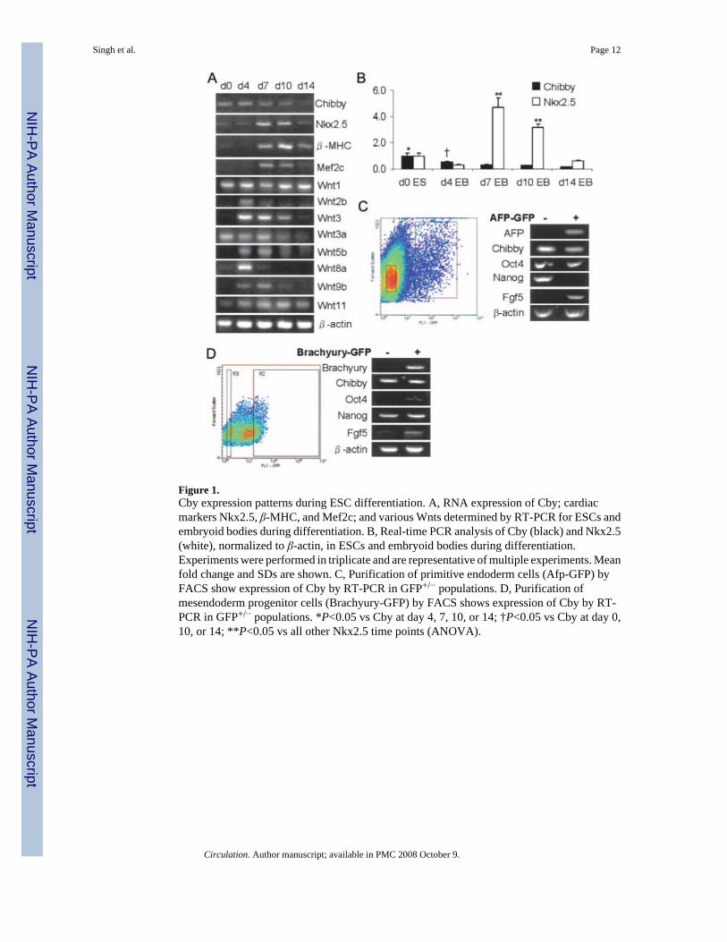
Figure 1.Cby expression patterns during ESC differentiation. A, RNA expression of Cby; cardiacmarkers Nkx2.5, β-MHC, and Mef2c; and various Wnts determined by RT-PCR for ESCs andembryoid bodies during differentiation. B, Real-time PCR analysis of Cby (black) and Nkx2.5(white), normalized to β-actin, in ESCs and embryoid bodies during differentiation.Experiments were performed in triplicate and are representative of multiple experiments. Meanfold change and SDs are shown. C, Purification of primitive endoderm cells (Afp-GFP) byFACS show expression of Cby by RT-PCR in GFP+/− populations. D, Purification ofmesendoderm progenitor cells (Brachyury-GFP) by FACS shows expression of Cby by RT-PCR in GFP+/− populations. *P<0.05 vs Cby at day 4, 7, 10, or 14; †P<0.05 vs Cby at day 0,10, or 14; **P<0.05 vs all other Nkx2.5 time points (ANOVA).
Singh et al. Page 12
Circulation. Author manuscript; available in PMC 2008 October 9.
NIH
-PA Author Manuscript
NIH
-PA Author Manuscript
NIH
-PA Author Manuscript
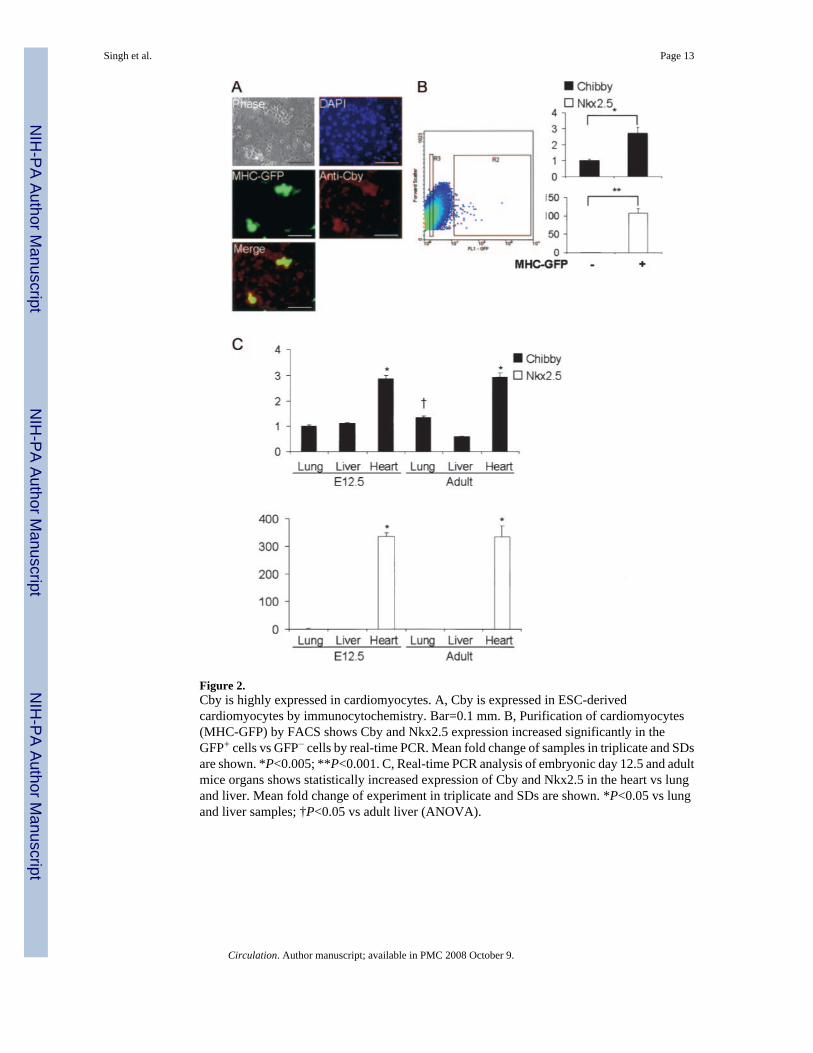
Figure 2.Cby is highly expressed in cardiomyocytes. A, Cby is expressed in ESC-derivedcardiomyocytes by immunocytochemistry. Bar=0.1 mm. B, Purification of cardiomyocytes(MHC-GFP) by FACS shows Cby and Nkx2.5 expression increased significantly in theGFP+ cells vs GFP− cells by real-time PCR. Mean fold change of samples in triplicate and SDsare shown. *P<0.005; **P<0.001. C, Real-time PCR analysis of embryonic day 12.5 and adultmice organs shows statistically increased expression of Cby and Nkx2.5 in the heart vs lungand liver. Mean fold change of experiment in triplicate and SDs are shown. *P<0.05 vs lungand liver samples; †P<0.05 vs adult liver (ANOVA).
Singh et al. Page 13
Circulation. Author manuscript; available in PMC 2008 October 9.
NIH
-PA Author Manuscript
NIH
-PA Author Manuscript
NIH
-PA Author Manuscript
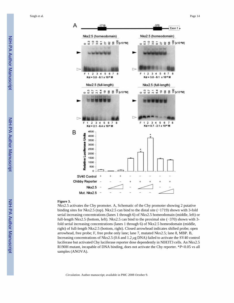
Figure 3.Nkx2.5 activates the Cby promoter. A, Schematic of the Cby promoter showing 2 putativebinding sites for Nkx2.5 (top). Nkx2.5 can bind to the distal site (−1719) shown with 3-foldserial increasing concentrations (lanes 1 through 6) of Nkx2.5 homeodomain (middle, left) orfull-length Nkx2.5 (bottom, left). Nkx2.5 can bind to the proximal site (−370) shown with 3-fold serial increasing concentrations (lanes 1 through 6) of Nkx2.5 homeodomain (middle,right) of full-length Nkx2.5 (bottom, right). Closed arrowhead indicates shifted probe; openarrowhead, free probe; F, free probe only lane; lane 7, mutated Nkx2.5; lane 8, MBP. B,Increasing concentrations of Nkx2.5 (0.6 and 1.2 μg DNA) failed to activate the SV40 controlluciferase but activated Cby luciferase reporter dose dependently in NIH3T3 cells. An Nkx2.5R190H mutant, incapable of DNA binding, does not activate the Cby reporter. *P<0.05 vs allsamples (ANOVA).
Singh et al. Page 14
Circulation. Author manuscript; available in PMC 2008 October 9.
NIH
-PA Author Manuscript
NIH
-PA Author Manuscript
NIH
-PA Author Manuscript

Figure 4.Loss of Cby leads to decreased cardiac differentiation of murine ESCs. A, Western blotshowing Cby was successfully knocked down by RNAi. β-Actin was used as a loading control.Densitometry analysis shows >80% reduction in Cby protein. B, In vitro differentiation of CbyRNAi clone showed severely decreased differentiation during days 5 through 14. Re-expression of hCby was able to compensate for the loss of endogenous mouse Cby and restorenormal outgrowth size for embryoid bodies. Bar=1 mm. C, Percentage of embryoid bodiescontaining beating areas. Cby RNAi failed to develop beating areas compared with the emptyRNAi vector control. Introduction of hCby compensated for loss of mouse Cby to restorebeating areas. More than 25 embryoid bodies were counted per condition per day (>75embryoid bodies per condition in total). Open bars indicate control vector; closed bars, CbyRNAi; and shaded bars, +hCby. Data are representative from multiple experiments. D, Marker
Singh et al. Page 15
Circulation. Author manuscript; available in PMC 2008 October 9.
NIH
-PA Author Manuscript
NIH
-PA Author Manuscript
NIH
-PA Author Manuscript
expression at days 0 and 4 analyzed by RT-PCR. In Cby knockdown or rescue cells, expressionof ES cell markers Oct4 and Nanog, early mesoderm marker Brachyury, and primitiveendoderm markers Afp and transthyretin appeared normal. E, Cby RNAi blocks induction ofcardiac markers Nkx2.5, β-MHC, and Mef2c and endodermal markers Afp, albumin, and α-antitrypsin. Expression of these markers was rescued by introduction of hCby.Neuroectodermal markers Nestin and Sox2 were upregulated in the Cby knockdown. Markerscollagen II, Flk1, and cytokeratin-19 were unaffected by the knockdown of Cby. All data arerepresentative from multiple experiments. The effect of the loss of Cby (outgrowth formation,cardiomyocyte differentiation defect, and reduced marker expression) was confirmed in 3independent knockdown clones (data not shown).
Singh et al. Page 16
Circulation. Author manuscript; available in PMC 2008 October 9.
NIH
-PA Author Manuscript
NIH
-PA Author Manuscript
NIH
-PA Author Manuscript
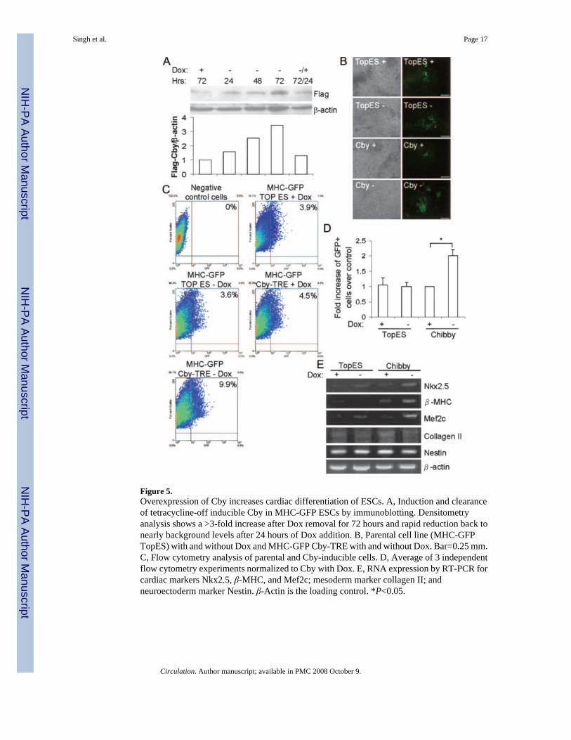
Figure 5.Overexpression of Cby increases cardiac differentiation of ESCs. A, Induction and clearanceof tetracycline-off inducible Cby in MHC-GFP ESCs by immunoblotting. Densitometryanalysis shows a >3-fold increase after Dox removal for 72 hours and rapid reduction back tonearly background levels after 24 hours of Dox addition. B, Parental cell line (MHC-GFPTopES) with and without Dox and MHC-GFP Cby-TRE with and without Dox. Bar=0.25 mm.C, Flow cytometry analysis of parental and Cby-inducible cells. D, Average of 3 independentflow cytometry experiments normalized to Cby with Dox. E, RNA expression by RT-PCR forcardiac markers Nkx2.5, β-MHC, and Mef2c; mesoderm marker collagen II; andneuroectoderm marker Nestin. β-Actin is the loading control. *P<0.05.
Singh et al. Page 17
Circulation. Author manuscript; available in PMC 2008 October 9.
NIH
-PA Author Manuscript
NIH
-PA Author Manuscript
NIH
-PA Author Manuscript