role for interleukin-1 in trypanosoma cruzi-induced cardiomyocyte hypertrophy
TRANSCRIPT

10.1128/IAI.71.8.4441-4447.2003.
2003, 71(8):4441. DOI:Infect. Immun. Christine A. Petersen and Barbara A. Burleigh -Induced Cardiomyocyte Hypertrophy
Trypanosoma cruzi in βRole for Interleukin-1
http://iai.asm.org/content/71/8/4441Updated information and services can be found at:
These include:
REFERENCEShttp://iai.asm.org/content/71/8/4441#ref-list-1This article cites 23 articles, 6 of which can be accessed free at:
CONTENT ALERTS more»cite this article),
Receive: RSS Feeds, eTOCs, free email alerts (when new articles
http://journals.asm.org/site/misc/reprints.xhtmlInformation about commercial reprint orders: http://journals.asm.org/site/subscriptions/To subscribe to to another ASM Journal go to:
on Septem
ber 10, 2014 by The U
niversity of Iowa Libraries
http://iai.asm.org/
Dow
nloaded from
on Septem
ber 10, 2014 by The U
niversity of Iowa Libraries
http://iai.asm.org/
Dow
nloaded from

INFECTION AND IMMUNITY, Aug. 2003, p. 4441–4447 Vol. 71, No. 80019-9567/03/$08.00�0 DOI: 10.1128/IAI.71.8.4441–4447.2003Copyright © 2003, American Society for Microbiology. All Rights Reserved.
Role for Interleukin-1� in Trypanosoma cruzi-InducedCardiomyocyte Hypertrophy
Christine A. Petersen and Barbara A. Burleigh*Department of Immunology and Infectious Diseases, Harvard School of Public Health,
Boston, Massachusetts 02115
Received 21 February 2003/Returned for modification 17 April 2003/Accepted 24 May 2003
Chagas’ disease, the leading cause of heart failure in Latin America, results from infection with theintracellular protozoan parasite Trypanosoma cruzi. Host cell responses elicited in the myocardium early in theinfective process are thought to be critical for establishment of infection by this pathogen; however, thesechanges have not been well characterized. We report here that primary cardiomyocytes undergo hypertrophyas an early response to T. cruzi infection. The T. cruzi-elicited hypertrophic response is characterized byincreased expression of genes encoding the contractile proteins MyHC� and MyHC�, followed by an approx-imately twofold increase in cell size. Hypertrophy was observed in both parasite-containing and noninfectedcell populations represented in T. cruzi-infected cultures, indicating the involvement of a soluble mediator inthis process. Conditioned medium harvested from T. cruzi-infected cultures, which contained significant levelsof interleukin-1� (IL-1�) but not endothelin-1 or tumor necrosis factor alpha, was sufficient to inducehypertrophy in isolated cardiomyocytes. Addition of a high-affinity receptor chimera, IL-1 trap, to cardiomy-ocyte cultures blocked the overall increase in cell size elicited by T. cruzi. These novel findings indicate thatIL-1�, which is rapidly induced in response to T. cruzi, promotes cardiomyocyte hypertrophy early in theinfective process and may contribute to maintenance of cardiomyocyte function during establishment of T. cruziinfection in the heart.
The intracellular protozoan pathogen Trypanosoma cruzicauses Chagas’ disease, the leading cause of cardiomyopathyand heart failure in regions of endemicity in Latin America(19). While the acute stages of Chagas’ disease are generallyasymptomatic, the ability of T. cruzi to infect and persist withincells of the myocardium is a prerequisite for the developmentof Chagasic cardiomyopathy (25). Modulation of host cellularresponses early in the T. cruzi infective process promotes par-asite internalization (4) and is likely to influence intracellularsurvival of this pathogen. Thus, initial cardiomyocyte responsesto T. cruzi are predicted to play a critical role in the establish-ment of long-term infection in the heart and thereby influencedisease progression. Several studies have examined T. cruzi-dependent activation of host cell signaling pathways (4) andtranscriptional responses (24) in a wide range of nonprofes-sional phagocytic cells; however, few have addressed T. cruzi-induced responses in cardiac myocytes (10, 15), the critical siteof infection and pathology in the host.
Cardiomyocytes are postmitotic cells that respond to a vari-ety of cellular stresses by initiating a hypertrophic program,which typically involves reactivation of an embryonic pattern ofgene expression, increased production of myofibrils followedby an increase in cell size (20). Hypertrophy is a commonendpoint that can be initiated in cardiomyocytes by a variety ofexternal stimuli through multiple signaling pathways. Despitethe potential for extensive cross talk between early signalingpathways, distinct molecular and morphological patterns ofhypertrophy can arise from different stimuli (8, 18, 20). The net
effect of these signaling, transcriptional, and cellular changes isincreased myocardial contractility and increased cardiac out-put, ensuring maintenance of cardiac function in the shortterm. If prolonged, cardiac hypertrophy can lead to failure(12).
In Chagasic cardiomyopathy, hypertrophy has generallybeen considered to be a feature of chronic disease: i.e., anindirect consequence of persistent inflammation and myocar-dial damage (2). Recent studies have shown increased expres-sion in rodents of a vasoactive peptide, endothelin-1 (ET-1),and a hypertrophic factor, cardiotrophin-1 (CT-1), as early as10 to 15 days postinfection with T. cruzi (6). ET-1 has beenimplicated in the pathogenesis of Chagas’ disease and is estab-lished as a causative agent of cardiac hypertrophy in T. cruzi-infected animals (14, 16). Because several factors with hyper-trophic activity, including the proinflammatory cytokinesinterleukin-1� (IL-1�) and tumor necrosis factor alpha (TNF-�), are more rapidly induced in the hearts of T. cruzi-infectedanimals (7, 21), these observations suggest that cardiomyocytehypertrophy is likely to be an important feature of acute as wellas chronic Chagas’ disease. Here, we demonstrate that withinthe first 48 h of infection, T. cruzi elicits hypertrophy in isolatedcardiomyocytes, characterized by an increase in mean cell sizeand upregulation of genes encoding contractile proteins. Ourdata demonstrate that IL-1�, secreted by T. cruzi-infected car-diomyocytes, is a critical soluble mediator of the overall hy-pertrophic response for both parasite-infected and uninfectedcell populations.
MATERIALS AND METHODS
Cardiomyocyte isolation. Primary cultures of ventricular cardiomyocytes fromneonatal Sprague-Dawley rats (Charles River Laboratories) were prepared asdescribed previously (3). Briefly, pups were sacrificed, and their hearts were
* Corresponding author. Mailing address: Department of Immunol-ogy and Infectious Diseases, Harvard School of Public Health, 665Huntington Ave., Bldg. I, Rm. 713, Boston, MA 02115. Phone: (617)432-2495. Fax: (617) 432-4766. E-mail: [email protected].
4441
on Septem
ber 10, 2014 by The U
niversity of Iowa Libraries
http://iai.asm.org/
Dow
nloaded from

removed and placed in Hanks’ balanced salt solution (HBSS). Ventricular tissuewas digested overnight in HBSS–1% trypsin (Sigma), washed, and further di-gested with HBSS–1% collagenase (Worthington Biochemical). To enrich forcardiomyocytes, isolated cells were preplated twice in Dulbecco’s modified Ea-gle’s medium (DMEM) onto 10-cm2 dishes, resulting in supernatants containing�90 to 95% cardiomyocytes. Cells were then plated onto laminin-coated dishes(BD Biosciences), allowed to adhere for 24 h, and treated with 1 mM bromode-oxyuridine. Cells were shifted to serum-free media 24 h prior to use in experi-ments.
Parasite infections. T. cruzi (Y strain) was propagated in monolayers of LLC-MK2 cells in DMEM (Gibco) with 2% fetal bovine serum (FBS), and infectivetrypomastigotes were harvested as described previously (23). Parasites werewashed in DMEM, and 2 �107 parasites per ml were incubated with cardiomy-ocytes for 2 h at 37°C in 5% CO2 to allow invasion. The remaining extracellularparasites were aspirated, fresh medium was added, and cultures were incubatedfor a total of 8, 24, or 48 h. Where indicated, cardiomyocytes were stimulatedwith ET-1 (10�7 M), CT-1 (1 nM), or IL-1� (1 ng/ml) and/or incubated in thepresence of the murine homologue of the receptor chimera IL-1 trap (1 �g/ml)(9), generously provided by N. Stahl (Regeneron Pharmaceuticals).
CCM. To generate cardiomyocyte-conditioned media (CCM), cardiomyocyteswere mock treated or infected with T. cruzi for 48 h as described above. Culturesupernatants were collected, filtered through a 0.4-�m-pore-diameter membrane(Millipore), and incubated with serum-starved cardiomyocytes for 24 h.
Immunofluorescence. Following infection of cardiomyocytes on coverslips,cells were rinsed with phosphate-buffered saline (PBS) and fixed with 2% para-formaldehyde–PBS. Cardiomyocytes were stained with a mouse anti-�-actininmonoclonal antibody (Sigma) (1:1,000) followed by rabbit anti-mouse Alexa 488(1:2,000) (Molecular Probes) in Tris-buffered saline (TBS)–1% bovine serumalbumin (BSA) containing 0.1% saponin. Parasites were immunostained with arabbit anti-T. cruzi antibody (23) followed by Alexa 488 anti-rabbit (1:2,000)(Molecular Probes). DNA was stained with DAPI (4�,6�-diamidino-2-phenylin-dole). Slides were visualized with a Nikon TE300, and images were captured withan Orca-100 charge-coupled device camera (Hammamatsu).
FACS analysis. Following various treatments, cardiomyocytes were trypsin-ized, and isolated cells fixed with 2% PFA–PBS. Immunostaining with a rabbitanti-T. cruzi antibody followed by Alexa 488 goat-anti-rabbit (1:3000) (Molecu-lar Probes) was carried out as described above. A minimum of 10,000 cells foreach condition were analyzed by fluorescence-activated cell sorter (FACS)(FACScalibur; Becton Dickinson) measuring fluorescein isothiocyanate (FITC)fluorescence and forward scatter (FSC). Data were analyzed with Cell Questsoftware (Becton Dickinson). The geometric mean of FSC data is used as acorrelate of cell size. Fold change in population cell size was calculated as theratio geometric mean FSC (treated)/geometric mean FSC (control).
RPA. The RNase protection assay (RPA) was performed as follows. 32P-labeled probes specific for the genes coding for GAPDH, SERCA, myosin heavychain � (MyHC�), and MyHC� were generated by T7 RNA polymerase tran-scription (Maxiscript; Ambion) from 1 �g of gel-purified plasmid DNA (plasmidsgenerously provided by Mark Jeong, Denver Health Medical Center). Probeswere hybridized to 5 �g of total RNA extracted from cardiomyocytes at 8 and24 h postinfection according to the manufacturers’ recommendations (RPA III;Ambion). Protected probes were separated on 6% polyacrylamide gels mixedwith Tris-borate-EDTA (TBE), and the relative intensity of each probe wasdetermined by PhosphorImager analysis (Molecular Dynamics).
Northern hybridization. Ten micrograms of total RNA was prepared fromcardiomyocytes by using RNAeasy (Qiagen), separated on 1% agarose gelscontaining 0.2 M formaldehyde, and blotted onto Hybond N� nylon membranes(Amersham). Membranes were hybridized sequentially with 32P-labeled probes(Rediprime II; Amersham-Pharmacia) for atrial natriuretic factor (ANF) (kindlyprovided M. Kuroski de Bold, Ottawa Heart Institute) or rat GAPDH (Clon-tech) in ExpressHYB (Clontech) and exposed to Hyperfilm (Amersham-Phar-macia), and densitometric analysis was performed.
ELISA. Five hundred microliters of culture supernatant collected from con-trol, ET-1-stimulated, or T. cruzi-infected monolayers at 24 and 48 h posttreat-ment was clarified by centrifugation at 14,000 � g for 10 min. Secreted IL-1�,TNF-�, and ET-1 were measured in supernatants using antibody-specific ELISAkits as directed by the manufacturer (R&D for TNF-� and IL-1�; Peninsula Labsfor ET-1) and analyzed with a Spectra Max 190 enzyme-linked immunosorbentassay (ELISA) reader (Molecular Devices).
Statistical analysis. Student’s t test was used for comparison between controland experimental samples. Data represented as fold change over control wereanalyzed with a one-sample t test. Values of P 0.05 were considered significant.
RESULTS
Cardiomyocyte hypertrophy is an early host response to T.cruzi infection. T. cruzi infection of myocardial cells is a deci-sive step in the pathogenesis of Chagas’ disease; however, littleis known regarding early responses of cardiomyocytes to thispathogen. To determine whether T. cruzi infection inducesadaptive hypertrophy early in the infective process, FACSanalysis of cultured, infected cardiomyocytes was carried out toexamine changes in cell size. As compared to mock-infectedcontrols, a marked increase in the population cell size wasobserved in cardiomyocytes analyzed from T. cruzi-infectedcultures, as indicated by increased sarcomeric �-actinin stain-ing (Fig. 1A and B) and FCS analysis (Fig. 1D to G), in whichsignificant increases in cell size were observed by 48 h postin-fection. Similar results were obtained following treatment ofcardiomyocytes with ET-1, a well-characterized hypertrophicstimulant (Fig. 1C, F, and G). Since the efficiency of T. cruziinfection of cardiomyocytes varied between 25 and 60% of thetotal cells in culture, cardiomyocyte populations analyzed byflow cytometry contained both parasite-infected and nonin-fected cells. To distinguish the parasite-containing cell popu-lation from the noninfected cells, intracellular staining for T.cruzi was carried out prior to FACS analysis (Fig. 1D to F). Asdemonstrated (Fig. 1E), hypertrophic cells were detected inboth parasite-infected and noninfected cardiomyocyte popula-tions, suggesting a role for soluble mediators in this process.
To determine whether soluble factors present in T. cruzi-infected cardiomyocyte cultures participate in the hypertrophicresponse, medium harvested from mock-treated or T. cruzi-infected cultures was used to stimulate uninfected cardiomyo-cytes. CCM from T. cruzi-infected cells alone was capable ofinducing a significant increase in population cell size comparedto medium from mock-treated controls (Fig. 1H). The effect ofCCM mimicked that of T. cruzi infection or stimulation withthe hypertrophic agents ET-1, CT-1, and IL-1� (Fig. 1H).Combined, the data indicate that T. cruzi induces a rapid hy-pertrophic response in cardiomyocytes that is mediated, atleast in part, by a diffusible host cell or parasite factor(s)released into the medium from infected cardiomyocytes.
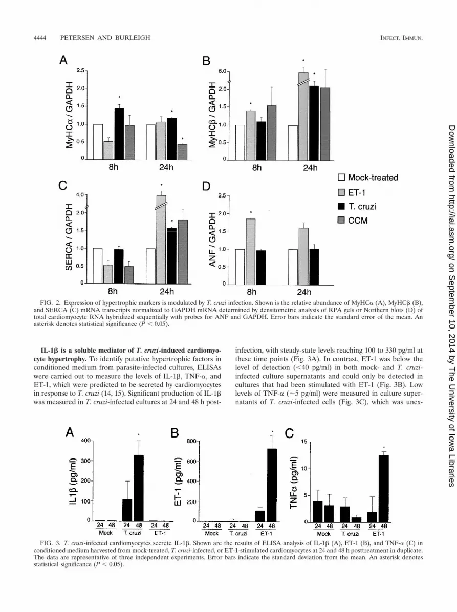
Atypical modulation of cardiomyocyte hypertrophic re-sponse genes by T. cruzi. Reversion to the fetal program ofcardiomyocyte gene expression is central to many hypertrophicresponse pathways and commonly characterized by reactiva-tion of ANF and MyHC� gene expression and downregulationof the genes coding for MyHC� and SERCA (18). To assesswhether T. cruzi infection affects expression of these hypertro-phic response genes, their expression relative to that of thegene coding for GAPDH was determined at 8 and 24 h postin-fection in at least three independent experiments (Fig. 2).Because ET-1 has been implicated as an important solublemediator of cardiac hypertrophy in Chagasic cardiomyopathy(14), it was used as a positive hypertrophic stimulant in theseexperiments. Overall, the expression data represented in Fig. 2demonstrate that significant and reproducible increases in theexpression of the MyHC�, MyHC�, and SERCA genes occurfollowing infection of cardiomyocytes with T. cruzi. Notabledifferences in the profile of gene expression were seen whencomparing the cardiomyocyte responses to T. cruzi, ET-1, andCCM. Most striking was the observation that ANF transcript
4442 PETERSEN AND BURLEIGH INFECT. IMMUN.
on Septem
ber 10, 2014 by The U
niversity of Iowa Libraries
http://iai.asm.org/
Dow
nloaded from

levels were consistently unaltered by T. cruzi, while a significantincrease in ANF expression was observed in response to ET-1(Fig. 2D). In addition, increased MyHC� transcript abundancewas observed at 8 and 24 h following T. cruzi infection; how-ever, ET-1 failed to modulate expression of this marker, andstimulation of cells with CCM from T. cruzi-infected cardio-myocytes resulted in a twofold decrease in MyHC� mRNAlevels by 24 h (Fig. 2A). A slight increase in SERCA mRNAlevels was observed at 24 h postinfection or post-CCM treat-ment (Fig. 2C). Collectively, these data indicate that while
T. cruzi, CCM, and ET-1 can promote marked increases in cellsize (Fig. 1), distinct patterns of cardiomyocyte gene expres-sion are observed in response to these various stimuli. How-ever, a common feature of the response elicited by T. cruzi,CCM, or ET-1 was the consistent increase in expression ofMyHC� observed at 24 h (Fig. 2B). The observation that theT. cruzi-induced response was not faithfully reproduced by theaddition of CCM likely reflects a more complex transcriptionalresponse elicited in cells harboring intracellular parasites thanthat in cells stimulated with soluble factors alone.
FIG. 1. T. cruzi induces hypertrophy in isolated cardiomyocytes. (A) Immunofluorescence staining of sarcomeric �-actinin in fixed cardiomy-ocytes. (B and C) FACS analysis of relative �-actinin expression in cardiomyocytes infected with T. cruzi (B; black) or stimulated with ET-1 (C;black) for 48 h compared to mock-treated controls (gray). Shown are representative plots depicting relative cell size by FSC of mock-treated (D),T. cruzi-infected (E), and ET-1-stimulated (F) cardiomyocytes stained with anti-T. cruzi antibodies to detect intracellular parasites (y axis). (G)T. cruzi infection of cardiomyocytes causes an increase in population cell size. FSC analysis of fixed cardiomyocytes infected with T. cruzi or treatedwith ET-1 for 24 or 48 h is shown. The data are represented as the ratio of the geometric mean of the treated cell population to that of themock-treated control. (H) Cardiomyocyte hypertrophy induced following T. cruzi infection (Tc) or stimulation of cells with conditioned mediumfrom T. cruzi-infected cells (CCM) and represented as an average from three independent experiments standard deviation.
VOL. 71, 2003 T. CRUZI-INDUCED CARDIOMYOCYTE HYPERTROPHY 4443
on Septem
ber 10, 2014 by The U
niversity of Iowa Libraries
http://iai.asm.org/
Dow
nloaded from
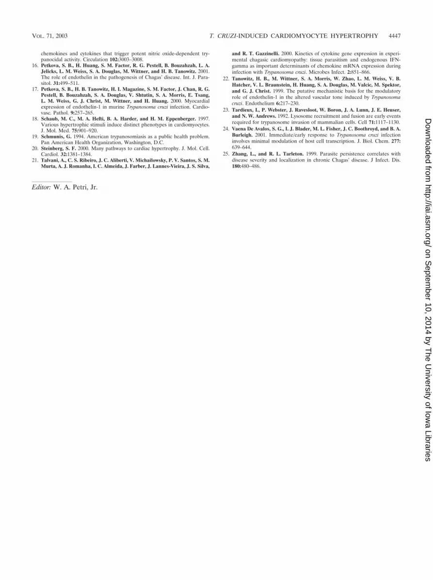
IL-1� is a soluble mediator of T. cruzi-induced cardiomyo-cyte hypertrophy. To identify putative hypertrophic factors inconditioned medium from parasite-infected cultures, ELISAswere carried out to measure the levels of IL-1�, TNF-�, andET-1, which were predicted to be secreted by cardiomyocytesin response to T. cruzi (14, 15). Significant production of IL-1�was measured in T. cruzi-infected cultures at 24 and 48 h post-
infection, with steady-state levels reaching 100 to 330 pg/ml atthese time points (Fig. 3A). In contrast, ET-1 was below thelevel of detection (40 pg/ml) in both mock- and T. cruzi-infected culture supernatants and could only be detected incultures that had been stimulated with ET-1 (Fig. 3B). Lowlevels of TNF-� (�5 pg/ml) were measured in culture super-natants of T. cruzi-infected cells (Fig. 3C), which was unex-
FIG. 2. Expression of hypertrophic markers is modulated by T. cruzi infection. Shown is the relative abundance of MyHC� (A), MyHC� (B),and SERCA (C) mRNA transcripts normalized to GAPDH mRNA determined by densitometric analysis of RPA gels or Northern blots (D) oftotal cardiomyocyte RNA hybridized sequentially with probes for ANF and GAPDH. Error bars indicate the standard error of the mean. Anasterisk denotes statistical significance (P 0.05).
FIG. 3. T. cruzi-infected cardiomyocytes secrete IL-1�. Shown are the results of ELISA analysis of IL-1� (A), ET-1 (B), and TNF-� (C) inconditioned medium harvested from mock-treated, T. cruzi-infected, or ET-1-stimulated cardiomyocytes at 24 and 48 h posttreatment in duplicate.The data are representative of three independent experiments. Error bars indicate the standard deviation from the mean. An asterisk denotesstatistical significance (P 0.05).
4444 PETERSEN AND BURLEIGH INFECT. IMMUN.
on Septem
ber 10, 2014 by The U
niversity of Iowa Libraries
http://iai.asm.org/
Dow
nloaded from

pected, given that TNF-� transcripts were previously shown tobe induced following infection of isolated cardiomyocytes withT. cruzi (15). Thus, ET-1 and TNF-� were unlikely to functionas soluble mediators of T. cruzi-induced cardiomyocyte hyper-trophy in this system, whereas IL-1� was produced in sufficientquantity to cause an increase in MyHC� transcript levels andpromote an approximately twofold increase in cell size (datanot shown).
To test whether IL-1� secreted by T. cruzi-infected cardio-myocytes could mediate the observed hypertrophic response,infections were carried out in the presence and absence ofrecombinant IL-1 trap (rIL-1 trap), a multicomponent IL-1receptor chimera that exhibits high affinity for IL-1� and IL-1�(9). While rIL-1 trap alone caused control cells to undergo aslight increase in size (data not shown), incubation of T. cruzi-infected cardiomyocytes with rIL-1 trap for 48 h abrogated theoverall increase in population cell size induced by this patho-gen (Fig. 4A). Specificity of the effect was demonstrated in theability of rIL-1 trap to inhibit IL-1�-mediated hypertrophy butnot the ET-1-stimulated response (Fig. 4A). Moreover, thepresence of rIL-1 trap resulted in a decrease in the overallnumber of hypertrophic cardiomyocytes found in both the in-fected (Fig. 4D, upper right quadrant) and noninfected (Fig.4D, lower right quadrant) populations present in T. cruzi-infected cardiomyocyte cultures. Independent analysis of T.
cruzi-infected cardiomyocytes by immunofluorescence micros-copy indicated that parasite infectivity, intracellular replica-tion, and host cell viability were not adversely affected by theaddition of rIL-1 trap (data not shown). In contrast, attemptsto block the CCM-mediated hypertrophic response with rIL-1trap consistently resulted in a loss of cell viability, suggestingthat IL-1� may provide a protective function under these con-ditions. Overall, the dramatic reduction in overall size of car-diomyocyte populations by rIL-1 trap provides compelling ev-idence that IL-1� secreted by T. cruzi-infected cells promoteshypertrophy in isolated cardiomyocytes infected with T. cruzi.
DISCUSSION
The complex array of host cellular responses elicited byintracellular pathogens is likely to reflect both host defensemechanisms and pathways utilized for pathogen survival. Es-tablishment of cardiomyocyte infection by the intracellularparasite T. cruzi is critical to the development of Chagasiccardiomyopathy (2). In this study, we have exploited an iso-lated cardiomyocyte culture system to begin to characterizeearly host responses induced in this clinically relevant cell typefollowing T. cruzi infection. Potentially confounding effects ofsecondary inflammation and/or host factors produced by othercell types were avoided in this simplified in vitro system,
FIG. 4. rIL-1 trap blocks T. cruzi-induced cardiomyocyte hypertrophy. (A) Relative population cell size of cardiomyocytes treated with mediumalone (mock), IL-1� (1 ng/�l), or ET-1 (10�7 M) or infected with T. cruzi for 48 h in the presence (black) or absence (gray) of 1 �g of rIL-1 trapper ml. Fixed cells were analyzed by FACS, and FSC data are represented as the ratio of the geometric mean of the treated population to thatof the mock-treated population. (B and C) Representative FACS plots of T. cruzi-infected cells at 48 h stained for intracellular T. cruzi (y axis)and incubated in the absence (B) or presence (C) of 1 �g of rIL-1 trap per ml. (D) rIL-1 trap inhibits hypertrophy of both T. cruzi-infected (upperright quadrant [UR]) and noninfected (lower right quadrant [LR]) cells in the total cell population. The data are represented as the average ofthree experiments standard deviation.
VOL. 71, 2003 T. CRUZI-INDUCED CARDIOMYOCYTE HYPERTROPHY 4445
on Septem
ber 10, 2014 by The U
niversity of Iowa Libraries
http://iai.asm.org/
Dow
nloaded from

thereby permitting detection of cardiomyocyte-specific re-sponses. Our studies reveal that as an early response to T. cruziinfection, cardiomyocytes undergo a rapid hypertrophic re-sponse that is mediated by the secreted proinflammatory cy-tokine IL-1�. Parasite-induced cardiomyocyte hypertrophy,which occurs in both T. cruzi-infected and uninfected myocytesin culture, was inhibited by the addition of a soluble multicom-ponent IL-1 receptor (IL-1 trap). These novel findings indicatethat in addition to its primary immune regulatory function,IL-1� may play an important role as a mediator of cardiomy-ocyte hypertrophy as part of the acute response to T. cruziinfection in the heart.
ET-1 has been implicated in the pathogenesis of Chagas’disease (16). A recent study that utilized a targeted cardiac-specific deletion of ET-1 further demonstrated that cardiacmyocytes are an important source of ET-1 production in ani-mal models of T. cruzi infection, where reduced ET-1 expres-sion correlated with reduced hypertrophy (14). Interestingly,the results presented in the present study clearly demonstratethat ET-1 is not produced by isolated cardiomyocytes as anearly response to T. cruzi infection. Furthermore, we show thatET-1 does not play a significant role in the induction of para-site-induced cardiomyocyte hypertrophy in a simplified in vitroculture system. These data suggest that induction of ET-1production from cardiomyocytes is not a primary response toT. cruzi infection and likely requires additional autocrineand/or paracrine factors produced by other cell types (e.g.,myocardial and/or infiltrating inflammatory cells) absent fromour in vitro cardiomyocyte culture system. Consistent with thisidea, increases in ET-1 expression in the hearts of T. cruzi-infected rodents were not detectable until 10 to 15 days postin-fection (17). In contrast, IL-1� is rapidly upregulated duringthe initiation of cardiac infection by T. cruzi (3, 15, 21). SinceIL-1� is a known hypertrophic mediator (13), it is plausiblethat cardiomyocyte hypertrophy, mediated by this proinflam-matory cytokine, may be a critical, unrecognized feature ofacute Chagas’ disease. As infection progresses beyond the ini-tiation period (3 to 5 days), it is predicted that a complex arrayof host factors released from infiltrating cells as well as otherknown hypertrophic mediators produced during acute infec-tion, including ET-1 (17), CT-1 (6), and TNF-� (3, 15, 21),would modulate an initial hypertrophic response mediated byIL-1�.
While T. cruzi factors responsible for induction of IL-1�expression in cardiomyocytes have yet to be determined, it iswell established that a family of T. cruzi glycosylphosphatidylinositol-linked surface mucin-like molecules (1) trigger proin-flammatory cytokine expression in macrophages (5) throughthe activation of Toll-like receptor 2 (TLR2) (5). Since cardi-omyocytes express TLR2, which has been shown to be anantiapoptotic signal under oxidative stress, (11), it is temptingto speculate that this innate signaling pathway may regulate theinduction of IL-1� expression in cardiomyocytes and the con-comitant hypertrophic response triggered by T. cruzi.
Overall, the results from the present study begin to chal-lenge the concept that cardiomyocyte hypertrophy in Chagas’disease is restricted to a late stage of T. cruzi infection, follow-ing chronic inflammation and myocardial damage. Instead, weprovide evidence that IL-1�-mediated cardiomyocyte hyper-trophy occurs in isolated cardiomyocytes in response to T. cruzi
and may play an important role in the maintenance of cardi-omyocyte function during the initial phases of infection. IL-1�is produced by other myocardial cells, including vascular en-dothelial cells, following T. cruzi infection (22), indicating thatlocal secretion of IL-1� could promote cardiomyocyte hyper-trophy regardless of the site of initial site of parasite invasion.Our novel findings provide the basis for further investigation ofthe role of IL-1�-mediated cardiomyocyte hypertrophy inacute Chagas’ disease.
ACKNOWLEDGMENTS
We thank K. Shibata and G. Koren, Brigham and Women’s Hospi-tal, for advice on neonatal rat cardiomyocyte isolation. We are ex-tremely grateful to P. Thomas and O. Atochin for assistance withFACS analysis. We acknowledge M. Fisher for technical assistance andG. Reed, A. Woolsey, M. Unnikrishnan, and J. Daily for helpful dis-cussions.
This research has been supported in part by the Investigators inPathogenesis of Infectious Diseases Award from the Burroughs Well-come Fund and National Institutes of Health grants R01 AI47960 toB.A.B. and K08 AI50803 to C.A.P.
REFERENCES
1. Acosta-Serrano, A., I. C. Almeida, L. H. Freitas-Junior, N. Yoshida, and S.Schenkman. 2001. The mucin-like glycoprotein super-family of Trypanosomacruzi: structure and biological roles. Mol. Biochem. Parasitol. 114:143–150.
2. Arnaiz, M. R., L. E. Fichera, and M. Postan. 2002. Cardiac myocyte hyper-trophy and proliferating cell nuclear antigen expression in Wistar rats in-fected with Trypanosoma cruzi. J. Parasitol. 88:919–925.
3. Baliga, R. R., D. R. Pimental, Y. Y. Zhao, W. W. Simmons, M. A. Mar-chionni, D. B. Sawyer, and R. A. Kelly. 1999. NRG-1-induced cardiomyocytehypertrophy. Role of PI-3-kinase, p70(S6K), and MEK-MAPK-RSK. Am. J.Physiol. 277:H2026–H2037.
4. Burleigh, B. A., and A. M. Woolsey. 2002. Cell signalling and Trypanosomacruzi invasion. Cell Microbiol. 4:701–711.
5. Campos, M. A., I. C. Almeida, O. Takeuchi, S. Akira, E. P. Valente, D. O.Procopio, L. R. Travassos, J. A. Smith, D. T. Golenbock, and R. T.Gazzinelli. 2001. Activation of Toll-like receptor-2 by glycosylphosphatidyl-inositol anchors from a protozoan parasite. J. Immunol. 167:416–423.
6. Chandrasekar, B., P. C. Melby, D. Pennica, and G. L. Freeman. 1998.Overexpression of cardiotrophin-1 and gp130 during experimental acuteChagasic cardiomyopathy. Immunol. Lett. 61:89–95.
7. Chandrasekar, B., P. C. Melby, D. A. Troyer, J. T. Colston, and G. L.Freeman. 1998. Temporal expression of pro-inflammatory cytokines andinducible nitric oxide synthase in experimental acute Chagasic cardiomyop-athy. Am. J. Pathol. 152:925–934.
8. Chien, K. R. 1993. Molecular advances in cardiovascular biology. Science260:916–917.
9. Economides, A. N., L. R. Carpenter, J. S. Rudge, V. Wong, E. M. Koehler-Stec, C. Hartnett, E. A. Pyles, X. Xu, T. J. Daly, M. R. Young, J. P. Fandl, F.Lee, S. Carver, J. McNay, K. Bailey, S. Ramakanth, R. Hutabarat, T. T.Huang, C. Radziejewski, G. D. Yancopoulos, and N. Stahl. 2003. Cytokinetraps: multi-component, high-affinity blockers of cytokine action. Nat. Med.9:47–52.
10. Ferreira, L. R., E. F. Abrantes, C. V. Rodrigues, B. Caetano, G. C. Cerqueira,A. C. Salim, L. F. Reis, and R. T. Gazzinelli. 2002. Identification and char-acterization of a novel mouse gene encoding a Ras-associated guanine nu-cleotide exchange factor: expression in macrophages and myocarditis elicitedby Trypanosoma cruzi parasites. J. Leukoc. Biol. 72:1215–1227.
11. Frantz, S., R. A. Kelly, and T. Bourcier. 2001. Role of TLR-2 in the activa-tion of nuclear factor kappaB by oxidative stress in cardiac myocytes. J. Biol.Chem. 276:5197–5203.
12. Frey, N., and E. N. Olson. 2003. Cardiac hypertrophy: the good, the bad, andthe ugly. Annu. Rev. Physiol. 65:45–79.
13. Harada, E., O. Nakagawa, M. Yoshimura, M. Harada, M. Nakagawa, Y.Mizuno, Y. Shimasaki, M. Nakayama, H. Yasue, K. Kuwahara, Y. Saito, andK. Nakao. 1999. Effect of interleukin-1 beta on cardiac hypertrophy andproduction of natriuretic peptides in rat cardiocyte culture. J. Mol. Cell.Cardiol. 31:1997–2006.
14. Huang, H., M. Yanagisawa, Y. Y. Kisanuki, L. A. Jelicks, M. Chandra, S. M.Factor, M. Wittner, L. M. Weiss, R. G. Pestell, V. Shtutin, J. Shirani, andH. B. Tanowitz. 2002. Role of cardiac myocyte-derived endothelin-1 in cha-gasic cardiomyopathy: molecular genetic evidence. Clin. Sci. (London) 103(Suppl. 1):263S–266S.
15. Machado, F. S., G. A. Martins, J. C. Aliberti, F. L. Mestriner, F. Q. Cunha,and J. S. Silva. 2000. Trypanosoma cruzi-infected cardiomyocytes produce
4446 PETERSEN AND BURLEIGH INFECT. IMMUN.
on Septem
ber 10, 2014 by The U
niversity of Iowa Libraries
http://iai.asm.org/
Dow
nloaded from
chemokines and cytokines that trigger potent nitric oxide-dependent try-panocidal activity. Circulation 102:3003–3008.
16. Petkova, S. B., H. Huang, S. M. Factor, R. G. Pestell, B. Bouzahzah, L. A.Jelicks, L. M. Weiss, S. A. Douglas, M. Wittner, and H. B. Tanowitz. 2001.The role of endothelin in the pathogenesis of Chagas’ disease. Int. J. Para-sitol. 31:499–511.
17. Petkova, S. B., H. B. Tanowitz, H. I. Magazine, S. M. Factor, J. Chan, R. G.Pestell, B. Bouzahzah, S. A. Douglas, V. Shtutin, S. A. Morris, E. Tsang,L. M. Weiss, G. J. Christ, M. Wittner, and H. Huang. 2000. Myocardialexpression of endothelin-1 in murine Trypanosoma cruzi infection. Cardio-vasc. Pathol. 9:257–265.
18. Schaub, M. C., M. A. Hefti, B. A. Harder, and H. M. Eppenberger. 1997.Various hypertrophic stimuli induce distinct phenotypes in cardiomyocytes.J. Mol. Med. 75:901–920.
19. Schmunis, G. 1994. American trypanosomiasis as a public health problem.Pan American Health Organization, Washington, D.C.
20. Steinberg, S. F. 2000. Many pathways to cardiac hypertrophy. J. Mol. Cell.Cardiol. 32:1381–1384.
21. Talvani, A., C. S. Ribeiro, J. C. Aliberti, V. Michailowsky, P. V. Santos, S. M.Murta, A. J. Romanha, I. C. Almeida, J. Farber, J. Lannes-Vieira, J. S. Silva,
and R. T. Gazzinelli. 2000. Kinetics of cytokine gene expression in experi-mental chagasic cardiomyopathy: tissue parasitism and endogenous IFN-gamma as important determinants of chemokine mRNA expression duringinfection with Trypanosoma cruzi. Microbes Infect. 2:851–866.
22. Tanowitz, H. B., M. Wittner, S. A. Morris, W. Zhao, L. M. Weiss, V. B.Hatcher, V. L. Braunstein, H. Huang, S. A. Douglas, M. Valcic, M. Spektor,and G. J. Christ. 1999. The putative mechanistic basis for the modulatoryrole of endothelin-1 in the altered vascular tone induced by Trypanosomacruzi. Endothelium 6:217–230.
23. Tardieux, I., P. Webster, J. Ravesloot, W. Boron, J. A. Lunn, J. E. Heuser,and N. W. Andrews. 1992. Lysosome recruitment and fusion are early eventsrequired for trypanosome invasion of mammalian cells. Cell 71:1117–1130.
24. Vaena De Avalos, S. G., I. J. Blader, M. L. Fisher, J. C. Boothroyd, and B. A.Burleigh. 2001. Immediate/early response to Trypanosoma cruzi infectioninvolves minimal modulation of host cell transcription. J. Biol. Chem. 277:639–644.
25. Zhang, L., and R. L. Tarleton. 1999. Parasite persistence correlates withdisease severity and localization in chronic Chagas’ disease. J Infect. Dis.180:480–486.
Editor: W. A. Petri, Jr.
VOL. 71, 2003 T. CRUZI-INDUCED CARDIOMYOCYTE HYPERTROPHY 4447
on Septem
ber 10, 2014 by The U
niversity of Iowa Libraries
http://iai.asm.org/
Dow
nloaded from