chemistry, biosynthesis and pharmacology of sarsasapogenin
TRANSCRIPT
�����������������
Citation: Mustafa, N.H.; Sekar, M.;
Fuloria, S.; Begum, M.Y.; Gan, S.H.;
Rani, N.N.I.M.; Ravi, S.;
Chidambaram, K.; Subramaniyan, V.;
Sathasivam, K.V.; et al. Chemistry,
Biosynthesis and Pharmacology of
Sarsasapogenin: A Potential Natural
Steroid Molecule for New Drug
Design, Development and Therapy.
Molecules 2022, 27, 2032. https://
doi.org/10.3390/molecules27062032
Academic Editor: Hiroyuki Kataoka
Received: 2 February 2022
Accepted: 16 March 2022
Published: 21 March 2022
Publisher’s Note: MDPI stays neutral
with regard to jurisdictional claims in
published maps and institutional affil-
iations.
Copyright: © 2022 by the authors.
Licensee MDPI, Basel, Switzerland.
This article is an open access article
distributed under the terms and
conditions of the Creative Commons
Attribution (CC BY) license (https://
creativecommons.org/licenses/by/
4.0/).
molecules
Review
Chemistry, Biosynthesis and Pharmacology of Sarsasapogenin:A Potential Natural Steroid Molecule for New Drug Design,Development and TherapyNur Hanisah Mustafa 1,†, Mahendran Sekar 1,*,† , Shivkanya Fuloria 2,*,†, M. Yasmin Begum 3 ,Siew Hua Gan 4 , Nur Najihah Izzati Mat Rani 5 , Subban Ravi 6, Kumarappan Chidambaram 7 ,Vetriselvan Subramaniyan 8 , Kathiresan V. Sathasivam 9, Srikanth Jeyabalan 10 , Subasini Uthirapathy 11,Sivasankaran Ponnusankar 12 , Pei Teng Lum 1, Vijay Bhalla 13 and Neeraj Kumar Fuloria 2,14,*,†
1 Department of Pharmaceutical Chemistry, Faculty of Pharmacy and Health Sciences, Royal College ofMedicine Perak, Universiti Kuala Lumpur, Ipoh 30450, Malaysia; [email protected] (N.H.M.);[email protected] (P.T.L.)
2 Faculty of Pharmacy, AIMST University, Bedong 08100, Malaysia3 Department of Pharmaceutics, College of Pharmacy, King Khalid University, Abha 61421, Saudi Arabia;
[email protected] School of Pharmacy, Monash University Malaysia, Bandar Sunway, Kuala Lumpur 47500, Malaysia;
[email protected] Faculty of Pharmacy and Health Sciences, Royal College of Medicine Perak, Universiti Kuala Lumpur,
Ipoh 30450, Malaysia; [email protected] Department of Chemistry, Karpagam Academy of Higher Education, Coimbatore 641021, Tamil Nadu, India;
[email protected] Department of Pharmacology, College of Pharmacy, King Khalid University, Abha 62529, Saudi Arabia;
[email protected] Faculty of Medicine, Bioscience and Nursing, MAHSA University, Jalan SP 2, Bandar Saujana Putra,
Jenjarom 42610, Malaysia; [email protected] Faculty of Applied Sciences, AIMST University, Bedong, Sungai Petani 08100, Malaysia;
[email protected] Department of Pharmacology, Sri Ramachandra Faculty of Pharmacy, Sri Ramachandra Institute of Higher
Education and Research (DU), Porur, Chennai 600116, Tamil Nadu, India; [email protected] Faculty of Pharmacy, Tishk International University, Erbil 44001, Kurdistan Region, Iraq;
[email protected] Department of Pharmacy Practice, JSS College of Pharmacy, JSS Academy of Higher Education & Research,
Ooty 643001, Tamil Nadu, India; [email protected] SGT College of Pharmacy, SGT University, Gurugram 122505, Haryana, India; [email protected] Center for Transdisciplinary Research, Department of Pharmacology, Saveetha Institute of Medical and
Technical Sciences, Saveetha Dental College and Hospital, Saveetha University,Chennai 600077, Tamil Nadu, India
* Correspondence: [email protected] (M.S.); [email protected] (S.F.);[email protected] (N.K.F.); Tel.: +60-163346653 (M.S.); +60-143034057 (S.F.); +60-164037685 (N.K.F.)
† These authors contributed equally to this work.
Abstract: Sarsasapogenin is a natural steroidal sapogenin molecule obtained mainly from Anemar-rhena asphodeloides Bunge. Among the various phytosteroids present, sarsasapogenin has emergedas a promising molecule due to the fact of its diverse pharmacological activities. In this review, thechemistry, biosynthesis and pharmacological potentials of sarsasapogenin are summarised. Between1996 and the present, the relevant literature regarding sarsasapogenin was obtained from scientificdatabases including PubMed, ScienceDirect, Scopus, and Google Scholar. Overall, sarsasapogenin isa potent molecule with anti-inflammatory, anticancer, antidiabetic, anti-osteoclastogenic and neu-roprotective activities. It is also a potential molecule in the treatment for precocious puberty. Thisreview also discusses the metabolism, pharmacokinetics and possible structural modifications aswell as obstacles and opportunities for sarsasapogenin to become a drug molecule in the near future.More comprehensive preclinical studies, clinical trials, drug delivery, formulations of effective dosesin pharmacokinetics studies, evaluation of adverse effects and potential synergistic effects with other
Molecules 2022, 27, 2032. https://doi.org/10.3390/molecules27062032 https://www.mdpi.com/journal/molecules
Molecules 2022, 27, 2032 2 of 26
drugs need to be thoroughly investigated to make sarsasapogenin a potential molecule for futuredrug development.
Keywords: sarsasapogenin; steroid; phytochemistry; biosynthesis; pharmacology; drug development
1. Introduction
Since ancient times, natural products have been a key source of pharmaceuticals, andmany of them have been approved as effective drugs or drug candidates. Plants, animals,marines and microbes are the most common sources of natural products. Plants are livingchemical factories, synthesizing an enormous variety of secondary metabolites that canserve as the basis for numerous commercial pharmaceutical preparations. Although thevarious chemical elements of medicinal plants have biological activities that are beneficialto health via the pharmaceutical and food industries, they also have significant values inthe perfume, agrochemical and cosmetic businesses [1–3]. Drug discovery from naturalorigin entails a number of steps including botanical identification and collection, extraction,isolation, purification, and structure elucidation of the desired bioactive molecule as wellas its biological evaluation. Formulation development, stability testing of the establishedformulation, preclinical and clinical trial investigations and the submission of a New DrugApplication (NDA) are all steps in the process [4,5].
Anemarrhena asphodeloides Bunge, commonly referred to as Zhimu (Rhizoma Anemar-rhenae), is a member of the Anemarrhena genus in the Liliaceae family [6] and is foundprimarily in China, Japan, Korea and other eastern Asian nations [7–9]. The rhizomes havebeen used to cure febrile diseases, hyperpyrexia, fever, cough, polydipsia, osteopyrexia andconstipation since ancient times [10]. Anemarrhena asphodeloides is predominantly composedof steroidal saponins including sarsasapogenin [11]. Sarsasapogenin has been reportedfor many pharmacological actions and widely investigated for anti-inflammatory, neuro-protective and memory impairment related to ageing [12–15]. Additionally, its modifiedstructure and a number of derivatives identified have been confirmed to have biologicalproperties such as antitumour activity, antidepressant activity, anxiolytic-like action andan Aβ-reducing effect [16–21]. Despite its variable therapeutic properties, there has beenno thorough and comprehensive review on sarsasapogenin. Hence, the phytochemistry,biosynthesis and physicochemical characteristics of sarsasapogenin are summarised inthis review with a focus on its biological activities. This review discusses the metabolism,pharmacokinetics and potential structural modifications, as well as the limitations andprospects for sarsasapogenin becoming a drug molecule. The scientific evidence is antici-pated to provide a solid foundation for future research as well as vital information for thedrug development of sarsasapogenin as a therapeutic agent and health product.
2. Phytochemistry of Sarsasapogenin
Sarsasapogenin is present in many medicinal plants and the details are summarisedin Table 1.
Molecules 2022, 27, 2032 3 of 26
Table 1. List of medicinal plants containing sarsasapogenin.
Plant Family Genus Plant Species References
Asparagaceae Anemarrhena Anemarrhena asphodeloidesWang et al. [11]; Moon et al. [22]; Peng et al. [23]; Pan et al. [24];Ren et al. [25]; Liu et al. [26]; Hu et al. [27]; Lim et al. [28];Bao et al. [29]; Tang et al. [30]; Pei et al. [31]
Asparagus Asparagus racemosus Kashyap et al. [32]
Asparagus officinalis Paczkowski and Wojciechowski [33]; Huang et al. [34]
Asparagus cochinchinensis Okanishi et al. [35]; Zhang et al. [36]
Yucca Yucca schidigera Flåøyen et al. [37]
Smilacaceae Smilax Smilax china Ingawale et al. [38]
Smilax ornata Ingawale et al. [38]; Power and Salway [39]; Mandlik et al. [40]
Smilax aspera Ingawale et al. [38]; Mandlik et al. [40]
Smilax febrifuga Ingawale et al. [38]; Mandlik et al. [40]
Smilax aristolochiifolia Ingawale et al. [38]; Mandlik et al. [40]
Smilax officinalis Ingawale et al. [38]
Smilax medica Marker et al. [41]
Nartheciaceae Narthecium Narthecium ossifragum Uhlig et al. [42]; Ceh and Hauge [43]
Ranunculaceae Helleborus Helleborus niger Duckstein and Stintzing [44]
3. Isolation of Sarsasapogenin
The following is the simplest approach for isolating sarsasapogenin from Anemarrhenaeasphodeloides among the several methods available. The dry powder form of Anemarrhenaeasphodeloides rhizome (1 kg) was extracted for 4 h at 70 ◦C with 20 L of 95% aqueousethanol. The ethanolic extract was then concentrated using a rotary evaporator at a specifictemperature and pressure. After, the extract was then packed in a column chromatographyon macroporous resin and eluted with 10%, 30%, 50%, and 90% ethanol. The 90% ethanolfractions were concentrated using a rotary evaporator at a controlled temperature andpressure. After mixing the concentrated solution with an equal quantity of 10% HCl, itwas incubated for 2 h at 50 ◦C. After concentration, the residue should be dissolved inabsolute ethanol and decolorised for 30 min with activated carbon. The crystalline formof sarsasapogenin was then filtrated, saturated with absolute ethanol, and kept at roomtemperature. The formation of white acicular crystals of sarsasapogenin (4.6 g, 0.46%) wasobtained after repeating the recrystallisation technique as mentioned by Bao et al. [29].
4. Chemistry of Sarsasapogenin
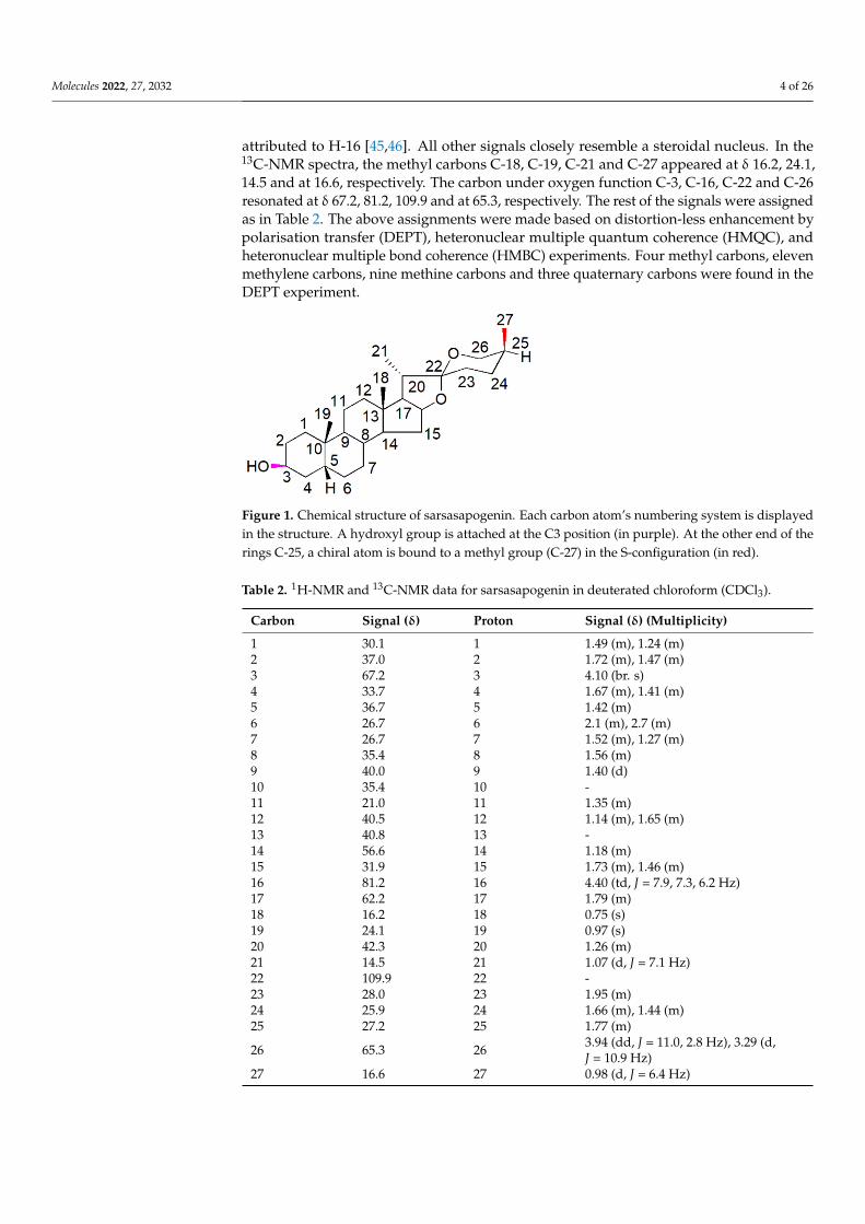
Spirostane saponins are monodesmosidic glycosides with six rings (A–F) that aresimilar to sapogenin but differ in having an axially oriented methyl or hydroxymethyl(C-27) on the F-ring. Sarsasapogenin is a spirostane glycoside sapogenin that has a 5βconfiguration between rings A and B (Figure 1).
Based on electron spray ionisation mass spectrometry (ESI-MS), sarsasapogenin pro-duced a molecular ion peak at m/z 417.34 [M + H]+ ion corresponding to a molecularformula of C27H44O3. The infrared (IR) spectra exhibited characteristic absorption bands at3407 (–OH), 2931, 1449, 1378 (–C–H) and at 1069 cm−1, characteristic of C–O groups. Inthe proton nuclear magnetic resonance (H-NMR) spectra, the H-3 proton appeared at δ4.10 as a broad singlet. The doublet of doublet signals appeared at δ 3.94 (dd, J = 11.0, 2.8Hz) and 3.29 (d, J = 10.9 Hz) each integrating for one proton, attributed to H-26 methyleneprotons. The two singlets appeared at δ 0.75 and 0.97 each integrating for three protonsdue to the H-18 and H-19 methyl groups, respectively. The pair of doublets at 0.98 (3H,d, J = 6.2 Hz) and 1.07 (3H, d, J = 7.4 Hz) were assigned to the H-21 and H-27 methylgroups respectively. The triplet of doublets signals at δ 4.40 (1H, td, J = 7.9, 7.3, 6.2 Hz) were
Molecules 2022, 27, 2032 4 of 26
attributed to H-16 [45,46]. All other signals closely resemble a steroidal nucleus. In the13C-NMR spectra, the methyl carbons C-18, C-19, C-21 and C-27 appeared at δ 16.2, 24.1,14.5 and at 16.6, respectively. The carbon under oxygen function C-3, C-16, C-22 and C-26resonated at δ 67.2, 81.2, 109.9 and at 65.3, respectively. The rest of the signals were assignedas in Table 2. The above assignments were made based on distortion-less enhancement bypolarisation transfer (DEPT), heteronuclear multiple quantum coherence (HMQC), andheteronuclear multiple bond coherence (HMBC) experiments. Four methyl carbons, elevenmethylene carbons, nine methine carbons and three quaternary carbons were found in theDEPT experiment.
Figure 1. Chemical structure of sarsasapogenin. Each carbon atom’s numbering system is displayedin the structure. A hydroxyl group is attached at the C3 position (in purple). At the other end of therings C-25, a chiral atom is bound to a methyl group (C-27) in the S-configuration (in red).
Table 2. 1H-NMR and 13C-NMR data for sarsasapogenin in deuterated chloroform (CDCl3).
Carbon Signal (δ) Proton Signal (δ) (Multiplicity)
1 30.1 1 1.49 (m), 1.24 (m)2 37.0 2 1.72 (m), 1.47 (m)3 67.2 3 4.10 (br. s)4 33.7 4 1.67 (m), 1.41 (m)5 36.7 5 1.42 (m)6 26.7 6 2.1 (m), 2.7 (m)7 26.7 7 1.52 (m), 1.27 (m)8 35.4 8 1.56 (m)9 40.0 9 1.40 (d)10 35.4 10 -11 21.0 11 1.35 (m)12 40.5 12 1.14 (m), 1.65 (m)13 40.8 13 -14 56.6 14 1.18 (m)15 31.9 15 1.73 (m), 1.46 (m)16 81.2 16 4.40 (td, J = 7.9, 7.3, 6.2 Hz)17 62.2 17 1.79 (m)18 16.2 18 0.75 (s)19 24.1 19 0.97 (s)20 42.3 20 1.26 (m)21 14.5 21 1.07 (d, J = 7.1 Hz)22 109.9 22 -23 28.0 23 1.95 (m)24 25.9 24 1.66 (m), 1.44 (m)25 27.2 25 1.77 (m)
26 65.3 26 3.94 (dd, J = 11.0, 2.8 Hz), 3.29 (d,J = 10.9 Hz)
27 16.6 27 0.98 (d, J = 6.4 Hz)
Molecules 2022, 27, 2032 5 of 26
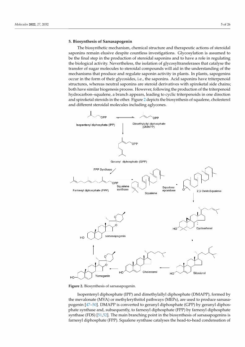
5. Biosynthesis of Sarsasapogenin
The biosynthetic mechanism, chemical structure and therapeutic actions of steroidalsaponins remain elusive despite countless investigations. Glycosylation is assumed tobe the final step in the production of steroidal saponins and to have a role in regulatingthe biological activity. Nevertheless, the isolation of glycosyltransferases that catalyse thetransfer of sugar molecules to steroidal compounds will aid in the understanding of themechanisms that produce and regulate saponin activity in plants. In plants, sapogeninsoccur in the form of their glycosides, i.e., the saponins. Acid saponins have triterpenoidstructures, whereas neutral saponins are steroid derivatives with spiroketal side chains;both have similar biogenesis process. However, following the production of the triterpenoidhydrocarbon–squalene, a branch appears, leading to cyclic triterpenoids in one directionand spiroketal steroids in the other. Figure 2 depicts the biosynthesis of squalene, cholesteroland different steroidal molecules including aglycones.
Figure 2. Biosynthesis of sarsasapogenin.
Isopentenyl diphosphate (IPP) and dimethylallyl diphosphate (DMAPP), formed bythe mevalonate (MVA) or methylerythritol pathways (MEPs), are used to produce sarsasa-pogenin [47–50]. DMAPP is converted to geranyl diphosphate (GPP) by geranyl diphos-phate synthase and, subsequently, to farnesyl diphosphate (FPP) by farnesyl diphosphatesynthase (FDS) [51,52]. The main branching point in the biosynthesis of sarsasapogenins isfarnesyl diphosphate (FPP). Squalene synthase catalyses the head-to-head condensation of
Molecules 2022, 27, 2032 6 of 26
two units of farnesyl diphosphate to create a 30-carbon linear squalene. Squalene is thentransformed to squalene epoxide by squalene epoxidase. Subsequently, the latter is cyclisedby a variety of triterpene cyclases by protonation and an epoxide ring opening by a numberof triterpene cyclases. With the help of cycloartenol synthase, tetracyclic cycloartenol isformed, serving as a precursor for the biosynthesis of steroidal saponin. Moreover, cy-cloartenol undergoes a series of rearrangements to form sitosterol, which are catalysed byenzymes such as cycloeucalenol cycloisomerase, methylsterol monooxygenase, sterol 14alpha-demethylase, 7-dehydrocholesterol reductases and lanosterol oxidase, among others.Sitosterol is a direct precursor for the manufacture of steroidal saponins and is glucosylatedat different locations to create saponins, which are catalysed by glucosyltransferases [53–55].
6. Physicochemical and Drug-Likeness Properties of Sarsasapogenin
For drug molecules to enter their active sites, several obstacles need to be overcome.Complex biological processes can be presented using physiochemical characteristics, andknowing them can help to move a drug development programme forward in both thelead optimisation and lead identification phases. The physicochemical properties of sarsas-apogenin were primarily derived from PubChem and other reliable databases. BioviaDiscovery studio 19.0 was used to determine the drug-likeness parameters (molecularweight, H-bond donors, H-bond acceptors, log P-value and rotatable bonds) as indicatedby Lipinski’s rule of five (Table 3).
Table 3. Physicochemical and drug-likeness properties of sarsasapogenin.
Property/Rule Result
Molecular formula C27H44O3Molecular weight 416.6 g/molHydrogen bond donors 1Hydrogen bond acceptors 3Rotatable bonds 0Log P (partition coefficient, predicted value) 7.306Molar refractivity 122.07 cm3
Topological polar surface area 38.7 Å2
7. Biological Properties of Sarsasapogenin7.1. Anti-Inflammatory Activity
Inflammation is an essential protective process that protects organisms from phys-ical, chemical and infectious risks. However, it is common for the inflammatory re-sponse to many exposures to erroneously damage normal tissues [56]. Nonsteroidalanti-inflammatory drugs (NSAIDs) are commonly used to treat inflammation, have a sideeffect profile, despite their significant function in pain and inflammation management [57].This highlights the need for novel anti-inflammatory drugs that are both safety and efficacy.Yu et al. [58] confirmed the anti-inflammatory activity of sarsasapogenin that enters adiposetissue following a single oral administration to inhibit acute adipose inflammation causedby an intraperitoneal injection of lipopolysaccharide (LPS) to C57BL/6J mice (Figure 3). LPScontains bacterial endotoxin broadly used for observing the response of acute inflammationin mice, since it stimulates the production of cytokines such as tumour necrosis factor alpha(TNF-α) and interleukin-1 (IL)-1β and IL-6. Pre-treatment with sarsasapogenin (80 mg/kg)for 18 days ameliorates TNF-α, IL-1β and IL-6 levels in the plasma and reduces the concen-tration of the anti-inflammatory cytokine-10. Other than releasing cytokines, LPS bindsto Toll-like receptors to stimulate the IKK/nuclear factor kappa B (NF-κB) and the JNKsignalling pathways and elevate both the expression and secretion of pro-inflammatorygenes. Sarsasapogenin can supress the expression of the pro-inflammatory genes, includingTNF-α, IL–1β and IL-6, monocyte chemo-attractant protein-1 (MCP-1), nitric oxide synthase2 (Nos2) and cyclooxygenase-2 (COX2), in white adipose tissue, simultaneously increasingthe transcription of M2, including Arg1, YM1, Fizz1 and IL-10, which further stimulate
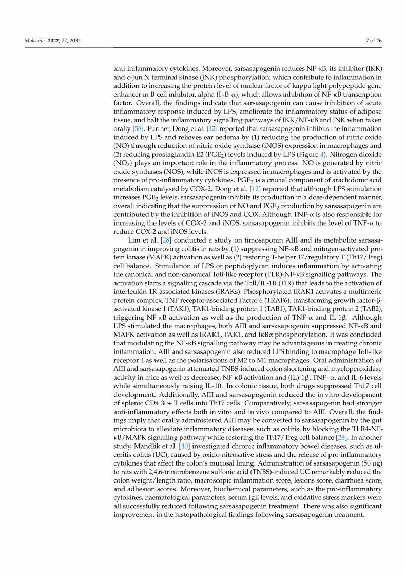
Molecules 2022, 27, 2032 7 of 26
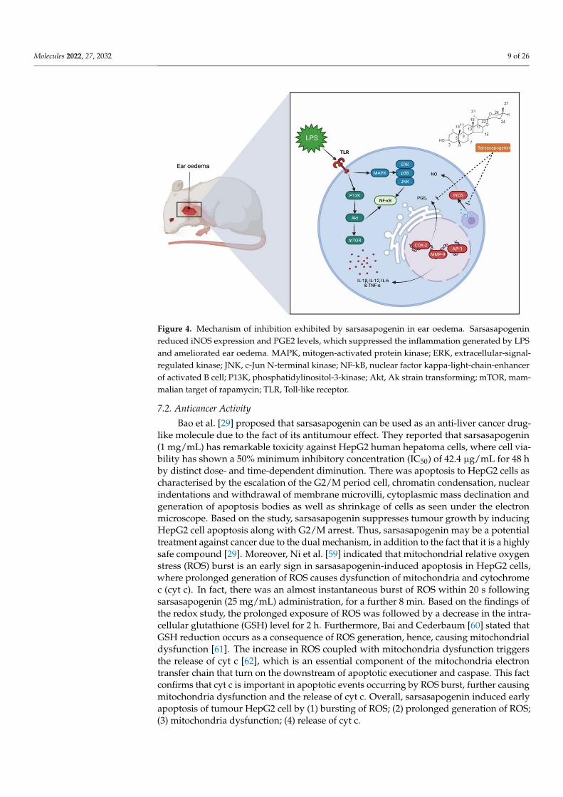
anti-inflammatory cytokines. Moreover, sarsasapogenin reduces NF-κB, its inhibitor (IKK)and c-Jun N terminal kinase (JNK) phosphorylation, which contribute to inflammation inaddition to increasing the protein level of nuclear factor of kappa light polypeptide geneenhancer in B-cell inhibitor, alpha (IκB-α), which allows inhibition of NF-κB transcriptionfactor. Overall, the findings indicate that sarsasapogenin can cause inhibition of acuteinflammatory response induced by LPS, ameliorate the inflammatory status of adiposetissue, and halt the inflammatory signalling pathways of IKK/NF-κB and JNK when takenorally [58]. Further, Dong et al. [12] reported that sarsasapogenin inhibits the inflammationinduced by LPS and relieves ear oedema by (1) reducing the production of nitric oxide(NO) through reduction of nitric oxide synthase (iNOS) expression in macrophages and(2) reducing prostaglandin E2 (PGE2) levels induced by LPS (Figure 4). Nitrogen dioxide(NO2) plays an important role in the inflammatory process. NO is generated by nitricoxide synthases (NOS), while iNOS is expressed in macrophages and is activated by thepresence of pro-inflammatory cytokines. PGE2 is a crucial component of arachidonic acidmetabolism catalysed by COX-2. Dong et al. [12] reported that although LPS stimulationincreases PGE2 levels, sarsasapogenin inhibits its production in a dose-dependent manner,overall indicating that the suppression of NO and PGE2 production by sarsasapogenin arecontributed by the inhibition of iNOS and COX. Although TNF-α is also responsible forincreasing the levels of COX-2 and iNOS, sarsasapogenin inhibits the level of TNF-α toreduce COX-2 and iNOS levels.
Lim et al. [28] conducted a study on timosaponin AIII and its metabolite sarsasa-pogenin in improving colitis in rats by (1) suppressing NF-κB and mitogen-activated pro-tein kinase (MAPK) activation as well as (2) restoring T-helper 17/regulatory T (Th17/Treg)cell balance. Stimulation of LPS or peptidoglycan induces inflammation by activatingthe canonical and non-canonical Toll-like receptor (TLR)-NF-κB signalling pathways. Theactivation starts a signalling cascade via the Toll/IL-1R (TIR) that leads to the activation ofinterleukin-1R-associated kinases (IRAKs). Phosphorylated IRAK1 activates a multimericprotein complex, TNF receptor-associated Factor 6 (TRAF6), transforming growth factor-β-activated kinase 1 (TAK1), TAK1-binding protein 1 (TAB1), TAK1-binding protein 2 (TAB2),triggering NF-κB activation as well as the production of TNF-α and IL-1β. AlthoughLPS stimulated the macrophages, both AIII and sarsasapogenin suppressed NF-κB andMAPK activation as well as IRAK1, TAK1, and IκBα phosphorylation. It was concludedthat modulating the NF-κB signalling pathway may be advantageous in treating chronicinflammation. AIII and sarsasapogenin also reduced LPS binding to macrophage Toll-likereceptor 4 as well as the polarisations of M2 to M1 macrophages. Oral administration ofAIII and sarsasapogenin attenuated TNBS-induced colon shortening and myeloperoxidaseactivity in mice as well as decreased NF-κB activation and (IL)-1β, TNF- α, and IL-6 levelswhile simultaneously raising IL-10. In colonic tissue, both drugs suppressed Th17 celldevelopment. Additionally, AIII and sarsasapogenin reduced the in vitro developmentof splenic CD4 30+ T cells into Th17 cells. Comparatively, sarsasapogenin had strongeranti-inflammatory effects both in vitro and in vivo compared to AIII. Overall, the find-ings imply that orally administered AIII may be converted to sarsasapogenin by the gutmicrobiota to alleviate inflammatory diseases, such as colitis, by blocking the TLR4-NF-κB/MAPK signalling pathway while restoring the Th17/Treg cell balance [28]. In anotherstudy, Mandlik et al. [40] investigated chronic inflammatory bowel diseases, such as ul-ceritis colitis (UC), caused by oxido-nitrosative stress and the release of pro-inflammatorycytokines that affect the colon’s mucosal lining. Administration of sarsasapogenin (50 µg)to rats with 2,4,6-trinitrobenzene sulfonic acid (TNBS)-induced UC remarkably reduced thecolon weight/length ratio, macroscopic inflammation score, lesions score, diarrhoea score,and adhesion scores. Moreover, biochemical parameters, such as the pro-inflammatorycytokines, haematological parameters, serum IgE levels, and oxidative stress markers wereall successfully reduced following sarsasapogenin treatment. There was also significantimprovement in the histopathological findings following sarsasapogenin treatment.
Molecules 2022, 27, 2032 8 of 26
Overall, sarsasapogenin’s anti-inflammatory activity appears to be mainly due tothe reduction in oxidative stress and inhibition of TNF-α release. Overall, the findingsimply that sarsasapogenin could be a promising drug-like treatment for a wide range ofinflammatory disorders.
Figure 3. Sarsasapogenin’s anti-inflammatory action in adipose tissue. While lean adipose tissuecontributes to metabolic balance, inflammation regulation and insulin resistance, as obesity pro-gresses, adipocytes enlarge and release adipokines, resulting in an increase in the production ofpro-inflammatory factors and immune cell infiltration. Sarsasapogenin has been shown to inhibitpro-inflammatory cytokines, such as TNF-α, IL–1β, IL–6, IL-10, IL-12 and MCP-1, in white adiposetissue. TNF-α, tumour necrosis factor alpha; interleukin-1 beta, -6, -10 and -12; MCP-1, monocytechemoattractant protein-1.
Molecules 2022, 27, 2032 9 of 26
Figure 4. Mechanism of inhibition exhibited by sarsasapogenin in ear oedema. Sarsasapogeninreduced iNOS expression and PGE2 levels, which suppressed the inflammation generated by LPSand ameliorated ear oedema. MAPK, mitogen-activated protein kinase; ERK, extracellular-signal-regulated kinase; JNK, c-Jun N-terminal kinase; NF-kB, nuclear factor kappa-light-chain-enhancerof activated B cell; P13K, phosphatidylinositol-3-kinase; Akt, Ak strain transforming; mTOR, mam-malian target of rapamycin; TLR, Toll-like receptor.
7.2. Anticancer Activity
Bao et al. [29] proposed that sarsasapogenin can be used as an anti-liver cancer drug-like molecule due to the fact of its antitumour effect. They reported that sarsasapogenin(1 mg/mL) has remarkable toxicity against HepG2 human hepatoma cells, where cell via-bility has shown a 50% minimum inhibitory concentration (IC50) of 42.4 µg/mL for 48 hby distinct dose- and time-dependent diminution. There was apoptosis to HepG2 cells ascharacterised by the escalation of the G2/M period cell, chromatin condensation, nuclearindentations and withdrawal of membrane microvilli, cytoplasmic mass declination andgeneration of apoptosis bodies as well as shrinkage of cells as seen under the electronmicroscope. Based on the study, sarsasapogenin suppresses tumour growth by inducingHepG2 cell apoptosis along with G2/M arrest. Thus, sarsasapogenin may be a potentialtreatment against cancer due to the dual mechanism, in addition to the fact that it is a highlysafe compound [29]. Moreover, Ni et al. [59] indicated that mitochondrial relative oxygenstress (ROS) burst is an early sign in sarsasapogenin-induced apoptosis in HepG2 cells,where prolonged generation of ROS causes dysfunction of mitochondria and cytochromec (cyt c). In fact, there was an almost instantaneous burst of ROS within 20 s followingsarsasapogenin (25 mg/mL) administration, for a further 8 min. Based on the findings ofthe redox study, the prolonged exposure of ROS was followed by a decrease in the intra-cellular glutathione (GSH) level for 2 h. Furthermore, Bai and Cederbaum [60] stated thatGSH reduction occurs as a consequence of ROS generation, hence, causing mitochondrialdysfunction [61]. The increase in ROS coupled with mitochondria dysfunction triggersthe release of cyt c [62], which is an essential component of the mitochondria electrontransfer chain that turn on the downstream of apoptotic executioner and caspase. This factconfirms that cyt c is important in apoptotic events occurring by ROS burst, further causingmitochondria dysfunction and the release of cyt c. Overall, sarsasapogenin induced earlyapoptosis of tumour HepG2 cell by (1) bursting of ROS; (2) prolonged generation of ROS;(3) mitochondria dysfunction; (4) release of cyt c.
Molecules 2022, 27, 2032 10 of 26
Sarsasapogenin induced apoptosis through an ROS-mediated mitochondrial path-way and endoplasmic reticulum (ER) stress pathway in HeLa cells. ER is responsible inapoptosis regulation where ER stress can stimulate several signalling pathways includingER-associated protein degradation and unfolded protein response (URP). After HeLa cellswere cultured with or without sarsasapogenin (60 µM), the stress sensor of ER, such asprotein kinase RNA-like ER kinase (PERK) and elF21, were phosphorylated at an earlystage within 6 h of treatment with sarsasapogenin. However, the level of PERK and eu-karyotic translation initiation factor 2 subunit 1 (elF21) remained unchanged. Additionally,the activating transcription factor 6 (ATF6), which is also a protein of ER, was cleaved andturned into the activated form or ATF6 fragmentation. Additionally, ER chaperon, suchas GR78 and GRP94, were also stimulated. The transduction signalling pathways wereupregulated to counteract the burden of ER by impeding protein synthesis. Nevertheless,should the ER stress continue for a longer period and become even more severe, theseconditions can induce apoptosis [63]. Sarsasapogenin can also cause C/EBP homologousprotein (CHOP), a key transcription factor stimulated by ER to be activated as well as theoverexpression of CHOP, resulting in growth arrest and apoptosis. Overall, the findingsindicate that sarsasapogenin induces apoptosis by an ER stress. Apart from that, ROS,which are mediators of intracellular cascades, can disintegrate the mitochondrial membrane,leading to apoptosis. Consequently, the overproduction of ROS, leads to oxidative stress,cell dysfunction, necrosis and apoptosis. All of the above, indicates that sarsasapogenininduced apoptosis by ER stress, ROS formation and mitochondria dysfunction [63].
7.3. Neuroprotection
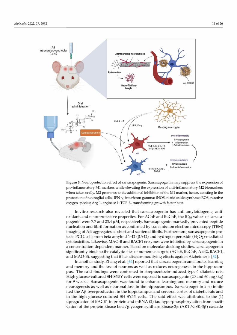
Alzheimer’s disease (AD), also known as an irreversible neurodegenerative disorder,is specified by the deposition of beta-amyloid (Aβ) in the extracellular as well as phospho-rylation of tau protein to the intracellular cells that are associated with the neuronal andsynaptic loss, leading to cognitive deterioration and memory loss. The disparity betweenAβ production and clearance results in amassment and deposition in the brain that lead to asporadic AD. Huang et al. [13] demonstrated the (1) therapeutic effect of sarsasapogenin-13,a sarsasapogenin derivative on learning and memory impairments in Aβ-injected mice;(2) the role of sarsasapogenin-13 performed in neuroglia-mediated anti-inflammation andAβ clearance. Their findings indicated that oral administration of sarsasapogenin reducedmemory deficits in mice injected with Aβ intracerebroventricular (i.c.v.) while protectingneuroglial cells against Aβ-induced cytotoxicity. Further mechanical studies revealed thatsarsasapogenin inhibits the elevation of pro-inflammatory M1 markers while increasingthe expression of anti-inflammatory M2 markers in Aβ-treated cells (Figure 5). Moreover,Aβ clearance through Aβ phagocytosis and breakdown were further aided by sarsasa-pogenin. Fatty acid translocase (CD36), insulin degrading enzyme (IDE), neprilysin (NEP)and endothelin-converting enzyme (ECE) expression in neuroglia were also influencedby sarsasapogenin. It was concluded that the neuroprotective action of sarsasapogenin isrelated to its modulatory effects on the microglia activation state, phagocytic capabilitiesand the production of Aβ-degrading enzymes, making it a viable therapeutic agent in theearly stages of Alzheimer’s disease [13].
To develop a therapeutic strategy for Alzheimer’s disease, a multi-target directedligand (MTDL) approach is often necessary. Kashyap et al. [32] selected the aqueous ex-tract of Asparagus racemosus, including its secondary metabolite sarsasapogenin, for hisinvestigation, since it has a wide range of therapeutic properties. The team conductedseveral tests, such as Aβ-induced neurotoxicity on PC12 cells, to confirm its nootropic effectthat has been referenced in ancient Ayurvedic literature. They found that sarsasapogenininhibited key enzymes involved in Alzheimer’s disease pathogenesis, including acetyl-cholinesterase (AChE), butyrylcholinesterase (BuChE), beta-secretase 1 precursor (BACE1),and monoaminoxidase-B (MAO-B) in a concentration-dependent way [32].
Molecules 2022, 27, 2032 11 of 26
Figure 5. Neuroprotection effect of sarsasapogenin. Sarsasapogenin may suppress the expression ofpro-inflammatory M1 markers while elevating the expression of anti-inflammatory M2 biomarkerswhen taken orally. M2 promotes to the additional inhibition of the M1 marker, hence, assisting in theprotection of neuroglial cells. IFN-γ, interferon gamma; iNOS, nitric oxide synthase; ROS, reactiveoxygen species; Arg-1, arginase 1; TGF-β, transforming growth factor beta.
In vitro research also revealed that sarsasapogenin has anti-amyloidogenic, anti-oxidant, and neuroprotective properties. For AChE and BuChE, the IC50 values of sarsasa-pogenin were 7.7 and 23.4 µM, respectively. Sarsasapogenin markedly prevented peptidenucleation and fibril formation as confirmed by transmission electron microscopy (TEM)imaging of Aβ aggregates as short and scattered fibrils. Furthermore, sarsasapogenin pro-tects PC12 cells from beta amyloid 1-42 (βA42) and hydrogen peroxide (H2O2)-mediatedcytotoxicities. Likewise, MAO-B and BACE1 enzymes were inhibited by sarsasapogenin ina concentration-dependent manner. Based on molecular docking studies, sarsasapogeninsignificantly binds to the catalytic sites of numerous targets (AChE, BuChE, Aβ42, BACE1,and MAO-B), suggesting that it has disease-modifying effects against Alzheimer’s [32].
In another study, Zhang et al. [64] reported that sarsasapogenin ameliorates learningand memory and the loss of neurons as well as reduces neurogenesis in the hippocam-pus. The said findings were confirmed in streptozotocin-induced type-1 diabetic rats.High glucose-cultured SH-SY5Y cells were exposed to sarsasapogenin (20 and 60 mg/kg)for 9 weeks. Sarsasapogenin was found to enhance learning and memory and reduceneurogenesis as well as neuronal loss in the hippocampus. Sarsasapogenin also inhib-ited the Aβ overproduction in the hippocampus and cerebral cortex of diabetic rats andin the high glucose-cultured SH-SY5Y cells. The said effect was attributed to the (1)upregulation of BACE1 in protein and mRNA (2) tau hyperphosphorylation from inacti-vation of the protein kinase beta/glycogen synthase kinase-3β (AKT/GSK-3β) cascade
Molecules 2022, 27, 2032 12 of 26
and (3) peroxisome proliferator-activated receptor gamma (PPARγ) antagonism that abol-ished sarsasapogenin’s effects on key molecules. Furthermore, it was discovered that highglucose-stimulated Aβ overproduction occurred prior to tau hyperphosphorylation inneurons. Hu et al. [65] further confirmed that sarsasapogenin can enhance learning andmemory. Interestingly, sarsasapogenin neither hindered acetylcholinesterase nor occupiedthe binding sites of the muscarinic acetylcholine receptor (M receptor), indicating thatit is neither a cholinesterase inhibitor nor an agonist or antagonist of M receptors. Thedensities of total M receptor and its M1 subtype in the brain of the three models, however,were considerably lower than that shown by the control rats. Sarsasapogenin markedlyincreased the densities of total M receptor and its M1 subtype. The investigation done byan autoradiography using 3H-pirenzipine further confirmed that the density of M1 subtypelocated in the cortex, hippocampus as well as striatum in elderly rats were remarkablylowered, yet can be reversed to normalcy by sarsasapogenin’s administration.
Interactions between molecular and cellular abnormalities, as well as genetic and envi-ronmental variables, may promote the formation of depression and stress-related mentaldisorders [66]. However, Islam et al. [67] found that taking an antidepressant increases theactivation of nicotinic acetylcholine receptors (nAChRs), which serve as excitatory cationchannels and include α-subunits (α2–α7) and β-subunits (β2–β4) [68,69]. The 4 and α7subunits of nAChRs expressed in the brain are the most noticeable. The α4-nAChR receptorcan be found all across the nervous system, but it is concentrated in the hippocampus,cerebral cortex, and several brainstem nuclei [70,71]. Nicotine or agonists of the 7- and4-nAChRs, according to a mouse study, may enhance the antidepressant’s beneficial effects.Sarsasapogenin enhances memory in a memory-impaired rat model by increasing thedensity of acetylcholine receptors in the brain. The behavioural despair test confirmed thatsarsasapogenin had antidepressant-like effects in mice [25]. The antidepressant effect ofsarsasapogenin and its relation to cholinergic signalling were investigated by Feng et al. [14].The olfactory bulbectomized (OB)-induced sucrose preference deficit was significantly re-covered by sarsasapogenin (20 and 40 mg/kg) and amitriptyline. In comparison to theSham group, OB rats displayed less grooming and more evident hyperactivity, as measuredby increased ambulation and rearing. The OB-induced enhanced locomotor activity wasconsiderably reduced by sarsasapogenin (20 and 40 mg/kg) and amitriptyline (10 mg/kg)therapy. Administration of sarsasapogenin (20 and 40 mg/kg) significantly enhanced 7-and 4-nAChRs protein levels in OB rats. Hence, sarsasapogenin can restore depression-likebehaviours caused by OB and improve cholinergic system malfunction. It is plausiblethat the antidepressant activity of sarsasapogenin is mediated by an increase expressionin 7- and 4-nAChRs, as well as by affecting AChE activity. Overall, the data indicate thatcholinergic signalling is a promising target for the development of a sarsasapogenin therapyfor depression.
Ren et al. [25] reported that sarsasapogenin reduced the duration of immobility ina dose-dependent manner in a forced swim test. Nevertheless, since sarsasapogenin didnot generate hyperlocomotion in the open-field test, its antidepressant effect cannot beattributed to an increase in motor activity. On the other hand, sarsasapogenin increase theconcentration of 5-HT (serotonin) in the brain, conversely reducing its turnover possiblydue to monoamine metabolism. Noradrenaline levels tend to increase in the hypotha-lamus, following the treatment with sarsasapogenin (25 and 50 mg/kg) indicating thatsarsasapogenin has an antidepressant effect by regulating the 5-HT and noradrenalinesystems. The activity of MAO-A influenced the release of neurotransmitter such as 5-HTand noradrenaline that are crucial in ameliorating depression. Further, Zhang et al. [72]explored the effects and the possible mechanisms of sarsasapogenin in protecting highglucose-induced amyloid-beta (Aβ) peptide overproduction in HT-22 cells where there wasincreased Aβ expression and Aβ42 levels, as well as elevated BACE1 protein, mRNA levelsand enzymatic activities which were ameliorated following sarsasapogenin administration.Administration of moderate and high doses of sarsasapogenin (5 and 25 mol/L) reducedPPARγ levels that were increased in the presence of glucose. Sarsasapogenin also possesses
Molecules 2022, 27, 2032 13 of 26
substantial neuroprotective effects as confirmed by their study in which it (1) inhibits Aβ
deposit and (2) increase cell survival in high glucose cultured HT-22 cells, likely to bemediated by PPAR activation and to be associated with BACE1 downregulation.
The effect of sarsasapogenin (6 mg/kg) on nootropic and neutrophic as well as theirmechanisms, in vitro as well in vivo was investigated by Dong et al. [12]. For the former,MTT assays were used to determine the viability of rat primary astrocytes treated withsarsasapogenin and neurons cultured with conditioned medium of sarsasapogenin-treatedrat primary astrocytes. As for the latter, scopolamine-induced cognitive deficit model wasused where the mice’s spatial memory was tested using a Morris water maze. Based on theirfindings, sarsasapogenin promoted the viability of primary astrocytes and sarsasapogenin-treated astrocyte within the conditioned medium and improved the survival rate of primaryneurons. Remarkably, sarsasapogenin significantly increased BDNF levels in astrocytes.Sarsasapogenin also alleviated cognitive impairments in animal models and elevated BDNFand PSD95 levels in the brain.
Overall, sarsasapogenin modulates growth factors, enzymes, transcription factors,kinase, inflammatory cytokines, and proapoptotic (by upregulation) and antiapoptotic (bydownregulation) proteins. Sarsasapogenin alone or in combination with other drugs, couldbe a promising treatment for various types of cancer.
7.4. Antidiabetic Activity
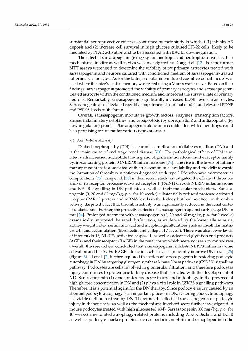
Diabetic nephropathy (DN) is a chronic complication of diabetes mellitus (DM) andis the main cause of end-stage renal disease [73]. The pathological effects of DN is re-lated with increased nucleotide binding and oligomerisation domain-like receptor familypyrin-containing protein 3 (NLRP3) inflammasome [74]. The rise in the levels of inflam-matory mediators is associated with an elevation of coagulability and the drift towardsthe formation of thrombus in patients diagnosed with type 2 DM who have microvascularcomplications [75]. Tang et al. [30] in their recent study, investigated the effects of thrombinand/or its receptor, protease-activated receptor 1 (PAR-1) on both NLRP3 inflammasomeand NF-κB signalling in DN patients, as well as their molecular mechanism. Sarsasa-pogenin (0, 20 and 60 mg/kg, p.o. for 10 weeks) substantially reduced protease-activatedreceptor (PAR-1) protein and mRNA levels in the kidney but had no effect on thrombinactivity, despite the fact that thrombin activity was significantly reduced in the renal cortexof diabetic rats. Further, the protective effects of sarsasapogenin against early stage DN inrats [26]. Prolonged treatment with sarsasapogenin (0, 20 and 60 mg/kg, p.o. for 9 weeks)dramatically improved the renal dysfunction, as evidenced by the lower albuminuria,kidney weight index, serum uric acid and morphologic alterations such extracellular matrixgrowth and accumulation (fibronectin and collagen IV levels). There was also lower levelsof interleukin 18, NLRP3, activated caspase 1, as well as advanced glycation end products(AGEs) and their receptor (RAGE) in the renal cortex which were not seen in control rats.Overall, the researchers concluded that sarsasapogenin inhibits NLRP3 inflammasomeactivation and the AGEs–RAGE interaction, which can significantly improve DN in rats [26](Figure 6). Li et al. [2] further explored the action of sarsasapogenin in restoring podocyteautophagy in DN by targeting glycogen synthase kinase 3 beta pathway (GSK3β) signallingpathway. Podocytes are cells involved in glomerular filtration, and therefore podocytesinjury contributes to proteinuric kidney disease that is related with the development ofND. Sarsasapogenin (1) ameliorates podocyte injury and autophagy in the presence ofhigh glucose concentration in DN and (2) plays a vital role in GSK3β signalling pathways.Therefore, it is a potential agent for the DN therapy. Since podocyte injury caused by anaberrant podocyte autophagy is an important process in DN, restoring podocyte autophagyis a viable method for treating DN. Therefore, the effects of sarsasapogenin on podocyteinjury in diabetic rats, as well as the mechanisms involved were further investigated inmouse podocytes treated with high glucose (40 µM). Sarsasapogenin (60 mg/kg, p.o. for10 weeks) ameliorated autophagy-related proteins including ATG5, Beclin1 and LC3Bas well as podocyte marker proteins such as podocin, nephrin and synaptopodin in the
Molecules 2022, 27, 2032 14 of 26
diabetic kidney. Subsequently, a network pharmacology used to evaluate GSK3 as a poten-tial target for sarsasapogenin’s impact on DN, confirmed the significant changes in GSK3signalling seen in the diabetic kidneys.
Figure 6. Sarsasapogenin mechanism of action in inflammasome activation and AGE. NLRP3 istriggered by a wide variety of stimuli, including LPS via Toll-like receptors (TLR), which results in themanufacture of the cytokine precursor via NF-kB and other inflammasome constituents, includingNLRP3. Sarsasapogenin inhibits the activation of the NLRP3 inflammasome and the AGE pathway,vastly improving diabetic nephropathy in rats. Abbreviations: N-GSDMD, N-terminal Gasdermin-D;GSDMD, Gasdermin-D; NLRP3, NLR family pyrin domain containing 3; NLRC4, NLR Family CARDDomain Containing 4; AGE, Advanced glycation end products; PKC, Protein kinase C.
The effects of sarsasapogenin on damaged vascular endothelium in high-glucose cul-tured human umbilical vein cultured cells (HUVECs) related to the gestational diabetesmellitus (GDM) were explored by Liu et al. [76]. GDM is a disease of uncommon glu-cose tolerance during pregnancy which may lead to fetal macrosomia, respiratory distresssyndrome as well as type 2 diabetes in the offspring. The pathogenic alterations of thesepathways were investigated using the umbilical cord and plasma of GDM patients, as wellas high glucose HUVECs. Meanwhile, HUVECs were used to explore the effects and thepossible mechanism of action of sarsasapogenin. Interestingly, co-treatment with sarsasa-pogenin affect the thrombin/PAR-1 pathway, AGEs/RAGE axis and reduced the NLRP1inflammasome. Endothelial damage in GDM was likely to be caused by the increasedinteraction between the AGEs/RAGE axis and the thrombin/PAR-1 pathway, followed byactivation of the NLRP1 inflammasome. Sarsasapogenin is also protective against endothe-lial injury in patients with chronically high glucose level. Through down-regulation of thePAR-1 receptor, Kong et al. [77] studied the effects of sarsasapogenin on diabetes-associatedmemory impairment and neuroinflammation. In streptozotocin-induced diabetic rats,sarsasapogenin (20 and 60 mg/kg, p.o. for 8 weeks) alleviated diabetes-related memoryimpairment, as evidenced by higher platform crossings and percentage of time spent inthe target quadrant in Morris water maze tests. In the hippocampus and cerebral cortex,sarsasapogenin blocked the nucleotide-binding domain and leucine-rich repeat-containingprotein 1 (NLRP1) inflammasome, repressed the AGEs/RAGE axis, and decreased theupregulation of the thrombin receptor PAR-1. In high glucose cultured SH-SY5Y cells,
Molecules 2022, 27, 2032 15 of 26
sarsasapogenin also reduced high hyperglycemia-induced neuronal damage, activation ofthe NLRP1 inflammasome, and PAR-1 upregulation, but it had no effect on thrombin activ-ity. Thus, sarsasapogenin was found to alleviate diabetes-induced memory impairment byreducing neuroinflammation caused by the activated NLRP1 inflammasome and NF-κB,which is controlled by cerebral PAR-1.
Overall, the preclinical investigations showed that sarsasapogenin can enhance glucosehomeostasis and minimise the diabetes phenotype by lowering blood glucose levels anddecreasing insulin resistance. However, more research is needed to completely comprehendthe effects of sarsasapogenin in specific body tissues such as skeletal muscle, adipose tissue,liver, and pancreatic β-cells. Further, sarsasapogenin appears to be a promising candidatefor clinical use in the treatment of insulin resistance and type 2 diabetes. Hence, clinicalinvestigations on sarsasapogenin are also necessary.
7.5. Anti-Osteoclastogenic Activity
Both osteoclasts and osteoblasts govern bone homeostasis [78]. Osteoclasts are polynu-clear cells that are responsible for bone resorption. An increased number of osteoclasts andrapid bone resorption influence osteoclast-related bone-lytic illnesses including osteoporo-sis. Osteoclasts produce tartrate-resistant phosphatase (TRAP) that adheres to the bonesurface via an actin-binding sealing zone, releasing proteases, such as tissue protease K, andcausing the mineralised bone matrix to degrade. The two cytokines that regulate osteoclastdifferentiation are macrophage-colony-stimulating factor (M-CSF), which (1) is required forcell proliferation and survival in monocyte cell lines, (2) enhances bone marrow precursordevelopment, and (3) boosts receptor activator of nuclear factor-B ligand (RANKL) expres-sion in bone marrow cells, and RANKL which is the primary cytokine required to promotemonocyte fusion that is essential in the formation of multinucleated osteoclasts [79–82].Osteoclastogenesis occurs when the RANK receptor is activated by its ligand RANK, wherethe interaction initiates a signal cascade that results in the expression of nuclear factor ofactivated T cells 1 (NFATc1), a key regulator for osteoclastogenesis. NFATc1 also activatesthe pathways of PI3K/Akt, MAPK/AP-1, and NF-B [83–85]. Peng et al. [23] conducted astudy to determine the effect of sarsasapogenin on the inhibition of osteoclastogenesis aswell as the prevention on the bone loss through the NF-κB B and JNK/MAPK signallingpathways. Sarsasapogenin (5 and 10 mg/kg, s.c.) suppressed the activation of the majorosteoclast transcription factor NFATc1 by blocking several RANKL-induced signallingcascades. The in vitro and in vivo findings in the mouse osteolysis model were consistent,where sarsasapogenin confers protection against the bone loss contributed by LPS. Overall,the findings support the use of sarsasapogenin as a novel therapy for osteoclast-relatedosteolytic disorders.
Based on the information shown above, sarsasapogenin, which has anti-inflammatoryand immunomodulatory effects, could be used to treat osteoclastic disorders. However,more research is needed, particularly on its mechanism of action and pharmacokinetics, inorder to validate its safety and efficacy and then to move forward with drug development.
7.6. Treatment for Precocious Puberty
Precocious puberty is the premature activation of gonadotropin-releasing hormone(GnRH) affecting growth and development [51]. It is also described as the manifestation ofsecondary sex traits in girls before the age of eight or in boys before the age of nine [86].Due to the early beginning of puberty, early growth and development as well as reducedbone growth years, children with early puberty tend to be small in stature. Additionally,the disease can lead to a variety of psychological and physical issues in patients and hasbeen linked to metabolic disorders such as diabetes, cardiovascular disease as well as breastand prostate cancers [87–89].
Currently, gonadotropin-releasing hormone analogue (GnRHa) is commonly utilisedto treat prematured puberty [90] with leuprolide most often used against precocious puberty.Leuprolide (1) acts on the pituitary in a constant non-pulsed manner, (2) downregulates
Molecules 2022, 27, 2032 16 of 26
the GnRH receptor, (3) limits the sensitivity to the GnRH receptor, and (4) inhibits thepituitary to secrete luteinizing hormone (LH) and follicle-stimulating hormone (FSH),overall slowing secondary sex development [91,92]. However, when taken for the first time,it may cause vaginal bleeding with an incidence of 16–60% [93] and increases the risk ofpolycystic ovary syndrome [52]. Therefore, the role of alternative therapies is important.Hu et al. [27] investigated the effects of sarsasapogenin in treating precocious puberty byregulating the hypothalamus–pituitary–gonadal (HPG) axis. To establish the precociouspuberty phase, 5 day old rats were given a single subcutaneous injection of danazol (300 µg).After 10 days, sarsasapogenin (4 mg/kg) was administered via oral gavaging for 10 days.Danazol affects the reproductive system of the rat and can rapidly stimulate the activationof the HPG axis to promote precocious puberty. However, sarsasapogenin prevents thedevelopment of gonads and downregulates the expression of GnRH and gonadotropin-releasing hormone receptor (GnRH-R) via the Kiss-1/GPR54 pathway, hence, delaying theactivation of the HPG axis and exerting its therapeutic effects.
Payment endocrinologists face a difficult task in assessing and managing this dis-ease [94]. The Pediatric Endocrinology Society has suggested that the combination ofGnRHa and recombinant growth hormone (rhGH) should not be recommended as a rou-tine treatment due to the fact of its high cost and the lack of large-scale randomised clinicaltrials evaluating the safety and effectiveness of this combination therapy [95]. Based on theabove studies, sarsasapogenin is a potential molecule to alleviate precocious puberty. Tosupport its therapeutic application, more information about the potential mechanism ofaction of sarsasapogenin is needed.
8. Bioavailability and Pharmacokinetics of Sarsasapogenin
The pharmacokinetics of sarsasapogenin were evaluated in rats after intragastricinjection of 25, 50, and 100 mg/kg [96]. This study measured sarsasapogenin plasmaconcentrations for 72 h. The results obtained were compared to the major steroidal saponinsextracted from A. asphodeloides, such as timosaponin H1, timosaponin E1, timosaponin E,timosaponin B-II, timosaponin B-III and anemarrhenasaponin I, based on the study fromLiu et al. 2015 [97]. After taking the sarsasapogenin, the sarsasapogenin content in theblood slowly increased. As consequence, the time for maximum plasma concentration(Tmax) of sarsasapogenin was significantly longer than the primary steroidal saponinsin A. asphodeloides (3.17–4.76 h). Sarsasapogenin has a low aqueous solubility, and thisis one of the main reasons for its poor absorption rate. Regarding the plasma half-life(t1/2), a single intragastric 100 mg/kg sarsasapogenin dosage in rats resulted in a t1/2 of17.72 h compared to the major steroidal saponins (15.1–16.1 h). The results indicating thatsarsasapogenin may have an incredibly long residence time shows that sarsasapogeninhas low bioavailability; thus, the excretion of sarsasapogenin is delayed. The linearityof the area under the plasma concentration versus time curve (AUC0–72h) and maximumplasma concentration (Cmax) to dosages performed well in regression analysis. As dosesincreased, the parameters of t1/2 and tmax were not remarkably affected. A dose range of25–100 mg/kg of sarsasapogenin was shown to be linear in pharmacokinetics [97].
9. Metabolism of Sarsasapogenin
Sarsasapogenin and smilagenin were hydrolysed in the rumen to yield the parentsapogenins, which were then oxidised and reduced at C-3 to provide the equivalent epi-sapogenins. Because of the reduced amount of sapogenins found in the small intestineand the identification of significant amounts of sapogenins, such as smilagenone, sarsasa-pogenin, smilagenin, episarsasapogenin and smilagenone, in the liver, this shows that thesesapogenins were absorbed from the upper intestine and transported to the liver via the por-tal vein, where smilagenin and sarsasapogenin that had not been oxidised and reduced inthe rumen were oxidised and reduced, and this activity is referred to as phase 1 metabolism,before epi-analogues were conjugated and excreted into the bile as episarsasapogenin andepismilagenin (phase II metabolism) [37].
Molecules 2022, 27, 2032 17 of 26
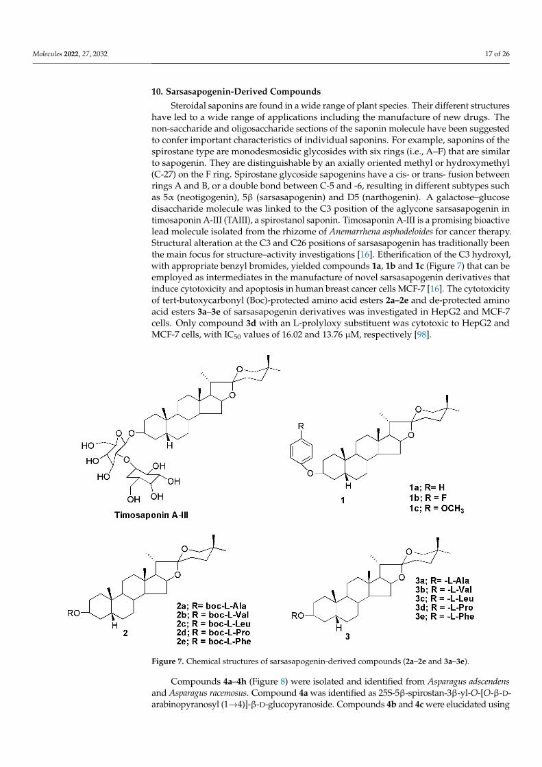
10. Sarsasapogenin-Derived Compounds
Steroidal saponins are found in a wide range of plant species. Their different structureshave led to a wide range of applications including the manufacture of new drugs. Thenon-saccharide and oligosaccharide sections of the saponin molecule have been suggestedto confer important characteristics of individual saponins. For example, saponins of thespirostane type are monodesmosidic glycosides with six rings (i.e., A–F) that are similarto sapogenin. They are distinguishable by an axially oriented methyl or hydroxymethyl(C-27) on the F ring. Spirostane glycoside sapogenins have a cis- or trans- fusion betweenrings A and B, or a double bond between C-5 and -6, resulting in different subtypes suchas 5α (neotigogenin), 5β (sarsasapogenin) and D5 (narthogenin). A galactose–glucosedisaccharide molecule was linked to the C3 position of the aglycone sarsasapogenin intimosaponin A-III (TAIII), a spirostanol saponin. Timosaponin A-III is a promising bioactivelead molecule isolated from the rhizome of Anemarrhena asphodeloides for cancer therapy.Structural alteration at the C3 and C26 positions of sarsasapogenin has traditionally beenthe main focus for structure–activity investigations [16]. Etherification of the C3 hydroxyl,with appropriate benzyl bromides, yielded compounds 1a, 1b and 1c (Figure 7) that can beemployed as intermediates in the manufacture of novel sarsasapogenin derivatives thatinduce cytotoxicity and apoptosis in human breast cancer cells MCF-7 [16]. The cytotoxicityof tert-butoxycarbonyl (Boc)-protected amino acid esters 2a–2e and de-protected aminoacid esters 3a–3e of sarsasapogenin derivatives was investigated in HepG2 and MCF-7cells. Only compound 3d with an L-prolyloxy substituent was cytotoxic to HepG2 andMCF-7 cells, with IC50 values of 16.02 and 13.76 µM, respectively [98].
Figure 7. Chemical structures of sarsasapogenin-derived compounds (2a–2e and 3a–3e).
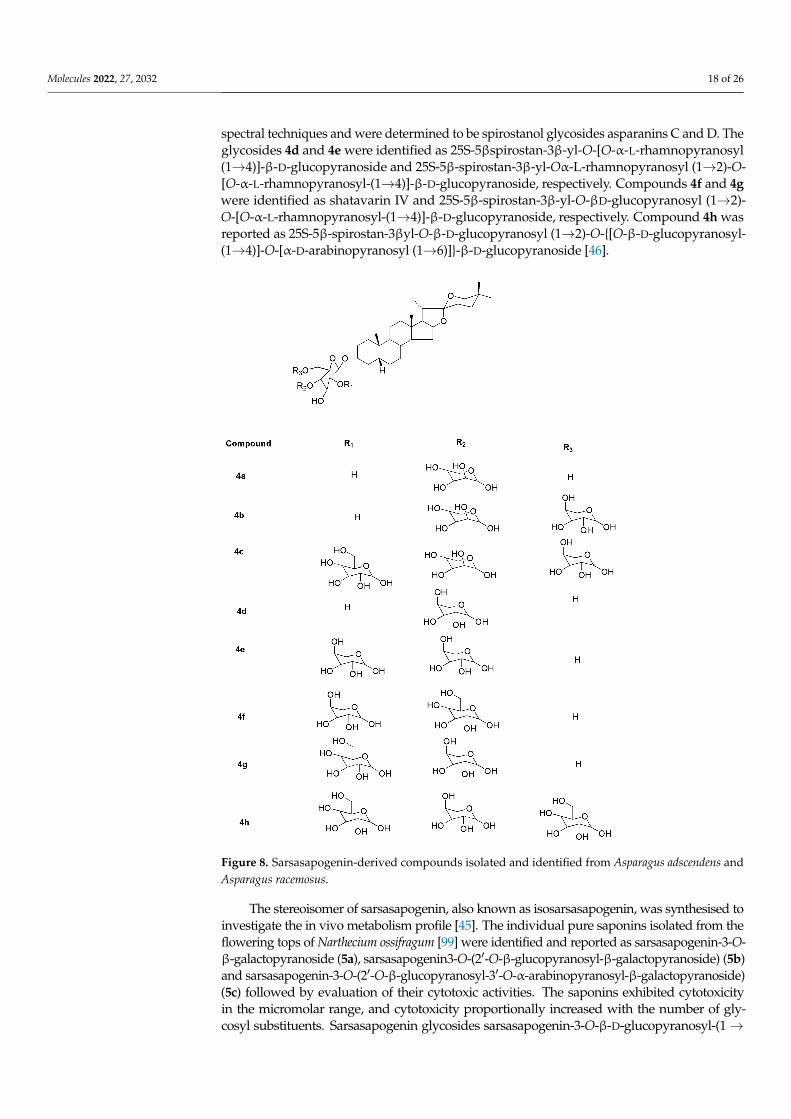
Compounds 4a–4h (Figure 8) were isolated and identified from Asparagus adscendensand Asparagus racemosus. Compound 4a was identified as 25S-5β-spirostan-3β-yl-O-[O-β-D-arabinopyranosyl (1→4)]-β-D-glucopyranoside. Compounds 4b and 4c were elucidated using
Molecules 2022, 27, 2032 18 of 26
spectral techniques and were determined to be spirostanol glycosides asparanins C and D. Theglycosides 4d and 4e were identified as 25S-5βspirostan-3β-yl-O-[O-α-L-rhamnopyranosyl(1→4)]-β-D-glucopyranoside and 25S-5β-spirostan-3β-yl-Oα-L-rhamnopyranosyl (1→2)-O-[O-α-L-rhamnopyranosyl-(1→4)]-β-D-glucopyranoside, respectively. Compounds 4f and 4gwere identified as shatavarin IV and 25S-5β-spirostan-3β-yl-O-βD-glucopyranosyl (1→2)-O-[O-α-L-rhamnopyranosyl-(1→4)]-β-D-glucopyranoside, respectively. Compound 4h wasreported as 25S-5β-spirostan-3βyl-O-β-D-glucopyranosyl (1→2)-O-{[O-β-D-glucopyranosyl-(1→4)]-O-[α-D-arabinopyranosyl (1→6)]}-β-D-glucopyranoside [46].
Figure 8. Sarsasapogenin-derived compounds isolated and identified from Asparagus adscendens andAsparagus racemosus.
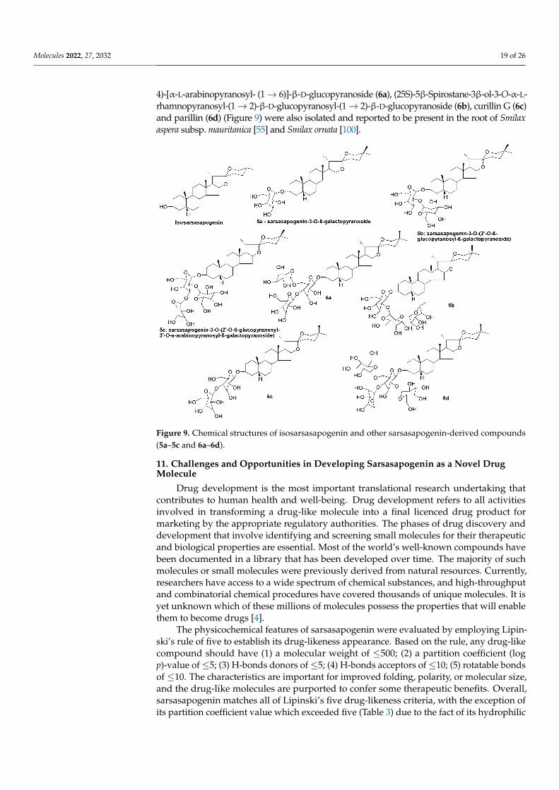
The stereoisomer of sarsasapogenin, also known as isosarsasapogenin, was synthesised toinvestigate the in vivo metabolism profile [45]. The individual pure saponins isolated from theflowering tops of Narthecium ossifragum [99] were identified and reported as sarsasapogenin-3-O-β-galactopyranoside (5a), sarsasapogenin3-O-(2′-O-β-glucopyranosyl-β-galactopyranoside) (5b)and sarsasapogenin-3-O-(2′-O-β-glucopyranosyl-3′-O-α-arabinopyranosyl-β-galactopyranoside)(5c) followed by evaluation of their cytotoxic activities. The saponins exhibited cytotoxicityin the micromolar range, and cytotoxicity proportionally increased with the number of gly-cosyl substituents. Sarsasapogenin glycosides sarsasapogenin-3-O-β-D-glucopyranosyl-(1→
Molecules 2022, 27, 2032 19 of 26
4)-[α-L-arabinopyranosyl- (1→ 6)]-β-D-glucopyranoside (6a), (25S)-5β-Spirostane-3β-ol-3-O-α-L-rhamnopyranosyl-(1→ 2)-β-D-glucopyranosyl-(1→ 2)-β-D-glucopyranoside (6b), curillin G (6c)and parillin (6d) (Figure 9) were also isolated and reported to be present in the root of Smilaxaspera subsp. mauritanica [55] and Smilax ornata [100].
Figure 9. Chemical structures of isosarsasapogenin and other sarsasapogenin-derived compounds(5a–5c and 6a–6d).
11. Challenges and Opportunities in Developing Sarsasapogenin as a Novel DrugMolecule
Drug development is the most important translational research undertaking thatcontributes to human health and well-being. Drug development refers to all activitiesinvolved in transforming a drug-like molecule into a final licenced drug product formarketing by the appropriate regulatory authorities. The phases of drug discovery anddevelopment that involve identifying and screening small molecules for their therapeuticand biological properties are essential. Most of the world’s well-known compounds havebeen documented in a library that has been developed over time. The majority of suchmolecules or small molecules were previously derived from natural resources. Currently,researchers have access to a wide spectrum of chemical substances, and high-throughputand combinatorial chemical procedures have covered thousands of unique molecules. It isyet unknown which of these millions of molecules possess the properties that will enablethem to become drugs [4].
The physicochemical features of sarsasapogenin were evaluated by employing Lipin-ski’s rule of five to establish its drug-likeness appearance. Based on the rule, any drug-likecompound should have (1) a molecular weight of ≤500; (2) a partition coefficient (logp)-value of ≤5; (3) H-bonds donors of ≤5; (4) H-bonds acceptors of ≤10; (5) rotatable bondsof ≤10. The characteristics are important for improved folding, polarity, or molecular size,and the drug-like molecules are purported to confer some therapeutic benefits. Overall,sarsasapogenin matches all of Lipinski’s five drug-likeness criteria, with the exception ofits partition coefficient value which exceeded five (Table 3) due to the fact of its hydrophilic
Molecules 2022, 27, 2032 20 of 26
nature. Nevertheless, the findings suggest that sarsasapogenin could be developed into apromising therapeutic agent for a variety of disorders.
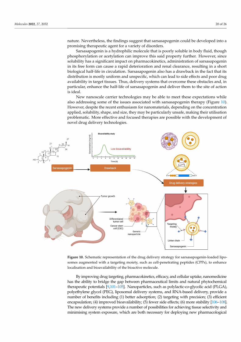
Sarsasapogenin is a hydrophilic molecule that is poorly soluble in body fluid, thoughphosphorylation or acetylation can improve this said property further. However, sincesolubility has a significant impact on pharmacokinetics, administration of sarsasapogeninin its free form can cause a rapid deterioration and renal clearance, resulting in a shortbiological half-life in circulation. Sarsasapogenin also has a drawback in the fact that itsdistribution is mostly uniform and unspecific, which can lead to side effects and poor drugavailability in target tissues. Thus, delivery systems that overcome these obstacles and, inparticular, enhance the half-life of sarsasapogenin and deliver them to the site of actionis ideal.
New nanoscale carrier technologies may be able to meet these expectations whilealso addressing some of the issues associated with sarsasapogenin therapy (Figure 10).However, despite the recent enthusiasm for nanomaterials, depending on the concentrationapplied, solubility, shape, and size, they may be particularly unsafe, making their utilisationproblematic. More effective and focused therapies are possible with the development ofnovel drug delivery technologies.
Figure 10. Schematic representation of the drug delivery strategy for sarsasapogenin-loaded lipo-somes augmented with a targeting moiety, such as cell-penetrating peptides (CPPs), to enhancelocalisation and bioavailability of the bioactive molecule.
By improving drug targeting, pharmacokinetics, efficacy, and cellular uptake, nanomedicinehas the ability to bridge the gap between pharmaceutical limits and natural phytochemicaltherapeutic potentials [9,101–105]. Nanoparticles, such as polylactic-co-glycolic acid (PLGA),polyethylene glycol (PEG), liposomal delivery systems, and RNA-based delivery, provide anumber of benefits including (1) better adsorption; (2) targeting with precision; (3) efficientencapsulation; (4) improved bioavailability; (5) fewer side effects; (6) more stability [106–108].The new delivery systems provide a number of possibilities for achieving tissue selectivity andminimising system exposure, which are both necessary for deploying new pharmacological
Molecules 2022, 27, 2032 21 of 26
molecules derived from sarsasapogenin for enhanced therapy. Many promising results havebeen reported in recent years, but clinical translation remains a barrier and progress remain slow.It is important to remember that while delivery vehicles can help minimize sarsasapogenin-related issues, they may also cause other problems due to the materials used. Hence, everynanoformulation should be examined not only for the therapeutic efficacy and minimised drugside effects but also for material-related toxicity and immunological reactions. Since this is atime-consuming process that must pass many stages of preclinical and clinical developments,it is not surprising that not many clinical studies have been conducted especially on naturalproducts. Nevertheless, some findings are largely favourable, leading to anticipation of evenmore breakthroughs, especially those involving the use of sarsasapogenin.
12. Conclusions and Future Perspectives
Overall, sarsasapogenin is a key sapogenin isolated from Anemarrhena asphodeloidesBunge, and it could be used to treat a range of health problems with safety and efficacy.The distribution of sarsasapogenin in medicinal plants as well as its isolation, structuralcharacterisation, physicochemical properties and biosynthesis were discussed in this re-view. In addition, this review also provided scientific evidence on its pharmacology andtherapeutic potential. Sarsasapogenin has anti-inflammatory, anticancer, antidiabetic, anti-osteoclastogenic, and neuroprotective properties, and it could be used to treat precociouspuberty. However, existing research mostly focused on the pharmacological actions; hence,interpretation of molecular mechanisms of its activities is still insufficient. The exceptionalanti-inflammatory capability of sarsasapogenin may mediate many of its therapeutic po-tentials as evidenced by the described investigations. Sarsasapogenin also has significantcytotoxic and apoptotic capabilities that enhance its anticancer actions. However, moreresearch is needed to fully comprehend the mechanisms of sarsasapogenin in inhibitionof cancer cell line proliferation. There is insufficient evidence on the toxicity evaluationsof this molecule, necessitating further research in the future to ensure its safety. Further-more, because many of the existing literature are only in vitro and in vivo, however, morepreclinical and clinical research is required to fully comprehend these activities, identifythe underlying molecular mechanisms and to see if the same benefits may be seen inhumans. Despite several changes and semi-synthetic investigations, additional researchis needed to better understand the structure–activity relationship and generate sarsasa-pogenin derivatives with improved activities. Furthermore, extensive research into thepharmacokinetics and bioavailability of sarsasapogenin is needed to prove its safety andefficacy in the treatment of numerous disorders. We hope that this review may providesome direction for future drug design, development and therapy of sarsasapogenin.
Author Contributions: Writing—original draft: N.H.M., M.S., S.F. and N.K.F.; Conceptualisation:N.H.M. and M.S.; Supervision: M.S., S.F. and N.K.F.; Resources: N.H.M., M.S., S.F., M.Y.B., S.H.G.,N.N.I.M.R., S.R., K.C., V.S., K.V.S., S.J., S.U., S.P., P.T.L., V.B. and N.K.F.; Data curation: N.H.M.,M.S., S.F., M.Y.B., S.H.G., N.N.I.M.R., S.R., K.C., V.S., K.V.S., S.J., S.U., S.P., P.T.L., V.B. and N.K.F.;Writing—review and editing: N.H.M., M.S., S.F., M.Y.B., S.H.G., N.N.I.M.R., S.R., K.C., V.S., K.V.S.,S.J., S.U., S.P., P.T.L., V.B. and N.K.F. All authors have read and agreed to the published version ofthe manuscript.
Funding: This study was funded by the Deanship of Scientific Research (RGP: 1/275/1442), KingKhalid University, Abha, Saudi Arabia.
Institutional Review Board Statement: Not applicable.
Informed Consent Statement: Not applicable.
Data Availability Statement: The data presented in this study are available upon request from thecorresponding author.
Acknowledgments: The authors extend their appreciation to the Deanship of Scientific Research(RGP: 1/275/1442), King Khalid University, Abha, Saudi Arabia for their funding support. All theauthors of this manuscript are thankful to their respective Departments/Universities for successful
Molecules 2022, 27, 2032 22 of 26
completion of this study. The figures in this manuscript were created with the support of https://biorender.com under a paid subscription (ref: C08A1A0B-0002; 3 November 2021; accessed on14 February 2022).
Conflicts of Interest: The authors declare no conflict of interest.
References1. Hassan, R.; Abdul, B. A Medicinal Plants (Importance and Uses). Pharm. Anal. Acta 2012, 3, 2153–2435.2. Li, X.; Jiang, H.; Xu, L.; Liu, Y.-Q.; Tang, J.-w.; Shi, J.-S.; Yu, X.-j.; Wang, X.; Du, L.; Lu, Q. Sarsasapogenin restores podocyte
autophagy in diabetic nephropathy by targeting GSK3β signaling pathway. Biochem. Pharmacol. 2021, 192, 114675. [CrossRef]3. Othman, S.N.N.; Lum, P.T.; Sekar, M.; Mazlan, N.A.; Yusri, P.Z.S.; Ghazali, N.F.; Idi, H.M.; Azman, S.; Ismail, M.; Noor, A.A.M.
Molecules of interest–embelin–a review. Res. J. Pharm. Technol. 2020, 13, 3485–3493. [CrossRef]4. Watroly, M.N.; Sekar, M.; Fuloria, S.; Gan, S.H.; Jeyabalan, S.; Wu, Y.S.; Subramaniyan, V.; Sathasivam, K.V.; Ravi, S.; Rani, N.N.I.M.
Chemistry, biosynthesis, physicochemical and biological properties of rubiadin: A promising natural anthraquinone for newdrug discovery and development. Drug Des. Dev. Ther. 2021, 15, 4527–4549. [CrossRef] [PubMed]
5. Singh, I.P.; Ahmad, F.; Chatterjee, D.; Bajpai, R.; Sengar, N. Natural products: Drug discovery and development. Drug Discov. Dev.Targets Mol. Med. 2021, 11–65. [CrossRef]
6. Jeong, J.-J.; Jang, S.-E.; Hyam, S.R.; Han, M.J.; Kim, D.-H. The rhizome mixture of Anemarrhena asphodeloides and Coptidis chinensisameliorates acute and chronic colitis in mice by inhibiting the binding of lipopolysaccharide to TLR4 and IRAK1 phosphorylation.Evid.-Based Complementary Altern. Med. 2014, 2014, 809083. [CrossRef] [PubMed]
7. Jigden, B.; Wang, H.; Samdan, N.; Yang, D.-C. Molecular identification of oriental medicinal plant Anemarrhena asphodeloidesBunge (‘Jimo’) by multiplex PCR. Mol. Biol. Rep. 2010, 37, 955. [CrossRef]
8. Nakashima, N.; Kimura, I.; Kimura, M.; Matsuura, H. Isolation of pseudoprototimosaponin AIII from rhizomes of Anemarrhenaasphodeloides and its hypoglycemic activity in streptozotocin-induced diabetic mice. J. Nat. Prod. 1993, 56, 345–350. [CrossRef]
9. Wang, S.; Su, R.; Nie, S.; Sun, M.; Zhang, J.; Wu, D.; Moustaid-Moussa, N. Application of nanotechnology in improvingbioavailability and bioactivity of diet-derived phytochemicals. J. Nutr. Biochem. 2014, 25, 363–376. [CrossRef]
10. Chinese Pharmacopoeia Commission. Pharmacopoeia of the People’s Republic of China; Chinese Medical Science and TechnologyPress: Beijing, China, 2010; Volume 1, p. 292.
11. Wang, J.; Liu, J.; Guo, X.; Zhang, J.; Yu, B. A sensitive indirect competitive enzyme-linked immunosorbent assay for the detectionof sarsasapogenin in rat plasma. Acta Pharmacol. Sin. 2010, 31, 984–989. [CrossRef]
12. Dong, D.; Zhou, N.; Liu, R.; Xiong, J.; Pan, H.; Sun, S.; Ma, L.; Wang, R. Sarsasapogenin-AA13 inhibits LPS-induced inflammatoryresponses in macrophage cells in vitro and relieves dimethylbenzene-induced ear edema in mice. Acta Pharmacol. Sin. 2017, 38,699–709. [CrossRef] [PubMed]
13. Huang, C.; Dong, D.; Jiao, Q.; Pan, H.; Ma, L.; Wang, R. Sarsasapogenin-AA 13 ameliorates Aβ-induced cognitive deficits viaimproving neuroglial capacity on Aβ clearance and antiinflammation. CNS Neurosci. Ther. 2017, 23, 498–509. [CrossRef]
14. Feng, B.; Zhao, X.-Y.; Song, Y.-Z.; Liang, W.-N.; Liu, J.-L. Sarsasapogenin reverses depressive-like behaviors and nicotinicacetylcholine receptors induced by olfactory bulbectomy. Neurosci. Lett. 2017, 639, 173–178. [CrossRef]
15. Ma, D.; Zhang, J.; Sugahara, K.; Sagara, Y.; Kodama, H. Effect of sarsasapogenin and its derivatives on the stimulus coupledresponses of human neutrophils. Clin. Chim. Acta 2001, 314, 107–112. [CrossRef]
16. Wang, W.; Wang, D.; Wang, Z.; Yao, G.; Li, X.; Gao, P.; Li, L.; Zhang, Y.; Wang, S.; Song, S. Synthesis of new sarsasapogeninderivatives with cytotoxicity and apoptosis-inducing activities in human breast cancer MCF-7 cells. Eur. J. Med. Chem. 2017, 127,62–71. [CrossRef] [PubMed]
17. Yin, Y.; Zhao, X.-C.; Wang, S.-J.; Gao, P.-Y.; Li, L.-Z.; Ikejima, T.; Song, S.-J. Synthesis and biological evaluation of novelsarsasapogenin derivatives as potential anti-tumor agents. Steroids 2015, 93, 25–31. [CrossRef]
18. Gu, G.; Fang, M.; Liu, J.; Gu, L. Concise synthesis and antitumor activities of trisaccharide steroidal saponins. Carbohydr. Res.2011, 346, 2406–2413. [CrossRef] [PubMed]
19. Guo, F.; Zhang, B.; Fu, Z.; Ma, Y.; Gao, Y.; Shen, F.; Huang, C.; Li, Y. The rapid antidepressant and anxiolytic-like effects of YY-21involve enhancement of excitatory synaptic transmission via activation of mTOR signaling in the mPFC. Eur. Neuropsychopharmacol.2016, 26, 1087–1098. [CrossRef]
20. Sy, L.-K.; Lok, C.-N.; Wang, J.-Y.; Liu, Y.; Cheng, L.; Wan, P.-K.; Leung, C.-T.; Cao, B.; Kwong, W.-L.; Chang, R.C.-C. Identificationof “sarsasapogenin-aglyconed” timosaponins as novel Aβ-lowering modulators of amyloid precursor protein processing. Chem.Sci. 2016, 7, 3206–3214. [CrossRef]
21. Ma, L.; Wang, R.; Zhou, N.; Xu, T.; Zhao, J.; Zhang, Y.; Pan, H. Sarsasapogenin derivative, its preparation method and application.Invention Patent CN 104513289, 15 April 2015.
22. Moon, E.; Kim, A.-J.; Kim, S.Y. Sarsasapogenin increases melanin synthesis via induction of tyrosinase and microphthalmia-associated transcription factor expression in melan-a cells. Biomol. Ther. 2012, 20, 340. [CrossRef]
23. Peng, J.; Zhao, K.; Zhu, J.; Wang, Y.; Sun, P.; Yang, Q.; Zhang, T.; Han, W.; Hu, W.; Yang, W. Sarsasapogenin suppressesRANKL-induced osteoclastogenesis in vitro and prevents lipopolysaccharide-induced bone loss in vivo. Drug Des. Dev. Ther.2020, 14, 3435. [CrossRef] [PubMed]
Molecules 2022, 27, 2032 23 of 26
24. Pan, H.; Van Khang, P.; Dong, D.; Wang, R.; Ma, L. Synthesis and SAR study of novel sarsasapogenin derivatives as potentneuroprotective agents and NO production inhibitors. Bioorganic Med. Chem. Lett. 2017, 27, 662–665. [CrossRef] [PubMed]
25. Ren, L.-X.; Luo, Y.-F.; Li, X.; Zuo, D.-Y.; Wu, Y.-L. Antidepressant-Like Effects of sarsasapogenin from Anemarrhena asphodeloides BUNGE (Liliaceae). Biol. Pharm. Bull. 2006, 29, 2304–2306. [CrossRef] [PubMed]
26. Liu, Y.W.; Hao, Y.C.; Chen, Y.J.; Yin, S.Y.; Zhang, M.Y.; Kong, L.; Wang, T.Y. Protective effects of sarsasapogenin against early stageof diabetic nephropathy in rats. Phytother. Res. 2018, 32, 1574–1582. [CrossRef]
27. Hu, K.; Sun, W.; Li, Y.; Zhang, B.; Zhang, M.; Guo, C.; Chang, H.; Wang, X. Study on the mechanism of sarsasapogenin in treatingprecocious puberty by regulating the HPG Axis. Evid. Based Complementary Altern. Med. 2020, 2020, 1978043. [CrossRef]
28. Lim, S.-M.; Jeong, J.-J.; Kang, G.-D.; Kim, K.-A.; Choi, H.-S.; Kim, D.-H. Timosaponin AIII and its metabolite sarsasapogeninameliorate colitis in mice by inhibiting NF-κB and MAPK activation and restoring Th17/Treg cell balance. Int. Immunopharmacol.2015, 25, 493–503. [CrossRef]
29. Bao, W.; Pan, H.; Lu, M.; Ni, Y.; Zhang, R.; Gong, X. The apoptotic effect of sarsasapogenin from Anemarrhena asphodeloides onHepG2 human hepatoma cells. Cell Biol. Int. 2007, 31, 887–892. [CrossRef]
30. Tang, Z.-Z.; Zhang, Y.-M.; Zheng, T.; Huang, T.-T.; Ma, T.-F.; Liu, Y.-W. Sarsasapogenin alleviates diabetic nephropathy throughsuppression of chronic inflammation by down-regulating PAR-1: In vivo and in vitro study. Phytomedicine 2020, 78, 153314.[CrossRef]
31. Pei, L.; Ye, Y.; Zhao, W.; Ye, Q.; Ge, S.; Jiang, Z.; Liang, X.; Gan, H.; Ma, L. A validated UPLC–MS/MS method for quantitativedetermination of a potent neuroprotective agent Sarsasapogenin-AA13 in rat plasma: Application to pharmacokinetic studies.Biomed. Chromatogr. 2020, 34, e4775. [CrossRef]
32. Kashyap, P.; Muthusamy, K.; Niranjan, M.; Trikha, S.; Kumar, S. Sarsasapogenin: A steroidal saponin from Asparagus racemosusas multi target directed ligand in Alzheimer’s disease. Steroids 2020, 153, 108529. [CrossRef]
33. Paczkowski, C.; Wojciechowski, Z.A. The occurrence of UDPG-dependent glucosyltransferase specific for sarsasapogenin inAsparagus officinalis. Phytochemistry 1988, 27, 2743–2747. [CrossRef]
34. Huang, X.F.; Lin, Y.Y.; Kong, L.Y. Steroids from the roots of Asparagus officinalis and their cytotoxic activity. J. Integr. Plant Biol.2008, 50, 717–722. [CrossRef] [PubMed]
35. Okanishi, T.; Akahori, A.; Yasuda, F.; Takeuchi, Y.; Iwao, T. Steroidal sapogenins of sixteen Liliaceae plants. Chem. Pharm. Bull.1975, 23, 575–579. [CrossRef]
36. Zhang, F.; Lu, Y.; Qian, W.; Pei, Z. Asparagus cochinchinensis (Lour.) Merr. (Tiandong, Chinese Asparagus). In Dietary ChineseHerbs; Springer: Berlin/Heidelberg, Germany, 2015; pp. 83–88.
37. Flåøyen, A.; Wilkins, A.; Sandvik, M. Ruminal metabolism in sheep of saponins from Yucca schidigera. Vet. Res. Commun. 2002, 26,159–169. [CrossRef] [PubMed]
38. Ingawale, D.K.; Mandlik, S.K.; Patel, S.S. Combination of sarsasapogenin and fluticasone attenuates ovalbumin-induced airwayinflammation in a mouse asthma model. Immunopharmacol. Immunotoxicol. 2020, 42, 128–137. [CrossRef]
39. Power, F.B.; Salway, A.H. XXV.—Chemical examination of sarsaparilla root. J. Chem. Soc. Trans. 1914, 105, 201–219. [CrossRef]40. Mandlik, D.S.; Mandlik, S.K.; Patel, S. Protective effect of sarsasapogenin in TNBS induced ulcerative colitis in rats associated
with downregulation of pro-inflammatory mediators and oxidative stress. Immunopharmacol. Immunotoxicol. 2021, 43, 571–583.[CrossRef]
41. Marker, R.E.; Wagner, R.; Ulshafer, P.R.; Wittbecker, E.L.; Goldsmith, D.P.; Ruof, C.H. Steroidal sapogenins1a. J. Am. Chem. Soc.1947, 69, 2167–2230. [CrossRef]
42. Uhlig, S.; Wisløff, H.; Petersen, D. Identification of cytotoxic constituents of Narthecium ossifragum using bioassay-guidedfractionation. J. Agric. Food Chem. 2007, 55, 6018–6026. [CrossRef]
43. Ceh, L.; Hauge, J.G. Alveld-producing saponins. I. Chemical studies. Acta Vet. Scand. 1981, 22, 391–402. [CrossRef]44. Duckstein, S.M.; Stintzing, F.C. LC–MSn characterization of steroidal saponins in Helleborus niger L. roots and their conversion
products during fermentation. Steroids 2015, 93, 47–59. [CrossRef] [PubMed]45. Fu, Z.; Li, Z.; Xue, R.; Jiang, J.; Huang, C. Stereoisomerism metabolites found in rats after oral administration of timosaponin B-II
using HPLC-Q-TOF-MS and NMR methods. RSC Adv. 2015, 5, 60650–60657. [CrossRef]46. Jadhav, A.; Bhutani, K. Steroidal saponins from the roots of Asparagus adscendens Roxb and Asparagus racemosus Willd. Indian J.
Chem. 2006, 45B, 1515–1524.47. Srivastava, P.L.; Daramwar, P.P.; Krithika, R.; Pandreka, A.; Shankar, S.S.; Thulasiram, H.V. Functional characterization of novel
sesquiterpene synthases from Indian sandalwood, Santalum album. Sci. Rep. 2015, 5, 10095. [CrossRef]48. Eisenreich, W.; Rohdich, F.; Bacher, A. Deoxyxylulose phosphate pathway to terpenoids. Trends Plant Sci. 2001, 6, 78–84. [CrossRef]49. Vickers, C.E.; Gershenzon, J.; Lerdau, M.T.; Loreto, F. A unified mechanism of action for volatile isoprenoids in plant abiotic stress.
Nat. Chem. Biol. 2009, 5, 283–291. [CrossRef]50. Lange, B.M.; Rujan, T.; Martin, W.; Croteau, R. Isoprenoid biosynthesis: The evolution of two ancient and distinct pathways
across genomes. Proc. Natl. Acad. Sci. USA 2000, 97, 13172–13177. [CrossRef]51. Bridges, N.; Christopher, J.; Hindmarsh, P.; Brook, C. Sexual precocity: Sex incidence and aetiology. Arch. Dis. Child. 1994, 70,
116–118. [CrossRef]52. Cantas-Orsdemir, S.; Eugster, E.A. Update on central precocious puberty: From etiologies to outcomes. Expert Rev. Endocrinol.
Metab. 2019, 14, 123–130. [CrossRef]
Molecules 2022, 27, 2032 24 of 26
53. Lu, Y.; Zhou, W.; Wei, L.; Li, J.; Jia, J.; Li, F.; Smith, S.M.; Xu, J. Regulation of the cholesterol biosynthetic pathway and itsintegration with fatty acid biosynthesis in the oleaginous microalga Nannochloropsis oceanica. Biotechnol. Biofuels 2014, 7, 81.[CrossRef]
54. Gholami, A.; De Geyter, N.; Pollier, J.; Goormachtig, S.; Goossens, A. Natural product biosynthesis in Medicago species. Nat. Prod.Rep. 2014, 31, 356–380. [CrossRef] [PubMed]
55. Haralampidis, K.; Trojanowska, M.; Osbourn, A.E. Biosynthesis of triterpenoid saponins in plants. Adv. Biochem. Eng. Biotechnol.2002, 75, 31–49. [CrossRef] [PubMed]
56. Kemp, M.W.; Saito, M.; Newnham, J.P.; Nitsos, I.; Okamura, K.; Kallapur, S.G. Preterm birth, infection, and inflammation advancesfrom the study of animal models. Reprod. Sci. 2010, 17, 619–628. [CrossRef] [PubMed]
57. James, M.W.; Hawkey, C.J. Assessment of non-steroidal anti-inflammatory drug (NSAID) damage in the human gastrointestinaltract. Br. J. Clin. Pharmacol. 2003, 56, 146–155. [CrossRef]
58. Yu, Y.Y.; Cui, S.C.; Zheng, T.N.; Ma, H.J.; Xie, Z.F.; Jiang, H.W.; Li, Y.F.; Zhu, K.X.; Huang, C.G.; Li, J.; et al. Sarsasapogeninimproves adipose tissue inflammation and ameliorates insulin resistance in high-fat diet-fed C57BL/6J mice. Acta Pharmacol. Sin.2021, 42, 272–281. [CrossRef]
59. Ni, Y.; Gong, X.G.; Lu, M.; Chen, H.M.; Wang, Y. Mitochondrial ROS burst as an early sign in sarsasapogenin-induced apoptosisin HepG2 cells. Cell Biol. Int. 2008, 32, 337–343. [CrossRef]
60. Bai, J.; Cederbaum, A.I. Overexpression of CYP2E1 in mitochondria sensitizes HepG2 cells to the toxicity caused by depletion ofglutathione. J. Biol. Chem. 2006, 281, 5128–5136. [CrossRef]
61. Ling, Y.-H.; Liebes, L.; Zou, Y.; Perez-Soler, R. Reactive oxygen species generation and mitochondrial dysfunction in the apoptoticresponse to Bortezomib, a novel proteasome inhibitor, in human H460 non-small cell lung cancer cells. J. Biol. Chem. 2003, 278,33714–33723. [CrossRef]
62. Chandra, D.; Liu, J.-W.; Tang, D.G. Early Mitochondrial activation and cytochrome c up-regulation during apoptosis*210. J. Biol.Chem. 2002, 277, 50842–50854. [CrossRef]
63. Shen, S.; Zhang, Y.; Zhang, R.; Gong, X. Sarsasapogenin induces apoptosis via the reactive oxygen species-mediated mitochondrialpathway and ER stress pathway in HeLa cells. Biochem. Biophys. Res. Commun. 2013, 441, 519–524. [CrossRef]
64. Zhang, Y.-M.; Zheng, T.; Huang, T.-T.; Gu, P.-P.; Gou, L.-s.; Ma, T.-F.; Liu, Y.-W. Sarsasapogenin attenuates Alzheimer-likeencephalopathy in diabetes. Phytomedicine 2021, 91, 153686. [CrossRef] [PubMed]
65. Hu, Y.; Xia, Z.; Sun, Q.; Orsi, A.; Rees, D. A new approach to the pharmacological regulation of memory: Sarsasapogeninimproves memory by elevating the low muscarinic acetylcholine receptor density in brains of memory-deficit rat models. BrainRes. 2005, 1060, 26–39. [CrossRef] [PubMed]
66. Krishnan, V.; Nestler, E.J. The molecular neurobiology of depression. Nature 2008, 455, 894–902. [CrossRef] [PubMed]67. Islam, M.; Moriguchi, S.; Tagashira, H.; Fukunaga, K. Rivastigmine improves hippocampal neurogenesis and depression-like
behaviors via 5-HT1A receptor stimulation in olfactory bulbectomized mice. Neuroscience 2014, 272, 116–130. [CrossRef]68. Andreasen, J.T.; Nielsen, E.Ø.; Redrobe, J.P. Chronic oral nicotine increases brain [3H] epibatidine binding and responsiveness to
antidepressant drugs, but not nicotine, in the mouse forced swim test. Psychopharmacology 2009, 205, 517–528. [CrossRef]69. Lendvai, B.; Kassai, F.; Szájli, Á.; Némethy, Z. α7 nicotinic acetylcholine receptors and their role in cognition. Brain Res. Bull. 2013,
93, 86–96. [CrossRef]70. Séguéla, P.; Wadiche, J.; Dineley-Miller, K.; Dani, J.A.; Patrick, J.W. Molecular cloning, functional properties, and distribution of
rat brain alpha 7: A nicotinic cation channel highly permeable to calcium. J. Neurosci. 1993, 13, 596–604. [CrossRef]71. Wada, E.; Wada, K.; Boulter, J.; Deneris, E.; Heinemann, S.; Patrick, J.; Swanson, L.W. Distribution of alpha2, alpha3, alpha4, and
beta2 neuronal nicotinic receptor subunit mRNAs in the central nervous system: A hybridization histochemical study in the rat. J.Comp. Neurol. 1989, 284, 314–335. [CrossRef]
72. Zhang, M.-Y.; Li, Y.; Yin, S.-Y.; Kong, L.; Liu, X.-L.; Yin, X.-X.; Liu, Y.-W. Sarsasapogenin suppresses Aβ overproduction inducedby high glucose in HT-22 cells. Naunyn Schmiedeberg’s Arch. Pharmacol. 2018, 391, 159–168. [CrossRef]
73. Shlipak, M. Diabetic nephropathy: Preventing progression. BMJ Clin. Evid. 2010, 7, 606.74. Qiu, Y.Y.; Tang, L.Q. Roles of the NLRP3 inflammasome in the pathogenesis of diabetic nephropathy. Pharmacol. Res. 2016, 114,
251–264. [CrossRef]75. Zheng, N.; Shi, X.; Chen, X.; Lv, W. Associations between inflammatory markers, hemostatic markers, and microvascular
complications in 182 Chinese patients with type 2 diabetes mellitus. Lab. Med. 2015, 46, 214–220. [CrossRef] [PubMed]76. Liu, Y.; Tang, Z.-Z.; Zhang, Y.-M.; Kong, L.; Xiao, W.-F.; Ma, T.-F.; Liu, Y.-W. Thrombin/PAR-1 activation induces endothelial
damages via NLRP1 inflammasome in gestational diabetes. Biochem. Pharmacol. 2020, 175, 113849. [CrossRef]77. Kong, L.; Liu, Y.; Zhang, Y.M.; Li, Y.; Gou, L.S.; Ma, T.F.; Liu, Y.W. Sarsasapogenin ameliorates diabetes-associated memory
impairment and neuroinflammation through down-regulation of PAR-1 receptor. Phytother. Res. 2021, 35, 3167–3180. [CrossRef][PubMed]
78. Lorenzo, J.; Horowitz, M.; Choi, Y. Osteoimmunology: Interactions of the bone and immune system. Endocr. Rev. 2008, 29,403–440. [CrossRef]
79. Arai, F.; Miyamoto, T.; Ohneda, O.; Inada, T.; Sudo, T.; Brasel, K.; Miyata, T.; Anderson, D.M.; Suda, T. Commitment anddifferentiation of osteoclast precursor cells by the sequential expression of c-Fms and receptor activator of nuclear factor κB(RANK) receptors. J. Exp. Med. 1999, 190, 1741–1754. [CrossRef]
Molecules 2022, 27, 2032 25 of 26
80. Takayanagi, H. Osteoimmunology: Shared mechanisms and crosstalk between the immune and bone systems. Nat. Rev. Immunol.2007, 7, 292–304. [CrossRef] [PubMed]
81. Darnay, B.G.; Besse, A.; Poblenz, A.T.; Lamothe, B.; Jacoby, J.J. TRAFs in RANK signaling. Adv. Exp. Med. Biol. 2007, 597, 152–159.[PubMed]
82. Mizukami, J.; Takaesu, G.; Akatsuka, H.; Sakurai, H.; Ninomiya-Tsuji, J.; Matsumoto, K.; Sakurai, N. Receptor activator of NF-κBligand (RANKL) activates TAK1 mitogen-activated protein kinase kinase kinase through a signaling complex containing RANK,TAB2, and TRAF6. Mol. Cell. Biol. 2002, 22, 992–1000. [CrossRef]
83. Wong, B.R.; Besser, D.; Kim, N.; Arron, J.R.; Vologodskaia, M.; Hanafusa, H.; Choi, Y. TRANCE, a TNF family member, activatesAkt/PKB through a signaling complex involving TRAF6 and c-Src. Mol. Cell 1999, 4, 1041–1049. [CrossRef]
84. Kim, M.S.; Yang, Y.-M.; Son, A.; Tian, Y.S.; Lee, S.-I.; Kang, S.W.; Muallem, S.; Shin, D.M. RANKL-mediated reactive oxygenspecies pathway that induces long lasting Ca2+ oscillations essential for osteoclastogenesis. J. Biol. Chem. 2010, 285, 6913–6921.[CrossRef] [PubMed]
85. Kim, J.H.; Kim, N. Regulation of NFATc1 in osteoclast differentiation. J. Bone Metab. 2014, 21, 233–241. [CrossRef] [PubMed]86. Soriano-Guillen, L.; Argente, J. Central precocious puberty, functional and tumor-related. Best Pract. Res. Clin. Endocrinol. Metab.
2019, 33, 101262. [CrossRef] [PubMed]87. Guaraldi, F.; Beccuti, G.; Gori, D.; Ghizzoni, L. Management of endocrine disease: Long-term outcomes of the treatment of central
precocious puberty. Eur. J. Endocrinol. 2016, 174, R79–R87. [CrossRef]88. Rostami, S.; Kohan, L.; Mohammadianpanah, M. The LEP G-2548A gene polymorphism is associated with age at menarche and
breast cancer susceptibility. Gene 2015, 557, 154–157. [CrossRef] [PubMed]89. Day, F.R.; Thompson, D.J.; Helgason, H.; Chasman, D.I.; Finucane, H.; Sulem, P.; Ruth, K.S.; Whalen, S.; Sarkar, A.K.; Albrecht, E.
Genomic analyses identify hundreds of variants associated with age at menarche and support a role for puberty timing in cancerrisk. Nat. Genet. 2017, 49, 834–841. [CrossRef]
90. Chen, M.; Eugster, E.A. Central precocious puberty: Update on diagnosis and treatment. Pediatric Drugs 2015, 17, 273–281.[CrossRef] [PubMed]
91. Kendirci, H.N.P.; Agladıoglu, S.Y.; Önder, A.; Bas, V.N.; Çetinkaya, S.; Aycan, Z. Effects of GnRH analogue treatment on anteriorpituitary hormones in children with central precocious puberty. J. Pediatric Endocrinol. Metab. 2015, 28, 1145–1151.
92. Yen, S. Clinical applications of gonadotropin-releasing hormone and gonadotropin-releasing hormone analogs. Fertil. Steril. 1983,39, 257–266.
93. Li, W.-J.; Gong, C.-X.; Guo, M.-J.; Xing, J.; Li, T.; Song, W.-H.; Luo, X.-P.; Wu, D.; Liang, J.-P.; Cao, B.-Y. Efficacy and safety ofdomestic leuprorelin in girls with idiopathic central precocious puberty: A multicenter, randomized, parallel, controlled trial.Chin. Med. J. 2015, 128, 1314. [CrossRef]
94. Latronico, A.C.; Brito, V.N.; Carel, J.-C. Causes, diagnosis, and treatment of central precocious puberty. Lancet Diabetes Endocrinol.2016, 4, 265–274. [CrossRef]
95. Carel, J.-C.; Eugster, E.A.; Rogol, A.; Ghizzoni, L.; Palmert, M.R. Consensus statement on the use of gonadotropin-releasinghormone analogs in children. Pediatrics 2009, 123, e752–e762. [CrossRef] [PubMed]
96. Yang, B.; Liu, Z.; Hu, J.; Lai, X.; Xia, P. Quantitative determination of sarsasapogenin in rat plasma using liquid chromatography-tandem mass spectrometry. J. Chromatogr. B 2016, 1022, 213–219. [CrossRef]
97. Liu, Z.; Qin, W.; Zhu, Z.; Liu, Y.; Sun, F.; Chai, Y.; Xia, P. Development and validation of liquid chromatography-tandemmass spectrometry method for simultaneous determination of six steroidal saponins in rat plasma and its application to apharmacokinetics study. Steroids 2015, 96, 21–29. [CrossRef] [PubMed]
98. Wang, W.; Zhang, Y.; Yao, G.; Wang, W.; Shang, X.; Zhang, Y.; Wang, X.; Wang, S.; Song, S. Synthesis of new sarsasapogeninderivatives with antiproliferative and apoptotic effects in MCF-7 cells. Steroids 2018, 131, 23–31. [CrossRef] [PubMed]
99. Díaz, A.E.C.; Herfindal, L.; Rathe, B.A.; Sletta, K.Y.; Vedeler, A.; Haavik, S.; Fossen, T. Cytotoxic saponins and other naturalproducts from flowering tops of Narthecium ossifragum L. Phytochemistry 2019, 164, 67–77. [CrossRef] [PubMed]
100. Tian, L.-W.; Zhang, Z.; Long, H.-L.; Zhang, Y.-J. Steroidal saponins from the genus Smilax and their biological activities. Nat. Prod.Bioprospect. 2017, 7, 283–298. [CrossRef]
101. Bonferoni, M.C.; Rossi, S.; Sandri, G.; Ferrari, F. Nanoparticle formulations to enhance tumor targeting of poorly solublepolyphenols with potential anticancer properties. In Proceedings of the Seminars in Cancer Biology; Academic Press: Cambridge,MA, USA, 2017; pp. 205–214.
102. Lagoa, R.; Silva, J.; Rodrigues, J.R.; Bishayee, A. Advances in phytochemical delivery systems for improved anticancer activity.Biotechnol. Adv. 2020, 38, 107382. [CrossRef]
103. Pistollato, F.; Bremer-Hoffmann, S.; Basso, G.; Cano, S.S.; Elio, I.; Vergara, M.M.; Giampieri, F.; Battino, M. Targeting glioblastomawith the use of phytocompounds and nanoparticles. Target. Oncol. 2016, 11, 1–16. [CrossRef]
104. Rahimi, H.R.; Nedaeinia, R.; Shamloo, A.S.; Nikdoust, S.; Oskuee, R.K. Novel delivery system for natural products: Nano-curcumin formulations. Avicenna J. Phytomed. 2016, 6, 383.
105. Siddiqui, I.A.; Sanna, V. Impact of nanotechnology on the delivery of natural products for cancer prevention and therapy. Mol.Nutr. Food Res. 2016, 60, 1330–1341. [CrossRef] [PubMed]
Molecules 2022, 27, 2032 26 of 26
106. Zuraini, N.Z.A.; Sekar, M.; Wu, Y.S.; Gan, S.H.; Bonam, S.R.; Rani, N.N.I.M.; Begum, M.Y.; Lum, P.T.; Subramaniyan, V.; Fuloria,N.K. Promising nutritional fruits against cardiovascular diseases: An overview of experimental evidence and understandingtheir mechanisms of action. Vasc. Health Risk Manag. 2021, 17, 739–769. [CrossRef] [PubMed]
107. Yap, K.M.; Sekar, M.; Fuloria, S.; Wu, Y.S.; Gan, S.H.; Rani, N.N.I.M.; Subramaniyan, V.; Kokare, C.; Lum, P.T.; Begum, M.Y. Drugdelivery of natural products through nanocarriers for effective breast cancer therapy: A comprehensive review of literature. Int. J.Nanomed. 2021, 16, 7891–7941. [CrossRef] [PubMed]
108. Davatgaran-Taghipour, Y.; Masoomzadeh, S.; Farzaei, M.H.; Bahramsoltani, R.; Karimi-Soureh, Z.; Rahimi, R.; Abdollahi, M.Polyphenol nanoformulations for cancer therapy: Experimental evidence and clinical perspective. Int. J. Nanomed. 2017, 12,2689–2702. [CrossRef] [PubMed]