chemistry and biod iversity of the biologically active natural glycosides
TRANSCRIPT
REVIEW
Chemistry and Biod iversity of the Biologically Active Natural Glycosides
by Valery M. Dembitsky
Department of Organic Chemistry, P.O. Box 39231,The Hebrew University of Jerusalem, Jerusalem, 91391, Israel
(fax: �972-2-590-2947; e-mail : [email protected])
This review covers the literature on isolation, bioactivities, and structures of nearly 800 natural glycosides.
Contents
1. Introduction2. Fatty Acid Glycosides2.1. Rhamnolipids2.2. Trehalose Lipids2.3. Sophorolipids2.4. Butanolide Glycosides
3. Terpenoid Glycoside Esters4. Aroma Glycoside Esters5. Quinone Glycoside Esters6. Flavonoid Glycoside Esters7. Iridoid Glycoside Esters8. Carotenoid Glycoside Esters9. Steroid Glycoside Esters9.1. Acylated Saponins
10. Fatty Acid Amide Glycosides11. Macrolactone Glycosides12. Polyether Glycosidic Ionophores13. Miscellaneous Compounds
References
1. Introduction. ± Many of the natural fatty acid glycosides are biologically activechemical complexes that include the molecules of fatty (carboxylic) and dicarboxylic(dioic) acids, fatty acid amides, lactones and sugar molecules, which are connected bychemical bonds. These chemical complexes can comprise also other moieties such asflavonoids, iridoids, quinones, anthraquinones, carotenoids, steroids, and othermolecular structures. Contemporary trends in discovery of drugs from natural productsemphasize investigation of the terrestrial and marine environments to yield numeroushighly active compounds [1 ± 8].
CHEMISTRY & BIODIVERSITY ± Vol. 1 (2004) 673
Low- and high-molecular-weight fatty acid glycosides (natural biosurfactants) areof great interest because of their physico-chemical and biological properties, which canbe exploited in oil, food, cosmetic, and pharmaceutical industries. As for the generaltypes of microbial amphiphiles, the data accumulated over recent years add to alreadywell-known compounds another new interesting molecular structure. Fatty acidglycosides are active amphipathic molecules with hydrophobic and hydrophilicmoieties. Surfactants constitute an important class of chemicals widely used in modernindustry [1] [2] [7]. It is well-established that many macrolactone glycosides are naturalantibiotics, which are used as antimicrobial agents in both clinical and veterinarymedicine [9]. Glyceroglycolipids and glycophospholipids have been recently reviewed[2] and are not included in this review.
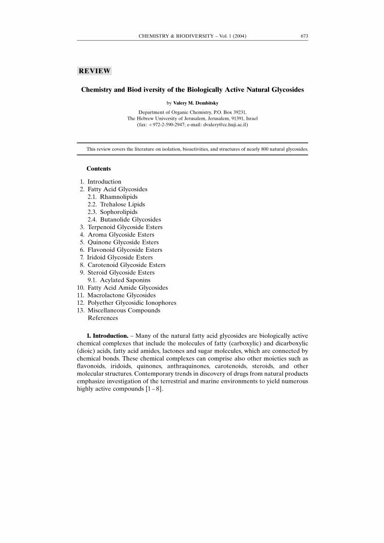
2. Fatty Acid Glycosides. ± Four hydroxy fatty acid glucosides, 1 ± 4, were found inthe time of ripening of the fruits of Persoonia linearis x pinifolia (Proteacea) grown inAustralia [10]. The monoterpene glucoside ester 5 has been isolated from the leaves ofthe medicinal plant Lantana lilacina used in the treatment of bronchitis [11]. 2,6-Dimethyl-6-O-(�-�-quinovopyranosyl)oct-7-enoic acid (6) has been isolated from theseeds of plant Albizia procera [12]. A. procera, which belongs to the Leguminosae andcommonly known as −Safe Siris× in Hindi, is widely distributed in India. Ethyl 3-O-(�-�-glucopyranosyl)butanoate (7) and butyl 3-O-(�-�-glucopyranosyl)butanoate (8) werefound in fruits of papaya, Carica pubescens [13].
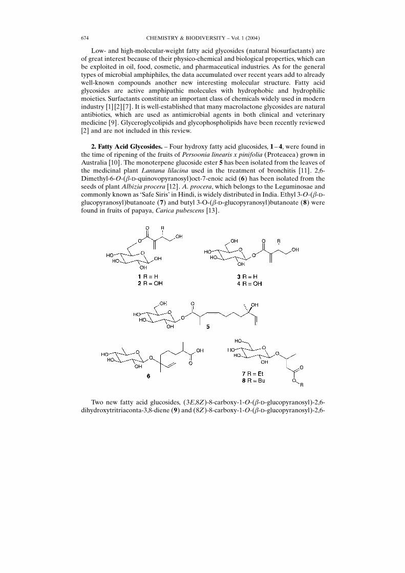
Two new fatty acid glucosides, (3E,8Z)-8-carboxy-1-O-(�-�-glucopyranosyl)-2,6-dihydroxytritriaconta-3,8-diene (9) and (8Z)-8-carboxy-1-O-(�-�-glucopyranosyl)-2,6-
CHEMISTRY & BIODIVERSITY ± Vol. 1 (2004)674
dihydroxy-3,4-epoxytritriacont-8-ene (10) have been isolated from the root bark ofOchna calodendron, grown in southern Cameroon [14].
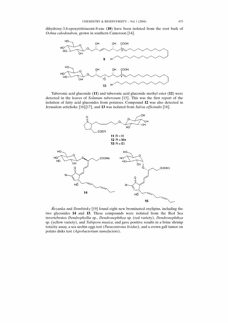
Tuberonic acid glucoside (11) and tuberonic acid glucoside methyl ester (12) weredetected in the leaves of Solanum tuberosum [15]. This was the first report of theisolation of fatty acid glucosides from potatoes. Compound 12 was also detected inJerusalem artichoke [16] [17], and 13 was isolated from Salvia officinalis [18].
Rœezanka and Dembitsky [19] found eight new brominated oxylipins, including thetwo glycosides 14 and 15. These compounds were isolated from the Red Seainvertebrates Dendrophyllia sp., Dendronephthya sp. (red variety), Dendronephthyasp. (yellow variety), and Tubipora musica, and gave positive results in a brine shrimptoxicity assay, a sea urchin eggs test (Paracentrotus lividus), and a crown gall tumor onpotato disks test (Agrobacterium tumefaciens).
CHEMISTRY & BIODIVERSITY ± Vol. 1 (2004) 675
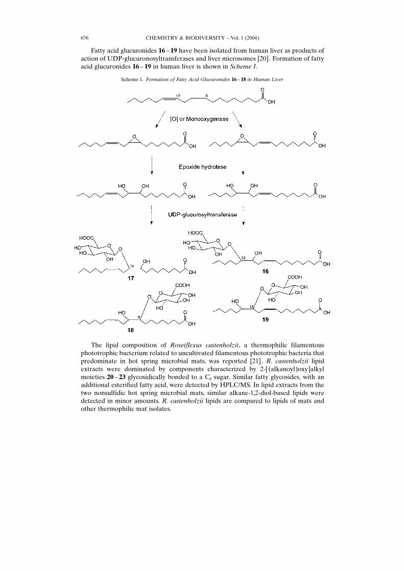
Fatty acid glucuronides 16 ± 19 have been isolated from human liver as products ofaction of UDP-glucuronosyltransferases and liver microsomes [20]. Formation of fattyacid glucuronides 16 ± 19 in human liver is shown in Scheme 1.
The lipid composition of Roseiflexus castenholzii, a thermophilic filamentousphototrophic bacterium related to uncultivated filamentous phototrophic bacteria thatpredominate in hot spring microbial mats, was reported [21]. R. castenholzii lipidextracts were dominated by components characterized by 2-[(alkanoyl)oxy]alkylmoieties 20 ± 23 glycosidically bonded to a C6 sugar. Similar fatty glycosides, with anadditional esterified fatty acid, were detected by HPLC/MS. In lipid extracts from thetwo nonsulfidic hot spring microbial mats, similar alkane-1,2-diol-based lipids weredetected in minor amounts. R. castenholzii lipids are compared to lipids of mats andother thermophilic mat isolates.
Scheme 1. Formation of Fatty Acid Glucuronides 16 ± 18 in Human Liver
CHEMISTRY & BIODIVERSITY ± Vol. 1 (2004)676
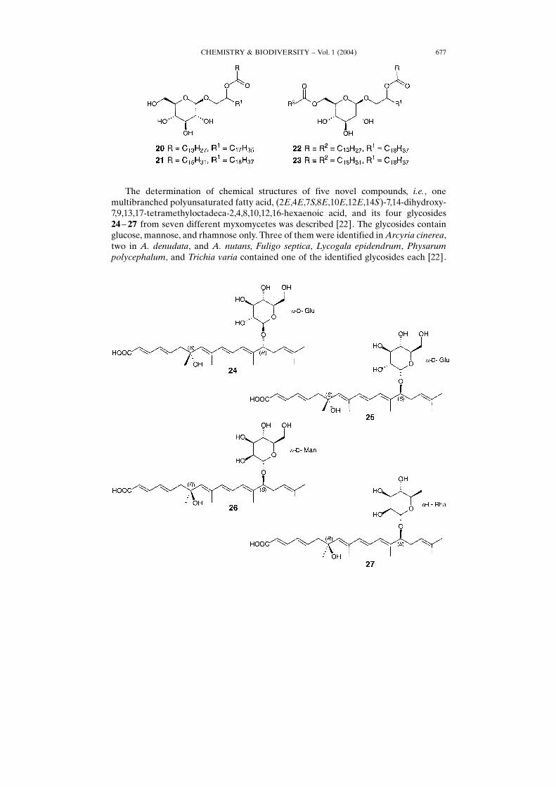
The determination of chemical structures of five novel compounds, i.e., onemultibranched polyunsaturated fatty acid, (2E,4E,7S,8E,10E,12E,14S)-7,14-dihydroxy-7,9,13,17-tetramethyloctadeca-2,4,8,10,12,16-hexaenoic acid, and its four glycosides24 ± 27 from seven different myxomycetes was described [22]. The glycosides containglucose, mannose, and rhamnose only. Three of them were identified inArcyria cinerea,two in A. denudata, and A. nutans, Fuligo septica, Lycogala epidendrum, Physarumpolycephalum, and Trichia varia contained one of the identified glycosides each [22].
CHEMISTRY & BIODIVERSITY ± Vol. 1 (2004) 677
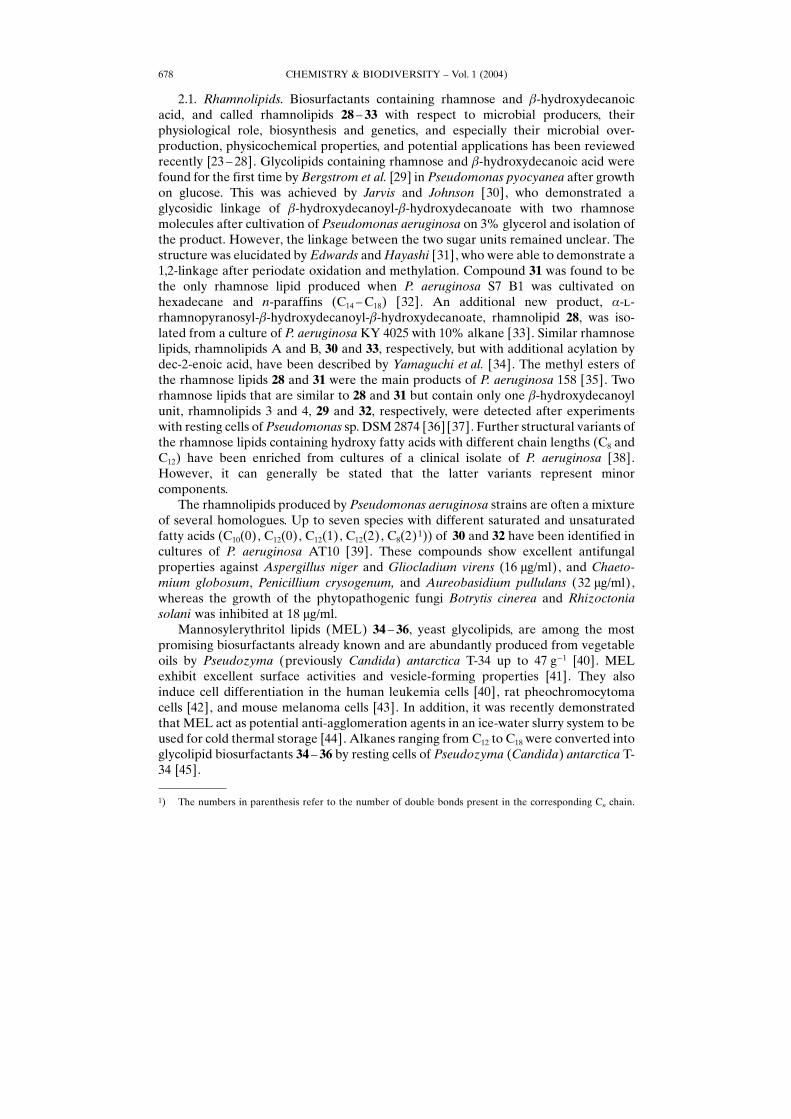
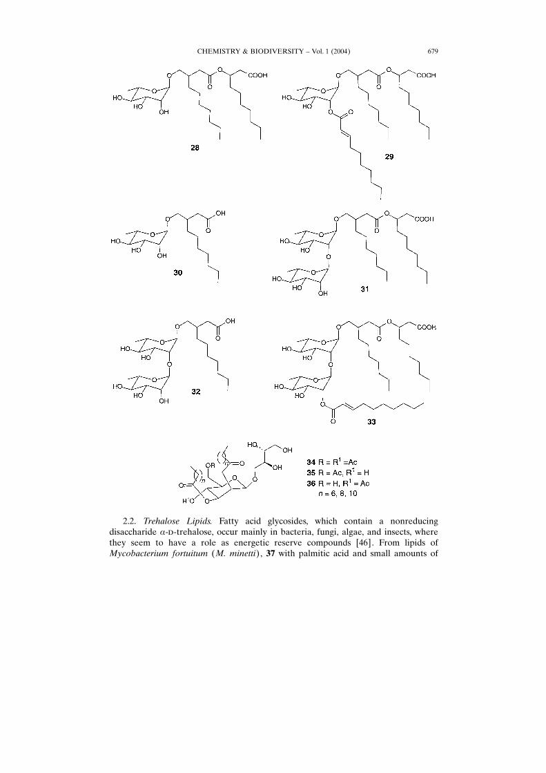
2.1. Rhamnolipids. Biosurfactants containing rhamnose and �-hydroxydecanoicacid, and called rhamnolipids 28 ± 33 with respect to microbial producers, theirphysiological role, biosynthesis and genetics, and especially their microbial over-production, physicochemical properties, and potential applications has been reviewedrecently [23 ± 28]. Glycolipids containing rhamnose and �-hydroxydecanoic acid werefound for the first time byBergstrom et al. [29] in Pseudomonas pyocyanea after growthon glucose. This was achieved by Jarvis and Johnson [30], who demonstrated aglycosidic linkage of �-hydroxydecanoyl-�-hydroxydecanoate with two rhamnosemolecules after cultivation of Pseudomonas aeruginosa on 3% glycerol and isolation ofthe product. However, the linkage between the two sugar units remained unclear. Thestructure was elucidated byEdwards andHayashi [31], who were able to demonstrate a1,2-linkage after periodate oxidation and methylation. Compound 31 was found to bethe only rhamnose lipid produced when P. aeruginosa S7 B1 was cultivated onhexadecane and n-paraffins (C14 ±C18) [32]. An additional new product, �-�-rhamnopyranosyl-�-hydroxydecanoyl-�-hydroxydecanoate, rhamnolipid 28, was iso-lated from a culture of P. aeruginosa KY 4025 with 10% alkane [33]. Similar rhamnoselipids, rhamnolipids A and B, 30 and 33, respectively, but with additional acylation bydec-2-enoic acid, have been described by Yamaguchi et al. [34]. The methyl esters ofthe rhamnose lipids 28 and 31 were the main products of P. aeruginosa 158 [35]. Tworhamnose lipids that are similar to 28 and 31 but contain only one �-hydroxydecanoylunit, rhamnolipids 3 and 4, 29 and 32, respectively, were detected after experimentswith resting cells of Pseudomonas sp. DSM 2874 [36] [37]. Further structural variants ofthe rhamnose lipids containing hydroxy fatty acids with different chain lengths (C8 andC12) have been enriched from cultures of a clinical isolate of P. aeruginosa [38].However, it can generally be stated that the latter variants represent minorcomponents.The rhamnolipids produced by Pseudomonas aeruginosa strains are often a mixture
of several homologues. Up to seven species with different saturated and unsaturatedfatty acids (C10(0), C12(0), C12(1), C12(2), C8(2)1)) of 30 and 32 have been identified incultures of P. aeruginosa AT10 [39]. These compounds show excellent antifungalproperties against Aspergillus niger and Gliocladium virens (16 �g/ml), and Chaeto-mium globosum, Penicillium crysogenum, and Aureobasidium pullulans (32 �g/ml),whereas the growth of the phytopathogenic fungi Botrytis cinerea and Rhizoctoniasolani was inhibited at 18 �g/ml.Mannosylerythritol lipids (MEL) 34 ± 36, yeast glycolipids, are among the most
promising biosurfactants already known and are abundantly produced from vegetableoils by Pseudozyma (previously Candida) antarctica T-34 up to 47 g�1 [40]. MELexhibit excellent surface activities and vesicle-forming properties [41]. They alsoinduce cell differentiation in the human leukemia cells [40], rat pheochromocytomacells [42], and mouse melanoma cells [43]. In addition, it was recently demonstratedthat MEL act as potential anti-agglomeration agents in an ice-water slurry system to beused for cold thermal storage [44]. Alkanes ranging from C12 to C18 were converted intoglycolipid biosurfactants 34 ± 36 by resting cells of Pseudozyma (Candida) antarctica T-34 [45].
CHEMISTRY & BIODIVERSITY ± Vol. 1 (2004)678
1) The numbers in parenthesis refer to the number of double bonds present in the corresponding Cn chain.
2.2. Trehalose Lipids. Fatty acid glycosides, which contain a nonreducingdisaccharide �-�-trehalose, occur mainly in bacteria, fungi, algae, and insects, wherethey seem to have a role as energetic reserve compounds [46]. From lipids ofMycobacterium fortuitum (M. minetti), 37 with palmitic acid and small amounts of
CHEMISTRY & BIODIVERSITY ± Vol. 1 (2004) 679
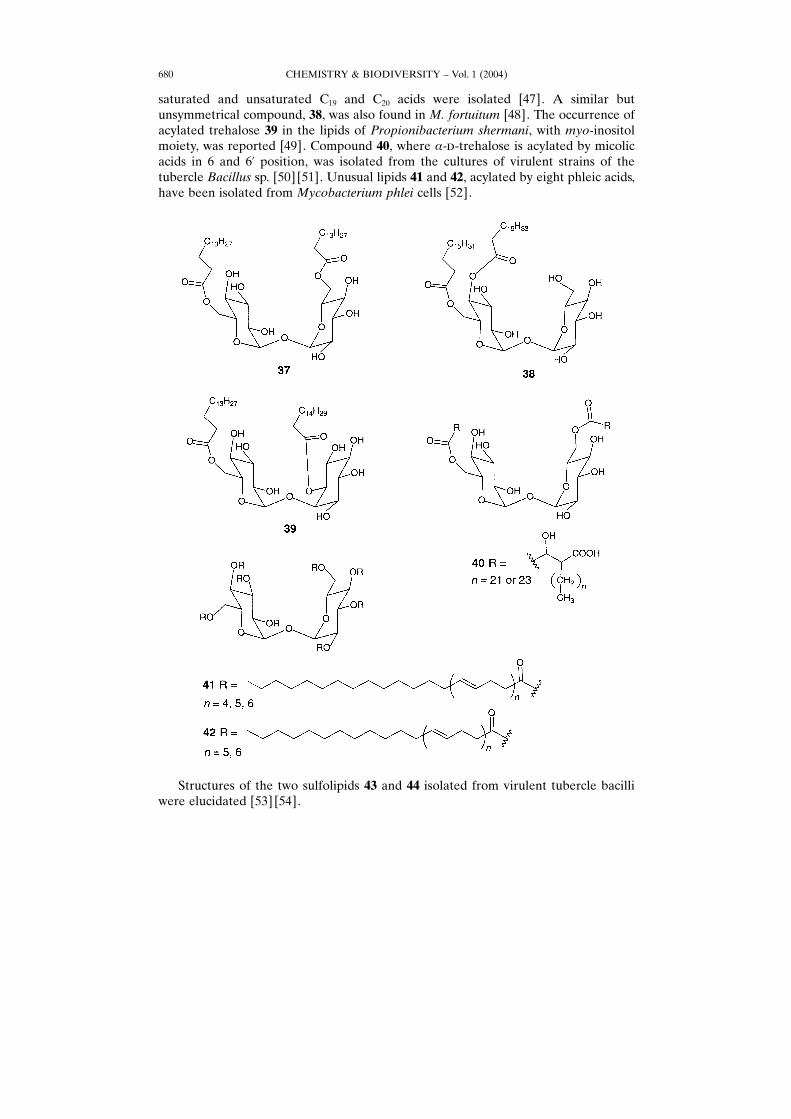
saturated and unsaturated C19 and C20 acids were isolated [47]. A similar butunsymmetrical compound, 38, was also found in M. fortuitum [48]. The occurrence ofacylated trehalose 39 in the lipids of Propionibacterium shermani, with myo-inositolmoiety, was reported [49]. Compound 40, where �-�-trehalose is acylated by micolicacids in 6 and 6� position, was isolated from the cultures of virulent strains of thetubercle Bacillus sp. [50] [51]. Unusual lipids 41 and 42, acylated by eight phleic acids,have been isolated from Mycobacterium phlei cells [52].
Structures of the two sulfolipids 43 and 44 isolated from virulent tubercle bacilliwere elucidated [53] [54].
CHEMISTRY & BIODIVERSITY ± Vol. 1 (2004)680
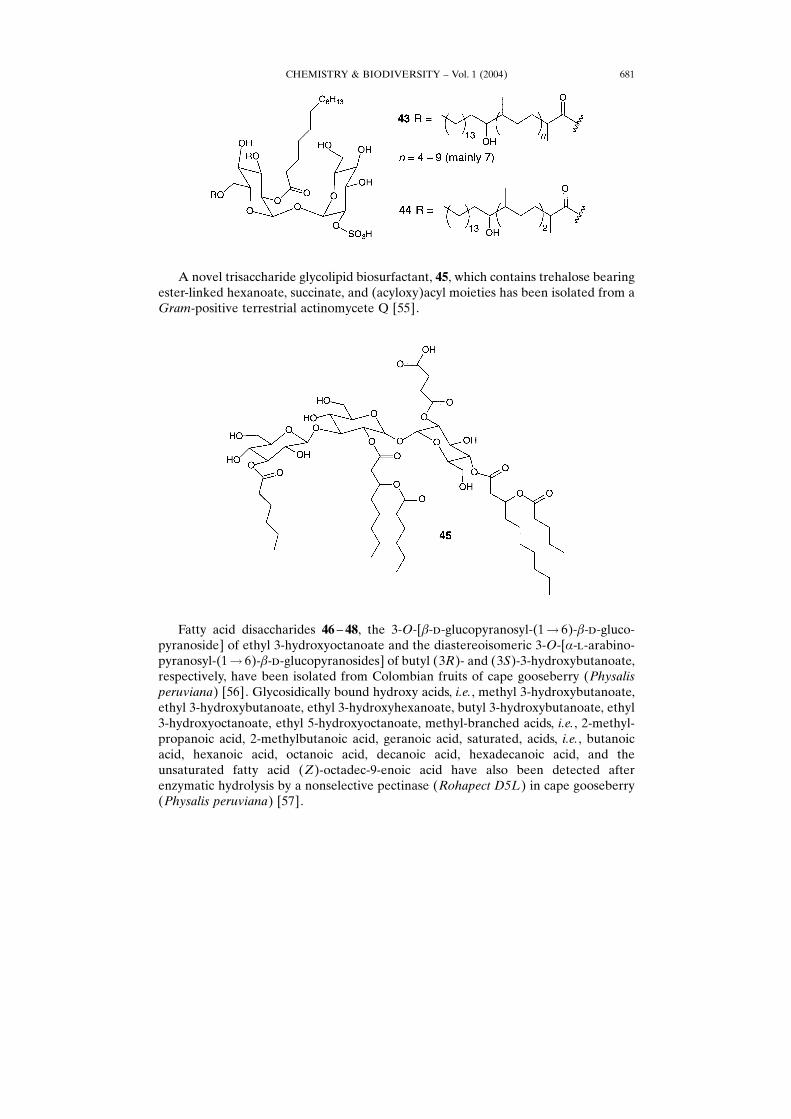
A novel trisaccharide glycolipid biosurfactant, 45, which contains trehalose bearingester-linked hexanoate, succinate, and (acyloxy)acyl moieties has been isolated from aGram-positive terrestrial actinomycete Q [55].
Fatty acid disaccharides 46 ± 48, the 3-O-[�-�-glucopyranosyl-(1� 6)-�-�-gluco-pyranoside] of ethyl 3-hydroxyoctanoate and the diastereoisomeric 3-O-[�-�-arabino-pyranosyl-(1� 6)-�-�-glucopyranosides] of butyl (3R)- and (3S)-3-hydroxybutanoate,respectively, have been isolated from Colombian fruits of cape gooseberry (Physalisperuviana) [56]. Glycosidically bound hydroxy acids, i.e., methyl 3-hydroxybutanoate,ethyl 3-hydroxybutanoate, ethyl 3-hydroxyhexanoate, butyl 3-hydroxybutanoate, ethyl3-hydroxyoctanoate, ethyl 5-hydroxyoctanoate, methyl-branched acids, i.e., 2-methyl-propanoic acid, 2-methylbutanoic acid, geranoic acid, saturated, acids, i.e., butanoicacid, hexanoic acid, octanoic acid, decanoic acid, hexadecanoic acid, and theunsaturated fatty acid (Z)-octadec-9-enoic acid have also been detected afterenzymatic hydrolysis by a nonselective pectinase (Rohapect D5L) in cape gooseberry(Physalis peruviana) [57].
CHEMISTRY & BIODIVERSITY ± Vol. 1 (2004) 681
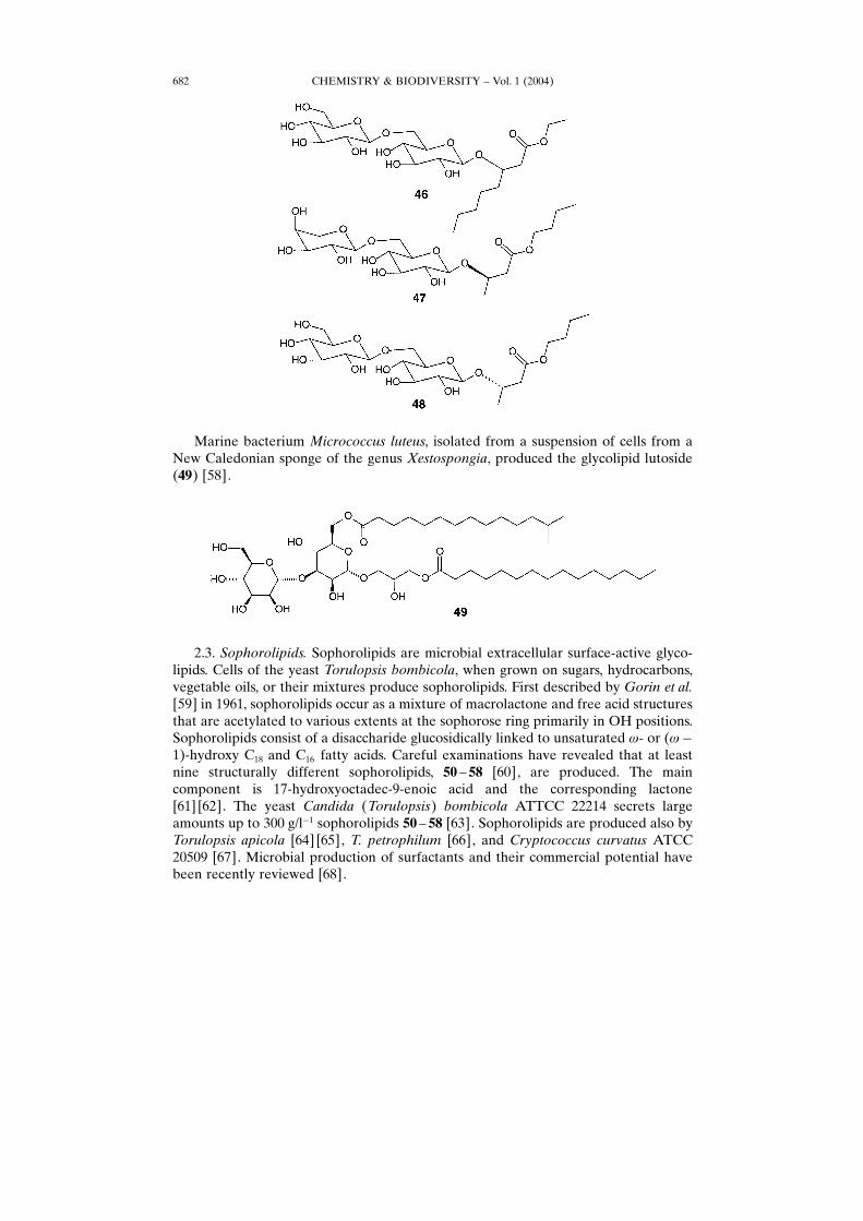
Marine bacterium Micrococcus luteus, isolated from a suspension of cells from aNew Caledonian sponge of the genus Xestospongia, produced the glycolipid lutoside(49) [58].
2.3. Sophorolipids. Sophorolipids are microbial extracellular surface-active glyco-lipids. Cells of the yeast Torulopsis bombicola, when grown on sugars, hydrocarbons,vegetable oils, or their mixtures produce sophorolipids. First described by Gorin et al.[59] in 1961, sophorolipids occur as a mixture of macrolactone and free acid structuresthat are acetylated to various extents at the sophorose ring primarily in OH positions.Sophorolipids consist of a disaccharide glucosidically linked to unsaturated �- or (��1)-hydroxy C18 and C16 fatty acids. Careful examinations have revealed that at leastnine structurally different sophorolipids, 50 ± 58 [60], are produced. The maincomponent is 17-hydroxyoctadec-9-enoic acid and the corresponding lactone[61] [62]. The yeast Candida (Torulopsis) bombicola ATTCC 22214 secrets largeamounts up to 300 g/l�1 sophorolipids 50 ± 58 [63]. Sophorolipids are produced also byTorulopsis apicola [64] [65], T. petrophilum [66], and Cryptococcus curvatus ATCC20509 [67]. Microbial production of surfactants and their commercial potential havebeen recently reviewed [68].
CHEMISTRY & BIODIVERSITY ± Vol. 1 (2004)682
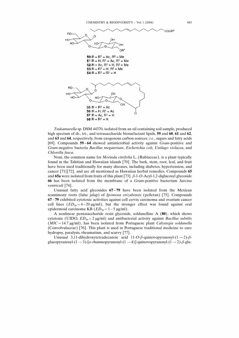
Tsukamurella sp. DSM 44370, isolated from an oil containing soil sample, producedhigh spectum of di-, tri-, and tetrasaccharide biosurfactant lipids, 59 and 60, 61 and 62,and 63 and 64, respectively, from exogenous carbon sources; i.e., sugars and fatty acids[69]. Compounds 59 ± 64 showed antimicrobial activity against Gram-positive andGram-negative bacteria Bacillus megaterium, Escherichia coli, Ustilago violacea, andChlorella fusca.Noni, the common name forMorinda citrifolia L. (Rubiaceae), is a plant typically
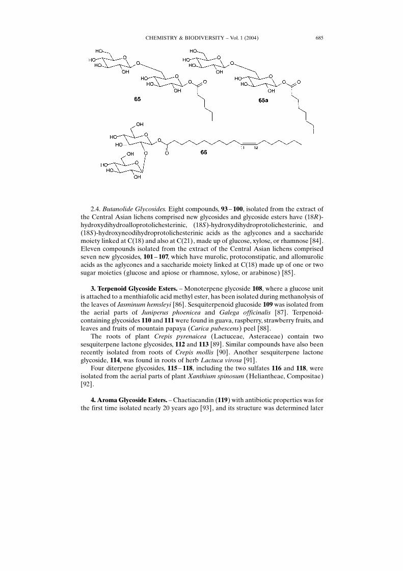
found in the Tahitian and Hawaiian islands [70]. The bark, stem, root, leaf, and fruithave been used traditionally for many diseases, including diabetes, hypertension, andcancer [71] [72], and are all mentioned as Hawaiian herbal remedies. Compounds 65and 65a were isolated from fruits of this plant [73]. �-1-O-Acyl-1,2-diglucosyl glycoside66 has been isolated from the membrane of a Gram-positive bacterium Sarcinaventriculi [74].Unusual fatty acid glycosides 67 ± 79 have been isolated from the Mexican
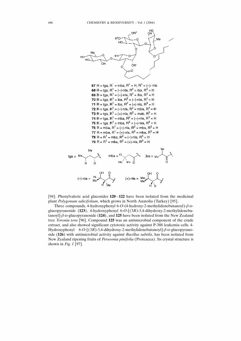
scamimony roots (false jalap) of Ipomoea orizabensis (pelletan) [75]. Compounds67 ± 79 exhibited cytotoxic activities against cell cervix carcinoma and ovarium cancercell lines (ED50� 4 ± 20 �g/ml), but the stronger effect was found against oralepidermoid carcinoma KB (ED50� 1 ± 5 �g/ml).A nonlinear pentasaccharide resin glycoside, soldanelline A (80), which shows
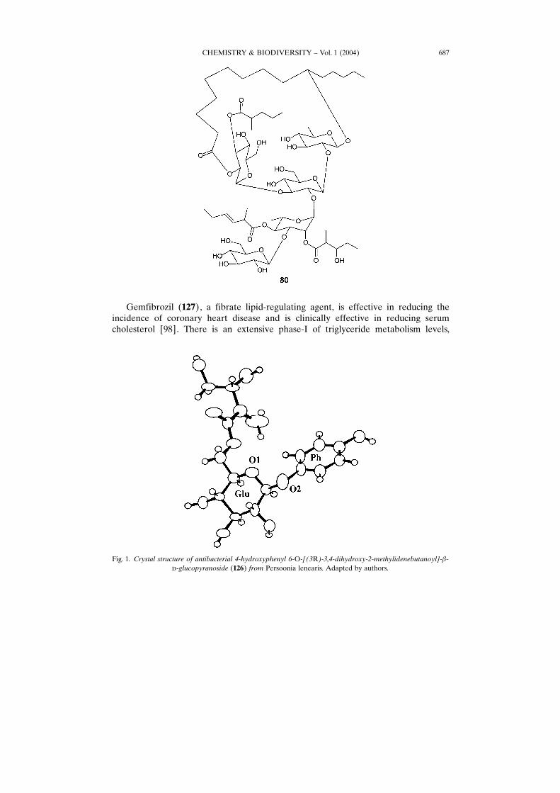
cytotoxic (UIDO, ED50� 2 �g/ml) and antibacterial activity against Bacillus subtilis(MIC� 14.7 �g/ml), has been isolated from Portuguese plant Calystegia soldanella(Convolvulaceae) [76]. This plant is used in Portuguese traditional medicine to curehydropsy, paralysis, rheumatism, and scurvy [77].Unusual 3,11-dihydroxytetradecanoic acid 11-O-�-quinovopyranosyl-(1� 2)-�-
glucopyranosyl-(1� 3)-[�-rhamnopyranosyl-(1� 4)]-quinovopyranosyl-(1� 2)-�-glu-
CHEMISTRY & BIODIVERSITY ± Vol. 1 (2004) 683
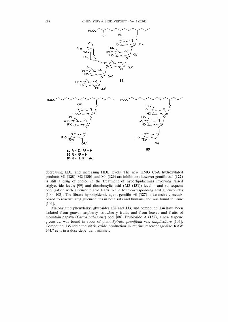
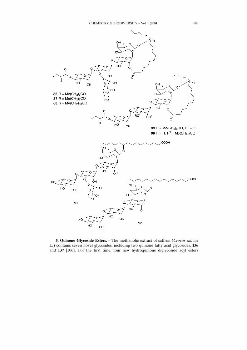
copyranosyl-(1� 2)-�-fucopyranoside (81) has been isolated from the aerial parts ofAustralian plant Ipomoea lonchophylla [78]. Many species of Ipomoea were and stillare used in folk medicine in different parts of the world [79]. Ethanolic extracts of theseplants have reported to exhibit antimicrobial, analgesic, spasmogenic, spasmolytic,hypotensive, psychotomimetic, and anticancer effects [80]. Fatty acid glycosides 82 ± 85have been isolated from the seeds of Cuscuta chinensis (Convolvulacea) or −CusculaSemen× well-known as Chinese traditional medicine plant used as a tonic [81].Seven new ether-soluble resin glycosides 86 ± 92 have been isolated from whole
plants of Ipomoea stolonifera [82]. These compounds were similar to the resinglycosides 81 ± 85 isolated previously from plant I. lonchophylla [83].
CHEMISTRY & BIODIVERSITY ± Vol. 1 (2004)684
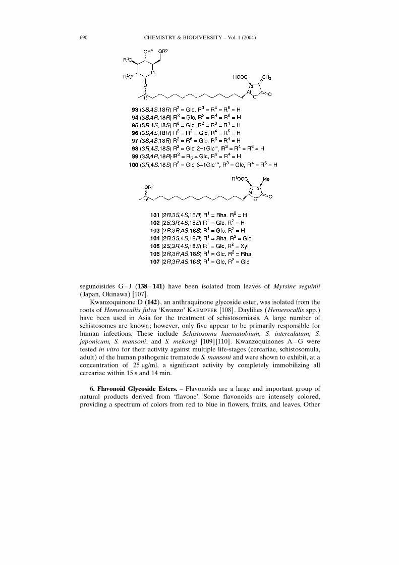
2.4. Butanolide Glycosides. Eight compounds, 93 ± 100, isolated from the extract ofthe Central Asian lichens comprised new glycosides and glycoside esters have (18R)-hydroxydihydroalloprotolichesterinic, (18S)-hydroxydihydroprotolichesterinic, and(18S)-hydroxyneodihydroprotolichesterinic acids as the aglycones and a saccharidemoiety linked at C(18) and also at C(21), made up of glucose, xylose, or rhamnose [84].Eleven compounds isolated from the extract of the Central Asian lichens comprisedseven new glycosides, 101 ± 107, which have murolic, protoconstipatic, and allomurolicacids as the aglycones and a saccharide moiety linked at C(18) made up of one or twosugar moieties (glucose and apiose or rhamnose, xylose, or arabinose) [85].
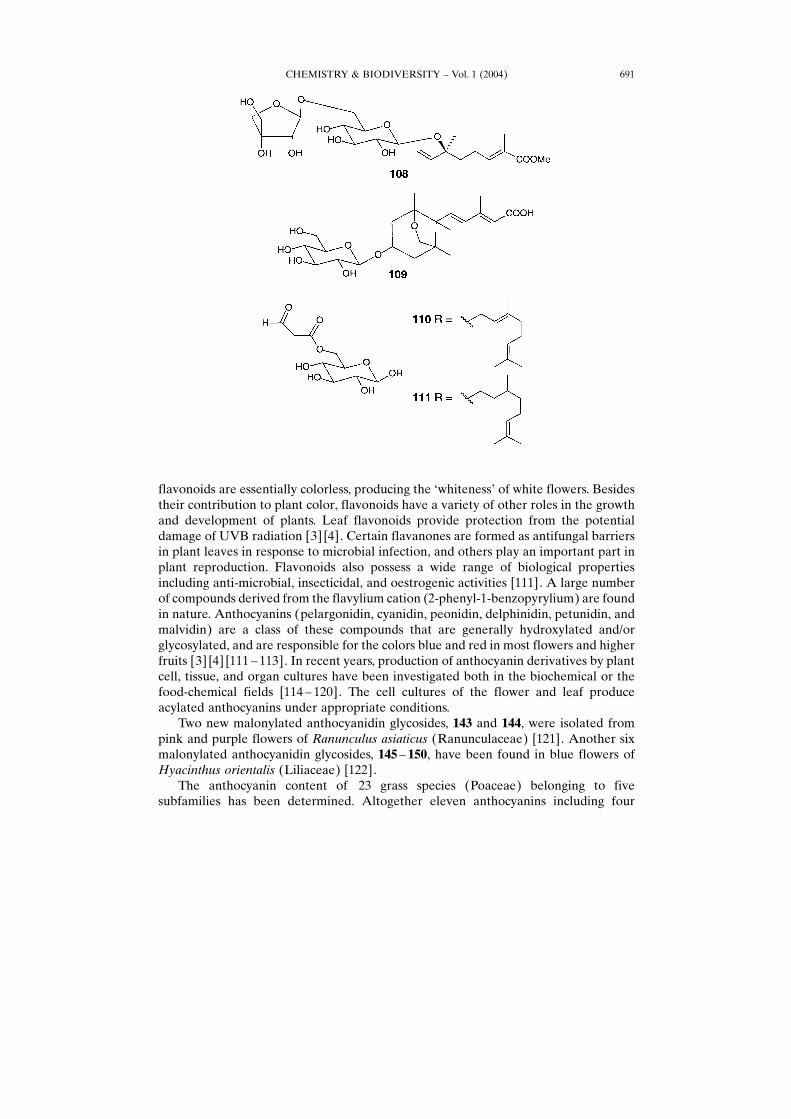
3. Terpenoid Glycoside Esters. ± Monoterpene glycoside 108, where a glucose unitis attached to a menthiafolic acid methyl ester, has been isolated during methanolysis ofthe leaves of Jasminum hemsleyi [86]. Sesquiterpenoid glucoside 109 was isolated fromthe aerial parts of Juniperus phoenicea and Galega officinalis [87]. Terpenoid-containing glycosides 110 and 111were found in guava, raspberry, strawberry fruits, andleaves and fruits of mountain papaya (Carica pubescens) peel [88].The roots of plant Crepis pyrenaicea (Lactuceae, Asteraceae) contain two
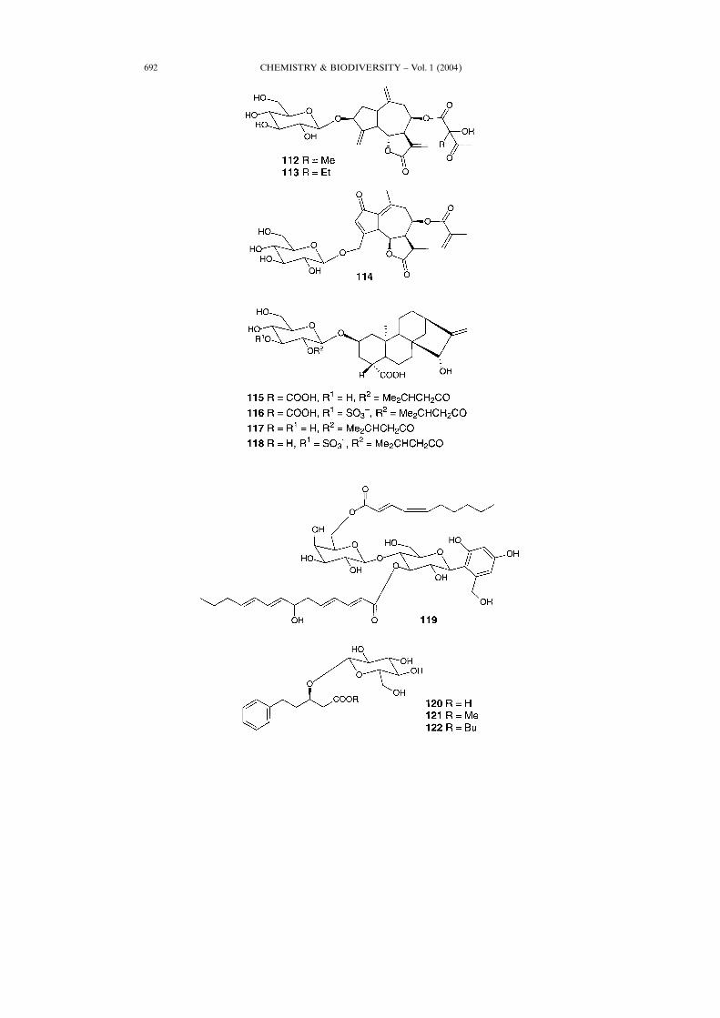
sesquiterpene lactone glycosides, 112 and 113 [89]. Similar compounds have also beenrecently isolated from roots of Crepis mollis [90]. Another sesquiterpene lactoneglycoside, 114, was found in roots of herb Lactuca virosa [91].Four diterpene glycosides, 115 ± 118, including the two sulfates 116 and 118, were
isolated from the aerial parts of plant Xanthium spinosum (Heliantheae, Compositae)[92].
4. AromaGlycoside Esters. ± Chaetiacandin (119) with antibiotic properties was forthe first time isolated nearly 20 years ago [93], and its structure was determined later
CHEMISTRY & BIODIVERSITY ± Vol. 1 (2004) 685
[94]. Phenylvaleric acid glucosides 120 ± 122 have been isolated from the medicinalplant Polygonum salicifolium, which grows in North Anatolia (Turkey) [95].Three compounds, 4-hydroxyphenyl 6-O-(4-hydroxy-2-methylidenebutanoyl)-�-�-
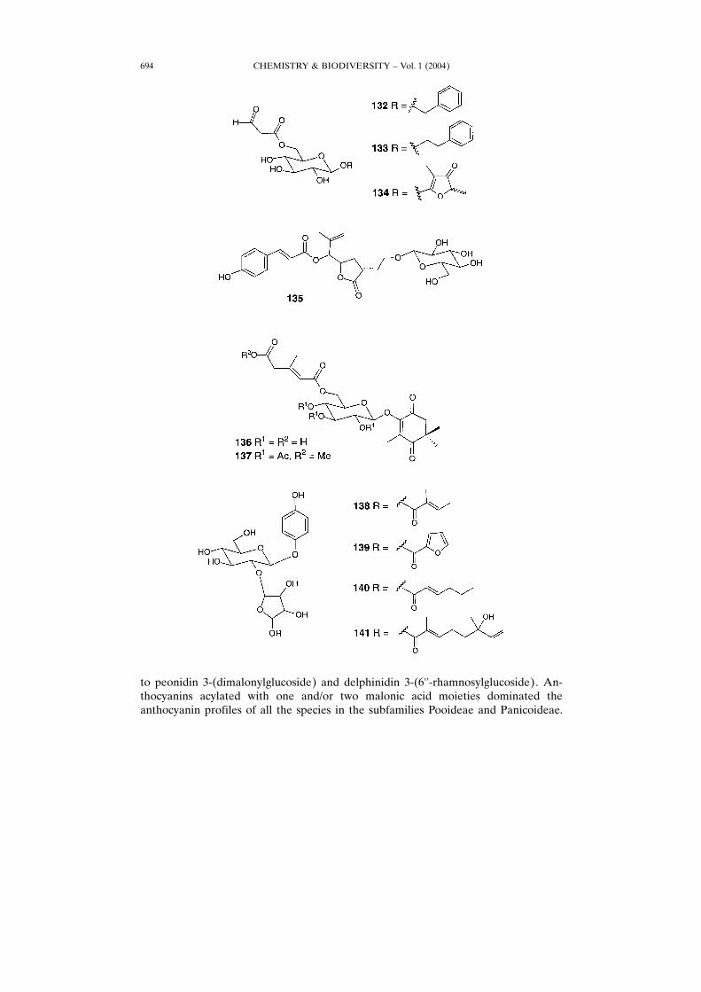
glucopyranoside (123), 4-hydroxyphenyl 6-O-[(3R)-3,4-dihydroxy-2-methylidenebu-tanoyl]-�-�-glucopyranoside (124), and 125 have been isolated from the New Zealandtree Toronia toru [96]. Compound 125 was an antimicrobial component of the crudeextract, and also showed significant cytotoxic activity against P-388 leukemia cells. 4-Hydroxyphenyl 6-O-[(3R)-3,4-dihydroxy-2-methylidenebutanoyl]-�-�-glucopyrano-side (126) with antimicrobial activity against Bacillus subtilis, has been isolated fromNew Zealand ripening fruits of Persoonia pinifolia (Proteacea). Its crystal structure isshown in Fig. 1 [97].
CHEMISTRY & BIODIVERSITY ± Vol. 1 (2004)686
Gemfibrozil (127), a fibrate lipid-regulating agent, is effective in reducing theincidence of coronary heart disease and is clinically effective in reducing serumcholesterol [98]. There is an extensive phase-I of triglyceride metabolism levels,
CHEMISTRY & BIODIVERSITY ± Vol. 1 (2004) 687
Fig. 1. Crystal structure of antibacterial 4-hydroxyphenyl 6-O-[(3R)-3,4-dihydroxy-2-methylidenebutanoyl]-�-�-glucopyranoside (126) from Persoonia lenearis. Adapted by authors.
decreasing LDL and increasing HDL levels. The new HMG CoA hydroxylatedproducts M1 (128), M2 (130), and M4 (129) are inhibitors; however gemfibrozil (127)is still a drug of choice in the treatment of hyperlipidaemias involving raisedtriglyceride levels [99] and dicarboxylic acid (M3 (131)) level ± and subsequentconjugation with glucuronic acid leads to the four corresponding acyl glucuronides[100 ± 103]. The fibrate hypolipidemic agent gemfibrozil (127) is extensively metab-olized to reactive acyl glucuronides in both rats and humans, and was found in urine[104].Malonylated phenylalkyl glycosides 132 and 133, and compound 134 have been
isolated from guava, raspberry, strawberry fruits, and from leaves and fruits ofmountain papaya (Carica pubescens) peel [88]. Prubioside A (135), a new terpeneglycoside, was found in roots of plant Spiraea prunifolia var. simpliciflora [105].Compound 135 inhibited nitric oxide production in murine macrophage-like RAW264.7 cells in a dose-dependent manner.
CHEMISTRY & BIODIVERSITY ± Vol. 1 (2004)688
5. Quinone Glycoside Esters. ± The methanolic extract of saffron (Crocus sativusL.) contains seven novel glycosides, including two quinone fatty acid glycosides, 136and 137 [106]. For the first time, four new hydroquinone diglycoside acyl esters
CHEMISTRY & BIODIVERSITY ± Vol. 1 (2004) 689
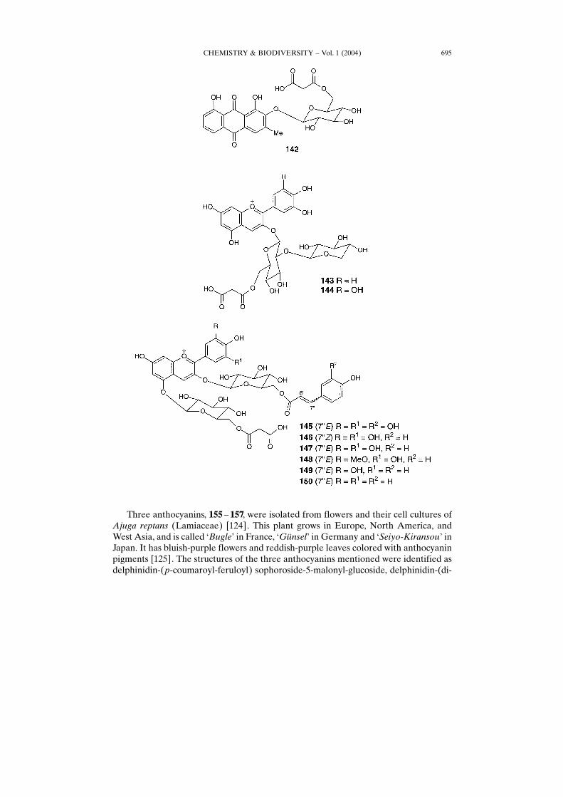
segunoisides G± J (138 ± 141) have been isolated from leaves of Myrsine seguinii(Japan, Okinawa) [107].Kwanzoquinone D (142), an anthraquinone glycoside ester, was isolated from the
roots of Hemerocallis fulva −Kwanzo× K������� [108]. Daylilies (Hemerocallis spp.)have been used in Asia for the treatment of schistosomiasis. A large number ofschistosomes are known; however, only five appear to be primarily responsible forhuman infections. These include Schistosoma haematobium, S. intercalatum, S.japonicum, S. mansoni, and S. mekongi [109] [110]. Kwanzoquinones A ±G weretested in vitro for their activity against multiple life-stages (cercariae, schistosomula,adult) of the human pathogenic trematode S. mansoni and were shown to exhibit, at aconcentration of 25 �g/ml, a significant activity by completely immobilizing allcercariae within 15 s and 14 min.
6. Flavonoid Glycoside Esters. ± Flavonoids are a large and important group ofnatural products derived from −flavone×. Some flavonoids are intensely colored,providing a spectrum of colors from red to blue in flowers, fruits, and leaves. Other
CHEMISTRY & BIODIVERSITY ± Vol. 1 (2004)690
flavonoids are essentially colorless, producing the −whiteness× of white flowers. Besidestheir contribution to plant color, flavonoids have a variety of other roles in the growthand development of plants. Leaf flavonoids provide protection from the potentialdamage of UVB radiation [3] [4]. Certain flavanones are formed as antifungal barriersin plant leaves in response to microbial infection, and others play an important part inplant reproduction. Flavonoids also possess a wide range of biological propertiesincluding anti-microbial, insecticidal, and oestrogenic activities [111]. A large numberof compounds derived from the flavylium cation (2-phenyl-1-benzopyrylium) are foundin nature. Anthocyanins (pelargonidin, cyanidin, peonidin, delphinidin, petunidin, andmalvidin) are a class of these compounds that are generally hydroxylated and/orglycosylated, and are responsible for the colors blue and red in most flowers and higherfruits [3] [4] [111 ± 113]. In recent years, production of anthocyanin derivatives by plantcell, tissue, and organ cultures have been investigated both in the biochemical or thefood-chemical fields [114 ± 120]. The cell cultures of the flower and leaf produceacylated anthocyanins under appropriate conditions.Two new malonylated anthocyanidin glycosides, 143 and 144, were isolated from
pink and purple flowers of Ranunculus asiaticus (Ranunculaceae) [121]. Another sixmalonylated anthocyanidin glycosides, 145 ± 150, have been found in blue flowers ofHyacinthus orientalis (Liliaceae) [122].The anthocyanin content of 23 grass species (Poaceae) belonging to five
subfamilies has been determined. Altogether eleven anthocyanins including four
CHEMISTRY & BIODIVERSITY ± Vol. 1 (2004) 691
CHEMISTRY & BIODIVERSITY ± Vol. 1 (2004)692
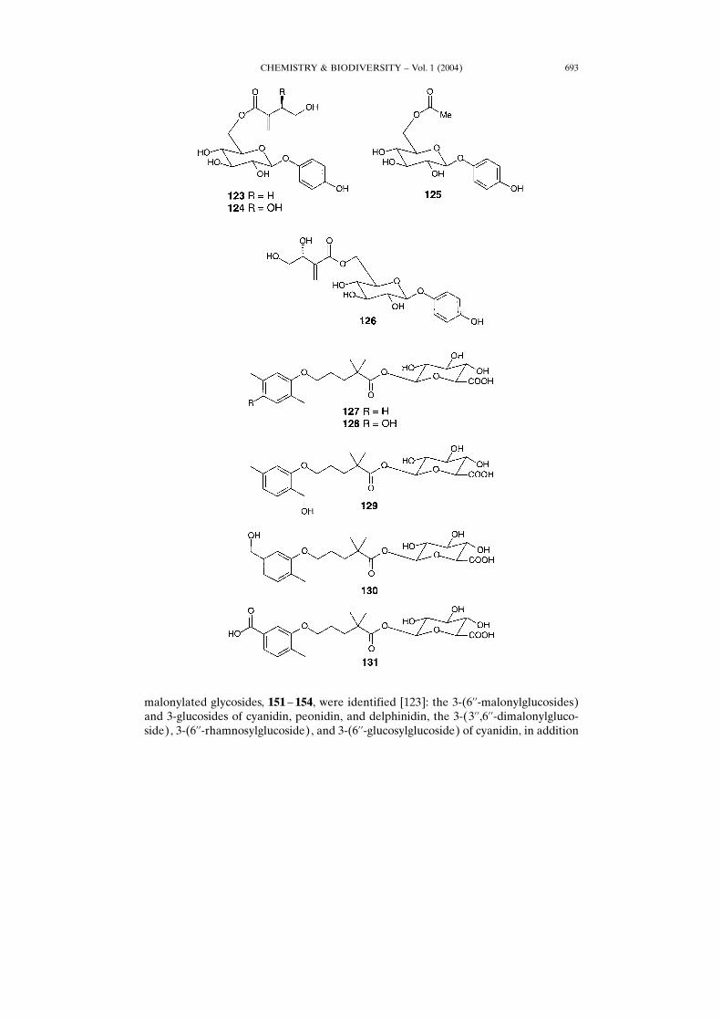
malonylated glycosides, 151 ± 154, were identified [123]: the 3-(6��-malonylglucosides)and 3-glucosides of cyanidin, peonidin, and delphinidin, the 3-(3��,6��-dimalonylgluco-side), 3-(6��-rhamnosylglucoside), and 3-(6��-glucosylglucoside) of cyanidin, in addition
CHEMISTRY & BIODIVERSITY ± Vol. 1 (2004) 693
to peonidin 3-(dimalonylglucoside) and delphinidin 3-(6��-rhamnosylglucoside). An-thocyanins acylated with one and/or two malonic acid moieties dominated theanthocyanin profiles of all the species in the subfamilies Pooideae and Panicoideae.
CHEMISTRY & BIODIVERSITY ± Vol. 1 (2004)694
Three anthocyanins, 155 ± 157, were isolated from flowers and their cell cultures ofAjuga reptans (Lamiaceae) [124]. This plant grows in Europe, North America, andWest Asia, and is called −Bugle× in France, −G¸nsel× in Germany and −Seiyo-Kiransou× inJapan. It has bluish-purple flowers and reddish-purple leaves colored with anthocyaninpigments [125]. The structures of the three anthocyanins mentioned were identified asdelphinidin-(p-coumaroyl-feruloyl) sophoroside-5-malonyl-glucoside, delphinidin-(di-
CHEMISTRY & BIODIVERSITY ± Vol. 1 (2004) 695
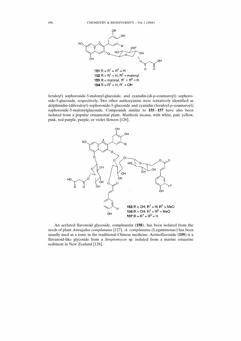
feruloyl) sophoroside-5-malonyl-glucoside, and cyanidin-(di-p-coumaroyl) sophoro-side-5-glucoside, respectively. Two other anthocyanins were tentatively identified asdelphinidin-(diferuloyl) sophoroside-5-glucoside and cyanidin-(feruloyl-p-coumaroyl)sophoroside-5-malonylglucoside. Compounds similar to 155 ± 157 have also beenisolated from a popular ornamental plant, Matthiola incana, with white, pale yellow,pink, red-purple, purple, or violet flowers [126].
An acylated flavonoid glycoside, complanatin (158), has been isolated from theseeds of plant Astragalus complanatus [127]. A. complanatus (Leguminosae) has beenusually used as a tonic in the traditional Chinese medicine. Actinoflavoside (159) is aflavanoid-like glycoside from a Streptomyces sp. isolated from a marine estuarinesediment in New Zealand [128].
CHEMISTRY & BIODIVERSITY ± Vol. 1 (2004)696
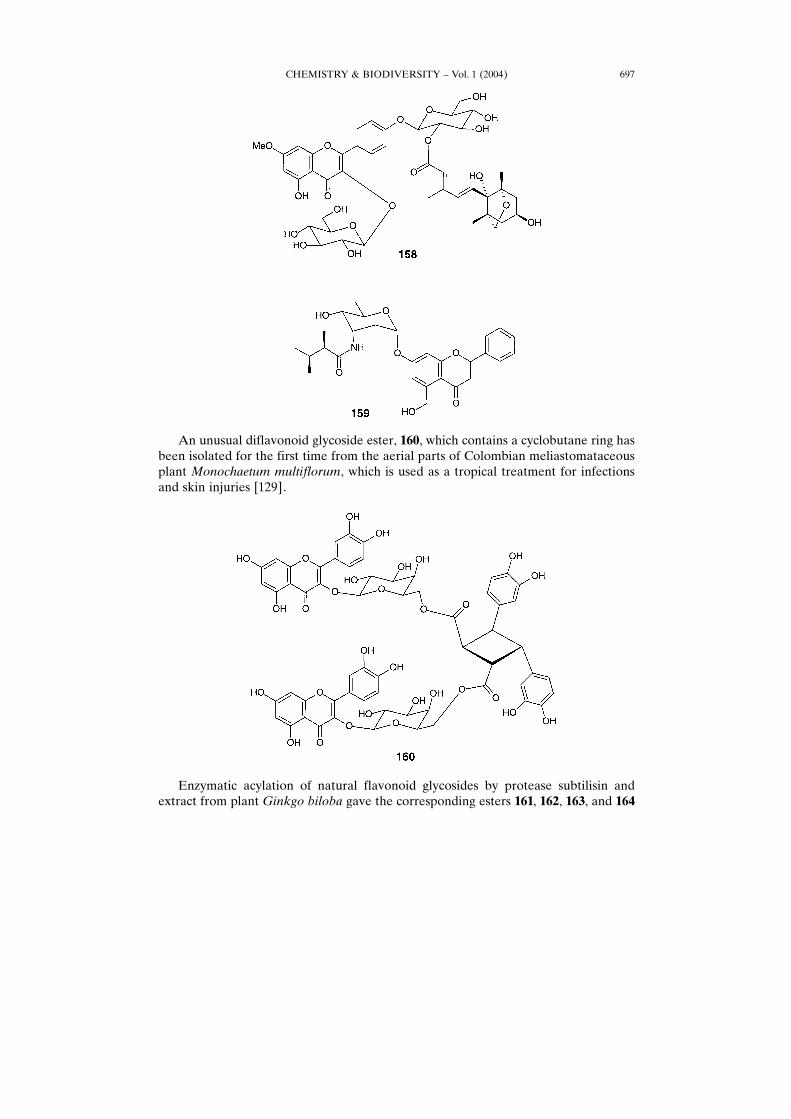
An unusual diflavonoid glycoside ester, 160, which contains a cyclobutane ring hasbeen isolated for the first time from the aerial parts of Colombian meliastomataceousplant Monochaetum multiflorum, which is used as a tropical treatment for infectionsand skin injuries [129].
Enzymatic acylation of natural flavonoid glycosides by protease subtilisin andextract from plant Ginkgo biloba gave the corresponding esters 161, 162, 163, and 164
CHEMISTRY & BIODIVERSITY ± Vol. 1 (2004) 697
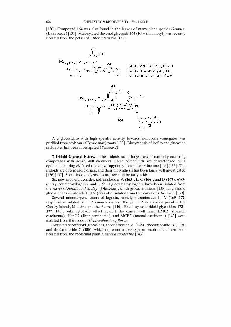
[130]. Compound 164 was also found in the leaves of many plant species Ocimum(Lamiaceae) [131]. Malonylated flavonol glycoside 164 (R1� rhamnosyl) was recentlyisolated from the petals of Clitoria ternatea [132].
A �-glucosidase with high specific activity towards isoflavone conjugates waspurified from soybean (Glycine max) roots [133]. Biosynthesis of isoflavone glucosidemalonates has been investigated (Scheme 2).
7. Iridoid Glycosyl Esters. ± The iridoids are a large class of naturally occurringcompounds with nearly 400 members. These compounds are characterized by acyclopentane ring cis-fused to a dihydropyran, �-lactone, or �-lactone [134] [135]. Theiridoids are of terpenoid origin, and their biosynthesis has been fairly well investigated[136] [137]. Some iridoid glycosides are acylated by fatty acids.Six new iridoid glucosides, jashemslosides A (165), B, C (166), and D (167), 6�-O-
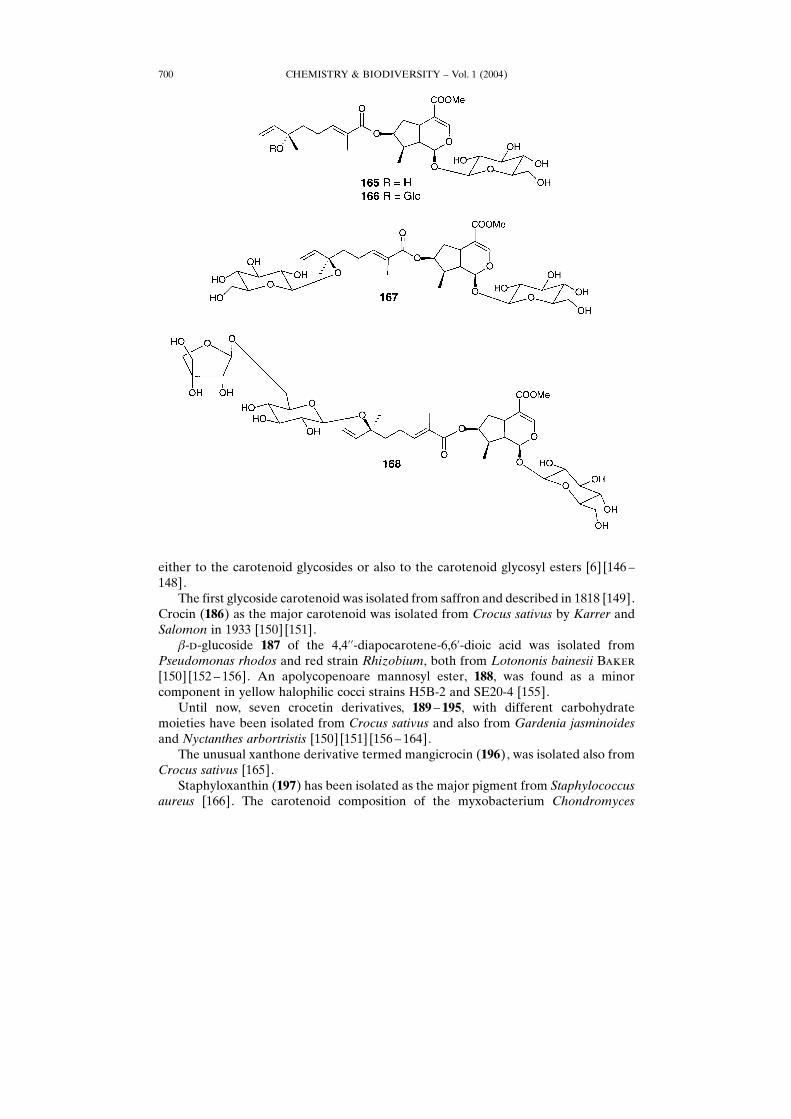
trans-p-coumaroylloganin, and 6�-O-cis-p-coumaroylloganin have been isolated fromthe leaves of Jasminum hemsleyi (Oleaceae), which grows in Taiwan [138], and iridoidglucoside jashemsloside E (168) was also isolated from the leaves of J. hemsleyi [139].Several monoterpene esters of loganin, namely picconiosides II ±V (169 ± 172,
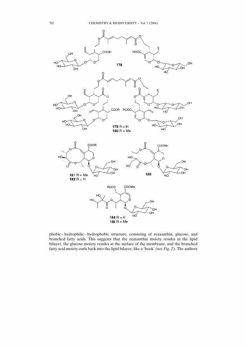
resp.) were isolated from Picconia excelsa of the genus Picconia widespread in theCanary Islands, Madeira, and the Azores [140]. Five fatty acid iridoid glycosides, 173 ±177 [141], with cytotoxic effect against the cancer cell lines HM02 (stomachcarcinoma), HepG2 (liver carcinoma), and MCF7 (mamal carcinoma) [142] wereisolated from the roots of Centranthus longiflorus.Acylated secoiridoid glucosides, rhodanthoside A (178), rhodanthoside B (179),
and rhodanthoside C (180), which represent a new type of secoiridoids, have beenisolated from the medicinal plant Gentiana rhodantha [143].
CHEMISTRY & BIODIVERSITY ± Vol. 1 (2004)698
Secoiridoid glycosyl esters 181 ± 185 were isolated from the leaves of the plantGonocaryum calleryanum, which is distributed from Indonesia to Taiwan [144]. Itsleaves are used in Philippine folk medicine for the treatment of stomach diseases [145].
8. Carotenoid Glycoside Esters. ± Carotenoids are an important group of naturalpigments in both the prokaryote and eukaryote kingdoms, exhibiting colors from dark-red to bright-yellow. They are important in human nutrition as a source of �-carotene(vitamin A) and as a prevention agent against cancer and heart disease. In addition,carotenoids add color to foods and beverages. In addition, carotenoids are theprecursors of many important chemicals responsible for the flavor of foods and thefragrance of flowers. Carotenoids are a class of hydrocarbons (carotenes) and theiroxygenated derivatives (xanthophylls). About 700 carotenoids have been isolated fromnatural sources and ca. 60 of these pigments contain a carbohydrate moiety and belong
CHEMISTRY & BIODIVERSITY ± Vol. 1 (2004) 699
Scheme 2. Enzymatic Synthesis and Catabolism of the Isoflavone 7-O-Glucoside-6��-O-malonates
either to the carotenoid glycosides or also to the carotenoid glycosyl esters [6] [146 ±148].The first glycoside carotenoid was isolated from saffron and described in 1818 [149].
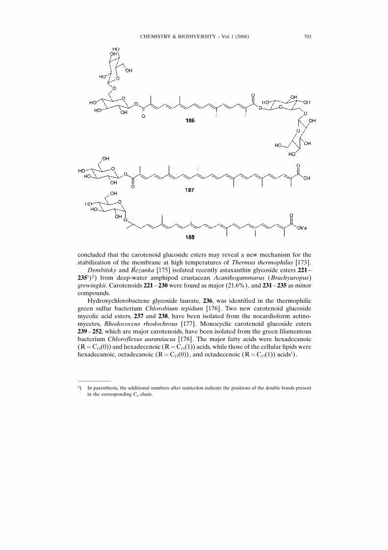
Crocin (186) as the major carotenoid was isolated from Crocus sativus by Karrer andSalomon in 1933 [150] [151].
�-�-glucoside 187 of the 4,4��-diapocarotene-6,6�-dioic acid was isolated fromPseudomonas rhodos and red strain Rhizobium, both from Lotononis bainesii B���[150] [152 ± 156]. An apolycopenoare mannosyl ester, 188, was found as a minorcomponent in yellow halophilic cocci strains H5B-2 and SE20-4 [155].Until now, seven crocetin derivatives, 189 ± 195, with different carbohydrate
moieties have been isolated from Crocus sativus and also from Gardenia jasminoidesand Nyctanthes arbortristis [150] [151] [156 ± 164].The unusual xanthone derivative termed mangicrocin (196), was isolated also from
Crocus sativus [165].Staphyloxanthin (197) has been isolated as the major pigment from Staphylococcus
aureus [166]. The carotenoid composition of the myxobacterium Chondromyces
CHEMISTRY & BIODIVERSITY ± Vol. 1 (2004)700
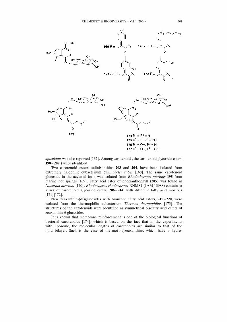
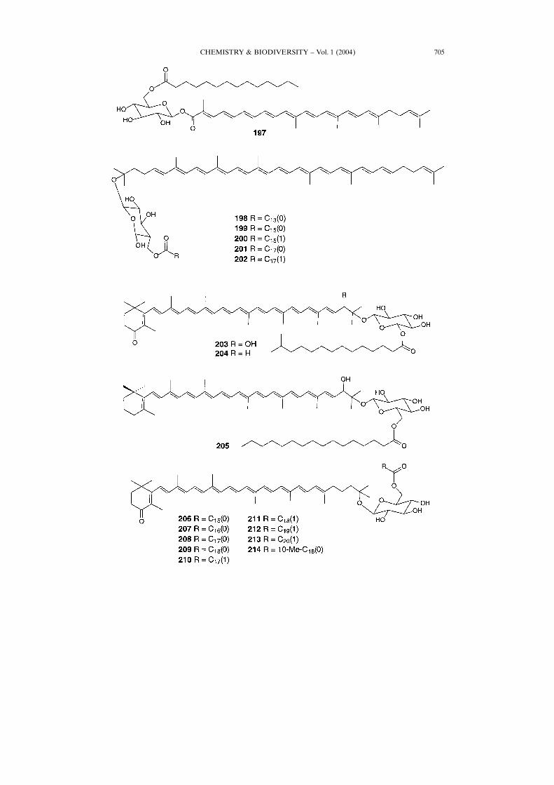
apiculatus was also reported [167]. Among carotenoids, the carotenoid glycoside esters198 ± 2021) were identified.Two carotenoid esters, salinixanthins 203 and 204, have been isolated from
extremely halophilic eubacterium Salinibacter ruber [168]. The same carotenoidglucoside in the acylated form was isolated from Rhodothermus marinus 195 frommarine hot springs [169]. Fatty acid ester of pheixanthophyll (205) was found inNocardia kirovani [170]. Rhodococcus rhodochrous RNMS1 (IAM 13988) contains aseries of carotenoid glycoside esters, 206 ± 214, with different fatty acid moieties[171] [172].New zeaxanthin-(di)glucosides with branched fatty acid esters, 215 ± 220, were
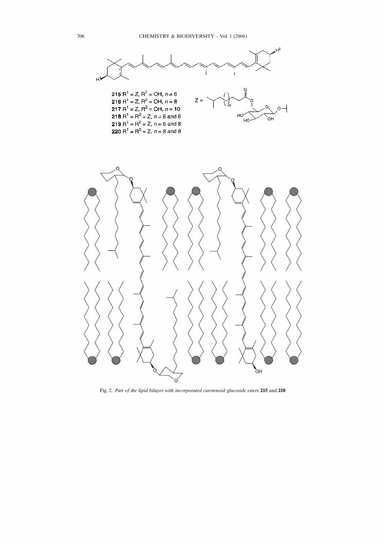
isolated from the thermophilic eubacterium Thermus thermophilus [173]. Thestructures of the carotenoids were identified as symmetrical bis-fatty acid esters ofzeaxanthin-�-glucosides.It is known that membrane reinforcement is one of the biological functions of
bacterial carotenoids [174], which is based on the fact that in the experimentswith liposome, the molecular lengths of carotenoids are similar to that of thelipid bilayer. Such is the case of thermo(bis)zeaxanthins, which have a hydro-
CHEMISTRY & BIODIVERSITY ± Vol. 1 (2004) 701
phobic�hydrophilic�hydrophobic structure, consisting of zeaxanthin, glucose, andbranched fatty acids. This suggests that the zeaxanthin moiety resides in the lipidbilayer, the glucose moiety resides at the surface of the membrane, and the branchedfatty acid moiety curls back into the lipid bilayer, like a −hook× (see Fig. 2). The authors
CHEMISTRY & BIODIVERSITY ± Vol. 1 (2004)702
concluded that the carotenoid glucoside esters may reveal a new mechanism for thestabilization of the membrane at high temperatures of Thermus thermophilus [173].
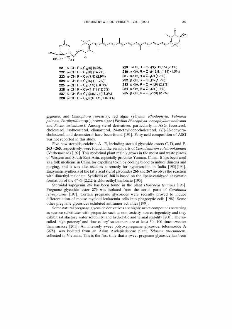
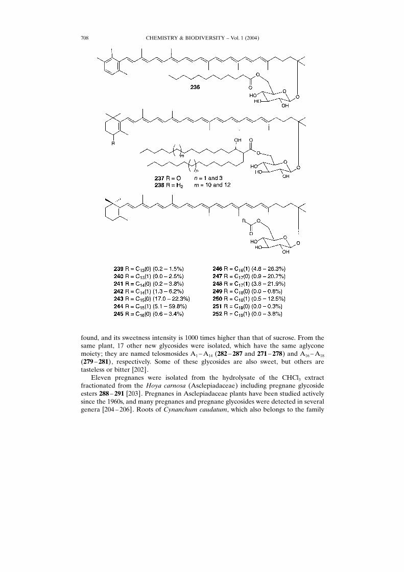
Dembitsky and Rœezanka [175] isolated recently astaxanthin glycoside esters 221 ±2351)2) from deep-water amphipod crustacean Acanthogammarus (Brachyuropus)grewingkii. Carotenoids 221 ± 230 were found as major (21.6%), and 231 ± 235 as minorcompounds.Hydroxychlorobactene glycoside laurate, 236, was identified in the thermophilic
green sulfur bacterium Chlorobium tepidum [176]. Two new carotenoid glucosidemycolic acid esters, 237 and 238, have been isolated from the nocardioform actino-mycetes, Rhodococcus rhodochrous [177]. Monocyclic carotenoid glucoside esters239 ± 252, which are major carotenoids, have been isolated from the green filamentousbacterium Chloroflexus aurantiacus [178]. The major fatty acids were hexadecanoic(R�C15(0)) and hexadecenoic (R�C15(1)) acids, while those of the cellular lipids werehexadecanoic, octadecanoic (R�C17(0)), and octadecenoic (R�C17(1)) acids1).
CHEMISTRY & BIODIVERSITY ± Vol. 1 (2004) 703
2) In parenthesis, the additional numbers after semicolon indicate the positions of the double bonds presentin the corresponding Cn chain.
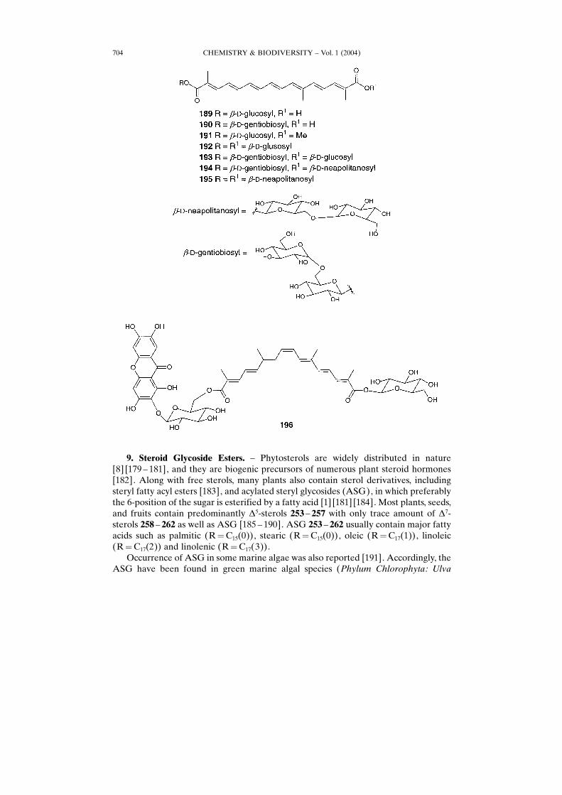
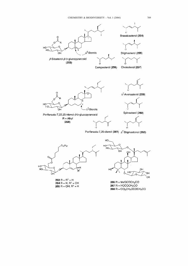
9. Steroid Glycoside Esters. ± Phytosterols are widely distributed in nature[8] [179 ± 181], and they are biogenic precursors of numerous plant steroid hormones[182]. Along with free sterols, many plants also contain sterol derivatives, includingsteryl fatty acyl esters [183], and acylated steryl glycosides (ASG), in which preferablythe 6-position of the sugar is esterified by a fatty acid [1] [181] [184]. Most plants, seeds,and fruits contain predominantly �5-sterols 253 ± 257 with only trace amount of �7-sterols 258 ± 262 as well as ASG [185 ± 190]. ASG 253 ± 262 usually contain major fattyacids such as palmitic (R�C15(0)), stearic (R�C15(0)), oleic (R�C17(1)), linoleic(R�C17(2)) and linolenic (R�C17(3)).Occurrence of ASG in some marine algae was also reported [191]. Accordingly, the
ASG have been found in green marine algal species (Phylum Chlorophyta: Ulva
CHEMISTRY & BIODIVERSITY ± Vol. 1 (2004)704
CHEMISTRY & BIODIVERSITY ± Vol. 1 (2004) 705
CHEMISTRY & BIODIVERSITY ± Vol. 1 (2004)706
Fig. 2. Part of the lipid bilayer with incorporated carotenoid glucoside esters 215 and 218
gigantea, and Cladophora rupestris), red algae (Phylum Rhodophyta: Palmariapalmata,Porphyridium sp.), brown algae (Phylum Phaeophyta: Ascophyllum nodosumand Fucus vesiculosus). Among sterol derivatives, particularly in ASG, fucosterol,cholesterol, isofucosterol, clionasterol, 24-methylidenecholesterol, (E)-22-dehydro-cholesterol, and desmosterol have been found [191]. Fatty acid composition of ASGwas not reported in this study.Five new steroids, colebrin A±E, including steroid glycoside esters C, D, and E,
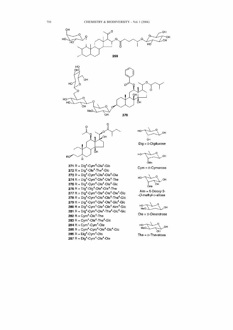
263 ± 265, respectively, were found in the aerial parts of Clerodendrum colebrookianum(Verbenaceae) [192]. This medicinal plant mainly grows in the moist and waste placesof Western and South-East Asia, especially province Yunnan, China. It has been usedas a folk medicine in China for expelling toxin by cooling blood to induce diuresis andpurging, and it was also used as a remedy for hypertension in India [193] [194].Enzymatic synthesis of the fatty acid sterol glycosides 266 and 267 involves the reactionwith dimethyl-malonate. Synthesis of 268 is based on the lipase-catalyzed enzymaticformation of the 6��-O-(2,2,2-trichloroethyl)malonate [195].Steroidal sapogenin 269 has been found in the plant Dioscorea tenuipes [196].
Pregnane glycoside ester 270 was isolated from the aerial parts of Carallumaretrospiciens [197]. Certain pregnane glycosides were recently proved to inducedifferentiation of mouse myeloid leukaemia cells into phagocytic cells [198]. Someother pregnane glycosides exhibited antitumor activities [199].Some natural pregnane glycoside derivatives are highly sweet compounds occurring
as sucrose substitutes with properties such as non-toxicity, non-cariogenicity and theyexhibit satisfactory water solubility, and hydrolytic and termal stability [200]. The so-called −high potency× and −low calory× sweeteners are at least 50 ± 100 times sweeterthan sucrose [201]. An intensely sweet polyoxypregnane glycoside, telosmoside A(278), was isolated from an Asian Asclepiadaceae plant, Telosma procumbens,collected in Vietnam. This is the first time that a sweet pregnane glycoside has been
CHEMISTRY & BIODIVERSITY ± Vol. 1 (2004) 707
found, and its sweetness intensity is 1000 times higher than that of sucrose. From thesame plant, 17 other new glycosides were isolated, which have the same aglyconemoiety; they are named telosmosides A1 ±A14 (282 ± 287 and 271 ± 278) and A16 ±A18(279 ± 281), respectively. Some of these glycosides are also sweet, but others aretasteless or bitter [202].Eleven pregnanes were isolated from the hydrolysate of the CHCl3 extract
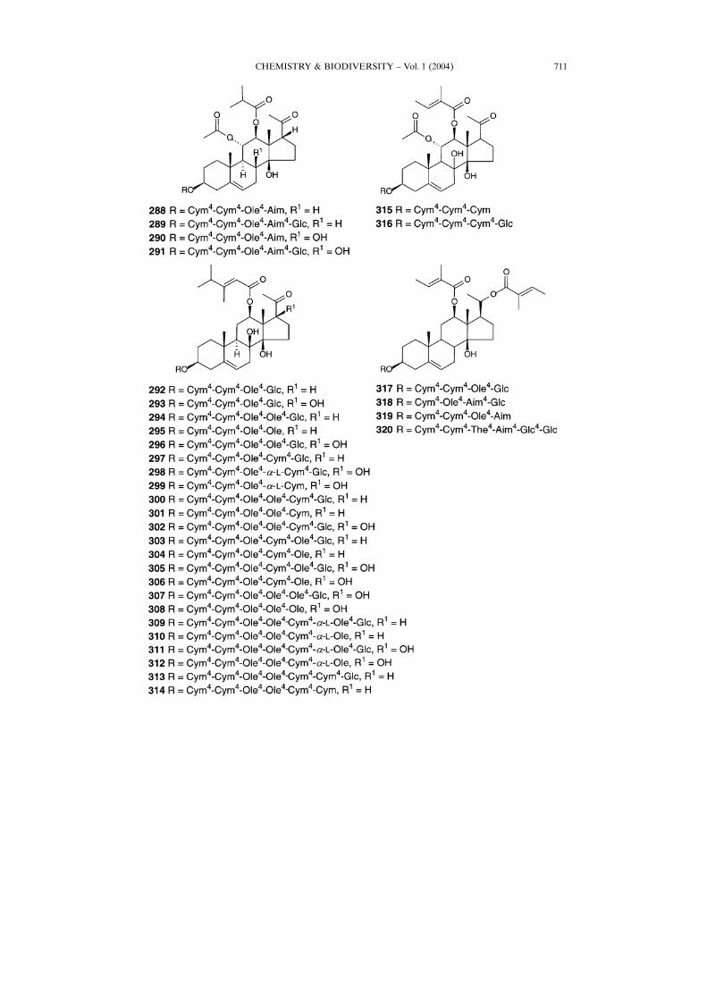
fractionated from the Hoya carnosa (Asclepiadaceae) including pregnane glycosideesters 288 ± 291 [203]. Pregnanes in Asclepiadaceae plants have been studied activelysince the 1960s, and many pregnanes and pregnane glycosides were detected in severalgenera [204 ± 206]. Roots of Cynanchum caudatum, which also belongs to the family
CHEMISTRY & BIODIVERSITY ± Vol. 1 (2004)708
CHEMISTRY & BIODIVERSITY ± Vol. 1 (2004) 709
CHEMISTRY & BIODIVERSITY ± Vol. 1 (2004)710
CHEMISTRY & BIODIVERSITY ± Vol. 1 (2004) 711
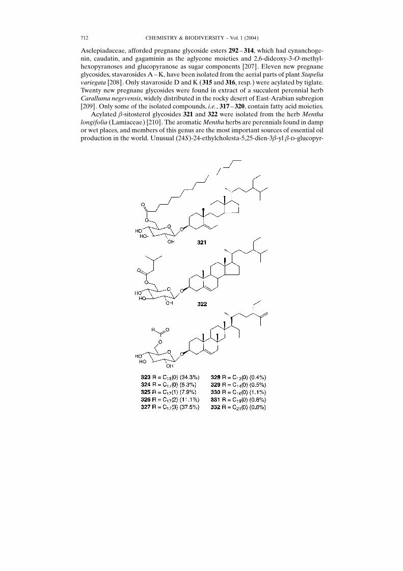
Asclepiadaceae, afforded pregnane glycoside esters 292 ± 314, which had cynanchoge-nin, caudatin, and gagaminin as the aglycone moieties and 2,6-dideoxy-3-O-methyl-hexopyranoses and glucopyranose as sugar components [207]. Eleven new pregnaneglycosides, stavarosides A±K, have been isolated from the aerial parts of plant Stapeliavariegata [208]. Only stavaroside D and K (315 and 316, resp.) were acylated by tiglate.Twenty new pregnane glycosides were found in extract of a succulent perennial herbCaralluma negevensis, widely distributed in the rocky desert of East-Arabian subregion[209]. Only some of the isolated compounds, i.e., 317 ± 320, contain fatty acid moieties.Acylated �-sitosterol glycosides 321 and 322 were isolated from the herb Mentha
longifolia (Lamiaceae) [210]. The aromaticMentha herbs are perennials found in dampor wet places, and members of this genus are the most important sources of essential oilproduction in the world. Unusual (24S)-24-ethylcholesta-5,25-dien-3�-yl �-�-glucopyr-
CHEMISTRY & BIODIVERSITY ± Vol. 1 (2004)712
anoside fatty acid esters 323 ± 3321) have been isolated from plant Teucrium fruticans(Labitae) [211]. GC/MS Analysis showed palmitic, steatic, oleic, linoleic, and linolenicacids as major acids.9.1. Acylated Saponins. Saponins are a class of natural compounds, which are
structurally described as glycosylated triterpenoids or steroids. Because of theglycosylation of the hydrophobic aglycons, they are biological detergents and, whenagitated in H2O, form a soapy lather, which gives rise to the name of the group ofcompounds. Triterpenoid and steroid glycosides are widely spread throughout the plantkingdom, and several have been found in marine animals. Some saponins have cardiacactivity, hemolytic activity, activity as fish poisons, cholesterol-reducing ability,bitterness, ability to act as sweeteners, and cosmetics activity, and can also serve asallelochemicals; these are features found in certain saponins rather than in all membersof this chemical family. From a biological point of view, saponins have diverse groupproperties, some deleterious but many beneficial. Their use in plant drugs, folkmedicines, etc., has generated great interest in the chemical characterization of thesecompounds. In the field of vaccine research, saponins from the South American treeQuillaja saponaria M���� are known as potent immunostimulants with a uniqueadjuvant activity [1] [196] [212]. Saponins belong to the group of soap-like naturalsurfactants. Steroidal saponins are found mainly in plants of Monocotyledoneae(Agavaceae, Amaryllidaceae, Dioscoreaceae, Gramineae, Liliaceae, Palmae) and alsoin Dicotyledoneae (Leguminosae, Solanaceae, Scrophurariaceae, and Zygophyllaceae)[213]. Some novel acylated saponins have been isolated from nature. Acyl sterolglycosides has been partly reviewed in [214].An interesting finding was reported byMuhr and co-workers [215]. They found that
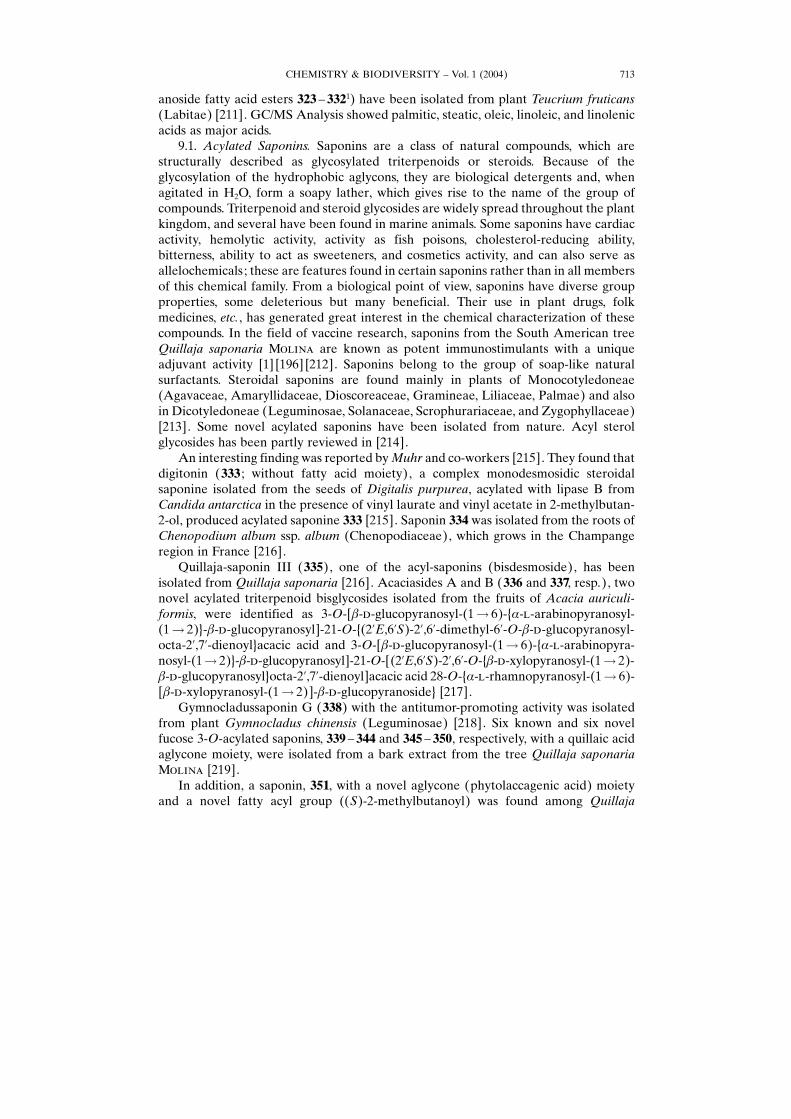
digitonin (333 ; without fatty acid moiety), a complex monodesmosidic steroidalsaponine isolated from the seeds of Digitalis purpurea, acylated with lipase B fromCandida antarctica in the presence of vinyl laurate and vinyl acetate in 2-methylbutan-2-ol, produced acylated saponine 333 [215]. Saponin 334 was isolated from the roots ofChenopodium album ssp. album (Chenopodiaceae), which grows in the Champangeregion in France [216].Quillaja-saponin III (335), one of the acyl-saponins (bisdesmoside), has been
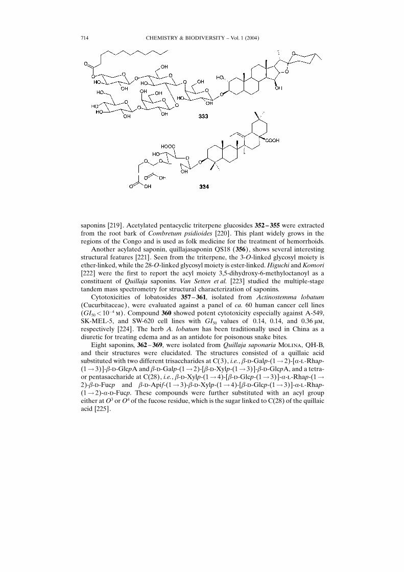
isolated from Quillaja saponaria [216]. Acaciasides A and B (336 and 337, resp.), twonovel acylated triterpenoid bisglycosides isolated from the fruits of Acacia auriculi-formis, were identified as 3-O-[�-�-glucopyranosyl-(1� 6)-{�-�-arabinopyranosyl-(1� 2)}-�-�-glucopyranosyl]-21-O-{(2�E,6�S)-2�,6�-dimethyl-6�-O-�-�-glucopyranosyl-octa-2�,7�-dienoyl}acacic acid and 3-O-[�-�-glucopyranosyl-(1� 6)-{�-�-arabinopyra-nosyl-(1� 2)}-�-�-glucopyranosyl]-21-O-[(2�E,6�S)-2�,6�-O-{�-�-xylopyranosyl-(1� 2)-�-�-glucopyranosyl}octa-2�,7�-dienoyl]acacic acid 28-O-{�-�-rhamnopyranosyl-(1� 6)-[�-�-xylopyranosyl-(1� 2)]-�-�-glucopyranoside} [217].Gymnocladussaponin G (338) with the antitumor-promoting activity was isolated
from plant Gymnocladus chinensis (Leguminosae) [218]. Six known and six novelfucose 3-O-acylated saponins, 339 ± 344 and 345 ± 350, respectively, with a quillaic acidaglycone moiety, were isolated from a bark extract from the tree Quillaja saponariaM���� [219].In addition, a saponin, 351, with a novel aglycone (phytolaccagenic acid) moiety
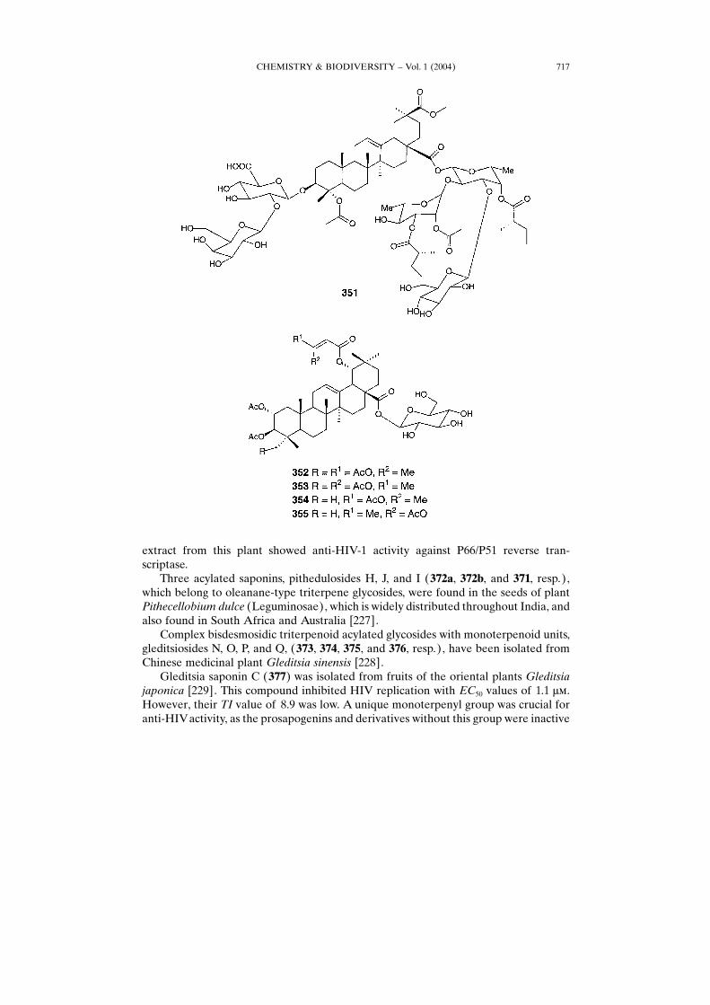
and a novel fatty acyl group ((S)-2-methylbutanoyl) was found among Quillaja
CHEMISTRY & BIODIVERSITY ± Vol. 1 (2004) 713
saponins [219]. Acetylated pentacyclic triterpene glucosides 352 ± 355 were extractedfrom the root bark of Combretum psidioides [220]. This plant widely grows in theregions of the Congo and is used as folk medicine for the treatment of hemorrhoids.Another acylated saponin, quillajasaponin QS18 (356), shows several interesting
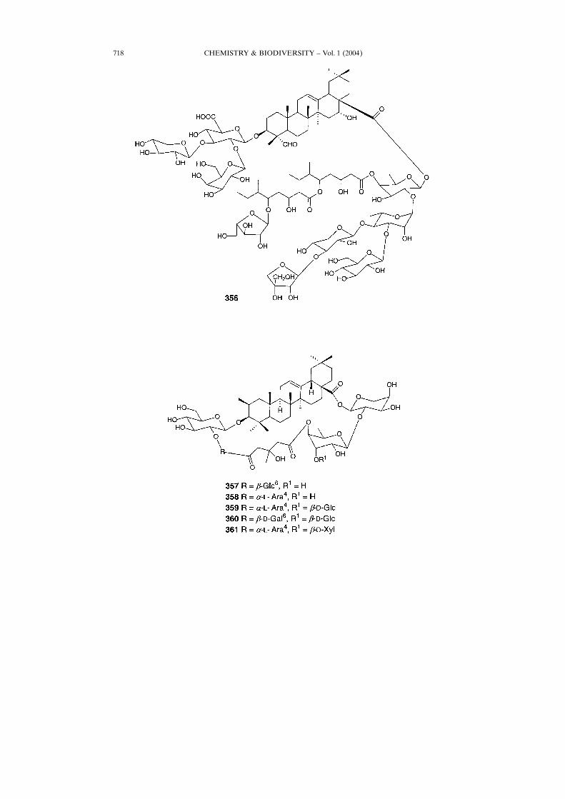
structural features [221]. Seen from the triterpene, the 3-O-linked glycosyl moiety isether-linked, while the 28-O-linked glycosyl moiety is ester-linked.Higuchi andKomori[222] were the first to report the acyl moiety 3,5-dihydroxy-6-methyloctanoyl as aconstituent of Quillaja saponins. Van Setten et al. [223] studied the multiple-stagetandem mass spectrometry for structural characterization of saponins.Cytotoxicities of lobatosides 357 ± 361, isolated from Actinostemma lobatum
(Cucurbitaceae), were evaluated against a panel of ca. 60 human cancer cell lines(GI50� 10�4 �). Compound 360 showed potent cytotoxicity especially against A-549,SK-MEL-5, and SW-620 cell lines with GI50 values of 0.14, 0.14, and 0.36 ��,respectively [224]. The herb A. lobatum has been traditionally used in China as adiuretic for treating edema and as an antidote for poisonous snake bites.Eight saponins, 362 ± 369, were isolated from Quillaja saponaria M����, QH-B,
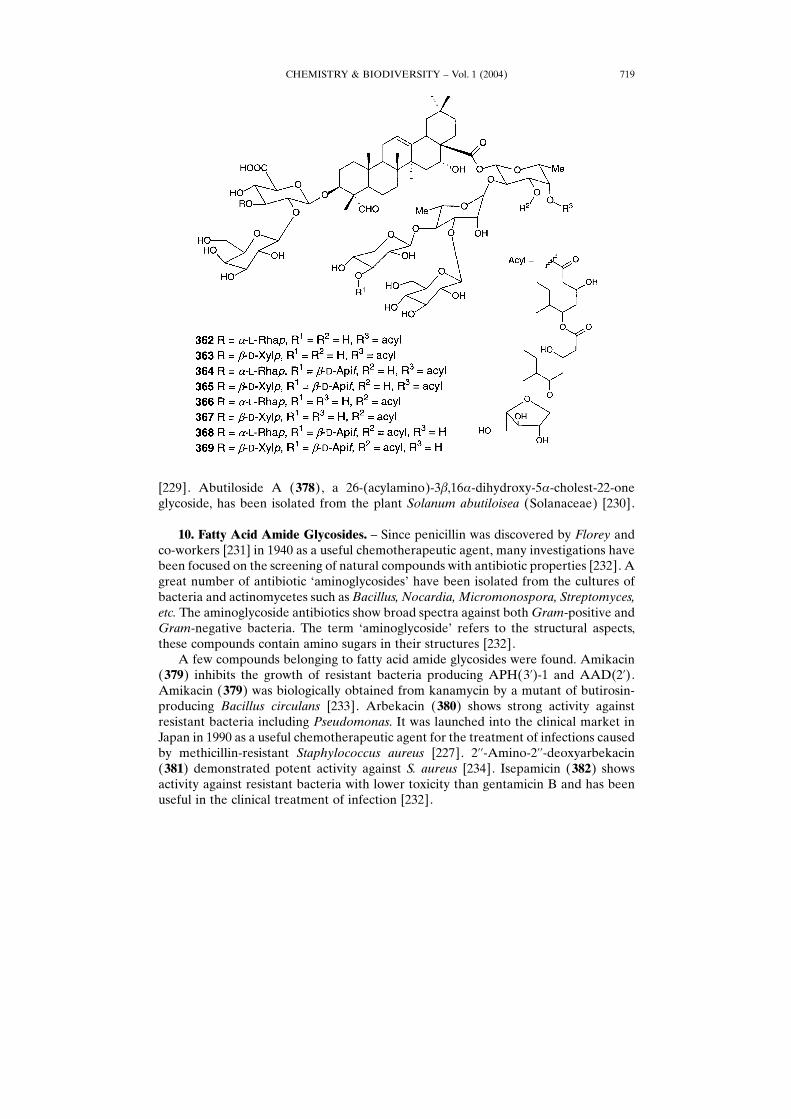
and their structures were elucidated. The structures consisted of a quillaic acidsubstituted with two different trisaccharides at C(3), i.e., �-�-Galp-(1� 2)-[�-�-Rhap-(1� 3)]-�-�-GlcpA and �-�-Galp-(1� 2)-[�-�-Xylp-(1� 3)]-�-�-GlcpA, and a tetra-or pentasaccharide at C(28), i.e., �-�-Xylp-(1� 4)-[�-�-Glcp-(1� 3)]-�-�-Rhap-(1�2)-�-�-Fucp and �-�-Apif-(1� 3)-�-�-Xylp-(1� 4)-[�-�-Glcp-(1� 3)]-� �-Rhap-(1� 2)-�-�-Fucp. These compounds were further substituted with an acyl groupeither atO3 orO4 of the fucose residue, which is the sugar linked to C(28) of the quillaicacid [225].
CHEMISTRY & BIODIVERSITY ± Vol. 1 (2004)714
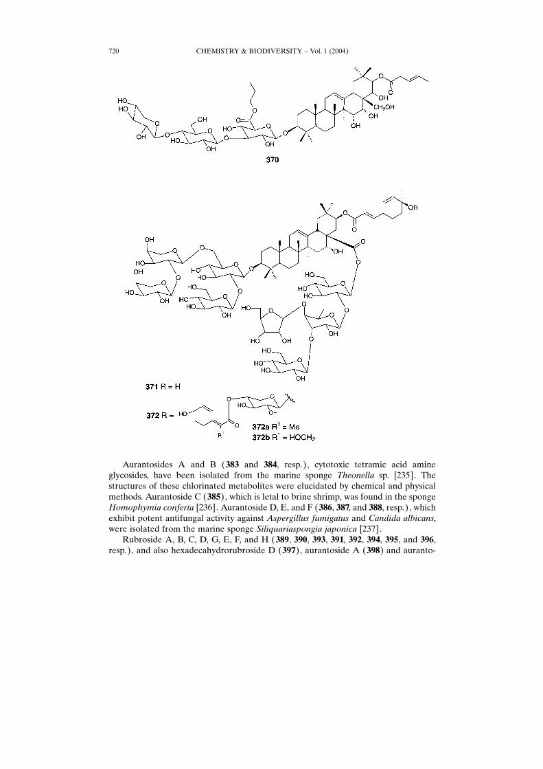
A new triterpene saponin glycoside, 21�-(angeloyloxy)-3-O-[�-�-arabinopyrano-syl-(1� 4)-�-�-glucopyranosyl-(1� 3)-�-�-glucuronopyranosyl propyl ester]-3�,15,16,22�,28�-pentahydroxy-�12-oleanene, named as saniculoside (370), has beenisolated from the aerial parts of the medicinal plant Sanicula europaea [226]. Crude
CHEMISTRY & BIODIVERSITY ± Vol. 1 (2004) 715

CHEMISTRY & BIODIVERSITY ± Vol. 1 (2004)716
extract from this plant showed anti-HIV-1 activity against P66/P51 reverse tran-scriptase.Three acylated saponins, pithedulosides H, J, and I (372a, 372b, and 371, resp.),
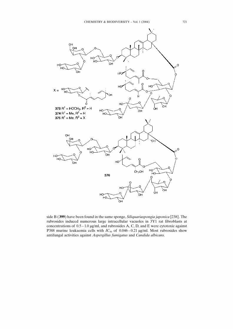
which belong to oleanane-type triterpene glycosides, were found in the seeds of plantPithecellobium dulce (Leguminosae), which is widely distributed throughout India, andalso found in South Africa and Australia [227].Complex bisdesmosidic triterpenoid acylated glycosides with monoterpenoid units,
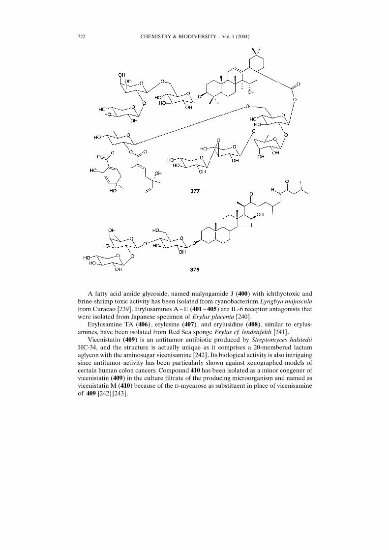
gleditsiosides N, O, P, and Q, (373, 374, 375, and 376, resp.), have been isolated fromChinese medicinal plant Gleditsia sinensis [228].Gleditsia saponin C (377) was isolated from fruits of the oriental plants Gleditsia
japonica [229]. This compound inhibited HIV replication with EC50 values of 1.1 ��.However, their TI value of 8.9 was low. A unique monoterpenyl group was crucial foranti-HIVactivity, as the prosapogenins and derivatives without this group were inactive
CHEMISTRY & BIODIVERSITY ± Vol. 1 (2004) 717
CHEMISTRY & BIODIVERSITY ± Vol. 1 (2004)718
[229]. Abutiloside A (378), a 26-(acylamino)-3�,16�-dihydroxy-5�-cholest-22-oneglycoside, has been isolated from the plant Solanum abutiloisea (Solanaceae) [230].
10. Fatty Acid Amide Glycosides. ± Since penicillin was discovered by Florey andco-workers [231] in 1940 as a useful chemotherapeutic agent, many investigations havebeen focused on the screening of natural compounds with antibiotic properties [232]. Agreat number of antibiotic −aminoglycosides× have been isolated from the cultures ofbacteria and actinomycetes such as Bacillus, Nocardia, Micromonospora, Streptomyces,etc. The aminoglycoside antibiotics show broad spectra against bothGram-positive andGram-negative bacteria. The term −aminoglycoside× refers to the structural aspects,these compounds contain amino sugars in their structures [232].A few compounds belonging to fatty acid amide glycosides were found. Amikacin
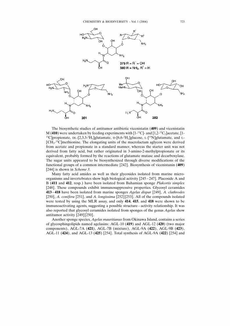
(379) inhibits the growth of resistant bacteria producing APH(3�)-1 and AAD(2�).Amikacin (379) was biologically obtained from kanamycin by a mutant of butirosin-producing Bacillus circulans [233]. Arbekacin (380) shows strong activity againstresistant bacteria including Pseudomonas. It was launched into the clinical market inJapan in 1990 as a useful chemotherapeutic agent for the treatment of infections causedby methicillin-resistant Staphylococcus aureus [227]. 2��-Amino-2��-deoxyarbekacin(381) demonstrated potent activity against S. aureus [234]. Isepamicin (382) showsactivity against resistant bacteria with lower toxicity than gentamicin B and has beenuseful in the clinical treatment of infection [232].
CHEMISTRY & BIODIVERSITY ± Vol. 1 (2004) 719
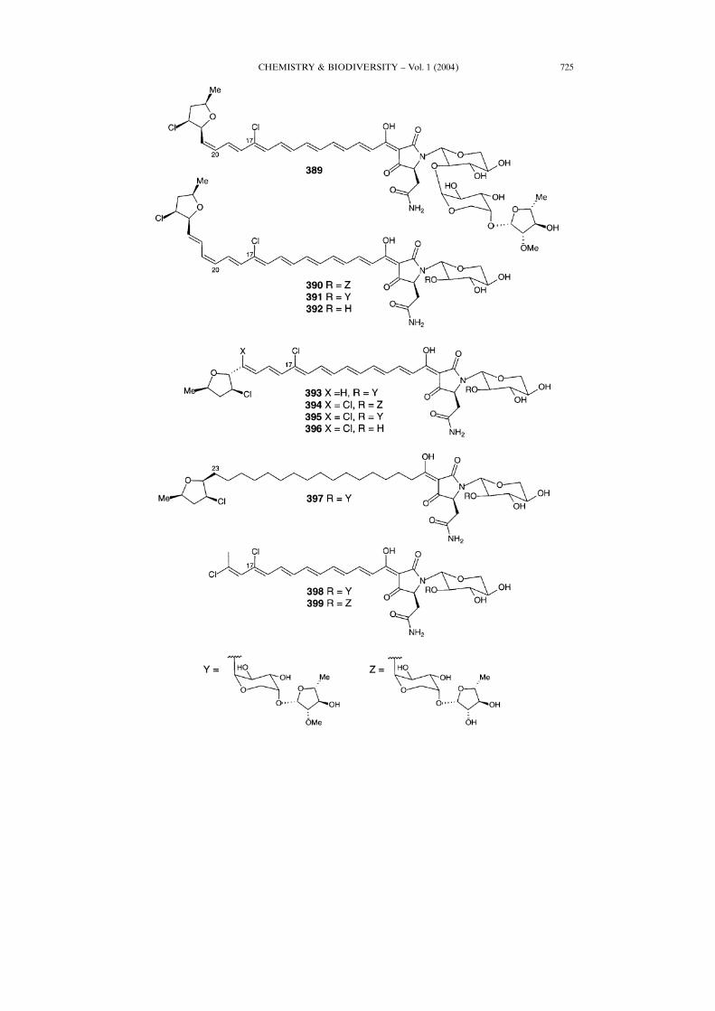
Aurantosides A and B (383 and 384, resp.), cytotoxic tetramic acid amineglycosides, have been isolated from the marine sponge Theonella sp. [235]. Thestructures of these chlorinated metabolites were elucidated by chemical and physicalmethods. Aurantoside C (385), which is letal to brine shrimp, was found in the spongeHomophymia conferta [236]. Aurantoside D, E, and F (386, 387, and 388, resp.), whichexhibit potent antifungal activity against Aspergillus fumigatus and Candida albicans,were isolated from the marine sponge Siliquariaspongia japonica [237].Rubroside A, B, C, D, G, E, F, and H (389, 390, 393, 391, 392, 394, 395, and 396,
resp.), and also hexadecahydrorubroside D (397), aurantoside A (398) and auranto-
CHEMISTRY & BIODIVERSITY ± Vol. 1 (2004)720
side B (399) have been found in the same sponge, Siliquariaspongia japonica [238]. Therubrosides induced numerous large intracellular vacuoles in 3Y1 rat fibroblasts atconcentrations of 0.5 ± 1.0 �g/ml, and rubrosides A, C, D, and E were cytotoxic againstP388 murine leukaemia cells with IC50 of 0.046 ± 0.21 �g/ml. Most rubrosides showantifungal activities against Aspergillus fumigatus and Candida albicans.
CHEMISTRY & BIODIVERSITY ± Vol. 1 (2004) 721
A fatty acid amide glycoside, named malyngamide J (400) with ichthyotoxic andbrine-shrimp toxic activity has been isolated from cyanobacterium Lyngbya majusculafrom Curacao [239]. Erylusamines A±E (401 ± 405) are IL-6 receptor antagonists thatwere isolated from Japanese specimen of Erylus placenta [240].Erylusamine TA (406), erylusine (407), and erylusidine (408), similar to erylus-
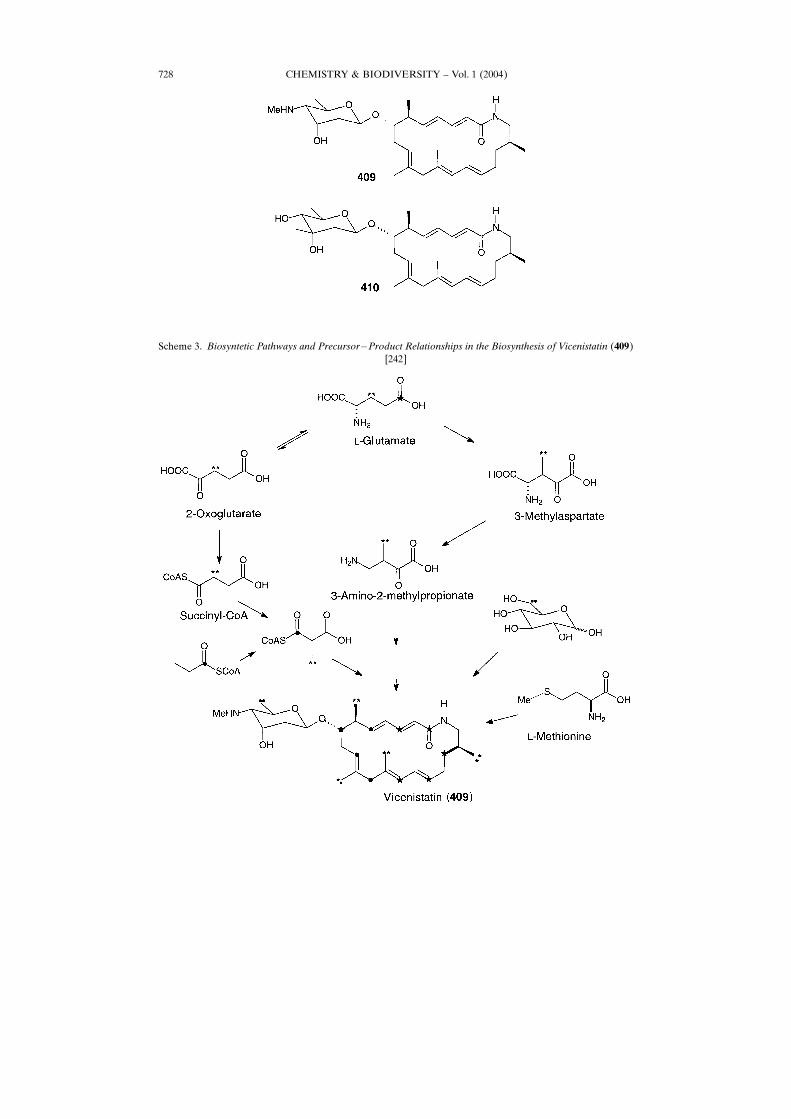
amines, have been isolated from Red Sea sponge Erylus cf. lendenfeldi [241].Vicenistatin (409) is an antitumor antibiotic produced by Streptomyces halstedii
HC-34, and the structure is actually unique as it comprises a 20-membered lactamaglycon with the aminosugar vicenisamine [242]. Its biological activity is also intriguingsince antitumor activity has been particularly shown against xenographed models ofcertain human colon cancers. Compound 410 has been isolated as a minor congener ofvicenistatin (409) in the culture filtrate of the producing microorganism and named asvicenistatin M (410) because of the �-mycarose as substituent in place of vicenisamineof 409 [242] [243].
CHEMISTRY & BIODIVERSITY ± Vol. 1 (2004)722
The biosynthetic studies of antitumor antibiotic vicenistatin (409) and vicenistatinM (410) were undertaken by feeding experiments with [1-13C]- and [1,2-13C2]acetate, [1-13C]propionate, ��-[2,3,3-2H3]glutamate, �-[6,6-2H2]glucose, �-[15N]glutamate, and �-[CH3-13C]methionine. The elongating units of the macrolactam aglycon were derivedfrom acetate and propionate in a standard manner, whereas the starter unit was notderived from fatty acid, but rather originated in 3-amino-2-methylpropionate or itsequivalent, probably formed by the reactions of glutamate mutase and decarboxylase.The sugar units appeared to be biosynthesized through diverse modifications of thefunctional groups of a common intermediate [242]. Biosynthesis of vicenistanin (409)[244] is shown in Scheme 3.Many fatty acid amides as well as their glycosides isolated from marine micro-
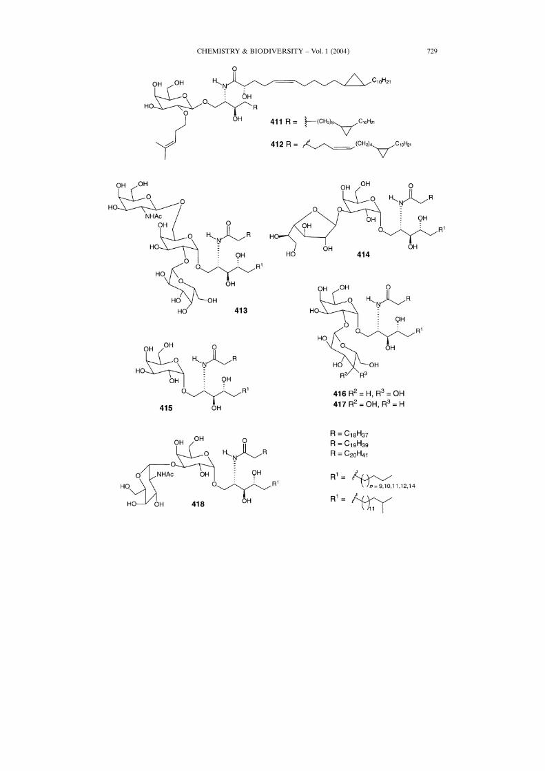
organisms and invertebrates show high biological activity [245 ± 247]. Placoside A andB (411 and 412, resp.) have been isolated from Bahamian sponge Plakortis simplex[248]. These compounds exhibit immunosuppressive properties. Glycosyl ceramides413 ± 418 have been isolated from marine sponges Agelas dispar [249], A. clathrodes[250], A. conifera [251], and A. longissima [252] [253]. All of the compounds isolatedwere tested by using the MLR assay, and only 414, 415, and 418 were shown to beimmunoactivating agents, suggesting a possible structure�activity relationship. It wasalso reported that glycosyl ceramides isolated from sponges of the genus Agelas showantitumor activity [249] [250].Another sponge species,Agelas mauritianus from Okinawa Island, contains a series
of glycosphingolipids named agelasins: AGL-10 (419) and AGL-12 (420) (two majorcomponents), AGL-7A (421), AGL-7B (mixture), AGL-9A (422), AGL-9B (423),AGL-11 (424), and AGL-13 (425) [254]. Total synthesis of AGL-9A (422) [254] and
CHEMISTRY & BIODIVERSITY ± Vol. 1 (2004) 723
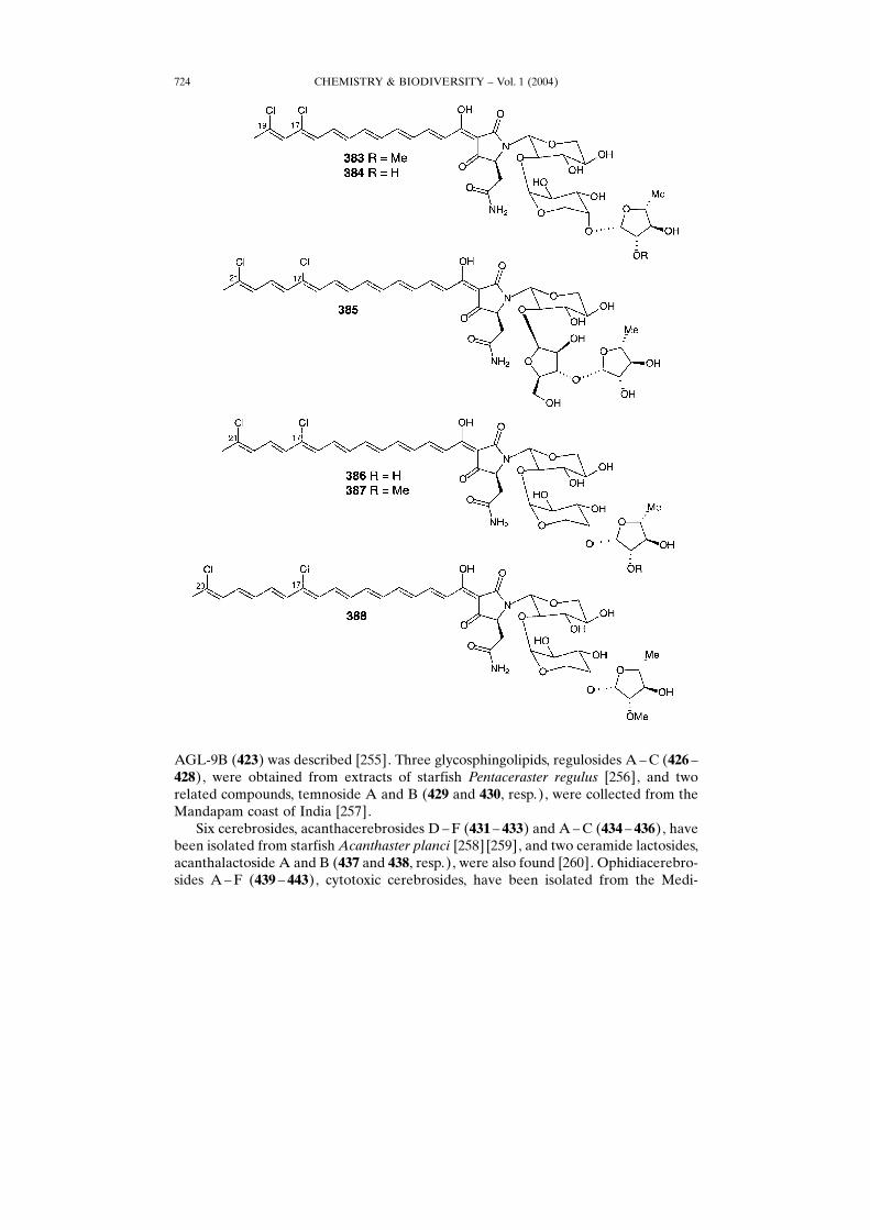
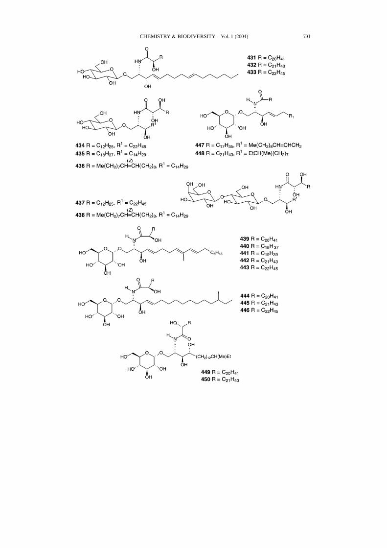
AGL-9B (423) was described [255]. Three glycosphingolipids, regulosides A ±C (426 ±428), were obtained from extracts of starfish Pentaceraster regulus [256], and tworelated compounds, temnoside A and B (429 and 430, resp.), were collected from theMandapam coast of India [257].Six cerebrosides, acanthacerebrosides D±F (431 ± 433) and A±C (434 ± 436), have
been isolated from starfish Acanthaster planci [258] [259], and two ceramide lactosides,acanthalactoside A and B (437 and 438, resp.), were also found [260]. Ophidiacerebro-sides A±F (439 ± 443), cytotoxic cerebrosides, have been isolated from the Medi-
CHEMISTRY & BIODIVERSITY ± Vol. 1 (2004)724
CHEMISTRY & BIODIVERSITY ± Vol. 1 (2004) 725
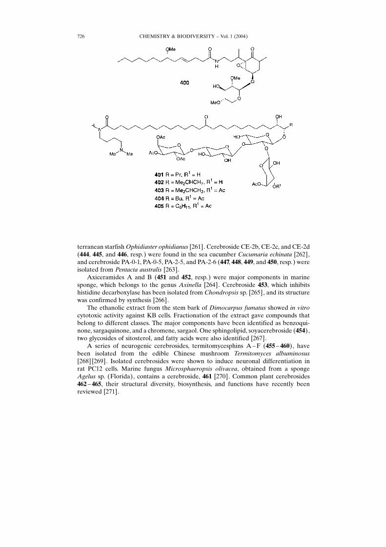
terranean starfishOphidiaster ophidianus [261]. Cerebroside CE-2b, CE-2c, and CE-2d(444, 445, and 446, resp.) were found in the sea cucumber Cucumaria echinata [262],and cerebroside PA-0-1, PA-0-5, PA-2-5, and PA-2-6 (447, 448, 449, and 450, resp.) wereisolated from Pentacta australis [263].Axiceramides A and B (451 and 452, resp.) were major components in marine
sponge, which belongs to the genus Axinella [264]. Cerebroside 453, which inhibitshistidine decarboxylase has been isolated from Chondropsis sp. [265], and its structurewas confirmed by synthesis [266].The ethanolic extract from the stem bark of Dimocarpus fumatus showed in vitro
cytotoxic activity against KB cells. Fractionation of the extract gave compounds thatbelong to different classes. The major components have been identified as benzoqui-none, sargaquinone, and a chromene, sargaol. One sphingolipid, soyacerebroside (454),two glycosides of sitosterol, and fatty acids were also identified [267].A series of neurogenic cerebrosides, termitomycesphins A ±F (455 ± 460), have
been isolated from the edible Chinese mushroom Termitomyces albuminosus[268] [269]. Isolated cerebrosides were shown to induce neuronal differentiation inrat PC12 cells. Marine fungus Microsphaeropsis olivacea, obtained from a spongeAgelus sp. (Florida), contains a cerebroside, 461 [270]. Common plant cerebrosides462 ± 465, their structural diversity, biosynthesis, and functions have recently beenreviewed [271].
CHEMISTRY & BIODIVERSITY ± Vol. 1 (2004)726
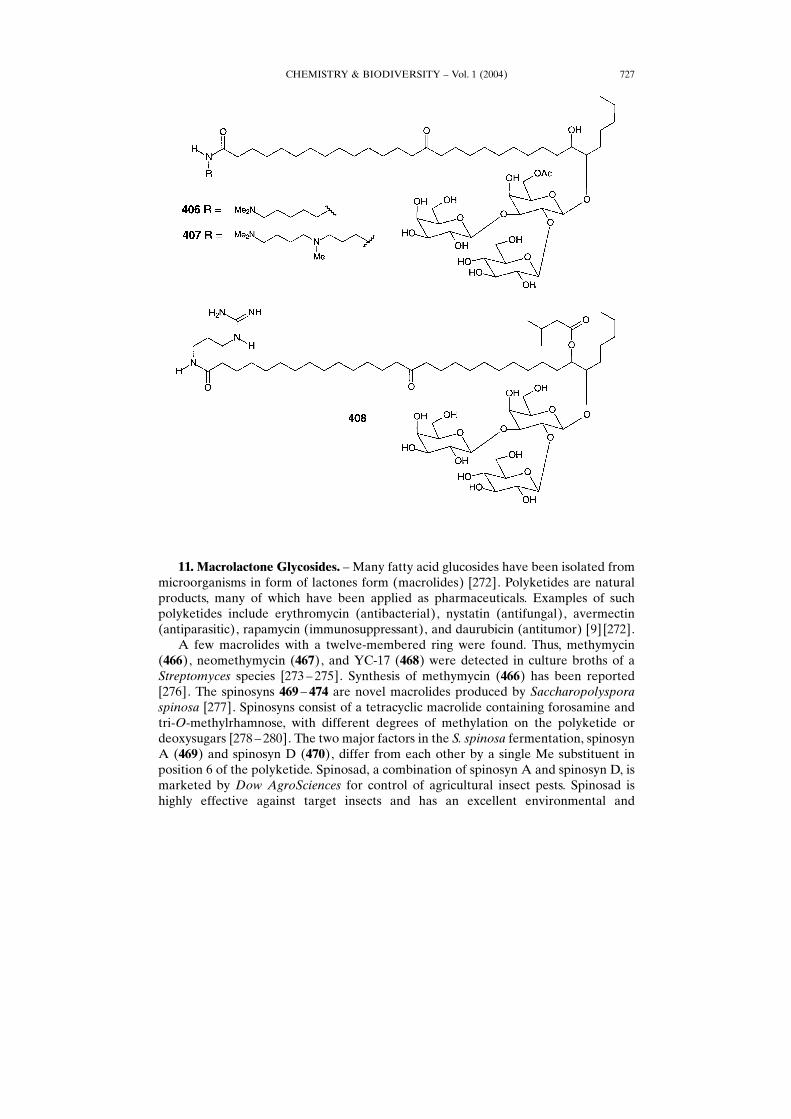
11. Macrolactone Glycosides. ± Many fatty acid glucosides have been isolated frommicroorganisms in form of lactones form (macrolides) [272]. Polyketides are naturalproducts, many of which have been applied as pharmaceuticals. Examples of suchpolyketides include erythromycin (antibacterial), nystatin (antifungal), avermectin(antiparasitic), rapamycin (immunosuppressant), and daurubicin (antitumor) [9] [272].A few macrolides with a twelve-membered ring were found. Thus, methymycin
(466), neomethymycin (467), and YC-17 (468) were detected in culture broths of aStreptomyces species [273 ± 275]. Synthesis of methymycin (466) has been reported[276]. The spinosyns 469 ± 474 are novel macrolides produced by Saccharopolysporaspinosa [277]. Spinosyns consist of a tetracyclic macrolide containing forosamine andtri-O-methylrhamnose, with different degrees of methylation on the polyketide ordeoxysugars [278 ± 280]. The two major factors in the S. spinosa fermentation, spinosynA (469) and spinosyn D (470), differ from each other by a single Me substituent inposition 6 of the polyketide. Spinosad, a combination of spinosyn A and spinosyn D, ismarketed by Dow AgroSciences for control of agricultural insect pests. Spinosad ishighly effective against target insects and has an excellent environmental and
CHEMISTRY & BIODIVERSITY ± Vol. 1 (2004) 727
CHEMISTRY & BIODIVERSITY ± Vol. 1 (2004)728
Scheme 3. Biosyntetic Pathways and Precursor�Product Relationships in the Biosynthesis of Vicenistatin (409)[242]
CHEMISTRY & BIODIVERSITY ± Vol. 1 (2004) 729
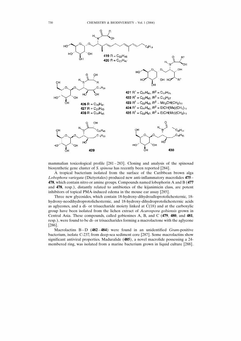
mammalian toxicological profile [281 ± 283]. Cloning and analysis of the spinosadbiosynthetic gene cluster of S. spinosa has recently been reported [284].A tropical bacterium isolated from the surface of the Caribbean brown alga
Lobophora variegata (Dictyotales) produced new anti-inflammatory macrolides 475 ±478, which contain nitro or amine groups. Compounds named lobophorin A and B (477and 478, resp.), distantly related to antibiotics of the kijanimicin class, are potentinhibitors of topical PMA-induced edema in the mouse ear assay [285].Three new glycosides, which contain 18-hydroxy-dihydroalloprotolichesternic, 18-
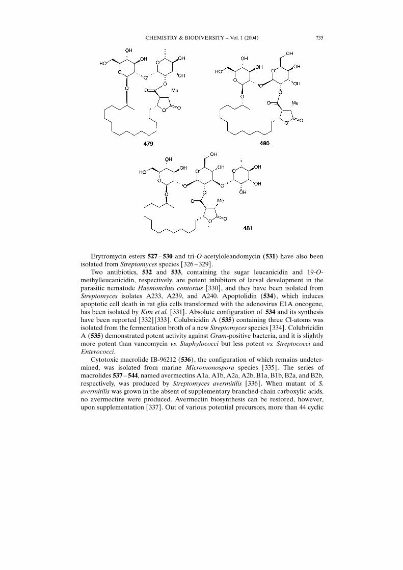
hydroxy-neodihydroprotolichesternic, and 18-hydroxy-dihydroprotolichesternic acidsas aglycones, and a di- or trisaccharide moiety linked at C(18) and at the carboxylicgroup have been isolated from the lichen extract of Acarospora gobiensis grown inCentral Asia. These compounds, called gobienines A, B, and C (479, 480, and 481,resp.), were found to be di- or trisaccharides forming a macrolactone with the aglycone[286].Macrolactins B ±D (482 ± 484) were found in an unidentified Gram-positive
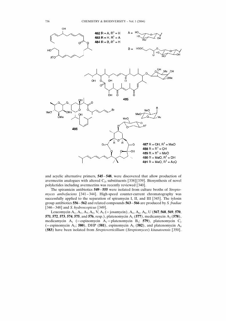
bacterium, isolate C-237, from deep-sea sediment core [287]. Some macrolactins showsignificant antiviral properties. Maduralide (485), a novel macrolide possessing a 24-membered ring, was isolated from a marine bacterium grown in liquid culture [288].
CHEMISTRY & BIODIVERSITY ± Vol. 1 (2004)730
CHEMISTRY & BIODIVERSITY ± Vol. 1 (2004) 731
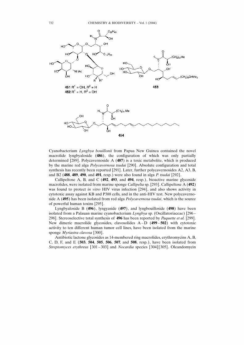
Cyanobacterium Lyngbya bouillonii from Papua New Guinea contained the novelmacrolide lyngbyaloside (486), the configuration of which was only partiallydetermined [289]. Polycavernoside A (487) is a toxic metabolite, which is producedby the marine red alga Polycavernosa tsudai [290]. Absolute configuration and totalsynthesis has recently been reported [291]. Later, further polycavernosides A2, A3, B,and B2 (488, 489, 490, and 491, resp.) were also found in alga P. tsudai [292].Callipeltose A, B, and C (492, 493, and 494, resp.), bioactive marine glycoside
macrolides, were isolated from marine sponge Callipelta sp. [293]. Callipeltose A (492)was found to protect in vitro HIV virus infection [294], and also shows activity incytotoxic assay against KB and P388 cells, and in the anti-HIV test. New polycaverno-side A (495) has been isolated from red alga Polycavernosa tsudai, which is the sourceof powerful human toxins [295].Lyngbyaloside B (496), lyngyaside (497), and lyngbouilloside (498) have been
isolated from a Palauan marine cyanobacterium Lyngbya sp. (Oscillatoriaceae) [296 ±298]. Stereoselective total synthesis of 496 has been reported by Paquette et al. [299].New dimeric macrolide glycosides, clavosolides A±D (499 ± 502) with cytotoxicactivity to ten different human tumor cell lines, have been isolated from the marinesponge Myriastra clavosa [300].Antibiotic lactone glycosides as 14-membered ring macrolides, erythromycins A, B,
C, D, F, and E (503, 504, 505, 506, 507, and 508, resp.), have been isolated fromStreptomyces erythreus [301 ± 303] and Nocardia species [304] [305]. Oleandomycin
CHEMISTRY & BIODIVERSITY ± Vol. 1 (2004)732
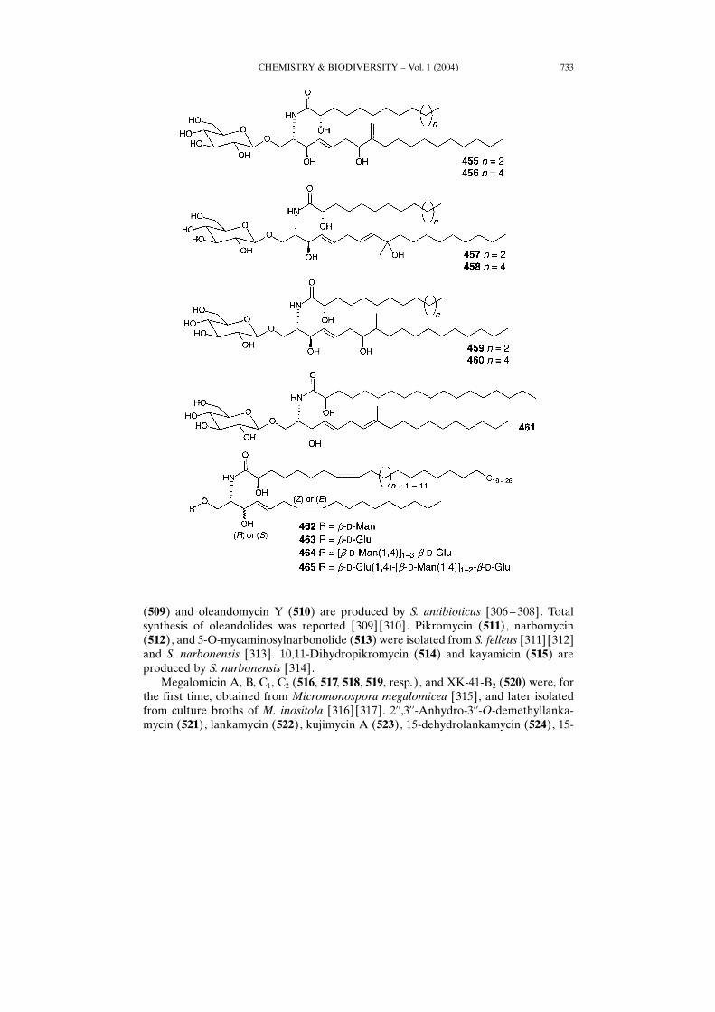
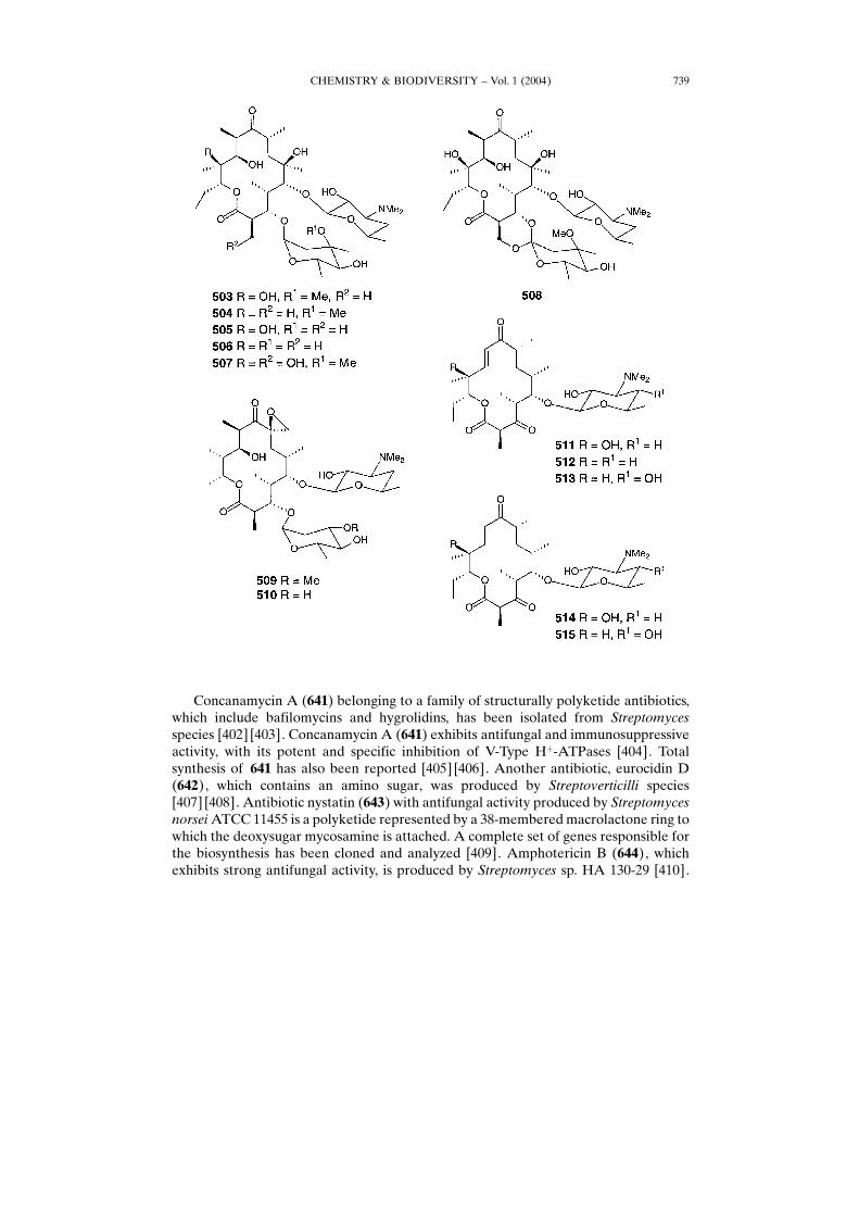
(509) and oleandomycin Y (510) are produced by S. antibioticus [306 ± 308]. Totalsynthesis of oleandolides was reported [309] [310]. Pikromycin (511), narbomycin(512), and 5-O-mycaminosylnarbonolide (513) were isolated from S. felleus [311] [312]and S. narbonensis [313]. 10,11-Dihydropikromycin (514) and kayamicin (515) areproduced by S. narbonensis [314].Megalomicin A, B, C1, C2 (516, 517, 518, 519, resp.), and XK-41-B2 (520) were, for
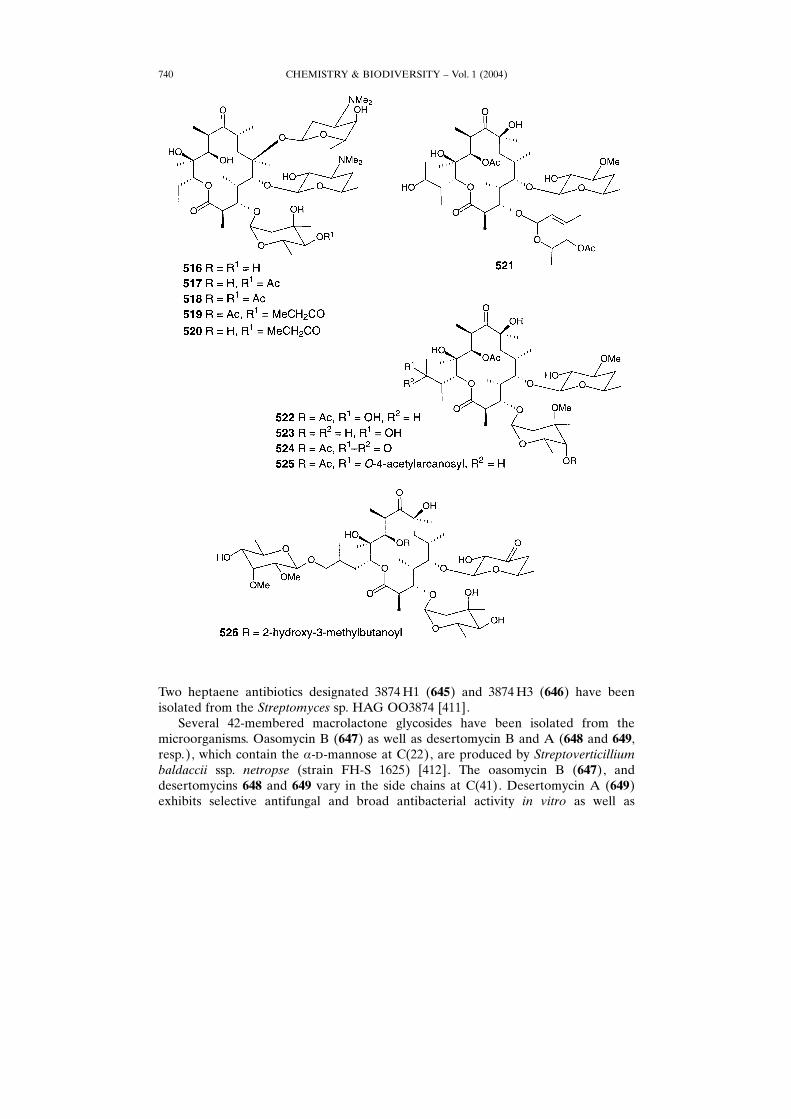
the first time, obtained from Micromonospora megalomicea [315], and later isolatedfrom culture broths of M. inositola [316] [317]. 2��,3��-Anhydro-3��-O-demethyllanka-mycin (521), lankamycin (522), kujimycin A (523), 15-dehydrolankamycin (524), 15-
CHEMISTRY & BIODIVERSITY ± Vol. 1 (2004) 733
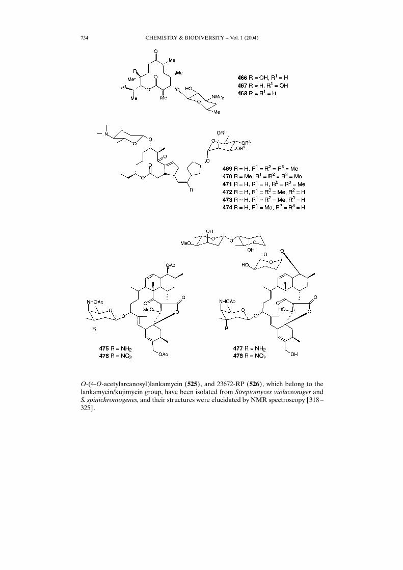
O-(4-O-acetylarcanosyl)lankamycin (525), and 23672-RP (526), which belong to thelankamycin/kujimycin group, have been isolated from Streptomyces violaceoniger andS. spinichromogenes, and their structures were elucidated by NMR spectroscopy [318 ±325].
CHEMISTRY & BIODIVERSITY ± Vol. 1 (2004)734
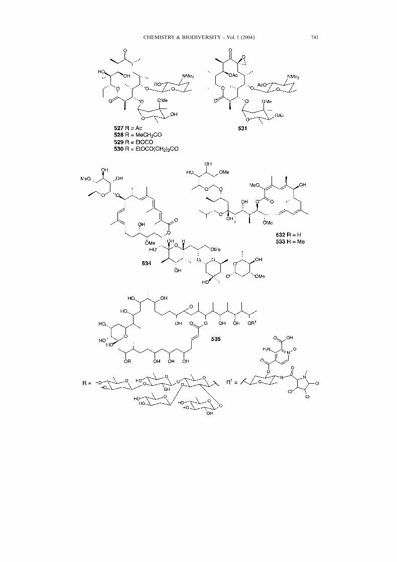
Erytromycin esters 527 ± 530 and tri-O-acetyloleandomycin (531) have also beenisolated from Streptomyces species [326 ± 329].Two antibiotics, 532 and 533, containing the sugar leucanicidin and 19-O-
methylleucanicidin, respectively, are potent inhibitors of larval development in theparasitic nematode Haemonchus contortus [330], and they have been isolated fromStreptomyces isolates A233, A239, and A240. Apoptolidin (534), which inducesapoptotic cell death in rat glia cells transformed with the adenovirus E1A oncogene,has been isolated by Kim et al. [331]. Absolute configuration of 534 and its synthesishave been reported [332] [333]. Colubricidin A (535) containing three Cl-atoms wasisolated from the fermentation broth of a new Streptomyces species [334]. ColubricidinA (535) demonstrated potent activity against Gram-positive bacteria, and it is slightlymore potent than vancomycin vs. Staphylococci but less potent vs. Streptococci andEnterococci.Cytotoxic macrolide IB-96212 (536), the configuration of which remains undeter-
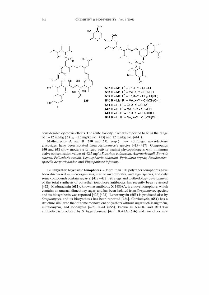
mined, was isolated from marine Micromonospora species [335]. The series ofmacrolides 537 ± 544, named avermectins A1a, A1b, A2a, A2b, B1a, B1b, B2a, and B2b,respectively, was produced by Streptomyces avermitilis [336]. When mutant of S.avermitilis was grown in the absent of supplementary branched-chain carboxylic acids,no avermectins were produced. Avermectin biosynthesis can be restored, however,upon supplementation [337]. Out of various potential precursors, more than 44 cyclic
CHEMISTRY & BIODIVERSITY ± Vol. 1 (2004) 735
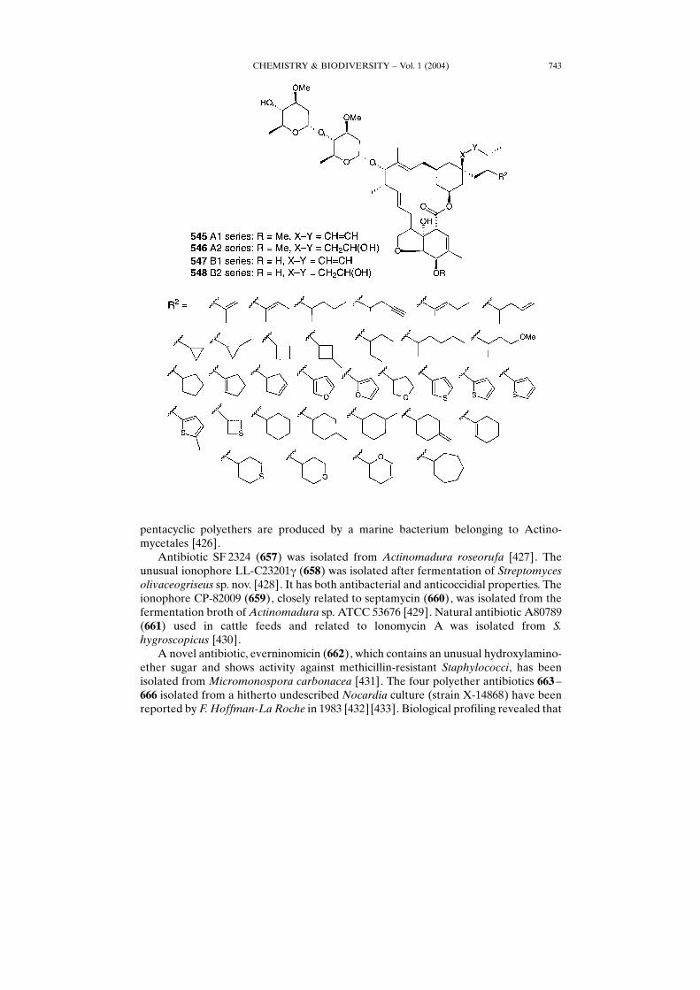
and acyclic alternative primers, 545 ± 548, were discovered that allow production ofavermectin analogues with altered C25 substituents [338] [339]. Biosynthesis of novelpolyketides including avermectins was recently reviewed [340].The spiramicin antibiotics 549 ± 555 were isolated from culture broths of Strepto-
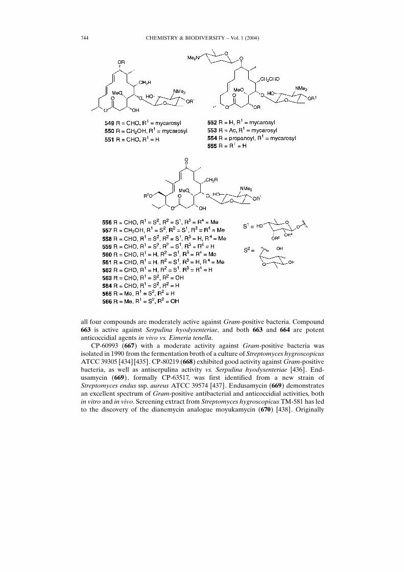
myces ambofaciens [341 ± 344]. High-speed counter-current chromatography wassuccessfully applied to the separation of spiramycin I, II, and III [345]. The tylosingroup antibiotics 556 ± 562 and related compounds 563 ± 566 are produced by S. fradiae[346 ± 348] and S. hydroscopicus [349].Leucomycin A1, A5, A7, A9, V, A3 (� josamycin), A4, A6, A8, U (567, 568, 569, 570,
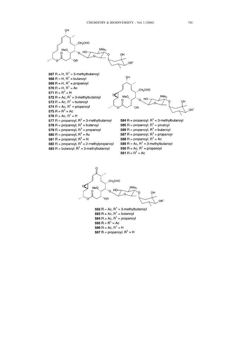
571, 572, 573, 574, 575, and 576, resp.), platenomycin A1 (577), medicamycin A2 (578),medicamycin A1 (�espinomycin A1�platenomycin B1; 579), platenomycin C2(�espinomycin A3; 580), DHP (581), espinomycin A2 (582), and platenomycin A0(583) have been isolated from Streptoverticillium (Streptomyces) kitasatoensis [350].
CHEMISTRY & BIODIVERSITY ± Vol. 1 (2004)736
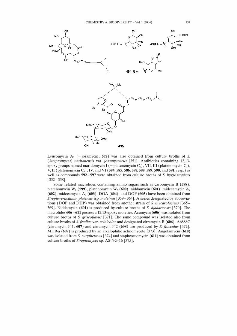
Leucomycin A3 (� josamycin; 572) was also obtained from culture broths of S.(Streptomyces) narbonensis var. josamyceticus [351]. Antibiotics containing 12,13-epoxy groups named maridomycin I (� platenomycin C3), VII, III (platenomycin C1),V, II (platenomycin C4), IV, and VI (584, 585, 586, 587, 588, 589, 590, and 591, resp.) aswell as compounds 592 ± 597 were obtained from culture broths of S. hygroscopicus[352 ± 358].Some related macrolides containing amino sugars such as carbomycin B (598),
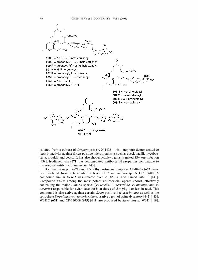
platenomycin W1 (599), platenomycin W2 (600), niddamycin (601), midecamycin A4(602), midecamycin A3 (603), DOA (604), and DOP (605) have been obtained fromStreptoverticillium platensis ssp. malvinus [359 ± 364]. A series designated by abbrevia-tions (DOP and DHP) was obtained from another strain of S. mycarofaciens [365 ±369]. Niddamycin (601) is produced by culture broths of S. djakartensis [370]. Themacrolides 606 ± 611 possess a 12,13-epoxy moieties. Acumycin (606) was isolated fromculture broths of S. griseoflavus [371]. The same compound was isolated also fromculture broths of S. fradiae var. acinicolor and designated cirramycin B (606). A6888C(cirramycin F-1; 607) and cirramycin F-2 (608) are produced by S. flocculus [372].M119-a (609) is produced by an alkalophilic actinomycete [373]. Angolamycin (610)was isolated from S. eurythermus [374] and staphcoccomycin (611) was obtained fromculture broths of Streptomyces sp. AS-NG-16 [375].
CHEMISTRY & BIODIVERSITY ± Vol. 1 (2004) 737
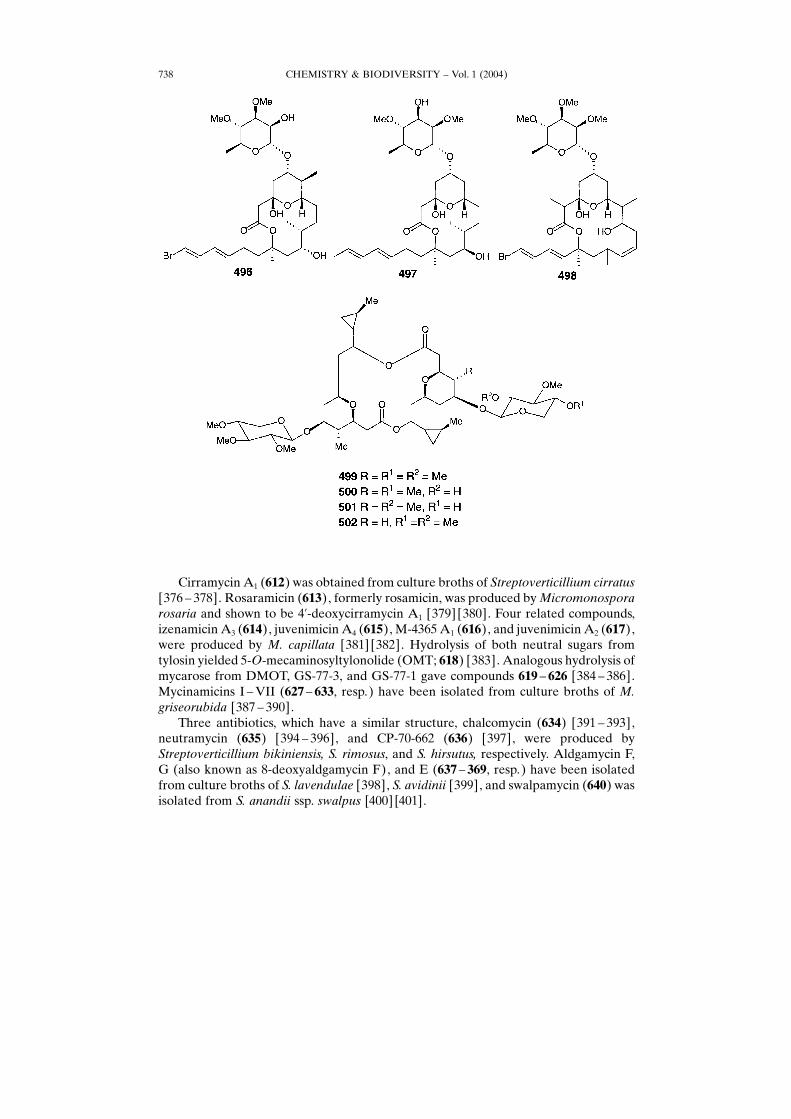
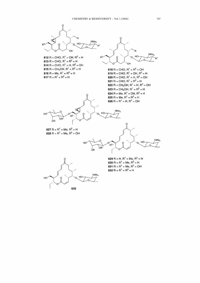
Cirramycin A1 (612) was obtained from culture broths of Streptoverticillium cirratus[376 ± 378]. Rosaramicin (613), formerly rosamicin, was produced byMicromonosporarosaria and shown to be 4�-deoxycirramycin A1 [379] [380]. Four related compounds,izenamicin A3 (614), juvenimicin A4 (615), M-4365 A1 (616), and juvenimicin A2 (617),were produced by M. capillata [381] [382]. Hydrolysis of both neutral sugars fromtylosin yielded 5-O-mecaminosyltylonolide (OMT; 618) [383]. Analogous hydrolysis ofmycarose from DMOT, GS-77-3, and GS-77-1 gave compounds 619 ± 626 [384 ± 386].Mycinamicins I ±VII (627 ± 633, resp.) have been isolated from culture broths of M.griseorubida [387 ± 390].Three antibiotics, which have a similar structure, chalcomycin (634) [391 ± 393],
neutramycin (635) [394 ± 396], and CP-70-662 (636) [397], were produced byStreptoverticillium bikiniensis, S. rimosus, and S. hirsutus, respectively. Aldgamycin F,G (also known as 8-deoxyaldgamycin F), and E (637 ± 369, resp.) have been isolatedfrom culture broths of S. lavendulae [398], S. avidinii [399], and swalpamycin (640) wasisolated from S. anandii ssp. swalpus [400] [401].
CHEMISTRY & BIODIVERSITY ± Vol. 1 (2004)738
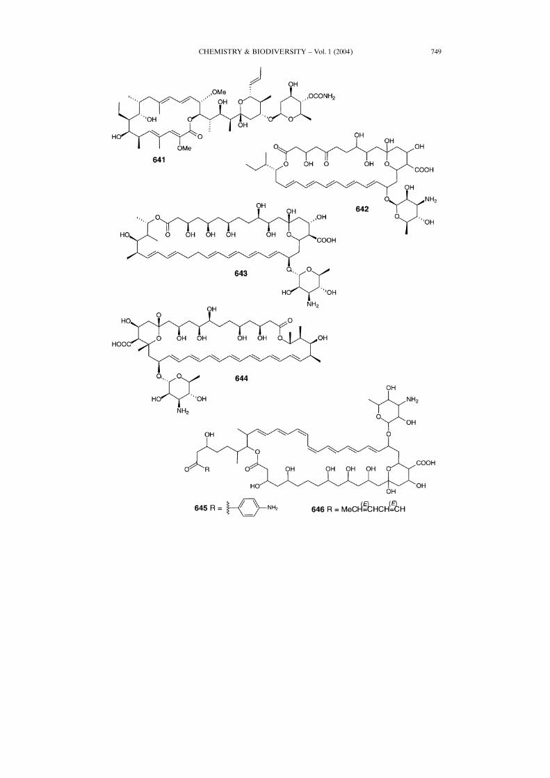
Concanamycin A (641) belonging to a family of structurally polyketide antibiotics,which include bafilomycins and hygrolidins, has been isolated from Streptomycesspecies [402] [403]. Concanamycin A (641) exhibits antifungal and immunosuppressiveactivity, with its potent and specific inhibition of V-Type H�-ATPases [404]. Totalsynthesis of 641 has also been reported [405] [406]. Another antibiotic, eurocidin D(642), which contains an amino sugar, was produced by Streptoverticilli species[407] [408]. Antibiotic nystatin (643) with antifungal activity produced by StreptomycesnorseiATCC 11455 is a polyketide represented by a 38-membered macrolactone ring towhich the deoxysugar mycosamine is attached. A complete set of genes responsible forthe biosynthesis has been cloned and analyzed [409]. Amphotericin B (644), whichexhibits strong antifungal activity, is produced by Streptomyces sp. HA 130-29 [410].
CHEMISTRY & BIODIVERSITY ± Vol. 1 (2004) 739
Two heptaene antibiotics designated 3874H1 (645) and 3874H3 (646) have beenisolated from the Streptomyces sp. HAG OO3874 [411].Several 42-membered macrolactone glycosides have been isolated from the
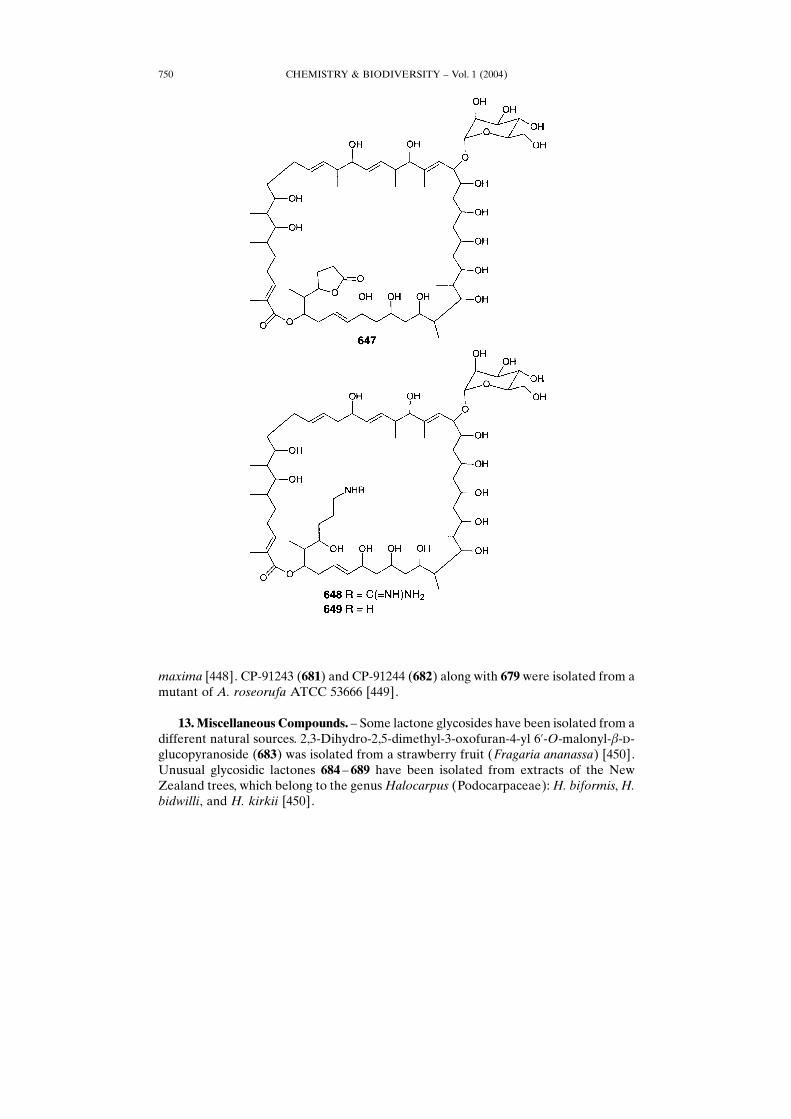
microorganisms. Oasomycin B (647) as well as desertomycin B and A (648 and 649,resp.), which contain the �-�-mannose at C(22), are produced by Streptoverticilliumbaldaccii ssp. netropse (strain FH-S 1625) [412]. The oasomycin B (647), anddesertomycins 648 and 649 vary in the side chains at C(41). Desertomycin A (649)exhibits selective antifungal and broad antibacterial activity in vitro as well as
CHEMISTRY & BIODIVERSITY ± Vol. 1 (2004)740
CHEMISTRY & BIODIVERSITY ± Vol. 1 (2004) 741
considerable cytotoxic effects. The acute toxicity in ice was reported to be in the rangeof 1 ± 12 mg/kg (LD50� 1.5 mg/kg s.c. [413] and 12 mg/kg p.o. [414]).Mathemycins A and B (650 and 651, resp.), new antifungal macrolactone
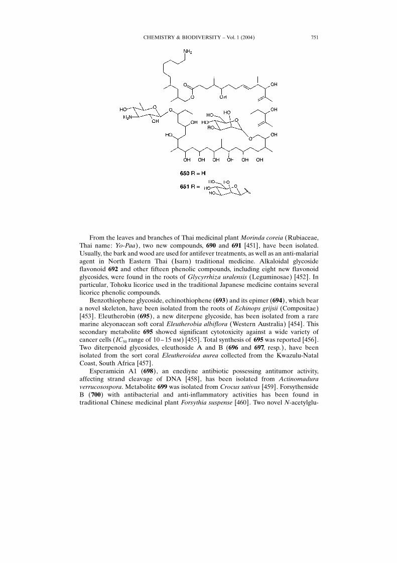
glycosides, have been isolated from Actinomycete species [415 ± 417]. Compounds650 and 651 show moderate in vitro activity against phytopathogens with minimumactive concentration values of 62.5 mg/l: Fusarium culmorum, Alternaria mali, Botrytiscinerea, Pellicularia sasakii, Leptosphaeria nodorum, Pyricularia oryzae, Pseudocerco-sporella herpotrichoides, and Phytophthora infestans.
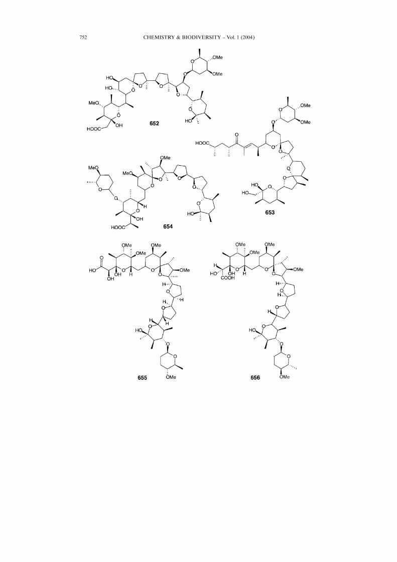
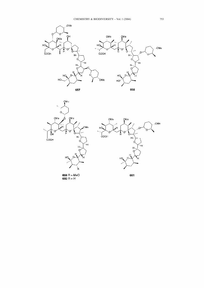
12. Polyether Glycosidic Ionophores. ± More than 100 polyether ionophores havebeen discovered in microorganisms, marine invertebrates, and algal species, and onlysome compounds contain sugar(s) [418 ± 422]. Strategy and methodology developmentof the total synthesis of polyether ionophore antibiotics has recently been reviewed[422]. Maduracimin (652), known as antibiotic X-14868A, is a novel ionophore, whichcontains an unusual dimethoxy sugar, and has been isolated from Streptomyces species,and its biosynthesis was reported [422] [423]. Lenoremycin (653) is produced also byStreptomyces, and its biosynthesis has been reported [424]. Carriomycin (654) has astructure similar to that of some monovalent polyethers without sugar such as nigericin,mutalomycin, and lonomycin [422]. K-41 (655), known as A32887 and RP37454antibiotic, is produced by S. hygroscopicus [425]. K-41A (656) and two other new
CHEMISTRY & BIODIVERSITY ± Vol. 1 (2004)742
pentacyclic polyethers are produced by a marine bacterium belonging to Actino-mycetales [426].Antibiotic SF 2324 (657) was isolated from Actinomadura roseorufa [427]. The
unusual ionophore LL-C23201� (658) was isolated after fermentation of Streptomycesolivaceogriseus sp. nov. [428]. It has both antibacterial and anticoccidial properties. Theionophore CP-82009 (659), closely related to septamycin (660), was isolated from thefermentation broth of Actinomadura sp. ATCC 53676 [429]. Natural antibiotic A80789(661) used in cattle feeds and related to lonomycin A was isolated from S.hygroscopicus [430].A novel antibiotic, everninomicin (662), which contains an unusual hydroxylamino-
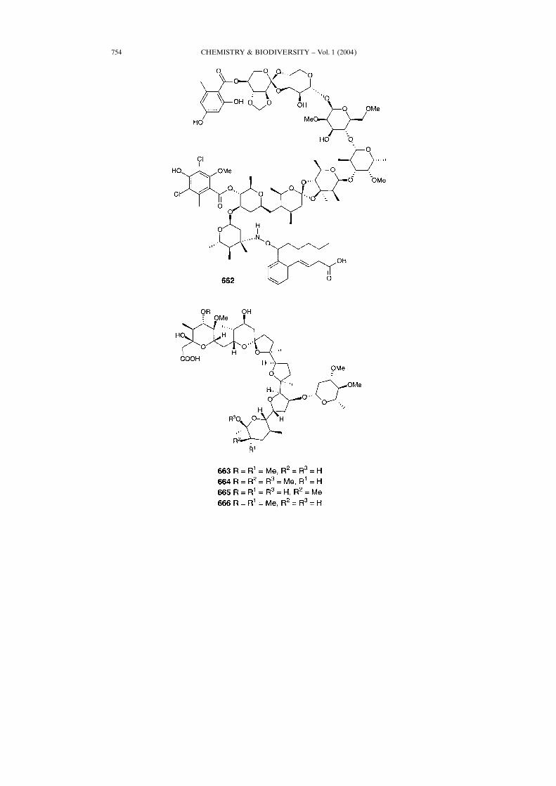
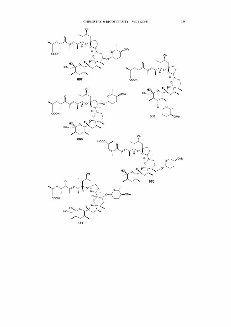
ether sugar and shows activity against methicillin-resistant Staphylococci, has beenisolated from Micromonospora carbonacea [431]. The four polyether antibiotics 663 ±666 isolated from a hitherto undescribed Nocardia culture (strain X-14868) have beenreported by F. Hoffman-La Roche in 1983 [432] [433]. Biological profiling revealed that
CHEMISTRY & BIODIVERSITY ± Vol. 1 (2004) 743
all four compounds are moderately active against Gram-positive bacteria. Compound663 is active against Serpulina hyodysenteriae, and both 663 and 664 are potentanticoccidial agents in vivo vs. Eimeria tenella.CP-60993 (667) with a moderate activity against Gram-positive bacteria was
isolated in 1990 from the fermentation broth of a culture of Streptomyces hygroscopicusATCC 39305 [434] [435]. CP-80219 (668) exhibited good activity againstGram-positivebacteria, as well as antiserpulina activity vs. Serpulina hyodysenteriae [436]. End-usamycin (669), formally CP-63517, was first identified from a new strain ofStreptomyces endus ssp. aureus ATCC 39574 [437]. Endusamycin (669) demonstratesan excellent spectrum of Gram-positive antibacterial and anticoccidial activities, bothin vitro and in vivo. Screening extract from Streptomyces hygroscopicus TM-581 has ledto the discovery of the dianemycin analogue moyukamycin (670) [438]. Originally
CHEMISTRY & BIODIVERSITY ± Vol. 1 (2004)744
CHEMISTRY & BIODIVERSITY ± Vol. 1 (2004) 745
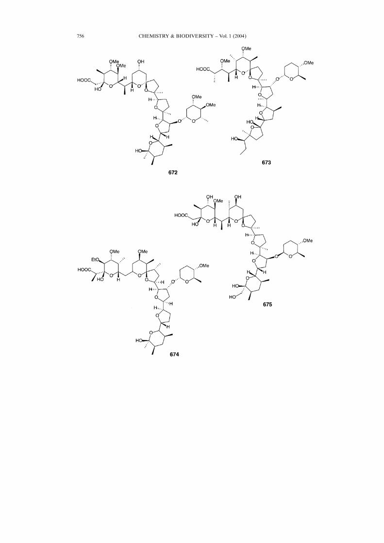
isolated from a culture of Streptomyces sp. X-14931, this ionophore demonstrated invitro bioactivity against Gram-positive microorganisms such as cocci, bacilli, mycobac-teria, moulds, and yeasts. It has also shown activity against a mixed Eimeria infection[439]. Isodianemycin (671) has demonstrated antibacterial properties comparable tothe original antibiotic dianemycin [440].Both maduramycin (672) and 12-methylportmicin ionophore CP-84657 (673) have
been isolated from a fermentation broth of Actinomadura sp. ATCC 53708. Acompound similar to 673 was isolated from A. fibrosa and named A82810 [441].Compound 673 is among the most potent anticoccidial agents known, effectivelycontrolling the major Eimeria species (E. tenella, E. acervulina, E. maxima, and E.necatrix) responsible for avian coccidiosis at doses of 5 mg/kg-1 or less in feed. Thiscompound is also active against certain Gram-positive bacteria in vitro as well as thespirochete Serpulina hyodysenteriae, the causative agent of swine dysentery [442] [443].W341C (674) and CP-120509 (675) [444] are produced by Streptomyces W341 [419].
CHEMISTRY & BIODIVERSITY ± Vol. 1 (2004)746
CHEMISTRY & BIODIVERSITY ± Vol. 1 (2004) 747
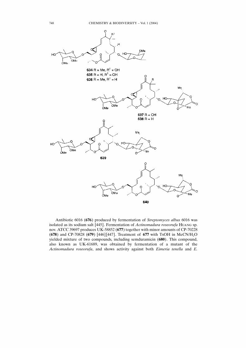
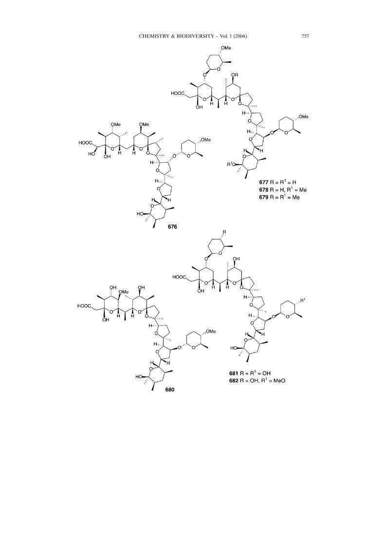
Antibiotic 6016 (676) produced by fermentation of Streptomyces albus 6016 wasisolated as its sodium salt [445]. Fermentation of Actinomadura roseorufa H���� sp.nov. ATCC 39697 produces UK-58852 (677) together with minor amounts of CP-70228(678) and CP-70828 (679) [446] [447]. Treatment of 677 with TsOH in MeCN/H2Oyielded mixture of two compounds, including semduramicin (680). This compound,also known as UK-61689, was obtained by fermentation of a mutant of theActinomadura roseorufa, and shows activity against both Eimeria tenella and E.
CHEMISTRY & BIODIVERSITY ± Vol. 1 (2004)748
CHEMISTRY & BIODIVERSITY ± Vol. 1 (2004) 749
maxima [448]. CP-91243 (681) and CP-91244 (682) along with 679 were isolated from amutant of A. roseorufa ATCC 53666 [449].
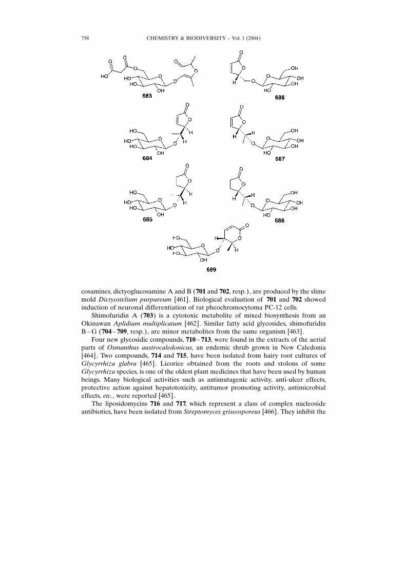
13. Miscellaneous Compounds. ± Some lactone glycosides have been isolated from adifferent natural sources. 2,3-Dihydro-2,5-dimethyl-3-oxofuran-4-yl 6�-O-malonyl-�-�-glucopyranoside (683) was isolated from a strawberry fruit (Fragaria ananassa) [450].Unusual glycosidic lactones 684 ± 689 have been isolated from extracts of the NewZealand trees, which belong to the genusHalocarpus (Podocarpaceae):H. biformis,H.bidwilli, and H. kirkii [450].
CHEMISTRY & BIODIVERSITY ± Vol. 1 (2004)750
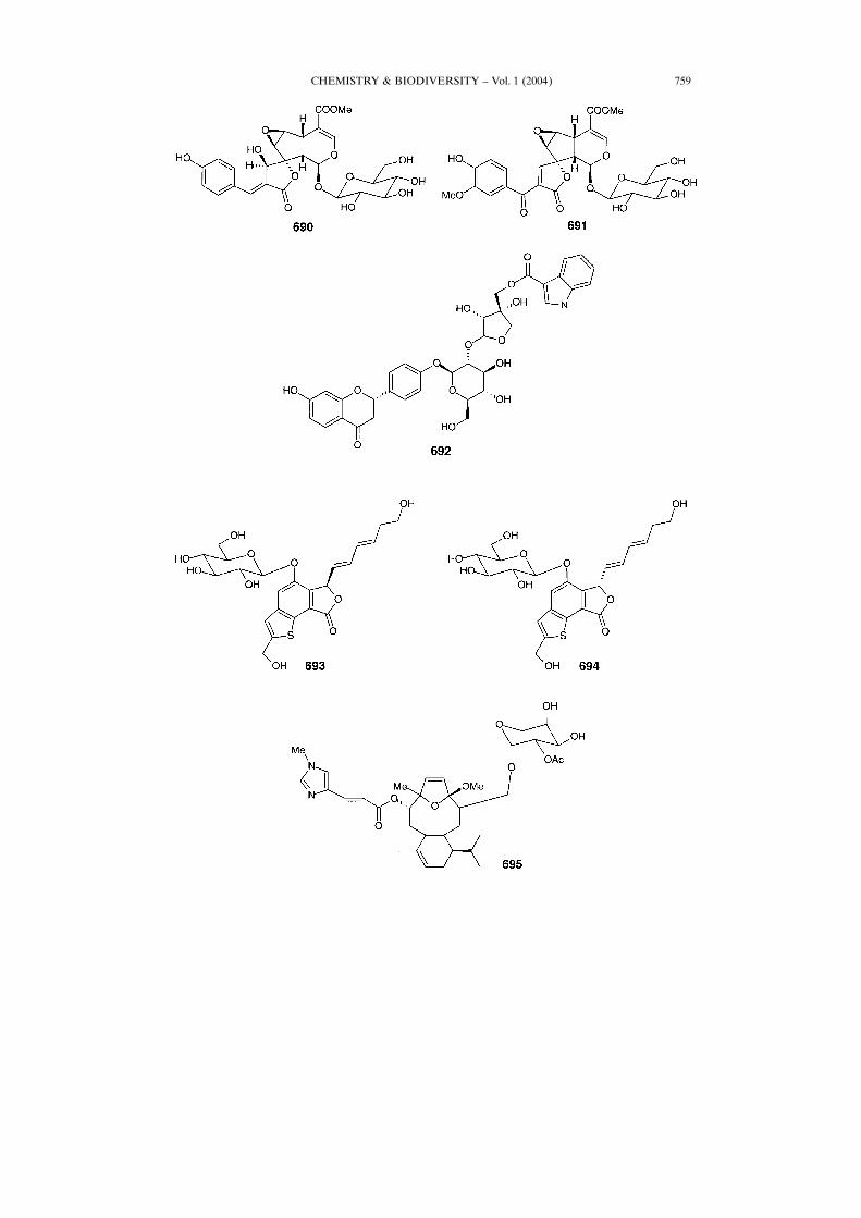
From the leaves and branches of Thai medicinal plantMorinda coreia (Rubiaceae,Thai name: Yo-Paa), two new compounds, 690 and 691 [451], have been isolated.Usually, the bark and wood are used for antifever treatments, as well as an anti-malarialagent in North Eastern Thai (Isarn) traditional medicine. Alkaloidal glycosideflavonoid 692 and other fifteen phenolic compounds, including eight new flavonoidglycosides, were found in the roots of Glycyrrhiza uralensis (Leguminosae) [452]. Inparticular, Tohoku licorice used in the traditional Japanese medicine contains severallicorice phenolic compounds.Benzothiophene glycoside, echinothiophene (693) and its epimer (694), which bear
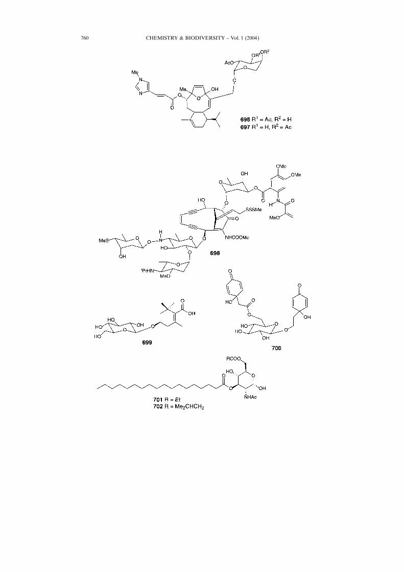
a novel skeleton, have been isolated from the roots of Echinops grijsii (Compositae)[453]. Eleutherobin (695), a new diterpene glycoside, has been isolated from a raremarine alcyonacean soft coral Eleutherobia albiflora (Western Australia) [454]. Thissecondary metabolite 695 showed significant cytotoxicity against a wide variety ofcancer cells (IC50 range of 10 ± 15 n�) [455]. Total synthesis of 695 was reported [456].Two diterpenoid glycosides, eleuthoside A and B (696 and 697, resp.), have beenisolated from the sort coral Eleutheroidea aurea collected from the Kwazulu-NatalCoast, South Africa [457].Esperamicin A1 (698), an enediyne antibiotic possessing antitumor activity,
affecting strand cleavage of DNA [458], has been isolated from Actinomaduraverrucosospora. Metabolite 699 was isolated from Crocus sativus [459]. ForsythensideB (700) with antibacterial and anti-inflammatory activities has been found intraditional Chinese medicinal plant Forsythia suspense [460]. Two novel N-acetylglu-
CHEMISTRY & BIODIVERSITY ± Vol. 1 (2004) 751
CHEMISTRY & BIODIVERSITY ± Vol. 1 (2004)752
CHEMISTRY & BIODIVERSITY ± Vol. 1 (2004) 753
CHEMISTRY & BIODIVERSITY ± Vol. 1 (2004)754
CHEMISTRY & BIODIVERSITY ± Vol. 1 (2004) 755
CHEMISTRY & BIODIVERSITY ± Vol. 1 (2004)756
CHEMISTRY & BIODIVERSITY ± Vol. 1 (2004) 757
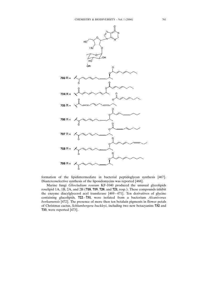
cosamines, dictyoglucosamine A and B (701 and 702, resp.), are produced by the slimemold Dictyostelium purpureum [461]. Biological evaluation of 701 and 702 showedinduction of neuronal differentiation of rat pheochromocytoma PC-12 cells.Shimofuridin A (703) is a cytotoxic metabolite of mixed biosynthesis from an
Okinawan Aplidium multiplicatum [462]. Similar fatty acid glycosides, shimofuridinB ±G (704 ± 709, resp.), are minor metabolites from the same organism [463].Four new glycosidic compounds, 710 ± 713, were found in the extracts of the aerial
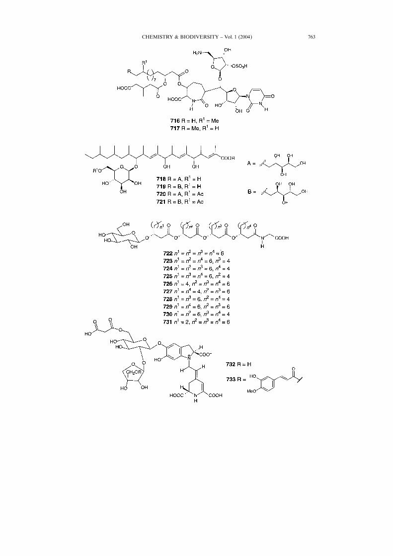
parts of Osmanthus austrocaledonicus, an endemic shrub grown in New Caledonia[464]. Two compounds, 714 and 715, have been isolated from hairy root cultures ofGlycyrrhiza glabra [465]. Licorice obtained from the roots and stolons of someGlycyrrhiza species, is one of the oldest plant medicines that have been used by humanbeings. Many biological activities such as antimutagenic activity, anti-ulcer effects,protective action against hepatotoxicity, antitumor promoting activity, antimicrobialeffects, etc., were reported [465].The liposidomycins 716 and 717, which represent a class of complex nucleoside
antibiotics, have been isolated from Streptomyces griseosporeus [466]. They inhibit the
CHEMISTRY & BIODIVERSITY ± Vol. 1 (2004)758
CHEMISTRY & BIODIVERSITY ± Vol. 1 (2004) 759
CHEMISTRY & BIODIVERSITY ± Vol. 1 (2004)760
formation of the lipidintermediate in bacterial peptidoglycan synthesis [467].Diastereoselective synthesis of the liposidomycins was reported [468].Marine fungi Gliocladium roseum KF-1040 produced the unusual glycolipids
roselipid 1A, 1B, 2A, and 2B (718, 719, 720, and 721, resp.). These compounds inhibitthe enzyme diacylglycerol acyl transferase [469 ± 471]. Ten derivatives of glycinecontaining glucolipids, 722 ± 731, were isolated from a bacterium Alcanivoraxborkumensis [472]. The presence of more then ten betalain pigments in flower petalsof Christmas cactus, Schlumbergera buckleyi, including two new betacyanins 732 and733, were reported [473].
CHEMISTRY & BIODIVERSITY ± Vol. 1 (2004) 761
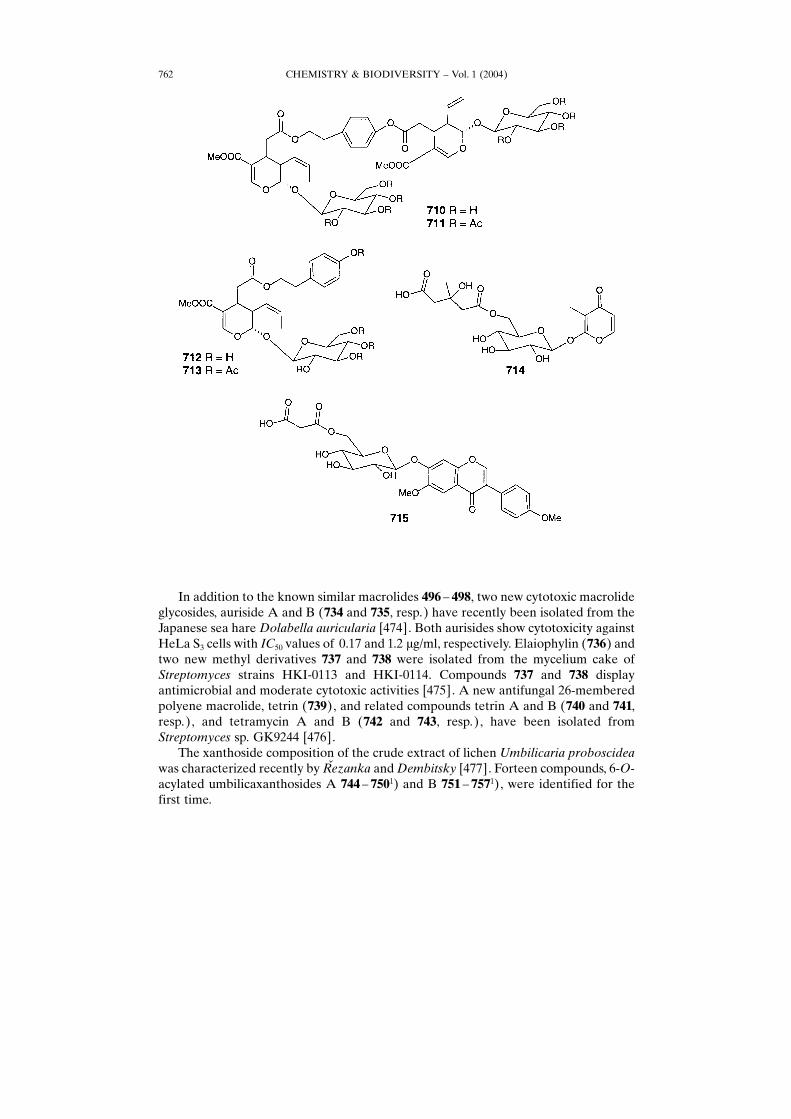
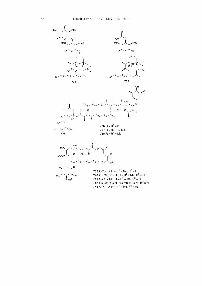
In addition to the known similar macrolides 496 ± 498, two new cytotoxic macrolideglycosides, auriside A and B (734 and 735, resp.) have recently been isolated from theJapanese sea hareDolabella auricularia [474]. Both aurisides show cytotoxicity againstHeLa S3 cells with IC50 values of 0.17 and 1.2 �g/ml, respectively. Elaiophylin (736) andtwo new methyl derivatives 737 and 738 were isolated from the mycelium cake ofStreptomyces strains HKI-0113 and HKI-0114. Compounds 737 and 738 displayantimicrobial and moderate cytotoxic activities [475]. A new antifungal 26-memberedpolyene macrolide, tetrin (739), and related compounds tetrin A and B (740 and 741,resp.), and tetramycin A and B (742 and 743, resp.), have been isolated fromStreptomyces sp. GK9244 [476].The xanthoside composition of the crude extract of lichen Umbilicaria proboscidea
was characterized recently by Rœezanka andDembitsky [477]. Forteen compounds, 6-O-acylated umbilicaxanthosides A 744 ± 7501) and B 751 ± 7571), were identified for thefirst time.
CHEMISTRY & BIODIVERSITY ± Vol. 1 (2004)762
CHEMISTRY & BIODIVERSITY ± Vol. 1 (2004) 763
CHEMISTRY & BIODIVERSITY ± Vol. 1 (2004)764
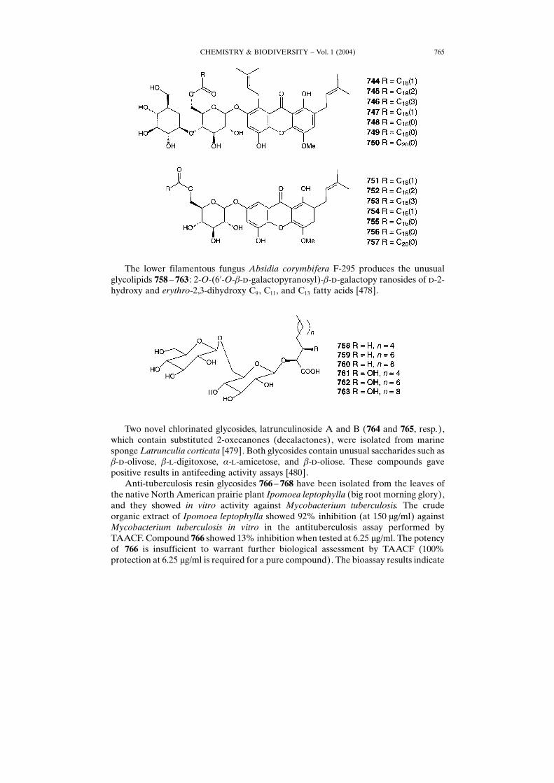
The lower filamentous fungus Absidia corymbifera F-295 produces the unusualglycolipids 758 ± 763 : 2-O-(6�-O-�-�-galactopyranosyl)-�-�-galactopy ranosides of �-2-hydroxy and erythro-2,3-dihydroxy C9, C11, and C13 fatty acids [478].
Two novel chlorinated glycosides, latrunculinoside A and B (764 and 765, resp.),which contain substituted 2-oxecanones (decalactones), were isolated from marinespongeLatrunculia corticata [479]. Both glycosides contain unusual saccharides such as�-�-olivose, �-�-digitoxose, �-�-amicetose, and �-�-oliose. These compounds gavepositive results in antifeeding activity assays [480].Anti-tuberculosis resin glycosides 766 ± 768 have been isolated from the leaves of
the native North American prairie plant Ipomoea leptophylla (big root morning glory),and they showed in vitro activity against Mycobacterium tuberculosis. The crudeorganic extract of Ipomoea leptophylla showed 92% inhibition (at 150 �g/ml) againstMycobacterium tuberculosis in vitro in the antituberculosis assay performed byTAACF. Compound 766 showed 13% inhibition when tested at 6.25 �g/ml. The potencyof 766 is insufficient to warrant further biological assessment by TAACF (100%protection at 6.25 �g/ml is required for a pure compound). The bioassay results indicate
CHEMISTRY & BIODIVERSITY ± Vol. 1 (2004) 765
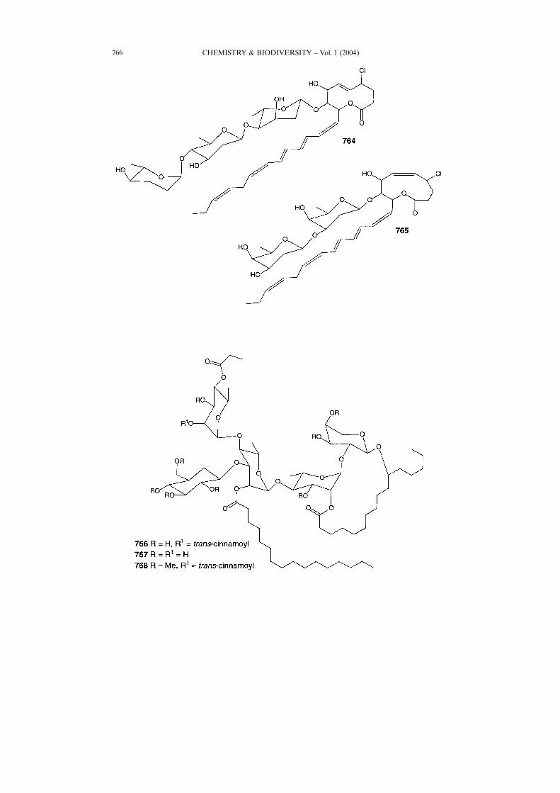
CHEMISTRY & BIODIVERSITY ± Vol. 1 (2004)766
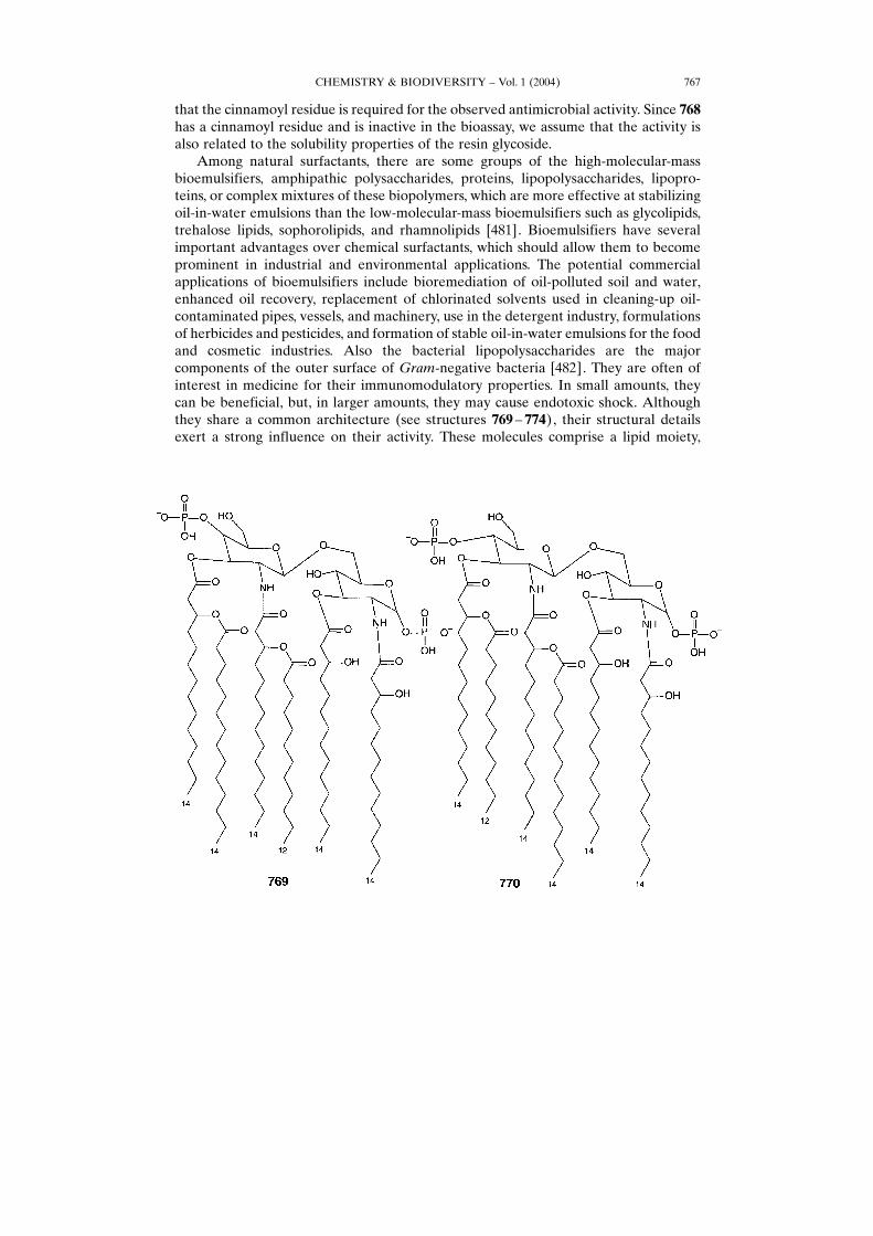
that the cinnamoyl residue is required for the observed antimicrobial activity. Since 768has a cinnamoyl residue and is inactive in the bioassay, we assume that the activity isalso related to the solubility properties of the resin glycoside.Among natural surfactants, there are some groups of the high-molecular-mass
bioemulsifiers, amphipathic polysaccharides, proteins, lipopolysaccharides, lipopro-teins, or complex mixtures of these biopolymers, which are more effective at stabilizingoil-in-water emulsions than the low-molecular-mass bioemulsifiers such as glycolipids,trehalose lipids, sophorolipids, and rhamnolipids [481]. Bioemulsifiers have severalimportant advantages over chemical surfactants, which should allow them to becomeprominent in industrial and environmental applications. The potential commercialapplications of bioemulsifiers include bioremediation of oil-polluted soil and water,enhanced oil recovery, replacement of chlorinated solvents used in cleaning-up oil-contaminated pipes, vessels, and machinery, use in the detergent industry, formulationsof herbicides and pesticides, and formation of stable oil-in-water emulsions for the foodand cosmetic industries. Also the bacterial lipopolysaccharides are the majorcomponents of the outer surface of Gram-negative bacteria [482]. They are often ofinterest in medicine for their immunomodulatory properties. In small amounts, theycan be beneficial, but, in larger amounts, they may cause endotoxic shock. Althoughthey share a common architecture (see structures 769 ± 774), their structural detailsexert a strong influence on their activity. These molecules comprise a lipid moiety,
CHEMISTRY & BIODIVERSITY ± Vol. 1 (2004) 767
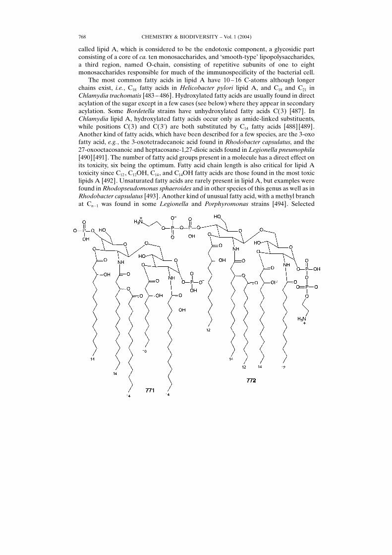
called lipid A, which is considered to be the endotoxic component, a glycosidic partconsisting of a core of ca. ten monosaccharides, and −smooth-type× lipopolysaccharides,a third region, named O-chain, consisting of repetitive subunits of one to eightmonosaccharides responsible for much of the immunospecificity of the bacterial cell.The most common fatty acids in lipid A have 10 ± 16 C-atoms although longer
chains exist, i.e., C18 fatty acids in Helicobacter pylori lipid A, and C18 and C21 inChlamydia trachomatis [483 ± 486]. Hydroxylated fatty acids are usually found in directacylation of the sugar except in a few cases (see below) where they appear in secondaryacylation. Some Bordetella strains have unhydroxylated fatty acids C(3) [487]. InChlamydia lipid A, hydroxylated fatty acids occur only as amide-linked substituents,while positions C(3) and C(3�) are both substituted by C14 fatty acids [488] [489].Another kind of fatty acids, which have been described for a few species, are the 3-oxofatty acid, e.g., the 3-oxotetradecanoic acid found in Rhodobacter capsulatus, and the27-oxooctacosanoic and heptacosane-1,27-dioic acids found in Legionella pneumophila[490] [491]. The number of fatty acid groups present in a molecule has a direct effect onits toxicity, six being the optimum. Fatty acid chain length is also critical for lipid Atoxicity since C12, C12OH, C14, and C14OH fatty acids are those found in the most toxiclipids A [492]. Unsaturated fatty acids are rarely present in lipid A, but examples werefound in Rhodopseudomonas sphaeroides and in other species of this genus as well as inRhodobacter capsulatus [493]. Another kind of unusual fatty acid, with a methyl branchat Cn�1 was found in some Legionella and Porphyromonas strains [494]. Selected
CHEMISTRY & BIODIVERSITY ± Vol. 1 (2004)768
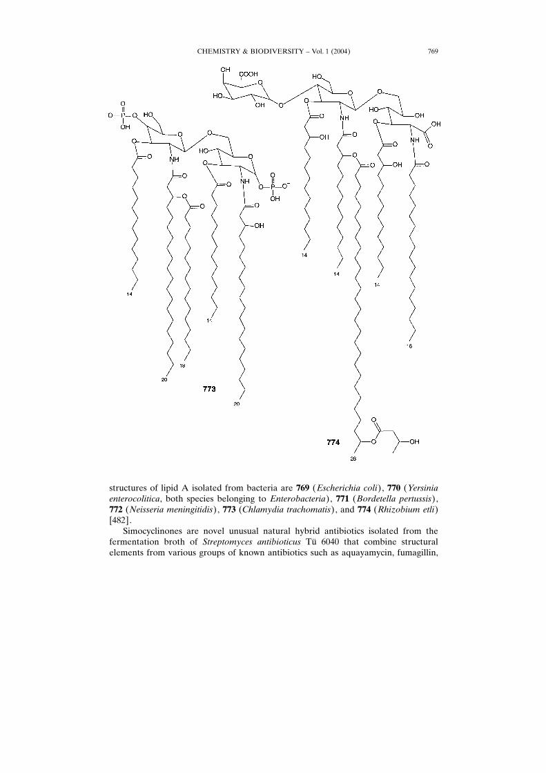
structures of lipid A isolated from bacteria are 769 (Escherichia coli), 770 (Yersiniaenterocolitica, both species belonging to Enterobacteria), 771 (Bordetella pertussis),772 (Neisseria meningitidis), 773 (Chlamydia trachomatis), and 774 (Rhizobium etli)[482].Simocyclinones are novel unusual natural hybrid antibiotics isolated from the
fermentation broth of Streptomyces antibioticus T¸ 6040 that combine structuralelements from various groups of known antibiotics such as aquayamycin, fumagillin,
CHEMISTRY & BIODIVERSITY ± Vol. 1 (2004) 769
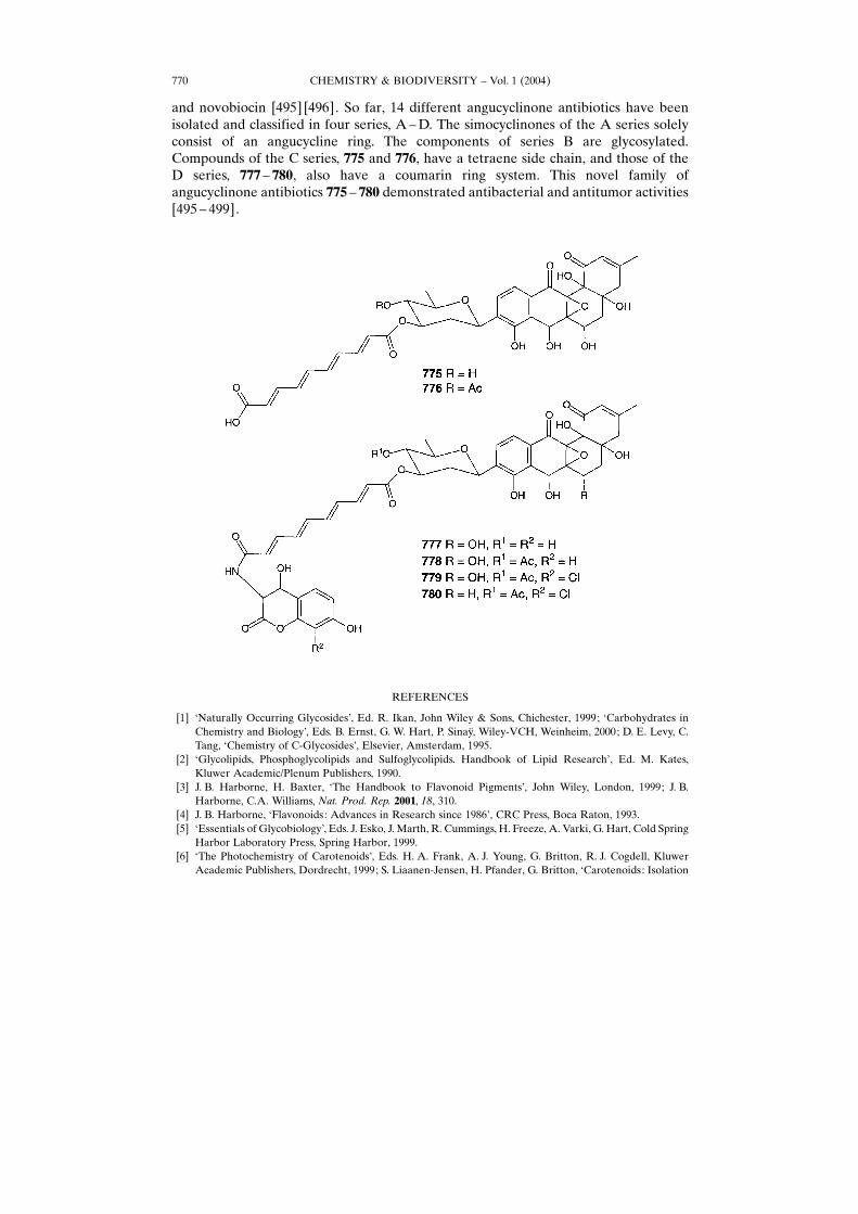
and novobiocin [495] [496]. So far, 14 different angucyclinone antibiotics have beenisolated and classified in four series, A ±D. The simocyclinones of the A series solelyconsist of an angucycline ring. The components of series B are glycosylated.Compounds of the C series, 775 and 776, have a tetraene side chain, and those of theD series, 777 ± 780, also have a coumarin ring system. This novel family ofangucyclinone antibiotics 775 ± 780 demonstrated antibacterial and antitumor activities[495 ± 499].
REFERENCES
[1] −Naturally Occurring Glycosides×, Ed. R. Ikan, John Wiley & Sons, Chichester, 1999; −Carbohydrates inChemistry and Biology×, Eds. B. Ernst, G. W. Hart, P. Sinay», Wiley-VCH, Weinheim, 2000; D. E. Levy, C.Tang, −Chemistry of C-Glycosides×, Elsevier, Amsterdam, 1995.
[2] −Glycolipids, Phosphoglycolipids and Sulfoglycolipids. Handbook of Lipid Research×, Ed. M. Kates,Kluwer Academic/Plenum Publishers, 1990.
[3] J. B. Harborne, H. Baxter, −The Handbook to Flavonoid Pigments×, John Wiley, London, 1999; J. B.Harborne, C.A. Williams, Nat. Prod. Rep. 2001, 18, 310.
[4] J. B. Harborne, −Flavonoids: Advances in Research since 1986×, CRC Press, Boca Raton, 1993.[5] −Essentials of Glycobiology×, Eds. J. Esko, J. Marth, R. Cummings, H. Freeze, A. Varki, G. Hart, Cold Spring
Harbor Laboratory Press, Spring Harbor, 1999.[6] −The Photochemistry of Carotenoids×, Eds. H. A. Frank, A. J. Young, G. Britton, R. J. Cogdell, Kluwer
Academic Publishers, Dordrecht, 1999; S. Liaanen-Jensen, H. Pfander, G. Britton, −Carotenoids: Isolation
CHEMISTRY & BIODIVERSITY ± Vol. 1 (2004)770
and Analysis×, Springer Verlag, Berlin, 1995; G. Britton, S. Liaanen-Jensen, H. Pfander, −Carotenoids:Spectroscopy×, Springer Verlag, Berlin, 1995.
[7] N. Kosaric, −Biosurfactants: Production, Properties, Applications×, Marcel Dekker, New York, 1993.[8] W. D. Nes, −Isopentenoids and Other Natural Products: Evolution and Function×, ACS Symposium Series
No. 562, 1994.[9] −Macrolide antibiotics: chemistry, biology, and practice×, Ed. O. Satoshi, Academic Press, Amsterdam,
Boston, 2002; −Macrolide Antibiotics×, Eds. W. Schˆnfeld, H. A. Kirst, Birkh‰user Verlag, Boston, 2002;K. J. Hale, M. G. Hummersone, S. Manaviazar, M. Frigerio, Nat. Prod. Rep. 2002, 19, 413.
[10] J. K. MacLeod, H. B. Rasmussen, A. C. Wills, J. Nat. Prod. 1997, 60, 620.[11] Y. Takeda, A. Takechi, T. Masuda, H. Otsuka, Planta Med. 1998, 64, 78.[12] K. Yoshikawa, Y. Satou, Y. Tokunaga, M. Tanaka, S. Arihara, S. K. Nigam, J. Nat. Prod. 1998, 61, 440.[13] D. Krajewaski, C. Duque, P. Schreier, Phytochemistry 1997, 45, 1627.[14] B. B. Messanga, S. fon Kimbu, B. L. Sondengam, B. Bodo, Fitoterapia 2001, 72, 732.[15] I. Simko, E. A. Omer, E. E. Ewing, S. McMurry, J. L. Koch, P. J. Davies, Phytochemistry 1996, 43, 727.[16] H. Matsuura, T. Yoshira, A. Ichihara, Y. Kikuta, Y. Koda, Biosci. Biotechnol. Biochem. 1993, 57, 1253.[17] T. Yoshira, H. Matsuura, A. Ichihara, Y. Kikuta, Y. Koda, in −Progress in Plant Growth Regulation×, Eds.
C. M. Karssen, L. C. Van Loon, D. Vreugdenhil, Kluwer Academic, Dordrecht, 1992, p. 286 ± 314.[18] M. Wang, H. Likuzaki, N. Zhu, N. Sang, N. Nakatani, C. T. Ho, J. Agric. Food Chem. 2000, 48, 235.[19] T. Rœ ezanka, V. M. Dembitsky, Eur. J. Org. Chem. 2003, 14, 309.[20] A. R. Jude, J. M. Little, J. P. Freeman, J. E. Evans, A. Radominska-Pandya, D. F. Grant, Arch. Biochem.
Biopys. 2000, 380, 294.[21] M. T. J. van der Meer, S. Schouten, S. Hanada, E. C. Hopmans, J. S. Sinninghe Damste¬, D. M. Ward, Arch.
Microbiol. 2002, 178, 229.[22] T. Rœ ezanka, Phytochemistry 2002, 60, 639.[23] S. Riva, J. Mol. Catal., B 2002, 19 ± 20, 43.[24] S. Lang, D. Wullbrandt, Appl. Microbiol. Biotechnol. 1999, 51, 22.[25] E. Rozenberg, E. Z. Ron, Appl. Microbiol. Biotechnol. 1999, 52, 154.[26] R. M. Mailer, G. Soberon-Chavez, Appl. Microbiol. Biotechnol. 2000, 54, 625.[27] M. G. Healy, C. M. Devine, R. Murphy, Resour. Conserv. Recycling 1996, 18, 41.[28] D. B. Sarney, E. N. Vulfson, Trends Biotechnol. 1995, 13, 164.[29] S. Bergstrom, H. Theorell, H. Davide, Ark. Kemi Mineral. Geol. 1946, 23A, 1.[30] F. G. Jarvis, M. J. Johnson, J. Am. Chem. Soc. 1949, 71, 4124.[31] J. R. Edwards, J. A. Hayashi, Arch. Biochem. Biophys. 1965, 111, 415.[32] K. Hisatsuka, T. Nakahara, N. Sano, K. Yamada, Agric. Biol. Chem. 1971, 35, 686.[33] S. Itoh, H. Honda, F. Tomita, T. Suzuki, J. Antibiotics (Tokyo) 1971, 24, 855.[34] M. Yamaguchi, A. Sato, A. Yukuyama, Chem. Ind. 1976, 4, 741.[35] T. Hirayama, I. Kato, FEBS Lett. 1982, 139, 81.[36] C. Syldatk, S. Lang, F. Wagner, V. Wray, L. Witte, Z. Naturforsch. 1985, 40, 51.[37] C. Syldatk, S. Lang, U. Matulovic, F. Wagner, Z. Naturforsch. 1985, 40, 61.[38] N. B. Rendell, G. W. Taylor, M. Somerville, H. Todd, R. Wilson, J. Cole, Biochim. Biophys. Acta 1990, 1045,
189.[39] A. Abalos, A. Pinazo, M. R. Infante, M. Casals, F. Garcy¬a, A. Manresa, Langmuir 2001, 17, 1367.[40] D. Kitamoto, H. Yanagishita, K. Haraya, H. K. Kitamoto, Biotechnol. Lett. 1998, 20, 813.[41] D. Kitamoto, G. Sangita, G. Ourisson, Y. Nakatani, Chem. Commun. 2000, 861.[42] Y. Wakamatsu, X. Zhao, C. Jin, N. Day, M. Shibahara, N. Nomura, T. Nakahara, T. Murata, K. K.
Yokoyama, Eur. J. Biochem. 2001, 268, 374.[43] X. Zhao, Y.Wakamatsu, M. Shibahara, N. Nomura, C. Geltinger, T. Nakahara, T.Murata, K. K. Yokoyama,
Cancer Res. 1999, 59, 482.[44] D. Kitamoto, H. Yanagishita, A. Endo, M. Nakaiwa, T. Nakane, T. Akiya, Biotechnol. Progr. 2001, 17, 362.[45] D. Kitamoto, T. Ikegami, G. T. Suzuki, A. Sasaki, Y. I. Takeyama, Y. Idemoto, N. Koura, H. Yanagishita,
Biotechnol. Lett. 2001, 23, 1709.[46] C. Asselineau, J. Asselineau, Prog. Chem. Fats Other Lipids 1978, 16, 59.[47] E. Vilkas, A. Rojas, Bull. Soc. Chim. Biol. 1964, 46, 689.[48] I. Azuma, Y. J. Yamamura, Biochem. (Tokyo) 1962, 52, 82; E. Vilkas, A. Adam, M. Senn, Chem. Phys.
Lipids 1968, 2, 11.[49] C. Prottey, C. E. Ballou, J. Biol. Chem. 1968, 243, 6196; N. Shaw, F. Dinglinger, Biochem. J. 1969, 112, 769.
CHEMISTRY & BIODIVERSITY ± Vol. 1 (2004) 771
[50] H. Noll, H. Bloch, J. Asselineau, E. Lederer, Biochim. Biophys. Acta 1956, 20, 764.[51] J. Asselineau, E. Lederer, Biochim. Biophys. Acta 1955, 17, 161.[52] C. Asselineau, H. Montrozier, J. C. Prome, A. Savagnac, M. Welby, Eur. J. Biochem. 1972, 28, 102.[53] M. B. Goren, Biochim. Biophys. Acta 1970, 210, 116.[54] M. B. Goren, Biochim. Biophys. Acta 1970, 210, 127.[55] S. W. Esch, M. D. Morton, T. D. Williams, C. S. Buller, Carbohydr. Res. 1999, 319, 112.[56] H. Mayorga, C. Duqur, H. Knapp, P. Winterhalter, Phytochemistry 2002, 59, 439.[57] H. Mayorga, H. Knapp, P. Winterhalter, C. Duque, J. Agric. Food Chem. 2001, 49, 1904.[58] V. Bultel-Ponce, C. Debitus, A. Blond, C. Cerceau, M. Guyot, Tetrahedron Lett. 1997, 38, 5805.[59] P. A. J. Gorin, J. F. T. Spencer, A. P. Tulloch, Can. J. Chem. 1961, 39, 846.[60] A. M. Davila, R. Marchal, N. Monin, J. P. Vandecasteele, J. Chromatogr. 1993, 648, 139.[61] A. P. Tulloch, J. F. T. Spencer, P. A. J. Gorin, Can. J. Chem. 1962, 40, 1326.[62] A. P. Tulloch, A. Hill, J. F. T. Spencer, Can. J. Chem. 1968, 46, 3337.[63] U. Rau, S. Hammen, R. Heckmann, V. Wray, S. Lang, Ind. Crops Prod. 2001, 13, 85.[64] R. K. Hommel, L. Weber, A. Weiss, U. Himelreich, O. Rilke, H. P. Kleber, J. Biotechnol. 1994, 26, 199.[65] A. P. Tulloch, A. Hill, J. F. T. Spencer, J. Chem. Soc., Chem. Commun. 1967, 584.[66] D. G. Cooper, D. A. Paddock, Appl. Environ. Microbiol. 1983, 46, 1426.[67] R. T. Otto, H. J. Daniel, G. Pekin, K. Muller-Decker, G. Furstenberger, M. Reuss, C. Syldatk, Appl.
Microbiol. Biotechnol. 1999, 52, 495.[68] J. D. Desai, I. M. Banat, Microbiol. Mol. Biol. Rev. 1997, 61, 47.[69] E. Vollbrecht, U. Rau, S. Lang, Fett/Lipid 1999, 101, 389.[70] O. Levand, H. Larson, Planta Med. 1979, 36, 186.[71] A. Hirazumi, E. Furusawa, S. C. Chou, Y. Hokama, Proc. West. Pharmacol. Soc. 1994, 37, 145.[72] A. Hirazumi, E. Furusawa, S. C. Chou, Y. Hokama, Proc. West. Pharmacol. Soc. 1996, 39, 25.[73] M. Wang, H. Kikuzaki, Y. Jin, N. Nakatani, N. Zhu, K. Csisar, C. Boyd, R. Rosen, G. Ghal, C. T. Ho, J. Nat.
Prod. 2000, 63, 1182.[74] J. Lee, R. I. Hollingsworth, Tetrahedron 1996, 52, 3873.[75] R. Perda-Miranda, S. Hernandez-Carlos, Tetrahedron 2002, 58, 3145.[76] E. M. M. Gasper, Tetrahedron Lett. 1999, 40, 6861.[77] P. Font Quer, −Plantas Medicinales. El Dioscorides Renovado×, Editorial Labor, Madrid, 1962, p. 543.[78] J. K. MacLeod, A. Ward, J. Nat. Prod. 1997, 60, 467.[79] R. A. Heacock, Prog. Med. Chem. 1975, 11, 91.[80] L. W. Bieber, F. da Silva, R. M. O. Correa Lima, A. de Andrade Chiappeta, S. C. do Naschimento, I. A. de
Souza, J. F. de Mello, H. J. Veith, Phytochemistry 1986, 25, 1077.[81] X.-M. Du, N.-Y. Sun, M. Nishi, T. Kawasaki, Y.-T. Guo, K. Miyahara, J. Nat. Prod. 1999, 62, 722.[82] N. Noda, N. Takahashi, K. Miyahara, C. R. Yang, Phytochemistry 1998, 48, 837.[83] J. K. MacLeod, A. Ward, J. Nat. Prod. 1997, 60, 467.[84] T. Rœ ezanka, I. A. Guschina, Phytochemistry 2001, 58, 509.[85] T. Rœ ezanka, I. A. Guschina, Phytochemistry 2000, 54, 635.[86] T. Tanahashi, A. Shimada, M. Kai, N. Nagakura, K. Inoue, C. C. Chen, J. Nat. Prod. 1996, 59, 798.[87] Y. Champavier, G. Comte, J. Varcuteren, D. P. Allais, A. J. Chulia, Phytochemistry 1999, 50, 1219.[88] B. Withopf, E. Richling, R. Roscher, W. Schwab, P. Schreier, J. Agric. Food Chem. 1997, 45, 907.[89] W. Kisiel, B. Barszcz, Phytochemistry 1995, 39, 1395.[90] W. Kisiel, K. Zielinska, S. P. Joshi, Phytochemistry 2000, 54, 763.[91] W. Kisiel, B. Barszcz, Phytochemistry 1997, 46, 1241.[92] S. Piacente, C. Pizza, N. De Tommasi, F. De Simone, Phytochemistry 1996, 41, 1357.[93] T. Komori, M. Yamashita, Y. Tsurumi, M. Kohsaka, J. Antibiot. 1985, 38, 455.[94] T. Komori, Y. Itoh, J. Antibiot. 1985, 38, 544.[95] I. Calis, A. Kuruuzum, O. Demirezer, O. Sticher, W. Ganci, P. Ruedi, J. Nat. Prod. 1999, 62, 1101.[96] N. B. Perry, N. J. Brennan, J. Nat. Prod. 1997, 60, 623.[97] J. K. MacLeod, H. B. Rasmussen, A. C. Wills, J. Nat. Prod. 1997, 60, 620.[98] M. H. Frick, O. Elo, K. Haapa, O. P. Heinonen, P. Heinsalmi, P. Helo, N. Engl. J. Med. 1987, 317, 1237.[99] P. A. Todd, A. Ward, Am. J. Cardiol. 1985, 35, 1637.[100] M. L. Hyneck, P. C. Smith, A. Munafo, A. F. McDonagh, L. Z. Benet, Clin. Pharmacol. Ther. 1988, 44, 107.[101] A. Nakagawa, A. Shigeta, H. Iwabuchi, M. Horiguchi, K. Nakamura, H. Takahagi, Biomed. Chromatogr.
1991, 5, 68.
CHEMISTRY & BIODIVERSITY ± Vol. 1 (2004)772
[102] R. A. Okerholm, F. J. Keeley, F. E. Peterson, A. J. Glazko, Proc. R. Soc. Med. 1976, 69, 11.[103] H. Knauf, E. U. Kolle, E. Mutschler, Klin. Wochenschr. 1990, 68, 692.[104] M. N. Cayen, Prog. Drug Metab. 1983, 7, 173.[105] H. Oh, G. S. Oh, W. G. Seo, H. O. Pae, K. Y. Chai, T. O. Kwon, Y. H. Lee, H. T. Chung, H. S. Lee, J. Nat.
Prod. 2001, 64, 942.[106] M. Straubinger, B. Bau, S. Eckstein, M. Fink, P. Winterhalter, J. Agric. Food Chem. 1998, 46, 3238.[107] X. N. Zhong, H. Otsuka, T. Ide, E. Hirata, Y. Takeda, Phytochemistry 1999, 52, 923.[108] R. H. Cichewicz, K. C. Lim, J. H. McKerrow, M. G. Nair, Tetrahedron 2002, 58, 8597.[109] D. E. Elliot, Gastroenterol. Clin. North Am. 1996, 25, 599.[110] T. W. Schafer, B. R. Hale, Curr. Gastroenterol. Rep. 2001, 3, 293.[111] M. Colgan, L. Colgan, −The Flavonoid Revolution: Grape Seed Extract and Other Flavonoids Against
Disease×, Apple Publishing, Vancouver 1997.[112] J. B. Harborne, R. J. Grayer, in −The Flavonoids: Advances in Research since 1980×, Ed. J. B. Harborne,
Chapman & Hall, London, 1988, p. 1 ± 20.[113] R. Brouillard, in −The Flavonoids: Advances in Research since 1980×, Ed. J. B. Harborne, Chapman &Hall,
London, 1988, p. 525 ± 538.[114] M.-R. V. Calsteren, F. Cormier, C. B. Do, R. R. Laing, Spectroscopy (Ottawa) 1991, 9, 1.[115] F. Cormier, C. B. Do, Y. Nicolas, In Vitro Cell. Dev. Biol., Plant 1994, 3, 171.[116] D. C. Baker, D. K. Dougall, W. E. Glassgen, S. C. Johnson, J. W. Metzger, A. Rose, H. U. Seitz, Plant Cell
Tissue Organ. Culture 1994, 39, 79.[117] T. Mori, M. Sakurai, J. Shigeta, K. Yoshida, T. Kondo, J. Food Sci. 1993, 58, 788.[118] K. Sakamoto, K. Iida, K. Sawamura, K. Hajiro, Y. Asada, T. Yoshikawa, T. Furuya, Phytochemistry 1993,
33, 357.[119] A. Takahashi, K. Takeda, T. Ohnishi, Plant Cell Physiol. 1991, 32, 541.[120] A. Tholakalabavi, J. J. Zwiazek, T. A. Thorpe, In Vitro Cell. Dev. Biol., Plant 1994, 3, 164.[121] K. Toki, M. Takeuchi, N. Saito, T. Honda, Phytochemistry 1996, 42, 1055.[122] K. Hosokawa, Y. Fukunaga, E. Fukushi, J. Kawabata, Phytochemistry 1995, 38, 1293.[123] T. Fossen, R. Slimestad, D. O. Èvstedal, È. M. Andersen, Biochem. Syst. Ecol. 2002, 30, 855.[124] N. Terahara, A. Callebaut, R. Ohba, T. Nagata, M. Ohnishi-Kameyama, M. Suzuki, Phytochemistry 2001,
58, 493.[125] A. Callebaut, N. Terahara, M. de Haan, M. Decleire, Plant Cell Tissue Organ. Culture 1997, 50, 195.[126] N. Saito, F. Tatsuzawa, A. Nishiyama, M. Yokoi, A. Shigihara, T. Honda, Phytochemistry 1995, 38,
1027.[127] B. Cui, J. Kinjo, M. Nakamura, T. Nohara, Tetrahedron Lett. 1991, 43, 6135.[128] Z. D. Jaing, P. R. Jensen, W. Fenical, Tetrahedron Lett. 1997, 38, 5065.[129] J. H. Isaza, H. Ito, T. Yoshida, Phytochemistry 2001, 58, 321.[130] C. Gao, P. Mayon, E. N. MacManus, E. N. Vulfson, Biotechnol. Bioeng. 2001, 71, 235.[131] R. J. Graer, G. C. Kite, N. C. Veitch, M. R. Eckert, P. D. Marin, P. Senanayake, A. J. Paton, Biochem. Syst.
Ecol. 2002, 30, 327.[132] K. Kazuma, N. Noda, M. Suzuki, Phytochemistry 2003, 62, 229.[133] M. C. Hsieh, T. L. Graham, Phytochemistry 2001, 58, 995.[134] M. Demuth, K. Schaffner, Angew. Chem., Int. Ed. 1982, 21, 820.[135] M. Demuth, K. Schaffner, Angew. Chem., Int. Ed. 1983, 22, 828.[136] H. Inouye, S. Uesato, Prog. Chem. Org. Nat. Prod. 1986, 50, 169.[137] S. R. Jensen, H. Franzyk, F. Wallander, Phytochemistry 2002, 60, 213.[138] T. Tanahashi, A. Shimada, N. Nagakura, K. Inoue, M. Ono, T. Fujita, C. C. Chen, Chem. Pharm. Bull. 1995,
43, 729.[139] T. Tanahashi, A. Shimada, M. Kai, N. Nagakura, K. Inoue, C. C. Chen, J. Nat. Prod. 1996, 59, 798.[140] S. Damtoft, H. Franzyk, S. R. Jensen, Phytochemistry 1997, 45, 743.[141] A. Kuruuzum, Z. Guvenalp, L. O. Demirezer, I. Bergere, K. Stroch, A. Zeek, Phytochemistry 2002, 61, 937.[142] M. R. Grever, S. A. Schepartz, B. A. Chabner, Semin. Oncol. 1992, 25, 2140.[143] W. G. Ma, N. Fuzzati, J. L. Wolfender, C. R. Yang, K. Hostettmann, Phytochemistry 1996, 43, 805.[144] T. Kaneko, M. Sakamoto, K. Ohtani, A. Ito, R. Hasai, K. Yamasaki, W. G. Padorina, Phytochemistry 1995,
39, 115.[145] E. Quisumbing, in −Medicinal Plants of the Philippines×, Katha Publishing, Quezon City, 1978, p. 547.[146] H. Pfander, −Key to Carotenoids×, Birkh‰user, Basel, 1987.
CHEMISTRY & BIODIVERSITY ± Vol. 1 (2004) 773

[147] D. Kull, H. Pfander, −Appendix: List of New carotenoids×, in −Carotenoids. Isolation and Analysis×, Eds. G.Britton, S. Liaaen-Jensen, H. Pfander, Birkh‰user, Basel, 1995.
[148] U. A. Higgli, H. Pfander, in −Naturally Occurring Glycosides×, Ed. R. Ikan, John Wiley, Chichester, 1999,p. 125 ± 145.
[149] S. Aschoff, Berlin. Jahrb. Pharm. 1818, 19, 142;[150] P. Karrer, F. Benz, R. Morf, H. Raudnitz, M. Stoll, T. Takahashi, Helv. Chim. Acta 1932, 15, 1399.[151] P. Karrer, H. Salomon, Helv. Chim. Acta 1933, 16, 643.[152] H. Kleinig, R. Schmitt, W. Meister, G. Englert, H. Thommen, Z. Naturforsch., C 1979, 34, 181.[153] H. Kleinig, W. J. Broughton, Arch. Microbiol. 1982, 133, 164.[154] H. Kleinig, R. Schmitt, Z. Naturforsch., C 1982, 37, 758.[155] A. J. Aasen, G. W. Francis, S. Liaaen-Jensen, Acta Chem. Scand. 1969, 23, 2605.[156] V. K. Dhingra, T. R. Seshadri, S. K. Mukerjee, Indian J. Chem. 1975, 13, 339.[157] H. Pfander, F. Wittwer, Helv. Chim. Acta 1975, 58, 2233.[158] H. Pfander, F. Wittwer, Helv. Chim. Acta 1975, 58, 1608.[159] H. Pfander, H. Schurtenberger, Phytochemistry 1982, 21, 1039.[160] H. Pfander, M. Rychener, J. Chromatogr. 1982, 234, 443.[161] P. A. Tarantilis, M. Polissiou, M. Manfait, J. Chromatogr. 1994, 664, 55.[162] S. Pfister, P. Meyer, A. Steck, H. Pfander, J. Agric. Food Chem. 1996, 44, 2612.[163] V. K. Dringra, T. R. Seshadri, S. K. Mukerjee, Indian J. Chem., Sect. B 1976, 14, 231.[164] M. Kamikura, K. Nakazato, Eisei Shikensho Hokoku 1985, 103, 305.[165] S. Ghosal, S. K. Singh, S. K. Battachaya, J. Chem. Res., Synop. 1989, 70.[166] J. H. Marshall, G. J. Wilmoth, J. Bacteriol. 1981, 147, 900.[167] H. Kleinig, H. Reichenbach, Phytochemistry 1973, 12, 2483.[168] B. F. Lutnaes, A. Oren, S. Liaaen-Jensen, J. Nat. Prod. 2002, 65, 1340.[169] G. A. Alfredsson, J. K. Kristjansson, S. Hjˆrleifsdo¬ ttir, K. O. Stetter, J. Gen. Microbiol. 1988, 134,
299.[170] M. J. Vacheron, N. Arpin, G. Michael, C. R. Acad. Sci., Ser. II C 1970, 271, 881.[171] S. Takaichi, K. Linoshita, T. Miyamoto, K. Azegami, J. Ishidsu, T. Seki, S. Sukuda, Bull. JFCC 1989, 5, 96.[172] S. Takaichi, J. I. Ishidsu, Methods Enzymol. 1992, 213, 367.[173] A. Yokoyama, G. Sandmann, T. Hoshino, K. Adachi, M. Sakai, Y. Shizuri, Tetrahedron Lett. 1995, 36, 4901.[174] G. Ourisson, Y. Nakatani, in −Carotenoids Chemistry and Biochemistry×, Eds. N. I. Krinsky, M. M.
Mathews-Roth, F. R. Taylor, Plenum Press, New York, 1989, p. 237 ± 245.[175] V. M. Dembitsky, T. Rœ ezanka, Comp. Biochem. Physiol., B 1996, 114, 383.[176] S. Takaichi, Z. Y. Wang, M. Umetsu, T. Nozawa, M. T. Shimada, M. T. Madigan,Arch. Microbiol. 1997, 168,
270.[177] S. Takaichi, Y. Tamura, K. Azegami, Y. Yamamoto, J. I. Ishidsu, Phytochemistry 1997, 45, 505.[178] S. Takaichi, K. Tshuji, K. Matsuura, K. Shimada, Plant Cell Physiol. 1995, 36, 773.[179] L. J. Goad, T. Akihisa, −Analysis of Sterols×, Blackie, London, 1997.[180] L. J. Goad, Methods Plant Biochem. 1991, 7, 369.[181] N. V. Kovganko, Z. N. Kashkan, Chem. Nat. Comp. 1999, 35, 479.[182] A. Tietz, Z. Naturforsch., C 1981, 36, 900.[183] L. Dyas, L. J. Goad, Phytochemistry 1993, 34, 17.[184] M. Ohnishi, Y. Fujino, Phytochemistry 1981, 20, 1357.[185] B. D. Whitaker, Phytochemistry 1991, 30, 757.[186] B. D. Whitaker, Phytochemistry 1988, 27, 3411.[187] R. Duperon, M. Thiersault, P. Duperon, Phytochemistry 1984, 23, 743.[188] L. Dyas, D. R. Threlfall, L. J. Goad, Phytochemistry 1994, 35, 655.[189] D. L. Davis, C. G. Poneleit, Phytochemistry 1975, 14, 1201.[190] M. S. Ali, M. Saleem, A. W. Erian, Fitoterapia 2001, 72, 712.[191] R. Duperon, M. Thiersault, P. Duperon, Phytochemistry 1983, 22, 535.[192] H. Yang, J. Wang, A. J. Hou, Y. P. Guo, Z. W. Lin, H. D. Sun, Fitoterapia 2000, 71, 641.[193] −Resource Lists of Yunnan Traditional Chinese Medicine×, Yunnan Med. Material Co., Science Press,
Beijing, p. 451, 1993.[194] K. C. Joshi, P. Singh, A. Mehra, Planta Med. 1979, 37, 64.[195] B. Danieli, M. Luisetti, S. Riva, A. Bertinotti, E. Ragg, E. Scaglioni, E. Bombardelli, J. Org. Chem 1995, 60,
3637.
CHEMISTRY & BIODIVERSITY ± Vol. 1 (2004)774
[196] K. Hostettmann, A. Marston, −Saponins: Chemistry and Pharmacology of Natural Products×, CambridgeUniversity Press, 1994.
[197] A. F. Halim, A. T. Khalil, Phytochemistry 1996, 42, 1135.[198] K. Umehara, M. Endoh, T. Miyase, M. Kuroyanagi, A. Ueno, Chem. Pharm. Bull. 1994, 42, 611.[199] S. I. Yoshima, H. Narita, K. Hayashi, H. Mitsuhashi, Chem. Pharm. Bull. 1982, 31, 3971.[200] N. C. Kim, A. D. Kinghorn, Arch. Pharm. Res. 2002, 25, 725.[201] G. E. DuBois, Ann. Rev. Med. Chem. 1982, 17, 323.[202] V. D. Huan, K. Ohtani, R. Kasai, K. Yamasaki, N. V. Tuu, Chem. Pharm. Bull. 2001, 49, 453.[203] F. Abe, H. Fujishima, Y. Iwase, T. Yamauchi, K. Kinjo, S. Yaga, Chem. Pharm. Bull. 1999, 47, 1128.[204] H. H. Sauer, E. Weiss, T. Reichstein, Helv. Chim. Acta 1965, 48, 857.[205] H. H. Sauer, E. Weiss, T. Reichstein, Helv. Chim. Acta 1966, 49, 1632.[206] H. H. Sauer, E. Weiss, T. Reichstein, Helv. Chim. Acta 1966, 49, 1655.[207] T. Warashina, T. Noro, Phytochemistry 1997, 44, 917.[208] K. A. El Sayed, A. F. Halim, A. M. Zaghloul, J. D. McChesney, M. P. Stone, M. Voehler, K. Hayashi,
Phytochemistry 1995, 39, 395.[209] A. Braca, A. Bader, I. Morelli, R. Scarpato, G. Turchi, C. Pizza, N. De Tommasi, Tetrahedron 2002, 58, 5837.[210] M. Shaiq Ali, M. Saleem, W. Ahmad, M. Parvez R. Yamdagni, Phytochemistry 2002, 59, 889.[211] G. Fontana, G. Savona, B. Rogriguez, Phytochemistry 1999, 50, 283.[212] R. Kasai, K. Yamasaki, O. Tanaka, in −Naturally Occurring Glycosides×, Ed. R. Ikan, John Wiley & Sons,
Chichester, 1999, p. 295 ± 310.[213] S. B. Mahato, S. K. Sarkar, G. Poddar, Phytochemistry 1988, 27, 3037.[214] E. Heinz, in −Advances in Lipid Methodology, Vol. 3×, Ed. W. W. Christie, Oily Press, Dundee, 1996,
p. 211 ± 332.[215] P. Muhr, W. Likussar, M. Schubert-Zsilavecz, Magn. Reson. Chem. 1996, 34, 137.[216] C. Lavaud, C. Voutquenne, P. Bal, I. Pouny, Fitoterapia 2000, 71, 338.[217] S. B. Mahato, B. C. Pal, K. Ashoke, A. K. Nandy, Tetrahedron 1992, 48, 6717.[218] T. Konoshima, Adv. Exp. Med. Biol. 1996, 404, 117.[219] I. L. Nord, L. Kenne, Carbohydr. Res. 2000, 329, 817.[220] A. Kilonda, B. Bila, O. Lohohola, S. Toppet, F. Compopernolle, ARKIVOC 2003, 3.[221] C. R. Kensil, S. Soltysik, U. Patel, D. J. Marciani, Vaccine 1992, 92, 35.[222] R. Higuchi, T. Komori, Phytochemistry 1987, 26, 2357.[223] D. C. Van Setten, G. J. Ten Hove, E. J. H. J. Wiertz, J. P. Kamerling, G. Van de Werken, Anal. Chem 1998,
70, 4401.[224] T. Fujioka, Y. Kashiwada, H. Okabe, K. Mihashi, K. H. Lee, Bioorg. Med. Chem. Lett. 1996, 6, 2807.[225] N. T. Nyberg, L. Kenne, B. Ronnberg, B. G. Sundquist, Carbohydr. Res. 2000, 323, 87.[226] N. Arda, N. Goren, A. Kuru, T. Penngsupaprp, J. M. Pezzuto, S. X. Qiu, G. A. Cordell, J. Nat. Prod. 1997, 60,
1170.[227] K. Yoshikawa, Y. Suzuki, M. Tanaka, S. Arihara, S. K. Nigam, J. Nat. Prod. 1997, 60, 1269.[228] Z. Zhang, K. Koike, Z. Jia, T. Nikaido, T. Guo, J. Zheng, J. Nat. Prod. 1999, 62, 877.[229] H. K. Wang, K. F. Bastow, L. M. Cosentino, K.H. Lee, J. Med. Chem. 1996, 39, 1975.[230] R. H. Tian, E. Ohmura, M. Matsui, T. Noharas, Phytochemistry 1997, 44, 723.[231] E. Chain, H. W. Florey, A. D. Gardner, N. G. Heatley, M. A. Jennings, J. Orr-Ewing, A. G. Sanders, Lancet
1940, 1, 226.[232] D. Ikeda, S. Umezawa, in −Naturally Occurring Glycosides×, Ed. R. Ikan, John Wiley & Sons, Chichester,
1999, p. 13 ± 42.[233] L. M. Cappelletti, R. Spagnoli, J. Antibiot. 1983, 36, 328.[234] K. Wada, M. Takeda, M. Arakawa, K. Ozaki, M. Takano, Chemotherapy (Tokyo) 1987, 35, 213.[235] S. Matsunaga, N. Fusetani, Y. Kato, H. Hiroda, J. Am. Chem. Soc. 1991, 113, 9690.[236] D. Wolf, F. J. Schmitz, F. Qiu, M. Kelly-Borges, J. Nat. Prod. 1999, 62, 170.[237] N. U. Sata, S. Matsunaga, N. Fusetani, R. W. M. Van Soest, J. Nat. Prod. 1999, 62, 969.[238] N. U. Sata, S. Wada, S. Matsunaga, S. Watabe, R. W. M. Van Soest, N. Fusetani, J. Org. Chem. 1999, 64, 2331.[239] M. Wu, K. E. Milligan, W. H. Gerwick, Tetrahedron 1997, 53, 15983.[240] N. Sata, N. Asai, S. Matsunaga, N. Fusetani, Tetrahedron 1994, 50, 1105; N. Fusetani, N. Sata, N. Asai, S.
Matsunaga, Tetrahedron Lett. 1993, 34, 4067.[241] R. Goobes, A. Rudi, Y. Kashman, Tetrahedron 1996, 52, 7921.[242] K. Shindo, M. Kamishohara, A. Odagawa, M. Matsuoka, H. Kawai, J. Antibiot. 1993, 46, 1076.
CHEMISTRY & BIODIVERSITY ± Vol. 1 (2004) 775

[243] Y. Matsushima, T. Nakayama, M. Fujita, R. Bhandari, T. Eguchi, K. Shindo, K. Kakinuma, J. Antibiot. 2001,54, 211.
[244] M. Otsuka, M. Fujita, Y. Matsushima, T. Eguchi, K. Shindo, K. Kakinuma, Tetrahedron 2000, 56, 8281.[245] A. M. Burja, B. Banaigs, E. Abou-Mansour, J. G. Burgess, P. C. Wright, Tetrahedron 2001, 57, 9347.[246] J. Kobayashi, M. Ishibashi, in −Comprehensive Natural Products Chemistry×, Ed. K. Mori, Elsevier,
Amsterdam, 1999, Vol. 8, p. 415 ± 649.[247] V. M. Dembitsky, M. Srebnik, Prog. Lipid Res. 2002, 41, 315.[248] V. Costantino, E. Fattorusso, A. Mangoni, M. Di Rosa, A. Ianaro, J. Am. Chem. Soc. 1997, 119, 12465.[249] V. Costantino, E. Fattorusso, A. Mangoni, M. Di Rosa, A. Ianaro, P. Maffia, Tetrahedron 1996, 52, 1573.[250] V. Costantino, E. Fattorusso, A. Mangoni, Liebigs Ann. Chem. 1995, 2133.[251] V. Costantino, E. Fattorusso, A. Mangoni, Liebigs Ann. Chem. 1995, 1471.[252] Y. Mahajnab, A. Mangoni, Liebigs Ann. Chem. 1994, 1187.[253] F. Cafieri, E. Fattorusso, A. Mangoni, O. Taglialatela-Scafati, Liebigs Ann. Chem. 1995, 1471.[254] T. Natori, M. Morita, K. Akimoto, Y. Koezuka, Tetrahedron 1994, 50, 2771.[255] K. Akimoto, T. Natori, M. Morita, Tetrahedron Lett. 1993, 34, 5593.[256] U. Venkannababu, S. P. S. Bhandari, H. S. Garg, Liebigs Ann. Chem. 1997, 1245.[257] U. V. Babu, S. P. S. Bhandari, H. S. Garg, J. Nat. Prod. 1997, 60, 732.[258] Y. Kawano, R. Higuchi, R. Isobe, T. Komori, Liebigs Ann. Chem. 1988, 9.[259] S. Sugiyama, M. Honda, T. Komori, Liebigs Ann. Chem. 1988, 619.[260] Y. Kawano, R. Higuchi, R. Isobe, T. Komori, Liebigs Ann. Chem. 1988, 1181.[261] W. Jin, K. L. Rinehart, E. A. Jares-Erijman, J. Org. Chem. 1994, 59, 144.[262] R. Higuchi, K. Inagaki, K. Togawa, T. Miyamoto, T. Komori, Liebigs Ann. Chem. 1994, 79.[263] R. Higuchi, K. Inagaki, K. Togawa, T. Miyamoto, T. Komori, Liebigs Ann. Chem. 1994, 653.[264] V. Costantino, E. Fattorusso, A. Mangoni, M. Aknin, E. M. Gaydou, Liebigs Ann. Chem. 1994, 1181.[265] M. Endo, M. Nakagawa, Y. Hamamoto, M. Ishihama, Pure Appl. Chem. 1986, 58, 387.[266] M. Honda, Y. Ueda, S. Sugiyama, T. Komori, Chem. Pharm. Bull. 1991, 39, 1385.[267] L. Voutquenne, C. Lavaud, G. Massiot, T. Sevenet, H. A. Hadi, Phytochemistry 1999, 50, 63.[268] J. Qi, M. Ojika, Y. Sakagami, Tetrahedron 2000, 56, 5835.[269] J. Qi, M. Ojika, Y. Sakagami, Bioorg. Med. Chem. 2001, 9, 2171.[270] M. Keusgen, C. M. Yu, J. M. Curtis, D. Brewer, S. W. Ayer, Biochem. Syst. Ecol. 1996, 24, 465.[271] P. Sperling, E. Heinz, Biochim. Biophys. Acta 2003, 1.[272] H. A. Kirst, in −Encyclopedia of Chemical Technology×, Eds. K. Othmer, J. I. Kroschwitz, M. Howe-Grant,
John Wiley & Sons, New York, 1992, Vol. 3, p. 169 ± 213.[273] M. N. Donin, J. Oagano, J. D. Dutcher, C. M. McKee, Antibiot. Annu. 1953, 179.[274] C. Djerassi, O. Halpern, J. Am. Chem. Soc. 1957, 79, 2022.[275] C. Djerassi, O. Halpern, Tetrahedron 1958, 3, 255.[276] S. Massamune, H. Yamamoto, S. Kamata, R. M. Smith, Tetrahedron Lett. 1970, 1029.[277] F. P. Mertz, R. C. Yao, Int. J. Syst. Bacteriol. 1990, 40, 34.[278] H. A. Kirst, K. H. Michel, J. W. Martin, L. C. Creemer, E. H. Chio, R. C. Yao, W. M. Nakatsukasa, L.
Boeck, J. L. Occolowitz, J. W. Paschal, J. B. Deeter, N. D. Jones, G. D. Thompson, Tetrahedron Lett. 1991,32, 4839.
[279] C. V. DeAmicis, J. E. Dripps, C. J. Hatten, L. Karr, in −Phytochemicals for Pest Control. ACS SymposiumSeries 658×, Eds. P. A. Hedin, R. M. Hollingworth, E. P. Masler, J. Miyamoto, D. G. Thompson, Am. Chem.Soc., Washington, DC, 1997, p. 144 ± 154.
[280] H. A. Kirst, K. H. Michel, J. S. Mynderse, E. H. Chio, R. C. Yao, W. M. Nakatsukasa, L. Boeck, J. L.Occolowitz, J. W. Paschal, J. B. Deeter, G. D. Thompson, in −Synthesis and Chemistry of AgrochemicalsIII×, Eds. D. R. Baker, J. G. Fenyes, J. J. Stevens, Am. Chem. Soc., Washington, DC, 1992, p. 214 ± 225.
[281] T. C. Sparks, G. D. Thompson, H. A. Kirst, M. B. Hertlein, J. S. Mynderse, J. R. Turner, T. V. Worden,Methods Biotechnol. 1999, 5, 171.
[282] T. C. Sparks, G. D. Thompson, H. A. Kirst, M. B. Hertline, L. L. Larson, T. V. Worden, S. T. Thibault, J.Econ. Entomol. 1998, 91, 1277.
[283] G. D. Crouse, T. C. Sparks, Rev. Toxicol. 1998, 2, 133.[284] C. Waldron, P. Matsushima, P. R. Rosteck Jr., M. C. Broughton, J. Turner, K. Madduri, K. P. Crawford, D. J.
Merlo, R. H. Baltz, Chem. Biol. 2001, 8, 487.[285] Z. D. Jiang, P. R. Jensen, W. Fenical, Bioorg. Med. Chem. Lett. 1999, 9, 2003.[286] T. Rœ ezanka, I. Guschina, Phytochemistry 2001, 58, 509.
CHEMISTRY & BIODIVERSITY ± Vol. 1 (2004)776
[287] K. Gustafson, M. Roman, W. Fenical, J. Am. Chem. Soc. 1989, 111, 7519.[288] C. Pathirana, D. Tapiolas, P. R. Jensen, R. Dwight, W. Fenical, Tetrahedron Lett. 1991, 32, 2323.[289] D. Klein, J. C. Braekman, D. Daloze, L. Hoffman, V. Demoulin, J. Nat. Prod. 1997, 60, 1057.[290] M. Yotsu-Yamashita, R. Haddock, T. Yasumoto, J. Am. Chem. Soc. 1993, 115, 1147.[291] L. Paquete, L. Barriault, D. Pissarnitski, J. Johnson, J. Am. Chem. Soc. 2000, 122, 619.[292] M. Yotsu-Yamada, T. Seki, V. Paul, H. Hideo, T. Yasumoto, Tetrahedron Lett. 1995, 36, 5563.[293] A. Zampella, M. V. D×Auria, L. Minale, Tetrahedron 1997, 53, 3443.[294] A. Zampella, M. V. D×Auria, L. Gomez-Paloma, A. Casapullo, L. Minale, C. Debitus, Y. Henin, J. Am.
Chem. Soc. 1996, 52, 9589.[295] M. Yotsu-Yamashita, R. L. Haddock, T. Yasumoto, J. Am. Chem. Soc. 1993, 115, 1147.[296] H. Luesch, W. Y. Yoshida, G. G. Harrigan, J. P. Doom, R. E. Moore, V. J. Paul, J. Nat. Prod 2002, 65, 1945.[297] D. Klein, J. C. Braekman, D. Daloze, L. Hoffmann, V. Demoulin, J. Nat. Prod. 1997, 60, 1057.[298] L. T. Tan, B. L. Marquez, W. H. Gerwick, J. Nat. Prod. 2002, 65, 925.[299] L. A. Paquette, L. Barriault, D. Pissarnitski, J. N. Johnston, J. Am. Chem. Soc. 2000, 122, 619.[300] K. L. Erickson, K. R. Gustafson, L. K. Pannell, J. A. Beutler, M. R. Boyd, J. Nat. Prod. 2002, 65, 1303.[301] J. M. McGuire, R. L. Bunch, R. C. Anderson, H. E. Doaz, E. H. Flyun, H. M. Powell, J. W. Smith,Antibiot.
Chemother. 1952, 2, 281.[302] P. F. Wiley, R. Gale, C. W. Pettinga, K. Gerzon, J. Am. Chem. Soc. 1957, 79, 6074.[303] J. Maier, J. R. Martin, R. S. Egan, J. W. Corcoran, J. Am. Chem. Soc. 1977, 99, 1620.[304] J. R. Martin, R. L. Devault, A. C. Sinclair, R. S. Stanaszek, P. Johnson, J. Antibiot. 1982, 35, 426.[305] T. Cachet, E. Roets, J. Hoogmartens, H. Van Der Haeghe, Tetrahedron 1975, 31, 1985.[306] B. A. Sobin, A. R. English, W. D. Celmer, Antibiot. Annu. 1955, 827.[307] W. D. Celmer, H. Els, K. Murai, Antibiot. Annu. 1958, 476.[308] W. D. Celmer, Pure Appl. Chem. 1971, 28, 413.[309] K. Tatsuta, Y. Kobayashi, H. Gunji, J. Antibiot. 1988, 41, 1520.[310] K. Tatsuta, Y. Kobayashi, H. Gunji, H. Masuda, Tetrahedron Lett. 1988, 29, 3975.[311] H. Brockmann, W. Henkel, Naturwissenschaften 1950, 37, 138.[312] H. Brockmann, W. Henkel, Chem. Ber. 1951, 84, 284.[313] R. Corbaz, P. H. Gregory, M. E. Lacey, Helv. Chim. Acta 1955, 38, 935.[314] J. Majer, J. B. McAlpine, R. S. Egan, J. W. Corcoran, J. Antibiot. 1976, 29, 769.[315] J. Marquez, A. Murawski, G. H. Wagman, R. S. Jaret, H. Reimann, J. Antibiot. 1969, 22, 259.[316] I. Kawamoto, R. Okachi, H. Kato, S. Yamamoto, I. Takahash, S. Takasawa, T. Nara, J. Antibiot. 1974, 27,
493.[317] R. S. Egan, S. L. Mueller, L. A. Mitscher, I. Kawamoto, R. Okachi, H. Kato, S. Yamamoto, S. Takasawa, T.
Nara, J. Antibiot. 1974, 27, 544.[318] E. Gaumann, L. Neipp, V. Prelog, W. Keller-Schierlein, Helv. Chim. Acta 1960, 43, 601.[319] G. Roncari, W. Keller-Schierlein, Helv. Chim. Acta 1966, 49, 705.[320] R. S. Egan, J. R. Martin, J. Am. Chem. Soc. 1970, 92, 4129.[321] S. Omura, S. Namiki, M. Shibata, T. Muro, H. Nakayosh, J. Sawada, J. Antibiot. 1969, 22, 500.[322] S. Omura, T. Muro, S. Namiki, M. Shibata, J. Sawada, J. Antibiot. 1969, 22, 629.[323] J. R. Martin, R. S. Egan, A. W. Goldstein, S. L. Mueller, W. Keller-Schierlein, L. A. Mitscher, R. L. Foltz,
Helv. Chim. Acta 1976, 59, 1886.[324] J. R. Martin, R. S. Egan, A. W. Goldstein, R. S. Stanaszek, J. Tadanier, W. Keller-Schierlein, Helv. Chim.
Acta 1977, 60, 2559.[325] B. Arnoux, C. Pascard, L. Raynaud, J. Lunel, J. Am. Chem. Soc. 1980, 102, 3605.[326] H. W. Murphy, Antibiot. Annu. 1953, 500.[327] V. C. Stephens, Antibiot. Annu. 1953, 514.[328] R. K. Clark, E. L. Varner, Antibiot. Chemother. 1957, 7, 487.[329] A. Naszmelyi, H. Bojarska-Dahling, J. Antibiot. 1978, 31, 487.[330] E. Lacey, J. H. Gill, M. L. Power, R. W. Rickards, M. G. O×shea, J. M. Rothshild, Int. J. Parasitol. 1995, 25,
349.[331] J. W. Kim, H. Adachi, K. Shinya, Y. Hayakawa, H. Seto, J. Antibiot. 1997, 50, 628.[332] Y. Hayakawa, J. W. Kim, H. Adachi, K. Shinya, K. Fujita, H. Seto, J. Am. Chem. Soc. 1998, 120, 3524.[333] K. Toshima, T. Arita, K. Kato, D. Tanaka, S. Matsumura, Tetrahedron Lett. 2001, 42, 8873.[334] F. Kong, D. O. Liu, J. Nietsche, M. Tischler, G. T. Carter, Tetrahedron Lett. 1999, 40, 9219.[335] L. M. Canedo, J. L. Fernandez Puentes, J. Perez Baz, X. H. Huang, K. L. Rinehart, J. Antibiot. 2000, 53, 479.
CHEMISTRY & BIODIVERSITY ± Vol. 1 (2004) 777
[336] H. Ikeda, S. Omura, Chem. Rev. 1997, 97, 2591.[337] C. D. Denoya, R. W. Fedenko, E. W. Hafner, H. A. I. McArtur, R. G. Wax, C. R. Hutchison, J. Bacteriol.
1995, 145, 2323.[338] T. S. Chen, E. S. Inamine, O. D. Hensens, D. Zink, D. A. Ostlind, Arch. Biochem. Biophys. 1989, 269, 544.[339] C. J. Dutton, S. P. Gibson, A. C. Goudie, K. S. Holdom, M. S. Pacey, J. C. Ruddock, J. D. Bu×Lock, M. K.
Richards, J. Antibiot. 1991, 44, 357.[340] B. S. Moore, C. Hertweek, Nat. Prod. Rep. 2002, 19, 70.[341] S. Pinnnert-Sindico, L. Ninet, J. Preud×Homme, C. Cosar, Antibiot. Annu. 1995, 724.[342] R. Corbaz, P. H. Gregory, M. E. Lacey, Helv. Chim. Acta 1956, 39, 304.[343] R. Paul, S. Tchelitcheff, Bull. Soc. Chim. France 1965, 650.[344] S. Omura, A. Nakagawa, M. Otani, T. Hata, H. Ogura, K. Furuhata, J. Am. Chem. Soc. 1969, 91, 3401.[345] H. Oka, K. Harada, M. Suzuki, Y. Ito, J. Chromatogr. 2000, 903, 93.[346] J. M. McGuire, K. S. Kim, B. Douthit, Antibiot. Chemother. 1961, 11, 360.[347] R. L. Hamil, M. E. Haney, M. Stamper, P. F. Wiley, Antibiot. Chemother. 1961, 11, 328.[348] R. H. Baltz, E. T. Seno, J. Stonessifer, G. M. Wild, J. Antibiot. 1983, 36, 131.[349] G. A. Ellestad, H. A. Whaley, E. L. Patterson, J. Am. Chem. Soc. 1966, 88, 4109.[350] T. Hata, H. Yamamoto, A. Matsumae, J. Antibiot. 1953, 6, 87.[351] T. Osono, Y. Oka, S. Watanabe, Y. Numazaki, K. Moriyama, H. Ishida, K. Suzaki, Y. Okami, H. Umezawa,
J. Antibiot., Ser. A 1967, 20, 174.[352] M. Muroi, M. Izawa, T. Kishi, Experientia 1972, 28, 129.[353] M. Muroi, T. Kishi, M. Izawa, E. Higashide, H. Ono, Experientia 1972, 28, 501.[354] M. Muroi, E. Higashide, H. Ono, M. Izawa, T. Kishi, Experientia 1972, 28, 878.[355] H. Ono, T. Hasegawa, E. Higashide, M. Shibata, J. Antibiot. 1973, 26, 191.[356] M. Muroi, M. Izawa, M. Asai, T. Kishi, K. Mizuno, J. Antibiot. 1973, 26, 199.[357] M. Muroi, M. Izawa, T. Kishi, Chem. Pharm. Bull. 1976, 24, 450.[358] M. Muroi, M. Izawa, T. Kishi, Chem. Pharm. Bull. 1976, 24, 463.[359] M. Suzuki, I. Takamori, A. Kinumaki, Y. Sugawara, T. Okuda, Tetrahedron Lett. 1971, 435.[360] M. Suzuki, I. Takamori, A. Kinumaki, Y. Sugawara, T. Okuda, J. Antibiot. 1971, 24, 904.[361] T. Furumai, Y. Shimizu, K. Takeda, N. Matsuzawa, K. Tani, T. Okuda, J. Antibiot. 1974, 27, 95.[362] A. Kinumaki, I. Takamori, Y. Sugawara, N. Nagahama, M. Suzuki, Y. Egawa, M. Sakuraza, T. Okuda, J.
Antibiot. 1974, 27, 102.[363] A. Kinumaki, I. Takamori, Y. Sugawara, M. Suzuki, T. Okuda, J. Antibiot. 1974, 27, 107.[364] A. Kinumaki, I. Takamori, Y. Sugawara, Y. Seki, M. Suzuki, T. Okuda, J. Antibiot. 1974, 27, 117.[365] T. Niida, T. Tsuruoka, N. Ezaki, Y. Sugawara, E. Akita, S. Inouye, J. Antibiot. 1971, 24, 319.[366] T. Tsuruoka, T. Shomura, N. Ezaki, H. Watanabe, E. Akita, S. Inouye, T. Niida, J. Antibiot. 1971, 24, 452.[367] S. Inouye, T. Tsuruoka, S. Shomura, S. Omoto, T. Niida, J. Antibiot. 1971, 24, 460.[368] T. Tsuruoka, N. Ezaki, T. Shomura, S. Amano, S. Inouye, T. Niida, J. Antibiot. 1971, 24, 476.[369] T. Tsuruoka, S. Inouye, T. Shomura, N. Ezaki, T. Niida, J. Antibiot. 1971, 24, 526.[370] G. Huber, Arzneim.-Forsch. 1962, 12, 1191.[371] H. Bickel, H. Keberle, E. Vischer, Helv. Chim. Acta 1962, 45, 1396.[372] Y. Sawada, T. Tsuno, T. Miyaki, T. Naito, T. Oki, J. Antibiot. 1989, 42, 242.[373] R. Paul, S. Tchelitcheff, Bull. Soc. Chim. Fr. 1957, 443.[374] R. Corbaz, Helv. Chim. Acta 1955, 38, 1202.[375] I. R. Shimi, S. Shoukry, F. T. Ali, J. Antibiot. 1979, 32, 1248.[376] H. Koshiyama H. Kawaguchi, M. Matsuzaki, H. Tsukiurai, T. Ohmori, T. Miyaki, M. Okanishi, J. Antibiot.,
Ser. A 1963, 16, 59.[377] H. Koshiyama, H. Tsukiura, K. I. Fujisawa, M. Konishi, M. Hatori, K. Tomita, H. Kawaguchi, J. Antibiot.
1969, 22, 61.[378] H. Tsukiura, M. Konishi, M. Saka, T. Naito, Kawaguchi, J. Antibiot. 1969, 22, 89.[379] G. H. Wagman, M. J. Weinstei, J. A. Waitz, R. T. Testa, E. M. Oden, A. Murawski, J. Marquez, J. Antibiot.
1972, 25, 641.[380] H. Reimann, R. S. Jaret, J. Chem. Soc., Chem. Commun. 1972, 1270.[381] T. Furumai, I. Maezawa, N. Matsuzawa, S. Yano, T. Yamaguchi, K. Takeda, T. Okuda, J. Antibiot. 1977, 30,
443.[382] A. Kinumaki, K. Harada, T. Suzuki, M. Suzuki, T. Okuda. J. Antibiot. 1977, 30, 450.[383] R. B. Morin, M. Gorman, R. L. Hamil, P. V. Demarco, Tetrahedron Lett. 1970, 4737.
CHEMISTRY & BIODIVERSITY ± Vol. 1 (2004)778
[384] R. H. Baltz, E. T. Seno, Antimicrob. Agents Chemother. 1981, 20, 214.[385] H. A. Kirst, G. M. Wild, R. H. Baltz, E. T. Seno, R. L. Hamill, J. W. Paschal, D. E. Dorman, J. Antibiot.
1983, 36, 376.[386] H. A. Kirst, G. M. Wild, R. H. Baltz, R. L. Hamill, J. L. Ott, F. T. Counter, E. E. Ose, J. Antibiot. 1982, 35,
1675.[387] M. Hayashi, M. Ohno, S. Satoi, J. Chem. Soc., Chem. Commun. 1980, 119.[388] S. Satoi, N. Muto, M. Hayashi, T. Fujii, M. Otani, J. Antibiot. 1980, 33, 364.[389] M. Hayashi, M. Ohno, S. Katsumata, S. Satoi, K. I. Harada, M. Takeda, M. Suzuki, J. Antibiot. 1981, 34, 276.[390] A. K. Ganuly, Y. T. Liu, A. T. McPhail, O. Sarre, J. Chem. Soc., Chem. Commun. 1982, 835.[391] P. W. K. Woo, H. W. Dion, Q. R. Bartz, J. Am. Chem. Soc. 1961, 83, 3352.[392] H. W. Dion, P. W. K. Woo, Q. R. Bartz, J. Am. Chem. Soc. 1962, 84, 880.[393] P. W. K. Woo, H. W. Dion, L. F. Johnson, J. Am. Chem. Soc. 1962, 84, 1066.[394] D. V. Lefemine, Antimicrob. Agents Chemother. 1963, 41.[395] M. P. Kunstmann, L. A. Mitscher, Experientia 1965, 21, 372.[396] L. A. Mitscher, M. P. Kunstmann, Experientia 1969, 25, 12.[397] J. R. Hauske, J. Dibrino, M. Guadliana, G. Kostek, J. Org. Chem. 1986, 51, 2808.[398] M. P. Kunstmann, L. A. Mitscher, E. L. Patterson, Antimicrob. Agents Chemother. 1964, 87.[399] S. Mizobuchi, J. Mochizuki, H. Soga, H. Tanba, H. Inoue, J. Antibiot. 1986, 39, 1776.[400] S. Chatterjee, G. C. S. Reddy, C. M. M. Franco, R. H. Rupp, B. N. Ganguli, H. W. Fehlhaber, H. Kogler, J.
Antibiot. 1987, 40, 1368.[401] C. M. M. Franco, J. N. Gandli, S. Chatterjee, B. N. Ganguli, J. Antibiot. 1987, 40, 1361.[402] H. Kinashi, K. Somero, K. Sakaguchi, T. Higashiijima, T. Miyazawa, Tetrahedron Lett. 1981, 22, 3857.[403] H. Kinashi, K. Somero, K. Sakaguchi, T. Higashiijima, T. Miyazawa, Tetrahedron Lett. 1981, 22, 3861.[404] M. Muroi, A. Takasu, M. Yamasaki, A. Takatsuki, Biochem. Biophys. Res. Commun. 1993, 193, 999.[405] T. Jyojima, M. Katohno, N. Miyamoto, M. Nakata, S. Matsumura, K. Toshima, Tetrahedron Lett. 1998, 39,
6003.[406] T. Jyojima, N. Miyamoto, M. Katohno, M. Nakata, S. Matsumura, K. Toshima, Tetrahedron Lett. 1998, 39,
6007.[407] S. Horii, T. Shima, A. Ouchoda, J. Antibiot. 1970, 23, 102.[408] K. Nakagomi, S. Sakai, H. Tanaka, N. Tomizuka, Y. Kawakami, T. Nakajima, J. Antibiot. 1990, 43, 470.[409] T. Brautaset, O. N. Sekurova, H. Sletta, T. E. Ellingsen, A. R. Strom, S. Valla, S. B. Zotchev, Chem. Biol.
2000, 7, 395.[410] K. Nakayama, T. Yamaguchi, T. Doi, Y. Usuki, M. Taniguchi, T. Tanaka, J. Biosci. Bioeng. 2002, 94, 207.[411] L. Vertesy, W. Aretz, E. Ehlers, S. Hawser, D. Isert, M. Knauf, H. Kurz, M. Scheill, M. Vogel, J. Wink, J.
Antibiot. 1998, 51, 921.[412] S. Grabley, G. Kretzschmar, M. Mayer, S. Philipps, R. Thiericke, R. Wink, A. Zeeck, Liebigs Ann. Chem.
1993, 573.[413] L. A. Dolak, F. Reusser, L. Baczynskyj, S. A. Mizsak, B. R. Hannon, T. M. Castle, J. Antibiot. 1983, 36, 13.[414] J. V. Uri, Acta Microbiol. Hung. 1986, 33, 271.[415] T. Mukhopadhyay, S. R. Nadkarni, R. G. Bhat, S. V. Gupte, B. N. Ganguli, S. Petry, H. Kogler, J. Nat. Prod.
1999, 62, 889.[416] T. Mukhopadhyay, E. K. S. Vijayakumer, S. R. Nadkarni, H. W. Fehlhader, H. Kogler, S. Petry, J. Antibiot.
1998, 51, 582.[417] S. R. Nadkarni, T. Mukhopadhyay, R. G. Bhat, S. V. Gupte, B. N. Ganguli, B. Sachse, J. Antibiot. 1998, 51,
579.[418] S. Omura, −Macrolide Antibiotics×, Academic Press, Orlando, 1984.[419] C. J. Dutton, B. J. Banks, C. B. Cooper, Nat. Prod. Rep. 1995, 12, 165.[420] M. Murata, T. Yasumoto, Nat. Prod. Rep. 2000, 17, 293.[421] K. S. Rein, J. Borrone, Comp. Biochem. Physiol., B 1999, 124, 117.[422] J. A. Robinson, Prog. Chem. Org. Nat. Prod. 1991, 58, 1; M. M. Faul, B. E. Huff,Chem. Rev. 2000, 100, 2407.[423] H. R Tsou, S. Rajan, R. Fiala, P. C. Mowery, M. W. Bullock, D. B. Borders, J. C. James, J. H. Martin, G. O.
Morton, J. Antibiot. 1984, 37, 1651.[424] D. E. Cane, B. R. Hubbard, J. Am. Chem. Soc. 1987, 109, 6533.[425] N. Tsuji, K. Nagashima, M. Kobayashi, Y. Wakisaka, Y. Kawamura, S. Kozuki, M. Mayama. J. Antibiot.
1976, 39, 10.[426] X. C. Cheng, P. R. Jensen, W. Fenical, J. Nat. Prod. 1999, 62, 605.
CHEMISTRY & BIODIVERSITY ± Vol. 1 (2004) 779
[427] T. Sassaki, Jap. Pat. 60130394, 1985.[428] D. P. Labeda, J. J. Goodman, J. H. E. J. Martin, U.S. Pat. 4,628,046, 1986.[429] J. P. Dirlam, A. M. Belton, J. Bordner, W. P. Cullen, L.H. Huang, Y. Kojima, H. Maeda, S. Nishiyama, J. R.
Oscarson, A. P. Ricketts, T. Sakakibara, J. Tone, K. Tsukuda, M. Yamada, J. Antibiot. 1992, 45, 331.[430] R. C. F. Yao, R. L. Hamill, Eur. Pat. 422818 A1, 1991.[431] A. K. Saksena, E. Jao, B. Murphy, D. Schumacher, T. M. Chan, M. S. Puar, J. K. Jenkins, D. Maloney, M.
Cordero, B. N. Pramanik, P. Bartner, P. R. Das, A. T. McPhail, V. M. Girijavallabhan, A. K. Ganguly,Tetrahedron Lett. 1998, 39, 8441.
[432] C. M. Liu, T. E. Hermann, A. Downey, B. La, T. Prosser, E. Schildknecht, N. J. Palleroni, J. W. Westley,P. A. Miller, J. Antibiot. 1983, 36, 343.
[433] C. M. Liu, T. Prosser, J. Westley, U.S. Pat. 4,707,493, 1987.[434] W. P. Cullen, J. Bordner, L. H. Huang, P. M. Moshier, J. R. Oscarson, L. A. Presseau, R. S. Ware, E. B.
Whipple, Y. Kojima, H. Maeda, J. Ind. Microbiol. 1990, 5, 365.[435] W. D. Celmer, W. P. Cullen, H. Maeda, J. C. Ruddock, J. Tone, U.S. Pat. 4,707,493, 1987.[436] J. P. Dirlam, L. Presseau-Linabury, D. A. Koss, J. Antibiot. 1990, 43, 727.[437] J. R. Oscarson, J. Bordner, W. D. Celmer, W. P. Cullen, L. H. Huang, H. Maeda, P. M. P. M. Moshier, S.
Nishiyama, L. Presseau, R. Shibakawa, J. Tone, J. Antibiot. 1989, 42, 37.[438] H. Nakayama, H. Seto, N. Otake, M. Yamaguchi, A. Kawashima, T. Mizutani, S. Omura, J. Antibiot. 1985,
38, 1433.[439] J. W.Westley, C. M. Liu, L. H. Sello, N. Troupe, J. F. Blount, A. M. Chiu, L. J. Todaro, P. A.Miller, M. Liu, J.
Antibiot. 1984, 37, 813.[440] J. R. Hauske, G. Kostek, J. Org. Chem. 1989, 54, 3500.[441] R. L. Hamill, L. D. Boeck, W. L. Current, R. P. Massing, F. P. Mertz, J. L. Occolowitz, J. W. Paschal, R. C.
Rao, −Program and Abstracts of the 29th Interscience Conference on Antimicrobial Agents andChemotherapy×, American Society for Microbiology, Houston, 1989, 423, 171.
[442] J. P. Dirlam, A. M. Belton, J. Bordner, W. P. Cullen, L. H. Huang, Y. Kojiima, H. Maeda, H. Hishida, S.Nishiyama, J. R. Oscarson, A. P. Rickets, T. Sakakibara, J. Tone, K. Tsukuda, J. Antibiot. 1990, 43, 668.
[443] A. P. Ricketts, J. P. Dirlam, J. E. Shively, Poult. Sci. 1992, 71, 1631.[444] J. P. Dirlam, J. Bordner, S. P. Chang, A. Grizzuti, H. Nelson, E. J. Tynan, E. B.Whipple, J. Antibiot. 1992, 45,
1544.[445] Y. Kusakabe, S. Mitsuoka, Y. Omuro, A. Seino, T. Arika, Y. Iwakaya, Jap. Pat. 7984576, 1979.[446] W. P. Cullen, H. Maeda, J. C. Ruddock, J. Tone, U.S. Pat. 474,650, 1988.[447] E. A. Glazer, D. A. Koss, J. A. Olson, A. P. Ricketts, T. K. Schaaf, R. J. Wiscount Jr., J. Med. Chem. 1992,
35, 1839.[448] A. P. Ricketts, E. A. Glazer, T. T. Migaki, J. A. Olson, Poult. Sci. 1992, 71, 98.[449] E. J. Tynan, Eur. Pat. 400915, 1990.[450] R. Roscher, M. Herderrich, J. P. Stefen, P. Schreier, W. Schwab, Phytochemistry 1996, 43, 155.[451] T. Kanchanapoom, R. Kasai, K. Yamasaki, Phytochemistry 2002, 59, 551.[452] T. Hatano, M. Tagaki, H. Ito, T. Yoshida, Phytochemistry 1998, 47, 287.[453] K. Koike, Z. Jia, T. Nikaido, Y. Liu, Y. Zhao, D. Guo, Org. Lett. 1999, 1, 197.[454] T. Lindel, P. R. Jensen, W. Fenical, B. H. Long, A. M. Casazza, J. Carboni, C. R. Fairchild, J. Am. Chem.
Soc. 1997, 119, 8744.[455] W. Fenical, P. R. Hensen, T. Lindel, U.S. Pat. 5,473,057, Issue 5, 1995.[456] K. C. Nicolaou, T. Ohshima, S. Hosokawa, F. L. Van Delf, D. Vourloumis, J. Y. Xu, J. Pfefferkorn, S. Kim, J.
Am. Chem. Soc. 1998, 120, 8674.[457] S. Ketzinel, A. Rudi, M. Schleyer, Y. Benayahu, Y. Kashman, J. Nat. Prod. 1996, 59, 873.[458] R. A. Kumar, N. Ikemoto, D. J. J. Patel, Mol. Biol. 1997, 265, 173.[459] M. Straubinger, B. Bau, S. Eckstein, M. Fink, P. Winterhalter, J. Agric. Food Chem. 1998, 46, 3238.[460] D. S. Ming, D. Q. Yu, S. S. Yu, J. Nat. Prod. 1998, 61, 377.[461] H. Kikuchi, J. Komiya, Y. Saito, J. Sekiya, S. Honma, N. Nakamichi, Y. Oshima, Tetrahedron Lett. 2002, 43,
1477.[462] J. Kobayashi, Y. Doi, M. Ishibashi, J. Org. Chem. 1994, 59, 614.[463] Y. Doi, M. Ishibashi, J. Kobayashi, Tetrahedron 1994, 50, 8651.[464] R. Benkrief, Y. Ranarivelo, A. L. Skaltsounis, F. Tillequin, M. Koch, J. Pusset, T. Sevenet, Phytochemistry
1998, 47, 825.[465] W. Li, Y. Asada, T. Yoshikawa, Phytochemistry 2000, 55, 447.
CHEMISTRY & BIODIVERSITY ± Vol. 1 (2004)780
[466] K. Isono, M. Uramoto, H. Kusakabe, K. Kimura, K. Izaki, C. Nelson, J. A. McCloskey, J. Antibiot. 1985, 38,1617.
[467] P. E. Brandish, K. Kimura, M. Inuki, R. Southgate, J. T. Lonsdale, T. D. Bugg, Antimicrob. AgentsChemother. 1996, 40, 1640.
[468] B. Drouillat, O. Poupardin, Y. Bourdreux, C. Greck, Tetrahedron Lett 2003, 44, 2781.[469] S. Omura, H. Tomoda, N. Tabata, T. Abe, M. Namikoshi, J. Antibiot. 1999, 52, 586.[470] H. Tomoda, Y. Ohyama, T. Abe, N. Tabata, M. Namikoshi, Y. Yamaguchi, R. Masuma, S. Omura, J.
Antibiot. 1999, 52, 689.[471] N. Tabata, Y. Ohyama, H. Tomoda, T. Abe, M. Namikoshi, S. Omura, J. Antibiot. 1999, 52, 815.[472] W. R. Abraham, H. Meyer, M. Yakimov, Biochim. Biophys. Acta 1998, 1393, 57.[473] N. Kobayashi, J. Schmidt, M. Nimtz, V. Wray, W. Schliemann, Phytochemistry 2000, 54, 19.[474] H. Sone, H. Kigoshi, K. Yamada, J. Org. Chem. 1996, 61, 8956.[475] M. R. Ritzau, S. Heinze, W. F. Fleck, H. M. Dahse, U. Grafe, J. Nat. Prod. 1998, 61, 1337.[476] G. Ryu, W. C. Choi, S. Hwang, W. H. Yeo, C. S. Lee, S. K. Kim, J. Nat. Prod. 1999, 62, 917.[477] T. Rœ ezanka, V. M. Dembitsky, J. Chromatogr., A 2003, 995, 109.[478] S. G. Batrakov, I. V. Konova, V. I. Sheichenko, L. A. Galanina, Chem. Phys. Lipids 2003, 123, 157.[479] T. Rœ ezanka, V. M. Dembitsky, Eur. J. Org. Chem. 2003, 2144.[480] C. C. Barnes, M. K. Smalley, K. P. Manfredi, K. Kindscher, H. Loring, D. M. Sheeley, J. Nat. Prod. 2003, 66,
1457.[481] E. Rosenberg, E. Z. Ron, Appl. Microbiol. Biotechnol. 1999, 52, 154; S. Lang, S. Fischer, in −Design and
selection of performance surfactants. Annual Surfactants Review. Vol. 2×, Ed. D. R. Karsa, SheffieldAcademic Press, Sheffield, 1999, p. 51 ± 103.
[482] M. Caroff, D. Karibian, Carbohydr. Res. 2003, 338, 2431.[483] A. P. Moran, B. Lindner, E. J. Walsh, J. Bacteriol. 1997, 179, 6453.[484] Y. Suda, T. Ogawa, W. Kashihara, M. Oikawa, T. Shimoyama, T. Hayashi, T. Tamura, S. Kusumoto, J.
Biochem. 1997, 121, 1129.[485] N. Qureshi, I. Kaltashov, K. Walker, V. Doroshenko, R. J. Cotter, K. Takayama, T. R. Sievert, P. A. Rice, J.-
S. L. Lin, D. T. Golenbok, J. Biol. Chem. 1999, 272, 10594.[486] H. Heine, S. M¸ller-Loennies, L. Brade, H. Brade, Eur. J. Biochem. 2003, 270, 440.[487] H. Zarrouk, D. Karibian, S. Bodie, M. B. Perry, J. Richards, M. Caroff, J. Bacteriol. 1997, 179, 3756.[488] S. Rund, B. Lindner, H. Brade, O. Holst, J. Biol. Chem. 1999, 274, 16819.[489] S. Rund, B. Lindner, H. Brade, O. Holst, Eur. J. Biochem. 2000, 267, 5717.[490] J. H. Krauss, U. Seydel, J. Weckesser, H. Mayer, Eur. J. Biochem. 1989, 180, 519.[491] H. Moll, A. Sonesson, E. Jantzen, R. Marre, U. Z‰hringer, FEMS Microbiol. Lett. 1992, 76, 1.[492] U. Z‰hringer, Y. A. Knirel, B. Lindner, J. H. Helbig, A. Sonesson, R. Marre, E. T. Rietschel, Prog. Clin.
Biol. Res. 1995, 392, 113.[493] H. Mayer, T. Merkofer, C. Warth, J. Weckesser, J. Endotoxin Res. 1996, 3, 345.[494] H. Kumada, Y. Haishima, T. Umemoto, K.-I. Tanamoto, J. Bacteriol. 1995, 177, 2098.[495] U. Theobald, J. Schimana, H. P. Fiedler, Antonie Van Leeuwenhoek Int. J. Gen. Mol. Microbiol. 2000, 78,
307.[496] J. Schimana, M. Walker, A. Zeeck, H. P. Fiedler, J. Ind. Microbiol. Biotechnol. 2001, 27, 144.[497] A. S. Eustaquio, T. Luft, Z. X. Wang, B. Gust, K. F. Chater, S. M. Li, L. Heide, Arch. Microbiol. 2003, 180,
25.[498] U. Galm, J. Schimana, H. P. Fiedler, J. Schmidt, S. M. Li, L. Heide, Arch. Microbiol. 2002, 178, 102.[499] M. Holzenkampfer, A. Zeeck, J. Antibiot. 2002, 55, 341.
Received February 10, 2004
CHEMISTRY & BIODIVERSITY ± Vol. 1 (2004) 781