please scroll down for article new acylated flavone and cyanogenic glycosides from linum...
TRANSCRIPT
PLEASE SCROLL DOWN FOR ARTICLE
This article was downloaded by: [Mohammed, Magdy Mostafa Desoky]On: 23 March 2009Access details: Access Details: [subscription number 909677454]Publisher Taylor & FrancisInforma Ltd Registered in England and Wales Registered Number: 1072954 Registered office: Mortimer House,37-41 Mortimer Street, London W1T 3JH, UK
Natural Product ResearchPublication details, including instructions for authors and subscription information:http://www.informaworld.com/smpp/title~content=t713398545
New acylated flavone and cyanogenic glycosides from Linum grandiflorumMagdy M. D. Mohammed abc; Lars P. Christensen a; Nabaweya A. Ibrahim c; Nagwa E. Awad c; Ibrahim F.Zeid d; Erik B. Pedersen b
a Department of Food Science, Danish Institute of Agricultural Sciences, Research Center Aarslev, Aarslev,Denmark b Nucleic Acid Center, Institute of Physics and Chemistry, University of Southern Denmark, OdenseM, Denmark c Pharmacognosy Department, National Research Center, Cairo, Egypt d Faculty of Science,Chemistry Department, El-Menoufia University, El-Menoufia, Egypt
Online Publication Date: 01 January 2009
To cite this Article Mohammed, Magdy M. D., Christensen, Lars P., Ibrahim, Nabaweya A., Awad, Nagwa E., Zeid, Ibrahim F. andPedersen, Erik B.(2009)'New acylated flavone and cyanogenic glycosides from Linum grandiflorum',Natural ProductResearch,23:5,489 — 497
To link to this Article: DOI: 10.1080/14786410802364168
URL: http://dx.doi.org/10.1080/14786410802364168
Full terms and conditions of use: http://www.informaworld.com/terms-and-conditions-of-access.pdf
This article may be used for research, teaching and private study purposes. Any substantial orsystematic reproduction, re-distribution, re-selling, loan or sub-licensing, systematic supply ordistribution in any form to anyone is expressly forbidden.
The publisher does not give any warranty express or implied or make any representation that the contentswill be complete or accurate or up to date. The accuracy of any instructions, formulae and drug dosesshould be independently verified with primary sources. The publisher shall not be liable for any loss,actions, claims, proceedings, demand or costs or damages whatsoever or howsoever caused arising directlyor indirectly in connection with or arising out of the use of this material.

Natural Product ResearchVol. 23, No. 5, 20 March 2009, 489–497
New acylated flavone and cyanogenic glycosides from Linum grandiflorum
Magdy M.D. Mohammedabc*, Lars P. Christensena, Nabaweya A. Ibrahimc,Nagwa E. Awadc, Ibrahim F. Zeidd and Erik B. Pedersenb
aDepartment of Food Science, Danish Institute of Agricultural Sciences, Research Center Aarslev,Aarslev, Denmark; bNucleic Acid Center, Institute of Physics and Chemistry, University ofSouthern Denmark, Odense M, Denmark; cPharmacognosy Department, National Research Center,Cairo, Egypt; dFaculty of Science, Chemistry Department, El-Menoufia University,El-Menoufia, Egypt
(Received 8 March 2008; final version received 8 August 2008)
The first investigation of Linum grandiflorum resulted in the isolation of one newacylated flavone O-diglycoside known as luteolin 7-O-a-D-(60 00-E-feruloyl)gluco-pyranosyl (1! 2)-�-D-glucopyranoside, and one new cyanogenic glycosideknown as 2-[(30-isopropoxy-O-�-D-glucopyranosyl)oxy]-2-methylbutanenitrile,together with four known flavonoid glycosides, three known cyanogenicglycosides and one alkyl glycoside. The new compounds were structurallyelucidated via the extensive 1D, 2D NMR and DIFNOE together with ESI-TOF-CID-MS/MS and HR-MALDI/MS.
Keywords: Linum grandiflorum; linaceae; acylated flavonoids; cyanogenic glyco-side; ESI-TOF-CID-MS/MS of flavonoids
1. Introduction
Linum grandiflorum (Linum; Linaceae) is native to North Africa and Southern Europe,and it can be cultivated in moderate climates (Duke, 2002; Hortus, 1976). Seed oil ofL. grandiflorum is used to improve fertility, is cyanogenetic, laxative, analgesic, emollient,expectorant and resolving (Bown, 1995), and is used as a treatment for cancer (Phillips &Foy, 1991). A literature survey of this species shows the following: anthocyanidintriglycoside was isolated from the flowers (Kenjiro, Norio, Koji, Atsushi, & Toshio, 1995)and there has been an evaluation of the fatty acid composition of the plant seeds (Plesser,1966; Yermanos, 1966; Yermanos, Bcard, Gill, & Anderson, 1966). So we started aphytochemical investigation of the aerial parts (leaves and seeds), which resulted in theisolation of two new compounds (1 and 6), together with eight known compounds, and thenew metabolites were structurally elucidated by both physical and chemical methods andon the bases of 1D, 2D NMR, DIFNOE spectra, ESI/MS and MALDI/MS.
2. Results and discussion
The MeOH soluble portion of the methanolic extract was subjected to fractionation withpreparative reversed-phase HPLC as described in the experimental section, and this
*Corresponding author. Email: [email protected]
ISSN 1478–6419 print/ISSN 1029–2349 online
� 2009 Taylor & Francis
DOI: 10.1080/14786410802364168
http://www.informaworld.com
Downloaded By: [Mohammed, Magdy Mostafa Desoky] At: 08:01 23 March 2009

resulted in the isolation of 10 compounds: flavone glycosides (1–5), which all gave a
positive Shinoda and Molish–Udransky reactions, four cyanogenic glycosides (6–9) and
one alkyl glycoside (10).Compounds 2–5, 7–9 and 10, as shown in Figure 1, were identified as vicenin 1, vicenin
2, vicenin 3 (Seikel, Chow, & Feldman, 1966), luteolin 7-O-glucoside (glucoluteolin)
(Harborne, 1994), linamarin, lotaustralin, neolinustatin (Cecil, David, Roger, Miller, &
Oscar, 1980) and butan-2-O-�-D-glucopyranoside (Cable & Nocke, 1975), by comparison
of their spectroscopic data with those in the literature.Compound 1 was obtained as a yellow amorphous solid, and its molecular
formula was determined to be C37H38O19 by HRESIMS. The UV spectrum of 1 was
similar to that of luteolin 7-O-glycoside; shift reagents confirmed that the 7-hydroxy group
O
O
OH
OH
R3O
R2
R1
R4
Compound R1 R2 R3 R4
Vicenin 1 Xylosyl Glucosyl H H
Vicenin 2 Glucosyl Glucosyl H H
Vicenin 3 Glucosyl Xylosyl H H
Glucoluteolin H H Glucosyl OH
C
CH3
O
HOOH
HO
O
R1O
R2R3
Compound R1 R2 R3
Linamarin H CH3 CN
Lotaustralin H CH2CH3 CN
Neolinustatin O-β-D-glucopyranosyl CH2CH3 CN
Butan-2-O-β-D-glucopyranoside
O-β-D-glucopyranosyl CH2CH3 H
Figure 1. The isolated known compounds.
490 M.M.D. Mohammed et al.
Downloaded By: [Mohammed, Magdy Mostafa Desoky] At: 08:01 23 March 2009
was not free. The 1HNMR spectrum in DMSO-d6 (Table 1) showed a singlet signal
(�H6.67) characteristic for the flavone type, a hydrogen-bonded hydroxyl proton
(�H12.95), two anomeric glucose protons at (�H 5.24) and (�H 5.18) representing �- anda-glucopyranoside, which were confirmed from the 3JH-1,H-2 coupling constant (7.3 and
3.7Hz), respectively (Day & Harborne, 1989), as determined from acid hydrolysis of 1.
H2SO4 released a-, �-glucose and luteolin, which were identified by PPC and TLC
comparison with authentic samples; alkaline hydrolysis of 1 gave ferulic acid, which was
identified by comparing with an authentic sample (Mabry, Markham, & Thomas, 1970).
The attachment positions of the two glucose moieties were established, as shown in
Figure 2, by the negative nuclear Overhauser enhancement (DIFNOE) difference
spectroscopy (David & Michael, 1989, 2000; Kondo, Kawai, Tamura, & Gota, 1987).
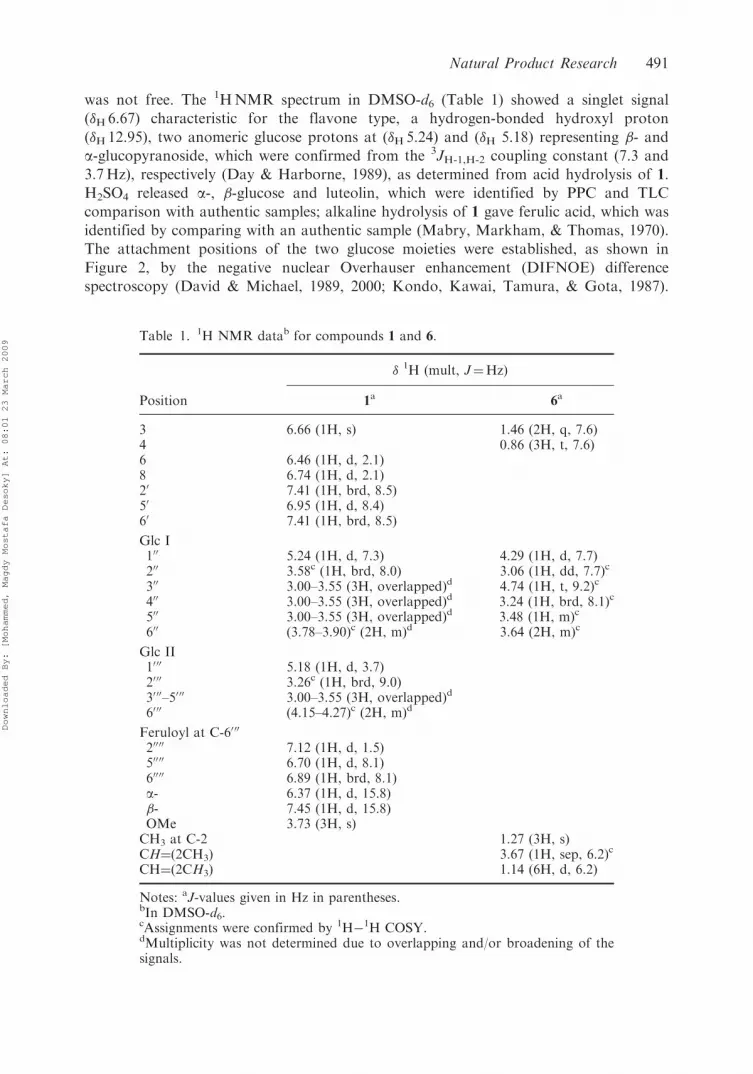
Table 1. 1H NMR datab for compounds 1 and 6.
� 1H (mult, J¼Hz)
Position 1a 6a
3 6.66 (1H, s) 1.46 (2H, q, 7.6)4 0.86 (3H, t, 7.6)6 6.46 (1H, d, 2.1)8 6.74 (1H, d, 2.1)20 7.41 (1H, brd, 8.5)50 6.95 (1H, d, 8.4)60 7.41 (1H, brd, 8.5)
Glc I100 5.24 (1H, d, 7.3) 4.29 (1H, d, 7.7)200 3.58c (1H, brd, 8.0) 3.06 (1H, dd, 7.7)c
300 3.00–3.55 (3H, overlapped)d 4.74 (1H, t, 9.2)c
400 3.00–3.55 (3H, overlapped)d 3.24 (1H, brd, 8.1)c
500 3.00–3.55 (3H, overlapped)d 3.48 (1H, m)c
600 (3.78–3.90)c (2H, m)d 3.64 (2H, m)c
Glc II1000 5.18 (1H, d, 3.7)2000 3.26c (1H, brd, 9.0)3000–50 00 3.00–3.55 (3H, overlapped)d
6000 (4.15–4.27)c (2H, m)d
Feruloyl at C-60 00
20000 7.12 (1H, d, 1.5)50000 6.70 (1H, d, 8.1)60000 6.89 (1H, brd, 8.1)a- 6.37 (1H, d, 15.8)�- 7.45 (1H, d, 15.8)OMe 3.73 (3H, s)CH3 at C-2 1.27 (3H, s)CH¼(2CH3) 3.67 (1H, sep, 6.2)c
CH¼(2CH3) 1.14 (6H, d, 6.2)
Notes: aJ-values given in Hz in parentheses.bIn DMSO-d6.cAssignments were confirmed by 1H�1H COSY.dMultiplicity was not determined due to overlapping and/or broadening of thesignals.
Natural Product Research 491
Downloaded By: [Mohammed, Magdy Mostafa Desoky] At: 08:01 23 March 2009
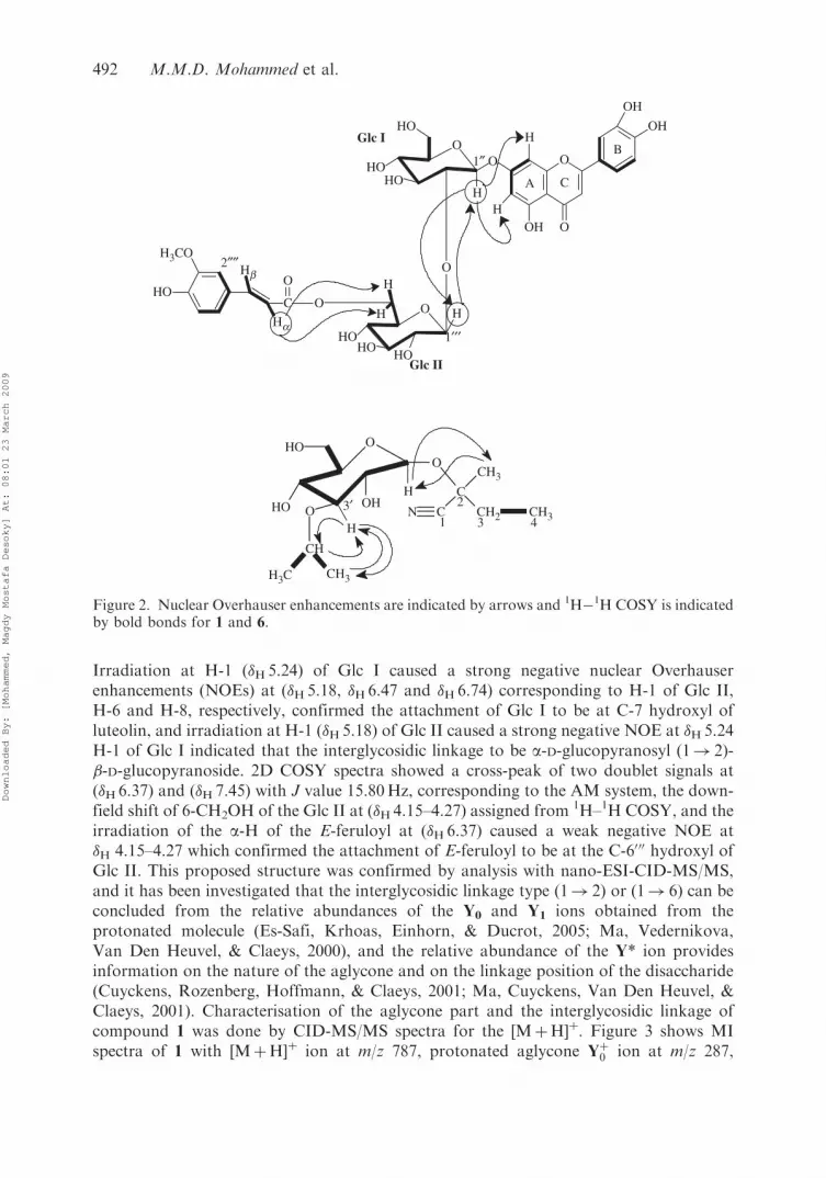
Irradiation at H-1 (�H 5.24) of Glc I caused a strong negative nuclear Overhauserenhancements (NOEs) at (�H 5.18, �H6.47 and �H 6.74) corresponding to H-1 of Glc II,H-6 and H-8, respectively, confirmed the attachment of Glc I to be at C-7 hydroxyl ofluteolin, and irradiation at H-1 (�H5.18) of Glc II caused a strong negative NOE at �H 5.24H-1 of Glc I indicated that the interglycosidic linkage to be a-D-glucopyranosyl (1! 2)-�-D-glucopyranoside. 2D COSY spectra showed a cross-peak of two doublet signals at(�H6.37) and (�H 7.45) with J value 15.80Hz, corresponding to the AM system, the down-field shift of 6-CH2OH of the Glc II at (�H 4.15–4.27) assigned from 1H–1H COSY, and theirradiation of the a-H of the E-feruloyl at (�H 6.37) caused a weak negative NOE at�H 4.15–4.27 which confirmed the attachment of E-feruloyl to be at the C-6000 hydroxyl ofGlc II. This proposed structure was confirmed by analysis with nano-ESI-CID-MS/MS,and it has been investigated that the interglycosidic linkage type (1! 2) or (1! 6) can beconcluded from the relative abundances of the Y0 and Y1 ions obtained from theprotonated molecule (Es-Safi, Krhoas, Einhorn, & Ducrot, 2005; Ma, Vedernikova,Van Den Heuvel, & Claeys, 2000), and the relative abundance of the Y* ion providesinformation on the nature of the aglycone and on the linkage position of the disaccharide(Cuyckens, Rozenberg, Hoffmann, & Claeys, 2001; Ma, Cuyckens, Van Den Heuvel, &Claeys, 2001). Characterisation of the aglycone part and the interglycosidic linkage ofcompound 1 was done by CID-MS/MS spectra for the [MþH]þ. Figure 3 shows MIspectra of 1 with [MþH]þ ion at m/z 787, protonated aglycone Yþ0 ion at m/z 287,
Glc I
Glc II
1″ O
1′′′
2″″
O
O
OH
OH
OH
H
H
A C
B
O H
HOHO
HO
O
HOHO
HO
H
O
C
O
O
H3CO
HO
Hb
Ha
H
H
C
CH3
CN
O
HO OHO
O
CH
HO
CH2H
H3C CH3
CH3
H
1
2
3 43′
Figure 2. Nuclear Overhauser enhancements are indicated by arrows and 1H�1H COSY is indicatedby bold bonds for 1 and 6.
492 M.M.D. Mohammed et al.
Downloaded By: [Mohammed, Magdy Mostafa Desoky] At: 08:01 23 March 2009

fragments at m/z 177 and 339 of feruloylþ and feruloylhexoseþ, respectively (Cuyckenset al., 2003), and the weak abundance of the Y* ion at m/z 625 confirmed the flavone type,where the high relative abundance of Y0 at m/z 287 compared with Y1 at m/z 449 (Y04Y1)confirmed the interglycosidic linkage to be (1! 2) agreed with DIFNOE spectra, and itwas confirmed from different NOE the exact location of the E-feruloyl was at the C-6000
hydroxyl of Glc II, from the CID-MS/MS spectra; the presence of molecular ion 0,4X1 atm/z 321 confirmed the attachment of E-feruloyl at the C-6000 hydroxyl of Glc II (Bylka,Franski, & Stobiecki, 2002). Hence, compound 1 was confirmed to be luteolin 7-O-a-D-(6000-E-feruloyl)glucopyranosyl (1! 2)-�-D-glucopyranoside.
Compound 6 was isolated as a yellow amorphous solid, and its molecular formula wasdetermined to be C14H25NO6 by HRMALDIMS. The IR spectral absorption showed thepresence of hydroxyl (3499 cm�1), C–H stretching (2859–2928 cm�1), nitrile (2252 cm�1),and ether stretching (1011–1094 cm�1) in the molecule. The UV spectrum showedabsorption band at 274 and 320 nm (sh). The 1HNMR in DMSO-d6 (Table 1) showed thespectrum looks like that of compound 8, with extra three signals: doublet (�H1.14 and�C 22.43), multiplet (�H3.67 and �C 73.80), and triplet (�H4.74 and �C 81.98). Analysis of1HNMR and 1H�1H COSY spectra showed a cross-peak correlate triplet methyl signal(�H0.86) with quartet methylene signal (�H 1.46), a cross-peak correlate doublet signalat �H1.14 (6H, –CH¼(2CH3)) with the methene group at �H 3.67 (1H, –CH¼(2Me))corresponding to the isopropyl group, the downfield shift of the methene group (�H3.67)suggested the form of the isopropoxy group, which indicated an attachment to the30-hydroxy of glucose because of the downfield shift of H-30 (�H 4.74), C-30 (�C 81.98); thiswas confirmed by the irradiation of the methene proton (�H 3.67), which caused a strongNOE enhancement at (�H4.74) and (�H1.14) corresponding to H-30 and the doublet signalof the two methyls, respectively. For 13CNMR, see Section 3.
3. Experimental section
3.1. General experimental procedures
UV spectra: Shimadzu MPS-2000. NMR: 300MHz (Varian VXR-Unite) (1H, 1H�1HCOSY and NOE difference) in DMSO-d6. 2D spectra were obtained using a pulse
Figure 3. Collision induced dissociation product ion spectra obtained for [MþH]þ ions and ionnomenclature for 1.
Natural Product Research 493
Downloaded By: [Mohammed, Magdy Mostafa Desoky] At: 08:01 23 March 2009

sequence supplied from Varian. Chemical shifts were given in values (ppm) relative to
trimethylsilane (TMS) as an internal reference. High-resolution ESI/MS: was obtained
using nano-electrospray tandem (MS/MS) mass spectrometry on a hybrid quadrupole
time-of-flight (Q-TOF) MS instrument equipped with Protana’s nano-ESI source for HR
ESI-MS and nano-spray needles from Proxeon (Applied Biosystems/MDS Sciex)
(QSTAR, prototype, PE-Sciex, Canada). Tandem (MS/MS) spectra were interpreted
using the programs BioMultiView (PE Sciex, Canada) and GPMAW (Lighthouse Data,
Denmark). For accurate mass measurements, the instrument was calibrated using a
10mM solution of NaI in isopropanol/water. The instrument’s mass scale was calibrated
for each determined ion mass using the cluster ions Nanþ1Iþn closest to the sought mass.
Collision induced dissociation (CID) spectra were obtained using N2 in the collision cell
and collision energies between 30–40 eV (Elab). Analyses were first conducted using ESI/
MS in positive mode to obtain ionised molecular species. Then tandem MS/MS spectra
were obtained by CID of the [MþH]þ ion (Nielsen, Freese, Cornett, & Dragsted, 2000;
Hakkinen & Auriola, 1998). Protonation is believed to occur preferentially in the aglycone
part of the molecule, in particular at the carbonyl oxygen atom. Charge delocalisation in
the C-ring can lead to a protonated molecular species, which are highly stabilised by
resonance. Subsequent charge-remote rearrangements take place, resulting in the Y0 and
Y1 ions, most likely involving hydrogen rearrangement from hydroxyl groups, which can
sterically approach the glycosidic bonds (Ma et al., 2000). The product ion spectra were
obtained in the continuous mode of acquisition of the quadruple analyser. Sequence ion
notations have been used, e.g. the Y1 and Y0 corresponding to the [MþH� (176uþ 162)]þ
and [MþH� (176uþ 162þ 162)]þ ions, respectively (Domon & Costello, 1988). An
irregular ion [MþH� 162]þ corresponding to the loss of an internal dehydrated glucose
residue from the precursor ions is denoted as a Y* ion (Ma et al., 2000). High-resolution
MALDIMS was recorded on an IonSpec Fourier Transform Ion Cyclotron resonance
mass spectrometer. HPLC consists of L-6200 Intelligent Pump (Merck-HITACHI)
connected with UV-VIS Detector SPD-10AV (SHIMADZU). HPLC solvents used for all
analyses were of grade M (Sigma-Aldrich Chemie, UK) and ultra-pure water; the
petroleum ether, chloroform and methanol for plant extraction were of AR grade; Kiesel
gel 60 F254 (Merck) was used for analytical TLC.
3.2. Plant material
The aerial parts (leaves and seeds) of L. grandiflorum were collected in March 2006 from
El-Orman Garden, Giza Governorate, Egypt. The plant samples were kindly identified by
Miss Tressa Labib, Head of Specialists at the Garden. A voucher specimen of the plant is
kept at the Herbarium of the National Research Center.
3.3. Extraction and isolation
The air-dried aerial parts (leaves (2.4 kg) and seeds (184.46 gm)) of L. grandiflorum were
extracted with hot MeOH which was defatted with petroleum ether and then the residue
was dissolved in H2O, followed by fractionation with CHCl3. Then the H2O soluble
fraction was concentrated till dryness and extracted with MeOH, which was examined with
TLC (silica gel 60 F254, Fluka) for flavonoids with different solvent systems; the spots were
494 M.M.D. Mohammed et al.
Downloaded By: [Mohammed, Magdy Mostafa Desoky] At: 08:01 23 March 2009
visualised by the dipping solutions (Jork, Funk, Fischer, & Wimmer, 1990, 1994; Marston
& Hostettmann, 2006). The MeOH fraction (15.5 gm) was dissolved in MeOH–H2O (1 : 1)
and subjected to fractionation (8mL injected into 10mL loop, after prefiltration with
nylon filter 0.45mm) using preparative ODS-HPLC (20’� 250mm, Develosil ODS-HG-5,
Nomura Chemicals) at room temperature. Solvent A¼ 1% HCOOH and Solvent
B¼ 100% MeOH were used in the elution profile 90% A, 50% A, and finally with
100% B, monitoring at 340 nm, with flow rate 3mLmin�1. This accumulated three main
fractions (I, II and III). After TLC examinations, fraction II (50% A) was chosen to be
refractionated using preparative ODS-HPLC at room temperature, linearly with 60% A,
monitoring at 280 nm, with 0.8mLmin�1 flow rate, and led to the isolation of 1 (5mg),
2 (12mg), 3 (8mg), 4 (15mg), 5 (10mg), 6 (10mg), 7 (8mg), 8 (12mg), 9 (6mg) and
10 (11mg).
3.3.1. Luteolin 7-O-a-D-(6000-E-feruloyl)glucopyranosyl (1!2)-�-D-glucopyranoside (1)
Pale yellow amorphous; UV �max (MeOH): 252, 269, 300(sh), 337; (NaOMe): 268, 390;
(AlCl3): 275, 300(sh), 325, 360(sh), 423; (AlCl3/HCl): 277, 298(sh), 325, 385; (NaOAc):
265(sh), 269(sh), 335, 408; (NaOAc/H3BO3): 258, 296(sh), 335, 376; 1HNMR
(DMSO-d6, 300MHz) � 5.24 (1H, d, J¼ 7.32Hz, H-100 of Glc I), 3.58 (1H, brd,
J¼ 8.0Hz, H-200), 3.78–3.90 (2H, m, H-600), 5.18 (1H, d, J¼ 3.66Hz, H-1000 of Glc II),
3.26 (1H, brd, J¼ 9.0Hz, H-2000), 4.15–4.27 (2H, m, H-6000), 3.00–3.55 (6H, overlapped,
H-300, 400, 500, 3000, 4000 and 5000) 6.67 (1H, s, H-3), 6.46 (1H, d, J¼ 2.1Hz, H-6), 6.74
(1H, d, J¼ 2.1Hz, H-8), 7.41 (2H, brd, J¼ 8.5Hz, H-20, 60), 6.95 (1H, d, J¼ 8.4Hz,
H-50), 3.73 (3H, s, feruloyl-OMe), 7.12 (1H, d, J¼ 1.5Hz, H-20000), 6.70 (1H, d,
J¼ 8.1 Hz, H-50000), 6.89 (1H, brd, J¼ 8.1 Hz, H-60000), 6.37 (1H, d, J¼ 15.80Hz, H-a),7.45 (1H, d, J¼ 15.8Hz, H-�); DIFNOE and COSY spectra: (Figure 2); HRESIMS
(Positive mode) m/z 809.1917 [MþNa]þ (Calcd for C37H38O19Na, 809.1905) (see
Figure 3 for the fragmentations).
3.3.2. 2-[(30-isopropoxy-O-�-D-glucopyranosyl)oxy]-2-methylbutanenitrile (6)
Yellow amorphous; IR �max 3499, 2859, 2928, 2252, 1094, 1011 cm�1; UV �max (MeOH):
274, 320 (sh); 1HNMR (DMSO-d6, 300MHz) � 0.86 (3H, t, J¼ 7.61Hz, CH3-4), 1.14 (6H,
d, J¼ 6.23Hz, –CH¼(2CH3)), 1.27 (3H, s, CH3), 1.46 (2H, q, J¼ 7.61Hz, CH2-3), 3.06
(1H, dd, J¼ 7.69Hz, H-20), 3.24 (1H, brd, J¼ 8.05Hz, H-40), 3.48 (1H, m, H-50), 3.64 (2H,
m, H-60), 3.67 (1H, sep, J¼ 6.23 Hz, –CH¼(2Me)), 4.29 (1H, d, J¼ 7.69Hz, H-10), 4.74
(1H, t, J¼ 9.16Hz, H-30); 13CNMR (DMSO-d6, 125MHz) �C 121.5 (C�N), 74.5 (C-2),
32.9 (C-3), 8.6 (C-4), 104.5 (C-10), 74.6 (C-20), 81.9 (C-30), 71.9 (C-40), 78.1 (C-50), 62.6
(C-60), 24.6 (CH3-2), 73.8 (–CH¼(2CH3)), 22.4 (–CH¼(2CH3)); DIFNOE and COSY
spectra: (Figure 2); HRMALDIMS (Positive mode) m/z 326.1579 [MþNa]þ, (Calcd
for C14H25NO9Na, 326.1575) m/z 303 [MþH]þ, m/z 284 [MþNa� (:C(CH3)2)]þ, m/z 611
[2MþNa�H2O]þ.
Acknowledgements
The authors are grateful to the Danish Institute of Agricultural Sciences, Research Center Aarslev,Denmark for financial support, and to the University of Southern Denmark, Odense, Denmark, forproviding the facilities for instrumental analysis.
Natural Product Research 495
Downloaded By: [Mohammed, Magdy Mostafa Desoky] At: 08:01 23 March 2009

References
Bown, D. (1995). Encyclopaedia of herbs and their uses. London: Dorling Kindersley.Bylka, W., Franski, R., & Stobiecki, M. (2002). Differentiation between isomeric acacetin-6-C-(600-
O-malonyl)glucoside and acacetin-8-C-(600-O-malonyl)glucoside by using low-energy CID
mass spectra. Journal of Mass Spectrometry: JMS, 37, 648–650.Cable, J., & Nocke, H. (1975). Isolation of s-Butyl B-D-glucopyranoside from Acripeza reticulata.
Australian Journal of Chemistry, 28, 2737–2739.Cecil Jr, R.S., David, W., Roger, W., Miller, I.S.P., & Oscar, E.O. (1980). Linustatin and
neolinustatin: Cyanogenic glycosides of Linseed meal that protect animals against selenium
toxicity. Journal of Organic Chemistry, 45, 507–510.
Cuyckens, F., Rozenberg, R., Hoffmann, E.-d., & Claeys, M. (2001). Structure characterization of
flavonoid O-diglycosides by positive and negative nano-electrospray ionization ion trap mass
spectrometry. Journal of Mass Spectrometry: JMS, 36, 1203–1210.Cuyckens, F., Shahat, A.A., Van Den Heuvel, H., Abdel-Shafeek, K.A., El-Messiry, M.M., Seif,
El-Nasr, et al. (2003). The application of LC-ESI-MS and Collision-induced dissociation in
the structural characterization of acylated flavonol O-glycosides from the seeds of Carrichtera
annua. Eurpean Journal of Mass Spectrometry, 9, 409–420.David, N., & Michael, P.W. The nuclear Overhauser effect in structural and conformational analysis.
Wiley-VCH Publishers Inc.: New York (1989) and (2000).Day, P.M., & Harborne, J.B. (1989). Plant phenolics, In Methods in plant biochemistry (Vol. 1).
London: Academic Press.Domon, B., & Costello, C. (1988). A systematic nomenclature for carbohydrate fragments in FAB-
MS/MS spectra of glycoconjugates. Glycoconjugate Journal, 5, 397–409.Duke, J.A. (2002). Handbook of medicinal herbs (2nd ed.). Boca Raton, FL: CRC Press.Es-Safi, N.-E., Krhoas, L., Einhorn, J., & Ducrot, P.-H. (2005). Application of ESI/MS, CID/MS
and tandem MS/MS to the fragmentation study of eriodictyol 7-O-glucosyl-(1!2)-glucoside
and luteolin 7-O-glucosyl-(1!2)-glucoside. International Journal of Mass Spectrometry, 247,
93–100.
Hakkinen, S., & Auriola, S. (1998). High-performance liquid chromatography with electrospray
ionization mass spectrometry and diode array ultraviolet detection in the identification of
flavonol aglycones and glycosides in berries. Journal of Chromatography, 829, 91–100.Harborne, J.B. (1994). The flavonoids in advances in research since 1986. New York: Chapman &
Hall.Hortus, E.Z. (1976). Liberty hyde bailey hortorium. New York, USA: MacMillan Publishing
Company.Jork, H. Funk, W. Fischer, W., & Wimmer, H. Thin-layer chromatography. VCH,
Verlagsgesellschaft mbH, D-6940 Weinheim, Germany, Vol. 1 (1990) and (1994).Kenjiro, T., Norio, S., Koji, H., Atsushi, S., & Toshio, H. (1995). Delphinidin 3-xylosylrutinoside in
petals of Linum grandiflorum. Phytochemistry, 39, 243–245.Kondo, T., Kawai, T., Tamura, H., & Gota, T. (1987). Structure determination of heavenly blue
anthocyanin, a complex monomeric anthocyanin from the morning glory Ipomoea tricolor, by
means of the negative NOE method. Tetrahedron Letters, 28, 2273–2276.Ma, Y.L., Cuyckens, F., Van Den Heuvel, H., & Claeys, M. (2001). Mass spectrometric methods for
the characterisation and differentiation of isomeric O-diglycosyl flavonoids. Phytochemical
Analysis: PCA, 12, 159–165.
Ma, Y.L., Vedernikova, I., Van Den Heuvel, H., & Claeys, M. (2000). Internal glucose residue loss
in protonated O-diglycosyl flavonoids upon low-energy collision-induced dissociation. Journal
of the American Society for Mass Spectometry, 11, 136–144.Mabry, T.J., Markham, K.R., & Thomas, M.B. (1970). The systematic identification of flavonoids.
Berlin: Springer.
496 M.M.D. Mohammed et al.
Downloaded By: [Mohammed, Magdy Mostafa Desoky] At: 08:01 23 March 2009

Marston, A., & Hostettmann, K. (2006). Separation and quantification of flavonoids.In Ø.M. Andersen & K.R. Markham (Eds.), Flavonoids: Chemistry, biochemistry andapplications. Boca Raton, FL: Taylor and Francis CRC Press.
Nielsen, S.E., Freese, R., Cornett, C., & Dragsted, L.O. (2000). Identification and quantification of
flavonoids in human urine samples by column-switching liquid chromatography coupled toatmospheric pressure chemical ionization mass spectrometry. Analytical Chemistry, 72,1503–1509.
Phillips, R., & Foy, N. (1991). Herbs. London: Pan Books Ltd.Plesser, A.G. (1966). The variation in fatty acid composition of the seed of Linum species. Canadian
Journal of Genetics and Cytology. Journal Canadien de genetique et de cytologie, 8, 328–335.
Seikel, M.K., Chow, J.H.S., & Feldman, L. (1966). The glycoflavonoid pigments of Vitex lucenswood. Phytochemistry, 5, 439–445.
Yermanos, D.M. (1966). Variation in seed oil composition of 43 Linum species. Journal of American
Oil Chemists’ Society, 43, 546–549.Yermanos, D.M., Bcard, B.H., Gill, K.S., & Anderson, M.P. (1966). Fatty acid composition of seed
oil of wild species of Linum. Agronomy Journal, 58, 30–32.
Natural Product Research 497
Downloaded By: [Mohammed, Magdy Mostafa Desoky] At: 08:01 23 March 2009