charles university in prague faculty of physical
TRANSCRIPT

CHARLES UNIVERSITY IN PRAGUE
FACULTY OF PHYSICAL EDUCATION AND SPORT
Lumbar Disc Herniation
Conservative versus Non-conservative Treatment
Master Thesis
Author: Supervisor:
Iakovos Diogenous Ass. Prof. PaedDr. Dagmar Pavlů,CSc.
CHARLES UNIVERSITY IN PRAGUE
FACULTY OF PHYSICAL EDUCATION AND SPORT
Lumbar Disc Herniation
Conservative versus Non-conservative Treatment
Master Thesis
Author: Supervisor:
Iakovos Diogenous Ass. Prof. PaedDr. Dagmar Pavlů,CSc.
Abstract
Objectives: The purpose of this Thesis is to create a general review to describe methods,
results and conclusion of treatments for lumbar disc herniation. To compare conservative
with non conservative treatment by meaning of using systematic reviews literature and
clinical trials.
Selection Criteria: We independently assessed each title and abstract using predetermined
inclusion criteria based on intervention, population, outcome measures, and study design
criteria. Full papers, reports, and meeting abstracts that met inclusion criteria were retrieved
and reviewed independently.
Results: A significant improvement in surgery in evaluation in the first six months which
follows in a slight decreasment the first 2 years comparing with conservative approach.
Microdicectomy effectiveness is comparable with standard discectomy. A stiff and flat back
has a good prognosis after lumbar disc surgery. Conservative treatments can improve patient
but there were no statistical significant however good results were shown in evaluation and
exercise by mckenzie also physical therapy had some good results with exercises, traction
with greater result with chiropractic approach. Conservative is needed after surgey for better
and longer timing results. By MRI finding there was conclusion that only with natural history
and conservative approach a disc herniation can relapse.
Key Words: Spinal disc herniation, surgery, conservative, sciatica, radiculopathy, back-leg
pain.

Declaration
Herewith I declare that I worked on this thesis on my own and independently I researched the
list of literature and data‟s, which included in this work. I elaborated using the literatures
listed and attached in the reference section and the knowledge I have gain during my study
time at Charles University.
IAKOVOS DIOGENOUS
Prague 2011

Dedication
I dedicate this diploma to my parents Andreas and Gianoulla Diogenous who have helped me
through my years of studies. I owe them everythin what i have achieved so far.
Acknowledgment
I would like to aknowledge all of my familly, my parents and my sisters family for their
support. Special thanks to all my friends for the support and their long lasting friendship
namely i will like to thank Giannis Polyviou and Apeslidis Theodoros.
I like to thank all my proffesors during my study time in Faculty of Physical Education.
Grateful thanks to my supervisor Ass. Prof. PaedDr. Dagmar Pavlů, CSc, for her guidance
during my studies and during my diploma thesis
Also grateful thanks to Doc. MUDr. František Véle, CSc. who explained us a great deal of
knowledge during my studies, he connected all the physiotherapy knowlede and described in
the most intresting way for a better understanding for us students.

Herewith I primate to lend my diploma thesis, for study purpose, I encourage my Colleagues‟
to quote and cite from this thesis.
Aims and Goals
Aim
To find evidence to compare conservative and non-conservative treatments for lumbar disc
herniations (mainly mentioned, L4/5 – L5/S1).
Goals
To determine if conservative is more or less beneficial from non-conservative
treatment.
Individualizing conservative treatments and finding their efficient potentials
To find the amount of relapses of disc herniation after surgery, with comparing
untreated herniations after conservative treatments.
To differentiate stages of herniation with treatment benefit
Role of body function with disc herniation
A general evaluation of our findings and making a general conclusion for a lumbar
herniated disc treatment if possible.
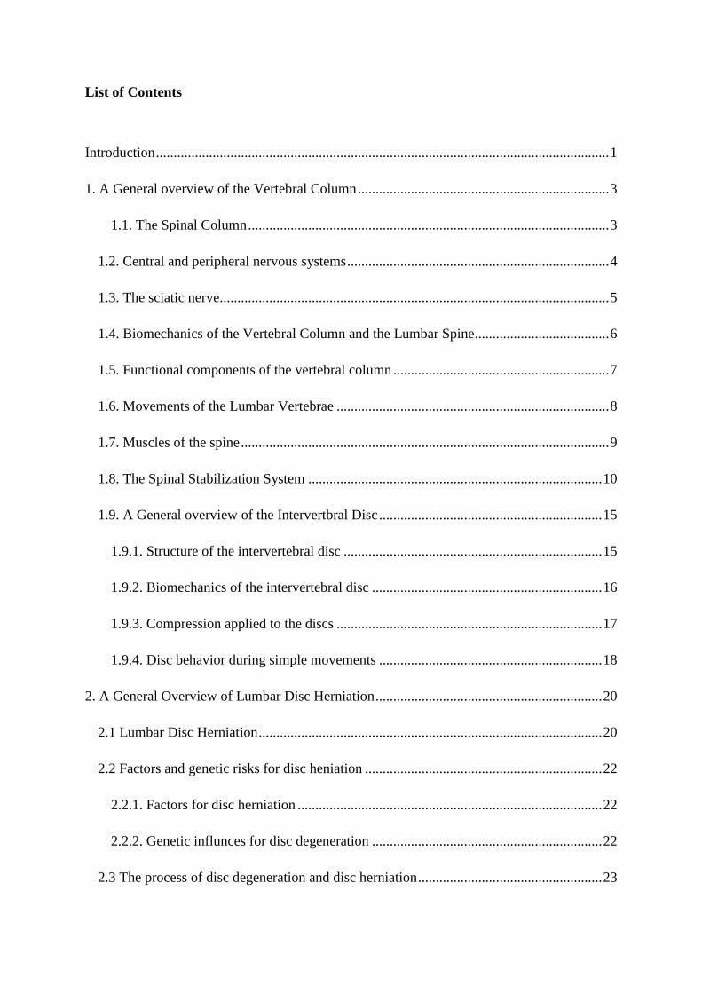
List of Contents
Introduction ................................................................................................................................ 1
1. A General overview of the Vertebral Column ....................................................................... 3
1.1. The Spinal Column ...................................................................................................... 3
1.2. Central and peripheral nervous systems .......................................................................... 4
1.3. The sciatic nerve.............................................................................................................. 5
1.4. Biomechanics of the Vertebral Column and the Lumbar Spine...................................... 6
1.5. Functional components of the vertebral column ............................................................. 7
1.6. Movements of the Lumbar Vertebrae ............................................................................. 8
1.7. Muscles of the spine ........................................................................................................ 9
1.8. The Spinal Stabilization System ................................................................................... 10
1.9. A General overview of the Intervertbral Disc ............................................................... 15
1.9.1. Structure of the intervertebral disc ......................................................................... 15
1.9.2. Biomechanics of the intervertebral disc ................................................................. 16
1.9.3. Compression applied to the discs ........................................................................... 17
1.9.4. Disc behavior during simple movements ............................................................... 18
2. A General Overview of Lumbar Disc Herniation ................................................................ 20
2.1 Lumbar Disc Herniation ................................................................................................. 20
2.2 Factors and genetic risks for disc heniation ................................................................... 22
2.2.1. Factors for disc herniation ...................................................................................... 22
2.2.2. Genetic influnces for disc degeneration ................................................................. 22
2.3 The process of disc degeneration and disc herniation .................................................... 23
2.4. Disc prolapse and the mechanism of nerve root compression ...................................... 25
2.5. Disc herniation provoking radicular syndrome ............................................................. 27
2.6. Differential diagnosis in Sciatic pain (17)
....................................................................... 28
2.6.1. Neuromuscular causes ............................................................................................ 28
2.6.2. Systemic/ extraspinal causes (17)
............................................................................. 29
2.6.3. Risk factors for sciatica .......................................................................................... 30
2.7. Diffferential classifications for low back pain and sciatica .......................................... 31
2.7.1. Mechanical pain syndromes described by Mckenzie ............................................. 31
2.7.2. Patho-mechanism classification for back related leg pain...................................... 32
2.7.3. Radicular syndromes in the lower extremities ....................................................... 33
2.7.4. Clinical signs and characteristics of disc herniation ............................................... 34
2.7.5. Problems in diagnosing radicular syndromes ......................................................... 35
3. Clinical evaluation and treatment procedure for lumbar disc herniation ............................. 37
3.1. Examination procedure ................................................................................................. 37
3.2. Symptoms and clincal findings of lumbar radicular syndromes ................................... 40
3.3. Physical examinations of lumbar disc herniation .......................................................... 42
3.4. Imaging techniques diagnosing lumbar disc herniation ................................................ 47
3.5. Therapy Procedures for disc herniation ........................................................................ 49
3.5. 1 Conservative therapy procedures............................................................................ 49
3.5.2. Non- conservative therapy approaches ................................................................... 55
4. Methodology ........................................................................................................................ 60
5. Conservative versus non conservative treatments trials for disc herniations ...................... 62
5.1. Non conservative treatments ......................................................................................... 62
5.1.1. Surgery versus non operative treatment for disc herniation ................................... 62
5.1.2. Minimal Invasive procedures research ................................................................... 71
5.2. Conservative treatments: Medications .......................................................................... 78
5.2.1 Oral Medications ..................................................................................................... 78
5.2.2. Injection .................................................................................................................. 79
5.3. Non-medical Conservative treatments .......................................................................... 84
5.3.1. Natural History ....................................................................................................... 84
5.3.2. Bed rest ................................................................................................................... 85
5.3.3. Physical therapy ...................................................................................................... 86
5.3.4. Traction ................................................................................................................... 87
5.3.5. Mckenzie therapy centralisation method ................................................................ 90
5.3.6. Chiropractic approach in general radiculopathy ..................................................... 92
5.3.7. Stabilization theory with case treatment ................................................................. 96
5.4. Post-surgery of disc herniation ...................................................................................... 98
5.5. Reoperation of disc herniations ................................................................................... 103
6. Results ................................................................................................................................ 107
7. Discussion .......................................................................................................................... 113
8. Conclusion ......................................................................................................................... 117
Refernces................................................................................................................................ 118
1
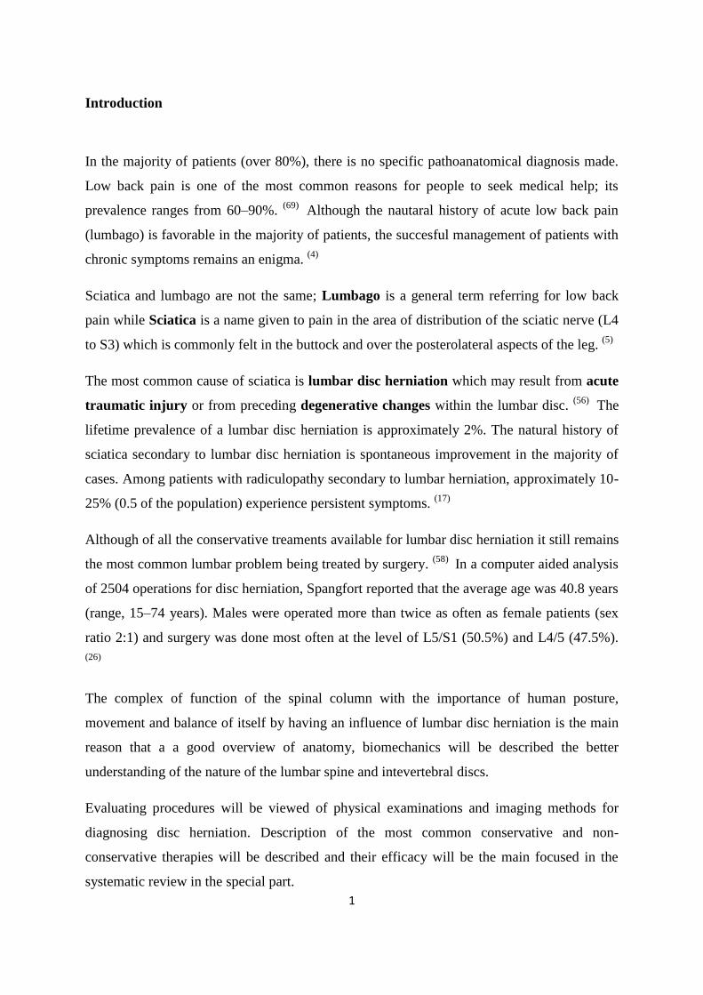
Introduction
In the majority of patients (over 80%), there is no specific pathoanatomical diagnosis made.
Low back pain is one of the most common reasons for people to seek medical help; its
prevalence ranges from 60–90%. (69)
Although the nautaral history of acute low back pain
(lumbago) is favorable in the majority of patients, the succesful management of patients with
chronic symptoms remains an enigma. (4)
Sciatica and lumbago are not the same; Lumbago is a general term referring for low back
pain while Sciatica is a name given to pain in the area of distribution of the sciatic nerve (L4
to S3) which is commonly felt in the buttock and over the posterolateral aspects of the leg. (5)
The most common cause of sciatica is lumbar disc herniation which may result from acute
traumatic injury or from preceding degenerative changes within the lumbar disc. (56)
The
lifetime prevalence of a lumbar disc herniation is approximately 2%. The natural history of
sciatica secondary to lumbar disc herniation is spontaneous improvement in the majority of
cases. Among patients with radiculopathy secondary to lumbar herniation, approximately 10-
25% (0.5 of the population) experience persistent symptoms. (17)
Although of all the conservative treaments available for lumbar disc herniation it still remains
the most common lumbar problem being treated by surgery. (58)
In a computer aided analysis
of 2504 operations for disc herniation, Spangfort reported that the average age was 40.8 years
(range, 15–74 years). Males were operated more than twice as often as female patients (sex
ratio 2:1) and surgery was done most often at the level of L5/S1 (50.5%) and L4/5 (47.5%).
(26)
The complex of function of the spinal column with the importance of human posture,
movement and balance of itself by having an influence of lumbar disc herniation is the main
reason that a a good overview of anatomy, biomechanics will be described the better
understanding of the nature of the lumbar spine and intevertebral discs.
Evaluating procedures will be viewed of physical examinations and imaging methods for
diagnosing disc herniation. Description of the most common conservative and non-
conservative therapies will be described and their efficacy will be the main focused in the
systematic review in the special part.

2
This thesis is a systematic review of literature and trial on the efficacy of the most common
conservative treatments and non-conservative treatment of disc herniations in the levels of the
lower lumbar spines which provokes sciatica. This study will provide important findings
necessary for the patients but also for the future researchers of the efficacy of non-
conservative treatments for sciatica.
3
1. A General overview of the Vertebral Column
1.1. The Spinal Column
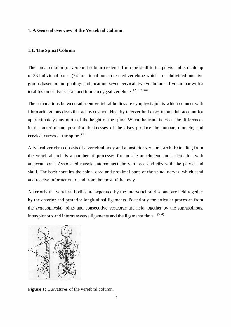
The spinal column (or vertebral column) extends from the skull to the pelvis and is made up
of 33 individual bones (24 functional bones) termed vertebrae which are subdivided into five
groups based on morphology and location: seven cervical, twelve thoracic, five lumbar with a
total fusion of five sacral, and four coccygeal vertebrae. (28, 12, 44)
The articulations between adjacent vertebral bodies are symphysis joints which connect with
fibrocartilaginous discs that act as cushion. Healthy intervertbral discs in an adult account for
approximately one/fourth of the height of the spine. When the trunk is erect, the differences
in the anterior and posterior thicknesses of the discs produce the lumbar, thoracic, and
cervical curves of the spine. (19)
A typical vertebra consists of a vertebral body and a posterior vertebral arch. Extending from
the vertebral arch is a number of processes for muscle attachment and articulation with
adjacent bone. Associated muscle interconnect the vertebrae and ribs with the pelvic and
skull. The back contains the spinal cord and proximal parts of the spinal nerves, which send
and receive information to and from the most of the body.
Anteriorly the vertebral bodies are separated by the intervertebral disc and are held together
by the anterior and posterior longitudinal ligaments. Posteriorly the articular processes from
the zygapophysial joints and consecutive vertebrae are held together by the supraspinous,
interspionous and intertransverse ligaments and the ligamenta flava. (3, 4)
Figure 1: Curvatures of the veretbral column.
4
1.2. Central and peripheral nervous systems
The brain and spinal cord constitute the central nervous system (CNS); the cranial nerves and
spinal nerves form the peripheral nervous systems (PNS). The vertebral column and
associated soft tissue of the back contain the spinal cord and proximal parts of the spinal
nerves. (5)
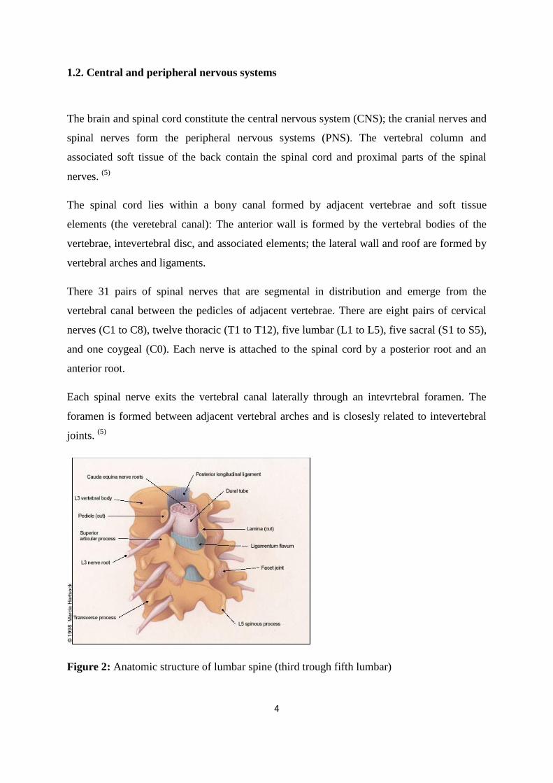
The spinal cord lies within a bony canal formed by adjacent vertebrae and soft tissue
elements (the veretebral canal): The anterior wall is formed by the vertebral bodies of the
vertebrae, intevertebral disc, and associated elements; the lateral wall and roof are formed by
vertebral arches and ligaments.
There 31 pairs of spinal nerves that are segmental in distribution and emerge from the
vertebral canal between the pedicles of adjacent vertebrae. There are eight pairs of cervical
nerves (C1 to C8), twelve thoracic (T1 to T12), five lumbar (L1 to L5), five sacral (S1 to S5),
and one coygeal (C0). Each nerve is attached to the spinal cord by a posterior root and an
anterior root.
Each spinal nerve exits the vertebral canal laterally through an intevrtebral foramen. The
foramen is formed between adjacent vertebral arches and is closesly related to intevertebral
joints. (5)
Figure 2: Anatomic structure of lumbar spine (third trough fifth lumbar)
5
In the lumbar regions, the cone-shaped terminus of the spinal cord (conus medullaris)
normally ends at about the L1 or L2 level in adults. Caudal to these levels, the roots of the
cauda equina are contained within the subarachnoid space of the dura-enclosed. Thus
pathologies (stenosis, herniation) under these levels results in nerve root dysfunction rather
than spinal cord dysfunction. (78)
1.3. The sciatic nerve
The lumbar intervertebral foramen (neural passageways) is relatively larger but nerve root
compression is more common than in the thoracic spine. Nerve roots exit the spinal canal
through small passageways between the vertebrae and discs. Pain and other symptoms can
develop when the damaged disc pushes into the spinal canal or nerve roots. (44)
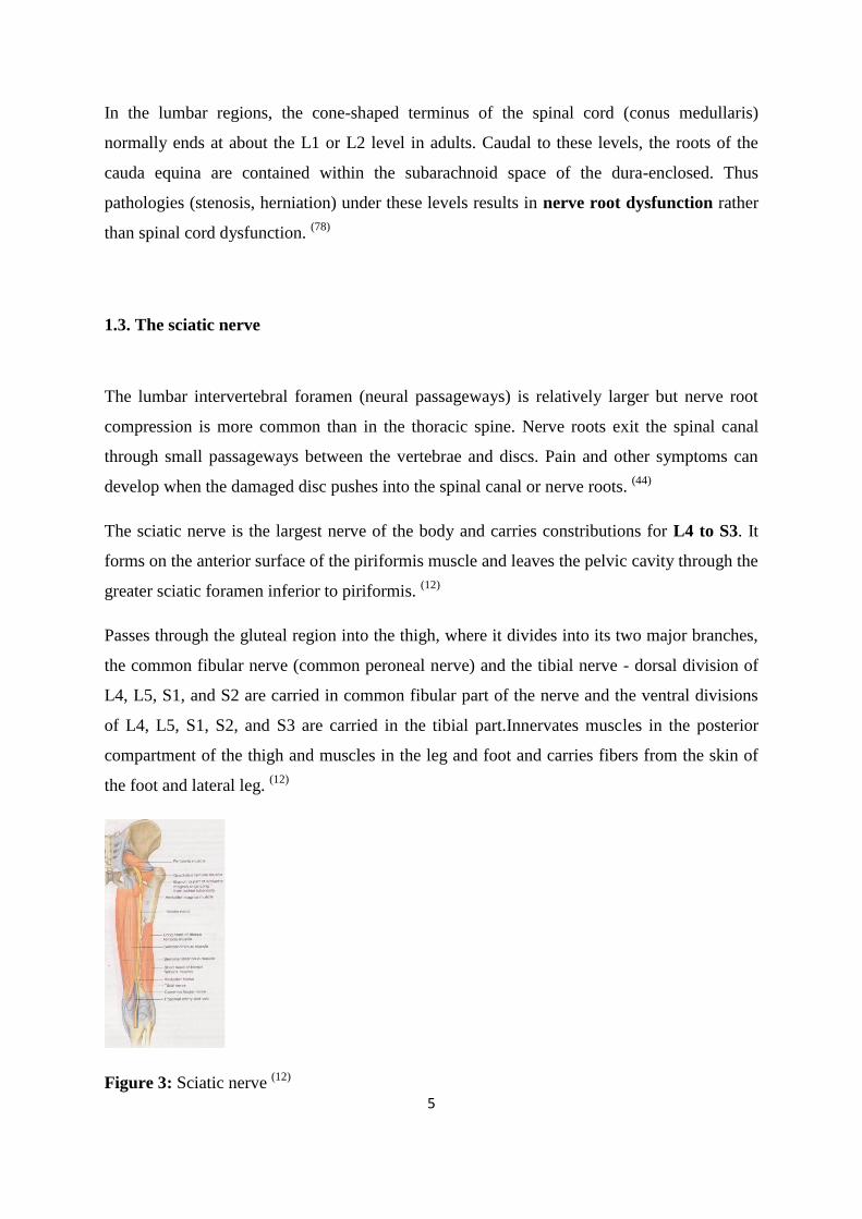
The sciatic nerve is the largest nerve of the body and carries constributions for L4 to S3. It
forms on the anterior surface of the piriformis muscle and leaves the pelvic cavity through the
greater sciatic foramen inferior to piriformis. (12)
Passes through the gluteal region into the thigh, where it divides into its two major branches,
the common fibular nerve (common peroneal nerve) and the tibial nerve - dorsal division of
L4, L5, S1, and S2 are carried in common fibular part of the nerve and the ventral divisions
of L4, L5, S1, S2, and S3 are carried in the tibial part.Innervates muscles in the posterior
compartment of the thigh and muscles in the leg and foot and carries fibers from the skin of
the foot and lateral leg. (12)
Figure 3: Sciatic nerve (12)
6
1.4. Biomechanics of the Vertebral Column and the Lumbar Spine
The spine builds the axis of the body. It is not straight as a rod but it is curved. It implies
cervical lordosis, thoracic kyphosis, and lumbar lordosis. Slight spinal curvatures represent
static spine with overloading intervertebral discs and tendency to disc protrusion. Increased
spinal curvatures represent dynamic spine; loading more the hip joint than the spine. (71)
By being curved, the lumbar spine is protected to an appreciable extent from compressive
forces and shocks. In a straight lumbar spine, an axial compressive force would be
transmitted through the vertebral bodies and intervertebral disc, and the only mechanism to
protect the lumbar vertebrae would be the shock-absorbing capacity of the intervertebral
discs. (3, 4)
The lumbar vertebrae graduate in size from L1 through L5. The pedicles are longer and wider
than those in the thoracic spine. The spinous processes are horizontal and more squared in
shape. The discs of the lumbar spine are approximately 7–10 mm thick and 40 mm in
diameter (anterior-posterior), representing one-third of the height of the spine. (5)
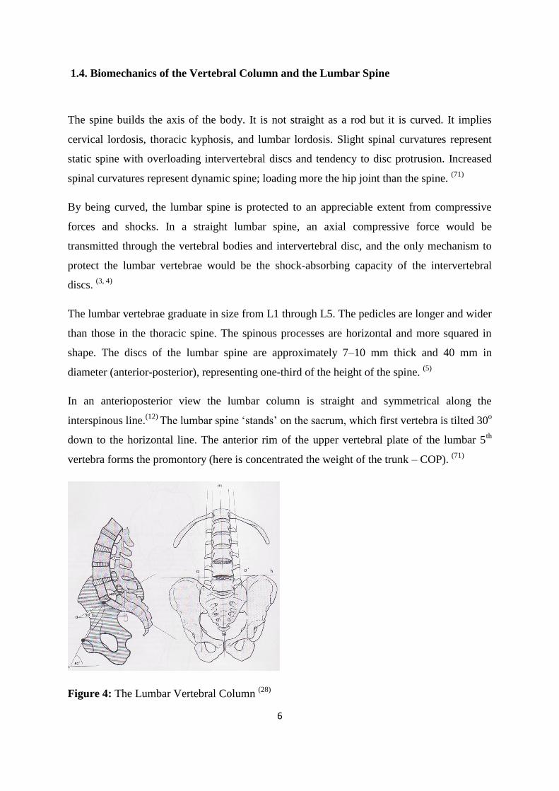
In an anterioposterior view the lumbar column is straight and symmetrical along the
interspinous line.(12)
The lumbar spine „stands‟ on the sacrum, which first vertebra is tilted 30o
down to the horizontal line. The anterior rim of the upper vertebral plate of the lumbar 5th
vertebra forms the promontory (here is concentrated the weight of the trunk – COP). (71)
Figure 4: The Lumbar Vertebral Column (28)
7
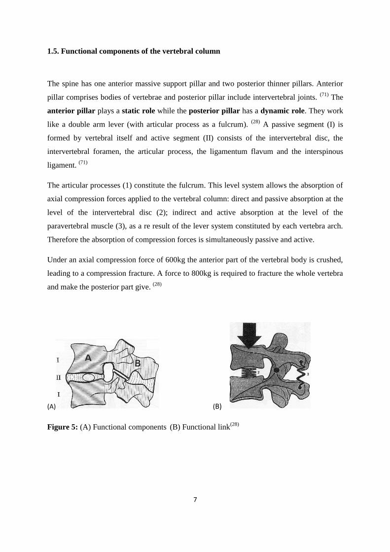
1.5. Functional components of the vertebral column
The spine has one anterior massive support pillar and two posterior thinner pillars. Anterior
pillar comprises bodies of vertebrae and posterior pillar include intervertebral joints. (71)
The
anterior pillar plays a static role while the posterior pillar has a dynamic role. They work
like a double arm lever (with articular process as a fulcrum). (28)
A passive segment (I) is
formed by vertebral itself and active segment (II) consists of the intervertebral disc, the
intervertebral foramen, the articular process, the ligamentum flavum and the interspinous
ligament. (71)
The articular processes (1) constitute the fulcrum. This level system allows the absorption of
axial compression forces applied to the vertebral column: direct and passive absorption at the
level of the intervertebral disc (2); indirect and active absorption at the level of the
paravertebral muscle (3), as a re result of the lever system constituted by each vertebra arch.
Therefore the absorption of compression forces is simultaneously passive and active.
Under an axial compression force of 600kg the anterior part of the vertebral body is crushed,
leading to a compression fracture. A force to 800kg is required to fracture the whole vertebra
and make the posterior part give. (28)
(A) (B)
Figure 5: (A) Functional components (B) Functional link
(28)
8
1.6. Movements of the Lumbar Vertebrae
During flexion and extension, the vertebral bodies roll over the nucleus while the facet joints
guide the movements. (19)
The greatest motion in the lumbar spine occurs between L4/L5 and
L5/S1. There is considerable individual variability in the range of motion of the lumbar spine.
In reality, little obvious movement occurs in the lumbar spine as a result of the shape of the
facet joints, tightness of the ligaments, intervertebral discs and size of the vertebral.
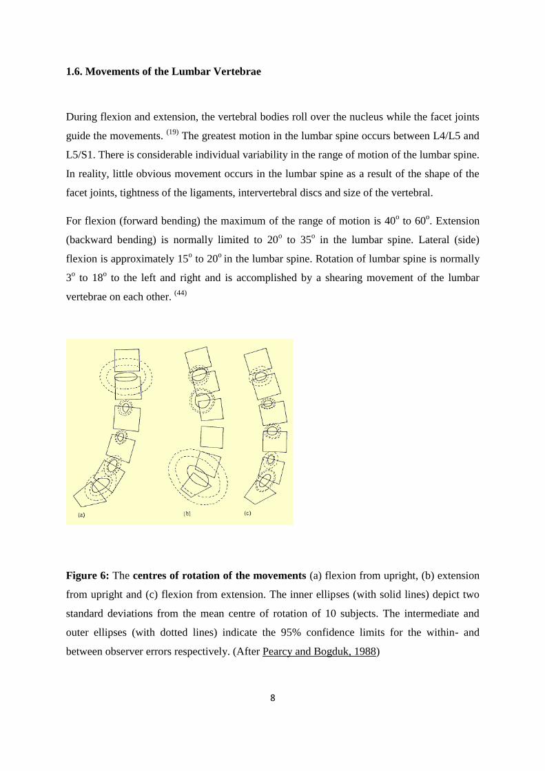
For flexion (forward bending) the maximum of the range of motion is 40o to 60
o. Extension
(backward bending) is normally limited to 20o to 35
o in the lumbar spine. Lateral (side)
flexion is approximately 15o to 20
o in the lumbar spine. Rotation of lumbar spine is normally
3o to 18
o to the left and right and is accomplished by a shearing movement of the lumbar
vertebrae on each other. (44)
Figure 6: The centres of rotation of the movements (a) flexion from upright, (b) extension
from upright and (c) flexion from extension. The inner ellipses (with solid lines) depict two
standard deviations from the mean centre of rotation of 10 subjects. The intermediate and
outer ellipses (with dotted lines) indicate the 95% confidence limits for the within- and
between observer errors respectively. (After Pearcy and Bogduk, 1988)
9
1.7. Muscles of the spine
The spine is an elastic column, with enhanced stability due to the complex curvature of the
spine (kyphosis and lordosis), the support of the longitudinal ligaments, the elasticity of the
ligamentum flavum, and most importantly the active muscle forces. In cadaver spines have
been shown to buckle with the application of very low vertical loads (20–40 N), however the
extrinsic support stabilizes and redistributes loading on the spine are the trunk muscles
making the spine to withstand loads several times the body weight.
The spatial distribution of muscles generally determines their function. The trunk
musculature can be divided functionally into extensors and flexors. The main flexors are the
abdominal muscles (rectus abdominis, internal and external oblique and transverse
abdominal muscle) and the psoas muscles.
The main extensors are the sacrospinalis group, transversospinal group, and short back
muscle group. Symmetric contraction of extensor muscles produces extension of the spine,
while asymmetric contraction induces lateral bending or twisting. The most superficial layer
of trunk muscles on the posterior and lateral walls are broad, connecting to the shoulder
blades, head and upper extremities (rhomboids, latissimus dorsi, pectoralis, and trapezius).
Some lower trunk muscles connect to a strong superficial fascial sheet, the lumbodorsal
fascia, which is a tensile-bearing structure attached to the upper borders of the pelvis e.g.
transversus abdominis. The iliopsoas muscle originates on the anterior aspect of the lumbar
spine and passes over the hip joint to the inside of the femur. Vertebral muscle is composed
of 50–60% type I muscle fibers, also called “slow twitch”, fatigue-resistant muscle fibers
found in most postural muscles. (5)
Along with the rotators (transversospinalis group), the multifidus is primarily a postural
muscle and stabilizes the lumbar spinal joints. A bilateral contraction extends the vertebral
column from the prone or the forward bent position and, conversely, performs in controlled
forward bending (eccentric contraction). In the lumbar spine during rotation, the contralateral
group is more active. A bilateral contraction may produce a posterior force on the pelvis
through its attachments with the erector spinae, the posterior superior iliac spine (PSIS), and
the posterior sacroiliac ligaments. A unilateral contraction may produce a posterior rotation
of the vertebrae on that side.
10
The importance of the abdominals in relation to the lumbar spine and the pelvis is in lifting.
By exerting pressure internally (Valsalva), the abdominals significantly reduce axial
compressive forces. The abdominals resist the shear forces produced by the multifidus and
the psoas on the lumbar facets. A bilateral contraction, especially of the rectus abdominis,
produces a posterior rotation of the pelvis when the vertebral column and the sternum are
fixed. Lack of abdominal tone will result in increased lumbar lordosis and an increased sacral
flexion position. (58)
1.8. The Spinal Stabilization System
Understanding the postural program provides a key in correction of reccurent back pain. A
special program stored in the central nervous system (CNS) controls postural muscles. Its
function is to control and stabilize posture, protect spinal joints, and prevent the effects of
micro-trauma. The postural program adapsts to its envornment e.g. prolonged sitting, altered
movemt patterns. It is needed for repair to prevent repeated micro-trauma damage on joint
structurs. (78)
The Spinal Stabilizing System can be thought to be consisted of three subsystems:
spinal column; muscles surrounding the spine; and the motor control unit.
The spinal column carries the loads and provides information about the position, motion, and
loads of the spinal column. This information is transformed into action by the control unit by
evaluating and determining the requirements for stability and coordinating the muscle
response. The action is provided by the muscles, which must take into consideration the
spinal column, but also the dynamic changes in spinal posture and loads. (47)
Panjabi (1992) redefined spinal instability in terms of a region of laxity around the neutral
position of spinal segment called the “neutral zone”. The neutral zone is shown to be
increased with intersegmental injury and intervertbral disc degeneration (Panjabi et al. 1989;
Mimura et al. 1994; Kaigle et al. 1995) and decreased with simulated muscle forces across a
motion segment (Panjabi et al. 1989; Kaigle et al. 1995; Wike et al. 1995). The neutral zone
is considered an important measure of spinal stability which is influenced with what Panjabi
(1992) describe as the passive, active and neural control systems:
11
The passive system constitutes the vertebrae, intervertbral disc, zygapophyseal joints
and ligaments;
The active system constitutes the muscles and tendons surrounding and acting on the
spinal column;
The neural system constitutes the nerves and central nervous system which direct and
control the active system in providing dynamic stability.
Cholewicki and McGill developed a comprehensive mathematical model to estimate the
mechanical stability of the human lumbar spine in vivo, taking in account the external load
on the body and the EMG signals of various muscles. Young healthy were tested, while
performing trunk flexion, extension, lateral bending and twisting. In a heavy external load the
recruitment of many muscles with the stability being greater and in a lighter external load the
opposite was true. Therefore if the system is challenged by a sudden increase in the external
load, e.g. a miss step or an awkward spinal movement, then the spine may be at more risk
for injury while lightly loaded. (47)
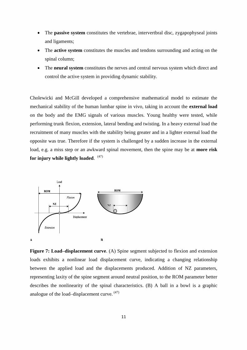
Figure 7: Load–displacement curve. (A) Spine segment subjected to flexion and extension
loads exhibits a nonlinear load displacement curve, indicating a changing relationship
between the applied load and the displacements produced. Addition of NZ parameters,
representing laxity of the spine segment around neutral position, to the ROM parameter better
describes the nonlinearity of the spinal characteristics. (B) A ball in a bowl is a graphic
analogue of the load–displacement curve. (47)
12
Bergmark (1989) hypothesized the presence of two muscle systems that act for maintenance
of spinal stability. The Global muscle system consists of large torque producing muscle that
act on the trunk and spine without directly attaching to it. These muscles include rectus
abdominus, obliquus abdominus externus and the thoracic part of lumbar iliocostalis and
provide general trunk stabilization, but are not capable of having a direct segmental influence
on the spine.
The Local muscle system consists of muscles that directly attach to the lumbar vertebrae,
and are responsible for providing segmental stability and directly controlling the lumbar
segments. These muscles are lumbar multifidus, psoas major, quadratus lumborum, the
lumbar parts of the lumbar iliocostalis and longissimus, transverses abdominus, the
diaphragm and the posterior fibres of obliquus abdominus internus all form part of this local
muscle system.
While the global muscle system provides the bulk of stiffness to the spinal column, the
activity of the local muscle system is necessary to maintain the segmental stability of the
spine. (45)
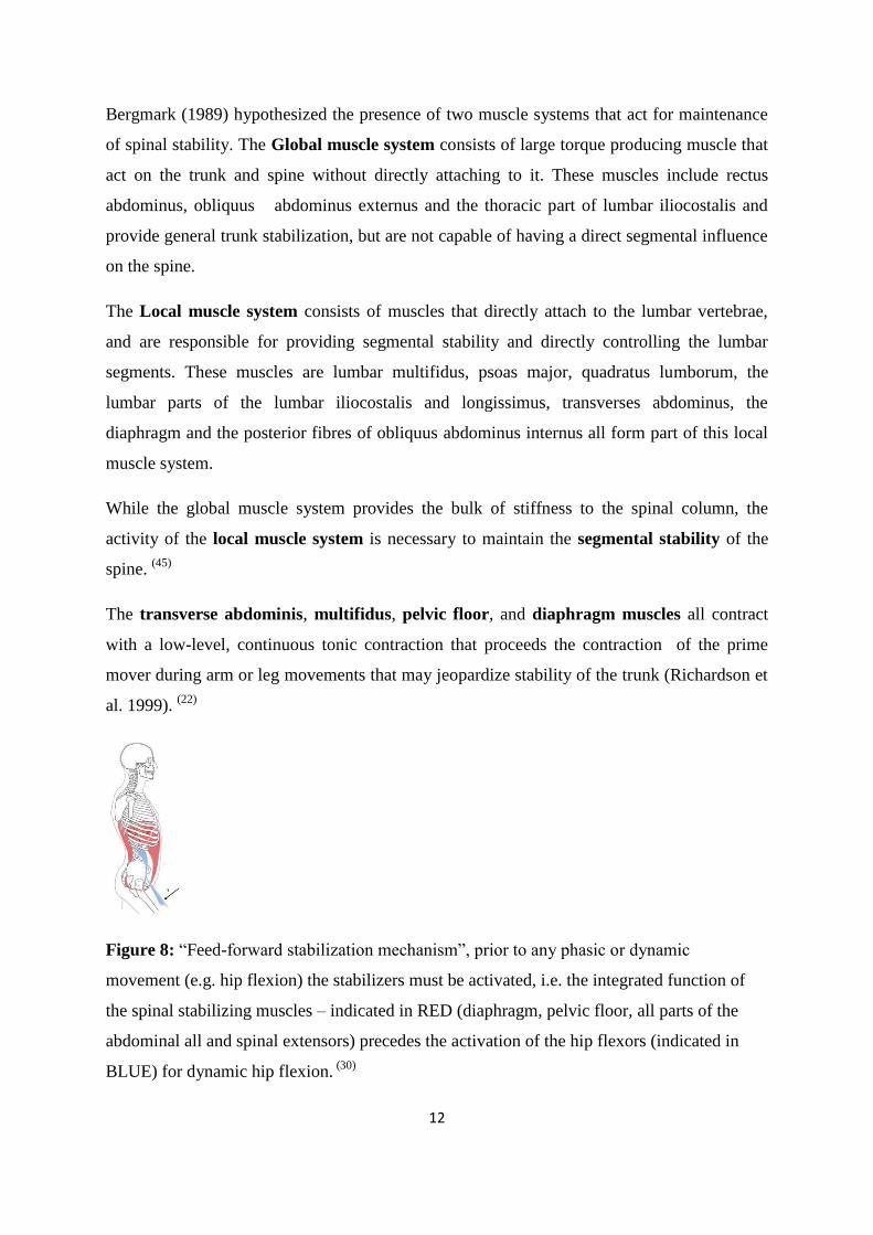
The transverse abdominis, multifidus, pelvic floor, and diaphragm muscles all contract
with a low-level, continuous tonic contraction that proceeds the contraction of the prime
mover during arm or leg movements that may jeopardize stability of the trunk (Richardson et
al. 1999). (22)
Figure 8: “Feed-forward stabilization mechanism”, prior to any phasic or dynamic
movement (e.g. hip flexion) the stabilizers must be activated, i.e. the integrated function of
the spinal stabilizing muscles – indicated in RED (diaphragm, pelvic floor, all parts of the
abdominal all and spinal extensors) precedes the activation of the hip flexors (indicated in
BLUE) for dynamic hip flexion. (30)
13
Activation of these muscles during any movement is automatic – subconscious, the muscle
coordination in not fully under our control; therefore often compromised. (20)
Anatomical arrangement of muscle control around the spine, coupled with critically
important patterns of activation, enables the spine to bear a much higher compressive load as
it stiffens and becomes more resistant to buckling but beacause of the stiffening of the muscle
activity the spine bears even more load. (78)
Postural muscles have different functions in stabilizing posture. Short inter-segmental
muscles, close to the joint, stabilize individuals segments providing flexible stability. Having
the long superficial muscle to stabilize larger sections of the spine and give a rigid stability.
Abdominal breathing provides flexible stability and has been shown by experiment after
external impact that flexible stabilization system is more dominant than the rigid stabiliy.
The diaphragm, inter-costal muscles, transversus abdominis (TrA), muscles of pelvic floor
and the deep intrinsic muscle of the spine are all muscles with either a horizontally or an
oblique orientation that segmentally can be activated. They participate as core stabilizers in
respiratory mechanics as well as in postural function. (78)
Inspiration tends to extension and to thoracic inflation; expiration tends to flexion and to
thoracic deflation. Breathing is connected with periodic changes of intra-abdominal and
intra-thoracic pressure. These changes influence continuously the stabilization of the
upright posture. (71)
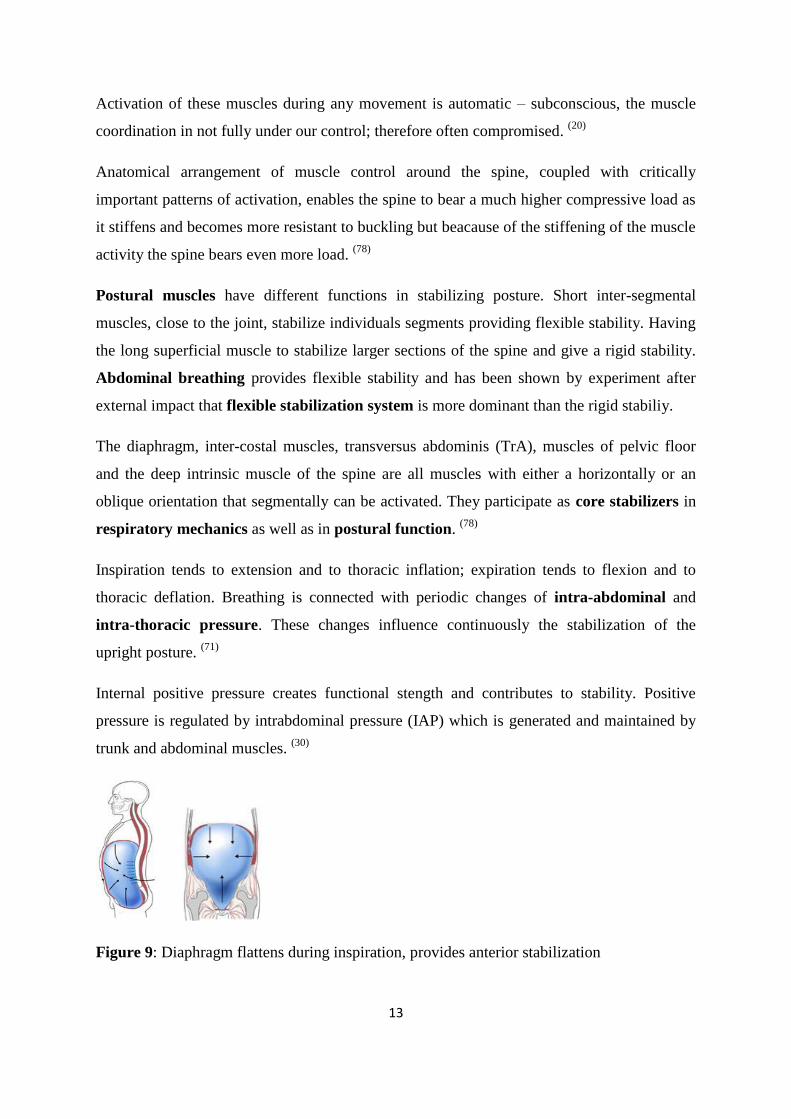
Internal positive pressure creates functional stength and contributes to stability. Positive
pressure is regulated by intrabdominal pressure (IAP) which is generated and maintained by
trunk and abdominal muscles. (30)
Figure 9: Diaphragm flattens during inspiration, provides anterior stabilization
14
When the diaphragm flattens during inspiration it acts against the resistance of the abdominal
wall. Synergistic action of the diaphragm, with the abdominal, and the pelvic floor controls
the intr-abdominal pressure (IAP) to provide anterior stabilization of the Lumbosacral spine.
(30)
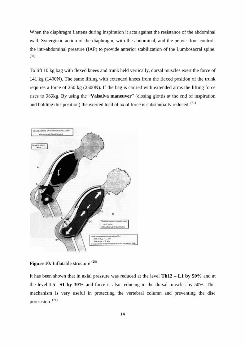
To lift 10 kg bag with flexed knees and trunk held vertically, dorsal muscles exert the force of
141 kg (1400N). The same lifting with extended knees from the flexed position of the trunk
requires a force of 250 kg (2500N). If the bag is carried with extended arms the lifting force
rises to 363kg. By using the “Valsalva maneuver” (closing glottis at the end of inspiration
and holding this position) the exerted load of axial force is substantially reduced. (71)
Figure 10: Inflatable structure (28)
It has been shown that in axial pressure was reduced at the level Th12 – L1 by 50% and at
the level L5 –S1 by 30% and force is also reducing in the dorsal muscles by 50%. This
mechanism is very useful in protecting the vertebral column and preventing the disc
protrusion. (71)
15
1.9. A General overview of the Intervertbral Disc
1.9.1. Structure of the intervertebral disc
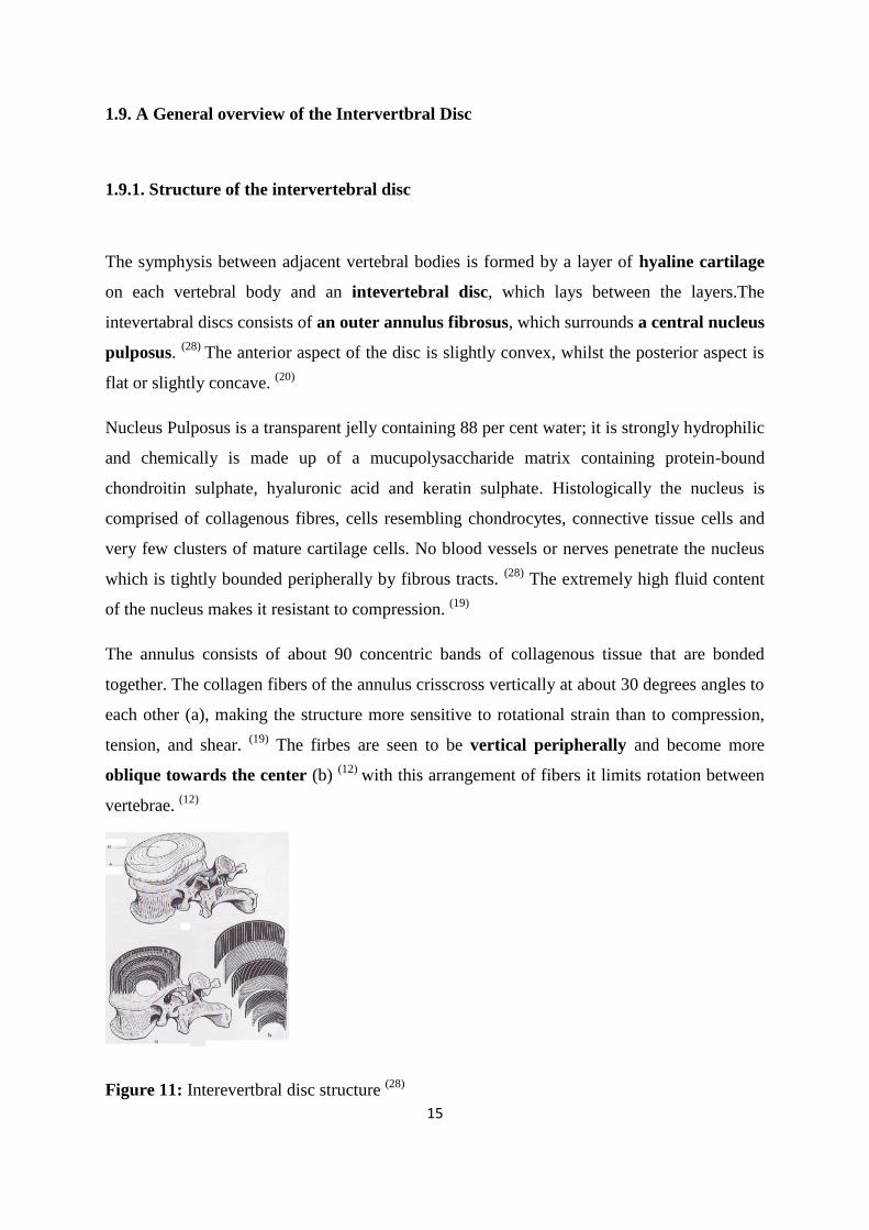
The symphysis between adjacent vertebral bodies is formed by a layer of hyaline cartilage
on each vertebral body and an intevertebral disc, which lays between the layers.The
intevertabral discs consists of an outer annulus fibrosus, which surrounds a central nucleus
pulposus. (28)
The anterior aspect of the disc is slightly convex, whilst the posterior aspect is
flat or slightly concave. (20)
Nucleus Pulposus is a transparent jelly containing 88 per cent water; it is strongly hydrophilic
and chemically is made up of a mucupolysaccharide matrix containing protein-bound
chondroitin sulphate, hyaluronic acid and keratin sulphate. Histologically the nucleus is
comprised of collagenous fibres, cells resembling chondrocytes, connective tissue cells and
very few clusters of mature cartilage cells. No blood vessels or nerves penetrate the nucleus
which is tightly bounded peripherally by fibrous tracts. (28)
The extremely high fluid content
of the nucleus makes it resistant to compression. (19)
The annulus consists of about 90 concentric bands of collagenous tissue that are bonded
together. The collagen fibers of the annulus crisscross vertically at about 30 degrees angles to
each other (a), making the structure more sensitive to rotational strain than to compression,
tension, and shear. (19)
The firbes are seen to be vertical peripherally and become more
oblique towards the center (b) (12)
with this arrangement of fibers it limits rotation between
vertebrae. (12)
Figure 11: Interevertbral disc structure (28)
16
The posterior part of the annulus is the weakest part: the anterior and lateral portions are
approximately twice as the posterior portion, where the layer appear to be narrower and less
numerous, the fibers in adjacent layers are oriented more nearly parral to each other, and
there is less binding substance. (15)
1.9.2. Biomechanics of the intervertebral disc
Mechanically, the annulus acts as a coiled spring whose tension holds the vertebral bodies
together against the resistance of the nucleus pulposus, and the nucleus pulposus acts like a
ball bearing composed of an incompressible gel.
The intervertbral discs have a blood supply up to about the age of 8 years, but after that the
disc must rely on a mechanically based means for maintaining a healthy nutritional status.
Intermittent changes in posture and body position after internal disc pressure, causing a
pumping action in the disc.
The inflow and outflow of water transport nutrients in and flushes metabolic waste products
out, basically fulfilling the same function that the circulatory system provide for vascularized
structures within the body. Maintaining even an extremely comfortable fixed body position
over a period of time curtails this pumping action and can negatively affect disc health. (19)
Compression is assumed greater when the disc is closer to the sacrum, which supports the
bulk of the body weight. For a man weighing 80kg the head weighs 3kg, the upper limbs
14kg and the trunk 30kg. If it is assumed that at the level of the disc L5-S1 the column
supports only two-thirds of the trunk, the weight borne is 37kg, which is nearly the half the
body weight. To this must be added the force exerted by the tone of the paravertebral muscle
necessary to maintain the trunk in the erect position at rest.
Disc height loss occurs during the first 30 minutes after getting up in the morning. During the
day a body‟s height may decrease 2 cm. When pressure on the discs is relieved, the discs
quickly reabsorb water and disc volumes and heights are decreased. (35)
This differences
decreases with increasing age. (71)
17
When a disc is loaded in compression, it tends to simultaneously lose water and absorb until
its internal electrolyte concentration is sufficient to prevent further water loss. (19)
When this
chemical equilibrium is achieved, internal disc pressure is equal to the external pressure.
Continued loading over a period of several hours results in a further slight decrease in disc
hydration. (28)
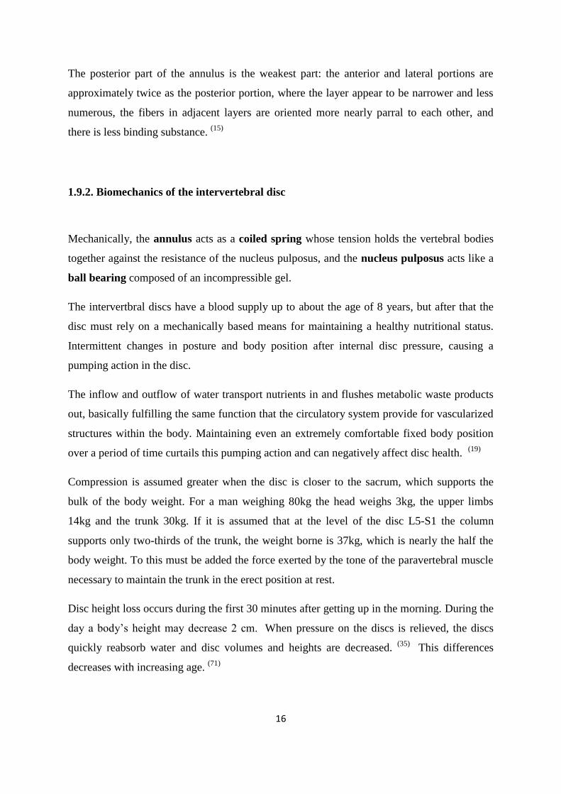
The loss of thickness of the disc depends on whether the disc is healthy or diseased. If a
healthy (A) disc is applied with a weight of 100 kg it is flattened by a distance of 1.4 mm and
becomes wider (B). If diseased and similar loaded the disc will be flattened by 2 mm this (C).
Figure 12: a) disc under load b) Progressive flattening (28)
1.9.3. Compression applied to the discs
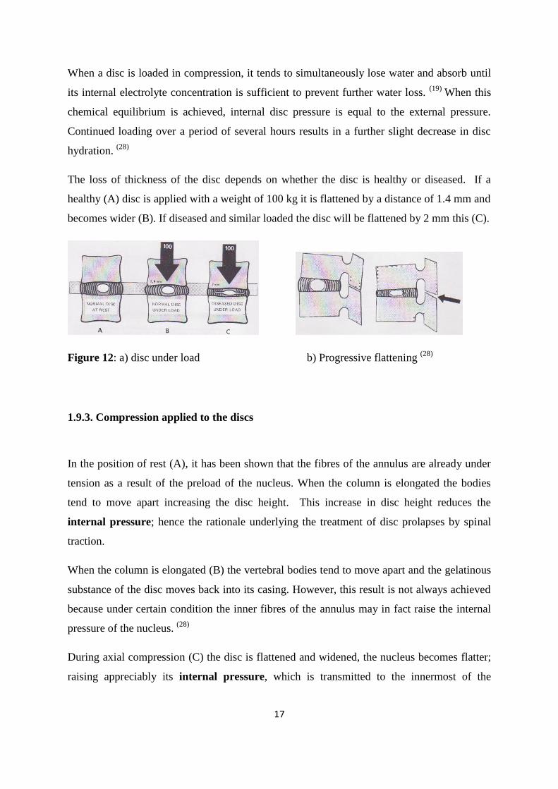
In the position of rest (A), it has been shown that the fibres of the annulus are already under
tension as a result of the preload of the nucleus. When the column is elongated the bodies
tend to move apart increasing the disc height. This increase in disc height reduces the
internal pressure; hence the rationale underlying the treatment of disc prolapses by spinal
traction.
When the column is elongated (B) the vertebral bodies tend to move apart and the gelatinous
substance of the disc moves back into its casing. However, this result is not always achieved
because under certain condition the inner fibres of the annulus may in fact raise the internal
pressure of the nucleus. (28)
During axial compression (C) the disc is flattened and widened, the nucleus becomes flatter;
raising appreciably its internal pressure, which is transmitted to the innermost of the
18
annulus.Thus the vertical force, is transformed into lateral forces tightening up the annular
fibres. (28)
In axial compression forces, it has been worked out that when a vertebral plateau presses on
the intervertebral disc the nucleus bears 75 per cent of the force and the annulus 25 per cent,
so that for a force equal to 20 kg; a 15 kg force is exerted on the nucleus and a 5kg force on
the annulus. However in the horizontal plane, the nucleus acts to transmit some of the force to
the annulus. (28)
Axial pressure on the vertebral body increases during bending forward to 58
kg/cm2
and in straightening back loads the plate with 107 kg/cm2.
(71)
Therefore the annulus and the nucleus constitute a functional couple whose effectiveness
depends on the integrity of each component. If the internal pressure of the nucleus decreases
or if the tightness of the annulus is impaired, this functional couple immediately loses its
effectiveness. (28)
Figure 13: Disc behaviour in (A) Rest position; (B) spinal elongation and (C) axial
compression (28)
1.9.4. Disc behavior during simple movements
The nucleus pulposus is roughly spherical and can be considered as a ball placed between
two planes. It has six degrees of freedom: flexion and extension, lateral flexion, gliding in the
sagittal plane, gliding in the frontal plane, rotation right and left. Each of these movements
has a small range and sizeable movements are only obtained by the simultaneous
participation of multiple intervertbral joints.
19
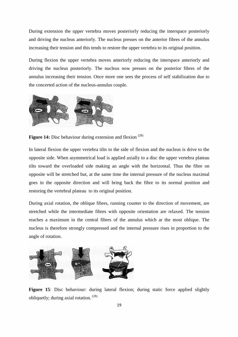
During extension the upper vertebra moves posteriorly reducing the interspace posteriorly
and driving the nucleus anteriorly. The nucleus presses on the anterior fibres of the annulus
increasing their tension and this tends to restore the upper vertebra to its original position.
During flexion the upper vertebra moves anteriorly reducing the interspace anteriorly and
driving the nucleus posteriorly. The nucleus now presses on the posterior fibres of the
annulus increasing their tension. Once more one sees the process of self stabilization due to
the concerted action of the nucleus-annulus couple.
Figure 14: Disc behaviour during extension and flexion (28)
In lateral flexion the upper vertebra tilts to the side of flexion and the nucleus is drive to the
opposite side. When asymmetrical load is applied axially to a disc the upper vertebra plateau
tilts toward the overloaded side making an angle with the horizontal. Thus the fibre on
opposite will be stretched but, at the same time the internal pressure of the nucleus maximal
goes in the opposite direction and will bring back the fibre to its normal position and
restoring the vertebral plateau to its original position.
During axial rotation, the oblique fibres, running counter to the direction of movement, are
stretched while the intermediate fibres with opposite orientation are relaxed. The tension
reaches a maximum in the central fibres of the annulus which ar the most oblique. The
nucleus is therefore strongly compressed and the internal pressure rises in proportion to the
angle of rotation.
Figure 15: Disc behaviour: during lateral flexion; during static force applied slightly
obliquetly; during axial rotation. (28)
20
2. A General Overview of Lumbar Disc Herniation
2.1 Lumbar Disc Herniation
The lumbar intervertebral discs are much more susceptible to symptomatic herniation than
either the cervical or the thoracic discs. Lumbar disc herniation usually presents with
radicular sciatica which most are dorsalateral and therefore compress the nerve root that
exits one level lower. However a lateral herniation can compress the spinal nerve of the same
level. (18)
Ruptured Discs in the Low Back may provoke shooting; stabbing pain that shoots from the
back or buttocks into the leg, this is called sciatica or radiculopathy. It can be associated with
numbness or weakness in the leg and foot. The most frequent cause of this condition is a
ruptured disc in the lower back. A herniation may develop suddenly or gradually over weeks
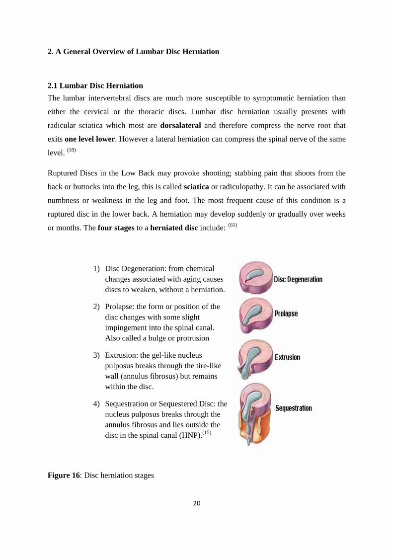
or months. The four stages to a herniated disc include: (61)
1) Disc Degeneration: from chemical
changes associated with aging causes
discs to weaken, without a herniation.
2) Prolapse: the form or position of the
disc changes with some slight
impingement into the spinal canal.
Also called a bulge or protrusion
3) Extrusion: the gel-like nucleus
pulposus breaks through the tire-like
wall (annulus fibrosus) but remains
within the disc.
4) Sequestration or Sequestered Disc: the
nucleus pulposus breaks through the
annulus fibrosus and lies outside the
disc in the spinal canal (HNP).(15)
Figure 16: Disc herniation stages
21
Degenerative changes cause the posterior annular fibers of the lumbar disc to become
incompetent and subsequently allow nuclear material from within the disc to extend into the
area of the spinal canal, causing nerve compression. Lumbar disc herniations may occur in
relation to traumatic injury, but they do not necessarily require traumatic injury to occur.
Lumbar disc herniations may occur with various degrees of severity. (58)
The disc protrusion happens in the lumbar spine in two ways. The protruded fluid disc-matter
(pulpous nucleus) becomes fixed to the wall of the spinal canal and may heal here and pains
may slowly disappear. Or the disc matter may freely move in the spinal canal as a foreign
body irritating steadily the meninges and nerve roots, which is very painful. In such a case the
protruded disc matter must be evacuated surgically. This depends on the development of the
symptoms, as well as on the structural situation in the spinal canal revealed by nuclear
resonance examination. (71)
Small herniations are often referred to as "bulges of the disc." These frequently do not cause
nerve compression, but they may be a source of back pain. Larger disc herniations frequently
cause compression of spinal nerves or the cauda equina in the lumbar spinal canal and may be
the source of significant leg pain.
The term extruded fragment refers to lumbar disc nuclear material which herniates beyond
the limits of the annulus fibrosis and frequently beyond the limits of the posterior longitudinal
ligament to cause direct compression of nerve elements at a distance from the area of the disc
itself. (58)
Loss of mechanical competence and flattening of the disc may generate diffuse bulging,
which should be differentiated for focal bulges or true herniations, characterized
macroscopically by nuclear migration though radial fissure of the disc. Disc herniation
requires pre-existing age-related degenerative changes. Ageing and degeneration are also
associated with dramatic changes in vascularization and innervations of the disc.
A normal healthy adult disc is avascular, apart from vascularization at the outer part of the
annulus. Presence of blood vessels has been demonstrated in degenerated disc and the
herniated disc tissue. Penetration of blood vessel through the rim lesion is promoted by
angiogenesis factors. Inflammatory cells as well as macrophages also invade the degenerated
disc. (1)
22
2.2 Factors and genetic risks for disc heniation
2.2.1. Factors for disc herniation
Many factors increase the risk for disc herniation:
1) Lifestyle choices such as tobacco use, lack of regular exercise, and inadequate nutrition
substantially contribute to poor disc health.
2) As the body ages, natural biochemical changes cause discs to gradually dry out affecting
disc strength and resiliency.
3) Poor posture combined with the habitual use of incorrect body mechanics stresses the
lumbar spine and affects its normal ability to carry the bulk of the body's weight. (20)
2.2.2. Genetic influnces for disc degeneration
Several recent studies have reported a strong familial predisposition for disc degeneration and
herniation. Heritability for disc herniation exceeded 60%. Genetic predisposition has been
confirmed by recent findings of associations between disc degeneration and polymorphisms
in various classes of genes: (5)
Genes Encoding for Matrix Components
- aggrecan
- collagen type IX
- collagen type I
- cartilage intermediate layer protein (CILP)
Genes Encoding for Cytokines
- interleukin-1 (IL-1)
- interleukin-6 (IL-6)
Genes Encoding for Proteinases
- matrix metalloproteinase-3 (MMP-3)
Genes Encoding for Miscellaneous Proteins
- vitamin D receptor
23
2.3 The process of disc degeneration and disc herniation
Disc degeneration has a profound effect on the mechanism of load transfer through the disc.
With degeneration, dehydration of the disc leads to a lower elasticity and viscoelasticity.
Loads are less evenly distributed, and the capacity of the disc to store and dissipate energy
decreases.
Using the technique of “stress profilometry”, it has been shown that age-related changes to
the disc composition result in a shift of load from the nucleus to the anulus.Therefore,
structural changes in the anulus and endplate with degeneration may lead to a transfer of
load from the nucleus to the posterior anulus, which may cause pain and also lead to
annular rupture. (5)
Annulus fibres begin to degenerate after 25 years of age allowing tearing of fibres within
each of its layers. (20)
Aging causes a loss of disc height and compression of the vertebral
body. The bone attempts to cushion itsel by forming a lip or extra rim around the periphery of
the endplate. This lipping can extend far enough to obstruct the opening to the vertebral
canal.
At the same time, the ligamentum flavum begins to hypertrophy or thicken and osteophytes
(bone spurs) may develop. Degenerative disease can cause the apophyseal (facet) joints to
flatten out or become misshapen. Any or all of these variables can contribute to spinal
stenosis. (4)
Stenosis of the spinal canal is strictly related to disc herniation not only from the
pathologic point of view, since the condition of coexist, but also for the historical evolution
of the knowledge of the disease.(16)
The emerging nerve root exits through a shallow lateral recess and also may be compressed
easily. Any combination of degenerative changes, such as disc protrusion, osteophyte
formation, and ligamentous thickening, reduces the space needed for the spinal cord and its
nerve roots. (17)
Injury and aging irreversibly reduce the water-absorption capacity of the discs, with a
concomitant decrease in shock-absorbing capability. The fluid content of the disc begin to
diminish around the second decade of life. A typical geriatric disc has a fluid content that is
reduced by approximately 35%.
24
As this normal degenerative change occurs, abnormal movements occur between adjacent
vertebral bodies, and more of the compressive, tensile, and shear loads on the spine must be
assumed by other structures – particularly the facets and joint capsules. Results include
reduced height of the spinal structures that are forced to assume the discs loads. Postural
alterations may also occur. The normal lordotic curve of the lumbar region may be reduced
as an individual attempts to relieve compression on the facet joints by maintaining a posture
of spinal flexion. Factors such as habitual smoking and exposure to vibration can negatively
affect disc nutrition whereas regular exercise can improve it. (19)
In a hypothesis by Kirkaldy-Willis describing degeneration; in the first stage of may result in
some spinal dysfunction but no instability. In the third stage the spine is restabilized probably
because of ligament calcification and osteophytes. However in the second stage, which
occurs between the ages of 40 and 50 years, the disc degeneration has progressed to the point
where the nucleus is still mobile. This the instability phase and at this stage is increased risk
of disc prolapse at the L4-L5 or L5-S1 levels because of traumatic overload of the spine.
It is not unreasonable to speculate that the high incidence of clinically evident disc disease at
L4-L5 and L5-S1 may be related to mechanics. These two areas bear the highest loads and
tend to undergo the most motion in the sagittal plane. (30)
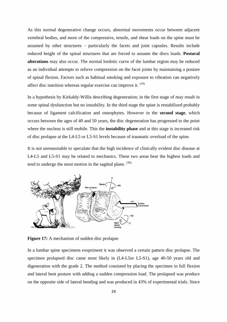
Figure 17: A mechanism of sudden disc prolapse
In a lumbar spine specimens exepriment it was observed a certain pattern disc prolapse. The
specimen prolapsed disc came most likely in (L4-L5or L5-S1), age 40-50 years old and
digeneration with the grade 2. The method consisted by placing the specimen in full flexion
and lateral bent posture with adding a sudden compression load. The prolapsed was produce
on the opposite side of lateral bending and was produced in 43% of experimental trials. Since
25
the majority of the low back patients with disc prolapse seen clinically do not report a
traumatic event but one may conclude that the gradual disc prolapse maybe the result of a
combination of factors, such as weaken posterior disc annulus, relatively degenerated annulus
with fissures, and another kind of loading(e.g., bending and twisting). (30)
This initial acute lumbago can regress spontaneously with or without treatment but, as a
result of repeated trauma, the hernia grows in size and protrudes more and more into the
vertebral canal. At this point it comes into contact with a nerve root, often one of the nerve
roots of the sciatic nerve.
In fact the hernia usually protrudes posterolaterally where the posterior longitudinal
ligament is at its weakest and progressively pushes the nerve root away until the latter is
jammed against the posterior wall of the intervertebral foremen formed by the joint between
the articular process, its anterior capsular ligament and the lateral border of the ligamentum
flavum. The compressed now nerve root will give rise to pain felt in the spinal segment
corresponding to the root and finally to impaired reflexes (loss of the Achilles tendon reflex)
and to motor disturbances, as in sciatica with paralysis. (28)
2.4. Disc prolapse and the mechanism of nerve root compression
The covering plates of lumbar vertebrae are very plat and axial pressure combined with
shearing forces are the very cause of lumbar disc protrusions. This occurs particularly, if a
heavy burden is heaved from the bottom thru erection of the bent trunk with extended knees
in combination with synchronous quick twist movement. This torsion produces shearing force
added to existing axial pressure. These both forces together damage the disc.
Forward bending of the trunk pushes the disc toward the spinal canal and if combined with
additional torsion, the disc bursts and the rests of the pulpous nucleus protrude into the spinal
canal and irritate the meninges and the roots of spinal nerves (usually L4-L5 or L5-S1). This
disc protrusion endangers not the spinal cord, but affects only meninges (pia mater, dura
26
mater arachnoidea) and the roots of spinal nerves which provoke sciatica exactly the radicular
lumbar symptoms with positive Lasegue sign and lower back pains. (71)
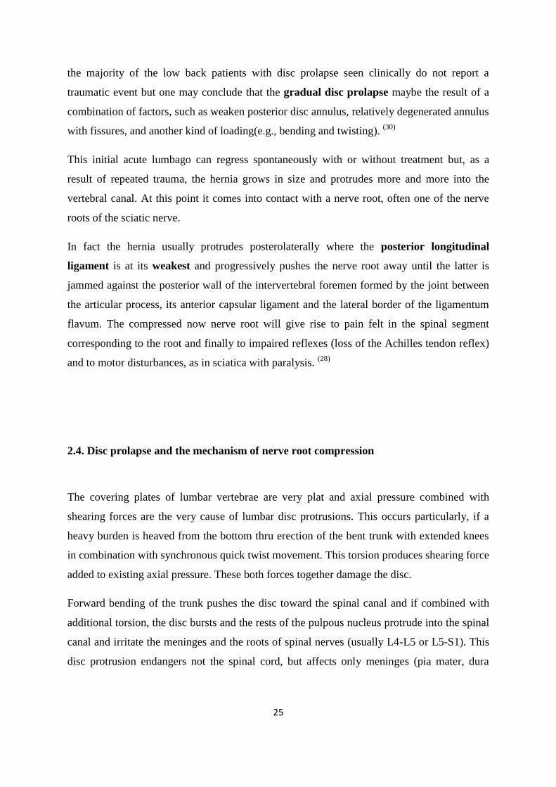
It is now generally believed that disc prolapse occurs in three phases in lifting a weight. It
usually occurs if the disc deteriorated as a result of repeated microtraumas and if the
annulus fibres have started to degenerate. Disc prolapse usually follows lifting of a weight
with the trunk flexed forward.
During the first phase trunk flexion (A) flattens the discs anteriorly and opens out the
intervertebral space posteriorly. During the second phase (B), as soon as the weight is lifted,
the increased axial compression force crushes the whole disc and violently drives the nuclear
substance posteriorly until it reaches the deep surface of the posterior longitudinal ligament.
During the third phase (C) with the trunk nearly straight, the path taken by the herniating
mass is closed by the pressure of the vertebral plateaus and the hernia remains trapped under
the posterior longitudinal ligament. This cause the acute pain felt in the loin or lumbago
which corresponds to the initial phase of the lumbago-sciatica complex. (28)
Figure 18: Mechanism of disc prolapse (28)
27
2.5. Disc herniation provoking radicular syndrome
Neurogenic pain is not easily differentiated; radicular pain results from irritation of axons of a
spinal nerve or neurons in the dorsal root ganglion whereas refferd pain results from
activation of nociceptive free nerve endings (nociceptors) in somatic or visceral tissue.
Neurologic signs are produced by conduction block in motor or sensory nerves, but
conduction block does not cause pain. Thus, even in a client with back pain and neurologic
signs, whatever causes the neurologic signs is not causing the back pain by the same
mechanism. Therefore, finding the cause of neurologic signs does not always identify the
cause of the back pain. The therapist must look further. (80)
Nor is disc herniation the only possible cause of pain in radicular syndromes of the lower
limbs; in operation statistics no disc herniation is found in about 10% of the cases; many
radicular syndrome resolve without operation, and this is true even of cases which medical
imaging had found a herniated disc. Disc herniation may sometime persist after the symptoms
have disappeared, although resorption is also possible. Clinical images (CT and MRI) also
reveal a herniated disc in healthy individuals in whom it is of little relevance. It is only
significant when it correlates with clinical findings.
The mechanical compression of a nerve does not itself cause pain but anesthesia, paresthesia,
and paresis. However, we should bear in mind that the herniated disc causing the
compression cannot impinge on the nerve fibers until after it has affected the dura an the
dural sheaths, which are richly supplied with pain receptor, and that every movement of the
legs and trunk the dura is being rubbed against the disc. (35)
The lumbar intervertebral disc (IVD) plays a central role in the development of low back –
related leg pain and radiculopathy (Yoshizawa et al., 1995). The pathomechanisms involved
are internal disc disruption, fissure formation and nucleus pulposus (NP) prolapse or
sequestration leading to inflammation of the nerve root, and subsequent pain of nerve origin,
even without mechanical compression. Inflammation caused by biomechanical substance
from the NP plays a significant role in the development of low back-related leg pain
(Olmarker et al., 1993; Olmarke 1997; Brisby, 2003). (1)
28
Degenerative changes of the IVD, associated with internal disc disruption, commonly lead to
fissures in the annulus, which allow inflammatory mediators to disperse through the disc and
contact the innervated outer third of the annulus (Videman and Nurminen, 2004; Peng et al.
2005). These chemicals may cause excitation of nociceptive afferents and thereby discogenic
pain, which may then refer into the lower limb (O‟Neil et al., 2002). In case of a fuller
annular rupture, NP material and inflammatory mediators may leak into the spinal canal,
contact nerve tissues such as transiting of exiting nerve roots and lead to inflammation of
these structure (Videman and Nurminen, 2004). (1)
2.6. Differential diagnosis in Sciatic pain (17)
2.6.1. Neuromuscular causes
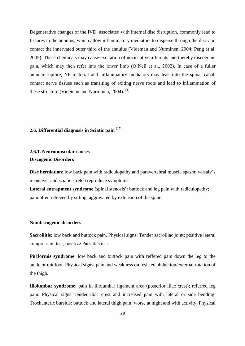
Discogenic Disorders
Disc herniation: low back pain with radiculopathy and paravertebral muscle spasm; valsalv‟s
maneuver and sciatic stretch reproduce symptoms.
Lateral entrapment syndrome (spinal stenosis): buttock and leg pain with radiculopathy;
pain often relieved by sitting, aggravated by extension of the spine.
Nondiscogenic disorders
Sacroilitis: low back and buttock pain. Physical signs: Tender sacroiliac joint; positive lateral
compression test; positive Patrick‟s test.
Piriformis syndrome: low back and buttock pain with reffered pain down the leg to the
ankle or midfoot. Physical signs: pain and weakness on resisted abduction/external rotation of
the thigh.
Iliolumbar syndrome: pain in iliolumbar ligament area (posterior iliac crest); referred leg
pain. Physical signs: tender iliac crest and increased pain with lateral or side bending.
Trochanteric bursitis: buttock and lateral thigh pain; worse at night and with activity. Physical
29
signs: tender greater trochanter; rule oute associated leglength discrepancy; positive “jump
sign” when pressure is applied over the greater trochanter.
Greater trochanteric pain syndrome: mimics lumbar nerve root compression.
Physical signs: low back, buttock, or lateral thigh pain; may radiate down the leg to the
iliotibial tract insertion on the proximal tibia; inability to sleep on the involved side.
Ischiogluteal bursitis: buttock and posterior thigh pain; worse with sitting.
Physical signs: tender ischial tuberosity; positive. SLR and Patrick‟s sign test‟s; rule out
associated leg-length discrepancy.
Posterior facet syndrome: low back pain.
Physical signs: lateral bending in spinal extension increases pain; side bending and rotation to
the opposite side are restricted at the involved level.
Fibromyalgia: back pain, difficulty sleeping, anxiety, and depression.
Physical signs: multiple tender points.
2.6.2. Systemic/ extraspinal causes (17)
Vascular disorders
- Ischemia of sciatic nerve
- Peripheral vascular disease
- Intrapelvic aneurysm (internal iliac artery)
- Neoplasm ( primary or metastatic)
- Diabetes mellitus (diabetic neuropathy)
- Megacolon
- Pregnancy; vaginal delivery
- Infections
- Bacterial endocarditis
- Wound contamination
30
- Herpes zoster (shingles)
- Psoas muscle abscess
- Reiter‟s syndrome
- Total hip replacement
- Endometriosis
- Deep venous thrombosis (blood clot)
2.6.3. Risk factors for sciatica
Musculoskeletal or neuromuscular factors
- Previous low back injury or trauma; direct fall on buttock(s); gunshot wound
- Total hip arthroplasty
- Pregnancy
- Work- or occupation- related postures or movements
- Fibromyalgia
- Leg-length discrepancy
- Congenital hip dysplasia; hip dislocation
- Degenerative disc disease
- Piriformis syndrome
- Spinal
Systemically induced factors
- Tobacco use
- History of diabetes mellitus
- Atherosclerosis
- Previous history of cancer (metastases)
- Presence of intra-abdominal or peritoneal inflammatory disease (abscess):
Crohn‟s disease, Pelvic inflammatory disease, Diverticulitis
Endometriosis of the sciatic nerve
Radiation therapy (delayed effects; rare)
Recent spinal surgery, especially with instrumentation
31
2.7. Diffferential classifications for low back pain and sciatica
2.7.1. Mechanical pain syndromes described by Mckenzie (43)
The postural syndrome:
This is caused by mechanical deformation of soft tissues as a result of postural stresses.
Maintenance of certain postures or positions which place some soft tissues under prolonged
stress will eventually be productive of pain. Thus, the postural syndrome is characterised by
intermittent pain brought on by particular postures or positions, and usually some time must
pass before the pain becomes apparent. The pain ceases only with a change of position or
after postural correction.
The dysfunction syndrome:
This is caused by mechanical deformation of soft tissues affected by adaptive shortening.
Adaptive shortening may occur for a variety of reasons. It leads to a loss of movement in
certain directions and causes pain to be produced before normal full range of movement is
achieved. Thus, the dysfunction syndrome is characterised by intermittent pain and a partial
loss of movement. The pain is brought on as soon as shortened structures are stressed by end
positioning or end movement and ceases almost immediately when the stress is released.
The derangement syndrome:
This is caused by mechanical deformation of soft tissues as a result of internal
derangement. Alteration of the position of the fluid nucleus within the disc, and possibly the
surrounding annulus, causes a disturbance in the normal resting position of the two vertebrae
enclosing the disc involved. Various forms and degrees of internal derangement are possible,
and each presents a somewhat different set of signs and symptoms.
Thus, the derangement syndrome is usually characterised by constant pain, but intermittent
pain may occur depending on the size and location of the derangement. There is a partial loss
32
of movement, some movements being full range and others partially or completely blocked.
This causes the deformities in kyphosis and scoliosis so typical of the syndrome in the acute
stage.
The three syndromes presented are totally different from each other, and each syndrome must
be treated as an entity on its own, requiring special procedures which are often unsuitable for
the other syndromes. In order to identify which syndrome is present in a particular patient a
history must be established and an examination must be performed.
2.7.2. Patho-mechanism classification for back related leg pain
A proposed patho-mechanism based approach for differentiating different sources of
radiating leg pain in 4 groups which is important to make the appropriate diagnosis (Schafer
et al. 2007). The first subgroup is central sensitization with mainly positive symptoms such
as hyperalgesia; the second subgroup involves denervation with significant axonal damage
showing predominantly negative sensory symptoms and possibly motor loss and the third
subgroup involves peripheral nerve sensitization with enhanced nerve trunk mechano-
sensitzation. The fourth subgroup features somatic referred pain from musculoskeletal
structures, such as the intervertebral disc or facet joints.
There has been a separation into four groups with of patients with low back-related leg pain,
but in reality there may be considerable overlap between them. Peripheral sensitization of
nerve tissue can trigger central sensitization, and inflammatory products released during
denervation may also alter the properties of intact nerve fibres. Many of radicular disorders
are mixture of nociceptive and neuropathic pain (Baron and Binder, 2004). This mechanism
of differentiation is evaluated by the patients complaints and to be identified it is need a
thorough physical examination protocol. (62)
33
2.7.3. Radicular syndromes in the lower extremities
Although radicular syndromes share many common features with other vertebrogenic
disorder, they possess certain special characteristics. The first is that, in most cases, pain
radiating into the lower extremity is preceded by low-back pain. This is why disc herniation
is thought to be the main cause not only of radicular pain, but also of low-back pain.
However, because low back pain occurs much more frequently than radicular syndromes this
merely indicates low-back pain caused by disc herniation but is likely to be a precursor of
radicular syndromes. There are, however, radicular syndromes in which the pain starts in the
legs and is never preceded by low-back pain. In such cases, low back pain usually appears
only later, if at all. Pain felt in the buttocks occurs commonly, hence the old term „sciatica‟.
Radicular pain may have a sudden onset after a lifting injury or when getting out of bed in the
morning. It may also begin so insidiously that the patient cannot remember precisely when it
started. For best advice to be given in individual cases, it is important to elicit from the
patient details of those circumstances that aggravate symptoms and that bring relief.
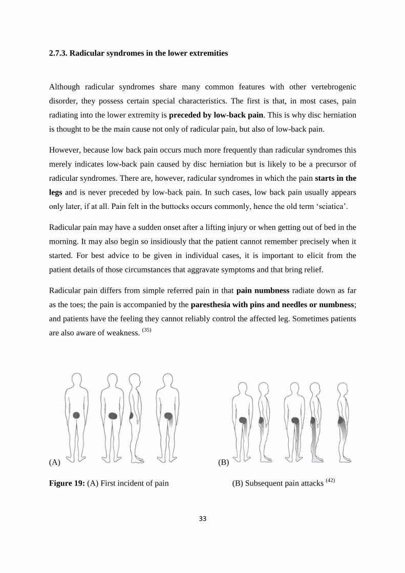
Radicular pain differs from simple referred pain in that pain numbness radiate down as far
as the toes; the pain is accompanied by the paresthesia with pins and needles or numbness;
and patients have the feeling they cannot reliably control the affected leg. Sometimes patients
are also aware of weakness. (35)
(A) (B)
Figure 19: (A) First incident of pain (B) Subsequent pain attacks (42)
34
2.7.4. Clinical signs and characteristics of disc herniation
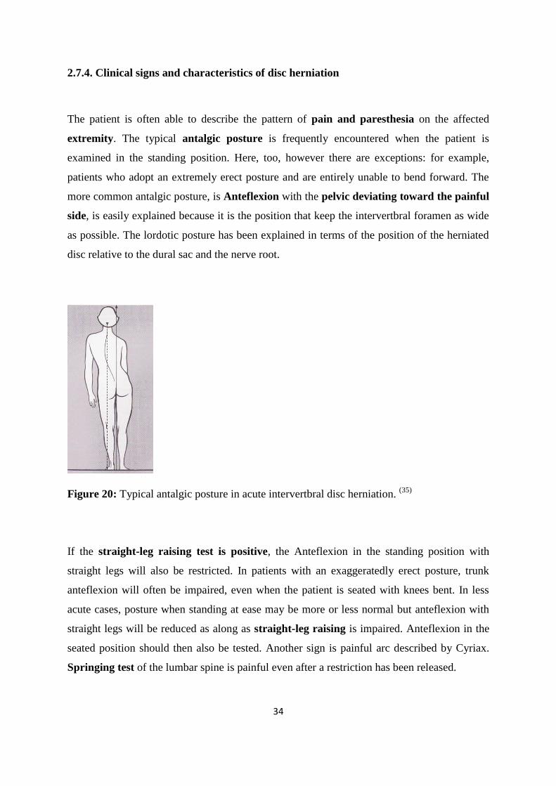
The patient is often able to describe the pattern of pain and paresthesia on the affected
extremity. The typical antalgic posture is frequently encountered when the patient is
examined in the standing position. Here, too, however there are exceptions: for example,
patients who adopt an extremely erect posture and are entirely unable to bend forward. The
more common antalgic posture, is Anteflexion with the pelvic deviating toward the painful
side, is easily explained because it is the position that keep the intervertbral foramen as wide
as possible. The lordotic posture has been explained in terms of the position of the herniated
disc relative to the dural sac and the nerve root.
Figure 20: Typical antalgic posture in acute intervertbral disc herniation. (35)
If the straight-leg raising test is positive, the Anteflexion in the standing position with
straight legs will also be restricted. In patients with an exaggeratedly erect posture, trunk
anteflexion will often be impaired, even when the patient is seated with knees bent. In less
acute cases, posture when standing at ease may be more or less normal but anteflexion with
straight legs will be reduced as along as straight-leg raising is impaired. Anteflexion in the
seated position should then also be tested. Another sign is painful arc described by Cyriax.
Springing test of the lumbar spine is painful even after a restriction has been released.
35
Major significance is the neurological signs of root involvement, such as motor weakness
and hypoesthesia, without which the diagnosis of true radicular syndrome inconclusive
because of the often highly deceptive nature of referred pain. For this reason, even minimal
weakness of a muscle, hypotonus, or hypoesthesia consistent with the segment in question
may be highly significant and should be carefully looked for. (35)
Pain is typically felt on coughing, sneezing, defecation, and, sometimes, laughing. Except
in acute cases, walking tends to alleviate the pain. However, if patients complain of pain
when walking, it is essential to ask whether they have to stop after a certain distance and what
position they then adopt. This is the only way to identify intermittent claudication. (35)
2.7.5. Problems in diagnosing radicular syndromes
In clinical terms, a radicular syndrome can be reliably distinguished from referred pain;
however, establishing when a radicular syndrome is caused by disc herniation is far more
difficult. A herniated disc may be clinically „silent‟ and radicular compression may be caused
by a narrow spinal canal, a narrow lateral recess, or a space-occupying lesions. Localization
can also be more problematic that would appear at first sight. (35)
Conditions such as radiculitis may cause both pain and neurologic signs but in that case the
pain occurs in the lower limp, not in the back. If root inflammation also happens to involve
the nerve root sleeve, neck or back pain might also arise. In such a case the individual will
have three problems each with a different mechanism: neurologic signs due to conduction
block, radicular pain due to nerve-root inflammation, and back pain due to inflammation of
the dura. Identifying a mechanical cause of pain does not always rule out serious spinal
pathology. For example neurogenic pain can be caused by a metastatic lesion applying
pressure or traction on any of the neural components. The therapist must rely on history,
clinical presentation, and the presence of any associated signs and symptoms to make a
determination about the need for medical referral. (17)
Anomalies are encountered along the course of nerve roots, and computed tomography (or
magnetic resonance imaging) often exposes more than one herniated disc. Only one of these
will probably be relevant clinically. Patients who have been immobilized for long periods
36
often develop thrombophlebitis, the pain of which must not be confused with radicular pain
and must be treated specifically. (35)
Confusion with spinal stenosis syndromes may occur when atheramatous change in the
internal iliac artery results in ischemia to the sciatic nerve. The subsequent sciatic pain with
vascular claudication like symptoms may go unrecognized as a vascular problem. The
therapist may be able to recognize the need for medical intervention by combining a careful
subjective and objective examination with knowledge of vascular and neurogenic pain
patterns. This is especially true in the treatment of unusual cases of sciatica or back pain with
leg pain. (17)
If surgery is indicated, the diagnosis must first be confirmed by imaging techniques.
However, even these are not infallible. Although imaging may reveal more than one
herniated disc, it can provide little information regarding their clinical relevance. (35)
Sciatica alone or sciatica accompanying back pain is an important but unreliable symptom.
For example, diabetic neuropathy can cause nerve root irritation. Prostatic metastases to the
lumbar and pelvic regions or other neoplasm‟s of the spine can create a clinical picture that is
indistinguishable from sciatica of musculoskeletal origin. This similarity may lead to long
and serious delays in diagnosis. Such a situation may require persistence on the part of the
therapist and client in requesting further medical follow up. (17)
Spinal stenosis caused by narrowing of the spinal canal, nerve root canals, or intervertebral
foramina may produce neurogenic claudication. The canal tends to be narrow at the
lumbosacral junction, and the nerve roots in the cauda equina are tightly packed. Pressure on
the cauda equina from tumor, disc protrusion, infection, or inflammation can result in cauda
equina syndrome, which is a medical emergency. (35)
Even without surgery the great majority of radicular syndromes heal as a result of functional
compensation and resorption of the intervertebral disc. This is also why conservative
treatment is so often successful, that is traction, manipulation, various types of reflex therapy,
remedial exercise, and stabilization methods. However surgery in isolation fails more often
than not it is not followed by appropriate rehabilitation that is if we do not help to restore
normal function. (35)
37
3. Clinical evaluation and treatment procedure for lumbar disc herniation
3.1. Examination procedure
History taking
Taking an accurate history is the most important part of the initial consultation when one is
dealing with any medical or surgical problem. Unfortunately, when the mechanical lesion is
involved there is still lack of understanding regarding the nature of the questions that should
be asked, the reasons for asking them, and the conclusions to be drawn from the answers.
Where is the present pain being felt?
We need to know all the details about the location of the pain, because this will give us some
indication of the level and extent of the lesion and the severity of the condition. If there are
any associated symptoms such as anaesthesia, paraesthesiae and numbness, their location
must be noted as well. Referred pain indicates that derangement is likely.
How long has the pain been present?
It is important to find out whether we are dealing with an acute, a subacute or chronic
condition. In recurrent low back pain we are not interested in an answer based on the length
of time since the first attack; at this stage of the examination we want to know how long the
present episode has been evident.
How did the pain commence?
Basically we want to find out if there was an apparent or no apparent reason for the onset of
the pain. Most of the histories commonly state that the pain appeared for no apparent reason.
Two of every three patients fall into this category, and only one patient will recognise a
causative strain.
38
Is the pain constant or intermittent?
This is the most important question we must ask patients with low back pain. If in patients
referred for mechanical therapy the pain is found to be constant, it is usually produced by
constant mechanical deformation. However, we must keep in mind that constant pain can also
be caused by chemical irritation. Intermittent pain is always produced by mechanical
deformation.
What makes the pain worse and what makes the pain better?
We must specifically ask about sitting, standing, walking, lying, and activities which involve
stooping or prolonged stooping. In these positions the joint mechanics of the lumbar spine are
relatively well understood, and therefore we will be able to determine which situations
increase and which decrease mechanical deformation. We must carefully record any position
or activity reported to reduce or relieve the pain, as we will utilise this information in our
initial treatment.
Have there been previous episodes of low back pain?
We should enquire about the nature of any similar or other low back pain episodes, the time
span over which they occurred, and their frequency. At this stage we should also find out
about previous treatments and their results. Episodic history indicates derangement. (15)
- Pain on cough/sneeze?
- Disturbed sleep?
- Pain on arising in the morning?
- Recent X-Rays? - Results?
- On medication at present?
- On steroids, in past or at present?
- General health? - Recent weight loss?
- Major surgery or accident, recently or previously?
- Saddle anaesthesia? – Bladder control?

39
The disc appears to live and thrive on movement, and to change and die slowly through lack
of it. The objective examination covers the following factors: (21)
- Observation of posture and gait
- Active movements of the spine
- Passive movements of the spine
- Accessory movements of the spine
- Tests for muscle function (e.g., weakness, lack of coordination, or muscle pain)
- Tests of other joints (e.g., sacroiliac, hip, knee, and ankle)
- Neurologic tests (e.g., reflexes, sensation, and motor power)
- Dural tension signs tested singly or in combination (e.g., passive neck flexion, straight
leg raise, prone knee flexion, and slumped sitting)
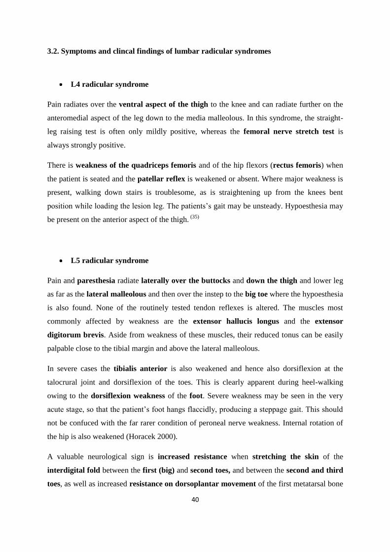
40
3.2. Symptoms and clincal findings of lumbar radicular syndromes
L4 radicular syndrome
Pain radiates over the ventral aspect of the thigh to the knee and can radiate further on the
anteromedial aspect of the leg down to the media malleolous. In this syndrome, the straight-
leg raising test is often only mildly positive, whereas the femoral nerve stretch test is
always strongly positive.
There is weakness of the quadriceps femoris and of the hip flexors (rectus femoris) when
the patient is seated and the patellar reflex is weakened or absent. Where major weakness is
present, walking down stairs is troublesome, as is straightening up from the knees bent
position while loading the lesion leg. The patients‟s gait may be unsteady. Hypoesthesia may
be present on the anterior aspect of the thigh. (35)
L5 radicular syndrome
Pain and paresthesia radiate laterally over the buttocks and down the thigh and lower leg
as far as the lateral malleolous and then over the instep to the big toe where the hypoesthesia
is also found. None of the routinely tested tendon reflexes is altered. The muscles most
commonly affected by weakness are the extensor hallucis longus and the extensor
digitorum brevis. Aside from weakness of these muscles, their reduced tonus can be easily
palpable close to the tibial margin and above the lateral malleolous.
In severe cases the tibialis anterior is also weakened and hence also dorsiflexion at the
talocrural joint and dorsiflexion of the toes. This is clearly apparent during heel-walking
owing to the dorsiflexion weakness of the foot. Severe weakness may be seen in the very
acute stage, so that the patient‟s foot hangs flaccidly, producing a steppage gait. This should
not be confuced with the far rarer condition of peroneal nerve weakness. Internal rotation of
the hip is also weakened (Horacek 2000).
A valuable neurological sign is increased resistance when stretching the skin of the
interdigital fold between the first (big) and second toes, and between the second and third
toes, as well as increased resistance on dorsoplantar movement of the first metatarsal bone
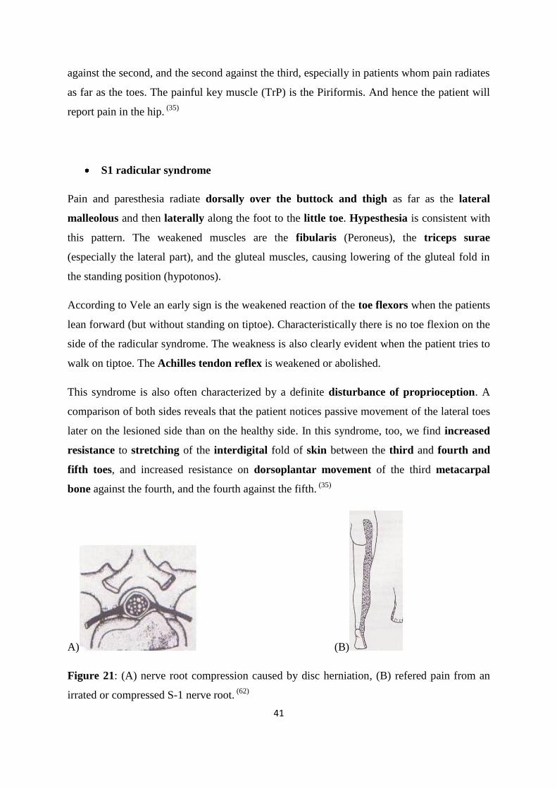
41
against the second, and the second against the third, especially in patients whom pain radiates
as far as the toes. The painful key muscle (TrP) is the Piriformis. And hence the patient will
report pain in the hip. (35)
S1 radicular syndrome
Pain and paresthesia radiate dorsally over the buttock and thigh as far as the lateral
malleolous and then laterally along the foot to the little toe. Hypesthesia is consistent with
this pattern. The weakened muscles are the fibularis (Peroneus), the triceps surae
(especially the lateral part), and the gluteal muscles, causing lowering of the gluteal fold in
the standing position (hypotonos).
According to Vele an early sign is the weakened reaction of the toe flexors when the patients
lean forward (but without standing on tiptoe). Characteristically there is no toe flexion on the
side of the radicular syndrome. The weakness is also clearly evident when the patient tries to
walk on tiptoe. The Achilles tendon reflex is weakened or abolished.
This syndrome is also often characterized by a definite disturbance of proprioception. A
comparison of both sides reveals that the patient notices passive movement of the lateral toes
later on the lesioned side than on the healthy side. In this syndrome, too, we find increased
resistance to stretching of the interdigital fold of skin between the third and fourth and
fifth toes, and increased resistance on dorsoplantar movement of the third metacarpal
bone against the fourth, and the fourth against the fifth. (35)
A) (B)
Figure 21: (A) nerve root compression caused by disc herniation, (B) refered pain from an
irrated or compressed S-1 nerve root. (62)
42
3.3. Physical examinations of lumbar disc herniation
Nerve root tension test: L5, S1, S2 roots
Straight leg raising test (SLRT)
Lasegue test is aimed to differentiate sciatic from hip pain. It consists from to parts the first
is in raising the lower limb with fully extendend; and the second the patient‟s place the foot
on the bed and the hip is further flexed. Positievely the first maneuver is an expression known
of sciatic pain. Persistance or apperarance in the second part of the maneuver indicates a
pathologic condition of the hip. (33)
This tests corespond to the first part of lasegue‟s maneuver. The patient is place supine with
the hips and knees in full extension and the ankles in a relaxed position. The examinar takes
the heel with one hand, while exerting, with the other a slight pressure on the knee to avoid it
bending when raising the limp. Normally the limb can be raised at least 60 degrees without
causing the patient any discomfort.
Patients with radicular pain experience a progressive increase in discomfort, upon limb
raising, to which they begin to resist as soon as pain becomes severe. Resisistance is affected
initially by contracting the hamstrings and then by raising the homologous hemipelvis from
the bed. Numerous studies have evaluated the sensitivity of SLRT in patients with disc
herniation: the values range from 80% to 99%. Specificity however does not exceed 40%. (33)
Figure 22: Laseque sign
43
Charnley showed that Nerve root glides freely through the intrevertbral foramina and during
elevation of the extented lower limb, nerve roots are pulled out for a distance up to 12 mm at
L5 level.
Signs of root tension such as limitation in straight leg raising are common in disc prolapse but
generally absent in pure degenerative stenosis. If however a stenotic patient develops a small
disc prolapse the straight leg raise test may convert to positive. (32)
In lifting the lower limb with the knee extended, the ankle is dorsiflexed (Braggard sign) by
applying the hand which raises the limb on the heel or sole of the foot. The sciatic nerve is
thus further stretched through the tibialis posterior nerve and pain, may therefore, be elicited
when SLRT is negative or, anyway, at lower degrees of leg raising. This may be helpful
when the stand rd maneuver is only slightly positive.
Similar maneuver is the so-called ankle dorisflexion maneuver; the limb is raised until the
patient feels radiiated pain and then is lowered until the pain decreases or disappears. At
this pomt, a force ankle dorsiflexion is performed, which again elicits radiated pain, when the
maneuvrer is positive. This clinal test, which is less sensitive than the former, may confirm
the positivity of SLRT. Its practical usefulness, however, is very limited. (33)
Pathogenic mechanism of SLRT
In several studies on cadavers, the motion of the lumbosacral roots was examined upon SLR.
Observation that both nerve root (or radicula r nerve) and the dura mater undergo translation
and lengthenig movements. Between 0 and 30 degrees motion is ver limites; between 30 and
60 degrees the root moves in the vertical direction; and between 60 and 90 degrees the
displacement occurs perpendicularly to the direction of the root; toward the pedicle.
Knee extension in the sitting postion (Slump test - simpler form as Flip test)
The patient sits with knees over the side of the bed and the leg is raised until the knee is fully
extended. When the test is positive, the greater the nerve-root tension, the less the knee
extension and the greater the radiated pain felt by the patient. Furthermore the patient will
44
simultaneously throw back his trunk and take his/ her hand away from the edge of the bed.
This maneuver, described by Oppenheim, has the same clinical significance as the SLRT.
However it is less sensitive, and may, thus, be negative or weakly positive when the SLRT is
clearly positive. The flip test may be useful in those patients with sever lumporadicular pain
who have difficulty in taking up a supine position.
Cross-leg SLRT
Rising of the asymptomatic leg may cause pain on the symptomatic side. The patients who
most freguently presesent a positive crossed-leg SLRT are those with a paramedian
herniation. In these cases, stretching of the contralateral nerve roots involved traction on the
thecal sac and, through this, the compressed nerve root. The latter tend to displace toward the
midline, and is thus compressed by herniation. However, contralateral pain lifting the
asymptomatic leg may also be observed in patients with midline or posterolateral herniation.
The sensitivity of crossed-leg SLRT is only 25% - 44%, but specificity os high as 90%.
This manuever is extremely reliable for diagnosis not only of disc herniation, but particularly
of herniation responsible for marked-nerve root compression. Most patients with crossed-leg
SLRT, in fact have a medium-sized or large contained herniation, or an extruded or migrated
herniated disc. (33, 64)
Neurodynamic tests
Nerve-root tension tests: L2, L3, L4 roots
Femoral nerve stretch test (FNST)
The femoral nerve stretch test me be may be carried out in various ways, with the patient in
the prone position. The simplest modality, described by wassermann is to bend the knee until
pain appears on the anterior aspect of the thigh. It is positive as soon as 90 degrees are
reached and pain becomes progressively more severe if knee flexion is increased even
slightly. The test can be reinforced if the hip is extendend by placing a hand under the knee to
raise the thigh.
45
An alternative method is to extend the hip, while kepping the knee extendend. This is done by
placing one hand on the buttock to keep the pelvis in position, whilst the other hand, placed
under the knee, raises the limb.
Stretching on the femoral nerve on the healthy side may cause anterior thigh pain on the
symptomatic side. The crossed-leg FNST is much more rarely positive than the crossed leg
SLRT.
Other tests and signs:
‘Painful arc’ described by Cyriax (1978): during anteflexion, often shortly after beginning
to bend forward, the patient feels considerable pain. An evasive movement of the spinal
column can often be seen, as if the patient is working around some obstacle, after which the
action continues quite normally. On the straightening again the pain reappears and there is an
evasive reaction at the same point. This sign indicates disc herniation.
If the springing test produces pain in the lumbar spine and joint restriction is either absent or
has been resolved, this indicates a disc lesion. (35)
Increase intra-bdominal pressure provokes sometime pain in patients with disc herniation.
(64)
Vele test by providing this test we evaluate the stability of a patient‟s foot flexors by
providing of loss of action may evaluate of compression of nerve of any reason. (71)
Unilateral blocking of nutation movement in sacroiliac joint causes the distortion of the
pelvis. But also if gluteus maximus on one side is weak, the subgluteal line is lowered and
intergluteal line is tilted to one side. Such shifting in sacroiliac joints is painful and can be
suggested as a sign for sciatica. (71)
Lumbar traction test and mobilization
The patient stands with feet slightly apaart, and the lumbar spine in the resting position.
Therapist stands behind the patient. May need to stand on a stool to position he‟s arms at or
above the height of the patient‟s arms. Holding the patient just below the rib cage, with the
arms in an interlocked grip and leaning slightly backward to apply a Grade I, II, or III traction
force. Testing lumbar traction is for alleviation of provoking the pain. (5)
46
Lumbar Compression test
The patient stands with feet slightly appart and the lumbar spine in the resting position.
Standing behind the patient the therpaist places he‟s hands on to the patient‟s shoulders or
grip around the lower part of the patient‟s rib cage. Pressing the patient‟s sholders (or trunk)
in a caudal direction may provoke pain and increase radicular symptoms. (5)
Waddell signs nonorganic signs:
- Superficial tederness to light pinch
- Noanatomictenderness which is not localized and often extends from the lumbar spine
to thorax or pelvis.
- Axial loading pain, when low back pain is reported with vertical loading to the
patients head.
- Pain with whole body rotation, when shoulders and pelvis are rotaed in the same
plane
- Discrepancy between seated and lying straight leg raise
- Give –way or cogwheel weakness that cannot be explained on a localized neurolic
basis
- Sensory disturbances in a stocking rather than a dermatomal distribution
- Disproportionate vertabization and facial expressions during examination
Lumbar instability tests:
Segmental stabilization tests are in general passive accesory motion tests.
- Anterior shear test
- Posterior shear test
- Torsion test
Local stabilizing tests
- Transversus abdominis test (biofeedback pressure)
- Segmental lumbar multifidus
Global stabilizing system tests (66)
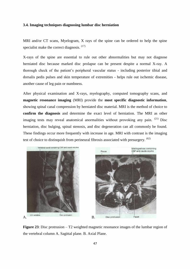
47
3.4. Imaging techniques diagnosing lumbar disc herniation
MRI and/or CT scans, Myelogram, X rays of the spine can be ordered to help the spine
specialist make the correct diagnosis. (17)
X-rays of the spine are essential to rule out other abnormalities but may not diagnose
herniated disc because marked disc prolapse can be present despite a normal X-ray. A
thorough check of the patient‟s peripheral vascular status - including posterior tibial and
dorsalis pedis pulses and skin temperature of extremities - helps rule out ischemic disease,
another cause of leg pain or numbness.
After physical examination and X-rays, myelography, computed tomography scans, and
magnetic resonance imaging (MRI) provide the most specific diagnostic information,
showing spinal canal compression by herniated disc material. MRI is the method of choice to
confirm the diagnosis and determine the exact level of herniation. The MRI as other
imaging tests may reveal anatomical anormalities without provoking any pain. (21)
Disc
herniation, disc bulging, spinal stenosis, and disc degeneration can all commonly be found.
These findings occur more frequently with increase in age. MRI with contrast is the imaging
test of choice to distinquish from perineural fibrosis associated with presurgery. (62)
A.
B.
Figure 23: Disc protrusion – T2 weighted magnetic resonance images of the lumbar region of
the vertebral column A. Sagittal plane. B. Axial Plane.
48
A myelogram can define the size and location of disc herniation. An electromyogram can
determine the exact nerve root involved. A nerve conduction velocity test may also be
performed.
Lumbar myelography (liquid dye is injected into the spinal column and appears white
against bone on an x-ray film. A myelogram can show pressure on the spinal cord or nerves
from herniated discs, bone spurs or tumors.), in the past was the usual method for establishing
a diagnosis, but it is usually not necessary today. (17)
Discography is an invasive diagnostic procedure designed to determine whether a disc is
intrinsically painful. Discograhy involves the injection of contast material or saline into the
nucleus pulposus of the inter-vertebral disc. Postdiscography cumputed tomography can be
used to highlight the features of internal disc diruption, which is the most common known
cause of discogenic pain in patients with chronic spinal problems. It has been shown by this
method that spinal pain caused by internal disc diruption is present in more than 39% of
patients with chronic low back pain. However discography is not recommended as parts of
the diagnostic process for patients of acute lower back pain neither with suspected disc
herniation. (62)
In spinal disorders, electromyography (EMG) is the method of choice for the identification
of damage within the peripheral motor nerve fibers (highest sensitivity). However, the delay
between the time of the actual damage and the first signs of denervation (acute denervation
potentials occur after a mean of 21 days) must be considered. Also the activation pattern
(complete or reduced interference) assessed during voluntary activation (here the patient
needs to cooperate and perform a voluntary activation) can be applied as soon as the very first
few days after a lesion to disclose a pathological innervation.
The performance of EMG in several muscles allows the specific localization of the nerve
damage (somatotopic localization of a lesion) to be indicated and for the differentiation of
acute, subacute and chronic axonal damage (denervation). The EMG has a high specificity
and will rarely be abnormal in asymptomatic individuals but is usually not performed until at
least 3 weeks after the symptoms begin. CT myelography is also considered an important tool
for identifying herniated disc. (5)
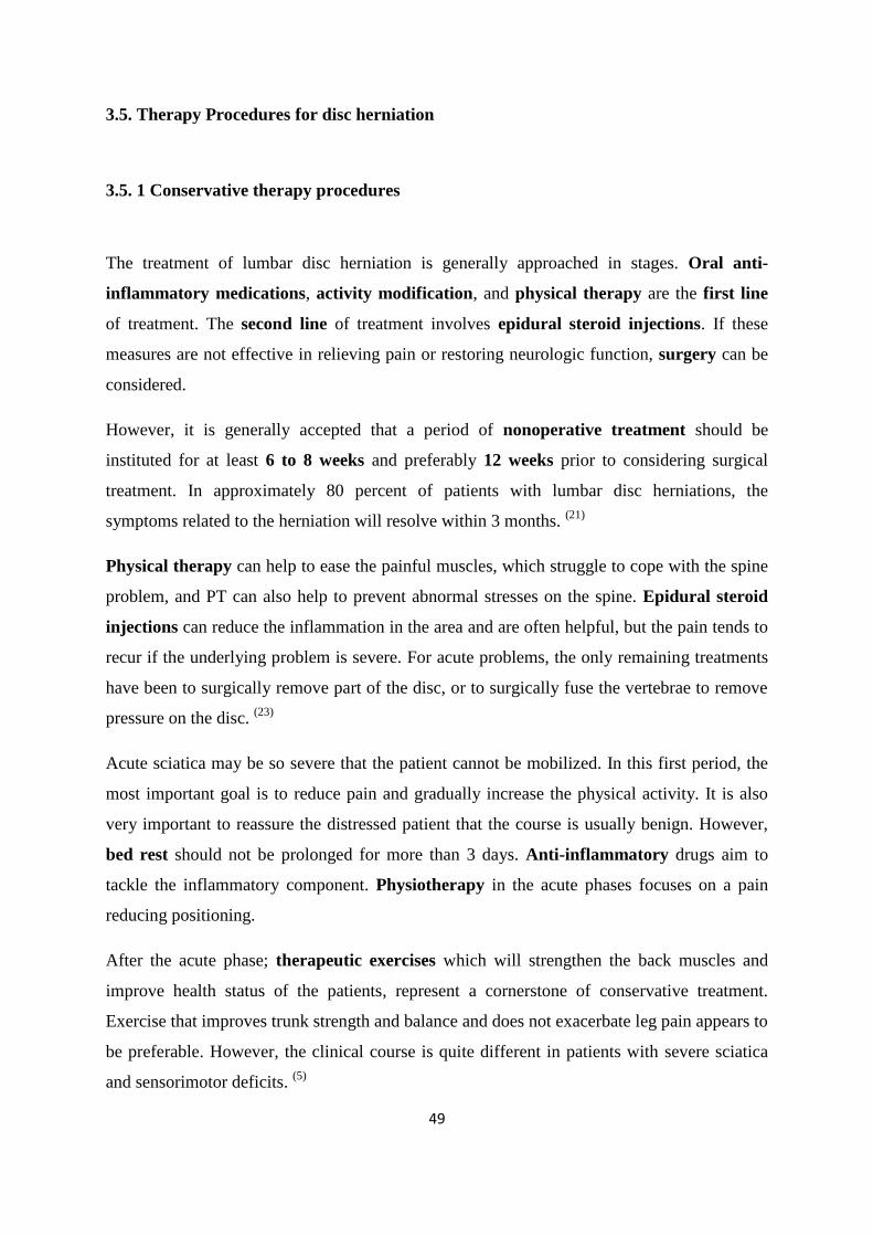
49
3.5. Therapy Procedures for disc herniation
3.5. 1 Conservative therapy procedures
The treatment of lumbar disc herniation is generally approached in stages. Oral anti-
inflammatory medications, activity modification, and physical therapy are the first line
of treatment. The second line of treatment involves epidural steroid injections. If these
measures are not effective in relieving pain or restoring neurologic function, surgery can be
considered.
However, it is generally accepted that a period of nonoperative treatment should be
instituted for at least 6 to 8 weeks and preferably 12 weeks prior to considering surgical
treatment. In approximately 80 percent of patients with lumbar disc herniations, the
symptoms related to the herniation will resolve within 3 months. (21)
Physical therapy can help to ease the painful muscles, which struggle to cope with the spine
problem, and PT can also help to prevent abnormal stresses on the spine. Epidural steroid
injections can reduce the inflammation in the area and are often helpful, but the pain tends to
recur if the underlying problem is severe. For acute problems, the only remaining treatments
have been to surgically remove part of the disc, or to surgically fuse the vertebrae to remove
pressure on the disc. (23)
Acute sciatica may be so severe that the patient cannot be mobilized. In this first period, the
most important goal is to reduce pain and gradually increase the physical activity. It is also
very important to reassure the distressed patient that the course is usually benign. However,
bed rest should not be prolonged for more than 3 days. Anti-inflammatory drugs aim to
tackle the inflammatory component. Physiotherapy in the acute phases focuses on a pain
reducing positioning.
After the acute phase; therapeutic exercises which will strengthen the back muscles and
improve health status of the patients, represent a cornerstone of conservative treatment.
Exercise that improves trunk strength and balance and does not exacerbate leg pain appears to
be preferable. However, the clinical course is quite different in patients with severe sciatica
and sensorimotor deficits. (5)
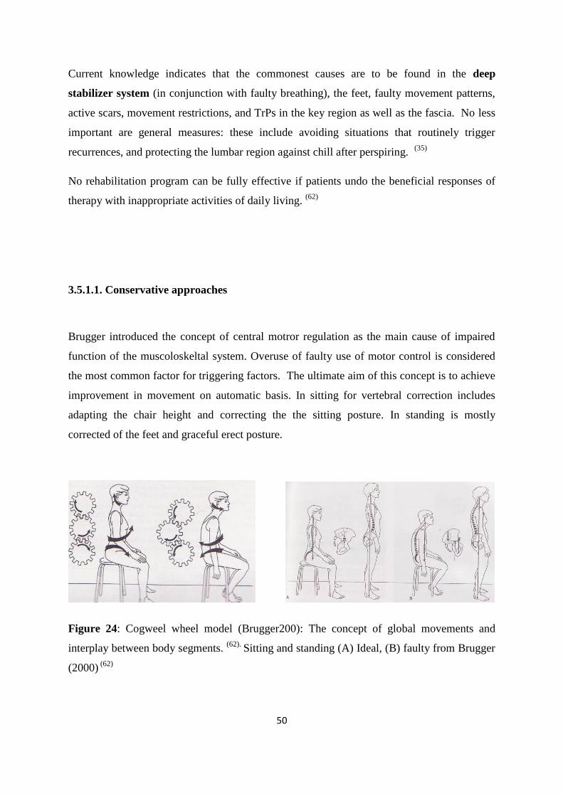
50
Current knowledge indicates that the commonest causes are to be found in the deep
stabilizer system (in conjunction with faulty breathing), the feet, faulty movement patterns,
active scars, movement restrictions, and TrPs in the key region as well as the fascia. No less
important are general measures: these include avoiding situations that routinely trigger
recurrences, and protecting the lumbar region against chill after perspiring. (35)
No rehabilitation program can be fully effective if patients undo the beneficial responses of
therapy with inappropriate activities of daily living. (62)
3.5.1.1. Conservative approaches
Brugger introduced the concept of central motror regulation as the main cause of impaired
function of the muscoloskeltal system. Overuse of faulty use of motor control is considered
the most common factor for triggering factors. The ultimate aim of this concept is to achieve
improvement in movement on automatic basis. In sitting for vertebral correction includes
adapting the chair height and correcting the the sitting posture. In standing is mostly
corrected of the feet and graceful erect posture.
Figure 24: Cogweel wheel model (Brugger200): The concept of global movements and
interplay between body segments. (62).
Sitting and standing (A) Ideal, (B) faulty from Brugger
(2000) (62)
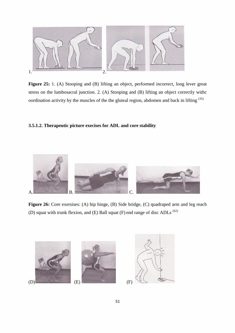
51
1..
2.
Figure 25: 1. (A) Stooping and (B) lifting an object, performed incorrect, long lever great
stress on the lumbosacral junction. 2. (A) Stooping and (B) lifting an object correctly withc
oordination activity by the muscles of the the gluteal region, abdomen and back in lifting (35)
3.5.1.2. Therapeutic picture execises for ADL and core stability
A. B. C.
Figure 26: Core exersises: (A) hip hinge, (B) Side bridge, (C) quadraped arm and leg reach
(D) squat with trunk flexion, and (E) Ball squat (F) end range of disc ADLs
(62)
(D) (E) (F)
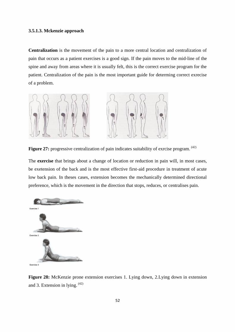
52
3.5.1.3. Mckenzie approach
Centralization is the movement of the pain to a more central location and centralization of
pain that occurs as a patient exercises is a good sign. If the pain moves to the mid-line of the
spine and away from areas where it is usually felt, this is the correct exercise program for the
patient. Centralization of the pain is the most important guide for determing correct exrecise
of a problem.
Figure 27: progressive centralization of pain indicates suitability of exrcise program. (42)
The exercise that brings about a change of location or reduction in pain will, in most cases,
be exetension of the back and is the most effective first-aid procedure in treatment of acute
low back pain. In theses cases, extension becomes the mechanically determined directional
preference, which is the movement in the direction that stops, reduces, or centralises pain.
Figure 28: McKenzie prone extension exercises 1. Lying down, 2.Lying down in extension
and 3. Extension in lying. (42)
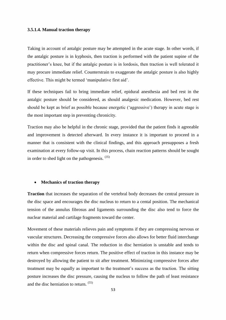
53
3.5.1.4. Manual traction therapy
Taking in account of antalgic posture may be attempted in the acute stage. In other words, if
the antalgic posture is in kyphosis, then traction is performed with the patient supine of the
practitioner‟s knee, but if the antalgic posture is in lordosis, then traction is well tolerated it
may procure immediate relief. Counterstrain to exaggerate the antalgic posture is also highly
effective. This might be termed „manipulative first aid‟.
If these techniques fail to bring immediate relief, epidural anesthesia and bed rest in the
antalgic posture should be considered, as should analgesic medication. However, bed rest
should be kept as brief as possible because energetic („aggressive‟) therapy in acute stage is
the most important step in preventing chronicity.
Traction may also be helpful in the chronic stage, provided that the patient finds it agreeable
and improvement is detected afterward. In every instance it is important to proceed in a
manner that is consistent with the clinical findings, and this approach presupposes a fresh
examination at every follow-up visit. In this process, chain reaction patterns should be sought
in order to shed light on the pathogenesis. (35)
Mechanics of traction therapy
Traction that increases the separation of the vertebral body decreases the central pressure in
the disc space and encourages the disc nucleus to return to a cental position. The mechanical
tension of the annulus fibrosus and ligaments surrounding the disc also tend to force the
nuclear material and cartilage fragments toward the center.
Movement of these materials relieves pain and symptoms if they are compressing nervous or
vascular structures. Decreasing the compressive forces also allows for better fluid interchange
within the disc and spinal canal. The reduction in disc herniation is unstable and tends to
return when compressive forces return. The positive effect of traction in this instance may be
destroyed by allowing the patient to sit after treatment. Minimizing compressive forces after
treatment may be equally as important to the treatment‟s success as the traction. The sitting
posture increases the disc pressure, causing the nucleus to follow the path of least resistance
and the disc herniation to return. (55)
54
3.5.1.5. Injection procedures
Pain pathogenesis in cases with nerve root compromise is caused not only by a mechanical
compression but also by a chemical irritation due to proinflammatory cytokines.The rationale
for nerve root blocks is therefore to tackle the inflammatory component of the nerve root
compromise.
The peri-radicular foraminal nerve root block is always performed under image intensifier
control, allowing for a direct application of the antiinflammatory agent to the target nerve
root. The objective of a therapeutic selective nerve root block is not to cure the patient by
interfering with pathogenetic factors that are responsible for sciatica but rather to provide
temporary relief from peak pain during the time required for spontaneous resolution of
radiculopathy. (21)
Epidural injections include a variety of injection techniques such as caudal (sacral),
interlaminar lumbar and cervicothoracic. In contrast to the selective nerve root blocks,
epidural steroid injections have the drawback that the pharmacological agent has to diffuse to
the site of inflammation and there is no guarantee that it does so. In cases with multilevel
involvement or non-specific leg pain the epidural route has some advantages compared to
selective nerve root blocks.
The preferred level is one level above the target level. Others favor the level which
corresponds to the segment of origin of the patient‟s symptoms. One or two percent
anesthetic agent is injected to anesthetize the needle track. Using an interlaminar approach, a
22- or 25-gauge spinal needle is advanced between the spinous processes of the target level.
Aiming at the upper edge of the lower lamina, the needle is inserted into the posterior
epidural space with or without fluoroscopic control depending on one‟s personal experience
with this technique. The location is confirmed using a small amount of contrast material. (5)
Injection of the enzyme chymopapain into the herniated disc produces a loss of water and
proteoglycans from the disc, thereby reducing both the disc‟s size and the pressure in the
nerve root. (21)
55
3.5.2. Non- conservative therapy approaches
3.5.2.1 Surgical indications for disc heniation or spinal stenosis
The symptomatic lumbar disc herniation has been estimated to be 1-2% and only 1.6% of
people have sciatica/radiculopathy lasting more than two weeks. (58)
Surgery for sciatica is
estimated to be necessary in between 1.3% and 3.1% of the population. In contrast, spinal
stenosis does not have such good prognosis. Approximately 15%of patients improve, 15%
worsen, whereas the remainder remain fairly stable. (62)
According to the Rand Corporation (62)
- Appropriate:
Pain in lower limb, imaging positive for disc hernia or spinal stenosis, major or
minor neurological findings, restricted activity for more than 6 weeks.
- Equivocal:
Pain in lower limb, imaging positive for disc hernia or spinal stenosis, major
neurological findings, restricted activity for less than 4 weeks.
Pain in lower limb, imaging equivocal for disc hernia or spinal stenosis, minor
neurological findings, restricted activity for mopre than 6 weeks.
- Inappropriate:
Pain in lower limb, imaging positive for disc hernia por spinal stenosis, minor
neurologica findings, restricted activity for less than 4 weeks.
- Minor neuro findings (2 or more items)
Assymetric ankle reflex
Dematomal sensory deficit
Positive ipsilateral SLR (straight leg raise) test
Sciatica
- Major neurologic findings
Progressive unilatel leg wekness, or
Positive contralateral SLR test. (62)
56
3.5.2.2 Minimal invasive procedures
Contained discs, which are completely covered by outer annular fibers or posterior
longitudinal ligament, are not in direct contact with epidural tissue. By contrast, non-
contained discs are in direct contact with epidural tissue. This differentiation is of importance
for minimally invasive surgical procedures such as chemonucleolysis or percutaneous disc
decompression. (5)
Percutaneous discectomy
An advanced form of percutaneous discectomy developed to date uses a plasma technology
to remove tissue from the center of the disc. During the procedure, an instrument is
introduced through a needle and placed into the center of the disc where a series of channels
are created to remove tissue from the nucleus. Tissue removal from the nucleus acts to
decompress the disc and relieve the pressure exerted by the disc on the nearby nerve root. As
pressure is relieved, pain is reduced, consistent with the clinical results of earlier
percutaneous discectomy procedures. There is little tissue trauma and recovery times may be
improved in many patients.
Intradiscal Electrothermoplasty (IDET)
This procedure involves the insertion of a needle into the affected disc with the guidance of
an x-ray machine. A wire is then threaded down through the needle and into the disc until it
lies along the inner wall of the annulus. The wire is then heated which destroys the small
nerve fibers that have grown into the cracks and have invaded the degenerating disc.
The heat also partially melts the annulus, which triggers the body to generate new reinforcing
proteins in the fibers of the annulus. A study of fifty-three patients with discogenic back pain
was published in the October issue of the journal, Spine. Depending on the stringency of
criteria used, the success rate of IDET may be as low as 23% or as high as 60%.
57
Radiofrequency Discal Nucleoplasty (Coblation Nucleoplasty)
Nucleoplasty is similar to the IDET procedure, a needle is inserted into the disc. Instead of a
heating wire, a special radiofrequency probe is inserted through the needle into the disc. This
probe generates a highly focused plasma field with enough energy to break up the molecular
bonds of the gel in the nucleus, essentially vaporizing some of the nucleus. The result is that
10-20% of the nucleus is removed which decompresses the disc and reduces the pressure both
on the disc and the surrounding nerve roots.
This technique may be more beneficial for sciatica type of pain than the IDET, since
nucleoplasty can actually reduce the disc bulge, which is pressing on a nerve root. The high-
energy plasma field is actually generated at relatively low temperatures, so danger to
surrounding tissues is minimized. These new techniques are exciting. They offer the
possibility of treating discogenic low back pain and sciatica with much less trauma and risk
than surgery, but we must remember that these are still unproven technologies. (23)
3.5.2.3. Lumbar spinal surgery procedures
Lumbar spinal surgery is much more commonly performed in the United States than it is in
other countries. The most common lumbar spinal surgery in America is surgical treatment of
lumbar disc herniation, and it is performed far more frequently than it is in most European
countries.
In most afflictions of the lumbar spine, surgical treatment should be seen as the final phase
of treatment when all other forms of treatment have not been effective in relieving pain or
restoring normal neurologic or physiologic function. (21)
Laminotomy and Microdiscectomy
The standard technique for lumbar disc excision is a laminotomy through a technique that is
currently known as microdiscectomy. This involves a limited exposure of the hemilamina on
the right or left side, depending on the location of the disc.
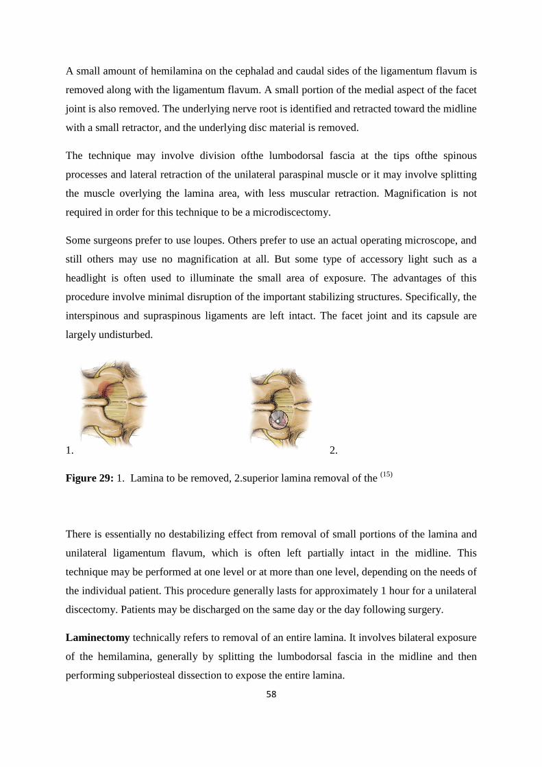
58
A small amount of hemilamina on the cephalad and caudal sides of the ligamentum flavum is
removed along with the ligamentum flavum. A small portion of the medial aspect of the facet
joint is also removed. The underlying nerve root is identified and retracted toward the midline
with a small retractor, and the underlying disc material is removed.
The technique may involve division ofthe lumbodorsal fascia at the tips ofthe spinous
processes and lateral retraction of the unilateral paraspinal muscle or it may involve splitting
the muscle overlying the lamina area, with less muscular retraction. Magnification is not
required in order for this technique to be a microdiscectomy.
Some surgeons prefer to use loupes. Others prefer to use an actual operating microscope, and
still others may use no magnification at all. But some type of accessory light such as a
headlight is often used to illuminate the small area of exposure. The advantages of this
procedure involve minimal disruption of the important stabilizing structures. Specifically, the
interspinous and supraspinous ligaments are left intact. The facet joint and its capsule are
largely undisturbed.
1. 2.
Figure 29: 1. Lamina to be removed, 2.superior lamina removal of the (15)
There is essentially no destabilizing effect from removal of small portions of the lamina and
unilateral ligamentum flavum, which is often left partially intact in the midline. This
technique may be performed at one level or at more than one level, depending on the needs of
the individual patient. This procedure generally lasts for approximately 1 hour for a unilateral
discectomy. Patients may be discharged on the same day or the day following surgery.
Laminectomy technically refers to removal of an entire lamina. It involves bilateral exposure
of the hemilamina, generally by splitting the lumbodorsal fascia in the midline and then
performing subperiosteal dissection to expose the entire lamina.
59
In a true complete laminectomy, the entire lamina, along with the spinous process and the
ligamentum flavum caudal and cephalad to the lamina, is removed. This allows access to the
entire spinal canal at a given laminar level, as well as to four nerve roots, two cephalad and
two caudal.
The facet joints are generally preserved, but when this technique is used-and it is often used
in spinal stenosis-the facets are undercut. This procedure involves removal of inferior
osteophytes underneath the facet by reaching from within the spinal canal rather than
removing the entire facet, which causes significant destabilization of the motion segment.
Complete laminectomy produces a destabilizing effect. However, this procedure is generally
performed in older individuals with degenerative disc settling and degenerative facet joints
and these areas are generally stable due to the degenerative changes. It is not necessary to
perform a fusion whenever a complete laminectomy is performed.
In spinal stenosis it is common to perform laminectomy at multiple levels, as symptomatic
spinal stenosis is often a multilevel disease. When a complete laminectomy is performed to
decompress a spondylolisthesis, it is often referred to as a Gill procedure. (21)
60
4. Methodology
This thesis completed a systematic review of the relevant literature on the efficacy of
conservative treatment of Lumbar Disc Herniation (LDH). In order to evaluate and in order to
make the thesis more reliable, the author selected mostly research studies that used the most
recent reliable experimental designs and systematic reviews.
The main criteria in this systemtic review for trials analyzed were that all patients would have
sciatica radiculopathy symptoms due to disc herniation. The assesment was to evaluate
patients with certain duration of symptoms due its good overlaps over time. The research was
made to describe and evaluate all important information assessing the disc pain and the
rehabilitation of it.
Literature Search, Study Selection and Data
Extraction
We search a broad range of database to identify published and unpublished studies with
information about disc herniation with main concern of treatments benefits and differntations.
We selected and extracted rescent articles that we felt relevant or intresting as well as
choosing what we were aware of being potentially important.
Each database was searched from its starting date on September 2009. The databases
searched were:
MEDLINE
Pre MEDLINE
EMBASE
The Spine Journal
Europran Spine Journal
British Medical Journal
61
Cochrane Database of Systematic Reviews
The New England Journal of Medicine
American academy of orthopaedic surgeons
Journal of the american medical association
Journal of chiropractic medicine
To identify the patient groups, interventions, and outcomes that should be included in the
review, we read background material from diverse sources including textbooks, reports, and
proceedings with Doctors, and Web sites. The quality of all trials in the review was assessed
using a list of items indicating components of internal validity.
All reviews were to assess all different documentations on disc herniation in the lumar region
and the most treatments known for it. Evaluating results of each treatment effect and to
compare from different conclusions from the differnent trials mentioned.
Measures of outcomes for pain and disability will be analyzed in detail to understand better
the process of the effect of each treatment and to distinquish them to reach a conclusion of
when and which treatment is appropriate for the patients with symptomatic disc herniation.
Most outcomes were measued with a baseline and duration of meeting of examinations.
These were mostly: Visuual analogue scale (VAS), Oswestry Disability Index (OID), Roland
morris disability index and other outcomes were included by sciatica severity measured by
the Sciatica Bothersomeness Index, satisfaction with symptom self-reported improvement
and employment status.
All reviews and articles were in English language.
In the Appendix the the level of evidence are shown.
62
5. Conservative versus non conservative treatments trials for disc herniations
5.1. Non conservative treatments
5.1.1. Surgery versus non operative treatment for disc herniation
The spine patient outcomes research trial (S.P.O.R.T.) assessed a four year outcome of
surgery versus non operative care in 13 spine clinics in 11 states in the U.S.A. 1244
participants with lumbar intervertebral disc herniation were enrolled out of 1991 eligible for
enrollment. The patients had the chance to choose if they would follow a randomized or a
cohort observation trial. The assessment was made for an observational cohort of 743
participants and a randomized trial of 501 participants which evaluation was at 6 weeks, 3
months, 6 months and 1 and 2 years. (75)
In the randomized clinical trial Weinstein et al. patients were enrolled from March 2000
until November 2004 whom 501 patients being surgical candidates with mean age of 42
years and 42% being females. A total of 472 participants completed at least 1 follow-up visit
and were included in the analysis. There was limititaion in compliance of assigned treatment:
50% of patient‟s asigned to surgery received surgery within 3 month of enrollment, while
30% of those assigned to nonoperative treatment received surgery in the same period.
In the non operative treatments: most patients received education/counselling (93%) and anti-
inflammatory medication (61%) (no steroidal anti-inflammatory drugs, cyclooxygenase 2
inhibitors or oral steroids, 46% received opiates, more than 50% received injections (e.g.
epidural steroids) and 29% were prescribed activity restrictions. 44% received active physical
therapy during the trial but 67% had received it before the trial.
In the surgical treatments was standard open discetomy, it took a median time of 75minutes
had median bloody loss of 49.5ml, with only 2% requiring transfusion. The most common
complication was dural tear (4%). There were no post-operative complications in 95%.
Reoperation occurred in 4% in 1 year of initial surgery of which 5% were from recurrent
herniation at the same level.
63
Greater improvements in the Sciatica Bothersomeness Index were in the surgery group at all
designated follow-up times: 3 months (treatment effect, −2.1; 95% CI, −3.4 to −0.9), 1 year
(treatment effect, −1.6; 95% CI, −2.9 to −0.4), and 2 years (treatment effect, −1.6; 95% CI,
−2.9 to −0.3), with results of the global hypothesis test being statistically significant
(P=.003). Patient satisfaction with symptoms and treatment showed small effects in favor of
surgery while employment status showed small effects in favor of nonoperative care, but
none of these changes was statistically significant. Self-rated progress showed a small
statistically significant advantage for surgery (P=.04). (77)
As-treated analyses based on treatment received were performed with adjustments for the
time of surgery and factors affecting treatment crossover and missing data. These yielded far
different results than the intent-to-treat analysis, with strong, statistically significant
advantages seen for surgery at all follow-up times through 2 years. For example, at 1 year the
estimated treatment effects for the SF-36 bodily pain and physical function scales, the ODI,
and the sciatica measures were 15.0 (95% CI, 10.9 to 19.2), 17.5 (95% CI, 13.6 to 21.5),
−15.0 (95% CI, −18.3 to −11.7), and −3.2 (95% CI, −4.3 to −2.1), respectively. (77)
In the observational cohort Weinstein et al. enrolled 743 patients in which initially 521
patients chose surgery and 222 chose non operative care. In 91% surgery was received in the
first 6 weeks of enrolment, a remaining 4% of patients received surgery in 6 months and the
remaining 4% didn‟t receive any surgery and stayed with non operative care. From the non
operative cohort 2% underwent surgery in the first 6 weeks while another 16% in the
following 6 months and 22% had surgery in a 2 year period. Overall 528 patients received
surgery and 191 patients received conservative treatment.
The mean age was 41.4 years old, being mostly men of Caucasian race which had full time or
part time jobs. 98% of them had dermatome radiation pain; most herniations where at the
level of L5-S1 which were posterolateral extrusions confirmed by an imaging technique. (74)
In the non operative treatment of the observational cohort 92% of the patients received
education and counseling; 58% received non-steroidal anti-inflammatory drugs; 35%
received narcotic analgesic agents; 43% underwent physical therapy and 38% had epidural
injections. The average time of the surgery was 70 minutes with a blood loss of 50ml on
average and only 2 patients required transfusions. There were some surgical complications,
64
2% for Dural tear. There were also re-operations in 7% of the cases by the first year and 9%
at 2 years with more the and half being recurrent herniations at the same level.
Of the 743 patients enrolled in the observational cohort, 528 patients received surgery and
191 received usual nonoperative care. At 3 months, patients who chose surgery had greater
improvement in the primary outcome measures of bodily pain (mean change: surgery, 40.9 vs
nonoperative care, 26.0; treatment effect, 14.8; 95% CI, 10.8-18.9), physical function (mean
change: surgery, 40.7 vs nonoperative care, 25.3; treatment effect, 15.4; 95% CI, 11.6-19.2),
and O.D. I (mean change: surgery, −36.1 vs nonoperative care, −20.9; treatment effect,
−15.2; 95% CI, −18.5. to −11.8).
These differences narrowed somewhat at 2 years: bodily pain (mean change: surgery, 42.6 vs
nonoperative care, 32.4; treatment effect, 10.2; 95% CI, 5.9-14.5), physical function (mean
change: surgery, 43.9 vs nonoperavtive care 31.9; treatment effect, 12.0; 95% CI; 7.9-16.1),
and O.D.I (mean change: surgery −37.6 vs nonoperative care −24.2; treatment effect, −13.4;
95% CI, −17.0 to −9.7).
There was substantial improvement over time in both conservative and non-conservative
groups of patients, but greater results were in those patients who underwent surgery. The
benefit of surgery was noticeable at the first 6 weeks and was maintained for at least 2 years.
With the primary outcome of 3 months, surgery was the most effective method in contrast
with the non operative treatment. There was a greater improvement with surgery than
conservative treatment, but after the second year the difference in effectiveness was
slightlydecreased. (74)
Pearson et al. combining the S.P.O.R.T randomized and observational trial mentioned
above; was made to evaluate if treatment, location and the morphology of disc herniation
mattered. From the 1191 patients; 775 had surgery and 416 had non-operative treatment in
2 years time.
In this analysis for location it demonstrated that 131 (11%) had central herniations and
patients were younger and at baseline the pain was more severe than those patients with
lateral herniation but benefited similarly with surgery. In the herniation morphology the
analysis demonstrated that 322 (27%) had disc protrusions and from the questionnaires
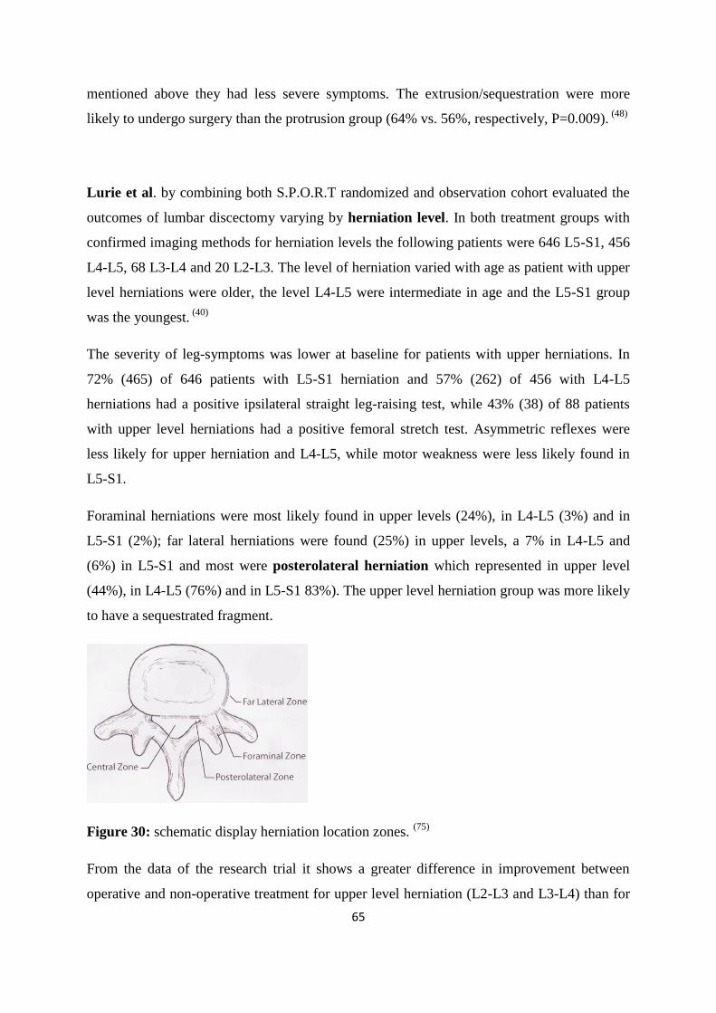
65
mentioned above they had less severe symptoms. The extrusion/sequestration were more
likely to undergo surgery than the protrusion group (64% vs. 56%, respectively, P=0.009). (48)
Lurie et al. by combining both S.P.O.R.T randomized and observation cohort evaluated the
outcomes of lumbar discectomy varying by herniation level. In both treatment groups with
confirmed imaging methods for herniation levels the following patients were 646 L5-S1, 456
L4-L5, 68 L3-L4 and 20 L2-L3. The level of herniation varied with age as patient with upper
level herniations were older, the level L4-L5 were intermediate in age and the L5-S1 group
was the youngest. (40)
The severity of leg-symptoms was lower at baseline for patients with upper herniations. In
72% (465) of 646 patients with L5-S1 herniation and 57% (262) of 456 with L4-L5
herniations had a positive ipsilateral straight leg-raising test, while 43% (38) of 88 patients
with upper level herniations had a positive femoral stretch test. Asymmetric reflexes were
less likely for upper herniation and L4-L5, while motor weakness were less likely found in
L5-S1.
Foraminal herniations were most likely found in upper levels (24%), in L4-L5 (3%) and in
L5-S1 (2%); far lateral herniations were found (25%) in upper levels, a 7% in L4-L5 and
(6%) in L5-S1 and most were posterolateral herniation which represented in upper level
(44%), in L4-L5 (76%) and in L5-S1 83%). The upper level herniation group was more likely
to have a sequestrated fragment.
Figure 30: schematic display herniation location zones. (75)
From the data of the research trial it shows a greater difference in improvement between
operative and non-operative treatment for upper level herniation (L2-L3 and L3-L4) than for
66
herniations at the levels L4-L5 and L5-S1. Patients with upper level herniation had less
improvement with non-operative treatment and slightly better operative outcomes than those
with lower level herniations.
The difference between the upper level herniation and L5-S1 herniation was at range of 10 to
15 points on the SF-36 bodily pain and physical functioning scale and the O.D.I giving the
relationship between level and outcome of surgery that the upper level herniation was greater
than the L4-L5 results, which were greater than the L5-S1 results. (39)
Back pain improved in intervertebral disc herniation (IDH) patients treated either by surgery
or non-operative care but the magnitude of improvement was greater in surgery. The surgery
treatment effect was diminished during time but the difference between the two treatment
groups was still significant at the two years landmark. Leg pain relief was also greater in
surgery and it was noticed that leg pain relief was greater than back pain relief in both
treatment groups. However work status didn‟t show significant benefit for surgery. (76)
There were a number of limitations in this study as of patient‟s crossingover treatment
groups. Specifying the morphology of one herniation for concluding a more reliable study but
would have a lesser amount of patients. The non operative had greater result than other
studies compared to this study but it was specified in “usual care‟‟ which gave it a more
generalization. Only 43% of the non operative patient‟s went to a physical therapist which
show‟s that if a more intensive program was given, it may had been more effective for
some patients. (40)
Nevertheless the combination as-treated analysis at 4 years concluded that the patients who
had surgery achieved a greater improvement than no-operatively treated patients in all
primary and secondary outcomes except work status. (75)
In an even longer trial Atlas et al. conducted a 10-year outcome of patients with sciatica
resulting from a lumbar disc herniation treated surgically or nonsurgically. Patients were
recruited from the practices of orthopedic surgeons, neurosurgeons, and occupational
medicine physicians throughout Maine. (2)
Clinical data were obtained at baseline from a physician questionnaire. Primary analyses
were based on initial treatments received, either surgical or nonsurgical. Secondary analyses
examined actual treatments received by 10 years. Outcomes included patienst reporting
67
symptoms of leg and back pain, functional status, satisfaction, and work and disability
compensation status.
From 507 initially enrolled patients for 10-year outcomes, available were 400 of 477 (84%)
surviving patients; 217 of 255 (85%) were treated surgically, and 183 of 222 (82%) were
treated nonsurgically. Patients undergoing surgery had worse baseline symptoms and
functional status than those initially treated nonsurgically. By 10 years, 25% of surgical
patients had undergone at least one additional lumbar spine operation, and 25% of
nonsurgical patients had at least one lumbar spine operation.
In the 10-year follow-up, 69% of the patients were initially treated surgically reported
improvement in their predominant symptom (back or leg pain) versus 61% of those initially
treated nonsurgically (P = 0.2). A larger proportion of surgical patients reported that their low
back and leg pain were much better or completely gone (56% vs. 40%, P = 0.006) and were
more satisfied with their current status (71% vs. 56%, P = 0.002).
There were treatment group differences persisted after adjusting other determed outcomes in
multivariate models. Change in the modified Roland back-specific functional status scale in
favored of surgical treatment, and the relative benefit persisted over the follow-up period.
Despite these differences, work and disability status at 10 years were comparable among
those treated surgically or nonsurgically.
Surgically treated patients with a herniated lumbar disc had a more complete relief of leg pain
with improved function and satisfaction compared with nonsurgically treated patients over 10
years. However, improvements in patient‟s predominant symptoms amd work disabilities
outcomes were similar reagrdless of treatment received.
Although, improvement in the patient‟s predominant symptom and work and disability
outcomes were similar regardless of treatment received. A patient must require in an
individualized treatment plan which requires to each patient and their physicians to integrate
clinical findings with patient preferences based on their symptoms and goals. (2)
In another randomized controlled trial in research Peul et al provided a prolonged
conservative care vrs early surgery in patients with sciatica due to lumbar disc herniation
which had 3 articles providing information: 1) Efficiency after 1 year of trial 2) cost utility
analysis for 1 year outcome and 3) efficiency after 2 years of trial.
68
The enrollment of patients was from November 2002 until February 2005 at a Dutch hospital.
There were 599 patients with indication for surgical treatment from their practitioner. After
initial consultation with neurologist 395 patients met the criteria and were verified by MRI.
At by the second visit 283 patients whom continued to have sciatica due to lumbar disc
herniation were randomized into 2 treatment strategies. Before randomization patients were
again evaluated and if any recoveries of symptoms were found were excluded.
Early surgery was scheduled within 2 weeks from assignment. The symptomatic disc
herniation was removed by a minimal unilateral transflaval approach with magnification with
general or local anesthesia of the patient. Surgery was performed to decompress the nerve
root and reduce the risk of recurrent disc herniation by performing an annular fenestration,
curretage, and to remove of loose degenerated disc material from disc space using a rongeur,
without attempting a subtotal discectomy. Duration of staying in hospital depended on
patient‟s mobility. At home patients were supervised for rehabilitation by physiotherapist
using a standard exercise protocol and were advised to resume their regular jobs. (51)
Prolonged conservative treatment was provided by general practitioners; patients were
informed about their diagnoses and the natural course of the disease; for successful recovery
expectations and intensity of pain. The main aim of treatments was resuming daily activities.
Pain medications were prescribed if necessary and for patients who had limited movement
were assigned to a physiotherapist.
Functional disability intensity of leg pain, and global perceived recovery were the primary
outcomes and were assessed at 2,4,8,12,26,38 and 52 weeks. Secondary outcomes were
repeated neurologic examination, functional and economic observation assessments which
monitored scheduled at 8,12,52 weeks.
From the 283 patients having sciatica pain due to disc herniation divided randomly and
assighned of 143 patients to undergo early surgery, 125 (89%) underwent microdiscketomy
after a mean of 2.2 weeks. Of 142 patients designated for conservative treatment, 55 (39%)
were treated surgically after a mean of 18.7 weeks. There was no significant overall
difference in disability scores during the first year (P=0.13). Relief of leg pain was faster for
the patients assigned to early surgery (P<0.001). Patients assigned to early surgey also
reported a faster rate of perceived recovery. In both groups, however the probability of
perceived recovery after 1 year of follow-up was 95%.
69
Patients were assigned for early surgical treatment, 16 (11%) recovered before surgery was
performed without receiving conservative treatment. For the remaining 125 patients early
surgery the median, was 1.9 weeks after randomization. 142 patients were assigned to
prolonged conservative treatment but 55 underwent surgery during the first year, an average
of 14.6weeks. Another 7 underwent surgery in the second year because of persistent pain,
resulting in 62 surgically treated patients (44%) from the conservative group. (52)
In both groups which underwent surgery there was reccurent sciatica in 6% during the 2 year
follow-up representing 3% of the total conservative, and 5% of the early surgery group. After
the first 12 weeks of randomization, there was a faster recovery and relief of pain. Surgery
complications occured in 1.6% of the patients, involving two dural tears and one wound
haematoma but no interventions was needed.
In this randomized trial we conclude that: when patients had sciatica for 6-12 weeks, an early
surgery provided faster recovery than prolonged care. However after a year the results are
simalar with having the absence of outcome differences remaining the same in 2 years time.
Knowing that early surgery has an earlier effect compared with prolonged conservative care
it can be said that surgery shouldn‟t be discouraged becauese higher economic costs. The
timing of surgery can not be determined from this trial and further research is needed. (50)
However since the treatment effects of early surgery are gone after six months, information
concerning early surgery vs. prolonged conservative treatment should be provided to the
patient and the patient should choose based on the information provided. (52, 51)
Conservative vs. non conservative trials evaluation
Webers study is a landmark in history on prospective studies for lumbar disc herniation
treatments approach. Two hundred eighty patients with herniated lumbar discs, verified by
radiculography, were divided into three groups. (40)
The mainly first group was consisted of 126 patients with uncertain indication for surgical
treatment, who had their therapy decided by randomization which permitted comparison
between the results of surgical and conservative treatment. Another group comprised of 67
70
patients had symptoms and signs that beyond doubt, required surgical therapy. The third
group of 87 patients was treated conservatively because there was no indication for operative
intervention.
In the follow-up examinations the first group were performed after one, four, and ten years.
The controlled trial showed a statistically significant better result in the surgically treated
group at the one-year follow-up examination. After four years the operated patients still
showed better results, but the difference was no longer statistically significant. Only minor
changes took place during the last six years of observation. (71)
Without any valid measures outcomes the 4 year result of Weber‟s study was similar with
comparison of SPORT‟s with “good” results of 70% and 79.2% is surgery groups and 51%
and 51.7% in the non-operative groups respectively.
Maine study reported larger adjusted treatment effect differences at 5 years between patients
who had surgery within 3 months versus those that had not when compared with SPORT trial
4 year data; -7.1 vs. -3.3 9 (sciatica bothersomness), -2.0 vs.-0.9 (leg pain past week) and -1.2
vs.-.8 (low back pain past week). However SPORT had greater improvements in the non-
operative group with the sciatica bothersomness mean change from baseline were -8.2 at 4
years vs. Maine trial -4.6 at 5 years. (79)
In a general view of the above trials a significant efficiency in all clinical outcomes in favor
of surgery against non-operative. There was some significant decrease over years in the
following outcomes between these procedures but surgery maintained greater results. For
early surgery it showed that after having sciatica for 6-12 weeks earlier pain relief in contrast
of conservative treatment was evaluated. But these results were not so significant in 6 months
and the difference between the two groups was decreased. Needed to be mentioned is the
“usual care” given in conservative treatments advising in general instructions and treatments
and not in specific treatments for better rehabilitation results. However good results in non-
operative were also acknowledged in the trials. In all the trials a same limitation of criss-cross
of patients between the groups due to randomization of treatment approaches produce a
dysbalance of the initial group‟s enrollment.
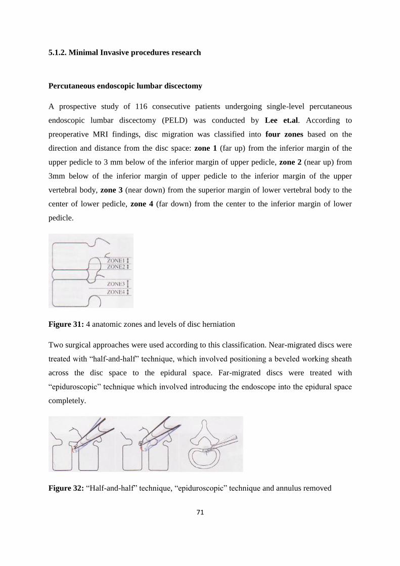
71
5.1.2. Minimal Invasive procedures research
Percutaneous endoscopic lumbar discectomy
A prospective study of 116 consecutive patients undergoing single-level percutaneous
endoscopic lumbar discectomy (PELD) was conducted by Lee et.al. According to
preoperative MRI findings, disc migration was classified into four zones based on the
direction and distance from the disc space: zone 1 (far up) from the inferior margin of the
upper pedicle to 3 mm below of the inferior margin of upper pedicle, zone 2 (near up) from
3mm below of the inferior margin of upper pedicle to the inferior margin of the upper
vertebral body, zone 3 (near down) from the superior margin of lower vertebral body to the
center of lower pedicle, zone 4 (far down) from the center to the inferior margin of lower
pedicle.
Figure 31: 4 anatomic zones and levels of disc herniation
Two surgical approaches were used according to this classification. Near-migrated discs were
treated with “half-and-half” technique, which involved positioning a beveled working sheath
across the disc space to the epidural space. Far-migrated discs were treated with
“epiduroscopic” technique which involved introducing the endoscope into the epidural space
completely.
Figure 32: “Half-and-half” technique, “epiduroscopic” technique and annulus removed
72
There were 73 males and 43 females, with a mean age of 35.5 (range 18-65) years. The mean
follow-up period was 14.5 (range 9-20) months. The L4-5 disc was the most commonly
herniated level (65 cases, 56.0%) followed by L5-S1 (47 cases, 40.5%), L3-4 (3 cases, 2.6%),
and L2-3 (1case, 0.9%). Herniations were localized at zones 1- in 4 (3.5%), 5(4.3%),
73(62.9%), and 34(29.3%) cases, respectively.
According to modified Macnab criteria, satisfactory (excellent or good) results were
distributed as follows: 91.6% (98/107) in the down-migrated discs; 88.9% (8/9) in the up-
migrated discs; 97.4% (76/78) in the near-migrated discs; and 78.9 (30/38) in the far-
migrated discs. The difference between near-migrated group (97.4%) and far-migrated group
(78.9%) was statistically significant (P=0.002) while the difference between down-migrated
group (91.6%) and up-migrated group (88.9%) was not statistically significant (P=0.569).
Two techniques of PELD can be used according classification: of disc migration into four
zones. The results suggest that open microdiscectomy may give superior results in far-
migrated discs. The results of this study will contribute to the establishment of surgical
guidelines for PELD in the treatment of migrated disc herniation. A prolonged follow up is
necessary to determine the validity of these initial results. (33)
Comparison of microscopic techniques
Franke et.al in a prospective randomized controlled clinical trial compared two operative
techniques standard microscopic technique and microscopic assisted percutaneous
nucleotomy (MAPN) technique. A two-center design was chosen with the developing
(index) center and a less MAPN experienced (transfer) center. (16)
All procedures in either center were performed by the same senior surgeon (R.G.-P. or J.F.)
or at least assisted by that senior surgeon in order to get more valid results excluding the
surgeon factor as far as possible. The pre-study surgical training of lumbar disc surgeries of
both senior surgeons exceeded 300 cases. There was pre-study experience for MAPN
technique with 150 cases (R.G.-P.) and with 20 cases (J.F.).
73
Unlike the microsurgical technique the X-ray converter remains in the lateral view within the
operation area. The level localization with a spinal needle was done on the opposite side. The
pinpoint was directed at the open interlaminar window. The skin incision of 15 mm at the
side of the pathology was performed in height of the needle entry point approximately 2 cm
paramedian.
Both the thoracolumbar fascia and the paraspinal muscles were dilated till the working
channel could be brought in. in the context of this study we utilized work channels with an
outside diameter of 14 mm. All work steps were performed under direct vision via
microscope in the fundamental technique.
Between September 2002 and May 2004, 100 patients were included to the study. A
microsurgical procedure underwent 48 and 52 patients a MAPN procedure. At the index
center 25 patients were randomized to both groups, whereas at the center 27 patients were
randomized to the MAPN group and 23 to the MC group.
The average age of the complete group was 44 years (min 21, max 72, SD 11.7). There were
40 women and 60 men with an equal distribution between groups. The pathologic segment
was L5/S1 in 42%, L4/5 in 51%, L3/4 in 6% and L2/3 in 1%. Preoperatively, 82 patients
showed neurological deficits, 33 only sensory disturbances, and 49 motor and sensory
deficits. According to the inclusion criteria they diagnosed 45% free sequestra, 42%
subligamentous sequestra and 13% subligamentous herniation. (16)
There was a huge clinical improvement for all patients regardless of center or method by a
repeated measures ANOVA on the sum VAS for all time points (F=165, P<0.0001),
moreover, there was a significant center-method interaction (F=4.9, P=0.006).
These results suggest that the clinical outcome of both surgical methods was
indistinguishable at the transfer center, whereas MAPN patients at the index center showed a
slightly faster recovery when compared to MC patients. This was mainly due to the fact that
there was a more pronounced reduction in the VAS scale for back pain. This phenomenon
reached statistical significance at discharge (P<0.001), 8 weeks (P=0.002) and 6-month
(P=0.003) follow up. For the 12-month follow up this difference could was not found
(P=0.467). No difference was found for the VAS scale for leg pain at any of the time points.
74
In two cases a deterioration of the neurological situation occurred immediately
postoperatively (<48 h), however, it recovered later uneventfully. In 83% of the patient a
primary observed motor deficit resolved completely within the follow up period. Sensory
deficits completely resolved in 68% of the patients, improved in 18%, and remained
unchanged in 14%. For the neurological situation there was no obvious difference was
found for the transfer center.
The mean hospital stay at the index center was significantly shorter for the MAPN group with
3.8 days compared to the MC group with 4.9 days. All patients, who were preoperatively in
an employment relation, resumed their occupation within 14 weeks, 77% within 8 weeks. The
average postoperative inability to work was 7 weeks.
Altogether, seven patients had to have a reoperation (7%). Five patients developed after a
symptom free (ranging from 3 to 11 months) a genuine relapse (same level, same side). These
five patients (four from the MC group, one from the MAPN group) underwent a reoperation
using the same technique performed previously. At the remaining two patients (one with
MAPN and one patient from the MC group) a segmental instability within the follow-up
period got apparent due to progressive disc degeneration. Eleven months after the original
intervention one total disc arthroplasty was implantes, whereas the other patient underwent a
PLF (posterior lumbar fusion) procedure. No further complications did occur.
Within the study period no real clinical advantages of the less traumatized posterior muscles
could be found. Thus, the hypothesis that with a lesser traumatized back muscle leads to a
quiker recovery and to less chronic pain could not be confirmed. (16)
Percutaneous Laser Disc Decompression
In a review of Percutaneous Laser Disc Decompression (PLDD) Schenk B. et.al evaluated
sixteen clinical trials included, representing a total of 1579 patient. The basic technique of
PLDD is the same for all trials. The procedure is conducted under local anesthesia of the skin
and underlying muscles.
After assessment of the correct disc level by using a fluoroscopy, a hollow needle was
inserted 10 cm from the midline, pointing toward the center of the disc. When the needle was
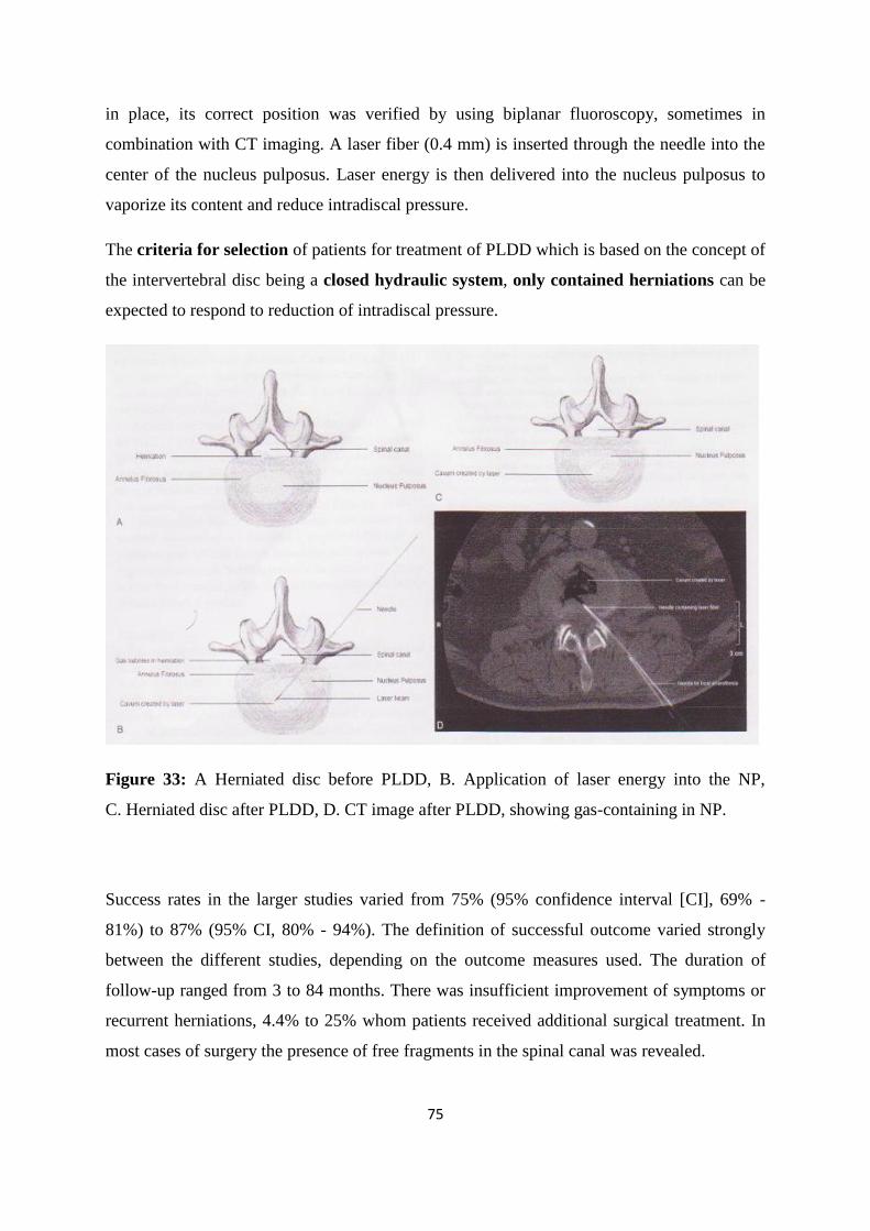
75
in place, its correct position was verified by using biplanar fluoroscopy, sometimes in
combination with CT imaging. A laser fiber (0.4 mm) is inserted through the needle into the
center of the nucleus pulposus. Laser energy is then delivered into the nucleus pulposus to
vaporize its content and reduce intradiscal pressure.
The criteria for selection of patients for treatment of PLDD which is based on the concept of
the intervertebral disc being a closed hydraulic system, only contained herniations can be
expected to respond to reduction of intradiscal pressure.
Figure 33: A Herniated disc before PLDD, B. Application of laser energy into the NP,
C. Herniated disc after PLDD, D. CT image after PLDD, showing gas-containing in NP.
Success rates in the larger studies varied from 75% (95% confidence interval [CI], 69% -
81%) to 87% (95% CI, 80% - 94%). The definition of successful outcome varied strongly
between the different studies, depending on the outcome measures used. The duration of
follow-up ranged from 3 to 84 months. There was insufficient improvement of symptoms or
recurrent herniations, 4.4% to 25% whom patients received additional surgical treatment. In
most cases of surgery the presence of free fragments in the spinal canal was revealed.
76
No randomized, controlled trials were available. Almost all trials were case series, with a
relatively low strength of evidence. Furthermore, the sample size in most trials was
relatively small, resulting in broad 95% CIs that made interpretation of success rates difficult.
Generalization of the results into general practice remains difficult, because of the different
inclusion and exclusion criteria, laser types, and outcome measures used and the large
variation in duration of follow-up. (62)
In the largest study to date Tassi reviewed the outcomes from 500 patients with discogenic
pain and herniated discs treated with microdiscectomy (1997-2001 by 6 surgeons) and 500
patients treated with percutaneous laser disc decompression (2002-2005 by a single surgeon).
Patients with sequestrated disc were excluded. This retrospective review found that the
hospital stay (six vs. two days), overall recovery time (60 vs.35) and repeat procedure rate
(7% vs. 3%) were lower in the laser group. The percentages of patients with overall
good/excellent outcomes were found to be similar in the two groups (85.7% vs. 83.8%) at a
two year assessment. (63)
Despite the fact that PLDD has been around for almost 20 years, scientific proof of its
efficacy still remains relatively poor, though the potential medical and economic benefits of
PLDD are too high to justify discarding it as experimental or ineffective on the sole basis of
insufficient scientific proof. (63)
A standard Cochrane review method to analyze of all randomized controlled trials which
were published up to January 1, 2007 was used and identified forty randomized controlled
trials (RCTs) and 2 quasi-RCTs.
Many of the early trials were of some form of chemonucleolysis, whereas the majority of the
later studies either compared different techniques of discectomy or the use of some form of
membrane to reduce epidural scarring.
Four trials directly compared discectomy with conservative management, and these gave
suggestive rather than conclusive results. However, other trials showed that discectomy
produces better clinical outcomes than chemonucleolysis, and that in turn is better than
placebo.
77
Microdiscectomy gives broadly comparable results to standard discectomy. Recent trials of
an interposition gel covering the dura (5 trials) and of fat (4 trials) show that they can reduce
scar formation, however there is limited evidence about the effect on clinical outcomes.
There is insufficient evidence on other percutaneous discectomy techniques to draw firm
conclusions. Three small RCTs of laser discectomy did not provide conclusive evidence on
its efficacy. There were no published RCTs of coblation therapy or transforaminal
endoscopic discectomy.
Surgical discectomy for carefully selected patients with sciatica due to lumbar disc prolapse
provides faster relief from the acute attack than conservative management, although any
positive or negative effects on the lifetime natural history of the underlying disc disease are
still unclear. The evidence for other minimally invasive techniques remains unclear except
for chemonucleolysis using chymopapain, which is no longer widely available. (46)
78
5.2. Conservative treatments: Medications
5.2.1 Oral Medications
Medications are common used for pain associated with lumbar disc herniations which include
nonsteroidal anti-inflammatory drugs, corticosteroids, muscle relaxants, and opioid pain
medications. (Rhee et.al) (57,58)
Nonsteroidal anti-inflammatory drugs have been shown to be helpful for the management of
acute low-back pain (Tulder et al), but a meta-analysis of the literature demonstrated that they
had no benefit in the treatment of radiculopathy compared with controls (odds ratio = 0.99)
(Vroomen et al.).
Corticosteroids are administered orally or by injection. While oral steroids are commonly
prescribed in clinical practice, we found only one study on their use for the treatment of
lumbosacral radicular pain (Haimovic). In this study, dexamethasone was not superior to a
placebo for either early or longterm relief of lumbosacral radicular pain, but it helped patients
who had presented with a positive result on the straight-leg-raise test. (57,58)
Non-steroidal anti-inflammatory drugs vs. Placebo in sciatica caused by disc herniation
One systematic review of medical treatments for sciatica caused by disc herniation (search
date 1998, 3 RCTs, 321 people) (Vroomen et al). The RCTs compared non-steroidal anti-
inflammatory drugs (NSAIDs) (piroxicam 40 mg daily for 2 days or 20 mg daily for 12 days;
indometacin [indomethacin] 75–100 mg 3 times daily; phenylbutazone 1200 mg daily for 3
days or 600 mg daily for 2 days) versus placebo.
The review found no significant difference between NSAIDs and placebo in global
improvement at 5–30 days (pooled AR for improvement in pain 80/172 [46.5%] v 57/149
[38.3%]; OR for global improvement 0.99, 95% CI 0.6 to 1.7.
The systematic review did not report the adverse effects of NSAIDs. NSAIDs may cause
gastrointestinal complications.
The absolute numbers in the RCTs relate to the outcomes of improvement in pain (3 RCTs)
and return to work (1 RCT). However, the meta-analysis used the outcome measure of global
improvement. The relationship between these measures is unclear. (78)
79
5.2.2. Injection
Valat et.al to determine the efficacy of epidural corticosteroid injections for sciatica
provided the study which was a multicenter, randomized double blind, controlled clinical
trial, conducted in five rheumatology departments of university hospitals in France from
October 1997 to January 2000. Randomization took place after written informed consent and
baseline information which were obtained from the study participants.
Inpatients referred for sciatica were eligible if they had a first or recurrent episode of sciatica
lasting for more than 15 and less than 180 days. With pain intensity which had to be >30 mm
on visual analogue scale (VAS).
The patients received three epidural injections (at two day intervals) of either 2 ml
prednisolone acetate (50mg) or 2 ml isotonic saline by a lumbar interlaminar approach using
loss of resistance technique, without fluoroscopic guidance.
Lumbar exercises and other spinal injections were not authorized during the study. Non-
steroidal anti-inflammatory drugs (NSAIDs) were authorized only 20 days after the first
injection. Non-opioid analgesics, bed rest, mild lumbar tractions, and lumbar belts were
authorized. The doctors making the assessments were not the same doctors as those giving
the injections. So the patient and the assessing doctor were both unaware of the treatment
received.
The patients were re-evaluated five days after inclusion (after the last injection, before
leaving the hospital) and as outpatients 20 and 35 days after the first injection. Each patient
was examined by the same doctor throughout the trial. At each visit, information on the use
of analgesics and NSAIDs was recorded.
Between October 1997 and December 1999, 85 patients were enrolled in the study, 43
patients in the steroid group (SG), and 42 in the control group (CG). The baseline
characteristics were similar in the two groups. The large majority of the injections (89.5%)
were performed at the L4-5 level, 7% at the L3-4 level and 3.5% at the level L5-S1 level.
On an intention to treat analysis at day 20 the groups did not differ significantly with respect
to the primary outcome: 15/42 (36%) patients in the CG and 22/43 (51%) in the SG (p=0.15)
were considered as success (difference 15.5%; 95% confidence interval (CI) for the
80
difference-5.4 to 36.3). At the end the study of the study (day 35), 20/42 (48%) patients in
the CG and 21/43 (49%) in the SG (p=0.91) were considered as success (difference 1.2%;
95% CI for the difference -20.0 to 22.5).
Among the 48 failures, 14 patients (six in the CG and eight in the SG) required NSAIDs,
three required surgery (2 in the CG and I SG) another two required chemonucleolysis (one
each group) and five (two in the CG and three in the SG) other routes of corticosteroids
administration. On analysis according to protocol, at day 20 in the 74 remaining patients,
12/35 (34%) in the CG and 22/39 (56%) in the SG (p=0.057) were considered at success
(difference 22.1%; 95% CI for the difference 0.0 to 44.2).
In conclusion we cannot exclude an efficacy of isotonic saline administered epidurally for
sciatic, but epidural corticosteroid injections did not provide any additional improvement. (47)
One systematic review of medical treatments for sciatica caused by disc herniation (search
date 1998, 4 RCTs of epidural steroids, 265 people) and one subsequent RCT.14 The review
compared four different doses of epidural steroid injections (8 mL methylprednisolone 80
mg, 2mL methylprednisolone 80 mg, 10 mL methylprednisolone 80 mg , and 2mL
methylprednisolone acetate 80 mg) versus placebo (saline or lidocaine [lignocaine] 2 mL)
after follow up periods of 2, 21, and 30 days.13 The review found limited evidence that
epidural steroids increased participant perceived global improvement (which was not defined)
compared with placebo.
The results were of borderline significance (73/160 [45.6%] with steroid v 56/172 [32.5%]
with placebo; OR 2.2, 95% CI 1.0 to 4.7). The subsequent RCT (36 people with disc
herniation confirmed by magnetic resonance imaging) compared epidural steroids (3
injections of methylprednisolone 100 mg in 10 mL bupivacaine 0.25% during the first 14
days of hospitalisation) plus conservative non-operative treatment versus conservative
treatment alone.
Conservative treatment involved initial bed rest and analgesia followed by graded
rehabilitation (hydrotherapy, electroanalgesia, postural exercise classes) followed by
physiotherapy. It found no significant difference in mean pain scores at 6 weeks and 6
months measured on a visual analogue scale (at 6 months, 32.9 [range 0–85] with steroids v
39.2 [range 0–100] with conservative treatment). There were no significant differences in
81
mean mobility scores (Hannover Functional Ability Questionnaire: 61.8 [range 25–88] with
steroids v 57.2 [range 13–100]), in the number of people who had back surgery (2/17 [12%]
with steroids v 4/19 [21%]; RR 0.56, 95% CI 0.09 to 2.17), or in people returning to work
within 6 months (15/17 [88%] with steroids v 14/19 [74%]; RR 1.19, 95% CI 0.75 to 1.33).
No serious adverse effects were reported in the RCTs included in the systematic review,
although 26 people complained of transient headache or transient increase in sciatic pain. The
subsequent RCT did not report adverse effects of epidural injections. (78)
Caudal epidural vs transforaminal epidural steroid injections
In a retrospective case control during the period between June 2002 and July 2004; 132
patients were diagnosed for primary lumbar radiculopathy of L4, L5, or S1 and where
randomized into 2 groups to evaluate the efficacy of 2 different approaches of epidural
steroid injection.
Conservative care consisted of activity modifications, physical therapy and epidural steroid
injections. Physical therapy included extension-based exercise program and light, isometric
core strengthening. Medications were also used, and included non-steroidal anti-
inflammatory agents, muscle relaxants, and in some cases, narcotics.
These patients were subject to two different types of epidural steroid injections as part of
their treatment plan. Treatment groups were allocated according with randomly assigned
appointments to the clinics of either of the two senior authors.
In each of these clinics, a different preference of epidural injection technique was maintained.
All procedures were performed by the same group of interventional muscular-skeletal
radiologists, with a standardized technique.
The caudal epidural steroid injections (ESI) were performed under fluoroscopic guidance
with a 22g needle, and either 2cc of Depo-Medrol (40mg/ml) or 3cc Celestone (6mg/ml)
were injected. The trans-foraminal epidural steroid injections (TESI) were also performed
under fluoroscopic guidance for the L4 and L5 nerve roots, and under CT-guidance for the S1
nerve roots. A 20/25 coaxial system was utilized, and 1.5–2cc 1:1 solution of Marcaine
0.25% with Depo-Medrol (40mg/ml) or Celestone (6mg/ml) were injected.
82
Patients who consented to participate in the study included 39 of 58 patients (67.2%) treated
with caudal ESI and 54 of 74 (72.9%) treated with TESI. Of the 93 patients, 16 were lost to
follow up. Of these, 20% of the chymopapain group, and 47% of the saline group requested
surgery during the observation period of 2 years. Karppinen (29)
found that periradicular
infiltrations of Methylprednisolone + Bupivacaine had only short-term clinically meaningful
benefits over periradicular infiltrations with normal saline solution.
The effectiveness of TESI is comparable to that of ESI (approximately 60%) for treatment of
primary lumbar radiculopathy. The increased complexity of TESI is not justified for primary
cases, and may have a more specific role in a recurrent disease or for diagnostic purposes. (55)
Between March 1, 2001 and August 25, 2002, a total of 263 patients were referred to the
Department of Diagnostic Radiology for image-guided percutaneous steroid injections. 91 of
263 patients (50 males and 41 females, age range: 13 78 yrs) were treated with transforaminal
ESI for lumbosacral disc herniations.
Twenty-three of 91 patients whose imaging studies were CT/oust side MRI (n = 13) or whose
clinical follow-ups after the injections were missed (n = 10) were excluded in this analysis. A
total 68 patients were included in this study with an average follow-up period of 3.6 months.
All procedures were performed in a sterile manner, and fluoroscopic guidance was used for
the injections. The patients were placed prone on a fluoroscopy table and the C-arm was
rotated to an ipsilateral oblique angle with respect to the suspected nerve root (L4, 5 or S1).
Lidocaine (1%) was used for the cutaneous and needle tract local anesthesia. The MR
findings were assessed for the type, hydration, location and size (volume) of the herniated
disc, the grade of nerve root compression and the associated findings of spinal stenosis. The
type of herniated disc was classified as protrusion, extrusion and sequestration.
The final analysis included 68 patients with an average follow-up period of 3.6 months
(range: 7 days 24 months). Transforaminal ESIs were given at the following levels and all the
procedures were done at a single level: L4 in two patients, L5 in 36 patients and S1 in 30
patients. Forty one of 68 patients (60.3%) had successful outcomes (the responders), and
twenty seven patients showed unsatisfactory results (the non-responders). The pre-injection
symptom duration of the responders ranged from 3 days to 2.5 years, and that of the non-
responders was from 6 days to 2 years. There were no serious side effects of the injections.
83
There was no significant difference between the responders and non-responders in terms of
injection level, age, gender or the pre-injection symptom duration (p > 0.05). Seven of 41
responders and three of 27 non-responders underwent repeat injections. MR analysis showed
no significant difference between the responders and non-responders in terms of the type,
hydration and size of the herniated disc, or an association with spinal stenosis (p > 0.05).
This study has its limitation but nevetherless MRI could play an important role in predicting
the clinical outcome of non-surgical transforminal ESI treatment.
Several outcome studies of ESI treatment for lumbar herniated discs have evaluated the
clinical differences between responders and non-responders. Lutz et al. have reported that
patients with pre-injection symptom durations of less than 36 weeks were most likely to
respond to treatment. Viton et al. have found that the decrease in pain was greater for the
patients less than 50 years of age than for the older patients after transforaminal ESI for
lumbar radiculopathy. (29)
84
5.3. Non-medical Conservative treatments
5.3.1. Natural History
Takata et al. made an investigation for morphological changes of 42 patients (28 men and 14
women) with a mean age of 42 years (range 16–64 years) who presented with unilateral leg
pain and low back pain that was definitely diagnosed as being caused by LDH.
All patients were treated conservatively (bed rest, oral non-steroidal, anti-inflammatory
drugs, pelvic traction and caudal epidural block) and followed up by serial MRI. The
symptomatic disc level was L2-L3 in 8 cases, L3-L4 in 6 cases, L4-L5 in 15 cases and L5-S1
in 13 cases. All patients underwent MRI examinations every three months for a period of 3-
24 months (mean 10.3months). Images were obtained using a 1.5 tesla superconducting MR
scanner.
LDH was classified into three types: protrusion (n=7), extrusion (n=17) and sequestration
(n=18), using T2-weighted sagittal MR images. The size of the herniated mass was
determined from the ratio of the anterior-posterior diameter of the spinal canal to the
maximum diameter of the mass on T2-weighted axial images.
All patients were re-examined and their MRI findings were re-evaluated by the same
physician during follow-up. The clinical outcome was evaluated using the JOA (Japanese
Orthopaedic Association) score for LDH 9 and recovery rate. Radicular leg pain was rated as
follows: Excellent - no pain (recovery rate 100%), Good - slight pain but bearable during
daily activity (recovery rate 80%), Poor - sometimes unable to perform daily activities due to
pain (recovery rate < 50%). (66)
The time taken for spontaneous involution of the herniated mass by >50% (classified as
effective regression ) was 3 months in 8 cases, 6 months in 15 cases, 9 months in 12 cases
and 12 months in two cases. No marked reduction of the herniated mass was recognized in 5
patients in the final MRI study of up to 24 months.
Two patients with protruded LDH showed spontaneous involution of the herniated mass by
>50% 12 months after the onset of symptoms. The other 5 patients with this type of LDH
showed no morphologic changes at the time of the final MRI study.
85
Among patients with extruded LDH, there was a decrease of the herniated mass by >50% in 7
patients 6 months after the onset. In another 10 patients with this type of LDH, the majority
showed a significant reduction of size after 9 months. Among patients with sequestrated type,
10 showed over 50% involution of their herniated mass after 3 months, and 6 others after 6
months. Among these 16 patients, 8 showed complete disappearance of their hernia. Two
patients showed a significant reduction of hernia size 12 months after the onset of symptoms.
The recovery of symptoms, especially radicular leg pain, preceded the involution of the
herniated mass on MRI. In the patients with sequestrated LDH, severe radicular leg pain was
the initial symptom and the pain improved 1–5 weeks after the onset, leaving sensory
changes or a motor deficit, while a permanent severe motor deficit (MMT ≤3) was observed
in six cases.
In the patients with extruded LDH, radicular leg pain was not so severe compared to the
patients with sequestration and leg pain lessened 4-8 weeks after the onset. Patients with the
protruded LDH usually complained of leg pain on walking and improved after 3-14 weeks.
Four patients with this type of hernia showed no MRI changes and had no decrease of their
leg pain.
In conclusion of this investigation were that 37 out of 42 patients (88 %) showed effective
(>50%) reduction of the herniated mass on MRI 3–12 months after the onset of symptoms.
Sequestered hernia and transligamentous extrusions seem to be more easily and rapidly
absorbed. MRI changes and improvement of symptoms are well correlated with MRI
changes, but a time lag is observed. The basic treatment of LDH should be conservative
according to the results of this study; however surgery may be necessary for large central
protruding hernias and patients who show severe motor deficits. (66)
5.3.2. Bed rest
When comparing bed rest to no treatment for patients with acute LRS they found no
difference (1 HQ trial: moderate evidence) regarding overall improvement at short-term
follow-up and no difference (1 HQ tria: moderate evidence) regarding pain and disability at
short and intermediate follow-up.
86
In one low quality study comparing manipulation to other conservative care for patient with
LRS they found no difference (1LQ trial: moderate evidence) regarding overall improvement
pain and return to work at short-term follow-up. (37)
5.3.3. Physical therapy
Physical therapy and General Practioner vs General practioner alone
Luijsterburg et al. conducted a randomized clinical trial in primary care with a 12- months
follow-up period in two groups: The intervention group received physical therapy added to
the general practitioners‟ care and and the control group with general practitioners‟ care only.
To assess the effectiveness of PT additional to general practitioners‟ care compared to
general practitioners‟ care alone, in patients with acute sciatica. (38)
About 135 patients with acute sciatica were treated by the GP according to their clinical
guideline. GPs gave information and advice about LRS and, if necessary, prescribed (pain)
medication. Physica therapy treatment consisted of exercise therapy as well as giving
information and advice about LRS. Passive modalities such as massage and manipulation
techniques or applications such as ultrasound therapy or electrotherapy were not allowed.
The treatment protocol was developed in a consensus meeting with participating physical
therapists. They acted as coaches and guided the patient in order to stimulate return to activity
(type/ content of exercises was left to the expertise of the participating physical therapists),
despite the pain experience.
Both GP and PT interventions (only in a one to one setting; group settings were not allowed)
were restricted to a maximum of nine treatments/consultations in the first 6 weeks after
randomization. At 3, 6 and 12 weeks after baseline there was no significant difference
between the two groups on the primary outcome: GPE. However, at these follow-up moments
the intervention group showed a higher proportion of „improved‟ patients. At 52 weeks after
baseline there was difference between the groups on the GPE measurement, in favor of the
intervention group. About 53 patients (79%) in the intervention group versus 38 patients
(56%) in the control group reported to be improved (RR, 1.4 95% CI: 1.1; 1.8).
87
There were no significant differences between the groups in the most of secondary outcomes
at 3, 6, 12 and 52 weeks after baseline. At 12 and 52 weeks follow-up the mean improvement
on leg pain was clinically relevant in both groups; respectively, 3.9 and 4.4 points for the
intervention group and 3.7 and 3.7 points for the control group. The mean improvement on
disability (RDQ) was also clinically relevant at 12 and 52 weeks follow-up in both groups;
respectively, 7.7 and 10.0 points for the intervention group and, respectively, 8.5 and 9.1
points for the control group. The results in this study indicated that PT added to GP care had
better results than a GP alone however it was a more expensive procedure. (38)
Two low quality studies compared physical therapy to other conservative treatments. In these
studies they found no difference in overall improvement pain and return to work between
groups. When comparing physical therapy to other conservative care for patients with LRS
they found no difference (2LQ trials: moderate evidence) regarding overall improvement,
pain and return to work at short-term.
In one low quality study they found a difference in improvement at 1-year follow-up, in favor
of surgery. In the same study they found no difference in improvement at 4 and 10-years
follow-up between the two groups.
They concluded that there was limited evidence that surgery is more effective for patients
with LRS regarding overall improvement than physical therapy at 1-year follow-up. At 4 and
10-years follow-up they found no difference regarding overall improvement between surgery
and physical therapy.
In one low quality study they found no differences in overall improvement, pain, and
disability at short-term follow-up between groups. In one high quality study they found no
differences in pain, disability at short and intermediate follow-up between groups. (38)
5.3.4. Traction
Luijsterburg et al. systematic review found in four studies compared traction to inactive/sham
traction. One low quality study did not report any data. In one high quality and one low
quality studies they found no difference in pain between traction and inactive/sham traction at
short-term. Also, in one low quality study they found no difference in improvement between
groups at short-term.
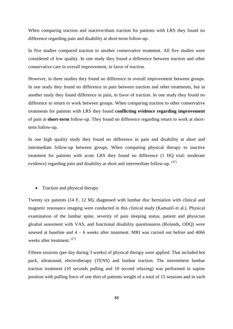
88
When comparing traction and inactive/sham traction for patients with LRS they found no
difference regarding pain and disability at short-term follow-up.
In five studies compared traction to another conservative treatment. All five studies were
considered of low quality. In one study they found a difference between traction and other
conservative care in overall improvement, in favor of traction.
However, in three studies they found no difference in overall improvement between groups.
In one study they found no difference in pain between traction and other treatments, but in
another study they found difference in pain, in favor of traction. In one study they found no
difference to return to work between groups. When comparing traction to other conservative
treatments for patients with LRS they found conflicting evidence regarding improvement
of pain at short-term follow-up. They found no difference regarding return to work at short-
term follow-up.
In one high quality study they found no difference in pain and disability at short and
intermediate follow-up between groups. When comparing physical therapy to inactive
treatment for patients with acute LRS they found no difference (1 HQ trial: moderate
evidence) regarding pain and disability at short and intermediate follow-up. (37)
Traction and physical therapy
Twenty six patients (14 F, 12 M), diagnosed with lumbar disc herniation with clinical and
magnetic resonance imaging were conducted in this clinical study (Kamanli et al.). Physical
examination of the lumbar spine, severity of pain sleeping status, patient and physician
gloabal assesment with VAS, and functional disability questionaires (Rolands, ODQ) were
assesed at baseline and 4 – 6 weeks after treatment. MRI was carried out before and 4066
weeks after treatment. (27)
Fifteen sessions (per day during 3 weeks) of physical therapy were applied. That included hot
pack, ultrasound, electrotherapy (TENS) and lumbar traction. The intermittent lumbar
traction treatment (10 seconds pulling and 10 second relaxing) was performed in supine
position with pulling force of one thirt of patients weight of a total of 15 sessions and in each
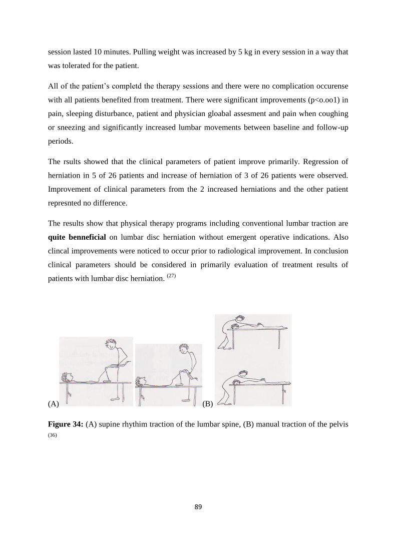
89
session lasted 10 minutes. Pulling weight was increased by 5 kg in every session in a way that
was tolerated for the patient.
All of the patient‟s completd the therapy sessions and there were no complication occurense
with all patients benefited from treatment. There were significant improvements (p<o.oo1) in
pain, sleeping disturbance, patient and physician gloabal assesment and pain when coughing
or sneezing and significantly increased lumbar movements between baseline and follow-up
periods.
The rsults showed that the clinical parameters of patient improve primarily. Regression of
herniation in 5 of 26 patients and increase of herniation of 3 of 26 patients were observed.
Improvement of clinical parameters from the 2 increased herniations and the other patient
represnted no difference.
The results show that physical therapy programs including conventional lumbar traction are
quite benneficial on lumbar disc herniation without emergent operative indications. Also
clincal improvements were noticed to occur prior to radiological improvement. In conclusion
clinical parameters should be considered in primarily evaluation of treatment results of
patients with lumbar disc herniation. (27)
(A) (B)
Figure 34: (A) supine rhythim traction of the lumbar spine, (B) manual traction of the pelvis
(36)
90
5.3.5. Mckenzie therapy centralisation method
Mechanical Diagnosis and Therapy (Mckenzie)
For disc herniation, 104 patients examined and 60 of them recruited underwent a standardized
Mechanical diagnosis and therapy (MDT) evaluation to expose 2 groups: central (CG)‟n and
non-central‟n group (NCG). All patients were treated in the same way and were followed for
one year. If patients did not have improvement surgery was considered.
Outcomes included back and leg pain, disability, Nottingham Health Profile, and surgical
outcome. 25 patients were classified in the CG, 35 were NCG and other baseline
characteristics were similar between groups.
At 1, 2 and 3 months the CG had significant better outcomes than the NCG. At 2 months the
CG had more improvements in leg pain (P< 0.007) disability (P<0.001) and Nottingham
Health Profile ( P< 0.001). After 1 year, disability was less in CG (P<0.029). 3 CG patients
underwent surgery vs 16 NCG (P<0.01). The odds ratio for surgery in NCG WAS 6.2.
Conclusion of the examination of patient with sciatica and suspected disc herniation who
have a centralization response will have significantly better outcomes. Patients who do not
have centralization will be 6 times more likely to undergo surgery. (65)
A trial providing diagnostics to patients with LBP and varying degrees of leg pain and altered
sensation referred for lumbar discography due to pain sufficiently severe to warrant invasive
testing, failure of conservative care, and 1 or more MRI studies w/o compelling surgical
findings. Underwent MDT assessment and classified as centralizers, peripheralizers, or not
effect on pain.
Routine lumbar discography and CT imaging followed. Outcomes of the two types of
assessment were compared. 50% centralized, 25% peripheralized only, and 25% reported no
effect on their leg pain with the MDT evaluation. 57% had positive discograms of which 81%
did not leak. 43% were negative. 74% of centralizers, 69% of peripheralizers and only 12%
of no effect patients had positive discograms. 91% of centralizers had intact annuli but only
54% of peripheralizers.
91
Despite the chronicity of the patients, 50% still demonstrated a reversibility of their
pathology, which was shown in 74% of cases to be disc. The ability to distinguish between a
positive and negative discogram based on MDT findings was highly significant (P<0.001).
(10, 11)
In North Jutland Country Denmark (500,000 population), in 1997, two non-surgical spine
clinics established focused on MDT care and educational program for general doctors for pts
with 1 to 3 months‟ of sciatica, with or without low back pain. Data on rates of lumbar disc
surgery were obtained from the National Registry of Patients over the next four years.
The annual rate of lumbar disc operations for pts in N. Jutland Country decreased from 60-80
per 100,000 before 1997 to 40 per 100,000 in 2001 (P=0.00), and the rate elective, first-time
disc surgeries decreased by approximately two thirds (P=0.00). The rate of lumbar disc
operations in the rest of Denmark remained unchanged during the same period.
The implementation of these non-surgical spine clinics coincided closely with a significant
reduction in the rate of lumbar disc surgery. The odserved reduction seems most likely to be
causally associated with educational activities and improved patient care provided by the
clinics. (11)
Extension exercises
After examining 67 patients with low back pain (LBP) radiating to the calf or foot with at
least one significant sign of nerve root irritation and at least 6 weeks of failed non- operative
therapy. All patients underwent an initial trial of extension exercises. Those who had no
symptom worsening were prescribed an extension exercise program for several days while
still hospitalized. The 52% of patient (34 of them) shown no worsening during extension test.
All 34 performed extension exercise program. 100% regained their full lumbar extension
range and eliminated symptoms within 2-5 days. The other 32 underwent laminectomy and
discectomy. Pre- operatively, there was no difference between the groups with regard to age,
gender, pain below knee, or any of the neurological signs or SLR. 56% of the surgical
patients had a free fragment, 19% had swollen of displaced nerve root, and 19% had a
bulging disc. Finally, some of these patients responded so dramatically to extension therapy
that the use of extension exercises as a therapeutic modality was recommended. (31)
92
5.3.6. Chiropractic approach in general radiculopathy
In a retrospective review concerning the clinical outcomes of patients with cervical and
lumbar radiculopathy treated with a nonsurgical, chiropractic treatment protocol in
combination with other interventions.
Chiropractic manipulation was performed on all patients. Joint dysfunction was determined
by palpation demonstrating abnormal resistance to springing of the joint, reactivity of the
underlying musculature, and/or the patient‟s report of pain. In selected cases, radiographic
biomechanical analysis was performed to determine deviations from neutral to position to
patient of manipulation, centration and stabilization procedures that center the cervical
and lumbar spine in the frontal and sagittal plane.
In cases of lumbar spine radiculopathy, distraction manipulation (DM), a low-velocity
technique, and a secondary high-velocity low-amplitude (HVLA) manipulation technique
were used. The maneuvers were always delivered in a direction that did not cause
peripheralization of the pain. Distraction manipulation was performed on a flexion-distraction
table with the patient prone using a stomach pillow to position the patient in lumbar
semiflexion. Distraction manipulation motions included flexion, extension, and lateral flexion
in the direction of spinal centration (centering) only. There was no rotation performed, with
extension motion limited from semiflexion to neutral. In the HVLA technique, the lumbar
spine deviation from neutral (translation or lateral flexion) was performed using a drop table
in the side-lying position so that there would be no introduction of rotation to the HVLA
technique. (8)
How does manipulation work?
Increases joint motion induced by manipulation allows the inflammation from disc
material to subside more easily, and/or:
Manipulation may provide input to functional reflexes, modifying pain; and/or
Manipulation reduces related and overlying pain from facet joints. (65)
93
Neuromobilization was used as a manual- and exercise –oriented method that is theorized to
mobilize nerve roots that are suspected to be the source of nerve root pain. All patients were
given the same series of stabilization exercises for home performance. The spine stabilization
procedures used were progressive, meaning beginning procedures were performed before
intermediate and advanced procedures. Each patient demonstrated the assigned procedure
correctly before they left the office. Each patient on subsequent visits was required to
demonstrate satisfactory “pain-free” performance of beginning procedures before they
allowed advancing to the intermediate and advance procedures.
Of the 162 consecutive patient reviewed, 99 were female and 63 were male. Upper extremity
symptoms were seen in 61 cases and lower extremity in 108 cases, with 7 of these cases
demonstrating both. Of 162 total cases, 85.8% resolved their significant subjective radicular
complaints; and the patients were discharged from active treatment. The treatment trial was 9
(mean) treatment sessions, ranging from 1 to 23 visits and lasting between the first treatment
date and the first symptom improvement was 4.2 (mean) days, with a range (standard
deviation) of 0.78 to 7.7 day(s). The initial numerical pain rating scale score (median) was
5.8 with a final score of 2.1 (median). The pain change between initial and final score was 4.2
(median).
Of 162 total cases, acute and chronic presentations were represented. Sixty-seven (41.36%)
were classified as “acute”, meaning their symptom duration was less than 3 months. Ninety-
five (58.64%) were classified as “chronic”, meaning their symptom duration was greater than
3 months. Ninety-one percent of acute presentations resolved with a treatment trial (mean)
of 6.2 sessions, ranging from 1 to 20 visits. Eighty-one percent of chronic presentations
resolved with a treatment trial of 8.6 (mean) sessions, ranging from 1 to 23 visits.
There were 10 unresolved cases referred for medication management as a result of the
nonsurgical treatment not resolving the radicular complaints. Six cases experienced minimal
to moderate improvement before referral. The treatment trial ranged from 3 to 25 visits.
Three unresolved cases were referred for and underwent surgery. Of the 162 total cases, these
represent 1.85%. All 3 cases showed no symptom improvement before referral. The treatment
trial ranged from 2 to 7 visits, lasting between 9 and 17 days. The MRI on each of these cases
demonstrated a significant disc extrusion.
94
Incidentally noted, with lumbar radiculopathy cases, there was a consistent increase in active
lumbar flexion (standing tension angle), passive hip flexion (sciatic tension angle), and
passive hip extension (femoral tension angle) in cases that resolved. Similarly there was a
common finding of the straight leg raise. Braggard, slump, and femoral nerve stretch tests
when positive progressing to negative testing in cases that resolved. Comparative palpatory
tenderness, sacroiliac and sciatic notch regions commonly decreased or resolved in cases
where the radicular complaints resolved.
Most of the cases that resolved their radiculopathy complaint completed up to 50% of the
stabilization procedures. It was noted that very seldom did it require completion of all the
stabilization procedures listed above.
There are several important limitations to this study. It is not a randomized controlled trial;
thus, interpretation of the result must be made with worth of caution. The natural history of
radiculopathy is generally thought to be favorable; however, data on untreated patients with
this disorder are lacking.
The current study supports the notion that chiropractic management and nonsurgical
measures are a viable alternative to surgery in patients with radiculopathy. Randomized
controlled trials are needed for to further assess of this nonsurgical approach compared with
untreated controls and surgical treatments. (8)
Two systematic reviews and one subsequent RCT were found. The first systematic review
(search date 1998), which did not perform meta-analysis, identified two RCTs of spinal
manipulation for sciatica caused by disc herniation. The second systematic review (search
date not stated) identified no RCTs.15 The first RCT (207 people) included in the review
compared spinal manipulation (every day if necessary) versus placebo (infrared heat 3 times
weekly). It found that spinal manipulation increased overall self perceived improvement at 2
weeks compared with placebo (98/123 [80%] v 56/84 [67%]; RR 1.19, 95% CI 1.01 to 1.32;
NNT 8, 95% CI 5 to 109). (24)
The second included RCT (322 people) compared four interventions: spinal manipulation,
manual traction, exercise, and corsets, in a factorial design. It found no significant difference
among treatments in overall self-perceived improvement after 28 days (quantified results not
available). The subsequent RCT (112 people with symptomatic herniated lumbar disc)
95
compared pulling and turning manipulation versus traction. It found that significantly more
people were “improved” (absence of lumbar pain, improvement in lumbar functional
movement) or “cured” (absence of lumbar pain, straight leg raising of>70°, ability to return
to work) with spinal manipulation compared with traction (54/62 [87.1%] with manipulation
v 33/50 [66%] with traction; RR 1.32, 95% CI 1.06 to 1.65; NNT 5, 95% CI 4 to 16;
timescale not stated).
The first systematic review did not report adverse effects. The second systematic review
identified one review of 135 case reports of serious complications after spinal manipulation
published between 1950 and 1980. The case review attributed these complications to cervical
manipulation, misdiagnosis, presence of coagulation dyscrasias, presence of herniated
nucleus pulposus, or improper techniques. The subsequent RCT found that two out of 60
people receiving traction had syncope; no adverse effects were reported in people receiving
manipulation.
In another systematic review (search to date 2001, 5 prospective observational studies). The
largest study included in the review (4712 treatments in 1058 people undergoing both
cervical and lumbar spinal manipulations) found that the most common reaction was local
discomfort (53%), followed by headache (12%); tiredness (11%); radiating discomfort
(10%); dizziness (5%); nausea (4%); hot skin (2%); and other complaints (2%).
The incidence of serious adverse effects is reported as rare, and is estimated from published
case series and reports to occur in one in 1–2 million treatments. The most common of these
serious effects were cerebrovascular accidents (the total number of people undergoing
manipulations was not reported and the rate of this adverse effect cannot be estimated).
However, it is difficult to assess whether such events are directly related to treatment.
In the third review, which examined risks, the percentages include both cervical and lumbar
spinal manipulations, which may overestimate the effect of lumbar spinal manipulations. The
authors of the review advise caution in interpreting these results, as they are speculative and
based on assumptions about the numbers of manipulations performed and unreported cases.
More reliable data are needed on the incidence of specific risks. (24)
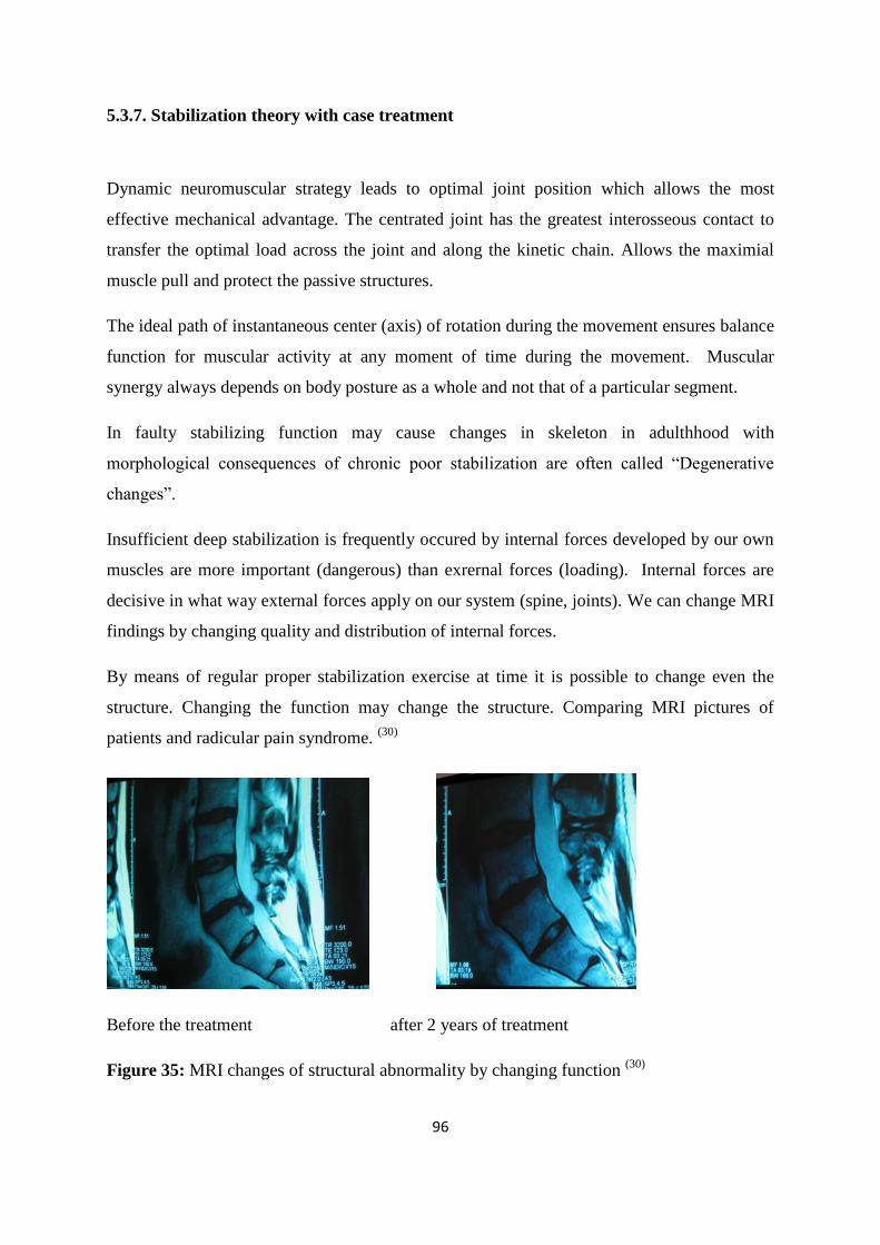
96
5.3.7. Stabilization theory with case treatment
Dynamic neuromuscular strategy leads to optimal joint position which allows the most
effective mechanical advantage. The centrated joint has the greatest interosseous contact to
transfer the optimal load across the joint and along the kinetic chain. Allows the maximial
muscle pull and protect the passive structures.
The ideal path of instantaneous center (axis) of rotation during the movement ensures balance
function for muscular activity at any moment of time during the movement. Muscular
synergy always depends on body posture as a whole and not that of a particular segment.
In faulty stabilizing function may cause changes in skeleton in adulthhood with
morphological consequences of chronic poor stabilization are often called “Degenerative
changes”.
Insufficient deep stabilization is frequently occured by internal forces developed by our own
muscles are more important (dangerous) than exrernal forces (loading). Internal forces are
decisive in what way external forces apply on our system (spine, joints). We can change MRI
findings by changing quality and distribution of internal forces.
By means of regular proper stabilization exercise at time it is possible to change even the
structure. Changing the function may change the structure. Comparing MRI pictures of
patients and radicular pain syndrome. (30)
Before the treatment after 2 years of treatment
Figure 35: MRI changes of structural abnormality by changing function (30)
97
The structural change may result from rehabilitation, but may also be sponatneous. However
function is the most important, providing stabilization giving pain resolution. Surgery
removes a disc protrusion but structural pathology frequently results from spinal instability, it
is necessary to improve (restore) spinal stabilization and influence primary cause of structural
damage. Important is to emphasis on finding out the cause of proble and not the source. ((30))
98
5.4. Post-surgery of disc herniation
Exercise programme after surgery
In this open, prospective and controlled study they examined 42 patients who had undergone
microdiscectomy between January and September 1998 in the Neurosurgery Clinics os Sisli
Etfal and Taksim Education and Research Hospitals. Lumbar disc herniation was diagnosed
using a clinical radiological (MRI) examination in the neurosurgery clinics.
The most common level of disc herniation was between L4 and L5 (45.24%, 19 patients).
During the initial examination of the patients there were no differences between the 3 groups
in terms of pain, functional capacity, depression, fingertip-floor distance LS, MLS, LE, LF,
rotation and PILE (neck) scores. Prior exercise PILE (back) and body strength scores in the
first group were worse than those in the groups. (79)
The examination of the patients in the first group at completion of the exercise programme
showed significant improvement had occurred in all parameters. The examination of the
control group patients, after the 8-week period, showed improvement in the functional
capacity, fingertip-floor distance, modified lumbar Schober and in left rotation while in the
other parameters there were no significant differences.
At the end of the follow-up period the physical and neurological progress of the patient was
satisfactory. Abolished deep tendon reflexes did not recover. Among 10 patients with
paravertebral muscular spasm, it had disappeared in 1. Among 8 patients with sensory deficit,
3 became asymptomatic. Four of the 8 patients with motor deficits showed healing in motor
loss.
In the second group only one patient with paravertebral muscular spasm and another patient
with sensory deficit improved, while the others remained the same. The patients‟ progress
was satisfactory. In the third group none of the patients with paravertebral spasm. Sensory or
motor deficit improved. In addition, at the second group the number of patients with sensory
deficit and paravertebral spam increased.
99
It is known that approximately 25% of patients who have been operated on for lumbar
herniation have post-operative complaints. It is also, known that in operated and non-
operated patients there is lumbar, abdominal and dorsal muscle weakness. Kahanowits et al.
have reported that the trunk muscles’ strength is decreased about 30% after discectomy
operation. Hence both operated and non-operated low back patients need to follow an
exercise programme for the relief their painful backs. (79)
In this prospective study, they examined 75 patients who had undergone discectomy between
March 2002 and August 2002 at the Wooridul Spine Hospital, Seoul, Korea. The inclusion
criteria were: 1) unilateral radiating leg pain with or without back pain not responding to
conservative treatment methods and having a good radiological correlation; 2) first-time
lumbar spine operation; 3) single-level disc herniation; and 4) absence of associated systemic
diseases such as cardiac ailments or orthopedic contraindications for subsequent exercise
program. Patients were randomized into two groups, a control group and an exercise group.
The CT scan measurements were taken by an independent observer using a software PiView
program. At the end of the sixth postoperative week, all patients underwent measurement of
the isometric strength of the extensor muscles of the lumbar spine using the MedX (Ocala,
FL) system. This marked the baseline measurement. The measurements were obtained at
various angles of lumbar flexion from 0 to 72 degrees. The maximal voluntary effort applied
was calculated and was quantified by the machine as the torque generated.
After the sixth postoperative week, the control group continuedwith the home-based basic
lumbar conditioning exercises. The exercise group started with the intensive schedule with a
defined set of exercises for extensor muscle strengthening for the next 12 weeks. It was a
supervised and graded program that also included aerobic and limb-strengthening exercises.
The exercises included both dynamic and isometric exercises for the lumbar extensors. This
exercise program used the MedX system, which by restricting the hip and pelvic motion
using restraints, isolates the lumbar extensor muscles. Also, progressive resistance exercises
can be given by increasing the weight.
At the end of the 12-week program (eighteenth postoperative week), all patients, including
the control group, again underwent CT scanning of the lumbar spine for crosssectional area
measurements of the longissimus and the multifidus, in addition to the strength of the erector
spinae being calculated by the MedX system. The VAS for pain and the ODI also were noted.
100
The patients were then followed up to assess their return to work. At the end of 1 year, all
patients were evaluated for pain by VAS.
Hakkinen et al. evaluated trunk muscle strength, pain, flexibility, and disability in patients
who had been operated for lumbar disc herniation 2 months after surgery. Although the leg
pain and back pain decreased by approximately 80%, 30% of the patients perceived moderate
to severe disability measured by ODI score. It was also noted that decreased muscle strength
and mobility, especially with older patients and with considerable postoperative pain, caused
significant functional disability. The recovery of multifidus muscle is not spontaneous after
resolution of painful symptoms, and this inhibition can be reversed only by specific training
exercises. This then can prevent the subsequent incidence of low back pain and thus also
prevent recurrences. (18)
Ostelo et al. in a Cohrane systematic review (72)
for rehabilitation after surgery conducted
fourteen studies, of which seven had a low risk of bias. Most programs were only assessed in
one study. Statistical pooling was only completed for three comparisons in which exercises
were started four to six weeks post-surgery: exercise programs versus no treatment, high
versus low intensity exercise programs, and supervised versus home exercises.
There was low quality evidence (three RCTS, N = 156) that exercises are more effective than
no treatment for pain at short-term followup (WMD -11.13; 95% CI -18.44 to -3.82) and
moderate evidence (two RCTs, N = 136) that they are more effective for functional status on
short-term follow-up (WMD -6.50; 95% CI -9.26 to -3.74). None of the studies reported that
exercises increased the reoperation rate.
There was low quality evidence (two RCTs, N =103) that high intensity are slightly more
effective than low intensity exercise programs for pain in the short term (WMD -10.67; 95%
CI -17.04 to -4.30) and moderate evidence (two RCTs, N = 103) that they are more effective
for functional status in the short term (SMD -0.77; 95% CI -1.17 to -0.36).
There was low quality evidence (three RCTS, N = 95) that there were no significant
differences between supervised and home exercises for short-term pain relief (SMD -1.12;
95% CI -2.77 to 0.53) or functional status (three RCTs, N = 88; SMD -1.18; 95% CI -2.63 to
0.26).
101
Only one RCT with a high risk of bias (N = 212) (Alaranta 1986) compared a
multidisciplinary rehabilitation program that consisted of sessions with a physical
therapist, psychiatrist, occupational therapist, psychologist, social worker and an intensive
back school with usual care. There was low quality evidence that at one year follow-up, there
were no statistically significant differences between groups for global perceived effect, sick
leave or re-operativerates (3.7% in both groups).
One RCT with a low risk of bias (N = 105) compared a behavioural graded activity program
with standard physiotherapy (Ostelo 2003). There was low quality evidence (one RCT only)
that in the short term there was a clinically relevant and statistically significant difference of
19% in global perceived recovery in favour of the physiotherapy program, but there were
no differences on the long term. There was also low quality evidence that there were no
differences (short-termor long-term) in pain (VAS), functional status (RDQ) or return-to-
work. This trial also included a cost effectiveness analysis that suggested that the behavioural
program was associated with higher costs during the one-year follow-up.
Exercise programs starting in four to six weeks post-surgery seem to lead to a faster decrease
in pain and disability than no treatment. In high intensity exercise programs led to a faster
decrease in pain and disability than low intensity programs. There was no significant
difference between supervised and home exercises for pain relief, disability, or global
perceived effect. There is no evidence that active programs increase the re-operation rate after
first-time lumbar surgery. (72)
Lumbar status assosiated with surgery
In a prospective study of 80 people Lundin et al. evaluated the condition of a stiff back spine
to be associated with outcome in surgery. The degree of lumbar lordosis and reduces lumbar
mobility are regarded as important clinical finding in patient with low back pain and in
lumbar disc herniation.
The main aim of this study was to investigate whether there were any correlation between
lumbar lordosis and flexion associated with good prognosis for microdiscectomy. The second
aim was to determine the pattern of post operative improvement in pain, perceived disability,
102
and flexion/lordosis for 2 years surgery. Pain (VAS) and disability and lordosis
(Debrunner‟s kyfometrer) were the methods for measuring the outcomes.
From the 79 enrolled patients provided flexion with mean range of 24o. Patients with higher
values than the median were categorized as having hyperflexion (n=39) and patients with
lower values in hypoflexion (n=40) while for hyperlordosis 41 patients and with hypolordosis
38. Patients with hypoflexion were represented by 16 patients with hyperlordosis and 24
patients with hypolordosis. In hyperflexion patients were represented by 25 patients with
hyperlordosis and hypolordosis 14. This showed a low but significant of which patients with
hyperlordosis was more with hyperflexion and patients with hypolordosis were more with
hypoflexion.
Patients with hyperlordosis before the surgery had shown more pain and disability after
surgery procedure than patients with. Preoperatively patients with hyperlordosis had more
severe pain (51%) compared to patients with hypolordosis (34%). Similarly the patients with
hypoflexion had more severe pain (50%) than hyperflexion
Neither the preoperative degree of flexion (p=0.8) nor lordosis (p-0.5) was statistically
significant associated with severe pain preoperatively. There was a tendency for patients with
hypolordosis preoperatively to have less severe pain postoperatively than patients with
hyperlordosis (OR 0.42, 95% CI: 0.16-1.1; p=0.07).
There were differences from 2 week onward until the 6-week time point. Patients with
hyperflexion preoperatively had somewhat more pain postoperatively than patient‟s
hypoflexion, but the difference was far from statistical significant. (OR 1.1, CL: 0.45-2.9;
p=0.8) The statistical interaction between flexion and lordosis was significant (p=0.004)
meaning that difference in postoperative pain between hyper and hypolordosis depended on
the patient degree of flexion. With stratification of the analyses of flexion at patients with
hypoflexion preoperatively, patients with hypolordosis (OR 0.06, CI:-0.27; p=0.003) but
when looking at hyperflexion patients preoperatively no association was found.
In conclusion patients with a stiff and flat back have good prognosis after lumbar disc
surgery, and in most cases the pain will reach 2-year level during the 2-6 weeks, while the
physical restoration measured by lumbar flexion and lordosis and the perceived disability,
will continue to improve over the first 6 months after surgery. (39)
103
5.5. Reoperation of disc herniations
A total of 40 recurrent disc herniation cases performed between 2004 and 2007 were selected
for this study. Criteria for inclusion in the study were (a) previous operation due to lumbar
disc herniation (b) recurrent disc herniation on the operated side and (c) lack of a response to
medical treatment at six weeks. Cases with different spinal pathology, spondylolisthesis,
traumatic vertebral fracture, scoliosis, infection and serious systemic disease were excluded
from the study.
All patients were taken into the operating room under general anaesthesia in the prone
position. Prophylactic antibiotics were given to all patients before the operation. All
operations were performed using operational microscopy and standard surgical technique.
The level of operation was determined via intraoperative fluoroscopy. When the interlaminar
level with recurrent disc herniation was approached from the medial aspect, existing
laminotomy was widened with the help of a high-speed drill and the facet joints‟ medial
portions were removed.
After identifying the correct nerve root, free disc fragments under the nerve root and
passageway were removed. Decompression was finished by performing the required for
laminotomy. After carrying out the microdecompression procedure, they also executed
posterior dynamic transpedicular stabilisation from the same incision, with the help of lateral
intraoperative fluoroscopy by Wiltse approach via inside lateral paravertebral muscle. The
dynamic pedicle hinged screws used in thiscases were Cosmic (Ulrich Gmbh & Co. KG,
Ulm, Germany). Dynamic pedicle screws were used in combination with rigid rods.
The mean follow-up period for all 40 patients was 41 months (range: 24-63 months). The
VAS and Oswestry scores showed significant improvements at 3, 12 and 24 months
postoperatively as compared to preoperative scores (p < 0.01). Variation in Oswestry
measurements was found to be highly significant (p < 0.01) during the follow-up period.
Post-hoc Bonferroni test evaluations revealed highly significant decreases in post-operative
3rd-, 12th- and 24th-month measurements (p < 0.01). Variation in VAS scores during the
follow-up period was also found to be highly significant (p < 0.01). According to post-hoc
Wilcoxon signed-rank test, decreases in the 3rd, 12th and 24th post-operative months were
highly significant (p < 0.01). (34)
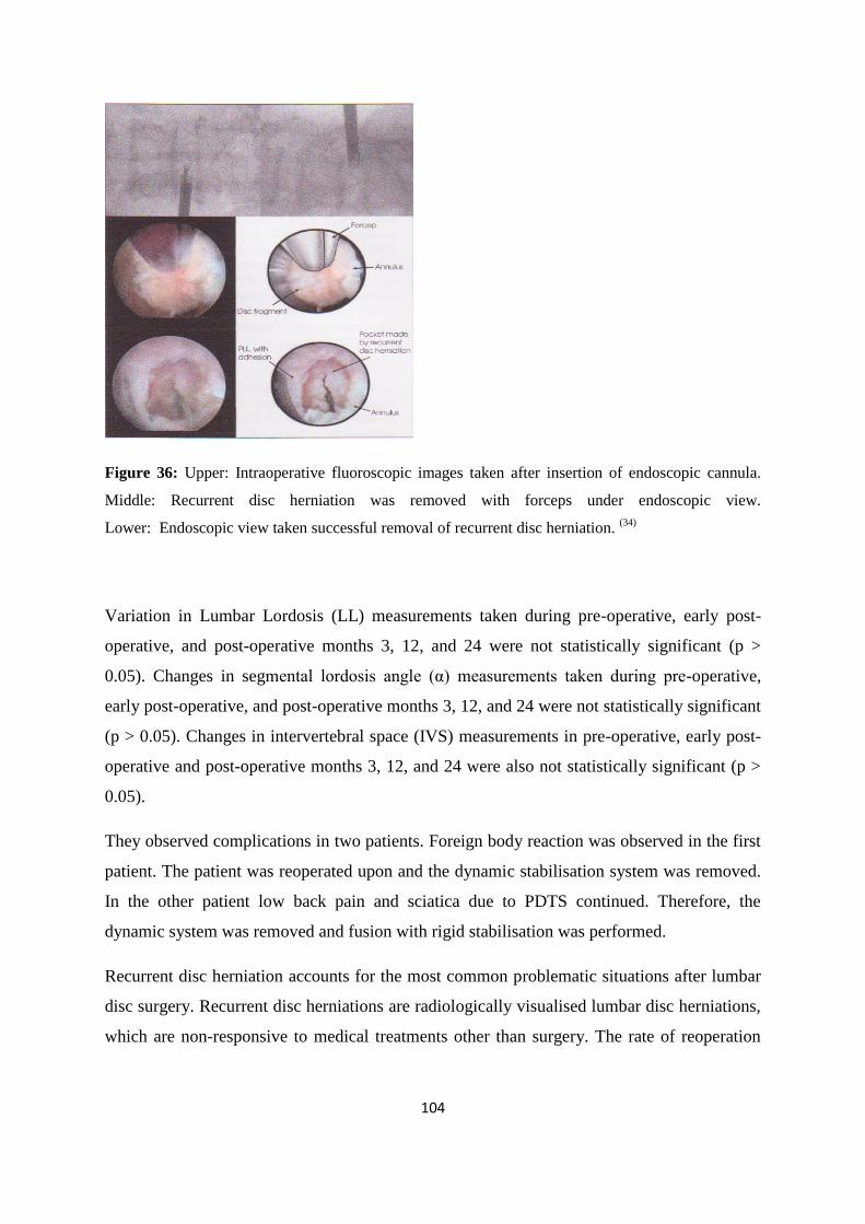
104
Figure 36: Upper: Intraoperative fluoroscopic images taken after insertion of endoscopic cannula.
Middle: Recurrent disc herniation was removed with forceps under endoscopic view.
Lower: Endoscopic view taken successful removal of recurrent disc herniation. (34)
Variation in Lumbar Lordosis (LL) measurements taken during pre-operative, early post-
operative, and post-operative months 3, 12, and 24 were not statistically significant (p >
0.05). Changes in segmental lordosis angle (α) measurements taken during pre-operative,
early post-operative, and post-operative months 3, 12, and 24 were not statistically significant
(p > 0.05). Changes in intervertebral space (IVS) measurements in pre-operative, early post-
operative and post-operative months 3, 12, and 24 were also not statistically significant (p >
0.05).
They observed complications in two patients. Foreign body reaction was observed in the first
patient. The patient was reoperated upon and the dynamic stabilisation system was removed.
In the other patient low back pain and sciatica due to PDTS continued. Therefore, the
dynamic system was removed and fusion with rigid stabilisation was performed.
Recurrent disc herniation accounts for the most common problematic situations after lumbar
disc surgery. Recurrent disc herniations are radiologically visualised lumbar disc herniations,
which are non-responsive to medical treatments other than surgery. The rate of reoperation
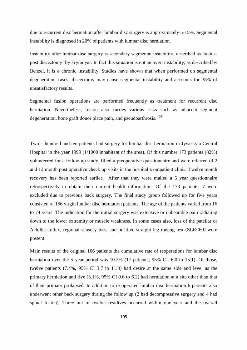
105
due to recurrent disc herniation after lumbar disc surgery is approximately 5-15%. Segmental
instability is diagnosed in 20% of patients with lumbar disc herniation.
Instability after lumbar disc surgery is secondary segmental instability, described as „status-
post discectomy‟ by Frymoyer. In fact this situation is not an overt instability; as described by
Benzel, it is a chronic instability. Studies have shown that when performed on segmental
degeneration cases, discectomy may cause segmental instability and accounts for 38% of
unsatisfactory results.
Segmental fusion operations are performed frequently as treatment for recurrent disc
herniation. Nevertheless, fusion also carries various risks such as adjacent segment
degeneration, bone graft donor place pain, and pseudoarthrosis. (69)
Two – hundred and ten patients had surgery for lumbar disc herniation in Jyvaskyla Central
Hospital in the year 1999 (1/1000 inhabitant of the area). Of this number 173 patients (82%)
volunteered for a follow up study, filled a preoperative questionnaire and were referred of 2
and 12 month post operative check up visits in the hospital‟s outpatient clinic. Twelve month
recovery has been reported earlier. After that they were mailed a 5 year questionnaire
retrospectively to obtain their current health information. Of the 173 patients, 7 were
excluded due to previous back surgery. The final study group followed up for five years
consisted of 166 virgin lumbar disc herniation patients. The age of the patients varied from 16
to 74 years. The indication for the initial surgery was extensive or unbearable pain radiating
down to the lower extremity or muscle weakness. In some cases also, loss of the patellar or
Achilles reflex, regional sensory loss, and positive straight leg raising test (SLR<60) were
present.
Main results of the original 166 patients the cumulative rate of reoperations for lumbar disc
herniation over the 5 year period was 10.2% (17 patients, 95% CL 6.0 to 15.1). Of those,
twelve patients (7.4%, 95% CI 3.7 to 11.3) had desire at the same side and level as the
primary herniation and five (3.1%, 95% CI 0.6 to 6.2) had herniation at a site other than that
of their primary prolapsed. In addition to re operated lumbar disc herniation 6 patients also
underwent other back surgery during the follow up (2 had decompressive surgery and 4 had
spinal fusion). Three out of twelve residives occurred within one year and the overall
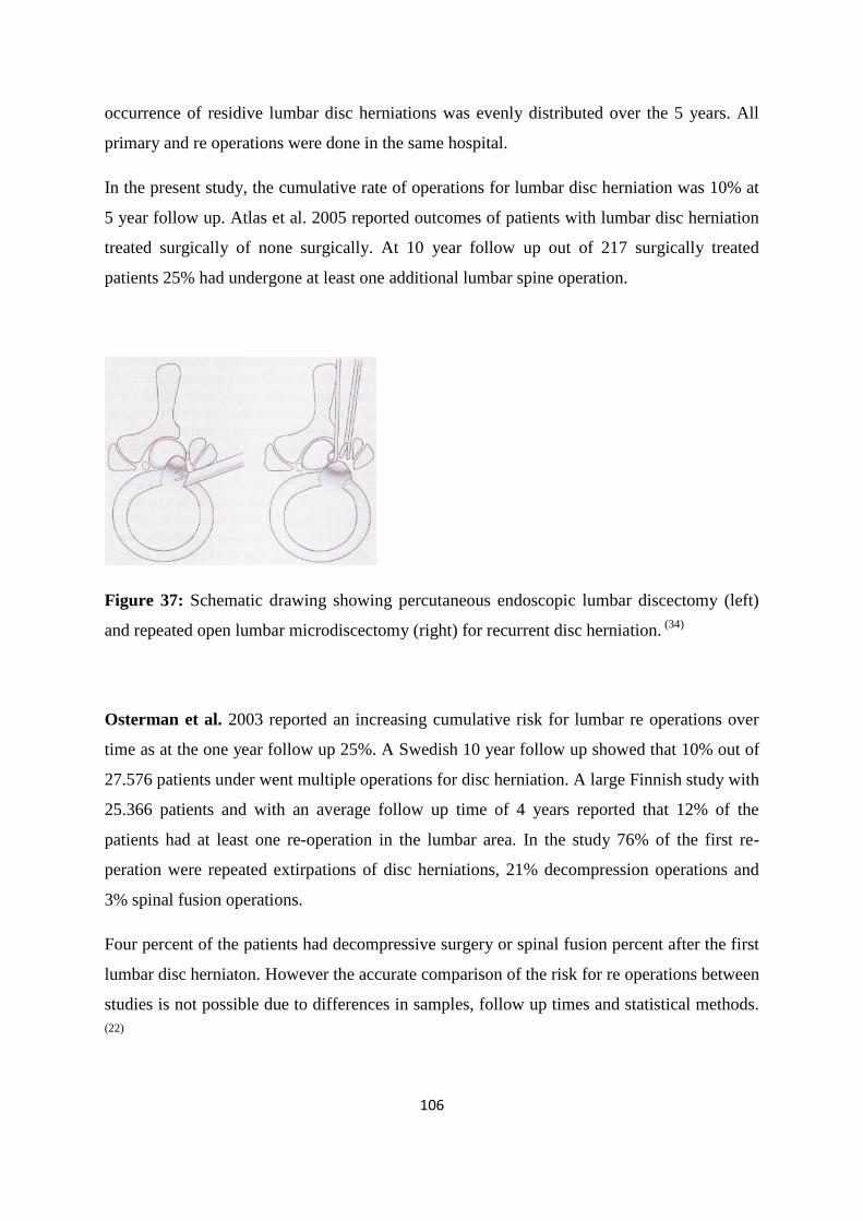
106
occurrence of residive lumbar disc herniations was evenly distributed over the 5 years. All
primary and re operations were done in the same hospital.
In the present study, the cumulative rate of operations for lumbar disc herniation was 10% at
5 year follow up. Atlas et al. 2005 reported outcomes of patients with lumbar disc herniation
treated surgically of none surgically. At 10 year follow up out of 217 surgically treated
patients 25% had undergone at least one additional lumbar spine operation.
Figure 37: Schematic drawing showing percutaneous endoscopic lumbar discectomy (left)
and repeated open lumbar microdiscectomy (right) for recurrent disc herniation. (34)
Osterman et al. 2003 reported an increasing cumulative risk for lumbar re operations over
time as at the one year follow up 25%. A Swedish 10 year follow up showed that 10% out of
27.576 patients under went multiple operations for disc herniation. A large Finnish study with
25.366 patients and with an average follow up time of 4 years reported that 12% of the
patients had at least one re-operation in the lumbar area. In the study 76% of the first re-
peration were repeated extirpations of disc herniations, 21% decompression operations and
3% spinal fusion operations.
Four percent of the patients had decompressive surgery or spinal fusion percent after the first
lumbar disc herniaton. However the accurate comparison of the risk for re operations between
studies is not possible due to differences in samples, follow up times and statistical methods.
(22)
107
6. Results
In this review we sought to analyze the symptomatic lumbar disc herniation and to compare
overall benefits of conservative and non conservative treatments. Relationships effecting
lumbar disc herniation will be also be mentioned.
Conservative versus non-conservative treatments for lumbar disc herniation
The most recent clinical trials available were SPORTs having one randomized and one
observation cohort comparing conservative and surgery treatment for a symptomatic lumbar
disc herniation with duration at least 6 week of pain.
Overall, 1,244 SPORT participants with lumbar intervertebral disc herniation were enrolled
(501 in the randomized cohort, and 743 in the observational cohort). In both cohorts
combined, 805 patients received surgery at some point during the first 4 years; 439 (35%)
remained non-operative. Over the 4 years, 1,192 (96%) of the original enrollees completed at
least 1 follow-up visit and were included in the analysis (randomized cohort: 94% and
observational cohort 97%). (75)
The treatment effects significantly favored surgery in both cohorts. In the combined
analysis, treatment effects were statistically significant in favor of surgery for all primary and
secondary outcome measures (with the exception of work status) at each time point.
The treatment effects for the secondary measures of sciatica bothersomness, satisfaction, and
self-rated improvement narrowed between 3 months and 2 years but remained significant at
all periods. Work status was significantly worse in the surgery group at 3 months due to
surgery patients recovering from surgery; work status thereafter showed a small but non-
significant benefit for surgery. At 4 years, the adjusted percentage of patients working was
84.4% surgical vs. 78.4% non-operative, treatment effect 6.0 (95% CI −0.9, 12.9).
The results of SPORT are similar to the Maine Lumbar Spine Study (MLSS) and the Weber
landmark study. The MLSS reported somewhat larger adjusted treatment effect differences
at 5 years between patients who had received surgery within 3 months versus those that had
not when compared to the SPORT 4-year data: −7.1 vs. − 3.3 (sciatica bothersomeness), −2.0
108
vs. −0.9 (leg pain in the past week) and −1.2 vs. −0.8 (low back pain in the past week). The
differences are mainly related to greater improvements in the non-operative group in
SPORT vs. MLSS; that is, the sciatica bothersomeness non-operative mean change from
baseline for SPORT was −8.2 at 4 years vs. MLSS −4.6 at 5 years. While there are no
validated outcome measures that can be directly compared between SPORT and the Weber
study, their 4 year results of 70% more patients in the surgical group and 51% in the
conservative treatment group with “Good” results is similar to SPORT‟s 79.2% self-rated
major improvement in the surgery group and 51.7% in the non-operative group at 4 years. (75)
In a general overview of the trials comparing conservative and non-conservative treatments a
significant efficiency in all clinical outcomes were favor of surgery against non-operative.
There were some significant decrease after 6 monthss in the following outcomes between
these procedures but surgery maintained greater results.
Peul et al. trial for early surgery showed that after having sciatica there was earlier pain relief
for surgery in contrast of conservative treatment. But these results were not significant in 6
months and the difference between the two groups was decreased. (50)
In all trials there was
also good results overtime for conservative especially in SPORTS in the “usual care”
mentioned. With having only a 43% of physical therapy intervention and those we can
conclude that if there better interventions to conservative patients there may have been better
results.
In all the trials the same limitation of crossover of patients were found between the groups
due to randomization of treatment approaches produced a dysbalance of the initial group‟s
enrollment.
Minimal invasive procedures and their efficiency for lumbar disc herniation
Microdiscectomy gives broadly comparable results to standard discectomy and show that
they can reduce scar formation, however there is limited evidence about the effect on clinical
outcomes. (46)
There is insufficient evidence on other percutaneous discectomy techniques to
draw firm conclusions. The evidence for other minimally invasive techniques remains unclear
except for chemonucleolysis using chymopapain, which is no longer widely available.
By comparing two operative techniques standard microscopic technique and microscopic
assisted percutaneous nucleotomy (MAPN) technique the clinical outcomes of both surgical
109
methods were indistinguishable at the transfer center, whereas MAPN patients at the index
center showed a slightly faster recovery when compared to MC patients.
In 83% of the patient a primary observed motor deficit resolved completely within the follow
up period. Sensory deficits completely resolved in 68% of the patients, improved in 18%, and
remained unchanged in 14%. For the neurological situation there was no obvious difference
found for the transfer center.
Within this study period no real clinical advantages of the less traumatized posterior muscles
could be found. Thus, the hypothesis that with a lesser traumatized back muscle leads to a
quiker recovery and to less chronic pain could not be confirmed. (53)
Tassi reviewed outcomes from 500 patients with discogenic pain and herniated discs treated
with microdiscectomy (1997-2001 by 6 surgeons) and 500 patients treated with percutaneous
laser disc decompression (2002-2005 by a single surgeon). Patients with sequestrated disc
were excluded. This retrospective review found that the hospital stay (six vs. two days),
overall recovery time (60 vs.35) and repeat procedure rate (7% vs. 3%) were lower in the
laser group. The percentages of patients with overall good/excellent outcomes were found to
be similar in the two groups (85.7% vs. 83.8%) at a two year assessment. (51)
PLDD has been around for almost 20 years, scientific proof of its efficacy still remains
relatively poor also the criteria for selection of patients for treatment of PLDD which is based
on the concept of the intervertebral disc being a closed hydraulic system, and only contained
herniations can be expected to respond to reduction of intradiscal pressure.
Conservative treatments and the efficiency for lumbar disc herniation
Luijsterburg et al randomized to evaluate physical therapist efficiencey compared a general
practioner with a physical therapist and the only treatment by a general practioner. About 53
patients (79%) in the intervention group versus 38 patients (56%) in the control group
reported to be improved in the primary care within a 12- month‟s follow-up period. More
significant results is needed with longer time evaluation. In other reviews on systematic
reviews no differences were found in overall improvement, pain, and disability at short-term.
110
Conflicting evidence regarding improvement of pain at short-term follow-up was found.
Physical therapy programs included conventional lumbar traction and had quite benneficial
on lumbar disc herniation without emergent operative indications with clincal improvements
noticed to occur prior to radiological improvement. In conclusion clinical parameters should
be considered in primarily evaluation of treatment results of patients with lumbar disc
herniation. (76)
Mckenzie method is a knowable evaluation and treament procedure with give us the status
of the patients pain in different positions. There was information found that surgery was
decreased when McKenzie approach was available in Denmark and that it had good acute
outcomes when extension exercises were tolerable for patients. However clinical significant
results where not available to conclude further.
From a review analyzed with the reusults there is belief that chiropractic management and
nonsurgical measures (manipulation, stabilization and neuromobilization) are a viable
alternative to surgery in patients with radiculopathy. Of the 162 consecutive patient reviewed,
99 were female and 63 were male. Upper extremity symptoms were seen in 61 cases and
lower extremity in 108 cases, with 7 of these cases demonstrating both. (8)
Ninety-five (58.64%) were classified as “chronic”, meaning their symptom duration was
greater than 3 months. Ninety-one percent of acute presentations resolved with a treatment
trial (mean) of 6.2 sessions, ranging from 1 to 20 visits. Eighty-one percent of chronic
presentations resolved with a treatment trial of 8.6 (mean) sessions, ranging from 1 to 23
visits.
There were several important limitations to this study and randomized controlled trials are
needed for further assess of this nonsurgical approach compared with untreated controls and
surgical treatments.
For stabilzation there was no significant finding but there is an effect by providing it. An
intresting case mentioned of a structural change of a disc herniation by providing efficient
functional stabitization approach. This goes hand in hand with the stabilization theory in the
general part by decribing how important is functional stereotypes performed and with
abdominal breathing (stability) providing increase abdominal pressuse which gives lower
compression on the lumbar spine and simultaneously lesser on the interververtbral discs.
111
Does morphological, location and natural history play a role in lumbar disc herniation?
In a combination of SPORTS data Pearson et al. evaluating morphology and lumbar spinal
level herniations showed that a greater difference in improvement between operative and
non-operative treatment for upper level herniation (L2-L3 and L3-L4) than for herniations at
the levels L4-L5 and L5-S1. Patients with upper level herniation had less improvement with
non-operative treatment and slightly better operative outcomes than those with lower level
herniations resulting outcome of surgery that the upper level herniation was greater than the
L4-L5 results, which were greater than the L5-S1 results. (49)
Foraminal herniations were most likely found in upper levels (24%), in L4-L5 (3%) and in
L5-S1 (2%); far lateral herniations were found (25%) in upper levels, a 7% in L4-L5 and
(6%) in L5-S1 and most were posterolateral herniation which represented in upper level
(44%), in L4-L5 (76%) and in L5-S1 83%). The upper level herniation group was more likely
to have a sequestrated fragment. The extrusion/sequestration was more likely to undergo
surgery than the protrusion group and in the severity of leg-symptoms was lower at baseline
for patients with upper herniations.
Takata et al. investigation for morphological changes of 42 patients (28 men and 14 women)
with a mean age of 42 years (range 16–64 years) who presented with unilateral leg pain and
low back pain that was definitely diagnosed as being caused by LDH.
In conclusion of this investigation were that 37 out of 42 patients (88 %) showed effective
(>50%) reduction of the herniated mass on MRI 3-12 months after the onset of symptoms.
Sequestered hernia and transligamentous extrusions seem to be more easily and rapidly
absorbed. MRI changes and improvement of symptoms are well correlated with MRI
changes, but a time lag is observed. (66)
The basic treatment of LDH should be conservative according to the results of this study;
however surgery may be necessary for large central protruding hernias and patients who
show severe motor deficits. There is a need of more randomized experiments for conservative
treatments with chronicity within at least 2 years for better statistical significant results. (66)
112
Post surgey results, reopartion, training and posture influence
The rate of reoperation due to recurrent disc herniation after lumbar disc surgery is
approximately 5-15%. Segmental instability is diagnosed in 20% of patients with lumbar disc
herniation.
Hakkinen et al. evaluated trunk muscle strength, pain, flexibility, and disability in patients
who had been operated for lumbar disc herniation 2 months after surgery. Although the leg
pain and back pain decreased by approximately 80%, 30% of the patients perceived moderate
to severe disability measurements. It was also noted that decreased muscle strength and
mobility, especially with older patients and with considerable postoperative pain, caused
significant functional disability. The recovery of multifidus muscle is not spontaneous after
resolution of painful symptoms, and this inhibition can be reversed only by specific training
exercises. This then can prevent the subsequent incidence of low back pain and thus also
prevent recurrences. (18)
Ostelo et al. in a Cohrane systematic review concluded that: exercise programs starting in
four to six weeks post-surgery seem to lead to a faster decrease in pain and disability than no
treatment. In high intensity exercise programs led to a faster decrease in pain and disability
than low intensity programs. There was no significant difference between supervised and
home exercises for pain relief, disability, or global perceived effect. There is no evidence that
active programs increase the re-operation rate after first-time lumbar surgery. (72)
In a prospective study of 79 people Lundin et al. (39)
evaluated the condition of stiff back
spine to be associated with outcome in surgery and found that patients with a stiff and flat
back had good prognosis after lumbar disc surgery, and in most cases the pain was reach a 2-
year level during the 2-6 weeks, while the physical restoration measured by lumbar flexion
and lordosis and the perceived disability, will continue to improve over the first 6 months
after surgery.
In conclusion specific training influence the post surgery results by strengthening the
weakend muscles and limiting a relapse for the disc herniation. Nevertheless there is a great
amount of relapses and reoperations that give us information that a better examination is need
to determine which patients are for surgery. In the study for back status associating with
surgery it showed a good prognosis but there is also a need of evaluating clinical outcomes.
113
7. Discussion
In this review for lumbar disc herniation provoking radiculopathy research on conservative
and non conservative was performed to compare and evaluate their efficiency. The results of
the most recent trials were comparing directly treatments were found and described as the
question in this review was the
Weber‟s landmark study was similar with comparison of the most recent available SPORT‟s
with “good” results of 70% and 79.2% is surgery groups and 51% and 51.7% in the non-
operative groups respectively. (40)
Limitations of the trials were found similarity in the randomized trials from the crossover of
patients with the SPORTs groups with the other recent randomized trial recent for disc
herniation; Peuls et al. shared nearly the same amount of crossover 38% in 6 months and 39%
in 5 months had crossed into surgery respectively.
The estimated improvements 1 year after surgery in these two studies were similar (Peul vs.
SPORT): SF-36 BP 59.3 vs. 43.7; SF-36 PF 50.3 vs. 44.4; and Sciatica Bothersomeness
−11.5 vs. −11.2. In addition, the differences at 1 year between randomized groups in the
intent-to-treat analyses were also quite similar: SF-36 BP 2.7 vs. 3.6; SF-36 PF 2.2 vs. 2.0;
and Sciatica Bothersomeness −0.4 vs. −1.9.
These results further validate the SPORT randomized cohort results but again highlight is
needed to also consider the as-treated analysis in this study population to estimate the true
effect of surgery and to avoid bias towards no significant results. (40)
From other trials there was low evidence on treatments efficiency but some results were
obtained and analyzed. Franke et al. (46)
in a prospective randomized controlled clinical trial
compared two operative techniques standard microscopic technique and microscopic assisted
percutaneous nucleotomy (MAPN) technique. The microsurgical procedure underwent 48
and 52 patients a MAPN procedure. In 83% of the patient a primary observed motor deficit
resolved completely within the follow up period. Sensory deficits completely resolved in
68% of the patients, improved in 18%, and remained unchanged in 14%.
114
Within the study period no real clinical advantages of the less traumatized posterior muscles
could be found. Thus, the hypothesis that with a lesser traumatized back muscle leads to a
quiker recovery and to less chronic pain could not be confirmed. (74)
After these trials the review focused on conservative treatments in trials and reviews there
was also low evidence for conclusions but information were optained and evaluated.
Luijsterburg et al. showed greater improvement when using a physical therapist with a
general practioner rather than only treatment by a general practioner. About 53 patients
(79%) in the intervention group versus 38 patients (56%) in the control group reported to be
improved (RR, 1.4 95% CI: 1.1; 1.8). (76)
In a chiropractic approach 162 total cases, of acute and chronic presentation had general
radiculopathy which this is the limitation of the trial. Nevertheless the results were so that
needed to be mentioned. Ninety-one percent of acute presentations resolved with a treatment
trial (mean) of 6.2 sessions, ranging from 1 to 20 visits. Eighty-one percent of chronic
presentations resolved with a treatment trial of 8.6 (mean) sessions, ranging from 1 to 23
visits. (8)
Evaluating with McKenzie method using the extension exercise showed that after examining
67 patients with low back pain (LBP) radiating to the calf or foot with at least one significant
sign of nerve root irritation and at least 6 weeks of failed non- operative therapy. The 52% of
patient (34 of them) shown no worsening during extension test. All 34 performed extension
exercise program with 100% regaining their full lumbar extension range and eliminating their
symptoms within 2-5 days. (31)
For postoperative patients Ostelo et al. in a Cohrane systematic concluded that exercise
programs starting in four to six weeks post-surgery seem to lead to a faster decrease in pain
and disability than no treatment. In high intensity exercise programs led to a faster decrease
in pain and disability than low intensity programs. There was no significant difference
between supervised and home exercises for pain relief, disability, or global perceived effect.
There is no evidence that active programs increase the re-operation rate after first-time
lumbar surgery. (46)
115
This review gave positive results that with function and exercises there was improvement for
patients with lumbar disc herniation however this results were with low evidence to obtain
significant results.
The outcome of the trials are low in evidence and not significant valued however the
intresting of the review of lumbar disc herniation was also describing the mechanical
overview of the lumbar spine and by trying to understand what influence has body posture
and stabilization function for disc herniation and especially in the lumbar region.
Expirements of stabilization of the lumbar spine were described in the general part showing
the importance of stability for any lumbar problem of the abdominal breathing in stability.
Abdominal breathing provides stability in the trunk by just providing resistance on an
extremity with abdominal breathing has quicker return to initial state rather than chest
breathing. (71)
Internal positive pressure creates functional stength and contributes to stability. Positive
pressure is regulated by intrabdominal pressure (IAP) which is generated and maintained by
trunk and abdominal muscles. (63)
Does morphology of a disc herniation change?
When the diaphragm flattens during inspiration it acts against the resistance of the abdominal
wall. Synergistic action of the diaphragm, with the abdominal, and the pelvic floor controls
the intr-abdominal pressure (IAP) to provide anterior stabilization of the Lumbosacral spine.
By means of regular proper stabilization exercises at time it is possible to change even the
structure by comparing a 2 year difference MRI pictures of patients with radicular pain
syndrome. (63)
Takata et al. investigated morphological changes of 42 patients (28 men and 14 women)
with a mean age of 42 years (range 16–64 years) who presented with unilateral leg pain and
low back pain and was definitely diagnosed as being caused by LDH.
In conclusion of this investigation were that 37 out of 42 patients (88 %) showed effective
(>50%) reduction of the herniated mass on MRI bewteen 3-12 months after the onset of
symptoms. Sequestered hernia and transligamentous extrusions seem to be more easily and
116
rapidly absorbed. MRI changes and improvement of symptoms are well correlated with MRI
changes, but a time lag was observed. (66)
Low evidence significance but intresting information showing that disc herniation benefits by
time and can change its morphology by only usual care. If we can progress this information
with functional stability mentioned and Mckenzie evaluation and treatment with other
conservative care such as traction, mobilization and reflex changes may provide in a long run
beneficial effects on a lumbar disc herniation.
According to the results of this study the basic treatment of LDH should be conservative;
however surgery may be necessary for large central protruding hernias and patients who
show severe motor deficits. (66)
There is a need of more randomized experiments for conservative treatments with specific
and combining treatments with chronicity at least 2 years for better statistical significant
results.
117
8. Conclusion
This thesis completed a systematic review of the relevant literature on the efficacy of
conservative treatment of symptmatic lumbar disc herniation (LDH). Thirty clinical trials and
four systematic reviews were found and evaluated according their results with conclusions:
A significant improvement in surgery in evaluation in the first six months which follows in a
slight decreasment in the first 2 years comparing with conservative approach.
Microdicectomy effectiveness is comparable with standard discectomy but without any
chronic significant findings. A stiff and flat back has a good prognosis for patients
undertaking lumbar disc surgery. Conservative treatments can improve patient but there were
no statistical significant however good results were shown in evaluation and exercise by
McKenzie also physical therapy had some good results with exercises, and traction but with
greater result in chiropractic approach. Conservative is needed after surgery for better and
longer timing results. By MRI findings there was conclusion that only with natural history
and conservative approach a disc herniation can relapse.
Future recommendation
In enrolling patients after having at least duration of 4 weeks sciatica due to disc herniation is
needed. Providing a randomized of 3 groups: a control group (usual care), a conservative
treatment group with specific treatments and not usual methods, a surgery group. With low
amount of enrollment no more than 100 patients in each group for a better significant reslut
data. Conservative treatments group will provide combination of treatments in deep
stabilization training, traction, mobilization, soft tissue basically in a manual therapy
approach. This with combining MRI every 3 months for 2 years of trial will be the best
combination of a randomizarion to evaluate the treatment for lumbar disc herniation in favour
for the physiotherapist.
118
Refernces
1) Aebi M., Gunzburg R., Szpalski. The ageing spine. Springer copyright. ISBN 3-540-
24408-5. 2005
2) Atlas SJ, Keller RB, Wu YA, et al. Long-term outcomes of surgical and nonsurgical
management of sciatica secondary to a lumbar disc herniation: 10 Year results from the
Maine lumbar spine study. Spine 2005;30 (8): 927-935
3) Bogduk et al. The nerve supply to the human lumbar intervertebral discs. Journal
anatomy 1981, 132, 1, pages 39-56
4) Bogduk Nikolai Clinical anatomy of the lumbar spine and sacrum, 4rth edition.
Churchill Livingston ISBN -13: 978-0-443-1019-9
5) Boss Norbet and Aebi Max. Spinal Disorders: Spinal foundamental diagnosis ISBN
978-3-540-40511-5 Springer-Verlag Berlin Heidelberg New York. Library of Congress
Control Number: 2006927571. 2008 Springer-Verlag Berlin Heidelberg.
6) Choi Gun. The effect of early isolated lumbar extension exercise program for patients
with herniated disc undergoing lumbar discectomy. Neurosurgery. Volume 57, Number
4, October 2005. p. 764-772.
7) Choi Soo-Jung et al. The Use of Magnetic Resonance Imaging to Predict the Clinical
Outcome of Non-Surgical Treatment for Lumbar Interverterbal Disc Herniation. Korean
J Radiol 8(2), April 2007.
8) Christensen D. Kim and Buswell Kirsten. Chiropractic outcomes managing
radiculopathy in a hospital setting: a retrospective review of 162 patients. Journal of
Chiropractic Medicine (2008) 7, 115-125. Elsevier
9) Devlin J. Vincent Spine secrets, Hanley & Belfus Publishing,2003. ISBN-13: 978-
1560533580
119
10) Donelosn Ronald, Rapidly reversible low back pain, ISBN 978-0-9790387-0-9
11) Donelson R, Et aL: A prospective study of centralization of lumbar spine and
referred pain. Spine 22: 115-22,1997
12) Drake L. Richard, Vogle Wayne, Mitchell W.M Adam, Gray‟s Anatomy For
Students ISBN 0-443-06612-4
13) Fahy D and Nixon J.E. Lumbar spinal stenosis. Current Orthopaedics (2001) 15, 91-
100. Harcourt Publishers Ltd
14) Fessler G. Richard, MD, PhD, Isaacs E. Robert, MD and Laurie Rice-Wyllie.
Lumbar Discectomy: Minimally Invasive Spine Surgery. Updated on: 02/10/10
Available at: http://www.spineuniverse.com/treatments/surgery/lumbar-discectomy-
minimally-invasive-spine-surgery
15) Foley T. Kevin MD., University of Tennesee, USA, Available at:
http:/www.spineuniverse.com/displayarticle.php/article 510.
16) Franke J., et.al. Comparison of a minimally invasive procedure versus standard
microscopic discotomy: a prospective randomized controlled clinical trial. Euro Spine
Journal, 2009 July; 18(7): 992-1000. Published online 2009 April 10. Doi:
10.1007/s00586-009-0964-2
17) Goodman, Snyder, Differential Diagnosis for Physical Therapists: screening for
referaral.Saunders. Elsevier 2009. ISBN 0-7216-0619-9
18) Hakkinen Arja et. Al: Reoperations after first lumbar disc herniation surgery; a
special interest on residives during a 5-year follow-up. Published: 09 January 2007.
19) Hall suzan: Basic Biomechanics 2nd
edition. ISBN: 0-8151-4077-0. Mosby-year
book copyright.1995
120
20) Herkowitz N. Harry, Gordon R bell. The lumbar spine. International Society for Study
of the Lumbar spine.ISBN 0721647774. . 2004
21) Hirsch A., Joshua MD, Director Interventional Neuroradiology/Endovascular
Neurosurgery Massachusetts General Hospital, Boston, MA
http://www.spineuniverse.com/treatments/surgery/percutaneous-discectomy Posted on:
05/11/03 | Updated on: 01/12/10
22) Huijbregts Peter. Muscloskeletal Examination and Intervention for Neck and Trunk
Clinical Instability and Lumbopelvic core stabilization. Pacific University: School of
Physical Therapy
23) Huntoon Elizabeth, MD and Huntoon, MD. Differential Diagnosis of Low Back Pain.
Seminars in Pain Medicine 2:138-144, 2004 Elsevier Inc.
ISBN-10: 1588900452
24) Jordan et al. Musculoskeletal disorders: Herniated lumbar disc. Search date
December 2002. main/x1118 02/05/03
25) Kaczmarska Agnieszka. Lectures Differential diagnostics in lumbar spine and pelvis
region by. 2008/09. FTVS. Charles University.
26) Kaltenborn M. Freddy, Manual mobilization of the joints, Volume 2 The Spine
ISBN 82-7054-069-2
27) Kamanli et al. Conventional physical therapy with lumbar traction; clinical
evaluation and magnetic resonance imaging for lumbar disc herniation. Clinical study.
Bratisl Lek Listy 2010; 111 (10), 541-544.
28) Kapandji I.A. The Physiology of joints, Volume 3 The trunk and the vertebral
column ISBN: 0-443-01209-1
121
29) Karppinen Jaro. Sciatica. Studies of symptoms, genetic factors, and treatment with
periradicular infiltration. ISBN 951-42-6480-0. Oulu. 2001
30) Kolar Pavel. Course of Dynamic Neuromuscular Stabilization. December 2010
31) Kopp J Et al: The use of lumbar extension in the evaluation and treatment of patients
with acute herniated nucleus pulposus, a preliminary report. Clinical orthopaedics, 1986.
202: 211- 8.
32) Lattes-Mendoza S. Comparable effectiveness of caudal vs. tran-foraminal epidular
steroid injections. The Iowa Orthopaedic Journal. Volume 29. p. 91-96
33) Lee Seungcheol, Kim Seok-Kang, Lee Sang-Ho, et.al. Percutaneous endoscopic
lumbar discectomy for migrated disc herniation: classification of disc migration and
surgical approaches. Eur Spine J (2007) 16:431-437, DOI 10.1007/s00586-006-0219-4
34) Lee Yeob Dong, Shim Shik Chan, Ahn Yong et al. Comparison of percutaneous
endoscopic lumbar discectomy and open lumbar microdiscectomy for recurrent disc
herniation
35) Lewit Karel, Manipulative Thearpy: Muscoloskeletal Medicine, 2010, Churchill
Livingston, Elsevier. ISBN: 978-0-7020-3056-7
36) Liebenson Craig. Rehabilitation of the spine: A practitioner‟s Manual. 2nd
edition.
Lippincott Williams & Wilkins, 2006. ISBN: 978-0-7817-2997-0
37) Luijsterburg A.j Pim, Verhagen P. Arianne, Ostelo W. J. G Raymond. Effectiveness
of conservative treatments for the lumbosacral radicular syndrome: a systematic review
Euro spine Journal (2007) 16:881-889 DOI 10.1007/s00586-007-0367-1
38) Luijsterburg A.j Pim, Verhagen P. Arianne, Ostelo W. J. G Raymond. Physical
therapy plus general practitioners‟ care versus general practitioners‟ care alone for
sciatica: randomized clinical trial with 12-month follow –up. Euro Spine Journal (2008),
17:509-517.
122
39) Lundin et al. A stiff and straaight back preoperatively is associated with a good
outcome 2 years after lumbar disc surgery: A prospective study of 80 patients. Acta
orthopaedica 2009; 80 (5): 573-578
40) Lurie Jon D, David M. Doman, Kevin F. Et al. Magnetic Resonance Imaging
Interpretation in Patients with Symptomatic Lumbar Spine Disc Herniations:
Comparison of Clinician and Radiologist Readings. Spine Volume 34, Number 7, pp
701–705, 2009, Lippincott Williams & Wilkins
41) Mckenzie R., Treat your own back, Eighth edition, 2006. ISBN: 978-0-9582692-3-0
42) Mkenzie R, the lumbar spine: mechanical diagnosis and therapy ISBN: 0958364761.
1981
43) Moore, K. L., Clinically Oriented Anatomy, 4th
ed, Williams & Willkings, 2002,
ISBN:950-06-1574-6
44) Mumenthaler Marco, Heinrich Mattle, Neurology, Thieme. 2002 ISBN-13: 978-
3133800112
45) O‟Sullivan P.B Lumbar segmental „instability‟: clinical presentation and specific
stabilizing exercise management. Manual Therapy (2000) 5(1), 2-12. Harcourt
Publishers Ltd.
46) Ostelo RWJG et al. Rehabilitation after lumbar disc surgery (Review). The Cochrane
Library 2008, Issue 4.
47) Panjabi M Manohar: Clinical spinal instability and low back pain: Journal of
Electromyography and Kinesiogy 13 (2003) 371-379
48) Pavlu Dagmar Lectures Differential Diagnosis. 2009. FTVS. Charles University
49) Pearson M. Adam. , Blood A. Emily, Frymoyer W. John. Et al. S.P.O.R.T Lumbar
intervertebral disc herniation and back pain: Does treatment, location or morphology
123
matter? Spine (Phila Pa 1976). 2008 Febraury 15; 33(4) 428-435.
doi:10.1097/BRS.0b013e31816469de. : available in PMC 2009 Novemeber 30.
50) Peul C. Wilco et al. Timing of surgery for sciatica: subgroup analysis alongside a
randomized trial. Eur Spine Journal (2009) 18:538-545, DOI 10.10007/s00586-008-
0867-7
51) Peul C. Wilco, Van Houwelingen C. Hans, Van Den Hout B Wilbert et. Al.
Prolonged conservative treatment versus early surgery in patients with sciatica caused
by lumbar disc herniation: BMJ 2008; 336: 1355-1358 (14 June), doi:10.1136/bmj.a143
(published 23 May 2008).
52) Peul C. Wilco, Van Houwelingen C. Hans, Van Den Hout B. Wilbert et al. Surgery
versus prolonged conservative treatment for sciatica. The New England Journal of
Medicine. 356;22. May 31, 2007
53) Piantadosi S. Clinical Trials: A Methodologic Perspective. New York: John Wiley,
1997.
54) Postacchini, F. Lumbar disc herniation. Springer Verlag Wien. ISBN:
9783211831182. 1999
55) Prentice E., William Therapeutic modalities in sports medicine (3rd
edition), ISBN:
0071441239
56) Rasmussen C., et.al. Rates of lumbar disc surgery before and after implementation of
multidisciplinary nonsurgical spine clinics. Spine, 2005.30: p. 2469-73.
57) Reider Bruce, Orthopaedic physical examination, 2nd
edition. Elsevier. ISBN: 0-
77216-0264-9
124
58) Rhee m. John et al. Radiculopathy and the herniated lumbar disc. Controversies
regarding pathophysiology and movement. J Bone Joint Surg Am. 2006;88:2070-2080
59) Rhee. American academic of orthopaedic surgeons. 2011 Annual Meeting Istructional
Course Lecture Handout. Submitted on: 09/21/2010.
60) Richeimer, Steven M.D, Chief, Division of Pain Medicine, Keck School of Medicine,
University of Southern California, Los Angeles, CA. Available at:
http://www.spineuniverse.com/conditions/herniated-disc/progressive-steps-toward-
lumbar-disc-herniationPosted on: April 22nd, 2001; Last Updated on: December 10th,
2009
61) Schafer A et. Al. Classification of low back-related leg pain - A proposed patho-
mechanism-based approach. Manual Therapy (2007).
62) Schenk B., et.al. Percutaneous Laser Disk Decompression: A Review of the
Literature. Review Article. American Journal Neuroradiol, January 2006.
63) Singh Pal Arun. Posterior Longitudinal Ligament. Posted: February 6, 2010.
Available at: www.boneandspine.com/definitions/posterior-longitudinal-ligament
64) Skytte I etal. Centralization- its prognostic value in patients with referred symptoms
and sciatica. Spine, 2005. 30: E(293-9)
65) Smith Chapman-David. The Chiropractic report. Chiropractic management for
lumbar disc herniation. Vol.18.No.5 September 2004
66) Takata et al. Natural history of lumbar disc hernia with radicular leg pain:
Spontaneous MRI changes of the herniated mass and correlation with clinical outcome.
Journal of Orthopaedic Surgery 2001, 9(1): 1–7
67) Tortora, G. J. Grabwski., Principles of Anatomy and physiology, 9th ed, Jhon, Wiley
& Sons, 2000, USA, ISBN 0-471-36692-7
125
68) Tsementzis A. Sotirios, Differential Diagnosis in Neurology and Neurosurgery, New
York 2000, ISBN 3-13-116151-5
69) Tuncay Kaner et al. Minimum Two-Year Follow-Up of Cases with Recurrent Disc
Herniation Treated with Microdiscectomy and Posterior Dynamic Transpedicular
Stabilisation. Open Orthop J. 2010; 4: 120–125.
70) Valat J-P, Giraudeau B, Rozenberg S et al. Epidural corticosteroid injections for
sciatica : a randomized, double blind controlled clinical trial. Ann Rheum Dis
2003;62:639-643 doi:10.1136/ard.62.7.639
71) Vele F. Lectures of Clinical Kinesiology, Prague 2006-10, FTVS, Charles University
72) Vroomen PC, de Krom MC, Slofstra PD, et al. Conservative treatment of sciatica: a
systematic review. J Spinal Disord 2000;13:463–469.; primary sources Medline and
73) Weber H. Lumbar disc herniation: a controlled, prospective study with ten years of
observation. Spine. 1983;8:131–140. [PubMed: 6857385]
74) Weinstein JN, Lurie JD, Tosteson TD, et al. Surgical vs nonoperative treatment for
lumbar disk herniation. The Spine Patient Outcomes Research Trial (SPORT):
Observational cohort. JAMA 2006; 296:2451–9. [PubMed: 17119141]
75) Weinstein JN, Lurie JD, Tosteson TD, et al.Surgical versus Non-Operative Treatment
for Lumbar Disc Herniation: Four-Year Results for the Spine Patient Outcomes
Research Trial (SPORT) Spine (Phila Pa 1976). 2008 December 1; 33(25): 2789–2800.
doi:10.1097/BRS.0b013e31818ed8f4.
76) Weinstein N.James, DO, MSc, Tosteson D.Tor ScD , Lurie D.Jon, MD et al.
Surgical vs Non operative Treatment for Lumbar Disk Herniation. The spine Patient
126
Outcomes Research Trial (SPORT): A Randomized Trial, JAMA 2006 November 22,
296(20): 2441-2450
77) Weinstein PR. Anatomy of the lumbar spine, 2nd edition, Raven, 1993, New York
78) White AA, Panjabi MM. Clinical Biomechanics 1990 second edition ISBN 0-397-
50720-8
79) Yilmaz Figen et al. Efficacy of dynamic lumbar stabilization exercise in lumbar
microdiscectomy. J Rehab Med 2003; 35: 163-167
80) Youmans JR. Neurological Surgery, 3rd ed, Philadelphia, 2000, USA, ISBN 84-
8174-367-4. Embase/Excerpta Medical.
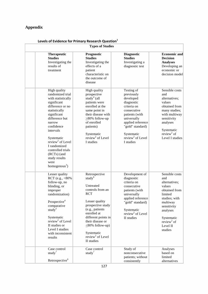
127
Appendix
Levels of Evidence for Primary Research Question1
Types of Studies
Therapeutic
Studies Investigating the
results of
treatment
Prognostic
Studies Investigating the
effects of a
patient
characteristic on
the outcome of
disease
Diagnostic
Studies Investigating a
diagnostic test
Economic and
Decision
Analyses Developing an
economic or
decision model
L
e
v
e
l
I
High quality
randomized trial
with statistically
significant
difference or no
statistically
significant
difference but
narrow
confidence
intervals
Systematic
review2 of Level
I randomized
controlled trials
(RCTs) (and
study results
were
homogenous3)
High quality
prospective
study4 (all
patients were
enrolled at the
same point in
their disease with
>80% follow-up
of enrolled
patients)
Systematic
review2 of Level
I studies
Testing of
previously
developed
diagnostic
criteria on
consecutive
patients (with
universally
applied reference
"gold" standard)
Systematic
review2 of Level
I studies
Sensible costs
and
alternatives;
values
obtained from
many studies;
with multiway
sensitivity
analyses
Systematic
review2 of
Level I studies
L
e
v
e
l
I
I
Lesser quality
RCT (e.g., <80%
follow-up, no
blinding, or
improper
randomization)
Prospective4
comparative
study5
Systematic
review2 of Level
II studies or
Level I studies
with inconsistent
results
Retrospective
study6
Untreated
controls from an
RCT
Lesser quality
prospective study
(e.g., patients
enrolled at
different points in
their disease or
<80% follow-up)
Systematic
review2 of Level
II studies
Development of
diagnostic
criteria on
consecutive
patients (with
universally
applied reference
"gold" standard)
Systematic
review2 of Level
II studies
Sensible costs
and
alternatives;
values
obtained from
limited
studies; with
multiway
sensitivity
analyses
Systematic
review2 of
Level II
studies
L
e
v
e
Case control
study7
Retrospective6
Case control
study7
Study of
nonconsecutive
patients; without
consistently
Analyses
based on
limited
alternatives
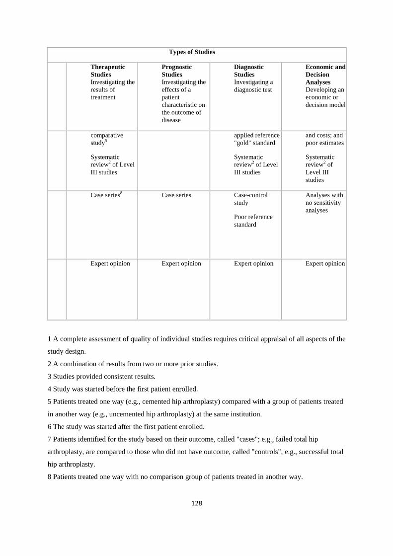
128
Types of Studies
Therapeutic
Studies Investigating the
results of
treatment
Prognostic
Studies Investigating the
effects of a
patient
characteristic on
the outcome of
disease
Diagnostic
Studies Investigating a
diagnostic test
Economic and
Decision
Analyses Developing an
economic or
decision model
l
I
I
I
comparative
study5
Systematic
review2 of Level
III studies
applied reference
"gold" standard
Systematic
review2 of Level
III studies
and costs; and
poor estimates
Systematic
review2 of
Level III
studies
L
e
v
e
l
I
V
Case series8 Case series Case-control
study
Poor reference
standard
Analyses with
no sensitivity
analyses
L
e
v
e
l
V
Expert opinion Expert opinion Expert opinion Expert opinion
1 A complete assessment of quality of individual studies requires critical appraisal of all aspects of the
study design.
2 A combination of results from two or more prior studies.
3 Studies provided consistent results.
4 Study was started before the first patient enrolled.
5 Patients treated one way (e.g., cemented hip arthroplasty) compared with a group of patients treated
in another way (e.g., uncemented hip arthroplasty) at the same institution.
6 The study was started after the first patient enrolled.
7 Patients identified for the study based on their outcome, called "cases"; e.g., failed total hip
arthroplasty, are compared to those who did not have outcome, called "controls"; e.g., successful total
hip arthroplasty.
8 Patients treated one way with no comparison group of patients treated in another way.