changes in fucosylation of human seminal ig g and secretory component of ig a in leukocytospermic...
TRANSCRIPT
Changes in fucosylation of human seminal IgG and secretorycomponent of IgA in leukocytospermic patients
Ewa M. Kratz & Mirosława Ferens-Sieczkowska &
Ricardo Faundez & Iwona Kątnik-Prastowska
Received: 2 July 2013 /Revised: 21 August 2013 /Accepted: 2 September 2013# The Author(s) 2013. This article is published with open access at Springerlink.com
Abstract Our study compares the status of human seminalplasma immunoglobulin G (IgG) and IgA secretory component(SC) fucosylation between infertile leukocytospermic and nor-mal, fertile normozoospermic patients. The seminal IgG andSC are decorated with AAL-reactive core fucose, andantennary UEA- and LTA-reactive fucose of Lewisy andLewisx structures, respectively. However, a correlation betweenIgG core fucosylation and IgG concentration (r =−0.52;p <0.0003) was observed. The IgG present in leukocytospermicsamples is characterized by lower expression of core fucosethan in the normal group (0.82±0.3 AU and 1.2±0.3 AU,respectively; p <0.002). In seminal plasma the SC is presentin two forms: 78-kDa and 63-kDa. The present study has alsoshown a higher AAL and LTA specific reactivity of glycansexpressed in 63-kDa SC, in comparison to 78-kDa SC, in thenormal group. In leukocytospermia, the values of specific lectinreactivity for core fucose, fucose α(1-2)- and α(1-3)- linked,were similar for both SC bands. Moreover, the present studyhas shown that in leukocytospermic samples the mean concen-trations of IgG and S-IgA are twice as high (131.68±102.6mg/land 36±27 mg/l, respectively) as in the normal group (67.68±29.2 mg/l; p <0.02, and 19±18 mg/l, p <0.019, respectively).The analysis of IgG and SC fucosylation status and the deter-mination of IgG and S-IgA concentrations in seminal plasma
might constitute a valuable diagnosis tools for the evaluation ofmale infertility associated with leukocytospermia with accom-panying inflammation.
Keywords Immunoglobulin G . Secretory component .
Fucosylation . Leukocytospermia .Male infertility
Introduction
From an immunological point of view, seminal plasma isdesigned to protect male sperm cells against infection and toinhibit the immune response against sperm cells in both maleand female reproductive organs. Seminal plasma has inhibi-tory properties due to the presence of biologically activefactors, such as enzymes, steroids, hormones, proteins, cyto-kines and immunoglobulins, mainly G (IgG) and A (IgA).
Physiologically, normal human ejaculate does not containmore than 1×10⁶/ml of leukocytes, but in leukocytospermia thenumber of leukocytes exceeds this value [1]. Leukocytes play animportant role in the immune “supervision” [2] and removal ofabnormal sperm cells by phagocytosis [3]. The increased numberof leukocytes in semen indicates urogenital tract infection andinflammation [4]. Adversely, leukocytes are a source of oxida-tive stress [5] and thus lower the quality of male gametes [6].Leukocytospermia is frequently observed in 10–20 % ofsubfertile and 10–44 % of infertile men [7, 8]. Yilmaz et al.[9] showed that the amount of sperm and number of sperm cellswith fast progressive movement in leukocytospermic men istwo times lower than those observed in patients withoutleukocytospermia with abnormal sperm parameters.
Immunoglobulin G, the most abundant immunoglobulin inblood plasma, is involved in the recognition, neutralization andelimination of pathogens and toxic antigens. Glycans attachedto blood plasma IgG molecules may differ in the content offucose, galactose, sialic acid and presence or absence ofbisecting N -acetylglucosamine [10]. An important role is
E. M. Kratz (*) :M. Ferens-Sieczkowska : I. Kątnik-PrastowskaDepartment of Chemistry and Immunochemistry, Wrocław MedicalUniversity, Bujwida 44a, 50-345 Wrocław, Polande-mail: [email protected]
R. FaundezEmbryology Laboratory InviMed - European Center of Motherhood,Rakowiecka 36, 02-532 Warsaw, Poland
R. FaundezDepartment of Large Animal Diseases and Clinic Division of AnimalReproduction, Andrology and Biotechnology of Reproduction,Warsaw University of Life Sciences Faculty of Veterinary Medicine,Nowoursynowska 100, 02-797 Warsaw, Poland
Glycoconj JDOI 10.1007/s10719-013-9501-y
attributed to the presence of core fucose α(1-6)- linked on N-glycans of IgG [11]: its absence causes a 50-fold increase inantibody binding to the FcγRIIIA present on natural killer cells[12], thus increasing antibody or complement-mediated cellularcytotoxicity [13]. Little is known about the profile of humanseminal plasma IgG fucosylation.
Secretory immunoglobulin A (S-IgA), accounts for twothirds of antibodies found on mucosal surfaces and is presentin colostrum, tears, sweat, gastrointestinal secretions, respira-tory and urogenital tracts [14, 15]. Secretory IgA preventsmicroorganisms and other antigens from penetrating the sur-face of the mucous membranes. It is also responsible forneutralization of viruses and enhances non-specific defencemechanisms [16]. Glycans of S-IgA form additional bindingsites for bacteria and may participate in both the innate andacquired immunity [17]. The most important feature of S-IgAis the presence of a secretory component (SC) [18], which is aglycoprotein of 50–90 kDa [14]. The secretory component issynthesized by epithelial cells of the digestive system, respira-tory and urogenital tracts. SC may be attached to immunoglob-ulin A or M, and/or may exist in free form in secretions. Thesecretory module gives S-IgA structural stability and increasesits resistance to proteolytic digestion during transportationthrough the epithelium to the mucous-serous secretions [19].Secretory component contains 5–7 N-linked oligosaccharides,which constitute about 22 % of its total molecular mass [20].Royle et al. [17] in their studies on human colostrum S-IgA,showed that most of the oligosaccharide structures expressedon human SC are bi-antennary. Tri- and tetra-antennary gly-cans are less abundant (11.7 % and <1%, respectively). All theglycans contain galactose, but not bisecting GlcNAc [17].Most N-glycans (70 %) expressed on SC contain sialic acid,and over 65 % of the glycans contain core fucose. Some of theoligosaccharide antennas may also be fucosylated. The secre-tory molecule may express each of the known structures ofLewis- and/or sialyl-Lewis-types, which are responsible forspecific binding of bacterial adhesins [17].
Our previous study [21] showed that seminal S-IgA concen-tration is not associated with sperm parameters. Immunoblottinganalysis has shown that seminal SC is present in two forms, withmolecular masses around 80- and 60-kDa [21]. In the presentstudy we investigated the differences in fucosylation of seminalIgG and IgA secretory component forms and their associationwith leukocytospermia of infertile males.
Materials and methods
Specimens
Human ejaculates were collected from fertile donors (26–45 years old) and leukocytospermic infertile patients (agematched to the normal group). The ejaculates were collected
by masturbation into sterile containers after 3–5 days of sexualabstinence, and were allowed to stand at 37 °C until liquefac-tion (no longer than 1 h). Next, standard semen analysis(volume, pH, morphology, sperm concentration, motility, andviability) was done at the Warsaw InviMed semen analysislaboratory according to WHO criteria [1]. Semen sampleswere centrifuged at 3500 × g for 10 min at room temperatureto obtain plasma. Seminal plasma was divided into smallaliquots and frozen at −76 °C until use. Ejaculates werecollected according to ethical standards (Ethical Committeeapproval KB-216/2011).
Seminal plasma samples were divided into two groups:normal (n =17) and leukocytospermic (n =28). In the normalgroup, the count of spermatozoa was higher than 15×106/mland more than 4 % expressed the correct sperm morphologywith a total motility of ≥40% or progressive motility ≥32% at1 h after ejaculation. The leukocytospermic group was formedfrom samples in which the leukocyte number was higher than1×106/ml according to WHO criteria [1]. None of theleukocytospermic samples were normozoospermic.
IgG and S-IgA concentration
Immunoglobulin G concentration was determined by radialimmunodiffusion [22] using goat anti-human IgG polyclonalantibodies (BIOMED, Warsaw, Poland) and Human SerumProtein Calibrator (DakoCytomation, Denmark) as a standard.The concentration of S-IgA was determined by sandwichELISA, using mouse monoclonal antibody directed againsthuman SC of IgA (1:20,000; Sigma Chemical Co., St. Louis,MO, USA) and preparation of IgA from human colostrum(Sigma Chemical Co., St. Louis, MO, USA) as a standard.Rabbit anti-human IgA polyclonal antibodies (1:100,000;DakoCytomation, Denmark) were used to quantify theamount of seminal S-IgA bound by the capture antibody.Goat anti-rabbit IgG-HRP (1:10,000; Sigma Chemical Co.,St. Louis, MO, USA) was used as a detection antibody.O -phenylenediamine dihydrochloride activated with H2O2
was used as the enzyme substrate. Colorimetric reaction wassubsequently stopped with 12.5 %H2SO4. The colour intensitywas measured in a Stat Fax 2100 Microplate Reader(Awareness Technology Inc., Palm City, FL, USA) at 492 nmusing a reference filter at 630 nm. All ELISA immunobindingreactions and washing steps were carried out in 10 mM Tris-buffered saline (TBS) containing 0.05 % Tween 20, pH 7.5,and the blocking step in the presence of 0.1 % BSA (SigmaChemical Co., St. Louis, MO, USA). Background absorbance(lower than 0.1 AU) was measured for TBS containing 0.05 %Tween 20, pH 7.5 instead of a seminal plasma sample, but withall other reagents. Seminal plasmas with known concentrationsof S-IgA were used as positive controls. All samples wereanalysed in duplicate.
Glycoconj J
Determination of IgG fucose exposure by lectin-ELISA
Three biotinylated fucose-specific lectins (Vector LaboratoriesInc., Burlingame, CA, USA): Aleuria aurantia lectin (AAL),Ulex europaeus agglutinin (UEA) and Lotus tetragonolobusagglutinin (LTA) were used to determine expression of fucosemoiety in IgG by lectin-ELISA according to the proceduredescribed previously for fibronectin and α1-acid glycoprotein[23]. The lectins differ with respect to their reactivity withdifferently bound terminal sugars on glycoproteins. TheAleuria aurantia lectin reacts mainly with the innermost fucoseα(1-6)- linked to N-acetylglucosamine core of N-glycans andwith lower affinity with fucoses α(1-2)-, α(1-3)- and α(1-4)-linked of the outer arms [24]. Ulex europaeus agglutinin isspecific to antennary fucoses α1,2-linked to Gal and α1,3-linked to GlcNAc, typical for Lewisy glycan structures [25].The presence of fucose α1,2-linked prevents the formation ofsialyl-Lewisx oligosaccharide structures [26]. Lotustetragonolobus agglutinin specifically reacts with fucose α1,3-linked to GlcNAc, characteristic for Lewisx structures, however,it can also slightly react with fucose typical for Lewisa andLewisy oligosaccharide structures. The presence of terminalsialic acidα(2-3)- linked in glycoprotein glycan structures limitsthe recognition of fucose α(1-3)- linked by LTA [27].
Removal of terminal sugars from capture antibodies
Monoclonal anti-human IgG antibodies had to be defucosylatedbefore use in lectin-ELISA to avoid the lectin binding tocapture antibodies. We have previously described the IgGdefucosylation procedure [23]. Briefly, one volume of poly-clonal goat anti-human IgG antibodies (200 μl, pH=8.1) wasmixed with an equal volume of 100 mmol/l NaIO4 in100 mmol/l NaHCO3, 0.2 % Tween 20, pH 8.1. The mixturewas incubated for 90 min. at room temperature in the dark andsubsequently dialysed against 100 mmol/l NaHCO3, pH 9.2,for 3 h at 4 °C. Such treatment resulted in elimination ofantibody reactivity with fucose-specific lectins.
Lectin-ELISA procedure
Expression of exposed fucosyl-residues of glycoproteins wasdetermined by fucose-specific lectins AAL, UEA and LTA, asdescribed earlier [23]. Defucosylated polyclonal goat anti-human IgG antibodies (BIOMED, Warsaw, Poland) were di-luted in 10 mM TBS pH 8.5 (1:10,000), coupled to a polysty-rene microtiter ELISA plate and incubated for 2 h at 37 °C.Seminal plasma samples were diluted in 10 mM TBS, 1 mMCaCl2, 1 mM MgCl2, 0.05 % Tween 20, and 0.5 % glycerol,pH 7.5, to obtain a glycoprotein solution containing 100 ng ofIgG in 100μl. Upon addition of seminal plasma samples, plateswere incubated for 2 h at 37 °C. All samples were analysed induplicate. Background absorbance was measured for samples
in which all reagents were present, but seminal plasma wasreplaced with a 10 mM TBS, 1 mM CaCl2, 1 mM MgCl2,0.05 % Tween 20, and 0.5 % glycerol, pH 7.5. To control forthe specificity of lectin-glycoprotein interaction and to checkthe absence of detectable endogenous reactive materials, con-trol probes were included for the test. Haptoglobin and asialo-haptoglobin preparations derived from ovarian cancer fluidwere used as positive controls [28], whereas a human albuminpreparation served as negative control (Sigma Chemical Co.,St. Louis, MO, USA). The α(1-6)-, α(1-3)- and α(1-2)- linkedfucose residues in IgG were detected by biotinylated AAL,LTA and UEA, respectively. Lectin dilutions (1:7500, 1:100and 1:250, respectively) were established in preliminary exper-iments. All lectins were diluted in 10 mM TBS containing1 mM CaCl2, 1 mM MgCl2, 0.05 % Tween 20, and 0.5 %glycerine, pH 7.5, and the plate was incubated for 1 h at 37 °C.The formed complex of IgG-biotinylated lectin was quantitatedusing phosphatase-labelled ExtrAvidin (1 h at 37 °C; 1:20,000;Sigma Chemical Co., St. Louis, MO, USA) and detected by thereaction with di-sodium 4-nitrophenyl phosphate (Merck,Darmstadt, Germany). The results were expressed in absor-bance units (AU) measured at 405 nm with a reference filterof 630 nm in ELISA Stat Fax 2100 Microplate Reader(Awareness Technology Inc., Palm City, FL, USA). To removeany excess protein, the plate was washed three times with10 mM TBS, 0.05 % Tween 20, pH=7.5 between eachELISA-step. The background absorbance values were nothigher than 0.2 AU.
Determination of SC bands by immunoblotting
To determine the most characteristic immunoblot pattern forSC in leukocytospermic and normal seminal groups, equalvolumes of samples in each seminal group were pooled. Next,the samples were diluted with 0.06 M Tris–HCl, pH=6.8containing 3 % SDS (w/v), 10 % glycerol (v/v) and 7.5 %2-mercaptoethanol (v/v), and boiled at 100 °C for 5 min.Then 250 ng of S-IgA, determined by ELISA (for details seeS-IgA concentration determination) was subjected to SDS-polyacrylamide gel electrophoresis on a 10 % gel accordingto Laemmli [29] in a BioRad-vertical system (BioRad,Richmond, CA, USA). The separated proteins were subse-quently blotted onto nitrocellulose (Serva ElectrophoresisGmbH, Heidelberg, Germany). After blocking (3% powderedskim milk in 50 mM TBS, pH 7.5), the blots were incubatedwith mouse monoclonal antibodies anti-human SC of IgAdiluted at 1:1000 in 3 % powdered skim milk in 50 mMTBS, pH 7.5, and probedwith goat anti-mouse IgG polyclonalantibodies conjugated with horseradish peroxidase (1:2000dilution in 3 % powdered skim milk dissolved in 50 mMTBS; Sigma Chemical Co., St. Louis, MO, USA). The colourreaction was developed with diaminobenzidine and H2O2.After each step, the nitrocellulose was washed at least 3 times
Glycoconj J
using 50 mM TBS, 0.05 % Tween; pH=7.5. Finally, the blotswere dried and analysed. The blots were digitised and bandscorresponding to secretory component of IgA (SC) wereanalysed with densitometry ImageJ 1.42q gel analysis soft-ware (National Institutes of Health, U.S. Department of Health& Human Services public domain). The relative amounts ofparticular SC bands were expressed as the percentage of thetotal number of pixels in a lane. To calculate the molecularmasses of SC bands, low molecular-weight protein massstandard (97.4–6.5 kDa; Serva Electrophoresis GmbH,Heidelberg, Germany) and human colostrum S-IgA prepara-tion (Sigma Chemical Co., St. Louis, MO, USA) were used.
Lectin-blotting determination of secretory component fucoseexpression
The relative reactivity of SC bands was analysed in lectin-blotting with biotinylated fucose-specific lectins, according tothe procedure described earlier by Kratz et al. [30] for syno-vial fluid IgG and IgA. For lectin-binding analysis only thosebands that corresponded to SC detected in immunoblottingwith mouse anti-human SC of IgA monoclonal antibodieswere taken into consideration (see the section above).Nitrocellulose membranes were labelled with AAL (1:400),LTA (1:200) and UEA (1:200) (Vector Laboratories Inc.,Burlingame, CA, USA) diluted in 3 % powdered skim milkin 50 mM TBS pH=7.5, and then detected with alkalinephosphatase-conjugated ExtrAvidin (1:10,000 dilution in3 % milk in 50 mM TBS pH=7.5; Sigma Chemical Co., St.Louis, MO, USA). The colour reaction was developed with0.1 M Tris–HCl; 0.1 M NaCl; 0.05 M MgCl2, pH=9.5 con-taining 100 μl 7.7 % nitro blue tetrazolium in dimethyl-formamide (DMF) (7:3; v/v), and 75 μl 5 % 5-bromo-4-chloro-3-indolyl phosphate, disodium salt in DMF. Lastly,the blots were dried and analysed. Bands corresponding toparticular AAL-, LTA- and UEA-reactive SC were digitisedand analysed with densitometry ImageJ 1.42q gel analysissoftware (National Institutes of Health, U.S. Department ofHealth &Human Services public domain). The relative amountsof particular fucose-specific SC bands were expressed as thepercentage of the total number of pixels in a lane. Asdescribed previously, to calculate the molecular masses ofSC bands, lowmolecular-weight protein mass standard (ServaElectrophoresis GmbH, Heidelberg, Germany) and humancolostrum S-IgA preparation (Sigma Chemical Co., St.Louis, MO, USA) were used. Specific lectin reactivityreflecting the density of fucosylated epitopes [31, 32] wasdefined as the ratio of lectin reactivity to the protein content,measured as reactivity with anti-human SC of IgA in a partic-ular band, and calculated as: Lectin reactivity (pixels/band)/Antibody reactivity (pixels/band). Specific lectin reactivitywas used to compare relative fucosylation of both SC forms(Table 2).
Statistical analysis
Statistical analysis was performed using STATISTICA 10.0software (StatSoft Inc., Tulsa, OK, USA). Experimental datawere presented as means and standard deviations (SD),and distribution of the values within analysed groups asbox-whisker plots with median and interquartile (25th-75th
percentile) range. According to a Shapiro-Wilk W test, thevalues did not fit normal distribution, thus the nonparametricMann–Whitney U test was used to determine differencesbetween groups. Correlations between determined parameterswere estimated according to a Spearman test. A two-tailedp -value of less than 0.05 was considered significant.
Results
Concentration of IgG and secretory IgA
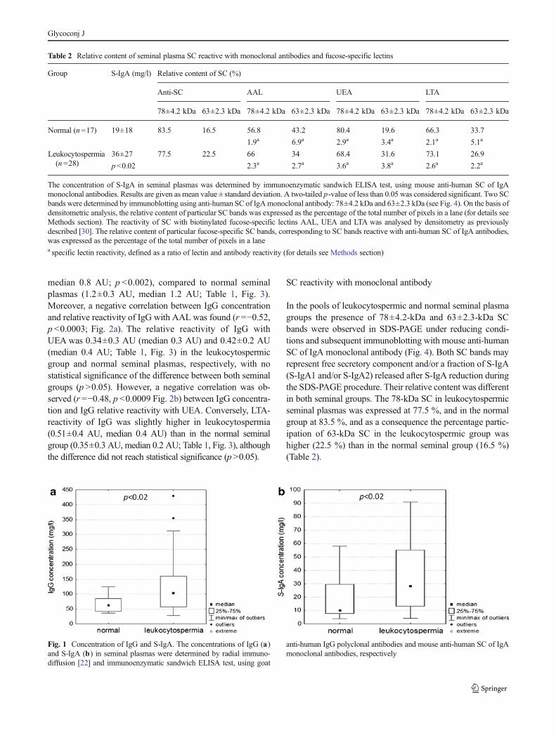
IgG concentration in leukocytospermic seminal plasmas wassignificantly higher than that observed in normal seminalplasma (131.7±103 mg/l and 67.7±29 mg/l, respectively;p <0.02; Table 1). Also S-IgA concentration was significantlyhigher in the leukocytospermic group compared to normalseminal plasmas (36±27 mg/l and 19±18 mg/l, respectively;p <0.02; Table 2). The median value of IgG concentration was104 and 63.3 mg/l, and 28.7 and 9.9 mg/l for S-IgA inleukocytospermic and normal seminal groups, respectively(Fig. 1a, b).
IgG reactivity with lectins
The relative reactivity of IgG with AAL was significantlylower in the leukocytospermic seminal group (0.82±0.3 AU,
Table 1 Relative reactivity of seminal plasma IgG with fucose-specificlectins
Group IgG (mg/l) Relative reactivity with lectins(AU)
AAL UEA LTA
Normal (n =17) 67.7±29 1.2±0.3 0.42±0.2 0.35±0.3
Leukocytospermia(n =28)
131.7±103 0.82±0.3 0.34±0.3 0.51±0.4p <0.02 p<0.002
The concentration of IgG in seminal plasmas was determined by radialimmunodiffusion according toMancini et al. [22], using goat anti-humanIgG polyclonal antibodies. In both seminal plasma groups, the relativereactivity of IgG (100 ng/100 μl) with fucose-specific lectins was deter-mined by lectin-ELISA [23] using biotinylated lectins AAL, UEA andLTA, and expressed in absorbance units (AU). Results are given as meanvalue ± standard deviation. A two-tailed p-value of less than 0.05 wasconsidered significant
Glycoconj J
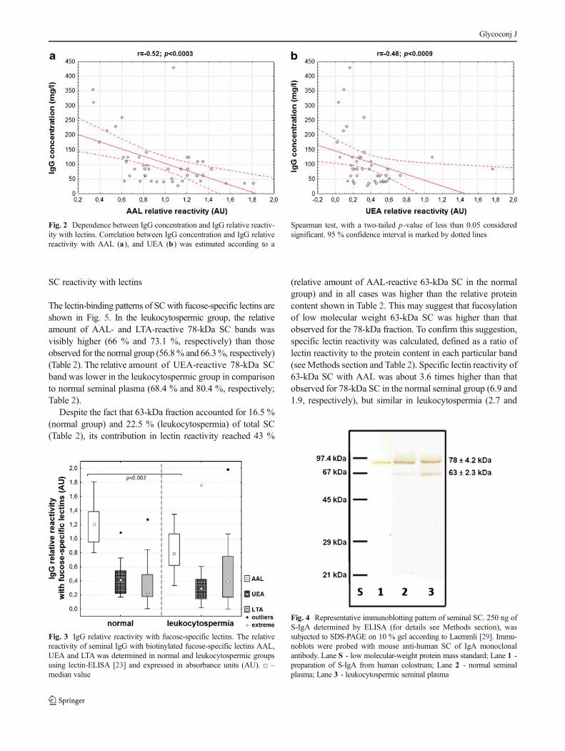
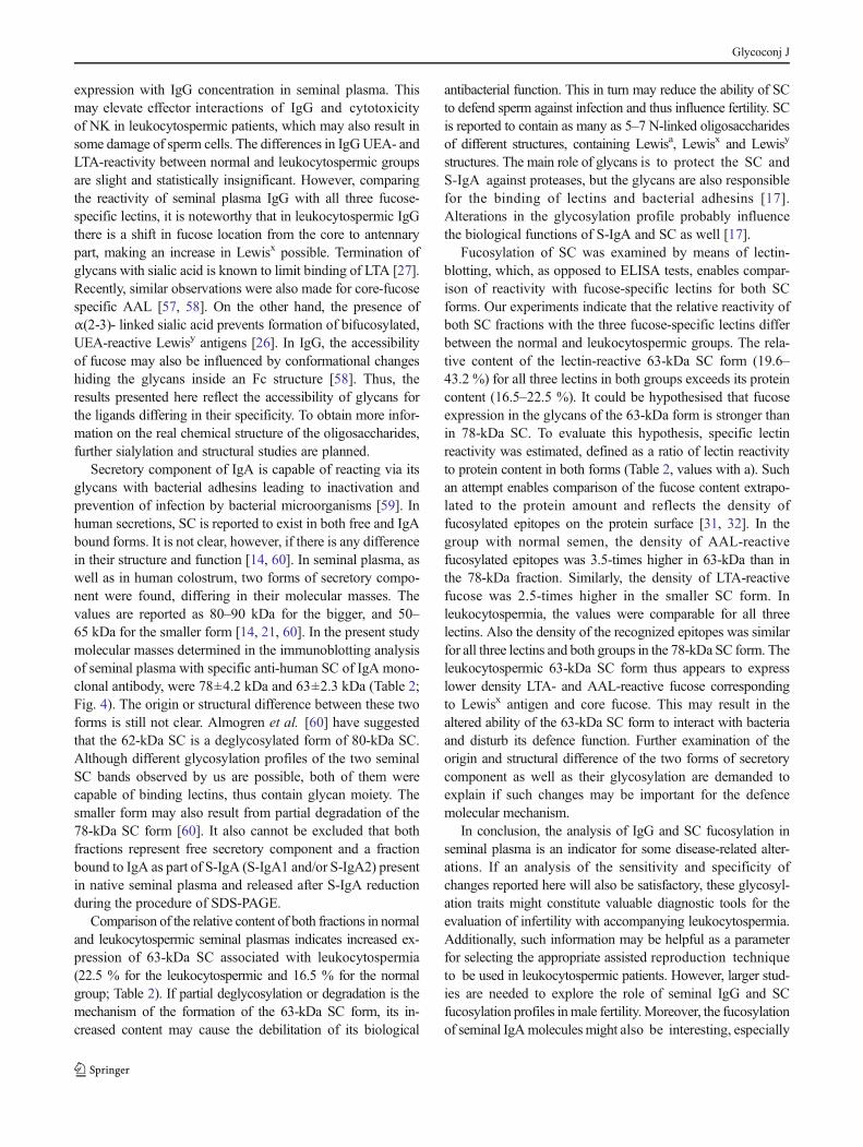
median 0.8 AU; p <0.002), compared to normal seminalplasmas (1.2±0.3 AU, median 1.2 AU; Table 1, Fig. 3).Moreover, a negative correlation between IgG concentrationand relative reactivity of IgG with AAL was found (r =−0.52,p <0.0003; Fig. 2a). The relative reactivity of IgG withUEA was 0.34±0.3 AU (median 0.3 AU) and 0.42±0.2 AU(median 0.4 AU; Table 1, Fig. 3) in the leukocytospermicgroup and normal seminal plasmas, respectively, with nostatistical significance of the difference between both seminalgroups (p >0.05). However, a negative correlation was ob-served (r =−0.48, p <0.0009 Fig. 2b) between IgG concentra-tion and IgG relative reactivity with UEA. Conversely, LTA-reactivity of IgG was slightly higher in leukocytospermia(0.51±0.4 AU, median 0.4 AU) than in the normal seminalgroup (0.35±0.3 AU, median 0.2 AU; Table 1, Fig. 3), althoughthe difference did not reach statistical significance (p>0.05).
SC reactivity with monoclonal antibody
In the pools of leukocytospermic and normal seminal plasmagroups the presence of 78±4.2-kDa and 63±2.3-kDa SCbands were observed in SDS-PAGE under reducing condi-tions and subsequent immunoblotting with mouse anti-humanSC of IgA monoclonal antibody (Fig. 4). Both SC bands mayrepresent free secretory component and/or a fraction of S-IgA(S-IgA1 and/or S-IgA2) released after S-IgA reduction duringthe SDS-PAGE procedure. Their relative content was differentin both seminal groups. The 78-kDa SC in leukocytospermicseminal plasmas was expressed at 77.5 %, and in the normalgroup at 83.5 %, and as a consequence the percentage partic-ipation of 63-kDa SC in the leukocytospermic group washigher (22.5 %) than in the normal seminal group (16.5 %)(Table 2).
Table 2 Relative content of seminal plasma SC reactive with monoclonal antibodies and fucose-specific lectins
Group S-IgA (mg/l) Relative content of SC (%)
Anti-SC AAL UEA LTA
78±4.2 kDa 63±2.3 kDa 78±4.2 kDa 63±2.3 kDa 78±4.2 kDa 63±2.3 kDa 78±4.2 kDa 63±2.3 kDa
Normal (n =17) 19±18 83.5 16.5 56.8 43.2 80.4 19.6 66.3 33.7
1.9a 6.9a 2.9a 3.4a 2.1a 5.1a
Leukocytospermia(n =28)
36±27 77.5 22.5 66 34 68.4 31.6 73.1 26.9
p <0.02 2.3a 2.7a 3.6a 3.8a 2.6a 2.2a
The concentration of S-IgA in seminal plasmas was determined by immunoenzymatic sandwich ELISA test, using mouse anti-human SC of IgAmonoclonal antibodies. Results are given as mean value ± standard deviation. A two-tailed p-value of less than 0.05 was considered significant. Two SCbands were determined by immunoblotting using anti-human SC of IgAmonoclonal antibody: 78±4.2 kDa and 63±2.3 kDa (see Fig. 4). On the basis ofdensitometric analysis, the relative content of particular SC bands was expressed as the percentage of the total number of pixels in a lane (for details seeMethods section). The reactivity of SC with biotinylated fucose-specific lectins AAL, UEA and LTA was analysed by densitometry as previouslydescribed [30]. The relative content of particular fucose-specific SC bands, corresponding to SC bands reactive with anti-human SC of IgA antibodies,was expressed as the percentage of the total number of pixels in a lanea specific lectin reactivity, defined as a ratio of lectin and antibody reactivity (for details see Methods section)
Fig. 1 Concentration of IgG and S-IgA. The concentrations of IgG (a)and S-IgA (b) in seminal plasmas were determined by radial immuno-diffusion [22] and immunoenzymatic sandwich ELISA test, using goat
anti-human IgG polyclonal antibodies and mouse anti-human SC of IgAmonoclonal antibodies, respectively
Glycoconj J
SC reactivity with lectins
The lectin-binding patterns of SCwith fucose-specific lectins areshown in Fig. 5. In the leukocytospermic group, the relativeamount of AAL- and LTA-reactive 78-kDa SC bands wasvisibly higher (66 % and 73.1 %, respectively) than thoseobserved for the normal group (56.8% and 66.3%, respectively)(Table 2). The relative amount of UEA-reactive 78-kDa SCband was lower in the leukocytospermic group in comparisonto normal seminal plasma (68.4 % and 80.4 %, respectively;Table 2).
Despite the fact that 63-kDa fraction accounted for 16.5 %(normal group) and 22.5 % (leukocytospermia) of total SC(Table 2), its contribution in lectin reactivity reached 43 %
(relative amount of AAL-reactive 63-kDa SC in the normalgroup) and in all cases was higher than the relative proteincontent shown in Table 2. This may suggest that fucosylationof low molecular weight 63-kDa SC was higher than thatobserved for the 78-kDa fraction. To confirm this suggestion,specific lectin reactivity was calculated, defined as a ratio oflectin reactivity to the protein content in each particular band(seeMethods section and Table 2). Specific lectin reactivity of63-kDa SC with AAL was about 3.6 times higher than thatobserved for 78-kDa SC in the normal seminal group (6.9 and1.9, respectively), but similar in leukocytospermia (2.7 and
Fig. 2 Dependence between IgG concentration and IgG relative reactiv-ity with lectins. Correlation between IgG concentration and IgG relativereactivity with AAL (a), and UEA (b) was estimated according to a
Spearman test, with a two-tailed p-value of less than 0.05 consideredsignificant. 95 % confidence interval is marked by dotted lines
Fig. 3 IgG relative reactivity with fucose-specific lectins. The relativereactivity of seminal IgG with biotinylated fucose-specific lectins AAL,UEA and LTA was determined in normal and leukocytospermic groupsusing lectin-ELISA [23] and expressed in absorbance units (AU). □ –median value
Fig. 4 Representative immunoblotting pattern of seminal SC. 250 ng ofS-IgA determined by ELISA (for details see Methods section), wassubjected to SDS-PAGE on 10 % gel according to Laemmli [29]. Immu-noblots were probed with mouse anti-human SC of IgA monoclonalantibody. Lane S - low molecular-weight protein mass standard; Lane 1 -preparation of S-IgA from human colostrum; Lane 2 - normal seminalplasma; Lane 3 - leukocytospermic seminal plasma
Glycoconj J
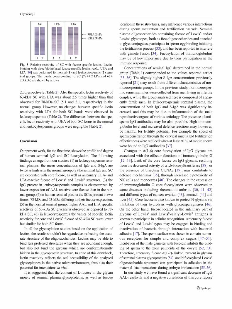
2.3, respectively; Table 2). Also the specific lectin reactivity of63-kDa SC with LTA was about 2.5 times higher than thatobserved for 78-kDa SC (5.1 and 2.1, respectively) in thenormal group. However, no changes between specific lectinreactivity with LTA for both SC bands were observed inleukocytospermia (Table 2). The differences between the spe-cific lectin reactivity with UEA of both SC forms in the normaland leukocytospermic groups were negligible (Table 2).
Discussion
Our present work, for the first time, shows the profile and degreeof human seminal IgG and SC fucosylation. The followingfindings emerge from our studies: (1) in leukocytospermic sem-inal plasmas, the mean concentrations of IgG and S-IgA aretwice as high as in the normal group, (2) the seminal IgG and SCare decorated with core fucose, as well as antennary UEA- andLTA-reactive fucose of Lewisy and Lewisx structures, (3) theIgG present in leukocytospermic samples is characterized bylower expression of AAL-reactive core fucose than in the nor-mal group, (4) in human seminal plasma the SC is present in twoforms: 78-kDa and 63-kDa, differing in their fucose expression,(5) in the normal seminal group, higher AAL and LTA specificreactivity of 63-kDa SC glycans is observed as opposed to 78-kDa SC, (6) in leukocytospermia the values of specific lectinreactivity for core and Lewisx fucose of 63-kDa SC were lowerbut similar for both SC forms.
In all the glycosylation studies based on the application oflectins, the results shouldn’t be regarded as reflecting the accu-rate structure of the oligosaccharides. Lectins may be able tobind less preferred structures when they are abundant enough,but also not bind the glycans which are conformationallyhidden in the glycoprotein structure. In spite of this drawback,lectin reactivity reflects the real accessibility of the analysedglycoepitopes in the native microenvironment, thus also theirpotential for interactions in vivo .
It is suggested that the content of L-fucose in the glycanmoiety of seminal plasma glycoproteins, as well as fucose
location in these structures, may influence various interactionsduring sperm maturation and fertilization cascade. Seminalplasma oligosaccharides containing fucose of Lewisx and/orLewisy glycotopes, both as free oligosaccharides and attachedto glycoconjugates, participate in sperm-egg binding initiatingthe fertilization process [33], and has been reported to interferewith gamete fusion [34]. Fucosylation of immunoglobulinsmay be of key importance due to their participation in theimmune response.
Concentrations of seminal IgG determined in the normalgroup (Table 1) corresponded to the values reported earlier[35, 36]. The slightly higher S-IgA concentrations previouslyreported [21] may result from different characteristics of nor-mozoospermic groups. In the previous study, normozoosper-mic semen samples were collected frommen living in infertilecouples, while the group analysed here is composed of appar-ently fertile men. In leukocytospermic seminal plasma, theconcentration of both IgG and S-IgA was significantly in-creased, and this may be due to inflammation of the malereproductive organs of various aetiology. The presence of anti-sperm IgG antibodies may be also possible. High immuno-globulin level and increased defence reactions may, however,be harmful for fertility potential. For example the speed ofsperm penetration through the cervical mucus and fertilizationeffectiveness were reduced when at least 50% ofmotile spermwere bound to IgG antibodies [37].
Changes in α(1-6) core fucosylation of IgG glycans areassociated with the effector functions of immunoglobulin G[12, 13]. Lack of the core fucose on IgG glycans, resultingfrom the decreased activity ofα1,6-fucosyltransferase [38], orthe presence of bisecting GlcNAc [39], may contribute todefence mechanisms [35], through increased cytotoxicity ofNK cells and monocytes [40]. The changes in the expressionof immunoglobulin G core fucosylation were observed insome diseases including rheumatoid arthritis [30, 41, 42]and different types of cancer: ovarian [43], stomach [44] andliver [45]. Core fucose is also known to protect N-glycans viainhibition of their hydrolysis with glycoasparaginases [46].On the other hand, fucose located in the antennary part ofglycans of Lewisy and Lewisx-/sialyl-Lewisx antigens isknown to participate in cellular recognition. Antennary fucoseof Lewisx and Lewisy types may be engaged in binding andinactivation of bacteria through interaction with bacterialadhesins [17]. The sperm surface was shown to contain numer-ous receptors for simple and complex sugars [47–51].Incubation of the male gametes with fucoidin inhibits the bind-ing of sperm to the zona pellucida of the oocyte [52, 53].Therefore, antennary fucose α(1-2)- linked, present in glycansof seminal plasma glycoproteins [54], and bifucosylated Lewisy
oligosaccharide structures can participate in adhesion in thematernal-fetal interactions during embryo implantation [55, 56].
In our study we have found a significant decrease of IgGAAL-reactivity and a negative correlation of this core fucose
Fig. 5 Relative reactivity of SC with fucose-specific lectins. Lectin-blotting with three biotinylated fucose-specific lectins AAL, UEA andLTA [30] was performed for normal (1) and leukocytospermic (2) sem-inal groups. The bands corresponding to SC (78±4.2 kDa and 63±2.3 kDa) are shown by arrows
Glycoconj J
expression with IgG concentration in seminal plasma. Thismay elevate effector interactions of IgG and cytotoxicityof NK in leukocytospermic patients, which may also result insome damage of sperm cells. The differences in IgGUEA- andLTA-reactivity between normal and leukocytospermic groupsare slight and statistically insignificant. However, comparingthe reactivity of seminal plasma IgG with all three fucose-specific lectins, it is noteworthy that in leukocytospermic IgGthere is a shift in fucose location from the core to antennarypart, making an increase in Lewisx possible. Termination ofglycans with sialic acid is known to limit binding of LTA [27].Recently, similar observations were also made for core-fucosespecific AAL [57, 58]. On the other hand, the presence ofα(2-3)- linked sialic acid prevents formation of bifucosylated,UEA-reactive Lewisy antigens [26]. In IgG, the accessibilityof fucose may also be influenced by conformational changeshiding the glycans inside an Fc structure [58]. Thus, theresults presented here reflect the accessibility of glycans forthe ligands differing in their specificity. To obtain more infor-mation on the real chemical structure of the oligosaccharides,further sialylation and structural studies are planned.
Secretory component of IgA is capable of reacting via itsglycans with bacterial adhesins leading to inactivation andprevention of infection by bacterial microorganisms [59]. Inhuman secretions, SC is reported to exist in both free and IgAbound forms. It is not clear, however, if there is any differencein their structure and function [14, 60]. In seminal plasma, aswell as in human colostrum, two forms of secretory compo-nent were found, differing in their molecular masses. Thevalues are reported as 80–90 kDa for the bigger, and 50–65 kDa for the smaller form [14, 21, 60]. In the present studymolecular masses determined in the immunoblotting analysisof seminal plasma with specific anti-human SC of IgA mono-clonal antibody, were 78±4.2 kDa and 63±2.3 kDa (Table 2;Fig. 4). The origin or structural difference between these twoforms is still not clear. Almogren et al. [60] have suggestedthat the 62-kDa SC is a deglycosylated form of 80-kDa SC.Although different glycosylation profiles of the two seminalSC bands observed by us are possible, both of them werecapable of binding lectins, thus contain glycan moiety. Thesmaller form may also result from partial degradation of the78-kDa SC form [60]. It also cannot be excluded that bothfractions represent free secretory component and a fractionbound to IgA as part of S-IgA (S-IgA1 and/or S-IgA2) presentin native seminal plasma and released after S-IgA reductionduring the procedure of SDS-PAGE.
Comparison of the relative content of both fractions in normaland leukocytospermic seminal plasmas indicates increased ex-pression of 63-kDa SC associated with leukocytospermia(22.5 % for the leukocytospermic and 16.5 % for the normalgroup; Table 2). If partial deglycosylation or degradation is themechanism of the formation of the 63-kDa SC form, its in-creased content may cause the debilitation of its biological
antibacterial function. This in turn may reduce the ability of SCto defend sperm against infection and thus influence fertility. SCis reported to contain as many as 5–7 N-linked oligosaccharidesof different structures, containing Lewisa, Lewisx and Lewisy
structures. The main role of glycans is to protect the SC andS-IgA against proteases, but the glycans are also responsiblefor the binding of lectins and bacterial adhesins [17].Alterations in the glycosylation profile probably influencethe biological functions of S-IgA and SC as well [17].
Fucosylation of SC was examined by means of lectin-blotting, which, as opposed to ELISA tests, enables compar-ison of reactivity with fucose-specific lectins for both SCforms. Our experiments indicate that the relative reactivity ofboth SC fractions with the three fucose-specific lectins differbetween the normal and leukocytospermic groups. The rela-tive content of the lectin-reactive 63-kDa SC form (19.6–43.2 %) for all three lectins in both groups exceeds its proteincontent (16.5–22.5 %). It could be hypothesised that fucoseexpression in the glycans of the 63-kDa form is stronger thanin 78-kDa SC. To evaluate this hypothesis, specific lectinreactivity was estimated, defined as a ratio of lectin reactivityto protein content in both forms (Table 2, values with a). Suchan attempt enables comparison of the fucose content extrapo-lated to the protein amount and reflects the density offucosylated epitopes on the protein surface [31, 32]. In thegroup with normal semen, the density of AAL-reactivefucosylated epitopes was 3.5-times higher in 63-kDa than inthe 78-kDa fraction. Similarly, the density of LTA-reactivefucose was 2.5-times higher in the smaller SC form. Inleukocytospermia, the values were comparable for all threelectins. Also the density of the recognized epitopes was similarfor all three lectins and both groups in the 78-kDa SC form. Theleukocytospermic 63-kDa SC form thus appears to expresslower density LTA- and AAL-reactive fucose correspondingto Lewisx antigen and core fucose. This may result in thealtered ability of the 63-kDa SC form to interact with bacteriaand disturb its defence function. Further examination of theorigin and structural difference of the two forms of secretorycomponent as well as their glycosylation are demanded toexplain if such changes may be important for the defencemolecular mechanism.
In conclusion, the analysis of IgG and SC fucosylation inseminal plasma is an indicator for some disease-related alter-ations. If an analysis of the sensitivity and specificity ofchanges reported here will also be satisfactory, these glycosyl-ation traits might constitute valuable diagnostic tools for theevaluation of infertility with accompanying leukocytospermia.Additionally, such information may be helpful as a parameterfor selecting the appropriate assisted reproduction techniqueto be used in leukocytospermic patients. However, larger stud-ies are needed to explore the role of seminal IgG and SCfucosylation profiles inmale fertility.Moreover, the fucosylationof seminal IgAmolecules might also be interesting, especially
Glycoconj J
in the context of male fertility. Our study did not show theexact glycan structures, but reflects the exposition of theconformationally accessible fucose of seminal IgG and SCfor external and internal ligands. Further studies focused onthe explanation of the origin and structural differences be-tween two SC forms present in seminal plasma are requiredbecause the different fucose expression may be of significantimportance for the appropriate function of both free and boundSC forms. Our results suggest that the decrease of both coreand Lewisx type fucose content in the 63-kDa SC form inleukocytospermic seminal plasmas may influence the effec-tiveness of SC in its defence against internal infections of themale reproductive tract.
Acknowledgments This work was supported by the Medical Faculty(No ST-562), Wrocław Medical University (Poland). The authors thankDr. Felix Toka from the Warsaw Agricultural University (Poland) forcomments and critical reading of the manuscript.
Open Access This article is distributed under the terms of the CreativeCommons Attribution License which permits any use, distribution, andreproduction in any medium, provided the original author(s) and thesource are credited.
References
1. World Health Organization: WHO laboratory manual for the exam-ination and processing of human semen. WHO Press, 20 AvenueAppia, 1211 Geneva 27, Switzerland (2010)
2. Kiessling, A.A., Lamparelli, N., Yin, H.Z., Seibel, M.M., Eyre, R.C.:Semen leukocytes: friends or foes? Fertil. Steril. 64, 196–198 (1995)
3. Kaleli, S., Oçer, F., Irez, T., Budak, E., Aksu, M.F.: Doesleukocytospermia associate with poor semen parameters and spermfunctions in male infertility? The role of different seminal leukocyteconcentrations. Eur. J. Obstet. Gynecol. Reprod. Biol. 89, 185–191(2000)
4. Comhaire, F., Verschraegen, G., Vermeulen, L.: Diagnosis of acces-sory gland infection and its possible role in male infertility. Int. J.Androl. 3 , 32–45 (1980)
5. Henkel, R., Maass, G., Hajimohammad, M., Menkveld, R., Stalf, T.,Villegas, J., Sánchez, R., Kruger, T.F., Schill, W.B.: Urogenitalinflammation: changes of leukocytes and ROS. Andrologia 35 ,309–313 (2003)
6. Aitken, R.J., Baker, H.W.: Seminal leukocytes: passengers, terroristsor good samaritans? Hum. Reprod. 10 , 1736–1739 (1995)
7. Sharma, R.K., Pasqualalotto, A.E., Nelson, D.R., Thomas Jr., A.J.,Agarwal, A.: Relationship between seminal white blood cell countsand oxidative stress in men treated at an infertility clinic. J. Androl.22, 575–583 (2001)
8. Punab, M., Lõivukene, K., Kermes, K., Mändar, R.: The limit ofleucocytospermia from the microbiological viewpoint. Andrologia35, 271–278 (2003)
9. Yilmaz, S., Koyuturk, M., Kilic, G., Alpak, O., Aytoz, A.: Effectsof leukocytospermia on semen parameters and outcomes ofintracytoplasmic sperm injection. Int. J. Androl. 28, 337–342 (2005)
10. Routier, F.H., Hounsell, E.F., Rudd, P.M., Takahashi, N., Bond, A.,Hay, F.C., Alavi, A., Axford, J.S., Jefferis, R.: Quantitation of theoligosaccharides of human serum IgG from patients with rheumatoidarthritis: a critical evaluation of different methods. J. Immunol.Methods 213 , 113–130 (1998)
11. Wada, Y., Azadi, P., Costello, C.E., Dell, A., Dwek, R.A., Geyer, H.,Geyer, R., Kakehi, K., Karlsson, N.G., Kato, K., Kawasaki, N., Khoo,K.H., Kim, S., Kondo, A., Lattova, E., Mechref, Y., Miyoshi, E.,Nakamura, K., Narimatsu, H., Novotny, M.V., Packer, N.H., Perreault,H., Peter-Katalinic, J., Pohlentz, G., Reinhold, V.N., Rudd, P.M.,Suzuki, A., Taniguchi, N.: Comparison of the methods for profilingglycoprotein glycans-HUPO Human Disease Glycomics/ProteomeInitiative multi-institutional study. Glycobiology 17, 411–422 (2007)
12. Shields, R.L., Lai, J., Keck, R., O’Connell, L.Y., Hong, K., Meng,Y.G., Weikert, S.H., Presta, L.G.: Lack of fucose on human IgG1 N-linked oligosaccharide improves binding to human Fcgamma RIIIand antibody-dependent cellular toxicity. J. Biol. Chem. 277 , 26733–26740 (2002)
13. Huhn, C., Selman, M.H., Ruhaak, L.R., Deelder, A.M., Wuhrer, M.:IgG glycosylation analysis. Proteomics 9 , 882–913 (2009)
14. Norderhaug, I.N., Johannes, F.E., Schijerven, H., Brandtzaeg, P.:Regulation of the formation and external transport of secretory im-munoglobulins. Crit. Rev. Immunol. 19 , 481–508 (1999)
15. Woof, J.M., Kerr, M.A.: The function of immunoglobulin A inimmunity. J. Pathol. 208 , 270–282 (2006)
16. Dickinson, E.C., Gorga, J.C., Garrett, M., Tuncer, R., Boyle, P.,Watkins, S.C., Alber, S.M., Parizhskaya, M., Trucco, M., Rowe,M.I., Ford, H.R.: Immunoglobulin A supplementation abrogatesbacterial translocation and preserves the architecture of the intestinalepithelium. Surgery 124 , 284–290 (1998)
17. Royle, L., Roos, A., Harvey, D.J., Wormald, M.R., van Gijlswijk-Janssen, D., Redwan, e.-R.M., Wilson, I.A., Daha, M.R., Dwek,R.A., Rudd, P.M.: Secretory IgA N- and O-glycans provide a linkbetween the innate and adaptive immune systems. J. Biol. Chem.278 , 20140–20153 (2003)
18. Kaetzel, C.S., Mostov, K.: Immunoglobulin transport and thepolymeric immunoglobulin receptor. In: Mestecky, J., Bienenstock,J., Lamm,M.E., Mayer, L., McGhee, J.R., Strober,W. (eds.) MucosalImmunology, pp. 211–250. Elsevier/Academic Press, Amsterdam(2005)
19. Crottet, P., Corthésy, B.: Secretory component delays the con-version of secretory IgA antigen-binding component F(ab’)2: apossible implication for muscosal defence. J. Immunol. 161 ,5445–5453 (1998)
20. Woof, J.M., Mestecky, J.: Mucosal immunoglobulins. Immunol. Rev.206 , 64–82 (2005)
21. Kratz, E.M., Pupek, M., Chełmońska-Soyta, A., Kątnik-Prastowska,I.: The molecular forms of immunoglobulin A in human seminalplasma. Adv. Clin. Exp. Med. 13, 541–547 (2004)
22. Mancini, G., Carbonara, A.O., Heremans, J.F.: Immunochemical quan-titation of antigens by single radial immunodiffusion. Immunochemistry2, 235–254 (1965)
23. Kratz, E.M., Faundez, R., Katnik-Prastowska, I.: Fucose and sialicacid expressions in human seminal fibronectin and α1-acid glycopro-tein associated with leukocytospermia of infertile men. Dis. Markers31 , 317–325 (2011)
24. Yamashita, K., Kochibe, N., Ohkura, T., Ueda, I., Kobata, A.:Fractionation of L-fucose-containing oligosaccharides on immobilizedAleuria aurantia lectin. J. Biol. Chem. 260 , 4688–4693 (1985)
25. Loris, R., De Greve, H., Dao-Thi, M.H., Messens, J., Imberty, A.,Wyns, L.: Structural basis of carbohydrate recognition by lectin IIfrom Ulex europaeus, a protein with a promiscuous carbohydrate-binding site. J. Mol. Biol. 301 , 987–1002 (2000)
26. Zerfaoui, M., Fukuda, M., Sbarra, V., Lombardo, D., El-Battari, A.:Alpha(1,2)-fucosylation prevents sialyl Lewis x expression and E-selectin-mediated adhesion of fucosyltransferase VII-transfectedcells. Eur. J. Biochem. 267 , 53–61 (2000)
27. Yan, L., Wilkins, P.P., Alvarez-Manilla, G., Do, S.I., Smith, D.F.,Cummings, R.D.: Immobilized Lotus tetragonolobus agglutinin bindsoligosaccharides containing the Le(x) determinant. Glycoconj. J. 14 ,45–55 (1997)
Glycoconj J
28. Kątnik, I., Jadach, J., Krotkiewski, H., Gerber, J.: Investigating theglycosylation of normal and ovarian cancer haptoglobins usingdigoxigenin-labeled lectins. Glycosyl. Dis. 1 , 97–104 (1994)
29. Laemmli, U.K.: Cleavage of structural proteins during the assemblyof the fead of bacteriophage T4. Nature 227 , 680–685 (1970)
30. Kratz, E.M., Borysewicz, K., Kątnik-Prastowska, I.: Terminal mono-saccharide screening of synovial immunoglobulins G and A for theearly detection of rheumatoid arthritis. Rheumatol. Int. 30, 1285–1292 (2010)
31. Kossowska, B., Ferens-Sieczkowska, M., Gancarz, R., Passowicz-Muszyńska, E., Jankowska, R.: Fucosylation of serum glycoproteinsin lung cancer patients. Clin. Chem. Lab. Med. 43, 361–369 (2005)
32. Ferens-Sieczkowska, M., Kossowska, B., Gancarz, R., Dudzik, D.,Knas, M., Popko, J., Zwierz, K.: Fucosylation in synovial fluid as anovel clinical marker for differentiating joint diseases-a preliminarystudy. Clin. Exp. Rheumatol. 25 , 92–95 (2007)
33. Chalabi, S., Easton, R.L., Patankar, M.S., Lattanzio, F.A., Morrison,J.C., Panico, M., Morris, H.R., Dell, A., Clark, G.F.: The expressionof free oligosaccharides in human seminal plasma. J. Biol. Chem.277 , 32562–32570 (2002)
34. Nakano, M., Yonezawa, N.: Localization of sperm ligand carbohy-drate chains in pig zona pellucida glycoproteins. Cells TissuesOrgans 168 , 65–75 (2001)
35. Fowler Jr., J.E., Mariano, M.: Immunoglobulin in seminal fluid offertile, infertile, vasectomy and vasectomy reversal patients. J. Urol.129 , 869–872 (1983)
36. Luckas, M.J.M., Buckett, W.M., Aird, I.A., Johnson, P.M., Lewis-Jones, D.I.: Seminal plasma immunoglobulin concentrations in auto-immune male subfertility. J. Reprod. Immunol. 37, 171–180 (1998)
37. Abshagen, K., Behre, H.M., Cooper, T.G., Nieschlag, E.: Influence ofsperm surface antibodies on spontaneous pregnancy rates. Fertil.Steril. 70, 355–356 (1998)
38. Imai-Nishiya, H., Mori, K., Inoue, M., Wakitani, M., Iida, S., Shitara,K., Satoh, M.: Double knockdown of alpha1,6-fucosyltransferase(FUT8) and GDP-mannose 4,6-dehydratase (GMD) in antibody-producing cells: a new strategy for generating fully non-fucosylatedtherapeutic antibodies with enhanced ADCC. BMC Biotechnol. 7 ,84 (2007)
39. Ferrara, C., Brünker, P., Suter, T., Moser, S., Püntener, U., Umaña, P.:Modulation of therapeutic antibody effector functions by glycosylationengineering: influence of Golgi enzyme localization domain and co-expression of heterologous beta1, 4-N-acetylglucosaminyltransferaseIII and Golgi alpha-mannosidase II. Biotechnol. Bioeng. 93 , 851–861(2006)
40. Forero, A., Lobuglio, A.F.: History of antibody therapy for non-Hodgkin’s lymphoma. Semin. Oncol. 30 , 1–5 (2003)
41. Gornik, I., Maravić, G., Dumić, J., Flögel,M., Lauc, G.: Fucosylationof IgG heavy chains is increased in rheumatoid arthritis. Clin.Biochem. 32 , 605–608 (1999)
42. Alavi, A., Axford, J.S.: Sweet and sour: the impact of sugars ondisease. Rheumatology (Oxford) 47, 760–770 (2008)
43. Alley Jr., W.R., Vasseur, J.A., Goetz, J.A., Svoboda, M., Mann, B.F.,Matei, D.E., Menning, N., Hussein, A., Mechref, Y., Novotny, M.V.:N-linked glycan structures and their expressions change in the bloodsera of ovarian cancer patients. J. Proteome Res. 11 , 2282–2300 (2012)
44. Kodar, K., Izotova, J., Klaamas, K., Sergeyev, B., Järvekülg,L., Kurtenkov, O.: Aberrant glycosylation of the anti-Thomsen-Friedenreich glycotope immunoglobulin G in gastric cancer patients.World J. Gastroenterol. 19, 3573–3582 (2013)
45. Comunale, M.A., Wang, M., Hafner, J., Krakover, J., Rodemich, L.,Kopenhaver, B., Long, R.E., Junaidi, O., Bisceglie, A.M., Block,T.M., Mehta, A.S.: Identification and development of fucosylatedglycoproteins as biomarkers of primary hepatocellular carcinoma. J.Proteome Res. 8 , 595–602 (2009)
46. Becker, D.J., Lowe, J.B.: Fucose: biosynthesis and biological func-tion in mammals. Glycobiology 13, 41R–53R (2003)
47. Oehninger, S., Clark, G.F., Acosta, A.A., Hodgen, G.D.: Nature ofthe inhibitory effect of complex saccharide moieties on the tightbinding of human spermatozoa to the human zona pellucida. Fertil.Steril. 55, 165–169 (1991)
48. Mori, K., Daitoh, T., Kamada, M., Maeda, N., Maegawa,M., Hirano,K., Irahara, M., Aono, T.: Blocking of human fertilization by carbo-hydrates. Hum. Reprod. 8 , 1729–1732 (1993)
49. Brandelli, A., Miranda, P.V., Tezon, J.G.: Participation ofglycosylated residues in the human sperm acrosome reaction: possi-ble role of N-acetylglucosaminidase. Biochim. Biophys. Acta 1220 ,299–304 (1994)
50. Lucas, H., Bercegeay, S., Le Pendu, J., Jean, M., Mirallie, S.,Barriere, P.: A fucose-containing epitope potentially involved ingamete interaction on the human zona pellucida. Hum. Reprod. 9 ,1532–1538 (1994)
51. Maymon, B.B., Maymon, R., Ben-Nun, I., Ghetler, Y., Shalgi, R.,Skutelsky, E.: Distribution of carbohydrates in the zona pellucida ofhuman oocytes. J. Reprod. Fertil. 102 , 81–86 (1994)
52. Huang, T.T.F., Ohzu, E., Yanagimachi, R.: Evidence suggesting thatL-fucose is part of the recognition signal for sperm-zona pellucidaattachment in mammals. Gamete Res. 5 , 355–361 (1982)
53. Pang, P.C., Chiu, P.C., Lee, C.L., Chang, L.Y., Panico, M., Morris,H.R., Haslam, S.M., Khoo, K.H., Clark, G.F., Yeung, W.S., Dell, A.:Human sperm binding is mediated by the sialyl-Lewis(x) oligosac-charide on the zona pellucida. Science 333 , 1761–1764 (2011)
54. Pang, P.C., Tissot, B., Drobnis, E.Z., Morris, H.R., Dell, A., Clark,G.F.: Analysis of the human seminal plasma glycome reveals thepresence of immunomodulatory carbohydrate functional groups. J.Proteome Res. 8 , 4906–4915 (2009)
55. Ge, C.H., Kong, Y., Wang, H., Zhu, Z.M.: Effects of blastocystsurface ligosaccharide LeY on secretion and expression of matrixmetalloproteinase in vivo. Sheng Wu Hua Xue Yu Sheng Wu Wu LiXue Bao (Shanghai) 34, 45–49 (2002)
56. Li, Y., Ma, K., Sun, P., Liu, S., Qin, H., Zhu, Z., Wang, X., Yan, Q.:LeYoligosaccharide upregulates DAG/PKC signaling pathway in thehuman endometrial cells. Mol. Cel. Biochem. 331 , 1–7 (2009)
57. Mehta, A.S., Long, R.E., Comunale, M.A.,Wang, M., Rodemich, L.,Krakover, J., Philip, R., Marrero, J.A., Dwek, R.A., Block, T.M.:Increased levels of galactose-deficient anti-Gal immunoglobulin G inthe sera of hepatitis C virus-infected individuals with fibrosis andcirrhosis. J. Virol. 82, 1259–1270 (2008)
58. Chen, S., Lu, C., Gu, H., Mehta, A., Li, J., Romano, P.B., Horn, D.,Hooper, D.C., Bazemore-Walker, C.R., Block, T.: AleuriaAurantia Lectin (AAL)-reactive immunoglobulin G rapidly ap-pears in sera of animals following antigen exposure. PLoS One 7 ,e44422 (2012)
59. Dallas, S.D., Rolfe, R.D.: Binding of Clostridium difficile toxin A tohuman milk secretory component. J. Med. Microbiol. 47, 879–888(1998)
60. Almogren, A., Bonner, A., Perkins, S.J., Kerr, M.A.: Functional andstructural characterisation of human colostrum free secretory compo-nent. Mol. Immunol. 46 , 1534–1541 (2009)
Glycoconj J