cephalic morphology of pythonichthys macrurus (heterenchelyidae: anguilliformes): specializations...
TRANSCRIPT

Cephalic Morphology of Pythonichthys macrurus(Heterenchelyidae: Anguilliformes): Specializationsfor Head-First Burrowing
Soheil Eagderi1,2* and Dominique Adriaens1
1Evolutionary Morphology of Vertebrates, Department of Biology, Ghent University, 9000 Gent, Belgium2Department of Fisheries and Environmental Sciences, Faculty of Natural Resources, Tehran University,Karaj, Iran
ABSTRACT The Heterenchelyidae, a family of Anguil-liformes, are highly specialized fossorial eels. This studywas conducted to evaluate the cranial specialization inrelation to head-first burrowing behavior in the heteren-chelyid, Pythonichthys macrurus. Thereby, detaileddescriptions are provided of the cranial myology and os-teology of P. macrurus and its differences with that ofrepresentatives of three families: the Moringuidae (Mor-ingua edwardsi), a head-first burrower; the Anguillidae(Anguilla anguilla), a nonburrowing representative andthe Ophichthidae (Pisodonophis boro), a head and tail-first burrower. This comparison may help to get a betterunderstanding of the cranial specialization of head-firstburrowers in heterenchelyids and moringuids. We recog-nize as morphological adaptations to burrowing: reducedeye size, a caudoventral orientation of the anteromedialsection of the adductor mandibulae muscle complex, theposterior position of the quadrate-mandibular joint, asolid conical skull, large insertion sites of epaxial andhypaxial muscle on the neurocranium, a widened ce-phalic lateral line canals extending into the dermal cav-ities, and a ventral position of the gill opening. J. Mor-phol. 271:1053–1065, 2010. � 2010 Wiley-Liss, Inc.
KEY WORDS: Anguilliformes; Heterenchelyidae; cranialmorphology; adaptation; burrowing behavior
INTRODUCTION
Anguilliform fishes, a large group of elongated,cosmopolitan teleosts (Nelson, 2006), are adaptedfor wedging through small openings or crevices(Gosline, 1971; Smith, 1989c). They have evolvedto a range of different life styles from pelagic toburrowing, the latter from head-first (Heterenche-lyidae, Moringuidae) to tail-first (Ophichthidae).The head-first burrowers have been shown to havespecific cephalic features (McCosker et al., 1989;Smith 1989a,b,c; De Schepper et al., 2005, 2007),but an interesting question is whether head-firstburrowing eels converge on a similar morphologyor whether there are different ways to be a head-first burrower. De Schepper et al. (2005, 2007) con-sidered some features such as eye reduction, modi-fications of the cranial lateral line system, jawadductor hypertrophy, hyperossification, and taper-
ing of the skull as cephalic adaptations for head-first burrowing behavior in Ophichthidae andMoringuidae.
The mud eels or heterenchelyids, one of the 15families of the Anguilliformes, are a highly special-ized family of burrower eels. They comprise twogenera (Panturichthys and Pythonichthys) witheight species and are considered well adapted totheir burrowing habits (Smith, 1989a). The mem-bers of this family are found in the tropical zone ofthe Atlantic, Mediterranean, and eastern Pacific andvirtually nothing is known about their behavior,except for their burrowing habits (Smith, 1989a).Blache (1968) described that they live on sandy orsilty bottoms and feed on small worms, crustaceans,and molluscs. In contrast to tail-first burrowing taxa,such as Ophichthidae, the heterenchelyids are head-first burrowers, thereby resembling the habit of theMoringuidae (Smith and Castle, 1972; Smith, 1989a).
The aim of this study was to describe the cranialmorphology and recognize possible specializationsfor head-first burrowing behavior in Pythonichthysmacrurus, a member of the Heterenchelyidae.Hence, a comparison of the cephalic characteristicsof this heterenchelyid species with those of anothergroup of head-first burrowing eels, the Moringuidae,is conducted. Also, a comparison of the cranial mor-phology of head-first burrower eels with that of anonburrowing, closely related anguilliform familywill allow to recognize structural changes that areadaptive for a head-first burrowing lifestyle. Hence,P. macrurus will be compared with representativesof three families: the Moringuidae (Moringuaedwardsi), a head-first burrower; the Anguillidae
*Correspondence to: Soheil Eagderi, Evolutionary Morphology ofVertebrates, Ghent University, K.L. Ledeganckstraat 35, 9000 Gent,Belgium. E-mail: [email protected]
Received 20 October 2009; Revised 26 January 2010;Accepted 12 February 2010
Published online 4 May 2010 inWiley Online Library (wileyonlinelibrary.com)DOI: 10.1002/jmor.10852
JOURNAL OF MORPHOLOGY 271:1053–1065 (2010)
� 2010 WILEY-LISS, INC.

(Anguilla anguilla), a nonburrowing representative;and the Ophichthidae (Pisodonophis boro), a head-and tail-first burrower.
Bohlke (1989a) considered the Heterenchelyidae,Anguillidae, and Moringuidae to form a monophy-letic clade based on morphological data (seeFig. 1). However, Smith (1989a) mentioned thatthe relationship of the Heterenchelyidae and Mor-inguidae is a matter of conjecture and resemblan-ces between them are based on plesiomorphiccharacters or characters related to their fossorialhabits. Smith (1989a) also pointed out that it isdifficult to find synapomorphies that are not basedon reduction or losses of characters in Heterenche-lyidae and Moringuidae. Because earlier studieshave based the relationship of Heterenchelyidaeand Moringuidae on the analysis of external mor-phology and osteological data, we hope to add val-
uable data to the cladistic analysis by studyingtheir cranial myology. It seems that these head-firstburrowers have undergone a strong selection forprotecting the head during burrowing. By analyzingcharacter state transformations of the myologicalcomponents involved, the evolution of these adapta-tions for head-first burrowing may become evidentin an explicit phylogenetic context. In particular,the study of Belouze (2001) did not support themonophyly of Heterenchelyidae and Moringuidae.Also, the phylogenetic tree based on mitochondrialribosomal DNA sequences did not support the mono-phyly of the Anguillidae and Moringuidae (Ober-miller and Pfeiler, 2003). Although Anguillidae andMoringuidae are more closely related to each otherthan to heterenchelyids, we predict that theheterenchelyid, P. macrurus will exhibit more mor-phological features in common with M. edwarsi, butwith different pattern being related to synapomor-phies of the Moringuidae and Anguillidae. Also, thepresence of the convergent features observed instrictly head-first burrowers is predicted in the non-strictly head-first burrower, P. boro, but to a lesserdegree.
MATERIALS AND METHODS
Five alcohol-preserved specimens of Pythonichthys macrurus,obtained from the Musee National d’Histoire Naturel of Paris(MNHN 1965-0640), were examined. The specimens were thefollowing sizes in Standard Length (SL): PM1: SL 5 288.9 mm,PM2: SL 5 229.6 mm, PM3: SL 5 442.5 mm, PM4: SL 5 245mm, and PM5: SL 5 189.3 mm). Five commercially obtainedspecimens of Anguilla anguilla (AA1: SL 5 503 mm, AA2: SL 5484 mm, AA3: SL 5 548 mm, AA4: SL 5 518 mm, and AA5: SL5 491 mm) were examined. Two specimens (PM2 and PM4) ofP. macrurus were cleared and stained with alizarin red S andalcian blue according to the protocol of Hanken and Wassersug(1981). Muscle fibers of the dissected specimens were stainedaccording to Bock and Shear (1972). The specimens were stud-ied using a stereoscopic microscope (Olympus SZX-7), equippedwith a camera lucida. To examine the histological nature of twoorobranchial tongue-like appendages of P. macrurus, a series ofhistological sections (5 lm) was cut, stained with an improvedtrichrome staining protocol and mounted with DPX (based onPM1). Images of the sections were captured using a digital cam-era (Colorview 8, Soft Imaging System, Munster, Germany).
Muscle terminology follows Winterbottom (1974) and DeSchepper et al. (2005). Terminology of cranial skeletal elementsfollows Bohlke (1989b) and Rojo (1991). The epiotic of teleosts isconsidered to be an ossification of the occipital arch that hasinvaded the otic region and so this bone is termed ‘‘epioccipital’’(Patterson, 1975). The cranial osteology of Moringua edwardsi,Pisodonophis boro, and Anguila anguilla has been described indetail by De Schepper et al. (2005), Tilak and Kanji (1969), andTesch (2003), respectively. The head myology of M. edwardsi, P.boro, and A. anguilla have already been described in detail byDe Schepper et al. (2005), De Schepper et al. (2007), and DeSchepper (2007), respectively.
RESULTSOsteology of Pythonichthys macrurus
The neurocranium of Pythonichthys macrurusforms one solid unit and tapers from the otic
Fig. 1. Cladogram of the taxa examined in this study (modi-fied after Bohlk (1989a). Anguillidae and Moringuidae aremonophyletic following Obermiller and Pfeiler (2003). Featuresrelated to head-first burrowing: Eye reduction (hf1), Widenedcephalic lateral line system that extends into the dermal cav-ities (hf2), Ventral positioning of gill opening (hf3), Caudoven-tral orientation of the anterior section of the adductor mandibu-lae muscle complex (hf4), Skull with short and sharp snout(hf5), Large insertion sites of body muscles on the neurocra-nium (hf6); Unique characters of Pythonichthys macrurus:Frontal arches (u1), Preopercle with arch-like structures (u2),Tubular and arch-like circumorbital bones (u3), Arch-like supra-preopercular bone (u4), Two ring-like extrascapular bones (u5)and Caudal positioning of the levator arcus palatini muscle(u6), Merging left and right bundles of the sternohyoideus andprotractor hyoidei muscles (u7); Others: Absence of basisphe-noid (o1), Separate vomeronasal bone (o2), Premaxillo-ethmovo-merine complex (o3), Connection of the palatopterygoid at itstwo ends (o4), Well-developed pectoral fins (o5), Absence of A1section of the adductor mandibulae muscle complex (o6). [Colorfigure can be viewed in the online issue, which is available atwileyonlinelibrary.com.]
1054 S. EAGDERI AND D. ADRIAENS
Journal of Morphology

region toward the tip of the snout (see Fig. 2). Theethmoid region is comprised of the premaxillo-eth-moid complex, vomeral bone and nasal bones. Thelatter are described with respect to the lateral linesystem (see below). The vomeral bone forms theanterior base of the skull and is situated ventralto the parasphenoid (Figs. 2b and 3a). The vomer-ine teeth are arranged in one to two rows withteeth of various lengths. The olfactory fossa is rel-atively small and lies between the pars ethmoid ofthe premaxillo-ethmoid complex and the anteriorborder of the lateral expansion of the frontal bone(Fig. 3a). A pore is present on the olfactory fossa(Fig. 3a).
Anteriorly, a small median ridge is presentwhere the two frontal bones meet. Each frontalbone bears two arches on its dorsal surface, whichsupport the posterior part of the supraorbital canaland the frontal commissure (Figs. 3a and 4a).These two frontal arches are in a perpendicularposition to each other, with the lateral expansionsof the frontal bones forming the base of thesearches (Fig. 2a). The small eye of P. macrurus issituated under the lateral expansion of the frontalbone. Anteriorly, the frontal bones border the pre-
maxillo-ethmoid complex. The entire frontal archesand the arch-like infraorbital bones are coveredwith thick connective tissue. The basisphenoidforms the posterior border of a small orbit and issituated dorsal to the parasphenoid. It bears twodorsal wings that are surrounded by the frontalbones, pterosphenoids, and parasphenoids (Fig.2b). The foramen opticum is present between thetwo wings of the basisphenoid that bears a tube-like structure on its anteroventral portion. Theparasphenoid runs from the orbits posteriorly,with its middle ridge continuing below the prooticand basioccipital bones (Fig. 2b). The parasphenoidis bifurcated at its posterior end with a basioccipi-tal process wedged in-between (Fig. 2b). Togetherwith the prootic bone, the pterosphenoid bordersthe foramen for the trigemino-facial nerve complex(Fig. 2b). The lacrimal and infraorbital bones aredescribed with respect to the cranial lateral linesystem (see below).
Fig. 2. Neurocranial bones of Pythonichthys macrurus. (a)Dorsal view and (b) Ventral view. Abbreviations: af susp A, an-terior suspensorial articulation facet; af susp p, posterior sus-pensorial articulation facet; fr-Tri.fac, foramen trigemino-facia-lis; Lat exp-F, lateral expansion of frontal bone; o-BOc, basiocci-pital bone; o-BSph, basisphenoid; o-Epi, epioccipital bone;o-ExOc, exoccipital bone; o-F, frontal bone; o-Par, parietalbone; o-PMx-Et, premaxillo-ethmoid complex; o-Pro, prooticbone; o-PSph, parasphenoid; o-Pt, pterotic bone; o-PtSph, pter-osphenoid; o-SOc, supraoccipital bone; o-Sph, sphenotic bone; o-Vo, vomeral bone.
Fig. 3. Cranial skeleton in Pythonichthys macrurus (lateralview). (a) complete skull (right side) and (b) suspensorium(right side). Abbreviations: ac Op, opercular articular condyle;af Op, opercular articular facet; ac susp A, anterior suspensorialcondyle; ac susp P, posterior suspensorial condyle; fs Olf, olfac-tory fossa; o-BSph, basisphenoid bone; o-D, dentary complex;o-Epi, epioccipital bone; o-F, frontal bone; o-Hm, hyomandibularbone; o-Iop, interopercle; o-Mx, maxillary bone; o-Op, opercle;o-Par, parietal bone; o-PMx-Et, premaxillo-ethmoid complex;o-POp, preopercle; o-PP, palatopterygoid; o-PSph, parasphenoid;o-Pt, pterotic bone; o-PtSph, pterosphenoid bone; o-SOc, supra-occipital bone; o-Q, quadrate; o-SOp, subopercle; o-Sph, sphe-notic bone; o-Sup Pop, suprapreoperclar bone; o-Sy, symplecticbone; o-Vo, vomeral bone; Orb, orbit.
CEPHALIC MORPHOLOGY OF Pythonichthys macrurus 1055
Journal of Morphology
The otic region comprises the sphenotic, pterotic,prootic, epioccipital, and parietal bones. The sphe-notic bone bears a rostroventrally directed processand forms the anterior wall of the anterior articu-latory facet of the hyomandibula (Fig. 2b). Theprocess bears a small pore on its ventral face,whereas the rest of the ventral surface is pitted.The pterotic bone overlaps with the frontal, parie-tal, and prootic bones. The pterotic bone anteriorlybears a tubular structure in which the temporalcanal enters and which opens at its posterior end.The posterior facet of the hyomandibula and theposterior portion of the anterior articulatory facetare enclosed by the pterotic bone. The prooticbones form the anterior portion of the large oticbullae, which are also enclosed by the exoccipitalbones and basioccipital bone. The epioccipitalbones form the medial margin of the posterioropening of the pterotic tubular structure. The pari-etal bones show a serrated suture at the midline(Fig. 2a). They bear an anterolateral processesthat overlaps with the frontal bones.
The occipital region comprises the exocipitalbones, basioccipital bone, and supraoccipital bone.The supraoccipital bone is surrounded by the epi-occipital and parietal bones and bears a smallcrest at the midline (Fig. 2a). The exoccipital bonesare domed and bear pitted borders with the fora-
men magnum. Two ring-like extrascapular bonesare present posterior to the occipital region. Theyare covered with a thick layer of the connectivetissue and also support the supratemporalcommissure.
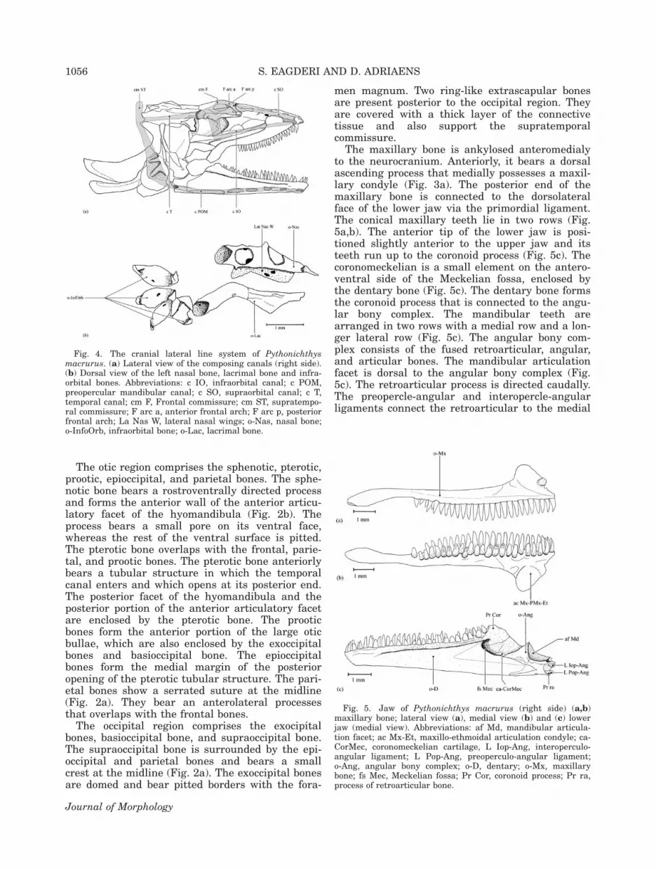
The maxillary bone is ankylosed anteromedialyto the neurocranium. Anteriorly, it bears a dorsalascending process that medially possesses a maxil-lary condyle (Fig. 3a). The posterior end of themaxillary bone is connected to the dorsolateralface of the lower jaw via the primordial ligament.The conical maxillary teeth lie in two rows (Fig.5a,b). The anterior tip of the lower jaw is posi-tioned slightly anterior to the upper jaw and itsteeth run up to the coronoid process (Fig. 5c). Thecoronomeckelian is a small element on the antero-ventral side of the Meckelian fossa, enclosed bythe dentary bone (Fig. 5c). The dentary bone formsthe coronoid process that is connected to the angu-lar bony complex. The mandibular teeth arearranged in two rows with a medial row and a lon-ger lateral row (Fig. 5c). The angular bony com-plex consists of the fused retroarticular, angular,and articular bones. The mandibular articulationfacet is dorsal to the angular bony complex (Fig.5c). The retroarticular process is directed caudally.The preopercle-angular and interopercle-angularligaments connect the retroarticular to the medial
Fig. 4. The cranial lateral line system of Pythonichthysmacrurus. (a) Lateral view of the composing canals (right side).(b) Dorsal view of the left nasal bone, lacrimal bone and infra-orbital bones. Abbreviations: c IO, infraorbital canal; c POM,preopercular mandibular canal; c SO, supraorbital canal; c T,temporal canal; cm F, Frontal commissure; cm ST, supratempo-ral commissure; F arc a, anterior frontal arch; F arc p, posteriorfrontal arch; La Nas W, lateral nasal wings; o-Nas, nasal bone;o-InfoOrb, infraorbital bone; o-Lac, lacrimal bone.
Fig. 5. Jaw of Pythonichthys macrurus (right side) (a,b)maxillary bone; lateral view (a), medial view (b) and (c) lowerjaw (medial view). Abbreviations: af Md, mandibular articula-tion facet; ac Mx-Et, maxillo-ethmoidal articulation condyle; ca-CorMec, coronomeckelian cartilage, L Iop-Ang, interoperculo-angular ligament; L Pop-Ang, preoperculo-angular ligament;o-Ang, angular bony complex; o-D, dentary; o-Mx, maxillarybone; fs Mec, Meckelian fossa; Pr Cor, coronoid process; Pr ra,process of retroarticular bone.
1056 S. EAGDERI AND D. ADRIAENS
Journal of Morphology

faces of the preopercle and interopercle bones,respectively.
The suspensorium consists of four bones, i.e.,the hyomandibula, quadrate, palatopterygoid, andpreopercle. The preopercle is described as part ofthe opercular series (see below). The symplecticbone is fused to the posterior portion of the quad-rate. The hyomandibula bears a distinct ridge onits lateral surface. The hyomandibula articulatesdorsally with the neurocranium via two articularcondyles, the anterior one situated on its antero-dorsal corner (Fig. 3b). The long, posterior suspen-sorial articular condyle is formed by a caudallyextended posterodorsal process of the hyomandib-ula (Fig. 3b). The opercular bone articulates with aventrocaudally directed condyle, at the posterioredge of the hyomandibula. The hyomandibula-quadrate axis is directed vertically, thus position-ing the quadrate-mandibular articulation posteriorto the orbit. The palatopterygoid is a broad boneand only its posterior end is connected to the quad-rate by connective tissue (Fig. 3b). The hyomandib-ular-palatopterygoid ligament connects the dorsalmargin of the palatopterygoid to the hyomandibu-lar ridge.
The opercular series comprises five elements(opercle, preopercle, interopercle, subopercle, andsupra-preopercle). The preopercle bears a tubular,arch-like structure with an anterodorsal projectionon its anterolateral face (Fig. 3b). This arch-likestructure, which is covered by thick connective tis-sue, encloses a part of the preoperculo-mandibularcanal (Figs. 3b and 4a). The posterior portion ofthe preopercle overlaps and is connected to the an-terior half of the interopercle. The preopercle-angular ligament connects the anteromedial rim ofthe preopercle to the angular bony complex. Theinteropercle is a relatively large rectangular bone.The interopercle-angular ligament is attached tothe anteromedial face of the interopercle and con-tinues posteriorly to the posterior end of the poste-rior ceratohyal bone. The subopercle is situatedventral to the opercle, to which it is firmlyattached. Connective tissue also attaches it to theinteropercle. A small ventral process is present onthe rostroventral end of the opercle and acts asthe attachment point for the dilatator operculimuscle. The supra-preopercular bone, which enclo-ses part of the preoperculo-mandibular canal, liesabove the anterior portion of the opercle (Fig. 3b).
The hyoid complex consists of the unpaired me-dian basihyal and urohyal bones and paired ante-rior and posterior ceratohyal bones (see Fig. 6).This expansion, comprising the urohyal articularfacets, lies ventrally against the anterior end ofthe ceratohyal bones, and the basihyo-ceratohyalligaments connects the lateroventral faces of thebasihyal bone to the ventral face of the anteriorceratohyal bones (Fig. 7a). The urohyal bone isexpanded anteriorly and is connected to the ante-
rior end of the anterior ceratohyal bones via smallurohyo-ceratohyal ligaments (Fig. 7a). Thebasihyal bone is a cylindrical bone, which isexpanded posteriorly (Fig. 6a). No separate hypo-hyal bone could be distinguished. The anteriorceratohyal bone is connected to the suspensoriumby means of the ceratohyo-hyomandibular liga-ment (Fig. 7a). A total of nine branchiostegal raysare supported by the anterior and posterior cera-tohyal bone (Fig. 6b). All of them articulate withthe posterior ceratohyal bone, with the exceptionof one. The branchiostegal rays curve dorsallyalong the ventral border of the interopercle andreach up to the caudal border of the opercle.
The cranial lateral line system comprises thesupraorbital, infraorbital, temporal and preoper-culo-mandibular canals, and the frontal and supra-temporal commissures. The ethmoid and adnasalcanals are absent. The cranial lateral line canalsare widened due to the arched bony elements thatenclose them. The nasal bone lies on the antero-dorsal face of the olfactory organ and supports theanterior portion of the supraorbital canal. Themain body of the nasal bone is tube-like, bearing aventrolaterally positioned wing (Fig. 4b). The fron-tal-nasal ligament connects the nasal bone to theanteroventral corner of the lateral expansion ofthe frontal bone (Fig. 7b). The posterior part of the
Fig. 6. Hyoid apparatus of Pythonichthys macrurus. (a) Dor-sal view of the basihyal and urohyal bones; (b) The anteriorand posterior ceratohyal bones in a medial view (right side).Abbreviations: af CH, ceratohyal articular facet; L BH-CH, basi-hyo-ceratohyal ligament; L CH-Hm, ceratohyo-hyomandibularligament; o-BH, basihyal bone; o-CH A, anterior ceratohyalbone; o-CH P, posterior ceratohyal bone; R Br, branchiostegalrays; o-UH, urohyal bone.
CEPHALIC MORPHOLOGY OF Pythonichthys macrurus 1057
Journal of Morphology
supraorbital canal is supported by the anteriorfrontal arch, which is expanded laterally (Fig. 4a).The frontal commissure is enclosed by the poste-rior frontal arches (Fig. 4a). Four infraorbitalbones seem merely to be lying in an arched posi-tion (Fig. 4b). The lacrimal and four infraorbitalbones support the infraorbital canal. The anteriorpart of the infraorbital canal is enclosed by the lac-rimal bone. The tubular lacrimal bone bears ananteriorly flattened part superimposing the maxil-lary dorsal process. Dorsally, this flattened part ofthe lacrimal bone bears a more narrow tubularstructure (Fig. 4b). The primordial ligament con-tinues anteriorly and connects to the posteriorpart of the lacrimal bone. The supra- and infraor-
bital canal anastomose in the front of the pteroticopening and continue into the temporal canal. Thepreoperculo-mandibular canal begins in the ven-troanterior tip of the dentary bone. It runs insidethe mandibula, which bears six pores (Fig. 4b).The preoperculo-mandibular canal is curved cau-dodorsally after exiting the mandibula and isenclosed by the arch-like structure of the preop-ercle (Fig. 4b). A supra-preopercular bone enclosesthe posterior portion of the preoperculo-mandibu-lar canal before anastomosing with the temporalcanal. The supratemporal commissure is enclosedby two ring-like extrascapular bones (Fig. 7b). De-spite existing pores on some bony elements thatenclose the lateral line system, such as those of
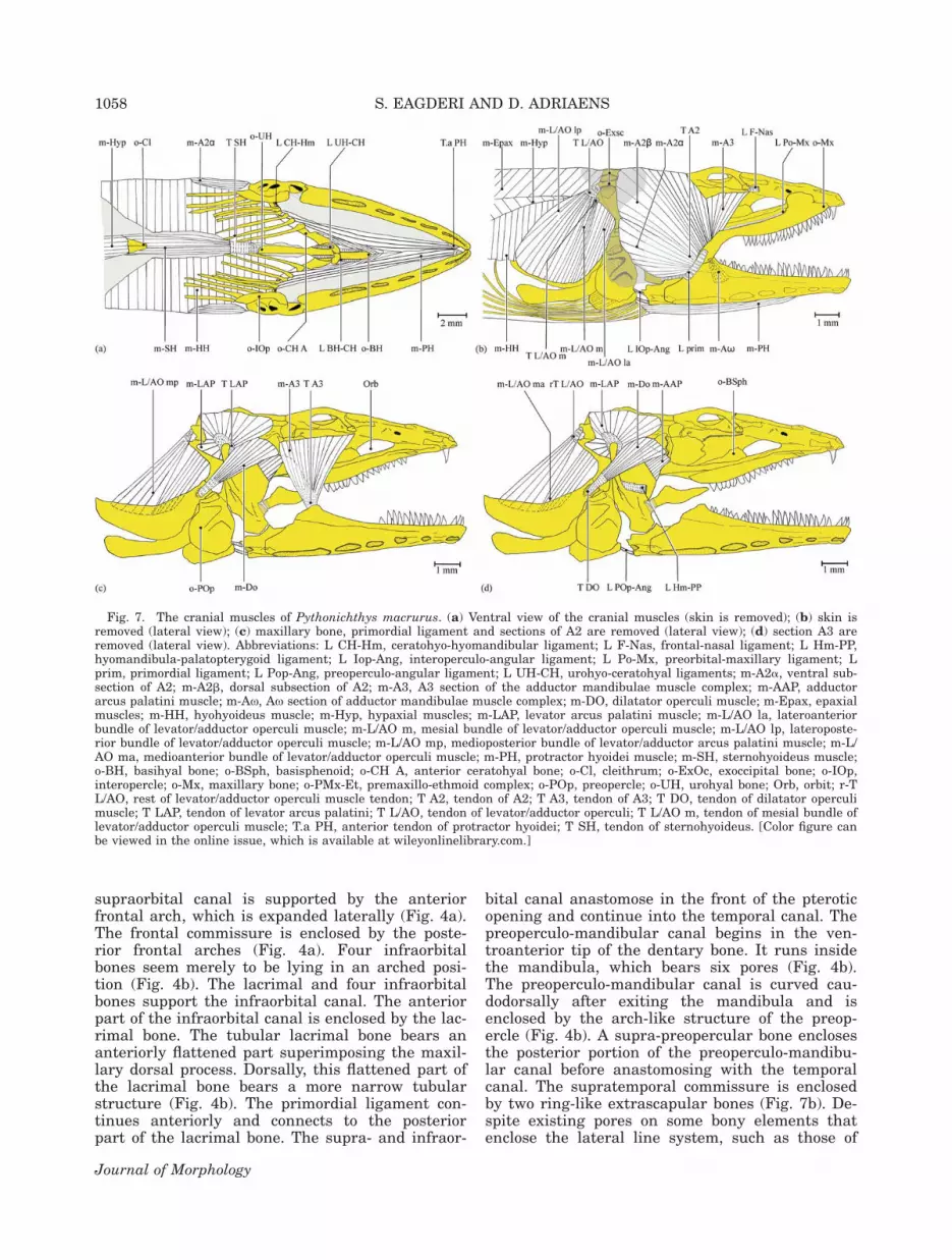
Fig. 7. The cranial muscles of Pythonichthys macrurus. (a) Ventral view of the cranial muscles (skin is removed); (b) skin isremoved (lateral view); (c) maxillary bone, primordial ligament and sections of A2 are removed (lateral view); (d) section A3 areremoved (lateral view). Abbreviations: L CH-Hm, ceratohyo-hyomandibular ligament; L F-Nas, frontal-nasal ligament; L Hm-PP,hyomandibula-palatopterygoid ligament; L Iop-Ang, interoperculo-angular ligament; L Po-Mx, preorbital-maxillary ligament; Lprim, primordial ligament; L Pop-Ang, preoperculo-angular ligament; L UH-CH, urohyo-ceratohyal ligaments; m-A2a, ventral sub-section of A2; m-A2b, dorsal subsection of A2; m-A3, A3 section of the adductor mandibulae muscle complex; m-AAP, adductorarcus palatini muscle; m-Ax, Ax section of adductor mandibulae muscle complex; m-DO, dilatator operculi muscle; m-Epax, epaxialmuscles; m-HH, hyohyoideus muscle; m-Hyp, hypaxial muscles; m-LAP, levator arcus palatini muscle; m-L/AO la, lateroanteriorbundle of levator/adductor operculi muscle; m-L/AO m, mesial bundle of levator/adductor operculi muscle; m-L/AO lp, lateroposte-rior bundle of levator/adductor operculi muscle; m-L/AO mp, medioposterior bundle of levator/adductor arcus palatini muscle; m-L/AO ma, medioanterior bundle of levator/adductor operculi muscle; m-PH, protractor hyoidei muscle; m-SH, sternohyoideus muscle;o-BH, basihyal bone; o-BSph, basisphenoid; o-CH A, anterior ceratohyal bone; o-Cl, cleithrum; o-ExOc, exoccipital bone; o-IOp,interopercle; o-Mx, maxillary bone; o-PMx-Et, premaxillo-ethmoid complex; o-POp, preopercle; o-UH, urohyal bone; Orb, orbit; r-TL/AO, rest of levator/adductor operculi muscle tendon; T A2, tendon of A2; T A3, tendon of A3; T DO, tendon of dilatator operculimuscle; T LAP, tendon of levator arcus palatini; T L/AO, tendon of levator/adductor operculi; T L/AO m, tendon of mesial bundle oflevator/adductor operculi muscle; T.a PH, anterior tendon of protractor hyoidei; T SH, tendon of sternohyoideus. [Color figure canbe viewed in the online issue, which is available at wileyonlinelibrary.com.]
1058 S. EAGDERI AND D. ADRIAENS
Journal of Morphology
the lower jaw, the cephalic lateral line systembears few tiny pores on the preoperculo-mandibu-lar canal. These are visible under high magnifica-tion, implying reduced head pores.
Myology of Pythonichthys macrurusMuscles of the cheek. The adductor mandibu-
lae complex, the most conspicuous muscle of thecheek, is subdivided into the sections A2, A3, andAx. The section A2 is subdivided into two subsec-tions: A2a and A2b. The A2a is the ventral ele-ment of the adductor mandibulae muscle complexand has a fleshy origin at the lateral face of thehyomandibula, quadrate, and the anterior rim ofthe preopercle; it has an additional origin as a ten-don from the posterolateral face of the hyomandib-ula. This tendon is also connected to the thick con-nective tissue, which covers the arch-like structureof the preopercle. The A2a inserts as a tendon onthe medial face of the coronoid process and as amuscle on the posteromedial face of the angularbony complex. The lateral face of the A2-tendonconnects to the posterior edge of the primordial lig-ament and inserts on the posteromedial face ofthe mandibula (Fig. 7b). The subsection A2b isthe largest and most posterodorsal element of theadductor mandibulae muscle complex. It has amuscular origin on the frontal, parietal, andsupraoccipital bones and inserts as a muscle andwith tendons on the dorsomedial face of theMeckelian fossa. A posterior portion of the subsec-tion A2b that is situated lateral to the levatorarcus palatini muscle, bulges posteriorly. The A3-section of the adductor mandibulae muscle com-plex has a fleshy origin from the ventral face ofthe lateral expansion of the frontal bone, the an-terolateral face of the sphenotic process and thepterosphenoid bone, and inserts as a tendon ontothe Meckelian fossa. The posterior fibers of thesection A3 are directed ventrally whereas its ante-rior fibers are directed more caudoventrally (Fig.7c). The section Ax arises as a tendon from the an-teroventral fibers of the subsection A2b. It insertsinto the Meckelian fossa along with the ventralfibers of the subsection A2b.
The levator arcus palatini muscle originates as atendon from the dorsal portion of the hyomandibu-lar ridge. Its fibers are spread radially and inserton the parietal bone, the supraoccipital bone, theposterior region of the pterotic bone, and the pos-terodorsal process of the hyomandibular bone (Fig. 7c).
The adductor arcus palatini is a rectangularmuscle with its fibers originating from the postero-ventral side of the pterosphenoid and the ventro-lateral face of the sphenotic bone. It inserts withfibers on both the lateral and medial face of theanterodorsal part of the hyomandibular bone.There is no insertion on the palatopterygoid. Itsdorsal fibers are covered by the anterior portion ofthe dilatator operculi muscle (Fig. 7d).
The adductor hyomandibulae muscle connectswith fibers to the basioccipital, prootic, and para-sphenoid bones and runs to the dorsomedial faceof the hyomandibular bone. Its anterior margin issituated at the level of the anterior margin of thehyomandibular bone.
The undifferentiated levator operculi and adduc-tor operculi muscles, which can be considered as asingle levator/adductor opercula muscle, directcaudoventraly (Fig. 7b). Based on different originsof muscle parts, the levator/adductor operculi mus-cle can be divided into an anterior, mesial, andposterior bundle. The anterior bundle has a fleshyorigin from the ventral edge of the posterodorsalprocess of the hyomandibular bone. Its lateralfibers insert on the dorsolateral face of the opercle,whereas the medial fibers insert on the dorsome-dial face of the opercle. The mesial bundle origi-nates as a tendon from the posterior end of theposterodorsal process of the hyomandibular boneand inserts with fibers on the dorsolateral face ofthe opercle (Fig. 7b). The posterior bundle origi-nates as a tendon from the posterior process of thepterotic bone and its lateral and medial fibersinsert on the dorsolateral and dorsomedial faces ofthe opercle, respectively (Fig. 7d).
The dilatator operculi is triangular in shape(Fig. 7d). It originates with muscle fibers from theposterolateral face of the sphenotic bone and ven-trolateral face of the pterotic bone. It inserts as atendon on the rostroventral process of the opercle.
Ventral muscles of the head. Left and righthalves of the protractor hyoidei muscle merge atthe midline of their length, but remain separated attheir ends. Each halves of this muscle originate asa tendon from the medial face of the dentary bone,just at the rear of the dental symphysis. They insertwith muscle fibers on the ventral faces of the ante-rior and posterior ceratohyal bones (Fig. 7a).
Left and right bundles of the sternohyoideusmuscle merge in the midline after originating withfibers from the entire anterior and the anterolat-eral margin of left and right cleithra. They inserton the posterior end of the urohyal bone via thesternohyoideus tendon (Fig. 7a).
The hyohyoideus muscle complex in teleosts gen-erally includes three components (the hyohyoideusinferioris, hyohyoideus abductor, and hyohyoideiadductores). However, in P. macrurus they are un-differentiated, merging anteriorly as a thin sac-like muscle sheet meeting their counterparts atthe ventral midline. Posteriorly, both muscle halvesremain separate at the end of the orobranchial cav-ity. This sac-like muscle interconnects the medialfaces of the branchiostegal rays, opercle, and subo-percle and reaches dorsally up to the horizontal sep-tum between the epaxial and hypaxial muscles.
Body muscles. The epaxial muscles insertdirectly on the epioccipital bones, dorsal rim of theexoccipital bone, and as an aponeurosis on the
CEPHALIC MORPHOLOGY OF Pythonichthys macrurus 1059
Journal of Morphology

supraoccipital bone. The dorsal fibers of the epax-ial muscles connect to the fascia of the adductormandibulae muscle complex and thick connectivetissue, which covers the ring-like extrascapularbones. The hypaxial muscles insert as an aponeu-rosis on the ventrocaudal border of the basioccipi-tal and exoccipital bones.
Two tongue-like appendages are present in theroof of the orobranchial mucosa (see Fig. 8). Eachappendage contains of a cavity with few, shortmuscle fibers inserting on its internal base. Thismay resemble a morphology similar to that of thepalatal organ of cypriniform fishes, and hence asimilar function in assisting the transport of foodcould be speculated (Sibbing et al., 1986, Hernandezet al., 2007). However, further research is requiredto elucidate the true nature of this organ.
DISCUSSIONCranial Specializations Related toHead-First Burrowing
Burrowing behavior is considered useful to usethe substratum for protection, crypsis, and feeding
purposes (Subramanian, 1984; Bozzano, 2003), andis observed to occur in various degrees in anguilli-form fishes. Pythonichthys macrurus appears toremain buried in the substratum except, perhaps,for brief excursions in search of food (Smith,1989a). Moringua edwardsi spends all its timeburrowed in the sand when it is immature(Gordon, 1954; Gosline, 1965), whereas adults ofthis species limit their fossorial behavior to whenthey leave their burrows during the night (Smith,1989b). Pisodonophis boro penetrates the substratetail-first but it is able to burrow head-first as well(Tilak and Kanji, 1969; Subramanian, 1984; Atkinsonand Taylor, 1991). It is known to burrow for shelterand to find food (Subramanian, 1984). Anguillaanguilla is a nonburrowing species (Tesch, 2003).
Eye reduction. Pythonichthys macrurus sharesseveral cranial features with two non-heterenche-lyid burrowing anguilliforms. One of these charac-ters is eye reduction. Eye reduction in ophichthidsand moringuids are considered to be adaptationsto head-first burrowing (De Schepper et al., 2005,2007). Eye reduction is observed in many other
Fig. 8. The orobranchial organ of Pythonichthys macrurus. (a) Two tongue-like appendages of the orobranchial organ onthe roof of the mouth; (b) Closer view of the two tongue-like appendages of the orobranchial organ; (c,d) sagittal section of theorobranchial organ (c) base region; (d) apex region. [Color figure can be viewed in the online issue, which is available atwileyonlinelibrary.com.]
1060 S. EAGDERI AND D. ADRIAENS
Journal of Morphology

fossorial elongated fishes such as Clariidae(Devaere et al., 2001, 2004), Mastacembelidae(Poll, 1973), and Anguilliformes (De Schepperet al., 2004, 2005, 2007; Aoyama et al., 2005). Thereduced eye of the Ophichthidae and Moringuidaehas been reported to be adaptations for head-firstburrowing (McCosker et al., 1989; Smith 1989b,c;De Schepper et al., 2005, 2007). The smaller eyesize, particularly in relation to the macrophthalmicancestral state of Anguilliformes (Bohlk, 1989a),can have a substantial effect on the spatial designof head (Boel, 1985). Eye reduction allows a caudo-ventral orientation of the anteromedial section ofthe adductor mandibulae muscle complex inP. boro and M. edwardsi (De Schepper et al., 2005,2007), and the reduction of the circumorbital bonesto small tubular bones in M. edwardsi, clariidsand ophichthids (Tilak and Kanji, 1969; McCoskeret al., 1989; Bozzano, 2003; Devaere et al., 2004;De Schepper et al., 2007). Eye reduction also hasbeen linked to the hypertrophy of the jaw musclein anguilliform clariids (Devaere et al., 2001).Hence, the reduction of the eye in P. macrurusmay be linked to the caudoventral orientation ofthe anteromedial section of the adductor mandibu-lae muscles complex (the section A3) and tubularcircumorbital bones. Furthermore, the opaque skinof the eye in P. macrurus may also be adaptive toburrowing. This feature has been reported inOphichthus rufus (Ophichthidae) as an adaptationto protect from mechanical injury when this eelroutinely enters and leaves a burrow (Bozzano,2003).
Widened cephalic lateral line canals.Because of the associated reduced vision, othersensory systems (such as olfactory, somatosensory,lateral line, and auditory systems) may becomemore important to provide environmental informa-tion (Gordon, 1954; Montgomery, 1989). Chemicalsensory cues may predominate in burrowingorganisms that their vision is considered poorlydeveloped (Pankurst and Lythgoe, 1983). Klageset al. (2002) has pointed out that finding prey fororganisms that usually wait for food by resting onthe sea floor or inside burrows is based on thedetection of the noise (the residual low-levelsound) produced by elastic waves at the water-seafloor interface. Unlike M. edwardsi, which has itscephalic lateral line canals widened into dermalcavities, P. macrurus has widened canals enclosedin the arched bony elements of the skull under theskin. This widened cephalic lateral line canalsmay function as a kind of sensory pads, which arestimulated mechanically during burrowing whenin contact with prey (De Schepper et al., 2005).Most of the widened canals of the cephalic lateralline system of P. macrurus lack external pores,except for the tiny pores of the preoperculo-man-dibular canal. Moringua edwardsi lacks any exter-nal pores of the cephalic lateral line system,
whereas tail-first burrower, P. boro, retains poresin all the canals of the cephalic lateral line sys-tem. The lack of head pores has the advantage ofavoiding the entering of sediment into the canalsduring head-first burrowing (De Schepper et al.,2005).
Gill opening. From hyomandibula-opercle artic-ulation, the opercle is orientated posteroventrallyin P. macrurus, M. edwardsi, and P. boro whereasthat one of A. anguilla is directed caudally. As aresult of this configuration, the gill opening is posi-tioned more ventrally in the burrowing species.This may prevent entering of sediment into gillcavity (Gosline, 1971; Smith, 1989c).
Different orientation of the anteriorsection of the adductor mandibulae musclecomplex. The hyomandibulo-quadrate axis ofP. macrurus, M. edwardsi, and P. boro is directeddorsoventrally, in contrast to the forwardlyinclined one in A. anguilla. As a result, the quad-rate-mandibular articulation is positioned poste-rior to the orbit. In addition to the small size ofeye, the posterior position of the quadrate-mandib-ular joint in these species also creates space allow-ing a caudoventral orientation of the anterior sec-tion of the adductor mandibulae muscle complex.The anterior fibers of the section A3 in P. macru-rus are directed caudoventrally, whereas its poste-rior fibers are directed vertically. An oppositeorientation of the different section of the adductormandibulae muscle complex (caudoventrally versusrostroventrally) can thereby help in preventing thedislocation of the mandibular joint, even when largeforces are exerted that induce torque forces (DeSchepper et al., 2007). This stabilization of the man-dibular joint may especially prevent the dislocationof the lower jaw during initial head-first penetrationconsidered as first stage of burrow construction(James et al., 1995).
Despite of a short and sharp snout, which is con-sidered to be an adaptation to head-first burrowing(McCosker et al., 1989; Smith, 1989b,c), the poste-rior position of the quadrate-mandibular joint alsoresults in a large mouth gap and a longer lowerjaw. A large gape and longer lower jaw combinedwith large teeth on the upper and lower jaws inP. macrurus suggest a potential for rapid mouthclosing and dealing with large prey items. Manybentic, lie-in-wait or cryptic predators have partic-ularly large mouths that can be opened and closedrapidly to grasp prey (Helfman et al., 1997; Mehtaand Wainwright, 2007; Mehta, 2009; Eagderi andAdriaens, 2010).
Skull shape and large insertion sites ofbody muscles on the neurocranium. The modi-fication of the skull into a solid conical structurewith sharp snout, as seen in P. macrurus and twoother fossorial species, M. edwardsi and P. boro,may facilitate burrowing, where power is provided
CEPHALIC MORPHOLOGY OF Pythonichthys macrurus 1061
Journal of Morphology
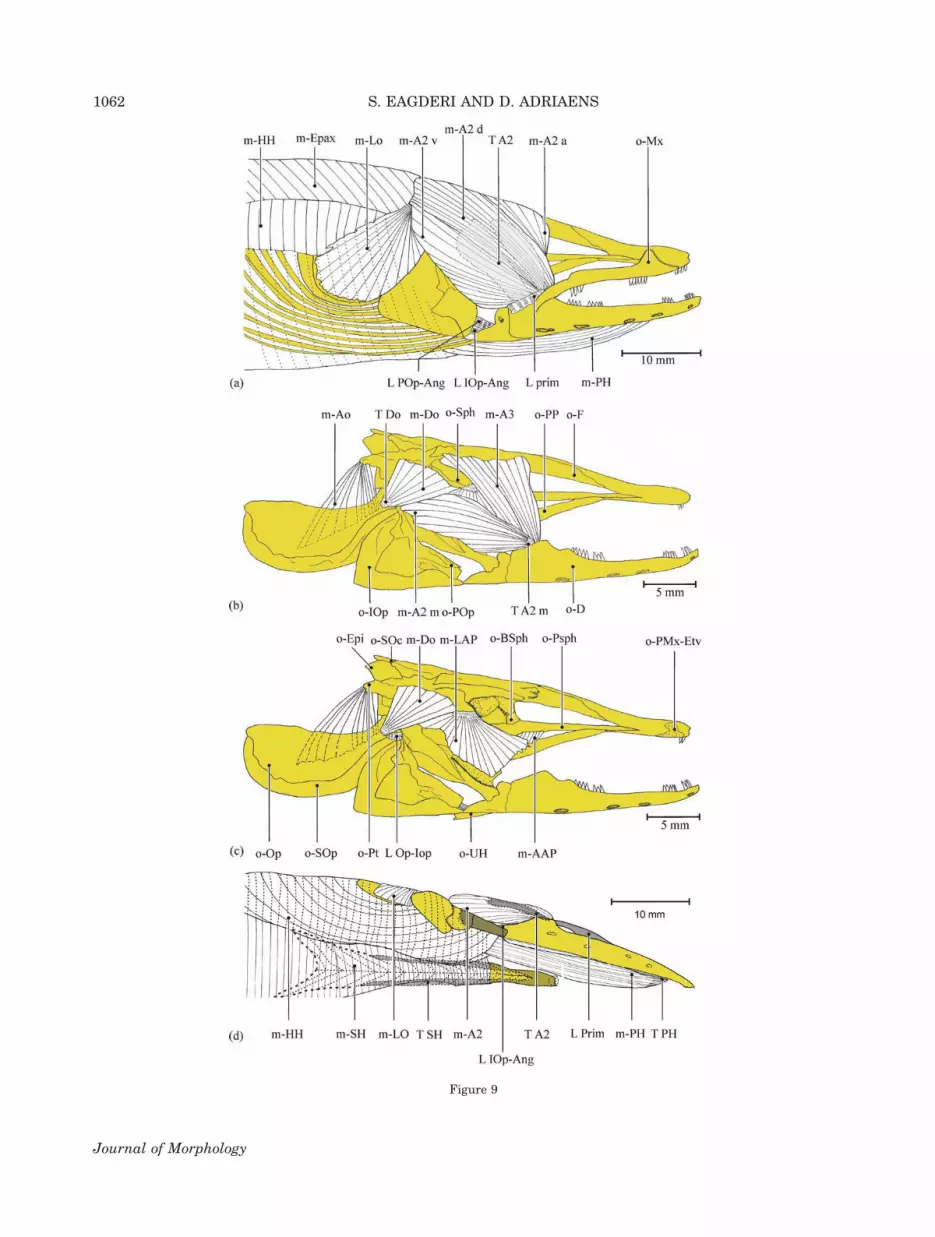
Figure 9
1062 S. EAGDERI AND D. ADRIAENS
Journal of Morphology

by the cylindrical body (Castle, 1968; Smith,1989b). Smith (1989b) mentions rapid movementsof the body, just beneath the surface, for subterra-nean hunting and feeding in burrowing Anguilli-formes. The large insertion sites of the epaxial andhypaxial muscles on the neurocranium (comprisingexoccipital, epioccipital, basioccipital, and supraoc-cipital bones), which are also connected to theadductor mandibulae muscle complex, in P. macru-rus can be considered an advantage for transfer-ring forces from the body onto the head. The phys-ical coupling between the jaw muscles and theepaxial muscles has been observed in M. edwardsiand P. boro (De Schepper et al., 2005, 2007). Bur-rowing fishes appear to use two main strategies:mouth excavation or compression and displace-ment of sediment by body movement (Atkinsonand Taylor, 1991). The head morphology ofP. macrurus suggest that the second strategy isthe burrowing method applied.
Variable Degree of Head-First BurrowingSpecializations in Anguilliformes
Compared with Anguilla anguilla, Moringuaedwarsi, and Pisodonophis boro, Pythonichthysmacrurus exhibits several unique characters suchas presence of the frontal arches, the preoperclewith its arch-like structures, the tubular and arch-like circumorbital bones, the arch-like suprapreo-percular bone, and two ring-like extrascapularbones, which support portions of the cephalic lat-eral line canals. These characters are all consid-ered synapomorphies for heterenchelyids (Belouze,2001).
Only M. edwarsi lacks a basisphenoid, andP. macrurus was the only anguillid in this study toexhibit a separate vomeral bone. Fusion of pre-maxillary, ethmoid, and vomeral bones, formingthe premaxillo-ethmovomerine complex in M.edwardsi, A. anguilla, and P. boro, is consideredsynapomorphic for Anguilliformes (Eaton, 1935;Gosline, 1980; Smith, 1989c). The palatopterygoidof A. anguilla is connected at both ends to the
quadrate bone and the premaxillo-ethmovomerinecomplex, respectively, whereas the palatopterygoidof M. edwardsi, P. macrurus, and P. boro attachesonly to the suspensorium. The pectoral fins arepresent in both A. anguilla and P. boro but areweakly developed in M. edwardsi. According toBohlke (1989a), all heterenchelyids lack pectoralfins, whereas moringuids have weakly developedpectoral fins. Body elongation combined with limb-lessness allows unhindered movement below thesubstrate surface (Gans, 1975; Pough et al., 1998).
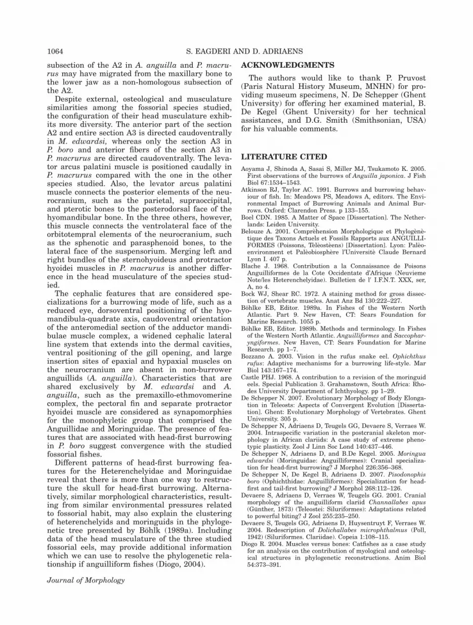
The adductor mandibulae muscle complex ofP. macrurus shows similar components but a dif-ferent configuration with that of the other fossorialspecies studied, except for the lack of the sectionA1 in M. edwardsi and P. boro. In basal actino-pterygian fishes, the section A1 is defined by itsdorsal position in the adductor mandibulae musclecomplex and its insertion on the maxillary bonethrough a long tendon or on the maxillary boneitself (Eaton, 1935; Winterbottom, 1974). The pres-ence of such a section A1 has been reported inM. edwardsi and P. boro (De Schepper et al., 2005,2007). In A. anguilla and P. macrurus, a small ten-dinous connection was found between the ventralsubsection of the section A2 and the maxillarybone via the primordial ligament. Based on theterminology of Winterbottom (1974) and regardingits main insertion on the lower jaw, this section inA. anguilla and P. macrurus was considered as asubsection of A2. Hence, the section A2 ofthe adductor mandibulae muscle complex ofA. anguilla consists of four subsections (A2a, A2d,A2v, and A2m; Fig. 9a,b,d). The homology of eachmuscle of the adductor mandibulae complex (vari-ously determined as a subdivision, section, or sub-section) is accepted on the basis of sharing anidentical insertion pattern onto the jaws, followingthe traditional nomenclature used for the adductormandibulae muscles (Vetter, 1878; Winterbottom,1974; Nakae and Sasaki, 2004). Migrations of themuscle insertions have likely occurred in evolu-tionary history of the elopomorphs, resulting inthe lumping of nonhomologous muscles under asingle name. Hence, the insertion of the ventral
Fig. 9. The cranial muscles of Anguilla anguilla. (a) Skin is removed (lateral view); (b) Sections A2 except the medial fibers ofsubsection A2m, hyohyoideus muscle, levator operculi muscle, epaxial muscles, hypaxial muscles, maxillary bone, and primordialligament are removed (lateral view); (c) Subsection A2m and subsection A3 are removed (lateral view); (d) Ventral view of the rightside of the cranial muscles (skin is removed; modified after De Schepper, 2007). Abbreviations: L Iop-Ang, interoperculo-angularligament; L Op-Iop, preoperculo-interopercular ligament; L prim, primordial ligament; L Pop-Ang, preoperculo-angular ligament;m-A2 a, anterior subsection of A2; m-A2 d, dorsal subsection of A2; m-A2 v, ventral subsection of A2; m-A2 m, medial subsection ofA2; A3 section of the adductor mandibulae muscle complex; m-AAP, adductor arcus palatini muscle; m-DO, dilatator operculi mus-cle; m-Epax, epaxial muscles; m-HH, hyohyoideus muscle; m-LAP, levator arcus palatini muscle; m-AO, adductor operculi muscle;m-LO m, levator operculi muscle; m-PH, protractor hyoidei muscle; m-SH, sternohyoideus muscle; o-BSph, basisphenoid; o-D, den-tary complex; o-Epi, epioccipital bone; o-F, frontal bone; o-IOp, interopercle; o-Op, opercle; POp, preopercle; o-PP, palatopterygoid;o-PSph, parasphenoid; o-Pt, pterotic bone; o-Mx, maxillary bone; SOc, supraoccipital bone; o-SOp, subopercle; o-Sph, sphenoticbone; o-UH, urohyal bone; T A2, tendon of A2; T A2 m, tendon of A2 m; T DO, tendon of dilatator operculi muscle; T PH, tendon ofprotractor hyoidei; T SH, tendon of sternohyoideus. [Color figure can be viewed in the online issue, which is available atwileyonlinelibrary.com.]
CEPHALIC MORPHOLOGY OF Pythonichthys macrurus 1063
Journal of Morphology
subsection of the A2 in A. anguilla and P. macru-rus may have migrated from the maxillary bone tothe lower jaw as a non-homologous subsection ofthe A2.
Despite external, osteological and musculaturesimilarities among the fossorial species studied,the configuration of their head musculature exhib-its more diversity. The anterior part of the sectionA2 and entire section A3 is directed caudoventrallyin M. edwardsi, whereas only the section A3 inP. boro and anterior fibers of the section A3 inP. macrurus are directed caudoventrally. The leva-tor arcus palatini muscle is positioned caudally inP. macrurus compared with the one in the otherspecies studied. Also, the levator arcus palatinimuscle connects the posterior elements of the neu-rocranium, such as the parietal, supraoccipital,and pterotic bones to the posterodorsal face of thehyomandibular bone. In the three others, however,this muscle connects the ventrolateral face of theorbitotempral elements of the neurocranium, suchas the sphenotic and parasphenoid bones, to thelateral face of the suspensorium. Merging left andright bundles of the sternohyoideus and protractorhyoidei muscles in P. macrurus is another differ-ence in the head musculature of the species stud-ied.
The cephalic features that are considered spe-cializations for a burrowing mode of life, such as areduced eye, dorsoventral positioning of the hyo-mandibula-quadrate axis, caudoventral orientationof the anteromedial section of the adductor mandi-bulae muscle complex, a widened cephalic lateralline system that extends into the dermal cavities,ventral positioning of the gill opening, and largeinsertion sites of epaxial and hypaxial muscles onthe neurocranium are absent in non-burroweranguillids (A. anguilla). Characteristics that areshared exclusively by M. edwardsi and A.anguilla, such as the premaxillo-ethmovomerinecomplex, the pectoral fin and separate protractorhyoidei muscle are considered as synapomorphiesfor the monophyletic group that comprised theAnguillidae and Moringuidae. The presence of fea-tures that are associated with head-first burrowingin P. boro suggest convergence with the studiedfossorial fishes.
Different patterns of head-first burrowing fea-tures for the Heterenchelyidae and Moringuidaereveal that there is more than one way to restruc-ture the skull for head-first burrowing. Alterna-tively, similar morphological characteristics, result-ing from similar environmental pressures relatedto fossorial habit, may also explain the clusteringof heterenchelyids and moringuids in the phyloge-netic tree presented by Bohlk (1989a). Includingdata of the head musculature of the three studiedfossorial eels, may provide additional informationwhich we can use to resolve the phylogenetic rela-tionship if anguilliform fishes (Diogo, 2004).
ACKNOWLEDGMENTS
The authors would like to thank P. Pruvost(Paris Natural History Museum, MNHN) for pro-viding museum specimens, N. De Schepper (GhentUniversity) for offering her examined material, B.De Kegel (Ghent University) for her technicalassistances, and D.G. Smith (Smithsonian, USA)for his valuable comments.
LITERATURE CITED
Aoyama J, Shinoda A, Sasai S, Miller MJ, Tsukamoto K. 2005.First observations of the burrows of Anguilla japonica. J FishBiol 67:1534–1543.
Atkinson RJ, Taylor AC. 1991. Burrows and burrowing behav-iour of fish. In: Meadows PS, Meadows A, editors. The Envi-ronmental Impact of Burrowing Animals and Animal Bur-rows. Oxford: Clarendon Press. p 133–155.
Boel CDN. 1985. A Matter of Space [Dissertation]. The Nether-lands: Leiden University.
Belouze A. 2001. Comprehension Morphologique et Phylogene-tique des Taxons Actuels et Fossils Rapports aux ANGUILLI-FORMES (Poissons, Teleosteens) [Dissertation]. Lyon: Paleo-environment et Paleobiosphere l’Universite Claude BernardLyon I. 407 p.
Blache J. 1968. Contribution a la Connaissance de PoisonsAnguilliformes de la Cote Occidentate d’Afrique (NeuviemeNote/les Heterenchelyidae). Bulletien de l0 I.F.N.T. XXX, ser,A, no 4.
Bock WJ, Shear RC. 1972. A staining method for gross dissec-tion of vertebrate muscles. Anat Anz Bd 130:222–227.
Bohlke EB, Editor. 1989a. In Fishes of the Western NorthAtlantic. Part 9. New Haven, CT: Sears Foundation forMarine Research. 1055 p.
Bohlke EB, Editor. 1989b. Methods and terminology. In Fishesof the Western North Atlantic. Anguilliformes and Saccophar-yngiformes. New Haven, CT: Sears Foundation for MarineResearch. pp 1–7.
Bozzano A. 2003. Vision in the rufus snake eel. Ophichthusrufus: Adaptive mechanisms for a burrowing life-style. MarBiol 143:167–174.
Castle PHJ. 1968. A contribution to a revision of the moringuideels. Special Publication 3. Grahamstown, South Africa: Rho-des University Department of Ichthyology. pp 1–29.
De Schepper N. 2007. Evolutionary Morphology of Body Elonga-tion in Teleosts: Aspects of Convergent Evolution [Disserta-tion]. Ghent: Evolutionary Morphology of Vertebrates. GhentUniversity. 305 p.
De Schepper N, Adriaens D, Teugels GG, Devaere S, Verraes W.2004. Intraspecific variation in the postcranial skeleton mor-phology in African clariids: A case study of extreme pheno-typic plasticity. Zool J Linn Soc Lond 140:437–446.
De Schepper N, Adriaens D, and B.De Kegel. 2005. Moringuaedwardsi (Moringuidae: Anguilliformes): Cranial specializa-tion for head-first burrowing? J Morphol 226:356–368.
De Schepper N, De Kegel B, Adriaens D. 2007. Pisodonophisboro (Ophichthidae: Anguilliformes): Specialization for head-first and tail-first burrowing? J Morphol 268:112–126.
Devaere S, Adriaens D, Verraes W, Teugels GG. 2001. Cranialmorphology of the anguilliform clariid Channallabes apus(Gunther, 1873) (Teleostei: Siluriformes): Adaptations relatedto powerful biting? J Zool 255:235–250.
Devaere S, Teugels GG, Adriaens D, Huysentruyt F, Verraes W.2004. Redescription of Dolichallabes microphthalmus (Poll,1942) (Siluriformes. Clariidae). Copeia 1:108–115.
Diogo R. 2004. Muscles versus bones: Catfishes as a case studyfor an analysis on the contribution of myological and osteolog-ical structures in phylogenetic reconstructions. Anim Biol54:373–391.
1064 S. EAGDERI AND D. ADRIAENS
Journal of Morphology

Eagderi S, Adriaens D. (2010). Head morphology of the DuckbillEel, Hoplunnis punctata (Regan, 1915; Nettastomatidae:Anguilliformes): A case of jaw elongation. Zool, doi: 10.1016/j.zool.2009.09.004.
Eaton TH Jr. 1935. Evolution of the upper jaw mechanisme inteleost fishes. J Morphol 58:157–72.
Gans C. 1975. Tetrapod limblessness: Evolution and functionalcorollaries. Anim Zool 15:455–467.
Gordon SM. 1954. The eel genus Stilbiscus. Copeia 1:11–15.Gosline WA. 1965. Teleostean phylogeny. Copeia 1:186–194.Gosline WA. 1971. Functional morphology and classification of
teleostean fishes. Honolulu, H1: University Press of Hawaii.208 p.
Gosline WA. 1980. The evolution of some structural systemswith reference to the interrelationships of modern lower tele-ostean fish groups. Jap J Ichth 21:1–24.
Hanken J, Wassersug R. 1981. The visible skeleton. A new dou-ble-stain technique reveals the nature of the ‘‘hard’’ tissues.Funct Photogr 16:22–26.
Helfman,GS, Collette BB, Facey DE. 1997. The Diversity ofFishes. Malden, MA: Blackwell Science, Inc. 507 p.
Hernandez LP, Bird NC, Staab KL. 2007. Using zebrafish toinvestigate cypriniform evolutionary novelties: Functional de-velopment and evolutionary diversification of the kinethmoid.J Exp Zool 308B:625–641.
James R, Atkinson A, Roger S, Pullin V. 1995. Observations onthe burrows and burrowing behavior of the red band-fish.Sepola rubescens L. Mar Ecol 17(1–3):23–40.
Klages M, Muyakshin S, Soltwedel T, Arntz WE. 2002. Mecha-noreception, a possible mechanism for food fall detection indeep-see scavengers. Deep-SEA Res I 49:143–155.
McCosker JE, Bohlke EB, Bohlke JE. 1989. Family Ophichthi-dae. In: Bohlke EB, editor. Fishes of the Western North At-lantic. New Haven, CT: Sears Foundation for MarineResearch. pp 254–412.
Mehta RS. 2009. Ecomorphology of the moray bite: Relationshipbetween dietary extremes and morphological diversity. Phys-iol Biochem Zool 82:90–103.
Mehta RS, Wainwright PC. 2008. Functional morphology of thepharyngeal jaw apparatus in moray eels. J Morphol 269:604–619.
Montgomery JC. 1989. Lateral line detection of planktonic prey.In: Coombs S, Gorner P, Munz H, editors. The Mechanosen-sory Lateral Line. New York: Springer-Verlag. Chapter 28.
Nakae M, Sasaki K. 2004. Homologies of the adductor mandibu-lae muscles in Tetraodontiformes as indicated by nervebranching patterns. Ichthyol Res 51:327–336.
Nelson JS. 2006. Fishes of the World, Forth edition. Hoboken,NJ: Wiley. 601 p.
Obermiller LE, Pfeiler E. 2003. Phylogenetic relationships ofelopomorph fishes inferred from mitochondrial ribosomalDNA sequences. Mol Phylogenet Evol 26:202–214.
Pankurst NW, Lythgoe JN. 1983. Changes in vision and olfac-tion during sexual maturation in the European eel. J FishBiol. 23:229–240.
Patterson C. 1975. The braincase of pholidophorid and leptole-pid fishes, with a review of the actinopterygian braincase.Philos Trans R Soc London B Biol Sci 269:275–579.
Poll M. 1973. Les yeux des poissons aveugles Africains et deCaecomastacembelus brichardi Poll en particulier. Ann duSpeleologie 28:221–230.
Pough HF, Andrews RM, Cadle JE, Crump ML, Savitzky AH,Wells KD. 1998. Body support and locomotion. In: Pough HF,editor. Herpetology. New Jersey: Prentice Hall. Chapter 8.
Rojo AL. 1991. Dictionary of Evolutionary of Fish Osteology.Florida: CRC Press. 273 p.
Sibbing FA, Osse JWM, Terlouw A. 1986. Food handling in thecarp (Cyprinus carpio): its movement patterns, mechanismsand limitations. J Zool Lond A 210:161–203.
Smith DG. 1989a. Family Heterenchelyidae. In: Bohlke EB, edi-tor. Fishes of the Western North Atlantic. Anguilliformes andSaccopharyngiformes. New Haven, CT: Sears Foundation forMarine Research. pp 48–54.
Smith DG. 1989b. Family Moringuidae. In: Bohlke EB, editor.Fishes of the Western North Atlantic. Anguilliformes andSaccopharyngiformes. New Haven, CT: Sears Foundation forMarine Research. pp 55–71.
Smith DG. 1989c. Family Congridae. In: Bohlke EB, editor.Fishes of the Western North Atlantic. Anguilliformes andSaccopharyngiformes. New Haven, CT: Sears Foundation forMarine Research. pp 460–567.
Smith DG, Castle PHJ. 1972. The eel genus Neoconger Girard:Systematics, osteology, and life history. Bull Mar Sci 22:196–249.
Subramanian A. 1984. Burrowing behavior and ecology of thecrab-eating Indian snake eel Pisodonophis boro. Environ BiolFish 10:195–202.
Tesch F-W. 2003. The Eel. Third edition. Oxford: Blackwell Sci-ence.408 p.
Tilak R, Kanji SK. 1969. Studies on the osteology of Pisodono-phis boro (Hamilton). Gegenbaurs Morphol Jahrb 113:501–523.
Vetter B. 1878. Untersuchungen zur vergleichende anatomieder kiemen und kiefermuskulatur der fische. II. JenaischeZeitschrift fur Naturwissenschaft 12:431–550.
Winterbottom R. 1974. A descriptive synonymy of the striatedmuscles of the Teleostei. Proc Acad Nat Sci Phil 125:225–317.
CEPHALIC MORPHOLOGY OF Pythonichthys macrurus 1065
Journal of Morphology