cellular and molecular mechanisms in chronic obstructive pulmonary disease: an overview
TRANSCRIPT

REVIEW
Cellular and molecular mechanisms in chronic obstructive pulmonary disease:an overview
A. Di Stefano*, G. Caramoriw, F. L. M. Ricciardoloz, A. Capelli*, I. M. Adcock§ and C. F. Donner*
*Salvatore Maugeri Foundation, IRCCS, Medical Center of Rehabilitation, Division of Pulmonary Disease, Veruno (NO), Italy, wCentro di Ricerca suAsma e BPCO, Universita di Ferrara, Ferrara, Italy, zGaslini Institute, Pulmonary Department, Genoa, Italy and §Department of Thoracic Medicine,National Heart and Lung Institute, Imperial College, London, UK
SummaryIn the last decade, the analysis of bronchial biopsies and lung parenchyma obtained from chronic
obstructive pulmonary disease (COPD) patients compared with those from smokers with normal
lung function and non-smokers has provided new insights on the role of the different inflammatory
and structural cells, their signalling pathways and mediators, contributing to a better knowledge of
the pathogenesis of COPD. This review summarizes and discusses the lung pathology of COPD
patients with emphasis on inflammatory cell phenotypes that predominate in different clinical
conditions. In bronchial biopsies, a cascade of events takes place during progression from mild-to-
severe disease. T lymphocytes, particularly CD81 cells and macrophages are the prevalent
inflammatory cells in the lung of healthy smokers and patients with mild COPD, while total and
activated neutrophils predominate in severe COPD. The number of CD41, CD81 cells and
macrophages expressing nuclear factor-kappa B (NF-kB), STAT-4 and IFN-g proteins as well as
endothelial adhesion molecule-1 in endothelium is increased in mild/moderate disease. In contrast,
activated neutrophils (MPO1 cells) and increased nitrotyrosine immunoreactivity develops in severe
COPD. In bronchial biopsies obtained during COPD exacerbations, some studies have shown an
increased T cell and granulocyte infiltration. Regular treatment with high doses of inhaled
glucocorticoids does not significantly change the number of inflammatory cells in bronchial biopsies
from patients with moderate COPD. The profile in lung parenchyma is similar to bronchial biopsies.
‘Healthy’ smokers and mild/moderate diseased patients show increased T lymphocyte infiltration in
the peripheral airways. Pulmonary emphysema is associated with a general increase of inflammatory
cells in the alveolar septa. The molecular mechanisms driving the lymphocyte and neutrophilic
prevalence in mild and severe disease, respectively, needs to be extensively studied. Up-regulation of
pro-inflammatory transcription factors NF-kB and STAT-4 in mild, activated epithelial and
endothelial cells in the more severe disease may contribute to this differential prevalence of
infiltrating cells.
Introduction
Chronic obstructive pulmonary disease (COPD) can bedefined as chronic irreversible airflow limitation confirmedby spirometry [1–3] and is characterized by a slowlyprogressive and irreversible deterioration in lung function[1–3]. Cigarette smoking is the major risk factor for thedevelopment of COPD and cigarette smokers constitute over90% of COPD patients in developed countries [1–3]. Thepathological hallmarks of COPD are destruction of the lungparenchyma, which characterizes pulmonary emphysema,inflammation of the peripheral airways, which characterizesrespiratory bronchiolitis, and inflammation of the centralairways. Pathological studies show that inflammation inCOPD occurs in the central and peripheral airways (bronch-ioles) and lung parenchyma.
We will focus our attention on studies that have analysedbronchial biopsies or specimens from bronchial rings andperipheral lung tissue obtained during lung resection for aperipheral carcinoma (see Tables 1–3). Data obtained on lungparenchyma by lung volume reduction surgery (LVRS) per-formed in patients with emphysema have also been report-ed because they represent almost the only source of tissuespecimens from severely diseased patients with pulmonaryemphysema.It should be noted that in the last 15 years, the definition of
COPD in the national and international guidelines haschanged many times [1–4]. This could explain some dis-crepancies in results observed in the studies reviewed here (seeTables 1–3).Recent studies performed on bronchial biopsies have fo-
cused on characterizing the inflammatory process and struc-tural alterations developing in stable mild/moderate COPD[5–12] and in the mild forms of COPD during exacerbations[13, 14]. The severe disease state has also been recently investi-gated [10, 11]. Bronchial biopsy changes after corticosteroidtreatment of stable moderate COPD have also been evaluated
Correspondence: Antonino Di Stefano, Salvatore Maugeri Foundation,
IRCCS, Medical Center of Rehabilitation, Division of Pulmonary Disease,
Via Revislate 13, 28010 Veruno (NO), Italy. E-mail: [email protected]
Clin Exp Allergy 2004; 34:1156–1167 doi:10.1111/j.1365-2222.2004.02030.x
r 2004 Blackwell Publishing Ltd1156

[15]. Structural alterations in the peripheral airways andparenchyma have been investigated mostly in mild forms ofCOPD and, more rarely, in severe disease. These studies onthe pathology of the lung in this disease have contributed sig-nificantly to the present knowledge on the structural alter-ations associated to airflow limitation in different clinicalconditions. Previous papers have extensively reviewed thestructural alterations occurring in the lung of patients withCOPD [16–18]. However, these have not focused on changesoccurring with different degrees of disease severity [16–20]. Inthe last few years, studies on this topic have made important
contributions that have extended the previous notions on theinflammatory cell prevalence in the large and small airwaysand parenchyma. Furthermore, important preliminary find-ings are, at present, available on the molecular mechanismsinvolved in the development of COPD and pulmonary em-physema. Planning of well-focused future studies requires aclear preliminary picture on the inflammatory cells andmediators that predominate in COPD at different clinicalstages and after pharmacologic treatment. We review here thecontributions made over the last � 15 years, based on theanalysis of bronchial biopsies of COPD patients in different
Table 1. Variations of inflammatory cells and markers of inflammation in the bronchial submucosa of COPD patients, control smokers and non-smokers
" , significantly increased values in comparison with that indicated by ! ; ! , basal values or values non-significantly changed; # , significantly decreased
values in comparison with that indicated by ! .
Numbers close to the arrows indicate references.
ELAM-1, endothelial adhesion molecule-1; NK, natural killer; NF-kB, nuclear factor-kappa B; STAT-4, signal transducer and activators of transcription; MPO,
myeloperoxidase; NT, nitrotyrosine; COPD, chronic obstructive pulmonary disease.
Table 2. Variations of inflammatory cells and markers of inflammation in the central and peripheral airways obtained from surgical specimens of COPD patients,
chronic bronchitics with normal lung function and control smokers
Peripheral airways(< 3mm ∅)
Central airways
→→
↑
↑→
Totalinflam.
→
−
↑
TGF-β1
→
−
↑→
CD8
→
−
→
CD4
→
−
↑→
CD68
→
−
→
Neutr
→→→→→→→Control*smokers
↓→
→↓→→→→Mild/moderate*COPD
↑→↑−−−−Chronic*bronchitis
with normal FEV1
Total inflam.
IL-5IL-4CD8CD4CD68Neutr
11
11
11
1
1,3
1,3
2 2 2 2
2 2 2 2
2
4,5,7,9
4,5,7,9 4,5,6,7,9 4,5,6,7,9 4,5,6,7,9 4,5,6,7,8,96,10
66,9
4,5,7
4,5,6,7,9
5,6,7,9
4 4
5,6,7,9
8
→10
1, from reference [26]; 2, from reference [56]; 3, from reference [55]; 4, from reference [60]; 5, from reference [61]; 6, from reference [63]; 7, from reference [62];8, from reference [57]; 9, from reference [67]; 10, from reference [64]
" , significantly increased values in comparison with that indicated by ! ; ! , basal values or values non-significantly changed; # , significantly decreased
values in comparison with that indicated by ! .
Numbers close to the arrows indicate references.*Specimens obtained from resected lungs for localized tumours.
FEV1, forced expiratory volume in 1 s; COPD, chronic obstructive pulmonary disease.
Cellular and molecular mechanisms in COPD 1157
r 2004 Blackwell Publishing Ltd, Clinical and Experimental Allergy, 34:1156–1167

clinical stages and after steroid treatment and of specimensfrom resected lung in COPD patients with stable disease.
Bronchial biopsies
Bronchial biopsies in stable chronic obstructive pulmonarydisease
Mild/moderate chronic obstructive pulmonary diseasevs. control smokers and non-smokers Analysis of inflam-matory cell infiltration in bronchial biopsies of patients withmild/moderate COPD shows an increased inflammatory cellinfiltration in comparison with control non-smokers [5–9]. Allauthors agree on the observation that T lymphocytes, mainlyCD81 cells, and macrophages (CD681 cells) predominate inthe bronchial mucosa of these patients [5–9]. When mild/moderate COPD patients are compared with control smo-kers, matched for age and smoking habit, we reported nosignificant differences in the numbers of CD31 and CD81
cells in the submucosa [21]. Smokers with normal lungfunction also showed, though to a lesser extent, increasednumbers of CD31 and CD81 cells compared with controlnon-smokers [11, 21] (Table 1). These data suggest that the Tlymphocyte increases may be an effect of smoking that occursin a wide range of smokers. This rises doubts as to the validityof the O’Shaughnessy’s speculation that increases in CD81
cells are restricted to a small subgroup of smokers whodevelop airflow limitation [22, 23]. An increased presence ofCD81 cells had also been previously documented in thebronchoalveolar lavage (BAL) and blood of smokers [24, 25].However, the CD31 and particularly the CD81 functionalactivity remains under investigation. Zhu et al. [26] reportedthat in central airways from surgical specimens of smokerswith chronic bronchitis, 15% of CD81 cells coexpress TNF-aprotein. Perforin and granzyme as well as Fas-L-mediated
cytotoxicity, potentially expressed by CD81 cells, have notbeen studied in bronchial biopsies or surgical specimens ofCOPD patients. Liu et al. [27] reported a TNF-a-dependentas well as perforin and Fas-L-independent cytolitic activity ofmice CD81 cells against cultured alveolar epithelial cells [27].If these in vitro observations are confirmed by further in vivostudies, cytotoxicity of CD81 cells in the bronchial mucosa ofsmokers could be restricted to a limited percentage of CD81
cells (15% in Zhu’s study) and could depend on theproduction and release of TNF-a protein. The question,however, of the role and functions of increased CD81 cellspopulating the bronchial mucosa of smokers remains, at leastin part, open. The results of ongoing trials of anti-TNF-atherapy in COPD may help clarify the role of this cytokine.
The chemokine receptor CCR5, preferentially expressedby T-helper type 1 (Th1)/T-cytotoxic (Tc1) cells producingIFN-g, was reported to be increased in mild/moderate diseasein comparison with control smokers [11], suggesting that,at variance with asthma, a prevalent Th1/Tc1 immuno-surveillance develops in mild/moderate COPD patients instable conditions. The expression of the transcription factorsignal transducer and activators of transcription (STAT)-4 iscritical for the differentiation of Th1/Tc1 cells [28, 29]. Werecently reported increased STAT-4 nuclear expression inbronchial epithelium and submucosa of mild/moderateCOPD patients in comparison with both control smokersand non-smokers [30]. STAT-4 nuclear expression correlatedsignificantly with the number of IFN-g1, CD31 and CD41
cells but not with CD81 cells in the submucosa of smokers:50% of CD41, one-third of CD81 and one-third of CD681
cells in the bronchial submucosa coexpressed the STAT-4protein. These data on the expression of the transcriptionfactor STAT-4 in COPD patients reinforce the notion of aprevalent Th1/Tc1 immunological response in mild/moderatedisease.
Table 3. Variations of inflammatory cells in the lung parenchyma obtained from surgical specimens of mild/moderate COPD patients, smokers with normal lung
function and control non-smokers
−−→→→→→→Control*non-smokers
→→→→↑↑→
Smokers*with
normalFEV1
Totalinflam.
→↑→→→Mild/moderate*COPD
TGF-β1EosinCD8CD4CD3CD68Neutr
43
2,3 2 2,3 2
2
2
1,2,32,3
2,3
1 1
2,3 1,2 1,3 2,3 2,3 2
↑4
→3
→
→
3
2,3
↑3
→3
↑1
→
1, from reference [68]; 2, from reference [70]; 3, from reference [71]; 4, from reference [72]
" , significantly increased values in comparison with that indicated by ! ; ! , basal values or values non-significantly changed; # , significantly decreased
values in comparison with that indicated by ! .
Numbers close to the arrows indicate references.*Specimens obtained from resected lungs for localized tumours.
FEV1, forced expiratory volume in 1 s; COPD, chronic obstructive pulmonary disease.
1158 A. Di Stefano et al.
r 2004 Blackwell Publishing Ltd, Clinical and Experimental Allergy, 34:1156–1167
The demonstration that the production of IL-4 in the glandsof chronic bronchitics is independent of CD81 lymphocytes,as reported by Zhu et al. [26], again suggests that CD41
lymphocytes could be mainly involved in the Th2 cytokineresponse associated with hypersecretion of mucous glands insmokers. In the light of these results, the Th1 or Th2 responseinvolving CD41 cells, may be functionally important inCOPD, even though their relative number has not beenreported as increased in comparison with control subjects.The epithelial and nuclear expression of the transcription
factor nuclear factor-kappa B (NF-kB) (p65 protein) wasincreased in mild/moderate COPD and, to a lesser extent, incontrol smokers in comparison with control non-smokers[21]. We reported that approximately one-third of CD41 andCD81 T lymphocytes coexpressed the p65 NF-kB subunit inthe bronchial biopsies of COPD patients. Nuclear expressionof NF-kB can modulate the transcription of a number of pro-inflammatory cytokines such as IL-1, IL-6, IL-8, monocytechemotactic protein-1 (MCP-1), TNF-a and intercellularadhesion molecule-1 (ICAM-1) [21]. Enhanced nuclearexpression of NF-kB demonstrates the activation of a keymolecular mechanism involved in the ongoing inflammatoryprocess in the airways of these subjects.Endothelial adhesion molecule (ELAM-1) in endothelium,
ICAM-1 and IL-8 in bronchial epithelium have been reportedto be increased in mild/moderate disease in comparison withboth control non-smokers and current smokers [9, 31]. Sinceneutrophils accumulate in the sputum [32, 33] and BAL fluid[34, 35] of COPD patients, the observation of ELAM-1overexpression in endothelial cells [9] and of IL-8 in primarycultured bronchial epithelial cells [31] provide a possiblemechanism involved in the neutrophil recruitment in COPD.Overexpression of ICAM-1 in epithelium may play a role inthe T cell epithelial adhesion [9] and T cell-mediated responseto viral infections [9, 36]. Thickness of the subepithelialbasement membrane was reported to be similar [5, 6] orslightly increased [12] in COPD patients compared withcontrol smokers and non-smokers. The selection criteria ofCOPD patients and the possible inclusion of few asthmaticpatients may have influenced the results obtained in thesestudies [7, 12]. The prevalent view, however, is that this lesionis a characteristic feature of asthmatic bronchi but is notpresent in COPD patients [5, 6, 17].
Severe chronic obstructive pulmonary disease vs. mild/moderate disease and control groups
We have reported increased expression of macrophageinflammatory protein-1 (MIP-1a), a chemokine inducingmigration and activation of mononuclear cells and granulo-cytes, in the bronchial epithelium of severe COPD patients incomparison with subjects with mild/moderate disease andcontrol smokers [10]. We also found increased myeloperox-idase (MPO) immunoreactivity in comparison with mild/moderate disease and both control groups [37]. In the sub-mucosa, we reported a further increase of neutrophils andmacrophages (CD681) in comparison with control smokers[10] and decreased numbers of T lymphocytes (CD31 cells)and of CD31 cells coexpressing the CCR5 receptor (CCR51
CD31 cells) in comparison with both mild/moderate COPDpatients and control smokers [11] (Figs 1 and 2, Table 4).
These data show a shift of the cellular types involved in severedisease, with a prevalence of cells possessing phagocytic andproteolytic activity in the bronchial tissue (Fig. 2). In con-trast, the T cell-mediated immunoresponse could be impairedor modified in COPD patients with severe disease [11], inconcomitance with the decrease in number of T cells in theseclinical conditions. This speculation is also in line with in vitrostudies showing that neutrophil-derived products can inhibitlymphocyte antigen expression of CD2, CD3, CD4 and CD8[38] and that CCR5 receptor expression is negativelycorrelated with numbers of neutrophils in the BAL fluid ofpatients with sarcoidosis and chronic bronchitis [39].We have also shown increased MPO1 and nitrotyrosine1
(NT) cells in the submucosa of severely diseased patients incomparison with mild/moderate COPD, control smokers andnon-smokers [37]. Since NT formation is related to perox-ynitrite activity, which causes tissue damage, we suggestedthat interaction between MPO and nitrites (NO2
� ), amechanism already reported as effective in different inflam-matory conditions [40], may cause tissue damage, small pro-inflammatory peptides release and increased adhesion andactivation of tissue neutrophils and macrophages. Since IL-8induces release of MPO from neutrophils [41], IL-8 secretedby bronchial epithelium, neutrophil themselves and macro-phages producing IL-8 [42], may further substain thisinflammatory process. One could argue that fibrotic eventsand remodelling may take place in the central airways at thisstage of the disease in association with the changes in thecellular types and in their state of activation in the bronchialmucosa. However, the molecular characteristics of theextracellular matrix and its potential role in the recruitment,adhesion and activation of neutrophils and macrophages [43],as well as the activation state of these cells, need to be moreextensively studied in bronchial biopsies of patients withmoderate/severe disease. At variance with asthma, thefunctional activity of bronchial epithelium in COPD patientsis poorly studied (Table 4 and Fig. 4).One limitation of the above-mentioned studies, particularly
those in patients with severe disease, is the glucocorticoidtreatment frequently assigned to such patients. It is not knownwhether a wash-out period of 1 month before bronchoscopyand biopsy collecting, adopted in most protocol studiesinvolving bronchial biopsy collection, is really sufficient toavoid the influence of treatment on the inflammatory cellinfiltration. Even though steroids have been recently reportedto have little influence on the inflammatory picture seen inbronchial biopsies of glucocorticoids-treated COPD patients[15], we cannot exclude a possible effect of prolongedtreatments with glucocorticoids when bronchial biopsies frommoderate/severe diseased patients are analysed.
Bronchial biopsies in stable chronic obstructive pulmonarydisease after treatment with glucocorticoids
Few data are available in the literature regarding inflam-matory changes after treatment with glucocorticoids in thebronchial biopsies of COPD patients. Hattotuwa et al. [15]compared bronchial biopsies from a group of moderate COPDpatients after 3 months of inhaled glucocorticoids with thosefrom a placebo-treated group. The authors observed a signi-ficant decrease in numbers of exacerbations and symptoms
Cellular and molecular mechanisms in COPD 1159
r 2004 Blackwell Publishing Ltd, Clinical and Experimental Allergy, 34:1156–1167

score together with a decreased number of mast cells in thebronchial mucosa of glucocorticoid-treated group. However,the number of total leucocytes (CD451 cells), eosinophils,neutrophils, CD41 and CD81 cells did not differ in the two
groups. The number of CD41 cells was increased in theplacebo compared with glucocorticoid-treated group [15].These data suggest that in stable COPD, glucocorticoidtreatment positively influences symptoms and exacerbations,but has little influence on the inflammatory process involvingthe airways. Data obtained from sputum analysis [44, 45] andBAL [46, 47] are in agreement with the few observationsreported for bronchial biopsies. Recently, a possible mechan-ism that underlie, at least in part, glucocorticoid insensitivityin inflammatory cells of smokers with COPD has beenproposed. Histone deacetylase (HDAC)-2 expression, whichis essential for the suppression of many inflammatory genesby glucocorticoids, is decreased in the bronchial biopsies andin the macrophages from BAL of cigarette smokers [48]. Thisresults in reduced glucocorticoid responsiveness and increasedhistone acetylation, which in turn leads to a subsequent increasein inflammatory cytokine expression. This observation suggestsa possible mechanism involved in the decreased efficacy ofglucocorticoids in subjects exposed to cigarette smoke.
Bronchial biopsies during exacerbations of chronicobstructive pulmonary disease
Patients with mild/moderate COPD during an exacerbationof the disease show an increased number of eosinophils intheir bronchial mucosa and increased mRNA for RANTES,neutrophils, T lymphocytes, very late antigen-1 and TNF-a incomparison with stable COPD patients [13, 14]. Increasedeotaxin-1 and CCR3 chemokine receptor expression has alsobeen reported in such patients in comparison with controlnon-smokers [49]. ELAM-1 and ICAM-1 proteins, as well asmRNA for IL-4 and IL-5 were not changed during COPDexacerbations compared with stable disease [13, 14]. Severeexacerbations of COPD were associated with increasedneutrophilia and up-regulation of epithelial mRNA forCXCL-5 (ENA-78), CXCL-8 (IL-8), CXCR-1 and CXCR-2in comparison with stable disease [50]. These data show thattotal inflammation, involving different inflammatory cells, issignificantly increased during COPD exacerbations. However,the eosinophilia observed during exacerbations in mild/moderate COPD, differs from that reported in asthma. Infact, during COPD exacerbations, the presence of eosinophilsis transitory and frequently confined to capillary vessels(Fig. 3). In stable disease, eosinophils have been occasionallydescribed, but it is generally accepted that eosinophils are nota cellular type generally populating the bronchial mucosa ofpatients with mild/moderate COPD, as occurs in mild/moderate asthma. Furthermore, apart from increasedRANTES [14], other mechanisms involved in the eosinophil
Fig. 1. Photomicrographs showing bronchial biopsies of patients withmild/moderate chronic obstructive pulmonary disease (COPD) immunos-tained for identification of (a) macrophages (CD681 cells) (red stain), (b)CD81 lymphocytes (red stain) and (c) of a patient with severe COPDimmunostained for identification of neutrophils (red stain). Results arerepresentative of the CD681, CD81 cells and neutrophils in mild/moderateand severe disease, respectively. Infiltration of CD681 cells is frequentlyextended to the whole submucosal area of the bronchial biopsies.Neutrophils and CD81 cells are more frequently localized in the moresuperficial layer of the bronchial mucosa. Macrophages (CD681 cells) andCD81 lymphocytes predominate in mild/moderate COPD; neutrophils andmacrophages predominate in severe disease. Original magnification:�10. E, epithelium.
1160 A. Di Stefano et al.
r 2004 Blackwell Publishing Ltd, Clinical and Experimental Allergy, 34:1156–1167

recruitment and increased survival (i.e. IL-5 and ICAM-1increase) were not changed in mild/moderate COPD duringexacerbations [14]. We suggest that the further increase of the‘stable’ component of inflammatory cells in the bronchial
mucosa of exacerbated COPD, constituted by T lymphocytesand neutrophils and activation of macrophages and TNF-aoverproduction [13], may play a prominent role in theexacerbation process and related functional changes.
Significantly higher in comparison with control non-smokers
Significantly lower in comparison with mild/moderate COPD#
0
100
200
300
400
500
600
700
Severe COPD Mild/moderateCOPD
Control smokers Controlnon-smokers
CD68/mm2
CD3/mm2
Neutrophils/mm2
#
*
*
*
*
**
Fig. 2. Schematic representation of the cellular variations of CD681, CD31 and neutrophils (cells/mm2) in the bronchial biopsy submucosa of control non-smokers, control smokers, mild/moderate and severe chronic obstructive pulmonary disease (COPD) patients. Macrophages (CD681 cells) are significantlyincreased in smokers and in both subgroups of patients with COPD (mild/moderate and severe) in comparison with control non-smokers. T lymphocytes areincreased in the mild/moderate forms of the disease in comparison with control non-smokers, but their number decreases significantly with worsening of thedisease. Neutrophils increase progressively in number from control non-smokers to severe COPD patients, where they reach the highest values in thebronchial biopsies. Graph based on references [10, 11, 21].
Table 4. Cascade of events in the bronchial mucosa from control non-smokers to severe COPD
Control non-smokers
In the normal subjects bronchial epithelium shows a relatively low expression of ICAM-1, NF-kB, STAT-4 and IL-8*
In the submucosa the number of T lymphocytes and macrophages is relatively low and occasionally can be observed in the presence of neutrophils and
eosinophils. Little expression of endothelial ELAM-1 is observed. The presence of activated T cells expressing NF-kB, T cells and macrophages expressing STAT-
4 and IFN-g as well as activated neutrophils expressing MPO is relatively low.
Control smokers
In ‘healthy’ smokers bronchial epithelium shows similar features of that reported in normal subjects
In the submucosa the number of CD31 and CD81 cells is increased even though their activation state is slightly or not changed. Numbers of NF-kB1 cells
(including CD41 and CD81 cells) is increased in concomitance with increased T cell infiltration. Endothelial activation (ELAM-1 overexpression) as well as the
presence of activated neutrophils (MPO1 cells) and eosinophils (EG-21 cells) is in the range of normal subjects
Mild/moderate COPD
In mild/moderate disease bronchial epithelium shows increased expression of ICAM-1, NF-kB, STAT-4 and IL-8*
In the submucosa the number of CD31, CD81 and CD681 cells is increased as well as numbers of activated T cells (both CD4 and CD8) and macrophages
expressing NF-kB, STAT-4 and IFN-g proteins. Endothelial ELAM-1 is increased in association with increased BAL and sputum neutrophilia. A rapid migration of
neutrophils through the bronchial mucosa has been postulated. The number of total and activated neutrophils and eosinophils in the bronchial tissue is slightly or
not changed
Severe COPD
In severe disease bronchial epithelium shows increased expression of MIP-1a and MPO. We can argue that overexpression of ICAM-1, IL-8* and of transcription
factors is mantained even though specific data are lacking
In the submucosa the number of CD31, CD81 and activated CD31 coexpressing CCR5 receptor is decreased. The number of CD681 cells remains elevated.
The number of total and activated (MPO1 cells) neutrophils increases in concomitance with increased nitrotyrosine immunoexpression. ELAM-1 remains elevated
in concomitance with the increased tissual neutrophilia. Epithelial and endothelial activation may account for increased neutrophilic and macrophagic migration in
the bronchial tissue. In turn, peroxynitrite formation via MPO reacting with nitrites (NO2� ) may cause tissual damage followed by small pro-inflammatory peptides
release and increased adhesion of neutrophils and macrophages. Since IL-8 induces release of MPO from neutrophils, IL-8 secreted by bronchial epithelium,
neutrophils themselves and macrophages may further substain this inflammatory process
ICAM-1, intercellular adhesion molecule-1; NF-kB, nuclear factor-kappa B; STAT-4, signal transducer and activators of transcription; ELAM-1, endothelial adhesion
molecule-1; MPO, myeloperoxidase; COPD, chronic obstructive pulmonary disease.*Reported from primary cultured bronchial epithelial cells [31].
Cellular and molecular mechanisms in COPD 1161
r 2004 Blackwell Publishing Ltd, Clinical and Experimental Allergy, 34:1156–1167

Lung parenchyma
Lung surgical specimens in stable chronic obstructivepulmonary disease
Central, peripheral airways and alveolar septa Whilebronchial biopsies are generally obtained from volunteers orfrom patients with a diagnostic indication for bronchoscopycoming from the general population of COPD patients orfrom control groups, lung surgical specimens are most fre-quently obtained from subjects undergoing lung resection forperipheral carcinomas or from subjects with emphysemaundergoing LVRS. In both cases, selection criteria of the re-sected lung tissue may create important biases in the analysisof inflammation of the airways and parenchyma. Inflamma-
tion in lungs of patients with lung carcinoma, as determinedby BAL analysis, is either increased [51] or unchanged [52]compared with lungs without lung carcinoma. A decreased[53] or increased [54] activity of natural killer (NK) cells hasbeen reported in lungs of patients with lung carcinoma. Thesedata suggest that a variable immunological response devel-ops, which in turn may influence the results when these tissuesare used for analysis of inflammatory and immunological re-sponses in different lung diseases such as COPD. Further-more, subjects selected for LVRS represent a limited subgroupof COPD patients suffering prevalently from emphysema.Notwithstanding these limitations, however, important re-sults on the inflammatory process have been obtained in thecentral, peripheral airways and lung parenchyma of patientswith COPD and control subjects.
Central airways Total inflammatory cell infiltration issignificantly increased in the central airways of patients withchronic bronchitis with normal lung function compared withcontrol smokers [26, 55] (Table 2). In the central airways andsubmucosal glands of chronic bronchitics, IL-4 but not IL-5was also reported to be increased in association with the in-creased mucus production [26]. These data support the hypoth-esis that inflammation plays an important role in theinduction of hypersecretion of mucus glands.
In the central airways of mild/moderate COPD patients,total inflammation was reported to be decreased comparedwith chronic bronchitics with normal lung function [26], or notchanged compared with control smokers [26, 56] (Table 2).Neutrophils, macrophages and T lymphocytes were notchanged in comparison with control smokers [26, 56] (Table 2).
Peripheral airways Total inflammatory cell infiltration hasbeen reported as significantly increased in peripheral airways(o3mm diameter) of patients with chronic bronchitis withnormal lung function compared with control smokers [57](Table 2). Since the glandular tissue is mainly confined to thelarger airways (43mm diameter), the increased totalinflammation observed in peripheral airways of chronicbronchitics may have a different action from that reportedin the central airways (increased mucous hypersecretion) ofthese patients [58]. This may favour the increased airway wallthickening, collagen deposition and smooth muscle hyper-trophy, when bronchial obstruction develops in the peripheralairways of these patients [59].
Conflicting results have been reported in the peripheralairways of mild/moderate COPD patients. Saetta et al. [60]showed increased total inflammation and CD81 cells incomparison with control smokers, but no significant changeswere reported by others [61–63]. Similar contrasting resultshave been reported for macrophages [60–63]. Neutrophils andCD41 cells were reported not changed in mild/moderateCOPD patients compared with control smokers [60–63].
These data show that an increased inflammatory cellinfiltration is evident in central and peripheral airways ofpatients with chronic bronchitis and near-normal lungfunction. In contrast, the presence of airflow limitation at amild/moderate level is associated with a relatively lowernumber of mononuclear cells infiltrating both central andperipheral airways. Parallel to this reduction, indices offibrosis and of smooth muscle thickening increase, as also
Fig. 3. Photomicrographs showing the bronchial biopsies of patients with(a) stable mild/moderate chronic obstructive pulmonary disease (COPD)and (b) exacerbated mild/moderate COPD stained with haematoxylin &eosin for identification of the total inflammatory cell infiltration andeosinophils in the bronchial mucosa. In stable mild/moderate COPD (a) themononuclear cell infiltration predominates. The thickness of the reticularbasement membrane is generally normal. In exacerbated mild/moderateCOPD (b) the appearance of eosinophils is a frequent observation.Eosinophils in exacerbated COPD are frequently confined to the capillaryvessels of the bronchial mucosa and, at variance with asthma, theirpresence is transitory and they are poorly or non-degranulated. Originalmagnification: � 40; arrows indicate mononuclear cells (a) and non-degranulated eosinophils (b). E, epithelium.
1162 A. Di Stefano et al.
r 2004 Blackwell Publishing Ltd, Clinical and Experimental Allergy, 34:1156–1167
documented with a semi-quantitative approach by Bosken etal. [59]. The molecular mechanisms inducing airway fibrosisand smooth muscle thickening need to be better evaluated.In mild/moderate COPD, conflicting results have been
reported for TGF-b1, a pro-fibrotic protein mainly secretedby epithelial cells, in comparison with control smokers [63, 64](Table 2). Furthermore, the CXCR-3 chemokine receptorexpression, indicative of a Th1/Tc1 immunological response,has been reported to be increased in comparison with controlnon-smokers [65]. In patients with a similar degree of airwayobstruction, increased MCP-1, IL-8 and macrophage infiltra-tion has also been reported in the bronchiolar epithelium incomparison with control smokers [42].These data, taken together, may suggest a differential
inflammatory cell response and remodelling of the peripheraland central airways in COPD, with decreased mononuclearcell infiltration and increased structural changes (i.e. fibrosis)developing early in the peripheral airways of mild/moderatediseased patients followed by similar modifications in thecentral airways in the worsened disease, as testified by ourdata based on bronchial biopsies from patients with severeCOPD [10, 11]. Anatomical differences between peripheraland central airways, showing different muscular and micro-vascular components, may contribute, however, to thesedifferences in inflammatory cell prevalence.One study, performed in the peripheral airways of patients
with severe COPD after LVRS showed a further increase of
CD41, CD81 cells and of total leucocytes (CD451 cells) incomparison with mild/moderate disease, without changes inthe numbers of neutrophils, mast cells or eosinophils [66].Similar results were seen in the lung parenchyma of severelydiseased patients [67]. Lung specimens obtained at LVRS foremphysema, however, are generally more inflamed than thoseobtained from lungs with localized tumours [61–63, 67] orfrom bronchial biopsies [10, 11]. Comparative studies, specifi-cally designed to evaluate inflammatory changes in specimensfrom LVRS and lung tumours, involving patients matchedfor age, degree of airway obstruction and smoking habit, needto be carried out to clarify this issue. Further studies are alsoneeded to define the inflammatory picture that predominatesin the bronchial tree (central and peripheral airways) ofseverely diseased COPD patients.
Lung parenchyma and alveoli Lung parenchyma analysistends to be carried out either on specimens obtained fromlungs with localized tumours, in the case of mild/moderateCOPD patients or control groups, or on specimens obtainedat LVRS for emphysema, in the case of severe COPD.Nevertheless, interesting results, which merit review, havebeen obtained.
Smokers with normal lung function showed increasednumbers of macrophages and T lymphocytes in the lungparenchyma compared with control non-smokers [68], with-out changes in CD41, CD81, B cells and eosinophils [69, 70]
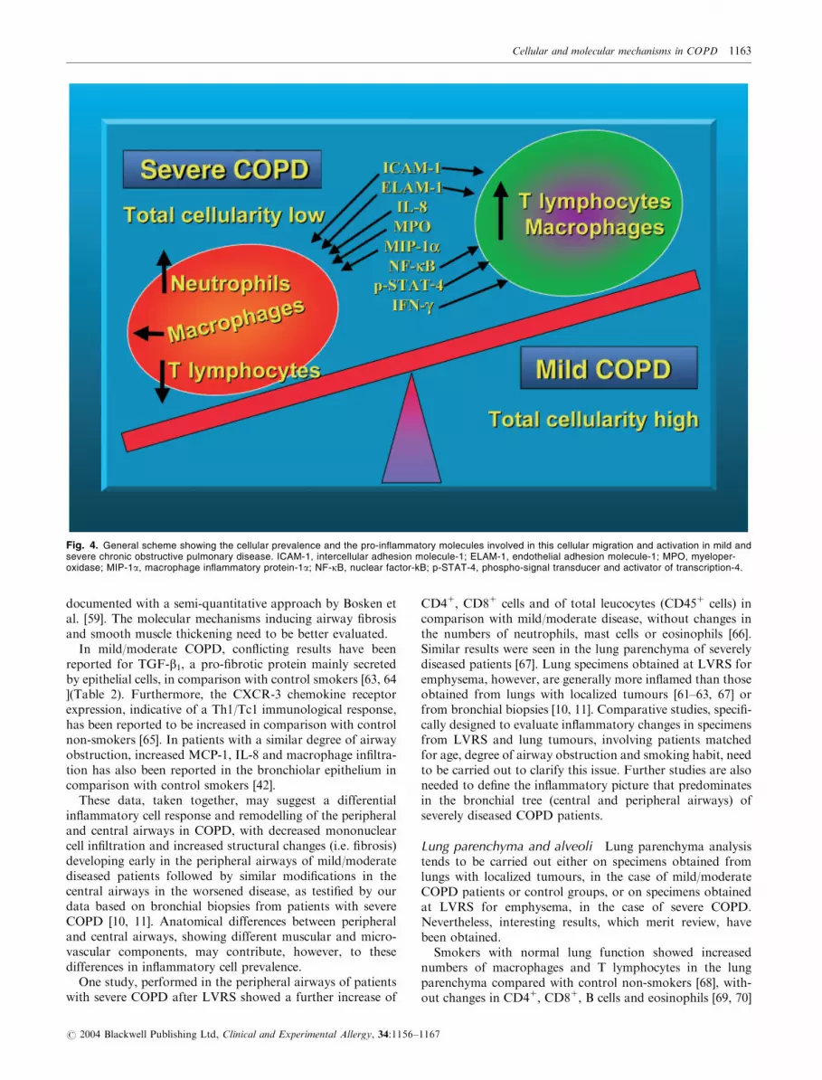
Fig. 4. General scheme showing the cellular prevalence and the pro-inflammatory molecules involved in this cellular migration and activation in mild andsevere chronic obstructive pulmonary disease. ICAM-1, intercellular adhesion molecule-1; ELAM-1, endothelial adhesion molecule-1; MPO, myeloper-oxidase; MIP-1a, macrophage inflammatory protein-1a; NF-kB, nuclear factor-kB; p-STAT-4, phospho-signal transducer and activator of transcription-4.
Cellular and molecular mechanisms in COPD 1163
r 2004 Blackwell Publishing Ltd, Clinical and Experimental Allergy, 34:1156–1167

(Table 3). In mild/moderate COPD patients, increased CD81
cells were reported in the alveolar septa compared withcontrol non-smokers [70, 71], with no changes in the numbersof neutrophils, macrophages, CD41 cells or eosinophils[70, 71] (Table 3). In patients with similar disease severity,TGF-b1 was increased in bronchiolar and alveolar epitheliumcompared with control smokers [72] (Table 3). Conflictingresults have been reported for g-glutamyl cysteine synthetase(g-GCS), a protective enzyme of alveolar epithelium againstoxidants: g-GCS in the alveolar epithelial cells of mild/moderate COPD patients was increased compared withcontrol smokers in one study [72] but not in another [73].Few data are available on severe COPD patients.
Retamales et al. [69] showed greater levels of total lungparenchymal inflammation in severe disease constituted by agreater presence of neutrophils, macrophages (CD681),CD41, CD81 cells and eosinophils compared with mild/moderate COPD and control smokers. In specimens obtainedin a way similar to that reported by Retamales et al.deoxyribonucleic acid (DNA) fragmentation and TUNELpositivity were reported to be increased in alveolar septa ofsevere emphysematous lungs as compared with controlsmokers and non-smokers [74]. A parallel decrease ofvascular endothelium growth factor (VEGF)-A and VEGFreceptor (VEGF-R)-2 was also reported in this study. Theauthors propose that an increased apoptosis of the endothe-lial cells in the alveolar septa triggered by a lack of growthfactors (VEGF) and its main receptor may play a role in thedevelopment of pulmonary emphysema [74]. Imai et al. [75]showed that matrix metalloprotease (MMP)-1 but not MMP-9 or MMP-12 was significantly increased in the alveolar septaof patients with severe emphysema compared with controlnon-smokers. MMP-1 was mainly localized in the type IIpneumocytes of emphysematous lungs. The authors speculatethat different enzymes such as elastase and MMP-12 mayplay a role in the early stages of the disease, while collagendestruction by collagenase-1 (MMP-1) seems to be theprevalent feature in the more severe forms of the disease[75]. This speculation is supported by a recent study showingthat MMP-12 and neutrophils are required for acute cigarettesmoke-induced connective tissue breakdown, and that thisresult is mediated by increased TNF-a release from alveolarmacrophages, up-regulation of NF-kB, endothelial activationand increased neutrophil influx in the lungs of mice [76].Few studies have directly compared mild and severe emphy-
sema [77, 78]. Decreased peribronchiolar decorin, biglycanand alveolar heparan sulphate proteoglycan (HSPG) havebeen found in severe compared with mild emphysema [77]without changes of collagen I, III, IV, laminin and fibronectinimmunoreactivity between the two groups [77]. The samegroup of authors showed decreased percentages of nitric oxidesynthase (NOS)2 or NOS31 alveolar macrophages in severecompared with mild emphysema [78]. The authors suggestedthat alterations of proteoglycans, subsequent to fibrillarcollagen stabilization, may play a role in the develop-ment of severe emphysema [77]. Increased NO production,related to the increased percentage of NOS21 and NOS31
macrophages, seen in mild compared with severe em-physema, may have a protective function in mild disease inresponse to increased inflammation due to cigarette smokeexposure [78].
Conflicting results have been reported for collagen synthesisin emphysematous lungs [79, 80]. Elastin expression wasdecreased in panacinar and severe centriacinar enphysema [80].A more recent report, performed in mild/moderate emphyse-ma, showed that alveolar septa close to the emphysematousareas are thickened and produce more elastin and collagen[81], though it is not known whether these newly synthetizedmolecules maintain their original functional status [81].
These data, taken together, suggest that T lymphocytesmay play a role in the alveolar septa disruption in mild em-physema and in the mild/moderate airflow obstruction relatedto emphysema. Severe emphysema seems to be accompaniedby a general increase of alveolar inflammatory cells includingneutrophils, macrophages and T lymphocytes, increased in-dices of alveolar apoptosis and collagenase-1, and decreasedalveolar expression of VEGF-A, VEGFR-2 and proteogly-cans. More studies comparing severe vs. mild forms ofemphysema are needed to clarify the prevalent molecularmechanisms involved in the progression of the disease.
The data summarized in Tables 2 and 3 on central/peripheral airways and lung parenchyma were obtained fromlung tissue resected for localized lung tumours and werefocussed on mild/moderate COPD and control groups.
Concluding comments
In the last decade, analysis of bronchial biopsies of COPDpatients and control subjects has yielded specific informationon the role of inflammation in the pathogenesis of thisdisease. T lymphocytes, in particular CD81 cells andmacrophages, are the prevalent inflammatory cells in thebronchial mucosa of all smokers with and without mildairflow limitation, while neutrophils and macrophages pre-dominate in the most severe forms of COPD. The molecularmechanisms causing the prevalence of T lymphocytes andneutrophils, respectively, in mild and severe disease need to beextensively studied. Up-regulation of pro-inflammatorytranscription factors NF-kB and STAT-4 in mild COPD,and activated epithelial and endothelial cells in the moresevere disease may contribute to this differential prevalence ofinfiltrating cells. To this purpose bronchial epithelial andendothelial cell alterations in number and functions need tobe studied in more detail. Through analysis of bronchialbiopsies, it is possible to evaluate inflammation in differentclinical conditions, such as during an exacerbation period ofthe disease or after pharmacological treatment. Even thoughrelatively few studies are published on these two latterconditions, a preliminary picture is today available. A generalincrease of inflammatory cells and the transient appearance ofeosinophils was reported in exacerbations of mild/moderateCOPD, while a 3-month glucocorticoid treatment of stablemoderate COPD patients had little effect on the presence ofinflammatory cells in bronchial biopsies. Follow-up studiesinvolving the same group of patients before and aftertreatment are needed to better evaluate the cellular changesafter treatment. Moreover, the effect of an anti-inflammatorytreatment in the bronchial biopsies from mild stable COPDpatients needs to be investigated. Data summarized in thisreview regarding peripheral airways and parenchyma of mild/moderate COPD and controls were obtained from resected
1164 A. Di Stefano et al.
r 2004 Blackwell Publishing Ltd, Clinical and Experimental Allergy, 34:1156–1167

lungs for localized tumours. Alterations in severe emphyse-ma, here reported, were studied in specimens obtained atLVRS from patients with emphysema. Whether these latterpatients are really representative of the entire population ofseverely diseased COPD patients needs to be confirmed.Conflicting results have been reported in the peripheral
airways of mild/moderate COPD patients, although in themildest forms of the disease an increased mononuclear cellinfiltration is documented, in agreement with observations onbronchial biopsies. We believe that the greater variability ofthe results obtained in peripheral airways of COPD patientsmay be the consequence of variable inflammatory states des-cribed in the lungs of patients suffering from lung cancer, or,alternatively, due to the development of structural modifica-tions such as bronchial fibrosis and smooth muscle hypertro-phy, with a concomitant reduction of the total inflammatorycell infiltration, which occurs early in the peripheral airwaysof patients with mild/moderate COPD and extends later tothe central airways.The analysis of inflammation in the lung parenchyma of
mild/moderate COPD yields similar results and suffers fromthe same constraints reported for peripheral airways. Guide-lines for recruitment of patients with COPD undergoingbronchoscopy and bronchial biopsy collection, as well as lungresection for tumours or LVRS for emphysema, would beuseful to obtain a more widespread application of standardi-zed selection criteria.While the inflammatory cell picture of the central/
peripheral airways and lung parenchyma in mild/moderateCOPD is becoming clearer, the analysis of the molecularmechanisms underlying COPD development has only justbegun. Current data regarding the molecular alterations inbronchial biopsies, peripheral airways and alveoli are frag-mentary and should be considered as preliminary observa-tions warranting further confirmation. A clear, organicpicture in the lung pathology concerning the type of cytokineprevalence, oxidant/anti-oxidant balance, proteinase-anti-proteinase imbalance and the molecules involved in inducingthe structural alterations in the lung of COPD patients in dif-ferent clinical conditions (stable, exacerbated, glucocorticoid-treated disease) and with different degrees of severity (frommild-to-severe forms) needs to be adequately defined througha quantitative approach.
Acknowledgements
The authors thank Rosemary Allpress for editing the lan-guage of the paper and Isabella Gnemmi and Ada Patriarcafor technical assistance.
References
1 Pauwels RA, Buist AS, Calverley PMA, Jenkins CR, Hurd SS on
behalf of the GOLD Scientific Committee. Global strategy for the
diagnosis, management, and prevention of Chronic Obstructive
Pulmonary Disease. NHLBI/WHO Global initiative for Chronic
Obstructive Lung Disease (GOLD) Workshop summary. Am J
Respir Crit Care Med 2001; 163:1256–76.
2 Siafakas NM, Vermeire P on behalf of the Task Force et al.
Optimal assessment and management of chronic obstructive
pulmonary disease (COPD). Eur Respir J 1995; 8:1398–420.
3 The COPD guidelines group of the standards of care committee of
the BTS. BTS guidelines for the management of chronic obstructive
pulmonary disease. Thorax 1997; 52 (Suppl. 5):S1–28.
4 American Thoracic Society. Standards for the diagnosis and care of
patients with chronic obstructive pulmonary disease (COPD) and
asthma. Am Rev Respir Dis 1987; 136:225–44.
5 Ollerenshaw SL, Woolcock AJ. Characteristics of the inflammation
in biopsies from large airways of subjects with asthma and subjects
with chronic airflow limitation. Am Rev Respir Dis 1992; 145:
922–7.
6 Saetta M, Di Stefano A, Maestrelli P et al. Activated T-
lymphocytes and macrophages in bronchial mucosa of subjects
with chronic bronchitis. Am Rev Respir Dis 1993; 147:301–6.
7 Lacoste JY, Bousquet J, Chanez P et al. Eosinophilic and
neutrophilic inflammation in asthma, chronic bronchitis, and
chronic obstructive pulmonary disease. J Allergy Clin Immunol
1993; 92:537–48.
8 Di Stefano A, Turato G, Maestrelli P et al. Airflow limitation in
chronic bronchitis is associated with T-lymphocyte and macro-
phage infiltration of the bronchial mucosa. Am J Respir Crit Care
Med 1996; 153:629–32.
9 Di Stefano A, Maestrelli P, Roggeri A et al. Upregulation of
adhesion molecules in the bronchial mucosa of subjects with
obstructive chronic bronchitics. Am J Respir Crit Care Med 1994;
149:803–10.
10 Di Stefano A, Capelli A, Lusuardi M et al. Severity of airflow
limitation is associated with severity of airway inflammation in
smokers. Am J Respir Crit Care Med 1998; 158:1277–85.
11 Di Stefano A, Capelli A, Lusuardi M et al. Decreased T-
lymphocyte infiltration in bronchial biopsies of subjects with severe
chronic obstructive pulmonary disease. Clin Exp Allergy 2001;
31:893–902.
12 Vignola AM, Chanez P, Chiappara G et al. Transforming
growth factor-beta expression in mucosal biopsies in asthma
and chronic bronchitis. Am J Respir Crit Care Med 1997; 156:
591–9.
13 Saetta M, Di Stefano A, Maestrelli P et al. Airway eosinophilia in
chronic bronchitis during exacerbations. Am J Respir Crit Care
Med 1994; 150:1646–52.
14 Zhu J, Qiu YS, Majumdar S et al. Exacerbations of bronchitis:
bronchial eosinophilia and gene expression for interleukin-4,
interleukin-5, and eosinophil chemoattractants. Am J Respir Crit
Care Med 2001; 164:109–16.
15 Hattotuwa KL, Gizycki MJ, Ansari TW, Jeffery PK, Barnes NC.
The effects of inhaled fluticasone on airway inflammation in
chronic obstructive pulmonary disease: a double-blind, placebo-
controlled biopsy study. Am J Respir Crit Care Med 2002;
165:1592–6.
16 Saetta M, Turato G, Maestrelli P, Mapp CE, Fabbri LM. Cellular
and structural bases of chronic obstructive pulmonary disease. Am
J Respir Crit Care Med 2001; 163:1304–9.
17 Jeffery PK. Structural and inflammatory changes in COPD: a
comparison with asthma. Thorax 1998; 53:129–36.
18 Barnes PJ. Chronic obstructive pulmonary disease. N Engl J Med
2000; 343:269–80.
19 Barnes PJ, Shapiro SD, Pauwels RA. Chronic obstructive
pulmonary disease: molecular and cellular mechanisms. Eur Respir
J 2003; 22:672–88.
20 Sutherland ER, Martin RJ. Airway inflammation in chronic
obstructive pulmonary disease: comparisons with asthma. J Allergy
Clin Immunol 2003; 112:819–27.
21 Di Stefano A, Caramori G, Oates T et al. Increased expression of
nuclear factor-kB in bronchial biopsies from smokers and patients
with COPD. Eur Respir J 2002; 20:556–63.
Cellular and molecular mechanisms in COPD 1165
r 2004 Blackwell Publishing Ltd, Clinical and Experimental Allergy, 34:1156–1167

22 O’Shaughmessy TC, Ansari TW, Barnes NC, Jeffery PK.
Inflammation in bronchial biopsies of subjects with chronic
bronchitis: inverse relationship of CD81 T lymphocytes with
FEV1. Am J Respir Crit Care Med 1997; 155:852–7.
23 Amadori A, Zamarchi R, De Silvestro G et al. Genetic control
of the CD4/CD8 T-cell ratio in humans. Nat Med 1995;
1:1279–83.
24 Costabel U, Maier K, Teschler H, Wang YM. Local immune
components in chronic obstructive pulmonary disease. Respiration
1992; 59 (Suppl. 1):17–9.
25 Hughes DA, Haslam PL, Townsend PJ, Turner-Warwick M.
Numerical and functional alterations in circulatory lymphocytes in
cigarette smokers. Clin Exp Immunol 1985; 61:459–66.
26 Zhu J, Majumdar S, Qiu Y et al. Interleukin-4 and interleukin-5
gene expression and inflammation in the mucus-secreting glands
and subepithelial tissue of smokers with chronic bronchitis. Lack of
relationship with CD81 cells. Am J Respir Crit Care Med 2001;
164:2220–8.
27 Liu AN, Mohammed AZ, Rice WR et al. Perforin-independent
CD81 T-cell-mediated cytotoxicity of alveolar epithelial cells is
preferentially mediated by tumor necrosis factor-a. Relative
insensitivity to Fas Ligand. Am J Respir Cell Mol Biol 1999;
20:849–58.
28 Thierfelder WE, van Deursen JM, Yamamoto K et al. Require-
ment for Stat4 in interleukin-12-mediated responses of natural
killer and T cells. Nature 1996; 382:171–4.
29 Bacon CM, Petricoin III EF, Ortaldo JR et al. Interleukin 12
induces tyrosine phosphorylation and activation of STAT4 in
human lymphocytes. Proc Natl Acad Sci USA 1995; 92:7307–11.
30 Di Stefano A, Caramori G, Capelli A et al. STAT-4 activation in
smokers and patients with COPD. Eur Respir J 2004; 24:78–85.
31 Schulz C, Wolf K, Harth M, Kratzel K, Kunz-Schughart L, Pfeifer
M. Expression and release of interleukin-8 by human bronchial
epithelial cells from patients with chronic obstructive pulmonary
disease, smokers, and never-smokers. Respiration 2003; 70:254–61.
32 Stanescu D, Sanna A, Veriter C et al. Airways obstruction, chronic
expectoration, and rapid decline of FEV1 in smokers are associated
with increased levels of sputum neutrophils. Thorax 1996;
51:267–71.
33 Balzano G, Stefanelle F, Iorio C et al. Eosinophilic inflammation in
stable chronic obstructive pulmonary disease. Relationship with
neutrophils and airway function. Am J Respir Crit Care Med 1999;
160:1486–92.
34 Capelli A, Di Stefano A, Gnemmi I et al. Increased MCP-1 and
MIP-1beta in bronchoalveolar lavage fluid of chronic bronchitics.
Eur Respir J 1999; 14:160–5.
35 Thompson AB, Daughton D, Robbins RA, Ghafouri MA,
Oehlerkin M, Rennard SI. Intraluminal airway inflammation in
chronic bronchitis: characterization and correlation with clinical
parameters. Am Rev Respir Dis 1989; 140:1527–37.
36 Suzuki T, Yamaya M, Sekizawa K et al. Bafilomycin A1 inhibits
rhinovirus infection in human airway epithelium: effects on
endosome and ICAM-1. Am J Physiol Lung Cell Mol Physiol 2001;
280:L1115–27.
37 Ricciardolo FLM, Di Stefano A. Smoking-induced corticosteroid
resistance in asthma: ‘the nitrosative stress hypothesis’. Am J
Respir Crit Care Med 2004; 169:1252 [author reply: 1252–3].
38 Doring G, Frank F, Boudier C, Herbert S, Fleischer B, Bellon G.
Cleavage of lymphocyte surface antigens CD2, CD4, CD8 by
polymorphonuclear leukocyte elastase and cathepsin G in patients
with cystic fibrosis. J Immunol 1995; 154:4842–50.
39 Capelli A, Di Stefano A, Lusuardi M, Gnemmi I, Donner CF.
Increased macrophage inflammatory protein-1a levels in bronch-
oalveolar lavage fluid of patients affected by different stages
of pulmonary sarcoidosis. Am J Respir Crit Care Med 2002;
165:236–41.
40 Van der Vliet A, Eiserich JP, Shigenaga MK, Cross CE. Reactive
nitrogen species and tyrosine nitration in the respiratory tract.
Epiphenomena or a pathobiologic mechanism of disease? Am J
Respir Crit Care Med 1999; 160:1–9.
41 Schroder JM, Mrowietz U, Morita E, Christophers E. Purification
and partial biochemical characterization of a human monocyte-
derived, neutrophil-activating peptide that locks interleukin-1
activity. J Immunol 1987; 139:3474–83.
42 de Boer WI, Sont JK, van Schadewijk A, Stolk J, van Krieken JH,
Hiemstra PS. Monocyte chemoattractant protein 1, interleukin 8,
and chronic airways inflammation in COPD. J Pathol 2000;
190:619–26.
43 Cantor JO, Cerreta JM, Armand G, Osman M, Turino GM. The
pulmonary matrix, glycosaminoglycans and pulmonary emphyse-
ma. Connect Tissue Res 1999; 40:97–104.
44 Keatings VM, Jatakanon A, Worsdell YM, Barnes PJ. Effects
of inhaled and oral glucocorticoids on inflammatory indices
in asthma and BPCO. Am J Respir Crit Care Med 1997;
155:542–8.
45 Confalonieri M, Mainardi E, Della Porta R et al. Inhaled
corticosteroids reduce neutrophilic bronchial inflammation in
patients with chronic obstructive pulmonary disease. Thorax 1998;
53:583–5.
46 Thompson AB, Mueller MB, Heires AJ et al. Aerosolized
beclomethasone in chronic bronchitis. Improved pulmonary
function and diminished airway inflammation. Am Rev Respir Dis
1992; 146:389–95.
47 Balbi B, Majori M, Bertacco S et al. Inhaled corticosteroids
in stable BPCO patients: do they have effects on cells and
molecular mediators of airway inflammation? Chest 2000;
117:1633–7.
48 Ito K, Lim S, Caramori G, Chung KF, Barnes PJ, Adcock IM.
Cigarette smoking reduces histone deacetylase 2 expression,
enhances cytokine expression, and inhibits glucocorticoid actions
in alveolar macrophages. FASEB J 2001; 15:1110–2.
49 Bocchino V, Bertorelli G, Bertrand LP et al. Eotaxin and CCR3
are up-regulated in exacerbations of chronic bronchitis. Allergy
2002; 57:17–22.
50 Qiu Y, Zhu J, Bandi V et al. Biopsy neutrophilia, chemokine and
receptor gene expression in severe exacerbations of COPD. Am J
Respir Crit Care Med 2003; 168:968–75.
51 Arias-Diaz J, Vara E, Torres-Melero J et al. Nitrite/nitrate and
cytokine levels in bronchoalveolar lavage fluid of lung cancer
patients. Cancer 1994; 74:1546–51.
52 Staal-van den Brekel AJ, Dentener MA, Drent M, Ten Velde
GPM, Buurman WA, Wouters EFM. The enhanced inflammatory
response in non-small cell lung carcinoma is not reflected in the
alveolar compartment. Respir Med 1998; 92:76–83.
53 Weissler JC, Nicod LP, Toews GB. Pulmonary natural killer cell
activity is reduced in patients with bronchogenic carcinoma. Am
Rev Respir Dis 1987; 135:1353–7.
54 Pitchenik AE, Guffee J, Stein-Streilein J. Lung natural killer
and interleukin-2 activity in lung cancer. A pulmonary
compartment of augmented natural killer activity occurs in
patients with bronchogenic carcinoma. Am Rev Respir Dis 1987;
136:1327–32.
55 Mullen JBM, Wright JL, Wiggs BR, Pare PD, Hogg JC.
Reassessment of inflammation of airways in chronic bronchitis. Br
Med J 1985; 291:1235–9.
56 Saetta M, Turato G, Facchini FM et al. Inflammatory cells in the
bronchial glands of smokers with chronic bronchitis. Am J Respir
Crit Care Med 1997; 156:1633–9.
57 Mullen JBM, Wright JL, Wiggs BR, Pare PD, Hogg JC. Structure
of central airways in current smokers and ex-smokers with and
without mucus hypersecretion: relationship to lung function.
Thorax 1987; 42:843–8.
1166 A. Di Stefano et al.
r 2004 Blackwell Publishing Ltd, Clinical and Experimental Allergy, 34:1156–1167

58 Neiwoehner DE, Kleinerman J, Rice DB. Pathologic changes in the
airways of young cigarette smokers. N Engl J Med 1974;
291:755–8.
59 Bosken CH, Wiggs BR, Pare PD, Hogg JC. Small airway
dimensions in smokers with obstruction to airflow. Am Rev Respir
Dis 1990; 142:563–70.
60 Saetta M, Di Stefano A, Turato G et al. CD81 T-lymphocytes in
peripheral airways of smokers with chronic obstructive pulmonary
disease. Am J Respir Crit Care Med 1998; 157:822–6.
61 Bosken CH, Hards J, Gatter K, Hogg J. Characterization of the
inflammatory reaction in the peripheral airways of cigarette
smokers using immunohistochemistry. Am Rev Respir Dis 1992;
145:911–7.
62 Lams BEA, Sousa AR, Rees PJ, Lee TH. Immunopathology of the
small-airway submucosa in smokers with and without chronic
obstructive pulmonary disease. Am J Respir Crit Care Med 1998;
158:1518–23.
63 De Boer WI, van Schadewijk A, Sont JK et al. Transforming
growth factor b1 and recruitment of macrophages and mast cells in
airways in chronic obstructive pulmonary disease. Am J Respir Crit
Care Med 1998; 158:1951–7.
64 Aubert JD, Dalal BI, Bai TR, Roberts CR, Hayashi S, Hogg JC.
Transforming growth factor beta 1 gene expression in human
airways. Thorax 1994; 49:225–32.
65 Saetta M, Mariani M, Panina-Bordignon P et al. Increased
expression of the chemokine receptor CXCR3 and its ligand
CXCL10 in peripheral airways of smokers with chronic
obstructive pulmonary disease. Am J Respir Crit Care Med
2002; 165:1404–9.
66 Turato G, Zuin R, Miniati M et al. Airway inflammation in severe
chronic obstructive pulmonary disease. Relationship with lung
function and radiologic emphysema. Am J Respir Crit Care Med
2002; 166:105–10.
67 Grashoff WF, Sont JK, Sterk PJ et al. Chronic obstructive
pulmonary disease: role of bronchial mast cells and macrophages.
Am J Pathol 1997; 151:1785–90.
68 Finkelstein R, Fraser RS, Ghezzo H, Cosio MG. Alveolar
inflammation and its relation to emphysema in smokers. Am J
Respir Crit Care Med 1995; 152:1666–72.
69 Retamales I, Elliott WM, Meshi B et al. Amplification of
inflammation in emphysema and its association with latent
adenoviral infection. Am J Respir Crit Care Med 2001; 164:469–73.
70 Saetta M, Baraldo S, Corbino L et al. CD81 cells in the lungs of
smokers with chronic obstructive pulmonary disease. Am J Respir
Crit Care Med 1999; 160:711–7.
71 Majo J, Ghezzo H, Cosio MG. Lymphocyte population and
apoptosis in the lungs of smokers and their relation to emphysema.
Eur Respir J 2001; 17:946–53.
72 Rahman I, van Schadewijk AAM, Crowther AJL et al. 4-hydroxy-
2-nonenal, a specific lipid peroxidation product, is elevated in lungs
of patients with chronic obstructive pulmonary disease. Am J
Respir Crit Care Med 2002; 166:490–5.
73 Harju T, Kaarteenaho-Wiik R, Soini Y, Sormunen R, Kinnula VL.
Diminished immunoreactivity of g-glutamylcysteine synthetase in
the airways of smokers lung. Am J Respir Crit Care Med 2002;
166:754–9.
74 Kasahara Y, Tuder RM, Cool CD, Lynch DA, Flores SC, Voelkel
NF. Endothelial cell death and decreased expression of vascular
endothelial growth factor and vascular endothelial growth factor
receptor 2 in emphysema. Am J Respir Crit Care Med 2001;
163:737–44.
75 Imai K, Dalal SS, Chen ES et al. Human collagenase (matrix
metalloproteinase-1) expression in the lungs of patients with
emphysema. Am J Respir Crit Care Med 2001; 163:786–91.
76 Churg A, Wang RD, Tai H et al. Macrophage metalloelastase
mediates acute cigarette smoke-induced inflammation via tumor
necrosis factor-a release. Am J Respir Crit Care Med 2003;
167:1083–9.
77 van Straaten JFM, Coers W, Noordhoek JA et al. Proteoglycan
changes in the extracellular matrix of lung tissue from patients with
pulmonary emphysema. Mod Pathol 1999; 12:697–705.
78 van Straaten JFM, Postma DS, Coers W, Noordhoek JA,
Kauffman HF, Timens W. Macrophages in lung tissue from
patients with pulmonary emphysema express both inducible and
endothelial nitric oxide synthase. Mod Pathol 1998; 11:648–55.
79 Lang MR, Fiaux GW, Gilloly M, Stewart JA, Hulmes DJS, Lamb
D. Collagen content of alveolar wall tissue in emphysematous and
nonemphysematous lungs. Thorax 1994; 49:319–26.
80 Cardoso WV, Sekhon HS, Hyde DM, Thurlbeck WM. Collagen
and elastin in human pulmonary emphysema. Am Rev Respir Dis
1993; 147:975–81.
81 Vlahovic G, Russell ML, Mercer RR, Crapo JD. Cellular and
connective tissue changes in alveolar septal walls in emphysema.
Am J Respir Crit Care Med 1999; 160:2086–92.
Cellular and molecular mechanisms in COPD 1167
r 2004 Blackwell Publishing Ltd, Clinical and Experimental Allergy, 34:1156–1167