cd5l deficiency attenuate acetaminophen-induced liver
TRANSCRIPT

ARTICLE OPEN
CD5L deficiency attenuate acetaminophen-induced liverdamage in mice via regulation of JNK and ERK signalingpathwayMengjing Li1,5, Tao Ling1,5, Fengmeng Teng2,5, Chao Hu1, Zhongping Su1, Chen Zhang1, Xiang Li2, Ting Zhao3, Xianmin Mu1,Yingchang Li3,4, Jinshun Pan 1✉ and Qiang You 1,3,4✉
© The Author(s) 2021
CD5 molecule like (CD5L), a member of the scavenger receptor cysteine-rich domain superfamily, plays a critical role in immunehomeostasis and inflammatory disease. Acetaminophen (APAP) is a safe and effective antipyretic analgesic. However, overdose maycause liver damage or even liver failure. APAP hepatotoxicity is characterized by extensive necrotic cell death and a sterileinflammatory response, in which the role of CD5L remains to be investigated. In this study, we found that the expression of CD5Lwas increased in the livers of mice after APAP overdose. Furthermore, CD5L deficiency reduced the increase of alaninetransaminase (ALT) level, histopathologic lesion area, c-Jun N-terminal kinase (JNK)/extracellular signal-regulated kinase (ERK)phosphorylation level, Transferase-Mediated dUTP Nick End-Labeling positive (TUNEL+) cells proportion, vascular endothelial cellpermeability and release of inflammatory cytokines induced by excess APAP. Therefore, our findings reveal that CD5L may be apotential therapeutic target for prevention and treatment of APAP-induced liver injury.
Cell Death Discovery (2021) 7:342 ; https://doi.org/10.1038/s41420-021-00742-3
INTRODUCTIONAcetaminophen (APAP), one of the most widely used analgesic-antipyretic in the United States, is safe at therapeutic doses.However, excess of APAP is capable for leading to a centrilobularhepatic necrosis [1]. N-acetyl-p-benzoquinoneimine (NAPQI), thetoxic product of APAP metabolized by the cytochromeP450 system, is depleted by the hepatic reduced glutathione(GSH) antioxidant system. The unconsumed NAPQI leads to APAPprotein adduct (APAP-AD) formation, which resulting oxidativestress of hepatocyte mitochondria that occurs in the first fewhours [2]. Necrotic hepatocytes can activate the innate immunesystem to mediate irreversible secondary damage [3]. Variousstudies suggest that oxidative stress and proinflammatory factorssuch as interleukin-6 (IL-6) activate the JNK signaling pathway [4]and associated with ERK signaling pathway [5, 6].CD5L, also termed as apoptosis inhibitor of macrophage (AIM),
is a soluble protein mainly produced by macrophages andbelongs to the scavenger receptor cysteine rich superfamily[7, 8]. Recent studies have shown that it can also be produced byother cells, such as Th17 cells [9], retinal epithelial cells [10] andlung epithelial cells [11]. Early studies suggested that CD5L cansupport macrophages survival [8]. Surprisingly, CD5L is found tobe associated with various diseases such as lipid metabolic disease[12], hepatocellular carcinoma [13, 14], fungus induced peritonitis[15], acute kidney injury [16] and myocardial infarction [17] in later
studies. Regarding the effect of CD5L in the liver disease, it hasbeen proved that CD5L plasma levels are upregulated in patientswith liver damage [18, 19]. In addition, CD5L is associated with thehepatic fibrosis [20] and hepatocellular carcinoma [14]. However,whether CD5L participates in the process of pathogenesis duringAPAP-induced liver injury has not been addressed to date.In this study, results showed that the expression of CD5L was
induced in the development of APAP-induced liver injury.Furthermore, CD5L-deficient reduced APAP-induced liver injuryin mice by impairing the activation of JNK and ERK signalingpathways.
RESULTSCD5L deficiency attenuates APAP-induced liver injury in miceTo determine the role of CD5L in APAP-induced liver injury, maleWT mice fasted overnight were treated with 300 mg/kg of APAPby intraperitoneal injection. CD5L mRNA level was increased twofolds in livers after APAP treatment for 1 h, and the peak occurredat 3 h (Fig. 1A). Correspondingly, the CD5L protein level wassignificantly upregulated within 24 h in a time-dependent manner(Fig. 1B). Intriguingly, under physiological conditions, CD5L wasonly expressed in the interstitium of liver tissue, while APAPoverdose induced its expression in liver parenchymal cells in thenecrotic area around the central vein besides the mesenchymal
Received: 10 August 2021 Revised: 19 October 2021 Accepted: 27 October 2021
1Department of Biotherapy, Department of Geriatrics, Second Affiliated Hospital of Nanjing Medical University, Nanjing 210011, China. 2Affilated Hospital of Nanjing University ofChinese Medicine, Nanjing 210029, China. 3Affiliated Cancer Hospital & Institute of Guangzhou Medical University, Guangzhou 510095, China. 4Key Laboratory of CellHomeostasis and Cancer Research of Guangdong Higher Education Institutes, Guangzhou Medical University, Guangzhou 510182, China. 5These authors contributed equally:Mengjing Li, Tao Ling, Fengmeng Teng. ✉email: [email protected]; [email protected]
www.nature.com/cddiscovery
Official journal of CDDpress
1234567890();,:

Fig. 1 CD5L deficiency alleviates APAP-induced liver injury in mice. APAP was intraperitoneally injected into wild-type (WT) mice (n= 5 foreach time point) at 300mg/kg, and the CD5L level in the liver at specified time point was detected by A q-PCR and B western blotting;C Immunohistochemical staining of CD5L in liver sections of WT (n= 4 for each group) treated with PBS or APAP for 24 h, representativeimages are shown; D The expression of CD5L in human liver tissue (www.proteinatlas.org); E The diagram of CRISP/Cas9 CD5L knockoutstrategy; F The expression of CD5L protein in the liver of WT and CD5L-/- mice was detected by western blotting; G Immunohistochemicalstaining of CD5L in liver sections of WT or CD5L−/− mice (n= 4 for each group), representative images are shown; H, I Serum ALT and liver H&Estaining at 8 h and 24 h after PBS or APAP injection in mice (n= 10 for each group). Necrotic area was measured by Image J. Representativeimages are shown. Scale: 100 μm. The results are presented as means ± SD of at least three independent experiments. *P < 0.05; **P < 0.01;***P < 0.001.
M. Li et al.
2
Cell Death Discovery (2021) 7:342

cells (Fig. 1C). Correspondingly, human CD5L is expressedabundantly in Kuffer cells and slightly in hepatocytes accordingto the analysis from the website (www.proteinatlas.org) (Fig. 1D).These results suggested that CD5L is associated with APAP-induced liver injury.To further determine the effect of CD5L on APAP-induced liver
injury, CRISPR/cas9 gene editing technology was applied toconstruct CD5L gene knockout (CD5L−/−) mice successfully (Fig.1E), in which CD5L expression proved to be absent in CD5L−/−
mice liver (Fig. 1F, G). After excessive APAP treatment, theelevation of serum hepatic injury index ALT in CD5L−/− mice wasmuch lower than that in WT mice (Fig. 1H) at 8 h and 24 h.Meanwhile, the lesion area caused by APAP in CD5L−/− mice wasmuch smaller than that in WT mice after treatment for 24 h,although no difference was observed at 8 h (Fig. 1I). Consequently,the results suggested that the absence of CD5L alleviated APAP-induced liver injury.
CD5L deficiency reduces the activation of JNK and ERKsignaling pathwaysCytochrome P450 2E1 (CYP2E1) is recognized as the mostimportant enzyme for initiation of APAP-induced toxicity sinceCYP2E1 knockout mice have a resistance to APAP-induced liverinjury [21]. CYP2E1 protein showed a transient increase at 1 h inWT mice and had no change in CD5L−/− mice (Fig. 2A). We nextexamined the changes of GSH and APAP-AD in liver tissue.Although we found no difference in glutathione between the twogroups at different time points after APAP treatment (Fig. 2B), theAPAP-AD was less in the liver tissue of CD5L−/− mice after APAPoverdose (Fig. 2C). The phosphorylation levels of JNK and ERKwere dramatically lower in CD5L−/− mice than those in WT mice atthe first 3 h after APAP administration which is the critical timingfor APAP hepatotoxicity, while no significant difference of p-AKTand p-NF-κB was observed (Fig. 2D). However, at 24 h after APAPtreatment, CD5L−/− mice exhibited weaker p-ERK, significantlylower p-AKT, and a trend to lower p-NF-κB expression comparedto WT mice (Fig. 2E).
CD5L protein activates JNK and ERK signaling pathway inmouse hepatocytes in vitroTo identify the role of CD5L protein in vitro, mouse bone marrow-derived macrophages (BMDMs) and macrophage cell lineRAW264.7 cells were incubated with mouse CD5L protein (1 μg/ml), respectively. The phosphorylation of NF-κB and AKT wereincreased after the stimulation by CD5L in macrophages, while nochange of p-JNK and p-ERK was observed (Fig. 3). To determinewhether CD5L has effects on liver parenchymal cells other thanthrough inflammatory cells, mouse hepatocytes (the transforminggrowth factor-α transgenic mouse hepatocyte, TAMH) weretreated with recombinant CD5L protein directly. The phosphoryla-tion levels of AKT, ERK, JNK, and NF-κB proteins in TAMH cells wereall increased upon CD5L treatment (Fig. 3).
CD5L deficiency decreases the expression of inflammatoryfactors in liverThe TUNEL assay was used to detect APAP-induced hepatocytenecrosis, and the results showed that the ratio of TUNEL+ cells inCD5L−/− mice liver tissue was decreased markedly (Fig. 4A). Tofurther explore the effect of CD5L on APAP-induced inflammation,the expression level of inflammatory cytokines in the livers ofCD5L−/− and WT mice was examined. The concentration of IL-6was much less in the serum of APAP treated CD5L−/− mice thanthat in WT mice (Fig. 4B). At 8 h after receiving APAP injection, themRNA levels of inflammatory factor IL-1β, IL-6, MIP-1α, KC, andMCP-1 were decreased and CCR2 was increased in CD5L−/− micecompared to those in WT mice (Fig. 4C). Therefore, a lack of CD5Lleads to a reduction in APAP-induced hepatic inflammatoryresponse.
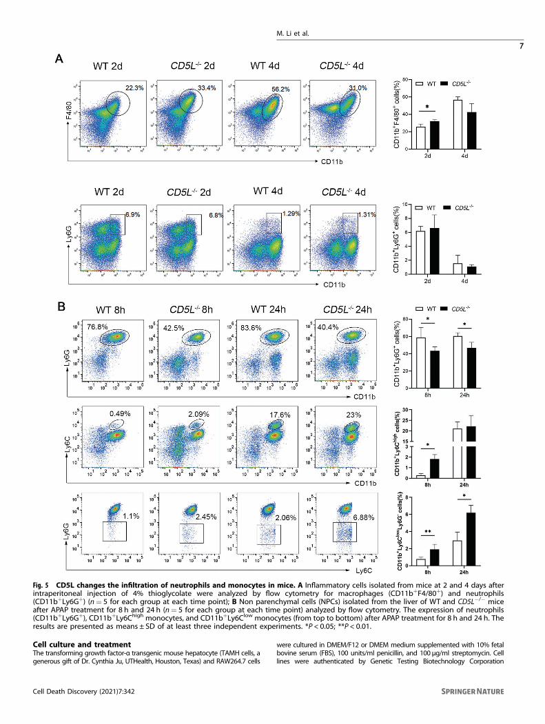
CD5L deficiency affects the infiltration of inflammatory cellsIn order to further understand the effect of CD5L on the infiltrationof inflammatory cells, thioglycolate was injected into theabdominal cavity of mice. After 2 days of treatment, there weremore peritoneal macrophages in CD5L−/− mice than that of WTmice. Interestingly, the amounts of macrophages in the CD5L−/−
mice were less than that of WT mice at 4 days post treatmentalthough no statistical significance (Fig. 5A). In addition, therewere more neutrophils after 2 days of treatment than that of4 days, but no difference between the two groups (Fig. 5A). Weconcluded that CD5L knockout could affect the increase ofinfiltration of macrophages in the abdominal cavity.Previous studies have shown that neutrophils and macrophages
exert important roles in APAP-induced liver inflammation [22–24].Flow cytometric analysis showed that the percentage of hepatic-infiltrating neutrophils (CD11b+Ly6G+) was obviously lower in thelivers from CD5L−/− mice after APAP injection than those in WTmice (Fig. 5B). Eight hours after APAP treatment, the percentage ofCD11b+Ly6Chigh (Ly6Chi) monocytes and CD11b+Ly6ClowLy6G-
(Ly6Clo) monocytes in the liver of CD5L−/− mice were higher thanthose in WT mice. As for 24 h, the Ly6Clo monocytes were muchhigher in CD5L−/− mice, and there was no significant differencebetween the two groups in Ly6Chi monocytes (Fig. 5B). These datademonstrate that CD5L deficiency can reduce the infiltration ofneutrophils and improve the infiltration of monocytes in theAPAP model.
CD5L deficiency enhances hepatocytes proliferationIt has been reported that neutrophils and macrophages contributeto the tissue repair process [25]. Therefore, the expression of PCNAwas examined to observe the proliferation of liver cells. At 8 h afterAPAP treatment there was few positive staining in the injured livertissues, while abundant positive nuclei were observed at 24 h.Intriguingly, the proportion of positive cells in CD5L−/− mice wassignificantly higher than that in WT mice at 24 h and 48 h,although there was no difference at 72 h (Fig. 6A). This suggestsan increase in proliferation of hepatocytes in APAP-induced liverinjury following CD5L deficiency. Furthermore, the permeability ofCD5L−/− mice endothelial cells was weaker than that of WT afterliver injury (Fig. 6B), indicating that CD5L−/− mice had betterability in the repair of damage.
DISCUSSIONAPAP-induced hepatotoxicity is confirmed as a ‘two-hit’ process:the oxidative stress of APAP metabolites and acute necroticinflammatory response [22]. The immunomodulatory effect ofCD5L is critical for the control of immune homeostasis [26]. In thisstudy, increased expression of CD5L was observed in the liver ofmice treated with excessive APAP. CYP2E1 is necessary to convertAPAP into toxic products and the damage caused by APAP can bereduced after CYP2E1 knocked out [27]. There was a transientincrease of CYP2E1 protein in WT mice after APAP treatment for1 h, while not observed in CD5L−/− mice. Meanwhile, consistentGSH depletion prompt that it made no difference in thedetoxification ability of NAPQI between the two group mice.Attenuated expression of CYP2E1, resulting in the reduction ofAPAP-AD, was the cause of the reduced damage of CD5L−/− mice.APAP-AD induces the production of reactive oxygen species
(ROS), which leads to continuous activation of JNK via severalpathways. Then the activated JNK translocate to the mitochondrialmembrane and further lead to hepatocyte death [4]. CD5Ldeficiency attenuated JNK activation resulting in the weaker liverdamage. In addition, CD5L protein directly activates the JNKsignaling pathway in hepatocytes but not in macrophages, whichmay play important role in promoting hepatocyte damage, exactlyas abnormal CD5L accumulation can also lead to kidney andhepatocellular carcinoma cells injury [28]. Coinciding with the
M. Li et al.
3
Cell Death Discovery (2021) 7:342

increased expression of p-JNK, CD5L also improved the level ofp-ERK in hepatocytes. APAP-induced hepatocyte necrosis isaccompanied by exposure of nuclear DNA fragments [29],therefore, fewer TUNEL+ signals detected in the livers of injuredCD5L−/− mice indicate reduced necrotic cells.In the present study, our experiments demonstrate that CD5L
protein can directly activate JNK and ERK signaling pathways in
hepatocytes. The presence of CD5L may contribute to thepromotion of mitochondrial oxidative stress by JNK in hepato-cytes after APAP administration. CD5L deficiency reduces thetoxic substance produced by APAP metabolism at an earlystage, thereby attenuating the activation of JNK and ERKsignaling pathways to alleviate APAP-induced injury. Thedetailed mechanism by which CD5L affects hepatocytes and
Fig. 2 CD5L deficiency inhibits the activation of JNK and ERK signaling pathways. A CYP-2E1 protein in the liver of each group wasexamined by Western blotting at specified time point after administration. A representative blot and the means ± SD of three independentanalyses were shown; B, C Level of GSH and APAP-AD in liver at specified time point after administration. The levels of specific proteins in WTand CD5L−/− mouse liver were detected by western blotting after treatment with PBS or APAP for 0,1,3 h (D) and 24 h (E). The phosphorylatedproteins were evaluated by western blotting analysis normalized to total proteins. A representative blot and the means ± SD of threeindependent analyses. *P < 0.05; **P < 0.01; ***P < 0.001.
M. Li et al.
4
Cell Death Discovery (2021) 7:342

macrophages in the APAP-induced liver injury requires furtherinvestigation.Numerous studies have shown that infiltrating neutrophils and
macrophages are involved in the process of acute liver inflamma-tion in the mouse hepatitis model [30–33]. Upon APAP over-dosage, neutrophils accumulate in the liver and mediate injuryand inflammation [30, 34, 35]. When the infiltration of neutrophilsis blocked, the hepatotoxicity can be significantly prevented.Chemokines (KC and MIP-1α), which account for neutrophilsinfiltration [36], were much lower in CD5L−/− mice. In thioglycolateinduced peritonitis, the acute inflammatory response peaks in1–2 days and begins to subside in 3–4 days [37], and CD5L did notinfluence the neutrophil recruitment in our results. Therefore, thedecreased neutrophils infiltration in the CD5L−/− mouse livermight be due to the difference in activation of JNK signalingpathway caused by APAP in the early stage.Our previous study indicates that both resident and infiltrating
macrophages play important roles in liver blood vessel repair [38].In aseptic peritonitis of mice, more macrophages were observedafter 2 days and fewer after 4 days in CD5L−/− mice than that inWT mice, prompting that the knockout of CD5L gene seemed tofacilitate the recruitment of monocytes. It has been reported thatthe number of resident macrophages in the liver is rapidlyreduced and bone marrow-derived monocytes infiltrate into theliver after acetaminophen poisoning [39]. The phenotype andfunction of monocytes depend on different microenvironments[40]. The activation pathways of monocytes are divided intoclassical and alternative activation. Classical activation into M1macrophages (Ly6Chi) can release proinflammatory mediators topromote inflammation, while alternative activation into M2macrophages (Ly6Clo) can release anti-inflammatory mediatorsthat down-regulate inflammation and promote inflammatoryrepair [41]. Our results showed that Ly6Clo monocytes were more
abundant in CD5L−/− mice. Combining with the results of PCNAassay, we concluded that CD5L deficiency can promote theenhancement of liver tissue repair ability by promoting theincrease of Ly6Clo monocytes.As the hepatic vasculature represents a target of APAP-induced
liver injury [42], Evans blue assay was performed to evaluatevascular permeability. CD5L deficiency relieved APAP-inducedhepatic sinusoidal vascular endothelial cell permeability. Theseresults indicated that the deletion of CD5L reduced the injury ofendothelial cells, and it might also be the reason for theenhancement of repair.In summary, our study identified that CD5L is involved in
hepatotoxicity caused by APAP overdose and may be involved ininjury repair. This discovery opened the door toward the researchof acute hepatitis treatment, additional efforts might lead to thedevelopment of therapeutic targets for different stages of drug-induced hepatitis.
MATERIALS AND METHODSAnimalsExperiments were performed with 6–8 weeks old male C57BL/6J WT(Nanjing Biomedical Research Institute of Nanjing University). The micewere housed in a temperature-controlled environment with a 12 hlight–dark cycle, and were allowed free access to water and food. Allanimal procedures were approved by the Laboratory Animal Core Facilityof Nanjing Medical University.
Construction of CD5L deficient miceTo achieve precise editing of specific gene sites in the mouse genome,we designed to knock out the Cd5l gene located in the C57BL/6J mousegenome Chr3, grcm38.p3 by using the CRISPR/Cas9 gene knockouttechnique. Cd5l gene consists of 6 exons. ATG initiation codon located inexon 1, and TGA termination codon located in exon 6. The sgRNA direct
Fig. 3 CD5L protein activates mouse macrophages and hepatocytes in vitro. CD5L protein (1 μg/ml) was co-incubate with BMDMs,RAW264.7 cells and TAMH cells, respectively. Cell proteins were collected at 0, 15, 30, 60, and 120min, and the protein levels were detected bywestern blotting. The AKT, ERK, JNK, and NF-κB phosphorylation were evaluated using total ERK, NF-κB, AKT, and JNK as controls. Therepresentative results from three independent experiments are shown and the data are presented as means ± SD. *P < 0.05; **P < 0.01.
M. Li et al.
5
Cell Death Discovery (2021) 7:342

Cas9 endonuclease cleavage of Cd5l gene and create a DSB (double-strand break). Such breaks will be repaired, and result in the deletion ofexon2–5. The sgRNA sequence (5’ to 3’) and protospacer adjacent motif(PAM) used are as follows, S1 (GGAAGGCACGAAGCCTCCAA, GGG); S2(CTAGCCTCAAAGAACACCAT, GGG); S3 (AGAGCAGGTAAAGACGCCAC,TGG); S4 (CCTTGAGATTTGTACAGAGC, AGG). CD5L KO mice weregenerated by Nanjing Biomedical Research Institute of NanjingUniversity.
In vivo induction of liver injuryThe mouse model of APAP-induced liver injury was performed aspreviously described [38]. Simply, 6–8 weeks old male WT or CD5L−/−
mice were fasted for ~16 h (overnight) by removing food and replacingbedding to deplete glutathione levels, prior to intraperitoneal injection ofPBS or APAP (300mg/kg, Sigma-Aldrich, St. Louis, MO, USA) dissolved inheated phosphate buffer saline (PBS, 60 °C). Mice were randomly assignedto PBS group or APAP group.
Fig. 4 Lacking of CD5L reduces the level of inflammatory cytokines in the liver. A DNA fragmentation induced by APAP in mouse livers wasdetected by TUNEL assay at different tine point after APAP treatment. Scale: 100 μm. B The serum IL-6 concentration of mice in the two groupsat 8 h after APAP treatment was measured by ELISA; C Q-PCR was used to detect mRNA levels of cytokines in the liver of WT and CD5L−/− miceat 8 h after APAP treatment (n= 4 for each genotype at each time point). The results are presented as means ± SD of at least threeindependent experiments. *P < 0.05; **P < 0.01; ***P < 0.001.
M. Li et al.
6
Cell Death Discovery (2021) 7:342
Cell culture and treatmentThe transforming growth factor-α transgenic mouse hepatocyte (TAMH cells, agenerous gift of Dr. Cynthia Ju, UTHealth, Houston, Texas) and RAW264.7 cells
were cultured in DMEM/F12 or DMEM medium supplemented with 10% fetalbovine serum (FBS), 100 units/ml penicillin, and 100 μg/ml streptomycin. Celllines were authenticated by Genetic Testing Biotechnology Corporation
Fig. 5 CD5L changes the infiltration of neutrophils and monocytes in mice. A Inflammatory cells isolated from mice at 2 and 4 days afterintraperitoneal injection of 4% thioglycolate were analyzed by flow cytometry for macrophages (CD11b+F4/80+) and neutrophils(CD11b+Ly6G+) (n= 5 for each group at each time point); B Non parenchymal cells (NPCs) isolated from the liver of WT and CD5L−/− miceafter APAP treatment for 8 h and 24 h (n= 5 for each group at each time point) analyzed by flow cytometry. The expression of neutrophils(CD11b+Ly6G+), CD11b+Ly6Chigh monocytes, and CD11b+Ly6Clow monocytes (from top to bottom) after APAP treatment for 8 h and 24 h. Theresults are presented as means ± SD of at least three independent experiments. *P < 0.05; **P < 0.01.
M. Li et al.
7
Cell Death Discovery (2021) 7:342

(Suzhou, China) using short tandem repeat markers profiling, and testednegative for mycoplasma. Cells were cultured at 37 °C in a humidified 95% air,5% CO2 atmosphere. The cells were starved for four hours in serum-freemedium before treatment, and stimulated with 1 μg/ml of CD5L protein forspecified duration to collect protein at each time point.
Serum aspartate aminotransferase (AST) and alanineaminotransferase (ALT) analysisBlood was collected from the post-orbitalvenous plexus at 8 and 24 h afterAPAP injection. The serum was separated by centrifugation and the levels
of AST and ALT in the serum were measured with an automated chemicalanalyzer (MODULAR EVO 4200, Switzerland).
Immunohistochemical assaysThe isolated liver tissues from the treated mice were fixed in 10% formalin for24 h followed by processing and paraffin embedding. Sections (4μm) ofparaffin-embedded tissues were stained with hematoxylin and eosin (H&E).Immunohistochemical (IHC) staining was performed using CD5L antibody(#50020-T24, Sino Biological, Beijing, China), PCNA antibody (#13110, CellSignaling Technology, Beverly, MA, USA) and sheep anti-APAP polyclonal
Fig. 6 The proliferation of hepatocytes is increased in CD5L−/− mice. A Immunohistochemical staining of PCNA in WT and CD5L−/− mouseliver at the indicated time point after PBS or APAP treatment (n= 5 for each group at each time point). Deeply stained nucleus indicatespositive results. The ratio of PCNA positive cells to total hepatocytes was calculated. Representative images are shown. Scale: 100 μm.B Vascular endothelial permeability after APAP treatment for 24 h was determined by Evans Blue assay (n= 8 for each group). The results arepresented as means ± SD of at least three independent experiments. *P < .05; **P < 0.01.
M. Li et al.
8
Cell Death Discovery (2021) 7:342

antibody (#0016-0104, Bio-Rad, Düsseldorf, Germany). Tissue sections stainedwith specific antibodies were evaluated by light-microscopic (Olympus IX51,Japan). Pathological images of tissue sections were analyzed by Image J(1.51j8).
Western blottingTotal proteins were prepared from mouse livers or cultured cell samplesusing STE buffer or RIPA lysis buffer containing protease and phosphataseinhibitors cocktail (Roche, Basel, Switzerland). After protein quantificationwith Pierce™ Rapid Gold BCA Protein Assay Kit (Thermo Fisher Scientific,Waltham, MA, USA), the lysate supernatants were heated in sodiumdodecyl sulfate-polyacrylamide gelelectrophoresis (SDS-PAGE) sample-loading buffer. Protein extracts were separated on 8–15% SDS-polyacrylamide gels and transferred to the PVDF membrane. Afterblocking in 5% bovine serum albumin (BSA), the membrane was probedwith specific primary antibodies followed by horseradish peroxidaseconjugated antibody. The antibodies were used as follows: Phospho-ERK(#4370), total ERK (#4695), Phospho-AKT (#9271), total AKT (#4691),Phospho-JNK (#9251), total JNK (#9258), Phospho-NF-κB P65 (#3033), totalNF-κB P65 (#8242). All above antibodies are from Cell SignalingTechnology (Beverly, MA, USA). Anti-CD5L antibody (ab45408) is fromAbcam (Cambridge, UK). Antibodies are diluted in the proportionindicated in the instructions.
Hepatic reduced glutathione (GSH) content measurementHepatic GSH was measured using a Glutathione Assay Kit (Sigma-Aldrich,St. Louis, MO, USA) as followed by the manufacturer’s protocol.
Transferase-mediated dUTP nick end-labeling (TUNEL) assayTo measure the hepatic nuclear DNA strand breaks, paraffin sections werestained with TUNEL method using an In Situ Cell Death Detection Kit, TMRred (Roche, Basel, Switzerland) according to the manufacturer’s protocols.
Permeability assay using Evans blue dyeMice were injected intraperitoneally with Evans blue dye (Sigma-Aldrich,St. Louis, MO, USA) at a dose of 20 mg/kg 24 h after APAP treatment.Four hours after administration, mouse liver tissues were perfused in situwith Hanks’ Balanced Salt Solution (HBSS) to remove excess dyeremaining in the circulation. Livers were excised and placed informamide (4 mL/g tissue) and incubated at 45 °C for 16 h to allow dyeextraction. The supernatant was measured using spectrophotometry atthe wavelength of 630 nm. The amount of Evans blue dye in the tissuewas calculated from the standard curve of known Evans blueconcentrations.
Quantitative real-time PCR for mRNA expression analysesTotal RNA was extracted from liver tissues collected at 8 h and 24 h afterAPAP injection using the Ultrapure RNA kit (Thermo Fisher Scientific,Invitrogen, MA, USA) and transcribed into cDNA using the reversetranscription kit (Thermo Fisher Scientific, Waltham, MA, USA). Subse-quently, the resultant cDNA was amplified with the Maxima SYBR-Green/Rox q-PCR Master Mix 2X kit (Thermo Fisher Scientific, Waltham, MA, USA)using the Step One Plus Real-Time PCR System (Thermo Fisher Scientific,Waltham, MA, USA). Primers used in the PCR-reactions were synthesized inInvitrogen (Shanghai, China) as followed: GAPDH (CAT CAC TGC CAC CCAGAA GAC TG, ATG CCA GTG AGC TTC CCG TTC AG); IL-1α (ACG GCT GAGTTT CAG TGA GAC C, CAC TCT GGT AGG TGT AAG GTG C); IL-1β (TGG ACCTTC CAG GAT GAG GAC A, GTT CAT CTC GGA GCC TGT AGT G); TNF-α (GGTGCC TAT GTC TCA GCC TCT T, GCC ATA GAA CTG ATG AGA GGG AG); TGF-β(TGA TAC GCC TGA GTG GCT GTC T, CAC AAG AGC AGT GAG CGC TGA A);IL-6 (TAC CAC TTC ACA AGT CGG AGG C, CTG CAA GTG CAT CAT CGT TGTTC); Mrc1 (GCT TCC GTC ACC CTG TAT GC, TCA TCC GTG GTT CCA TAGACC); Fizz1 (CCA ATC CAG CTA ACT ATC CCT CC, ACC CAG TAG CAG TCATCC CA); YM1 (CAG GTC TGG CAA TTC TTC TGA A, GTC TTG CTC ATG TGTGTA AGT GA); MIP-1α (TGT ACC ATG ACA CTC TGC AAC, CAA CGA TGA ATTGGC GTG GAA); KC (ACT GCA CCC AAA CCG AAG TC, TGG GGA CAC CTTTTA GCA TCT T); MCP1 (TGT ACC ATG ACA CTC TGC AAC, CAA CGA TGAATT GGC GTG GAA); CCR2 (ATC CAC GGC ATA CTA TCA ACA TC, TCG TAGTCA TAC GGT GTG GTG); CXCR4 (GAC TGG CAT AGT CGG CAA TG, AGA AGGGGA GTG TGA TGA CAA A). The relative gene expression levels werecalculated using the comparative 2−ΔΔCt method.
Enzyme-linked immunosorbent assay (ELISA)The concentration of IL-6 in the serum of mice was detected by ELISA kit(Bio Legend, San Diego, CA, USA) as followed by the manufacturer’sprotocol.
Isolation of bone marrow-derived macrophages (BMDM)The donor mice were sacrificed and leg bones were collected. The bonemarrow was washed out with RPMI medium, and cell suspensions werefiltered through a 70 µm cell strainer (BD Falcon, Bedford, MA, USA). Thered blood cells were lysed and removed after centrifugation. Then, the cellswere incubated in petri dish for three days with RPMI medium containing10% FBS and 10 ng/ml macrophage colony-stimulating factor (M-CSF) at37 °C in a humidified 95% air, 5% CO2 atmosphere. Each dish wassupplemented with 4ml of RPMI medium containing 10% serum andM-CSF (14 ng/ml) and cultured for three additional days. Then these cellswere subjected to subsequent protein-related stimulation tests.
Isolation of liver non-parenchymal cells (NPCs)LNPCs isolation was following a previously established method [33]. Inbrief, after perfusion with HBSS containing ethylenebis (oxyethylenenitrilo)tetraacetic acid (EGTA), the liver was excised and homogenized with HBSScontaining 0.5% FBS. The tissue was passed through a 100μm cell strainer(BD Falcon, Bedford, MA, USA). Then, 30% percoll (Sigma-Aldrich, St. Louis,MO, USA) was used to isolate liver NPCs. Red blood cells were further lysedwith red blood cell lysing buffer. Finally, these cells were resuspended inHBSS containing 2% FBS for antibody staining.
Isolation of peritoneal immunocytesThioglycolate (BD Falcon, Bedford, MA, USA) was dissolved in ddH2O at arate of 4%, then cooled to 4 °C temperature after autoclaved sterilization at121 °C for 20min, and each mouse was intraperitoneally injected with 3mlthioglycolate solution. Two or 4 days later, the mice were intraperitoneallywashed with 10ml of cold PBS. Infiltrated inflammatory cells werecollected from the peritoneal lavage fluids by centrifugation.
Flow cytometryFreshly isolated LNPCs were incubated with mouse Fc receptor blocker toprevent non-specific binding. Then, the cells were incubated with variousstaining antibodies, including APC Cyanine7conjugated anti-mouse CD45(clone 30F11, #130-105-506; Miltenyi Research Inc. San Diego, CA), PE-vio770 conjugate anti-mouse CD11b (clone M1/70, #25-0112-82,eBioscience), APC conjugated anti-mouse F4/80 (clone BM8, #17-4801-82,eBioscience), FITC-conjugated anti-mouse Ly6C (clone AL21, #553104, BDBiosciences, San Jose, CA) or PE-conjugated anti-mouse Ly6G (clone RB6-8C5, #561084, BD Biosciences).
Quantification and statistical analysisAll data were analyzed using GraphPad Prism software (version 8.0.2). Dataare expressed as mean ± SD. Comparisons between experimental groupswere conducted using ANOVA. For all experiments similar variancesbetween groups were observed. Normal distribution of samples wasdetermined. Differences were considered significant when p < 0.05. All theexperiments were repeated at least three times.
DATA AVAILABILITYThe data that support the findings of this study are available from the correspondingauthor upon reasonable request.
REFERENCES1. Larson AM, Polson J, Fontana RJ, Davern TJ, Lalani E, Hynan LS, et al.
Acetaminophen-induced acute liver failure: results of a United States multicenter,prospective study. Hepatology. 2005;42:1364–72.
2. Bunchorntavakul C, Reddy KR. Acetaminophen-related hepatotoxicity. Clin LiverDis. 2013;17:587–607, viii.
3. Maher JJ. DAMPs ramp up drug toxicity. J Clin Invest. 2009;119:246–9.4. Lee DH, Jung YS, Yun J, Han SB, Roh YS, Song MJ, et al. Peroxiredoxin 6 mediates
acetaminophen-induced hepatocyte death through JNK activation. Redox Biol.2020;32:101496.
M. Li et al.
9
Cell Death Discovery (2021) 7:342

5. Widjaja AA, Dong J, Adami E, Viswanathan S, Ng B, Pakkiri LS, et al. RedefiningIL11 as a regeneration-limiting hepatotoxin and therapeutic target inacetaminophen-induced liver injury. Sci Transl Med. 2021;13:eaba8146.
6. Zhang J, Zhang S, Bi J, Gu J, Deng Y, Liu C. Astaxanthin pretreatment attenuatesacetaminophen-induced liver injury in mice. Int Immunopharmacol.2017;45:26–33.
7. Gebe JA, Kiener PA, Ring HZ, Li X, Francke U, Aruffo A. Molecular cloning, map-ping to human chromosome 1 q21-q23, and cell binding characteristics ofSpalpha, a new member of the scavenger receptor cysteine-rich (SRCR) family ofproteins. J. Biol. Chem. 1997;272:6151–8.
8. Miyazaki T, Hirokami Y, Matsuhashi N, Takatsuka H, Naito M. Increased suscept-ibility of thymocytes to apoptosis in mice lacking AIM, a novel murinemacrophage-derived soluble factor belonging to the scavenger receptorcysteine-rich domain superfamily. J Exp Med. 1999;189:413–22.
9. Wang C, Yosef N, Gaublomme J, Wu C, Lee Y, Clish CB, et al. CD5L/AIM regulateslipid biosynthesis and restrains Th17 cell pathogenicity. Cell. 2015;163:1413–27.
10. Iannaccone A, Hollingsworth TJ, Koirala D, New DD, Lenchik NI, Beranova-Giorgianni S, et al. Retinal pigment epithelium and microglia express the CD5antigen-like protein, a novel autoantigen in age-related macular degeneration.Exp Eye Res. 2017;155:64–74.
11. Li Y, Qu P, Wu L, Li B, Du H, Yan C. Api6/AIM/Spalpha/CD5L overexpression inalveolar type II epithelial cells induces spontaneous lung adenocarcinoma. Can-cer Res. 2011;71:5488–99.
12. Kurokawa J, Arai S, Nakashima K, Nagano H, Nishijima A, Miyata K, et al.Macrophage-derived AIM is endocytosed into adipocytes and decreases lipiddroplets via inhibition of fatty acid synthase activity. Cell Metab. 2010;11:479–92.
13. Maehara N, Arai S, Mori M, Iwamura Y, Kurokawa J, Kai T, et al. Circulating AIMprevents hepatocellular carcinoma through complement activation. Cell Rep.2014;9:61–74.
14. Aran G, Sanjurjo L, Barcena C, Simon-Coma M, Tellez E, Vazquez-Vitali M, et al.CD5L is upregulated in hepatocellular carcinoma and promotes liver cancer cellproliferation and antiapoptotic responses by binding to HSPA5 (GRP78). FASEB J.2018;32:3878–91.
15. Tomita T, Arai S, Kitada K, Mizuno M, Suzuki Y, Sakata F, et al. Apoptosis inhibitorof macrophage ameliorates fungus-induced peritoneal injury model in mice. SciRep. 2017;7:6450.
16. Arai S, Kitada K, Yamazaki T, Takai R, Zhang X, Tsugawa Y, et al. Apoptosisinhibitor of macrophage protein enhances intraluminal debris clearance andameliorates acute kidney injury in mice. Nat Med. 2016;22:183–93.
17. Nishikido T, Oyama J, Shiraki A, Komoda H, Node K. Deletion of ApoptosisInhibitor of Macrophage (AIM)/CD5L Attenuates the Inflammatory Responseand Infarct Size in Acute Myocardial Infarction. J Am Heart Assoc. 2016;5:e002863.
18. Gangadharan B, Antrobus R, Dwek RA, Zitzmann N. Novel serum biomarkercandidates for liver fibrosis in hepatitis C patients. Clin Chem. 2007;53:1792–9.
19. Gray J, Chattopadhyay D, Beale GS, Patman GL, Miele L, King BP, et al. A pro-teomic strategy to identify novel serum biomarkers for liver cirrhosis andhepatocellular cancer in individuals with fatty liver disease. BMC Cancer.2009;9:271.
20. Barcena C, Aran G, Perea L, Sanjurjo L, Tellez E, Oncins A, et al. CD5L is a pleio-tropic player in liver fibrosis controlling damage, fibrosis and immune cell con-tent. EBioMedicine. 2019;43:513–24.
21. Cheung C, Yu AM, Ward JM, Krausz KW, Akiyama TE, Feigenbaum L, et al. Thecyp2e1-humanized transgenic mouse: role of cyp2e1 in acetaminophen hepa-totoxicity. Drug Metab. Dispos. 2005;33:449–57.
22. Zhang C, Feng J, Du J, Zhuo Z, Yang S, Zhang W, et al. Macrophage-derived IL-1alpha promotes sterile inflammation in a mouse model of acetaminophenhepatotoxicity. Cell Mol Immunol. 2018;15:973–82.
23. Michael SL, Pumford NR, Mayeux PR, Niesman MR, Hinson JA. Pretreatment ofmice with macrophage inactivators decreases acetaminophen hepatotoxicity andthe formation of reactive oxygen and nitrogen species. Hepatology.1999;30:186–95.
24. Antoniades CG, Quaglia A, Taams LS, Mitry RR, Hussain M, Abeles R, et al. Sourceand characterization of hepatic macrophages in acetaminophen-induced acuteliver failure in humans. Hepatology. 2012;56:735–46.
25. Bouchery T, Harris N. Neutrophil-macrophage cooperation and its impact ontissue repair. Immunol Cell Biol. 2019;97:289–98.
26. Gao X, Yan X, Zhang Q, Yin Y, Cao J. CD5L contributes to the pathogenesis ofmethicillin-resistant Staphylococcus aureus-induced pneumonia. Int Immuno-pharmacol. 2019;72:40–47.
27. Shayiq RM, Roberts DW, Rothstein K, Snawder JE, Benson W, Ma X, et al. Repeatexposure to incremental doses of acetaminophen provides protection againstacetaminophen-induced lethality in mice: an explanation for high acet-aminophen dosage in humans without hepatic injury. Hepatology. 1999;29:451–63.
28. Takahata A, Arai S, Hiramoto E, Kitada K, Kato R, Makita Y, et al. Crucial Role ofAIM/CD5L in the development of glomerular inflammation in IgA nephropathy. JAm Soc Nephrol. 2020;31:2013–24.
29. McGill MR, Sharpe MR, Williams CD, Taha M, Curry SC, Jaeschke H. Themechanism underlying acetaminophen-induced hepatotoxicity in humans andmice involves mitochondrial damage and nuclear DNA fragmentation. J ClinInvest. 2012;122:1574–83.
30. He Y, Feng D, Li M, Gao Y, Ramirez T, Cao H, et al. Hepatic mitochondrial DNA/Toll-like receptor 9/MicroRNA-223 forms a negative feedback loop to limit neu-trophil overactivation and acetaminophen hepatotoxicity in mice. Hepatology.2017;66:220–34.
31. Si Y, Tsou CL, Croft K, Charo IF. CCR2 mediates hematopoietic stem and pro-genitor cell trafficking to sites of inflammation in mice. J. Clin. Invest.2010;120:1192–203.
32. Laskin DL. Macrophages and inflammatory mediators in chemical toxicity: abattle of forces. Chem Res Toxicol. 2009;22:1376–85.
33. You Q, Cheng L, Reilly TP, Wegmann D, Ju C. Role of neutrophils in a mousemodel of halothane-induced liver injury. Hepatology. 2006;44:1421–31.
34. Marques PE, Amaral SS, Pires DA, Nogueira LL, Soriani FM, Lima BH, et al. Che-mokines and mitochondrial products activate neutrophils to amplify organ injuryduring mouse acute liver failure. Hepatology. 2012;56:1971–82.
35. Liu ZX, Han D, Gunawan B, Kaplowitz N. Neutrophil depletion protects againstmurine acetaminophen hepatotoxicity. Hepatology. 2006;43:1220–30.
36. Knudsen E, Iversen PO, Van Rooijen N, Benestad HB. Macrophage-dependentregulation of neutrophil mobilization and chemotaxis during development ofsterile peritonitis in the rat. Eur J Haematol. 2002;69:284–96.
37. Phillips BE, Geletzke AK, Smith PB, Podany AB, Chacon A, Kelleher SL, et al.Impaired recovery from peritoneal inflammation in a mouse model of milddietary zinc restriction. Mol Nutr Food Res. 2016;60:672–81.
38. You Q, Holt M, Yin H, Li G, Hu CJ, Ju C. Role of hepatic resident and infiltratingmacrophages in liver repair after acute injury. Biochemical Pharmacol.2013;86:836–43.
39. Gardner CR, Hankey P, Mishin V, Francis M, Yu S, Laskin JD, et al. Regulation ofalternative macrophage activation in the liver following acetaminophen intox-ication by stem cell-derived tyrosine kinase. Toxicol. Appl Pharm. 2012;262:139–48.
40. Dong X, Liu J, Xu Y, Cao H. Role of macrophages in experimental liver injury andrepair in mice. Exp Ther Med. 2019;17:3835–47.
41. Shapouri-Moghaddam A, Mohammadian S, Vazini H, Taghadosi M, Esmaeili SA,Mardani F, et al. Macrophage plasticity, polarization, and function in health anddisease. J Cell Physiol. 2018;233:6425–40.
42. Holt MP, Yin H, Ju C. Exacerbation of acetaminophen-induced disturbances ofliver sinusoidal endothelial cells in the absence of Kupffer cells in mice. ToxicolLett. 2010;194:34–41.
ACKNOWLEDGEMENTSWe thank Professor Cynthia Ju (UTHealth, USA) for the gift of TAMH cell line. Thiswork was supported by the National Natural Science Foundation of China (grantnumbers 81870409, 81671543 to QY), Guangzhou key medical discipline constructionproject, Guangzhou key medical discipline construction project, Jiangsu advantageddiscipline construction project (Clinical Medicine) (JX10231803 to QY), and the 789Outstanding Talent Program of SAHNMU (789ZYRC202070102).
AUTHOR CONTRIBUTIONSJP and QY designed the experiments. ML, TL, and FT performed the research. CH, ZS,CZ, XL, TZ, XM, and YL analyzed the data. ML, JP, and QY wrote the manuscript. Allauthors read and approved the final paper.
FUNDINGThis work was supported by the National Natural Science Foundation of China (grantnumbers 81870409, 81671543 to QY), Guangzhou key medical discipline constructionproject, Jiangsu advantaged discipline construction project (Clinical Medicine)(JX10231803 to QY), and the 789 Outstanding Talent Program of SAHNMU(789ZYRC202070102 to QY).
ETHICS APPROVAL AND CONSENT TO PARTICIPATEThe study was approved by the Institutional Animal Care and Use Committee ofNanjing Medical University, China. All animal procedures were performed inaccordance with “Guide for the Care and Use of Laboratory Animals” published bythe National Institutes of Health.
M. Li et al.
10
Cell Death Discovery (2021) 7:342

COMPETING INTERESTSThe authors declare no competing interests.
ADDITIONAL INFORMATIONCorrespondence and requests for materials should be addressed to Jinshun Pan orQiang You.
Reprints and permission information is available at http://www.nature.com/reprints
Publisher’s note Springer Nature remains neutral with regard to jurisdictional claimsin published maps and institutional affiliations.
Open Access This article is licensed under a Creative CommonsAttribution 4.0 International License, which permits use, sharing,
adaptation, distribution and reproduction in anymedium or format, as long as you giveappropriate credit to the original author(s) and the source, provide a link to the CreativeCommons license, and indicate if changes were made. The images or other third partymaterial in this article are included in the article’s Creative Commons license, unlessindicated otherwise in a credit line to the material. If material is not included in thearticle’s Creative Commons license and your intended use is not permitted by statutoryregulation or exceeds the permitted use, you will need to obtain permission directlyfrom the copyright holder. To view a copy of this license, visit http://creativecommons.org/licenses/by/4.0/.
© The Author(s) 2021
M. Li et al.
11
Cell Death Discovery (2021) 7:342