b-1a lymphocytes attenuate insulin resistance
TRANSCRIPT
Lei Shen,1 Melissa Hui Yen Chng,2 Michael N. Alonso,2 Robert Yuan,2 Daniel A. Winer,3
and Edgar G. Engleman2
B-1a Lymphocytes AttenuateInsulin ResistanceDOI: 10.2337/db14-0554
Obesity-associated insulin resistance, a common pre-cursor of type 2 diabetes, is characterized by chronic in-flammation of tissues, including visceral adipose tissue(VAT). Here we show that B-1a cells, a subpopulation of Blymphocytes, are novel and important regulators of thisprocess. B-1a cells are reduced in frequency in obesehigh-fat diet (HFD)-fed mice, and EGFP interleukin-10(IL-10) reporter mice show marked reductions in anti-inflammatory IL-10 production by B cells in vivo duringobesity. In VAT, B-1a cells are the dominant producers ofB cell–derived IL-10, contributing nearly half of theexpressed IL-10 in vivo. Adoptive transfer of B-1a cellsinto HFD-fed B cell–deficient mice rapidly improves insu-lin resistance and glucose tolerance through IL-10 andpolyclonal IgM-dependent mechanisms, whereas trans-fer of B-2 cells worsens metabolic disease. Geneticknockdown of B cell–activating factor (BAFF) in HFD-fed mice or treatment with a B-2 cell–depleting, B-1acell–sparing anti-BAFF antibody attenuates insulin resis-tance. These findings establish B-1a cells as a new classof immune regulators that maintain metabolic homeosta-sis and suggest manipulation of these cells as a potentialtherapy for insulin resistance.
Type 2 diabetes mellitus currently afflicts 257 millionpeople worldwide, and this number is expected to almostdouble by 2030 (1). Obesity-associated insulin resistance(IR) is considered to be the primary defect in the naturalhistory of type 2 diabetes (2). Although many factors ap-pear to govern the pathogenesis of IR, chronic low-gradeinflammation in insulin-sensitive (IS) tissues, such as the
liver and visceral adipose tissue (VAT), appears to play acentral role (3). Multiple studies have shown links betweenincreased levels of proinflammatory cytokines, such asinterleukin-6 (IL-6), tumor necrosis factor-a (TNF-a),interferon-g (IFN-g), and worsened IR (4–6). Conversely,anti-inflammatory cytokine expression (IL-10 and IL-4) isassociated with better glucose control (7–9). Similarly,immune cells with anti-inflammatory phenotypes (alter-natively activated M2 macrophages, Th2, regulatory T cells[Treg]) are resident in the adipose tissue of lean mice andindividuals, whereas proinflammatory cells (classically acti-vated M1 macrophages, Th1) become enriched and ex-panded in the adipose tissue of obese subjects (3,6,10,11).Lastly, adipose cells are themselves capable of producingimmune-related cytokines such as IL-6, IL-18, and B cell–activating factor (BAFF) (12–15). Thus, the complex inter-actions between innate and adaptive immune cells andadipocytes play a major role in IR.
We have previously shown in diet-induced obese micethat total CD19+ B cells and high-fat diet (HFD)-associatedIgG antibodies are pathogenic in IR and that B cell–depleting therapy can alleviate disease (16). B cells alsopromote systemic and T cell–mediated inflammation inobese mice and humans (9). B cells can be divided intotwo broad classes, B-1 or B-2 cells; B-1 cells can be furtherclassified as B-1a and B-1b cells (17). B-2 cells are theconventional adaptive B cells that produce antibodies toT cell–dependent antigens and are enriched in secondarylymphoid organs. B-1 cells are enriched in mucosal tissuesand in pleural and peritoneal cavities (PerC) and producenatural antibodies, which are a first line of defense against
1Shanghai Institute of Immunology, Shanghai Key Laboratory for Tumor Micro-environment and Inflammation, Shanghai Jiao Tong University School of Medi-cine, Shanghai, China2Department of Pathology, Stanford University School of Medicine, Stanford, CA3Division of Cellular & Molecular Biology, Diabetes Research Group, TorontoGeneral Research Institute (TGRI), University Health Network, Toronto, Ontario,Canada
Corresponding authors: Edgar G. Engleman, [email protected], andDaniel A. Winer, [email protected].
Received 4 April 2014 and accepted 8 September 2014.
This article contains Supplementary Data online at http://diabetes.diabetesjournals.org/lookup/suppl/doi:10.2337/db14-0554/-/DC1.
L.S. and M.H.Y.C. contributed equally to this paper.
D.A.W and E.G.E. contributed equally to this paper.
© 2015 by the American Diabetes Association. Readers may use this article aslong as the work is properly cited, the use is educational and not for profit, andthe work is not altered.
Diabetes 1
IMMUNOLOGYAND
TRANSPLANTATIO
N
Diabetes Publish Ahead of Print, published online October 22, 2014
pathogens (17,18). B-1a cells contribute 80% of the nat-ural circulating IgM in the blood of mice (19) and make upthe bulk of IL-10–expressing leukocytes in the PerC (20).Recently, human B-1 cells have been identified in umbil-ical cord and adult peripheral blood based on functionalcriteria that they share with mouse B-1 cells (21).
Coupled with the fact that B cells are a nonredundantsource of IL-10 (20,22) and that B cells from diabeticpatients and obese mice demonstrate an impaired anti-inflammatory cytokine profile (9), we hypothesized thatB-1a cells might play an important role in glucose metab-olism. Here we show that in opposition to B-2 cells, B-1acells are novel immune regulators that protect against IR.The protective effects of these cells are mediated by IL-10and polyclonal IgM, and these functions are impaired inobese mice. Depletion of B-2 cells in BAFF knockout miceand BAFF antibody treatment ameliorated IR in thesemice. These discoveries suggest that B-2– depleting B-1a–sparing therapies could prove useful in type 2 diabetes.
RESEARCH DESIGN AND METHODS
MiceC57BL/6, B cell–deficient mMT (B6.129S2-Ighmtm1Cgn/J),IL-10 EGFP (B6(Cg)-Il10tm1.1Karp/J) and IL-10-deficient(B6.129P2-Il10tm1Cgn/J) mice were purchased from TheJackson Laboratory. Secretory IgM-deficient (sIgMnull)mice (B6;129S4-Ighmtm1Che/J) were a gift from Troy Randall(University of Rochester). BAFF-deficient mice (B6.129S2-Tnfsf13btm1Msc/J) were a gift fromMark Krasnow (StanfordUniversity). The mice were maintained in a pathogen-free,temperature-controlled environment on a 12-h light anddark cycle. The mice were fed a normal chow diet (NCD;15 kcal% fat; LabDiet) or a HFD (60 kcal% fat; ResearchDiets) beginning at 6 weeks of age. Mice fed the HFDfor at least 6 weeks were considered obese. All mice usedin comparative studies were males and were age-matchedwithin individual experiments. The Stanford UniversityInstitutional Animal Care and Use Committee approvedall protocols.
B-Cell TransferTo obtain B-2 cells, spleens were mechanically dissociatedon 40-mm nylon cell strainers, followed by negative selec-tion with the EasySep Mouse B Cell Enrichment Kit, whichdepleted CD43+ cells (Stemcell Technologies). B-2 cell pu-rity was.90% as determined by flow cytometry. To obtainB-1a cells, peritoneal cells were harvested by injecting 7 mLRPMI, no phenol (Lonza), plus 3% newborn calf serum intoPerC. CD19+ CD22+ CD5+ B-1a cells were sorted as pre-viously described to ;95% purity (23). For B-2 versus B-1aexperiments, 5 3 106 B-2 and B-1a cells were injected in-traperitoneally. For other transfer experiments, 3 3 106
cells were used.
Metabolic StudiesGlucose tolerance test (GTT), insulin tolerance test (ITT),and serum insulin were measured as previously described (6).For intraperitoneal GTTs, mice were fasted for 14 h with
access to drinking water and then injected intraperitoneallywith glucose (2 g/kg body weight). Blood glucose from thetail tip was measured using a blood glucose meter justbefore the glucose injection and every 15 minutes there-after. For ITT, mice were fasted for 5 h and then given1 unit/kg body weight human regular insulin (Eli Lilly).Serum insulin was measured using the Ultra SensitiveMouse Insulin ELISA kit (Crystal Chem).
Isolation of VAT-Associated Immune Cellsand VAT LysatesVAT-associated immune cells were isolated from epididy-mal fat pads as previously described (6). VAT lysates wereprepared as previously described (16).
Cell CulturesUnless otherwise indicated, 300,000 cells were cultured in200 mL complete RPMI in a 96-well round bottom plate for24 h at 37°C in 5% CO2. Where noted, cells were stimulatedwith 1 mg/mL lipopolysaccharide (LPS; Sigma-Aldrich). Formacrophage–B-1a cocultures, macrophages (CD192 CD11b+
F4/80+) and B-1a cells (CD19+ F4/802 CD5+) were sortedby FACS and cocultured at a 1:3 ratio for 60 h.
Cytokine and Antibody MeasurementCytokines and antibodies were measured by ELISA(eBioscience and Bethyl, respectively) and cytometric beadarray (BD Biosciences) according to vendors’ instructions.The anti-phosphorylcholine (PC) IgM antibody ELISA wasadapted from a previous protocol (24). Antigens were di-luted to 5 mg/mL in assay buffer (PBS containing 0.27mmol/L EDTA and 1% BSA) and applied to a polystyreneenzyme immunosorbent assay plate (Costar) overnight at4°C. Wells were washed with PBS containing 0.27 mmol/LEDTA and blocked with assay buffer. Detection antibody forthe mouse anti-PC IgM ELISA was horseradish peroxidase–conjugated goat anti-mouse IgM and for the human anti-PCELISA was horseradish peroxidase–conjugated goat anti-human IgM (Bethyl). For the mouse anti-PC IgM ELISA,E06 IgM (Avanti Polar Lipids) was used as the standard.Results for the human anti-PC IgM ELISA were reportedas optical density. Antigens probed for the dosage curvewere BSA (Sigma-Aldrich), PC-BSA (Biosearch Technologies),human LDL, human high-oxidized LDL (ox-LDL; Kalen Bio-medical), and malondialdehyde-modified LDL (Cell Biolabs).
Human SubjectsWe obtained sera from 62 age- and BMI-matched over-weight to obese IR and IS male and female subjects (meanage IR: 546 9, IS: 546 7; mean BMI IR: 30.96 2.4 kg/m2;IS: 30.3 6 2.4 kg/m2). IS was determined by a modifiedinsulin-suppression test, and IR or IS were defined bysteady-state plasma glucose (SSPG) levels falling in thetop (IR) or bottom (IS) 40th percentile (25). Serum sam-ples were obtained under approval by the Stanford Inter-nal Review Board for Human Subjects.
Flow CytometryWe used the following gating schemes: total leukocytes(CD45.2+), total B cells (CD19+ CD32), B-2 (CD19+ CD32
2 B-1a Lymphocytes Attenuate IR Diabetes
B220hi CD52 CD23+), B-1a (CD19+ CD32 IgM+ IgD2
B220lo CD5+ CD232), B-1b (CD19+ CD32 IgM+ IgD2
B220lo CD52 CD232), regulatory B [Breg] cells (CD19+
B220+ CD22+ CD52 IgM+ IgD+), total T cells (CD192
CD3+), Treg cells (CD192 CD3+ CD4+ CD25+), and macro-phages (CD192 CD32 CD11b+ F4/80+). Dead cells weredistinguished by Live/Dead Fixable Aqua staining (LifeTechnologies). Baselines for IL-10 EGFP mice were set us-ing age- and diet-matched C57BL/6J mice. For macrophageintracellular cytokine staining, cells were stimulated withLPS (1 mg/mL; Sigma-Aldrich) and brefeldin A (5 mg/mL;BioLegend) overnight and stained using the Cytofix/Cytoperm Kit (BD Biosciences) according to the vendor’sinstructions. Data were acquired on an LSR II flow cytome-ter (BD Biosciences) and analyzed with FlowJo software(Tree Star).
CD16/32, CD3-PacBlue, CD5-PE-Cy5, CD19-PerCP-Cy5.5,CD22-PE, CD23-PE-Cy7, CD25-PE, CD45.2-APC, B220-APC-Cy7, F4/80-PE, F4/80-PerCP-Cy5.5, IgD-PE, and TNF-a–PEantibodies were from BioLegend. IgM-efluor650, CD4-efluor650, and CD11b-efluor605 antibodies were fromeBioscience.
IgM TreatmentWe modified a previous protocol for IgM treatment ofatherosclerosis (26). We gave 13-week-old HFD-fed B cell–deficient (Bnull) mice 400 mg mouse polyclonal IgM (RocklandImmunochemicals) or 200 mg mouse monoclonal anti-PCIgM (Clone E06, Avanti Polar Lipids) intraperitoneallyon days 0, 4, 7, and 11. The polyclonal IgM dose waschosen based on the observation that RAG2/2 micereconstituted with 0.4 mg IgM have serum IgM levelsthat are similar to wild-type (WT) mice (27) and thatthe half-life of IgM is 2–3 days (26,28). Two hundredmicrograms of monoclonal anti-PC IgM is five times theexpected amount of anti–ox-LDL IgM in 400 mg poly-clonal IgM (29). Control mice received PBS or 400 mgisotype control (Clone TEPC 183, Sigma-Aldrich). Formacrophage-IgM cultures, polyclonal IgM and isotype con-trol were used at 0.5 mg/mL.
BAFF Depletion With BAFF Monoclonal AntibodyTwo hundred micrograms per mouse BAFF/B lympho-cyte stimulator–specific monoclonal antibody (10F4,GlaxoSmithKline) or isotype control (hamster IgG1) wasadministered intraperitoneally to mice fed the HFD for 6weeks on days 0 and 4.
Real-Time PCR AnalysisTissue was dissociated in Trizol (Life Technologies), andRNA was extracted and converted to cDNA using the High-Capacity cDNA Reverse Transcription Kit (Applied Biosys-tems). Primer pairs and probes, including their specificity,orientation (forward [F]; reverse [R]), and sequence were asfollows: HPRT1 (F-TGGATACAGGCCAGACTTTGTT, R-CAGATTCAACTTGCGCTCATC), IL-10 (F-TTTGAATTCCCTGGGTGAGA, R-AGACACCTTGGTCTTGGAGC), and IL-6 (F-GATGGATGCTACCAAACTGGA, R- TCTGAAGGACTCTGGCT
TTG). Quantitative real-time -PCR was performed usingSYBR select (Applied Biosystems) on a QuantStudio 6Flex machine (Applied Biosystems). Results were nor-malized to HPRT1 expression.
StatisticsThe unpaired Student t test was performed. Values of P,0.05 were considered statistically significant. Area underthe curve (AUC) analysis was performed with correctionfor the starting glucose level.
RESULTS
B-1a Cells Protect Against Glucose IntoleranceTo examine the effects of diet-induced obesity on B cells,we fed C57BL/6J mice the NCD (15 kcal% fat) or the HFD(60 kcal% fat) for 9 weeks, which reliably results inobesity-associated IR and glucose intolerance (16). Wesubsequently compared the frequencies of CD19+ B-cellsubpopulations in the PerC, VAT, and spleen and foundthat the HFD induced a significant increase in the relativepercentage of B-2 cells and a reduction in the percentageof B-1a cells in the PerC and VAT but not in the spleen(Fig. 1A). Absolute cell counts of the B-cell subpopulationsindicated that the HFD led to a significant increase of Bcells in the VAT, particularly B-2 cells (Fig. 1B).
To assess the effects of distinct B-cell subsets on glucoseintolerance, we sorted B-1a cells from the PerC and B-2cells from the spleen of HFD mice (Supplementary Fig. 1)and transferred 5 3 106 of one or the other populationinto 15-week-old HFD Bnull mice by intraperitoneal injec-tion. Control mice received PBS. When we examined themice 1 week after transfer, there were no differences inweight among all groups (Fig. 1C). Flow cytometry con-firmed the presence of B-1a cells in the PerC and theVAT but not the spleen, whereas B-2 cells were presentin all three tissues (Supplementary Fig. 2). Consistentwith our previous findings (16), B-2 cells worsened glucoseintolerance and increased serum fasting insulin comparedwith controls (Fig. 1D and F). Remarkably, B-1a cell trans-fer had the opposite effect: B-1a cells induced marked im-provement in glucose tolerance, relative improvement ininsulin tolerance, and reduced fasting insulin (Fig. 1D–F).These results suggest that B-1a cells protect against glucoseintolerance. Given that the half-life of IgM is 2 days (28),detection of serum IgM and also IL-10 in culture super-natants from PerC cells in B-1a recipient mice confirmedthat the transferred B-1a cells remained viable and capableof producing IL-10 and IgM in recipients for at least 1 week(Fig. 1G and H).
The Protective Effect of B-1a Cells on GlucoseControl Is IL-10 DependentBecause B-1a cells are important producers of B cell–derived IL-10, we next examined how diet-induced obesityinfluences IL-10 production by these cells in vivo. IL-10EGFP reporter mice on the C57BL/6 background were fedthe NCD or HFD for 9 weeks. PerC, VAT, and spleen-resident leukocytes were analyzed for IL-10 expression ex
diabetes.diabetesjournals.org Shen and Associates 3
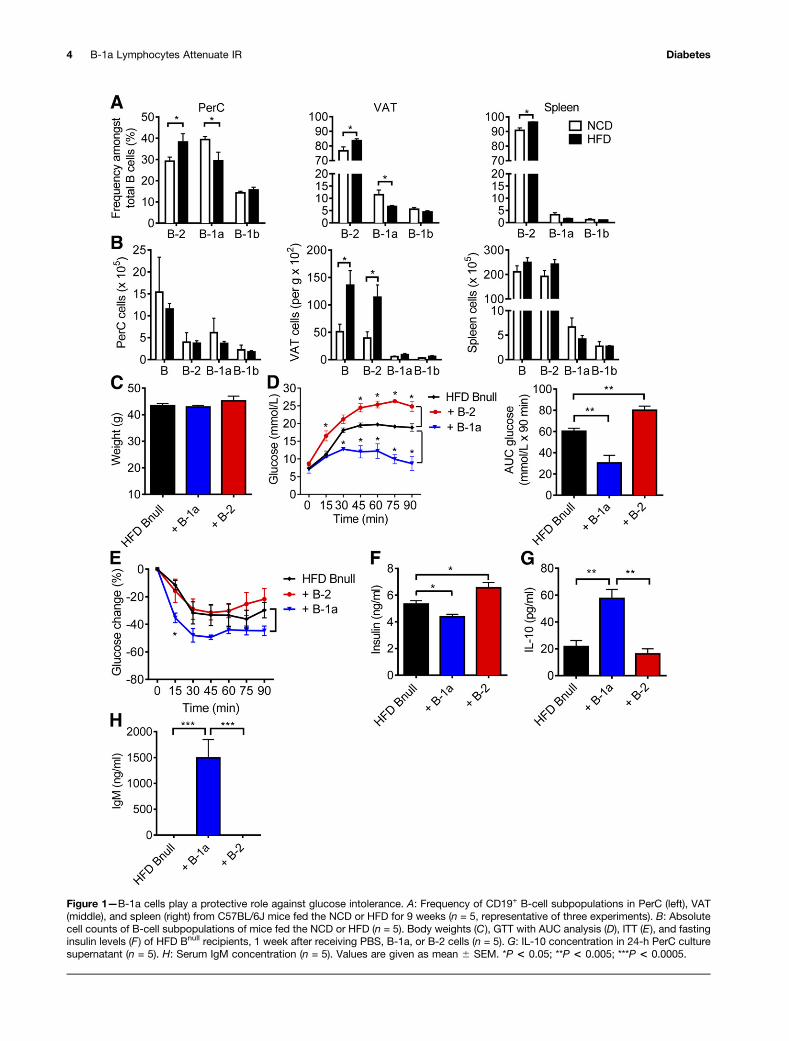
Figure 1—B-1a cells play a protective role against glucose intolerance. A: Frequency of CD19+ B-cell subpopulations in PerC (left), VAT(middle), and spleen (right) from C57BL/6J mice fed the NCD or HFD for 9 weeks (n = 5, representative of three experiments). B: Absolutecell counts of B-cell subpopulations of mice fed the NCD or HFD (n = 5). Body weights (C), GTT with AUC analysis (D), ITT (E ), and fastinginsulin levels (F ) of HFD Bnull recipients, 1 week after receiving PBS, B-1a, or B-2 cells (n = 5). G: IL-10 concentration in 24-h PerC culturesupernatant (n = 5). H: Serum IgM concentration (n = 5). Values are given as mean 6 SEM. *P < 0.05; **P < 0.005; ***P < 0.0005.
4 B-1a Lymphocytes Attenuate IR Diabetes
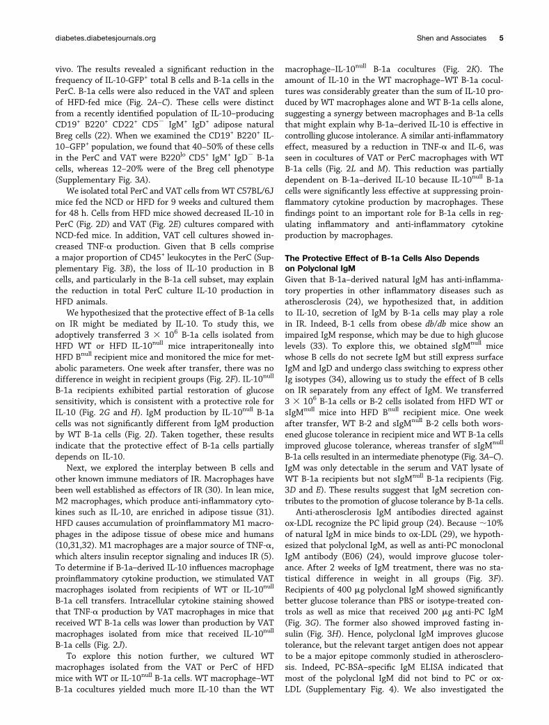
vivo. The results revealed a significant reduction in thefrequency of IL-10-GFP+ total B cells and B-1a cells in thePerC. B-1a cells were also reduced in the VAT and spleenof HFD-fed mice (Fig. 2A–C). These cells were distinctfrom a recently identified population of IL-10–producingCD19+ B220+ CD22+ CD52 IgM+ IgD+ adipose naturalBreg cells (22). When we examined the CD19+ B220+ IL-10–GFP+ population, we found that 40–50% of these cellsin the PerC and VAT were B220lo CD5+ IgM+ IgD2 B-1acells, whereas 12–20% were of the Breg cell phenotype(Supplementary Fig. 3A).
We isolated total PerC and VAT cells from WT C57BL/6Jmice fed the NCD or HFD for 9 weeks and cultured themfor 48 h. Cells from HFD mice showed decreased IL-10 inPerC (Fig. 2D) and VAT (Fig. 2E) cultures compared withNCD-fed mice. In addition, VAT cell cultures showed in-creased TNF-a production. Given that B cells comprisea major proportion of CD45+ leukocytes in the PerC (Sup-plementary Fig. 3B), the loss of IL-10 production in Bcells, and particularly in the B-1a cell subset, may explainthe reduction in total PerC culture IL-10 production inHFD animals.
We hypothesized that the protective effect of B-1a cellson IR might be mediated by IL-10. To study this, weadoptively transferred 3 3 106 B-1a cells isolated fromHFD WT or HFD IL-10null mice intraperitoneally intoHFD Bnull recipient mice and monitored the mice for met-abolic parameters. One week after transfer, there was nodifference in weight in recipient groups (Fig. 2F). IL-10null
B-1a recipients exhibited partial restoration of glucosesensitivity, which is consistent with a protective role forIL-10 (Fig. 2G and H). IgM production by IL-10null B-1acells was not significantly different from IgM productionby WT B-1a cells (Fig. 2I). Taken together, these resultsindicate that the protective effect of B-1a cells partiallydepends on IL-10.
Next, we explored the interplay between B cells andother known immune mediators of IR. Macrophages havebeen well established as effectors of IR (30). In lean mice,M2 macrophages, which produce anti-inflammatory cyto-kines such as IL-10, are enriched in adipose tissue (31).HFD causes accumulation of proinflammatory M1 macro-phages in the adipose tissue of obese mice and humans(10,31,32). M1 macrophages are a major source of TNF-a,which alters insulin receptor signaling and induces IR (5).To determine if B-1a–derived IL-10 influences macrophageproinflammatory cytokine production, we stimulated VATmacrophages isolated from recipients of WT or IL-10null
B-1a cell transfers. Intracellular cytokine staining showedthat TNF-a production by VAT macrophages in mice thatreceived WT B-1a cells was lower than production by VATmacrophages isolated from mice that received IL-10null
B-1a cells (Fig. 2J).To explore this notion further, we cultured WT
macrophages isolated from the VAT or PerC of HFDmice with WT or IL-10null B-1a cells. WT macrophage–WTB-1a cocultures yielded much more IL-10 than the WT
macrophage–IL-10null B-1a cocultures (Fig. 2K). Theamount of IL-10 in the WT macrophage–WT B-1a cocul-tures was considerably greater than the sum of IL-10 pro-duced by WT macrophages alone and WT B-1a cells alone,suggesting a synergy between macrophages and B-1a cellsthat might explain why B-1a–derived IL-10 is effective incontrolling glucose intolerance. A similar anti-inflammatoryeffect, measured by a reduction in TNF-a and IL-6, wasseen in cocultures of VAT or PerC macrophages with WTB-1a cells (Fig. 2L and M). This reduction was partiallydependent on B-1a–derived IL-10 because IL-10null B-1acells were significantly less effective at suppressing proin-flammatory cytokine production by macrophages. Thesefindings point to an important role for B-1a cells in reg-ulating inflammatory and anti-inflammatory cytokineproduction by macrophages.
The Protective Effect of B-1a Cells Also Dependson Polyclonal IgMGiven that B-1a–derived natural IgM has anti-inflamma-tory properties in other inflammatory diseases such asatherosclerosis (24), we hypothesized that, in additionto IL-10, secretion of IgM by B-1a cells may play a rolein IR. Indeed, B-1 cells from obese db/db mice show animpaired IgM response, which may be due to high glucoselevels (33). To explore this, we obtained sIgMnull micewhose B cells do not secrete IgM but still express surfaceIgM and IgD and undergo class switching to express otherIg isotypes (34), allowing us to study the effect of B cellson IR separately from any effect of IgM. We transferred3 3 106 B-1a cells or B-2 cells isolated from HFD WT orsIgMnull mice into HFD Bnull recipient mice. One weekafter transfer, WT B-2 and sIgMnull B-2 cells both wors-ened glucose tolerance in recipient mice and WT B-1a cellsimproved glucose tolerance, whereas transfer of sIgMnull
B-1a cells resulted in an intermediate phenotype (Fig. 3A–C).IgM was only detectable in the serum and VAT lysate ofWT B-1a recipients but not sIgMnull B-1a recipients (Fig.3D and E). These results suggest that IgM secretion con-tributes to the promotion of glucose tolerance by B-1a cells.
Anti-atherosclerosis IgM antibodies directed againstox-LDL recognize the PC lipid group (24). Because ;10%of natural IgM in mice binds to ox-LDL (29), we hypoth-esized that polyclonal IgM, as well as anti-PC monoclonalIgM antibody (E06) (24), would improve glucose toler-ance. After 2 weeks of IgM treatment, there was no sta-tistical difference in weight in all groups (Fig. 3F).Recipients of 400 mg polyclonal IgM showed significantlybetter glucose tolerance than PBS or isotype-treated con-trols as well as mice that received 200 mg anti-PC IgM(Fig. 3G). The former also showed improved fasting in-sulin (Fig. 3H). Hence, polyclonal IgM improves glucosetolerance, but the relevant target antigen does not appearto be a major epitope commonly studied in atherosclero-sis. Indeed, PC-BSA–specific IgM ELISA indicated thatmost of the polyclonal IgM did not bind to PC or ox-LDL (Supplementary Fig. 4). We also investigated the
diabetes.diabetesjournals.org Shen and Associates 5
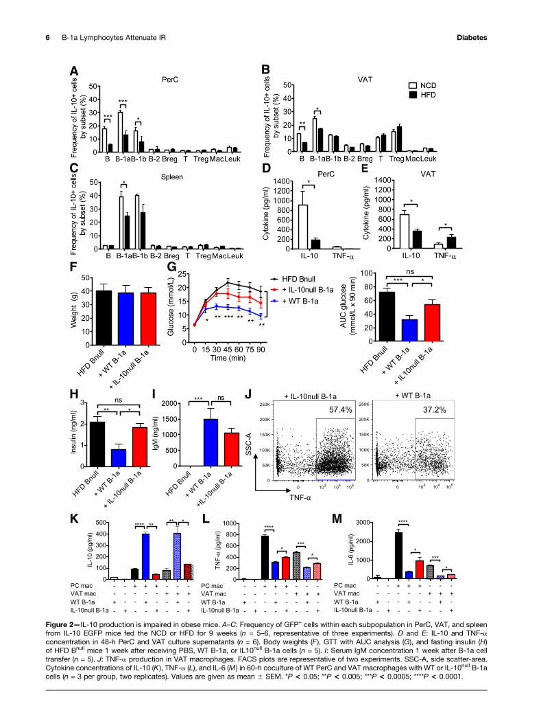
Figure 2—IL-10 production is impaired in obese mice. A–C: Frequency of GFP+ cells within each subpopulation in PerC, VAT, and spleenfrom IL-10 EGFP mice fed the NCD or HFD for 9 weeks (n = 5–6, representative of three experiments). D and E: IL-10 and TNF-aconcentration in 48-h PerC and VAT culture supernatants (n = 6). Body weights (F ), GTT with AUC analysis (G), and fasting insulin (H)of HFD Bnull mice 1 week after receiving PBS, WT B-1a, or IL10null B-1a cells (n = 5). I: Serum IgM concentration 1 week after B-1a celltransfer (n = 5). J: TNF-a production in VAT macrophages. FACS plots are representative of two experiments. SSC-A, side scatter-area.Cytokine concentrations of IL-10 (K), TNF-a (L), and IL-6 (M) in 60-h coculture of WT PerC and VAT macrophages with WT or IL-10null B-1acells (n = 3 per group, two replicates). Values are given as mean 6 SEM. *P < 0.05; **P < 0.005; ***P < 0.0005; ****P < 0.0001.
6 B-1a Lymphocytes Attenuate IR Diabetes
Figure 3—Polyclonal IgM, but not monoclonal anti-PC IgM, ameliorates glucose intolerance. Body weights (A) and GTT with AUC (B) 1week after receiving PBS, WT B-1a, sIgMnull B-1a, WT B-2, or sIgMnull B-2 cells (n = 5–8). Fasting insulin (C), IgM concentration in serum (D),and VAT lysate (E) 1 week after B-1a cell transfer (n = 4). Body weights (F), GTT with AUC (G), and fasting insulin (H) of HFD Bnull mice 1week after receiving PBS, isotype control, polyclonal IgM, or E06 monoclonal anti-PC IgM (n = 5 for E06 treatment, n = 15 for the rest). I:Anti-PC IgM in serum from NCD and HFD mice (n = 10). J: Anti-PC IgM in serum from IR and IS obese humans (n = 32 and 30). OD, opticaldensity. K: Cytokine concentrations in 24-h supernatants from PerC macrophages cultured with the isotype control or polyclonal IgM (n =3). MCP-1, monocyte chemoattractant protein-1. Values are given as mean 6 SEM. *P < 0.05; **P < 0.005; ***P < 0.0005; ****P < 0.0001.
diabetes.diabetesjournals.org Shen and Associates 7
levels of anti-PC IgM in NCD versus HFD mice, as well asin a cohort of 62 overweight to obese men and womenwho differed from one another only in their IR status, asdetermined by a modified insulin-suppression test (16).There was no difference in the levels of serum anti-PCIgM in NCD versus HFD mice (Fig. 3I) or between IRversus IS humans (Fig. 3J). To determine if polyclonalIgM affected macrophages, we cultured PerC macrophageswith polyclonal IgM or the isotype control. Compared withisotype-treated macrophages, polyclonal IgM–treated mac-rophages produced significantly less TNF-a, monocyte che-moattractant protein-1, and IL-6 but showed no changesin IL-10 production (Fig. 3K).
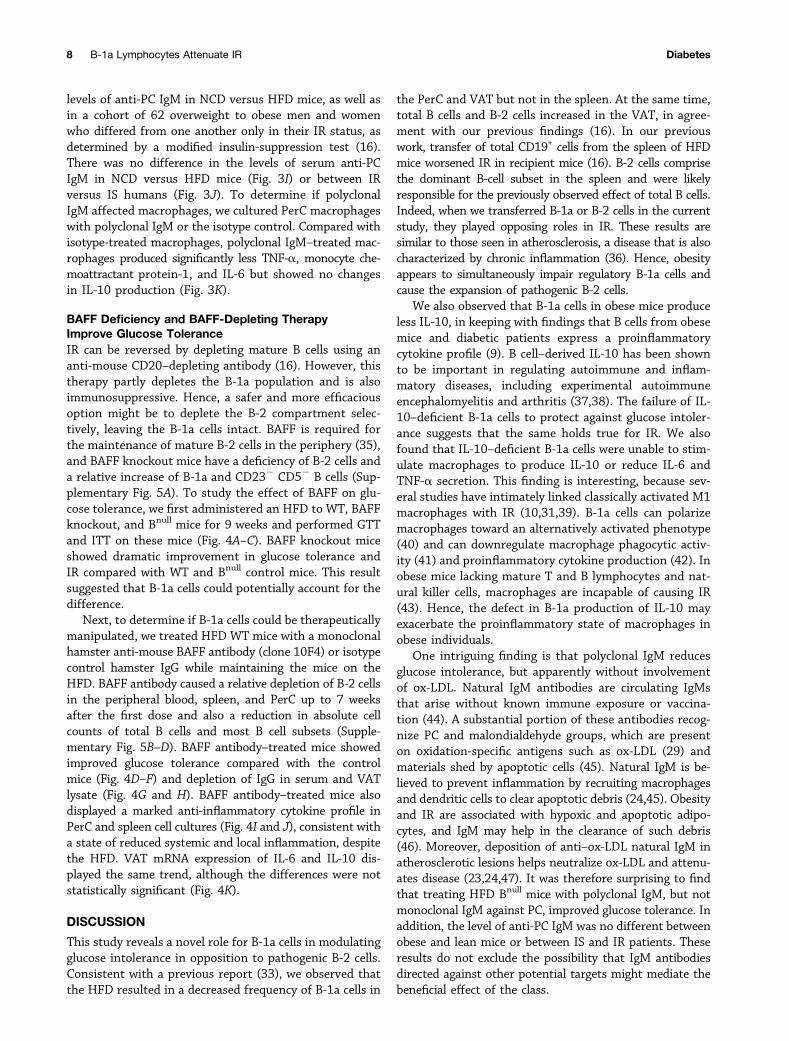
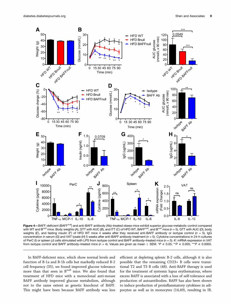
BAFF Deficiency and BAFF-Depleting TherapyImprove Glucose ToleranceIR can be reversed by depleting mature B cells using ananti-mouse CD20–depleting antibody (16). However, thistherapy partly depletes the B-1a population and is alsoimmunosuppressive. Hence, a safer and more efficaciousoption might be to deplete the B-2 compartment selec-tively, leaving the B-1a cells intact. BAFF is required forthe maintenance of mature B-2 cells in the periphery (35),and BAFF knockout mice have a deficiency of B-2 cells anda relative increase of B-1a and CD232 CD52 B cells (Sup-plementary Fig. 5A). To study the effect of BAFF on glu-cose tolerance, we first administered an HFD to WT, BAFFknockout, and Bnull mice for 9 weeks and performed GTTand ITT on these mice (Fig. 4A–C). BAFF knockout miceshowed dramatic improvement in glucose tolerance andIR compared with WT and Bnull control mice. This resultsuggested that B-1a cells could potentially account for thedifference.
Next, to determine if B-1a cells could be therapeuticallymanipulated, we treated HFD WT mice with a monoclonalhamster anti-mouse BAFF antibody (clone 10F4) or isotypecontrol hamster IgG while maintaining the mice on theHFD. BAFF antibody caused a relative depletion of B-2 cellsin the peripheral blood, spleen, and PerC up to 7 weeksafter the first dose and also a reduction in absolute cellcounts of total B cells and most B cell subsets (Supple-mentary Fig. 5B–D). BAFF antibody–treated mice showedimproved glucose tolerance compared with the controlmice (Fig. 4D–F) and depletion of IgG in serum and VATlysate (Fig. 4G and H). BAFF antibody–treated mice alsodisplayed a marked anti-inflammatory cytokine profile inPerC and spleen cell cultures (Fig. 4I and J), consistent witha state of reduced systemic and local inflammation, despitethe HFD. VAT mRNA expression of IL-6 and IL-10 dis-played the same trend, although the differences were notstatistically significant (Fig. 4K).
DISCUSSION
This study reveals a novel role for B-1a cells in modulatingglucose intolerance in opposition to pathogenic B-2 cells.Consistent with a previous report (33), we observed thatthe HFD resulted in a decreased frequency of B-1a cells in
the PerC and VAT but not in the spleen. At the same time,total B cells and B-2 cells increased in the VAT, in agree-ment with our previous findings (16). In our previouswork, transfer of total CD19+ cells from the spleen of HFDmice worsened IR in recipient mice (16). B-2 cells comprisethe dominant B-cell subset in the spleen and were likelyresponsible for the previously observed effect of total B cells.Indeed, when we transferred B-1a or B-2 cells in the currentstudy, they played opposing roles in IR. These results aresimilar to those seen in atherosclerosis, a disease that is alsocharacterized by chronic inflammation (36). Hence, obesityappears to simultaneously impair regulatory B-1a cells andcause the expansion of pathogenic B-2 cells.
We also observed that B-1a cells in obese mice produceless IL-10, in keeping with findings that B cells from obesemice and diabetic patients express a proinflammatorycytokine profile (9). B cell–derived IL-10 has been shownto be important in regulating autoimmune and inflam-matory diseases, including experimental autoimmuneencephalomyelitis and arthritis (37,38). The failure of IL-10–deficient B-1a cells to protect against glucose intoler-ance suggests that the same holds true for IR. We alsofound that IL-10–deficient B-1a cells were unable to stim-ulate macrophages to produce IL-10 or reduce IL-6 andTNF-a secretion. This finding is interesting, because sev-eral studies have intimately linked classically activated M1macrophages with IR (10,31,39). B-1a cells can polarizemacrophages toward an alternatively activated phenotype(40) and can downregulate macrophage phagocytic activ-ity (41) and proinflammatory cytokine production (42). Inobese mice lacking mature T and B lymphocytes and nat-ural killer cells, macrophages are incapable of causing IR(43). Hence, the defect in B-1a production of IL-10 mayexacerbate the proinflammatory state of macrophages inobese individuals.
One intriguing finding is that polyclonal IgM reducesglucose intolerance, but apparently without involvementof ox-LDL. Natural IgM antibodies are circulating IgMsthat arise without known immune exposure or vaccina-tion (44). A substantial portion of these antibodies recog-nize PC and malondialdehyde groups, which are presenton oxidation-specific antigens such as ox-LDL (29) andmaterials shed by apoptotic cells (45). Natural IgM is be-lieved to prevent inflammation by recruiting macrophagesand dendritic cells to clear apoptotic debris (24,45). Obesityand IR are associated with hypoxic and apoptotic adipo-cytes, and IgM may help in the clearance of such debris(46). Moreover, deposition of anti–ox-LDL natural IgM inatherosclerotic lesions helps neutralize ox-LDL and attenu-ates disease (23,24,47). It was therefore surprising to findthat treating HFD Bnull mice with polyclonal IgM, but notmonoclonal IgM against PC, improved glucose tolerance. Inaddition, the level of anti-PC IgM was no different betweenobese and lean mice or between IS and IR patients. Theseresults do not exclude the possibility that IgM antibodiesdirected against other potential targets might mediate thebeneficial effect of the class.
8 B-1a Lymphocytes Attenuate IR Diabetes
In BAFF-deficient mice, which show normal levels andfunction of B-1a and B-1b cells but markedly reduced B-2cell frequency (35), we found improved glucose tolerancemore than that seen in Bnull mice. We also found thattreatment of HFD mice with a monoclonal anti-mouseBAFF antibody improved glucose metabolism, althoughnot to the same extent as genetic knockout of BAFF.This might have been because BAFF antibody was less
efficient at depleting splenic B-2 cells, although it is alsopossible that the remaining CD23+ B cells were transi-tional T2 and T3 B cells (48). Anti-BAFF therapy is usedfor the treatment of systemic lupus erythematous, whereexcess BAFF is associated with a loss of self-tolerance andproduction of autoantibodies. BAFF has also been shownto induce production of proinflammatory cytokines in adi-pocytes as well as in monocytes (14,49), resulting in IR.
Figure 4—BAFF-deficient (BAFFnull) and anti-BAFF antibody (Ab)–treated obese mice exhibit superior glucose metabolic control comparedwith WT and Bnull mice. Body weights (A), GTT with AUC (B), and ITT (C ) of HFD WT, BAFFnull, and Bnull mice (n = 5). GTT with AUC (D), bodyweights (E), and fasting insulin (F) of HFD WT mice 4 weeks after they received anti-BAFF antibody or isotype control (n = 5). IgGconcentration in serum (G) and VAT lysate (H) 5 weeks after anti-BAFF antibody treatment (n = 5). Cytokine concentrations in 24-h culturesof PerC (I) or spleen (J) cells stimulated with LPS from isotype control and BAFF antibody–treated mice (n = 5). K: mRNA expression in VATfrom isotype control and BAFF antibody–treated mice (n = 4). Values are given as mean 6 SEM. *P < 0.05; **P < 0.005; ***P < 0.0005.
diabetes.diabetesjournals.org Shen and Associates 9
Hence, BAFF-depleting therapy, through its direct anti-inflammatory actions and its effect on the ratio of B-1ato B2 cells, may be effective in treating glucose intoler-ance. In a recent study, short-term treatment of fivelupus patients with belimumab resulted in a modest re-duction in HOMA IR, although the effect was not statis-tically significant (15). Thus, larger clinical trials in different patient populations will be needed to determinethe potential utility of this drug in the treatment of glucoseintolerance.
A recent report by Nishimura et al. (22) described aunique subset of Breg cells in the adipose tissue of mainlylean mice that produce IL-10 constitutively and contrib-ute to the maintenance of glucose homeostasis via IL-10.These CD19+ B220+ CD22+ CD52 IgM+ IgD+ cells aredistinct from the B1a cells studied here, not only in theirsurface phenotype but also in their tissue distribution.In addition, whether they produce IgM or are BAFF-dependent is not known. Nonetheless, it is interestingthat two distinct populations of B cells can regulate glu-cose metabolism through the secretion of IL-10. Becausethe cells described by Nishimura et al. (22) reside only inadipose tissue and apparently do not circulate, these cellslikely function locally, whereas the B1a cells described inour study serve to regulate glucose metabolism morebroadly. Moreover, in our study, IL-10–secreting B1a cellsaccounted for most of the B cell–derived IL-10 in VAT invivo and were at least five times more contributory thanthe cells described by Nishimura et al (22). For thesereasons, we believe that B1a cells may be the predominantB-cell population involved in regulating glucose metabo-lism, at least under the conditions studied here.
Collectively, our data support a model in which B-1acells oppose B-2 cells and promote IS through productionof IL-10 and natural IgM to modulate macrophage andT cell–mediated inflammation. These functions becomeimpaired in obesity, leading to chronic low-grade systemicand local tissue inflammation, which fuels IR. Thus, B-1acells represent a novel immune subset governing glucosetolerance and provide an important link in the complexinterplay of immunity and metabolism.
Acknowledgments. The authors thank Troy Randall, of the University ofRochester, for providing sIgMnull mice, Mark Krasnow, of Stanford University, forproviding BAFF-deficient mice, Tracey McLaughlin, of Stanford University, forproviding human serum, and GlaxoSmithKline for providing anti–B lymphocytestimulator/BAFF antibody 10F4. We also thank Joseph C. Gonzalez, of StanfordUniversity, for assistance with experiments.Funding. This work was partly supported by National Institutes of Health grant1R01-DK-096038 (E.G.E.), Canadian Institutes of Health Research grant 119414(D.A.W.), Canadian Diabetes Association grants OG-3-12-3844 (D.A.W.) and CS-5-12-3886 (D.A.W.), National Natural Science Foundation of China grant81373210 (L.S.), and Shanghai Pujiang Program grant 13PJ1405400 (L.S.).Duality of Interest. No potential conflicts of interest relevant to this articlewere reported.Author Contributions. L.S. and M.H.Y.C. designed and performed re-search, analyzed data, and wrote the paper. M.N.A. and R.Y. performed research,
D.A.W. and E.G.E. designed research, analyzed data, and wrote the paper. E.G.E.is the guarantor of this work and, as such, had full access to all the data in thestudy and takes responsibility for the integrity of the data and the accuracy of thedata analysis.
References1. Shaw JE, Sicree RA, Zimmet PZ. Global estimates of the prevalence ofdiabetes for 2010 and 2030. Diabetes Res Clin Pract 2010;87:4–142. Ioannidis I. The road from obesity to type 2 diabetes. Angiology 2008;59(Suppl. 2):39S–43S3. Xu H, Barnes GT, Yang Q, et al. Chronic inflammation in fat plays a crucialrole in the development of obesity-related insulin resistance. J Clin Invest 2003;112:1821–18304. Schultz O, Oberhauser F, Saech J, et al. Effects of inhibition of interleukin-6signalling on insulin sensitivity and lipoprotein (a) levels in human subjects withrheumatoid diseases. PLoS ONE 2010;5:e143285. Hotamisligil GS, Peraldi P, Budavari A, Ellis R, White MF, Spiegelman BM.IRS-1-mediated inhibition of insulin receptor tyrosine kinase activity in TNF-alpha- and obesity-induced insulin resistance. Science 1996;271:665–6686. Winer S, Chan Y, Paltser G, et al. Normalization of obesity-associated insulinresistance through immunotherapy. Nat Med 2009;15:921–9297. Hong EG, Ko HJ, Cho YR, et al. Interleukin-10 prevents diet-induced insulinresistance by attenuating macrophage and cytokine response in skeletal muscle.Diabetes 2009;58:2525–25358. Ricardo-Gonzalez RR, Red Eagle A, Odegaard JI, et al. IL-4/STAT6 immuneaxis regulates peripheral nutrient metabolism and insulin sensitivity. Proc NatlAcad Sci U S A 2010;107:22617–226229. DeFuria J, Belkina AC, Jagannathan-Bogdan M, et al. B cells promote in-flammation in obesity and type 2 diabetes through regulation of T-cell functionand an inflammatory cytokine profile. Proc Natl Acad Sci U S A 2013;110:5133–513810. Weisberg SP, McCann D, Desai M, Rosenbaum M, Leibel RL, Ferrante AWJr. Obesity is associated with macrophage accumulation in adipose tissue. J ClinInvest 2003;112:1796–180811. Cipolletta D, Feuerer M, Li A, et al. PPAR-g is a major driver of the ac-cumulation and phenotype of adipose tissue Treg cells. Nature 2012;486:549–55312. Fried SK, Bunkin DA, Greenberg AS. Omental and subcutaneous adiposetissues of obese subjects release interleukin-6: depot difference and regulationby glucocorticoid. J Clin Endocrinol Metab 1998;83:847–85013. Wood IS, Wang B, Jenkins JR, Trayhurn P. The pro-inflammatory cytokineIL-18 is expressed in human adipose tissue and strongly upregulated byTNFalpha in human adipocytes. Biochem Biophys Res Commun 2005;337:422–42914. Hamada M, Abe M, Miyake T, et al. B cell-activating factor controls theproduction of adipokines and induces insulin resistance. Obesity (Silver Spring)2011;19:1915–192215. Müller N, Schulte DM, Hillebrand S, et al. B lymphocyte stimulator (BLyS) isexpressed in human adipocytes in vivo and is related to obesity but not to insulinresistance. PLoS ONE 2014;9:e9428216. Winer DA, Winer S, Shen L, et al. B cells promote insulin resistance throughmodulation of T cells and production of pathogenic IgG antibodies. Nat Med 2011;17:610–61717. Baumgarth N. The double life of a B-1 cell: self-reactivity selects for pro-tective effector functions. Nat Rev Immunol 2011;11:34–4618. Hayakawa K, Hardy RR, Herzenberg LA, Herzenberg LA. Progenitors for Ly-1B cells are distinct from progenitors for other B cells. J Exp Med 1985;161:1554–156819. Baumgarth N, Herman OC, Jager GC, Brown L, Herzenberg LA, HerzenbergLA. Innate and acquired humoral immunities to influenza virus are mediated bydistinct arms of the immune system. Proc Natl Acad Sci U S A 1999;96:2250–2255
10 B-1a Lymphocytes Attenuate IR Diabetes
20. Madan R, Demircik F, Surianarayanan S, et al. Nonredundant roles forB cell-derived IL-10 in immune counter-regulation. J Immunol 2009;183:2312–232021. Griffin DO, Rothstein TL. Human “orchestrator” CD11b(+) B1 cells spon-taneously secrete IL-10 and regulate T cell activity. Mol Med 2012;18:1003–1008.22. Nishimura S, Manabe I, Takaki S, et al. Adipose Natural Regulatory B CellsNegatively Control Adipose Tissue Inflammation. Cell Metab 2013;18:759–76623. Kyaw T, Tay C, Krishnamurthi S, et al. B1a B lymphocytes are atheropro-tective by secreting natural IgM that increases IgM deposits and reduces necroticcores in atherosclerotic lesions. Circ Res 2011;109:830–84024. Shaw PX, Hörkkö S, Chang MK, et al. Natural antibodies with the T15idiotype may act in atherosclerosis, apoptotic clearance, and protective immunity.J Clin Invest 2000;105:1731–174025. McLaughlin T, Deng A, Gonzales O, et al. Insulin resistance is associatedwith a modest increase in inflammation in subcutaneous adipose tissue ofmoderately obese women. Diabetologia 2008;51:2303–230826. Faria-Neto JR, Chyu KY, Li X, et al. Passive immunization with monoclonalIgM antibodies against phosphorylcholine reduces accelerated vein graft ath-erosclerosis in apolipoprotein E-null mice. Atherosclerosis 2006;189:83–9027. Williams JP, Pechet TT, Weiser MR, et al. Intestinal reperfusion injury ismediated by IgM and complement. J Appl Physiol (1985) 1999;86:938–94228. Vieira P, Rajewsky K. The half-lives of serum immunoglobulins in adultmice. Eur J Immunol 1988;18:313–31629. Chou MY, Fogelstrand L, Hartvigsen K, et al. Oxidation-specific epitopes aredominant targets of innate natural antibodies in mice and humans. J Clin Invest2009;119:1335–134930. Chawla A, Nguyen KD, Goh YP. Macrophage-mediated inflammation inmetabolic disease. Nat Rev Immunol 2011;11:738–74931. Lumeng CN, Bodzin JL, Saltiel AR. Obesity induces a phenotypic switch inadipose tissue macrophage polarization. J Clin Invest 2007;117:175–18432. Aron-Wisnewsky J, Tordjman J, Poitou C, et al. Human adipose tissuemacrophages: m1 and m2 cell surface markers in subcutaneous andomental depots and after weight loss. J Clin Endocrinol Metab 2009;94:4619–462333. Jennbacken K, Ståhlman S, Grahnemo L, Wiklund O, Fogelstrand L. Glucoseimpairs B-1 cell function in diabetes. Clin Exp Immunol 2013;174:129–138.34. Boes M, Esau C, Fischer MB, Schmidt T, Carroll M, Chen J. Enhanced B-1cell development, but impaired IgG antibody responses in mice deficient in se-creted IgM. J Immunol 1998;160:4776–4787
35. Schiemann B, Gommerman JL, Vora K, et al. An essential role for BAFF inthe normal development of B cells through a BCMA-independent pathway. Sci-ence 2001;293:2111–211436. Kyaw T, Tay C, Hosseini H, et al. Depletion of B2 but not B1a B cells in BAFFreceptor-deficient ApoE mice attenuates atherosclerosis by potently amelioratingarterial inflammation. PLoS ONE 2012;7:e2937137. Fillatreau S, Sweenie CH, McGeachy MJ, Gray D, Anderton SM. B cellsregulate autoimmunity by provision of IL-10. Nat Immunol 2002;3:944–95038. Mauri C, Gray D, Mushtaq N, Londei M. Prevention of arthritis by interleukin10-producing B cells. J Exp Med 2003;197:489–50139. Odegaard JI, Ricardo-Gonzalez RR, Goforth MH, et al. Macrophage-specificPPARgamma controls alternative activation and improves insulin resistance.Nature 2007;447:1116–112040. Wong SC, Puaux AL, Chittezhath M, et al. Macrophage polarization toa unique phenotype driven by B cells. Eur J Immunol 2010;40:2296–230741. Popi AF, Lopes JD, Mariano M. Interleukin-10 secreted by B-1 cells mod-ulates the phagocytic activity of murine macrophages in vitro. Immunology 2004;113:348–35442. Barbeiro DF, Barbeiro HV, Faintuch J, et al. B-1 cells temper endotoxemicinflammatory responses. Immunobiology 2011;216:302–30843. Behan JW, Ehsanipour EA, Sheng X, et al. Activation of adipose tissuemacrophages in obese mice does not require lymphocytes. Obesity (Silver Spring)2013;21:1380–1388.44. Kaveri SV, Silverman GJ, Bayry J. Natural IgM in immune equilibrium andharnessing their therapeutic potential. J Immunol 2012;188:939–94545. Chen Y, Park YB, Patel E, Silverman GJ. IgM antibodies to apoptosis-associated determinants recruit C1q and enhance dendritic cell phagocytosis ofapoptotic cells. J Immunol 2009;182:6031–604346. Cinti S, Mitchell G, Barbatelli G, et al. Adipocyte death defines macrophagelocalization and function in adipose tissue of obese mice and humans. J Lipid Res2005;46:2347–235547. Rosenfeld ME, Palinski W, Ylä-Herttuala S, Butler S, Witztum JL. Distributionof oxidation specific lipid-protein adducts and apolipoprotein B in atheroscleroticlesions of varying severity from WHHL rabbits. Arteriosclerosis 1990;10:336–34948. Scholz JL, Crowley JE, Tomayko MM, et al. BLyS inhibition eliminatesprimary B cells but leaves natural and acquired humoral immunity intact. ProcNatl Acad Sci U S A 2008;105:15517–15522.49. Kawasaki K, Abe M, Tada F, et al. Blockade of B-cell-activating factorsignaling enhances hepatic steatosis induced by a high-fat diet and improvesinsulin sensitivity. Lab Invest 2013;93:311–321
diabetes.diabetesjournals.org Shen and Associates 11