cd1d and natural t cells: how their properties jump-start the immune system
TRANSCRIPT
Review
CD1d and natural T cells: how their properties jump-startthe immune systemS. Joyce
Department of Microbiology and Immunology, Vanderbilt University School of Medicine (Nashville, Tennessee37232, USA), Fax +16153437392, e-mail: [email protected]
Received 10 July 2000; received after revision 16 October 2000, accepted 16 November 2000
Abstract. Cellular and humoral immune mechanismsrecruited to defend the host from infectious agents de-pend upon the early immune events triggered by antigen.The cytokine milieu within which the immune responsematures is the most important of many factors that governthe nature of the immune response. Natural T cells, whosefunction is controlled by CD1d molecules, are an earlysource of cytokines that can bestow type 1 or type 2 dif-ferentiative potential upon helper T lymphocytes. This re-view attempts to illuminate the glycolipid antigen pre-
sentation properties of CD1d, how CD1d controls thefunction of natural T cells and how CD1d and natural Tcells interact to jump start the immune system. CD1d ispostulated to function as a sensor, sensing alterations incellular lipid content by virtue of its affinity for such li-gands. The presentation of a neo-self glycolipid, presum-ably by infectious assault of antigen-presenting cells,activates natural T cells, which promptly release pro-in-flammatory and anti-inflammatory cytokines and jump-start the immune system.
CMLS, Cell. Mol. Life Sci. 58 (2001) 442–4691420-682X/01/030442-28 $ 1.50 + 0.20/0© Birkhäuser Verlag, Basel, 2001 CMLS Cellular and Molecular Life Sciences
Key words. CD1d; glycolipid antigen; natural killer T cells; pro-inflammatory cytokines; anti-inflammatory cytokine;interleukin-4; interleukin-12; interferon-g; granulocyte-macrophage colony-stimulating factor.
Introduction
Exquisite specificity for antigen, memory of them at sub-sequent encounters and tolerance to potentially provo-cative self are key features of the vertebrate immune sys-tem. T and B lymphocytes, the effector cells of the adap-tive immune response, play a crucial role in impartingspecificity, memory and self tolerance. An effective prim-ary immune response to infectious agents, molecules ororganisms, requires the concerted and regulated interplayof cells and molecules of the innate and adaptive immunesystems. The cellular and humoral immune mechanismsrecruited to defend the host from the infectious agent de-pend upon the early immune events triggered by antigen.Activation and elicitation of innate immune response isessential to limit the spread of the infectious agent in thehost at early stages of infection. Through elicited humoralfactors, activated effector cells of the innate immune sys-
tem, communicate with the effectors of adaptive immu-nity [1]. Additionally, the innate effector cells sub-serve anessential function in presenting antigen to T lymphocytes[2]. Antigen recognition by naïve CD4+ helper T lympho-cytes leads them to differentiate into type 1 or type 2 ef-fector cells. The differentiation pathway followed by ac-tivated CD4+ helper T lymphocytes depends on the cyto-kine milieu in which they develop [3, 4]. Natural T (NK T)cells, whose function is controlled by CD1d molecules(see table 1), are an early source of cytokines that canbestow type 1 or type 2 differentiative potential upon CD4+helper T lymphocytes [5]. In conjunction with cytokineselicited by antigen-activated effector cells of the innateimmune system, NK T cell-derived cytokines can controladaptive immune responses towards an infectious agent.Thus, this review attempts to illuminate the structure andfunction of CD1d, the immunological role of NK T cellsand how the two interact to jump-start the immune system.
CMLS, Cell. Mol. Life Sci. Vol. 58, 2001 Review Article 443
Overview of an Immune ResponseEffector leukocytes of the innate immune system includephagocytes and natural killer (NK) cells. They recognisespecific molecular patterns on the infectious agent. Ex-amples of molecular patterns expressed by pathogens in-clude lipoteichoic acids and lipopolysaccharides ofGram-positive and Gram-negative bacteria, respectively,glycolipids of mycobacteria, mannans of yeast, anddouble-stranded RNA of viruses. These molecular pat-terns are recognised by specific pattern recognition re-ceptors expressed by the effector leukocytes. Some ex-amples of pattern recognition receptors include lipopoly-saccharide-binding proteins, CD14, Toll-like receptor,mannose-binding protein, surfactant protein-A and cer-tain components of the complement cascade. Recognitionof foreign patterns, patterns other than of self, leads toelicitation of extracellular and/or intracellular proteolyticcascades that eventually consume the infectious agentand hence limit its dissemination at an early stage of in-fection [6].Additionally, foreign pattern recognition can activate theinnate effector cell. A consequence of this activation pro-cess can be differentiation of the effector cell and/or theelaboration of humoral factors [2]. For example, interna-lisation of antigen by immature dendritic cells triggerstheir differentiation to professional antigen-presentingcells. During the process of differentiation they migratefrom tissues, the homing site of immature dendritic cells, to secondary lymphoid tissues such as the draininglymph node and spleen. Here, mature dendritic cells areessential for T cell antigen processing and presentation[2]. Additionally, both immature and mature dendriticcells elicit key immunomodulatory cytokines. Of these,interleukin (IL)-12 is the most notable. It has a widevariety of functions: induced interferon (IFN)-g elicita-tion and elaboration of cytotoxicity by NK cells as well as polarisation of activated CD4+ T lymphocytes towardstype I immunity are key roles attributed to this cyto-kine [7].Phagocytosis, pinocytosis and/or endocytosis of the in-fectious agent by macrophages and immature dendritic
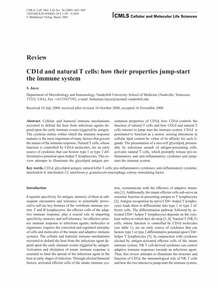
cells are essential early immune processes required for Tcell antigen processing and presentation. Antigen proces-sing and presentation are prerequisites for T cell antigenrecognition, activation and effector function [2]. Interna-lisation of infectious agents brings antigen to endoso-mal/lysosomal vesicles where they meet antigen present-ing molecules. The endosomal/lysosomal vesicles en-riched in antigen presenting molecules are called classII-enriched vesicles and the major histocompatibilitycomplex (MHC) class II compartment. The predominantantigen-presenting molecules in this compartment are theclass II molecules encoded by the MHC, hence theirname [8]. The MHC class II compartment also containsanother class of antigen-presenting molecules collec-tively called CD1 [9–11]. Thus, the delivery of infectiousagents to the MHC class II compartment followed bytheir processing provides a pool of derived products,some of which are presented to T cells by the antigen-pre-senting molecules.MHC class II belong to the classical peptide antigen-pre-senting molecules. They present peptide antigens toCD4+ helper T (Th) lymphocytes. Naive CD4+ Th cellsupon recognising antigen differentiate into Th1 or Th2effector cells. The decision to follow the Th1 or Th2 de-velopmental pathway depends on the cytokine milieu inwhich activation and differentiation of naive Th lym-phocytes occur. Thus, in the presence of IL-12 and/orIFN-g, activated Th cells differentiate into Th1 effectors,whereas in the presence of IL-4 they develop into Th2cells (fig. 1). It is in this context that NK T cells are pre-dicted to have a critical physiological role because theyproduce large amounts of cytokines, most notably IL-4,promptly, within 60–90 min of stimulation in vivothrough their antigen-specific receptors [12]. Recent stu-dies have suggested that NK T cells also play an impor-tant role in cross-talk between components of the innateimmune system as well as the adaptive system (fig. 1)[13–15]. These cross-talks are facilitated by cell-cell in-teractions as well as by pro-inflammatory and anti-in-flammatory cytokines secreted by NK T cells upon theiractivation by antigen.
Table 1. The nature of lymphocytes.
Lymphocyte subset Phenotype Antigen Immune function
Th CD4+8–, TCRab high MHC class II + peptide regulates cellular versus humoral effector function
CTL CD4–8+, TCRab high MHC class I + peptide adaptive cell-mediated immunity
NK T CD4+8–, CD4–8–, TCRab int, CD1d + glycolipid immune regulationCD5high, CD44high, CD122+,CD161high, Ly6Chigh
NK CD4–8–, TCRab–CD44high, MHC unrestricted viral and tumour immunityCD122+, CD161high
Three groups of b2m-associated molecules expressed byCD4+8+ thymocytes are known and include the non-clas-sical MHC class I-like molecules, H2Qa2 and H2TL, aswell as CD1d [22–26]. H2Qa2-negative and H2TL-nega-tive thymocytes support NK T cell ontogeny and function[16, 27]. Thus, the ontogeny and function of NK T cellswere predicted to be controlled by CD1d. NK T cell-de-rived hybridomas were specifically activated and hencestimulated to secrete IL-2 when co-cultured with cells in-fected with a recombinant vaccinia virus carrying thecDNA for mouse CD1d1 (rVV-CD1d1) but not rVV-H2Kb, a classical antigen-presenting molecule [22]. Si-milarly, freshly isolated thymic NK T cells co-culturedwith cells infected with rVV-CD1d1 were activated tosecrete IL-4 [22]. Finally, mice rendered CD1d deficient,through induced mutagenesis by homologous recombina-tion, did not develop NK T cells [28–30]. These data con-clusively demonstrated that CD1d indeed controls NK Tcell ontogeny and function.CD1d belongs to a group of evolutionarily conserved an-tigen-presenting molecules collectively called CD1 [31].On the basis of nucleotide and amino acid sequence ho-mology, CD1 is divided in two groups: group I CD1, con-sistsing of CD1a, CD1b, CD1c and CD1e, and group II,comprising CD1d [32]. Most mammals studied expressCD1d [33–36]; it is yet to be found in miniature swine andguinea pigs [37, 38]. In humans, CD1d is one of four func-tional CD1 isotypes [33, 39, 40], whereas in mice [34] andrats [41], it is the only representative. Mice carry twoCD1d genes, CD1D1 and CD1D2 [34]. CD1D2 is a pseu-dogene [42]; hence, targeted disruption of CD1D1 wassufficient to render mice CD1 deficient and hence defec-tive in NK T cell development and function as well [30].The expression of CD1d is tissue restricted. It is ex-pressed by specialised antigen-presenting cells such assplenic dendritic cells and marginal-zone B lymphocy-tes [43]; marginal-zone as well as a novel subset of follicu-lar B cells express higher levels of CD1d [44]. CD1d isadditionally expressed by CD4+8+ double-positive thy-mocytes [19, 23, 45, 46], hepatocytes [47, 48] and albeitcontroversial [48], also by intestinal epithelium [45, 47,49]. In all these cell types, CD1d expression is constitu-tive, and does not need inductive factors such as gran-ulocyte-macrophage colony-stimulating factor (GM-CSF) and IL-4, factors known to up-regulate the low le-vels of human group I CD1 expression on dendritic cells.Unlike group I CD1, CD1d expression is not up-regulat-ed by GM-CSF and IL-4 [46]. Notwithstanding, the ex-pression of mouse CD1d1 moderately increases in thepresence of GM-CSF and IL-4 [48]. Whether other fac-tors induce CD1d expression is currently unknown. Inthis regard, it is noteworthy that peritoneal macrophageselicited by thioglycolate express high levels of CD1d1(A.D. De Silva and S. Joyce, unpublished data); whetherthe high levels of CD1d1 expression was due to factors
444 S. Joyce CD1d and NK T cells
Thus this review focuses specifically on CD1d of bothmice and humans and draws upon parallels as well as dis-similarities with other CD1 molecules where essential.Whereas diverse T cells react to CD1d, the focus here ison a subset of these that secrete IL-4 upon in vivo activa-tion; in mice they express the invariant Va14Ja15 T cellreceptor (TCR) and in humans Va24JaQ.
CD1d, a glycolipid-presenting molecule
A series of studies led to the identification of CD1d as theantigen presenting molecule that controls NK T cell de-velopment and function. In the first of these studies, NKT cells failed to develop in b2-microglobulin (b2m)-defi-cient mice. Adoptive transfer of wild-type foetal livercells, as the donor for haematopoietic stem cells, into ir-radiated b2m-deficient mice reconstituted the thymuswith NK T cells [16–18]. Additionally, unlike the onto-geny of mainstream CD4+ and CD8+ T lymphocytes, theMHC type of the donor did not alter NK T cell develop-ment. These data suggested that the development of NKT cells is under the control of a conserved molecule(s)that associates with b2m and is expressed by haemato-poietic cells of the thymus [17]. Moreover, the ontogenyof NK T cells depended on the expression of the b2m-as-sociated molecule by CD4+8+ thymocytes [19]. Cu-riously, this is unlike the development of mainstreamCD8+ and CD4+ cells, which require the expression ofMHC class I and class II molecules by the thymic epithe-lium [20, 21].
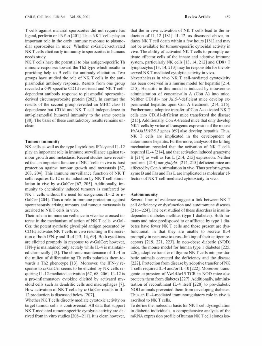
Figure 1. An overview of an immune response. Antigen recogni-tion by naive CD4+ T lymphocytes leads them to differentiate intotype 1 (Th1) or type 2 (Th2) effector cells. The differentiation path-way followed by activated CD4+ Th lymphocytes depends on thecytokine milieu in which they develop. Thus, in the presence of IL-12 secreted by dendritic cells and/or IFN-g secreted by activatedNK cells, activated Th cells differentiate into Th1 effectors. Like-wise, in the presence of IL-4 whose source is thought to be NK Tcells, activated Th cells develop into Th2 effectors. Solid arrows re-present established source and function of the soluble factors,whereas the dashed arrow indicates potential source.
released by macrophages, other leukocytes or tissue cellssurrounding the peritoneal cavity is currently not known.Strikingly, CD1d is not expressed by the cortical epithe-lium of the thymus, a site where MHC class I and class IIexpression is essential for positive and negative selectionof mainstream T lymphocytes. Whereas circulating hu-man monocytes as well as B and T lymphocytes expressCD1d [46], these cell types in mice do not. The tissue-re-stricted expression of CD1d has implications for the on-togeny and function of NK T cells.
CD1d structure dictates its functionPhylogenetically, the primary structure of CD1d is dis-tantly related to the MHC-encoded class I and class IIantigen-presenting molecules [5, 50]. Akin to MHC classI genes, CD1D encodes the a chain of CD1d, which non-covalently associates with b2m during assembly [34, 43].The nascent a chain is ~ 33,000 Da [43] which is modi-fied by four (in human) or five (in mouse) N-linked oli-gosaccharides [33, 34]. Thus, the mature a chain is~50,000–55,000 Da in molecular weight [34, 43; A.D.De Silva and S. Joyce, unpublished data]. The a chain ofCD1d, akin to MHC class I molecules, consists of threeextracellular domains, a1, a2 and a3, anchored to theplasma membrane through a transmembrane domainwhich is followed by a short cytosolic tail.
The three-dimensional structure of CD1dThe solution of the three-dimensional structure by X-raycrystallography revealed that mouse CD1d1 topologi-cally resembles the classical MHC class I and class II an-tigen-presenting molecules [51]. Consistent with its do-main organisation and its association with b2m, the three-dimensional structure of CD1d closely resembles that ofMHC class I molecules. Thus the membrane-distal a1and a2 domains fold to form a deep cleft. This cleft isbounded laterally by two a helices and at the floor by twob sheets, each made of four anti-parallel b strands; one ahelix and one b sheet are contributed by each domain.The a3 domain and b2m attain an immunoglobulin-likefold; they interact extensively with each other and alsowith the underside of the b sheet floor of the a1a2 su-perdomain. Because the primary structures of CD1dhomologues and its paralogues (group I CD1) are highlyconserved, the three-dimensional structures of all knownCD1 molecules are predicted to closely resemble that ofCD1d1 [31, 50].
The antigen-binding site of CD1dThe cleft formed by the design of the a1a2 superdo-main is predicted to be the antigen-binding site of CD1d,based on the finding that the X-ray defraction data con-tained extra electron density within this cleft, whichcould not be solved from the known primary structures
of the CD1d1 a chain and b2m [51]. Careful examinationof the extra electron density suggested the presence of analiphatic hydrocarbon chain within the cleft. Extra elec-tron density has also previously been observed in thecrystal structures of human MHC class I molecules,HLA-A2 and HLA-A68 [52–54]; a human cell line ex-pressing the MHC class I molecules was the source ofHLA-A2 and HLA-A68 used in the X-ray crystallogra-phic studies. Biochemical studies following the extrac-tion of the low- molecular weight fraction associatedwith MHC class I molecules have revealed that the extraelectron density observed in the crystal structures wasthe naturally processed peptide ligands that associatewith these antigen-presenting molecules during their as-sembly [55, 56]. Moreover, point mutations that alteramino acid residues within the putative antigen bindingsite of CD1d1 affect the presentation of a glycolipidantigen to an NK T cell hybridoma [57]. Taken together,the data provide compelling evidence that the cleft ofCD1d1, occupied by the extra electron density in itscrystal structure, is most likely the antigen-binding siteof CD1 molecules.Topological similarities notwithstanding, the three-di-mensional structure of CD1d1 differs from that of anMHC class I molecule; most of these differences arefound in the cleft [51]. The antigen-binding site of CD1d1is narrow at the opening that leads into two deep pocketslined exclusively by non-polar residues. MHC class Imolecules, on the other hand, have wider access to theircleft that is lined indiscriminately by polar and non-polaramino acid residues [58]. The opening to the CD1d1 clefthas interesting structural features. It begins approximat-ely at the centre of the antigen-binding site and extends tothe carboxyl terminus of the cleft [51]. It is bounded atboth ends, akin to the class I antigen-binding site, andthus is distinct from the open-ended groove of class IImolecules. Additionally, pockets A – F observed in classI grooves that accommodate side chains of peptides havefused in CD1d1 to form two deep pockets, the larger A¢and the smaller F¢ pockets. The A¢ pocket is largely co-vered at the top by interactions between residues of thea1 and a2 helices, i.e. a roof is formed over pocket A¢.Thus, the hydrophobic antigen-binding site of CD1d isaccessible to the ligand through a narrow opening [51].These differences in the physicochemical properties ofthe CD1d1 and class I antigen-binding sites have functio-nal consequences. The former specialises in glycolipidand the latter in peptide antigen presentation to specific T cells.
The ligands of CD1dThe structural feature of CD1 molecules discussed aboveand their ability to control T cell responses discussed be-low suggest that they can function as antigen-presentingmolecules. The best evidence for an antigen-presenting
CMLS, Cell. Mol. Life Sci. Vol. 58, 2001 Review Article 445
446 S. Joyce CD1d and NK T cells
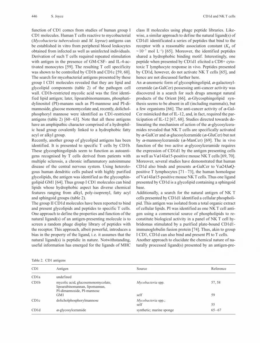
function of CD1 comes from studies of human group ICD1 molecules. Human T cells reactive to mycobacterial(Mycobacteria tuberculosis and M. leprae) antigens canbe established in vitro from peripheral blood leukocytesobtained from infected as well as uninfected individuals.Derivation of such T cells required repeated stimulationwith antigen in the presence of GM-CSF- and IL-4-ac-tivated monocytes [59]. The resulting T cell specificitywas shown to be controlled by CD1b and CD1c [59, 60].The search for mycobacterial antigens presented by thesegroup I CD1 molecules revealed that they are lipid andglycolipid components (table 2) of the pathogen cell wall. CD1b-restricted mycolic acid was the first identi-fied lipid antigen; later, lipoarabinomannans, phosphati-dylinositol (PI)-manans such as PI-mannose and PI-di-mannoside, glucose monomycolate and, recently, dolichol-phosphoryl mannose were identified as CD1-restrictedantigens (table 2) [60–63]. Note that all these antigenshave an amphipathic character comprised of a hydrophil-ic head group covalently linked to a hydrophobic fattyacyl or alkyl group.Recently, another group of glycolipid antigens has beenidentified. It is presented to specific T cells by CD1b.These glycosphingolipids seem to function as autoanti-gens recognised by T cells derived from patients withmultiple sclerosis, a chronic inflammatory autoimmunedisease of the central nervous system. Using heterolo-gous human dendritic cells pulsed with highly purifiedglycolipids, the antigen was identified as the glycosphin-golipid GM1 [64]. Thus group I CD1 molecules can bindlipids whose hydrophobic aspect has diverse chemicalfeatures ranging from alkyl, poly-isoprenyl, fatty acyland sphingoid groups (table 2).The group II CD1d molecules have been reported to bindand present glycolipids and peptides to specific T cells.One approach to define the properties and function of thenatural ligand(s) of an antigen-presenting molecule is toscreen a random phage display library of peptides withthe receptor. This approach, albeit powerful, introduces abias in the property of the ligand, i.e. it assumes that thenatural ligand(s) is peptidic in nature. Notwithstanding,useful information has emerged for the ligands of MHC
class II molecules using phage peptide libraries. Like-wise, a similar approach to define the natural ligand(s) ofCD1d1 identificated a series of peptides that bind to thereceptor with a reasonable association constant (Kd of~10–7 mol L–1) [65]. Moreover, the identified peptidesshared a hydrophobic binding motif. Interestingly, onepeptide when presented by CD1d1 elicited a CD8+ cyto-toxic T lymphocyte response in vivo. Peptides presentedby CD1d, however, do not activate NK T cells [65], andhence are not discussed further here.An a-anomeric form of glycosphingolipid, a-galactosyl-ceramide (a-GalCer) possessing anti-cancer activity wasdiscovered in a search for such drugs amongst naturalproducts of the Orient [66]. a-Glycosphingolipid syn-thesis seems to be absent in all (including mammals), buta few organisms [66]. The anti-cancer activity of a-Gal-Cer mimicked that of IL-12, and, in fact, required the par-ticipation of IL-12 [67, 68]. Studies directed towards de-lineating the mechanism of action of the a-glycosylcera-mides revealed that NK T cells are specifically activatedby a-GalCer and a-glucosylceramide (a-GluCer) but notby a-mannosylceramide (a-ManCer) [69]. The in vivofunction of the two active a-glycosylceramide requiresthe expression of CD1d1 by the antigen presenting cellsas well as Va14Ja15-positive mouse NK T cells [69, 70].Moreover, several studies have demonstrated that humanCD1d also binds and presents a-GalCer to Va24JaQ-positive T lymphocytes [71–73], the human homologueof Va14Ja15-positive mouse NK T cells. Thus one ligandpresented by CD1d is a glycolipid containing a sphingoidbase.Additionally, a search for the natural antigen of NK Tcells presented by CD1d1 identified a cellular phospholi-pid. This antigen was isolated from a total organic extractof cellular lipids. PI was identified as one NK T cell anti-gen using a commercial source of phospholipids to re-constitute biological activity in a panel of NK T cell hy-bridomas stimulated by a purified plate-bound CD1d1-immunoglobulin fusion protein [74]. Thus, akin to groupI CD1, CD1d can also bind and present PI to T cells.Another approach to elucidate the chemical nature of na-turally processed ligand(s) presented by an antigen-pre-
Table 2. CD1 antigens
CD1 Antigen Source Reference
CD1a undefined
CD1b mycolic acid, glucosemonomycolate, Mycobacteria spp. 57, 58lipoarabinomannan, lipomannan,PI-dimannoside, PI-mannoseGM1 self 59
CD1c dolicholphosphorylmannose Mycobacteria spp.;self 55
CD1d a-glycosylceramide synthetic; marine sponge 65–67
CMLS, Cell. Mol. Life Sci. Vol. 58, 2001 Review Article 447
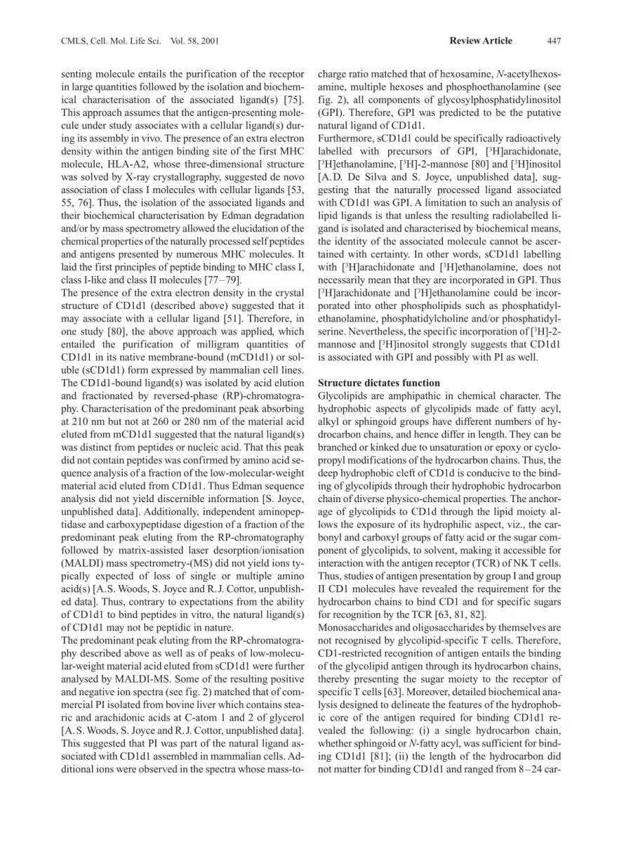
senting molecule entails the purification of the receptorin large quantities followed by the isolation and biochem-ical characterisation of the associated ligand(s) [75]. This approach assumes that the antigen-presenting mole-cule under study associates with a cellular ligand(s) dur-ing its assembly in vivo. The presence of an extra electrondensity within the antigen binding site of the first MHCmolecule, HLA-A2, whose three-dimensional structurewas solved by X-ray crystallography, suggested de novoassociation of class I molecules with cellular ligands [53,55, 76]. Thus, the isolation of the associated ligands andtheir biochemical characterisation by Edman degradationand/or by mass spectrometry allowed the elucidation of thechemical properties of the naturally processed self peptidesand antigens presented by numerous MHC molecules. Itlaid the first principles of peptide binding to MHC class I,class I-like and class II molecules [77–79].The presence of the extra electron density in the crystalstructure of CD1d1 (described above) suggested that itmay associate with a cellular ligand [51]. Therefore, inone study [80], the above approach was applied, whichentailed the purification of milligram quantities ofCD1d1 in its native membrane-bound (mCD1d1) or sol-uble (sCD1d1) form expressed by mammalian cell lines.The CD1d1-bound ligand(s) was isolated by acid elutionand fractionated by reversed-phase (RP)-chromatogra-phy. Characterisation of the predominant peak absorbingat 210 nm but not at 260 or 280 nm of the material acideluted from mCD1d1 suggested that the natural ligand(s)was distinct from peptides or nucleic acid. That this peakdid not contain peptides was confirmed by amino acid se-quence analysis of a fraction of the low-molecular-weightmaterial acid eluted from CD1d1. Thus Edman sequenceanalysis did not yield discernible information [S. Joyce,unpublished data]. Additionally, independent aminopep-tidase and carboxypeptidase digestion of a fraction of thepredominant peak eluting from the RP-chromatographyfollowed by matrix-assisted laser desorption/ionisation(MALDI) mass spectrometry-(MS) did not yield ions ty-pically expected of loss of single or multiple aminoacid(s) [A.S. Woods, S. Joyce and R.J. Cottor, unpublish-ed data]. Thus, contrary to expectations from the abilityof CD1d1 to bind peptides in vitro, the natural ligand(s)of CD1d1 may not be peptidic in nature.The predominant peak eluting from the RP-chromatogra-phy described above as well as of peaks of low-molecu-lar-weight material acid eluted from sCD1d1 were furtheranalysed by MALDI-MS. Some of the resulting positiveand negative ion spectra (see fig. 2) matched that of com-mercial PI isolated from bovine liver which contains stea-ric and arachidonic acids at C-atom 1 and 2 of glycerol[A.S. Woods, S. Joyce and R.J. Cottor, unpublished data].This suggested that PI was part of the natural ligand as-sociated with CD1d1 assembled in mammalian cells. Ad-ditional ions were observed in the spectra whose mass-to-
charge ratio matched that of hexosamine, N-acetylhexos-amine, multiple hexoses and phosphoethanolamine (seefig. 2), all components of glycosylphosphatidylinositol(GPI). Therefore, GPI was predicted to be the putativenatural ligand of CD1d1.Furthermore, sCD1d1 could be specifically radioactivelylabelled with precursors of GPI, [3H]arachidonate,[3H]ethanolamine, [3H]-2-mannose [80] and [3H]inositol[A.D. De Silva and S. Joyce, unpublished data], sug-gesting that the naturally processed ligand associatedwith CD1d1 was GPI. A limitation to such an analysis oflipid ligands is that unless the resulting radiolabelled li-gand is isolated and characterised by biochemical means,the identity of the associated molecule cannot be ascer-tained with certainty. In other words, sCD1d1 labellingwith [3H]arachidonate and [3H]ethanolamine, does notnecessarily mean that they are incorporated in GPI. Thus[3H]arachidonate and [3H]ethanolamine could be incor-porated into other phospholipids such as phosphatidyl-ethanolamine, phosphatidylcholine and/or phosphatidyl-serine. Nevertheless, the specific incorporation of [3H]-2-mannose and [3H]inositol strongly suggests that CD1d1is associated with GPI and possibly with PI as well.
Structure dictates functionGlycolipids are amphipathic in chemical character. Thehydrophobic aspects of glycolipids made of fatty acyl,alkyl or sphingoid groups have different numbers of hy-drocarbon chains, and hence differ in length. They can bebranched or kinked due to unsaturation or epoxy or cyclo-propyl modifications of the hydrocarbon chains. Thus, thedeep hydrophobic cleft of CD1d is conducive to the bind-ing of glycolipids through their hydrophobic hydrocarbonchain of diverse physico-chemical properties. The anchor-age of glycolipids to CD1d through the lipid moiety al-lows the exposure of its hydrophilic aspect, viz., the car-bonyl and carboxyl groups of fatty acid or the sugar com-ponent of glycolipids, to solvent, making it accessible forinteraction with the antigen receptor (TCR) of NK T cells.Thus, studies of antigen presentation by group I and groupII CD1 molecules have revealed the requirement for thehydrocarbon chains to bind CD1 and for specific sugarsfor recognition by the TCR [63, 81, 82].Monosaccharides and oligosaccharides by themselves arenot recognised by glycolipid-specific T cells. Therefore,CD1-restricted recognition of antigen entails the bindingof the glycolipid antigen through its hydrocarbon chains,thereby presenting the sugar moiety to the receptor ofspecific T cells [63]. Moreover, detailed biochemical ana-lysis designed to delineate the features of the hydrophob-ic core of the antigen required for binding CD1d1 re-vealed the following: (i) a single hydrocarbon chain,whether sphingoid or N-fatty acyl, was sufficient for bind-ing CD1d1 [81]; (ii) the length of the hydrocarbon did not matter for binding CD1d1 and ranged from 8–24 car-

448 S. Joyce CD1d and NK T cells
bon chains, the shortest and the longest tested [81]; (iii)kinked hydrocarbons (e.g. arachidonate) also bind CD1d1[80]; (iv) the dissociation constant for the binding of gly-cosphingolipids and phospholipids to CD1d1 is similar,~10–7 mol l–1 [80, 82]. Moreover, the affinity of the lipidfor CD1d1 is similar to that of lipids for group I CD1 [80,82, 83]. Additionally, the dissociation constant for lipid-CD1 interactions is similar to those reported for peptidesand their cognate antigen-presenting molecules [84, 85].Thus, the requirements for lipid-CD1 interactions are ra-ther loose compared to peptide-MHC interactions; this isdictated by the physico-chemical features of the respec-tive antigen-binding sites. In this regard, the resemblanceof the CD1d1 antigen-binding site to that of non-specificfatty acid-binding protein is noteworthy [51].T cells that recognise glycolipid antigen can discriminatebetween the different hexoses linked to the lipid [63, 69].In nearly all cases, the first hexose forms the epitope forthe specific T cell: glucose, galactose or mannose canform the epitopes [63, 69]. One study demonstrated thatCD1d1-restricted NK T cells expressing Va14Ja15/Vb8.2 transgenic receptors recognised a-GluCer and a-GalCer but not a-ManCer [69]. Glucose and galactose dif-fer from mannose by the axial (mannopyranose) and equi-torial (glucopyranose and galactopyranose) orientation of
the hydroxyl group of C-atom 2 of the pyranose ring. Ad-ditionally, glucose and galactose differ from each other bythe axial (galactopyranose) and equitorial (glucopyranose)predisposition of the hydroxyl at C-atom 4. Thus hydroxylsat C-atom 2, 3 and 6 are common to both glucose and ga-lactose and, hence, are predicted to impart specificity ofthe interaction between the presented glycolipid and theTCR. Because the above studies were performed with amonoclonal TCR, the possible presence of a-ManCer-re-active NK T cells remains possible. Similar discriminationof sugar epitopes as described for CD1d1-presented gly-colipids are also observed in the interaction of glycolipidspresented by group I CD1 to cognate TCR [63].
Topological biochemistry of antigen presentation by CD1dIt is evident from the studies described above that lipidsform the natural antigen(s) of NK T cells. A search for thenatural antigen of NK T cells has revealed a few featuresof lipid antigen presentation. The first of these is that theantigen is co-expressed by most cells that express CD1d;that is, CD1d expressed at the cell surface contains abound cellular lipid that activates NK T cells and hybri-domas and hence does not require exogenous addition of
Figure 2. The structure of a mammalian glycosylphosphatidylinositol. The mass-to-charge ratio (m/z) of the positive and negative ions ob-served upon MALDI-MS analysis of the natural ligand isolated from membrane-bound and soluble mouse CD1d1 are indicated on the left.It offers one solution to the chemical nature of the CD1d1-associated ligand.
CMLS, Cell. Mol. Life Sci. Vol. 58, 2001 Review Article 449
antigen [10, 86–88]. Second, presentation of antigen toNK T cells does not depend on transporters-associatedwith antigen processing (TAPs) [87, 89] or H2Ma [S.Mendiratta, A. Boesteanu, S. Joyce and L. Van Kaer, un-published data] molecules essential for antigen presenta-tion by MHC class I [90] and class II [91], respectively.Both TAP1- and H2Ma-deficient mice develop NK Tcells [92; Mendiratta et al. unpublished data]. Additio-nally, thymocytes from these animals as well as TAP1-and TAP2-deficient cell lines expressing CD1d1 are alsoable to activate Va14Ja15-positive NK T cell hybrido-mas [89]. Thus, the rules for lipid antigen-presentation byCD1d are distinct from those of peptide antigen presenta-
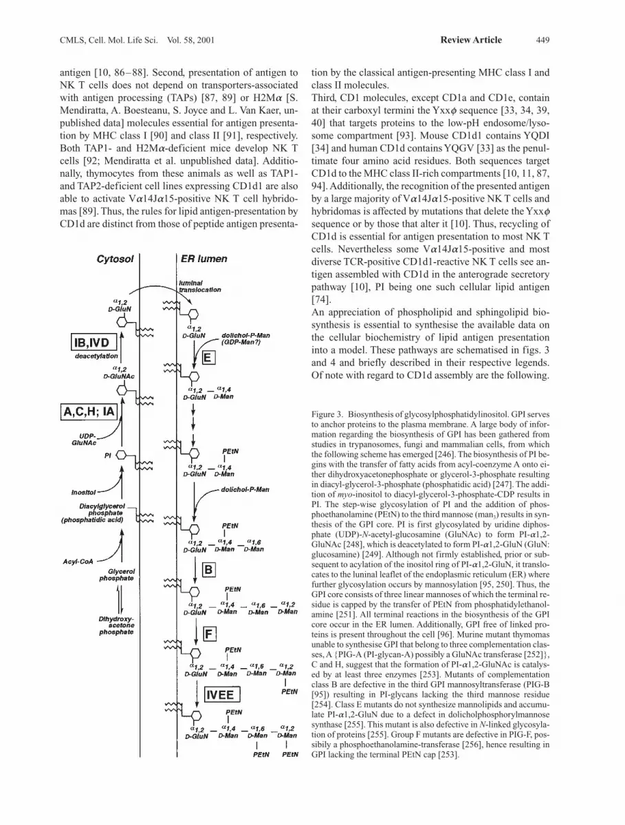
Figure 3. Biosynthesis of glycosylphosphatidylinositol. GPI servesto anchor proteins to the plasma membrane. A large body of infor-mation regarding the biosynthesis of GPI has been gathered fromstudies in trypanosomes, fungi and mammalian cells, from whichthe following scheme has emerged [246]. The biosynthesis of PI be-gins with the transfer of fatty acids from acyl-coenzyme A onto ei-ther dihydroxyacetonephosphate or glycerol-3-phosphate resultingin diacyl-glycerol-3-phosphate (phosphatidic acid) [247]. The addi-tion of myo-inositol to diacyl-glycerol-3-phosphate-CDP results inPI. The step-wise glycosylation of PI and the addition of phos-phoethanolamine (PEtN) to the third mannose (man3) results in syn-thesis of the GPI core. PI is first glycosylated by uridine diphos-phate (UDP)-N-acetyl-glucosamine (GluNAc) to form PI-a1,2-GluNAc [248], which is deacetylated to form PI-a1,2-GluN (GluN:glucosamine) [249]. Although not firmly established, prior or sub-sequent to acylation of the inositol ring of PI-a1,2-GluN, it translo-cates to the luninal leaflet of the endoplasmic reticulum (ER) wherefurther glycosylation occurs by mannosylation [95, 250]. Thus, theGPI core consists of three linear mannoses of which the terminal re-sidue is capped by the transfer of PEtN from phosphatidylethanol-amine [251]. All terminal reactions in the biosynthesis of the GPIcore occur in the ER lumen. Additionally, GPI free of linked pro-teins is present throughout the cell [96]. Murine mutant thymomasunable to synthesise GPI that belong to three complementation clas-ses, A {PIG-A (PI-glycan-A) possibly a GluNAc transferase [252]},C and H, suggest that the formation of PI-a1,2-GluNAc is catalys-ed by at least three enzymes [253]. Mutants of complementationclass B are defective in the third GPI mannosyltransferase (PIG-B[95]) resulting in PI-glycans lacking the third mannose residue[254]. Class E mutants do not synthesize mannolipids and accumu-late PI-a1,2-GluN due to a defect in dolicholphosphorylmannosesynthase [255]. This mutant is also defective in N-linked glycosyla-tion of proteins [255]. Group F mutants are defective in PIG-F, pos-sibily a phosphoethanolamine-transferase [256], hence resulting inGPI lacking the terminal PEtN cap [253].
tion by the classical antigen-presenting MHC class I andclass II molecules.Third, CD1 molecules, except CD1a and CD1e, containat their carboxyl termini the Yxxf sequence [33, 34, 39,40] that targets proteins to the low-pH endosome/lyso-some compartment [93]. Mouse CD1d1 contains YQDI[34] and human CD1d contains YQGV [33] as the penul-timate four amino acid residues. Both sequences targetCD1d to the MHC class II-rich compartments [10, 11, 87,94]. Additionally, the recognition of the presented antigenby a large majority of Va14Ja15-positive NK T cells andhybridomas is affected by mutations that delete the Yxxfsequence or by those that alter it [10]. Thus, recycling ofCD1d is essential for antigen presentation to most NK Tcells. Nevertheless some Va14Ja15-positive and mostdiverse TCR-positive CD1d1-reactive NK T cells see an-tigen assembled with CD1d in the anterograde secretorypathway [10], PI being one such cellular lipid antigen[74].An appreciation of phospholipid and sphingolipid bio-synthesis is essential to synthesise the available data onthe cellular biochemistry of lipid antigen presentationinto a model. These pathways are schematised in figs. 3and 4 and briefly described in their respective legends. Of note with regard to CD1d assembly are the following.
450 S. Joyce CD1d and NK T cells
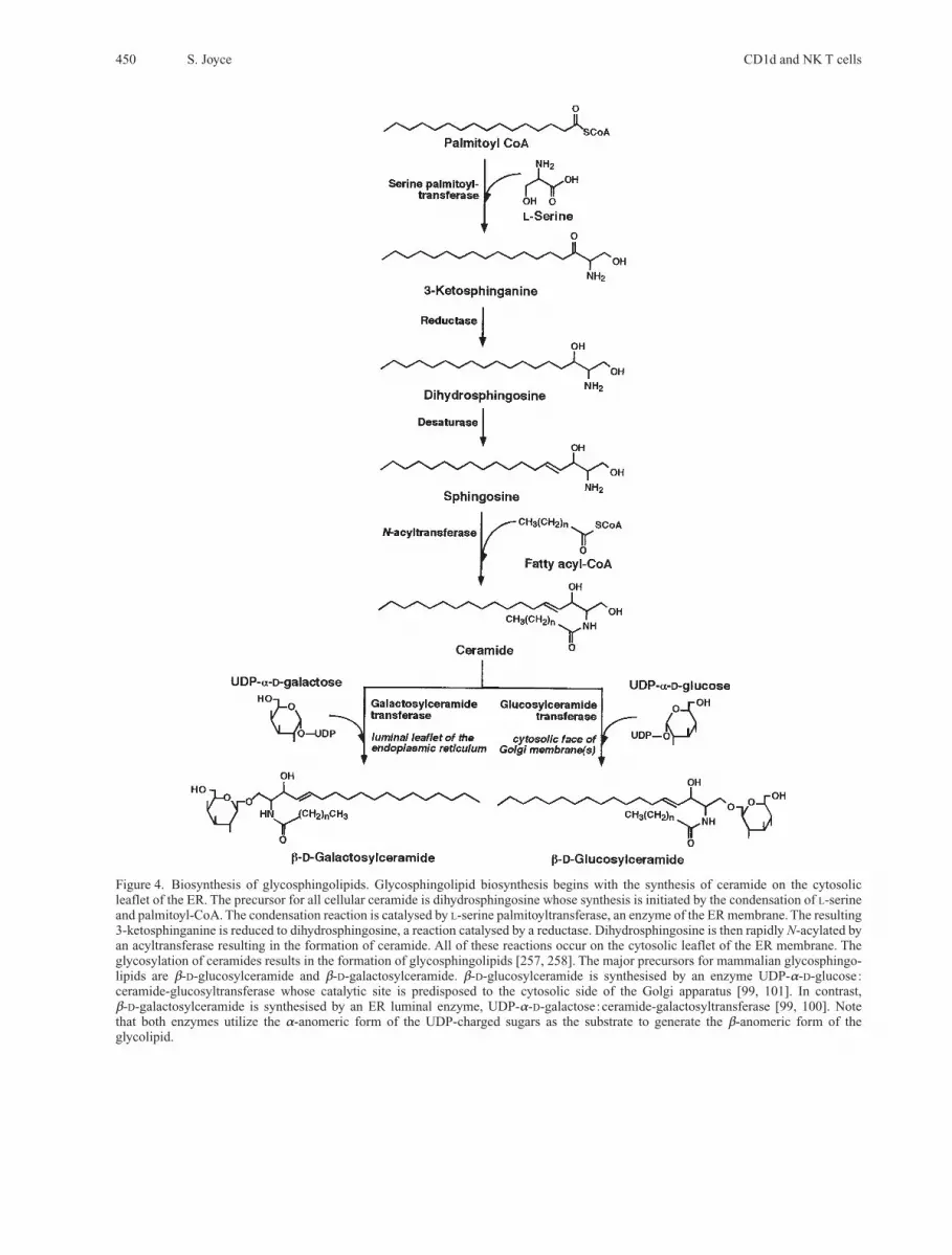
Figure 4. Biosynthesis of glycosphingolipids. Glycosphingolipid biosynthesis begins with the synthesis of ceramide on the cytosolicleaflet of the ER. The precursor for all cellular ceramide is dihydrosphingosine whose synthesis is initiated by the condensation of L-serineand palmitoyl-CoA. The condensation reaction is catalysed by L-serine palmitoyltransferase, an enzyme of the ER membrane. The resulting3-ketosphinganine is reduced to dihydrosphingosine, a reaction catalysed by a reductase. Dihydrosphingosine is then rapidly N-acylated byan acyltransferase resulting in the formation of ceramide. All of these reactions occur on the cytosolic leaflet of the ER membrane. Theglycosylation of ceramides results in the formation of glycosphingolipids [257, 258]. The major precursors for mammalian glycosphingo-lipids are b-D-glucosylceramide and b-D-galactosylceramide. b-D-glucosylceramide is synthesised by an enzyme UDP-a-D-glucose:ceramide-glucosyltransferase whose catalytic site is predisposed to the cytosolic side of the Golgi apparatus [99, 101]. In contrast, b-D-galactosylceramide is synthesised by an ER luminal enzyme, UDP-a-D-galactose:ceramide-galactosyltransferase [99, 100]. Note that both enzymes utilize the a-anomeric form of the UDP-charged sugars as the substrate to generate the b-anomeric form of the glycolipid.
CMLS, Cell. Mol. Life Sci. Vol. 58, 2001 Review Article 451
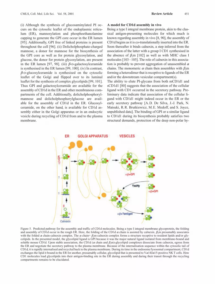
(i) Although the synthesis of glucosaminylated PI oc-curs on the cytosolic leaflet of the endoplasmic reticu-lum (ER), mannosylation and phosphoethanolaminecapping to generate the GPI core occur in the ER lumen[95]. Additionally, GPI free of linked proteins is presentthroughout the cell [96]. (ii) Dolicholphosphate-chargedmannose, a donor for mannose for the biosynthesis of the GPI core as well as for protein glycosylation, andglucose, the donor for protein glycosylation, are presentin the ER lumen [97, 98]. (iii) b-D-galactosylceramide is synthesised in the ER lumen [99, 100]. (iv) In contrast,b-D-glucosylceramide is synthesised on the cytosolicleaflet of the Golgi and flipped over to its luminal leaflet for the synthesis of complex glycolipids [99, 101].Thus GPI and galactosylceramide are available for theassembly of CD1d in the ER and other membranous com-partments of the cell. Additionally, dolicholphosphoryl-mannose and dolicholphosphorylglucose are avail-able for the assembly of CD1d in the ER. Glucosyl-ceramide, on the other hand, is available for CD1d as-sembly either in the Golgi apparatus or in an endocyticvesicle during recycling of CD1d from and to the plasmamembrane.
A model for CD1d assembly in vivoBeing a type I integral membrane protein, akin to the clas-sical antigen-presenting molecules for which much isknown regarding assembly in vivo [8, 90], the assembly ofCD1d begins as it is co-translationally inserted into the ER.Soon thereafter it binds calnexin, a step inferred from theassociation of the latter with a group I CD1 synthesised inthe absence of b2m [102] as well as with MHC class Imolecules [103–105]. The role of calnexin in this associa-tion is probably to prevent aggregation of unassembled achains. The monomeric a chain then assembles with b2mforming a heterodimer that is receptive to ligands of the ERand/or the downstream vesicular compartment(s).The ability to elute PI-glycans from both mCD1d1 andsCD1d1 [80] suggests that the association of the cellularligand with CD1 occurred in the secretory pathway. Pre-liminary data indicate that association of the cellular li-gand with CD1d1 might indeed occur in the ER or theearly secretory pathway [A.D. De Silva, J.-J. Park, N.Matsuki, R.R. Brutkiewicz, M.E. Medoff, and S. Joyce,unpublished data]. The binding of GPI or a similar ligandto CD1d1 during its biosynthesis probably satisfies twostructural demands, protection of the deep non-polar hy-
Figure 5. Predicted pathway for the assembly and traffic of CD1d molecules. Being a type I integral membrane glycoprotein, the foldingand assembly of CD1d occur in the rough ER. Here, the folding of the CD1d a chain is assisted by calnexin. b2m presumably associateswith the folded a chain-calnexin complex. The a chain+ b2m-calnexin complex forms a structure receptive to resident lipids and/or gly-colipids. In the presented model, the glycolipid ligand is GPI because it was the major natural ligand isolated from membrane-bound andsoluble mouse CD1d. Upon stable association, the CD1d (a chain and b2m)-glycolipid complexes dissociate from calnexin, egress fromthe ER and negotiate the secretory pathway to the plasma membrane. Because of the internalisation sequence within the cytosolic tail ofCD1d, it is rapidly internalised and recycled back to the plasma membrane. During its time in the endosome/lysosomal compartment, CD1dexchanges the lipid it bound in the ER for another, presumably cellular, glycolipid that is presented to Va14Ja15-positive NK T cells. HowCD1 molecules load glycolipids into their antigen-binding site in the ER during assembly and during their transit through the recyclingcompartments remains to be elucidated.
NK T cells
NK T cells are an unusual subset of lymphocytes. Pheno-typically they express molecules on their cell surface char-acteristic of NK and of T cells, and hence their name. NKcell-specific markers expressed by NK T cells includeCD161 (NKR-P1C, also known as NK1.1 of mice orNKR-P1A in humans) [87, 106, 107], some members ofthe Ly49 (a few express Ly49A or Ly49G2 and a largemajority express Ly49C/I) family of proteins during de-velopment [108] and low levels CD16, which is up-regu-lated upon activation [109]. Of the T cell-specific mar-kers, NK T cells express the TCR; most NK T cells ex-press the ab TCR and are described in detail below, butsome express the gd TCR [110, 111] whose biology re-mains unknown and hence is not discussed further. Addi-tionally, NK T cells express markers shared by NK cellsand activated T lymphocytes (table 1); these include CD5,CD44, CD69, CD122 (IL-2 receptor b chain) [106, 109,112], Ly6C [113–116] and IL-7 receptor a (IL-7Ra)chain [112]. Moreover, akin to activated T lymphocytes,NK T cells express low levels of CD24 (heat stable anti-gen) [92, 117, 118] and CD62L (LECAM/MEL-14) [110,115]. NK T cells also express ICAM-1 and LFA1-1 [119,120]. Expression of these phenotypic markers is taken toindicate that NK T cells are in a state of chronic activa-tion.A proportion of NK T cells express CD4 co-receptorsnormally displayed by Th cells and the rest express nei-ther CD4 nor CD8 [16, 106, 107, 117,121–124]. The func-tional consequence if any, of such a phenotypic dicho-tomy is presently unclear. Curiously, however, CD4 ex-pression by NK T cells does not require functional MHCclass II molecules during development [16]. Moreover,CD4 expression itself is not required for NK T cell onto-geny [16], suggesting that this co-receptor may be dis-pensable and non-essential for NK T cell biology.
NK T cell ontogenyThymic development of T lymphocytes occurs in waves;the first wave is of gd T cells followed by mainstream abT cells [125]. Very few NK T cells are present at the birthof mouse pups; their development begins during postna-tal lymphopoiesis and peaks around 6 weeks of age [16,113]. NK T cells are thought to develop in the thymus andhome to preferential secondary lymphoid organs [16,122, 126, 127]. They account for about a million cells ineach lymphoid organ except for their rarity in peripheralblood and the lymph nodes [16, 18, 123] or complete ab-sence within the intraepithelial lymphocyte population[18]. They constitute 10–20% of mature T cells in thethymus, 20–30% of hepatic and bone marrow T cells,0.5–1.0% of splenocytes, and 0.1–0.5% of lymph nodeand peripheral blood mononuclear cells [18, 128]. Addi-
452 S. Joyce CD1d and NK T cells
drophobic cleft from collapse as well as occupying it witha ligand that might be easily exchanged for an antigen ina late secretory vesicle. Thus GPI might play a cha-perone-like role (fig. 5) akin to the function of MHC classII-associated invariant chain in class II assembly and traf-fic in vivo [8].CD1d1 expression in PIG-A (refer to fig. 3 for a descrip-tion of PIG mutants) mutants is normal. Additionally,Va14Ja15-positive and – negative NK T cells recogniseCD1d1 expressed by the mutant as effectively as they re-cognise wild-type CD1d1. Thus GPI, albeit a major natu-ral ligand of CD1d1, is neither essential for the assemblyof CD1d in vivo nor is it the natural antigen of NK T cells[88]. Therefore, in the absence of GPI, PI or another lipid(e.g. phosphatidylethanolamine, phosphatidylserine, do-licholphosphoryl-monosaccharides and b-D-galactosyl-ceramide) present in the ER may substitute for the func-tion of GPI in the assembly of CD1d in vivo. Followingassembly of CD1d in the ER, it negotiates the secretorypathway and arrives at the MHC class II-enriched ve-sicles either directly from the trans-Golgi or via theplasma membrane. Targeting to the class II-enriched ve-sicles depends on the Yxxf internalisation sequencefound at the cytosolic tail of CD1d [10, 11]. Here, CD1dmeets the naturally processed antigen(s) and brings themto the plasma membrane for presentation to NK T cells(fig. 5) [10].How lipid loading onto CD1d in the ER is accomplishedand how these lipids are exchanged for antigen in en-docytic vesicles remains an important unsolved question.Because lipids are membrane embedded, they need to be‘plucked out’ of the membrane and loaded into the anti-gen-binding site of CD1d. This event might require anelaborate molecular machinery similar to the peptide-loading complex essential for peptide-antigen assemblywith MHC class I and class II molecules [8, 90]. Alterna-tively, CD1d itself might play a role in loading itself withglycolipids. CD1d molecules have four conserved N-lin-ked glycosylation sites, asparagines 20, 42, 109 and 166[33, 34, 50]. All of these sites are predicted to be solventexposed based on the three-dimensional structure ofCD1d1 [51]. Preliminary data suggest that all five glyco-sylation sites in CD1d1, including the four conservedones, are indeed modified [A.D. De Silva and S. Joyce,unpublished data]. One or more of the conserved glyco-syl groups may function as a lectin(s), thereby binding tomembrane-associated glycolipids, ‘plucking’ them out ofthe membrane and facilitating the loading of CD1d mole-cules; the loading could occur in cis, i.e. loading itself, orin trans, i.e. loading another CD1d molecule. Whateverthe mechanism, its solution should provide insights intothe basis for lipid-protein interactions in vivo and into theevolution of the lipid antigen-presenting system in verte-brates.
tionally, NK T cells seem to be recruited to the perito-neum following intraperitoneal delivery of pathogenssuch as Salmonella choleraesuis and Listeria monocyto-geneses [129, 130]. Thus NK T cells home to critical ana-tomical sites where they could execute their immunologi-cal function promptly.NK T cells that develop in the thymus (NTthy) are char-acterised by the expression of a highly restricted TCR re-pertoire [16, 27, 87, 106, 107, 113, 117, 121, 122, 124,131–136] and the absence of CD8 co-receptor [16, 128].The NTthy TCR repertoire consists predominantly of theVa14Ja15 (formerly Ja281 [137]; 80–90%) TCR achain that preferentially pairs with Vb8.2 (40–60%),Vb7 (10–15%) or Vb2 (4–6%) TCR b chain [16, 133].Their development requires the expression of the b2m-de-pendent CD1d1 molecule [16–18, 28–30] by haemato-poietic cells of the thymus, the CD4+8+ double-positivethymocytes [19, 138]. Humans express the Va24JaQTCR a chain that predominantly pairs with the Vb11TCR b chain; both these TCR chains are homologous tothe mouse Va14Ja15 and Vb8.2 receptors [87,132–136]. As in mice, diverse TCR b chains pair with theVa24JaQ TCR a chain of human NK T cells [132, 135,136].Interestingly, nu/nu mice deficient in a functional thymusgland develop NK T cells, but akin to mainstream T lym-phocytes of nu/nu, they are present at very low numbers[107, 128, 139, 140]. NK T cells of nu/nu mice are pre-sent in the liver, spleen and bone marrow, constituting~0.5% of hepatic mononuclear cells, ~0.5% of B cell-depleted splenic and ~0.2% B cell-depleted bone marrowcells [128]. NK T cells, NK1.1+CD3+, develop in nu/numice reconstituted with syngeneic bone marrow-derivedcells. Additionally, syngeneic bone marrow transfer intoan adult thymectomised and irradiated host results in thedevelopment of NK T cells [139, 141]. Curiously, theyexpress CD8 co-receptors and are found in the liver andspleen of the reconstituted recipients [141]. Thus, in somequarters, these results have been taken to indicate extra-thymic development of NK T cells. Extrathymic T lym-phopoiesis does occur in normal mice; a large majority ofthe resulting T cells predominantly express CD8aa co-receptor as opposed to the CD8ab and CD4 phenotype ofthymic émigrés [142].Recently, a second type of NK T (NT peri) cells have beendescribed. They develop independent of CD1d expres-sion and most express CD8a co-receptor while the re-mainder are devoid of either CD4 or CD8 molecules[128]. Additionally, NTperi differs from NTthy in two keyways. (i) The former expresses a wider TCR repertoire[128] in contrast to the restricted TCR repertoire of NTthy
(described above). (ii) NTperi cells are predominantly pre-sent within the spleen and bone marrow and are thoughtto originate from the bone marrow [128]. In this regard it is noteworthy that the CD8+ NK T cells are negatively
selected in the thymus and hence are not present amongstthymocytes [16] or hepatic mononuclear cells. Thesefindings can then explain why nu/nu mice develop NK Tcells; they actually belong to the NTperi lineage and hencemay have an extrathymic origin. NTperi cells are not dis-cussed any further.Preliminary results using genetically altered mice sup-port a thymic origin for the IL-4-secreting NK T cells.Mice deficient in Janus kinase-3 (Jak3), the commoncytokine receptor g chain (gc)-associated kinase, lackmature NK T cells in the thymus, liver and spleen. Thy-mocyte-specific expression of Jak3 by the proximal pro-motor of Lck through transgenesis in Jak3-null micerescues the NK T cell deficiency. Splenic NK T cells ofthymocyte-specific Jak3 transgenic mice promptlysecrete IL-4 when stimulated in vivo. Thus, akin to main-stream T cells, NK T cell development occurs in thethymus; thence the functionally mature NK T cells hometo the peripheral lymphoid organs [M.E. Embers and S.Joyce, unpublished data]. That they home to theperipheral lymphoid organs is supported by the dra-matically reduced numbers of hepatic NK T cells in LFA-1-deficient mice despite an almost normal con-tingent of thymic NK T cells [119, 120].The thymocentric bias in NK T cell development not-withstanding, there is compelling evidence for extrathy-mic NK T cell ontogeny. One study documented rearran-gement of Va14 and Ja15 gene segments within day 9mouse foetal liver progenitors. Temporally, this rearran-gement precedes that of Tcr gene segments within mousefoetal thymus; here Tcr gene rearrangements occuraround day 13 or 14 of gestation. Hence development isthought to occur in situ without the need for the thymus[143]. Additional independent data support an extrathy-mic NK T cell ontogeny [144]. Cytokine-supplementedcultures of wild type and nu/nu foetal liver develop NK Tcells that express the invariant TCR a chain. Moreover,foetal liver cultures derived from b2m-deficient mice(which lack MHC class I and other related molecules) orfrom wild type mice in the presence of CD1d-specificmonoclonal antibody did not support the development ofNK T cells. Additional data revealed that SCID (recom-bination-deficient but CD1d-positive) foetal liver recon-stitutes NK1+ T cell development within b2m-deficient(recombination-competent but CD1d-deficient) foetal li-ver cultures. Therefore, foetal liver progenitors have thepotential to beget NK1+ T cells in situ, lending supportfor extrathymic NK T cell ontogeny [144].
Signals for NK T lymphocyte developmentTheir unique phenotypic features (see table 1 and theirfunction, prompt secretion of immunoregulatory cyto-kines upon activation (table 3), distinguish NK T cellsfrom mainstream T lymphocytes. Amongst naive lym-phocytes, NK T cells represent a single antigen-reactive T
CMLS, Cell. Mol. Life Sci. Vol. 58, 2001 Review Article 453
454 S. Joyce CD1d and NK T cells
lymphocyte population present at high frequency [145, 146] compared to that of any antigen-reactive Tlymphocytes [147–149]. The phenotype (CD44high
CD62Lnull) of NK T cells suggests that they were recentlyactivated. They home within the spleen around theCD1d+ marginal-zone B cells as well as within the liverwhere resident lymphocytes meet antigen. Additionally,the phenotype and frequency of NK T cells of mice main-tained in normal and germ-free conditions remain unal-tered [150]. Therefore, the activated phenotype and highfrequency of NK T cells as well as their prompt responseto antigen (discussed below) seem not to be due to con-stant confrontation with antigen in vivo. Thus NK T cellsmight be developmentally wired and maintained in vivoat or just below the threshold of activation so that they canpromptly respond to antigen.Their unique phenotypic and functional features haveprompted studies on the signals that commit precursorthymocytes to the NK T cell lineage and the ontogeny ofthese T lymphocytes. Whereas much information is avail-able on the development of mainstream T cells, very littleis known regarding NK T cell ontogeny. A detailed fatemap of mainstream T cells in the thymus based on thecurrent knowledge is summarised and represented sche-matically in the upper panel of figure 6. In this scheme,there are three critical check-points: the first is the deci-sion toward T lineage commitment; it follows the firstwave of precursor expansion in the thymus. Commitment
occurs at the CD25+ thymocyte stage or shortly there-after. gc, the IL-7Ra chain and Jak3 provide critical sig-nals for precursor expansion at the CD44+CD25+ stagein T lymphopoiesis. b selection controls the secondcheck-point in T cell ontogeny. Signals critical for attain-ing this stage of T lymphopoiesis include those relayed bythe TCRb-pre-Ta heterodimer through Lck, Zap70, Syk,Sox4, Mek and Tcf1. The final checkpoint entails positiveand negative selection of the MHC-restricted but self-to-lerant TCR repertoire. Signals transmitted through theTCR and CD4 or CD8 co-receptors are critical at thisstage of T lymphopoiesis. Such signals are transduced in-tracellularly by Lck, Zap70, Vav, Itk, Raf, Ras and Mekand involve two transcription factors, NFkB [A. Moraand M. Boothby, personal communication] and Irf1.Thence the functionally mature MHC-restricted self-tolerant T lymphocytes home to the peripheral lymphoidorgans [reviewed in refs 151, 152–155].Several of the signals required for the development ofmainstream T cells are also required for NK T cell onto-geny. These include signals relayed through gc that aretransduced intracellularly by Jak3. gc- and Jak3-deficientmice completely lack mature NK T cells [112, 118]; M.E.Embers and S. Joyce, unpublished data]. Neverthelessstranscripts of functionally rearranged Va14Ja15 TCR achain are detected amongst thymocytes of gc-deficientmice; these transcripts are absent amongst splenocytesand hepatic mononuclear cells [112]. Additionally, they
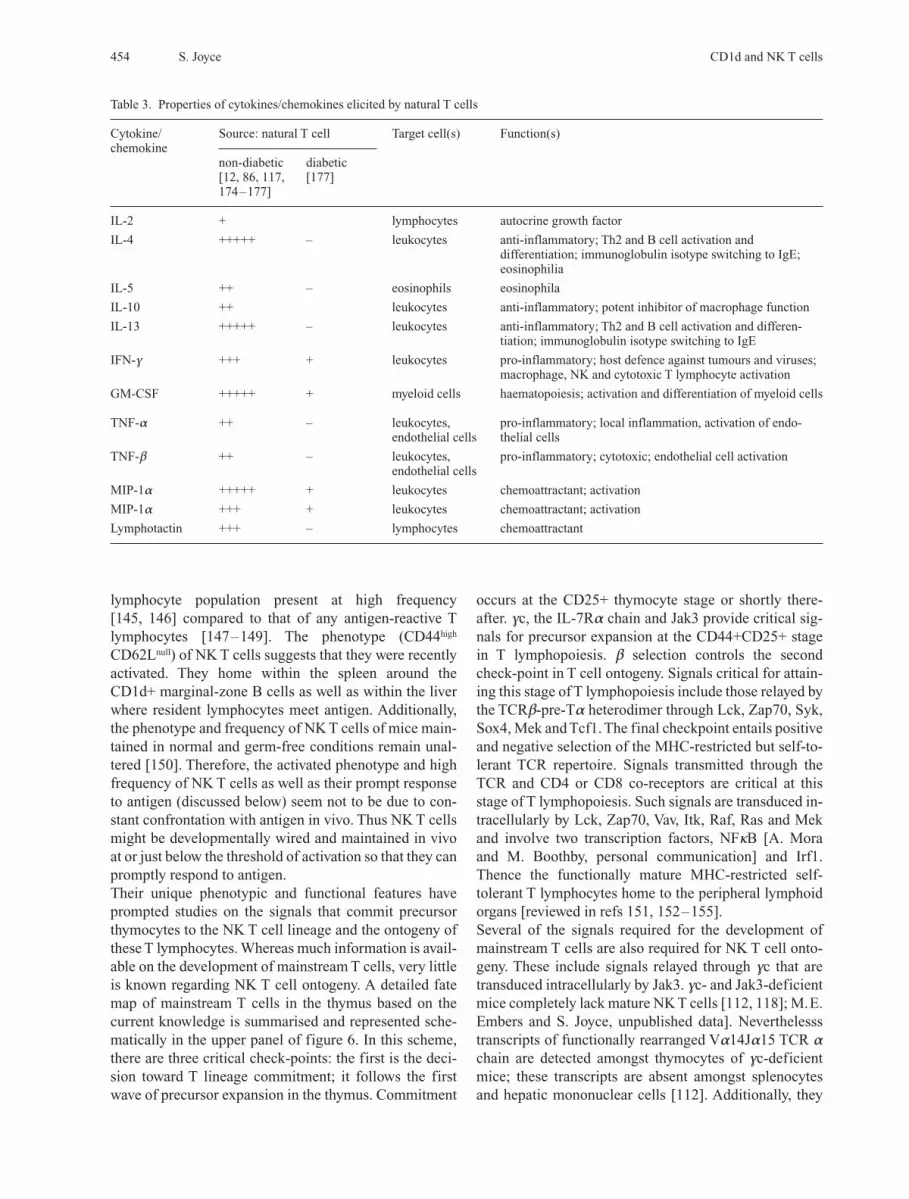
Table 3. Properties of cytokines/chemokines elicited by natural T cells
Cytokine/ Source: natural T cell Target cell(s) Function(s)chemokine
non-diabetic diabetic[12, 86, 117, [177]174–177]
IL-2 + lymphocytes autocrine growth factor
IL-4 +++++ – leukocytes anti-inflammatory; Th2 and B cell activation anddifferentiation; immunoglobulin isotype switching to IgE;eosinophilia
IL-5 ++ – eosinophils eosinophila
IL-10 ++ leukocytes anti-inflammatory; potent inhibitor of macrophage function
IL-13 +++++ – leukocytes anti-inflammatory; Th2 and B cell activation and differen-tiation; immunoglobulin isotype switching to IgE
IFN-g +++ + leukocytes pro-inflammatory; host defence against tumours and viruses;macrophage, NK and cytotoxic T lymphocyte activation
GM-CSF +++++ + myeloid cells haematopoiesis; activation and differentiation of myeloid cells
TNF-a ++ – leukocytes, pro-inflammatory; local inflammation, activation of endo-endothelial cells thelial cells
TNF-b ++ – leukocytes, pro-inflammatory; cytotoxic; endothelial cell activationendothelial cells
MIP-1a +++++ + leukocytes chemoattractant; activation
MIP-1a +++ + leukocytes chemoattractant; activation
Lymphotactin +++ – lymphocytes chemoattractant
CMLS, Cell. Mol. Life Sci. Vol. 58, 2001 Review Article 455
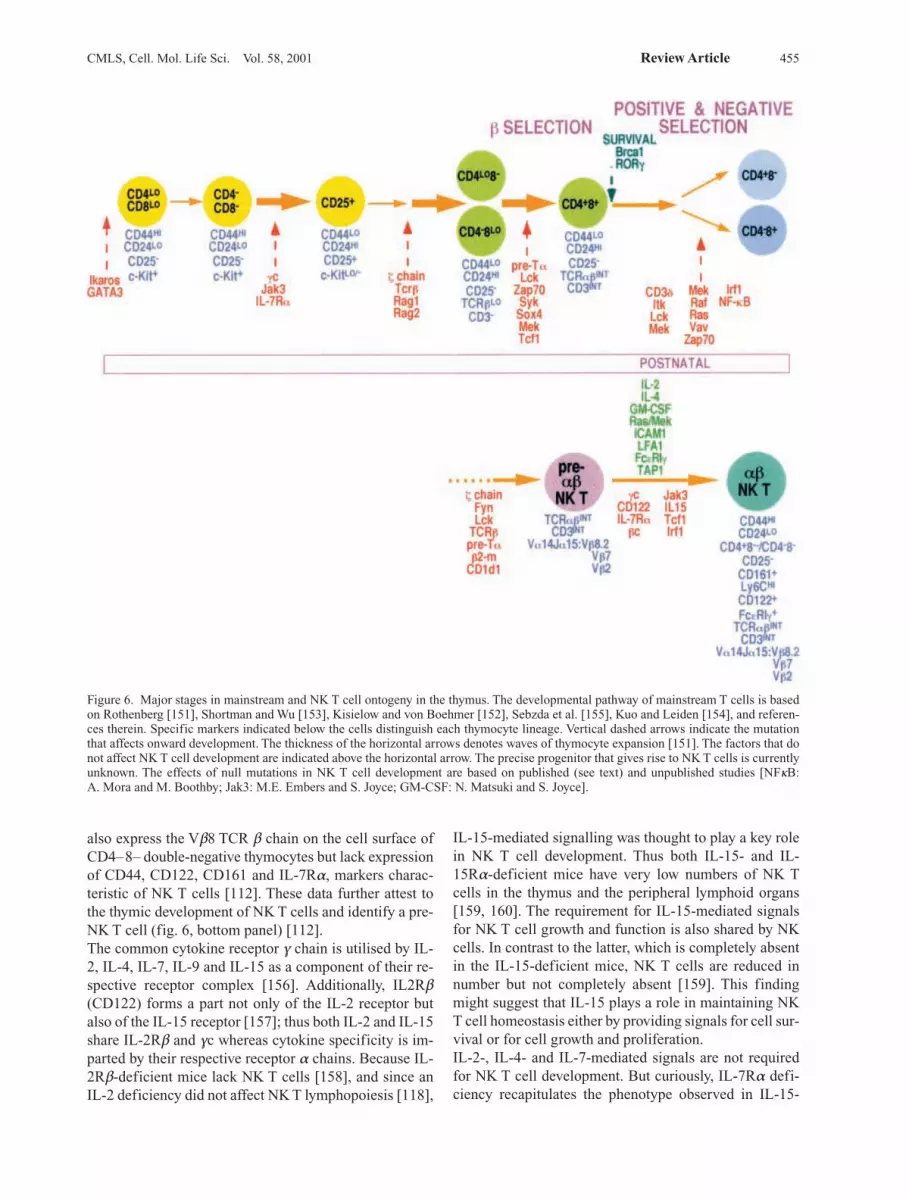
also express the Vb8 TCR b chain on the cell surface ofCD4–8– double-negative thymocytes but lack expressionof CD44, CD122, CD161 and IL-7Ra, markers charac-teristic of NK T cells [112]. These data further attest tothe thymic development of NK T cells and identify a pre-NK T cell (fig. 6, bottom panel) [112].The common cytokine receptor g chain is utilised by IL-2, IL-4, IL-7, IL-9 and IL-15 as a component of their re-spective receptor complex [156]. Additionally, IL2Rb(CD122) forms a part not only of the IL-2 receptor butalso of the IL-15 receptor [157]; thus both IL-2 and IL-15share IL-2Rb and gc whereas cytokine specificity is im-parted by their respective receptor a chains. Because IL-2Rb-deficient mice lack NK T cells [158], and since anIL-2 deficiency did not affect NK T lymphopoiesis [118],
IL-15-mediated signalling was thought to play a key rolein NK T cell development. Thus both IL-15- and IL-15Ra-deficient mice have very low numbers of NK Tcells in the thymus and the peripheral lymphoid organs[159, 160]. The requirement for IL-15-mediated signalsfor NK T cell growth and function is also shared by NKcells. In contrast to the latter, which is completely absentin the IL-15-deficient mice, NK T cells are reduced innumber but not completely absent [159]. This findingmight suggest that IL-15 plays a role in maintaining NKT cell homeostasis either by providing signals for cell sur-vival or for cell growth and proliferation.IL-2-, IL-4- and IL-7-mediated signals are not requiredfor NK T cell development. But curiously, IL-7Ra defi-ciency recapitulates the phenotype observed in IL-15-
Figure 6. Major stages in mainstream and NK T cell ontogeny in the thymus. The developmental pathway of mainstream T cells is basedon Rothenberg [151], Shortman and Wu [153], Kisielow and von Boehmer [152], Sebzda et al. [155], Kuo and Leiden [154], and referen-ces therein. Specific markers indicated below the cells distinguish each thymocyte lineage. Vertical dashed arrows indicate the mutationthat affects onward development. The thickness of the horizontal arrows denotes waves of thymocyte expansion [151]. The factors that donot affect NK T cell development are indicated above the horizontal arrow. The precise progenitor that gives rise to NK T cells is currentlyunknown. The effects of null mutations in NK T cell development are based on published (see text) and unpublished studies [NFkB: A. Mora and M. Boothby; Jak3: M.E. Embers and S. Joyce; GM-CSF: N. Matsuki and S. Joyce].
significantly alter the development of Vb8.1+ NK T cells[113]. Why the two thymocyte subsets respond differen-tially to Mls-1a has not yet been clarefied. The TCR achain with which Vb8.1 pairs and the antigen-presentingmolecule, MHC class II versus CD1d1, that interacts with the TCR may be critical factors that influence howMls-1a negatively selects Vb8.1+ mainstream T cells butnot NK T cells. Negative selection is also observed in mice that constitutively express a CD8ab transgene fromthe heterologous CD2 promoter [16]. Such animals ex-press CD8 from the immature TCR–/lowCD8+ stage onwardto the mature CD4+ and CD8+ single positive stage (seefig. 6) [171]. The CD8ab transgenic animals developedTCRint NK1+ NK T cells but had lost the expression of the skewed TCR repertoire; i.e. only a small fraction of the NK T cells expressed Vb8, Vb7 and Vb2 TCR b chainsbut instead expanded the repertoire expressing the Vb6TCR b chain [16]. Together, the studies with neonatal ad-ministration of SEB as well as transgenic mice that con-stitutively express CD8ab provide compelling evidencefor negative selection within the NK T cell compartment.Yet another question remains, and that is whether a cer-tain precursor commits to NK T cell lineage by specifi-cally rearranging the Va14 and Ja15 gene segmentswhich upon productive rearrangement beget the NK Tlineage – the instructive model. The competing modelwould predict that akin to mainstream T cells, Tcr genesegments rearrange randomly and the productive ones aresubjected to positive and negative selection by CD1d1and beget the NK T lineage – the stochastic model.Nucleotide sequence analyses of the V and J junctionswithin rearranged Va14Ja15 genes revealed that the ca-nonical TCRa sequence of the CD1d1-reactive NK Tcells is put together by random events that also include Nnucleotide additions [133, 172, 173]. Moreover, the se-cond allele also showed evidence of random rearrange-ment within the Tcr locus [133, 172, 173]. More recently,genomic DNA sequence analysis of NK T cell-derivedVb8.2 transgenic thymocytes prior to selection (TCR lowthymocytes) revealed random rearrangement of Va14with Ja15 segments resulting in a high frequency of non-canonical Va14+ TCR a chain [173]. The frequency ofthe canonical Va14Ja15+ TCR a chain containing thecanonical sequence increased within the Vb8.2 transgen-ic thymocytes following positive selection (TCRint thy-mocytes) by CD1d1 [173]. Together, these data supportthe stochastic model for thymocyte commitment to theNK T lymphocyte lineage.In summary, Va14Ja15-positive NK T cells develop inthe thymus where their TCR repertoire is moulded byboth positive and negative selection processes. The devel-opmental pathway that begets NK T cells and mainstreamT lymphocytes during thymic ontogeny seems very simi-lar. However, the signals required for NK T cell ontogeny,although in many ways matching those required for
456 S. Joyce CD1d and NK T cells
null mice with regard to NK T cell numbers, i.e. bothstrains of mice have dramatically lowered numbers of NKT cells [118, 159]. Signals relayed by IL-7Ra promotethe first wave of thymocyte expansion and survival [161,162]; this function of IL-7Ra may be essential for NK Tcell development as well. Because IL-7 deficiency doesnot affect NK T cell development [163], both IL-15 andthymic stromal cell lymphopoietin (TSLP); whose recep-tor complex includes IL-7Ra in common with IL-7 [164]may act synergistically to signal NK T lymphopoiesis aswell as in their homeostasis in vivo.
Models and pathwaysThe signals required for b selection are absolutely essen-tial for NK T cell lymphopoiesis [165]. Thus signallingthrough the TCRb-pre-Ta complex involving the z chainof CD3 [110], Lck [166], Fyn [166, 167] and Tcf1 [168]is important at this stage of NK T cell ontogeny (fig. 6).The development of NK T cells depends on CD1d[28–30]. CD1d is expressed predominantly by theCD4+8+ double-positive cells of the thymus [19]. There-fore, in contrast to positive selection of the mainstreamTCR repertoire by MHC class I and class II molecules ex-pressed by the thymic epithelium [20], positive selectionof the Va14Ja15-positive NK T cells is accomplished byinteractions with CD4+8+ thymocytes [19]. In this re-gard, it is interesting to note that positive selection of NKT cells does not require the Ras/Mek signalling pathway[169]. Positive selection of mainstream T cells, on theother hand, requires signal transduction down theRas/Mek pathway [169]. Thus, the signalling pathway forthe positive selection of NK T cells is critically differentfrom that of mainstream T cells.As described above, the expression of the TCR b chain ofNK T cells requires pre-Ta, i.e. for b selection [165]. To-gether with the fact that CD4+8+ thymocytes are requiredfor positive selection of NK T cells, the ontogeny of thissubset of T cells must proceed through the same lineagepathways as mainstream T cells (fig. 6). Nevertheless, thefact that NK cells and gd T cells differentiate earlier thanthe ab T cells and because the NK T cell developmentalprogramme closely matches that of NK cells, commit-ment to the NK T lineage might occur prior to that ofmainstream ab T cells. Future experiments with precursortransfer and/or gene-deficient mice should help resolvewhether the NK T lineage follows a distinct devel-opmental pathway or that of mainstream T lymphocytes.NK T cells also undergo negative selection. One model tostudy this process consisted of neonatal administration ofstaphylococcal enterotoxin B (SEB). SEB efficiently de-letes Vb8+ mainstream T cells [170]. Neonatal admin-istration of SEB also deleted Vb8+ NK T cells [113]. Theexpression of an endogenous antigen Mls-1a deletesVb8.1+ mainstream T cells in vivo during thymic on-togeny [170]. However, the expression of Mls-1a did not
mainstream T cell development, in some critical ways dif-fer from the latter and overlap signals required for NKcell development.
NK T cell functionThe physiological role of NK T cells, albeit elusive, isthought to be immunoregulatory in nature [reviewed inref. 5], a function controlled by CD1d [22, 87]. They areamongst leukocytes that secrete large amounts of IL-4without prior priming with this cytokine [12]. IL-4 is animmunomodulatory cytokine that can polarise newly ac-tivated CD4+ Th cells towards Th2 effector function [3].Their ability to secrete IL-4 promptly in response to prim-ary stimulation and hence their potential to polariseCD4+ T cells towards a Th2 immune response has arous-ed interest concerning their role in infection and immun-ity. NK T cells are also implicated in the control of auto-immune responses. What emerges from studies focusedon the role of NK T cells in autoimmune diseases is thatthey elaborate an IL-4-mediated immunoregulatory rolein vivo. Additionally, NK T cells are known to mediate tu-mour immunity by virtue of their ability to secrete IFNgthemselves or to induce this activity in NK cells. Studieson the mechanism(s) by which NK T cells function in tu-mour immunity have led to important insights into theirphysiological role.
Cytokine elicitation and functional consequencesThe first recognised function of freshly isolated NK Tcells was their ability to elicit both Th1 and Th2 cytokinesin vitro in response to a primary stimulus, without needfor priming with the elaborated cytokines. The elicitedTh1 cytokines include IFN-g and tumour necrosis factor-b (TNF-b) and the Th2 cytokines include IL-4, IL-5 andIL-10 [87, 117, 174–176]. More recently, the secretion ofother pro-inflammatory factors such as GM-CSF, lym-photactin, macrophage inflammatory protein (MIP)-1aand MIP-1b has also been attributed to NK T cells (table3) [87, 177]. Nevertheless in vivo stimulation of NK Tcells by cross-linking their TCR with anti-CD3e mono-clonal antibody results in the rapid, i.e. within 60–90min, secretion of large amounts of IL-4 [12, 178–180].IL-2 and IFN-g are also elicited by such polyclonal ac-tivation of T cells in vivo [12]. The prompt secretion ofIL-4 was attributed to NK T cells because both b2m- andCD1d1-null mice lacking NK T cells were deficient inthis function [28–30, 179]. Additionally, a-GalCer ac-tivates NK T cells in vivo resulting in the secretion ofboth IL-4 and IFN-g [13, 14]. Thus the cytokine elicita-tion pattern of NK T cells seems to resemble that of Th0cells. A recent study [177] focused on gene regulation inactivated NK T cells revealed that transcription factorsthat bestow a Th2 phenotype upon CD4+ cells were up-regulated within an IL-4-secreting NK T cell clone (de-
scribed below). Thus NK T cells may be hardwired duringdevelopment and differentiation for prompt secretion oflarge amounts of IL-4 upon in vivo activation.Akin to mainstream T cells, activation of NK T cells re-quires signals from the TCR as well as CD28. The pre-sence of soluble CTLA4 during intravenous anti-CD3eadministration inhibits the activation of NK T cells [12].Thus, the second signal emanating from the interactionsof CD28 expressed by the T lymphocyte with CD80and/or CD86 expressed by the antigen-presenting cell isimportant for NK T cells activation. Following activationin vivo with anti-CD3e, hepatic NK T cells rapidly, with-in 2 h, die [181]. Rapid loss of the large majority of NKT cells is observed in vivo following activation by the an-tigen a-GalCer [146]. Their death following in vivo stim-ulation by anti-CD3e is attributed to apoptosis because a large majority of the activated NK T cells express An-nexin V [181, 182]. NK T cells regenerate within the nextcouple of days. Regeneration requires proliferation,which occurs within the bone marrow [181]. This processof activation-induced cell death and the regeneration ofNK T cells from the bone marrow are mimicked by IL-12[181]. IL-12 mediated death of hepatic NK T cells couldthen explain how L. monocytogenes and M. bovis, both ofwhich are potent inducers of IL-12, down-regulate theNK T cell-mediated inflammatory process within the li-ver of infected mice [183–185]. Thus shortly upon exe-cuting their function, NK T cells die, a feature they sharewith other cells of the innate immune system.The physiological relevance of pro-inflammatory andanti-inflammatory cytokine secretion by activated NK Tcells has been addressed in numerous experimental sys-tems. The first of these predicted that IL-4 secreted byNK T cells by in vivo stimulation may play a role in Th2differentiation and hence in type 2 immune responses toantigens. Previous studies had established that adminis-tration of goat anti-immunoglobulin-D (IgD) into miceresulted in the elicitation of IgG1 and IgE antibodies. Inthis system, IgG1 and IgE production requires CD4+ Tlymphocytes and IL-4 [12, 180]. How anti-IgD stimulatesIgG1 and IgE production in vivo remains unclear. How-ever, b2m-null mice, deficient amongst other things inNK T cells, do not respond to anti-IgD-mediated stimu-lation and hence do not secrete IgE. These results impli-cated CD1d1-restricted NK T cells in the anti-IgD-me-diated IgE response [178, 179]. Contrarily however,CD1d1 and CD1d2 double-knockout as well as CD1d1-deficient mice, both of which have intact mainstream Tcells but lack NK T cells, were perfectly capable ofeliciting an anti-IgD-mediated IgE response [28–30].Moreover, the loss of anti-IgD-mediated IgE response inb2m0/0 mice is due to FcRn- (a b2m-dependent molecule)mediated rapid catabolism of injected antibody [186,187]. Thus NK T cells do not play a role in the anti-IgD-induced IgE response in vivo.
CMLS, Cell. Mol. Life Sci. Vol. 58, 2001 Review Article 457
Infection and immunityAdaptive immunity against infectious agents such as bac-teria as well as protozoan and helminthic parasites de-pends on the functions of CD4+ T lymphocytes. The kindof CD4 response, whether Th1 or Th2, to the infectiousagent is dictated by a variety of factors of which the cyto-kine milieu during the time of T cell priming by antigenplays a critical role in the outcome. Because NK T cellshave the potential for eliciting Th1 and Th2 cytokinespromptly upon in vivo activation, they are implicated inthe Th1 versus Th2 immune responses to bacterial and pa-rasitic infections.A comprehensive analysis was undertaken using solubleantigens and infectious agents that are known to elicitTh2 responses to determine the role of NK T cells in thispolarised immune outcome [188]. Various parameters,IL-4 and IgE response, eosinophilia, as well as airway hy-persensitivity, were monitored following antigen, solubleand particulate in the form of the parasite, deliverythrough various routes, subcutaneous, airway and intesti-nal, into wild-type and b2m-deficient mice. Note, the lat-ter are incapable of promptly secreting IL-4 in responseto intravenous administration of anti-CD3e. The datarevealed no difference in Th2 response between wild-typeand b2m-deficient mice to soluble keyhole limpet hae-mocyanin, aerosolised ovalbumin, embolisation withSchistosoma mansoni eggs, infection with metacyclicpromatigotes of Leishmania major or intestinal infectionof Nippostrongylus brasiliensis [188]. L. major does in-deed induce IL-4 early during infection, but is secreted bya non-NK T cell subset [189, 190].However, intraperitoneal infection of mice with S. chole-raesuis or L. monocytogenes results in the recruitment ofNK T cells to the peritoneum [129, 130]. The Salmonella-recruited NK T cells in the peritoneal exudate secrete IL-4 [129]. The elicited IL-4 down-regulates IL-12 produc-tion and hence negatively controls the generation of theTh1 immune response. Most importantly, the regulatoryrole of IL-4 over IL-12 was abolished in Ja15- (i.e.Ja281, the TCR Ja gene segment utilised by Va14-posi-tive NK T cells) deficient mice resulting in aberrantlyhigh levels of IL-12 [129]. Thus, NK T cells may play sig-nificant role in the early phase of infection that in turninfluences adaptive immunity to intracellular bacterial in-fections.Additionally, a role for NK T cells has been implicated ineliciting immune responses to other infectious agents. Forexample, Salmonella spp. [129], L. monocytogenes [183,184], M. tuberculosis, M. bovis (bacillus Calmette Gue-rin, BCG) [191], Toxoplasma gondii [192, 193] and L.major [194] in resistant strains of mice elicit Th1 immun-ity [reviewed in ref. 195]. In these cases, the ability of NKT cells to secrete IL-2 or IFN-g themselves or to inducethis activity in NK cells (described below) is exploited. Inthe case of T. gondii, CD8+ T cell function is recruited in
the absence of CD4+ T lymphocytes that are normally re-quired for IFN-g-mediated Th1 immunity. Further analy-sis revealed a role for IL-2 released by NK T cells inrecruiting CD8+ T cell-mediated immunity to T. gondii inthe absence of MHC class II-restricted T cells [193].Human CD1b- and CD1c-restricted presentation of my-cobacterial lipid and glycolipid antigens is well docu-mented [reviewed in ref. 31]. However, immunity to my-cobacteria in mice, which express only the CD1d isotype,suggests that CD1d may not be directly involved in anti-gen presentation [196, 197]. Nevertheless a recent studyrevealed that NK T cells contribute to the formation ofmycobacterial cell wall-induced granulomas [198]. PI-mannosides were tentatively identified as the causativeantigen in this granulomatous reaction in wild-type mice.Because this reaction was absent in the Ja15-deficientmice, granuloma formation involved NK T cells and sug-gests CD1d-mediated antigen presentation [198]. The re-sponse of CD1d-deficient mice to mycobacterial cell wallglycolipids has yet to be determined. In this context it isinteresting to note that whether or not direct antigen pre-sentation by CD1d is involved, M. bovis infection resultsin the disappearance of hepatic NK T cells, presumablydue to the induction of IL-12 and IL12-mediated NK Tcell death [191]. Inoculation with BCG altered the den-sity of NK T cells; the altered density correlated with anIFN-g-secreting phenotype [191]. It is possible that theIFN-g-secreting cells were NK cells and not NK T cells.The IFN-g elicited in response to BCG may be involvedin the mycobacterial cell wall-induced granuloma reac-tion.Although infection by Plasmodium spp. gets the better ofsome of us, resistant individuals elicit both cellular andhumoral responses to the malarial vector [199]. The roleof CD1d and NK T cells in immunity to the malarialvector has recently been a subject of much interest for itoffers the possibility for vaccine development in the faceof a rapid evolution of drug-resistant variants of Plasmo-dium spp. [199]. Experimental infection of mice withsporozoites of Plasmodium yeolii results in the expansionof hepatic NK T cells [200]. In vitro co-culture studiesusing the activated NK T cells generated during the acutephase of infection revealed that these lymphocytes inhibitgrowth of the liver stage parasite [200]. Moreover, theobserved anti-Plasmodium effector function requiresIFN-g [200]. In another study, activation of NK T cells invivo with the NK T cell antigen a-GalCer 1 or 2 daysprior to infection with P. yoelii sporozoites conferred im-munity to the liver-stage parasite [201]. Akin to the invitro findings, the anti-plasmodial effector function invivo was primarily mediated by IFN-g. Nevertheless, sur-prisingly, this IFN-g response requires neither IL-12 norNK cells, but IFN-g secreted by NK T cells sub-serves theanti-plasmodial effector function of NK T cells [201].Additional data revealed that the effector function of NK
458 S. Joyce CD1d and NK T cells
T cells against malarial sporozoites did not require Fasligand, perforin or TNF-a [201]. Thus NK T cells play animportant role in the early immune response to plasmo-dial sporozoites in mice. Whether a-GalCer-activatedNK T cells elicit early immunity to sporozoites in humansneeds study.NK T cells have the potential to bias antigen-specific Thimmune responses toward the Th2 type which results inproviding help to B cells for antibody elicitation. Twogroups have studied the role of NK T cells in the anti-plasmodial antibody response. Results from one grouprevealed a GPI-specific CD1d-restricted and NK T cell-dependent antibody response to plasmodial sporozoite-derived circumsporozoite protein [202]. In contrast theresults of the second group revealed an MHC class IIdependence but CD1d and NK T cell independence inanti-plasmodial humoral immunity to the same protein[88]. The basis of these contradictory results remains un-clear.
Tumour immunityNK cells as well as the type I cytokines IFN-g and IL-12play an important role in immune surveillance against tu-mour growth and metastasis. Recent studies have reveal-ed that an important function of NK T cells in vivo is hostprotection against tumour growth and metastasis [67,203, 204]. This immune surveillance function of NK Tcells requires IL-12 or its induction by NK T cell stimu-lation in vivo by a-GalCer [67, 205]. Additionally, im-munity to chemically induced tumours is conferred byNK T cells without the need for exogenous IL-12 or a-GalCer [204]. Thus a role in immune protection againstspontaneously arising tumours and tumour metastasis isascribed to NK T cells in vivo.Their role in immune surveillance in vivo has aroused in-terest in the mechanism of action of NK T cells. a-Gal-Cer, the potent synthetic glycolipid antigen presented byCD1d, activates NK T cells in vivo resulting in the secre-tion of both IFN-g and IL-4 [13, 14, 69]. Both cytokinesare elicited promptly in response to a-GalCer; however,IFN-g is maintained only acutely while IL-4 is maintain-ed chronically [13]. The chronic maintenance of IL-4 inthe milieu of differentiating Th cells polarises them to-wards a Th2 phenotype [13]. Moreover, the IFN-g re-sponse to a-GalCer seems to be elicited by NK cells re-quiring IL-12-mediated activation [67, 68, 206]. IL-12 isa pro-inflammatory cytokine elicited by activated my-eloid cells such as dendritic cells and macrophages [7].How activation of NK T cells by a-GalCer results in IL-12 production is discussed below [207].Whether NK T cells directly mediate cytotoxic activity ontarget tumour cells is controversial. All data that supportNK T-mediated tumour-specific cytolytic activity are de-rived from in vitro studies [208–211]. It is clear, however,
that the in vivo activation of NK T cells lead to the in-duction of IL-12 [181]. IL-12, as discussed above, in-duces NK T cell death within a few hours [181] and maynot be available for tumour-specific cytocidal activity invivo. The ability of activated NK T cells to promptly ac-tivate effector cells of the innate and adaptive immunesystem, particularly NK cells [13, 14, 212] and CD8+ Tlymphocytes [13, 14, 213] may be responsible for the ob-served NK T-mediated cytolytic activity in vivo. Neverthelesss in vivo NK T cell-mediated cytotoxicityhas been observed in a murine model for hepatitis [214,215]. Hepatitis in this model is induced by intravenousadministration of concanavalin A (Con A) into mice.Neither CD1d1- nor Ja15-1-deficient mice develop ex-perimental hepatitis upon Con A treatment [214, 215].Furthermore, adoptive transfer of Con A-activated NK Tcells into CD1d1-deficient mice transferred the disease[215]. Additionally, Con A-treated mice that only developNK T cells by virtue of transgenic expression of the mouseVa14Ja15/Vb8.2 genes [69] also develop hepatitis. Thus,NK T cells are implicated in the development ofautoimmune hepatitis. Furthermore, analysis of the killingmechanism revealed that the activation of NK T cellsrequired IL-4 [214], and that activation induced granzymeB [214] as well as Fas L [214, 215] expression. Neitherperforin- [214] nor gld/gld- [214, 215] deficient mice areaffected by Con A stimulation in vivo. Thus perforin-gran-zyme B and Fas and Fas L are implicated as molecular ef-fectors of NK T cell-mediated cytotoxicity in vivo.
AutoimmunitySeveral lines of evidence suggest a link between NK Tcell deficiency or dysfunction and autoimmune diseases[216–224]. The best studied of these disorders is insulin-dependent diabetes mellitus (type I diabetes). Both hu-mans and mice predisposed to or afflicted by type 1 dia-betes have fewer NK T cells and those present are dys-functional, in that they are unable to secrete IL-4promptly in response to cross-linking of their antigen re-ceptors [219, 221, 223]. In non-obese diabetic (NOD)mice, the mouse model for human type 1 diabetes [225,226], adoptive transfer of thymic NK T cells into pre-dia-betic animals corrected the deficiency and the disease[222]. Protection from disease by adoptive transfer of NKT cells required IL-4 and/or IL-10 [222]. Moreover, trans-genic expression of Va14Ja15 TCR in NOD mice alsoprotects them from diabetes [227]. Additionally, adminis-tration of recombinant IL-4 itself [228] to pre-diabeticNOD animals prevented them from developing diabetes.Thus an IL-4-mediated immunoregulatory role in vivo isascribed to NK T cells. To define the molecular basis for NK T cell dysregulationin diabetic individuals, a comprehensive analysis of themRNA expression profile of human NK T cell clones iso-
CMLS, Cell. Mol. Life Sci. Vol. 58, 2001 Review Article 459
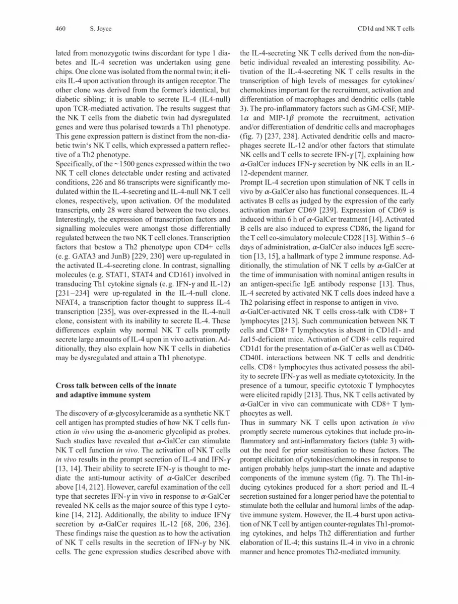
460 S. Joyce CD1d and NK T cells
lated from monozygotic twins discordant for type 1 dia-betes and IL-4 secretion was undertaken using genechips. One clone was isolated from the normal twin; it eli-cits IL-4 upon activation through its antigen receptor. Theother clone was derived from the former’s identical, butdiabetic sibling; it is unable to secrete IL-4 (IL4-null)upon TCR-mediated activation. The results suggest thatthe NK T cells from the diabetic twin had dysregulatedgenes and were thus polarised towards a Th1 phenotype.This gene expression pattern is distinct from the non-dia-betic twin‘s NK T cells, which expressed a pattern reflec-tive of a Th2 phenotype.Specifically, of the ~1500 genes expressed within the twoNK T cell clones detectable under resting and activatedconditions, 226 and 86 transcripts were significantly mo-dulated within the IL-4-secreting and IL-4-null NK T cellclones, respectively, upon activation. Of the modulatedtranscripts, only 28 were shared between the two clones.Interestingly, the expression of transcription factors andsignalling molecules were amongst those differentiallyregulated between the two NK T cell clones. Transcriptionfactors that bestow a Th2 phenotype upon CD4+ cells(e.g. GATA3 and JunB) [229, 230] were up-regulated inthe activated IL-4-secreting clone. In contrast, signallingmolecules (e.g. STAT1, STAT4 and CD161) involved intransducing Th1 cytokine signals (e.g. IFN-g and IL-12)[231–234] were up-regulated in the IL-4-null clone.NFAT4, a transcription factor thought to suppress IL-4transcription [235], was over-expressed in the IL-4-nullclone, consistent with its inability to secrete IL-4. Thesedifferences explain why normal NK T cells promptlysecrete large amounts of IL-4 upon in vivo activation. Ad-ditionally, they also explain how NK T cells in diabeticsmay be dysregulated and attain a Th1 phenotype.
Cross talk between cells of the innate and adaptive immune system
The discovery of a-glycosylceramide as a synthetic NK Tcell antigen has prompted studies of how NK T cells fun-ction in vivo using the a-anomeric glycolipid as probes.Such studies have revealed that a-GalCer can stimulateNK T cell function in vivo. The activation of NK T cellsin vivo results in the prompt secretion of IL-4 and IFN-g[13, 14]. Their ability to secrete IFN-g is thought to me-diate the anti-tumour activity of a-GalCer describedabove [14, 212]. However, careful examination of the celltype that secretes IFN-g in vivo in response to a-GalCerrevealed NK cells as the major source of this type I cyto-kine [14, 212]. Additionally, the ability to induce IFNgsecretion by a-GalCer requires IL-12 [68, 206, 236].These findings raise the question as to how the activationof NK T cells results in the secretion of IFN-g by NKcells. The gene expression studies described above with
the IL-4-secreting NK T cells derived from the non-dia-betic individual revealed an interesting possibility. Ac-tivation of the IL-4-secreting NK T cells results in thetranscription of high levels of messages for cytokines/chemokines important for the recruitment, activation anddifferentiation of macrophages and dendritic cells (table3). The pro-inflammatory factors such as GM-CSF, MIP-1a and MIP-1b promote the recruitment, activationand/or differentiation of dendritic cells and macrophages(fig. 7) [237, 238]. Activated dendritic cells and macro-phages secrete IL-12 and/or other factors that stimulateNK cells and T cells to secrete IFN-g [7], explaining howa-GalCer induces IFN-g secretion by NK cells in an IL-12-dependent manner.Prompt IL-4 secretion upon stimulation of NK T cells invivo by a-GalCer also has functional consequences. IL-4activates B cells as judged by the expression of the earlyactivation marker CD69 [239]. Expression of CD69 isinduced within 6 h of a-GalCer treatment [14]. ActivatedB cells are also induced to express CD86, the ligand forthe T cell co-simulatory molecule CD28 [13]. Within 5–6days of administration, a-GalCer also induces IgE secre-tion [13, 15], a hallmark of type 2 immune response. Ad-ditionally, the stimulation of NK T cells by a-GalCer atthe time of immunisation with nominal antigen results inan antigen-specific IgE antibody response [13]. Thus, IL-4 secreted by activated NK T cells does indeed have aTh2 polarising effect in response to antigen in vivo.a-GalCer-activated NK T cells cross-talk with CD8+ Tlymphocytes [213]. Such communication between NK Tcells and CD8+ T lymphocytes is absent in CD1d1- andJa15-deficient mice. Activation of CD8+ cells requiredCD1d1 for the presentation of a-GalCer as well as CD40-CD40L interactions between NK T cells and dendriticcells. CD8+ lymphocytes thus activated possess the abil-ity to secrete IFN-g as well as mediate cytotoxicity. In thepresence of a tumour, specific cytotoxic T lymphocyteswere elicited rapidly [213]. Thus, NK T cells activated bya-GalCer in vivo can communicate with CD8+ T lym-phocytes as well.Thus in summary NK T cells upon activation in vivopromptly secrete numerous cytokines that include pro-in-flammatory and anti-inflammatory factors (table 3) with-out the need for prior sensitisation to these factors. Theprompt elicitation of cytokines/chemokines in response toantigen probably helps jump-start the innate and adaptivecomponents of the immune system (fig. 7). The Th1-in-ducing cytokines produced for a short period and IL-4secretion sustained for a longer period have the potential tostimulate both the cellular and humoral limbs of the adap-tive immune system. However, the IL-4 burst upon activa-tion of NK T cell by antigen counter-regulates Th1-promot-ing cytokines, and helps Th2 differentiation and furtherelaboration of IL-4; this sustains IL-4 in vivo in a chronicmanner and hence promotes Th2-mediated immunity.
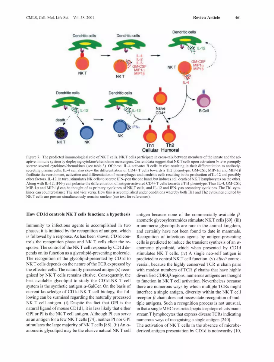
CMLS, Cell. Mol. Life Sci. Vol. 58, 2001 Review Article 461
How CD1d controls NK T cells function: a hypothesis
Immunity to infectious agents is accomplished in twophases; it is initiated by the recognition of antigen, whichis followed by a response. As has been shown, CD1d con-trols the recognition phase and NK T cells elicit the re-sponse. The control of the NK T cell response by CD1d de-pends on its function as a glycolipid-presenting molecule.The recognition of the glycolipid-presented by CD1d toNK T cells depends on the nature of the TCR expressed bythe effector cells. The naturally processed antigen(s) reco-gnised by NK T cells remains elusive. Consequently, thebest available glycolipid to study the CD1d-NK T cellsystem is the synthetic antigen a-GalCer. On the basis ofcurrent knowledge of CD1d-NK T cell biology, the fol-lowing can be surmised regarding the naturally processedNK T cell antigen. (i) Despite the fact that GPI is thenatural ligand of mouse CD1d1, it is Iess likely that eitherGPI or PI is the NK T cell antigen. Although PI can serveas an antigen for a few NK T cells [74], neither PI nor GPIstimulates the large majority of NK T cells [88]. (ii) An a-anomeric glycolipid may be the elusive natural NK T cell
antigen because none of the commercially available b-anomeric glycosylceramides stimulate NK T cells [69]. (iii)a-anomeric glycolipids are rare in the animal kingdom,and certainly have not been found to date in mammals.Recognition of infectious agents by antigen-presentingcells is predicted to induce the transient synthesis of an a-anomeric glycolipid, which when presented by CD1dstimulates NK T cells. (iv) A single neo-self antigen ispredicted to control NK T cell function. (v) Albeit contro-versial, because the highly conserved TCR a chain pairswith modest numbers of TCR b chains that have highlydiversified CDR3b regions, numerous antigens are thoughtto function in NK T cell activation. Nevertheless becausethere are numerous ways by which multiple TCRs mightinterface a single antigen, diversity within the NK T cellreceptor b-chain does not necessitate recognition of mul-tiple antigens. Such a recognition process is not unusual,in that a single MHC-restricted peptide epitope elicits main-stream T lymphocytes that express diverse TCRs indicatingnumerous ways of recognising a single antigen [240].The activation of NK T cells in the absence of microbe-derived antigen presentation by CD1d is noteworthy [10,
Figure 7. The predicted immunological role of NK T cells. NK T cells participate in cross-talk between members of the innate and the ad-aptive immune system by deploying cytokine/chemokine messengers. Current data suggest that NK T cells upon activation in vivo promptlysecrete several cytokines/chemokines (see table 3). Of these, IL-4 activates B cells in vivo resulting in their differentiation to antibody-secreting plasma cells. IL-4 can also skew the differentiation of CD4+ T cells towards a Th2 phenotype. GM-CSF, MIP-1a and MIP-1bfacilitate the recruitment, activation and differentiation of macrophages and dendritic cells resulting in the production of IL-12 and possiblyother factors. IL-12, in turn, stimulates NK cells to secrete IFN-g on the one hand, but induces cell death of NK T lymphocytes on the other.Along with IL-12, IFN-g can polarise the differentiation of antigen-activated CD4+ T cells towards a Th1 phenotype. Thus IL-4, GM-CSF,MIP-1a and MIP-1b can be thought of as primary cytokines of NK T cells, and IL-12 and IFN-g as secondary cytokines. The Th1 cyto-kines can counterbalance Th2 and vice versa. How this is accomplished under conditions whereby both Th1 and Th2 cytokines elicited byNK T cells are present simultaneously remains unclear (see text for references).
87, 88, 94]. NK T cells function in immune surveillanceagainst spontaneously arising tumours that do not expressCD1d [67, 204]. Additionally, NK T cells are quiescent invivo in the absence of any infectious stimuli. For thesereasons, the induced nature of the NK T cell antigen is po-stulated. Recently, a role for CD1 in monitoring mem-brane integrity has been evoked because it binds and pre-sents lipids and glycolipids to specific T cells [241]. Inkeeping with this hypothesis, infectious assaults, such asviral and bacterial infections as well as glucose star-vation, are speculated to induce unfolded protein re-sponse [reviewed in ref. 242]. An unfolded protein re-sponse increases cytosolic inositol levels, a key to the in-duction of membrane biogenesis [243]. Inositol plays animportant role in regulating phospholipid biosynthesis[244] and hence in membrane biogenesis [243, 245].Phospholipids and glycolipids play an important structu-ral role in maintaining membrane integrity. Thus the in-duction of the unfolded protein response may include thebiosynthesis of neo-self lipids that are normally not en-countered by NK T cells. Encounter with the neo-self gly-colipid, the elusive a-anomeric glycosylceramide, turnson NK T cells. Because the products of the unfolded pro-tein response are similar irrespective of the cause, the in-duction and presentation of a common antigen are post-ulated during viral or bacterial infections or glucose star-vation. A virtue of the unfolded protein response relevantto the CD1d-NK T cell antigen presentation and recogni-tion system is its rapidity and the short half-life (about 2 min!) of the induced products [reviewed in ref. 242].This might explain the rarity of the NK T cell antigen andhence why it has eluded identification.Thus CD1d functions as a sensor, sensing alterations incellular lipid content by virtue of its affinity for such li-gands. The presentation of a neo-self glycolipid, pres-umably induced by infectious assault of antigen-presen-ting cells, activates NK T cells. Activated NK T cellspromptly release pro-inflammatory and anti-inflamma-tory cytokines and jump-start the immune system.Shortly thereafter, NK T cells are lost to death but reple-nished over the next couple of days. Identification of theelusive natural antigen of NK T cells will shed more lighton how the CD1d-NK T cell system works in vivo, espe-cially with regard to antigen expression and presentationto this unique T lymphocyte. The significance of NK Tcell function in vivo, established with a-GalCer, in infec-tion and immunity, in tumour immunity as well as in au-toimmunity has been demonstrate, but most important,the NK T cell antigen will tell us whether the CD1d-NKT cell system has any physiological importance.
Acknowledgements. I thank members of my laboratory, past andpresent, as well as Drs. Albert Bendelac, Randy Brutkiewicz andLuc Van Kaer for helpful discussions on the biology of CD1d andNK T cells. For any, unintentional, omissions of important work, Iapologise. Supported by grants from the NIH (AI 42284 and
HL54977), the Juvenile Diabetes Foundation, International, and theHuman Frontiers in Science Programme.
1 Fearon D. T. and Locksley R. M. (1996) The instructive role ofinnate immunity in the acquired immune response. Science272: 50–54
2 Banchereau J. and Steinman R. M. (1998) Dendritic cells andthe control of immunity. Nature 392: 245–252
3 Mossman T. R. and Coffman R. L. (1989) Th1 and Th2 cells:different patterns of lymphokine secretion lead to differentfunctional properties. Annu. Rev. Immunol. 7: 145–173
4 Abbas A. K., Murphy K. M. and Sher A. (1996) Functionaldiversity of helper T lymphocytes. Nature 383: 787–793
5 Bendelac, A., Rivera, M. N., Park, S.-H. and Roark, J. H.(1997) Mouse CD1-specific NK1 T cells: development, spe-cificity and function. Annu. Rev. Immunol. 15: 535–562
6 Hoffmann J. A., Kafatos F. C., Janeway C. A. and EzekowitzR. A. (1999) Phylogenetic perspectives in innate immunity.Science 284: 1313–1318
7 Trinchieri G. (1995) Interleukin-12: a proinflammatory cyto-kine with immunoregulatory functions that bridge innate re-sistance and antigen-specific adaptive immunity. Annu. Rev.Immunol. 13: 251–276
8 Wolf P. R. and Ploegh H. L. (1995) How MHC class II molec-ules acquire peptide cargo: biosynthesis and traffickingthrough the endocytic pathway. Annu. Rev. Cell Biol. 11:267–306
9 Sugita, M., Jackman, R. M., Donselaar van, E., Behar, S. M.,Rogers, R. A., Peters, P. J. et al. (1996) Cytoplasmic tail de-pendent localization of CD1b antigen presenting molecules toMIICs. Science 273: 349–352
10 Chiu Y. H., Jayawardena J., Weiss A., Lee D., Park S. H., Dau-try-Varsat A. et al. (1999) Distinct subsets of CD1d-restrictedT cells recognize self-antigens loaded in different cellularcompartments. J. Exp. Med. 189: 103–110
11 Rodionov D. G., Nordeng T. W., Pedersen K., Balk S. P. andBakke O. (1999) A critical tyrosine residue in the cytoplasmictail is important for CD1d internalization but not for its baso-lateral sorting in MDCK cells. J. Immunol. 162: 1488–495
12 Yoshimoto T. and Paul W. E. (1994) CD4pos NK1pos T cellspromptly produced IL-4 in response to in vivo challenge withanti-CD3. J. Exp. Med. 179: 1285–1295
13 Singh N., Hong S., Scherer D. C., Serizawa I., Burdin N., Kro-nenberg M. et al. (1999) Activation of NK T cells by CD1dand a-galactosylceramide directs conventional T cells to theacquisition of a Th2 phenotype. J. Immunol. 163: 2373–2377
14 Carnaud C., Lee D., Donnars O., Park,S.-H., Beavis A., Koe-zuka Y. et al. (1999) Cross-talk between cells of the innate im-mune system: NKT cells rapidly activate NK cells. J. Immu-nol. 163: 4647–4650
15 Burdin N., Brossay L. and Kronenberg M. (1999) Immuniza-tion with a-galactosylceramide polarizes CD1-reactive NK Tcells towards Th2 cytokine synthesis. Eur. J. Immunol. 29:2014–2025
16 Bendelac A., Killeen N., Litman D. R. and Schwartz R. H.(1994) A subset of CD4+ thymocytes selected by MHC class Imolecules. Science 263: 1774–1778
17 Coles M. C. and Raulet D. H. (1994) Class I dependence of thedevelopment of CD4+CD8– NK1.1+ thymocytes. J. Exp. Med.180: 395–399
18 Ohteki T. and MacDonald H. R. (1994) Major histocompatib-ility complex class I related molecules control the develop-ment of CD4+8– and CD4–8– subsets of natural killer 1.1+ Tcell receptor-a/b cells in the liver of mice. J. Exp. Med. 180:699–704
19 Bendelac A. (1995) Positive selection of mouse NK1+ T cellsby CD1 expressing cortical thymocytes. J. Exp. Med. 182:2091–2096
462 S. Joyce CD1d and NK T cells
20 Cosgrove D., Chan S. H., Waltzinger C., Benoist C. and Ma-this D. (1992) The thymic compartment responsible for posi-tive selection of CD4+ T cells. Int. Immunol. 4: 707–710
21 Fowlkes B. J. and Schweighoffer E. (1995) Positive selectionof T cells. Curr. Opin. Immunol. 7: 188–195
22 Bendelac A., Lantz O., Quimby M. E., Yewdell J. W., BenninkJ. R. and Brutkiewicz R. R. (1995) CD1 recognition of mouseNK1+ T lymphocytes. Science 268: 863–865
23 Bradbury A., Calabi F. and Milstein C. (1990) Expression ofCD1 in the mouse thymus. Eur. J. Immunol. 20: 1831–1836
24 Hershberg R., Eghtesady P., Sydora B., Brorson K., CheroutreH., Modlin R. et al. (1990) Expression of the thymus leukemiaantigen in mouse intestinal epithelium. Proc. Natl. Acad. Sci.USA 87: 9727–9731
25 Mellor A. L., Antoniou J. and Robinson P. J. (1985) Structureand expression of genes encoding murine Qa-2 class I anti-gens. Proc. Natl. Acad. Sci. USA 82: 5910–5914
26 Mellor A. L., Tomlinson P. D., Anroniou J., Chadler P., Robin-son P. J., Felstein M. et al. (1991) Expression and function ofQa-2 major histocompatibility complex class I molecules intransgenic mice. Int. Immunol. 3: 493–502
27 Koseki H., Asano H., Inaba T., Miyashita N., Moriwaki K.,Fisher Lindahl F. et al. (1991) Dominant expression of a dis-tinctive V14+ T cell antigen receptor a chain in mice. Proc.Natl. Acad. Sci. USA 88: 7518–7522
28 Smiley S. T., Kaplan M. H. and Grusby M. J. (1997) Immuno-globulin E production in the absence of interleukin-4 secre-ting CD1 dependent cells. Science 275: 977–979
29 Chen Y.-H., Chiu N. M., Mandal M., Wang N. and Wang C. R.(1997) Impaired NK1+ T cell development and early IL-4production in CD1 deficient mice. Immunity 6: 459–467
30 Mendiratta S. K., Martin W. D., Hong S., Boesteanu A., Joyce S.and Van Kaer L. (1997) CD1d1 mutant mice are deficient in na-tural T cells that promptly produce IL-4. Immunity 6: 469–477
31 Porcelli S. A. and Modlin R. L. (1999) The CD1 system: anti-gen-presenting molecules for T cell recognition of lipids andglycolipids. Annu. Rev. Immunol. 17: 297–329
32 Calabi F., Jarvis J. M., Martin L. and Milstein C. (1989) Twoclasses of CD1 genes. Eur. J. Immunol. 19: 71–78
33 Balk, S. P., Bliecher P. A. and Terhorst C. (1989) Isolation andcharacterization of a cDNA and gene coding for a fourth CD1molecule. Proc. Natl. Acad. Sci. USA 86: 252–256
34 Balk S. P., Bliecher P. A. and Terhorst C. (1991) Isolation andexpression of cDNA encoding the murine homologues ofCD1. J. Immunol. 146: 768–774
35 Ichimiya S., Kikuchi K. and Matsuura A. (1994) Structuralanalysis of the rat homologue of CD1: evidence for evolutio-nary conservation of the CD1D class and widespread tran-scription by rat cells. J. Immunol. 153: 1112–1123
36 Hopkins J., Dutia B. M. and Rhind S. M. (2000) Sheep CD1genes and proteins. Vet. Immunol. Immunopathol. 73: 3–14
37 Chun T., Wang K., Zuckermann F. A. and Gaskins H. R.(1999) Molecular cloning and characterization of a novel CD1gene from the pig. J. Immunol. 162: 6562–6571
38 Dascher C. C., Hiromatsu K., Naylor J. W., Brauer P. P., BrownK. A., Storey, J. R. et al. (1999) Conservation of a CD1 multi-gene family in the guinea pig. J. Immunol. 163: 5478–5488
39 Martin L. H., Calabi F. and Milstein C. (1986) Isolation ofCD1 genes, a family of major histocompatibility complex-re-lated differentiation antigens. Proc. Natl. Acad. Sci. USA 83:9154–9158
40 Martin L. H., Calabi F., Lefebvre F.-A., Bilsland C. A. G. andMilstein C. (1987) Structure and expression of the human thy-mocyte antigens CD1a, CD1b and CD1c. Proc. Natl. Acad.Sci. USA 84: 9189–9193
41 Katabami S., Matsuura A., Chen H. Z., Imai K. and KikuchiK. (1998) Structural organization of rat CD1 typifies evolu-tionarily conserved CD1D class genes. Immunogenetics 48:22–31
42 Park S.-H., Roark J. H. and Bendelac A. (1998) Tissue-speci-fic recognition of mouse CD1 molecules. J. Immunol. 160:3128–3134
43 Roark J. H., Park S.-H., Jayawardena J., Kavita U., ShannonM. and Bendelac A. (1998) CD1.1 expression by mouse anti-gen-presenting cells and marginal zone B cells. J. Immunol.160: 3121–3127
44 Amano M., Baumgarth N., Dick M. D., Brossay L., Kronen-berg M., Hersenberg L. A. et al (1998) CD1 expression de-fines subsets of folicular and marginal zone B cells in thespleen: b2-microglobulin-dependent and independent forms.J. Immunol. 161: 1710–1717
45 Canchis P. W., Bhan A. K., Landau S. B., Yang L., Balk S. P.and Blumberg R. S. (1993) Tissue distribution of the non-po-lymorphic major histocompatibility complex class I-likemolecule, CD1d. Immunology 80: 561–565
46 Exley M., Garcia J., Wilson S. B., Spada F., Gerdes D., TahirS. M. et al. (2000) CD1d structure and regulation on humanthymocytes, peripheral blood T cells, B cells and monocytes.Immunology 100: 37–47
47 Bleicher P. A., Balk S. P., Hagen S. J., Blumberg R. S., FlotteT. J. and Terhorst C. (1990) Expression of murine CD1 on gas-trointestinal epithelium. Science 250: 679–682
48 Mandal M., Chen X. R., Alegre M. L., Chiu N. M., Chen Y. H.,Castano A. R. et al (1998) Tissue distribution, regulation andintracellular localization of murine CD1 molecules. Mol. Im-munol. 35: 525–536
49 Colgan S. P., Morales V. M., Madara J. L., Polischuk J. E., BalkS. P. and Blumberg R. S. (1996) IFN-g modulates CD1d sur-face expression on intestinal epithelia. Am. J. Physiol. 271:C276–C283
50 Porcelli S. A. (1995) The CD1 family: a third lineage of anti-gen presenting molecules. Adv. Immunol. 59: 1–98
51 Zeng Z., Castano A. R., Segelke B. W., Stura E. A., Peterson P.A. and Wilson I. A. (1997) Crystal structure of mouse CD1: anMHC-like fold with a large hydrophobic binding groove.Science 277: 339–345
52 Bjorkman P. J., Saper M. A., Samraoui B., Bennett W. S., Stro-minger J. L. and Wiley D. C. (1987) The foreign antigen bind-ing site and T cell recognition regions of class I histocompa-tibility antigens. Nature 329: 512–518
53 Bjorkman P. J., Saper M. A., Samraoui B., Bennett W. S., Stro-minger J. L. and Wiley D. C. (1987) Structure of the humanclass I histocompatibility antigen, HLA-A2. Nature 329:506–512
54 Garrett T. P. J., Saper M. A., Bjorkman P. J., Strominger J. L.and Wiley D. C. (1989) Specificity pockets for the side chainsof peptide antigens in HLA-Aw68. Nature 342: 692–696
55 Van Bleek, G. M. and Nathenson, S. G. (1990) Isolation of anendogenously processed immunodominant viral peptide fromclass I H-2Kb molecule. Nature 348: 213–216
56 Falk K., Rotzschke O., Stevanovic S., Jung G. and Rammen-see H.-G. (1991) Allele-specific motifs revealed by sequenc-ing of self peptides eluted from MHC molecules. Nature 351:290–296
57 Burdin N., Brossay L., Degano M., Iijima H., Gui M., WilsonI. A. et al. (2000) Structural requirements for antigen presenta-tion by mouse CD1. Proc. Natl. Acad. Sci. USA 97:10156–10161
58 Madden D. R. (1995) The three-dimensional structure ofpeptide-MHC complexes. Annu. Rev. Immunol. 13: 587–622
59 Porcelli, S. A., Morita, C. T. and Brenner, M. B. (1992) CD1brestricts the response of human CD4–8– T lymphocytes to amicrobial antigen. Nature 360: 593–597
60 Moody D. B., Ulrichs T., Muhlecker W., Young D. C., GurchaS. S., Grant E. et al. (2000) CD1c-mediated T-cell recognitionof isoprenoid glycolipids in Mycobacterium tuberculosis in-fection. Nature 404: 884–888
CMLS, Cell. Mol. Life Sci. Vol. 58, 2001 Review Article 463
61 Beckman E. M., Porcelli S. A., Morita C. T., Behar S. B., Fur-long S. T. and Brenner M. B. (1994) Recognition of a lipid an-tigen by CD1 restricted ab+ T cells. Nature 372: 691–694
62 Sieling P. A., Chatterjee D., Porcelli S. A., Prigozy T. I., Mazzac-caro R. A., Soriano T. et al. (1995) CD1 restricted T cell recog-nition of microbial lipoglycan antigen. Science 269: 227–230
63 Moody D. B., Reinhold B. B., Guy M. R., Beckman E.M., Fre-derique D. E., Furlong S. T., et al. (1997) Structural require-ments for glycolipid recognition by CD1b-restricted T cells.Science 278: 283–286
64 Shamshiev A., Donda A., Carena I., Mori L., Kappos L. andDe Libero G. (1999) Self glycolipids as T-cell autoantigens.Eur. J. Immunol. 29: 1667–1675
65 Castano A. R., Tangri S., Miller J. E. W., Holcombe H. R.,Jackson M. R., Huse W. D. et al. (1995) Peptide binding andpresentation by mouse CD1. Science 269: 223–226
66 Kobayashi E., Motoki K., Uchida T., Fukushima H. and Koe-zuka Y. (1995) KRN7000, a novel immunomodulator, and itsantitumor activities. Oncol. Res. 7: 529–534
67 Cui J., Tahiro, S., Kawano T., Sato H., Kondo E., Toura I. et al.(1997) Requirement for Va14 NKT cells in IL-12-mediatedrejection of tumors. Science 278: 1623–1626
68 Kitamura H., Iwakabe K., Yahata T., Nishimura S.-I., Ohta A.,Ohmi Y. et al. (1999) The natural killer T (NKT) cell ligand a-galactosylceramide demonstrates its immunopotentiating ef-fect by inducing interleukin (IL)-12 production by dendriticcells and IL-12 receptor expression on NKT cells. J. Exp.Med. 189: 1121–1127
69 Kawano T., Cui J., Koezuka Y., Toura I., Kaneko Y., Motoki K.et al. (1997) CD1d-restricted and TCR-mediated activation ofVa14 NKT cells by glycosylceramides. Science 278:1626–1629
70 Burdin N., Brossay L., Koezuka Y., Smiley S. T., Grusby M. J.,Gui M. et al. (1998) Selective ability of mouse CD1 to presentglycolipids: a-galactosylceramide specifically stimulatesVa14+ NK T lymphocytes. J. Immunol. 161:3271-3281
71 Brossay L., Chioda M., Burdin N., Koezuka Y., Casorati G.,Dellabona P. et al. (1998) CD1d-mediated recognition of an a-galactosylceramide by natural killer T cells is highly conserv-ed through mammalian evolution. J. Exp. Med. 188: 1521– 1528
72 Spada F. M., Koezuka Y. and Porcelli S. A. (1998) CD1d-re-stricted recognition of synthetic glycolipid antigens by humannatural killer T cells. J. Exp. Med. 188: 1529–1534
73 Nieda M., Nicol A., Koezuka Y., Kikuchi A., Takahashi T., Na-kamura H. et al. (1999) Activation of human Va24NKT cellsby a-glycosylceramide in a CD1d-restricted and Va24TCR-mediated manner. Hum. Immunol. 60: 10–19
74 Gumperz J. E., Roy C., Makowska A., Lum D., Sugita M., Po-drebarac T. et al. (2000) Murine CD1d-restricted T cell recog-nition of cellular lipids. Immunity 12: 211–221
75 Joyce S. and Nathenson S. G. (1994) Methods to study pep-tides associated with MHC class I molecules. Curr. OpinionImmunol. 6: 24–31
76 Falk K., Rotzschke O., Grahovac B., Schendel D., Stevanovic,S., Gnau V. et al. (1993) Allele-specific peptide ligand motifsof HLA-C molecules. Proc. Natl. Acad. Sci. USA 90:12005–12009
77 Hunt D. F., Henderson R. A., Shabanowitz J., Sakaguchi K.,Michel H., Seviler N. et al. (1992) Characterization ofpeptides bound to the class I MHC molecules HLA-A2.1 bymass spectrometry. Science 255: 1261–1263
78 Chicz R. M., Urban R. G., Gorga J. C., Vignali D. A., Lane W.S. and Strominger J. L. (1993) Specificity and promiscuityamong naturally processed peptides bound to HLA-DR alle-les. J. Exp. Med. 178: 27–48
79 Joyce S., Tabaczewski P., Angeletti R. H., Nathenson S. G. andStroynowski I. (1994) A non-polymorphic MHC class Ibmolecule binds a large array of diverse self peptides. J. Exp.Med. 179: 577–588
80 Joyce S., Woods A. S., Yewdell J. W., Bennink J. R., De SilvaA. D., Boesteanu A. et al. (1998) Natural ligand of mouseCD1d1: cellular glycosylphosphatidylinositol. Science 279:1541–1544
81 Brossay L., Naidenko O., Burdin N., Matsuda J., Sakai,T. andKronenberg M. (1998) Structural requirements for galactosyl-ceramide recognition by CD1-restricted NK T cells. J. Immu-nol. 161: 5124–5128
82 Naidenko O. V., Maher J. K., Ernst W. A., Sakai T., Modlin R.L. and Kronenberg M. (1999) Binding and antigen presenta-tion of ceramide-containing glycolipids by soluble mouse andhuman CD1d molecules. J. Exp. Med. 190: 1069–1080
83 Earnst W. A., Maher J., Cho S., Niazi K. R., Chatterjee D.,Moody D. B. et al. (1998) Molecular interactions of CD1bwith lipoglycan antigens. Immunity 8: 331–340
84 Boniface J. J., Allbritton N. L., Reay P. A., Kantor R. M.,Stryer L. and Davis M. M. (1993) pH affects both the mecha-nism and the specificity of peptide binding to a class-II majorhistocompatibility complex molecule. Biochemistry 32:11761–11768
85 Boyd L. F., Kozlowski S. and Margulies D. H. (1992) Solutionbinding of an antigenic peptide to a major histocompatibilitycomplex class I molecule and the role of b2-microglobulin.Proc. Natl. Acad. Sci. USA 89: 2242–2246
86 Chen, H. and Paul, W. E. (1997) Cultured NK1.1+CD4+ T cellsproduce large amounts of IL-4 and IFN-g upon activation byanti-CD3 or CD1. J. Immunol. 159: 2240–2249
87 Exley M., Garcia G., Balk S. P. and Porcelli S. (1997) Requi-rements for CD1d recognition by human invariant Va24+
CD4–CD8– T cells. J. Exp. Med. 186: 109–12088 Molano A., Park S. H., Chiu Y. H., Nosseir S., Bendelac A. and
Tsuji M. (2000) The IgG response to the circumsporozoiteprotein is MHC class II-dependent and CD1d-independent:exploring the role of GPIs in NK T cell activation and antima-larial responses. J. Immunol. 164: 5005–5009
89 Brutkiewicz R. R., Bennink J. R., Yewdell J. W. and BendelacA. (1995) TAP independent, b2-microglobulin dependent sur-face expression of functional mouse CD1.1. J. Exp. Med. 182:1913–1919
90 Cresswell P., Bangia N., Dick T. and Diedrich G. (1999) Thenature of the MHC class I peptide loading complex. Immunol.Rev. 172: 21–28
91 Cresswell, P. (1994) Assembly, transport, and function ofMHC class II molecules. Annu. Rev. Immunol. 12: 259–293
92 Joyce S., Negishi I., Boesteanu A., DeSilva A. D., Sharma P.,Chorney M. J. et al. (1996) Expansion of natural (NK1+) T cellsthat express ab T cell receptors in transporters associated withantigen presentation-null and thymus leukemia antigen posi-tive mice. J. Exp. Med. 184: 1579–1584
93 Sandoval I. V. and Bakke O. (1994) Targeting membrane pro-teins to endosomes and lysosomes. Trends Cell Biol. 4:292–97
94 Brossay L., Tangri S., Bix M., Cardell S., Locksley R. andKronenberg M. (1998) Mouse CD1-autoreactive T cells havediverse patterns of reactivity to CD1+ targets. J. Immunol.160: 3681-3688
95 Takahashi M., Inoue N., Ohishi K., Maeda Y., Nakamura N.,Endo Y. et al. (1995) PIG-B, a membrane protein of the endo-plasmic reticulum with a large luminal domain, is involved intransferring the third mannose of the GPI anchor. EMBO J.15: 4254–4261
96 Singh N., Liang L. N., Tykocinski M. L. and Tartakoff A. M.(1996) A novel class of cell surface glycolipids of mammaliancells: free glycosyl phosphatidylinositols. J. Biol. Chem. 271:12879–12884
97 Rip J. W., Rupar C. A., Ravi K. and Carroll K. K. (1985) Dis-tribution, metabolism and function of dolichol and polypre-nols. Prog. Lipid Res. 24: 269–309
464 S. Joyce CD1d and NK T cells
98 Kornfeld, R. and Kornfeld, S. (1985) Assembly of aspara-gine linked oligosaccharides. Annu. Rev. Biochem. 54: 631–664
99 Burger K. N. J., Bijl P. van der and Meer G. van (1996) Topo-logy of sphingolipid galactosyltransferases in ER and Golgi:transbilayer movement of monohexosyl sphingolipids is re-quired for higher glycosylsphingolipid biosynthesis. J. CellBiol. 133:15-28
100 Sprong H., Kruithof B., Leigendekker R., Slot J. W., Meer G.van and Sluijs P. van der (1998) UDP-galactose:ceramidegalactosyltransferase is a class I integral membrane protein ofthe endoplasmic reticulum. J. Biol. Chem. 273: 25880–25888
101 Marks D. L., Wu K., Paul P., Kaminska R. and Pagano R. E.(1999) Oligomerization and topology of the Golgi membraneprotein glucosylceramide synthase. J. Biol. Chem. 274:451–456
102 Sugita M., Porcelli S. A. and Brenner M. B. (1997) Assemblyand retention of CD1b heavy chains in the endoplasmic reti-culum. J. Immunol. 159: 2358–2365
103 Degan E. and Williams D. (1991) Participation of a novel 88 kD protein in the biogenesis of the murine class I histo-compatibility molecules. J. Cell Biol. 112: 1099–1115
104 Suh W.-K., Mitchell E. K., Yang Y., Peterson P. A., Waneck G.L. and Williams D. B. (1996) MHC class I molecules formternary complexes with calnexin and TAP and undergo pep-tide regulated interaction with TAP via their extracellulardomains. J. Exp. Med. 184: 337–348
105 Rajagopalan S. and Brenner M. B. (1994) Calnexin retains un-assembled major histocompatibility complex class I freeheavy chains in the endoplasmic reticulum. J. Exp. Med. 180:407–412
106 Ballas Z. K. and Rasmussen W. (1990) NK1.1+ thymocytes:adult murine CD4–CD8– thymocytes contain an NK1.1+,CD3+, CD5hi, CD44hi, TCR-Vb8+ subset. J. Immunol. 145:1039–1045
107 Sykes M. (1990) Unusual T cell populations in adult bonemarrow: prevalence of CD3+CD4–CD8– and abTCR+ NK1.1+
cells. J. Immunol. 145: 3209–3215108 MacDonald H. R., Lees R. K. and Held W. (1998) Develop-
mentally regulated extinsion of Ly-49 receptor expression per-mits maturation and selection of NK1.1+ T cells. J. Exp. Med.187: 2109–2114
109 Koyasu S. (1994) CD3+ CD16+ NK1.1+ B220+ large granularlymphocytes arise from both abTCR+ CD4–8– and gdTCR+
CD4–8– cells. J. Exp. Med. 179: 1957–1972110 Arase H., Ono S., Arase N., Park S. Y., Wakizaka K., Watan-
abe H. et al. (1995) Developmental arrest of NK1.1+ T cell an-tigen receptor (TCR)-a/b+ T cells and the expansion ofNK1.1+ TCR-g/d+ T cell development in the CD3z-deficientmice. J. Exp. Med. 182: 891–895
111 Nishimura H., Washizu J., Naiki Y., Hara T., Fukui Y., Sasa-zuki T. et al. (1999) MHC class II-dependent NK1.1+ gd Tcells are induced in mice by Salmonella infection. J. Immunol.162: 1573–1581
112 Lantz O., Sharara L. I., Tilloy F., Anderson A. and DiSanto J.P. (1997) Lineage relationships and differentiation of naturalkiller (NK) T cells: intrathymic selection and interleukin (IL)-4 production in the absence of NKR-P1 and Ly49 molecules.J. Exp. Med. 185: 1395–1401
113 Takahama Y., Sharrow S. O. and Singer A. (1991) Expressionof an unusual T cell receptor (TCR)-Vb repertoire by Ly-6C+
subpopulations of CD4+ and/or CD8+ thymocytes. Evidencefor a developmental relationship between Ly-6C+ thymocytesand CD4–CD8–TCR-ab thymocytes. J. Immunol. 147:2883–2891
114 Hammond K. J., Pelikan S. B., Crowe N. Y., Randle-Barrett E.,Nakayama T., Taniguchi M. et al. (1999) NKT cells are phe-notypically and functionally diverse. Eur. J. Immunol. 29:3768–3781
115 Kariv I., Hardy R. R. and Hayakawa K. (1994) Altered majorhistocompatibility complex restriction in the NK1.1+Ly6Chi
autoreactive CD4+ T cell subset from class II deficient mice.J. Exp. Med. 180: 2419–2424
116 Sato N., Yahata T., Santa K., Ohta A., Ohmi Y., Habu S et al.(1996) Functional characterization of NK1.1+ Ly-6C+ cells.Immunol. Lett. 54: 5–9
117 Hayakawa K., Lin B. T. and Hardy R. R. (1992) Murine thy-mic CD4+ T cell subsets: a subset (Thy0) that secretes diversecytokines and overexpresses the Vb8 T cell receptor gene fa-mily. J. Exp. Med. 176: 269–274
118 Boesteanu A., De Silva A. D., Nakajima H., Leonard W. J.,Peschon J. J. and Joyce S. (1997) Distinct roles for signalsrelayed through the common cytokine receptor g chain andinterleukin 7 receptor a chain in natural T cell development. J. Exp. Med. 186: 331–336
119 Emoto M., Mittrucker H. W., Schmits R., Mak T. W. and Kauf-mann S. H. (1999) Critical role of leukocyte function-associat-ed antigen-1 in liver accumulation of CD4+NKT cells. J. Im-munol. 162: 5094–5098
120 Ohteki T., Maki C., Koyasu S., Mak T. W. and Ohashi P. S. (1999) LFA-1 is required for liver NK1.1+TCR ab+ cell development: evidence that liver NK1.1+TCR ab+ cellsoriginate from multiple pathways. J. Immunol. 162: 3753–3756
121 Fowlkes B. J., Kruisbeek A. M., Ton-That H., Weston M. A.,Coligan J. E., Schwartz R. H. et al. (1987) A novel populationof T cell receptor ab bearing thymocytes which predo-minantly express a single Vb8 gene family. Nature 329:251–255
122 Levitsky H. I., Golumbeck P. T. and Pardoll D. M. (1990) Thefate of CD4–8– T cell receptor-ab+ thymocytes. J. Immunol.146: 1113–1117
123 Takahama, Y., Kosugi, A. and Singer, A. (1991) Phenotype,ontogeny and repertoire of CD4–CD8– T cell receptor ab+ thy-mocytes: variable influence of self antigens on T cell receptorVb usage. J. Immunol. 146: 1134–1141
124 Arase H., Arase N., Agasawara K., Good R. A. and Onoe K.(1992) An NK1.1+ CD4+8– single positive thymocyte subpo-pulation that expresses a highly skewed T cell antigen recep-tor family. Proc. Natl. Acad. Sci. USA 89: 6506–6510
125 Dunon D., Courtois D., Vainio O., Six A., Chen C. H., CooperM. D. et al. (1997) Ontogeny of the immune system: gd andab T cells migrate from thymus to the periphery in alternatingwaves. J. Exp. Med. 186: 977–988
126 Tilloy F., Di Santo J. P., Bendelac A. and Lantz O. (1999) Thy-mic dependence of invariant Va14+ natural killer-T cell devel-opment. Eur. J. Immunol. 29: 3313–3318
127 Coles M. C. and Raulet D. H. (2000) NK1.1+ T cells in the li-ver arise in the thymus and are selected by interactions withclass I molecules on CD4+CD8+ cells. J. Immunol. 164:2412–2418
128 Eberl G., Lees R., Smiley S. T., Taniguchi M., Grusby M. J.and MacDonald H. R. (1999) Tissue-specific segregation ofCD1d-dependent and CD1d-independent NK T cells. J. Im-munol. 162: 6410–6419
129 Naiki Y., Nishimura H., Kawano T., Tanaka Y., Itohara S., Ta-niguchi M. et al. (1999) Regulatory role of peritoneal NK1.1+ab T cells in IL-12 production during Salmonella infection. J.Immunol. 163: 2057–2063
130 Matsuzaki G., Li X. Y., Kadena T., Song F., Hiromatsu K., Yos-hida H. et al. (1995) Early appearance of T cell receptor ab+CD4– CD8– T cells with a skewed variable region repertoireafter infection with Listeria monocytogenes. Eur. J. Immunol.25: 1985–1991
131 Koseki H., Imai K., Nakayama F., Sado T., Moriwaki K. andTaniguchi M. (1990) Homogeneous junctional sequence ofthe V14+ T cell antigen receptor a chain expanded in unpri-med mice. Proc. Natl. Acad. Sci. USA 87: 5248–5252
CMLS, Cell. Mol. Life Sci. Vol. 58, 2001 Review Article 465
132 Porcelli S. A., Yockey C. E., Brenner M. B. and Balk S. P.(1993) Analysis of T cell antigen receptor (TCR) expressionby human peripheral blood CD4–8– ab T cells demonstratespreferential use of several Vb genes and an invariant TCR achain. J. Exp. Med. 178: 1–16
133 Lantz O. and Bendelac A. (1994) An invariant T cell receptora chain is used by a unique subset of MHC class I specificCD4+ and CD4–8– T cells in mice and humans. J. Exp. Med.180: 1097–1106
134 Dellabona P., Padovan E., Casorati G., Brockhaus M. and Lan-zavecchia A. (1994) An invariant Va24JaQ/Vb11 T cell re-ceptor is expressed in all individuals by clonally expandedCD4–8– T cells. J. Exp. Med. 180: 1171–1176
135 Porcelli S. A., Gerdes D., Fertig A. M. and Balk S. P. (1996)Human T cells expressing an invariant Va24-JaQ TCR a areCD4– and heterogeneous with respect to TCR b expression.Hum. Immunol. 48: 63–67
136 Kawano T., Tanaka Y., Shimizu E., Kaneko Y., Kamata N., SatoH. et al. (1999) A novel recognition motif of human NKT anti-gen receptor for a glycolipid ligand. Int. Immunol. 11: 881–887
137 Koop B. F., Wilson R. K., Wang K., Vernooij B., Zaller D., KuoC. L. et al. (1995) Organization, structure, and function of 95 kb of DNA spanning the murine T-cell receptor Ca/Cd re-gion. Genomics 13: 1209–1230
138 Bix M., Coles M. C. and Raulet D. H. (1993) Positive selec-tion of Vb8+ CD4–8– thymocytes by class I molecules expres-sed by hematopoietic cells. J. Exp. Med. 178: 901–908
139 Kikly K. and Dennert G. (1992) Evidence for extrathymic de-velopment of TNK cells: NK1+ CD3+ cells responsible foracute marrow graft rejection are present in thymus-deficientmice. J. Immunol. 149: 403–410
140 Hashimoto W., Takeda K., Anzai R., Ogasawara K., SakiharaH., Sugiura K. et al. (1995) Cytotoxic NK1.1 Ag+ ab T cellswith intermediate TCR induced in liver of mice by IL-12. J.Immunol. 154: 4333–4341
141 Sato K., Ohtsuka K., Hasegawa K., Yamagiwa S., WatanabeH., Asakura H. et al. (1995) Evidence for extrathymic genera-tion of intermediate T cell receptor cells in the liver revealedin thymectomized, irradiated mice subjected to bone marrowtransplantation. J. Exp. Med. 182: 759–767
142 Poussier P. and Julius M. (1993) Thymus independent T celldevelopment and selection in the intestinal epithelium. Annu.Rev. Immunol. 12: 521–553
143 Makino Y., Kanno R., Koseki H. and Taniguchi M. (1996)Development of Va14+ NK T cells in the early stages ofembryogenesis. Proc. Natl. Acad. Sci. USA 93: 6516–6520
144 Shimamura M., Ohteki T., Launois P., Garcia A. M. and Mac-Donald H. R. (1997) Thymus-independent generation ofNK1+ T cells in vitro from fetal liver precursors. J. Immunol.158: 3682–3689
145 Benlagha K., Weiss A., Beavis A., Teyton L. and Bendelac A.(2000) In vivo identification of glycolipid antigen-specific Tcells using fluorescent CD1d tetramers. J. Exp. Med. 191:1895–1904
146 Matsuda J. L., Naidenko O. V., Gapin L., Nakayama T., Tani-guchi, M., Wang C.-R. et al. (2000) Tracking the response ofnatural killer T cells to a glycolipid antigen using CD1d tetra-mers. J. Exp. Med. 192: 741–753
147 Altman J. D., Moss P., Goulder P., Barouch D. H., McHeyzerW. M., Bell J. I. et al. (1997) Phenotypic analysis of antigen-specific T lymphocytes. Science 274: 94–96
148 Murali-Krishna K., Altman J. D., Suresh M., Sourdive D. J. D.,Zajac A. J., Miller J. D. et al. (1998) Counting antigen-specif-ic CD8 T cells: a reevaluation of bystander activation duringviral infection. Immunity 8: 177–187
149 Busch D. H., Pilip I. M., Vijh S. and Pamer E. G. (1998) Co-ordinate regulation of complex T cell populations respondingto bacterial infection. Immunity 8: 353–362
150 Park S.-H., Benlagha K., Lee D., Balish E. and Bendelac A.(2000) Unaltered phenotype, tissue distribution and functionof Va14+ NKT cells in germ-free mice. Eur. J. Immunol. 30:620–625
151 Rothenberg E. V. (1992) Development of functionally respon-sive T cells. Adv. Immunol. 51: 85–214
152 Kisielow, P. and Boehmer, H. von (1995) Development andselection of T cells: facts and puzzles. Adv. Immunol. 58:87–209
153 Shortman K. and Wu L. (1996) Early T lymphocyte progeni-tors. Annu. Rev. Immunol. 14: 29–47
154 Kuo, C. T. and Leiden, J. M. (1999) Transcriptional regulationof T lymphocyte development and function. Annu. Rev. Im-munol. 17: 149–187
155 Sebzda E., Mariathasan S., Ohteki T., Jones R., Bachman M.F. and Ohashi P. S. (1999) Selection of the T cell repertoire.Annu. Rev. Immunol. 17: 829–874
156 Leonard W. J., Shores E. W. and Love P. E. (1995) Role ofcommon cytokine receptor g chain in cytokine signaling andlymphoid development. Immunol. Rev. 148: 97–114
157 Giri J. G., Ahdieh M., Eisenman J., Shanenbeck K., GrabsteinK., Kumaki S. et al. (1994) Utilization of the b and g chains ofthe IL-2 receptor by a novel cytokine IL-15. EMBO J. 13:2822–2830
158 Ohteki T., Ho S., Suzuki H., Mak T. W. and Ohashi P. S. (1997)Role for IL-15/IL-15 receptor b-chain in natural killer 1.1+ Tcell receptor-ab+ cell development. J. Immunol. 159:5931–5935
159 Kennedy M. K., Glaccum M., Brown S. N., Butz E. A., VineyJ. L., Embers M. et al. (2000) Reversible defects in natural kil-ler and memory CD8 T cell lineages in interleukin 15-defi-cient mice. J. Exp. Med. 191: 771–780
160 Lodolce J. P., Boone D. L., Chai S., Swain R. E., Dassopoulos,T., Trettin S. et al. (1988) IL-15 receptor maintains lymphoidhomeostasis by supporting lymphocyte homing and prolifera-tion. Immunity 9: 669–676
161 Maraskovky E., O’Reilly L. A., Teepe M., Corcoran L. M., Pe-schon J. J. and Strasser A. (1997) Bcl-2 can rescue T lym-phocyte development in interleukin-7 receptor-deficient micebut not in mutant rag-1–/– mice. Cell 89: 1011–1019
162 Akashi K., Kondo M., Freeden-Jeffry U. von, Murray R. andWeissman I. L. (1997) Bcl-2 rescues T lymphopoiesis in in-terleukin-7 receptor-deficient mice. Cell 89: 1033–1041
163 Vicari A., Herbelin A., Leite-de-Moraes M. d. C., von Free-den-Jeffry U., Murray R. and Zlotnik A. (1996) NK1.1+ T cellsfrom IL-7-deficient mice have normal distribution and selec-tion but exhibit impaired cytokine production. Int. Immunol.8: 1759–1766
164 Pandey A., Ozaki K., Baumann H., Levin S. D., Puel A., Farr A.G. et al. (2000) Cloning of a receptor subunit required for signa-ling by thymic stromal lymphopoietin. Nat. Immunol. 1: 59–64
165 Eberl G., Fehling H. J., Boehmer H. von and MacDonald H. R.(1999) Absolute requirement for the pre-T cell receptor achain during NK1.1+ TCR ab cell development. Eur. J. Im-munol. 29: 1966–1971
166 Eberl G., Lowin-Kropf B. and MacDonald H. R. (1999) NKTcell development is selectively impaired in Fyn-deficientmice. J. Immunol. 163: 4091–4094
167 Gadue P., Morton N. and Stein P. L. (1999) The src family ty-rosine kinase Fyn regulates natural killer T cell development.J. Exp. Med. 190: 1189–1195
168 Ohteki T., Wilson A., Verbeek S., MacDonald H. R. and Cle-vers H. (1996) Selectively impaired development of intestinalT cell receptor gd+ cells and liver CD4+ NK1+ T cell recep-tor ab+ cells in T cell factor-1-deficient mice. Eur. J. Immu-nol. 26: 351–355
169 Alberola-Ila J., Hogquist K. A., Swan K. A., Bevan M. J. andPerlmutter R. M. (1996) Positive and negative selection in-voke distinct signaling pathways. J. Exp. Med. 184: 9–18
466 S. Joyce CD1d and NK T cells
170 Scherer M. T., Ignatowicz L., Winslow G. M., Kappler J. W.and Marrack P. (1993) Superantigens: bacterial and viral pro-teins that manipulate the immune system. Annu. Rev. CellBiol. 9: 109–128
171 Robey E. A., Fowlkes B. J., Gordon J. W., Kioussis D., Boeh-mer H. von, Ramsdell F. et al. (1991) Thymic selection in CD8transgenic mice supports an instructive model for commit-ment to a CD4 or CD8 lineage. Cell 64: 99–107
172 Shimamura,M., Ohteki T., Beutner U. and MacDonald H. R.(1997) Lack of directed Va14-Ja281 rearrangements in NK1+
T cells. Eur. J. Immunol. 27: 1576–1579173 Viret C., Lantz O., He X., Bendelac A. and Janeway J. C. A.
(2000) A NK1.1+ thymocyte-derived TCR b-chain transgenepromotes positive selection of thymic NK1.1+ ab T cells. J.Immunol. 165: 3004–3014
174 Bendelac A., Matzinger P., Seder R. A., Paul W. E. andSchwartz R. H. (1992) Activation events during thymic selec-tion. J. Exp. Med. 175: 731–742
175 Zlotnik A., Godfrey D. I., Fischer M. and Suda I. (1992) Cyto-kine production by mature and immature CD4–8– T cells: ab-T cell receptor+ CD4–8– T cells produce IL-4. J. Immunol. 149:1211–1215
176 Arase H., Arase N., Nakagawa K.-i., Good R. A. and Onoe K.(1993) NK1.1+ CD4+ CD8– thymocytes with specific lympho-kine secretion. Eur. J. Immunol. 23: 307–310
177 Wilson, S. B., Kent, S. C., Horton, H. F., Hill, A. A., Bollyky,P. L., Hafler, D. A. et al. (2000) Multiple differences in geneexpression in regulatory Va24JaQ T cells from identicaltwins discordant for type I diabetes. Proc. Natl. Acad. Sci.USA 97: 7411–7416
178 Yoshimoto T., Bendelac A., Hu-Li J. and Paul W. E. (1995) De-fective IgE production by SJL mice is linked to the absence ofCD4+, NK1.1+ T cells that promptly produce interleukin 4.Proc. Natl. Acad. Sci. USA 92: 11931–11934
179 Yoshimoto T., Bendelac A., Watson C., Hu-Li J. and Paul W. E. (1995) Role of NK1.1+ T cells in a Th2 response and in immunoglobulin E production. Science 270: 1845–1847
180 Bendelac A., Hunziker R. D. and Lantz O. (1996) Increasedinterleukin 4 and immunoglobulin E production in transgenicmice overexpressing NK1 T cells. J. Exp. Med. 184:1285–1293
181 Eberl G. and MacDonald H. R. (1998) Rapid death and re-generation of NKT cells in anti-CD3e– or IL-12-treated mice:a major role for bone marrow in NKT cell homeostasis. Im-munity 9: 345–353
182 Falcone M., Yeung B., Tucker L., Rodriguez E. and Searvet-nick N. (1999) A defect in IL-12-induced activation and IFN-g secretion of peripheral natural killer T cells in nonobese dia-betic mice suggests new pathogenic mechanisms for insulin-dependent diabetes mellitus (IDDM). J. Exp. Med. 190:963–972
183 Emoto M., Emoto Y. and Kaufmann S. H. (1995) Interleukin-4-producing CD4+ NK1.1+ TCR a/b intermediate liver lym-phocytes are down-regulated by Listeria monocytogenes. Eur.J. Immunol. 25: 3321–3325
184 Emoto Y., Emoto M. and Kaufmann S. H. E. (1997) Transientcontrol of interleukin-4-producing natural killer T cells in thelivers of Listeria monocytogenes-infected mice by inter-leukin-12. Infect. Immun. 65: 5003–5009
185 Emoto M., Emoto Y. and Kaufmann S. H. E. (1997) BacilleCalmette Guerin and interleukin-12 down-modulate inter-leukin-4 producing CD4+NK1+ T lymphocytes. Eur. J. Immu-nol. 27: 183–188
186 Christianson G. J., Brooks W., Vekasi S., Manolfi E. A., NilesJ., Roopenian S. L. et al. (1997) b2-Microglobulin-deficientmice are protected from hypergammaglobulinemia and havedefective responses because of increased IgG catabolism. J.Immunol. 159: 4781–4782
187 Morris, S. C., Coffman, R. L. and Finkelman, F. D. (1998) Invivo IL-4 responses to anti-IgD antibody are MHC class II de-pendent and b2-microglobulin independent and develop nor-mally in the absence of IL-4 priming of T cells. J. Immunol.160: 3299–3304
188 Brown D. R., Fowell D. J., Corry D. B., Wynn T. A., Moskwitz,N. H., Cheever A. W. et al. (1996) b2-Microglobulin-depen-dent NK1.1+ T cells are not essential for T helper cell 2 im-mune responses. J. Exp. Med. 184: 1295–1304
189 Weid T. von der, Beebe A. M., Roopenian D. C. and CoffmannR. L. (1996) Early production of IL-4 and induction of Th2 re-sponses in the lymph node originate from an MHC class I-in-dependent CD4+NK1.1– T cell population. J. Immunol. 157:4421–4427
190 Launois P., Ohteki T., Swihart K., MacDonald H. R. and LouisJ. A. (1995) In susceptible mice, Leishmania major inducevery rapid interleukin-4 production by CD4+ T cells which areNK1.1. Eur. J. Immunol. 25: 3298–3307
191 Emoto M., Emoto Y., Buchwalow I. B. and Kaufmann S. H.(1999) Induction of IFN-g-producing CD4+ natural killer Tcells by Mycobacterium bovis bacillus Calmette Guerin. Eur.J. Immunol. 29: 650-659
192 Denkers E. Y., Gazzinelli R. T., Martin D. and Sher A. (1993)Emergence of NK1.1+ cells as effectors of IFN-g dependentimmunity to Toxoplasma gondii in MHC class I-deficientmice. J. Exp. Med. 178: 1465–1472
193 Denkers E. Y., Scharton-Kersten T., Barbieri, S., Caspar P. andSher A. (1996) A role for CD4+ NK1.1+ T lymphocytes asmajor histocompatibility complex class II independent helpercells in the generation of CD8+ effector function against in-tracellular infection. J. Exp. Med. 184: 131–139
194 Scharton-Kersten T., Afonso L. C., Wysocka M., Trincheri G.and Scott P. (1995) IL-12 is required for natural killer cell ac-tivation and subsequent T helper 1 cell development in expe-rimental leishmaniasis. J. Immunol. 154: 5320–5330
195 Kaufmann, S. H. E. (1993) Immunity to intracellular bacteria.Annu. Rev. Immunol. 11: 129–163
196 Behar S. M., Dascher C. C., Grusby M. J., Wang C. R. andBrenner M. B. (1999) Susceptibility of mice deficient inCD1D or TAP1 to infection with Mycobacterium tuberculosis.J. Exp. Med. 189: 1973–1980
197 Sousa A. O., Mazzaccaro R. J., Russell R. G., Lee F. K., Tur-ner O. C., Hong S. et al. (2000) Relative contributions of dis-tinct MHC class I-dependent cell populations in protection totuberculosis infection in mice. Proc. Natl. Acad. Sci. USA 97:4204–4208
198 Apostolou I., Takahama Y., Belmant C., Kawano T., HuerreM., Marchal G. et al. (1999) Murine natural killer T (NKT)cells contribute to the granulomatous reaction caused bymycobacterial cell walls. Proc. Natl. Acad. Sci. USA 96:5141–5146
199 Hoffman S. L. and Miller L. H. (1996) Perspectives on mala-ria vaccine development. In: Malaria Vaccine Development: aMulti-Immune Response Approach, pp. 1–13, Hoffman S. L.,ed. American Society for Microbiology), Washington, D.C.
200 Peid S., Roland J., Louise A., Voegtle D., Soulard V., Mazier,D. et al. (2000) Liver CD4–CD8– NK1.1+ TCRab intermediatecells increase during experimental malaria infection and areable to exhibit inhibitory activity against parasite liver stage invitro. J. Immunol. 164: 1463–1469
201 Gonzalez-Aseguinolaza G., Oliveira C. de, Tomaska M.,Hong S., Bruna-Romero O., Nakayama T. et al. (2000) a-Ga-lactosylceramide-activated Va14 natural killer T cells me-diate protection against murine malaria. Proc. Natl. Acad. Sci.USA 97: 8461–8466
202 Schofield L., McConville M. J., Hansen D., Campbell A. S.,Fraser-Reid B., Grusby M. J. et al.. (1999) CD1d-restrictedimmunoglobulin G formation to GPI-anchored antigens me-diated by NKT cells. Science 283: 225–229
CMLS, Cell. Mol. Life Sci. Vol. 58, 2001 Review Article 467
203 Toura I., Kawano T., Akutsu Y., Nakayama T., Ochiai T. andTaniguchi M. (1999) Inhibition of experimental tumor metas-tasis by dendritic cells pulsed with a-galactosylceramide. J.Immunol. 163: 2387–2391
204 Smyth M. J., Thia K. Y., Street S. E., Cretney E., Trapani J. A.,Taniguchi M. et al. (2000) Differential tumor surveillance bynatural killer (NK) and NKT cells. J. Exp. Med. 191: 661–668
205 Kodama T., Takeda K., Shimozato O., Hayakawa Y., AtsutaM., Kobayashi K. et al. (1999) Perforin-dependent NK cellcytotoxicity is sufficient for anti-metastatic effect of IL-12.Eur. J. Immunol. 29: 1390–1396
206 Kawamura T., Takeda K., Mendiratta S. K., Kawamura H., VanKaer L., Yagata H. et al. (1998) Critical role of NK1+ T cellsin IL-12-induced immune responses in vivo. J. Immunol. 160:16 –19
207 Joyce S. (2000) Natural T cells: cranking-up the immune sys-tem with prompt cytokine secretion. Proc. Natl. Acad. Sci.USA 97: 6933–6935
208 Kawano T., Cui J., Koezuka Y., Toura I., Kaneko Y., Sato H. etal. (1998) Natural killer-like nonspecific tumor cell lysis me-diated by specific ligand-activated Va14 NKT cells. Proc.Natl. Acad. Sci. USA 95: 5690–5693
209 Kawano T., Nakayama T., Kamada N., Kaneko Y., Harada, M.,Ogura N. et al. (1999) Antitumor cytotoxicity mediated by li-gand-activated human Va24 NKT cells. Cancer Res. 59:5102–5105
210 Nicol A., Nieda M., Koezuka Y., Porcelli S., Suzuki K., Tado-koro K. et al. (2000) Human invariant Va24+ natural killer Tcells activated by a-galactosylceramide (KRN7000) havecytotoxic anti-tumour activity through mechanisms distinctfrom T cells and natural killer cells. Immunology 99:229–234
211 Nicol A., Nieda M., Koezuka Y., Porcelli S., Suzuki K., Tado-koro K. et al. (2000) Dendritic cells are targets for human in-variant Va24+ natural killer T-cell cytotoxic activity: an im-portant immune regulatory function. Exp. Hematol. 28:276–282
212 Eberl, G. and MacDonald, H. R. (2000) Selective induction ofNK cell proliferation and cytotoxicity by activated NKT cells.Eur. J. Immunol. 30: 985–992
213 Nishimura T., Kitamura H., Iwakabe K., Yahata T., Ohta A.,Sato M. et al. (2000) The interface between innate and acquir-ed immunity: glycolipid antigen presentation by CD1d-ex-pressing dendritic cells to NKT cells induces the differentia-tion of antigen-specific cytotoxic T lymphocytes. Int. Immu-nol. 12: 987–994
214 Kaneko Y., Harada M., Kawano T., Yamashita M., Shibata Y.,Gejyo F. et al. (2000) Augmentation of Va14 NKT cell-me-diated cytotoxicity by interleukin 4 in an autocrine mechanismresulting in the development of concanavalin A-induced hepa-titis. J. Exp. Med. 191: 105–114
215 Takeda K., Hayakawa Y., Van Kaer L., Matsuda H., Yagita H.and Okumura K. (2000) Critical contribution of liver naturalkiller T cells to a murine model of hepatitis. Proc. Natl. Acad.Sci. USA 97: 5498–5503
216 Takeda, K. and Dennert, G. (1993) The development of au-toimmunity in C57BL/6 lpr mice correlates with the disap-pearance of the natural killer type 1 positive cells: evidencefor their suppressive action on bone marrow stem cell prolif-eration, B cell immunoglobulin secretion and autoimmunesymptoms. J. Exp. Med. 177: 155–164
217 Sumida T., Sakamoto A., Murata H., Makino Y., Takahashi H.,Yoshida S. et al. (1995) Selective reduction of T cells bearinginvariant Va24JaQ antigen receptor in patients with systemicsclerosis. J. Exp. Med. 182: 1163–1168
218 Mieza M. A., Itoh T., Cui J. Q., Makino Y., Kawano T., Tsu-chida K. et al. (1996) Selective reduction of Va14+ NK T cellsassociated with disease development in autoimmune pronemice. J. Immunol. 156: 4035–4040
219 Gombert J. M., Herbelin A., Tancrede-Bohin E., Dy M.,Carnaud C. and Bach J. F. (1996) Early quantitative and func-tional deficiency of NK1+-like thymocytes in the NOD mice.Eur. J. Immunol. 26: 2989–2998
220 Godfrey, D. I., Kinder, S. J., Silvera, P. and Baxter, A. G.(1997) Flow cytometric study of T cell development in NODmice reveals a deficiency in abTCR+CDR–CD8– thymocytes.J. Autoimmun. 10: 279–285
221 Baxter A. G., Kinder S. J., Hammond K. J. L., Scollay R. andGodfrey D. I. (1997) Association between abTCR+CD4–CD8–
T-cell deficiency and IDDM in NOD/Lt mice. Diabetes 46:572–582
222 Hammond K. J. L., Poulton L. D., Palmisano L., Silveira P.,Godfrey D. I. and Baxter A. G. (1998) a/b-T cell receptor(TCR)+ CD4–CD8– (NKT) thymocytes prevent insulin-depen-dent diabetes mellitus in non-obese diabetic (NOD)/Lt miceby the influence of interleukin (IL)-4 and/or IL-10. J. Exp.Med. 187: 1047–1056
223 Wilson S. B., Kent S. C., Patton K. T., Orban T., Jackson R. A.,Exley M. et al. (1998) Extreme Th1 bias of invariant Va24JaQT cells in type 1 diabetes. Nature 391: 177–181
224 Illes Z., Kondo T., Newcombe J., Oka N., Tabira T. and Yama-mura T. (2000) Differential expression of NK T cell Va24JaQinvariant TCR chain in the lesions of multiple sclerosis andchronic inflammatory demyelinating polyneuropathy. J. Im-munol. 164: 4375–4381
225 Delovitch T. L. and Singh B. (1997) The nonobese diabeticmouse as a model of autoimmune diabetes: immune dysregu-lation gets the NOD. Immunity 7: 727–738
226 Leiter E. and Atkinson M. (1998) NOD Mice and RelatedStrains: Research Applications in Diabetes, AIDS, Cancer andOther Diseases, Landes, Austin, Tex
227 Lehuen A., Lantz O., Beaudoin L., Laloux V., Carnaud C.,Bendelac A., Bach J. F. and Monteiro R. C. et al. (1998) Over-expression of natural killer T cells protects Va14Ja281 trans-genic nonobese diabetic mice against diabetes. J. Exp. Med.188: 1831–1839
228 Rapoport M. J., Jaramilo A., Zipris D., Lazarus A. H., SerrezeD. V., Leiter E. H., Cyopick P., Danska J. and Delovitch T. L.et al. (1993) Interleukin-4 reverses T cell unresponsivenessand prevents the onset of diabetes in nonobese diabetic mice.J. Exp. Med. 178: 87–99
229 Zheng W. and Flavell R. A. (1997) The transcription factorGATA-3 is necessary and sufficient for Th2 cytokine gene ex-pression in CD4 T cells. Cell 89: 587–596
230 Li B., Tournier C., Davis R. J. and Flavell R. A. (1999) Regu-lation of IL-4 expression by the transcription factor JunB dur-ing T helper cell differentiation. EMBO J. 18: 420–432
231 Durbin J. E., Hackenmiller R., Simon M. C. and Levy D. E.(1996) Targeted disruption of the mouse Stat1 gene results incompromised innate immunity to viral disease. Cell 84:443–450
232 Kaplan M. H., Sun,Y. L., Hoey T. and Grusby M. J. (1996) Im-paired IL-12 responses and enhanced development of Th2cells in Stat4-deficient mice. Nature 382: 174–177
233 Meraz M. A., White J. M., Sheehan K. C., Bach E. A., RodigS. J., Dighe A. S. et al. (1996) Targeted disruption of the Stat1gene in mice reveals unexpected physiologic specificity in theJAK-STAT signaling pathway. Cell 84: 431–442
234 Thierfelder W. E., Deursen J. M. van, Yamamoto K., Tripp R.A., Sarawar S. R., Carson R. T., et al. (1996) Requirement forStat4 in interleukin-12-mediated responses of natural killerand T cells. Nature 382: 171–174
235 Ranger A. M., Oukka M., Rengarajan J. and Glimcher L. H.(1998) Inhibitory function of two NFAT family members inlymphoid homeostasis and Th2 development. Immunity 9:627–635
236 Tomura M., Yu W. G., Ahn H. J., Yamashita M., Yang Y. F., OnoS. et al. (1999) A novel function of Va14+CD4+NKT cells:
468 S. Joyce CD1d and NK T cells
248 Masterson W. J., Doering T. L., Hart G. W. and Englund P. T.(1989) A novel pathway for glycan assembly: biosynthesis ofthe glycosylphosphatidylinositol anchor of the trypanosomevariant surface glycoprotein. Cell 56: 793–800
249 Hirose S., Ravi L., Hazra S. V. and Medof M. E. (1991) As-sembly and deacetylation of N-acetylglucosaminyl-plasmany-linositol in normal and affected paroxysmal nocturnal hemo-globinuria. Proc. Natl. Acad. Sci. USA 88: 3762–3766
250 Yomtovian R., Prince G. M. and Medof M. E. (1993) Themolecular basis for paroxysonocturnal hemoglobulinuria.Transfusion 33: 852–873
251 Menon A. K. and Stevens V. L. (1992) Phosphatidylethanol-amine is the donor of the ethanolamine residue linking a gly-cosylphosphatidylinositol anchor to protein. J. Biol. Chem.267: 15277–15280
252 Miyata T., Takeda J., Iida Y., Yamada N., Inoue N., Takahashi M.et al. (1993) The cloning of PIG-A, a component in the earlystep of GPI-anchor biosynthesis. Science 259: 1318–1320
253 Sugiyama E., DeGasperi R., Urakaze M., Chang H. M., Tho-mas L. J., Hyman R. et al. (1991) Identification of defects inglycosylphosphatidylinositol anchor biosynthesis in the Thy-1 expression mutants. J. Biol. Chem. 266: 12119–12122
254 Pouti A., Desponds C., Fankhauser C. and Conzelmann A.(1991) Characterization of a glycophospholipid intermediatein the biosynthesis of glycosylphosphatidylinositol anchorsaccumulating in the Thy-1-negative lymphoma line S1A-b. J.Biol. Chem. 266: 21051–21059
255 Chapman A., Fujimoto K. and Kornfeld S. (1980) The pri-mary glycosylation defect in class E Thy-1 mutant negativemouse lymphoma cells is an inability to synthesize dolichol-P-mannose. J. Biol. Chem. 255: 4441–4446
256 Inoue N., Kinoshita T., Orii T. and Takeda J. (1993) Cloning ofa human gene, PIG-F, a component of glycosylphosphatidyli-nositol anchor biosynthesis, by a novel expression cloningstrategy. J. Biol. Chem. 268: 6882–6885
257 Stoffel W. and Bosio A. (1997) Myelin glycolipids and theirfunction. Curr. Opin. Neurobiol. 7: 654–661
258 Ichikawa S. and Hirabayashi Y. (1998) Glucosylceramidesynthase and glycosphingolipid synthesis. Trends Cell Biol. 8:198–202
CMLS, Cell. Mol. Life Sci. Vol. 58, 2001 Review Article 469
stimulation of IL-12 production by antigen-presenting cells inthe innate immune system. J. Immunol. 163: 93–101
237 Metcalf D. (1999) Myeloid growth factors. In: Inflammation:Basic Principles and Clinical Correlates, pp. 405–418, Gallin,J. I. and Snyderman, R. (eds), Lippincott Williams & Wilkins,Philadelphia
238 Wang H. and Tracey K. J. (1999) Tumor necrosis factor, inter-leukin-6, macrophage migration inhibitory factor, and macro-phage inflammatory protein-1 in inflammation. In: Inflamma-tion: Basic Principles and Clinical Correlates, pp. 471–486,Gallin, J. I. and Snyderman, R. (eds), Lippincott Williams &Wilkins, Philadelphia
239 Kitamura H., Ohta A., Sekimoto M., Sato M., Iwakabe K., Na-kui M. et al. (2000) a-Galactosylceramide induces early B-cell activation through IL-4 production by NKT cells. Cell.Immunol. 199: 37–42
240 Imarai M., Goyarts E. C., Van Bleek G. M. and Nathenson S.G. (1995) Diversity of T cell receptors specific for the VSVantigenic peptide (N52–59) bound by the H-2Kb class I mole-cule. Cell. Immunol. 160: 33–42
241 Shinkai K. and Locksley R. M. (2000) CD1, tuberculosis, andthe evolution of major histocompatibility complex molecules.J. Exp. Med. 191: 907–914
242 Chapman R., Sidrauski C. and Walter P. (1998) Intracellularsignalling from the endoplasmic reticulum to the nucleus.Annu. Rev. Cell Biol. 14: 459–485
243 Cox J. S., Chapman R. E. and Walter P. (1997) The unfoldedprotein response coordinates the production of ER protein andER membrane. Mol. Biol. Cell 8: 1905–1914
244 Carman G. M. and Henry S. A. (1989) Phospholipid biosyn-thesis in yeast. Annu. Rev. Biochem. 58: 635–669
245 Nunnari J. and Walter P. (1996) Regulation of organelle bio-genesis. Cell 84: 389–394
246 McConville M. J. and Ferguson M. J. (1993) The structure,biosynthesis and function of glycosylated phosphatidylinosi-tols in the parasitic protozoa and higher eukaryotes. Biochem.J. 294: 305–324
247 Vance D. E. (1983) Metabolism of fatty acids and triacylgly-cerols. In: Biochemistry, pp. 471–503, Zubay, G. (ed.), Addi-son-Wesley, Menlo Park, Calif.