catheter ablation of atrial arrhythmias: state of the art
TRANSCRIPT
Series
www.thelancet.com Vol 380 October 27, 2012 1509
Lancet 2012; 380: 1509–19
See Editorial page 1446
See Comment page 1448
This is the second in a Series of three papers about cardiac arrhythmia
Department of Cardiology, Royal Melbourne Hospital, Melbourne, VIC, Australia (G Lee MBChB, Prof J M Kalman PhD); Department of Medicine, University of Melbourne, Melbourne, VIC, Australia (G Lee, J M Kalman); Centre for Heart Rhythm Disorders, Royal Adelaide Hospital, Adelaide, SA, Australia (Prof P Sanders PhD); and University of Adelaide, Adelaide, SA, Australia (P Sanders)
Correspondence to: Prof Jonathan M Kalman, Department of Cardiology, Royal Melbourne Hospital, Melbourne, VIC 3050, Australia [email protected]
Cardiac Arrhythmia 2
Catheter ablation of atrial arrhythmias: state of the artGeoffrey Lee, Prashanthan Sanders, Jonathan M Kalman
Catheter ablation is at the forefront of the management of a range of atrial arrhythmias. In this Series paper, we discuss the underlying mechanisms and the current role of catheter ablation for the three most common atrial arrhythmias encountered in clinical practice: focal atrial tachycardia, atrial flutter, and atrial fibrillation. The mechanisms of focal atrial tachycardia and atrial flutter are well understood, and these arrhythmias are amenable to curative catheter ablation with high success rates. In most cases, paroxysmal atrial fibrillation is initiated by triggers located within pulmonary vein musculature. Circumferential ablation to isolate this musculature is associated with high success rates for elimination of paroxysmal atrial fibrillation in selected populations. Because of the problem of recurrent pulmonary vein connection, more than one procedure will be needed in about 30% of patients, and new technologies are being developed to reduce this occurrence. The mechanisms that sustain persistent atrial fibrillation are not well understood and are the subject of continuing investigation. As such, ablation approaches and technologies for this arrhythmia are still evolving.
IntroductionAtrial arrhythmias include a range of different rhythm disturbances that encompass almost the full range of arrhythmia mechanisms. The three most frequently encountered arrhythmias, which are the focus of this review, are focal atrial tachycardia, atrial flutter, and atrial fibrillation. Generally, these arrhythmias respond poorly to antiarrhythmic drugs, and patients frequently have recurring and at times debilitating symptoms. Throughout the past decade, major technological ad vances in cardiac electrophysiology have brought cath eter ablation to the forefront of treatment algorithms for these arrhythmias. In this Series paper, we provide an overview of the underlying mechanisms, relevant anat omy, and catheter-based treatment of these arrhythmias.
Classification of atrial arrhythmiasThe nomenclature surrounding the classification of atrial arrhythmias continues to be unclear. Broadly, organised atrial tachycardias can be classified into two categories according to the arrhythmia mechanism: focal or macro-re-entry. Atrial fibrillation is a disorganised rhythm and its classification according to underlying mechanism is still evolving.
Focal atrial tachycardias are defined by early atrial activation from a discrete site with radial spread to the periphery.1 They can be paroxysmal or incessant and at times present as repetitive short bursts of tachycardia with one or more intervening sinus beats (figure 1). Such repetitive bursts can resemble short paroxysms of atrial fibrillation. On electrocardiogram (ECG), P waves might be discernible before each QRS complex. However, at rapid rates during sustained tachycardia, P waves can be difficult to distinguish because of superimposition on the preceding QRS or T wave. In this case, the arrhythmia appears as a regular supraventricular tachycardia.
Atrial flutter is due to a large re-entrant circuit (the macro-re-entry circuit; typically >2 cm in diameter)2 occurring around a central obstacle. This obstacle might
be either an anatomic structure such as a pulmonary vein or valve annulus or a functional obstacle caused by heterogeneities in tissue electrical properties. The classic example of macro-re-entry is typical atrial flutter, but included within this category are various forms of atypical flutter. On ECG, atrial flutter is classically described as having a so-called saw-tooth appearance, attributable to the presence of continuous electrical activity.
Atrial fibrillation is defined by the presence of atrial fibrillatory waves that show variation in rate (interval) and morphology. The dominant classification of atrial fibrill-ation is according to arrhythmia duration and termination.3
ECG considerationsECG cannot reliably distinguish tachycardia mechanism. No effective rate cutoff exists to differentiate focal atrial tachycardia from atrial flutter, and when viewing a 12-lead ECG snapshot, it can be difficult to distinguish between atrial fibrillation with coarse fibrillatory waves and atrial flutter. The distinction can be made with a careful analysis of P wave morphology and rate, which should be constant in atrial flutter and variable in atrial fibrillation.
Focal atrial tachycardiaGeneral considerations Focal atrial tachycardia is classified as a type of supra-ventricular tachycardia. It is the least common form of
Search strategy and selection criteria
We searched PubMed for reports published between 1980, and 2012, with the search terms “atrial fibrillation”, “atrial flutter”, and “atrial tachycardia” in combination with the term “ablation”. We mainly selected publications from the past 5 years, but did not exclude frequently referenced and highly regarded older publications. We also pursued articles referenced in primary sources and their relevant citations and selected those we judged relevant.
Series
1510 www.thelancet.com Vol 380 October 27, 2012
this arrhythmia (after atrioventricular node re-entry and atrioventricular re-entry tachycardia) and accounts for just 10–15% of patients referred for catheter ablation of supraventricular tachycardia.4 Although gen erally benign, up to 25% of patients will present with frequent paroxysms or incessant activity, with a third of these patients eventually developing a tachycardia-mediated cardiomyopathy.5 The underlying mechan ism(s) of focal atrial tachycardia might include abnormal automaticity, triggered activity, or re-entry.6 However, it is frequently not possible to precisely establish which of these mechanisms is responsible, and the distinction is of minor relevance to patients undergoing ablation.
Definitive data showing the superiority of one anti-arrhythmic drug over another in the treatment of focal atrial tachycardia are not available, and most infor-mation comes from small and mainly observational studies. Calcium-channel blockers and β blockers are often recommended as first-line agents because of their
low side-effect profiles.7 In refractory cases, class Ic (flecainide and propafenone) or class III (sotalol and amiodarone) antiarrhythmics can be considered. The routine use of these drugs should be balanced against their uncertain and relatively poor efficacy and risk of significant side-effects including ventricular pro-arrhythmia.7 In view of the poor efficacy of drugs alone, catheter ablation of atrial tachycardia is considered a first-line7 therapy in patients with recurrent symptoms or those with incessant focal atrial tachycardias or a tachycardia-mediated cardiomyopathy.
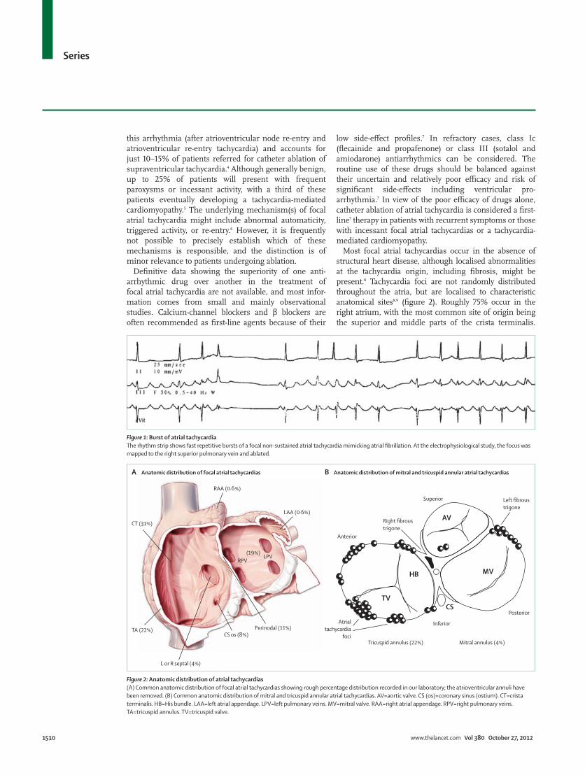
Most focal atrial tachycardias occur in the absence of structural heart disease, although localised abnormalities at the tachycardia origin, including fibrosis, might be present.8 Tachycardia foci are not randomly distributed throughout the atria, but are localised to characteristic anatomical sites8,9 (figure 2). Roughly 75% occur in the right atrium, with the most common site of origin being the superior and middle parts of the crista terminalis.
Figure 1: Burst of atrial tachycardiaThe rhythm strip shows fast repetitive bursts of a focal non-sustained atrial tachycardia mimicking atrial fibrillation. At the electrophysiological study, the focus was mapped to the right superior pulmonary vein and ablated.
A B
TV
MV
CS
HB
AV
Anatomic distribution of focal atrial tachycardias Anatomic distribution of mitral and tricuspid annular atrial tachycardias
LAA (0·6%)Right fibroustrigone
Left fibroustrigone
Anterior
Atrialtachycardia
foci
Superior
Posterior
Inferior
Tricuspid annulus (22%) Mitral annulus (4%)
(19%)RPV
LPV
Perinodal (11%)CS os (8%)
TA (22%)
CT (31%)
L or R septal (4%)
RAA (0·6%)
Figure 2: Anatomic distribution of atrial tachycardias(A) Common anatomic distribution of focal atrial tachycardias showing rough percentage distribution recorded in our laboratory; the atrioventricular annuli have been removed. (B) Common anatomic distribution of mitral and tricuspid annular atrial tachycardias. AV=aortic valve. CS (os)=coronary sinus (ostium). CT=crista terminalis. HB=His bundle. LAA=left atrial appendage. LPV=left pulmonary veins. MV=mitral valve. RAA=right atrial appendage. RPV=right pulmonary veins. TA=tricuspid annulus. TV=tricuspid valve.
Series
www.thelancet.com Vol 380 October 27, 2012 1511
Arrhythmias from this anatomic site have also been referred to as sinus node re-entry. In the left atrium, foci are most often noted at the ostium of the pulmonary veins.
Because of the characteristic anatomic clustering of atrial foci within the atria, the P-wave morphology on the surface ECG might provide important clues to the site of origin of the atrial tachycardia before catheter ablation.9 Kistler and colleagues9 developed an algorithm that prospectively localised the atrial site of origin in 93% of cases. Lead V1 was most useful in differentiating between left (positive P wave) and right (negative P wave) atrial focal sites.
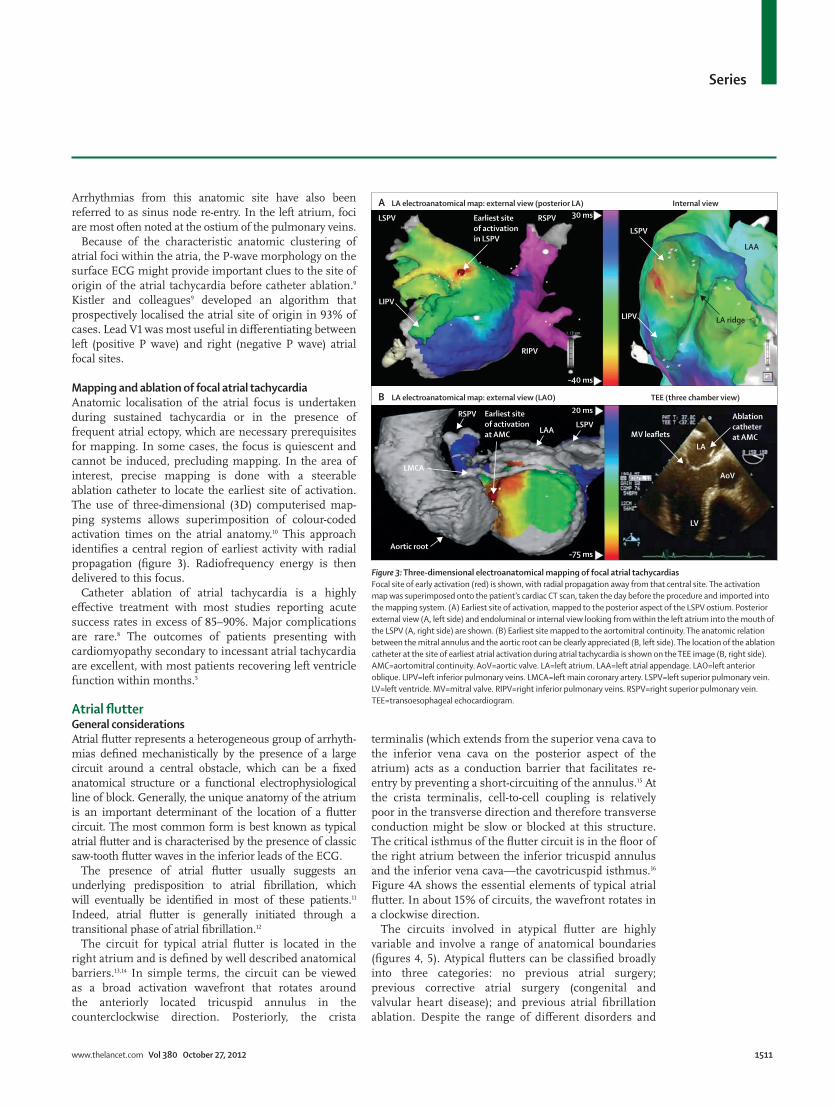
Mapping and ablation of focal atrial tachycardiaAnatomic localisation of the atrial focus is undertaken during sustained tachycardia or in the presence of frequent atrial ectopy, which are necessary prerequisites for mapping. In some cases, the focus is quiescent and cannot be induced, precluding mapping. In the area of interest, precise mapping is done with a steerable ablation catheter to locate the earliest site of activation. The use of three-dimensional (3D) computerised map-ping systems allows superimposition of colour-coded activation times on the atrial anatomy.10 This approach identifies a central region of earliest activity with radial propagation (figure 3). Radiofrequency energy is then delivered to this focus.
Catheter ablation of atrial tachycardia is a highly effective treatment with most studies reporting acute success rates in excess of 85–90%. Major complications are rare.8 The outcomes of patients presenting with cardiomyopathy secondary to incessant atrial tachycardia are excellent, with most patients recovering left ventricle function within months.5
Atrial flutterGeneral considerationsAtrial flutter represents a heterogeneous group of arrhyth-mias defined mechanistically by the presence of a large circuit around a central obstacle, which can be a fixed anatomical structure or a functional electrophysio logical line of block. Generally, the unique anatomy of the atrium is an important determinant of the location of a flutter circuit. The most common form is best known as typical atrial flutter and is characterised by the presence of classic saw-tooth flutter waves in the inferior leads of the ECG.
The presence of atrial flutter usually suggests an underlying predisposition to atrial fibrillation, which will eventually be identified in most of these patients.11 Indeed, atrial flutter is generally initiated through a transitional phase of atrial fibrillation.12
The circuit for typical atrial flutter is located in the right atrium and is defined by well described anatomical barriers.13,14 In simple terms, the circuit can be viewed as a broad activation wavefront that rotates around the anteriorly located tricuspid annulus in the counter clockwise direction. Posteriorly, the crista
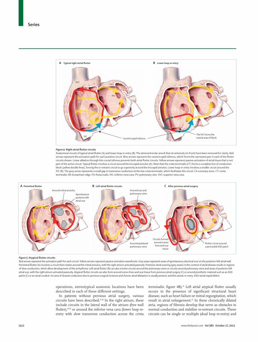
termin alis (which extends from the superior vena cava to the inferior vena cava on the posterior aspect of the atrium) acts as a conduction barrier that facilitates re-entry by preventing a short-circuiting of the annulus.15 At the crista terminalis, cell-to-cell coupling is relatively poor in the transverse direction and therefore transverse conduction might be slow or blocked at this structure. The critical isthmus of the flutter circuit is in the floor of the right atrium between the inferior tricuspid annulus and the inferior vena cava—the cavotricuspid isthmus.16 Figure 4A shows the essential elements of typical atrial flutter. In about 15% of circuits, the wavefront rotates in a clockwise direction.
The circuits involved in atypical flutter are highly variable and involve a range of anatomical boundaries (figures 4, 5). Atypical flutters can be classified broadly into three categories: no previous atrial surgery; previous corrective atrial surgery (congenital and valvular heart disease); and previous atrial fibrillation ablation. Despite the range of different disorders and
Figure 3: Three-dimensional electroanatomical mapping of focal atrial tachycardiasFocal site of early activation (red) is shown, with radial propagation away from that central site. The activation map was superimposed onto the patient’s cardiac CT scan, taken the day before the procedure and imported into the mapping system. (A) Earliest site of activation, mapped to the posterior aspect of the LSPV ostium. Posterior external view (A, left side) and endoluminal or internal view looking from within the left atrium into the mouth of the LSPV (A, right side) are shown. (B) Earliest site mapped to the aortomitral continuity. The anatomic relation between the mitral annulus and the aortic root can be clearly appreciated (B, left side). The location of the ablation catheter at the site of earliest atrial activation during atrial tachycardia is shown on the TEE image (B, right side). AMC=aortomitral continuity. AoV=aortic valve. LA=left atrium. LAA=left atrial appendage. LAO=left anterior oblique. LIPV=left inferior pulmonary veins. LMCA=left main coronary artery. LSPV=left superior pulmonary vein. LV=left ventricle. MV=mitral valve. RIPV=right inferior pulmonary veins. RSPV=right superior pulmonary vein. TEE=transoesophageal echocardiogram.
Earliest siteof activationin LSPV
Ablationcatheterat AMCMV leafletsLAA
Earliest siteof activationat AMC
LSPVRSPV
LMCA
Aortic root
LA
LV
AoV
RSPVLSPV
LAA
LA ridge
30 ms
–40 ms
20 ms
–75 ms
RIPV
LSPV
LA electroanatomical map: external view (posterior LA)A
B
Internal view
LA electroanatomical map: external view (LAO) TEE (three chamber view)
LIPV
LIPV
Series
1512 www.thelancet.com Vol 380 October 27, 2012
operations, stereo typical anatomic locations have been described in each of these different settings.
In patients without previous atrial surgery, various circuits have been described.17,18 In the right atrium, these include circuits in the lateral wall of the atrium (free wall flutter),19,20 or around the inferior vena cava (lower loop re-entry with slow transverse conduction across the crista
terminalis; figure 4B).21 Left atrial atypical flutter usually occurs in the presence of significant structural heart disease, such as heart failure or mitral regurgitation, which result in atrial enlarge ment.22 In these chronically dilated atria, regions of fibrosis develop that serve as obstacles to normal conduction and stabilise re-entrant circuits. These circuits can be single or multiple (dual loop re-entry) and
Figure 4: Right atrial flutter circuitsAnatomical circuits of typical atrial flutter (A) and lower loop re-entry (B). The atrioventricular annuli that sit anteriorly (in front) have been removed for clarity. Red arrows represent the activation path for each putative circuit. Blue arrows represent the cavotricuspid isthmus, which forms the narrowest part in each of the flutter circuits shown. Linear ablation through this crucial isthmus prevents both atrial flutter circuits. Yellow arrows represent passive activation of atrial tissue that is not part of the active circuit. Typical flutter involves a circuit around the tricuspid annulus (A). Note that the crista terminalis (CT) forms a complete line of conduction block (yellow double lines), forcing the re-entrant circuit to go superiorly around the tricuspid annulus. Lower loop re-entry involves a smaller circuit around the IVC (B). The grey arrow represents a small gap in transverse conduction at the low crista terminalis, which facilitates this circuit. CS=coronary sinus. CT=crista terminalis. ER=Eustachian ridge. FO=fossa ovalis. IVC=inferior vena cava. PV=pulmonary vein. SVC=superior vena cava.
A BTypical right atrial flutter Lower loop re-entry
Cavotricuspid isthmusThe IVC forms thecentral area of block
SVC
CT
IVC
ER
FO
PV
CS
A B CPerimitral flutter Left atrial flutter circuits After previous atrial surgery
Around mitral annulusSpontaneousposterior leftatrial scar
Around scar andpulmonary veins
Around ipsilateralpulmonary veins
Circuits formedaround suturelines and scar
tissue
Flutter circuit arounda pericardial ASD patch
Figure 5: Atypical flutter circuitsRed arrows represent the activation path for each circuit. Yellow arrows represent passive activation wavefronts. Grey areas represent areas of spontaneous electrical scar on the posterior left atrial wall. Perimitral flutter (A) involves a circuit that rotates around the mitral annulus, with the right atrium activated passively. Posterior atrial scarring (grey areas) in the context of atrial disease results in regions of slow conduction, which allow development of this arrhythmia. Left atrial flutter (B) can also involve circuits around the pulmonary veins or circuits around pulmonary veins and areas of posterior left atrial scar, with the right atrium activated passively. Atypical flutter circuits can also form around suture lines and scar tissue from previous atrial surgery (C) or around prosthetic material such as an ASD patch (C) or an atrial conduit. An area of slowed conduction due to previous surgical incisions and chronic atrial dilatation is usually present, and this assists re-entry. ASD=atrial septal defect.
Series
www.thelancet.com Vol 380 October 27, 2012 1513
frequently occur around the mitral annulus or pulmonary veins22 (figure 5).
In patients with previous atrial surgery, suture lines, scar, or prosthetic material can form the critical central barrier around which re-entry occurs, which has variously been referred to as incisional or scar-mediated re-entrant tachycardia. This disorder usually occurs in the context of surgical correction of congenital or valvular heart disease.23,24
Although the ECG pattern of typical flutter is characteristic, the flutter wave morphology of atypical flutters is highly variable and rarely gives a clue to precise anatomic location. Antiarrhythmic drugs are frequently ineffective and more than 50% of patients will eventually cross to a rate-control strategy because of an inability to maintain sinus rhythm.25 Catheter ablation is regarded as a first-line therapeutic option for patients with a first episode of typical atrial flutter26 and for flutter appearing after antiarrhythmic treatment of atrial fibrillation.27 Indications for ablation of atypical flutter include recurrent or poorly tolerated episodes and failed antiarrhythmic drug therapy.
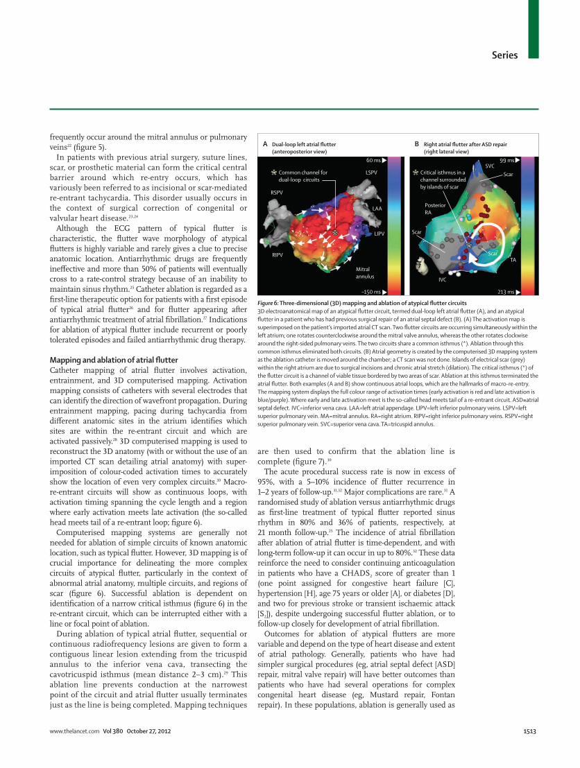
Mapping and ablation of atrial flutterCatheter mapping of atrial flutter involves activation, entrainment, and 3D computerised mapping. Activation mapping consists of catheters with several electrodes that can identify the direction of wavefront propagation. During entrainment mapping, pacing during tachycardia from different anatomic sites in the atrium identifies which sites are within the re-entrant circuit and which are activated passively.28 3D computerised mapping is used to reconstruct the 3D anatomy (with or without the use of an imported CT scan detailing atrial anatomy) with super-imposition of colour-coded activation times to accurately show the location of even very complex circuits.10 Macro-re-entrant circuits will show as con tinuous loops, with activation timing spanning the cycle length and a region where early activation meets late activation (the so-called head meets tail of a re-entrant loop; figure 6).
Computerised mapping systems are generally not needed for ablation of simple circuits of known anatomic location, such as typical flutter. However, 3D mapping is of crucial importance for delineating the more complex circuits of atypical flutter, particularly in the context of abnormal atrial anatomy, multiple circuits, and regions of scar (figure 6). Successful ablation is dependent on identification of a narrow critical isthmus (figure 6) in the re-entrant circuit, which can be interrupted either with a line or focal point of ablation.
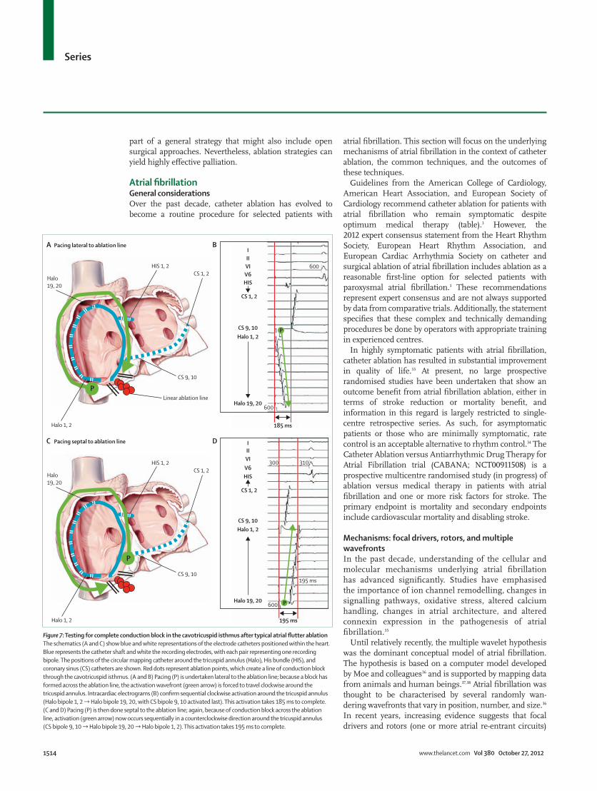
During ablation of typical atrial flutter, sequential or continuous radiofrequency lesions are given to form a contiguous linear lesion extending from the tricuspid annulus to the inferior vena cava, transecting the cavotricuspid isthmus (mean distance 2–3 cm).29 This ablation line prevents conduction at the narrowest point of the circuit and atrial flutter usually terminates just as the line is being completed. Mapping techniques
are then used to confirm that the ablation line is complete (figure 7).30
The acute procedural success rate is now in excess of 95%, with a 5–10% incidence of flutter recurrence in 1–2 years of follow-up.31,32 Major complications are rare.31 A randomised study of ablation versus antiarrhythmic drugs as first-line treat ment of typical flutter reported sinus rhythm in 80% and 36% of patients, respectively, at 21 month follow-up.25 The incidence of atrial fibrillation after ablation of atrial flutter is time-dependent, and with long-term follow-up it can occur in up to 80%.32 These data reinforce the need to consider continuing anti coagulation in patients who have a CHADS2 score of greater than 1 (one point assigned for congestive heart failure [C], hypertension [H], age 75 years or older [A], or diabetes [D], and two for previous stroke or transient ischaemic attack [S2]), despite undergoing successful flutter ablation, or to follow-up closely for development of atrial fibrillation.
Outcomes for ablation of atypical flutters are more variable and depend on the type of heart disease and extent of atrial pathology. Generally, patients who have had simpler surgical procedures (eg, atrial septal defect [ASD] repair, mitral valve repair) will have better outcomes than patients who have had several operations for complex congenital heart disease (eg, Mustard repair, Fontan repair). In these populations, ablation is generally used as
Dual-loop left atrial flutter(anteroposterior view)
A Right atrial flutter after ASD repair(right lateral view)
B
Common channel fordual-loop circuits
LSPV
LAA
LIPV
RIPV
RSPV
Mitralannulus
Scar
ScarSVC
Scar
PosteriorRA
TA
IVC
60 ms
–150 ms
99 ms
213 ms
Critical isthmus in achannel surroundedby islands of scar
Figure 6: Three-dimensional (3D) mapping and ablation of atypical flutter circuits3D electroanatomical map of an atypical flutter circuit, termed dual-loop left atrial flutter (A), and an atypical flutter in a patient who has had previous surgical repair of an atrial septal defect (B). (A) The activation map is superimposed on the patient’s imported atrial CT scan. Two flutter circuits are occurring simultaneously within the left atrium; one rotates counterclockwise around the mitral valve annulus, whereas the other rotates clockwise around the right-sided pulmonary veins. The two circuits share a common isthmus (*). Ablation through this common isthmus eliminated both circuits. (B) Atrial geometry is created by the computerised 3D mapping system as the ablation catheter is moved around the chamber; a CT scan was not done. Islands of electrical scar (grey) within the right atrium are due to surgical incisions and chronic atrial stretch (dilation). The critical isthmus (*) of the flutter circuit is a channel of viable tissue bordered by two areas of scar. Ablation at this isthmus terminated the atrial flutter. Both examples (A and B) show continuous atrial loops, which are the hallmarks of macro-re-entry. The mapping system displays the full colour range of activation times (early activation is red and late activation is blue/purple). Where early and late activation meet is the so-called head meets tail of a re-entrant circuit. ASD=atrial septal defect. IVC=inferior vena cava. LAA=left atrial appendage. LIPV=left inferior pulmonary veins. LSPV=left superior pulmonary vein. MA=mitral annulus. RA=right atrium. RIPV=right inferior pulmonary veins. RSPV=right superior pulmonary vein. SVC=superior vena cava. TA=tricuspid annulus.
Series
1514 www.thelancet.com Vol 380 October 27, 2012
part of a general strategy that might also include open surgical approaches. Nevertheless, ablation strat egies can yield highly effective palliation.
Atrial fibrillationGeneral considerationsOver the past decade, catheter ablation has evolved to become a routine procedure for selected patients with
atrial fibrillation. This section will focus on the underlying mechanisms of atrial fibrillation in the context of catheter ablation, the common techniques, and the outcomes of these techniques.
Guidelines from the American College of Cardiology, American Heart Association, and European Society of Cardiology recommend catheter ablation for patients with atrial fibrillation who remain symptomatic despite optimum medical therapy (table).3 However, the 2012 expert consensus statement from the Heart Rhythm Society, European Heart Rhythm Association, and European Cardiac Arrhythmia Society on catheter and surgical ablation of atrial fibrillation includes ablation as a reasonable first-line option for selected patients with paroxysmal atrial fibrillation.3 These recommendations represent expert consensus and are not always supported by data from comparative trials. Additionally, the statement specifies that these complex and technically demanding procedures be done by operators with appropriate training in experienced centres.
In highly symptomatic patients with atrial fibrillation, catheter ablation has resulted in substantial improve ment in quality of life.33 At present, no large prospective randomised studies have been undertaken that show an outcome benefit from atrial fibrillation ablation, either in terms of stroke reduction or mortality benefit, and information in this regard is largely restricted to single-centre retrospective series. As such, for asymptomatic patients or those who are minimally symptomatic, rate control is an acceptable alternative to rhythm control.34 The Catheter Ablation versus Antiarrhythmic Drug Therapy for Atrial Fibril lation trial (CABANA; NCT00911508) is a prospective multicentre randomised study (in progress) of ablation versus medical therapy in patients with atrial fibrillation and one or more risk factors for stroke. The primary endpoint is mortality and secondary endpoints include cardiovascular mortality and disabling stroke.
Mechanisms: focal drivers, rotors, and multiple wavefrontsIn the past decade, understanding of the cellular and molecular mechanisms underlying atrial fibrillation has advanced significantly. Studies have emphasised the importance of ion channel remodelling, changes in signalling pathways, oxidative stress, altered calcium handling, changes in atrial architecture, and altered connexin expression in the pathogenesis of atrial fibrillation.35
Until relatively recently, the multiple wavelet hy pothesis was the dominant conceptual model of atrial fibrillation. The hypothesis is based on a computer model developed by Moe and colleagues36 and is supported by mapping data from animals and human beings.37,38 Atrial fibrillation was thought to be characterised by several randomly wan-dering wavefronts that vary in position, number, and size.36 In recent years, increasing evidence suggests that focal drivers and rotors (one or more atrial re-entrant circuits)
Figure 7: Testing for complete conduction block in the cavotricuspid isthmus after typical atrial flutter ablationThe schematics (A and C) show blue and white representations of the electrode catheters positioned within the heart. Blue represents the catheter shaft and white the recording electrodes, with each pair representing one recording bipole. The positions of the circular mapping catheter around the tricuspid annulus (Halo), His bundle (HIS), and coronary sinus (CS) catheters are shown. Red dots represent ablation points, which create a line of conduction block through the cavotricuspid isthmus. (A and B) Pacing (P) is undertaken lateral to the ablation line; because a block has formed across the ablation line, the activation wavefront (green arrow) is forced to travel clockwise around the tricuspid annulus. Intracardiac electrograms (B) confirm sequential clockwise activation around the tricuspid annulus (Halo bipole 1, 2 → Halo bipole 19, 20, with CS bipole 9, 10 activated last). This activation takes 185 ms to complete. (C and D) Pacing (P) is then done septal to the ablation line; again, because of conduction block across the ablation line, activation (green arrow) now occurs sequentially in a counterclockwise direction around the tricuspid annulus (CS bipole 9, 10 → Halo bipole 19, 20 → Halo bipole 1, 2). This activation takes 195 ms to complete.
IIIVIV6HIS
CS 1, 2
CS 9, 10Halo 1, 2
Halo 19, 20
195 ms
195 ms
310300
600 P
IIIVI 600
600
185 ms
V6HIS
CS 1, 2
CS 9, 10Halo 1, 2
Halo 19, 20
P
A Pacing lateral to ablation line
PCS 9, 10
Linear ablation line
CS 1, 2HIS 1, 2
Halo19, 20
Halo 1, 2
C
B
DPacing septal to ablation line
P
CS 9, 10
CS 1, 2HIS 1, 2
Halo19, 20
Halo 1, 2
Series
www.thelancet.com Vol 380 October 27, 2012 1515
have an important role in the underlying mechanism of atrial fibrillation.
The mechanisms underlying paroxysmal atrial fibril-lation (defined by the presence of episodes that ter minate spontaneously within 7 days) and persistent atrial fibrillation (episodes lasting longer than 7 days and not self-terminating) are considerably different.
In 1998, Haissaguerre and colleagues39 described the presence of focal drivers originating from within atrial muscular extensions into the pulmonary veins. Very high frequency electrical activity (>300–400 bpm) from a focal source caused non-uniform conduction to the atrium, resulting in atrial fibrillation. Since that seminal finding, others have also shown that pulmonary vein foci represent the crucial initiating trigger to paroxysmal atrial fibrillation in 85–95% of patients. In the remainder of patients, foci outside the pulmonary veins seem to be responsible. The pathophysiological processes leading to development of these focal triggers are not known. Furthermore, whether the underlying mechanism of this focal activity is due to enhanced automaticity,40 triggered activity,41,42 or localised pulmonary vein re-entry is unclear.43 Nevertheless, an appreciation of the im-portance of pulmonary vein musculature in initiation of paroxysmal atrial fibrillation has led to development of highly effective ablation strategies to electrically isolate these veins.44,45
In patients with persistent atrial fibrillation, the mechanisms are less clear. Pulmonary vein foci might be important for arrhythmia initiation in a subset of patients. However, in those with more persistent atrial fibrillation and those with structural heart disease, mechanisms that maintain rather than initiate the arrhythmia play the dominant part. These mechanisms are located within atrial myocardium, and the develop-ment of atrial remodelling is of crucial importance to the persistence of atrial fibrillation.46 Atrial remodelling refers to the electrical and structural changes that develop in the atrium as a result of atrial fibrillation itself and the presence of a range of coexisting disorders. Heart failure, hypertension, valvular heart disease, and obstructive sleep apnoea lead to atrial structural changes or re-modelling, including atrial enlargement and regional fibrosis, forming the prerequisite substrate for persistent atrial fibrillation.46
The exact mechanism responsible for the maintenance of persistent atrial fibrillation is not known. Recently, accumulating evidence from animal studies has shown the important role of atrial rotors in driving persistent atrial fibrillation.47,48 Mapping studies of human beings have shown the presence of atrial rotors in patients with persistent atrial fibrillation.49,50 A recent study has shown that catheter ablation of these rotors might lead to termination of persistent atrial fibrillation.51 The efficacy of this potentially exciting new approach to the ablation of persistent atrial fibrillation will need to be verified by large multicentre studies.
Other mapping data from patients with persistent atrial fibrillation suggest that dissociation between the epicardial and endocardial atrial layers results in breakthrough wavefronts that continually renew and drive the atrial fibrillation process—a mechanism akin to multiple wavelet re-entry. These two mechanisms are not mutually exclusive. Importantly, in patients with different forms of structural heart disease, the under-lying mechanism of atrial fibrillation is most probably quite heterogeneous.
The uncertainty surrounding the mechanism of per-sistent atrial fibrillation and our inability to establish precise mechanisms in individual patients is shown in the myriad of different ablative approaches to this arrhythmia and the disappointing success rates.52
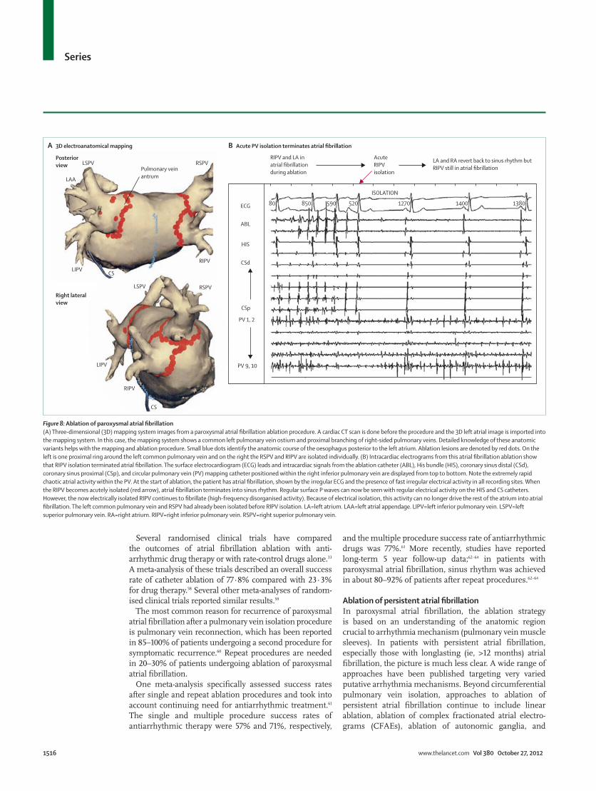
Ablation of paroxysmal atrial fibrillationPulmonary vein isolation is the cornerstone of catheter ablation for patients with paroxysmal atrial fibrillation.53 This method is an empirical approach based on the knowledge that most focal triggers occur within pul-monary vein muscular sleeves. Detailed assessment of the anatomic location of several triggers is not feasible. The current approach to isolation targets the proximal pulmonary vein antrum, thereby minimising the risk of stenosis (figure 8). The most widely used and assessed technique is that of point-by-point irrigated radio-frequency ablation. 3D mapping is routinely used to accurately delineate anatomy with or without the use of an imported CT scan (figure 8). More recently, in an attempt to simplify the procedure, various one-shot technologies for circum ferential isolation of the pul-monary veins have been developed.54–56 The cryoballoon and a circumferential radiofrequency ablation catheter are the most advanced of these technologies. However, neither the promise of shorter procedure and fluoroscopy times nor the goal of improved efficacy and safety have been definitively realised.54,57 Irrespective of technology used, the endpoint for antral pulmonary vein ablation is demonstration of complete conduction block into and out of the pulmonary vein.
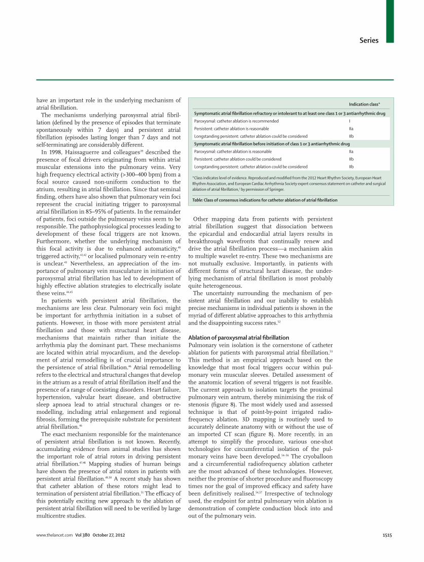
Indication class*
Symptomatic atrial fibrillation refractory or intolerant to at least one class 1 or 3 antiarrhythmic drug
Paroxysmal: catheter ablation is recommended I
Persistent: catheter ablation is reasonable IIa
Longstanding persistent: catheter ablation could be considered IIb
Symptomatic atrial fibrillation before initiation of class 1 or 3 antiarrhythmic drug
Paroxysmal: catheter ablation is reasonable IIa
Persistent: catheter ablation could be considered IIb
Longstanding persistent: catheter ablation could be considered IIb
*Class indicates level of evidence. Reproduced and modified from the 2012 Heart Rhythm Society, European Heart Rhythm Association, and European Cardiac Arrhythmia Society expert consensus statement on catheter and surgical ablation of atrial fibrillation,3 by permission of Springer.
Table: Class of consensus indications for catheter ablation of atrial fibrillation
Series
1516 www.thelancet.com Vol 380 October 27, 2012
Several randomised clinical trials have compared the outcomes of atrial fibrillation ablation with anti-arrhythmic drug therapy or with rate-control drugs alone.33 A meta-analysis of these trials described an overall success rate of catheter ablation of 77·8% compared with 23·3% for drug therapy.58 Several other meta-analyses of random-ised clinical trials reported similar results.59
The most common reason for recurrence of paroxys mal atrial fibrillation after a pulmonary vein isolation procedure is pulmonary vein reconnection, which has been reported in 85–100% of patients undergoing a second procedure for symptomatic recurrence.60 Repeat procedures are needed in 20–30% of patients undergoing ablation of paroxysmal atrial fibrillation.
One meta-analysis specifically assessed success rates after single and repeat ablation procedures and took into account continuing need for antiarrhythmic treatment.61 The single and multiple procedure success rates of antiarrhythmic therapy were 57% and 71%, respectively,
and the multiple procedure success rate of antiarrhythmic drugs was 77%.61 More recently, studies have reported long-term 5 year follow-up data;62–64 in patients with paroxysmal atrial fibrillation, sinus rhythm was achieved in about 80–92% of patients after repeat procedures.62–64
Ablation of persistent atrial fibrillationIn paroxysmal atrial fibrillation, the ablation strategy is based on an understanding of the anatomic region crucial to arrhythmia mechanism (pulmonary vein muscle sleeves). In patients with persistent atrial fibril lation, especially those with longlasting (ie, >12 months) atrial fibrillation, the picture is much less clear. A wide range of approaches have been published targeting very varied putative arrhythmia mechanisms. Beyond circumferential pulmonary vein isolation, ap proaches to ablation of persistent atrial fibrillation continue to include linear ablation, ablation of complex fractionated atrial electro-grams (CFAEs), ablation of autonomic ganglia, and
Figure 8: Ablation of paroxysmal atrial fibrillation(A) Three-dimensional (3D) mapping system images from a paroxysmal atrial fibrillation ablation procedure. A cardiac CT scan is done before the procedure and the 3D left atrial image is imported into the mapping system. In this case, the mapping system shows a common left pulmonary vein ostium and proximal branching of right-sided pulmonary veins. Detailed knowledge of these anatomic variants helps with the mapping and ablation procedure. Small blue dots identify the anatomic course of the oesophagus posterior to the left atrium. Ablation lesions are denoted by red dots. On the left is one proximal ring around the left common pulmonary vein and on the right the RSPV and RIPV are isolated individually. (B) Intracardiac electrograms from this atrial fibrillation ablation show that RIPV isolation terminated atrial fibrillation. The surface electrocardiogram (ECG) leads and intracardiac signals from the ablation catheter (ABL), His bundle (HIS), coronary sinus distal (CSd), coronary sinus proximal (CSp), and circular pulmonary vein (PV) mapping catheter positioned within the right inferior pulmonary vein are displayed from top to bottom. Note the extremely rapid chaotic atrial activity within the PV. At the start of ablation, the patient has atrial fibrillation, shown by the irregular ECG and the presence of fast irregular electrical activity in all recording sites. When the RIPV becomes acutely isolated (red arrow), atrial fibrillation terminates into sinus rhythm. Regular surface P waves can now be seen with regular electrical activity on the HIS and CS catheters. However, the now electrically isolated RIPV continues to fibrillate (high-frequency disorganised activity). Because of electrical isolation, this activity can no longer drive the rest of the atrium into atrial fibrillation. The left common pulmonary vein and RSPV had already been isolated before RIPV isolation. LA=left atrium. LAA=left atrial appendage. LIPV=left inferior pulmonary vein. LSPV=left superior pulmonary vein. RA=right atrium. RIPV=right inferior pulmonary vein. RSPV=right superior pulmonary vein.
Pulmonary veinantrum
RSPVLSPV
LIPVRIPV
LAA
ECG 80
RIPV and LA inatrial fibrillationduring ablation
RSPVLSPV
LIPV
RIPV
CS
AcuteRIPVisolation
ISOLATION
LA and RA revert back to sinus rhythm butRIPV still in atrial fibrillation
850 590 520 1270 1400 1380
ABL
HIS
CSd
CSp
PV 1, 2
PV 9, 10
CS
A 3D electroanatomical mapping
Right lateralview
Posteriorview
B Acute PV isolation terminates atrial fibrillation
Series
www.thelancet.com Vol 380 October 27, 2012 1517
isolation of other venous structures including the coronary sinus and superior vena cava. Additionally, hybrid or stepwise approaches that include two or more of these procedures have been described. Each of these approaches is empirical: the mechanisms have not been shown in patients specifically and they are predicated on unproven assumptions about arrhythmia mechanism. This con-trasts with ablation of atrial tachycardia and atrial flutter for which the individual patient-specific mechanism is clearly defined at the time of the procedure.
Disappointingly, the single procedure success rates have largely been under 50% and in some instances as low as 20–30%. A recent systematic review of outcomes of persistent atrial fibrillation ablation concluded that the varying ablation techniques (including pulmonary vein isolation alone, pulmonary vein isolation with linear ablation, pulmonary vein isolation with CFAE ablation, and the stepwise procedure) resulted in similar results (mean single procedure success rate of 47%).52 Clinical outcomes were improved with repeat procedures with success rates approaching 65%.
Studies with longer-term follow-up (2–5 years) have reported multiple procedure success rates ranging between 57% and 63%, and in many series persistent atrial fibrillation is an independent predictor of late recur-rence.65,66 Other factors that have been associated with lower long-term success rates have included older age, increased left atrial size, obesity, sleep apnoea, and structural heart disease.65,67 However, findings have not been consistent and most trials have enrolled a relatively small number of patients with advanced age, marked left atrial enlargement, or advanced structural heart disease (including significant left ventricular dysfunction). Efficacy of catheter ablation in these subgroups needs further assessment.
Proarrhythmia: development of atrial tachycardia after atrial fibrillation ablationNew atrial tachycardias can appear for the first time after atrial fibrillation ablation. After simple pulmonary vein isolation, such arrhythmias are seen infrequently. How-ever, with more extensive atrial ablation, especially that including linear ablation or widespread ablation of fractionated electrograms, recurrent atrial tachycardia can occur in 30–50% of patients. The most frequently reported mechanisms include atrial macro-re-entry (large circuits) or small re-entrant circuits (1–2 cm), both occurring around regions of previous ablation. These stable circuits, com pared with atrial fibrillation, result in more rapid ventri cular response rates, more frequently need cardioversion, and might actually be associated with an increase in symptoms.68 For these reasons, many labora-tories now limit the amount of extensive ablation done.
ComplicationsThe risk of major complications of radiofrequency catheter ablation of atrial fibrillation was previously reported to
range from 3·9–4·5%.69,70 Recognised major complications include death (in 1 of 1000 patients), stroke, cardiac tamponade, atrio-oesophageal fistula, or clinically sig-nificant pulmonary vein stenosis. These studies69,70 incorporate results from a heterogeneous population undergoing a variety of ablation procedures between 1999 and 2007. With increasing experience, a high-volume centre re ported that complication rates decreased from 11·1% in 2002, to 1·6% in 2010 (p<0·05), with no complications associated with permanent sequelae since 2005.71 In patients younger than 70 years with pre-dominantly paroxysmal atrial fibrillation without signifi-cant struc tural heart disease, pulmonary vein antral isolation is safe and associated with a very low risk of major complications (<1%).72 Factors that have been associated with higher complication rates include older age, previous stroke, and advanced structural heart disease, but data are inconsistent.
Future challengesFor paroxysmal atrial fibrillation the major challenge is the creation of enduring pulmonary vein isolation, because pulmonary vein reconnection is the over whelming reason for recurrence. Early data suggest that catheters that register tissue contact force and thereby improve lesion efficacy might result in lower rates of pulmonary vein reconnection.73,74 In patients with persistent atrial fibrill-ation, a better understanding of (probably hetero geneous) arrhythmia mechanisms is needed so that ablation approaches can be targeted to a clearly shown mechanism. Advances in technology have played a major part in our ability to ablate these arrhythmias; further improvements in catheter design and mapping capabilities will no doubt lead to better understanding and outcomes. By acting on the evolving substrate for atrial fibrillation, treatment of coexisting disorders might improve ablation outcomes or even slow clinical progression of atrial fibrillation so that ablation may be deferred. Studies are in progress.
ConclusionCatheter ablation is now at the forefront of the treatment algorithm for a broad range of atrial arrhythmias. In patients with focal atrial tachycardia and typical atrial flutter, it is a first line therapy with efficacy in excess of 90%. For patients with a range of complex atypical flutters, it is a highly effective approach in those not readily controlled with antiarrhythmic drugs. For patients with previous atrial surgery or more complex congenital heart disease, late flutter recurrence or atrial fibrillation development is common, but ablation never theless might offer a highly effective palliation.
In symptomatic patients with atrial fibrillation, cath eter ablation has resulted in substantial improvements in quality of life. Success rates in selected paroxysmal atrial fibrillation patients might be in excess of 80%, although several procedures are frequently needed, and complication rates have fallen to around 1% in low-risk populations.
Series
1518 www.thelancet.com Vol 380 October 27, 2012
More data are needed to understand the role and outcomes of ablation in broader subgroups, including elderly people and those with significant structural heart disease (including left ventricular dysfunction and atrial enlarge-ment). For patients with persistent atrial fibrillation, the role of catheter ablation continues to evolve, as does our understanding of the crucial underlying mechanisms.ContributorsGL did the detailed scientific literature search, drafted and revised the manuscript, and created the original figures. PS contributed to scientific literature inclusions; revised the manuscript, figures, and bibliography; and responded to the reviewers. JMK did the detailed literature search; revised the manuscript, figures, and bibliography; responded to the reviewers; and approved the final report.
Conflicts of interestPS has served on the advisory board of Bard Electrophysiology, Biosense-Webster, Medtronic, St Jude Medical, Merck, and Sanofi-Aventis, and has received lecture fees or research funding from Bard Electrophysiology, Biosense-Webster, Medtronic, and St Jude Medical. JMK is the recipient of research funding and fellowship support from Medtronic, St Jude Medical, and Johnson & Johnson Medical. GL declares that he has no conflicts of interest.
AcknowledgmentsGL is the recipient of a research scholarship from the National Health and Medical Research Council of Australia (NHMRC). JMK is the recipient of a grant from NHMRC (604908).
References1 Saoudi N, Cosio F, Waldo A, et al. A classification of atrial flutter
and regular atrial tachycardia according to electrophysiological mechanisms and anatomical bases: a Statement from a Joint Expert Group from The Working Group of Arrhythmias of the European Society of Cardiology and the North American Society of Pacing and Electrophysiology. Eur Heart J 2001; 22: 1162–82.
2 Saoudi N, Anselme F, Poty H, Cribier A, Castellanos A. Entrainment of supraventricular tachycardias: a review. Pacing Clin Electrophysiol 1998; 21: 2105–25.
3 Calkins H, Kuck KH, Cappato R, et al. 2012 HRS/EHRA/ECAS expert consensus statement on catheter and surgical ablation of atrial fibrillation: recommendations for patient selection, procedural techniques, patient management and follow-up, definitions, endpoints, and research trial design: a report of the Heart Rhythm Society (HRS) task force on catheter and surgical ablation of atrial fibrillation. Heart Rhythm 2012; 9: 632–96.e21.
4 Porter MJ, Morton JB, Denman R, et al. Influence of age and gender on the mechanism of supraventricular tachycardia. Heart Rhythm 2004; 1: 393–96.
5 Medi C, Kalman JM, Haqqani H, et al. Tachycardia-mediated cardiomyopathy secondary to focal atrial tachycardia: long-term outcome after catheter ablation. J Am Coll Cardiol 2009; 53: 1791–97.
6 Chen SA, Chiang CE, Yang CJ, et al. Sustained atrial tachycardia in adult patients: electrophysiological characteristics, pharmacological response, possible mechanisms, and effects of radiofrequency ablation. Circulation 1994; 90: 1262–78.
7 Blomstrom-Lundqvist C, Scheinman MM, Aliot EM, et al. ACC/AHA/ESC guidelines for the management of patients with supraventricular arrhythmias—executive summary: a report of the American College Of Cardiology/American Heart Association Task Force on Practice Guidelines and the European Society Of Cardiology Committee for Practice Guidelines (writing committee to develop guidelines for the management of patients with supraventricular arrhythmias), developed in collaboration with NASPE–Heart Rhythm Society. J Am Coll Cardiol 2003; 42: 1493–531.
8 Kalman JM, Olgin JE, Karch MR, Hamdan M, Lee RJ, Lesh MD. “Cristal tachycardias”: origin of right atrial tachycardias from the crista terminalis identified by intracardiac echocardiography. J Am Coll Cardiol 1998; 31: 451–59.
9 Kistler PM, Roberts-Thomson KC, Haqqani HM, et al. P-wave morphology in focal atrial tachycardia: development of an algorithm to predict the anatomic site of origin. J Am Coll Cardiol 2006; 48: 1010–17.
10 Natale A, Breeding L, Tomassoni G, et al. Ablation of right and left ectopic atrial tachycardias using a three-dimensional nonfluoroscopic mapping system. Am J Cardiol 1998; 82: 989–92.
11 Moubarak G, Pavin D, Laviolle B, et al. Incidence of atrial fibrillation during very long-term follow-up after radiofrequency ablation of typical atrial flutter. Arch Cardiovasc Dis 2009; 102: 525–32.
12 Waldo AL, Feld GK. Inter-relationships of atrial fibrillation and atrial flutter mechanisms and clinical implications. J Am Coll Cardiol 2008; 51: 779–86.
13 Cosio FG, Arribas F, Barbero JM, Kallmeyer C, Goicolea A. Validation of double-spike electrograms as markers of conduction delay or block in atrial flutter. Am J Cardiol 1988; 61: 775–80.
14 Kalman JM, Olgin JE, Saxon LA, Fisher WG, Lee RJ, Lesh MD. Activation and entrainment mapping defines the tricuspid annulus as the anterior barrier in typical atrial flutter. Circulation 1996; 94: 398–406.
15 Olgin JE, Kalman JM, Fitzpatrick AP, Lesh MD. Role of right atrial endocardial structures as barriers to conduction during human type I atrial flutter: activation and entrainment mapping guided by intracardiac echocardiography. Circulation 1995; 92: 1839–48.
16 Olshansky B, Okumura K, Hess PG, Waldo AL. Demonstration of an area of slow conduction in human atrial flutter. J Am Coll Cardiol 1990; 16: 1639–48.
17 Kalman JM, Olgin JE, Saxon LA, Lee RJ, Scheinman MM, Lesh MD. Electrocardiographic and electrophysiologic characterization of atypical atrial flutter in man: use of activation and entrainment mapping and implications for catheter ablation. J Cardiovasc Electrophysiol 1997; 8: 121–44.
18 Yang Y, Cheng J, Bochoeyer A, et al. Atypical right atrial flutter patterns. Circulation 2001; 103: 3092–98.
19 Kall JG, Rubenstein DS, Kopp DE, et al. Atypical atrial flutter originating in the right atrial free wall. Circulation 2000; 101: 270–79.
20 Tai CT, Huang JL, Lin YK, et al. Noncontact three-dimensional mapping and ablation of upper loop re-entry originating in the right atrium. J Am Coll Cardiol 2002; 40: 746–53.
21 Cheng J, Cabeen WRJ, Scheinman MM. Right atrial flutter due to lower loop reentry: mechanism and anatomic substrates. Circulation 1999; 99: 1700–05.
22 Jais P, Shah DC, Haissaguerre M, et al. Mapping and ablation of left atrial flutters. Circulation 2000; 101: 2928–34.
23 Chan DP, Van Hare GF, Mackall JA, Carlson MD, Waldo AL. Importance of atrial flutter isthmus in postoperative intra-atrial reentrant tachycardia. Circulation 2000; 102: 1283–89.
24 Kalman JM, VanHare GF, Olgin JE, Saxon LA, Stark SI, Lesh MD. Ablation of ‘incisional’ reentrant atrial tachycardia complicating surgery for congenital heart disease. Use of entrainment to define a critical isthmus of conduction. Circulation 1996; 93: 502–12.
25 Natale A, Newby KH, Pisano E, et al. Prospective randomized comparison of antiarrhythmic therapy versus first-line radiofrequency ablation in patients with atrial flutter. J Am Coll Cardiol 2000; 35: 1898–904.
26 Verma A, Macle L, Cox J, Skanes AC. Canadian Cardiovascular Society atrial fibrillation guidelines 2010: catheter ablation for atrial fibrillation/atrial flutter. Can J Cardiol 2011; 27: 60–66.
27 Reithmann C, Hoffmann E, Spitzlberger G, et al. Catheter ablation of atrial flutter due to amiodarone therapy for paroxysmal atrial fibrillation. Eur Heart J 2000; 21: 565–72.
28 Waldo AL. Atrial flutter: entrainment characteristics. J Cardiovasc Electrophysiol 1997; 8: 337–52.
29 Da Costa A, Faure E, Thevenin J, et al. Effect of isthmus anatomy and ablation catheter on radiofrequency catheter ablation of the cavotricuspid isthmus. Circulation 2004; 110: 1030–35.
30 Schumacher B, Pfeiffer D, Tebbenjohanns J, Lewalter T, Jung W, Luderitz B. Acute and long-term effects of consecutive radiofrequency applications on conduction properties of the subeustachian isthmus in type I atrial flutter. J Cardiovasc Electrophysiol 1998; 9: 152–63.
31 Willems S, Weiss C, Ventura R, et al. Catheter ablation of atrial flutter guided by electroanatomic mapping (CARTO): a randomized comparison to the conventional approach. J Cardiovasc Electrophysiol 2000; 11: 1223–30.
32 Da Costa A, Romeyer C, Mourot S, et al. Factors associated with early atrial fibrillation after ablation of common atrial flutter: a single centre prospective study. Eur Heart J 2002; 23: 498–506.
Series
www.thelancet.com Vol 380 October 27, 2012 1519
33 Jais P, Cauchemez B, Macle L, et al. Catheter ablation versus antiarrhythmic drugs for atrial fibrillation: the A4 study. Circulation 2008; 118: 2498–505.
34 Olshansky B, Rosenfeld LE, Warner AL, et al. The Atrial Fibrillation Follow-up Investigation of Rhythm Management (AFFIRM) study: approaches to control rate in atrial fibrillation. J Am Coll Cardiol 2004; 43: 1201–08.
35 Schotten U, Verheule S, Kirchhof P, Goette A. Pathophysiological mechanisms of atrial fibrillation: a translational appraisal. Physiol Rev 2011; 91: 265–325.
36 Moe GK, Rheinboldt WC, Abildskov JA. A computer model of atrial fibrillation. Am Heart J 1964; 67: 200–20.
37 Allessie MA, Bonke FIM, Schopman FJG. Circus movement in rabbit atrial muscle as a mechanism of tachycardia. Circ Res 1973; 33: 54–62.
38 Cox JL, Canavan TE, Schuessler RB, et al. The surgical treatment of atrial fibrillation. II. Intraoperative electrophysiologic mapping and description of the electrophysiologic basis of atrial flutter and atrial fibrillation. J Thorac Cardiovasc Surg 1991; 101: 406–26.
39 Haissaguerre M, Jais P, Shah DC, et al. Spontaneous initiation of atrial fibrillation by ectopic beats originating in the pulmonary veins. N Engl J Med 1998; 339: 659–66.
40 Chen YJ, Chen SA, Chen YC, et al. Effects of rapid atrial pacing on the arrhythmogenic activity of single cardiomyocytes from pulmonary veins: implication in initiation of atrial fibrillation. Circulation 2001; 104: 2849–54.
41 Patterson E, Jackman WM, Beckman KJ, et al. Spontaneous pulmonary vein firing in man: relationship to tachycardia-pause early afterdepolarizations and triggered arrhythmia in canine pulmonary veins in vitro. J Cardiovasc Electrophysiol 2007; 18: 1067–75.
42 Voigt N, Li N, Wang Q, et al. Enhanced sarcoplasmic reticulum Ca2+ leak and increased Na+-Ca2+ exchanger function underlie delayed afterdepolarizations in patients with chronic atrial fibrillation. Circulation 2012; 125: 2059–70.
43 Arora R, Verheule S, Scott L, et al. Arrhythmogenic substrate of the pulmonary veins assessed by high-resolution optical mapping. Circulation 2003; 107: 1816–21.
44 Ho SY, Sanchez-Quintana D, Cabrera JA, Anderson RH. Anatomy of the left atrium: implications for radiofrequency ablation of atrial fibrillation. J Cardiovasc Electrophysiol 1999; 10: 1525–33.
45 Vaitkevicius R, Saburkina I, Rysevaite K, et al. Nerve supply of the human pulmonary veins: an anatomical study. Heart Rhythm 2009; 6: 221–28.
46 Nattel S, Burstein B, Dobrev D. Atrial remodeling and atrial fibrillation: mechanisms and implications. Circ Arrhythm Electrophysiol 2008; 1: 62–73.
47 Gray RA, Pertsov AM, Jalife J. Spatial and temporal organization during cardiac fibrillation. Nature 1998; 392: 75–78.
48 Sahadevan J, Ryu K, Peltz L, et al. Epicardial mapping of chronic atrial fibrillation in patients: preliminary observations. Circulation 2004; 110: 3293–99.
49 Narayan SM, Krummen DE, Rappel WJ. Clinical mapping approach to diagnose electrical rotors and focal impulse sources for human atrial fibrillation. J Cardiovasc Electrophysiol 2012; 23: 447–54.
50 Lee G, Teh AW, Kumar S, et al. Characterisation of atrial activation patterns in human persistent atrial fibrillation: multiple wavelets or focal drivers? Heart Lung Circ 2012; 21 (suppl 1): S117–18
51 Narayan SM, Krummen DE, Shivkumar K, Clopton P, Rappel WJ, Miller JM. Treatment of atrial fibrillation by the ablation of localized sources: CONFIRM (Conventional Ablation for Atrial Fibrillation With or Without Focal Impulse and Rotor Modulation) trial. J Am Coll Cardiol 2012; 60: 628–36.
52 Brooks AG, Stiles MK, Laborderie J, et al. Outcomes of long-standing persistent atrial fibrillation ablation: a systematic review. Heart Rhythm 2010; 7: 835–46.
53 Jais P, Haissaguerre M, Shah DC, et al. A focal source of atrial fibrillation treated by discrete radiofrequency ablation. Circulation 1997; 95: 572–76.
54 Neumann T, Vogt J, Schumacher B, et al. Circumferential pulmonary vein isolation with the cryoballoon technique: results from a prospective 3-center study. J Am Coll Cardiol 2008; 52: 273–78.
55 Van Belle Y, Janse P, Theuns D, Szili-Torok T, Jordaens L. One year follow-up after cryoballoon isolation of the pulmonary veins in patients with paroxysmal atrial fibrillation. Europace 2008; 10: 1271–76.
56 Scharf C, Boersma L, Davies W, et al. Ablation of persistent atrial fibrillation using multielectrode catheters and duty-cycled radiofrequency energy. J Am Coll Cardiol 2009; 54: 1450–56.
57 Linhart M, Bellmann B, Mittmann-Braun E, et al. Comparison of cryoballoon and radiofrequency ablation of pulmonary veins in 40 patients with paroxysmal atrial fibrillation: a case-control study. J Cardiovasc Electrophysiol 2009; 20: 1343–48.
58 Bonanno C, Paccanaro M, La Vecchia L, Ometto R, Fontanelli A. Efficacy and safety of catheter ablation versus antiarrhythmic drugs for atrial fibrillation: a meta-analysis of randomized trials. J Cardiovasc Med (Hagerstown) 2010; 11: 408–18.
59 Kong MH, Piccini JP, Bahnson TD. Efficacy of adjunctive ablation of complex fractionated atrial electrograms and pulmonary vein isolation for the treatment of atrial fibrillation: a meta-analysis of randomized controlled trials. Europace 2011; 13: 193–204.
60 Gerstenfeld EP, Callans DJ, Dixit S, Zado E, Marchlinski FE. Incidence and location of focal atrial fibrillation triggers in patients undergoing repeat pulmonary vein isolation: implications for ablation strategies. J Cardiovasc Electrophysiol 2003; 14: 685–90.
61 Calkins H, Reynolds MR, Spector P, et al. Treatment of atrial fibrillation with antiarrhythmic drugs or radiofrequency ablation: two systematic literature reviews and meta-analyses. Circ Arrhythm Electrophysiol 2009; 2: 349–61.
62 Ouyang F, Tilz R, Chun J, et al. Long-term results of catheter ablation in paroxysmal atrial fibrillation: lessons from a 5-year follow-up. Circulation 2010; 122: 2368–77.
63 Pappone C, Rosanio S, Augello G, et al. Mortality, morbidity, and quality of life after circumferential pulmonary vein ablation for atrial fibrillation: outcomes from a controlled nonrandomized long-term study. J Am Coll Cardiol 2003; 42: 185–97.
64 Bhargava M, Di Biase L, Mohanty P, et al. Impact of type of atrial fibrillation and repeat catheter ablation on long-term freedom from atrial fibrillation: results from a multicenter study. Heart Rhythm 2009; 6: 1403–12.
65 Wokhlu A, Hodge DO, Monahan KH, et al. Long-term outcome of atrial fibrillation ablation: impact and predictors of very late recurrence. J Cardiovasc Electrophysiol 2010; 21: 1071–78.
66 Weerasooriya R, Khairy P, Litalien J, et al. Catheter ablation for atrial fibrillation: are results maintained at 5 years of follow-up? J Am Coll Cardiol 2011; 57: 160–66.
67 Arya A, Hindricks G, Sommer P, et al. Long-term results and the predictors of outcome of catheter ablation of atrial fibrillation using steerable sheath catheter navigation after single procedure in 674 patients. Europace 2010; 12: 173–80.
68 Daoud EG, Weiss R, Augostini R, et al. Proarrhythmia of circumferential left atrial lesions for management of atrial fibrillation. J Cardiovasc Electrophysiol 2006; 17: 157–65.
69 Bertaglia E, Zoppo F, Tondo C, et al. Early complications of pulmonary vein catheter ablation for atrial fibrillation: a multicenter prospective registry on procedural safety. Heart Rhythm 2007; 4: 1265–71.
70 Spragg DD, Dalal D, Cheema A, et al. Complications of catheter ablation for atrial fibrillation: incidence and predictors. J Cardiovasc Electrophysiol 2008; 19: 627–31.
71 Hoyt H, Bhonsale A, Chilukuri K, et al. Complications arising from catheter ablation of atrial fibrillation: temporal trends and predictors. Heart Rhythm 2011; 8: 1869–74.
72 Lee G, Sparks PB, Morton JB, et al. Low risk of major complications associated with pulmonary vein antral isolation for atrial fibrillation: results of 500 consecutive ablation procedures in patients with low prevalence of structural heart disease from a single center. J Cardiovasc Electrophysiol 2011; 22: 163–68.
73 Kerst G, Weig HJ, Weretka S, et al. Contact force-controlled zero-fluoroscopy catheter ablation of right-sided and left atrial arrhythmia substrates. Heart Rhythm 2012; 9: 709–14.
74 Kuck KH, Reddy VY, Schmidt B, et al. A novel radiofrequency ablation catheter using contact force sensing: Toccata study. Heart Rhythm 2012; 9: 18–23.