cascaded-automatic segmentation for schistosoma japonicum eggs in images of fecal samples
TRANSCRIPT
Cascaded-Automatic Segmentation for Schistosoma japonicum eggs inimages of fecal samples
Junjie Zhang a, Yunyu Lin a, Yan Liu a,n, Zhengyu Li a, Zhong Li b,nn, Shan Hu a, Zhiyuan Liu a,Dandan Lin c, Zhongdao Wu a
a Zhongshan School of Medicine, Sun Yat-sen University, Guangzhou 510060, Chinab Department of Neurology, The Sixth Affiliated Hospital, Sun Yat-sen University, No. 26, Yuancun 2nd Heng Roa, Tianhe District, Guangzhou 510655, Chinac Jiangxi Provincial Institute of Parasitic Disease Control, Nanchang 360046, China
a r t i c l e i n f o
Article history:Received 11 November 2013Accepted 27 May 2014
Keywords:Schistosoma japonicum eggAutomatic SegmentationMicroscopic image
a b s t r a c t
Background: To recognize parasite eggs automatically, the automatic segmentation of parasite eggimages is very important for the extraction of characteristics and genera classification.Methods: A Cascaded-Automatic Segmentation approach was proposed. Firstly, image contrast betweenthe border of an egg and its background for all samples was strengthened by the Radon-Like Featuresalgorithm and the enhanced image was processed into a binary image to get an initial set. Then, theelliptical targets are located with Randomized Hough Transform (RHT). The fitted data of an ellipticalborder are considered the initial border data and the accurate border of a Schistosoma japonicum egg canbe finally segmented using an Active Contour Model (Snake).Results: Seventy-three cases of S. japonicum eggs in fecal samples were found; 61 images contained aparasite egg and 12 did not. Although the illumination, noise pollution, boundary definitions of eggs, andegg position are different, they are all segmented and labeled accurately.Discussion: The results proved that accurate borders of S. japonicum eggs could be recognized preciselyusing the proposed method, and the robustness of the method is good even in images with heavy noise.This indicates that the proposed method can overcome the disadvantages of the traditional thresholdsegmentation method, which has limited adaptability to images with heavy background noise.& 2014 The Authors. Published by Elsevier Ltd. This is an open access article under the CC BY-NC-ND
license (http://creativecommons.org/licenses/by-nc-nd/3.0/).
1. Introduction
The microscopic image recognition of parasite eggs is one ofthe most important means of diagnosis for humans infected byparasites. In recent years, parasite researchers and computerresearchers have been cooperatively studying to enable the auto-matic recognition of parasite eggs in microscopic images in orderto reduce the erroneous judgment rate that is caused by indivi-dual's lacking experience in manual identification. They also aimedto achieve remote real-time differential diagnosis. To date, manyresearch papers have focused on how to classify specific kinds ofeggs, and only a few papers have dealt with how to identify theaccurate borders of a parasite egg [1–10]. Among these, papers[6,9,10] were from China, while the other reports were fromoutside of this country. Automatic segmentation of parasite eggs
image is very important for the extraction of characteristics andthe classification of parasite eggs in later stages. If the automaticsegmentation of parasite eggs is not performed, they cannot berecognized automatically. In the aforementioned papers, only afew have reported on automatic segmentation based on thethreshold method of parasite egg objects [1,3,4,6]. Other reportson the segmentation of parasite eggs can only be made withhuman participation to accurately obtain parasite eggs fromimages. In these research, Yang et al. [1] started research intothe recognition of parasite eggs in fecal samples, but the experi-mental materials contained much less noise than those utilized inthis paper, which were captured directly from fecal sampleswithout any preprocessing. Dogantekin et al. [3] and Castañónet al. [4] proposed two different parasite egg segmentationmethods based on threshold operations, but the threshold methodwas too simple to resolve the problem identified in the currentpaper. In addition, reported segmentation methods are generallyapplied to parasite egg images which are obtained from culturemedium containing smaller background noise. Nevertheless, eggimages from clinical testing samples are often obtained from fecalextracts; they have the same features regarding the surroundingand internal contents. Also, it is difficult to accurately identify egg
Contents lists available at ScienceDirect
journal homepage: www.elsevier.com/locate/cbm
Computers in Biology and Medicine
http://dx.doi.org/10.1016/j.compbiomed.2014.05.0120010-4825/& 2014 The Authors. Published by Elsevier Ltd. This is an open access article under the CC BY-NC-ND license (http://creativecommons.org/licenses/by-nc-nd/3.0/).
n Corresponding author. Tel.: 86 20 87331856.nn Corresponding author.E-mail addresses: [email protected] (J. Zhang), [email protected] (Y. Lin),
[email protected] (Y. Liu), [email protected] (Z. Li),[email protected] (Z. Li), [email protected] (S. Hu),[email protected] (Z. Liu), [email protected] (Z. Wu).
Computers in Biology and Medicine 52 (2014) 18–27
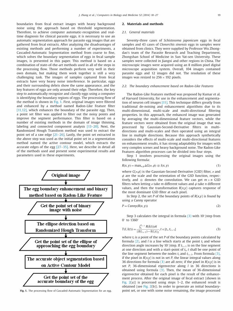
boundaries from fecal extract images with heavy backgroundnoise using the approach based on threshold segmentation.Therefore, to achieve computer automatic-recognition and real-time diagnosis for clinical parasite eggs, it is necessary to use anautomatic segmentation approach for parasite egg images that aregathered from fecal extracts. After analyzing the disadvantages ofexisting methods and performing a number of experiments, aCascaded-Automatic Segmentation method from coarse to fine,which solves the boundary segmentation of eggs in fecal sampleimages, is presented in this paper. This method is based on acombination of state-of-the-art methods used in all of the steps inthe processing flow. Those methods perform very well in theirown domain, but making them work together is still a verychallenging task. The images of samples captured from fecalextracts have very heavy noise interference. In particular, eggsand their surrounding debris show the same appearance, and thekey features of eggs are only around their edge. Therefore, the keystep to automatically recognize and classify eggs using a computeris identifying the boundary regions of eggs. The processing flow ofthe method is shown in Fig. 1. First, original images were filteredand enhanced by a method named Radon-Like Feature filter[11,12], which enhances the boundary of the parasite egg. Then,a point set filter was applied to filter out the noisy points andimprove the segment performance. This filter is based on anumber of existing methods in the domain of image thinning,labeling and connected component analysis [13–20]. Next, theRandomized Hough Transform method was used to extract thepoint set of a raw edge [21–26]. Lastly, the point set extracted inthe above step was used as the initial point set in a segmentationmethod named the active contour model, which extracts theaccurate edges of the egg [27–35]. Here, we describe in detail allof the methods used and present some experimental results andparameters used in these experiments.
2. Materials and methods
2.1. General materials
Seventy-three cases of Schistosoma japonicum eggs in fecalsamples and 43 cases of Clonorchis sinensis eggs in samples wereobtained from clinics. They were supplied by Professor Wu Zhong-dao's team of the Parasite Research and Teaching Department,Zhongshan School of Medicine in Sun Yat-sen University. Thesesamples were collected in Jiangxi and other regions in China. Themicroscopic images were acquired using an 8 million pixel digitalmicroscope acquisition system. Overall, 104 images containedparasite eggs and 12 images did not. The resolution of theseimages was resized to 256�192 pixels.
2.2. The boundary enhancement based on Radon-Like Features
The Radon-Like Features method was proposed by Kumar et al.of Harvard University, for use in the enhancement and segmenta-tion of neuron cell images [11]. This technique differs greatly fromtraditional de-noising and enhancement algorithms due to itsmulti-dimensional, multi-scale and multi-directional filteringproperties. In this approach, the enhanced image was generatedby averaging the multi-dimensional feature vectors, while thefeature vectors were obtained from the original image that wasprocessed by Gaussian-Second-Derivative filters in multi-directions and multi-scales and then operated using an integralline in multiple directions. Because this approach syntheticallyconsiders the effects of multi-scale and multi-directional featureson enhancement results, it has strong adaptability for images withvery complex scenes and heavy background noise. The Radon-LikeFeatures algorithm processes can be divided into four steps.
Step 1 involves processing the original images using thefollowing formula:
Rðx; yÞ ¼maxσ;ϕΔGðσ;ϕÞ � Iðx; yÞ ð1Þwhere G(σ,ϕ) is the Gaussian-Second-Derivative (GSD) filter, σ andϕ are the scale and the orientation of the GSD function, respec-tively, and � denotes the convolution. We can get m�n GSDfilters when letting σ take m different values and ϕ take n differentvalues, and then the transformation R(x,y) captures response ofthe most dominant GSD filter at each pixel.
In Step 2, the set P of the boundary points of R(x,y) is found byusing a Canny operator
P ¼ CannyðRðx; yÞÞ ð2Þ
Step 3 calculates the integral in formula (3) with 101/step from01 to 13601
TðI; lðtÞÞ ¼R tiþ 1ti
RðlðtÞÞ∂tjjlðtiþ1Þ� lðtiÞjj2
; tA ½ti; tiþ1� ð3Þ
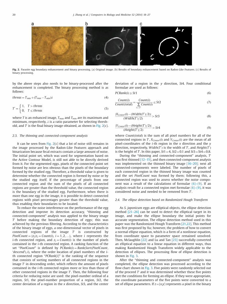
where ti is a point of the set P of the boundary points calculated byformula (2), and l is a line which starts at the point ti and whosedirection angle increases by 101/step. If tiþ1 is on the line segmentat one direction and with a start-point of ti, t shall be one point ofthe line segment between the nodes ti and tiþ1. From formula (3),if the pixel in R(x,y) is not in set P, the linear integral values along36 directions for formula (3) are all zero; if the pixel in R(x,y) is inset P, 36-dimensional eigenvector along l in 36 directions isobtained using formula (3). Then, the mean of 36-dimensionaleigenvector obtained for each pixel is the result of the enhance-ment process. After the original image of fecal extract (shown inFig. 2(a)) is processed using steps 1–2, the enhanced result isobtained (see Fig. 2(b)). In order to generate an initial boundary-point set, or one with some noise remaining, the image processedFig. 1. The processing flow of Cascaded-Automatic Segmentation for an egg.
J. Zhang et al. / Computers in Biology and Medicine 52 (2014) 18–27 19
by the above steps also needs to be binary-processed after theenhancement is completed. The binary processing method is asfollows:
threas¼ TminþðTmax�TminÞλ ð4Þ
T 0 ¼1; T4threas0; Trthreas
(ð5Þ
where T is an enhanced image, Tmax and Tmin are its maximum andminimum, respectively, λ is a ratio parameter for selecting thresh-old, and T0 is the final binary image obtained, as shown in Fig. 2(c).
2.3. The thinning and connected-component analysis
It can be seen from Fig. 2(c) that a lot of noise still remains inthe image processed by the Radon-Like Features approach andbinarization because fecal extracts contain a large amount of noise.The initial point set, which was used for segmentation based onthe Active Contour Model, is still not able to be directly derivedfrom it. For the segmented eggs, pixels of the connected point setformed by noise are less obvious than the pixels of the boundaryformed by the studied egg. Therefore, a threshold value is given todetermine whether the connected region is formed by noise or bythe studied egg itself. If the percentage of pixels from oneconnected region and the sum of the pixels of all connectedregions are greater than the threshold value, the connected regionis the boundary of the studied egg. Furthermore, when there ismore than one egg in the image, it is possible to detect connectedregions with pixel percentages greater than the threshold value,thus enabling their boundaries to be located.
To reduce the noise interference on the performance of the eggdetection and improve its detection accuracy, “thinning andconnected-component” analysis was applied to the binary imageT0 before making the boundary detection of eggs; this wasachieved by the previous filtering. According to the characteristicsof the binary image of eggs, a one-dimensional vector of pixels inconnected regions of the image T0 is constructed byPixelCount ¼ fzijzi ¼ CountðiÞ; i¼ 1;2;…g, where i represents thei-th connected region, and zi ¼ CountðiÞ is the number of pixelscontained in the i-th connected region. A ranking function of theset “PixelCount” is defined by PCRankðiÞ ¼ RankðSortðPixelCount;‘descend’Þ; iÞ, where the order location of pixel numbers of the i-th connected region “PCRank(i)” is the ranking of the sequencethat consists of sorting numbers of all connected regions in theimage T0 in descending order, which reflects the amount of pixelscontained in the i-th connected region more or less than those ofother connected regions in the image T0. Then, the following fourcriteria for reducing noise are used: the pixel-number ordinal of aregion, St1, the pixel-number proportion of a region, St2, thecenter deviation of a region in the x direction, St3, and the center
deviation of a region in the y direction, St4. Four conditionalformulae are used as follows:
PCRankðiÞZSt1 ð6Þ
CountðiÞCountðtotalÞ ¼
CountðiÞ∑iAT 0
CountðiÞrSt2 ð7Þ
jXCenterðiÞ�ðWidthðT 0Þ=2ÞjðWidthðT 0Þ=2Þ 4St3 ð8Þ
jYCenterðiÞ�ðHeightðT 0Þ=2ÞjðHeightðT 0Þ=2Þ 4St4 ð9Þ
where CountðtotalÞ is the sum of all pixel numbers for all of theconnected regions in T0, XCenterðiÞ and YCenterðiÞ are the mean of allpixel-coordinates of the i-th region in the x direction and the ydirection, respectively, WidthðT 0Þ is the width of T0, and HeightðT 0Þis the height of T0. In this paper, St1¼4, St2¼0.1 and St3¼St4¼0.8.
During the “thinning and connected-component” analysis, T0
was first thinned [12–15], and then connected-component analysiswas implemented on the thinned binary image [16–20]; next allconnected-components were labeled. The number of pixels ofeach connected region in the thinned binary image was countedand the set PixelCount was formed by them; following this, aconnected region was used to assess whether the noise compo-nent was a result of the calculations of formulae (6)–(9). If ananalysis result for a connected region met formulae (6)–(9), it wasconsidered noise and needed to be removed from T0.
2.4. The ellipse detection based on Randomized Hough Transform
As S. japonicum eggs are elliptical objects, the ellipse detectionmethod [21–26] can be used to detect the elliptical target in itsimage, and make the ellipse boundary the initial points foraccurate segmentation. The ellipse detection method used in thispaper was the Randomized Hough Transform [21,22]. This methodwas first proposed by Xu; however, the problem of how to converta normal ellipse equation, which is a form of a nonlinear equation,from coordinate space to parameter space remained unsolved.Then, Mclaughlin [22] and Lu and Tan [23] successfully convertedan elliptical equation to a linear equation in different ways, thusmaking Randomized Hough Transform widely applicable to thedetection of ellipses. The processing flow of ellipse detection isshown in Fig. 3.
After the “thinning and connected-component” analysis wascompleted, the ellipse detection was processed according to theflow chart shown in Fig. 3. Five points were randomly selected outof the proceed T0 and it was determined whether these five pointsmet the conditions for forming an ellipse. If they were appropriate,the coordinate parameters of the five points were converted to aset of ellipse parameters. If z¼(x,y) represents a pixel in the binary
Fig. 2. Parasite egg boundary enhancement and binary processing. (a) Original image. (b) Results of boundary enhancement based on Radon-Like Features. (c) Results ofbinary processing.
J. Zhang et al. / Computers in Biology and Medicine 52 (2014) 18–2720
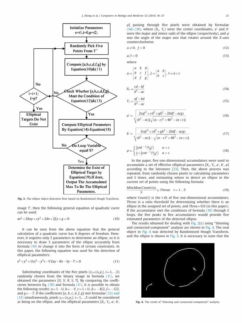
image T0, then the following general equation of quadratic curvecan be used:
ax2þ2bxyþcy2þ2dxþ2f yþg¼ 0 ð10Þ
It can be seen from the above equation that the generalcalculation of a quadratic curve has 6 degrees of freedom. How-ever, it requires only 5 parameters to determine an ellipse, so it isnecessary to draw 5 parameters of the ellipse accurately fromformula (10) to change it into the form of certain constraints. Inthis paper, the following equation was used for the detection ofelliptical parameters:
x2þy2þUðx2�y2Þ�V2xy�Rx�Sy�T ¼ 0 ð11Þ
Substituting coordinates of the five pixels (zi¼(xi,yi), i¼1,…,5)randomly chosen from the binary image in formula (11), weobtained the parameters [U, V, R, S, T]. By comparing the coeffi-cients between Eq. (10) and formula (11), it is possible to obtainthe following results: a¼1�U, b¼�V, c¼1þU, d¼�R/2, f¼�S/2,and g¼�T. If the coefficients [a, b, c, d, f, g] met formulae (12) and(13) simultaneously, pixels zi¼(xi,yi), i¼1,…,5 could be consideredas being on the ellipse, and the elliptical parameters [Xc, Yc, a0, b0,
ϕ] passing through five pixels were obtained by formulae(14)–(18), where (Xc, Yc) were the center coordinates, a0 and b0
were the major and minor radii of the ellipse (respectively), and ϕwas the angle of the major axis that rotates around the X-axiscounterclockwise.
Δa0; J40 ð12Þ
Δ=Io0 ð13Þwhere
Δ¼a b d
b c f
d f g
��������������; J ¼
a bb c
��������; I ¼ aþc
Xc ¼ cd�bf
b2�acð14Þ
Yc ¼af �bd
b2�acð15Þ
a0 ¼
ffiffiffiffiffiffiffiffiffiffiffiffiffiffiffiffiffiffiffiffiffiffiffiffiffiffiffiffiffiffiffiffiffiffiffiffiffiffiffiffiffiffiffiffiffiffiffiffiffiffiffiffiffiffiffiffiffiffiffiffiffiffiffiffiffiffiffiffiffiffi2ðaf 2þcd2þgb2�2bdf �acgÞ
ðb2�acÞ½ffiffiffiffiffiffiffiffiffiffiffiffiffiffiffiffiffiffiffiffiffiffiffiffiffiffiffiffiða�cÞ2þ4b2
q�ðaþcÞ�
vuuut ð16Þ
b0 ¼
ffiffiffiffiffiffiffiffiffiffiffiffiffiffiffiffiffiffiffiffiffiffiffiffiffiffiffiffiffiffiffiffiffiffiffiffiffiffiffiffiffiffiffiffiffiffiffiffiffiffiffiffiffiffiffiffiffiffiffiffiffiffiffiffiffiffiffiffiffiffiffiffiffiffiffi2ðaf 2þcd2þgb2�2bdf �acgÞ
ðb2�acÞ½�ffiffiffiffiffiffiffiffiffiffiffiffiffiffiffiffiffiffiffiffiffiffiffiffiffiffiffiffiða�cÞ2þ4b2
q�ðaþcÞ�
vuuut ð17Þ
ϕ¼12cot
�1 a� c2b
� �aoc
π2þ1
2cot�1 a� c
2b
� �a4c
(ð18Þ
In the paper, five one-dimensional accumulators were used toaccumulate a set of effective elliptical parameters [Xc, Yc, a0, b0, ϕ]according to the literature [23]. Then, the above process wasrepeated, from randomly chosen pixels to calculating parametersand S times, and estimating where to detect an ellipse in thecurrent set of points using the following formula:
MinðMaxðCountðiÞÞÞS
ZThreas i¼ 1…5 ð19Þ
where Count(i) is the i-th of five one-dimensional accumulators,Threas is a ratio threshold for determining whether there is anellipse in the assigned set of points, and Threa¼0.6 (in this paper).If the accumulator met the conditions of formula (19) through Sloops, the five peaks in five accumulators would provide fiveestimated parameters of the detected ellipse.
The results obtained for dealing with Fig. 2(c) using “thinningand connected-component” analysis are shown in Fig. 4. The ovalobject in Fig. 4 was detected by Randomized Hough Transform,and the ellipse is shown in Fig. 5. It is necessary to state that the
Fig. 3. The ellipse object detection flow based on Randomized Hough Transform.
Fig. 4. The result of “thinning and connected-component” analysis.
J. Zhang et al. / Computers in Biology and Medicine 52 (2014) 18–27 21
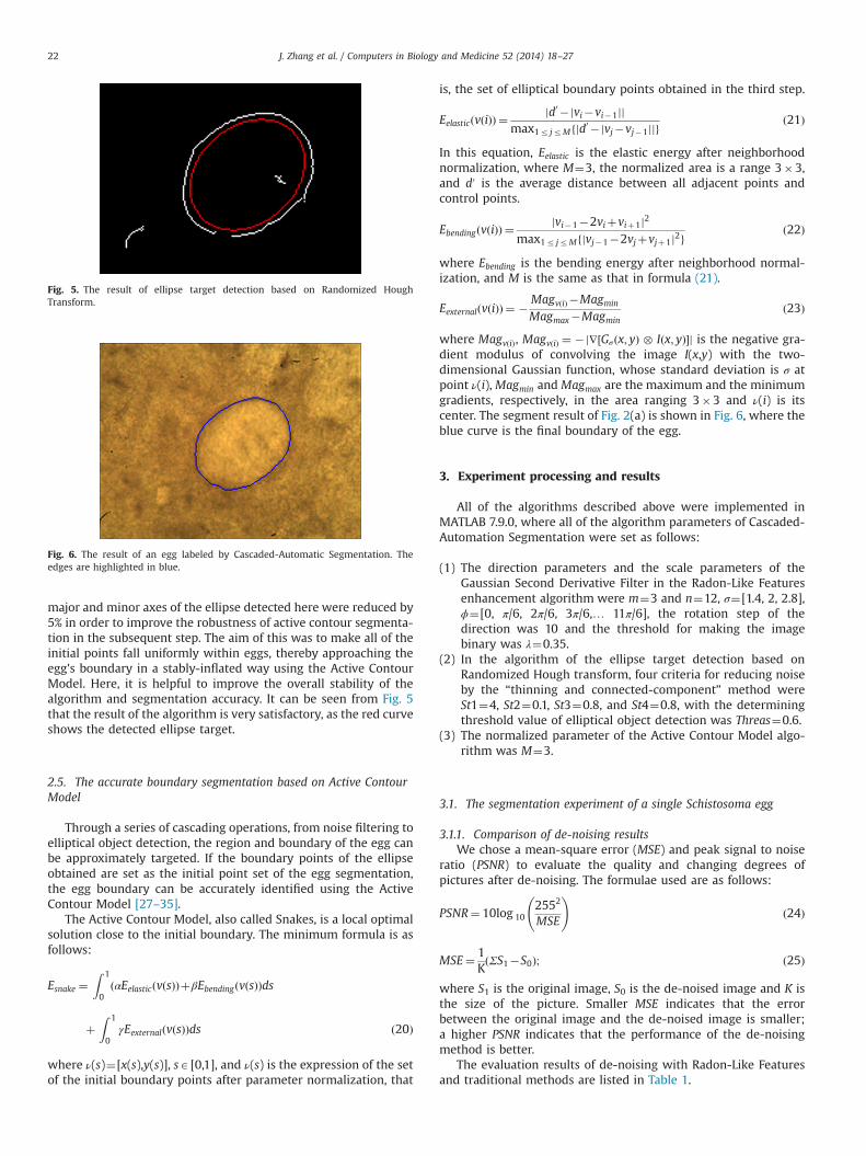
major and minor axes of the ellipse detected here were reduced by5% in order to improve the robustness of active contour segmenta-tion in the subsequent step. The aim of this was to make all of theinitial points fall uniformly within eggs, thereby approaching theegg's boundary in a stably-inflated way using the Active ContourModel. Here, it is helpful to improve the overall stability of thealgorithm and segmentation accuracy. It can be seen from Fig. 5that the result of the algorithm is very satisfactory, as the red curveshows the detected ellipse target.
2.5. The accurate boundary segmentation based on Active ContourModel
Through a series of cascading operations, from noise filtering toelliptical object detection, the region and boundary of the egg canbe approximately targeted. If the boundary points of the ellipseobtained are set as the initial point set of the egg segmentation,the egg boundary can be accurately identified using the ActiveContour Model [27–35].
The Active Contour Model, also called Snakes, is a local optimalsolution close to the initial boundary. The minimum formula is asfollows:
Esnake ¼Z 1
0ðαEelasticðvðsÞÞþβEbendingðvðsÞÞds
þZ 1
0γEexternalðvðsÞÞds ð20Þ
where ν(s)¼[x(s),y(s)], sA[0,1], and ν(s) is the expression of the setof the initial boundary points after parameter normalization, that
is, the set of elliptical boundary points obtained in the third step.
EelasticðvðiÞÞ ¼jd0 �jvi�vi�1jj
max1r jrMfjd0 �jvj�vj�1jjgð21Þ
In this equation, Eelastic is the elastic energy after neighborhoodnormalization, where M¼3, the normalized area is a range 3�3,and d0 is the average distance between all adjacent points andcontrol points.
EbendingðvðiÞÞ ¼jvi�1�2viþviþ1j2
max1r jrMfjvj�1�2vjþvjþ1j2gð22Þ
where Ebending is the bending energy after neighborhood normal-ization, and M is the same as that in formula (21).
EexternalðvðiÞÞ ¼ � MagvðiÞ �Magmin
Magmax�Magminð23Þ
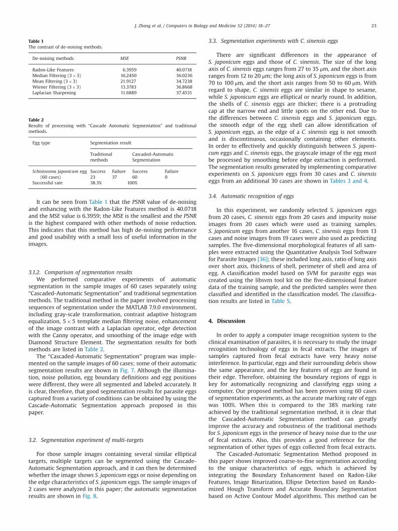
where MagvðiÞ, MagvðiÞ ¼ �j∇½Gσðx; yÞ � Iðx; yÞ�j is the negative gra-dient modulus of convolving the image I(x,y) with the two-dimensional Gaussian function, whose standard deviation is σ atpoint ν(i), Magmin andMagmax are the maximum and the minimumgradients, respectively, in the area ranging 3�3 and ν(i) is itscenter. The segment result of Fig. 2(a) is shown in Fig. 6, where theblue curve is the final boundary of the egg.
3. Experiment processing and results
All of the algorithms described above were implemented inMATLAB 7.9.0, where all of the algorithm parameters of Cascaded-Automation Segmentation were set as follows:
(1) The direction parameters and the scale parameters of theGaussian Second Derivative Filter in the Radon-Like Featuresenhancement algorithm were m¼3 and n¼12, σ¼[1.4, 2, 2.8],ϕ¼[0, π/6, 2π/6, 3π/6,… 11π/6], the rotation step of thedirection was 10 and the threshold for making the imagebinary was λ¼0.35.
(2) In the algorithm of the ellipse target detection based onRandomized Hough transform, four criteria for reducing noiseby the “thinning and connected-component” method wereSt1¼4, St2¼0.1, St3¼0.8, and St4¼0.8, with the determiningthreshold value of elliptical object detection was Threas¼0.6.
(3) The normalized parameter of the Active Contour Model algo-rithm was M¼3.
3.1. The segmentation experiment of a single Schistosoma egg
3.1.1. Comparison of de-noising resultsWe chose a mean-square error (MSE) and peak signal to noise
ratio (PSNR) to evaluate the quality and changing degrees ofpictures after de-noising. The formulae used are as follows:
PSNR¼ 10log 102552
MSE
!ð24Þ
MSE¼ 1KðΣS1�S0Þ; ð25Þ
where S1 is the original image, S0 is the de-noised image and K isthe size of the picture. Smaller MSE indicates that the errorbetween the original image and the de-noised image is smaller;a higher PSNR indicates that the performance of the de-noisingmethod is better.
The evaluation results of de-noising with Radon-Like Featuresand traditional methods are listed in Table 1.
Fig. 5. The result of ellipse target detection based on Randomized HoughTransform.
Fig. 6. The result of an egg labeled by Cascaded-Automatic Segmentation. Theedges are highlighted in blue.
J. Zhang et al. / Computers in Biology and Medicine 52 (2014) 18–2722
It can be seen from Table 1 that the PSNR value of de-noisingand enhancing with the Radon-Like Features method is 40.0718and the MSE value is 6.3959; the MSE is the smallest and the PSNRis the highest compared with other methods of noise reduction.This indicates that this method has high de-noising performanceand good usability with a small loss of useful information in theimages.
3.1.2. Comparison of segmentation resultsWe performed comparative experiments of automatic
segmentation in the sample images of 60 cases separately using“Cascaded-Automatic Segmentation” and traditional segmentationmethods. The traditional method in the paper involved processingsequences of segmentation under the MATLAB 7.9.0 environment,including gray-scale transformation, contrast adaptive histogramequalization, 5�5 template median filtering noise, enhancementof the image contrast with a Laplacian operator, edge detectionwith the Canny operator, and smoothing of the image edge withDiamond Structure Element. The segmentation results for bothmethods are listed in Table 2.
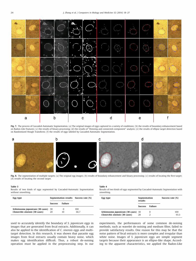
The “Cascaded-Automatic Segmentation” program was imple-mented on the sample images of 60 cases; some of their automaticsegmentation results are shown in Fig. 7. Although the illumina-tion, noise pollution, egg boundary definitions and egg positionswere different, they were all segmented and labeled accurately. Itis clear, therefore, that good segmentation results for parasite eggscaptured from a variety of conditions can be obtained by using theCascade-Automatic Segmentation approach proposed in thispaper.
3.2. Segmentation experiment of multi-targets
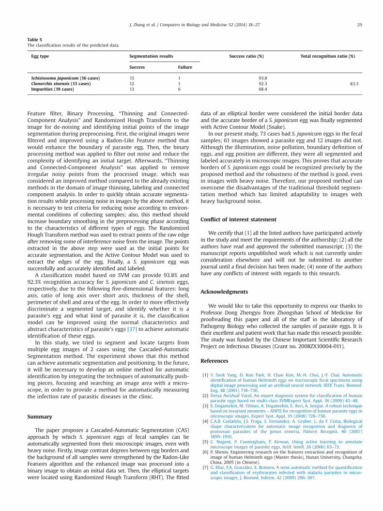
For those sample images containing several similar ellipticaltargets, multiple targets can be segmented using the Cascade-Automatic Segmentation approach, and it can then be determinedwhether the image shows S. japonicum eggs or noise depending onthe edge characteristics of S. japonicum eggs. The sample images of2 cases were analyzed in this paper; the automatic segmentationresults are shown in Fig. 8.
3.3. Segmentation experiments with C. sinensis eggs
There are significant differences in the appearance ofS. japonicum eggs and those of C. sinensis. The size of the longaxis of C. sinensis eggs ranges from 27 to 35 μm, and the short axisranges from 12 to 20 μm; the long axis of S. japonicum eggs is from70 to 100 μm, and the short axis ranges from 50 to 60 μm. Withregard to shape, C. sinensis eggs are similar in shape to sesame,while S. japonicum eggs are elliptical or nearly round. In addition,the shells of C. sinensis eggs are thicker; there is a protrudingcap at the narrow end and little spots on the other end. Due tothe differences between C. sinensis eggs and S. japonicum eggs,the smooth edge of the egg shell can allow identification ofS. japonicum eggs, as the edge of a C. sinensis egg is not smoothand is discontinuous, occasionally containing other elements.In order to effectively and quickly distinguish between S. japoni-cum eggs and C. sinensis eggs, the grayscale image of the egg mustbe processed by smoothing before edge extraction is performed.The segmentation results generated by implementing comparativeexperiments on S. japonicum eggs from 30 cases and C. sinensiseggs from an additional 30 cases are shown in Tables 3 and 4.
3.4. Automatic recognition of eggs
In this experiment, we randomly selected S. japonicum eggsfrom 20 cases, C. sinensis eggs from 20 cases and impurity noiseimages from 20 cases which were used as training samples.S. japonicum eggs from another 16 cases, C. sinensis eggs from 13cases and noise images from 19 cases were also used as predictingsamples. The five-dimensional morphological features of all sam-ples were extracted using the Quantitative Analysis Tool Softwarefor Parasite Images [36]; these included long axis, ratio of long axisover short axis, thickness of shell, perimeter of shell and area ofegg. A classification model based on SVM for parasite eggs wascreated using the libsvm tool kit on the five-dimensional featuredata of the training sample, and the predicted samples were thenclassified and identified in the classification model. The classifica-tion results are listed in Table 5.
4. Discussion
In order to apply a computer image recognition system to theclinical examination of parasites, it is necessary to study the imagerecognition technology of eggs in fecal extracts. The images ofsamples captured from fecal extracts have very heavy noiseinterference. In particular, eggs and their surrounding debris showthe same appearance, and the key features of eggs are found intheir edge. Therefore, obtaining the boundary regions of eggs iskey for automatically recognizing and classifying eggs using acomputer. Our proposed method has been proven using 60 casesof segmentation experiments, as the accurate marking rate of eggswas 100%. When this is compared to the 38% marking rateachieved by the traditional segmentation method, it is clear thatthe Cascaded-Automatic Segmentation method can greatlyimprove the accuracy and robustness of the traditional methodsfor S. japonicum eggs in the presence of heavy noise due to the useof fecal extracts. Also, this provides a good reference for thesegmentation of other types of eggs collected from fecal extracts.
The Cascaded-Automatic Segmentation Method proposed inthis paper shows improved coarse-to-fine segmentation accordingto the unique characteristics of eggs, which is achieved byintegrating the Boundary Enhancement based on Radon-LikeFeatures, Image Binarization, Ellipse Detection based on Rando-mized Hough Transform and Accurate Boundary Segmentationbased on Active Contour Model algorithms. This method can be
Table 1The contrast of de-noising methods.
De-noising methods MSE PSNR
Radon-Like Features 6.3959 40.0718Median Filtering (3�3) 16.2450 36.0236Mean Filtering (3�3) 21.9127 34.7238Wiener Filtering (3�3) 13.3783 36.8668Laplacian Sharpening 11.6889 37.4531
Table 2Results of processing with “Cascade Automatic Segmentation” and traditionalmethods.
Egg type Segmentation result
Traditionalmethods
Cascaded-AutomaticSegmentation
Schistosoma japonicum egg(60 cases)
Success Failure Success Failure23 37 60 0
Successful rate 38.3% 100%
J. Zhang et al. / Computers in Biology and Medicine 52 (2014) 18–27 23
used to accurately identify the boundary of S. japonicum eggs inimages that are generated from fecal extracts. Additionally, it canalso be applied to the identification of C. sinensis eggs and multi-target detection. In this research, it was shown that parasite eggimages from fecal extracts usually contain heavy noise, whichmakes egg identification difficult. Thus, a robust de-noisingoperation must be applied in the preprocessing step. In our
experiments, the performances of some common de-noisingmethods, such as wavelet de-noising and medium filter, failed toprovide satisfactory results. One reason for this may be that thenoise pattern of fecal extracts is more complex and irregular thanwhite noise. Images of S. japonicum eggs are simple segmenttargets because their appearance is an ellipse-like shape. Accord-ing to the apparent characteristics, we applied the Radon-Like
Fig. 7. The process of Cascaded-Automatic Segmentation. (a) The original images of eggs captured in a variety of conditions; (b) the results of boundary enhancement basedon Radon-Like Features; (c) the results of binary processing; (d) the results of “thinning and connected-component” analysis; (e) the results of ellipse target detection basedon Randomized Hough Transform; (f) the results of eggs labeled by Cascaded-Automatic Segmentation.
Fig. 8. The segmentation of multiple targets. (a) The original egg images; (b) results of boundary enhancement and binary processing; (c) results of locating the first target;(d) results of locating the second target.
Table 3Results of two kinds of eggs segmented by Cascaded-Automatic Segmentationwithout smoothing.
Egg type Segmentation results Success rate (%)
Success Failure
Schistosoma japonicum (30 cases) 30 0 100Clonorchis sinensis (30 cases) 20 10 66.7
Table 4Results of two kinds of eggs segmented by Cascaded-Automatic Segmentation withsmoothing.
Egg type Segmentationresults
Success rate (%)
Success Failure
Schistosoma japonicum (30 cases) 30 0 100Clonorchis sinensis (30 cases) 28 2 93.3
J. Zhang et al. / Computers in Biology and Medicine 52 (2014) 18–2724
Feature filter, Binary Processing, “Thinning and Connected-Component Analysis” and Randomized Hough Transform to theimage for de-noising and identifying initial points of the imagesegmentation during preprocessing. First, the original images werefiltered and improved using a Radon-Like Feature method thatwould enhance the boundary of parasite egg. Then, the binaryprocessing method was applied to filter out noise and reduce thecomplexity of identifying an initial target. Afterwards, “Thinningand Connected-Component Analysis” was applied to removeirregular noisy points from the processed image, which wasconsidered an improved method compared to the already existingmethods in the domain of image thinning, labeling and connectedcomponent analysis. In order to quickly obtain accurate segmenta-tion results while processing noise in images by the above method, itis necessary to test criteria for reducing noise according to environ-mental conditions of collecting samples; also, this method shouldincrease boundary smoothing in the preprocessing phase accordingto the characteristics of different types of eggs. The RandomizedHough Transform method was used to extract points of the raw edgeafter removing some of interference noise from the image. The pointsextracted in the above step were used as the initial points foraccurate segmentation, and the Active Contour Model was used toextract the edges of the egg. Finally, a S. japonicum egg wassuccessfully and accurately identified and labeled.
A classification model based on SVM can provide 93.8% and92.3% recognition accuracy for S. japonicum and C. sinensis eggs,respectively, due to the following five-dimensional features: longaxis, ratio of long axis over short axis, thickness of the shell,perimeter of shell and area of the egg. In order to more effectivelydiscriminate a segmented target, and identify whether it is aparasite's egg and what kind of parasite it is, the classificationmodel can be improved using the normal characteristics andabstract characteristics of parasite's eggs [37] to achieve automaticidentification of these eggs.
In this study, we tried to segment and locate targets frommultiple egg images of 2 cases using the Cascaded-AutomaticSegmentation method. The experiment shows that this methodcan achieve automatic segmentation and positioning. In the future,it will be necessary to develop an online method for automaticidentification by integrating the techniques of automatically push-ing pieces, focusing and searching an image area with a micro-scope, in order to provide a method for automatically measuringthe infection rate of parasitic diseases in the clinic.
Summary
The paper proposes a Cascaded-Automatic Segmentation (CAS)approach by which S. japonicum eggs of fecal samples can beautomatically segmented from their microscopic images, even withheavy noise. Firstly, image contrast degrees between egg borders andthe background of all samples were strengthened by the Radon-LikeFeatures algorithm and the enhanced image was processed into abinary image to obtain an initial data set. Then, the elliptical targetswere located using Randomized Hough Transform (RHT). The fitted
data of an elliptical border were considered the initial border dataand the accurate border of a S. japonicum egg was finally segmentedwith Active Contour Model (Snake).
In our present study, 73 cases had S. japonicum eggs in the fecalsamples; 61 images showed a parasite egg and 12 images did not.Although the illumination, noise pollution, boundary definition ofeggs, and egg position are different, they were all segmented andlabeled accurately in microscopic images. This proves that accurateborders of S. japonicum eggs could be recognized precisely by theproposed method and the robustness of the method is good, evenin images with heavy noise. Therefore, our proposed method canovercome the disadvantages of the traditional threshold segmen-tation method which has limited adaptability to images withheavy background noise.
Conflict of interest statement
We certify that (1) all the listed authors have participated activelyin the study and meet the requirements of the authorship; (2) all theauthors have read and approved the submitted manuscript; (3) themanuscript reports unpublished work which is not currently underconsideration elsewhere and will not be submitted to anotherjournal until a final decision has been made; (4) none of the authorshave any conflicts of interest with regards to this research.
Acknowledgments
We would like to take this opportunity to express our thanks toProfessor Dong Zhengyu from Zhongshan School of Medicine forproofreading this paper and all of the staff in the laboratory ofPathogeny Biology who collected the samples of parasite eggs. It istheir excellent and patient work that has made this research possible.The study was funded by the Chinese Important Scientific ResearchProject on Infectious Diseases (Grant no. 2008ZX10004-011).
References
[1] Y. Seok Yang, D. Kun Park, H. Chan Kim, M.-H. Choi, J.-Y. Chai, Automaticidentification of human Helminth eggs on microscopic fecal specimens usingdigital image processing and an artificial neural network, IEEE Trans. Biomed.Eng. 48 (2001) 718–730.
[2] Derya AvciAsaf Varol, An expert diagnosis system for classification of humanparasite eggs based on multi-class SVMExpert Syst. Appl. 36 (2009) 43–48.
[3] E. Dogantekin, M. Yilmaz, A. Dogantekin, E. Avci, A. Sengur, A robust techniquebased on invariant moments – ANFIS for recognition of human parasite eggs inmicroscopic images, Expert Syst. Appl. 35 (2008) 728–738.
[4] C.A.B. Castañón, J.S. Fraga, S. Fernandez, A. Gruber, L. da F. Costa, Biologicalshape characterization for automatic image recognition and diagnosis ofprotozoan parasites of the genus eimeria, Pattern Recognit. 40 (2007)1899–1910.
[5] C. Nugent, P. Cunningham, P. Kirwan, Using active learning to annotatemicroscope images of parasite eggs, Artif. Intell. 26 (2006) 63–73.
[6] P. Shexin, Engineering research on the features extraction and recognition ofimage of human Helminth eggs (Master thesis), Hunan University, Changsha,China, 2005 (in Chinese).
[7] G. Díaz, F.A. González, E. Romero, A semi-automatic method for quantificationand classification of erythrocytes infected with malaria parasites in micro-scopic images, J. Biomed. Inform. 42 (2009) 296–307.
Table 5The classification results of the predicted data.
Egg type Segmentation results Success ratio (%) Total recognition ratio (%)
Success Failure
Schistosoma japonicum (16 cases) 15 1 93.883.3Clonorchis sinensis (13 cases) 12 1 92.3
Impurities (19 cases) 13 6 68.4
J. Zhang et al. / Computers in Biology and Medicine 52 (2014) 18–27 25

[8] S.W.S. Sio, W. Sun, S. Kumar, W. Zeng Bin, S. Shan Tan, S. Heng Ong, H. Kikuchi,Y. OshimaK.S.W. Tan, MalariaCount: an image analysis-based program for theaccurate determination of parasitemia, J. Microbiol. Methods 68 (2007) 11–18.
[9] L. Zeju, S. Lihong, W. Xiao Ming, Z. Xi Mei, Study on recognition for parasiteovum images based on new method of feature extraction, J. Comput. Appl. 27(2007) 1485–1487 (in Chinese).
[10] G. Xiaomin, W. Xiaoming, Z. Ximei, Recognition of images of parasite eggbased on probabilistic neural network, Comput. Eng. Appl. 15 (2005) 198–220(in Chinese).
[11] R. Kumar, A. Vázquez-Reina, H. Pfister, Radon-Like Features and their applica-tion to connectomics, in: IEEE Computer Society Workshop on MathematicalMethods in Biomedical Image Analysis (MMBIA), 2010.
[12] R.C. Gonzalez, Digital Image Processing, 3rd ed., Addison-Wesley PublishingCompany, New Jersey, 2010.
[13] M. Melhi, S.S. Ipson, et al., A novel triangulation procedure for thinning hand-written textPattern Recognit. Lett. 22 (2001) 1059–1071.
[14] I. Abuhaiba, M. Holt, S. Datta, Processing of binary images of handwritten textdocuments, Pattern Recognit. 29 (1996) 1161–1177.
[15] L. Lam, S. Lee, C.Y. Suen, Thinning methodologies – a comprehensive survey,IEEE Trans. Pattern Anal. Mach. Intell. 14 (1992) 869–885.
[16] P. Ronsen, A. Denjiver, Connected Components in Binary Images: The Detec-tion Problem, Research Studies Press, England, 1984.
[17] L. He, Y. Chao, K. Suzuki, A linear-time two-scan labeling algorithm, in:Proceedings of the 2007 IEEE International Conference on Image Processing(ICIP), San Antonio, Texas, USA, September 2007, pp. V-241–V-244.
[18] L. He, Y. Chao, K. Suzuki, A run-based two-scan labeling algorithm, IEEE Trans.Image Process. 17 (2008) 749–756.
[19] K. Suzuki, I. Horiba, N. Sugie, Linear-time connected-component labelingbased on sequential local operations, Comput. Vis. Image Underst. 89 (2003)1–23.
[20] Q. Hu, G. Qian, W.L. Nowinski, Fast connected-component labeling in three-dimensional binary images based on iterative recursion, Comput. Vis. ImageUnderst. 99 (2005) 414–434.
[21] L. Xu, E. Oja, P. Kultanen, A new curve detection method: Randomized HoughTransform (RHT), Pattern Recognit. Lett. 11 (1990) 331–338.
[22] R.A. McLaughlin, Randomized Hough Transform: improved ellipse detectionwith comparison, Pattern Recognit. Lett. 19 (1998) 299–305.
[23] W. Lu, J. Tan, Detection of incomplete ellipse in images with strong noise byiterative Randomized Hough Transform (IRHT), Pattern Recognit. 41 (2008)1268–1279.
[24] W.-Y. Wu, M.-J.J. Wang, Elliptical object detection by using its geometricproperties, Pattern Recognit. 26 (1993) 1499–1509.
[25] A. Fitzgibbon, M. Pilu, R.B. Fisher, Direct least square fitting of ellipses, IEEETrans. Pattern Anal. Mach. Intell. 21 (1999) 476–480.
[26] C.-T. Ho, L.-H. Chen, High-speed algorithm for elliptical object detection, IEEETrans. Image Process. 5 (1996) 547–550.
[27] P. Brigger, J. Hoeg, M. Unser, B-spline snakes: a flexible tool for parametriccontour detection, IEEE Trans. Image Process. 9 (2000) 1484–1496.
[28] V. Caselles, R. Kimmel, G. Sapiro, Geodesic active contours, Int. J. Comput. Vis.22 (1997) 61–79.
[29] A. Chakraborty, L. Staib, J. Duncan, Deformable boundary finding in medicalimages by integrating gradient and region information, IEEE Trans. Med.Imaging 15 (1996) 859–870.
[30] M. Kass, A. Witkin, D. Terzopoulos, Snakes: active contour models, Int. J.Comput. Vis. 1 (1988) 321–332.
[31] V. Caselles, F. Catte, T. Coll, F. Dibos, A geometric model for active contours inimage processing, Numer. Math. 66 (1993) 1–33.
[32] C. Chesnaud, P. Refregier, V. Boulet, Statistical region snake-based segmenta-tion adapted to different physical noise modelsIEEE Trans. Pattern Anal. Mach.Intell. 21 (1999) 1145–1156.
[33] T. Chan, L. Vese, Active contours without edges, IEEE Trans. Image Process. 10(2001) 266–277.
[34] P. Charbonnier, L. Blanc-Féraud, G. Aubert, M. Barlaud, Deterministic edge-preserving regularization in computed imaging, IEEE Trans. Image Process. 6(1997) 298–311.
[35] S. Osher, J. Sethian, Fronts propagating with curvature-dependent speed:algorithms based on Hamilton–Jacobi formulation, J. Comput. Phys. 79 (1988)12–49.
[36] L. Zhiyuan, L. Yan, Z. Xiaodong, et al., Development of a quantitative analysistool software for parasite's image, J. China Digit. Med. 7 (2012) 87–90.
[37] Z.-H. Fang, Y. Liu, Z.-Y. Li, et al., Construction on a WEB-based classificationretrieval system for parasite egg features, J. Trop. Med. 12 (2012) 4–6.
Zhang Junjie (Master candidate) was born in JiaoZuo, Henan Province in China onNovember 24, 1989. He received Bachelor of Science in Biomedical Engineering inXinXiang Medical University, China, in 2012. Now he is studying in the Departmentof Biomedical Engineering in Zhongshan Medical School of SYSU, Guangzhou,China. His Master research has focused on medical imaging and image processingon parasite's eggs. He was also involved in projects of different types of parasiteimages analysis and using CUDA platform to accelerate the computation of imageprocessing. E-mail: [email protected].
Lin Yunyu was born in WenChang, Hainan Province in China on December 20,1984. He received Bachelor of Science in Instrument Engineering in 2007 from theBeiHang University, Beijing, China. His master research at Sun Yat-sen Universityhas focused on medical image processing where he was involved in projects ofparasite image analysis and recognition. E-mail: [email protected].
Liu Yan was born in Zhanjiang, Guangdong Province in China on January 3, 1957.She received her Bachelor's degree from the Zhongshan University and the M.D.degree from the Sun Yat-sen University of Medical Science, Guangzhou, China in1982 and 1997, respectively. Her research area includes medical image and medicalsignal processing with applications, information system and medical informatics.She has published over 25 papers and 15 textbooks about computer science andcomputer application in the medical field. She is now an associate professor,supervisor of master graduate, and director of Computer Center in ZhongshanSchool of medicine, Sun Yat-sen University, China. Tel. 86 20 87331856, E-mail:[email protected].
Li Zhengyu (Ph.D. candidate) was born in Nanchang, JiangXi Province in China onDecember 24, 1982. He received Master of Medicine in Preclinical Medicine in 2009from the Sun Yat-sen University, Guangzhou, China. His doctorate research at theDepartment of Parasitology of Sun Yat-sen University has focused on the micro-RNAs of Angiostrongylus cantonensis which is important for both understandingparasite host interaction and finding new candidate vaccines. E-mail: [email protected].
Li Zhong (supervisor of master graduate) was born in WuXi, Jiangsu Province inChina on November 26, 1965. He received Bachelor of Medical Science in ClinicalMedicine in 1990 from the Sun Yat-sen University of Medical Science, Guangzhou,China. His doctorate research at the Sun Yat-sen University has focused ontreatment of neurodegeneration diseases with stem cells. He has been an associateprofessor of Neurology and the director of Neurology Department, the sixthaffiliated hospital of Sun Yat-sen University since 2003, and was also involved inresearch projects on early diagnosis and treatment of neurodegeneration diseases,especially in image analysis of brain for patients with Alzheimer's disease. E-mail:[email protected]; No. 26, Yuancun 2nd Heng Roa, Tianhe District, Guangzhou,510655, China.
Hu Shan (Ph.D. candidate) was born in ShanXi, ShanXi Province in China onOctober 3, 1972. She received Master Degree of Science in Biomedical Engineeringin 2002 from Sun Yat-sen University, Guangzhou, China. She is now studying for aPh.D. degree at the Sun Yat-sen University. Her research mostly focused on medicalimaging and image processing with applications. E-mail: [email protected].
Liu Zhiyuan (Master candidate) was born in Yinchuan, Ningxia province in Chinaon April 9, 1988. He received Bachelor of Science in Biomedical Engineering in SunYet-sen University, China, in 2010. Now he is studying in the Department ofBiomedical Engineering in Zhongshan Medical School of SYSU, Guangzhou, China.He is researching on the image analysis and he has taken part in the project ofremote parasite image recognition, during which he was in charge of patternrecognition and constructing the application. E-mail: [email protected].
Lin Dandan is engaged on parasite research, as an engineer. She works at JiangxiProvincial Institute of Parasitic Disease Control, Nanchang 360046, China.
Wu Zhongdao (supervisor of Ph.D.) received the Master degree in medicine fromJiangxi Medical College, China, in 1982 and received Ph.D. (medical pathogenbiology) degree from Nanjing Medical University, China, in 1997. He worked inJiangxi Institute for Parasitic Diseases Control, Nanchang, Jiangxi, China, from 1982to 1989. He has been with the Department of Parasitology, Zhongshan School of
J. Zhang et al. / Computers in Biology and Medicine 52 (2014) 18–2726

Medicine, Sun Yat-sen University since 1997. He is now Professor and Chairpersonof the Department, and a deputy dean of Zhongshan School of Medicine. For abroadstudies, he worked on epidemiology, immunity to parasites and genomics ofparasites. From 1992 to 1993, he studied in the Department of Tropical Medicine,Harvard School of Public Health, Boston, USA, as a visiting fellow. He has published
over 40 peer-reviewed academic papers in the field of medical parasitology andthree textbook on clinical parasitology. His research area includes a wide range ofparasites of human beings including Schistosoma japonicum, Angiostrongylus canto-nensis etc, but he has been mostly interested in the researches on the interactionbetween parasite and host. E-mail: [email protected].
J. Zhang et al. / Computers in Biology and Medicine 52 (2014) 18–27 27