cardiovascular system: cardiac muscle
TRANSCRIPT
CARDIOVASCULAR SYSTEM:
CARDIAC MUSCLE: STRUCTURE
AND PROPERTIES
For: Semester II
CC2TH/ GEN 2TH
Prepared and Compiled By:
OLIVIA CHOWDHURY
DEPARTMENT OF PHYSIOLOGY
SURENDRANATH COLLEGEApril 29, 2020 OLIVIA CHOWDHURY
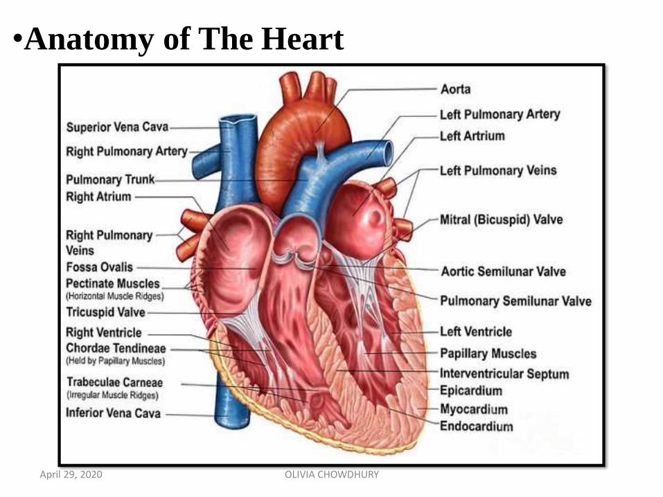
•Anatomy of The Heart
April 29, 2020 OLIVIA CHOWDHURY
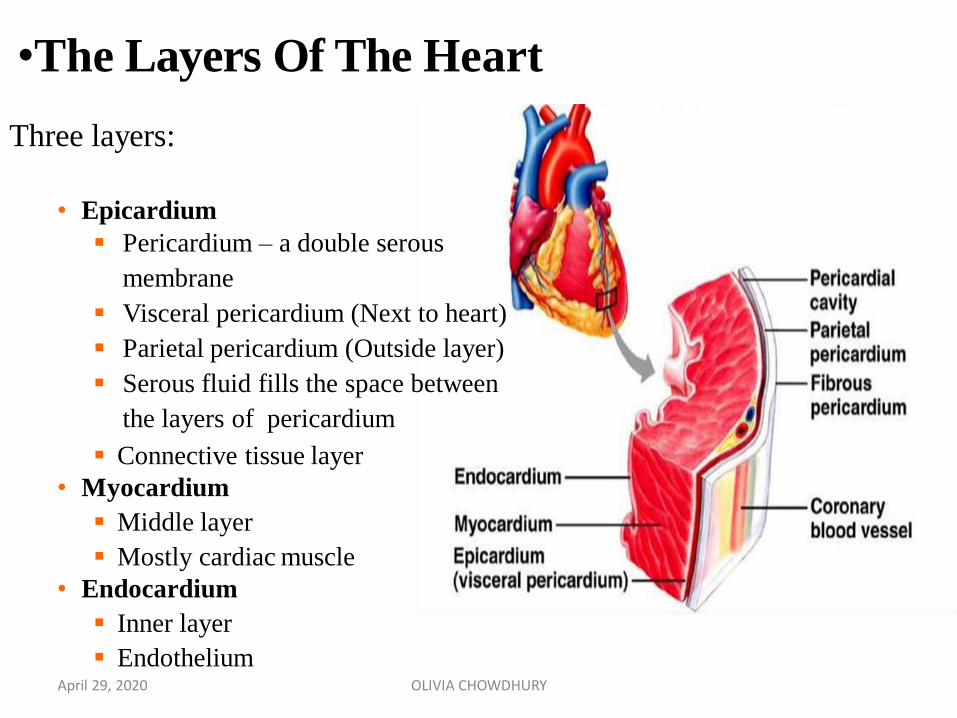
Three layers:
• Epicardium
Pericardium – a double serous
membrane
Visceral pericardium (Next to heart)
Parietal pericardium (Outside layer)
Serous fluid fills the space between
the layers of pericardium
Connective tissue layer
• Myocardium
Middle layer
Mostly cardiac muscle
• Endocardium
Inner layer
Endothelium
•The Layers Of The Heart
April 29, 2020 OLIVIA CHOWDHURY
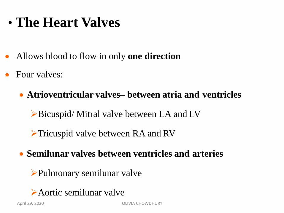
Allows blood to flow in only one direction
Four valves:
Atrioventricular valves– between atria and ventricles
Bicuspid/ Mitral valve between LA and LV
Tricuspid valve between RA and RV
Semilunar valves between ventricles and arteries
Pulmonary semilunar valve
Aortic semilunar valve
• The Heart Valves
April 29, 2020 OLIVIA CHOWDHURY
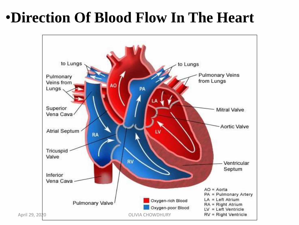
•Direction Of Blood Flow In The Heart
April 29, 2020 OLIVIA CHOWDHURY
Right side of the heart:
• receives venous blood from systemic circulation via
superior and inferior vena cava into right atrium
• pumps blood to pulmonary circulation from right
ventricle
Left side of the Heart:
• receives oxygenated blood from pulmonary veins
• pumps blood into systemic circulation
April 29, 2020 OLIVIA CHOWDHURY
Myocardium has three types of muscle fibers:
Muscle fibers which form contractile unit of heart
Muscle fibers which form the pacemaker
Muscle fibers which form conductive system
•The Cardiac Muscle
April 29, 2020 OLIVIA CHOWDHURY
Striated and resemble the skeletal muscle fibre
Sarcomere is the functional unit
Sarcomere of the cardiac muscle has all the contractile proteins,
namely actin, myosin, troponin tropomyosin.
Cardiac muscle fibre is bound by sarcolemma. It has a centrally
placed nucleus. Myofibrils are embedded in the sarcoplasm.
Sarcoplasmic reticulum is less abundant than in skeletal muscle.
Sarcolemma of cardiac muscle has specialized ion channels that skeletal muscle does not have: voltage-gated Ca2+ channels.
•The Cardiac Muscle
April 29, 2020 OLIVIA CHOWDHURY
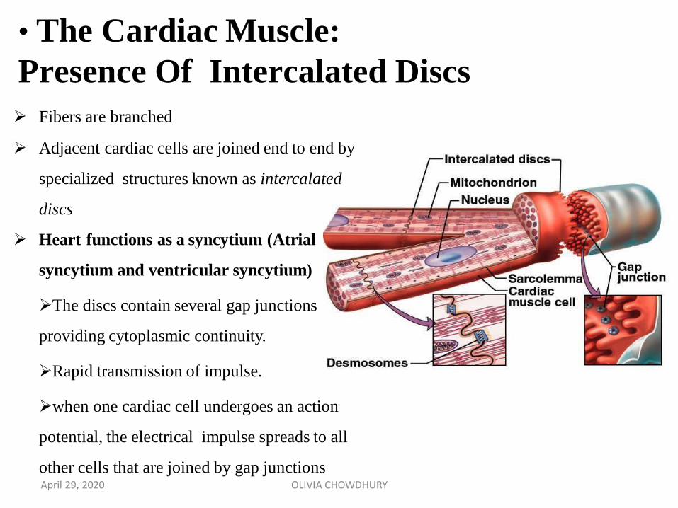
• The Cardiac Muscle:
Presence Of Intercalated Discs
Fibers are branched
Adjacent cardiac cells are joined end to end by
specialized structures known as intercalated
discs
Heart functions as a syncytium (Atrial
syncytium and ventricular syncytium)
The discs contain several gap junctions
providing cytoplasmic continuity.
Rapid transmission of impulse.
when one cardiac cell undergoes an action
potential, the electrical impulse spreads to all
other cells that are joined by gap junctionsApril 29, 2020 OLIVIA CHOWDHURY
Some of the muscle fibres of heart are modified into a specialized
structure known as pacemaker.
These muscle fibres forming the pacemaker have less striation.
They are named pacemaker cells or P cells.
Sino-atrial (SA) node forms the pacemaker in human heart.
•Muscle Fibres which Form the Pacemaker
April 29, 2020 OLIVIA CHOWDHURY
•Muscle Fibres Which Form Conductive System
Conductive system of the heart is formed by modified cardiac
muscle fibres
Impulses from SA node are transmitted to the atria directly. However,
the impulses are transmitted to ventricles through various components
of conducting system
April 29, 2020 OLIVIA CHOWDHURY
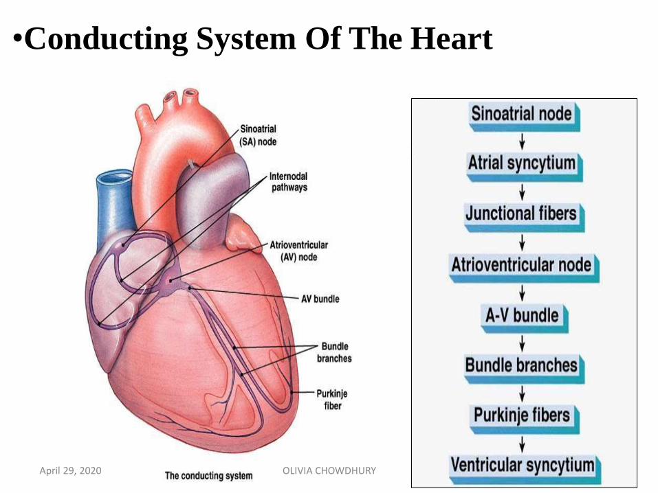
•Conducting System Of The Heart
April 29, 2020 OLIVIA CHOWDHURY
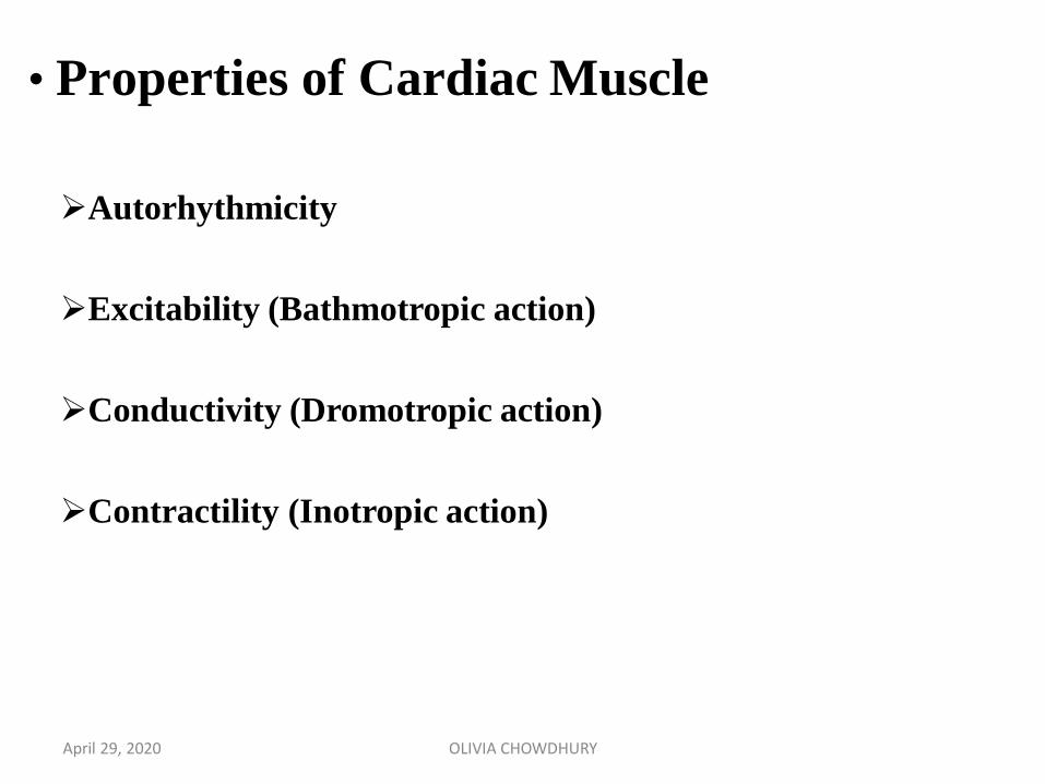
• Properties of Cardiac Muscle
Autorhythmicity
Excitability (Bathmotropic action)
Conductivity (Dromotropic action)
Contractility (Inotropic action)
April 29, 2020 OLIVIA CHOWDHURY
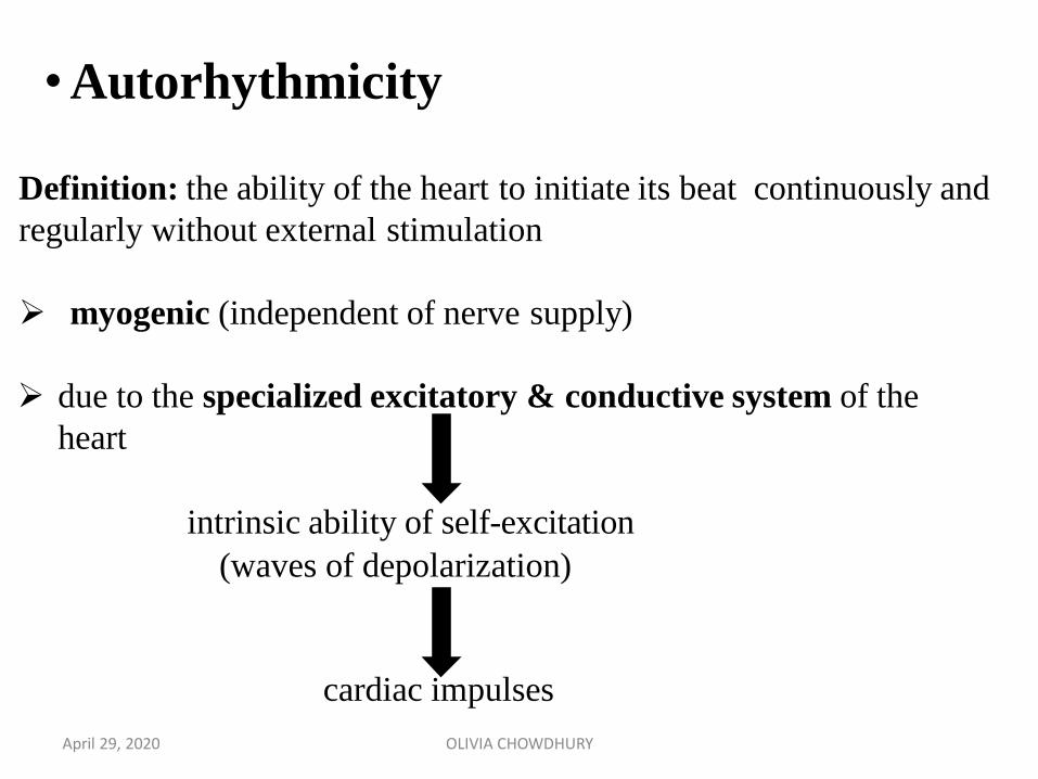
Definition: the ability of the heart to initiate its beat continuously and
regularly without external stimulation
myogenic (independent of nerve supply)
due to the specialized excitatory & conductive system of the
heart
intrinsic ability of self-excitation
(waves of depolarization)
cardiac impulses
•Autorhythmicity
April 29, 2020 OLIVIA CHOWDHURY

•Locations Of Autorhythmic Cells Sinoatrial node (SA node) Specialized region in right
atrial wall near opening of superior
vena cava.
Atrioventricular node (AV node) Small bundle of
specialized cardiac cells located at base of right
atrium near septum
Bundle of His (atrioventricular bundle)
Cells originate at AV node and enters
interventricular septum. Divides to form right and
left bundle branches which travel down septum,
curve around tip of ventricular chambers, travel
back toward atria along outer walls
Purkinje fibers
Small, terminal fibers that extend from bundle of His and
spread throughout ventricular myocardiumApril 29, 2020 OLIVIA CHOWDHURY
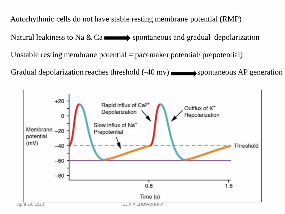
Autorhythmic cells do not have stable resting membrane potential (RMP)
Natural leakiness to Na & Ca spontaneous and gradual depolarization
Unstable resting membrane potential = pacemaker potential/ prepotential)
Gradual depolarization reaches threshold (-40 mv) spontaneous AP generation
April 29, 2020 OLIVIA CHOWDHURY
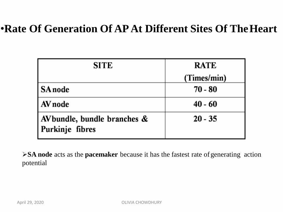
•Rate Of Generation Of AP At Different Sites Of TheHeart
SA node acts as the pacemaker because it has the fastest rate ofgenerating action
potential
April 29, 2020 OLIVIA CHOWDHURY
April 29, 2020 OLIVIA CHOWDHURY
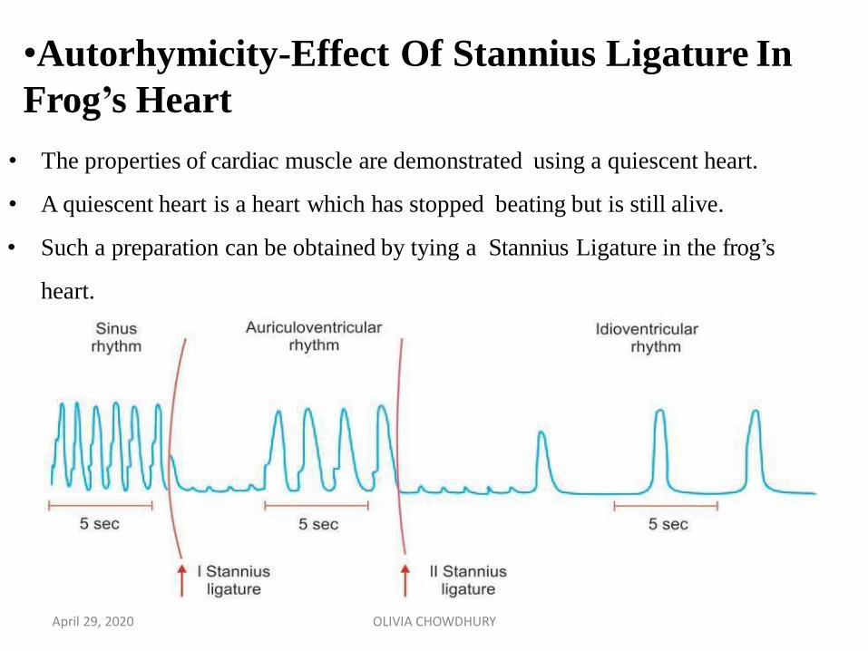
•Autorhymicity-Effect Of Stannius Ligature In
Frog’s Heart
• The properties of cardiac muscle are demonstrated using a quiescent heart.
• A quiescent heart is a heart which has stopped beating but is still alive.
• Such a preparation can be obtained by tying a Stannius Ligature in the frog’s
heart.
April 29, 2020 OLIVIA CHOWDHURY
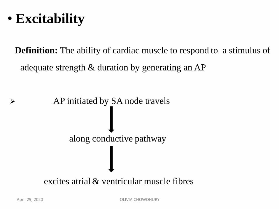
Definition: The ability of cardiac muscle to respond to a stimulus of
adequate strength & duration by generating an AP
AP initiated by SA node travels
excites atrial & ventricular muscle fibres
along conductive pathway
• Excitability
April 29, 2020 OLIVIA CHOWDHURY
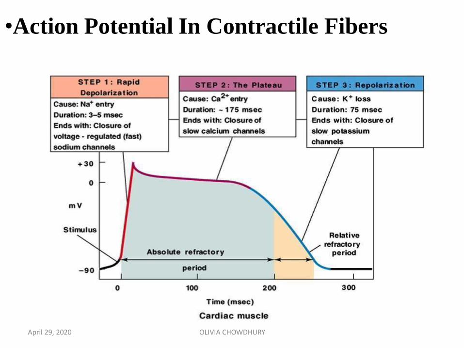
•Action Potential In Contractile Fibers
April 29, 2020 OLIVIA CHOWDHURY
•Refractory Period
It is that period during which a second stimulus fails to evoke aresponse.
Absolute Refractory Period : It is that period during which a
second stimulus however strong , fails to evoke a response.
Relative Refractory Period : It is that period during which a
second stimulus evokes a response if it is sufficiently high.
April 29, 2020 OLIVIA CHOWDHURY
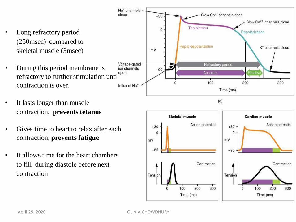
• Long refractory period
(250msec) compared to
skeletal muscle (3msec)
• During this period membrane is
refractory to further stimulation until
contraction is over.
• It lasts longer than muscle
contraction, prevents tetanus
• Gives time to heart to relax after each
contraction, prevents fatigue
• It allows time for the heart chambers
to fill during diastole before next
contraction
April 29, 2020 OLIVIA CHOWDHURY
Definition: Ability of cardiac muscle to contract in response to
stimulation.
All Or None Law
• The response to a threshold stimulus is maximal. If the stimulus is
below threshold there is no response
• The cardiac muscle follows the all or none law as a whole.
• In the case of skeletal muscle, all-or-none law is applicable
only to a single muscle fiber.
•Contractility
April 29, 2020 OLIVIA CHOWDHURY
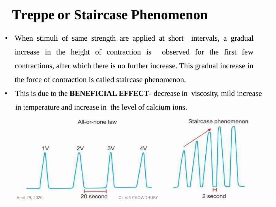
• When stimuli of same strength are applied at short intervals, a gradual
increase in the height of contraction is observed for the first few
contractions, after which there is no further increase. This gradual increase in
the force of contraction is called staircase phenomenon.
• This is due to the BENEFICIAL EFFECT- decrease in viscosity, mild increase
in temperature and increase in the level of calcium ions.
Treppe or Staircase Phenomenon
April 29, 2020 OLIVIA CHOWDHURY
When a stimulus of subliminal strength is applied the cardiac muscle
does not show any response. When a series of subliminal stimuli are
applied in succession, the muscle responds with a contraction. It is
due to the summation of all the subliminal stimuli that produce a
threshold stimulus.
Summation of Subliminal Stimuli
April 29, 2020 OLIVIA CHOWDHURY
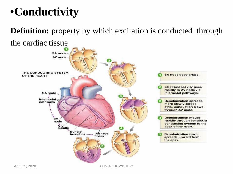
Definition: property by which excitation is conducted through
the cardiac tissue
•Conductivity
April 29, 2020 OLIVIA CHOWDHURY
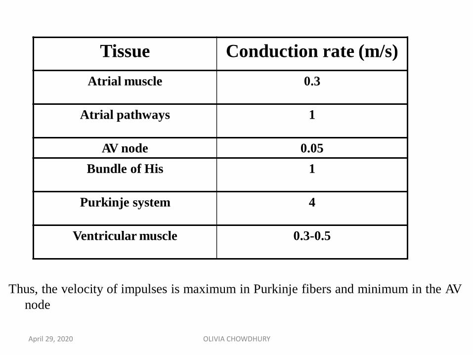
Tissue Conduction rate (m/s)
Atrial muscle 0.3
Atrial pathways 1
AV node 0.05
Bundle of His 1
Purkinje system 4
Ventricular muscle 0.3-0.5
Thus, the velocity of impulses is maximum in Purkinje fibers and minimum in the AV
node
April 29, 2020 OLIVIA CHOWDHURY
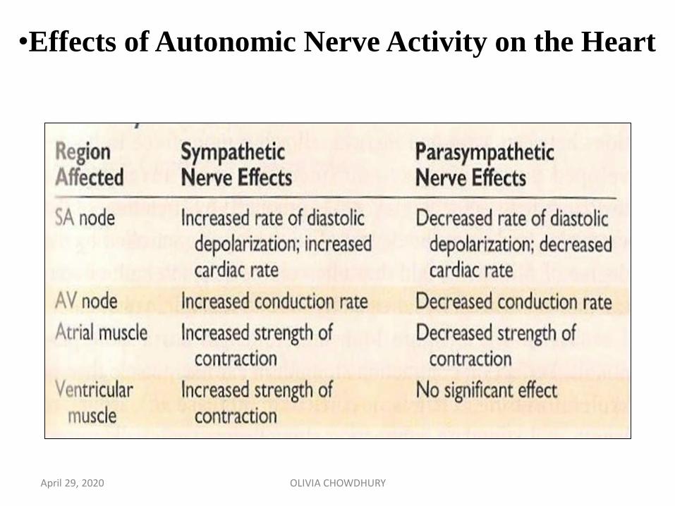
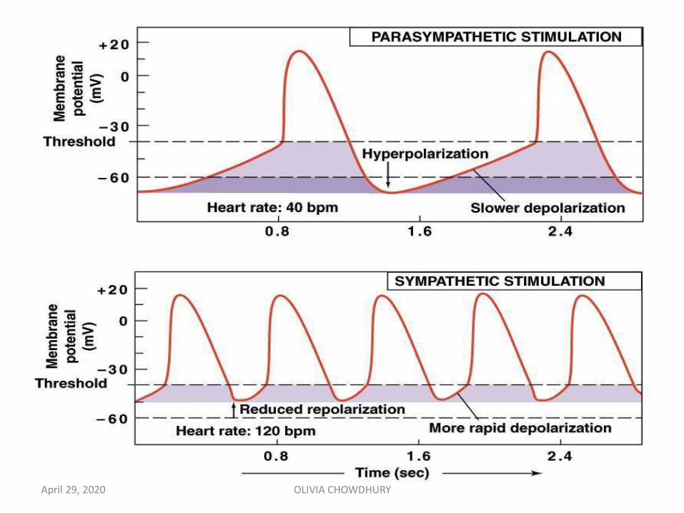
•Effects of Autonomic Nerve Activity on the Heart
April 29, 2020 OLIVIA CHOWDHURY
THANK YOU!
April 29, 2020 OLIVIA CHOWDHURY