can mango reverse bone loss in - core
TRANSCRIPT
CAN MANGO REVERSE BONE LOSS IN
OVARIECTOMIZED MICE?
By
ELIZABETH VITALE
Bachelor of Science in Food Science & Human Nutrition
University of Hawaii at Manoa
Honolulu, Hawaii
2008
Submitted to the Faculty of the Graduate College of the
Oklahoma State University in partial fulfillment of the requirements for
the Degree of MASTER OF SCIENCE
May, 2011
brought to you by COREView metadata, citation and similar papers at core.ac.uk
provided by SHAREOK repository
iv
CAN MANGO SUPPLEMENTATION REVERSE BONE LOSS IN
OVARIECTOMIZED MICE?
Thesis Approved:
Dr. Edralin A. Lucas
Thesis Adviser
Dr. Brenda J. Smith
Dr. Barbara J. Stoecker
Dr. Mark E. Payton
Dean of the Graduate College

v
ACKNOWLEDGMENTS
I thank all my professors at Oklahoma State University for deepening my
knowledge in nutrition. Particularly I’d like to thank my advisor Dr. Lucas, for her
ongoing support and critical analysis of the research material, and my committee
members for their time and detailed evaluation of the research. I would also like to thank
the National Mango Board for funding this research.
vi
TABLE OF CONTENTS
Chapter Page I. INTRODUCTION ......................................................................................................1
Introduction ..............................................................................................................1 Objectives ................................................................................................................4 Null hypothesis ........................................................................................................4 II. REVIEW OF LITERATURE....................................................................................6 Prevalence of osteoporosis ......................................................................................6 Bone remodeling .....................................................................................................7 Osteoblasts and osteocytes ......................................................................................8 Osteoclasts ..............................................................................................................9 Factors affecting bone remodeling ........................................................................12 Genetics .................................................................................................................12 Hormones ..............................................................................................................13 Cytokines and growth factors ...............................................................................16 Lifestyle factors ....................................................................................................16 Nutrition ................................................................................................................16 Lifestyle influences on bone ……………………………………………………..25 Postmenopausal osteoporosis.................................................................................27 Animal model.........................................................................................................31 Pharmacological options for osteoporosis treatment .............................................32 Nutrient composition of mango .............................................................................33 Studies on health benefits of mango pulp ..............................................................35 Mango and bone health ..........................................................................................37 III. METHODOLOGY ................................................................................................39 Animal grouping ....................................................................................................39 Diet formulation .....................................................................................................40 Necropsy, tissue processing and storage ................................................................40 Body composition and bone mineral density (BMD) assessment ........................42 Bone micro-architecture assessment ......................................................................42 Bone strength assessment ......................................................................................43 Biomarkers of bone homeostasis ...........................................................................43 Statistical analyses……………………………………………………… .............44
vii
Chapter Page
IV. FINDINGS .............................................................................................................45 Baseline data…………………………………………………………………… ..45 Effects of dried mango on weekly body weights of ovariectomized mice ...........46 Effects of dried mango on food intake, tissue weights, whole body bone parameters
and body composition of ovariectomized mice ...........................………………..46 Effects of dried mango on bone parameters: whole body, tibia, and lumbar vertebrae
(L4 and L5) ............................................................................................................47 Effects of dried mango on plasma markers of bone formation and resorption ......47 V. CONCLUSION ......................................................................................................55 REFERENCES ............................................................................................................62
viii
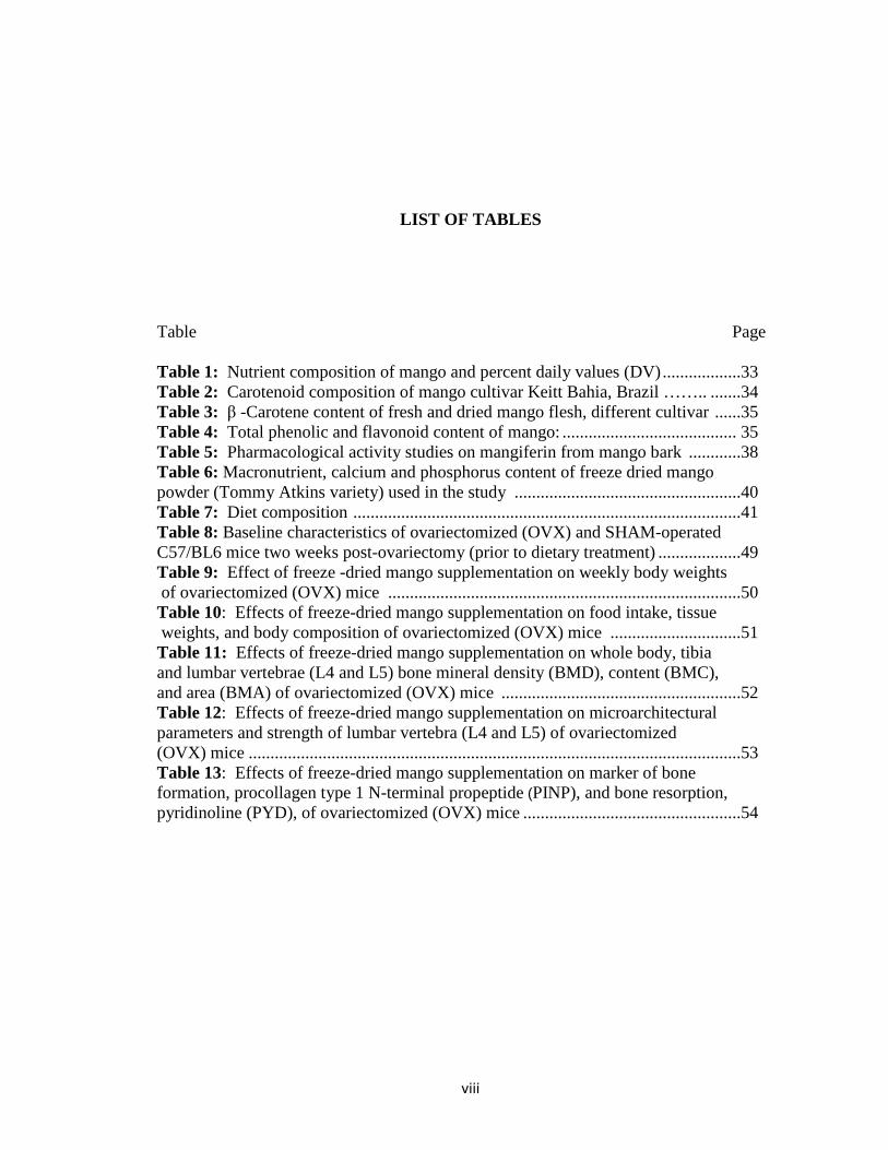
LIST OF TABLES
Table Page Table 1: Nutrient composition of mango and percent daily values (DV) ..................33 Table 2: Carotenoid composition of mango cultivar Keitt Bahia, Brazil …….. .......34 Table 3: β -Carotene content of fresh and dried mango flesh, different cultivar ......35 Table 4: Total phenolic and flavonoid content of mango: ........................................ 35 Table 5: Pharmacological activity studies on mangiferin from mango bark ............38 Table 6: Macronutrient, calcium and phosphorus content of freeze dried mango powder (Tommy Atkins variety) used in the study ....................................................40 Table 7: Diet composition .........................................................................................41 Table 8: Baseline characteristics of ovariectomized (OVX) and SHAM-operated C57/BL6 mice two weeks post-ovariectomy (prior to dietary treatment) ...................49 Table 9: Effect of freeze -dried mango supplementation on weekly body weights of ovariectomized (OVX) mice .................................................................................50 Table 10: Effects of freeze-dried mango supplementation on food intake, tissue weights, and body composition of ovariectomized (OVX) mice ..............................51 Table 11: Effects of freeze-dried mango supplementation on whole body, tibia and lumbar vertebrae (L4 and L5) bone mineral density (BMD), content (BMC), and area (BMA) of ovariectomized (OVX) mice .......................................................52 Table 12: Effects of freeze-dried mango supplementation on microarchitectural parameters and strength of lumbar vertebra (L4 and L5) of ovariectomized (OVX) mice .................................................................................................................53 Table 13: Effects of freeze-dried mango supplementation on marker of bone formation, procollagen type 1 N-terminal propeptide (PINP), and bone resorption, pyridinoline (PYD), of ovariectomized (OVX) mice ..................................................54

ix
LIST OF FIGURES
Figure Page Figure 1: Osteoclastogenesis regulation via the receptor activator of NF-B ligand (RANKL) activated signaling pathway .......................................................................11 Figure 2: NFATc1 signaling in osteoclastogenesis via reactive oxygen species (ROS) and Ca2+ oscillations .........................................................................................12 Figure 3: Insulin signaling and bone remodeling ......................................................14 Figure 4: Leptin’s influence on bone .........................................................................15 Figure 5: Bone metabolism ........................................................................................19 Figure 6: Hormonal stimulation on osteoclasts (OC) and osteoblasts (OB)..............28
1
CHAPTER I
INTRODUCTION
Osteoporosis and osteopenia are major health threats to more than 50% of the female
American population over age 50 [1]. An estimated 44 million Americans are affected by
osteoporosis and osteopenia. Osteoporosis is a disease characterized by low bone mass and
structural deterioration of bone tissue, resulting in poor bone quality, reduced bone strength, and
an increased risk for bone fracture, particularly in the hip, spine and wrist [1, 2].
Fractures of the hip often require hospitalization and surgery and can impair an
individual’s ability to walk, create a permanent disability, and may even result in death [1, 3].
Vertebral fractures can cause severe back pain, deformity and loss of height [1]. In the USA,
approximately 1.5 million fractures are attributable to osteoporosis every year [3] and $19 billion
dollars were spent on osteoporosis-related fractures in 2005 [1]. It is predicted that the incidence
of osteoporotic fractures will reach catastrophic proportions with a four-fold increase of
osteoporosis worldwide over the next 50 years [3]. The costs associated with bone fractures will
threaten the viability of health care systems in many countries [3] indicating an emergent need for
preventative action.
Postmenopausal osteoporosis accounts for 80% of osteoporosis cases that develop [4].
The decline in sex steroids that occurs with the loss in ovarian function at menopause results in
2
changes in calcium homeostasis resulting in a negative calcium balance [5]. Estrogen deficiency
also increases the production of inflammatory cytokines resulting in a further increase in bone
resorption [4, 6, 7]. Considering the high prevalence of postmenopausal osteoporosis and the
associated decline in sex hormone production, an option to prevent or reverse postmenopausal
osteoporosis is needed. For this study, the ovariectomized mouse is used as a model to emulate
the hormonal effects in a postmenopausal state.
Lifestyle modifications through diet may be a significant approach in preventing the
development of osteoporosis [1]. Although much research has been conducted on the beneficial
effects of dairy products and vitamin D in preventing bone loss, there are some important factors
to consider with widespread dairy promotion as a dietary approach to bone health. The first
issue is that over two thirds of the world’s adult populations (approximately 30 to 50 million
Americans; 75 % of African Americans and Native Americans and 90 % of Asian Americans)
are lactose intolerant [8]. Also, with the recommended daily allowance (RDA) of calcium set at
1200 mg per day for older adults, many individuals find it difficult to meet calcium requirements
through dietary intake. With milk being an excellent source of calcium, it can be challenging for
individuals to drink four glasses of milk a day. Another interesting point to consider is that the
USA is one of world’s greatest consumers of dairy products, yet globally, has one of the highest
rates of osteoporosis [3]. When considering these facts, the need for research into other nutrients
that may promote bone growth and/or protect an individual from age- or menopause-induced
bone loss becomes evident.
Another factor promoting further research into dietary approaches for osteoporosis
prevention is the fact that the commonly used osteoporotic medications, although effective, are
associated with undesirable side effects. For example, bisphosphonates have been associated
3
with bone, joint and/or muscle pain and most commonly reported, stomach upset and heartburn
[9] . Although uncommon with typical health care administration of the drug, a rapid injection of
bisphosphonates can cause renal failure[9]. Oral administration of bisphosphonates, particularly
with amines, can cause esophageal and gastrointestinal distress such as nausea, dyspepsia,
vomiting, gastric pain, diarrhea, and ulceration[9]. Estrogen therapy, although not encouraged
due to its serious side effects, has been associated with endometrial and breast cancer, vaginal
bleeding, breast tenderness, gallbladder disease, strokes, heart attacks, venous blood clots and
cognitive decline [1]. Selective estrogen receptor modulators (SERM), such as Raloxifene, are
more commonly used and do not have the majority of side effects common to estrogen therapy
but may induce hot flashes, blood clots in the legs or lungs, and strokes in those with coronary
artery disease[10]. Teriparatide, a recombinant parathyroid treatment, has been associated with
leg cramps, headache and myalgia[11]. In addition to these side effects, long term oral drug
compliance can be challenging for many. Research has shown that women taking oral
bisphosphonates stop treatment or take less than what was prescribed, reducing the effectiveness
of the medication. Hence, there is a need for alternative prevention strategies with fewer side
effects. Dietary interventions that are effective, inexpensive, and can easily be incorporated into
an individual’s daily diet deserves further research.
Consumption of fruits and vegetables or the phytonutrients derived from them has been
associated with reduced risk of osteoporosis in human observational studies or an increase in
bone mineral density (BMD) in animals [12], [13] [14, 15] [16], [17],[18]. Fruits and vegetables contain a
wide variety of vitamins and minerals as well as bioactive phytochemicals which may contribute
to bone health. Phytochemicals are non-nutritive food chemicals that the plant uses to protect
itself from the external environment. These same chemicals also may help to prevent disease in
4
humans by functioning as an antioxidant or hormone, stimulate enzymes, interfere with DNA
replication or have an antibacterial affect [19]. Fruits and vegetables have antioxidant and anti-
inflammatory properties and reduce the renal acid load, actions which may also function as bone-
sparing agents [20-24], [25]. Aging and sex hormone related changes cause inflammation and pro-
oxidant conditions in the bone environment which are involved in the development of
osteoporosis [26]. Women with osteoporosis have also been shown to have lower levels of plasma
dietary and endogenous antioxidant vitamins (i.e., vitamins C, E, and A) and the lower
enzymatic activities of superoxide dismutase and glutathione peroxidase when compared to
healthy controls[27]. The alkalizing, antioxidant and anti-inflammatory properties of fruits and
vegetables are just a few mechanisms by which bioactive food components may play a protective
role in preventing bone loss.
Among the fruits, mangos (Mangifera indica L) are an excellent source of vitamin C, and
carotenoids, and are good sources of dietary fiber, potassium, copper, and vitamin K [28].
Mangos also contain minerals such as magnesium, zinc, copper, manganese, and selenium [29].
Mangos contain carotenoids, triterpenes, and phenolic compounds such as tannins, mangiferin
and flavonoids which may contribute to its antioxidant and anti-inflammatory properties.
Considering the bioactive compounds found in mango, it is hypothesized that mango may offer
some dietary support in preventing and reversing hormone-related bone loss.
The objective of this study is to determine if the addition of 5% or 25% freeze-dried
mango powder to the daily diet would reverse bone loss in ovariectomized mice. Our null
hypotheses are that:
1) Mango supplementation will not reverse the loss of bone mineral content (BMC) and bone
mineral density (BMD) due to ovariectomy;
5
2) Trabecular and cortical bone micro-architectural parameters, assessed using micro-computed
tomography analyses (µCT), will not be affected by mango supplementation;
3) Bone strength, assessed using finite element analysis, will not be increased by mango
supplementation; and
4) Biomarkers of bone formation (i.e., plasma N-terminal propeptide of procollagen type 1
(PINP)) and bone resorption (i.e., plasma pyridinoline (PYD)) will not be affected by mango
supplementation.
Identifying dietary factors that can reverse bone loss in experimental animals would
establish a strong scientific framework for subsequent clinical trials in humans. The findings of
this proposed study will enable us to investigate the efficacy of mango as a novel nutritional
strategy to reverse osteoporosis. Furthermore, this study will provide fundamental knowledge
related to the health benefits of mango on body composition. Findings of this study will provide
a basis for consuming fruits to help combat postmenopausal bone loss. If mango treatment
shows effectiveness in reversing bone loss, these findings could potentially help the nation save
millions of dollars spent on the treatment of osteoporotic fractures and bone-protecting
pharmaceuticals.
6
CHAPTER II
REVIEW OF LITERATURE
Prevalence of Osteoporosis
By 2020, it is forecasted that there will be over 47 million cases of low bone density in
the US and 14 million cases of osteoporosis [1]. In the US, $17 billion was spent on over 2
million fractures in 2005. The majority of these fractures occurred in individuals over 65 years of
age. Seventy three percent of these fractures were non-vertebral fractures and accounted for the
majority of the cost expenditure. By 2025, annual fractures are projected to increase by 50% with
an estimated cost of $25 billion [30]. The increase in the number of hip fractures per year over the
past 30 years is partly attributed to the increase in lifespan [31].
Age is not the only important osteoporotic risk factor to consider. Gender and ethnicity
also influence bone density. Osteoporotic fractures affect 1 in 3 women and 1 in 5 men over the
age of 50 [3, 32]. In the US, over half of all postmenopausal Caucasian women are considered
osteopenic with an additional 30% having osteoporosis [3]. The tendency for fracture is higher in
the US Caucasian population and lower for other ethnic groups [33]. By the age of 80, up to 70%
of white women have osteoporosis and 27% are considered osteopenic [3]. Cauley and
colleagues[34] reported that Caucasian women are twice as likely to have hip BMD loss when
compared to African-American women with an increase in hip BMD loss with age in both
groups.
7
Bone Remodeling
Bone remodeling occurs as a continuous bone renewal process throughout the life span.
In the bone remodeling process, old weakened bone is replaced by mechanically strong bone. A
healthy balance is maintained between bone formation and bone resorption. Bone resorbing
cells, known as osteoclasts (OC), and bone forming cells, known as osteoblasts (OB), work
interdependently in the bone remodeling process [35]. With hormonal and cytokine signaling,
OCs degrade bone to liberate mineral ions such as calcium and phosphorous needed by the body
to replace old or damaged bone cells. The OC creates an acidic environment with proton pumps
and uses enzymes such as tartrate-resistant acid phosphatase and cathepsin K to catabolize the
bone matrix minerals and organic components forming resorption pits known as Howship’s
lacunae[36]. When the resorption phase is complete, the OC under goes apoptosis. Initiating the
bone renewal phase are coupling factors such as bone morphogenetic proteins, transforming
growth factor-β (TFG-β), insulin like growth factor (IGF)-I and II, platelet derived growth
factors and fibroblast growth factor that signal OBs and OB precursor cells to the Howship’s
lacunae[37]. The OBs then synthesizes the organic bone matrix by secreting osteoid and regulate
its mineralization by secreting matrix vesicles which concentrate calcium and phosphorous ions
and enzymatically degrade inhibitors of mineralization such as pyrophosphate and
proteoglycans[38]. As bone renewal process terminates, OBs can undergo apoptosis, become
entombed in the bone matrix and become osteocytes, or become bone-lining cells. When the
balance between the bone remodeling of OCs and OBs is uncoupled in favor of bone resorption
and osteoclastogenesis, osteopenia or osteoporosis may develop, weakening the bone, and hence,
predisposing it to an increased risk for fracture [35].
8
Osteoblasts and Osteocytes
Bone formation can be categorized in 3 stages; OB proliferation, extracellular matrix
development and maturation, and mineralization [39]. Identification of the bone formation stages
can be determined by serum biomarkers. For example, alkaline phosphatase and parathyroid
hormone 1 receptor (PTH1R) are considered early markers of OB proliferation stage, while
serum procollagen I N-terminal propeptide (PINP) and osteocalcin are considered marker of
extracellular matrix development and bone mineralization, respectively[40].
For bone formation to occur, OBs need to develop and mature before they can participate
in the bone building process. Mesenchymal stem cells in the bone marrow develop in to
progenitor cells which may become adipocytes, myocytes, chondrocytes, fibroblasts, stromal
cells or OBs. The canonical Wnt/β-catenin pathway indirectly mediates the initial cascade of
gene expression for skeletal development, bone formation and OB differentiation. Wnt10b shifts
the mesecnchymal stem cells fate to the OB lineage by suppressing adipogenic transcription
factors and inducing osteogenic transcription factors such as the runt-related transcription factor
2 (Runx2), D1x5 and Osterix [39, 41]. These pathways are induced in response to extra cellular
stimuli such as bone morphogenetic proteins, growth factors, hormones, cytokines, matrix
proteins, transcription factors, regulatory co-factors and environmental stress [39].
An active osteoblast is distinguishable by its large nucleus and golgi apparatus, well
developed endoplasmic reticulum, and high concentration of alkaline phosphatase enzyme
activity in the proliferation stage[40]. OBs are anabolic to bone and function by developing the
extracellular bone matrix and directing its mineralization. The OBs secrete a fibrous, non-
mineralized protein substance known as osteoid, which contains chondroitin sulfate, osteocalcin
and type I collagen and forms the bone matrix. Osteocalcin, osteopontin and bone sialoprotein,
9
also secreted by the OB, serve to mineralize the extracellular matrix, by binding calcium and
phosphates, and regulates both the amount and size of hydroxyapatite crystals in bone [39].
OBs, at the terminal differentiation stage, may develop into osteocytes. Osteocytes are
bone cells found throughout the bone matrix which support the structure and metabolic functions
of bone [39]. The cellular connection within the bone plays an important role in building strong
bones. Cell-to-cell and cell-to-matrix interactions are maintained by adhesion and trans-
membrane proteins. Surface lining bone cells, which are composed of flattened inactive OBs,
separate the bone from the marrow[39]. Communication exists between the lining cells and the
OBs via adherens junctions. In these junctions are cadherins, calcium dependent trans-
membrane proteins, which assist in anchoring the surface lining and OB cells through their
cytoskeleton [39]. N-cadherin, mediated by bone morphogenetic protein (BMP)2, functions in
cell-cell adhesion [39]. These type of cell-cell interactions encourage osteoprogenitor
differentiation and OB survival[42]. Integrin, an adhesion protein, joins the bone’s extracellular
matrix to the structural proteins in the cytoskeleton, and induces OB differentiation, through the
mitogen-activated protein kinase (MAPK) pathway[39]. Connexins, integral membrane proteins
at gap junctions, allow the osteocytes to communicate with each other in response to mechano-
stimulation. Mechano-sensory information stimulates signaling pathways for OB gene
transcription.
Osteoclasts
The bone resorbing OCs are also formed in the bone marrow, but from a monocyte-
macrophage myeloid lineage of hemopoietic stem cells [43]. OCs not only function in bone
10
degradation, but also as immunomodulators in pathological conditions and may also regulate OB
function [44].
Cytokines, receptor activator of nuclear factor-kB ligand (RANKL), macrophage colony
stimulating factor (M-CSF), and c-Fos, a RANKL activated transcription factor, are necessary
for OC differentiation [45]. Both RANKL and M-CSF are produced by bone marrow stromal
cells, OBs, T lymphocytes and synovial fibroblasts[46]. RANKL can also be found in other
tissues such as in the lymph nodes, thymus gland, lungs, spleen, mammary epithelial cells and in
some cancer cells [47]. RANKL is a member of the tumor necrosis super family of proteins and is
essential for OC formation. M-CSF contributes to OC precursor differentiation, proliferation,
and survival. Osteoclastogenesis can be detected by tartrate-resistant acid phosphatase (TRAP)
and proteinase cathepsin K enzyme activity, wich appear 3-5 days after differentiation [48, 49].
Mature OCs can be identified by its large size, approximately 40 micrometer in diameter,
multiple nuclei, resulting from cytoplasmic fusion of the precursor monocytes, a "foamy"
cytoplasm appearance and extensive golgi complex [50].
OCs attach at resorption sites via actin-rich podosomes securing a tight seal on bone [51].
Integrins act as receptors allowing the OC to bind to the bonding sites in the bone via
osteopontin and virtonectin glycoproteins [52]. Approximately 10 – 14 days after OCs attachment
to bone, OCs create a pocket which is acidified by a proton pump and a chlorine channel [39].
Hydrochloric acid and proteases degrade the bone matrix while the lysosomal enzyme, cathepsin
K, breaks down the mobilized type 1 collagen [39, 48, 50].
The RANKL signaling pathway plays an important role in bone resorption. RANKL, a
transmembrane protein on the osteoblast, binds to receptor activator of nuclear factor-kB,
RANK, on the OC precursor and OC, increasing OC formation, activity, and bone resoption.
11
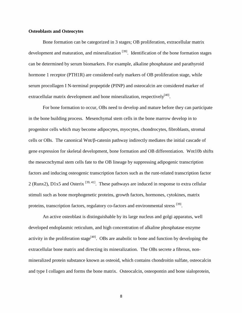
OPG functions to protect bone from breakdown by intercepting RANKL, reducing the
rate of OC formation, activity and bone resorption [53] (Figure 1). OPG expression is regulated
by WNT/β-catenin signaling in OBs [54] and it also expressed in other tissues aside from OBs.
Pro-inflammatory cytokines up-regulate RANKL while suppressing the expression of OPG[46].
RANKL stimulates osteoclastogenesis by activating multiple signaling pathways
encoding for TRAP, cathepsin K, the calcitonin receptor and β3-integrin[46]. One influential
signaling pathway induces long lasting oscillations of intracellular Ca2+ concentration while
generating reactive oxygen species (ROS) (Figure 2). RANKL mediates stimulation of Rac1,
which generates ROS that trigger phospholipase Cγ1 to initiate the Ca2+ oscillations. Btk kinase
activates phospholipase Cγ1 (PLCγ1), tumor necrosis receptor activator factor 6 (TRAF6), and c-
Fos (transcription factor). The Ca2+ oscillations then activates calcineurin and nuclear factor of
activated T-cells c1 (NFATc1) to stimulate osteoclastogenesis [55] (Figures 1 & 2). The role of
TRAF6, c-Fos and NFATc1 in the OC precursor nucleus are put in the perspective of the bone
remodeling cycle in Figures 1 and 2.
Figure 1: Ostoclastogenesis regulation via the receptor activator of nuclear factor-kB ligand (RANKL) signaling pathway [50]

12
Figure 2: NFATc1 signaling in osteoclastogenesis via ROS and Ca2+ oscillations [50]
Factors Affecting Bone Remodeling
The bone remodeling process is regulated by multiple factors. Genetics influence bone
remodeling as well as hormones, growth factors and locally produced cytokines [56]. Lifestyle
factors such as amount of weight bearing exercise, nutritional status, smoking, alcohol
consumption and caffeine intake also affect the bone remodeling process. A disturbance or
change in any of the bone regulatory factors can contribute to an uncoupling of the bone
remodeling process which can lead to osteoporosis.
Genetics
Genetics play an important role in regulating peak bone mass. Up to 60 -80% of
variance in peak bone mass between individuals can be attributed to genetics [56]. Although both
elderly men and women lose bone with increasing age, postmenopausal women lose more bone
than men [57]. The risk for fracture is doubled in women when compared to men of similar age
[57]. Ethnicity also influences BMD and fracture risk. The incidence of reported hip fractures is
13
lower in Asian than Caucasian populations [58]. Women also vary in terms of the rate and extent
of postmenopausal bone loss, some considered fast or slow losers. This variance is in part due to
inherited factors. In observational studies, associations between family history of fracture
incidence and type of fracture have suggested an inherited correlation in BMD and fracture risk
[59-61]. In human and animal studies, high levels of heritability have been shown in bone
phenotype as assessed by bone densitometry and ultrasound [62, 63]. Allelic variation in the
vitamin D receptor, has shown to influence genetic determination of bone phenotype. The
impact of the vitamin D receptor can be mediated by body size and development and by
hormonal regulation. Intronic polymorphisms of the collagen Iα1 gene have been related to
BMD and fracture risk[57]. Allelic variations in the estrogen receptor, TGFß receptor, TGFß1,
insulin-like growth factor-I pathway, IL-4, IL-6, calcitonin, PTHRs and for apolipoprotein E
have all been associated with BMD phenotypes [57]. Hormonal factors, nutrition and lifestyle
interact with genetic factors over time.
Hormones
Sex steroids, PTH, thyroid hormones, growth hormone, glucocorticoids, and
1,25(OH)2D3 influence the bone remodeling process[64]. Estrogen, have an inhibitory effect on
osteoclast function by promoting osteoclast apoptosis [65] and by modulating osteoclast
differentiation [66]. Estrogen also decreases the OB progenitor cell population but exerts an
anabolic influence on OBs.
PTH plays a dual role in the bone remodeling process. On one hand, PTH and
1,25(OH)2D3 encourage bone remodeling by increasing the expression of NF- kB, tumor
necrosis factor (TNF), RANK, RANKL, and M-CSF [67]. On the other hand, PTH, IGF and other

14
growth factors stimulate mesenchymal stem cell, osteoprogenitor and osteoblast differentiation.
The cell membrane of the parathyroid gland has calcium sensing receptors which regulate PTH
secretion. Low serum calcium signals the parathyroid gland to secrete more PTH which
enhances renal tubule reabsorption of calcium, increases bone resorption, and increases intestinal
calcium absorption indirectly by increasing 1α, 25(OH)2 D3 synthesis in the kidneys [68].
Calcitonin is a peptide hormone produced by the C cells of the thyroid gland which acts
to lower blood calcium levels when they are too high by inhibiting bone resorption [69]. The
secretion of calcitonin is regulated by serum calcium, gender and age [70, 71]. Calcitonin has been
manufactured as a pharmaceutical to inhibit osteoclast bone resorption and hypercalcemia [70].
The metabolic hormone, insulin, also regulates bone remodeling. Insulin signaling
activates osteoblasts and enhances osteocalcin production[72]. Osteocalcin facilitates bone
mineralization and calcium ion homeostasis. Osteocalcin also impacts glucose homeostasis via
its interaction with OCs [72]. With the acidic OC environment, osteocalcin, secreted by the
osteoblast, is decarboxylated to its active form [72]. Osteocalcin, in turn, mediates insulin
secretion and sensitivity (Figure 3). Ferron and colleagues (2010)[72] demonstrated that insulin
signaling in osteoblasts is a critical link between bone remodeling and energy metabolism.
Figure 3: Insulin signaling and bone remodeling [72]
15
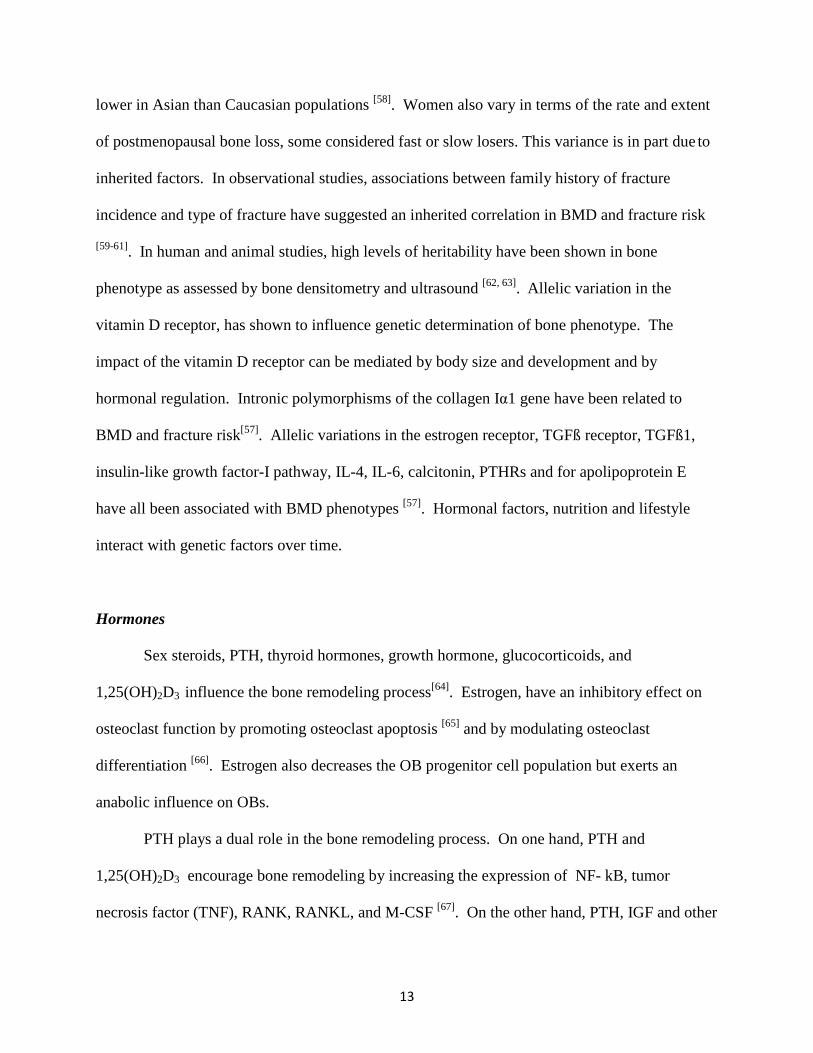
Leptin, a cytokine-like hormone secreted by adipose tissue, also influences bone
remodeling via hypothalamic mediation of the sympathetic nervous system [73]. Leptin has an
anabolic effect on osteoblasts and stimulates bone growth indirectly via the central nervous
system. Leptin stimulates the growth hormone/ insulin-like growth factor-1 (GH-IGF-1) axis of
the central nervous system and suppresses release of neuropeptide Y, an inhibitor of cortical
bone formation, from the hypothalamus. The GH-IGF-1 axis stimulation impacts both β1 and β2-
adrenergic receptors (Figure 4). Although β2- adrenergic stimulation encourages trabecular
bone remodeling, β1- adrenergic stimulation increases cortical bone mass. Cortical bone
becomes more important when dealing with the excess weight load from increasing amounts of
adipose tissue. A study done on leptin deficient mice showed that, not only are leptin deficient
mice obese with symptoms of metabolic syndrome, but they also exhibited skeletal
abnormalities[74]. Leptin repletion enhanced normal bone growth by increasing femoral length
and total bone volume although femoral and vertebral cancellous bone volume decreased [74].
Figure 4 : Leptin’s influence on bone [75]
[73]
Ct = cortical bone,;Tb = trabecular bone; GH = growth hormone; AdrB1= adrenergic receptor β1; AdrB2 = adrenergic receptor β2; IGF-1= Insulin like growth factor 1
16
Cytokines involved in Ovarian Hormone Deficiency
Local cytokines also influence bone remodeling. Immune and bone marrow cells
synthesize and secrete cytokines that may have autocrine and paracrine influences on bone
metabolism [76, 77]. With lack of estrogen regulation, caused by natural or surgically induced
menopause, blood, bone marrow, and monocytic levels of IL-1, IL-6, TNF-α, and the related
factors IL-1ra and IL-6R increase [78]. IL-1 and TNF activate OCs indirectly by influencing OB
secretory factors such as RANKL and inhibit OC apoptosis. They also enhance OC formation by
stimulating the proliferation of the OC precursors and stromal cells by activating IL-6, M-CSF,
and granulocyte M-CSF [78].
Lifestyle factors
Nutrition
Nutrition is an important determinant of bone health, yet the effects of nutrients and
minerals other than calcium involved in the bone remodeling process are still being elucidated.
Bone formation requires adequate supplies of energy, amino acids, and bone-forming minerals
such as calcium, phosphorus, magnesium, and zinc.
Adequate amounts of protein are necessary to prevent the development of osteoporosis
[79]. Sufficient protein intakes reduced bone loss and fracture risk in the elderly [80]. Excess
protein above the recommended allowances, however, can create renal acid load and lead to risks
of developing osteoporosis [81]. Excessive renal acid load may be more influential on bone size
and mass rather than volumetric bone density [82].
17
Consuming an alkaline diet may have bone protective effects. Protons are produced
during metabolism and are neutralized by the buffering action of anions generated from food or
liberated from bone [83]. Since increases in bone resorption lead to greater urinary calcium
excretion, a high renal acid load could potentially be associated with increased bone loss in older
adults [84]. Foods with a high acid load are those rich in sulfur amino acids, phosphorous or
chloride, such as meat, grains, nuts, and dairy products. Alkaline foods are those rich in
potassium and magnesium salts of organic acids, as is the case with fruits and vegetables.
Considering this information, fruits such as mango, may play a role in maintaining bone health.
Calcium, vitamins D, K, and C, magnesium, boron, copper, fluoride, manganese,
potassium, silicon, zinc, and isoflavones are dietary factors contributing to bone formation [29, 85,
86]. Copper, manganese, carbonate, citrate and vitamins C, D and K are involved in crystal and
collagen formation, cartilage and bone metabolism and/or the calcium and phosphorus
homeostasis [86]. Zinc, copper, iron and manganese are essential cofactors for enzymes involved
in the synthesis of the bone matrix [87]. Studies have shown that intake of these minerals has been
positively associated with bone mass, while deficiency has been correlated either with reduced
bone mass or slow fracture healing [88]. Evidence over the last 30 years strongly suggests that
silicon is beneficial to bone and connective tissue health including the synthesis of collagen
and/or its stabilization, and bone matrix mineralization [88]. In a human osteoblast cell culture
study, vitamin K2 enhanced osteocalcin production, induced by 1,25(OH)2D3 [89] and the
accumulation of γ-carboxyglutamic acid containing osteocalcin [89] . A type of vitamin K2,
addressed as K2-7, also demonstrated an anabolic effect on bone tissue in osteoblastic (MC3T3-
E1) cells [90]. In addition to vitamin K2 stimulating OB and osteocalcin production, vitamin K2
has also been shown to attenuate OC formation and activity in rat bone cells in vitro [91].
18
It is important to maintain adequate calcium intakes throughout life. Calcium helps to
optimize bone formation during growth and minimizes bone loss in later life. Ninety nine
percent of the human body’s calcium is stored in the bone as hydroxyapatite. Hydroxyapatite
bone crystals are comprised in a ratio of 2:1 calcium to phosphate with trace amounts of other
minerals. The hydroxyapatite crystals contribute to the weight bearing and mechanical
properties of the bone which can also be mobilized through bone remodeling to meet other
biological functions of calcium and phosphorous in the body [68]. According to a meta-analysis
[92], every 300 mg increase in calcium intake has been associated with a 4% decrease of fracture
risk in postmenopausal women. Another meta-analysis study of randomized controlled trials on
post menopausal women showed a 2.02% reduction in bone loss with calcium supplementation
of at least 400 mg/day over a two year period [93].
Calcium in the blood is found in the form of free ions or bound to albumin, globulin or
phosphate, citrate or other anions. Intracellular calcium concentrations are about 10,000 times
less than extracellular concentrations. Calcium increases bone strength by being a principal
component of mineralized bone and by reducing serum PTH which lowers bone turnover rate. In
addition to building bone, calcium also is involved in blood clotting and intracellular adhesion.
Calcium also acts as a signal transducer in muscle contraction, hormone secretion, kinase
phosphorylation, neurotransmitter release, vision, glycogen metabolism, cellular differentiation,
proliferation and motility [68].
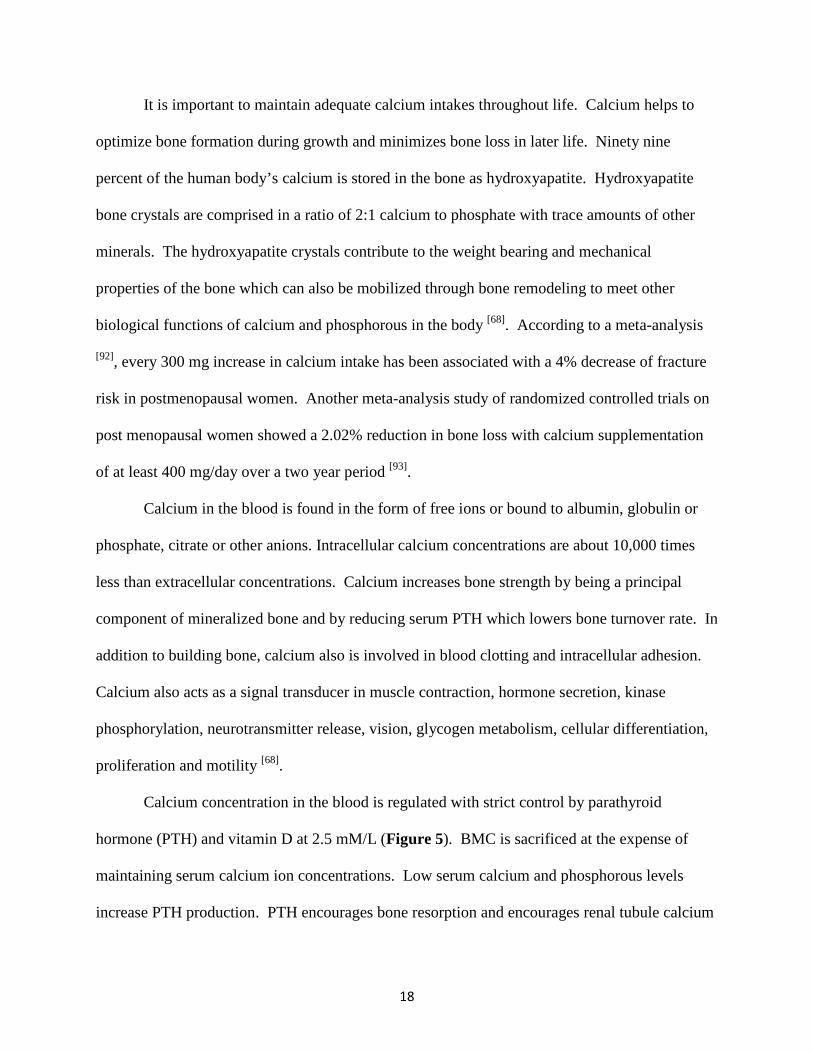
Calcium concentration in the blood is regulated with strict control by parathyroid
hormone (PTH) and vitamin D at 2.5 mM/L (Figure 5). BMC is sacrificed at the expense of
maintaining serum calcium ion concentrations. Low serum calcium and phosphorous levels
increase PTH production. PTH encourages bone resorption and encourages renal tubule calcium
19
re-absorption by converting vitamin D to its active form (Figure 5) to meet serum calcium
needs. Vitamin D increases calcium absorption in the intestines and reabsortion in the kidneys.
1,25(OH)2 vitamin D3 interacts with vitamin D receptor in the enterocyte and stimulates the
synthesis of calcium-binding protein. Calcium receptors are located in the intestine, osteoblast
cell lines, calcitionin secreting c-cells of the thyroid gland, parthyroid gland, and the 1,25(OH)2
vitamin D3 producing cells of the renal proximal tubule [94]. Low blood calcium status increases
calcium absorption in the intestine. However, chronic low calcium intake will lead to increased
bone resorption due to increase in PTH.[71].
Figure 5: Bone Metabolism[95]
20
Adequate calcium intake for postmenopausal women is 1200 mg/day with most women
falling short in meeting this requirement through diet alone[96]. Intestinal calcium absorption
declines with age or when calcium intake is high. Calcium absorption is more efficient if
calcium is consumed in amount spread throughout the day rather than consuming it all at once.
Calcium bioavailability needs to be considered when choosing calcium food sources. Calcium
absorption from diet is between 20% and 60 % [71]. Calcium is absorbed more easily when it can
disassociate more readily from its ligands. However, low molecular weight calcium salts can be
absorbed intact without a vitamin D induced calcium transporter. Enhancers of calcium
absorption are soluble salts that prevent precipitation of calcium by phosphates (i.e. calcium
citrate malate), inulin and fructooligosaccharides. Some casein and whey peptides also prevent
precipitation of calcium by phosphates. Inhibitors to calcium absorption are oxalate and phytic
acid[71].
In addition to assuring adequate calcium intake it is also important to consider calcium
losses through the urine. Urinary excretion impacts calcium retention by about 50% [97].
However, increases in dietary calcium only affect urinary output of calcium by approximately
6% while dietary sodium exerts a much greater effect on urinary calcium excretion and
contributes to low BMD in postmenopausal women [97]. Although high protein intake increases
urinary calcium excretion, net calcium retention is not affected because of changes in calcium
absorption and endogenous calcium secretion [98]. Urinary calcium tests are not done routinely
but conducted to determine the etiology of kidney stones or to check for problems with the
parathyroid gland[99]. In addition to low calcium having a negative impact on BMD, low
extracellular calcium also increases the risk for hypertension, preeclampsia, premenstrual
syndrome, obesity, polycystic ovary syndrome and hyperparathyroidism [68].
21
Vitamin D is involved in bone formation by regulating calcium and phosphorous amounts
in the blood and by promoting bone mineralization. Indirectly, vitamin D is involved in bone
remodeling by its role in differentiation and proliferation of hematopoietic cells, keratinocytes,
parathyroid secreting cells and beta cells of the pancreas [68]. Vitamin D must be metabolized by
both the liver and kidney before becoming biologically active as a steroid hormone. The active
metabolite 1α, 25(OH)2 vitamin D3 functions as a steroid hormone by generating biological
responses via regulation of gene transcription and activation of signal transduction pathways [68,
100] . The other kidney metabolite of vitamin D, 24R,25 (OH)2D3 has been shown to improve
bone fracture healing in chickens [24]. In the intestine, vitamin D assists with calcium,
magnesium and phosphorous absorption [101] (Figure 5). Vitamin D receptors are both nuclear
and membrane specific and are close to plasma membrane as well as found in bone and bone
marrow. Vitamin D participates in bone remodeling through its involvement in OB
differentiation, inhibiting OB apoptosis, encouraging calcium mobilization, and inducing
osteocalcin production [101]. When plasma calcium and phosphorous levels are low, the kidneys
convert 25(OH) vitamin D3 to 1α, 25(OH)2 vitamin D3, its active steroid hormone form.. The
enzyme 1α, 25-hydroxylase is an important regulator of the active metabolite of vitamin D
production in the kidneys. It is regulated by 1α, 25(OH)2 vitamin D3 concentrations, serum
calcium and phosphate levels and PTH. The enzyme 1α, 25-hydroxylase is also iron dependent.
Therefore, variations in iron levels indirectly affect bone by decreasing 1α,25(OH)2 vitamin D3
production.
Optimal health may require a vitamin D status much greater >25 nmol/L, the threshold
for 25-hydroxyvitamin D set to avoid clinical deficiency[102]. The RDA for elderly females is
600 IU/day (10-15 µg/day) [2]. Aside from dietary sources, the skin produces a precursor to the
22
active vitamin D when the sun’s ultraviolet rays transform 7-dehydrocholesterol into
cholecalciferol. Cholecalciferol is hydroxylated in the liver to form 25-hydroxycholecalciferol,
also known as calcidiol, which is stored in the liver until needed. When needed, calcidiol is
hydroxylated to its biologically active form, calcitriol, in the kidney. Minimum levels ranging
from 25 nmol/L to >100 nmol/L have been proposed, largely due to the inverse relationship of
25(OH)D3 (calcidiol) and PTH. High PTH is a risk factor for osteoporosis in older adults[102].
An estimated 80% of elderly postmenopausal women may have vitamin D deficiency, defined by
levels less than 20 ng/mL or 50 nmol/L [103]. Production of vitamin D binding protein, which is
responsible for vitamin D (calcitriol) transport in the blood, increases by 50% in high estrogen
states [68].
Vitamin C, present in high amounts in mango, plays a critical role in collagen synthesis.
Collagen is necessary for bone, skin and tendon formation. Vitamin C is a cofactor which
promotes enzyme activity by maintaining metal ions in their reduced form. It is involved in the
hydroxylation of proline and lysine residues which form the cross linking necessary to form the
triple helical structure of collagen. More specifically, α-ketoglutarate-dependant dioxygenases
incorporate one atom of oxygen into succinate and into the oxidized product in the presence of
ferrous iron [68]. Ascorbic acid stimulated collagen production in vitro fibroblast cells by 60-
100% [104]. A study analyzing the effects of vitamin C supplementation (745 mg/day) on BMD
of postmenopausal women, demonstrated that vitamin C users had BMD levels that were 3%
higher at the midshaft radius, femoral neck, and total hip than non-users[105]. Women taking both
estrogen and vitamin C had significantly higher BMD levels at all sites. Women who took
vitamin C plus calcium and estrogen had the highest BMD at the femoral neck, total hip,
ultradistal radius, and lumbar spine [105].
23
The phytonutrients found in fruits and vegetables may play an important role in
modulating bone metabolism without the side effects associated with pharmaceutical
interventions. Phytonutrients, such as phenolics, may function as antioxidants, donating
hydrogen ions, scavenging free radicals, inhibiting radical generation, inhibiting enzyme activity
or by specific receptor interactions[19]. Antioxidant functions include enzyme inhibition, metal
chelation, hydrogen donation and by oxidizing non-propagating radicals[19]. Phenolics can
inhibit xanthine oxidase and other enzymes involved in the production of ROS[19]. They can also
function as anti-inflammatory agents by inhibiting lipoxygenase, the enzyme responsible for
creating prostaglandins and leucotrienes from arachidonic acid[106]. Some phenolics modulate
signal transduction by inhibiting tyrosine kinase[19]. Some flavonoids and isoflavonoids inhibit
17 β – hydroxysteroid oxidoreductase which may influence sex hormone metabolism[107].
Population studies show a positive correlation with fruit and vegetable intake and BMD
[25], 109[13, 18, 108]. For example, women who consumed vegetables more than 9 times a week had
higher BMD compared to those with lower intakes [109]. Higher BMD was observed at the heel
site of women consuming more than 1.5 servings of vegetables a day in a study on a rural
population in Iran [108]. The benefits of fruits and vegetables results from an additive and
synergistic effect from the combination of phytochemicals[25]. The antioxidant action of the
phytonutrients may be responsible for these findings and for this reason it is important to
consider the oxidative status associated with osteoporosis.
Oxidative stress is considered as an independent risk factor for osteoporosis[110].
Increased OC activity and decreased OB activity have been associated with an imbalance
between oxidant and antioxidant status in postmenopausal osteoporosis[111]. When bone
remodeling is imbalanced, as in osteoporosis, ROS production by the OC may overwhelm the
24
body’s endogenous antioxidants defense mechanisms leading to further bone loss. Enhanced OC
activity increases superoxide anion generation and inhibits superoxide dismutase and glutathione
peroxidase activities[112]. In murine models, superoxide anion was proposed to oxidize calcium
binding sites and/or acidify the local environment near OCs, favoring bone loss[113].
Transcription factors, such as nuclear factor-kB (NF-kB) which signals TNF production and
inflammation, are sensitive to oxidative changes[19]. Basu and colleagues established a
biochemical link between increased oxidative stress and reduced bone density by showing
elevated oxidative stress marker 8-iso-prostaglandin F2α and elevated inflammatory marker 15-
keto-13,14-dihydro-prostaglandin F2α in osteoporotic subjects when compared to those without
osteoporosis[114, 115]. Hence, antioxidant supplementation has been proposed as a novel
therapeutic strategy for osteoporosis.
Phytonutrients have been shown to play a protective role in preserving BMD in animals
[14, 15, 20, 116, 117] and in vitro [16, 118-120]. Orange pulp and citrus fruits in general contain high
amounts of vitamin C and flavonoids and have shown to improve bone density in male
orchidectomized rats [14, 15]. Citrus hesperidin and olive oleuropein together, were also shown to
prevent bone loss and improve peek bone mass in female rats [117]. Green tea polyphenols
reduced the extent of bone micro-architecture deterioration in female rats [116]. Pholridzin in
apples, rutin, and isoflavones have all been shown to prevent bone loss in ovariectomized rats
[20]. Flavonoids, quercetin and kaempferol was shown to inhibit bone resorption in vitro [118].
Antioxidants present in fruits and vegetables may reduce oxidative stress in the bone
environment by reducing OC differentiation. For example α-lipoic acid was able to inhibit OC
differentiation [121], while lycopene was shown to reduce oxidative stress and bone turnover
markers in postmenopausal women[122]. Dried plum polyphenols inhibited osteoclastogenesis by
25
down regulating NFATc1 and inflammatory mediators and attenuated TNF-α on osteoblast
function with upregulation of Runx2, Osterix and IGF-1 in MC3T3-E1 cells and in gonadal
hormone deficient, inflammation induced animal model [16, 119, 120].
Lifestyle Influences on Bone
Weight-bearing exercise exerts a mechanical stimulation on bone tissue. Resistance and
high-impact activities contribute to development of peak bone mass and may reduce risk of falls
in older individuals [2]. Many studies have confirmed bone loss resulting from immobilization
and micro-gravity in both animals and humans [123-125]. Studies with increased loading activity
have resulted in increased bone mass often localized to the bones where the loading took place
[126, 127]. Mechano-stimulation to the osetocytes creates a signaling pathway to generate more
OBs.
Current research indicates that smoking not only increases the risk for osteoporosis in
women but also in men and adolescents [128]. Even second hand smoke has been correlated with
an increased risk for osteoporosis and fracture according to a study done on Chinese men and
pre-menopausal women [129]. Findings from this study showed that premenopausal women
exposed to second- hand smoke had a “threefold higher risk of having osteoporosis and a 2.6
times greater risk for a non-spine fracture” when a family member smoked [129]. Other studies
show that second-hand smoke may alter estrogen levels thereby effecting health [128]. A study
conducted on young Sweden men demonstrated that 24% of smokers had bone fractures
compared to only 14% or those who never smoked [130, 131]. Moreover, smokers had lower bone
density in the spine, hip, and whole body when compared to non-smoking peers [130, 131]. Overall,
smoking can double osteoporosis risks in men [128].
26
Alcohol, consumed moderately, may provide benefits to the BMD of postmenopausal
women [132, 133] and in men over age 45[133]. In contrast, studies done on alcoholics report lower
bone mineral density[134, 135]. In the research conducted by Holrook and colleagues (1993), social
drinking of up to 180 g/day of alcohol in men and 120 g/day in women [133] resulted in an
increase in BMD at the femoral neck of men and the spine of women in a one week study and in
the radial shaft and spine of women in the 24 hour study [133]. Stronger associations on BMD
were seen with beer or wine consumption compared to liquor consumption[132]. In men, 2 or
more servings per day of liquor resulted in lower BMD [132]. This may suggests that constituents
in the wine and beer, other than ethanol, may contribute to bone health. For example, the high
silicon content of beer has been associated with increases in BMD [88, 132].
The effects of caffeine on bone are inconclusive. Some studies have shown detrimental
effects of caffeine on bone, some of which have disappeared after making statistical adjustments
for osteoporotic risk factors [136]. A study conducted by Rapuri and colleagues (2001) [137] found
that women who consumed more than 300 mg/day of caffeine had a significantly higher rate of
bone loss in the spine. In contrast, Japanese women who drank green tea regularly had
significantly higher BMD than those who did not [138]. In addition to the observational findings
correlating green tea and BMD, in inflammation induced rats, green tea polyphenols attenuated
BMD loss [116] and in bone marrow mesenchymal stem cells green tea catechins encouraged
osteogeneis [139]. The interaction of an individual’s genotype with caffeine may affect the BMD
study outcomes with some caffeine metabolizing genotypes being more susceptible to bone loss
than others [140]. This genotypic variation among individuals may help explain some of the
controversies and inability of other studies to show clearly the effect of caffeine on bone.
27
Postmenopausal Osteoporosis
Being postmenopausal or in a state of surgically-induced menopause increases bone
resorption [2]. For the most part, this phenomenon is attributed to a decrease in estrogen
production. Estrogen production declines several years before the cessation of menstruation
which typically occurs between 48 and 50 years of age [141]. Consequently, osteoporosis is most
commonly found in the elderly female population. Development of osteoporosis in women has
also been associated with the time of menarche, menopause, and the duration of lactation [142]. Li
and Zhu (2005) [143] demonstrated that the later menarche, the earlier the menopause, the greater
number of child births and the longer the duration of lactation all lead to lower BMD and a
higher degree of osteoporosis.
Estrogen has a protective influence on bone by attenuating OC proliferation and activity.
Estrogen receptor α (ER α) is present on both the osteoprogenitor cells and the OCs. When 17β-
estradiol binds to the ER α, it triggers apoptosis in the OC and inhibits OC differentiation in the
progenitor cells[144]. With a dramatic decline in estrogen production during menopause, bone
remodeling increases several fold. Postmenopausal bone resorption doubles and biomarkers of
bone formation are less than half the levels of that observed premenopause [145].
Estrogen deficiency also increases FSH secretion which encourages the production of
inflammatory cytokines, such as TNF-α and several ILs by bone marrow and immune cells. The
cytokines, in turn, encourage OC differentiation. The lack of estrogen stimulation during the
postmenopausal state complemented with declined levels of GH which occurs with aging leads
to weakened signaling on the OB (Figure 6).
Estrogen not only exerts an influence on the OCs but also on the OBs. Estrogen
attenuates OB progenitor/ mesenchymal stem cell production by as much as 50% [146]. In the low
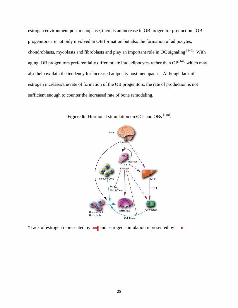
28
estrogen environment post menopause, there is an increase in OB progenitor production. OB
progenitors are not only involved in OB formation but also the formation of adipocytes,
chondroblasts, myoblasts and fibroblasts and play an important role in OC signaling [146]. With
aging, OB progenitors preferentially differentiate into adipocytes rather than OB[147] which may
also help explain the tendency for increased adiposity post menopause. Although lack of
estrogen increases the rate of formation of the OB progenitors, the rate of production is not
sufficient enough to counter the increased rate of bone remodeling.
Figure 6: Hormonal stimulation on OCs and OBs [148].
*Lack of estrogen represented by and estrogen stimulation represented by
29
Although estrogen modulates OB progenitor production, it still exhibits an anabolic effect
on OBs. Estrogen encourages OB differentiation, proliferation and increases OB proteins such
IGF-1, type I procollagen, transforming growth factor beta (TGF-β) and BMP [141]. Estrogen
also inhibits OB and osteocyte apoptosis [149]. Estrogen suppresses bone resorption by increasing
OC apoptosis [150] increasing OPG production and decreasing OB/stromal cell production of
RANKL [151]. The activation of the Src/Shc/ERK–signaling pathway influences the estrogen
receptors in the OB lineage of cells to prevent apoptosis of OB cells [152]. A decrease in the
number of cells that support osteoclast development, together with decreased production of
osteoclastogenic cytokines by these same cells may be responsible for the potent suppression of
osteoclastogenesis by estrogens [146].
Estrogen also increases nitric oxide (NO) and TGF-β levels. An appropriate level of NO
is necessary to protect bones and organs from ischemic damage while chronically high levels of
NO can cause tissue toxicity, inflammation and carcinomas [153]. NO and TGF-β regulate T cell
production of TNF-α and act to inhibit osteoclastogenesis and bone resorption [6]. Estrogen
deficiency in postmenopausal women is associated with an increase in pro-inflammatory
cytokine production such as IL-1, TNF- α, and IL-6 [4, 6] in the bone environment (Figure 6).
These pro-inflammatory cytokines are involved in osteoclastogenesis and encourage bone
resorption [7]. The inflammatory cytokines also produce oxygen free radicals which contribute to
osteoclast formation, differentiation and activation (Figures 1 & 2). IL-1 and IL-6 also increase
the production of M-CSF which further influences OC differentiation. Estrogen also regulates
the release of growth factors and growth hormones such as TGF-β and IGF-I [154].
In addition to the menopausal effects on OB progenitors, OBs, and OCs, estrogen
deficiency also affects calcium balance. In an estrogen deficient state, intestinal calcium
30
absorption is decreased while urinary calcium loss is increased [155]. Intestinal mucosal cells
have receptors for 17β-estradiol which increase calcium transport/absorption [156]. The kidneys
also reabsorb calcium better when estrogen is present [157]. This is attributed to an increase in
circulating 1,25-(OH)2D3 which is indirectly mediated through stimulation of renal la-
hydroxylase by increased serum PTH[158]. In addition to the direct effects of estrogen on calcium
absorption, there is also reduced total circulating concentration of 1,25(OH)2D3 as one ages,
which effects both calcium absorption and, indirectly, bone formation.
Estrogen receptors α and β are activated by 17β-estradiol and function as a DNA binding
transcription factor. In the absence of estrogen, the inactive α receptor located on the nucleus is
associated with several heat shock proteins [159]. In the presence of a receptor stimulus, like 17β-
estradiol, the receptor undergoes a conformational change displacing the inhibitory heat shock
proteins and forms dimers. The dimers allow the receptor to interact with “specific steroid
response elements (SRE) located within the regulatory regions of target promoters [160]. The
ligand-activated receptor can interact with the general transcription apparatus (GTA) directly or
indirectly through adaptor proteins. Ultimately, these interactions stabilize the transcription
preinitiation complex and enhance RNA polymerase activity [160] .
Bone also degrades with age and can be attributed to the degree of mineralization,
anisotropy, skeletal geometry and the periosteal response to trabecular bone mass [161]. Growth
hormone secretion declines 14% per decade and is one of the primary factors responsible for low
serum IGF-I levels which are directly correlated with BMD [103]. Adrenal androgens,
dehydroepiandrosterone (DHEA) and DHEA sulfate are also 10-20% lower than young adults
further contributing to a lack of bone building stimulus factors[162].

31
Animal Model
Laboratory animals have played a major role in understanding osteoporosis and
developing treatment options for the management of osteoporosis[163]. The success of an animal
model is based on its ability to successfully predict outcome measures in people. Because the
average life-span of the laboratory mouse is about 2 years, one year is roughly equivalent to
middle age. A menopausal state, however, can be induced surgically by ovariectomy [163].
Ovariectomized mice are comparable to women who undergo bilateral ovariectomy prior to
menopause and prematurely experiences menopausal symptoms soon after surgery [163]. Hence,
the ovariectomized mouse experiences an increase in bone turn over, osteopenia and weight gain
[163]. It takes the mice 21 days post ovariectomy to reach a state similar to that of early
menopause whereas after 35 days corresponds to late menopause in women[164]. Research
conducted by Lynch and colleagues demonstrated that the mouse tibia did not experience
significant bone loss or architectural adaptation following OVX in growing mice [165]. However,
ovariectomy-induced estrogen deficiency resulted in a reduction in vertebral bone mass due to
decreased trabecular thickness and increased separation when compared to SHAM control mice
at 6 weeks post OVX in 26week old C57Bl/6 mice[166]. The decreases in bone mass did not
affect mechanical integrity of lumbar vertebrae, suggesting that a duration longer than 6 weeks is
needed before bone mass changes result in deterioration of mechanical properties[166]. Several
studies have used the ovariectomized mouse model to study both nutrition and pharmaceuticals
in preventing further postmenopausal bone loss[139,[17, 167, 168]. Some nutrition-related examples
of research conducted on ovariectomized rodents with successful results in improving or
maintaining bone parameters are nano calcium products[168], soy protein [163], and dried plum[17,
167].
32
Pharmacological Options for Osteoporosis Treatment
Drugs for the prevention and treatment of osteoporosis are classified as anti-resorptive or
anabolic depending on their effect on bone remodeling[169]. Anti-resorptive drugs act by
inhibiting OC formation, OC activity or by inducing premature osteoclast cell death. Anti-
resorptive drugs include SERMs, bisphosphonates (alendronate, risedronate, ibandronate, and
zoledronic acid), an estrogen agonist/antagonist (raloxifene), and calcitonin. Some resorption
inhibitors, such as SERMs, PTH, some interleukins, and Denosumab, inhibit OC formation by
working on OBs since OBs secrete essential factors for OC differentiation such as M-CSF and
RANKL [170]. Denosumab is a fully human monoclonal antibody (IgG2 immunoglobulin
isotype) antiresorptive drug that has recently been approved for use in the US. It functions by
binding to RANKL, preventing it’s binding to RANK. This results in a reduction in OC
formation, activity and survival, thereby reducing the bone resorption rate [171]. Denosumab also
has the advantage of being a subcutaneous medication administered every six months thereby
improving long-term adherence to therapy compared with oral treatment options. Calcitonin and
aminobisphosphonates, on the other hand, work by inhibiting OC resorption activity, where as
the first generation bisphosphonate, clodronate, works by inducing osteoclast apoptosis [170] .
Anabolic agents work by increasing osteoblast activity. The only pharmaceutical
anabolic agents to bone are teriparatide, a recombinant PTH, and strontium ranelate [172].
Strontium ranelate has both antiresorptive and anabolic properties [173]. The present study looks
at mango as a possible anabolic agent to bone, thereby reversing the effects of ovariectomy
induced bone loss.
33
Nutrient Composition of Mango
Mangos (Mangifera indica L) are excellent sources of vitamin C and vitamin A and are
good sources of dietary fiber, potassium, copper, and vitamin K [28]. Mangos also contain
minerals involved in bone health such as magnesium, zinc, copper, manganese, and selenium [29].
Table 1 details the nutrient composition and daily value of the mango fruit.
Table 1: Nutrient composition of mango and percent daily values (DV).ab[28]
aAmount of nutrient a food provides based on DRI (dietary reference intake) guide and reference values for nutrition labeling (2000 calories per day). b Values were obtained from USDA National Nutrient Database for Standard Reference, Release 22 (2009) c Foods that provide %DV of ≥10% are considered a 'good' source; those ≥20% are considered an 'excellent' source.
Nutrient DV reference (units/day)
Amount in 100g mango
% DVc Amount in 1 fruit (no peel or pit)
(207 g)
% DVc
Macronutrient Carbohydrate, g 300 17 5.7 35.2 11.7 Protein, g 50 0.51 1.0 1.06 2.1 Total fat, g 65 0.27 0.4 0.56 0.9 Dietary fiber 25 1.8 7.2 3.7 14.8
Minerals Calcium, mg 1000 10 1.0 21 2.1 Potassium, mg 3500 156 4.4 323 9.2 Phosphorus, mg 1000 11 1.1 23 2.3 Copper, mg 2 0.110 5.5 .228 11.4 Sodium, mg 2400 2 0.1 4 0.2
Vitamins Vitamin A, IU 5000 765 15.3 1584 31.7 Vitamin C, mg 60 27.7 46.2 57.3 95.5 Vitamin K, ug 80 4.2 5.2 8.7 10.9 Thiamin, mg 1.5 0.058 3.9 0.12 8.0 Riboflavin, mg 1.7 0.057 3.4 0.118 6.9 Folate, ug 400 14 3.5 29 7.2 Vitamin E, IU 30 1.12 3.7 2.32 7.7 Niacin, mg 20 0.584 2.9 1.209 6.0
34
Mangos also contain carotenoids, triterpenes, and phenolic compounds such as tannins,
mangiferin and flavonoids which contribute to its antioxidant and anti-inflammatory functions.
Total phenolics in mango vary depending on the cultivar and the processing methods. Total
phenolics of dry weight with peel ranges from 350 to 4860 mg/kg, while 60 to 180 mg/kg is
common in the fresh fruit [174-177]. Mercadante and colleagues show the following carotenoid
profile as detected by HPLC and confirmed by mass spectrometry on a Brazilian mango
cultivar, Keitt; β-carotene (all-trans), β-cryptoxanthin (all-trans and cis), zeaxanthin (all-trans),
luteoxanthin isomers, violaxanthin (all-trans and cis), and neoxanthin (all-trans and cis) [178].
Carotenoid composition of the mango cultivar Keitt is shown in Table 2. β –Carotene is a
predominant carotenoid in mango and functions as pro-vitamin A. Table 3 details the β –
Carotene and vitamin A content of various varieties of mango in their fresh and dried forms.
Although the ripe mango flesh is not particularly high in flavonoids, higher amounts of
flavonoids can be obtained by consuming them unripened and with the peel intact, a common
practice done in Indian cuisine know as the spicy mango pickle (Table 4).
Table 2: Carotenoid Composition of Mango Cultivar Keitt Bahia, Brazil [178]
carotenoid (µg/g) range meana all-trans-β-carotene 13.4-16.2 15.1 ± 1.5 unidentified compound 0.2 ± 0.0 cis- β -cryptoxanthin tr-0.1 0.1 ± 0.1 all-trans- β -cryptoxanthin 0.3-0.3 0.3 ± 0.0 all-trans-zeaxanthin 0.6-0.9 0.8 ± 0.2 luteoxanthin isomers 3.1-4.1 3.8 ± 0.6 all-trans-violaxanthin 18.2-23.9 21.1 ± 2.9 9-cis-violaxanthinb 9.9-10.3 10.1 ± 0.2 13-cis-violaxanthinb 1.3-1.5 1.4 ± 0.1 cis-neoxanthin tr-0.2 0.1 ± 0.1 all-trans-neoxanthin 1.0-3.6 2.1 ± 1.3 total 49.9-59.8 55.0 ± 5.0 vitamin A value 222-270 251 ± 26 a Mean and standard deviation of three sample lots. b Tentative identification.
35
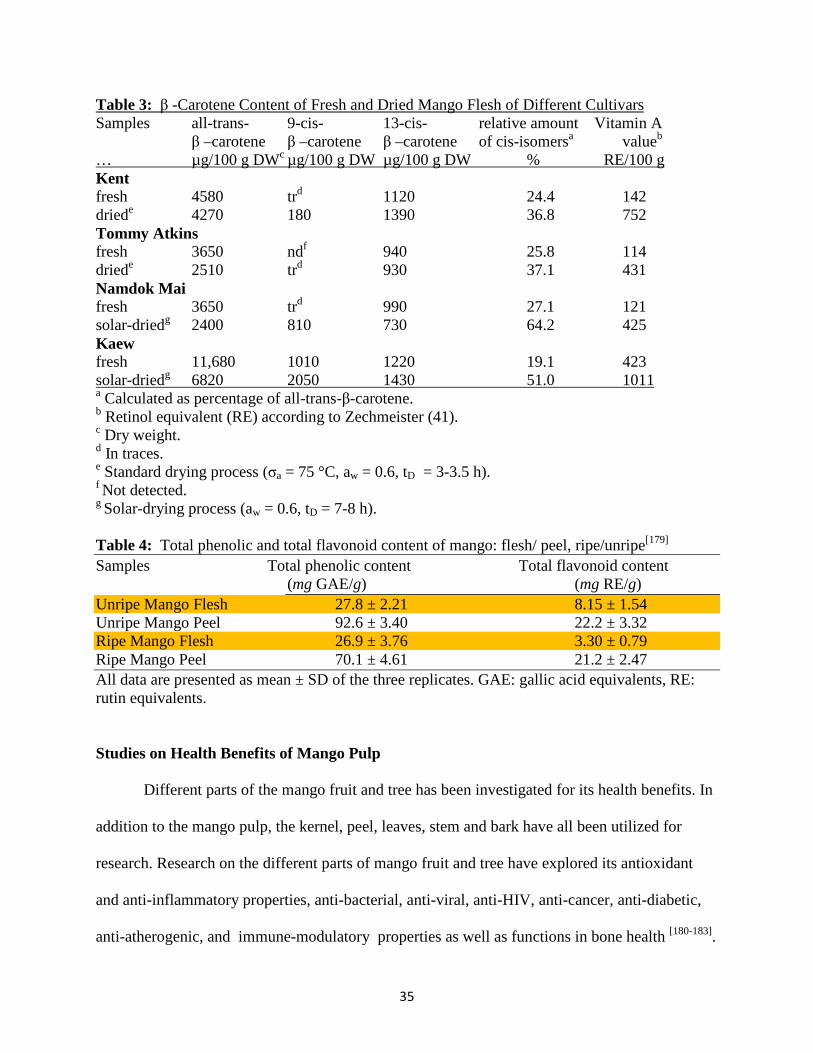
Table 3: β -Carotene Content of Fresh and Dried Mango Flesh of Different Cultivars Samples all-trans- 9-cis- 13-cis- relative amount Vitamin A
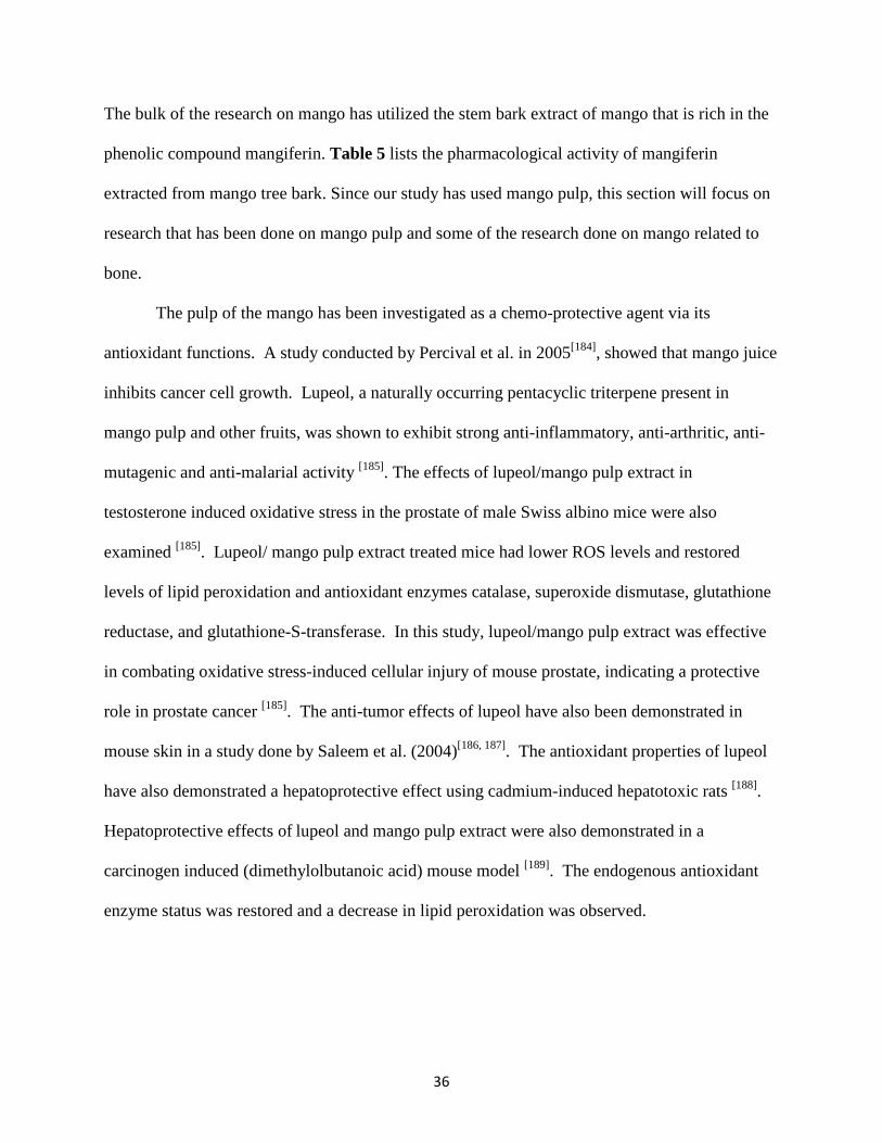
β –carotene β –carotene β –carotene of cis-isomersa valueb … µg/100 g DWc µg/100 g DW µg/100 g DW % RE/100 g Kent fresh 4580 trd 1120 24.4 142 driede 4270 180 1390 36.8 752 Tommy Atkins fresh 3650 ndf 940 25.8 114 driede 2510 trd 930 37.1 431 Namdok Mai fresh 3650 trd 990 27.1 121 solar-driedg 2400 810 730 64.2 425 Kaew fresh 11,680 1010 1220 19.1 423 solar-driedg 6820 2050 1430 51.0 1011 a Calculated as percentage of all-trans-β-carotene. b Retinol equivalent (RE) according to Zechmeister (41). c Dry weight. d In traces. e Standard drying process (σa = 75 °C, aw = 0.6, tD = 3-3.5 h). f Not detected. g Solar-drying process (aw = 0.6, tD = 7-8 h). Table 4: Total phenolic and total flavonoid content of mango: flesh/ peel, ripe/unripe[179] Samples Total phenolic content Total flavonoid content
(mg GAE/g) (mg RE/g) Unripe Mango Flesh 27.8 ± 2.21 8.15 ± 1.54 Unripe Mango Peel 92.6 ± 3.40 22.2 ± 3.32 Ripe Mango Flesh 26.9 ± 3.76 3.30 ± 0.79 Ripe Mango Peel 70.1 ± 4.61 21.2 ± 2.47 All data are presented as mean ± SD of the three replicates. GAE: gallic acid equivalents, RE: rutin equivalents. Studies on Health Benefits of Mango Pulp
Different parts of the mango fruit and tree has been investigated for its health benefits. In
addition to the mango pulp, the kernel, peel, leaves, stem and bark have all been utilized for
research. Research on the different parts of mango fruit and tree have explored its antioxidant
and anti-inflammatory properties, anti-bacterial, anti-viral, anti-HIV, anti-cancer, anti-diabetic,
anti-atherogenic, and immune-modulatory properties as well as functions in bone health [180-183].
36
The bulk of the research on mango has utilized the stem bark extract of mango that is rich in the
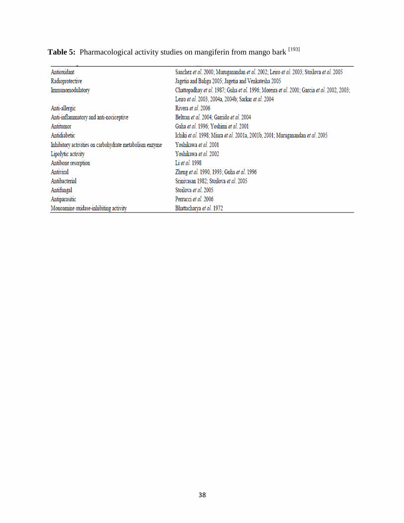
phenolic compound mangiferin. Table 5 lists the pharmacological activity of mangiferin
extracted from mango tree bark. Since our study has used mango pulp, this section will focus on
research that has been done on mango pulp and some of the research done on mango related to
bone.
The pulp of the mango has been investigated as a chemo-protective agent via its
antioxidant functions. A study conducted by Percival et al. in 2005[184], showed that mango juice
inhibits cancer cell growth. Lupeol, a naturally occurring pentacyclic triterpene present in
mango pulp and other fruits, was shown to exhibit strong anti-inflammatory, anti-arthritic, anti-
mutagenic and anti-malarial activity [185]. The effects of lupeol/mango pulp extract in
testosterone induced oxidative stress in the prostate of male Swiss albino mice were also
examined [185]. Lupeol/ mango pulp extract treated mice had lower ROS levels and restored
levels of lipid peroxidation and antioxidant enzymes catalase, superoxide dismutase, glutathione
reductase, and glutathione-S-transferase. In this study, lupeol/mango pulp extract was effective
in combating oxidative stress-induced cellular injury of mouse prostate, indicating a protective
role in prostate cancer [185]. The anti-tumor effects of lupeol have also been demonstrated in
mouse skin in a study done by Saleem et al. (2004)[186, 187]. The antioxidant properties of lupeol
have also demonstrated a hepatoprotective effect using cadmium-induced hepatotoxic rats [188].
Hepatoprotective effects of lupeol and mango pulp extract were also demonstrated in a
carcinogen induced (dimethylolbutanoic acid) mouse model [189]. The endogenous antioxidant
enzyme status was restored and a decrease in lipid peroxidation was observed.
37
Mango and Bone Health
We are only aware of a few studies investigating the effect of mango on bone. Er- Xian
Decoction, has long been used for the treatment of osteoporosis and menopausal syndrome in
China. Bioactivity-guided fractionation of Er Xian decoction has led to the successful isolation
of the anti-osteoporotic constituents, identifying one as mangiferin [190].
Another study investigated the effect of the xanthonoid compound found in mangoes,
mangiferin, on periodontitis [191]. Periodontitis is a chronic inflammatory disease related to the
formation of colonies of microorganisms present in subgingival plaque leading to inflammatory
periodontal pockets, destruction of the periodontal ligaments, alveolar bone resorption and tooth
loss[192]. Carvalho and colleagues (2008)[191], induced periodontitis in Wistar rats by applying a
ligature around the lower right first molar with subsequent oral treatment of 100 mg/kg
mangiferin for 1, 4 or 7 days. Oral administration of mangiferin significantly reduced alveolar
bone loss and cellularity, inhibited COX-2 expression and the adhesion of leukocytes, while
maintaining normal lipoxin A4 levels. Lipoxin A4 acts as an endogenous anti-inflammatory
mediator by its anti-chemotaxic action in inhibiting the rolling of leukocytes. The mangiferin-
treated rats presented an earlier peak of cell proliferation and augmented angiogenesis in the
injured region.
38
Table 5: Pharmacological activity studies on mangiferin from mango bark [193]
39
CHAPTER III
METHODOLOGY
This study was designed to determine the dose-dependent effects of freeze-dried mango
pulp on reversing ovariectomy-induced bone loss. To mimic a post-menopausal state, the
ovariectomized mice model was used.
Animal Grouping
Thirty 12–week old ovariectomized (OVX) mice and 14 sham-operated (SHAM)
C57BL/6 mice were purchased from Charles River Laboratory (Kingston, NY) and housed in
Oklahoma State University (OSU) animal research laboratory. Mice were fed a standardized
AIN-93M powdered rodent diet [194] for two weeks post-ovariectomy. All animal handling and
procedures were approved by Institutional Animal Care and Use Committee at OSU.
After a 2 week period, a total of 12 mice (n= 6 SHAM; n= 6 OVX) were necropsied to
confirm ovariectomy-induced bone loss. Whole body dual-energy X-ray absorptiometry (DXA)
scans were preformed prior to necropsy. The remaining 32 mice were weighed and divided into
4 groups and housed in groups of four mice per cage. Mice were assigned to one of the
following dietary treatment groups (n = 8 mice/ group) for 8 weeks: (1) SHAM - control diet;
(2) OVX - control diet; (3) OVX - 5% dried mango diet; and (4) OVX - 25% dried mango diet.
The OVX groups were match-fed to the SHAM group. All mice were provided deionized water
ad libitum and were weighed weekly.
40
Diet Formulation
Ripe Tommy Atkins variety mangoes were purchased from a local grocery store. The
pulp was separated from the skin and kernel and freeze-dried. The freeze-dried mango pulp was
ground to a powder and sent to Nestle Purina Analytical Laboratories (St. Louis, MO) for
analysis of macronutrients and calcium and phosphorous content (Table 6). Freeze-dried mango
powder was incorporated into the diet at 5% or 25% concentration by weight. The macro-
nutrient composition as well as the calcium and phosphorus content of the mango diets were
adjusted to be similar to the control diet (Table 7). The control diet was an AIN-93M
formulation containing 72% carbohydrate, 4% fat and 14% protein by weight [194].
Table 6: Macronutrient, calcium and phosphorus content of freeze dried mango powder (TommyAtkins variety) used in the study. * Parameter Amount (g/100g)
Carbohydrate 73.70 Protein 4.80 Fat 2.89 Crude Fiber 7.58 Ash 3.00 Calcium 0.0854 Phosphorus 0.115 Calories 340
*Analysis performed by Nestle Purina Analytical Laboratories (St. Louis, MO)
Necropsy, Tissue Processing, and Storage
After eight weeks of dietary treatment, mice were fasted for 12 hours, weighed and
injected with ketamine/xylazine cocktail (80 and 8 mg/kg body weight, respectively). Body
composition was determined using DXA (GE Lunar Piximus, Fitchburg, WI). Blood was
collected via exsanguination from carotid artery and placed into ethylenediamine-tetraacetic acid
(EDTA) coated tubes. Plasma was obtained by centrifugation of whole blood for 20 minutes at
1300 X g and stored at -80°C for later analyses. Thymus, spleen, liver and uterus were removed
41
Table 7: Diet composition Control
(AIN-93M)* 5% Mango 25% Mango
Ingredients (g/kg) Dried mangoa 50.0 250 Total Carbohydrate 720.7 720.7 720.7
Cornstarchb 465.7 428.9 281.5 Maltodextrinb 155.0 155.0 155.0 Sucroseb 101.1 101.4 102.4 Dried mangoa 36.6 184.3 Total Protein 140.0 140.0 140.0 Caseinb 140.0 137.6 128.0 Dried mangoa 2.4 12.0 Total Fat 40.0 40.0 40.0 Soybean oilb 40.0 38.6 32.8 Dried mangoa 1.5 7.2 Total Fiber 50.0 50.0 50.0 Celluloseb 50.0 46.2 31.1 Dried mangoa 3.8 18.9 Vitamin Mixb 10.0 10.0 10.0 Total Mineral Mix 35.00 35.00 35.00 Mineral Mixb (Ca & P deficient)
13.40 13.40 13.40
Calcium carbonateb 12.50 12.39 11.97 Calcium from mangoa 0.04 0.21 Sodium phosphatec monobasic (NaH2PO4)
5.60 5.48 5.03
Potassium phosphated monobasic (KH2PO4)
2.40 2.36 2.16
Phosphorus from mangoa
0.06 0.29
Choline bitartrateb 2.50 2.50 2.50 L-cysteinb 1.80 1.80 1.80 *Based on AIN-93M formulations containing 72% carbohydrate, 4% fat and 14% protein by calories (Reeves et al., 1997). a Freeze dried Tommy Atkins Mango Pulp bHarlan Teklad Diets (Madison, WI) cSigma Aldrich Co. (St. Louis, MO) dFisher Scientific Bio Reagents (Fair Lawn, NJ)
42
and weighed. A portion of abdominal white adipose tissue was collected and stored in formalin
with the remainder snap-frozen in liquid nitrogen and stored at -80°C.
The femurs, tibiae, and spine were removed. Femurs and tibiae were cleaned of
surrounding soft tissue. The tibiae were stored in 70% ethanol and the femurs snap frozen in
liquid nitrogen and stored at -80°C. Spines were collected and stored at -20°C.
Body Composition and Bone Mineral Density (BMD) Assessment
Whole body, tibial, and vertebral bone mineral density (BMD), bone mineral content
(BMC), bone mineral area (BMA) as well as total body composition (i.e. lean and fat mass, and
% body fat) were assessed using DXA (Lunar PIXI, GE Medical Systems, Madison, WI) with
PIXImus Series Software version 1.4X. Percent fat free mass was calculated using total body
composition and lean tissue weight. A quality control phantom mouse was scanned before
sample measurements were taken to assure no more than 2% error in measurements. Mice were
spread with the frontal/coronal plane down, limbs extended away from the body, and the tail was
not included in the scan. A region of interest (ROI) was selected after the scan was taken with
the head excluded from analysis as recommended by the manufacturer.
Bone Micro-Architecture Assessment
Micro-architecture trabecular parameters of the spine were assessed using micro-
computed tomography (Scanco µCT 40 scanner; SCANCO Medical, Switzerland). Scans were
taken using 2048 x 2048 pixels. The area between L4 and L5 lumbar vertebrae was analyzed by
acquiring 160 ± 5 slices (600 µm) and evaluating in the volume of interest (VOI). Semi-
automated contours were placed starting 10 slices (60 µm) distal to the growth plate to assess
secondary spongiosa within the VOI. Trabecular bone volume expressed per unit of total
43
volume (BV/TV), trabecular number (TbN), trabecular separation (TbSp), and trabecular
thickness (TbTh), were determined and connectivity density (ConnDens) and structural model
index (SMI) were calculated. The bone volume fraction is a measure of the volume of bone
tissue (including internal pores like lacunae and canaliculi) per total volume.
Bone Strength Assessment
Bone strength was assessed using finite element (FE) pressure simulation on the µCT
images of the area between L4 and L5vertebra (SCANCO Medical). The FE data is obtained by
converting bone voxels, which represent the bone tissue, from the VOI into 8-node brick
elements using a meshing technique [194]. FE uses compression on the simulated reconstructed 3-
D images of the spine to determine the total force and size-independent stiffness [195].
Biomarkers of Bone Homeostasis
The bone formation marker, N-terminal propeptide of procollagen type 1 (PINP), and
bone resorption marker, pyridinoline (PYD), were assessed in the plasma. Both biomarkers were
assessed using enzyme-immuno-assays (EIA). PINP was purchased from Immunodiagnostic
Systems Ltd. (Fountain Hills, AZ) and PYD from Quidel Corporation (San Diego, CA). The
PINP assay used a polyclonal rabbit anti-PINP antibody. Standards, controls, and samples were
added to a 96-well plate in duplicate. PINP labelled with biotin was then added and incubated
for 1 hour at room temperature. Well contents were then decanted and the wells were washed
three times with wash solution. Enzyme conjugate (PBS with avidin linked to horseradish
peroxidase) was then added to all wells and incubated for another 30 min. Wells were again
washed three times and tetramethylbenzidine substrate was added to all wells and incubated for
30 min. The reaction was stopped with hydrochloric acid and absorbance was measured at 450
44
nm using a microplate reader. A standard curve was created using the mean of each standard for
the ordinate and the PINP concentration (abscissa) and was in read in ng/mL. PINP
concentration values were multiplied by the dilution factor used (x10)
In the PYD assay (MicroVueTM), PYD in the samples or standards competes with the
PYD on the micro plate for polyclonal rabbit anti-PYD antibody. The bound antibody is then
detected by goat anti-rabbit antibody conjugated to alkaline phosphatase. This reaction is
detected with pNPP substrate. The plasma sample was filtered through 30k MWCO Spinfilter
by centrifugation for 30 min at 3,000xg-10,00xg to obtain the sample filtrate used in the assay.
Diluted standard, control or undiluted filtered plasma sample was then added to each well of the
coated strip followed by the addition of PYD antibody. The plate was covered with an adhesive
plate sealer and incubated overnight in the dark at 2-8ºC. Wells were decanted, washed with
wash buffer, and enzyme conjugate was then added to each well followed by incubation for one
hour. Wells were washed again with wash buffer followed by addition of working substrate
solution and incubation for 40 minutes at room temperature. The reaction was stopped by the
addition of sodium hydroxide stop solution and absorbance was read at 405 nm using a
microplate reader. The results were analyzed with a 4 parameter calibration curve.
Statistical Analyses
Statistical analyses used a student’s t - test and were derived using least square means and
standard deviation (SD) for each treatment group using SAS version 9.1 (SAS Institute, Cary,
NC). Analysis of variance and least square means were computed using the general linear model
procedure and the means were compared using Fisher’s least significant difference for
comparing groups. Differences were considered significant at P values < 0.05.
45
CHAPTER IV
FINDINGS Baseline Data
Two weeks post-ovariectomy, a baseline group of SHAM (n = 6) and OVX (n = 6) mice
were necropsied to confirm bone loss prior to dietary treatment. A significant decrease in whole
body BMD was observed in the OVX mice when compared to that of that of the SHAM mice
(Table 8). However, no significant differences were observed in the BMD of the tibia or in the
lumbar vertebrae (L4 and L5). In addition, there were no significant differences in both BMC
and BMA of the whole body, tibia or vertebrae at two weeks post-ovariectomy between OVX
and SHAM-operated mice. There were also no significant differences in plasma concentrations
of the bone formation marker, N-terminal propeptide for type I procollagen (PINP), or the bone
resorption marker, pyridinoline (PYD), in the ovariectomized and SHAM-operated C57/BL6
mice prior to dietary treatment.
Being that the ovaries were removed from the OVX group, the uterine tissue weights of
the SHAM mice were higher than that of the OVX mice (Table 8) indicating success of the
ovariectomy procedure. No significant differences in liver, spleen, and thymus weights were
observed between the SHAM and OVX mice. Body composition also did not vary between the
SHAM-operated and OVX groups after two weeks of surgery. However, fat mass tended
(P=0.0516) to be lower in the OVX mice compared to the SHAM mice.
46
Effects of dried mango on weekly body weights of ovariectomized mice
Before the dietary treatment initiation, mice had similar body weights. After the first
week of feeding, significant changes in body weight were already observed between the SHAM
and OVX mice (Table 9). All OVX mice experienced similar weight gain during the first four
weeks of dietary treatment (Table 9). However, during the last four weeks of treatment, the
OVX mice receiving the control diet weighed more than the ones receiving the mango diet,
despite similar food intake (Table 9). Among the OVX group mice, the 25% mango diet group
had lower body weights than the OVX control and 5% mango group, although still higher than
the SHAM animals. At the time of necropsy, 25% mango group mice had weights that were
statistically similar to the SHAM animals. The 5% mango group had body weights that were
similar to the 25% mango as well as to the OVX control group.
Effects of dried mango on food intake, tissue weights, whole body bone parameters and
body composition of ovariectomized mice
Throughout the duration of the study, OVX mice were matched-fed to the SHAM mice.
The average food intake throughout the duration of the study for ovariectomized mice was less
than the SHAM mice. The OVX mice were matched-fed the food intake of the SHAM group
that is one feeding cycle behind. Despite lower food intake by OVX mice when compared to
SHAM mice, the weekly body weights of OVX mice were higher than those of the SHAM mice
(Table 9).
Spleen, thymus and liver weights were not significantly different among treatment
groups. Similar to baseline measurements, uterine weights of SHAM mice were significantly
higher than OVX mice (Table 10).
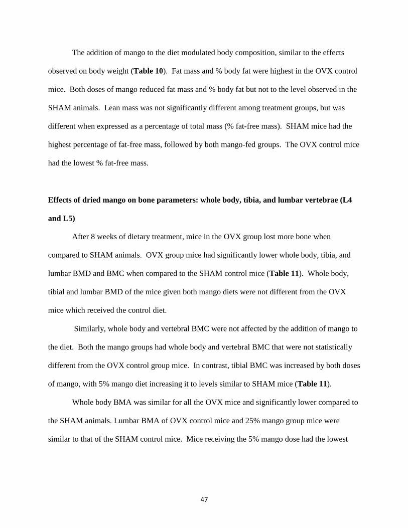
47
The addition of mango to the diet modulated body composition, similar to the effects
observed on body weight (Table 10). Fat mass and % body fat were highest in the OVX control
mice. Both doses of mango reduced fat mass and % body fat but not to the level observed in the
SHAM animals. Lean mass was not significantly different among treatment groups, but was
different when expressed as a percentage of total mass (% fat-free mass). SHAM mice had the
highest percentage of fat-free mass, followed by both mango-fed groups. The OVX control mice
had the lowest % fat-free mass.
Effects of dried mango on bone parameters: whole body, tibia, and lumbar vertebrae (L4
and L5)
After 8 weeks of dietary treatment, mice in the OVX group lost more bone when
compared to SHAM animals. OVX group mice had significantly lower whole body, tibia, and
lumbar BMD and BMC when compared to the SHAM control mice (Table 11). Whole body,
tibial and lumbar BMD of the mice given both mango diets were not different from the OVX
mice which received the control diet.
Similarly, whole body and vertebral BMC were not affected by the addition of mango to
the diet. Both the mango groups had whole body and vertebral BMC that were not statistically
different from the OVX control group mice. In contrast, tibial BMC was increased by both doses
of mango, with 5% mango diet increasing it to levels similar to SHAM mice (Table 11).
Whole body BMA was similar for all the OVX mice and significantly lower compared to
the SHAM animals. Lumbar BMA of OVX control mice and 25% mango group mice were
similar to that of the SHAM control mice. Mice receiving the 5% mango dose had the lowest

48
BMA. The opposite effect was observed with tibial BMA. The mice receiving the mango diets
had higher BMA compared to those receiving the control diet (Table 11).
As expected, OVX animals had inferior microarchitectural parameters of the vertebra
compared to the SHAM mice regardless of dietary treatment (Table 12). OVX mice had lower
vertebral BV/TV, Conn. D., TbN, TbTH, Mean TV and Mean BV than SHAM animals.
Supplementation of mango, at either dose, was not able to restore the deterioration of the micro-
architectural parameters resulting from the ovariectomy.
Finite element analyses demonstrate that ovariectomized mice had lower size
independent stiffness and total force in comparison to the SHAM mice (Table 12). Dietary
treatment had no significant effects on vertebral strength of OVX mice
Effects of dried mango on plasma markers of bone formation and resorption
N-terminal propeptide for type I procollagen (PINP), a serum marker of bone formation,
was not significantly different between any of the groups but tended (P = 0.0510) to be lower in
the mango groups (Table 13). Plasma pyridinoline (PYD), a marker of bone resorption, was
significantly higher in OVX when compared to SHAM mice. PYD values are inversely
proportional to the level of bone resorption. Bone resorption rates were similar between all OVX
groups. Mango was not able to attenuate changes in bone resorption markers due to
ovariectomy.

49
Table 8: Baseline characteristics of ovariectomized (OVX) and SHAM-operated C57/BL6 mice two weeks post-ovariectomy (prior to dietary treatment).
Sham OVX P Value Bone Parameters Whole Body BMD (mg/cm2) 49.8 ± 1.1 48.0 ± 1.3 0.0269 BMC (mg) 539.5 ± 33.6 539.5 ± 32.7 1.0000 BMA (cm2) 10.83 ± .456 11.24 ± .530 0.1798
Tibia BMD (mg/cm2) 44.48 ± 2.26 45.30 ± 2.41 0.5586 BMC (mg) 19.00 ± 0.89 18.83 ± 1.17 0.7872 BMA (cm2) 0.43 ± 0.02 0.42 ± 0.01 0.4160
Lumbar Vertebrae (L4 and L5) BMD (mg/cm2) 49.85 ± 3.23 50.32 ± 4.2 0.8337 BMC (mg) 114.0 ± 21.16 118.83 ± 10.07 0.6243 BMA (cm2) 2.28 ± 0.35 2.36 ± 0.10 0.5921
Tissue Weights Uterus (mg) 133.3 ± 52.8 33.3 ± 5.2 0.0010 Liver (mg) 803.3 ± 43.2 916.7 ± 154.9 0.1150 Spleen (mg) 76.7 ± 10.3 81.7 ± 35.4 0.7470 Thymus (mg) 51.7 ± 9.8 60.0 ± 16.7 0.3177
Body composition Lean mass (g) 14.0 ± 0.51 13.71 ± 0.78 0.4739 Fat mass (g) 4.07 ± 0.16 3.85 ± 0.18 0.0516 Total mass (g) 18.05 ± 0.50 18.22 ± 2.13 0.8559 % Fat 22.45 ± 1.08 21.83 ± 0.68 0.2630 % Fat-free mass 77.54 ± 1.09 76.15 ± 5.01 0.5190
Plasma PINP (ng/ml) 35.39 ± 6.64 32.94 ± 7.27 0.5564 PYD (ng/ml) 1.92 ± 0.65 2.49 ± 0.97 0.2649 Values are mean ± SD (n=6/group), P < 0.05 were considered significant. BMD, bone mineral density; BMC, bone mineral content; BMA, bone mineral area.
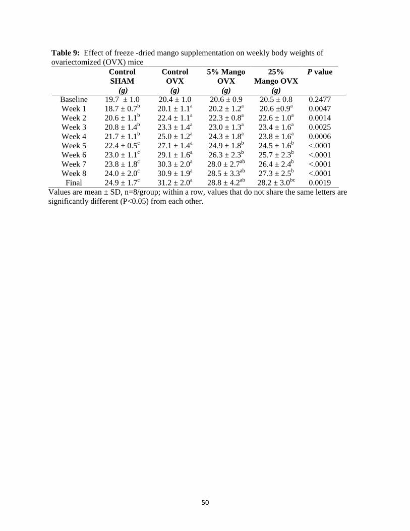
50
Table 9: Effect of freeze -dried mango supplementation on weekly body weights of ovariectomized (OVX) mice
Control SHAM
(g)
Control OVX
(g)
5% Mango OVX
(g)
25% Mango OVX
(g)
P value
Baseline 19.7 ± 1.0 20.4 ± 1.0 20.6 ± 0.9 20.5 ± 0.8 0.2477 Week 1 18.7 ± 0.7b 20.1 ± 1.1a 20.2 ± 1.2a 20.6 ±0.9a 0.0047 Week 2 20.6 ± 1.1b 22.4 ± 1.1a 22.3 ± 0.8a 22.6 ± 1.0a 0.0014 Week 3 20.8 ± 1.4b 23.3 ± 1.4a 23.0 ± 1.3a 23.4 ± 1.6a 0.0025 Week 4 21.7 ± 1.1b 25.0 ± 1.2a 24.3 ± 1.8a 23.8 ± 1.6a 0.0006 Week 5 22.4 ± 0.5c 27.1 ± 1.4a 24.9 ± 1.8b 24.5 ± 1.6b <.0001 Week 6 23.0 ± 1.1c 29.1 ± 1.6a 26.3 ± 2.3b 25.7 ± 2.3b <.0001 Week 7 23.8 ± 1.8c 30.3 ± 2.0a 28.0 ± 2.7ab 26.4 ± 2.4b <.0001 Week 8 24.0 ± 2.0c 30.9 ± 1.9a 28.5 ± 3.3ab 27.3 ± 2.5b <.0001 Final 24.9 ± 1.7c 31.2 ± 2.0a 28.8 ± 4.2ab 28.2 ± 3.0bc 0.0019
Values are mean ± SD, n=8/group; within a row, values that do not share the same letters are significantly different (P<0.05) from each other.
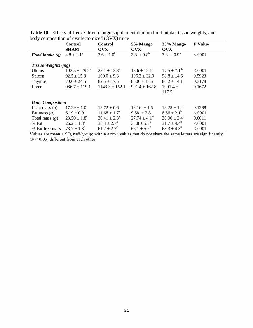
51
Table 10: Effects of freeze-dried mango supplementation on food intake, tissue weights, and body composition of ovariectomized (OVX) mice Control
SHAM Control OVX
5% Mango OVX
25% Mango OVX
P Value
Food intake (g) 4.8 ± 1.1a 3.6 ± 1.0b 3.8 ± 0.8b 3.8 ± 0.9b <.0001 Tissue Weights (mg) Uterus 102.5 ± 29.2a 23.1 ± 12.8b 18.6 ± 12.1b 17.5 ± 7.1 b <.0001 Spleen 92.5 ± 15.8 100.0 ± 9.3 106.2 ± 32.0 98.8 ± 14.6 0.5923 Thymus 70.0 ± 24.5 82.5 ± 17.5 85.0 ± 18.5 86.2 ± 14.1 0.3178 Liver 986.7 ± 119.1 1143.3 ± 162.1 991.4 ± 162.8 1091.4 ±
117.5 0.1672
Body Composition Lean mass (g) 17.29 ± 1.0 18.72 ± 0.6 18.16 ± 1.5 18.25 ± 1.4 0.1288 Fat mass (g) 6.19 ± 0.9c 11.68 ± 1.7a 9.58 ± 2.8b 8.66 ± 2.1b <.0001 Total mass (g) 23.50 ± 1.8c 30.41 ± 2.3a 27.74 ± 4.1ab 26.90 ± 3.4b 0.0011 % Fat 26.2 ± 1.8c 38.3 ± 2.7a 33.8 ± 5.3b 31.7 ± 4.4b <.0001 % Fat free mass 73.7 ± 1.8a 61.7 ± 2.7c 66.1 ± 5.2b 68.3 ± 4.3b <.0001
Values are mean ± SD, n=8/group; within a row, values that do not share the same letters are significantly (P < 0.05) different from each other.
52
Table 11: Effects of freeze-dried mango supplementation on whole body, tibia and lumbar vertebrae (L4 and L5) bone mineral density (BMD), content (BMC), and area (BMA) of ovariectomized (OVX) mice
Control SHAM
Control OVX
5% Mango OVX
25% Mango OVX
P Value
Whole Body BMD (mg/cm2) 53.8 ± 1.5a 47.3 ± 1.0 b 48.0 ± 1.6 b 48.3 ± 1.3 b <.0001 BMC (mg) 564.1 ± 43.0a 414.9 ± 40.0 b 444.9 ± 54.8b 450.4 ± 16.7 b <.0001 Area (cm2) 10.5 ± 0.6a 8.8 ± 0.7 b 9.2 ± 0.9 b 9.33 ± 0.4 b 0.0002
Tibia BMD (mg/cm2) 49.79 ± 1.02a 43.65 ± 1.76b 42.58 ± 1.33b 43.36 ± 0.86b <.0001 BMC (mg) 18.75 ± 1.16a 16.00 ± 1.51c 17.75 ± 1.58ab 17.38 ± 0.92b 0.0029 BMA (cm2) 0.38 ± 0.02b 0.37 ± 0.02b 0.42 ± 0.03a 0.40 ± 0.02a 0.0006
Lumbar Vertebrae (L4 and L5) BMD (mg/cm2) 56.66 ± 4.7a 44.13 ± 2.7b 45.51 ± 3.9b 45.61 ± 2.7b <.0001 BMC (mg) 17.00 ± 1.7a 13.00 ± 1.5b 12.25 ± 1.4b 13.00 ± 1.2b <.0001 BMA (cm2) 0.30 ± 0.02a 0.30 ± 0.02a 0.27 ± 0.02b 0.29 ± 0.02ab 0.0049 Values are mean ± SD, n=8/group; within a row, values that do not share the same letters are significantly different (P < 0.05) from each other.
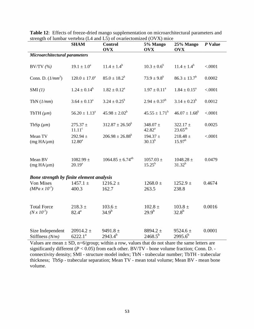
53
Table 12: Effects of freeze-dried mango supplementation on microarchitectural parameters and strength of lumbar vertebra (L4 and L5) of ovariectomized (OVX) mice SHAM Control
OVX 5% Mango OVX
25% Mango OVX
P Value
Microarchitectural parameters BV/TV (%) 19.1 ± 1.0a 11.4 ± 1.4b 10.3 ± 0.6b 11.4 ± 1.4b <.0001
Conn. D. (1/mm3) 120.0 ± 17.0a 85.0 ± 18.2b 73.9 ± 9.8b 86.3 ± 13.7b 0.0002
SMI (1) 1.24 ± 0.14b 1.82 ± 0.12a 1.97 ± 0.11a 1.84 ± 0.15a <.0001
TbN (1/mm) 3.64 ± 0.13a 3.24 ± 0.25b 2.94 ± 0.37b 3.14 ± 0.23b 0.0012
TbTH (µm) 56.20 ± 1.13a 45.98 ± 2.02b 45.55 ± 1.71b 46.07 ± 1.68b <.0001
TbSp (µm) 275.37 ± 11.11c
312.87 ± 26.50b 348.07 ± 42.82a
322.17 ± 23.65ab
0.0025
Mean TV (mg HA/µm)
292.94 ± 12.80a
206.98 ± 26.88b 194.37 ± 30.13b
218.48 ± 15.97b
<.0001
Mean BV (mg HA/µm)
1082.99 ± 20.19a
1064.85 ± 6.74ab 1057.03 ± 15.25b
1048.28 ± 31.32b
0.0479
Bone strength by finite element analysis Von Mises (MPa x 10-3)
1457.1 ± 400.3
1216.2 ± 162.7
1268.0 ± 263.5
1252.9 ± 238.8
0.4674
Total Force (N x 10-3)
218.3 ± 82.4a
103.6 ± 34.9b
102.8 ± 29.9b
103.8 ± 32.8b
0.0016
Size Independent Stiffness (N/m)
20914.2 ± 6222.1a
9491.8 ± 2943.4b
8894.2 ± 2468.5b
9524.6 ± 2995.6b
0.0001
Values are mean ± SD, n=6/group; within a row, values that do not share the same letters are significantly different (P < 0.05) from each other. BV/TV - bone volume fraction; Conn. D. -connectivity density; SMI - structure model index; TbN - trabecular number; TbTH - trabecular thickness; TbSp - trabecular separation; Mean TV - mean total volume; Mean BV - mean bone volume.
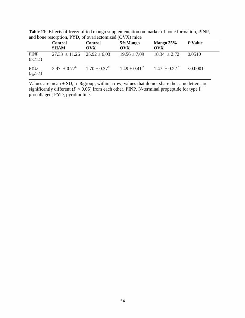
54
Table 13: Effects of freeze-dried mango supplementation on marker of bone formation, PINP, and bone resorption, PYD, of ovariectomized (OVX) mice Control
SHAM Control OVX
5%Mango OVX
Mango 25% OVX
P Value
PINP (ng/mL)
27.33 ± 11.26 25.92 ± 6.03 19.56 ± 7.09 18.34 ± 2.72 0.0510
PYD (ng/mL)
2.97 ± 0.77a 1.70 ± 0.37b 1.49 ± 0.41 b 1.47 ± 0.22 b <0.0001
Values are mean ± SD, n=8/group; within a row, values that do not share the same letters are significantly different (P < 0.05) from each other. PINP, N-terminal propeptide for type I procollagen; PYD, pyridinoline.
55
CHAPTER V
CONCLUSION
The purpose of this study was to determine whether freeze-dried mango pulp powder
supplementation, in 5 and 25% doses, could reverse bone loss in ovariectomized mice. The null
hypothesis was accepted in that mango was not able to restore the ovariectomy induced bone loss
as evidenced by whole body, vertebral, and tibial BMD measurements, micro-architectural
parameters and biomechanical properties of the lumbar vertebrae, and had no effect on serum
markers of bone formation (PINP) or bone resorption (PYD). Although mango was not effective
in reversing ovariectomy- induced bone loss, it did modulate body weight and % body fat.
Although mango has no effect on BMD, some positive effects were observed on BMC.
Whole body and lumbar BMC did not differ from the OVX control diet, but BMC of the tibia
was improved with mango supplementation. The 5% dosage even restored tibial BMC to SHAM
levels. However, when looking at BMC it is important to consider it in relation to BMA. The
higher tibial BMA in the mango groups explains why the BMC is also higher. Although the tibia
may not be the best place to detect ovariectomy induced bone loss in mice, according to Lynch
and colleagues (2010)[165], in human subjects, the tibia is a valuable assessment measurement for
osteoporotic bone loss due to the fact that tibial cortical bone decreases about 5.6–11.0% every
10 years in women while only an average of 5.5% is lost every 10 years at the femoral neck.
56
Even though tibial BMC and BMA of the mango group are higher than controls, it has
been pointed out that BMD accounts for 60-80% of the variations observed in bone strength [3,
196]. It is also important to recognize that the relationship between BMD, BMC and BMA may
be different at different bone sites. For example, a study conducted on 1449 women over 30
years of age by Deng and colleagues in 2002[197], demonstrated that, at the spine, 86.2% of BMD
variation was attributable to BMC and 12.6% to bone area while at the hip, 98.0% of BMD
variation was due to BMC and 1.1% due to bone area.
As expected, micro-architectural parameters in the OVX mice were significantly lower
than all SHAM mice as assessed by micro-computed tomography. MicroCT delivers high
resolution 3D images which directly measure bone micro-architecture with excellent
reproducibility and accuracy [198]. X-ray snapshots at multiple viewing angles are reconstructed
to a 3D image which shows the spatial distribution of the bone at the micrometer voxel size.
Samples were contoured by hand every ten slices with algorithms used to complete the slices in
between with the exclusion of cortical bone. Mango did not exert an influence on the micro-
architectural parameters in the secondary spongiosia (trabecular bone) of the lumbar vertebra.
Bone volume fraction (BV/TV), the ratio of segmented bone volume to the total volume of the
region of interest, was not influenced by mango. Connectivity density, trabecular number,
thickness, mean total volume, and mean bone volume were also not affected by mango. The 5%
mango group mice did exhibit greater trabecular space which is associated with a weaker micro-
architectural arrangement. The structural model index estimates the plate verses rod
characteristics of the trabecular bone [198]. Zero values represent plate arrangement while a value
of 3 represents rods and 4 represents spheres. All OVX mice exhibited higher SMI values than
SHAM mice indicating more rod like arrangements than the SHAM animals. Changes of
57
trabeculae from plate-like to rod-like are known to occur in aging, bone remodeling, and
osteoporosis [199] indicating that the OVX mice were experiencing more bone remodeling than
SHAM animals as evidenced by the SMI values.
Bone strength was also not improved by mango supplementation. Von Mises stress is
calculated by combining stresses in three dimensions, with the result compared to the tensile
strength of the material loaded in one dimension, yielding one numeric measurement[199]. Von
Mises stress did not differ between OVX and SHAM animals. This may be due to the fact that
animal bones do not tend towards fracture and have a high resiliency to pressure despite their age
or hormonal influences [200]. Biomechanical assessment measurements of total force and size
independent stiffness, however, were lower in OVX mice indicating that with estrogen
withdrawal there is a subsequent loss in bone strength. Mango supplementation did not improve
biomechanical properties of the bone when compared to the OVX control diet mice. Using
different evaluation tools may help to further evaluate the effect of mango on bone strength. For
example, quantitative ultrasound measurements can also reflect bone elasticity, fragility and
density closely associated with bone strength and fracture risk [201].
Although many biological compounds may assist in preventing bone loss, few agents are
successful at reversing the bone loss associated with osteoporosis. Fruits and vegetables or their
bioactive compounds may be effective in alleviating the inflammatory environment associated
with the postmenopausal unregulated production of pro-inflammatory cytokines and may also
exert a protective antioxidant effect on the bone tissue while supplying important vitamins and
minerals necessary for reversing bone loss. Although the OC secretes ROS under normal bone
remodeling conditions to assist with the destruction of calcified bone tissue, in a postmenopausal
low estrogen state, the increased ROS production with the increase in bone resorption may
58
overwhelm the bodies endogenous antioxidant system and lead to osteoporosis [202, 203] .
Sheweita and Khoshhal suggested that the enhanced OC activity may increase the superoxide
anion generation and/or inhibit superoxide dismutase and glutathione peroxidase activities[112].
Mango possesses antioxidants, such as the carotenoids, flavonoids, triterpenes, phenolic
compounds and vitamin C, which, in theory, may help attenuate the inflammatory environment
associated with osteoporosis.
Some studies have demonstrated the effectiveness of bioactive food compounds in
improving bone quality or preventing bone loss but few have shown bone loss reversal. For
example, citrus pulp, containing high amounts of flavonoids and vitamin C, improved BMD in
male orchidectomized rats [14, 15]. Pholridzin in apples, rutin, and isoflavones have all prevented
further bone loss in ovariectomized rats [20]. Green tea polyphenols reduced bone micro-
architecture deterioration in female rats [116] and blueberries prevented whole body BMD loss in
OVX rats[167]. To our knowledge, only dried plum has been demonstrated to effectively restore
bone that has been lost [17]. Dried plum polyphenols not only inhibited osteoclastogenesis by
down regulating NFATc1 and inflammatory mediators such as TNF-α, they also upregulated OB
transcription factors such as Runx2, Osterix and IGF-1 in MC3T3-E1 cells [16]. In another study
involving dried plum in ovariectomized rats, trabecular micro-architectural parameters were
improved as well as femoral and tibial BMD by the 5% dose while lumbar BMD was improved
at a 25% dose [17].
The pro-vitamin A compound, β-carotene, found in mango may also play a role in OB
cell differentiation. Research done by Park and colleagues on mouse osteoblastic cells
demonstrated that vitamin A and carotenoids increased OB cell differentiation[204] while another
cell culture study showed an inhibited OC like cell formation[205]. In a study involving 334 men

59
and 540 women over the age of 75, a positive correlation between carotenoid consumption and
BMD [18] and a negative correlation with carotenoid consumption and hip fracture rates was
observed [206]. A study conducted by Yuyama and colleagues determined that the pro vitamin A
carotenoids in mango was absorbed with 82% efficiency in rats[207]. Drammeh and colleagues
used dried mango powder along with fat supplementation to successfully improve the vitamin A
status in young Gambian children[208]. These findings suggest that pro vitamin A content of the
mango in the present study may influence bone. The present study, however, demonstrated that
mango, despite being a rich source of antioxidants and β-carotene, was not able to reverse bone
loss.
As expected, the OVX mice experienced more bone resorption than the SHAM control
mice. Mango did not attenuate bone resorption as evidenced by serum PYD values that were
similar between the mango groups and the OVX control mice. Despite PINP being one of the
most specific and sensitive indicators of bone formation[209], serum PINP levels were not
significantly different between any of the groups. The trend (P = 0.0510) was that SHAM mice
had the highest PINP levels with 25% mango group exhibiting the lowest values. Considering
this trend along with the fact that the mango group mice exhibited a lower body fat body
composition, one may want to consider the influence of the metabolically active adipose tissue
on bone parameters.
The higher body fat composition in the OVX mice may exhibit a greater hormonal effect
contributing to increased rates of bone formation. For example, earlier studies have
demonstrated that leptin, secreted by the adipose tissue, stimulates cortical bone formation[74, 75]
via its effects on the GH-IGF-1 axis of the central nervous system and by suppressing the release
of neuropeptide Y, an inhibitor of cortical bone formation, from the hypothalamus. Leptin’s
60
combined effects of neuropeptide Y suppression and the β1- adrenergic stimulation result in an
increase in cortical bone mass [74]. Adiponectin, also secreted by adipose tissue, is known to
modulate glucose and fatty acid catabolism [210] with levels inversely associated with percent
body fat [211]. In a study conducted on postmenopausal women, increasing adiponectin levels
were associated with a decrease in BMD. The percent change in BMD was associated with a
doubling in serum adiponectin levels [212]. A study conducted by Richards and colleagues
showed that serum adiponectin levels were associated with a 2.7% average decrease in BMD at
the hip, femoral neck, forearm and spine[213, 214]. Post-menopausal status increased the
magnitude of this inverse relationship. The fact that the decrease in BMD was also evident at
non-weight bearing sites indicates an important role of adipose hormones in regulating BMD.
Future implications of this research may want to consider the effects of adipose hormones on
BMD.
Although mango fed for 8 weeks was not effective in reversing bone loss, mango
modulated body weight and body composition. Mango attenuated weight gain and percent body
fat. After four weeks of dietary treatment, both mango diet groups weighed less than the OVX
control group. Final body weight of the 25% mango diet group was similar to both the 5%
mango group and that of the SHAM mice. Similarly, fat mass and % body fat of both mango
diet mice groups were less than that of the OVX control mice despite similar food intake patterns
between all OVX groups. Following suit, both mango containing diets had more fat free mass
than the OVX control mice. Although the fiber content of the mango and control groups were
matched, mango may have exerted an effect on digestion or absorption of the diet either by
inhibiting fat absorption to a degree or by increasing the metabolism. Stool assessments or direct
calorimetry may be ways to assess these possibilities in the future studies. Further investigation
61
on mango and bone biology and mechanisms in modulating body composition in
postmenopausal women are warranted.
The results of our study failed to reject our null hypotheses. Freeze-dried mango powder
in 5% and 25% of the daily diet was not effective in reversing ovariectomy induced bone loss in
mice. Mango did not improve whole body, tibial or lumbar vertebral BMD , nor did it improve
micro-architectural parameters or bone strength in the vertebrae. Plasma markers of bone
formation (PINP) and bone resorption (PYD) also were not affected by mango supplementation.
Freeze-dried mango powder supplementation in both doses did attenuate weight gain and
modulate body composition. Although mango was not able to reverse the bone lose associated
with gonadal hormone difficiency, its role in preventing osteoporosis still needs to be explored.

References
1. National Osteoporosis Foundation, The Health Health of our Nation, The State of Osteoporosis and Low Bone Mass in Our Nation.2003
2. Klibanski, A., Adams-Campbell, L., Bassford, T., Blair, S.N., Boden, S.D., Dickersin, K., Gifford, D.R., Glasse, L., Goldring, S.R., Hruska, K., Johnson, S.R., McCauley, L.K., Russell, W.E., and Osteopor, N.I.H.C.D.P., Osteoporosis prevention, diagnosis, and therapy. Jama-Journal of the American Medical Association, 2001. 285(6): p. 785-795.
3. Riggs, B.L. and Melton, L.J., The worldwide problem of osteoporosis - insights afforded by epidemiology. Bone, 1995. 17(5): p. S505-S511.
4. Mundy, G.R., Osteoporosis and inflammation. Nutrition Reviews, 2007. 65(12): p. S147-S151.
5. Silverberg, S.J. and Lindsay, R., Postmenopausal osteoporosis. Medical Clinics of North America, 1987. 71(1): p. 41-57.
6. Ralston, S.H., Analysis of gene-expression in human bone biopsies by polymerase chain-reaction - evidence for enhanced cytokine expression in postmenopausal osteoporosis. Journal of Bone and Mineral Research, 1994. 9(6): p. 883-890.
7. Pacifici, R., Cytokines, estrogen, and postmenopausal osteoporosis - The second decade. Endocrinology, 1998. 139(6): p. 2659-2661.
8. Diseases, N.I.o.H.O.a.R.B. (2003) What people with lactose intolerance need to know about osteoporosis:.
9. Fleisch, H., Bisphosphonates: mechanisms of action. Endocrine Reviews, 1998. 19(1): p. 80-100.
10. FDA Drug Safety Medication Guide, EVISTA (raloxifene hydrochloride), in BNL 3123 AMP. 2007, Eli Lilly and Company.
11. Lyritis, G.P., Georgoulas, T., and Zafeiris, C.P., Bone anabolic versus bone anticatabolic treatment of postmenopausal osteoporosis, in Women's Health and Disease, G. Creatsas and G. Mastorakos, Editors. 2010. p. 277-283.
12. Macdonald, H.M., New, S.A., Golden, M.H., Campbell, M.K., and Reid, D.M., Nutritional associations with bone loss during the menopausal transition: evidence of a beneficial effect of calcium, alcohol, and fruit and vegetable
63
nutrients and of a detrimental effect of fatty acids. American Journal of Clinical Nutrition, 2004. 79(1): p. 155-165.
13. Tucker, K.L., Hannan, M.T., Chen, H.L., Cupples, L.A., Wilson, P.W.F., and Kiel, D.P., Potassium, magnesium, and fruit and vegetable intakes are associated with greater bone mineral density in elderly men and women. American Journal of Clinical Nutrition, 1999. 69(4): p. 727-736.
14. Morrow, R., Deyhim, F., Patil, B.S., and Stoecker, B.J., Feeding orange pulp improved bone quality in a rat model of male osteoporosis. Journal of Medicinal Food, 2009. 12(2): p. 298-303.
15. Mandadi, K., Ramirez, M., Jayaprakasha, G.K., Faraji, B., Lihono, M., Deyhim, F., and Patil, B.S., Citrus bioactive compounds improve bone quality and plasma antioxidant activity in orchidectomized rats. Phytomedicine, 2009. 16(6-7): p. 513-520.
16. Bu, S.Y., Hunt, T.S., and Smith, B.J., Dried plum polyphenols attenuate the detrimental effects of TNF-alpha on osteoblast function coincident with up-regulation of Runx2, Osterix and IGF-I. Journal of Nutritional Biochemistry, 2009. 20(1): p. 35-44.
17. Deyhim, F., Stoecker, B.J., Brusewitz, G.H., Devareddy, L., and Arjmandi, B.H., Dried plum reverses bone loss in an osteopenic rat model of osteoporosis. Menopause-the Journal of the North American Menopause Society, 2005. 12(6): p. 755-762.
18. Sahni, S., Hannan, M.T., Blumberg, J., Cupples, L.A., Kiel, D.P., and Tucker, K.L., Inverse association of carotenoid intakes with 4-y change in bone mineral density in elderly men and women: the Framingham Osteoporosis Study. American Journal of Clinical Nutrition, 2009. 89(1): p. 416-424.
19. Parr, A.J. and Bolwell, G.P., Phenols in the plant and in man. The potential for possible nutritional enhancement of the diet by modifying the phenols content or profile. Journal of the Science of Food and Agriculture, 2000. 80(7): p. 985-1012.
20. Puel, C., Quintin, A., Mathey, J., Obled, C., Davicco, M.J., Lebecque, P., Kati-Coulibaly, S., Horcajada, M.N., and Coxam, V., Prevention of bone loss by phloridzin, an apple polyphenol, in ovariectomized rats under inflammation conditions. Calcified Tissue International, 2005. 77(5): p. 311-318.
21. Wachman, A. and Bernstei.Ds, Diet and osteoporosis. Lancet, 1968. 1(7549): p. 958-62.
64
22. Remer, T. and Manz, F., Potential renal acid load of foods and its influence on urine ph. Journal of the American Dietetic Association, 1995. 95(7): p. 791-797.
23. Sebastian, A., Harris, S.T., Ottaway, J.H., Todd, K.M., and Morris, R.C., Improved mineral balance and skeletal metabolism in postmenopausal women treated with potassium bicarbonate. New England Journal of Medicine, 1994. 330(25): p. 1776-1781.
24. Lean, J.M., Davies, J.T., Fuller, K., Jagger, C.J., Kirstein, B., Partington, G.A., Urry, Z.L., and Chambers, T.J., A crucial role for thiol antioxidants in estrogen-deficiency bone loss. The Journal of Clinical Investigation, 2003. 112(6): p. 915-923.
25. Liu, R.H., Health benefits of fruit and vegetables are from additive and synergistic combinations of phytochemicals. American Journal of Clinical Nutrition, 2003. 78(3): p. 517S-520S.
26. Manolagas, S.C., Kousteni, S., and Jilka, R.L., Sex steroids and bone. Recent Progress in Hormone Research, Vol 57, 2002. 57: p. 385-409.
27. Maggio, D., Barabani, M., Pierandrei, M., Polidori, M.C., Catani, M., Mecocci, P., Senin, U., Pacifici, R., and Cherubini, A., Marked decrease in plasma antioxidants in aged osteoporotic women: Results of a cross-sectional study. Journal of Clinical Endocrinology & Metabolism, 2003. 88(4): p. 1523-1527.
28. Base, U.D.o.A.N.D. (2009) National Nutrient Database for Standard Reference. US Department of Agriculture Nutrient Data Base.
29. Odabasi, E., Turan, M., Aydin, A., Akay, C., and Kutlu, M., Magnesium, zinc, copper, manganese, and selenium levels in postmenopausal women with osteoporosis. Can magnesium play a key role in osteoporosis? Annals Academy of Medicine Singapore, 2008. 37(7): p. 564-567.
30. Burge, R., Dawson-Hughes, B., Solomon, D.H., Wong, J.B., King, A., and Tosteson, A., Incidence and economic burden of osteoporosis-related fractures in the United States, 2005-2025. Journal of Bone and Mineral Research, 2007. 22(3): p. 465-475.
31. Ahlborg, H.G., Rosengren, B.E., Jarvinen, T.L.N., Rogmark, C., Nilsson, J.A., Sernbo, I., and Karlsson, M.K., Prevalence of osteoporosis and incidence of hip fracture in women - secular trends over 30 years. Bmc Musculoskeletal Disorders, 2010. 11.
65
32. Melton, L.J., Atkinson, E.J., O'Connor, M.K., O'Fallon, W.M., and Riggs, B.L., Bone density and fracture risk in men. Journal of Bone and Mineral Research, 1998. 13(12): p. 1915-1923.
33. Lauderdale, D.S., Jacobsen, S.J., Fumer, S.E., Levy, P.S., Brody, J.A., and Goldberg, J., Hip fracture incidence among elderly Asian-American populations. American Journal of Epidemiology, 1997. 146(6): p. 502-509.
34. Cauley, J.A., Lui, L.Y., Stone, K.L., Hillier, T.A., Zmuda, J.M., Hochberg, M., Beck, T.J., and Ensrud, K.E., Longitudinal study of changes in hip bone mineral density in Caucasian and African-American women. Journal of the American Geriatrics Society, 2005. 53(2): p. 183-189.
35. Bekker, P.J., Holloway, D.L., Rasmussen, A.S., Murphy, R., Martin, S.W., Leese, P.T., Holmes, G.B., Dunstan, C.R., and DePaoli, A.M., A single-dose placebo-controlled study of AMG 162, a fully human monoclonal antibody to RANKL, in postmenopausal women. Journal of Bone and Mineral Research, 2004. 19(7): p. 1059-1066.
36. Ekrylander, B., Flores, M., Wendel, M., Heinegard, D., and Andersson, G., Dephosphorylation of osteopontin and bone sialoprotein by osteoclastic tartrate-resistant acid-phosphatase - modulation of osteoclast adhesion in-vitro. Journal of Biological Chemistry, 1994. 269(21): p. 14853-14856.
37. Raggatt, L.J. and Partridge, N.C., Cellular and molecular mechanisms of bone remodeling. Journal of Biological Chemistry, 2010. 285(33): p. 25103-25108.
38. Anderson, Matrix vesicls and calcification. Current rheumatology reports, 2003. 5: p. 222-226.
39. Aubin, L., Stein, Primer on the Metabolic Bone Disease and Disorders of Mineral Metabolism. 6 ed. Bone Formation: Maturation and Functional Activities of Osteoblast Lineage Cells. 2006, American Society for Bone and Mineral Research: Washington, D.C.
40. Franz-Odendaal, T.A., Hall, B.K., and Witten, P.E., Buried alive: How osteoblasts become osteocytes. Developmental Dynamics, 2006. 235(1): p. 176-190.
41. Logan, C.Y. and Nusse, R., The Wnt signaling pathway in development and disease. Annual Review of Cell and Developmental Biology, 2004. 20: p. 781-810.
66
42. Stains, J.P. and Civitelli, R., Cell-to-cell interactions in bone. Biochemical and Biophysical Research Communications, 2005. 328(3): p. 721-727.
43. Walker, D.G., Control of bone-resorption by hematopoietic tissue - induction and reversal of congenital osteopetrosis in mice through use of bone-marrow and splenic transplants. Journal of Experimental Medicine, 1975. 142(3): p. 651-663.
44. Martin, T.J. and Sims, N.A., Osteoclast-derived activity in the coupling of bone formation to resorption. Trends in Molecular Medicine, 2005. 11(2): p. 76-81.
45. Karsenty, G. and Wagner, E.F., Reaching a genetic and molecular understanding of skeletal development. Developmental Cell, 2002. 2(4): p. 389-406.
46. Boyle, W.J., Simonet, W.S., and Lacey, D.L., Osteoclast differentiation and activation. Nature, 2003. 423(6937): p. 337-342.
47. Wada, T., Nakashima, T., Hiroshi, N., and Penninger, J.M., RANKL-RANK signaling in osteoclastogenesis and bone disease. Trends in Molecular Medicine, 2006. 12(1): p. 17-25.
48. Blair, H.C. and Zaidi, M., Osteoclastic differentiation and function regulated by old and new pathways. Reviews in Endocrine & Metabolic Disorders, 2006. 7(1-2): p. 23-32.
49. Lam, K.W., Townsend, D., Garza, A., Li, C.Y., and Yam, L.T., Comparison of tartrate resistant acid-phosphatase in a giant-cell bone-tumor and a spleen infiltrated with hairy-cells. Clinical Biochemistry, 1990. 23(4): p. 335-340.
50. Boyce, B.F. and Xing, L.P., Biology of RANK, RANKL, and osteoprotegerin. Arthritis Research & Therapy, 2007. 9.
51. Scheven, B.A.A., Visser, J.W.M., and Nijweide, P.J., Invitro osteoclast generation from different bone-marrow fractions, including a highly enriched hematopoietic stem-cell population. Nature, 1986. 321(6065): p. 79-81.
52. Medhora, M.M., Teitelbaum, S., Chappel, J., Alvarez, J., Mimura, H., Ross, F.P., and Hruska, K., 1-alpha,25-dihydroxyvitamin-d3 up-regulates expression of the osteoclast integrin alpha-v-beta-3. Journal of Biological Chemistry, 1993. 268(2): p. 1456-1461.
53. Simonet, W.S., Lacey, D.L., Dunstan, C.R., Kelley, M., Chang, M.S., Luthy, R., Nguyen, H.Q., Wooden, S., Bennett, L., Boone, T., Shimamoto, G., DeRose, M., Elliott, R., Colombero, A., Tan, H.L., Trail, G., Sullivan, J., Davy, E., Bucay, N.,
67
RenshawGegg, L., Hughes, T.M., Hill, D., Pattison, W., Campbell, P., Sander, S., Van, G., Tarpley, J., Derby, P., Lee, R., and Boyle, W.J., Osteoprotegerin: A novel secreted protein involved in the regulation of bone density. Cell, 1997. 89(2): p. 309-319.
54. Boyce, B.F., Xing, L.P., and Chen, D., Osteoprotegerin, the bone protector, is a surprising target for beta-catenin signaling. Cell Metabolism, 2005. 2(6): p. 344-345.
55. Kim, M.S., Yang, Y.M., Son, A., Tian, Y.S., Lee, S.I., Kang, S.W., Muallem, S., and Shin, D.M., RANKL-mediated reactive oxygen species pathway that induces long lasting Ca2+ oscillations essential for osteoclastogenesis. Journal of Biological Chemistry, 2010. 285(10): p. 6913-6921.
56. Compston, J.E., Sex steroids and bone. Physiological Reviews, 2001. 81(1): p. 419-447.
57. Eisman, J.A., Genetics of osteoporosis. Endocrine Reviews, 1999. 20(6): p. 788-804.
58. Gullberg, B., Johnell, O., and Kanis, J.A., World-wide projections for hip fracture. Osteoporosis International, 1997. 7(5): p. 407-413.
59. McKay, H.A., Bailey, D.A., Wilkinson, A.A., and Houston, C.S., Familial comparison of bone-mineral density at the proximal femur and lumbar spine. Bone and Mineral, 1994. 24(2): p. 95-107.
60. Seeman, E., Tsalamandris, C., Formica, C., Hopper, J.L., and McKay, J., Reduced femoral-neck bone-density in the daughters of women with hip-fractures - the role of low peak bone-density in the pathogenesis of osteoporosis. Journal of Bone and Mineral Research, 1994. 9(5): p. 739-743.
61. Soroko, S.B., Barrettconnor, E., Edelstein, S.L., and Kritzsilverstein, D., Family history of osteoporosis and bone-mineral density at the axial skeleton - the rancho-bernardo study. Journal of Bone and Mineral Research, 1994. 9(6): p. 761-769.
62. Jouanny, P., Guillemin, F., Kuntz, C., Jeandel, C., and Pourel, J., Environmental and genetic-factors affecting bone mass - similarity of bone-density among members of healthy families. Arthritis and Rheumatism, 1995. 38(1): p. 61-67.
63. Krall, E.A. and Dawsonhughes, B., Soft-tissue body-composition - familial resemblance and independent influences on bone-mineral density. Journal of
68
Bone and Mineral Research, 1995. 10(12): p. 1944-1950.
64. Compston, Sex steroids and bone. PHysiological Review, 2001(81): p. 419-447.
65. Oursler, M.J., Landers, J.P., Riggs, B.L., and Spelsberg, T.C., Estrogen effects on osteoblasts and osteoclasts. Annals of Medicine, 1993. 25(4): p. 361-371.
66. Huang, W.H., Lau, A.T.T., Daniels, L.L., Fujii, H., Seydel, U., Wood, D.J., Papadimitriou, J.M., and Zheng, M.H., Detection of estrogen receptor alpha, carbonic anhydrase II and tartrate-resistant acid phosphatase mRNAs in putative mononuclear osteoclast precursor cells of neonatal rats by fluorescence in situ hybridization. Journal of Molecular Endocrinology, 1998. 20(2): p. 211-219.
67. Itoh, K., Shinki, T., Udagawa, N., Ueno, Y., Matsuzaki, K., Takahashi, N., and Suda, T., The mechanism of osteoclastogenesis induced by PTH. Journal of Bone and Mineral Research, 1999. 14: p. SU205.
68. Weaver, ed. Present Knowledge in Nutrition. 9th ed., ed. B. Bowman, Russel, R. Vol. 1. 2006, International Life Sciences Institute: Washington DC. 526.
69. Cornish, J., Callon, K.E., Bava, U., Kamona, S.A., Cooper, G.J.S., and Reid, I.R., Effects of calcitonin, amylin, and calcitonin gene-related peptide on osteoclast development. Bone, 2001. 29(2): p. 162-168.
70. Deftos, L.J., Hypercalcemia in malignant and inflammatory diseases. Endocrinology and Metabolism Clinics of North America, 2002. 31(1): p. 141-+.
71. Favus, M., Bushinsky, DA, Lemann, J, ed. Primer on the Metabolic Bone Diseases and Disorder sof Mineral Metabolism. 6th ed. 2006, American Society for Bone and Mineral Research: Washington, D.C. 503.
72. Ferron, M., Wei, J.W., Yoshizawa, T., Del Fattore, A., DePinho, R.A., Teti, A., Ducy, P., and Karsenty, G., Insulin signaling in osteoblasts integrates bone remodeling and energy metabolism. Cell, 2010. 142(2): p. 296-308.
73. Ducy, P., Amling, M., Takeda, S., Priemel, M., Schilling, A.F., Beil, F.T., Shen, J.H., Vinson, C., Rueger, J.M., and Karsenty, G., Leptin inhibits bone formation through a hypothalamic relay: A central control of bone mass. Cell, 2000. 100(2): p. 197-207.
74. Iwaniec, U.T., Boghossian, S., Lapke, P.D., and Turner, R.T., Central leptin gene therapy corrects skeletal abnormalities in leptin-deficient ob/ob mice. Peptides, 2007. 28(5): p. 1012-1019.
69
75. Hamrick, M.W. and Ferrari, S.L., Leptin and the sympathetic connection of fat to bone. Osteoporosis International, 2008. 19(7): p. 905-912.
76. Boyce, B.F., Aufdemorte, T.B., Garrett, I.R., Yates, A.J.P., and Mundy, G.R., Effects of interleukin-1 on bone turnover in normal mice. Endocrinology, 1989. 125(3): p. 1142-1150.
77. Horowitz, M., Vignery, A., Gershon, R.K., and Baron, R., Thymus-derived lymphocytes and their interactions with macrophages are required for the production of osteoclast-activating factor in the mouse. Proceedings of the National Academy of Sciences of the United States of America-Biological Sciences, 1984. 81(7): p. 2181-2185.
78. Pacifici, R., Editorial: Cytokines, Estrogen, and Postmenopausal Osteoporosis--The Second Decade. Endocrinology, 1998. 139(6): p. 2659-2661.
79. Bell, J. and Whiting, S.J., Elderly women need dietary protein to maintain bone mass. Nutrition Reviews, 2002. 60(10): p. 337-341.
80. Dawson-Hughes, B., Interaction of dietary calcium and protein in bone health in humans. Journal of Nutrition, 2003. 133(3): p. 852S-854S.
81. Pedone, C., Napoli, N., Pozzilli, P., Lauretani, F., Bandinelli, S., Ferrucci, L., and Antonelli-Incalzi, R., Quality of diet and potential renal acid load as risk factors for reduced bone density in elderly women. Bone, 2010. 46(4): p. 1063-1067.
82. Remer, T., Shi, L., and Alexy, U., Potential renal acid load may more strongly affect bone size and mass than volumetric bone mineral density. Bone. In Press, Corrected Proof.
83. Buclin, T., Cosma, M., Appenzeller, M., Jacquet, A.F., Decosterd, L.A., Biollaz, J., and Burckhardt, P., Diet acids and alkalis influence calcium retention in bone. Osteoporosis International, 2001. 12(6): p. 493-499.
84. Barzel, U.S., The skeleton as an ion-exchange system - implications for the role of acid-base imbalance in the genesis of osteoporosis. Journal of Bone and Mineral Research, 1995. 10(10): p. 1431-1436.
85. Palacios, C., The role of nutrients in bone health, from A to Z. Critical Reviews in Food Science and Nutrition, 2006. 46(8): p. 621-628.
86. Prentice, A., Schoenmakers, I., Laskey, M.A., de Bono, S., Ginty, F., and Goldberg, G.R., Nutrition and bone growth and development. Proceedings of the

70
Nutrition Society, 2006. 65(4): p. 348-360.
87. Saltman, P.D. and Strause, L.G., The role of trace minerals in osteoporosis. Journal of the American College of Nutrition, 1993. 12(4): p. 384-389.
88. Jugdaohsingh, R., Silicon and bone health. Journal of Nutrition Health & Aging, 2007. 11(2): p. 99-110.
89. Koshihara, Y. and Hoshi, K., Vitamin K-2 enhances osteocalcin accumulation in the extracellular matrix of human osteoblasts in vitro. Journal of Bone and Mineral Research, 1997. 12(3): p. 431-438.
90. Yamaguchi, M., Sugimoto, E., and Hachiya, S., Stimulatory effect of menaquinone-7 (vitamim K-2) on osteoblastic bone formation in vitro. Molecular and Cellular Biochemistry, 2001. 223(1-2): p. 131-137.
91. Yamaguchi, M. and Ma, Z.J., Inhibitory effect of menaquinone-7 (vitamin K-2) on osteoclast-like cell formation and osteoclastic bone resorption in rat bone tissues in vitro. Molecular and Cellular Biochemistry, 2001. 228(1-2): p. 39-47.
92. Cumming, R.G. and Nevitt, M.C., Calcium for prevention of osteoporotic fractures in postmenopausal women. Journal of Bone and Mineral Research, 1997. 12(9): p. 1321-1329.
93. Shea, B., Wells, G., Cranney, A., Zytaruk, N., Robinson, V., Griffith, L., Ortiz, Z., Peterson, J., Adachi, J., Tugwell, P., Guyatt, G., and Osteoporosis Methodol Grp; Osteoporosis Res Advisory, G., Meta-analysis of calcium supplementation for the prevention of postmenopausal osteoporosis. Endocrine Reviews, 2002. 23(4): p. 552-559.
94. Brown, E.M., Physiology and pathophysiology of the extracellular calcium-sensing receptor. American Journal of Medicine, 1999. 106(2): p. 238-253.
95. orthoteers.com, Bone Metabolism, B. Metabolism, Editor. 1998.
96. Bryant, C., Weaver, The new dietary reference intakes for calcium: implications for osteoporosis. Journal of the American College of Nutrition, 1999. 18(5): p. 406S-412S.
97. Devine, A., Criddle, R.A., Dick, I.M., Kerr, D.A., and Prince, R.L., A Longitudinal-study of the effect of sodium and calcium intakes on regional bone-density in postmenopausal women. American Journal of Clinical Nutrition, 1995. 62(4): p. 740-745.
71
98. Kerstetter, J.E., O'Brien, K.O., Caseria, D.M., Wall, D.E., and Insogna, K.L., The impact of dietary protein on calcium absorption and kinetic measures of bone turnover in women. Journal of Clinical Endocrinology & Metabolism, 2005. 90(1): p. 26-31.
99. Rhodes , WebMD. Information and Resources; Calcium in Urine, 2008.
100. Kato, A., Seo, E.G., Einhorn, T.A., Bishop, J.E., and Norman, A.W., Studies on 24R,25-dihydroxyvitamin D-3: Evidence for a nonnuclear membrane receptor in the chick tibial fracture-healing callus. Bone, 1998. 23(2): p. 141-146.
101. Norman, A.W., Okamura, W.H., Farachcarson, M.C., Allewaert, K., Branisteanu, D., Nemere, I., Muralidharan, K.R., and Bouillon, R., Structure-function studies of 1,25-dihydroxyvitamin-D3 and the vitamin-D endocrine system - 1,25-dihydroxy-pentadeuterio-previtamin-D3 (as a 6-s-cis analog) stimulates nongenomic but not genomic biological responses. Journal of Biological Chemistry, 1993. 268(19): p. 13811-13819.
102. Willett, A.M., Vitamin D status and its relationship with parathyroid hormone and bone mineral status in older adolescents. Proceedings of the Nutrition Society, 2005. 64(2): p. 193-203.
103. Rosen, C.J., Postmenopausal osteoporosis. New England Journal of Medicine, 2005. 353(6): p. 595-603.
104. Kipp, D.E., McElvain, M., Kimmel, D.B., Akhter, M.P., Robinson, R.G., and Lukert, B.P., Scurvy results in decreased collagen synthesis and bone density in the guinea pig animal model. Bone, 1996. 18(3): p. 281-288.
105. Morton, D.J., Barrett-Connor, E.L., and Schneider, D.L., Vitamin C supplement use and bone mineral density in postmenopausal women. Journal of Bone and Mineral Research, 2001. 16(1): p. 135-140.
106. Lucker, M., Secondary Metabolism in Microorganisms, Plants, and Animals. 3rd Edition ed. 1990, Springer: Berlin.
107. Makela, S., Poutanen, M., Kostian, M., Lehtimaki, N., Strauss, L., Santti, P., and Vihko, R., Inhibition of 17 β – hydroxysteroid oxidoreductase by flavonoids in breast and prostrate cancer cells. Proc Soc Exp Bio Med, 1998. 217: p. 310-316.
108. Ebrahimof, S., Adibi, H, Salehomom, N, Hosseinni, S, Larijani B, Fruit and Vegetable intake and bone mineral density in residents of villages surrounding Tehran. Iranian Journal of Public Health, 2004: p. 49-56.
72
109. Moayyeri, A., Ahmadi-Abhari, S., Hossein-Nezhad, A., Larijani, B., and Soltani, A., Bone mineral density and estimated height loss based on patients' recalls. Osteoporosis International, 2006. 17(6): p. 834-840.
110. Sanchez-Rodriguez, M.A., Ruiz-Ramos, M., Correa-Munoz, E., and Mendoza-Nunez, V.M., Oxidative stress as a risk factor for osteoporosis in elderly Mexicans as characterized by antioxidant enzymes. Bmc Musculoskeletal Disorders, 2007. 8.
111. Altindag, O., Erel, O., Soran, N., Celik, H., and Selek, S., Total oxidative/anti-oxidative status and relation to bone mineral density in osteoporosis. Rheumatology International, 2008. 28(4): p. 317-321.
112. Sheweita, S.A. and Khoshhal, K.I., Calcium metabolism and oxidative stress in bone fractures: Role of antioxidants. Current Drug Metabolism, 2007. 8(5): p. 519-525.
113. Yang, S., Madyastha, P., Bingel, S., Ries, W., and Key, L., A new superoxide-generating oxidase in murine osteoclasts. Journal of Biological Chemistry, 2001. 276(8): p. 5452-5458.
114. Helmersson, J. and Basu, S., F-2-isoprostane and prostaglandin F-2 alpha metabolite excretion rate and day to day variation in healthy humans. Prostaglandins Leukotrienes and Essential Fatty Acids, 2001. 65(2): p. 99-102.
115. Basu, S., Bioactive eicosanoids: Role of prostaglandin F-2 alpha and F-2-isoprostanes in inflammation and oxidative stress related pathology. Molecules and Cells, 2010. 30(5): p. 383-391.
116. Shen, C.L., Yeh, J.K., Cao, J.J., Tatum, O.L., Dagda, R.Y., and Wang, J.S., Green tea polyphenols mitigate bone loss of female rats in a chronic inflammation-induced bone loss model. Journal of Nutritional Biochemistry, 2010. 21(10): p. 968-974.
117. INRA (2007) Prevention of osteoporosis: the role of polyphenols and phytoestrogens. French National Institute for Agriculture and Research.
118. Wattel, A., Kamel, S., Mentaverri, R., Lorget, F., Prouillet, C., Petit, J.P., Fardelonne, P., and Brazier, M., Potent inhibitory effect of naturally occurring flavonoids quercetin and kaempferol on in vitro osteoclastic bone resorption. Biochemical Pharmacology, 2003. 65(1): p. 35-42.
119. Smith, B.J., Cullen, D.M., Bu, S.Y., Clarke, S.L., Rendina, E., Lim, Y.F., and
73
Lucas, E.A., Dried plum alters bone metabolism via a different mechanism than PTH. Journal of Bone and Mineral Research, 2008. 23: p. S459-S459.
120. Bu, S.Y., Lerner, M., Stoecker, B.J., Boldrin, E., Brackett, D.J., Lucas, E.A., and Smith, B.J., Dried plum polyphenols inhibit osteoclastogenesis by downregulating NFATc1 and inflammatory mediators. Calcified Tissue International, 2008. 82(6): p. 475-488.
121. Kim HJ, C.E., Kim HM, Lee SB, Kim HD, Su Kim G,, Antioxidant alpha-lipoic acid inhibits osteoclast differentiation by reducing nuclear factor-kappaB DNA binding and prevents in vivo bone resorption induced by receptor activator of nuclear factor-kappaB ligand and tumor necrosis factor-alpha. Free Radical Bioloical Medicine, 2006. 40: p. 1483–93.
122. Rao LG, M.E., Josse RG, Murray TM, Strauss and A, R.A., Lycopene consumption decreases oxidative stress and bone resorption markers in postmenopausal women. Osteoporos International, 2007. 18: p. 109–15.
123. Lonzer, M.D., Imrie, R., Rogers, D., Worley, D., Licata, A., and Secic, M., Effects of heredity, age, weight, puberty, activity, and calcium intake on bone mineral density in children. Clinical Pediatrics, 1996. 35(4): p. 185-189.
124. Ensrud, K.E., Cauley, J., Lipschutz, R., and Cummings, S.R., Weight change and fractures in older women. Archives of Internal Medicine, 1997. 157(8): p. 857-863.
125. Salamone, L.M., Glynn, N.W., Black, D.M., Ferrell, R.E., Palermo, L., Epstein, R.S., Kuller, L.H., and Cauley, J.A., Determinants of premenopausal bone mineral density: The interplay of genetic and lifestyle factors. Journal of Bone and Mineral Research, 1996. 11(10): p. 1557-1565.
126. Sessions, N.D., Halloran, B.P., Bikle, D.D., Wronski, T.J., Cone, C.M., and Moreyholton, E., Bone response to normal weight bearing after a period of skeletal unloading. American Journal of Physiology, 1989. 257(4): p. E606-E610.
127. Slemenda, C.W. and Johnston, C.C., High-intensity activities in young-women - site specific bone mass effects among female figure skaters. Bone and Mineral, 1993. 20(2): p. 125-132.
128. International Osteoporosis Foundation, Researchers identify links between smoking and osteoporosis. Women's Health Law Weekly, 2006. 77.
129. Afghani, A., Xie, B., Wiswell, R.A., Gong, J., Li, Y., and Johnson, C.A., Bone
74
mass of Asian adolescents in China: Influence of physical activity and smoking. Medicine and Science in Sports and Exercise, 2003. 35(5): p. 720-729.
130. Jutberger, H., Lorentzon, M., Barrett-Connor, E., Johansson, H., Kanis, J.A., Ljunggren, O., Karlsson, M.K., Rosengren, B.E., Redlund-Johnell, I., Orwoll, E., Ohlsson, C., and Mellstrom, D., Smoking Predicts Incident Fractures in Elderly Men: Mr OS Sweden. Journal of Bone and Mineral Research, 2010. 25(5): p. 1010-1016.
131. Lorentzon, M., Mellstrom, D., Haug, E., and Ohlsson, C., Smoking is associated with lower bone mineral density and reduced cortical thickness in young men. Journal of Clinical Endocrinology & Metabolism, 2007. 92(2): p. 497-503.
132. Tucker, K.L., Jugdaohsingh, R., Powell, J.J., Qiao, N., Hannan, M.T., Sripanyakorn, S., Cupples, L.A., and Kiel, D.P., Effects of beer, wine, and liquor intakes on bone mineral density in older men and women. American Journal of Clinical Nutrition, 2009. 89(4): p. 1188-1196.
133. Holbrook, T.L. and Barrettconnor, E., A prospective-study of alcohol-consumption and bone-mineral density. British Medical Journal, 1993. 306(6891): p. 1506-1509.
134. Rico, H., Alcohol and bone-disease. Alcohol and Alcoholism, 1990. 25(4): p. 345-352.
135. Laitinen, K. and Valimaki, M., Alcohol and bone. Calcified Tissue International, 1991. 49: p. S70-S73.
136. Johansson, C., Mellstrom, D., Lerner, U., and Osterberg, T., Coffee-drinking - a minor risk factor for bone loss and fractures. Age and Ageing, 1992. 21(1): p. 20-26.
137. Rapuri, P.B., Gallagher, J.C., Kinyamu, H.K., and Ryschon, K.L., Caffeine intake increases the rate of bone loss in elderly women and interacts with vitamin D receptor genotypes. American Journal of Clinical Nutrition, 2001. 74(5): p. 694-700.
138. Muraki, S., Yamamoto, S., Ishibashi, H., Oka, H., Yoshimura, N., Kawaguchi, H., and Nakamura, K., Diet and lifestyle associated with increased bone mineral density: cross-sectional study of Japanese elderly women at an osteoporosis outpatient clinic. Journal of Orthopaedic Science, 2007. 12(4): p. 317-320.
139. Chen, C.H., Ho, M.L., Chang, J.K., Hung, S.H., and Wang, G.J., Green tea
75
catechin enhances osteogenesis in a bone marrow mesenchymal stem cell line. Osteoporosis International, 2005. 16(12): p. 2039-2045.
140. Ilich, J.Z., Brownbill, R.A., Tamborini, L., and Crncevic-Orlic, Z., To drink or not to drink: How are alcohol, caffeine and past smoking related to bone mineral density in elderly women? Journal of the American College of Nutrition, 2002. 21(6): p. 536-544.
141. Reid, I.R., Obesity and osteoporosis. Annales D Endocrinologie, 2006. 67(2): p. 125-129.
142. Li, H.-l. and Zhu, H.-m., Relationship between the age of menarche, menopause and other factors and postmenopause osteoporosis. Zhonghua Fu Chan Ke Za Zhi, 2005. 40(12): p. 796-8.
143. Zhu, L.a., Relationship between the age of menarche, menopause and other factors and postmenopause osteoporosis. Zhonghua Fu Chan Ke Za Zhi, 2005. 40(12): p. 796-8.
144. Martin-Millan, M., Almeida, M., Ambrogini, E., Han, L., Zhao, H., Weinstein, R.S., Jilka, R.L., O'Brien, C.A., and Manolagas, S.C., The estrogen receptor-{alpha} in osteoclasts mediates the protective effects of estrogens on cancellous but not cortical bone. Molecular Endocrinolgy, 2010. 24(2): p. 323-334.
145. Garnero, P., SornayRendu, E., Chapuy, M.C., and Delmas, P.D., Increased bone turnover in late postmenopausal women is a major determinant of osteoporosis. Journal of Bone and Mineral Research, 1996. 11(3): p. 337-349.
146. Gregorio-King, C.C., Collier, F.M., Bolton, K.A., Ferguson, M., Hosking, J.B., Collier, G.R., and Kirkland, M.A., Effect of oxysterols on hematopoietic progenitor cells. Experimental Hematology, 2002. 30(7): p. 670-678.
147. Jilka, R.L., Osteoblast progenitor fate and age-related bone loss. Journal of Musculoskeletal Neuronal Interactions, 2002. 2(6): p. 581-3.
148. Imai, Y., Kondoh, S., Kouzmenko, A., and Kato, S., Minireview: Osteoprotective action of estrogens is mediated by osteoclastic estrogen receptor-alpha. Molecular Endocrinology, 2010. 24(5): p. 877-885.
149. Gohel, A., McCarthy, M.B., and Gronowicz, G., Estrogen prevents glucocorticoid-induced apoptosis in osteoblasts in vivo and in vitro. Endocrinology, 1999. 140(11): p. 5339-5347.
76
150. Kameda, T., Mano, H., Yuasa, T., Mori, Y., Miyazawa, K., Shiokawa, M., Nakamaru, Y., Hiroi, E., Hiura, K., Kameda, A., Yang, N.N., Hakeda, Y., and Kumegawa, M., Estrogen inhibits bone resorption by directly inducing apoptosis of the bone-resorbing osteoclasts. Journal of Experimental Medicine, 1997. 186(4): p. 489-495.
151. Hofbauer, L.C., Kühne, C.A., Viereck, V., The OPG/RANKL/RANK system in metabolic bone diseases. Journal of Musculoskel Neuron Interactions, 2004. 4(3): p. 268-275.
152. Kousteni, S., Bellido, T., Plotkin, L.I., Han, L., Weinstein, R.S., Jilka, R.L., and Manolagas, S.C., Nongenotropic activation of MAP kinases and prevention of apoptosis by SERMs in osteoblasts/osteocytes: A property shared by hydroxytamoxifene and idoxifene, but not raloxifene. Journal of Bone and Mineral Research, 2001. 16: p. S415-S415.
153. Taylor, B.S., Kim, Y.M., Wang, Q., Shapiro, R.A., Billiar, T.R., and Geller, D.A., Nitric oxide down-regulates hepatocyte-inducible nitric oxide synthase gene expression. Archives of Surgery, 1997. 132(11): p. 1177-1183.
154. Friend, K.E., Hartman, M.L., Pezzoli, S.S., Clasey, J.L., and Thorner, M.O., Both oral and transdermal estrogen increase growth hormone release in postmenopausal women - A clinical research center study. Journal of Clinical Endocrinology & Metabolism, 1996. 81(6): p. 2250-2256.
155. Heaney, R.P., Recker, R.R., and Saville, P.D., Menopausal changes in calcium balance performance. Journal of Laboratory and Clinical Medicine, 1978. 92(6): p. 953-963.
156. Colin, E.M., Van den Bemd, G., Van Aken, M., Christakos, S., De Jonge, H.R., DeLuca, H.F., Prahl, J.M., Birkenhager, J.C., Buurman, C.J., Pols, H.A.P., and van Leeuwen, J., Evidence for involvement of 17 beta-estradiol in intestinal calcium absorption independent of 1,25-dihydroxyvitamin D-3 level in the rat. Journal of Bone and Mineral Research, 1999. 14(1): p. 57-64.
157. McKane, W.R., Khosla, S., Burritt, M.F., Kao, P.C., Wilson, D.M., Ory, S.J., and Riggs, B.L., Mechanism of renal calcium conservation with estrogen replacement therapy in women in early postmenopause - a clinical research-center study. Journal of Clinical Endocrinology & Metabolism, 1995. 80(12): p. 3458-3464.
158. GALLAGHER, J.C., RIGGS, B.L., and DELUCA, H.F., Effect of estrogen on calcium absorption and serum vitamin D metabolites in postmenopausal Osteoporosis. Journal of Clinical Endocrinology & Metabolism, 1980. 51(6): p. 1359-1364.
77
159. Smith, D.F. and Toft, D.O., Steroid-receptors and their associated proteins. Molecular Endocrinology, 1993. 7(1): p. 4-11.
160. McDonnell, D.P. and Norris, J.D., Analysis of the molecular pharmacology of estrogen receptor agonists and antagonists provides insights into the mechanism of action of estrogen in bone. Osteoporosis International, 1997. 7: p. 29-34.
161. Rosen, C.J. and Bouxsein, M.L., Mechanisms of disease: is osteoporosis the obesity of bone? Nature Clinical Practice Rheumatology, 2006. 2(1): p. 35-43.
162. Young, Skibinski, Mason, and James, The influence of age and gender on serum dehydroepiandrosterone sulphate (DHEA-S), IL-6, IL-6 soluble receptor (IL-6 sR) and transforming growth factor beta 1 (TGF-β1) levels in normal healthy blood donors. Clinical & Experimental Immunology, 1999. 117(3): p. 476-481.
163. Arjmandi, B.H., Birnbaum, R., Goyal, N.V., Getlinger, M.J., Juma, S., Alekel, L., Hasler, C.M., Drum, M.L., Hollis, B.W., and Kukreja, S.C., Bone-sparing effect of soy protein in ovarian hormone-deficient rats is related to its isoflavone content. American Journal of Clinical Nutrition, 1998. 68(6): p. 1364S-1368S.
164. PatentStorm, Ovariectomized mouse model for human menopause, 2003. 165. Lynch, M., Orthopaedic Research Society, 2010.
166. Theresa L. Galie, M.E.L., Russell P. Main, Marjolein C. H. van der Meulen, The ovariectomized mouse as a model for osteoporosis. Bioengineering Conference, Proceedings of the 2010 IEEE 36th Annual Northeast, 2010.
167. Devareddy, L., Hooshmand, S., Collins, J.K., Lucas, E.A., Chai, S.C., and Arjmandi, B.H., Blueberry prevents bone loss in ovariectomized rat model of postmenopausal osteoporosis. Journal of Nutritional Biochemistry, 2008. 19(10): p. 694-699.
168. Huang, S., Chen, J.C., Hsu, C.W., and Chang, W.H., Effects of nano calcium carbonate and nano calcium citrate on toxicity in ICR mice and on bone mineral density in an ovariectomized mice model. Nanotechnology, 2009. 20(37).
169. McClung, M.R., San Martin, J., Miller, P.D., Civitelli, R., Bandeira, F., Omizo, M., Donley, D.W., Dalsky, G.P., and Eriksen, E.F., Opposite bone remodeling effects of teriparatide and alendronate in increasing bone mass. Archives of Internal Medicine, 2005. 165(15): p. 1762-1768.
170. Vaananen, K., Mechanism of osteoclast mediated bone resorption - rationale for
78
the design of new therapeutics. Advanced Drug Delivery Reviews, 2005. 57(7): p. 959-971.
171. Lewiecki, E.M., Denosumab for the treatment of postmenopausal osteoporosis. Womens Health (Lond Engl), 2009. 5(1): p. 15-22.
172. Sambrook, P. and Cooper, C., Osteoporosis. Lancet, 2006. 367: p. 2010-2018.
173. Meunier, P.J., Slosman, D.O., Delmas, P.D., Sebert, J.L., Brandi, M.L., Albanese, C., Lorenc, R., Pors-Nielsen, S., de Vernejoul, M.C., Roces, A., and Reginster, J.Y., Strontium ranelate: Dose-dependent effects in established postmenopausal vertebral osteoporosis - A 2-year randomized placebo controlled trial. Journal of Clinical Endocrinology & Metabolism, 2002. 87(5): p. 2060-2066.
174. Shivashankara, K.S., Isobe, S., Al-Haq, M.M., Takenaka, M., and Shiina, T., Fruit antioxidant activity, ascorbic acid, total phenol, quercetin, and carotene of Irwin mango fruits stored at low temperature after high electric field pretreatment. Journal of Agricultural and Food Chemistry, 2004. 52(5): p. 1281-1286.
175. Gorinstein, S., Zemser, M., Haruenkit, R., Chuthakorn, R., Grauer, F., Martin-Belloso, O., and Trakhtenberg, S., Comparative content of total polyphenols and dietary fiber in tropical fruits and persimmon. Journal of Nutritional Biochemistry, 1999. 10(6): p. 367-371.
176. Berardini, N., Knodler, M., Schieber, A., and Carle, R., Utilization of mango peels as a source of pectin and polyphenolics. Innovative Food Science & Emerging Technologies, 2005. 6(4): p. 442-452.
177. Berardini, N., Fezer, R., Conrad, J., Beifuss, U., Carle, R., and Schieber, A., Screening of mango (Mangifera indica L.) cultivars for their contents of flavonol O- and xanthone C-glycosides, anthocyanins, and pectin. Journal of Agricultural and Food Chemistry, 2005. 53(5): p. 1563-1570.
178. Mercadante, A.Z., Steck, A., and Pfander, H., Isolation and structure elucidation of minor carotenoids from annatto (Bixa orellana L.) seeds. Phytochemistry, 1997. 46(8): p. 1379-1383.
179. Kim, H., Moon, J.Y., Lee, D.S., Cho, M., Choi, H.K., Kim, Y.S., Mosaddik, A., and Cho, S.K., Antioxidant and antiproliferative activities of mango (Mangifera indica L.) flesh and peel. Food Chemistry, 2010. 121(2): p. 429-436.
180. Fatema, K., Ali, L., Rahman, M.H., Parvin, S., and Hassan, M.Z., Serum glucose
79
and insulin response to mango and papaya in type 2 diabetic subjects. Nutrition Research, 2003. 23(1): p. 9-14.
181. Guevarra, M.T.B. and Panlasigui, L.N., Blood glucose responses of diabetes mellitus type II patients to some local fruits. Asia Pacific Journal of Clinical Nutrition, 2000. 9(4): p. 303-308.
182. Roongpisuthipong, C., Banphotkasem, S., Komindr, S., and Tanphaichitr, V., Postprandial glucose and insulin responses to various tropical fruits of equivalent carbohydrate content in non-insulin-dependent diabetes-mellitus. Diabetes Research and Clinical Practice, 1991. 14(2): p. 123-131.
183. Teixeira, C.C., Pinto, L.P., Kessler, F.H.P., Da Paixao, L.Q., Miura, C.S., Guimaraes, M.S., Miura, M.S., Gastaldo, G.J., and Fuchs, F.D., Is the decoction of mango leaves an antihyperglycemic tea? Fitoterapia, 1998. 69(2): p. 165-168.
184. Percival, S.S., Talcott, S.T., Chin, S.T., Mallak, A.C., Lounds-Singleton, A., and Pettit-Moore, J., Neoplastic transformation of BALB/3T3 cells and cell cycle of HL-60 cells are inhibited by Mango (Mangifera indica L.) juice and mango juice extracts. Journal of Nutrition, 2006. 136(5): p. 1300-1304.
185. Prasad, S., Kalra, N., Singh, M., and Shukla, Y., Protective effects of lupeol and mango extract against androgen induced oxidative stress in Swiss albino mice. Asian Journal of Andrology, 2008. 10(2): p. 313-318.
186. Mukhta, H., Saleem, M., Afaq, F., and Adhami, V.M., Lupeol inhibits TPA-induced activation of nuclear factor kappa B (NF kappa B) and phosphatidyl inositol 3-kinase (PI3K)/Akt and skin tumor promotion in CD-1 mouse. Journal of Investigative Dermatology, 2004. 122(3): p. 139.
187. Saleem, M., Afaq, F., Adhami, V.M., and Mukhtar, H., Lupeol modulates NF-kappa B and PI3K/Akt pathways and inhibits skin cancer in CD-1 mice. Oncogene, 2004. 23(30): p. 5203-5214.
188. Sunitha, S., Nagaraj, M., and Varalakshmi, P., Hepatoprotective effect of lupeol and lupeol linoleate on tissue antioxidant defence system in cadmium-induced hepatotoxicity in rats. Fitoterapia, 2001. 72(5): p. 516-523.
189. Prasad, S., Kalra, N., and Shukla, Y., Hepatoprotective effects of lupeol and mango pulp extract of carcinogen induced alteration in Swiss albino mice. Molecular Nutrition & Food Research, 2007. 51(3): p. 352-359.
190. Qin, L.P., Han, T., Zhang, Q.Y., Cao, D.P., Nian, H., Rahman, K., and Zheng,
80
H.C., Antiosteoporotic chemical constituents from Er-Xian Decoction, a traditional Chinese herbal formula. Journal of Ethnopharmacology, 2008. 118(2): p. 271-279.
191. Carvalho, R.R., Pellizzon, C.H., Justulin Jr, L., Felisbino, S.L., Vilegas, W., Bruni, F., Lopes-Ferreira, M., and Hiruma-Lima, C.A., Effect of mangiferin on the development of periodontal disease: Involvement of lipoxin A4, anti-chemotaxic action in leukocyte rolling. Chemico-Biological Interactions, 2009. 179(2-3): p. 344-350.
192. De Lima, V., Bezerra, M.M., De Menezes Alencar, V.B., Daniel Portela Vidal, F., Da Rocha, F.A.C., De Castro Brito, G.A., and De Albuquerque Ribeiro, R., Effects of chlorpromazine on alveolar bone loss in experimental periodontal disease in rats. European Journal of Oral Sciences, 2000. 108(2): p. 123-129.
193. Wauthoz, N., Balde, A., Balde, E., Van Damme, M., and Duez, P., Ethnopharmacology of mangifera indica l. bark and pharmacological studies of its main c-glucosylxanthone, mangiferin. International Journal of Biomedical and Pharmaceutical Sciences, 2007(1): p. 112-119.
194. Reeves, P.G., Components of the AIN-93 diets as improvements in the AIN-76A diet. Journal of Nutrition, 1997. 127: p. S838-S841.
195. VanRietbergen, B., Odgaard, A., Kabel, J., and Huiskes, R., Direct mechanics assessment of elastic symmetries and properties of trabecular bone architecture. Journal of Biomechanics, 1996. 29(12): p. 1653-1657.
196. Cooper, R.G., Hollis, S., and Jayson, M.I.V., Gender variation of human spinal and paraspinal structures. clinical biomechanics, 1992. 7(2): p. 120-124.
197. Deng, H.W., Xu, F.H., Davies, K.M., Heaney, R., and Recker, R.R., Differences in bone mineral density, bone mineral content, and bone areal size in fracturing and non-fracturing women, and their interrelationships at the spine and hip. Journal of Bone and Mineral Metabolism, 2002. 20(6): p. 358-366.
198. Bouxsein, M.L., Boyd, S.K., Christiansen, B.A., Guldberg, R.E., Jepsen, K.J., and Muller, R., Guidelines for assessment of bone microstructure in rodents using micro-computed tomography. Journal of Bone and Mineral Research, 2010. 25(7): p. 1468-1486.
199. Ding, M. and Hvid, I., Quantification of age-related changes in the structure model type and trabecular thickness of human tibial cancellous bone. Bone, 2000. 26(3): p. 291-295.
81
200. Turner, R.T., Maran, A., Lotinun, S., Hefferan, T., Evans, G.L., Zhang, M., and Sibonga, J.D., Animal models for osteoporosis. Reviews in Endocrine & Metabolic Disorders, 2001. 2(1): p. 117-127.
201. Wu, X.P., Liao, E.Y., Luo, X.H., Dai, R.C., Zhang, H., and Peng, J., Age-related variation in quantitative ultrasound at the tibia and prevalence of osteoporosis in native Chinese women. Br J Radiol, 2003. 76(909): p. 605-610.
202. Abdollahi, M., Larijani, B., Rahimi, R., and Salari, P., Role of oxidative stress in osteoporosis. Therapy, 2005. 2(5): p. 787-796.
203. Banfi, G., Iorio, E.L., and Corsi, M.M., Oxidative stress, free radicals and bone remodeling. Clinical Chemistry and Laboratory Medicine, 2008. 46(11): p. 1550-1555.
204. Park, C.K., Ishimi, Y., Ohmura, M., Yamaguchi, M., and Ikegami, S., Vitamin A and carotenoids stimulate differentiation of mouse osteoblastic cells. Journal of Nutritional Science and Vitaminology, 1997. 43(3): p. 281-296.
205. Ishimi, Y., Ohmura, M., Wang, X.X., Yamaguchi, M., and Ikegami, S., Inhibition by carotenoids and retinoic acid of osteoclast-like cell formation induced by bone-resorbing agents in vitro. Journal of Clinical Biochemistry and Nutrition, 1999. 27(3): p. 113-122.
206. Sahni, S., Hannan, M.T., Blumberg, J., Cupples, L.A., Kiel, D.P., and Tucker, K.L., Protective effect of total carotenoid and lycopene intake on the risk of hip fracture: A 17-year follow-up from the Framingham osteoporosis study. Journal of Bone and Mineral Research, 2009. 24(6): p. 1086-1094.
207. Yuyama, L.K.O., Favaro, R.M.D., Yuyama, K., and Vannucchi, H., Bioavailability of vitamin-a from peach palm (Bactris-gasipaes hbk and from mango (Mangifera-indica l) in rats. Nutrition Research, 1991. 11(10): p. 1167-1175.
208. Drammeh, B.S., Marquis, G.S., Funkhouser, E., Bates, C., Eto, I., and Stephensen, C.B., A randomized, 4-month mango and fat supplementation trial improved vitamin A status among young Gambian children. Journal of Nutrition, 2002. 132(12): p. 3693-3699.
209. Melkko, J., Kauppila, S., Niemi, S., Risteli, L., Haukipuro, K., Jukkola, A., and Risteli, J., Immunoassay for intact amino-terminal propeptide of human type I procollagen. Clin Chem, 1996. 42(6): p. 947-954.
82
210. Diez, J.J. and Iglesias, P., The role of the novel adipocyte-derived hormone adiponectin in human disease. European Journal of Endocrinology, 2003. 148(3): p. 293-300.
211. Ukkola, O. and Santaniemi, M., Adiponectin: a link between excess adiposity and associated comorbidities? Journal of Molecular Medicine-Jmm, 2002. 80(11): p. 696-702.
212. Richards, J.B., Valdes, A.M., Burling, K., Perks, U.C., and Spector, T.D., Serum adiponectin and bone mineral density in women. Journal of Clinical Endocrinology Metabolism, 2007. 92(4): p. 1517-1523.
213. Richards, J.B., Valdes, A.M., Burling, K., Perks, U., and Spector, T.D., Adiponectin and bone mineral density in postmenopausal women. Osteoporosis International, 2007. 18: p. S23-S23.
214. Richards, J.B., Valdes, A.M., Burling, K., Perks, U.C., and Spector, T.D., Serum adiponectin and bone mineral density in women. Journal of Clinical Endocrinology & Metabolism, 2007. 92(4): p. 1517-1523.
83
VITA
Elizabeth Mary Vitale
Candidate for the Degree of
Master of Science Thesis: CAN MANGO REVERSE BONE LOSS IN OVARIECTOMIZED MICE? Major Field: Nutritional Sciences Education: Completed the requirements for the Master of Science in Nutritional Sciences at Oklahoma State University, Stillwater, Oklahoma in May 2011. Completed the requirements for the Bachelor of Science in Nutritional Sciences at University of Hawaii at Manoa, Honolulu, HI in 2008.
Experience: Graduate Research Assistant at; Oklahoma State University, Stillwater, OK Sept. 09 – Dec. 2010
Operated x-ray machinery, analyzed bone micro-architecture (MicroCT), prepared and mixed diet for laboratory animal studies, fed and weighed laboratory mice, participated in necropsies’, perform experimental procedures on research tissues such as RNA extraction and ELISA assays, executed cell culture work, assisted in professional journal reviews and report writing
Receptionist, Medical Biller – Davinci Dental Center, Honolulu, HI. Jan 09 – July 09
Prepared medical charts, scheduled appointments, verified insurance coverage, billed insurance companies, processed explanation of benefit statements (EOBs), prepared monthly billing statements.
Nutrition Education – Hawaii Medical Service Association (HMSA), Honolulu, HI. Feb. 08 – Nov. 09
Created and presented nutrition education materials for the HMSA Eat Healthy Campaign. Audiences range from grade school students to senior citizen groups.
Apprentice to a RD - Castle Medical Center, Kailua, HI. Feb. 08 – May 2008
Composed bariatric testimonial sheets, participated in bariatric, diabetic, and Medifast consultation sessions and support groups, practiced nutrition related patient documentation and assisted with group cooking classes
Professional Memberships: American Dietetic Association/ Oklahoma Dietetic Association
ADVISER’S APPROVAL: Dr. Edralin A. Lucas
Name: Elizabeth Mary Vitale Date of Degree: May, 2011 Institution: Oklahoma State University Location: Stillwater, Oklahoma Title of Study: CAN MANGO REVERSE BONE LOSS IN OVARIECTOMIZED
MICE? Pages in Study: 82 Candidate for the Degree of Master of Science
Major Field: Nutritional Sciences Scope and Method of Study: Dietetics Findings and Conclusions: After menopause or surgically induced menopause, estrogen
deficiency results in a pro-oxidant and pro-inflammatory environment which increases
bone resorption and hence, fracture risk. Mango, a fruit rich in phytonutrients, vitamins
and minerals may help mediate hormone-related bone loss. This study examined the
dose- dependent effects of mango on the reversal of bone loss in ovariectomized (OVX)
mice. C57BL/6 mice were randomly divided into one of four dietary treatment groups
(n=8/group) for eight weeks: SHAM - control diet; OVX - control diet; OVX - 5% or
25% (wt/wt) dried mango diet. Bone mineral density (BMD) of the whole body, lumbar,
and tibia of mice in the mango diets were lower than that of SHAM animals (P < 0.0001)
but were not different from the OVX-control. Similarly, bone micro-architecture and
strength parameters of mice receiving the mango diets mice were not statistically
different from OVX control and were inferior to the SHAM-control group. Although the
mango diets were not able to reverse bone loss in OVX mice, it modulated body weight
from week 5 through week 8 and % body fat (P < 0.0001) despite similar food intake
patterns among OVX mice.