bringing kash under the sun: the many faces of nucleo
TRANSCRIPT
Washington University School of Medicine Washington University School of Medicine
Digital Commons@Becker Digital Commons@Becker
Open Access Publications
2009
Bringing KASH under the SUN: The many faces of nucleo-Bringing KASH under the SUN: The many faces of nucleo-
cytoskeletal connections cytoskeletal connections
David Razafsky Washington University School of Medicine in St. Louis
Didier Hodzic Washington University School of Medicine in St. Louis
Follow this and additional works at: https://digitalcommons.wustl.edu/open_access_pubs
Recommended Citation Recommended Citation Razafsky, David and Hodzic, Didier, ,"Bringing KASH under the SUN: The many faces of nucleo-cytoskeletal connections." Journal of Cell Biology. 186,4. 461-72. (2009). https://digitalcommons.wustl.edu/open_access_pubs/4029
This Open Access Publication is brought to you for free and open access by Digital Commons@Becker. It has been accepted for inclusion in Open Access Publications by an authorized administrator of Digital Commons@Becker. For more information, please contact [email protected].
The Rockefeller University Press $30.00J. Cell Biol. Vol. 186 No. 4 461–472www.jcb.org/cgi/doi/10.1083/jcb.200906068 JCB 461
JCB: REVIEW
Nucleus and chromosome movement are essential macroscopic manifestations of complex molecular events involving anchors, motors, and the cytoskeleton. In this review, we will describe how Sad1/UNC-84 (SUN) and Klarsicht/ANC-1/Syne-1 homol-ogy (KASH) domain–containing protein families confer a range of previously unsuspected functional versatilities to the nuclear envelope (NE) in order to display such prowess. We will also discuss the current evidence for the involvement of these pro-teins in human pathologies.
Setting the stage: the NEThe NE is composed of two lipid bilayers, the inner and the outer nuclear membrane (INM and ONM, respectively), which are connected at nuclear pores, thus delineating the perinuclear space (Fig. 1). The ONM is an extension of the rough ER, and the INM adheres to the nuclear lamina, a meshwork of type-V intermediate filaments composed of A- and B-type lamins
Correspondence to Didier Hodzic: [email protected] used in this paper: AchR, acetylcholine receptor; INM, inner nuclear membrane; KASH, Klarsicht/ANC-1/Syne-1 homology; LINC, linkers of the nucleoskeleton to the cytoskeleton; MTOC, microtubule-organizing center; NE, nuclear envelope; Nesprin, NE spectrin; ONM, outer nuclear membrane; SPB, spindle pole body; SUN, Sad1/UNC-84; Syne, synaptic NE.
(Stuurman et al., 1998; Hutchison, 2002). In contrast to other intermediate filaments, all lamins harbor a nuclear localization signal, and B-type lamins retain a farnesyl group through which they associate with the INM. Although A-type lamins are devel-opmentally regulated, B-type lamins are essential for cell via-bility (Lenz-Böhme et al., 1997; Sullivan et al., 1999; Liu et al., 2000; Vergnes et al., 2004). Although the higher order of lamin assembly has not been established in mammalian cells, the supra-molecular organization of the 10-nm B-type lamin filament has been determined in Caenorhabditis elegans (Ben-Harush et al., 2009). Overall, the nuclear lamina appears to form a compressed network that functions as a “molecular shock absorber” (Dahl et al., 2004; Panorchan et al., 2004).
The nuclear lamina fulfills many diverse regulatory func-tions (Gruenbaum et al., 2000). Accordingly, A-type lamins bind to a myriad of architectural, chromatin, gene-regulatory, and sig-naling proteins (Moir and Spann, 2001; Zastrow et al., 2004). The nuclear lamina interacts directly with the nucleoplasmic do-mains of single and multitransmembrane INM proteins (Burke and Stewart, 2002) such as the lamin B receptor (Worman et al., 1988), lamin-associated peptides 1 and 2 (Foisner and Gerace, 1993), emerin (Bione et al., 1994), and Man1 (Lin et al., 2000). Hence, these proteins display decreased lateral diffusion across the INM and a characteristic nuclear rim-like pattern in immuno-fluorescence microscopy (Soullam and Worman, 1995; Ellenberg and Lippincott-Schwartz, 1999; Holmer and Worman, 2001; Lusk et al., 2007).
Proteomic analyses of the NE (Schirmer et al., 2003) suggest the existence of no less than 60 novel putative INM proteins, which indicates that our picture of the NE is still in-complete. The up-regulation of some of these proteins during cellular differentiation (Chen et al., 2006) stresses the need to fully characterize their structure and function to obtain a more integrated view of the NE.
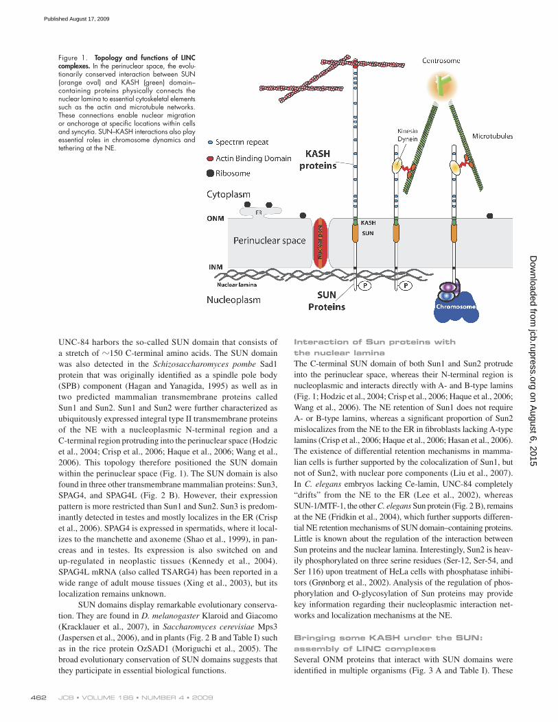
The rise of the SUN domain: identification and evolutionary conservationStudies of mutant C. elegans embryos with defects in nuclear migration and anchorage led to the identification of UNC-84, a transmembrane protein of the NE (Fig. 2 A; Malone et al., 1999).
The nucleus is the most prominent cellular organelle, and its sharp boundaries suggest the compartmentalization of the nucleoplasm from the cytoplasm. However, the recent identification of evolutionarily conserved linkers of the nucleoskeleton to the cytoskeleton (LINC) complexes, a family of macromolecular assemblies that span the dou-ble membrane of the nuclear envelope, reveals tight physical connections between the two compartments. Here, we review the structure and evolutionary conserva-tion of SUN and KASH domain–containing proteins, whose interaction within the perinuclear space forms the “nuts and bolts” of LINC complexes. Moreover, we dis-cuss the function of these complexes in nuclear, centro-somal, and chromosome dynamics, and their connection to human disease.
Bringing KASH under the SUN: the many faces of nucleo-cytoskeletal connections
David Razafsky and Didier Hodzic
Department of Ophthalmology and Visual Sciences, Washington University School of Medicine, St. Louis, MO 63110
© 2009 Razafsky and Hodzic This article is distributed under the terms of an Attribution–Noncommercial–Share Alike–No Mirror Sites license for the first six months after the publica-tion date (see http://www.jcb.org/misc/terms.shtml). After six months it is available under a Creative Commons License (Attribution–Noncommercial–Share Alike 3.0 Unported license, as described at http://creativecommons.org/licenses/by-nc-sa/3.0/).
TH
EJ
OU
RN
AL
OF
CE
LL
BIO
LO
GY
on August 6, 2015
jcb.rupress.orgD
ownloaded from
Published August 17, 2009
JCB • VOLUME 186 • NUMBER 4 • 2009 462
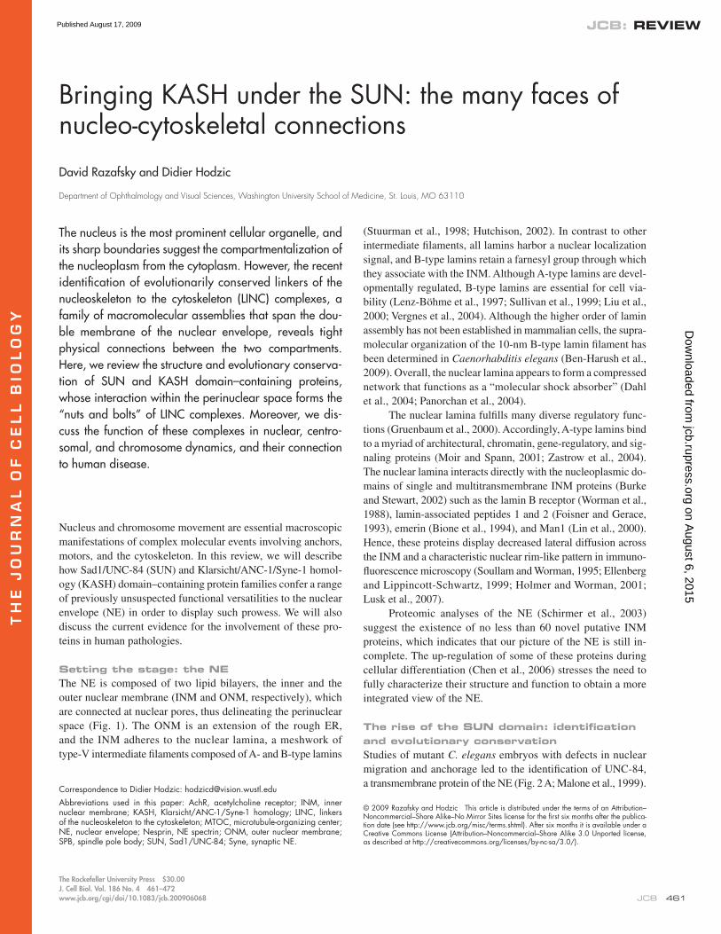
Interaction of Sun proteins with the nuclear laminaThe C-terminal SUN domain of both Sun1 and Sun2 protrude into the perinuclear space, whereas their N-terminal region is nucleoplasmic and interacts directly with A- and B-type lamins (Fig. 1; Hodzic et al., 2004; Crisp et al., 2006; Haque et al., 2006; Wang et al., 2006). The NE retention of Sun1 does not require A- or B-type lamins, whereas a significant proportion of Sun2 mislocalizes from the NE to the ER in fibroblasts lacking A-type lamins (Crisp et al., 2006; Haque et al., 2006; Hasan et al., 2006). The existence of differential retention mechanisms in mamma-lian cells is further supported by the colocalization of Sun1, but not of Sun2, with nuclear pore components (Liu et al., 2007). In C. elegans embryos lacking Ce-lamin, UNC-84 completely “drifts” from the NE to the ER (Lee et al., 2002), whereas SUN-1/MTF-1, the other C. elegans Sun protein (Fig. 2 B), remains at the NE (Fridkin et al., 2004), which further supports differen-tial NE retention mechanisms of SUN domain–containing proteins. Little is known about the regulation of the interaction between Sun proteins and the nuclear lamina. Interestingly, Sun2 is heav-ily phosphorylated on three serine residues (Ser-12, Ser-54, and Ser 116) upon treatment of HeLa cells with phosphatase inhibi-tors (Grønborg et al., 2002). Analysis of the regulation of phos-phorylation and O-glycosylation of Sun proteins may provide key information regarding their nucleoplasmic interaction net-works and localization mechanisms at the NE.
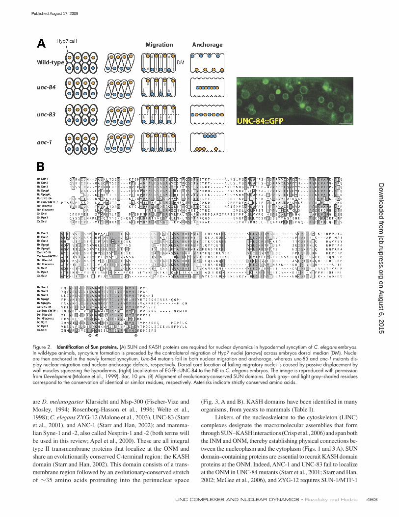
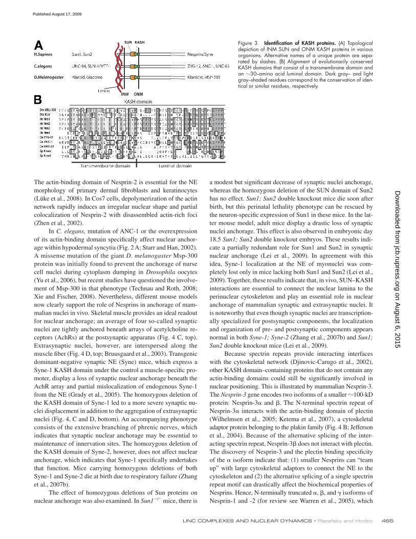
Bringing some KASH under the SUN: assembly of LINC complexesSeveral ONM proteins that interact with SUN domains were identified in multiple organisms (Fig. 3 A and Table I). These
UNC-84 harbors the so-called SUN domain that consists of a stretch of 150 C-terminal amino acids. The SUN domain was also detected in the Schizosaccharomyces pombe Sad1 protein that was originally identified as a spindle pole body (SPB) component (Hagan and Yanagida, 1995) as well as in two predicted mammalian transmembrane proteins called Sun1 and Sun2. Sun1 and Sun2 were further characterized as ubiquitously expressed integral type II transmembrane proteins of the NE with a nucleoplasmic N-terminal region and a C-terminal region protruding into the perinuclear space (Hodzic et al., 2004; Crisp et al., 2006; Haque et al., 2006; Wang et al., 2006). This topology therefore positioned the SUN domain within the perinuclear space (Fig. 1). The SUN domain is also found in three other transmembrane mammalian proteins: Sun3, SPAG4, and SPAG4L (Fig. 2 B). However, their expression pattern is more restricted than Sun1 and Sun2. Sun3 is predom-inantly detected in testes and mostly localizes in the ER (Crisp et al., 2006). SPAG4 is expressed in spermatids, where it local-izes to the manchette and axoneme (Shao et al., 1999), in pan-creas and in testes. Its expression is also switched on and up-regulated in neoplastic tissues (Kennedy et al., 2004). SPAG4L mRNA (also called TSARG4) has been reported in a wide range of adult mouse tissues (Xing et al., 2003), but its localization remains unknown.
SUN domains display remarkable evolutionary conserva-tion. They are found in D. melanogaster Klaroid and Giacomo (Kracklauer et al., 2007), in Saccharomyces cerevisiae Mps3 (Jaspersen et al., 2006), and in plants (Fig. 2 B and Table I) such as in the rice protein OzSAD1 (Moriguchi et al., 2005). The broad evolutionary conservation of SUN domains suggests that they participate in essential biological functions.
Figure 1. Topology and functions of LINC complexes. In the perinuclear space, the evolu-tionarily conserved interaction between SUN (orange oval) and KASH (green) domain–containing proteins physically connects the nuclear lamina to essential cytoskeletal elements such as the actin and microtubule networks. These connections enable nuclear migration or anchorage at specific locations within cells and syncytia. SUN–KASH interactions also play essential roles in chromosome dynamics and tethering at the NE.
on August 6, 2015
jcb.rupress.orgD
ownloaded from
Published August 17, 2009
463LINC COMPLEXES AND NUCLEAR DYNAMICS • Razafsky and Hodzic
(Fig. 3, A and B). KASH domains have been identified in many organisms, from yeasts to mammals (Table I).
Linkers of the nucleoskeleton to the cytoskeleton (LINC) complexes designate the macromolecular assemblies that form through SUN–KASH interactions (Crisp et al., 2006) and span both the INM and ONM, thereby establishing physical connections be-tween the nucleoplasm and the cytoplasm (Figs. 1 and 3 A). SUN domain–containing proteins are essential to recruit KASH domain proteins at the ONM. Indeed, ANC-1 and UNC-83 fail to localize at the ONM in UNC-84 mutants (Starr et al., 2001; Starr and Han, 2002; McGee et al., 2006), and ZYG-12 requires SUN-1/MTF-1
are D. melanogaster Klarsicht and Msp-300 (Fischer-Vize and Mosley, 1994; Rosenberg-Hasson et al., 1996; Welte et al., 1998); C. elegans ZYG-12 (Malone et al., 2003), UNC-83 (Starr et al., 2001), and ANC-1 (Starr and Han, 2002); and mamma-lian Syne-1 and -2, also called Nesprin-1 and -2 (both terms will be used in this review; Apel et al., 2000). These are all integral type II transmembrane proteins that localize at the ONM and share an evolutionarily conserved C-terminal region: the KASH domain (Starr and Han, 2002). This domain consists of a trans-membrane region followed by an evolutionary-conserved stretch of 35 amino acids protruding into the perinuclear space
Figure 2. Identification of Sun proteins. (A) SUN and KASH proteins are required for nuclear dynamics in hypodermal syncytium of C. elegans embryos. In wild-type animals, syncytium formation is preceded by the contralateral migration of Hyp7 nuclei (arrows) across embryos dorsal median (DM). Nuclei are then anchored in the newly formed syncytium. Unc-84 mutants fail in both nuclear migration and anchorage, whereas unc-83 and anc-1 mutants dis-play nuclear migration and nuclear anchorage defects, respectively. Dorsal cord location of failing migratory nuclei is caused by passive displacement by wall muscles squeezing the hypodermis. (right) Localization of EGFP::UNC-84 to the NE in C. elegans embryos. The image is reproduced with permission from Development (Malone et al., 1999). Bar, 10 µm. (B) Alignment of evolutionary-conserved SUN domains. Dark gray– and light gray–shaded residues correspond to the conservation of identical or similar residues, respectively. Asterisks indicate strictly conserved amino acids.
on August 6, 2015
jcb.rupress.orgD
ownloaded from
Published August 17, 2009
JCB • VOLUME 186 • NUMBER 4 • 2009 464
Providing functional diversity to the NE: the many faces of KASH proteinsIn the following paragraphs, we describe the functional aspects of various KASH domain-containing proteins in different or-ganisms. KASH domains provide a generic NE tethering device for functionally distinct proteins whose cytoplasmic domains mediate nuclear positioning, maintain physical connections with other cellular organelles, and even influence chromosome dy-namics (Fig. 1 and Table I).
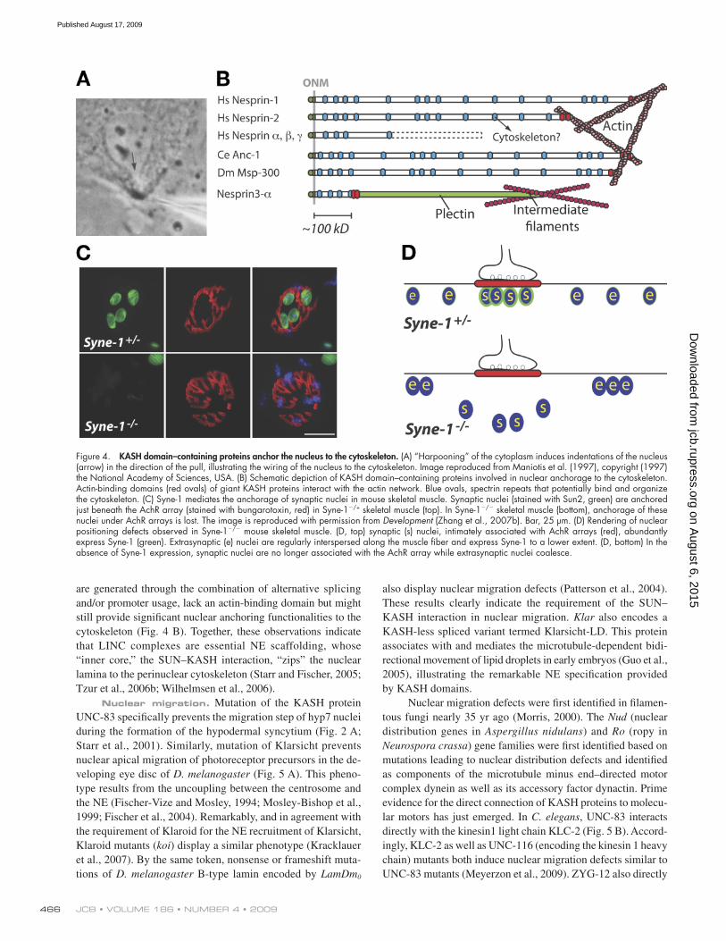
Nuclear anchorage to the cytoskeleton. The di-rect “harpooning” of the cytoskeleton with a micropipette tip results in a direct and immediate force transfer to the nucleus, whose NE locally extends and moves in the direction of the pull (Fig. 4 A). This effect is microtubule independent and suggests that the nucleus is “hard-wired” to the cytoskeleton (Maniotis et al., 1997; Wang et al., 2009). As shown in Fig. 4 B, C. elegans ANC-1, D. melanogaster Msp-300, and the giant isoforms of mammalian Nesprin-1 (also called Syne-1 [Apel et al., 2000], Myne1 [Mislow et al., 2002], and Enaptin [Padmakumar et al., 2004]) and Nesprin-2 (also called Syne-2 [Apel et al., 2000] and NUANCE [Zhen et al., 2002]) are gigantic proteins localiz-ing to the ONM, and they are predicted to extend as a rod-like structure of up to 300–400 nm into the cytoplasm (Zhang et al., 2002). They all share a common architecture: an N-terminal actin-binding domain and interspersed spectrin repeats. The latter are triple-helical coiled-coil domains with elastic properties that might be important in terms of deformability (Lenne et al., 2000).
for its NE localization (Malone et al., 2003). In D. melanogaster, Klaroid is strictly required for the ONM localization of Klarsicht and Msp-300 (Kracklauer et al., 2007; Technau and Roth, 2008). Similarly, the simultaneous siRNA-mediated down-regulation of both mammalian Sun1 and Sun2 prevents the localization of Nesprin-2–giant at the NE (Padmakumar et al., 2005; Crisp et al., 2006). The expression of either the recombinant SUN domain of Sun1 and Sun2 within the ER lumen or the KASH domain of Nesprin-1, -2, and -3 invariably results in the displace-ment of all endogenous NE spectrins (Nesprins) from the NE to the ER (Padmakumar et al., 2005; Crisp et al., 2006; Stewart-Hutchinson et al., 2008). Coupled with the observation that the KASH domain of Nesprin-1, -2, and -3 is equally able to interact with both Sun1 and Sun2, SUN–KASH interactions seem pro-miscuous (Stewart-Hutchinson et al., 2008). In mammalian cells, SUN–KASH interactions strictly require the C-terminal poly-proline motif of KASH domains (Fig. 3 B; Padmakumar et al., 2005; Ketema et al., 2007) as well as the last 20 C-terminal amino acids of the SUN domain, which contains three strictly conserved amino acid residues (Fig. 2 B; Stewart-Hutchinson et al., 2008). Consistent with the proposed interaction between Sun proteins and Nesprins across the NE, disruption of LINC complexes pro-vokes a significant enlargement of the perinuclear space between the ONM and the INM (Crisp et al., 2006). As we will see, in addition to widening the landscape of known NE proteins, the discovery of LINC complexes has radically redefined our view of NE function (Stewart et al., 2007).
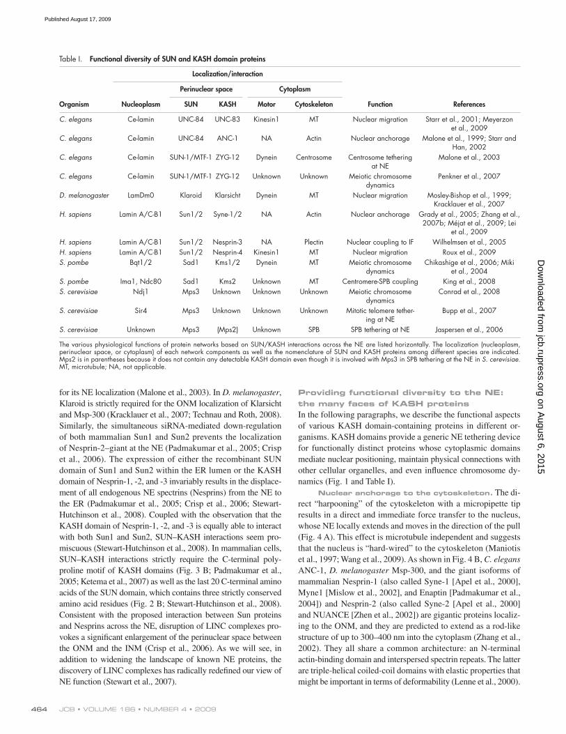
Table I. Functional diversity of SUN and KASH domain proteins
Localization/interaction
Perinuclear space Cytoplasm
Organism Nucleoplasm SUN KASH Motor Cytoskeleton Function References
C. elegans Ce-lamin UNC-84 UNC-83 Kinesin1 MT Nuclear migration Starr et al., 2001; Meyerzon et al., 2009
C. elegans Ce-lamin UNC-84 ANC-1 NA Actin Nuclear anchorage Malone et al., 1999; Starr and Han, 2002
C. elegans Ce-lamin SUN-1/MTF-1 ZYG-12 Dynein Centrosome Centrosome tethering at NE
Malone et al., 2003
C. elegans Ce-lamin SUN-1/MTF-1 ZYG-12 Unknown Unknown Meiotic chromosome dynamics
Penkner et al., 2007
D. melanogaster LamDm0 Klaroid Klarsicht Dynein MT Nuclear migration Mosley-Bishop et al., 1999; Kracklauer et al., 2007
H. sapiens Lamin A/C-B1 Sun1/2 Syne-1/2 NA Actin Nuclear anchorage Grady et al., 2005; Zhang et al., 2007b; Méjat et al., 2009; Lei
et al., 2009H. sapiens Lamin A/C-B1 Sun1/2 Nesprin-3 NA Plectin Nuclear coupling to IF Wilhelmsen et al., 2005H. sapiens Lamin A/C-B1 Sun1/2 Nesprin-4 Kinesin1 MT Nuclear migration Roux et al., 2009S. pombe Bqt1/2 Sad1 Kms1/2 Dynein MT Meiotic chromosome
dynamicsChikashige et al., 2006; Miki
et al., 2004S. pombe Ima1, Ndc80 Sad1 Kms2 Unknown MT Centromere-SPB coupling King et al., 2008S. cerevisiae Ndj1 Mps3 Unknown Unknown Unknown Meiotic chromosome
dynamicsConrad et al., 2008
S. cerevisiae Sir4 Mps3 Unknown Unknown Unknown Mitotic telomere tether-ing at NE
Bupp et al., 2007
S. cerevisiae Unknown Mps3 (Mps2) Unknown SPB SPB tethering at NE Jaspersen et al., 2006
The various physiological functions of protein networks based on SUN/KASH interactions across the NE are listed horizontally. The localization (nucleoplasm, perinuclear space, or cytoplasm) of each network components as well as the nomenclature of SUN and KASH proteins among different species are indicated. Mps2 is in parentheses because it does not contain any detectable KASH domain even though it is involved with Mps3 in SPB tethering at the NE in S. cerevisiae. MT, microtubule; NA, not applicable.
on August 6, 2015
jcb.rupress.orgD
ownloaded from
Published August 17, 2009
465LINC COMPLEXES AND NUCLEAR DYNAMICS • Razafsky and Hodzic
a modest but significant decrease of synaptic nuclei anchorage, whereas the homozygous deletion of the SUN domain of Sun2 has no effect. Sun1; Sun2 double knockout mice die soon after birth, but this perinatal lethality phenotype can be rescued by the neuron-specific expression of Sun1 in these mice. In the lat-ter mouse model, adult mice display a drastic loss of synaptic nuclei anchorage. This effect is also observed in embryonic day 18.5 Sun1; Sun2 double knockout embryos. These results indi-cate a partially redundant role for Sun1 and Sun2 in synaptic nuclear anchorage (Lei et al., 2009). In agreement with this idea, Syne-1 localization at the NE of myonuclei was com-pletely lost only in mice lacking both Sun1 and Sun2 (Lei et al., 2009). Together, these results indicate that, in vivo, SUN–KASH interactions are essential to connect the nuclear lamina to the perinuclear cytoskeleton and play an essential role in nuclear anchorage of mammalian synaptic and extrasynaptic nuclei. It is noteworthy that even though synaptic nuclei are transcription-ally specialized for postsynaptic components, the localization and organization of pre- and postsynaptic components appears normal in both Syne-1; Syne-2 (Zhang et al., 2007b) and Sun1; Sun2 double knockout mice (Lei et al., 2009).
Because spectrin repeats provide interacting interfaces with the cytoskeletal network (Djinovic-Carugo et al., 2002), other KASH domain–containing proteins that do not contain any actin-binding domains could still be significantly involved in nuclear positioning. This is illustrated by mammalian Nesprin-3. The Nesprin-3 gene encodes two isoforms of a smaller 100-kD protein: Nesprin-3 and . The N-terminal spectrin repeat of Nesprin-3 interacts with the actin-binding domain of plectin (Wilhelmsen et al., 2005; Ketema et al., 2007), a cytoskeletal adaptor protein belonging to the plakin family (Fig. 4 B; Jefferson et al., 2004). Because of the alternative splicing of the inter-acting spectrin repeat, Nesprin-3 does not interact with plectin. The discovery of Nesprin-3 and the plectin binding specificity of the isoform indicate that: (1) smaller Nesprins can “team up” with large cytoskeletal adaptors to connect the NE to the cytoskeleton and (2) the alternative splicing of a single spectrin repeat motif can drastically affect the biochemical properties of Nesprins. Hence, N-terminally truncated , , and isoforms of Nesprin-1 and -2 (for review see Warren et al., 2005), which
The actin-binding domain of Nesprin-2 is essential for the NE morphology of primary dermal fibroblasts and keratinocytes (Lüke et al., 2008). In Cos7 cells, depolymerization of the actin network rapidly induces an irregular nuclear shape and partial colocalization of Nesprin-2 with disassembled actin-rich foci (Zhen et al., 2002).
In C. elegans, mutation of ANC-1 or the overexpression of its actin-binding domain specifically affect nuclear anchor-age within hypodermal syncytia (Fig. 2 A; Starr and Han, 2002). A missense mutation of the giant D. melanogaster Msp-300 protein was initially found to prevent the anchorage of nurse cell nuclei during cytoplasm dumping in Drosophila oocytes (Yu et al., 2006), but recent studies have questioned the involve-ment of Msp-300 in that phenotype (Technau and Roth, 2008; Xie and Fischer, 2008). Nevertheless, different mouse models now clearly support the role of Nesprins in anchorage of mam-malian nuclei in vivo. Skeletal muscle provides an ideal readout for nuclear anchorage; an average of four so-called synaptic nuclei are tightly anchored beneath arrays of acetylcholine re-ceptors (AchRs) at the postsynaptic apparatus (Fig. 4 C, top). Extrasynaptic nuclei, however, are interspersed along the muscle fiber (Fig. 4 D, top; Bruusgaard et al., 2003). Transgenic dominant-negative synaptic NE (Syne) mice, which express a Syne-1 KASH domain under the control a muscle-specific pro-moter, display a loss of synaptic nuclear anchorage beneath the AchR array and partial mislocalization of endogenous Syne-1 from the NE (Grady et al., 2005). The homozygous deletion of the KASH domain of Syne-1 led to a more severe synaptic nu-clei displacement in addition to the aggregation of extrasynaptic nuclei (Fig. 4, C and D, bottom). An accompanying phenotype consists of the extensive branching of phrenic nerves, which indicates that synaptic nuclear anchorage may be essential to maintenance of innervation sites. The homozygous deletion of the KASH domain of Syne-2, however, does not affect nuclear anchorage, which indicates that Syne-1 specifically undertakes that function. Mice carrying homozygous deletions of both Syne-1 and Syne-2 die at birth due to respiratory failure (Zhang et al., 2007b).
The effect of homozygous deletions of Sun proteins on nuclear anchorage was also examined. In Sun1/ mice, there is
Figure 3. Identification of KASH proteins. (A) Topological depiction of INM SUN and ONM KASH proteins in various organisms. Alternative names of a unique protein are sepa-rated by slashes. (B) Alignment of evolutionarily conserved KASH domains that consist of a transmembrane domain and an 30–amino acid luminal domain. Dark gray– and light gray–shaded residues correspond to the conservation of iden-tical or similar residues, respectively.
on August 6, 2015
jcb.rupress.orgD
ownloaded from
Published August 17, 2009
JCB • VOLUME 186 • NUMBER 4 • 2009 466
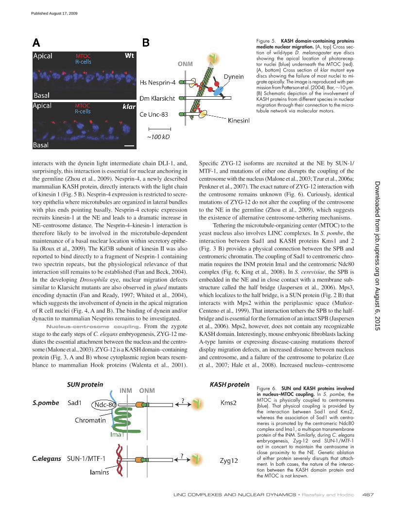
also display nuclear migration defects (Patterson et al., 2004). These results clearly indicate the requirement of the SUN–KASH interaction in nuclear migration. Klar also encodes a KASH-less spliced variant termed Klarsicht-LD. This protein associates with and mediates the microtubule-dependent bidi-rectional movement of lipid droplets in early embryos (Guo et al., 2005), illustrating the remarkable NE specification provided by KASH domains.
Nuclear migration defects were first identified in filamen-tous fungi nearly 35 yr ago (Morris, 2000). The Nud (nuclear distribution genes in Aspergillus nidulans) and Ro (ropy in Neurospora crassa) gene families were first identified based on mutations leading to nuclear distribution defects and identified as components of the microtubule minus end–directed motor complex dynein as well as its accessory factor dynactin. Prime evidence for the direct connection of KASH proteins to molecu-lar motors has just emerged. In C. elegans, UNC-83 interacts directly with the kinesin1 light chain KLC-2 (Fig. 5 B). Accord-ingly, KLC-2 as well as UNC-116 (encoding the kinesin 1 heavy chain) mutants both induce nuclear migration defects similar to UNC-83 mutants (Meyerzon et al., 2009). ZYG-12 also directly
are generated through the combination of alternative splicing and/or promoter usage, lack an actin-binding domain but might still provide significant nuclear anchoring functionalities to the cytoskeleton (Fig. 4 B). Together, these observations indicate that LINC complexes are essential NE scaffolding, whose “inner core,” the SUN–KASH interaction, “zips” the nuclear lamina to the perinuclear cytoskeleton (Starr and Fischer, 2005; Tzur et al., 2006b; Wilhelmsen et al., 2006).
Nuclear migration. Mutation of the KASH protein UNC-83 specifically prevents the migration step of hyp7 nuclei during the formation of the hypodermal syncytium (Fig. 2 A; Starr et al., 2001). Similarly, mutation of Klarsicht prevents nuclear apical migration of photoreceptor precursors in the de-veloping eye disc of D. melanogaster (Fig. 5 A). This pheno-type results from the uncoupling between the centrosome and the NE (Fischer-Vize and Mosley, 1994; Mosley-Bishop et al., 1999; Fischer et al., 2004). Remarkably, and in agreement with the requirement of Klaroid for the NE recruitment of Klarsicht, Klaroid mutants (koi) display a similar phenotype (Kracklauer et al., 2007). By the same token, nonsense or frameshift muta-tions of D. melanogaster B-type lamin encoded by LamDm0
Figure 4. KASH domain–containing proteins anchor the nucleus to the cytoskeleton. (A) “Harpooning” of the cytoplasm induces indentations of the nucleus (arrow) in the direction of the pull, illustrating the wiring of the nucleus to the cytoskeleton. Image reproduced from Maniotis et al. (1997), copyright (1997) the National Academy of Sciences, USA. (B) Schematic depiction of KASH domain–containing proteins involved in nuclear anchorage to the cytoskeleton. Actin-binding domains (red ovals) of giant KASH proteins interact with the actin network. Blue ovals, spectrin repeats that potentially bind and organize the cytoskeleton. (C) Syne-1 mediates the anchorage of synaptic nuclei in mouse skeletal muscle. Synaptic nuclei (stained with Sun2, green) are anchored just beneath the AchR array (stained with bungarotoxin, red) in Syne-1/+ skeletal muscle (top). In Syne-1/ skeletal muscle (bottom), anchorage of these nuclei under AchR arrays is lost. The image is reproduced with permission from Development (Zhang et al., 2007b). Bar, 25 µm. (D) Rendering of nuclear positioning defects observed in Syne-1/ mouse skeletal muscle. (D, top) synaptic (s) nuclei, intimately associated with AchR arrays (red), abundantly express Syne-1 (green). Extrasynaptic (e) nuclei are regularly interspersed along the muscle fiber and express Syne-1 to a lower extent. (D, bottom) In the absence of Syne-1 expression, synaptic nuclei are no longer associated with the AchR array while extrasynaptic nuclei coalesce.
on August 6, 2015
jcb.rupress.orgD
ownloaded from
Published August 17, 2009
467LINC COMPLEXES AND NUCLEAR DYNAMICS • Razafsky and Hodzic
Specific ZYG-12 isoforms are recruited at the NE by SUN-1/MTF-1, and mutations of either one disrupts the coupling of the centrosome with the nucleus (Malone et al., 2003; Tzur et al., 2006a; Penkner et al., 2007). The exact nature of ZYG-12 interaction with the centrosome remains unknown (Fig. 6). Curiously, identical mutations of ZYG-12 do not alter the coupling of the centrosome to the NE in the germline (Zhou et al., 2009), which suggests the existence of alternative centrosome-tethering mechanisms.
Tethering the microtubule-organizing center (MTOC) to the yeast nucleus also involves LINC complexes. In S. pombe, the interaction between Sad1 and KASH proteins Kms1 and 2 (Fig. 3 B) provides a physical connection between the SPB and centromeric chromatin. The coupling of Sad1 to centromeric chro-matin requires the INM protein Ima1 and the centromeric Ndc80 complex (Fig. 6; King et al., 2008). In S. cerevisiae, the SPB is embedded in the NE and in close contact with a membrane sub-structure called the half bridge (Jaspersen et al., 2006). Mps3, which localizes to the half bridge, is a SUN protein (Fig. 2 B) that interacts with Mps2 within the periplasmic space (Muñoz-Centeno et al., 1999). That interaction tethers the SPB to the half-bridge and is essential for the formation of an intact SPB (Jaspersen et al., 2006). Mps2, however, does not contain any recognizable KASH domain. Interestingly, mouse embryonic fibroblasts lacking A-type lamins or expressing disease-causing mutations thereof display migration defects, an increased distance between nucleus and centrosome, and a failure of the centrosome to polarize (Lee et al., 2007; Hale et al., 2008). Increased nucleus–centrosome
interacts with the dynein light intermediate chain DLI-1, and, surprisingly, this interaction is essential for nuclear anchoring in the germline (Zhou et al., 2009). Nesprin-4, a newly described mammalian KASH protein, directly interacts with the light chain of kinesin 1 (Fig. 5 B). Nesprin-4 expression is restricted to secre-tory epithelia where microtubules are organized in lateral bundles with plus ends pointing basally. Nesprin-4 ectopic expression recruits kinesin-1 at the NE and leads to a dramatic increase in NE–centrosome distance. The Nesprin-4–kinesin-1 interaction is therefore likely to be involved in the microtubule-dependent maintenance of a basal nuclear location within secretory epithe-lia (Roux et al., 2009). The Kif3B subunit of kinesin II was also reported to bind directly to a fragment of Nesprin-1 containing two spectrin repeats, but the physiological relevance of that interaction still remains to be established (Fan and Beck, 2004). In the developing Drosophila eye, nuclear migration defects similar to Klarsicht mutants are also observed in glued mutants encoding dynactin (Fan and Ready, 1997; Whited et al., 2004), which suggests the involvement of dynein in the apical migration of R cell nuclei (Fig. 4, A and B). The binding of dynein and/or dynactin to mammalian Nesprins remains to be investigated.
Nucleus–centrosome coupling. From the zygote stage to the early steps of C. elegans embryogenesis, ZYG-12 me-diates the essential attachment between the nucleus and the centro-some (Malone et al., 2003). ZYG-12 is a KASH domain–containing protein (Fig. 3, A and B) whose cytoplasmic region bears resem-blance to mammalian Hook proteins (Walenta et al., 2001).
Figure 5. KASH domain-containing proteins mediate nuclear migration. (A, top) Cross sec-tion of wild-type D. melanogaster eye discs showing the apical location of photorecep-tor nuclei (blue) underneath the MTOC (red). (A, bottom) Cross section of klar mutant eye discs showing the failure of most nuclei to mi-grate apically. The image is reproduced with per-mission from Patterson et al. (2004). Bar, 10 µm. (B) Schematic depiction of the involvement of KASH proteins from different species in nuclear migration through their connection to the micro-tubule network via molecular motors.
Figure 6. SUN and KASH proteins involved in nucleus–MTOC coupling. In S. pombe, the MTOC is physically coupled to centromeres (blue). That physical coupling is provided by the interaction between Sad1 and Kms2, whereas the association of Sad1 with centro-meres is promoted by the centromeric Ndc80 complex and Ima1, a multispan transmembrane protein of the INM. Similarly, during C. elegans embryogenesis, Zyg-12 and SUN-1/MTF-1 act in concert to maintain the centrosome in close proximity to the NE. Genetic ablation of either protein severely disrupts that attach-ment. In both cases, the nature of the interac-tion between the KASH domain protein and the MTOC is not known.
on August 6, 2015
jcb.rupress.orgD
ownloaded from
Published August 17, 2009
JCB • VOLUME 186 • NUMBER 4 • 2009 468
muscular dystrophy and dilated cardiomyopathy. Two hypotheses, which are probably not mutually exclusive, view LMNA mutations as triggers of either gene expression deregulation or structural cel-lular disorganization. We will focus on the second hypothesis and emphasize the evidence for an involvement of the disruption of LINC complexes in myolaminopathies.
LMNA/ mice display normal embryonic development; however, at 3 wk after birth, a decline in growth is accompa-nied by cardiac and skeletal myopathies reminiscent of human Emery-Dreifuss muscular dystrophy and dilated cardiomyopa-thy (Sullivan et al., 1999). A reduction of axon density and the presence of nonmyelinated axons resembling human peripheral axonopathies are also significant (De Sandre-Giovannoli et al., 2002). These mice die at 6 wk. At the cellular level, embry-onic fibroblasts from LMNA/ mice (MEFlmna/) display an irregular nuclear shape and a loss of peripheral chromatin.
In MEFlmna/ cells, Sun2, Nesprin-1, and Nesprin-2 all mis-localize from the NE to the ER, whereas Sun1 seems unaffected (Libotte et al., 2005; Crisp et al., 2006; Haque et al., 2006). In vivo, Sun2 and Nesprin-1 also mislocalize from synaptic nuclei of LMNA/ and LMNAH222P/H222P knock-in mice (Méjat et al., 2009); the latter model presents muscle and cardiac phenotypes similar to Emery-Dreifuss muscular dystrophy but with a later disease onset than LMNA/ mice (Arimura et al., 2005). In both models, anchor-age of synaptic nuclei under the array of AchR is lost, phrenic nerves are highly ramified, and the innervation area is enlarged. These phenotypes are remarkably similar to Syne-1/ mice (see Nuclear anchorage section). This indicates that (1) A-type lamins are essen-tial for the integrity of LINC complexes in mammalian tissues and (2) A-type lamin alterations phenocopy the disruption of LINC complexes in terms of nuclear positioning and innervation pattern. However, synaptic nuclear mispositioning might not be involved in muscle pathology per se. First, dominant-negative Syne mice, Syne-1/ mice, and Sun1; Sun2 double knockout mice express-ing Sun1 in the nervous system do not display any muscle pathol-ogy despite extensive synaptic nuclei mispositioning (Grady et al., 2005; Zhang et al., 2007b; Lei et al., 2009). Second, patients affected by autosomal recessive cerebellar ataxia associated with mutations of Nesprin-1 do not display any muscle pathology de-spite a severe mispositioning of synaptic nuclei in skeletal muscle (Gros-Louis et al., 2007). A major phenotypic difference, however, is that Syne-1/ mice, Sun1; Sun2 double knockout mice express-ing Sun1 in the nervous system, and autosomal recessive cerebellar ataxia patients do not show any detectable organization defect of AchR, whereas LMNA/ and LMNAH222P/H222P muscle fibers dis-play poorly structured and discontinuous arrays of AchR (Méjat et al., 2009). It therefore seems that laminopathic muscle pheno-types are correlated to disorganized AchR arrays, but the question still remains as to how LMNA mutations alter the organization of these arrays. It is tempting to hypothesize that disruption of LINC complexes through the lack or mutation of A-type lamins alters the structural organization of the cytoskeleton. To that regard, a com-plete disorganization of the desmin network has been reported in LMNA/ cardiomyocytes, and the cytoskeleton of MEFlmna/ displays a drastic loss of mechanical stiffness (Broers et al., 2004; Lammerding et al., 2004; Lee et al., 2007; Hale et al., 2008). In cul-tured fibroblasts, disruption of LINC complexes induces a similar
distance was also observed upon LINC complex disruption in mammalian cells (our unpublished data). Collectively, these results suggest an evolutionarily conserved role for LINC com-plexes to position the MTOC in close proximity to the NE.
Chromosome dynamics. The “chromosomal bouquet” (Scherthan, 2001) refers to the “floral” arrangement of chromo-somes during prophase I after the convergence of telomeres to a re-stricted area of the NE facing the centrosome (Fig. 1). SUN and KASH proteins play a central role in that dynamic event (Table I). In S. pombe, Sad1 colocalizes with the telomeric bouquet and inter-acts with meiotic-specific Bouquet (Bqt) 1 and 2 proteins to pro-vide a physical connection between the nucleoplasmic region of Sad1 and telomeres (Chikashige et al., 2006). Because Kms1 inter-acts with both Sad1 and dynein (Miki et al., 2004), a model there-fore emerges where telomere dynamics during bouquet formation are mediated through the Bqt2–Bqt1–Sad1–Kms1–dynein connec-tion across the meiotic NE (Chikashige et al., 2006).
In S. cerevisiae, the truncation of the N-terminal region of either Mps3 or Ndj1 reduces telomere mobility of pachytene chromosomes. Ndj1 interacts with the cytoplasmic domain of Mps3 and mediates telomere attachment to the NE (Conrad et al., 2008). In conjunction with Sir4 (silent information regu-lator protein 4), Mps3 is also required for telomere anchoring at the NE during mitosis (Bupp et al., 2007). It is important to note that telomere dynamics are essentially mediated by actin in S. cerevisiae, whereas microtubules are used in mammals, plants, and fission yeast (Koszul et al., 2008).
Mammalian Sun1 clearly colocalizes with telomeres be-tween leptotene and diplotene stages (Ding et al., 2007). Although a similar localization was reported for Sun2 in mouse and rat sper-matocytes (Schmitt et al., 2007), another group was unable to de-tect any Sun2 immunoreactivity at meiotic telomeres (Ding et al., 2007; Lei et al., 2009). In Sun1/ mice, telomere association with the NE as well as homologue pairing and synapsis are prevented (Ding et al., 2007). Accordingly, Sun1/ mice are sterile, whereas Sun2/ mice do not display any fertility issues (Lei et al., 2009). In C. elegans, a single point mutation within the SUN domain of SUN-1/MTF-1 (G311V) is also associated with defective homolo-gous pairing (Penkner et al., 2007; Fridkin et al., 2009). These results indicate that Sun proteins are “hijacked” by accessory meiosis-specific proteins required for chromosome dynamics. A KASH protein that acts in concert with Sun1 in mammalian meiosis still awaits characterization.
LINC complexes and human diseasesMuscle pathologies. Over 200 missense mutations scat-
tered along LMNA (the gene that encodes the A-type lamins: lamin A and lamin C) are associated with a variety of human diseases called laminopathies. Laminopathies involve either specific or combined pathologies of neurons, muscle, and bone tissues (Ben Yaou et al., 2005; Jacob and Garg, 2006; Worman et al., 2009). A main question in the field is how mutations of a protein expressed in most differentiated cells can lead to tissue-specific diseases (Mounkes et al., 2003; Worman and Courvalin, 2004). How-ever, even though LMNA mutations are associated with >10 dis-tinct human pathologies, the vast majority are associated with skeletal and/or cardiac muscle pathologies such as Emery-Dreifuss
on August 6, 2015
jcb.rupress.orgD
ownloaded from
Published August 17, 2009
469LINC COMPLEXES AND NUCLEAR DYNAMICS • Razafsky and Hodzic
nucleokinesis, i.e., the translocation of the nucleus within a cell, and may underlie other categories of human diseases. In filamen-tous fungi, NudF and ro-15 are essential for nuclear positioning and encode proteins with 40% identity to human LIS1 (Morris et al., 1998). Deletion or mutations of LIS1 are associated with lissencephalies, pathologies of the developing brain that are as-sociated with a failure of the nucleokinetic step during neuronal migration in the cortex (Solecki et al., 2004; Vallee and Tsai, 2006). In cerebellar granule neurons, nucleokinesis requires intact microtubules that literally wrap the nucleus (Rivas and Hatten, 1995; Solecki et al., 2004) and a functional dynein–dynactin complex as well as Lis1 (Tanaka et al., 2004) and Ndel1, one of its binding partners (Shu et al., 2004). Current models suggest that dynein, anchored at the NE, pulls the nucleus toward the minus end of microtubules (Samuels and Tsai, 2004; Tsai and Gleeson, 2005). How the microtubule network is physically connected to the neuronal NE remains a central question (Tsai and Gleeson, 2005). However, as we have seen, LINC complexes are involved in nuclear migration, and the recent demonstration that UNC-83 and Nesprin-4 both interact with molecular motors strongly predicts a central involvement of LINC complexes in neuronal migration. In that regard, either the mutation of mikre oko (mok), which encodes a subunit of the dynactin complex, or interference with the function of dynamitin, LIS1, or LINC com-plexes results in the mislocalization of zebrafish photoreceptor nuclei (Tsujikawa et al., 2007). The discovery of Syne-1 muta-tions in patients with autosomal recessive cerebellar ataxia (Gros-Louis et al., 2007) could be the first description of the involvement of Nesprins in neurological diseases.
Finally, interkinetic nuclear migration designates the cou-pling of nuclear migration with the cell cycle of neuroepithelial cells. This phenomenon is essential for regulation of cell cycle exit and neurogenesis (Baye and Link, 2008). The down-regulation of Syne-2 or the expression of its KASH domain alters interkinetic nuclear migration (Del Bene et al., 2008). Together, these results predict exciting times ahead for LINC complexes in nuclear migra-tion and neurogenesis not only during development but also in the adult brain, where focal neurogenesis and nuclear migration are still significant (Ming and Song, 2005; Ayala et al., 2007).
ConclusionThe past decade has seen remarkable progress in our understand-ing of the functional contribution of NE proteins to essential biological processes. Such progress largely benefited from multi-disciplinary approaches in different organisms. The accumulated data clearly established that SUN proteins act as NE “receptors” of KASH domain-containing proteins. The variety of cytoplas-mic “flavors” of KASH proteins, in turn, provides specific func-tions related to nuclear and chromosome dynamics. However, many questions still remain. For example, how the SUN–KASH interaction is regulated is only beginning to emerge, with early indications pointing to TorsinA, an AAA+ATPase (Nery et al., 2008; Vander Heyden et al., 2009). The interaction network of the nucleoplasmic region of Sun proteins also needs more inves-tigation in interphase cells. We have seen that gigantic macro-molecular complexes form both within the nucleoplasm and the cytoplasm around LINC complexes, but how cell signaling affects
loss of cytoskeletal mechanical stiffness (Stewart-Hutchinson et al., 2008). In the syncytial C. elegans gonad, a mutation of ZYG-12 (Q44P, zyg-12(ct350)) that results in the failure to recruit dynein to the NE has far-reaching deleterious effects on micro-tubule organization, membrane architecture, and nuclear position-ing throughout the whole gonad (Zhou et al., 2009). In agreement with the concept of mechanotransduction at a distance (Wang et al., 2009), these observations support the finding that alteration of either the nuclear lamina or LINC complexes drastically affects cellular biomechanical properties of the cytoskeleton. HL-60– derived granulocytes may provide a physiological adaptation of that phenomenon. The cytoskeletal malleability and extensive nu-clear lobulation that allow these cells to cross the vasculature has been correlated to the expression of a paucity of LINC complex components, whereas the stiffer macrophage-derived cells express most of the LINC complex components (Olins et al., 2009).
A-type lamin mutations affecting the structural integrity of LINC complexes may therefore compromise the organization and mechanical integrity of the myoskeleton. Because AChR arrays are primarily supported by a submembranous organization of actin and desmin filaments (Mitsui et al., 2000), a major cytoskeletal disruption caused by a mutation of the nuclear lamina could there-fore drastically impact the organization of these receptors. Finally, Nesprins and dystrophins are giant spectrin-repeat proteins with actin-binding domains that mechanically connect to protein-aceous meshworks—nuclear lamina or extracellular matrix—through KASH domains or via the sarcoglycan–dystroglycan complex, respectively. Accordingly, cultured myotubes from dystrophin-deficient Mdx mice are mechanically compromised (Pasternak et al., 1995). Collectively, these observations suggest that laminopathic mutations affecting the organization of LINC complexes may induce significant mechanical deficiency and ensuing structural disorganization of the muscle fiber.
Several indications also support the direct involvement of mutations of LINC complex components in muscle pathologies. First, D. melanogaster Msp-300 was initially shown to be required for embryonic muscle morphogenesis (Rosenberg-Hasson et al., 1996), and Nesprin immunoreactivity was also detected in sar-comeres and Z lines, which supports additional structural roles for Nesprins (Zhang et al., 2005). Second, another mouse model with the homozygous deletion of the KASH domain of Syne-1 (/ KASH model) displays 50% perinatal lethality, and sur-vivors exhibit Emery-Dreifuss muscular dystrophy phenotypes (Puckelwartz et al., 2009). This striking difference with the Syne-1/ model may stem from either a dominant-negative effect of truncated Syne-1 proteins detected in / KASH mice or from different genetic backgrounds. Third, Nesprin missense mutations have recently been identified in Emery-Dreifuss muscular dys-trophy patients (Zhang et al., 2007a) and in autosomal recessive arthrogryposis multiplex congenita of myogenic origin (Attali et al., 2009). These observations, in addition to the lack of molecu-lar diagnoses in >50% of Emery-Dreifuss muscular dystrophy phenotypes, stress the need to screen patients with idiopathic mus-cular dystrophies for mutations of Nesprin and Sun genes.
Neuronal diseases. Because SUN and KASH domain–containing proteins are involved in nuclear migration, they may be required in essential developmental processes relying on
on August 6, 2015
jcb.rupress.orgD
ownloaded from
Published August 17, 2009
JCB • VOLUME 186 • NUMBER 4 • 2009 470
Chikashige, Y., C. Tsutsumi, M. Yamane, K. Okamasa, T. Haraguchi, and Y. Hiraoka. 2006. Meiotic proteins bqt1 and bqt2 tether telomeres to form the bouquet arrangement of chromosomes. Cell. 125:59–69.
Conrad, M.N., C.Y. Lee, G. Chao, M. Shinohara, H. Kosaka, A. Shinohara, J.A. Conchello, and M.E. Dresser. 2008. Rapid telomere movement in meiotic prophase is promoted by NDJ1, MPS3, and CSM4 and is modulated by recombination. Cell. 133:1175–1187.
Crisp, M., Q. Liu, K. Roux, J.B. Rattner, C. Shanahan, B. Burke, P.D. Stahl, and D. Hodzic. 2006. Coupling of the nucleus and cytoplasm: role of the LINC complex. J. Cell Biol. 172:41–53.
Dahl, K.N., S.M. Kahn, K.L. Wilson, and D.E. Discher. 2004. The nuclear enve-lope lamina network has elasticity and a compressibility limit suggestive of a molecular shock absorber. J. Cell Sci. 117:4779–4786.
De Sandre-Giovannoli, A., M. Chaouch, S. Kozlov, J.M. Vallat, M. Tazir, N. Kassouri, P. Szepetowski, T. Hammadouche, A. Vandenberghe, C.L. Stewart, et al. 2002. Homozygous defects in LMNA, encoding lamin A/C nuclear-envelope proteins, cause autosomal recessive axonal neuropathy in human (Charcot-Marie-Tooth disorder type 2) and mouse. Am. J. Hum. Genet. 70:726–736.
Del Bene, F., A.M. Wehman, B.A. Link, and H. Baier. 2008. Regulation of neuro-genesis by interkinetic nuclear migration through an apical-basal notch gradient. Cell. 134:1055–1065.
Ding, X., R. Xu, J. Yu, T. Xu, Y. Zhuang, and M. Han. 2007. SUN1 is required for telomere attachment to nuclear envelope and gametogenesis in mice. Dev. Cell. 12:863–872.
Djinovic-Carugo, K., M. Gautel, J. Ylänne, and P. Young. 2002. The spectrin repeat: a structural platform for cytoskeletal protein assemblies. FEBS Lett. 513:119–123.
Ellenberg, J., and J. Lippincott-Schwartz. 1999. Dynamics and mobility of nuclear envelope proteins in interphase and mitotic cells revealed by green fluores-cent protein chimeras. Methods. 19:362–372.
Fan, J., and K.A. Beck. 2004. A role for the spectrin superfamily member Syne-1 and kinesin II in cytokinesis. J. Cell Sci. 117:619–629.
Fan, S.S., and D.F. Ready. 1997. Glued participates in distinct microtubule-based activities in Drosophila eye development. Development. 124:1497–1507.
Fischer, J.A., S. Acosta, A. Kenny, C. Cater, C. Robinson, and J. Hook. 2004. Drosophila klarsicht has distinct subcellular localization domains for nuclear envelope and microtubule localization in the eye. Genetics. 168:1385–1393.
Fischer-Vize, J.A., and K.L. Mosley. 1994. Marbles mutants: uncoupling cell determination and nuclear migration in the developing Drosophila eye. Development. 120:2609–2618.
Foisner, R., and L. Gerace. 1993. Integral membrane proteins of the nuclear enve-lope interact with lamins and chromosomes, and binding is modulated by mitotic phosphorylation. Cell. 73:1267–1279.
Fridkin, A., E. Mills, A. Margalit, E. Neufeld, K.K. Lee, N. Feinstein, M. Cohen, K.L. Wilson, and Y. Gruenbaum. 2004. Matefin, a Caenorhabditis elegans germ line-specific SUN-domain nuclear membrane protein, is essential for early embryonic and germ cell development. Proc. Natl. Acad. Sci. USA. 101:6987–6992.
Fridkin, A., A. Penkner, V. Jantsch, and Y. Gruenbaum. 2009. SUN-domain and KASH-domain proteins during development, meiosis and disease. Cell. Mol. Life Sci. 66:1518–1533.
Grady, R.M., D.A. Starr, G.L. Ackerman, J.R. Sanes, and M. Han. 2005. Syne pro-teins anchor muscle nuclei at the neuromuscular junction. Proc. Natl. Acad. Sci. USA. 102:4359–4364.
Grønborg, M., T.Z. Kristiansen, A. Stensballe, J.S. Andersen, O. Ohara, M. Mann, O.N. Jensen, and A. Pandey. 2002. A mass spectrometry-based proteomic approach for identification of serine/threonine-phosphorylated proteins by enrichment with phospho-specific antibodies: identification of a novel protein, Frigg, as a protein kinase A substrate. Mol. Cell. Proteomics. 1:517–527.
Gros-Louis, F., N. Dupré, P. Dion, M.A. Fox, S. Laurent, S. Verreault, J.R. Sanes, J.P. Bouchard, and G.A. Rouleau. 2007. Mutations in SYNE1 lead to a newly discovered form of autosomal recessive cerebellar ataxia. Nat. Genet. 39:80–85.
Gruenbaum, Y., K.L. Wilson, A. Harel, M. Goldberg, and M. Cohen. 2000. Review: nuclear lamins—structural proteins with fundamental functions. J. Struct. Biol. 129:313–323.
Guo, Y., S. Jangi, and M.A. Welte. 2005. Organelle-specific control of intracellular transport: distinctly targeted isoforms of the regulator Klar. Mol. Biol. Cell. 16:1406–1416.
Hagan, I., and M. Yanagida. 1995. The product of the spindle formation gene sad1+ associates with the fission yeast spindle pole body and is essential for viability. J. Cell Biol. 129:1033–1047.
Hale, C.M., A.L. Shrestha, S.B. Khatau, P.J. Stewart-Hutchinson, L. Hernandez, C.L. Stewart, D. Hodzic, and D. Wirtz. 2008. Dysfunctional connections between the nucleus and the actin and microtubule networks in lamino-pathic models. Biophys. J. 95:5462–5475.
these assemblies is still poorly characterized. Hypotheses on how these molecular assemblies might affect mechanochemical conversion in the nucleus and alter gene activities have recently begun to emerge (Wang et al., 2009). Molecular tools are now available to ask essential questions about the physiological signifi-cance of nuclear positioning in other tissues. From a disease stand-point, examining the role of SUN–KASH interactions in neuronal migration will also be essential for establishing whether these inter-actions participate in the etiology of lissencephaly-like phenotypes. Finally, accumulated evidence calls for mutation screenings of Sun proteins and Nesprins in patients affected by idiopathic muscular dystrophies. Such findings could provide new therapeutic insights into these devastating human pathologies.
The authors are grateful to Drs. D. Starr and D. Wirtz for their critical reading of this manuscript, and to Drs. M. Han, J. Fischer, D. Ingber, and J. Sanes for helpful discussions and/or permissions for figure reproduction.
This work was supported by the Muscular Dystrophy Association, the National Institutes of Health (1R01GM084204, National Institute of Gen-eral Medical Sciences), an unrestricted grant (No. 34821) from Research to Prevent Blindness, Inc., and National Institutes of Health Vision Core grant P30 EY 02687.
Submitted: 12 June 2009Accepted: 28 July 2009
ReferencesApel, E.D., R.M. Lewis, R.M. Grady, and J.R. Sanes. 2000. Syne-1, a dystrophin-
and Klarsicht-related protein associated with synaptic nuclei at the neuro-muscular junction. J. Biol. Chem. 275:31986–31995.
Arimura, T., A. Helbling-Leclerc, C. Massart, S. Varnous, F. Niel, E. Lacène, Y. Fromes, M. Toussaint, A.M. Mura, D.I. Keller, et al. 2005. Mouse model carrying H222P-Lmna mutation develops muscular dystrophy and dilated cardiomyopathy similar to human striated muscle laminopathies. Hum. Mol. Genet. 14:155–169.
Attali, R., N. Warwar, A. Israel, I. Gurt, E. McNally, M. Puckelwartz, B. Glick, Y. Nevo, Z. Ben-Neriah, and J. Melki. 2009. Mutation of SYNE-1, encoding an essential component of the nuclear lamina, is responsible for auto-somal recessive arthrogryposis. Hum. Mol. Genet. In press.
Ayala, R., T. Shu, and L.H. Tsai. 2007. Trekking across the brain: the journey of neuronal migration. Cell. 128:29–43.
Baye, L.M., and B.A. Link. 2008. Nuclear migration during retinal development. Brain Res. 1192:29–36.
Ben-Harush, K., N. Wiesel, D. Frenkiel-Krispin, D. Moeller, E. Soreq, U. Aebi, H. Herrmann, Y. Gruenbaum, and O. Medalia. 2009. The supramolecu-lar organization of the C. elegans nuclear lamin filament. J. Mol. Biol. 386:1392–1402.
Ben Yaou, R., A. Muchir, T. Arimura, C. Massart, L. Demay, P. Richard, and G. Bonne. 2005. Genetics of laminopathies. Novartis Found Symp. 264:81–90; discussion 90–97, 227–230.
Bione, S., E. Maestrini, S. Rivella, M. Mancini, S. Regis, G. Romeo, and D. Toniolo. 1994. Identification of a novel X-linked gene responsible for Emery-Dreifuss muscular dystrophy. Nat. Genet. 8:323–327.
Broers, J.L., E.A. Peeters, H.J. Kuijpers, J. Endert, C.V. Bouten, C.W. Oomens, F.P. Baaijens, and F.C. Ramaekers. 2004. Decreased mechanical stiffness in LMNA-/- cells is caused by defective nucleo-cytoskeletal integrity: implications for the development of laminopathies. Hum. Mol. Genet. 13:2567–2580.
Bruusgaard, J.C., K. Liestøl, M. Ekmark, K. Kollstad, and K. Gundersen. 2003. Number and spatial distribution of nuclei in the muscle fibres of normal mice studied in vivo. J. Physiol. 551:467–478.
Bupp, J.M., A.E. Martin, E.S. Stensrud, and S.L. Jaspersen. 2007. Telomere an-choring at the nuclear periphery requires the budding yeast Sad1-UNC-84 domain protein Mps3. J. Cell Biol. 179:845–854.
Burke, B., and C.L. Stewart. 2002. Life at the edge: the nuclear envelope and human disease. Nat. Rev. Mol. Cell Biol. 3:575–585.
Chen, I.H., M. Huber, T. Guan, A. Bubeck, and L. Gerace. 2006. Nuclear enve-lope transmembrane proteins (NETs) that are up-regulated during myo-genesis. BMC Cell Biol. 7:38.
on August 6, 2015
jcb.rupress.orgD
ownloaded from
Published August 17, 2009
471LINC COMPLEXES AND NUCLEAR DYNAMICS • Razafsky and Hodzic
Giant (NUANCE) maintains nuclear envelope architecture and composi-tion in skin. J. Cell Sci. 121:1887–1898.
Lusk, C.P., G. Blobel, and M.C. King. 2007. Highway to the inner nuclear mem-brane: rules for the road. Nat. Rev. Mol. Cell Biol. 8:414–420.
Malone, C.J., W.D. Fixsen, H.R. Horvitz, and M. Han. 1999. UNC-84 localizes to the nuclear envelope and is required for nuclear migration and anchor-ing during C. elegans development. Development. 126:3171–3181.
Malone, C.J., L. Misner, N. Le Bot, M.C. Tsai, J.M. Campbell, J. Ahringer, and J.G. White. 2003. The C. elegans hook protein, ZYG-12, mediates the essential attachment between the centrosome and nucleus. Cell. 115:825–836.
Maniotis, A.J., C.S. Chen, and D.E. Ingber. 1997. Demonstration of mechanical connections between integrins, cytoskeletal filaments, and nucleoplasm that stabilize nuclear structure. Proc. Natl. Acad. Sci. USA. 94:849–854.
McGee, M.D., R. Rillo, A.S. Anderson, and D.A. Starr. 2006. UNC-83 IS a KASH protein required for nuclear migration and is recruited to the outer nuclear membrane by a physical interaction with the SUN protein UNC-84. Mol. Biol. Cell. 17:1790–1801.
Méjat, A., V. Decostre, J. Li, L. Renou, A. Kesari, D. Hantaï, C.L. Stewart, X. Xiao, E. Hoffman, G. Bonne, and T. Misteli. 2009. Lamin A/C-mediated neuromuscular junction defects in Emery-Dreifuss muscular dystrophy. J. Cell Biol. 184:31–44.
Meyerzon, M., H.N. Fridolfsson, N. Ly, F.J. McNally, and D.A. Starr. 2009. UNC-83 is a nuclear-specific cargo adaptor for kinesin-1-mediated nuclear migration. Development. 136:2725–2733.
Miki, F., A. Kurabayashi, Y. Tange, K. Okazaki, M. Shimanuki, and O. Niwa. 2004. Two-hybrid search for proteins that interact with Sad1 and Kms1, two membrane-bound components of the spindle pole body in fission yeast. Mol. Genet. Genomics. 270:449–461.
Ming, G.L., and H. Song. 2005. Adult neurogenesis in the mammalian central nervous system. Annu. Rev. Neurosci. 28:223–250.
Mislow, J.M., M.S. Kim, D.B. Davis, and E.M. McNally. 2002. Myne-1, a spec-trin repeat transmembrane protein of the myocyte inner nuclear mem-brane, interacts with lamin A/C. J. Cell Sci. 115:61–70.
Mitsui, T., M. Kawajiri, M. Kunishige, T. Endo, M. Akaike, K. Aki, and T. Matsumoto. 2000. Functional association between nicotinic acetyl-choline receptor and sarcomeric proteins via actin and desmin filaments. J. Cell. Biochem. 77:584–595.
Moir, R.D., and T.P. Spann. 2001. The structure and function of nuclear lamins: implications for disease. Cell. Mol. Life Sci. 58:1748–1757.
Moriguchi, K., T. Suzuki, Y. Ito, Y. Yamazaki, Y. Niwa, and N. Kurata. 2005. Functional isolation of novel nuclear proteins showing a variety of sub-nuclear localizations. Plant Cell. 17:389–403.
Morris, N.R. 2000. Nuclear migration. From fungi to the mammalian brain. J. Cell Biol. 148:1097–1101.
Morris, N.R., V.P. Efimov, and X. Xiang. 1998. Nuclear migration, nucleokinesis and lissencephaly. Trends Cell Biol. 8:467–470.
Mosley-Bishop, K.L., Q. Li, L. Patterson, and J.A. Fischer. 1999. Molecular analysis of the klarsicht gene and its role in nuclear migration within dif-ferentiating cells of the Drosophila eye. Curr. Biol. 9:1211–1220.
Mounkes, L., S. Kozlov, B. Burke, and C.L. Stewart. 2003. The laminopathies: nuclear structure meets disease. Curr. Opin. Genet. Dev. 13:223–230.
Muñoz-Centeno, M.C., S. McBratney, A. Monterrosa, B. Byers, C. Mann, and M. Winey. 1999. Saccharomyces cerevisiae MPS2 encodes a membrane protein localized at the spindle pole body and the nuclear envelope. Mol. Biol. Cell. 10:2393–2406.
Nery, F.C., J. Zeng, B.P. Niland, J. Hewett, J. Farley, D. Irimia, Y. Li, G. Wiche, A. Sonnenberg, and X.O. Breakefield. 2008. TorsinA binds the KASH domain of nesprins and participates in linkage between nuclear envelope and cytoskeleton. J. Cell Sci. 121:3476–3486.
Olins, A.L., T.V. Hoang, M. Zwerger, H. Herrmann, H. Zentgraf, A.A. Noegel, I. Karakesisoglou, D. Hodzic, and D.E. Olins. 2009. The LINC-less granu-locyte nucleus. Eur. J. Cell Biol. 88:203–214.
Padmakumar, V.C., S. Abraham, S. Braune, A.A. Noegel, B. Tunggal, I. Karakesisoglou, and E. Korenbaum. 2004. Enaptin, a giant actin-binding protein, is an element of the nuclear membrane and the actin cytoskeleton. Exp. Cell Res. 295:330–339.
Padmakumar, V.C., T. Libotte, W. Lu, H. Zaim, S. Abraham, A.A. Noegel, J. Gotzmann, R. Foisner, and I. Karakesisoglou. 2005. The inner nuclear membrane protein Sun1 mediates the anchorage of Nesprin-2 to the nu-clear envelope. J. Cell Sci. 118:3419–3430.
Panorchan, P., B.W. Schafer, D. Wirtz, and Y. Tseng. 2004. Nuclear envelope breakdown requires overcoming the mechanical integrity of the nuclear lamina. J. Biol. Chem. 279:43462–43467.
Pasternak, C., S. Wong, and E.L. Elson. 1995. Mechanical function of dystrophin in muscle cells. J. Cell Biol. 128:355–361.
Haque, F., D.J. Lloyd, D.T. Smallwood, C.L. Dent, C.M. Shanahan, A.M. Fry, R.C. Trembath, and S. Shackleton. 2006. SUN1 interacts with nuclear lamin A and cytoplasmic nesprins to provide a physical connection between the nuclear lamina and the cytoskeleton. Mol. Cell. Biol. 26:3738–3751.
Hasan, S., S. Güttinger, P. Mühlhäusser, F. Anderegg, S. Bürgler, and U. Kutay. 2006. Nuclear envelope localization of human UNC84A does not require nuclear lamins. FEBS Lett. 580:1263–1268.
Hodzic, D.M., D.B. Yeater, L. Bengtsson, H. Otto, and P.D. Stahl. 2004. Sun2 is a novel mammalian inner nuclear membrane protein. J. Biol. Chem. 279:25805–25812.
Holmer, L., and H.J. Worman. 2001. Inner nuclear membrane proteins: functions and targeting. Cell. Mol. Life Sci. 58:1741–1747.
Hutchison, C.J. 2002. Lamins: building blocks or regulators of gene expression? Nat. Rev. Mol. Cell Biol. 3:848–858.
Jacob, K.N., and A. Garg. 2006. Laminopathies: multisystem dystrophy syndromes. Mol. Genet. Metab. 87:289–302.
Jaspersen, S.L., A.E. Martin, G. Glazko, T.H. Giddings Jr., G. Morgan, A. Mushegian, and M. Winey. 2006. The Sad1-UNC-84 homology domain in Mps3 interacts with Mps2 to connect the spindle pole body with the nuclear envelope. J. Cell Biol. 174:665–675.
Jefferson, J.J., C.L. Leung, and R.K. Liem. 2004. Plakins: goliaths that link cell junctions and the cytoskeleton. Nat. Rev. Mol. Cell Biol. 5:542–553.
Kennedy, C., K. Sebire, D.M. de Kretser, and M.K. O’Bryan. 2004. Human sperm associated antigen 4 (SPAG4) is a potential cancer marker. Cell Tissue Res. 315:279–283.
Ketema, M., K. Wilhelmsen, I. Kuikman, H. Janssen, D. Hodzic, and A. Sonnenberg. 2007. Requirements for the localization of nesprin-3 at the nuclear envelope and its interaction with plectin. J. Cell Sci. 120:3384–3394.
King, M.C., T.G. Drivas, and G. Blobel. 2008. A network of nuclear envelope mem-brane proteins linking centromeres to microtubules. Cell. 134:427–438.
Koszul, R., K.P. Kim, M. Prentiss, N. Kleckner, and S. Kameoka. 2008. Meiotic chromosomes move by linkage to dynamic actin cables with transduction of force through the nuclear envelope. Cell. 133:1188–1201.
Kracklauer, M.P., S.M. Banks, X. Xie, Y. Wu, and J.A. Fischer. 2007. Drosophila klaroid encodes a SUN domain protein required for Klarsicht localization to the nuclear envelope and nuclear migration in the eye. Fly (Austin). 1:75–85.
Lammerding, J., P.C. Schulze, T. Takahashi, S. Kozlov, T. Sullivan, R.D. Kamm, C.L. Stewart, and R.T. Lee. 2004. Lamin A/C deficiency causes defective nuclear mechanics and mechanotransduction. J. Clin. Invest. 113:370–378.
Lee, K.K., D. Starr, M. Cohen, J. Liu, M. Han, K.L. Wilson, and Y. Gruenbaum. 2002. Lamin-dependent localization of UNC-84, a protein required for nu-clear migration in Caenorhabditis elegans. Mol. Biol. Cell. 13:892–901.
Lee, J.S., C.M. Hale, P. Panorchan, S.B. Khatau, J.P. George, Y. Tseng, C.L. Stewart, D. Hodzic, and D. Wirtz. 2007. Nuclear lamin A/C deficiency induces defects in cell mechanics, polarization, and migration. Biophys. J. 93:2542–2552.
Lei, K., X. Zhang, X. Ding, X. Guo, M. Chen, B. Zhu, T. Xu, Y. Zhuang, R. Xu, and M. Han. 2009. SUN1 and SUN2 play critical but partially redundant roles in anchoring nuclei in skeletal muscle cells in mice. Proc. Natl. Acad. Sci. USA. 106:10207–10212.
Lenne, P.F., A.J. Raae, S.M. Altmann, M. Saraste, and J.K. Hörber. 2000. States and transitions during forced unfolding of a single spectrin repeat. FEBS Lett. 476:124–128.
Lenz-Böhme, B., J. Wismar, S. Fuchs, R. Reifegerste, E. Buchner, H. Betz, and B. Schmitt. 1997. Insertional mutation of the Drosophila nuclear lamin Dm0 gene results in defective nuclear envelopes, clustering of nuclear pore complexes, and accumulation of annulate lamellae. J. Cell Biol. 137:1001–1016.
Libotte, T., H. Zaim, S. Abraham, V.C. Padmakumar, M. Schneider, W. Lu, M. Munck, C. Hutchison, M. Wehnert, B. Fahrenkrog, et al. 2005. Lamin A/C-dependent localization of Nesprin-2, a giant scaffolder at the nuclear envelope. Mol. Biol. Cell. 16:3411–3424.
Lin, F., D.L. Blake, I. Callebaut, I.S. Skerjanc, L. Holmer, M.W. McBurney, M. Paulin-Levasseur, and H.J. Worman. 2000. MAN1, an inner nuclear membrane protein that shares the LEM domain with lamina-associated polypeptide 2 and emerin. J. Biol. Chem. 275:4840–4847.
Liu, J., T. Rolef Ben-Shahar, D. Riemer, M. Treinin, P. Spann, K. Weber, A. Fire, and Y. Gruenbaum. 2000. Essential roles for Caenorhabditis elegans lamin gene in nuclear organization, cell cycle progression, and spatial organization of nuclear pore complexes. Mol. Biol. Cell. 11:3937–3947.
Liu, Q., N. Pante, T. Misteli, M. Elsagga, M. Crisp, D. Hodzic, B. Burke, and K.J. Roux. 2007. Functional association of Sun1 with nuclear pore com-plexes. J. Cell Biol. 178:785–798.
Lüke, Y., H. Zaim, I. Karakesisoglou, V.M. Jaeger, L. Sellin, W. Lu, M. Schneider, S. Neumann, A. Beijer, M. Munck, et al. 2008. Nesprin-2
on August 6, 2015
jcb.rupress.orgD
ownloaded from
Published August 17, 2009
JCB • VOLUME 186 • NUMBER 4 • 2009 472
Tzur, Y.B., K.L. Wilson, and Y. Gruenbaum. 2006b. SUN-domain proteins: ‘Velcro’ that links the nucleoskeleton to the cytoskeleton. Nat. Rev. Mol. Cell Biol. 7:782–788.
Vallee, R.B., and J.W. Tsai. 2006. The cellular roles of the lissencephaly gene LIS1, and what they tell us about brain development. Genes Dev. 20:1384–1393.
Vander Heyden, A.B., T.V. Naismith, E.L. Snapp, D. Hodzic, and P.I. Hanson. 2009. LULL1 retargets TorsinA to the nuclear envelope revealing an ac-tivity that is impaired by the DYT1 dystonia mutation. Mol. Biol. Cell. 20:2661–2672.
Vergnes, L., M. Péterfy, M.O. Bergo, S.G. Young, and K. Reue. 2004. Lamin B1 is required for mouse development and nuclear integrity. Proc. Natl. Acad. Sci. USA. 101:10428–10433.
Walenta, J.H., A.J. Didier, X. Liu, and H. Krämer. 2001. The Golgi-associated hook3 protein is a member of a novel family of microtubule-binding pro-teins. J. Cell Biol. 152:923–934.
Wang, Q., X. Du, Z. Cai, and M.I. Greene. 2006. Characterization of the struc-tures involved in localization of the SUN proteins to the nuclear envelope and the centrosome. DNA Cell Biol. 25:554–562.
Wang, N., J.D. Tytell, and D.E. Ingber. 2009. Mechanotransduction at a distance: mechanically coupling the extracellular matrix with the nucleus. Nat. Rev. Mol. Cell Biol. 10:75–82.
Warren, D.T., Q. Zhang, P.L. Weissberg, and C.M. Shanahan. 2005. Nesprins: intracellular scaffolds that maintain cell architecture and coordinate cell function? Expert Rev. Mol. Med. 7:1–15.
Welte, M.A., S.P. Gross, M. Postner, S.M. Block, and E.F. Wieschaus. 1998. Developmental regulation of vesicle transport in Drosophila embryos: forces and kinetics. Cell. 92:547–557.
Whited, J.L., A. Cassell, M. Brouillette, and P.A. Garrity. 2004. Dynactin is re-quired to maintain nuclear position within postmitotic Drosophila photo-receptor neurons. Development. 131:4677–4686.
Wilhelmsen, K., S.H. Litjens, I. Kuikman, N. Tshimbalanga, H. Janssen, I. van den Bout, K. Raymond, and A. Sonnenberg. 2005. Nesprin-3, a novel outer nuclear membrane protein, associates with the cytoskeletal linker protein plectin. J. Cell Biol. 171:799–810.
Wilhelmsen, K., M. Ketema, H. Truong, and A. Sonnenberg. 2006. KASH-domain proteins in nuclear migration, anchorage and other processes. J. Cell Sci. 119:5021–5029.
Worman, H.J., and J.C. Courvalin. 2004. How do mutations in lamins A and C cause disease? J. Clin. Invest. 113:349–351.
Worman, H.J., J. Yuan, G. Blobel, and S.D. Georgatos. 1988. A lamin B receptor in the nuclear envelope. Proc. Natl. Acad. Sci. USA. 85:8531–8534.
Worman, H.J., L.G. Fong, A. Muchir, and S.G. Young. 2009. Laminopathies and the long strange trip from basic cell biology to therapy. J. Clin. Invest. 119:1825–1836.
Xie, X., and J.A. Fischer. 2008. On the roles of the Drosophila KASH domain proteins Msp-300 and Klarsicht. Fly (Austin). 2:74–81.
Xing, X.W., L.Y. Li, J.J. Fu, W.B. Zhu, G. Liu, S.F. Liu, and G.X. Lu. 2003. Cloning of cDNA of TSARG4, a human spermatogenesis related gene. [In Chinese.] Sheng Wu Hua Xue Yu Sheng Wu Wu Li Xue Bao (Shanghai). 35:283–288.
Yu, J., D.A. Starr, X. Wu, S.M. Parkhurst, Y. Zhuang, T. Xu, R. Xu, and M. Han. 2006. The KASH domain protein MSP-300 plays an essential role in nu-clear anchoring during Drosophila oogenesis. Dev. Biol. 289:336–345.
Zastrow, M.S., S. Vlcek, and K.L. Wilson. 2004. Proteins that bind A-type lamins: integrating isolated clues. J. Cell Sci. 117:979–987.
Zhang, Q., C. Ragnauth, M.J. Greener, C.M. Shanahan, and R.G. Roberts. 2002. The nesprins are giant actin-binding proteins, orthologous to Drosophila melanogaster muscle protein MSP-300. Genomics. 80:473–481.
Zhang, Q., C.D. Ragnauth, J.N. Skepper, N.F. Worth, D.T. Warren, R.G. Roberts, P.L. Weissberg, J.A. Ellis, and C.M. Shanahan. 2005. Nesprin-2 is a multi-isomeric protein that binds lamin and emerin at the nuclear envelope and forms a subcellular network in skeletal muscle. J. Cell Sci. 118:673–687.
Zhang, Q., C. Bethmann, N.F. Worth, J.D. Davies, C. Wasner, A. Feuer, C.D. Ragnauth, Q. Yi, J.A. Mellad, D.T. Warren, et al. 2007a. Nesprin-1 and -2 are involved in the pathogenesis of Emery Dreifuss muscular dystrophy and are critical for nuclear envelope integrity. Hum. Mol. Genet. 16:2816–2833.
Zhang, X., R. Xu, B. Zhu, X. Yang, X. Ding, S. Duan, T. Xu, Y. Zhuang, and M. Han. 2007b. Syne-1 and Syne-2 play crucial roles in myonuclear anchor-age and motor neuron innervation. Development. 134:901–908.
Zhen, Y.Y., T. Libotte, M. Munck, A.A. Noegel, and E. Korenbaum. 2002. NUANCE, a giant protein connecting the nucleus and actin cytoskeleton. J. Cell Sci. 115:3207–3222.
Zhou, K., M.M. Rolls, D.H. Hall, C.J. Malone, and W. Hanna-Rose. 2009. A ZYG-12-dynein interaction at the nuclear envelope defines cytoskeletal architecture in the C. elegans gonad. J. Cell Biol. 186:229–241.
Patterson, K., A.B. Molofsky, C. Robinson, S. Acosta, C. Cater, and J.A. Fischer. 2004. The functions of Klarsicht and nuclear lamin in developmentally regulated nuclear migrations of photoreceptor cells in the Drosophila eye. Mol. Biol. Cell. 15:600–610.
Penkner, A., L. Tang, M. Novatchkova, M. Ladurner, A. Fridkin, Y. Gruenbaum, D. Schweizer, J. Loidl, and V. Jantsch. 2007. The nuclear envelope pro-tein Matefin/SUN-1 is required for homologous pairing in C. elegans meiosis. Dev. Cell. 12:873–885.
Puckelwartz, M.J., E. Kessler, Y. Zhang, D. Hodzic, K.N. Randles, G. Morris, J.U. Earley, M. Hadhazy, J.M. Holaska, S.K. Mewborn, et al. 2009. Disruption of nesprin-1 produces an Emery Dreifuss muscular dystrophy-like phenotype in mice. Hum. Mol. Genet. 18:607–620.
Rivas, R.J., and M.E. Hatten. 1995. Motility and cytoskeletal organization of migrating cerebellar granule neurons. J. Neurosci. 15:981–989.
Rosenberg-Hasson, Y., M. Renert-Pasca, and T. Volk. 1996. A Drosophila dystrophin-related protein, MSP-300, is required for embryonic muscle morphogenesis. Mech. Dev. 60:83–94.
Roux, K.J., M.L. Crisp, Q. Liu, D. Kim, S. Kozlov, C.L. Stewart, and B. Burke. 2009. Nesprin 4 is an outer nuclear membrane protein that can induce kinesin-mediated cell polarization. Proc. Natl. Acad. Sci. USA. 106:2194–2199.
Samuels, B.A., and L.H. Tsai. 2004. Nucleokinesis illuminated. Nat. Neurosci. 7:1169–1170.
Scherthan, H. 2001. A bouquet makes ends meet. Nat. Rev. Mol. Cell Biol. 2:621–627.
Schirmer, E.C., L. Florens, T. Guan, J.R. Yates III, and L. Gerace. 2003. Nuclear membrane proteins with potential disease links found by subtractive pro-teomics. Science. 301:1380–1382.
Schmitt, J., R. Benavente, D. Hodzic, C. Höög, C.L. Stewart, and M. Alsheimer. 2007. Transmembrane protein Sun2 is involved in tethering mammalian meiotic telomeres to the nuclear envelope. Proc. Natl. Acad. Sci. USA. 104:7426–7431.
Shao, X., H.A. Tarnasky, J.P. Lee, R. Oko, and F.A. van der Hoorn. 1999. Spag4, a novel sperm protein, binds outer dense-fiber protein Odf1 and localizes to microtubules of manchette and axoneme. Dev. Biol. 211:109–123.
Shu, T., R. Ayala, M.D. Nguyen, Z. Xie, J.G. Gleeson, and L.H. Tsai. 2004. Ndel1 operates in a common pathway with LIS1 and cytoplasmic dynein to regulate cortical neuronal positioning. Neuron. 44:263–277.
Solecki, D.J., L. Model, J. Gaetz, T.M. Kapoor, and M.E. Hatten. 2004. Par6alpha signaling controls glial-guided neuronal migration. Nat. Neurosci. 7:1195–1203.
Soullam, B., and H.J. Worman. 1995. Signals and structural features involved in integral membrane protein targeting to the inner nuclear membrane. J. Cell Biol. 130:15–27.
Starr, D.A., and J.A. Fischer. 2005. KASH ‘n Karry: the KASH domain family of cargo-specific cytoskeletal adaptor proteins. Bioessays. 27:1136–1146.
Starr, D.A., and M. Han. 2002. Role of ANC-1 in tethering nuclei to the actin cytoskeleton. Science. 298:406–409.
Starr, D.A., G.J. Hermann, C.J. Malone, W. Fixsen, J.R. Priess, H.R. Horvitz, and M. Han. 2001. unc-83 encodes a novel component of the nuclear envelope and is essential for proper nuclear migration. Development. 128:5039–5050.
Stewart, C.L., K.J. Roux, and B. Burke. 2007. Blurring the boundary: the nuclear envelope extends its reach. Science. 318:1408–1412.
Stewart-Hutchinson, P.J., C.M. Hale, D. Wirtz, and D. Hodzic. 2008. Structural requirements for the assembly of LINC complexes and their function in cellular mechanical stiffness. Exp. Cell Res. 314:1892–1905.
Stuurman, N., S. Heins, and U. Aebi. 1998. Nuclear lamins: their structure, assembly, and interactions. J. Struct. Biol. 122:42–66.
Sullivan, T., D. Escalante-Alcalde, H. Bhatt, M. Anver, N. Bhat, K. Nagashima, C.L. Stewart, and B. Burke. 1999. Loss of A-type lamin expression compromises nuclear envelope integrity leading to muscular dystrophy. J. Cell Biol. 147:913–920.
Tanaka, T., F.F. Serneo, C. Higgins, M.J. Gambello, A. Wynshaw-Boris, and J.G. Gleeson. 2004. Lis1 and doublecortin function with dynein to mediate coupling of the nucleus to the centrosome in neuronal migration. J. Cell Biol. 165:709–721.
Technau, M., and S. Roth. 2008. The Drosophila KASH domain proteins Msp-300 and Klarsicht and the SUN domain protein klaroid have no essential function during oogenesis. Fly (Austin). 2:82–91.
Tsai, L.H., and J.G. Gleeson. 2005. Nucleokinesis in neuronal migration. Neuron. 46:383–388.
Tsujikawa, M., Y. Omori, J. Biyanwila, and J. Malicki. 2007. Mechanism of po-sitioning the cell nucleus in vertebrate photoreceptors. Proc. Natl. Acad. Sci. USA. 104:14819–14824.
Tzur, Y.B., A. Margalit, N. Melamed-Book, and Y. Gruenbaum. 2006a. Matefin/SUN-1 is a nuclear envelope receptor for CED-4 during Caenorhabditis elegans apoptosis. Proc. Natl. Acad. Sci. USA. 103:13397–13402.
on August 6, 2015
jcb.rupress.orgD
ownloaded from
Published August 17, 2009