bond strength and morphology of resin materials applied to the occlusal surface of primary molars
TRANSCRIPT

DOI: 10.1111/j.1365-263X.2011.01213.x
the occlusal surface of primary
Bond strength and morphology of resin materials applied tomolars
LETICIA VARGAS FREIRE MARTINS LEMOS1, KLISSIA ROMERO FELIZARDO1, SILVIO ISSAOMYAKI2, MURILO BAENA LOPES1 & SANDRA KISS MOURA1
1School of Dentistry, UNOPAR, Londrina, PR, Brazil, and 2School of Dentistry, UNESP, Sao Jose dos Campos, Sao Paulo,
Brazil
International Journal of Paediatric Dentistry 2012; 22: 435–
441
Background. Hydrophilic adhesives may be used
as pit and fissure sealants (sealants), but there is
concern about the ability of self-etching adhesives
to bond sealants to enamel.
Aim. To study the bond strength (BS) and mor-
phology of adhesive systems used as sealants.
Design. OptiBond FL, OptiBond All-in-One, com-
bined OptiBond All-in-One + OptiBond FL adhe-
sive, and Fluroshield were applied to the occlusal
surfaces of 16 primary molars (n = 4). Teeth were
stored in distilled water (24 h at 37�C) and sectioned
through the interface to obtain sticks (0.8 mm2)
Correspondence to:
S. K. Moura, Universidade Norte do Parana (UNOPAR),
Curso de Odontologia, Rua Marselha, no 183, Jardim Piza,
CEP 86041-140, Londrina, PR, Brazil.
E-mail: [email protected]
� 2012 The Authors
International Journal of Paediatric Dentistry � 2012 BSPD, IAPD and Bla
tested under a tensile load (0.5 mm ⁄ min). Failure
modes were observed. Data were analysed by
ANOVA and Tukey’s tests (a = 5%). The morpho-
logy of 12 primary molars was examined in terms
of the etching pattern and resin reproduction.
Results. Differences in the BS were found
(P = 0.001), with OptiBond FL showing the highest
(36.84 ± 5.7 MPa), Fluroshield (24.26 ± 2.13 MPa)
and OptiBond All-in-One (17.12 ± 4.97 MPa) simi-
lar, and OptiBond All-in-One + OptiBond FL adhe-
sive the lowest (9.8 ± 2.94 MPA). OptiBond FL
showed the best results in terms of morphology.
Conclusion. Under the conditions of this study,
OptiBond FL was the best material to be used for
sealing.
Introduction
Primary and permanent tooth enamel have
some important differences. For example, pri-
mary enamel has a thicker aprismatic layer
and lower degree of mineralization; therefore,
it is more susceptible to decay1,2. In 81.5% of
cases in children aged 6–36 months who have
caries, injury occurs in the posterior teeth3.
With the discovery of acid etching4, which
demineralizes enamel and creates irregulari-
ties that can be filled by resins, it has become
possible for resin materials to be adhered to
enamel, resulting in micromechanical reten-
tion5.
Use of occlusal sealants is an effective method
for preventing decay6,7, because sealants act as
a mechanical barrier to biofilm accumulation.
Studies evaluating the use of sealants in pri-
mary teeth8,9 indicate that contamination by
saliva or gingival fluid during the adhesive pro-
cedure is a main reason for sealant failure10.
Because conventional sealants are hydropho-
bic, studies have evaluated the use of hydro-
philic adhesive systems as intermediary agents
between the etched enamel and occlusal seal-
ant11,12. Other authors have clinically evalu-
ated the use of hydrophilic adhesive systems as
a substitute for proper occlusal sealants10,13.
Favourable bond strength (BS) results were
obtained for adhesive systems applied as a sin-
gle material to intact primary molars, regardless
of contamination by saliva14,15.
Self-etching adhesives reduce the clinical
time needed for restorations by eliminating
the separate etching step. This technique
could have particular application for paediat-
ric dentistry, where the use of resin materials
is constantly challenged by the potential risk
of saliva contamination. Hydrophilic adhe-
sives may be used as sealants, but there is
concern about the ability of self-etching
ckwell Publishing Ltd 435
436 L. V. F. M. Lemos et al.
adhesives to bond sealants to enamel. There-
fore, the objective of this study was to evalu-
ate the BS and morphology of resin materials
applied to the occlusal surface of primary
molars. The null hypothesis was that adhesive
systems provide similar BS to enamel and
morphological findings as sealants.
Material and methods
The research protocol was approved by the
Ethical Committee of the Dental Scholl (Pro-
tocol PP0108 ⁄09). A total of 28 primary
molars obtained within 6 months of exfolia-
tion15 were disinfected in 0.5% chloramine
solution at 4�C 16 and cleaned with a slurry
of pumice and water. Teeth were randomly
divided into two groups: 16 teeth were used
for BS measurement, and 12 teeth were used
for micromorphological experiments.
Bond strength measurement
Because a very thin layer of dentin remains
within the crown after physiologic tooth
resorption, the purpose of restoring the pulp
chamber is to prevent enamel cracking during
preparation of the specimen for the BS test15.
Pulp chambers of 16 primary molars were
acid-etched with 35% phosphoric acid for
15 s, washed for 15 s, dried, and restored
with the Adper Single Bond 2 adhesive sys-
tem and Filtek Z250 composite (3M ESPE
Dental Products; St Paul, MN, USA), accord-
ing to the manufacturer’s directions. Occlusal
surfaces were cleaned with pumice and water
and randomly divided into four groups
(n = 4).
Surfaces were restored by a sealant (Fluro-
shield, Dentsply, Petropolis, RJ, Brazil),
conventional adhesive system (OptiBond
FL; Kerr Co, Orange, CA, USA), self-etching
system (OptiBond All-in-One; Kerr Co,
Orange, CA, USA), or combination system
(OptiBond All-in-One + OptiBond FL). After
the materials were applied (Table 1) and light
curing was preformed (60 mW ⁄cm2, VIP, Bi-
sco; Schaumburg, IL, USA), resin blocks (Fil-
tek Z250, 3M ESPE Dental Products, St Paul,
MN, USA) were built up in three increments
of 2 mm each.
International Journal of Pa
Restored teeth were kept in distilled water
for 24 h at 37�C. They were serially sec-
tioned14,15 with a diamond disc (Extec 12205
High Concentration, Enfield, CT, USA) in a
cutting machine (Isomet 1000; Buehler Ltd,
Lake Bluff, IL, USA). Sectioning was per-
formed perpendicularly to the bonding inter-
face in the mesio-distal direction, yielding
0.9-mm-thick slices. The flattest area of the
occlusal interface of each slice was delimited
for the second section of 0.9 mm thick,
producing stick samples of 0.8 mm2.
Specimens were tested in a universal testing
machine (EMIC DL2000; Sao Jose Dos Pinhais,
PR, Brazil) under tension at 0.5 mm ⁄min14,15.
Fragments were mounted on an aluminium
support, covered with gold ⁄palladium, and
observed in a scanning electron microscope
(SEM; JEOL 5600; Tokyo, Japan). Fractures
were classified as cohesive (enamel or compos-
ite), adhesive (interface), or mixed (presence
of composite and ⁄or enamel in the same frag-
ment). The percentages of fracture modes and
of specimens fractured before testing were
recorded for all groups15.
For statistical analysis, the tooth was con-
sidered to be the experimental unit. Average
values of BS (MPa) of each group were trea-
ted by one-way analysis of variance (ANOVA)
and Tukey’s test (a = 5%). Power analysis
was performed with the G Power program,
considering a critical effect size of 1.03 after
one-way ANOVA.
Micromorphology analysis
The micromorphology of resin materials
adhered to enamel was observed in 12 pri-
mary molars. Four molars were used to eval-
uate the etching pattern. Phosphoric acid
(37%) or self-etching adhesive was applied as
described in Table 1. For molars treated with
phosphoric acid, the acid was washed with an
air–water spray, and the occlusal surface was
dried with compressed air. For molars treated
with self-etching adhesive, after application,
the molars were immersed in baths of
acetone and ethanol (3 · 20 s each) to
remove the monomers, and the surfaces were
dried with compressed air17. All teeth were
placed in aluminium holders, coated with
� 2012 The Authors
ediatric Dentistry � 2012 BSPD, IAPD and Blackwell Publishing Ltd
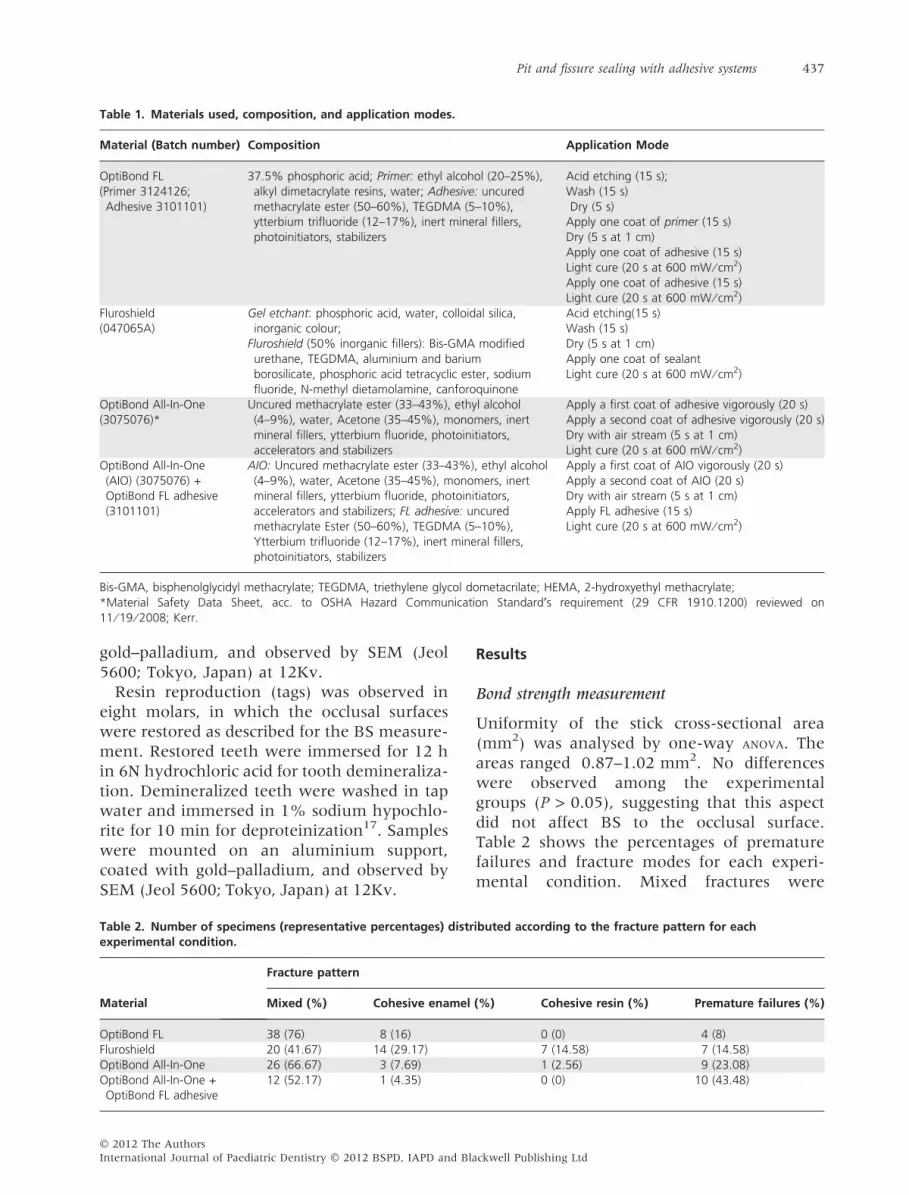
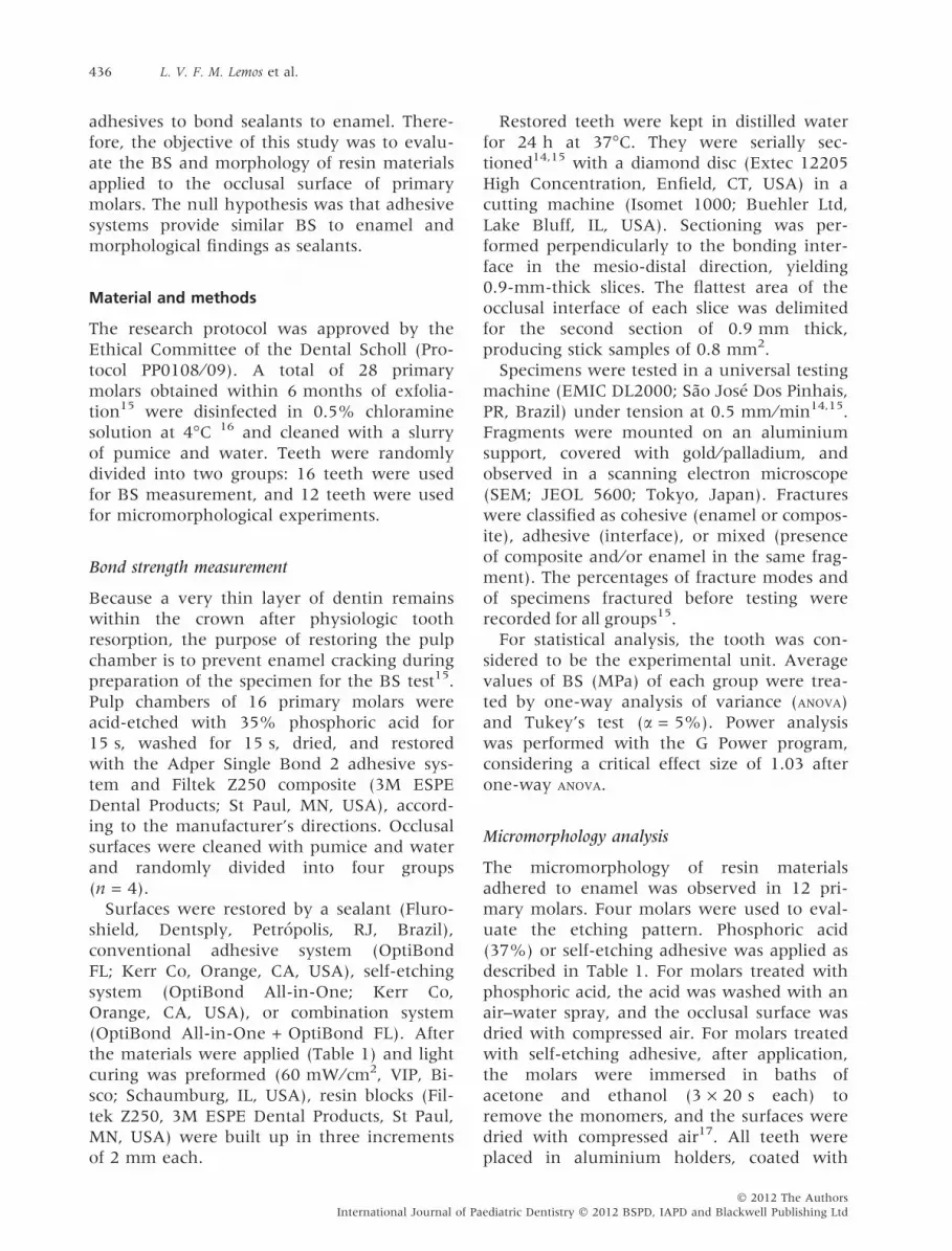
Table 1. Materials used, composition, and application modes.
Material (Batch number) Composition Application Mode
OptiBond FL(Primer 3124126;Adhesive 3101101)
37.5% phosphoric acid; Primer: ethyl alcohol (20–25%),alkyl dimetacrylate resins, water; Adhesive: uncuredmethacrylate ester (50–60%), TEGDMA (5–10%),ytterbium trifluoride (12–17%), inert mineral fillers,photoinitiators, stabilizers
Acid etching (15 s);Wash (15 s)Dry (5 s)
Apply one coat of primer (15 s)Dry (5 s at 1 cm)Apply one coat of adhesive (15 s)Light cure (20 s at 600 mW ⁄ cm2)Apply one coat of adhesive (15 s)Light cure (20 s at 600 mW ⁄ cm2)
Fluroshield(047065A)
Gel etchant: phosphoric acid, water, colloidal silica,inorganic colour;
Fluroshield (50% inorganic fillers): Bis-GMA modifiedurethane, TEGDMA, aluminium and bariumborosilicate, phosphoric acid tetracyclic ester, sodiumfluoride, N-methyl dietamolamine, canforoquinone
Acid etching(15 s)Wash (15 s)Dry (5 s at 1 cm)Apply one coat of sealantLight cure (20 s at 600 mW ⁄ cm2)
OptiBond All-In-One(3075076)*
Uncured methacrylate ester (33–43%), ethyl alcohol(4–9%), water, Acetone (35–45%), monomers, inertmineral fillers, ytterbium fluoride, photoinitiators,accelerators and stabilizers
Apply a first coat of adhesive vigorously (20 s)Apply a second coat of adhesive vigorously (20 s)Dry with air stream (5 s at 1 cm)Light cure (20 s at 600 mW ⁄ cm2)
OptiBond All-In-One(AIO) (3075076) +OptiBond FL adhesive(3101101)
AIO: Uncured methacrylate ester (33–43%), ethyl alcohol(4–9%), water, Acetone (35–45%), monomers, inertmineral fillers, ytterbium fluoride, photoinitiators,accelerators and stabilizers; FL adhesive: uncuredmethacrylate Ester (50–60%), TEGDMA (5–10%),Ytterbium trifluoride (12–17%), inert mineral fillers,photoinitiators, stabilizers
Apply a first coat of AIO vigorously (20 s)Apply a second coat of AIO (20 s)Dry with air stream (5 s at 1 cm)Apply FL adhesive (15 s)Light cure (20 s at 600 mW ⁄ cm2)
Bis-GMA, bisphenolglycidyl methacrylate; TEGDMA, triethylene glycol dometacrilate; HEMA, 2-hydroxyethyl methacrylate;*Material Safety Data Sheet, acc. to OSHA Hazard Communication Standard¢s requirement (29 CFR 1910.1200) reviewed on11 ⁄ 19 ⁄ 2008; Kerr.
Pit and fissure sealing with adhesive systems 437
gold–palladium, and observed by SEM (Jeol
5600; Tokyo, Japan) at 12Kv.
Resin reproduction (tags) was observed in
eight molars, in which the occlusal surfaces
were restored as described for the BS measure-
ment. Restored teeth were immersed for 12 h
in 6N hydrochloric acid for tooth demineraliza-
tion. Demineralized teeth were washed in tap
water and immersed in 1% sodium hypochlo-
rite for 10 min for deproteinization17. Samples
were mounted on an aluminium support,
coated with gold–palladium, and observed by
SEM (Jeol 5600; Tokyo, Japan) at 12Kv.
Table 2. Number of specimens (representative percentages) distrexperimental condition.
Material
Fracture pattern
Mixed (%) Cohesive enamel
OptiBond FL 38 (76) 8 (16)Fluroshield 20 (41.67) 14 (29.17)OptiBond All-In-One 26 (66.67) 3 (7.69)OptiBond All-In-One +OptiBond FL adhesive
12 (52.17) 1 (4.35)
� 2012 The Authors
International Journal of Paediatric Dentistry � 2012 BSPD, IAPD and Bla
Results
Bond strength measurement
Uniformity of the stick cross-sectional area
(mm2) was analysed by one-way ANOVA. The
areas ranged 0.87–1.02 mm2. No differences
were observed among the experimental
groups (P > 0.05), suggesting that this aspect
did not affect BS to the occlusal surface.
Table 2 shows the percentages of premature
failures and fracture modes for each experi-
mental condition. Mixed fractures were
ibuted according to the fracture pattern for each
(%) Cohesive resin (%) Premature failures (%)
0 (0) 4 (8)7 (14.58) 7 (14.58)1 (2.56) 9 (23.08)0 (0) 10 (43.48)
ckwell Publishing Ltd

Table 3. Mean values and standard deviation (MPa) ofbond strength for the experimental groups*.
MaterialMeans(Standard deviations)
OptiBond FL 36.84 (5.7)aFluroshield 24.26 (2.13)bOptiBond All-In-One 17.12 (4.97)bOptiBond All-In-One +OptiBond FL adhesive
9.8 (2.94)c
*Similar letters shows similar means (P > 0.05).
438 L. V. F. M. Lemos et al.
prevalent in all groups, indicating that the
enamel ⁄ adhesive interfaces were tested at
tensile.
Table 3 shows the mean BS values (MPa)
and standard deviations. Power analysis per-
formed after one-way ANOVA revealed a power
of 1.0. Significant differences in BS were
(a) (
(c) (
(e) (
Fig. 1. Etching pattern of 37.5% phosphoric acid gel (a) and Opt
Fluroshield (d), OptiBond All-In-One (e), OptiBond All-In-One + O
International Journal of Pa
found among the materials (P = 0.001). Opti-
Bond FL showed the highest, OptiBond
All-in-One + OptiBond FL adhesive showed
the lowest, and Fluroshield and OptiBond
All-in-One showed similar and intermediate
values of BS.
Micromorphology
Figure 1 shows the results of the micromor-
phological analysis. Dissolution of the prism
periphery after etching in 37.5% phosphoric
acid (i.e., type II etching pattern18) is shown
on Fig. 1a. OptiBond All-in-One resulted in
smooth and undefined etched surfaces
(Fig. 1b). Well-defined resin tags were
observed for OptiBond FL (Fig. 1c). Fluro-
shield (Fig. 1d) and OptiBond All-in-One
(Fig. 1e) showed similar resin tags, which
b)
d)
f)
iBond All-In-One (b); resin tags of OptiBond FL (c),
ptiBond FL adhesive (f). Bar = 10 micrometres.
� 2012 The Authors
ediatric Dentistry � 2012 BSPD, IAPD and Blackwell Publishing Ltd

Pit and fissure sealing with adhesive systems 439
were less defined than those for OptiBond FL.
The combination of OptiBond All-in-One +
OpfiBond FL adhesive did not produce well-
defined resin tags (Fig. 1f).
Discussion
Among the factors that favour caries develop-
ment on the occlusal surface is the eruption
stage, or functional use of the tooth and its
specific anatomy19. Obliteration of pits and
fissures of the tooth by sealants was first
described by Cueto and Buonocore (1967).
Previous studies have shown the effectiveness
of sealing with hydrophilic adhesive systems
in caries prevention7,13. In the case of pri-
mary dentition, the seal should remain at
least until the tooth comes into occlusion,
implying post-eruptive maturation and instal-
lation of self-cleaning20.
Few studies of adhesion to enamel by self-
etching systems on the occlusal surface of pri-
mary teeth have been carried out14,15. Some
studies have shown less enamel conditioning
by self-etching, compared to phosphoric acid
etching, in permanent21 and primary
teeth22,23. The etch-and-rinse approach uses
phosphoric acid to demineralize the enamel.
A washing step is included to remove the acid
gel and create irregularities4 that are filled
with resin5. In the self-etch approach, the
etching and priming steps are combined into
a single procedure, without the need for
washing. Self-etching systems differ from
etch-and-rinse adhesives in several aspects:
for example, they generally have a higher
pH24, because bonding relies on the ability of
self-etching materials to infiltrate and par-
tially dissolve hydroxyapatite for filling by
resin. The effectiveness of this process varies
with the acidity of the self-etch25, although
the performance of self-etching systems can-
not solely be attributed to their acidity26.
Therefore, this study analysed the adhesion of
resin materials with different etching
approaches to the occlusal surface of primary
molars.
OptiBond FL had the highest BS to the
occlusal surface of primary molars, consistent
with the results of previous studies14,15. The
high BS of conventional 3-step adhesives to
� 2012 The Authors
International Journal of Paediatric Dentistry � 2012 BSPD, IAPD and Bla
enamel has been observed consistently in the
literature27 and clinical research10,13. We
observed a relationship between the BS of
this adhesive and the enamel micromorpho-
logy, which displayed a well-defined etching
pattern and resin tags. Daronch et al. (2003)
studied the etching patterns of conventional
and self-etching adhesive systems on primary
molars. They observed that the thicker layer
of aprismatic enamel in the primary tooth
influenced the conditioning etching pattern,
which was discrete in the case of self-etching
and formed resin tags. Similar findings were
observed by Fava et al. (2003). These previous
observations help explain why the BS of
OptiBond All-in-One (which also showed a
relationship between BS and micromorpho-
logy) was lower than that of OptiBond FL.
The BS of Fluroshield was similar to that of
OptiBond All-in-One, but no relationship was
observed between the etching pattern and
BS. The etching pattern of Fluroshield was
similar to that of OptiBond FL, but the resin
tags were similar to those of OptiBond All-in-
One. This result may be explained by the vis-
cosity of the sealant, which contains 50%
inorganic filler, compared with the viscous
OptiBond All-in-One that contains ytterbium
fluoride in some extensions. Hydrophilic
adhesives that are less viscous than the seal-
ant can improve the marginal sealing of
sealants on the enamel surface by filling the
microporosity produced by the acid28.
In general, all-in-one adhesives are associ-
ated with low bonding effectiveness24. The
application of an additional hydrophobic resin
layer of OptiBond FL after the OptiBond
All-in-One adhesive was based on a previous
study that found an improvement in the
immediate BS of some all-in-one adhesives to
enamel29. In the present study, however, the
lowest BS to enamel was achieved with the
combination of OptiBond All-in-One + Opti-
Bond FL adhesive. This low BS may be
explained by a possible mismatch interaction
between the acidic primer, which contains a
high amount of solvents (45% acetone and
9% ethanol), and the OptiBond FL resin.
Adhesives containing solvents have lower
cohesive strengths than hydrophobic adhe-
sives without solvents30 (e.g., OptiBond FL
ckwell Publishing Ltd

440 L. V. F. M. Lemos et al.
adhesive), in part because of the inability to
remove all of the water and solvent mixtures
during clinical application. The BS results for
this group were related to the micromorpho-
logical analysis of enamel and resin tag
formation.
Among the groups, the combination of
OptiBond All-in-One + OptiBond FL resulted
in the most premature debonding specimens
(around 43.48%; Table 2). Premature deb-
onding occurred at the sectioning step of the
method. The micromorphological findings
and high percentage of premature debonding
emphasize the fragility of the enamel bonding
in this association. Unfortunately, most previ-
ous studies have neglected to analyse the
amounts of premature debonding and other
types of fracture modes recorded with micro-
tensile BS31. Such analyses are needed to
improve the understanding of the drawbacks
of the method and to analyse the BS results.
Under the conditions of this study, it may
be concluded that BS to enamel and morpho-
logical findings varied among the studied
materials. For this reason, the null hypothesis
must be rejected. Future studies should ana-
lyse the bonding durability of resin materials
used for occlusal sealing.
Why this study is important to paediatric dentists
d Owing to the risk of saliva contamination in paediatricdentistry, the use of hydrophilic adhesive systems
should be encouraged.d The conventional 3-step etch-and-rinse adhesive is a
possible sealant choice for paediatric dentistry.
Acknowledgements
The authors thank Dr. Karen Barros Parron
Fernandes for the Power Analysis evaluation.
Conflict of Interest
The authors declare no conflict of interest.
References
1 Gwinnett AJ. The ultrastructure of the ‘‘prismless’’
enamel of permanent human teeth. Arch Oral Biol
1967; 12: 381–388.
International Journal of Pa
2 Newman HN, Poole DF. Observations with scanning
and transmission electron microscopy on the
structure of human: surface enamel. Arch Oral Biol
1974; 19: 1135–1143.
3 Mattos-Graner RO, Rontani RMP, Gaviao MBD,
Bocatto HARC. Caries prevalence in 6-36 month-old
Brazilian children. Community Dent Health 1996; 13:
96–98.
4 Buonocore MG. A simple method of increasing the
adhesion of acrylic filling materials to enamel
surfaces. J Dent Res 1955; 34: 849–853.
5 Gwinnett AJ, Buonocore MG. Adhesives and caries
prevention: a preliminary report. Br Dent J 1965;
119: 77–80.
6 Cueto EI, Buonocore MG. Sealing of pits and
fissures with an adhesive resin: its use in caries
prevention. J Am Dent Assoc 1967; 75: 121–128.
7 Grande RHM, Ballester RY, Singer JM, Santos JFF.
Microleakage of a universal adhesive used as a
fissure sealant. Am J Dent 1998; 11: 109–113.
8 Hotuman E, Rolling I, Poulsen S. Fissure sealants in
a group of 3-4-year-old children. Int J Paediatr Dent
1998; 8: 159–160.
9 Zuanon ACC. Microleakage of an adhesive system
used as a fissure sealant. J Contemp Dent Pract 2009;
10: 26–33.
10 Feigal RJ, Musherure P, Gillespie B, Levy-Polack M,
Quelhas I, Hebling J. Improved sealant retention
with bonding agents: a clinical study of two-bottle
and single-bottle systems. J Dent Res 2000; 79: 1850–
1856.
11 Perry AO, Rueggeberg FA. The effect of acid primer
and conventional acid etching on microleakage in a
photoactivated sealant. Pediatr Dent 2003; 25: 127–
131.
12 Pinar A, Sepet E, Aren G, Bolukbasi N, Ulukapi H,
Turan N. Clinical performance of sealants with and
without a bonding agent. Quintessence Int 2005; 36:
355–360.
13 Grande RHM, Lima ACP, Rodrigues Filho LE, Witzel
MF. Clinical evaluation of an adhesive used as a
fissure sealant. Am J Dent 2000; 13: 167–170.
14 Ramires-Romito AC, Reis A, Loguercio AD, de Goes
MF, Grande RH. Micro-tensile bond strength of
adhesive systems applied on occlusal primary
enamel. J Clin Pediatr Dent 2004; 28: 333–338.
15 Ramires-Romito AC, Reis A, Loguercio AD et al.
Microtensile bond strength of sealant and adhesive
systems applied to occlusal primary enamel. Am J
Dent 2007; 20: 114–120.
16 DeWald JP. The use of extracted teeth for in vitro
bonding studies: a review of infection control
considerations. Dent Mater 1997; 13: 74–81.
17 Di Hipolito V, de Goes MF, Carrilho MR, Chan DC,
Daronch M, Sinhoreti MA. SEM evaluation of
contemporary self-etching primers applied to ground
and unground enamel. J Adhes Dent 2005; 7: 203–
211.
18 Silverstone LM, Saxton CA, Dogon IL, Fejerskov O.
Variation in the pattern of acid etching of human
� 2012 The Authors
ediatric Dentistry � 2012 BSPD, IAPD and Blackwell Publishing Ltd

Pit and fissure sealing with adhesive systems 441
dental enamel examined by scanning electron
microscopy. Caries Res 1975; 9: 373–387.
19 Arrow P. Oral hygiene in the control of occlusal
caries. Community Dent Oral Epidemiol 1998; 26: 324–
330.
20 De Menezes Oliveira MA, Torres CP, Gomes-Silva
JM et al. Microstructure and mineral composition of
dental enamel of permanent and deciduous teeth.
Microsc Res Tech 2010; 73: 572–577.
21 Hashimoto M, Ohno H, Yoshida E et al. Resin-
enamel bonds made with self-etching primers on
ground enamel. Eur J Oral Sci 2003; 111: 447–453.
22 Daronch M, De Goes MF, Grande RH, Chan DC.
Antibacterial and conventional self-etching primer
system: morphological evaluation of intact primary
enamel. J Clin Pediatr Dent 2003; 27: 251–256.
23 Fava M, Myaki SI, Arana-Chavez VE, Fava-
de-Moraes F. Effects of a non-rinse conditioner on
the enamel of primary teeth. Braz Dent J 2003; 14:
168–171.
24 Van Meerbeek B, Yoshihara K, Yoshida Y, Mine A,
De Munck J, Van Landuyt KL. State of the art of
self-etch adhesives. Dent Mater 2011; 27: 17–28.
25 Pashley DH, Tay FR. Aggressiveness of
contemporary self-etching adhesives Part II: etching
� 2012 The Authors
International Journal of Paediatric Dentistry � 2012 BSPD, IAPD and Bla
effects on unground enamel. Dent Mater 2001; 17:
430–444.
26 Moura SK, Reis A, Dal-Bianco K, Loguercio AD,
Arana-Chavez VE, Grande RH. Bond strength and
morphology of enamel using self-etching adhesive
systems with different acidities. J Adhes Dent 2005;
8: 75–83.
27 Pashley DH, Tay FR, Breschi L et al. State of the art
etch-and-rinse adhesives. Dent Mater 2011; 27: 1–16.
28 Irinoda Y, Matsumura Y, Kito H et al. Effect of
sealant viscosity on the penetration of resin into
etched human enamel. Oper Dent 2000; 25: 274–
282.
29 Albuquerque M, Pegoraro M, Mattei G, Reis A,
Loguercio AD. Effect of double application or a
hydrophobic layer for improvement efficacy of one-
step self-etch systems in enamel and dentin. Oper
Dent 2008; 33: 564–570.
30 Shinchi MJ, Soma K, Nakabayashi N. The effect of
phosphoric acid concentration on resin tag length
and bond strength of a photo-cured resin to acid-
etched enamel. Dent Mater 2000; 16: 324–329.
31 Roulet JF, Van Meerbeek B. Editorial: statistics: a
nuisance, a tool, or a must? J Adhes Dent 2007; 9:
287–288.
ckwell Publishing Ltd