biophysical studies on interactions and assembly of full-size e3 ubiquitin ligase suppressor of...
TRANSCRIPT

Biophysical Studies on Interactions and Assembly of Full-sizeE3 Ubiquitin LigaseSUPPRESSOR OF CYTOKINE SIGNALING 2 (SOCS2)-ELONGIN BC-CULLIN 5-RING BOXPROTEIN 2 (RBX2)*
Received for publication, October 29, 2014, and in revised form, November 28, 2014 Published, JBC Papers in Press, December 11, 2014, DOI 10.1074/jbc.M114.616664
Emil Bulatov‡§, Esther M. Martin¶, Sneha Chatterjee¶, Axel Knebel�, Satoko Shimamura**, Albert Konijnenberg¶,Clare Johnson�, Nico Zinn**, Paola Grandi**, Frank Sobott¶1, and Alessio Ciulli‡§2
From the ‡Division of Biological Chemistry and Drug Discovery, College of Life Sciences, and the �Medical Research CouncilPhosphorylation and Ubiquitylation Unit, College of Life Sciences, Sir James Black Center, University of Dundee, Dundee DD1 5EH,Scotland, United Kingdom, the §Department of Chemistry, University of Cambridge, Cambridge CB2 1EW, United Kingdom, the¶Department of Chemistry, University of Antwerp, 2020 Antwerp, Belgium, and **Cellzome GmbH, Meyerhofstrasse 1,69117 Heidelberg, Germany
Background: The component subunits of CRL E3 ligases assemble into specific complexes.Results: Components of CRL5SOCS2 were identified from human cell lysate, the full-size complex was reconstituted in vitro, andprotein-protein interactions were biophysically characterized.Conclusion: CRL5SOCS2 components exist in a monomeric state, and proposed structural models are supported by ion mobilitymass spectrometry.Significance: We provide structural insights into the assembly of full-size CRL5SOCS2 that can aid development of smallmolecules targeting CRL complexes.
The multisubunit cullin RING E3 ubiquitin ligases (CRLs) tar-get post-translationally modified substrates for ubiquitinationand proteasomal degradation. The suppressors of cytokine sig-naling (SOCS) proteins play important roles in inflammatoryprocesses, diabetes, and cancer and therefore represent attrac-tive targets for therapeutic intervention. The SOCS proteins,among their other functions, serve as substrate receptors ofCRL5 complexes. A member of the CRL family, SOCS2-EloBC-Cul5-Rbx2 (CRL5SOCS2), binds phosphorylated growth hor-mone receptor as its main substrate. Here, we demonstrate thatthe components of CRL5SOCS2 can be specifically pulled fromK562 human cell lysates using beads decorated with phosphor-ylated growth hormone receptor peptides. Subsequently,SOCS2-EloBC and full-length Cul5-Rbx2, recombinantlyexpressed in Escherichia coli and in Sf21 insect cells, respec-tively, were used to reconstitute neddylated and unneddylatedCRL5SOCS2 complexes in vitro. Finally, diverse biophysicalmethods were employed to study the assembly and interactionswithin the complexes. Unlike other E3 ligases, CRL5SOCS2 wasfound to exist in a monomeric state as confirmed by size exclu-sion chromatography with inline multiangle static light scatter-
ing and native MS. Affinities of the protein-protein interactionswithin the multisubunit complex were measured by isothermaltitration calorimetry. A structural model for full-size neddy-lated and unneddylated CRL5SOCS2 complexes is supported bytraveling wave ion mobility mass spectrometry data.
Cullin RING E3 ubiquitin ligases (CRLs)3 represent the larg-est known family of E3 enzymes in the ubiquitin-proteasomesystem (�200 CRLs of a total of �600 E3 enzymes) and play asignificant role in cancer and other diseases (1). In some types ofcells, up to 20% of the proteasome-dependent degradation ismediated by CRLs (2). Assembly of the multisubunit CRLs wasinitially reported for the archetypal Skp1-Cul1-Rbx1 complex(3). CRLs use modular subunit organization consisting of inter-changeable adaptors (Skp1, elongin B, elongin C, DDB1, andBTB), substrate receptors (F box, SOCS box, DCAF, and BTB),cullin scaffolds (cullin 1–7), and RING domain proteins (Rbx1and Rbx2) to enable the assembly of a large number of function-ally diverse E3 ligase complexes (1, 4, 5).
* This work was supported by United Kingdom Biotechnology and BiologicalSciences Research Council David Phillips Fellowship BB/G023123/1 (toA. C.), European Research Council Starting Grant ERC-2012-StG-311460DrugE3CRLs (to A. C.), and the Government of the Republic of Tatarstan(Ph.D. studentship to E. B.). Molecular biophysics was supported by Well-come Trust strategic award 100476/Z/12/Z (to the University of Dundee).The Synapt G2 mass spectrometer was funded by a grant from the Hercu-les Foundation-Flanders.Author’s Choice—Final version full access.
1 A Francqui Research Professor at the University of Antwerp.2 To whom correspondence should be addressed: College of Life Sciences,
University of Dundee, Dow Street, Dundee DD15EH, Scotland, United King-dom. Tel.: 44-1382-386230; E-mail: [email protected].
3 The abbreviations used are: CRL, cullin-RING E3 ubiquitin ligase; BTB, bric-a-brac�tram-track�broad complex; CCS, collision cross-section(s); CRL5SOCS2,SOCS2-EloBC-Cul5-Rbx2 complex; CTD, C-terminal domain; Cul5, cullin 5;DCAF, DDB1 and Cul4-associated factor; DDB1, DNA damage-binding pro-tein 1; EloB, elongin B; EloC, elongin C; EloBC, elongin B-elongin C complex;EHSS, exact hard sphere scattering; FBXO31, F box-only protein 31; GHR,growth hormone receptor; TWIM-MS, travelling wave ion mobility-massspectrometry; ITC, isothermal titration calorimetry; NEDD8, neural precur-sor cell expressed developmentally down-regulated protein 8; NTD, N-ter-minal domain; PA, projection approximation; PDB, Protein Data Bank; Rbx,RING box protein; Skp, S-phase kinase-associated protein; SEC-MALS, sizeexclusion chromatography and multiangle light scattering; SOCS2, sup-pressor of cytokine signaling 2; TCEP, tris(2-carboxyethyl) phosphinehydrochloride; TEV, tobacco etch virus; SH2, Src homology 2.
THE JOURNAL OF BIOLOGICAL CHEMISTRY VOL. 290, NO. 7, pp. 4178 –4191, February 13, 2015Author’s Choice © 2015 by The American Society for Biochemistry and Molecular Biology, Inc. Published in the U.S.A.
4178 JOURNAL OF BIOLOGICAL CHEMISTRY VOLUME 290 • NUMBER 7 • FEBRUARY 13, 2015
by guest on October 8, 2015
http://ww
w.jbc.org/
Dow
nloaded from
The N-terminal domain (NTD) of cullin proteins consists ofthree five-helix bundles (“cullin repeats”) that form a long stalkarchitecture, and a globular C-terminal domain (CTD, or “cul-lin homology domain”). Cullin NTD recruits variable substratereceptors either directly or via an adaptor protein, whereas cul-lin CTD serves as a docking site for RING domain proteins thatin turn recruit a cognate ubiquitin-loaded E2 (6). RING domainproteins contain a distinct Zn2�-binding domain characterizedby a canonical RING motif. Selection of the substrate receptorsfor a particular CRL occurs through a specific receptor LPXPmotif that forms a minor yet crucial supplementary interactionwith cullin NTD (7, 8).
SOCS2 is a member of the SOCS box protein family that, inassociation with the adaptor elongin B-elongin C complex(EloBC), cullin 5 scaffold, and Rbx2, constitutes a CRL5SOCS2
E3 ligase. SOCS2 contains three structural domains: a con-served C-terminal SOCS box domain that binds to adaptorEloBC; a central SH2 domain mediating recruitment of phos-phorylated tyrosine-containing sequence of the substrate; and avariable N-terminal region that facilitates interaction with thesubstrate. CRL5SOCS2 negatively regulates growth hormonesignaling by targeting growth hormone receptor (GHR) forubiquitination and proteasomal degradation (9). Phosphory-lated tyrosine 595 serves as the key structural determinant ofGHR recognition by the SH2 domain of SOCS2 (10). Crystalstructures of SOCS2-EloBC (PDB code 2C9W) (11) andSOCS2-EloBC-Cul5NTD (PDB code 4JGH) (12) describe struc-tural features of the protein-protein interfaces within these left-arm complexes. However, the details of assembly of the full-sizeCRL5SOCS2 E3 complex both in vivo and in vitro remainmissing.
In recent years, interest in studying the structure, function,and assembly of CRLs has been growing, notably driven in partby their potential role as drug targets in a number of humandiseases (13–16). However, only a few studies have investigatedthe full-size CRL complexes biophysically, primarily due to dif-ficulties in obtaining some of the protein components recom-binantly, in particular full-length cullins. Furthermore, largeheteromeric protein complexes such as CRLs are notoriouslydifficult to crystallize into diffraction quality crystals. There-fore, it seems promising to engage the strengths of diverse bio-physical methods in order to facilitate characterization of boththe individual subunits and the full-size complexes as well asto provide a means for examining their association andinteractions.
Here, we show that all components of the CRL5SOCS2
could be pulled down from a cell lysate via SOCS2-mediatedrecognition of the phosphorylated GHR_pY595 peptideimmobilized on beads. The full-length E3 ligase complex wasthen reconstituted in vitro using purified recombinant pro-teins and characterized biophysically. Investigations ofassembly and interactions within the complex were carriedout using size exclusion chromatography and multianglelight scattering (SEC-MALS), isothermal titration calorime-try (ITC), and nanoelectrospray traveling wave-ion mobilitymass spectrometry (TWIM-MS).
EXPERIMENTAL PROCEDURES
Pull-down Experiments—Pull-down experiments were per-formed using biotinylated GHR-derived 11-mer peptides phos-phorylated (GHR_pY595) or not (GHR_Y595) on tyrosine 595,harboring an aminohexanoic acid as spacer after the biotin(Biotin-aminohexanoic acid-PVPDpYTSIHIV-amide), andimmobilized on high capacity Neutravidin beads. Competitionexperiments were performed by incubating human K562 totalcell lysate with 100 �M non-biotinylated phosphorylated pep-tide (GHR_pY595) and the immobilized (Biotin-amino-hexanoic acid-PVPDpYTSIHIV-amide) beads for 2 h at 4 °C.After washing, bound proteins were eluted with SDS-samplebuffer and prepared for tandem mass tags labeling and MS anal-ysis as described previously (17).
Protein Expression and Purification—Recombinant humanSOCS2 (amino acids 32–198), elongin C (amino acids 17–112),and elongin B (amino acids 1–118) were co-expressed in Esch-erichia coli BL21(DE3) from the pLIC (SOCS2) and pCDF_Duet (EloBC) plasmids (gifts from A. Bullock, StructuralGenomics Consortium, Oxford, UK). A starter culture wasgrown overnight from a single transformant colony using 50 mlof LB medium containing 100 �g/ml ampicillin and 50 �g/mlstreptomycin. Starter culture then was used to inoculate 7 litersof LB medium containing 100 �g/ml ampicillin and 50 �g/mlstreptomycin. The cells were grown at 37 °C until A600 �0.7 andcooled to 18 °C, and then protein expression was induced with1 mM isopropyl �-D-1-thiogalactopyranoside for 12 h.
Recombinant human Cul5NTD (N-terminal domain, residues1–386) was expressed in E. coli BL21(DE3) from a pNIC plas-mid encoding sequence for Cul5NTD, containing His6 andFLAG tags at the C-terminal end and a tobacco etch virus(TEV) cleavage site, as described previously (18). Briefly, 50 mlof starter culture was grown overnight using a single transfor-mant colony in LB medium containing 50 �g/ml kanamycinand used to inoculate 2 liters of LB medium supplemented with50 �g/ml kanamycin. The cells were grown at 37 °C until A600�0.7 and cooled to 18 °C, and protein expression was inducedwith 0.5 mM isopropyl �-D-1-thiogalactopyranoside for 12 h.
Recombinant SOCS2-EloBC and Cul5NTD were indepen-dently purified using the following protocol. The cell pelletswere harvested by centrifugation at 5,000 rpm and 4 °C for 30min and resuspended in binding buffer (50 mM HEPES, pH 7.5,500 mM NaCl, 5% glycerol, 0.5 mM TCEP). The supernatant wastreated with 10 �g/ml DNase I, 10 mM MgCl2 for 30 min andthen filtered through a 0.22-�m filter. The sample was appliedon a HisTrap column (GE Healthcare), and the resin waswashed with wash buffer (50 mM HEPES, pH 7.5, 20 mM imid-azole, 500 mM NaCl, 5% glycerol, 0.5 mM TCEP) and then thebound proteins were eluted with an incremental gradient ofelution buffer (50 mM HEPES, pH 7.5, 500 mM imidazole, 500mM NaCl, 5% glycerol, 0.5 mM TCEP). Fractions containingprotein were pooled, and the His6 tag was cleaved off by over-night dialysis in the presence of TEV protease at 4 °C in bindingbuffer. The protein was applied to a HisTrap column for a sec-ond time, collecting the flow-through, and then concentratedand purified on a HiLoad 16/60 Superdex 75 column with run-ning buffer 25 mM HEPES, pH 7.5, 250 mM NaCl, 0.5 mM TCEP.
Assembly and Interactions of CRL5SOCS2 E3 Ligase
FEBRUARY 13, 2015 • VOLUME 290 • NUMBER 7 JOURNAL OF BIOLOGICAL CHEMISTRY 4179
by guest on October 8, 2015
http://ww
w.jbc.org/
Dow
nloaded from

Recombinant human Cul5 (amino acids 1–780) and Rbx2(amino acids 1–113) were co-expressed in Sf21 insect cellsusing pFastBacTM Dual vector in the Bac-to-Bac� baculovirusexpression system. In this vector, Cul5 is N-terminally taggedwith a fragment of bacterial PBP5 (Dac tag) (19), which can beremoved with TEV protease, as described previously (20).
Bacmids for Dac-TEV-Cul5/Rbx2 were generated inDH10BAC cells and transfected into Sf21 cells, using CellfectinII� reagent (Invitrogen). The transfected cells were kept for 7days at 27 °C in Insect Express� medium (Lonza), supple-mented with ANTI-ANTI� (Invitrogen). The cells were sedi-mented, and the virus-containing supernatant was used toinfect 150 ml of Sf21 culture at a density of 1.5 � 106 cells/ml.For the expression of RING E3 ligases, we supplemented theInsect Express medium with 5 �M ZnCl2. After 5 days the cells,were collected under sterile conditions, and the supernatantwas used to infect 2 liters of Sf21 cell culture for 3 days. TheCul5-Rbx2 protein complex was purified using the followingprocedure. Cells were harvested by centrifugation at 3,500 rpmand 4 °C for 15 min and then resuspended in 25 ml of 50 mM
HEPES, pH 7.4, 0.1 mM EGTA, 1 �M ZnCl2, 1 mM TCEP, 1 mM
Pefabloc�, and 20 �g/ml leupeptin (both from Apollo Scien-tific) and incubated for 15 min at 4 °C. Cells were sheared usinga 50-ml tight fit Dounce homogenizer, and insoluble materialwas removed by centrifugation at 40,000 rpm and 10 °C for 20min. To perform Dac affinity purification, the supernatant wasgently mixed with ampicillin-Sepharose at room temperaturefor 50 min. The Sepharose was collected by centrifugation andwashed six times in 10 volumes of 50 mM HEPES, pH 7.4, 150mM NaCl, 0.1 mM EGTA, 1 �M ZnCl2, 1 mM TCEP. To recoveruntagged Cul5-Rbx2, the Sepharose was incubated with TEVprotease (50 �g of TEV, 1 ml of Sepharose) overnight at roomtemperature and drained and washed through Econopac� filterunits (Bio-Rad). The Cul5-Rbx2 protein was concentrated andfurther purified by preparative size exclusion chromatographyusing HiLoad 16/600 Superdex 200 column (GE Healthcare)with 50 mM HEPES, pH 7.4, 150 mM NaCl, 10% glycerol, 0.5 mM
TCEP.The identity and purity of all obtained proteins was con-
firmed using denaturing electrospray ionization-MS and SDS-PAGE. All proteins were flash-frozen using liquid nitrogen andstored at �80 °C.
Reconstitution of SOCS2-EloBC-Cul5NTD and SOCS2-EloBC-Cul5-Rbx2 Protein Complexes—To form quaternary SOCS2-EloBC-Cul5NTD complex, Cul5NTD and SOCS2-EloBC weremixed together at a 1:1.1 molar ratio and incubated at roomtemperature for 30 min, following purification of the complexusing a HiLoad 16/600 Superdex 75 column in 25 mM HEPES,pH 7.5, 250 mM NaCl, 0.5 mM TCEP.
To form the pentameric complex, Cul5-Rbx2 and SOCS2-EloBC were mixed at a 1:1.1 molar ratio and incubated at roomtemperature for 30 min. The protein complex was purifiedusing a HiLoad 16/600 Superdex 200 column in 50 mM HEPES,pH 7.4, 150 mM NaCl, 10% glycerol, 0.5 mM TCEP. SDS-PAGEanalysis of SOCS2-EloBC, Cul5NTD, SOCS2-EloBC-Cul5NTD,Cul5-Rbx2, and SOCS2-EloBC-Cul5-Rbx2 is shown in Fig. 3B.
Formation of NEDD8�Cul5-Rbx2 Covalent Conjugate—Toobtain the conjugate, E1 activating enzyme APP-BP1/UBA3 (1
�M), E2 conjugating enzyme UBE2F (5 �M), NEDD8 (40 �M),and Cul5-Rbx2 (5 �M) were incubated at 37 °C for 1 h in 50 mM
HEPES, pH 7.4, 150 mM NaCl, 5 mM DTT, 10 mM MgCl2, 0.2mM ATP. A negative control experiment was performed in thesame solution containing 0.1 mM EDTA, with no MgCl2 or ATPadded. The completion of the neddylation on the Cul5 subunitwas confirmed by SDS-PAGE (Fig. 8).
SEC-MALS—SEC-MALS experiments were performed usingDionex Ultimate 3000 UHPLC system (Thermo Scientific) withan inline Wyatt miniDAWN TREOS MALS detector and Opti-lab T-rEXTM refractive index detector. Molar masses spanningelution peaks were calculated using ASTRA version 6.0.0.108(Wyatt). SEC-MALS data were collected for the following sam-ples: 1) SOCS2-EloBC at 130 �M; 2) Cul5NTD at 110 �M; 3)SOCS2-EloBC-Cul5NTD at 90 �M; 4) Cul5-Rbx2 at 30 �M; 5)SOCS2-EloBC-Cul5-Rbx2 at 35 �M. The experiments wereperformed using a Superdex 200 10/300 GL column (GEHealthcare) with running buffer 50 mM HEPES, pH 7.5, 150 mM
NaCl, 0.5 mM TCEP. The scattering signal was collected at 44,90, and 136° using � � 658.5 nm incident light. Resulting datawere processed in Microsoft Excel, and peaks were normalized.
TWIM-MS—The native TWIM-MS experiments were con-ducted on a Synapt HDMS G2 instrument (Waters, Milford,MA), which has been described previously (21). Samples fol-lowing gel filtration were buffer-exchanged into 500 mM aque-ous ammonium acetate at pH 7.0, using Micro Bio-Spin P-6columns (Bio-Rad), at concentrations in the range of 5–10 �M.Aliquots of 3–5 �l were transferred to gold-coated nano-elec-trospray ionization needles prepared in house. The instrumentwas tuned to ensure the preservation of non-covalent interac-tions (22), using the following parameters: capillary, 1.2 kV;sample cone, 40 V; extraction cone, 0.5 V; nanoflow gas pres-sure, 0.3 bar; trap collision energy, 4.0 V; transfer collisionenergy, 3.5 V; backing pressure, 4 millibars, trap pressure, 3.4millibars. For the measurement of the full 148-kDa complex,SOCS2-EloBC-Cul5-Rbx2, the backing pressure was increasedto 5 millibars to facilitate the transmission of high m/z signal.Gas pressure in the ion mobility cell was 3.0 millibars, andhelium and N2 gas flows were 180 and 90 ml/min, respectively,with a trap bias of 50 V. The traveling wave velocity was 800 m/swith a traveling wave height of 40 V. The data were acquiredand processed with MassLynx version 4.1 software (Waters),and drift times were extracted using Driftscope version 2.3(Waters). The experimental collision cross-sections (CCS) ofthe protein complexes were determined by calibration withknown protein cross-sections determined under native condi-tions as described previously (23).
Calculation of Theoretical CCS—Theoretical CCS values ofthe protein complexes were calculated from model structures,obtained by docking individual protein subunits together, usingthe program MOBCAL with both the projection approxima-tion (PA) and the exact hard sphere scattering (EHSS) methods(24, 25). The PDB files were cleaned (i.e. by resolving dihedralconflicts and adding missing side chains and removing crystalwater molecules) prior to the PA or EHSS calculation. The the-oretical CCS was compared with the experimental CCS of thelowest available charge state for that species in the mass spectra,
Assembly and Interactions of CRL5SOCS2 E3 Ligase
4180 JOURNAL OF BIOLOGICAL CHEMISTRY VOLUME 290 • NUMBER 7 • FEBRUARY 13, 2015
by guest on October 8, 2015
http://ww
w.jbc.org/
Dow
nloaded from
which corresponds to the most native-like structure of the pro-tein complex (Fig. 4, A–C, bottom panels).
ITC—Experiments were conducted using an iTC200 micro-calorimeter instrument (GE Healthcare). GHR_pY595 peptide(350 �M, PVPDpYTSIHIV-amide), GHR_Y595 (350 �M, PVP-DYTSIHIV-amide), and phosphotyrosine (2 mM, Tyr(P)) weretitrated into SOCS2-EloBC (30 �M) at 298 K. Temperature-de-pendent experiments to study interaction between SOCS2-EloBC and Cul5NTD were performed by titrating Cul5NTD (450�M) into SOCS2-EloBC (60 �M) at 298, 303, and 308 K. Prior toall titration experiments, sample proteins were dialyzed into 50mM HEPES, pH 7.5, 250 mM NaCl, 0.5 mM TCEP. Peptides andTyr(P) were dissolved in the same buffer. Obtained data wereanalyzed and fitted using the Microcal Origin version 7.0 soft-ware package. Binding enthalpy, dissociation constants, andstoichiometry were determined by fitting the data using a one-set-of-site binding model.
Molecular Modeling of Protein Complexes—Due to theabsence of an Rbx2 crystal structure, its closest homolog, Rbx1,was used for the model construction. The structural model ofthe SOCS2-EloBC-Cul5-Rbx1 complex was prepared inPyMOL, using the crystal structure of Cul1-Rbx1-Skp1-Skp2
(PDB code 1LDK) (26) as the initial template. To construct themodel, SOCS2-EloBC-Cul5NTD (PDB code 4JGH) (12) wassuperimposed on the template by aligning its Cul5NTD subunitwith the Cul1NTD of the template. After that, Cul5CTD-Rbx1(PDB code 3DPL) (27) was aligned with Cul1CTD subunit of thetemplate to generate a model of the full-length E3 ligase. Theresulting model of the CRL5SOCS2 complex was used to obtainthe model for Cul5-Rbx1. To generate a model of the “open”neddylated complexes, NEDD8�Cul5CTD-Rbx1 (PDB code3DQV) (27) was aligned with the Cul1CTD subunit of the tem-plate. Alternatively, to prepare a model of the closed neddylatedcomplexes, NEDD8 was simply added from aligned 3DQVonto the non-neddylated Cul5CTD-Rbx1 and SOCS2-EloBC-Cul5-Rbx1.
RESULTS
Components of CRL5SOCS2 E3 Ligase Can Be Pulled Downfrom Human Cell Lysates Using Phosphopeptide-modifiedBeads—Specific subunits of E3 ligase SOCS2, EloB, EloC, Cul5,and Rbx2 are known to function as a CRL5SOCS2 complex (28).The SH2 domain of SOCS2 recognizes and specifically bindsa GHR sequence containing the phosphorylated tyrosine
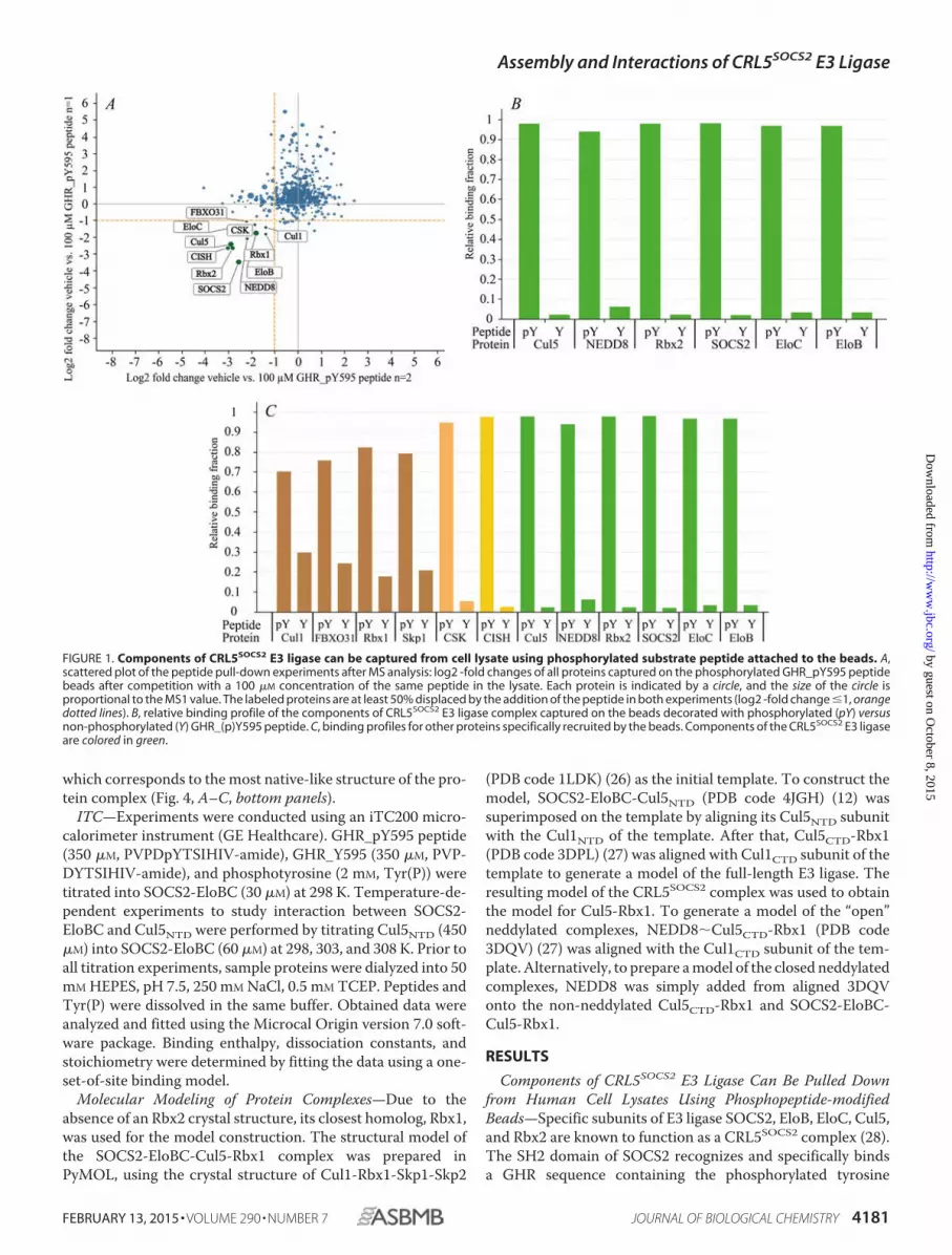
FIGURE 1. Components of CRL5SOCS2 E3 ligase can be captured from cell lysate using phosphorylated substrate peptide attached to the beads. A,scattered plot of the peptide pull-down experiments after MS analysis: log2 -fold changes of all proteins captured on the phosphorylated GHR_pY595 peptidebeads after competition with a 100 �M concentration of the same peptide in the lysate. Each protein is indicated by a circle, and the size of the circle isproportional to the MS1 value. The labeled proteins are at least 50% displaced by the addition of the peptide in both experiments (log2 -fold change �1, orangedotted lines). B, relative binding profile of the components of CRL5SOCS2 E3 ligase complex captured on the beads decorated with phosphorylated (pY) versusnon-phosphorylated (Y) GHR_(p)Y595 peptide. C, binding profiles for other proteins specifically recruited by the beads. Components of the CRL5SOCS2 E3 ligaseare colored in green.
Assembly and Interactions of CRL5SOCS2 E3 Ligase
FEBRUARY 13, 2015 • VOLUME 290 • NUMBER 7 JOURNAL OF BIOLOGICAL CHEMISTRY 4181
by guest on October 8, 2015
http://ww
w.jbc.org/
Dow
nloaded from
Tyr(P)595. We envisaged that SOCS2 and components of full-length CRL5SOCS2 E3 ligase should be amenable for capturingfrom cell lysate using substrate peptides immobilized on beads.With this aim, we performed pull-down experiments fromhuman K562 cell lysate using beads decorated with bothphosphorylated GHR_pY595 (PVPDpYTSIHIV-amide, posi-tive control) and non-phosphorylated GHR_Y595 (PVP-DYTSIHIV-amide, negative control) peptides. Mass spectrom-etry analysis revealed a reproducible and limited set of proteinscaptured and subsequently displaced by the phosphorylatedpeptide (Fig. 1A, bottom left corner). All components of theCRL5SOCS2 (SOCS2, EloB, EloC, Cul5, and Rbx2) were amongthis protein set.
Binding profile (Fig. 1B) shows that endogenous componentsof CRL5SOCS2 E3 ligase were only captured by GHR_pY595-beads. In contrast, no significant capturing from cell lysates wasobserved with non-phosphorylated GHR_Y595-containingbeads (Fig. 1B). This observation shows that the Tyr(P) residueplays a key role in recognition of the substrate by the CRLSOCS2
complex in the cell and that phosphorylation of the peptide isessential for specific interaction with the E3 ligase. Interest-ingly, we also detected NEDD8 as a protein specifically pulleddown by the phosphorylated peptide. NEDD8 is a ubiquitin-likeprotein that is known to be covalently attached to cullins andacts as a CRL activator by inducing scaffold dynamics andincreasing conformational flexibility of the E3 enzyme (27, 29).Identification of NEDD8 suggests that the active neddylatedcomplex is also being pulled down in the assay.
Surprisingly, in addition to the expected CRL5SOCS2 complexsubunits, we detected subunits of the CRL1FBXO31 E3 ligase,namely FBXO31, Rbx1, Cul1, and Skp1 proteins, as being cap-
tured by the beads and displaced by the phosphorylated peptide(Fig. 1A and Table 1), indicating specific binding. FBXO31 is anF box protein that binds phosphorylated substrates; therefore,it could have been recruited by the GHR_pY595 peptide directly.However, significant recruitment of the four CRL1FBXO31 subunitswas also observed by the non-phosphorylated GHR peptide(Fig. 1C). This would imply a degree of phosphorylation-inde-pendent interaction, either directly with the beads or indirectlyvia binding to the components of the CRL5SOCS2 E3 ligasecomplex.
Moreover, CSK (C-terminal Src kinase) and CISH (cytokine-inducible SH2-containing) proteins were also recruited (Fig. 1Aand Table 1). Both CSK and CISH contain the SH2 domain;therefore, both proteins were probably directly recruited by thephosphorylated peptide.
SOCS2-EloBC Forms a Weak Interaction with GHR and aTight Interaction with Cul5NTD—To determine the affinity ofinteraction and the thermodynamic parameters of bindingbetween SOCS2-EloBC and substrate GHR or scaffoldCul5NTD, we performed isothermal titration calorimetry exper-iments (Fig. 2). SOCS2-EloBC binds GHR_pY595 peptide withKd � 1.8 �M, which is consistent with the previously reportedvalue (11), and binds Tyr(P) with Kd � 191 �M, both at 298 K(Fig. 2A). The binding affinity for phosphotyrosine is �100-foldweaker than for the phosphorylated peptide, suggesting thatother peptide residues make some contribution to interactionwith the protein. However, negative control titration usingnon-phosphorylated GHR_Y595 peptide showed no binding(Fig. 2A), reinforcing the key contribution of the phosphategroup to substrate binding.
We next determined the affinity of SOCS2-EloBC for Cul5NTD bymeasuring a Kd � 11 nM for the interaction (Fig. 2C). These dataare in good agreement with the previously reported Kd of 28 nM
(by ITC) (12). In addition, two groups independently reportedKd � 7 nM (by ITC) (30), Kd � 10 nM (by ITC), and Kd � 47 nM
(by surface plasmon resonance) (31) for this interaction, albeitusing SOCS box domain instead of the whole SOCS2 protein incomplex with EloBC.
To test the potential cooperativity of interactions at theGHR/SOCS2-EloBC/Cul5NTD interfaces, we performed titra-tion of GHR_pY595 peptide into SOCS2-EloBC-Cul5NTD andtitration of Cul5NTD into GHR_pY595-SOCS2-EloBC complex.No change in the Kd or �H values was observed in eithercase, suggesting no cooperativity or cross-talk between theseinteractions.
The interaction between SOCS2-EloBC and the Cul5 scaf-fold is high affinity and crucial to the assembly of CRL complex.To provide further insights into the nature of this interaction,we performed temperature-dependent ITC titrations anddetermined a change in heat capacity �CP � �450 cal/mol/K(titration curves shown in Fig. 2B). Fig. 2D demonstrates a plotwith a temperature-dependent change of thermodynamicparameters of SOCS2-EloBC/Cul5NTD interaction. The exper-imental �CP value is calculated from the slope of the �H linearregression. As a comparison, previously reported �CP valuesfor ASB9-EloBC/Cul5NTD and Vif-EloBC/Cul5NTD interac-tions were found to be �350 cal/mol/K (18) and �300 cal/mol/K (30), respectively.
TABLE 1Proteins enriched by the phosphorylated GHR_pY595-modifiedbeadsThe proteins were specifically captured by the beads and displaced by the phosphor-ylated GHR peptide.
Protein UniProt ID Comments
SOCS2 O14508 Suppressor of cytokine signaling 2. Substraterecognition domain of Cul5SOCS2 E3 ligase.Contains SH2 domain that recognizessubstrate phosphotyrosine residues.
EloB Q15370 Transcription elongation factor B.EloC Q15369 Transcription elongation factor C. Complex of
EloB and EloC (EloBC) serves as adaptordomain of Cul5SOCS2 E3 ligase.
Cul5 Q93034 Cullin 5. Scaffold domain of the Cul5SOCS2 E3ligase.
Rbx2 Q9UBF6 RING box protein 2. Contains RING-type zincfinger, recruits E2 conjugating enzyme.
NEDD8 Q15843 Neural precursor cell expressed developmentallydown-regulated protein 8. Ubiquitin-likeprotein, can form covalent conjugate withCullin that enhances the E3 ligase activity.
CISH Q9NSE2 Cytokine-inducible SH2-containing protein.Component of SCF E3 ligase, can recognizephosphotyrosine.
Rbx1 P62877 RING-box protein 1. Contains RING-type zincfinger, recruits E2 conjugating enzyme.
CSK P41240 c-Src kinase. Contains SH2 domain that canrecognize phosphotyrosine residues.
Cul1 Q13616 Cullin 1. Scaffold component of SCF E3 ligase.FBXO31 Q5XUX0 F box-only protein. Substrate recognition
component of SCF E3 ligase. Can recognizecertain phosphorylated substrates.
Skp1 P63208 S-phase kinase-associated protein 1. Adaptorcomponent of SCF E3 ligase.
Assembly and Interactions of CRL5SOCS2 E3 Ligase
4182 JOURNAL OF BIOLOGICAL CHEMISTRY VOLUME 290 • NUMBER 7 • FEBRUARY 13, 2015
by guest on October 8, 2015
http://ww
w.jbc.org/
Dow
nloaded from

We next calculated the theoretical solvent-accessible surfacearea values in GetArea (32) and NACCESS (33) software usingthe crystal structure of SOCS2-EloBC-Cul5NTD complex (PDBcode 4JGH) as a model (12) (Table 2). Theoretical �CP valueswere calculated using the following equation (34),
�Cp � �cap�ASAap � �cp�ASAp (Eq. 1)
where �ASA is the apolar (ap) and polar (p) surface buried uponinteraction of the proteins, and �c is the area coefficient, rep-resenting per Å2 contribution of residues in heat capacitychange. The polar and non-polar area coefficients represent
values empirically determined from a range of protein data sets bydifferent groups (35–39) (reviewed in Ref. 34). We observe goodagreement between theoretical and experimental data when usingarea coefficients according to Refs. 39 and 37 (Table 3).
SOCS2-EloBC Forms Stable Monomeric Complexes withCul5NTD and Cul5-Rbx2—To validate formation of CRL5SOCS2
and determine the stoichiometry of subunits in the complex, wedemonstrated assembly of the full-length E3 ligase in vitro usingrecombinantly expressed and purified protein components (sche-matic representation in Fig. 3A). SOCS2 and EloBC were co-ex-pressed in E. coli to obtain the SOCS2-EloBC ternary complex, and
FIGURE 2. ITC data demonstrate weak interaction of SOCS2-EloBC with phosphorylated substrate GHR and tight interaction with scaffoldCul5NTD. A, ITC titration curves of 350 �M GHR peptides (left) and 2 mM Tyr(P) (right) into 30 �M SOCS2-EloBC protein at 298 K. PhosphorylatedGHR_pY595 peptide (black squares) and non-phosphorylated GHR_Y595 (white circles) are shown on the left. B, titration curves for temperature-depen-dent ITC of 450 �M Cul5NTD into 60 �M SOCS2-EloBC at 298 (left), 303 (middle), and 308 K (right). C, table summarizing data obtained from ITCexperiments. D, plot demonstrating temperature dependence of thermodynamic parameters and calculation of heat capacity (�CP) for SOCS2-EloBC/Cul5NTD interaction.
TABLE 2Large negative �CP value for SOCS2-EloBC/Cul5NTD interaction indicates a highly hydrophobic interface between the proteins; theorectical�SASA values for SOCS2-EloBC/Cul5NTD interaction calculated using GetArea and NACCESS programs
Assembly and Interactions of CRL5SOCS2 E3 Ligase
FEBRUARY 13, 2015 • VOLUME 290 • NUMBER 7 JOURNAL OF BIOLOGICAL CHEMISTRY 4183
by guest on October 8, 2015
http://ww
w.jbc.org/
Dow
nloaded from

Cul5NTD was independently expressed in E. coli. The Cul5-Rbx2protein complex was co-expressed in Sf21 insect cells.
To characterize the purified protein complexes, biophysicalanalyses were carried out using SEC-MALS (Fig. 3), native MS,and TWIM-MS techniques (Figs. 4 and 5). SOCS2-EloBC-Cul5NTD and SOCS2-EloBC-Cul5-Rbx2 protein complexeswere formed by mixing SOCS2-EloBC and either Cul5NTD orCul5-Rbx2 components in equimolar amounts and then puri-fied using size exclusion chromatography (Fig. 6).
The SEC-MALS elution profiles of the different protein com-ponents and complexes show that they all exist as monomericand monodisperse entities (Fig. 3D). The molar mass over elu-tion peaks is shown in corresponding colors. Molecular weightvalues of eluted proteins are summarized Fig. 3C. The results ofSEC-MALS analysis confirm the formation of expected proteincomplexes with experimentally determined molecular weightsthat correlate well with theoretical values.
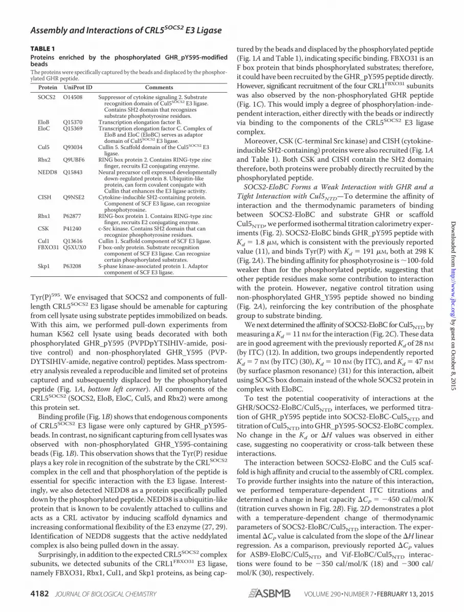
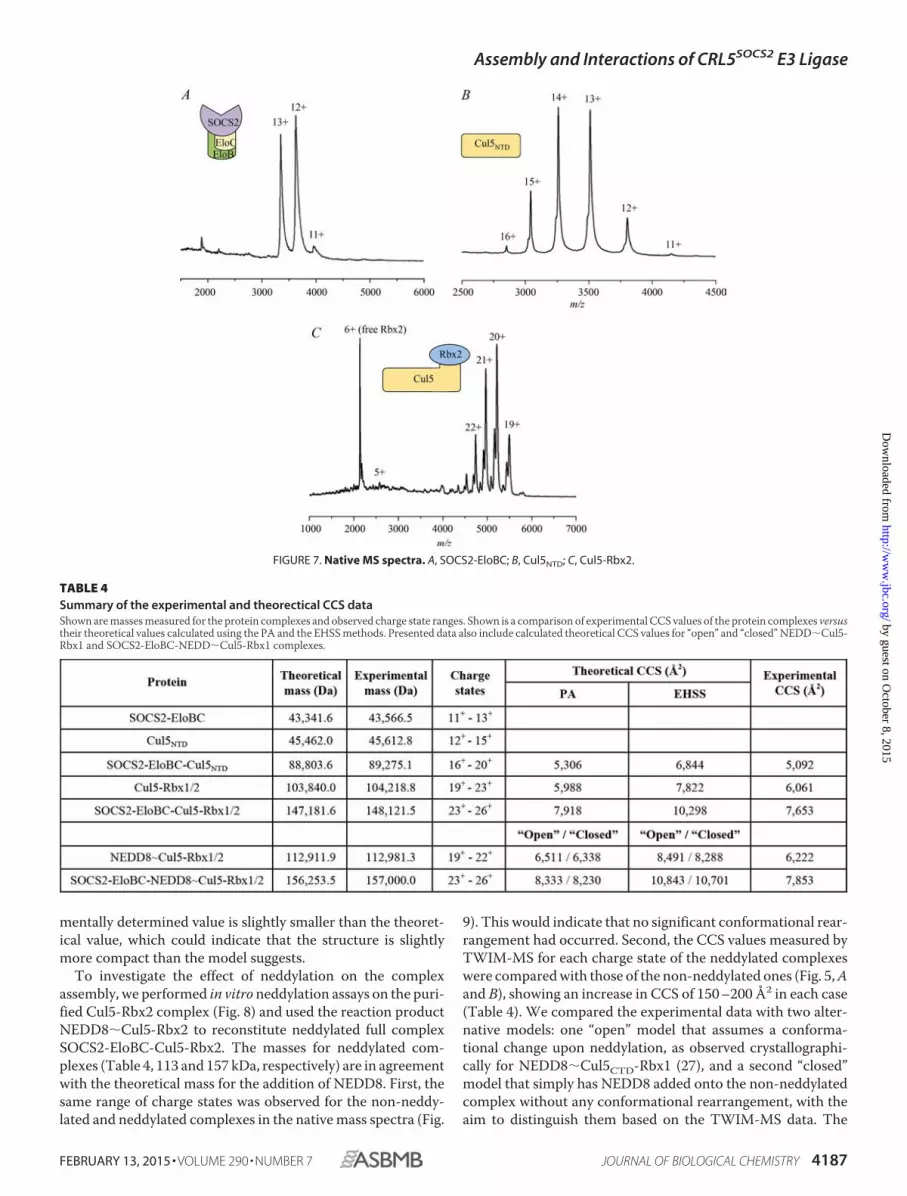
Experimental Collision Cross-sections for Protein ComplexesAre in Good Agreement with Theoretical Values—To validatethe structural model of SOCS2-EloBC-Cul5-Rbx2, TWIM-MSwas used to examine the molecular weight and stoichiometry ofthe intact protein complexes as well as to confirm their topology byCCS measurements. The protein components SOCS2-EloBC andCul5NTD alone were first analyzed using native MS, and the result-ing spectra are shown in Fig. 7, A and B. The masses were con-firmed as �43 and 45 kDa, respectively. Theoretical and experi-mental masses for each complex are shown in Table 4.
Combining SOCS2-EloBC with Cul5NTD produced an 89-kDacomplex, which could be detected with charge states ranging from16� to 20� (Fig. 4A). Some free SOCS2-EloBC was also observedin this spectrum, with the same charge states as the native massspectrum of SOCS2-EloBC alone, indicating that some dissocia-
FIGURE 3. Recombinant components of CRL5SOCS2 assemble into the monomeric full-size protein complex. A, schematic representation of CRL5SOCS2,which includes substrate receptor SOCS2, adaptor EloBC complex, scaffold Cul5, and RING domain protein Rbx2. B, SDS-polyacrylamide gel images of thepurified protein complexes. C, table showing a comparison of theoretical protein molecular weights against values experimentally determined by SEC-MALS.D, SEC-MALS elution profiles for the individual components of CRL5SOCS2 and their complexes, including full-size SOCS2-EloBC-Cul5-Rbx2 complex.
TABLE 3Large negative �CP value for SOCS2-EloBC/Cul5NTD interaction indi-cates a highly hydrophobic interface between the proteins; compari-son between theorectical and experimental �CP values shows goodagreement
Assembly and Interactions of CRL5SOCS2 E3 Ligase
4184 JOURNAL OF BIOLOGICAL CHEMISTRY VOLUME 290 • NUMBER 7 • FEBRUARY 13, 2015
by guest on October 8, 2015
http://ww
w.jbc.org/
Dow
nloaded from
tion in solution occurs (Fig. 7A). Table 4 shows the experimentalCCS values compared with the theoretical ones calculated with thePA and the EHSS methods. It is normally expected that the exper-imental values would be smaller than the EHSS results and largerthan the PA results (24, 25). The collision cross-section deter-mined using ion mobility for SOCS2-EloBC-Cul5NTD is 5,092 Å2
for the most native charge state (16�; Fig. 4A, bottom), which isreasonably close to the theoretical value calculated for the model(Table 4, PA value 5,306 Å2).
A typical spectrum of the Cul5-Rbx2 complex is shown in Fig.7C with a predominant 6� charge state for Rbx2 and a series ofcharge states from 19� to 22� representing the binary complex(104 kDa). Fig. 4B shows the Cul5-Rbx2 complex in more detail in
addition to the drift time plot. Moreover, there are less intensepeaks to the left-hand side of the predominant peak correspondingto a loss of �1 kDa from the complex that may represent a trun-cation in the Cul5 subunit. These species are clearly separated,however, by their ion mobility (Fig. 4B), so it is possible to calculatea collisional cross-section for the intact complex. The CCS valuefrom these data for the most native 19� charge state was found tobe 6,061 Å2 (Fig. 4B, bottom), which compared well with the the-oretical value (Table 4, PA value 5,988 Å2).
The native mass spectrum of the SOCS2-EloBC-Cul5-Rbx2showed intense peaks at 3,000 – 4,000 m/z, indicating a relativeabundance of free SOCS2-EloBC, with charge states 11� to13�, as described previously (Fig. 7A). It is possible that there is
FIGURE 4. Ion mobility drift time plot (top), corresponding native mass spectra (middle), and collision cross-sections (bottom) for the CRL5SOCS2
complexes and their components. A, SOCS2-EloBC-Cul5NTD; B, Cul5-Rbx2; C, full-size complex SOCS2-EloBC-Cul5-Rbx2. Error bars, S.D.
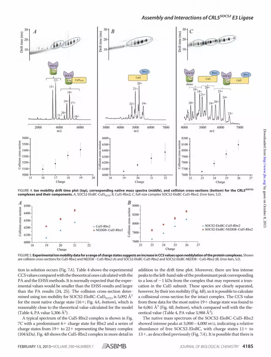
FIGURE 5. Experimental ion mobility data for a range of charge states suggests an increase in CCS values upon neddylation of the protein complexes. Shownare collision cross-sections for Cul5-Rbx2 and NEDD8�Cul5-Rbx2 (A) and SOCS2-EloBC-Cul5-Rbx2 and SOCS2-EloBC-NEDD8�Cul5-Rbx2 (B). Error bars, S.D.
Assembly and Interactions of CRL5SOCS2 E3 Ligase
FEBRUARY 13, 2015 • VOLUME 290 • NUMBER 7 JOURNAL OF BIOLOGICAL CHEMISTRY 4185
by guest on October 8, 2015
http://ww
w.jbc.org/
Dow
nloaded from
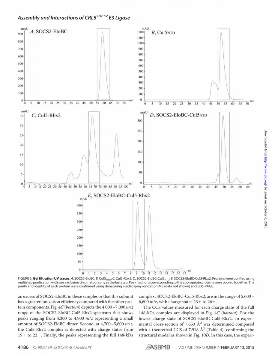
an excess of SOCS2-EloBC in these samples or that this subunithas a greater ionization efficiency compared with the other pro-tein components. Fig. 4C (bottom) depicts the 4,000 –7,000 m/zrange of the SOCS2-EloBC-Cul5-Rbx2 spectrum that showspeaks ranging from 4,300 to 4,900 m/z representing a smallamount of SOCS2-EloBC dimer. Second, at 4,700 –5,600 m/z,the Cul5-Rbx2 complex is detected with charge states from19� to 22�. Finally, the peaks representing the full 148-kDa
complex, SOCS2-EloBC-Cul5-Rbx2, are in the range of 5,600 –6,600 m/z, with charge states 23� to 26�.
The CCS values measured for each charge state of the full148-kDa complex are displayed in Fig. 4C (bottom). For thelowest charge state of SOCS2-EloBC-Cul5-Rbx2, an experi-mental cross-section of 7,653 Å2 was determined comparedwith a theoretical CCS of 7,918 Å2 (Table 4), confirming thestructural model as shown in Fig. 10D. In this case, the experi-
FIGURE 6. Gel filtration UV traces. A, SOCS2-EloBC; B, Cul5NTD; C, Cul5-Rbx2; D, SOCS2-EloBC-Cul5NTD; E, SOCS2-EloBC-Cul5-Rbx2. Proteins were purified usingmultistep purification with size exclusion chromatography as the last step. Peak fractions corresponding to the appropriate proteins were pooled together. Thepurity and identity of each protein were confirmed using denaturing electrospray ionization-MS (data not shown) and SDS-PAGE.
Assembly and Interactions of CRL5SOCS2 E3 Ligase
4186 JOURNAL OF BIOLOGICAL CHEMISTRY VOLUME 290 • NUMBER 7 • FEBRUARY 13, 2015
by guest on October 8, 2015
http://ww
w.jbc.org/
Dow
nloaded from
mentally determined value is slightly smaller than the theoret-ical value, which could indicate that the structure is slightlymore compact than the model suggests.
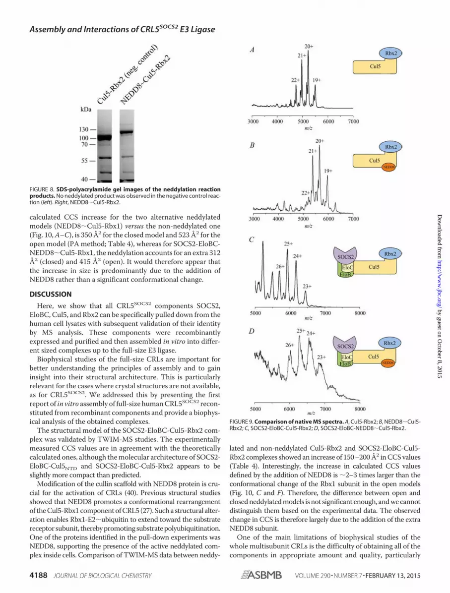
To investigate the effect of neddylation on the complexassembly, we performed in vitro neddylation assays on the puri-fied Cul5-Rbx2 complex (Fig. 8) and used the reaction productNEDD8�Cul5-Rbx2 to reconstitute neddylated full complexSOCS2-EloBC-Cul5-Rbx2. The masses for neddylated com-plexes (Table 4, 113 and 157 kDa, respectively) are in agreementwith the theoretical mass for the addition of NEDD8. First, thesame range of charge states was observed for the non-neddy-lated and neddylated complexes in the native mass spectra (Fig.
9). This would indicate that no significant conformational rear-rangement had occurred. Second, the CCS values measured byTWIM-MS for each charge state of the neddylated complexeswere compared with those of the non-neddylated ones (Fig. 5, Aand B), showing an increase in CCS of 150 –200 Å2 in each case(Table 4). We compared the experimental data with two alter-native models: one “open” model that assumes a conforma-tional change upon neddylation, as observed crystallographi-cally for NEDD8�Cul5CTD-Rbx1 (27), and a second “closed”model that simply has NEDD8 added onto the non-neddylatedcomplex without any conformational rearrangement, with theaim to distinguish them based on the TWIM-MS data. The
FIGURE 7. Native MS spectra. A, SOCS2-EloBC; B, Cul5NTD; C, Cul5-Rbx2.
TABLE 4Summary of the experimental and theorectical CCS dataShown are masses measured for the protein complexes and observed charge state ranges. Shown is a comparison of experimental CCS values of the protein complexes versustheir theoretical values calculated using the PA and the EHSS methods. Presented data also include calculated theoretical CCS values for “open” and “closed” NEDD�Cul5-Rbx1 and SOCS2-EloBC-NEDD�Cul5-Rbx1 complexes.
Assembly and Interactions of CRL5SOCS2 E3 Ligase
FEBRUARY 13, 2015 • VOLUME 290 • NUMBER 7 JOURNAL OF BIOLOGICAL CHEMISTRY 4187
by guest on October 8, 2015
http://ww
w.jbc.org/
Dow
nloaded from
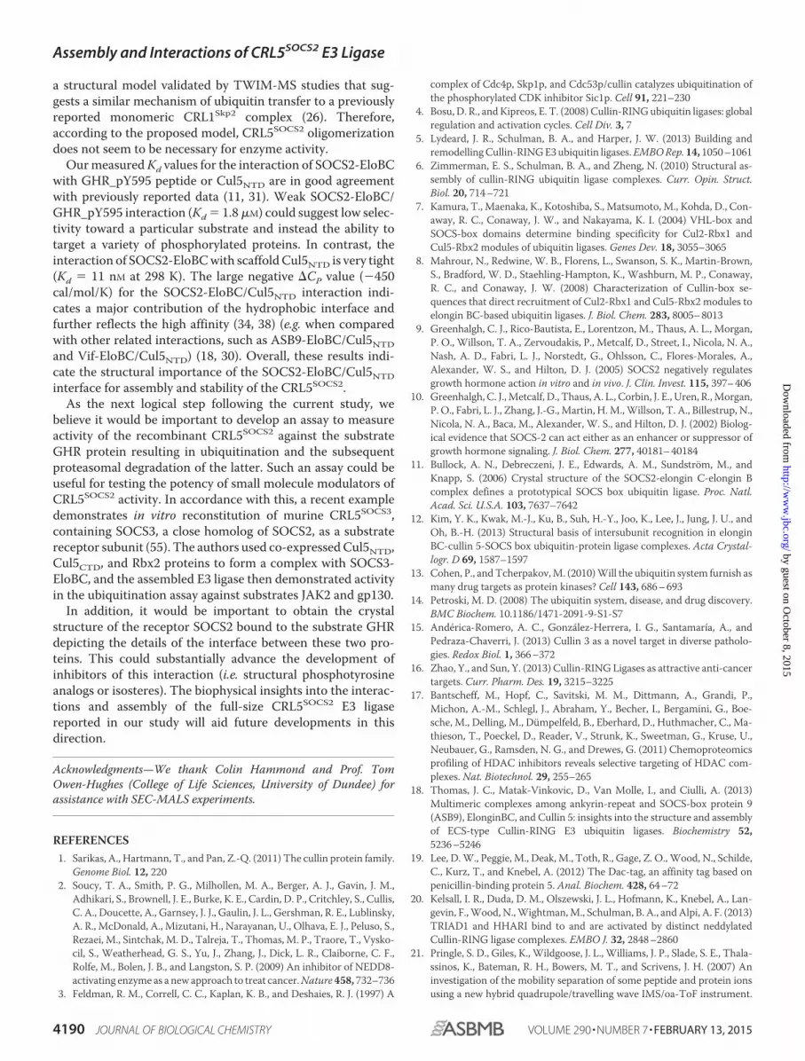
calculated CCS increase for the two alternative neddylatedmodels (NEDD8�Cul5-Rbx1) versus the non-neddylated one(Fig. 10, A–C), is 350 Å2 for the closed model and 523 Å2 for theopen model (PA method; Table 4), whereas for SOCS2-EloBC-NEDD8�Cul5-Rbx1, the neddylation accounts for an extra 312Å2 (closed) and 415 Å2 (open). It would therefore appear thatthe increase in size is predominantly due to the addition ofNEDD8 rather than a significant conformational change.
DISCUSSION
Here, we show that all CRL5SOCS2 components SOCS2,EloBC, Cul5, and Rbx2 can be specifically pulled down from thehuman cell lysates with subsequent validation of their identityby MS analysis. These components were recombinantlyexpressed and purified and then assembled in vitro into differ-ent sized complexes up to the full-size E3 ligase.
Biophysical studies of the full-size CRLs are important forbetter understanding the principles of assembly and to gaininsight into their structural architecture. This is particularlyrelevant for the cases where crystal structures are not available,as for CRL5SOCS2. We addressed this by presenting the firstreport of in vitro assembly of full-size human CRL5SOCS2 recon-stituted from recombinant components and provide a biophys-ical analysis of the obtained complexes.
The structural model of the SOCS2-EloBC-Cul5-Rbx2 com-plex was validated by TWIM-MS studies. The experimentallymeasured CCS values are in agreement with the theoreticallycalculated ones, although the molecular architecture of SOCS2-EloBC-Cul5NTD and SOCS2-EloBC-Cul5-Rbx2 appears to beslightly more compact than predicted.
Modification of the cullin scaffold with NEDD8 protein is cru-cial for the activation of CRLs (40). Previous structural studiesshowed that NEDD8 promotes a conformational rearrangementof the Cul5-Rbx1 component of CRL5 (27). Such a structural alter-ation enables Rbx1-E2�ubiquitin to extend toward the substratereceptor subunit, thereby promoting substrate polyubiquitination.One of the proteins identified in the pull-down experiments wasNEDD8, supporting the presence of the active neddylated com-plex inside cells. Comparison of TWIM-MS data between neddy-
lated and non-neddylated Cul5-Rbx2 and SOCS2-EloBC-Cul5-Rbx2 complexes showed an increase of 150–200 Å2 in CCS values(Table 4). Interestingly, the increase in calculated CCS valuesdefined by the addition of NEDD8 is �2–3 times larger than theconformational change of the Rbx1 subunit in the open models(Fig. 10, C and F). Therefore, the difference between open andclosed neddylated models is not significant enough, and we cannotdistinguish them based on the experimental data. The observedchange in CCS is therefore largely due to the addition of the extraNEDD8 subunit.
One of the main limitations of biophysical studies of thewhole multisubunit CRLs is the difficulty of obtaining all of thecomponents in appropriate amount and quality, particularly
FIGURE 8. SDS-polyacrylamide gel images of the neddylation reactionproducts. No neddylated product was observed in the negative control reac-tion (left). Right, NEDD8�Cul5-Rbx2.
FIGURE 9. Comparison of native MS spectra. A, Cul5-Rbx2; B, NEDD8�Cul5-Rbx2; C, SOCS2-EloBC-Cul5-Rbx2; D, SOCS2-EloBC-NEDD8�Cul5-Rbx2.
Assembly and Interactions of CRL5SOCS2 E3 Ligase
4188 JOURNAL OF BIOLOGICAL CHEMISTRY VOLUME 290 • NUMBER 7 • FEBRUARY 13, 2015
by guest on October 8, 2015
http://ww
w.jbc.org/
Dow
nloaded from

full-length cullins. Expression and purification of stable full-length cullin scaffolds in complex with RING domain proteinsis not trivial and has been previously reported only for Cul1-Rbx1 (26), Cul4A-Rbx1 (41), Cul4B-Rbx1 (42), and Cul5-Rbx2(20). As a result, there were only a few cases in the literaturedescribing characterization of the full-size CRLs assembledfrom recombinant subunits (26, 42). To purify the Cul5-Rbx2complex in this study, we used the Dac tag technology, whichprovides additional stability and solubility to the protein com-plex and additionally improves the yield of recombinant pro-teins (19). This approach has also proven to be successful forpurification of Cul2-Rbx1 complex4 and could be furtherextended to other cullins and large multisubunit complexes.
In certain cases, CRLs exist and function in homo- or hetero-oligomeric states. The biological implications of CRL oligomer-
ization are postulated to include activity regulation, enhance-ment of substrate ubiquitination, and alternative mechanisticaspects of ubiquitin transfer (6). For example, several studieshave shown that CRL3 can dimerize via an adaptor BTB domain(43) or through NEDD8-mediated interaction between twoCul3 scaffolds (44). CRL1 was also demonstrated to be able todimerize via the receptor Cdc4 (cell division control protein 4),resulting in enhanced ubiquitination of substrate Sic1 (45).Additional examples include other BTB receptor/adaptor sub-units of CRL3 (46 – 49), F box receptors of CRL1 (45, 50 –53),and the DCAF receptor of CRL4 (54). More recently, a two-sitemodel for substrate recognition was proposed for CRL3KLHL11
based on the crystal structure of KLHL11-Cul3NTD (49). How-ever, no evidence for dimerization of elongins, cullin 2 or cullin5, or SOCS subunits has been reported to date. In this work,using SEC-MALS and native MS techniques, we have estab-lished that CRL5SOCS2 exists in a monomeric state. We provide4 A. Knebel, unpublished results.
FIGURE 10. Structural models provide important insights into the assembly and architecture of CRL5SOCS2. A, Cul5-Rbx1; B, NEDD8�Cul5-Rbx1 (closedmodel); C, NEDD8�Cul5-Rbx1 (open model); D, SOCS2-EloBC-Cul5-Rbx1; E, SOCS2-EloBC-NEDD8�Cul5-Rbx1 (closed); F, SOCS2-EloBC-NEDD8�Cul5-Rbx1(open). The models were assembled using available crystal structures. Due to the lack of Rbx2 crystal structure, its closest homolog, Rbx1, was used instead.These models were used for calculation of the theoretical CCS values.
Assembly and Interactions of CRL5SOCS2 E3 Ligase
FEBRUARY 13, 2015 • VOLUME 290 • NUMBER 7 JOURNAL OF BIOLOGICAL CHEMISTRY 4189
by guest on October 8, 2015
http://ww
w.jbc.org/
Dow
nloaded from
a structural model validated by TWIM-MS studies that sug-gests a similar mechanism of ubiquitin transfer to a previouslyreported monomeric CRL1Skp2 complex (26). Therefore,according to the proposed model, CRL5SOCS2 oligomerizationdoes not seem to be necessary for enzyme activity.
Our measured Kd values for the interaction of SOCS2-EloBCwith GHR_pY595 peptide or Cul5NTD are in good agreementwith previously reported data (11, 31). Weak SOCS2-EloBC/GHR_pY595 interaction (Kd � 1.8 �M) could suggest low selec-tivity toward a particular substrate and instead the ability totarget a variety of phosphorylated proteins. In contrast, theinteraction of SOCS2-EloBC with scaffold Cul5NTD is very tight(Kd � 11 nM at 298 K). The large negative �CP value (�450cal/mol/K) for the SOCS2-EloBC/Cul5NTD interaction indi-cates a major contribution of the hydrophobic interface andfurther reflects the high affinity (34, 38) (e.g. when comparedwith other related interactions, such as ASB9-EloBC/Cul5NTDand Vif-EloBC/Cul5NTD) (18, 30). Overall, these results indi-cate the structural importance of the SOCS2-EloBC/Cul5NTDinterface for assembly and stability of the CRL5SOCS2.
As the next logical step following the current study, webelieve it would be important to develop an assay to measureactivity of the recombinant CRL5SOCS2 against the substrateGHR protein resulting in ubiquitination and the subsequentproteasomal degradation of the latter. Such an assay could beuseful for testing the potency of small molecule modulators ofCRL5SOCS2 activity. In accordance with this, a recent exampledemonstrates in vitro reconstitution of murine CRL5SOCS3,containing SOCS3, a close homolog of SOCS2, as a substratereceptor subunit (55). The authors used co-expressed Cul5NTD,Cul5CTD, and Rbx2 proteins to form a complex with SOCS3-EloBC, and the assembled E3 ligase then demonstrated activityin the ubiquitination assay against substrates JAK2 and gp130.
In addition, it would be important to obtain the crystalstructure of the receptor SOCS2 bound to the substrate GHRdepicting the details of the interface between these two pro-teins. This could substantially advance the development ofinhibitors of this interaction (i.e. structural phosphotyrosineanalogs or isosteres). The biophysical insights into the interac-tions and assembly of the full-size CRL5SOCS2 E3 ligasereported in our study will aid future developments in thisdirection.
Acknowledgments—We thank Colin Hammond and Prof. TomOwen-Hughes (College of Life Sciences, University of Dundee) forassistance with SEC-MALS experiments.
REFERENCES1. Sarikas, A., Hartmann, T., and Pan, Z.-Q. (2011) The cullin protein family.
Genome Biol. 12, 2202. Soucy, T. A., Smith, P. G., Milhollen, M. A., Berger, A. J., Gavin, J. M.,
Adhikari, S., Brownell, J. E., Burke, K. E., Cardin, D. P., Critchley, S., Cullis,C. A., Doucette, A., Garnsey, J. J., Gaulin, J. L., Gershman, R. E., Lublinsky,A. R., McDonald, A., Mizutani, H., Narayanan, U., Olhava, E. J., Peluso, S.,Rezaei, M., Sintchak, M. D., Talreja, T., Thomas, M. P., Traore, T., Vysko-cil, S., Weatherhead, G. S., Yu, J., Zhang, J., Dick, L. R., Claiborne, C. F.,Rolfe, M., Bolen, J. B., and Langston, S. P. (2009) An inhibitor of NEDD8-activating enzyme as a new approach to treat cancer. Nature 458, 732–736
3. Feldman, R. M., Correll, C. C., Kaplan, K. B., and Deshaies, R. J. (1997) A
complex of Cdc4p, Skp1p, and Cdc53p/cullin catalyzes ubiquitination ofthe phosphorylated CDK inhibitor Sic1p. Cell 91, 221–230
4. Bosu, D. R., and Kipreos, E. T. (2008) Cullin-RING ubiquitin ligases: globalregulation and activation cycles. Cell Div. 3, 7
5. Lydeard, J. R., Schulman, B. A., and Harper, J. W. (2013) Building andremodelling Cullin-RING E3 ubiquitin ligases. EMBO Rep. 14, 1050 –1061
6. Zimmerman, E. S., Schulman, B. A., and Zheng, N. (2010) Structural as-sembly of cullin-RING ubiquitin ligase complexes. Curr. Opin. Struct.Biol. 20, 714 –721
7. Kamura, T., Maenaka, K., Kotoshiba, S., Matsumoto, M., Kohda, D., Con-away, R. C., Conaway, J. W., and Nakayama, K. I. (2004) VHL-box andSOCS-box domains determine binding specificity for Cul2-Rbx1 andCul5-Rbx2 modules of ubiquitin ligases. Genes Dev. 18, 3055–3065
8. Mahrour, N., Redwine, W. B., Florens, L., Swanson, S. K., Martin-Brown,S., Bradford, W. D., Staehling-Hampton, K., Washburn, M. P., Conaway,R. C., and Conaway, J. W. (2008) Characterization of Cullin-box se-quences that direct recruitment of Cul2-Rbx1 and Cul5-Rbx2 modules toelongin BC-based ubiquitin ligases. J. Biol. Chem. 283, 8005– 8013
9. Greenhalgh, C. J., Rico-Bautista, E., Lorentzon, M., Thaus, A. L., Morgan,P. O., Willson, T. A., Zervoudakis, P., Metcalf, D., Street, I., Nicola, N. A.,Nash, A. D., Fabri, L. J., Norstedt, G., Ohlsson, C., Flores-Morales, A.,Alexander, W. S., and Hilton, D. J. (2005) SOCS2 negatively regulatesgrowth hormone action in vitro and in vivo. J. Clin. Invest. 115, 397– 406
10. Greenhalgh, C. J., Metcalf, D., Thaus, A. L., Corbin, J. E., Uren, R., Morgan,P. O., Fabri, L. J., Zhang, J.-G., Martin, H. M., Willson, T. A., Billestrup, N.,Nicola, N. A., Baca, M., Alexander, W. S., and Hilton, D. J. (2002) Biolog-ical evidence that SOCS-2 can act either as an enhancer or suppressor ofgrowth hormone signaling. J. Biol. Chem. 277, 40181– 40184
11. Bullock, A. N., Debreczeni, J. E., Edwards, A. M., Sundstrom, M., andKnapp, S. (2006) Crystal structure of the SOCS2-elongin C-elongin Bcomplex defines a prototypical SOCS box ubiquitin ligase. Proc. Natl.Acad. Sci. U.S.A. 103, 7637–7642
12. Kim, Y. K., Kwak, M.-J., Ku, B., Suh, H.-Y., Joo, K., Lee, J., Jung, J. U., andOh, B.-H. (2013) Structural basis of intersubunit recognition in elonginBC-cullin 5-SOCS box ubiquitin-protein ligase complexes. Acta Crystal-logr. D 69, 1587–1597
13. Cohen, P., and Tcherpakov, M. (2010) Will the ubiquitin system furnish asmany drug targets as protein kinases? Cell 143, 686 – 693
14. Petroski, M. D. (2008) The ubiquitin system, disease, and drug discovery.BMC Biochem. 10.1186/1471-2091-9-S1-S7
15. Anderica-Romero, A. C., Gonzalez-Herrera, I. G., Santamaría, A., andPedraza-Chaverri, J. (2013) Cullin 3 as a novel target in diverse patholo-gies. Redox Biol. 1, 366 –372
16. Zhao, Y., and Sun, Y. (2013) Cullin-RING Ligases as attractive anti-cancertargets. Curr. Pharm. Des. 19, 3215–3225
17. Bantscheff, M., Hopf, C., Savitski, M. M., Dittmann, A., Grandi, P.,Michon, A.-M., Schlegl, J., Abraham, Y., Becher, I., Bergamini, G., Boe-sche, M., Delling, M., Dumpelfeld, B., Eberhard, D., Huthmacher, C., Ma-thieson, T., Poeckel, D., Reader, V., Strunk, K., Sweetman, G., Kruse, U.,Neubauer, G., Ramsden, N. G., and Drewes, G. (2011) Chemoproteomicsprofiling of HDAC inhibitors reveals selective targeting of HDAC com-plexes. Nat. Biotechnol. 29, 255–265
18. Thomas, J. C., Matak-Vinkovic, D., Van Molle, I., and Ciulli, A. (2013)Multimeric complexes among ankyrin-repeat and SOCS-box protein 9(ASB9), ElonginBC, and Cullin 5: insights into the structure and assemblyof ECS-type Cullin-RING E3 ubiquitin ligases. Biochemistry 52,5236 –5246
19. Lee, D. W., Peggie, M., Deak, M., Toth, R., Gage, Z. O., Wood, N., Schilde,C., Kurz, T., and Knebel, A. (2012) The Dac-tag, an affinity tag based onpenicillin-binding protein 5. Anal. Biochem. 428, 64 –72
20. Kelsall, I. R., Duda, D. M., Olszewski, J. L., Hofmann, K., Knebel, A., Lan-gevin, F., Wood, N., Wightman, M., Schulman, B. A., and Alpi, A. F. (2013)TRIAD1 and HHARI bind to and are activated by distinct neddylatedCullin-RING ligase complexes. EMBO J. 32, 2848 –2860
21. Pringle, S. D., Giles, K., Wildgoose, J. L., Williams, J. P., Slade, S. E., Thala-ssinos, K., Bateman, R. H., Bowers, M. T., and Scrivens, J. H. (2007) Aninvestigation of the mobility separation of some peptide and protein ionsusing a new hybrid quadrupole/travelling wave IMS/oa-ToF instrument.
Assembly and Interactions of CRL5SOCS2 E3 Ligase
4190 JOURNAL OF BIOLOGICAL CHEMISTRY VOLUME 290 • NUMBER 7 • FEBRUARY 13, 2015
by guest on October 8, 2015
http://ww
w.jbc.org/
Dow
nloaded from

Int. J. Mass Spectr. 261, 1–1222. Konijnenberg, A., Butterer, A., and Sobott, F. (2013) Native ion mobility-
mass spectrometry and related methods in structural biology. Biochim.Biophys. Acta 1834, 1239 –1256
23. Bush, M. F., Hall, Z., Giles, K., Hoyes, J., Robinson, C. V., and Ruotolo, B. T.(2010) Collision cross sections of proteins and their complexes: a calibra-tion framework and database for gas-phase structural biology. Anal.Chem. 82, 9557–9565
24. Mesleh, M. F., Hunter, J. M., Shvartsburg, A. A., Schatz, G. C., and Jarrold,M. F. (1996) Structural information from ion mobility measurements:effects of the long-range potential. J. Phys. Chem. 100, 16082–16086
25. Shvartsburg, A. A., and Jarrold, M. F. (1996) An exact hard-spheres scat-tering model for the mobilities of polyatomic ions. Chem. Phys. Lett. 261,86 –91
26. Zheng, N., Schulman, B. A., Song, L., Miller, J. J., Jeffrey, P. D., Wang, P.,Chu, C., Koepp, D. M., Elledge, S. J., Pagano, M., Conaway, R. C., Conaway,J. W., Harper, J. W., and Pavletich, N. P. (2002) Structure of the Cul1-Rbx1-Skp1-F boxSkp2 SCF ubiquitin ligase complex. Nature 416,703–709
27. Duda, D. M., Borg, L. A., Scott, D. C., Hunt, H. W., Hammel, M., andSchulman, B. A. (2008) Structural insights into NEDD8 activation of cul-lin-RING ligases: conformational control of conjugation. Cell 134,995–1006
28. Vesterlund, M., Zadjali, F., Persson, T., Nielsen, M. L., Kessler, B. M.,Norstedt, G., and Flores-Morales, A. (2011) The SOCS2 ubiquitin ligasecomplex regulates growth hormone receptor levels. PLoS One 6, e25358
29. Pan, Z.-Q., Kentsis, A., Dias, D. C., Yamoah, K., and Wu, K. (2004) Nedd8on cullin: building an expressway to protein destruction. Oncogene 23,1985–1997
30. Salter, J. D., Lippa, G. M., Belashov, I. A., and Wedekind, J. E. (2012)Core-binding factor � increases the affinity between human Cullin 5 andHIV-1 Vif within an E3 ligase complex. Biochemistry 51, 8702– 8704
31. Babon, J. J., Sabo, J. K., Zhang, J.-G., Nicola, N. A., and Norton, R. S. (2009)The SOCS box encodes a hierarchy of affinities for Cullin5: implicationsfor ubiquitin ligase formation and cytokine signalling suppression. J. Mol.Biol. 387, 162–174
32. Fraczkiewicz, R., and Braun, W. (1998) Exact and efficient analytical cal-culation of the accessible surface areas and their gradients for macromol-ecules. J. Comput. Chem. 19, 319 –333
33. Hubbard, S. J., and Thornton, J. M. (1993) NACCESS, University CollegeLondon
34. Prabhu, N. V., and Sharp, K. A. (2005) Heat capacity in proteins. Annu.Rev. Phys. Chem. 56, 521–548
35. Spolar, R. S., Livingstone, J. R., and Record, M. T. (1992) Use of liquidhydrocarbon and amide transfer data to estimate contributions to ther-modynamic functions of protein folding from the removal of nonpolar andpolar surface from water. Biochemistry 31, 3947–3955
36. Myers, J. K., Pace, C. N., and Scholtz, J. M. (1995) Denaturant m values andheat capacity changes: relation to changes in accessible surface areas ofprotein unfolding. Protein Sci. 4, 2138 –2148
37. Makhatadze, G. I., and Privalov, P. L. (1995) Energetics of protein struc-ture. Adv. Protein Chem. 47, 307– 425
38. Robertson, A. D., and Murphy, K. P. (1997) Protein structure and theenergetics of protein stability. Chem. Rev. 97, 1251–1268
39. Murphy, K. P., and Freire, E. (1992) Thermodynamics of structural stabil-ity and cooperative folding behavior in proteins. Adv. Protein Chem. 43,313–361
40. Read, M. A., Brownell, J. E., Gladysheva, T. B., Hottelet, M., Parent, L. A.,
Coggins, M. B., Pierce, J. W., Podust, V. N., Luo, R. S., Chau, V., andPalombella, V. J. (2000) Nedd8 modification of cul-1 activatesSCF(�(TrCP))-dependent ubiquitination of I�B. Mol. Cell Biol. 20,2326 –2333
41. Angers, S., Li, T., Yi, X., MacCoss, M. J., Moon, R. T., and Zheng, N. (2006)Molecular architecture and assembly of the DDB1-CUL4A ubiquitin li-gase machinery. Nature 443, 590 –593
42. Fischer, E. S., Scrima, A., Bohm, K., Matsumoto, S., Lingaraju, G. M., Faty,M., Yasuda, T., Cavadini, S., Wakasugi, M., Hanaoka, F., Iwai, S., Gut, H.,Sugasawa, K., and Thoma, N. H. (2011) The molecular basis ofCRL4DDB2/CSA ubiquitin ligase architecture, targeting, and activation.Cell 147, 1024 –1039
43. Chew, E.-H., Poobalasingam, T., Hawkey, C. J., and Hagen, T. (2007) Char-acterization of cullin-based E3 ubiquitin ligases in intact mammalian cells:evidence for cullin dimerization. Cell. Signal. 19, 1071–1080
44. Wimuttisuk, W., and Singer, J. D. (2007) The Cullin3 ubiquitin ligasefunctions as a Nedd8-bound heterodimer. Mol. Biol. Cell 18, 899 –909
45. Tang, X., Orlicky, S., Lin, Z., Willems, A., Neculai, D., Ceccarelli, D., Mer-curio, F., Shilton, B. H., Sicheri, F., and Tyers, M. (2007) Suprafacial ori-entation of the SCFCdc4 dimer accommodates multiple geometries forsubstrate ubiquitination. Cell 129, 1165–1176
46. Ji, A. X., and Prive, G. G. (2013) Crystal structure of KLHL3 in complexwith Cullin3. PLoS One 8, e60445
47. Errington, W. J., Khan, M. Q., Bueler, S. A., Rubinstein, J. L., Chakrabartty,A., and Prive, G. G. (2012) Adaptor protein self-assembly drives the con-trol of a cullin-RING ubiquitin ligase. Structure 20, 1141–1153
48. van Geersdaele, L. K., Stead, M. A., Harrison, C. M., Carr, S. B., Close, H. J.,Rosbrook, G. O., Connell, S. D., and Wright, S. C. (2013) Structural basis ofhigh-order oligomerization of the cullin-3 adaptor SPOP. Acta Crystal-logr. D Biol. Crystallogr. 69, 1677–1684
49. Canning, P., Cooper, C. D. O., Krojer, T., Murray, J. W., Pike, A. C. W.,Chaikuad, A., Keates, T., Thangaratnarajah, C., Hojzan, V., Ayinampudi,V., Marsden, B. D., Gileadi, O., Knapp, S., von Delft, F., and Bullock, A. N.(2013) Structural basis for Cul3 protein assembly with the BTB-Kelchfamily of E3 ubiquitin ligases. J. Biol. Chem. 288, 7803–7814
50. Suzuki, H., Chiba, T., Suzuki, T., Fujita, T., Ikenoue, T., Omata, M., Furui-chi, K., Shikama, H., and Tanaka, K. (2000) Homodimer of two F-boxproteins �TrCP1 or �TrCP2 binds to I�B for signal-dependent ubiquiti-nation. J. Biol. Chem. 275, 2877–2884
51. Seibert, V., Prohl, C., Schoultz, I., Rhee, E., Lopez, R., Abderazzaq, K.,Zhou, C., and Wolf, D. A. (2002) Combinatorial diversity of fission yeastSCF ubiquitin ligases by homo- and heterooligomeric assemblies of theF-box proteins Pop1p and Pop2p. BMC Biochem. 3, 22
52. Wolf, D. A., McKeon, F., and Jackson, P. K. (1999) F-box/WD-repeatproteins pop1p and Sud1p/Pop2p form complexes that bind and directthe proteolysis of cdc18p. Curr. Biol. 9, 373–376
53. Li, Y., and Hao, B. (2010) Structural basis of dimerization-dependentubiquitination by the SCF(Fbx4) ubiquitin ligase. J. Biol. Chem. 285,13896 –13906
54. Ahn, J., Novince, Z., Concel, J., Byeon, C.-H., Makhov, A. M., Byeon,I.-J. L., Zhang, P., and Gronenborn, A. M. (2011) The Cullin-RING E3ubiquitin ligase CRL4-DCAF1 complex dimerizes via a short helical re-gion in DCAF1. Biochemistry 50, 1359 –1367
55. Kershaw, N. J., Laktyushin, A., Nicola, N. A., and Babon, J. J. (2014) Re-construction of an active SOCS3-based E3 ubiquitin ligase complex invitro: identification of the active components and JAK2 and gp130 assubstrates. Growth Factors 32, 1–10
Assembly and Interactions of CRL5SOCS2 E3 Ligase
FEBRUARY 13, 2015 • VOLUME 290 • NUMBER 7 JOURNAL OF BIOLOGICAL CHEMISTRY 4191
by guest on October 8, 2015
http://ww
w.jbc.org/
Dow
nloaded from

CiulliZinn, Paola Grandi, Frank Sobott and Alessio Albert Konijnenberg, Clare Johnson, NicoChatterjee, Axel Knebel, Satoko Shimamura, Emil Bulatov, Esther M. Martin, Sneha (RBX2)BC-CULLIN 5-RING BOX PROTEIN 2SIGNALING 2 (SOCS2)-ELONGIN SUPPRESSOR OF CYTOKINEAssembly of Full-size E3 Ubiquitin Ligase: Biophysical Studies on Interactions andMolecular Biophysics:
doi: 10.1074/jbc.M114.616664 originally published online December 11, 20142015, 290:4178-4191.J. Biol. Chem.
10.1074/jbc.M114.616664Access the most updated version of this article at doi:
.JBC Affinity SitesFind articles, minireviews, Reflections and Classics on similar topics on the
Alerts:
When a correction for this article is posted•
When this article is cited•
to choose from all of JBC's e-mail alertsClick here
http://www.jbc.org/content/290/7/4178.full.html#ref-list-1
This article cites 54 references, 11 of which can be accessed free at
by guest on October 8, 2015
http://ww
w.jbc.org/
Dow
nloaded from