basic genetic
TRANSCRIPT

Human Genetics Dr. Belal Azab
First Semester 2013-14

Overview: The Key Roles of Cell Division
• The ability of organisms to produce more of their own kind best distinguishes living things from nonliving matter
• The continuity of life is based on the reproduction of cells, or cell division
© 2011 Pearson Education, Inc.
• Multicellular organisms depend on cell division for – Development from a fertilized cell – Growth – Repair
• Cell division is an integral part of the cell cycle, the life of a cell from formation to its own division
© 2011 Pearson Education, Inc.
Cellular Organization of the Genetic Material
• All the DNA in a cell constitutes the cell’s genome • A genome can consist of a single DNA molecule
(common in prokaryotic cells) or a number of DNA molecules (common in eukaryotic cells)
• DNA molecules in a cell are packaged into chromosomes
© 2011 Pearson Education, Inc.

• Eukaryotic chromosomes consist of chromatin, a complex of DNA and protein that condenses during cell division
• Every eukaryotic species has a characteristic number of chromosomes in each cell nucleus
• Somatic cells (nonreproductive cells) have two sets of chromosomes
• Gametes (reproductive cells: sperm and eggs) have half as many chromosomes as somatic cells
© 2011 Pearson Education, Inc.
Distribution of Chromosomes During Eukaryotic Cell Division
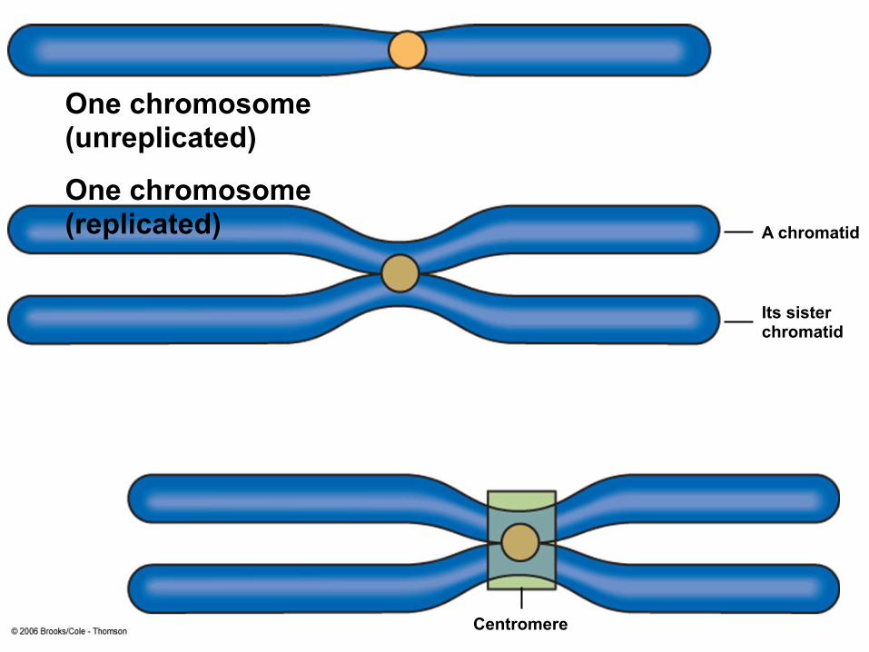
• In preparation for cell division, DNA is replicated and the chromosomes condense
• Each duplicated chromosome has two sister chromatids (joined copies of the original chromosome), which separate during cell division
• The centromere is the narrow “waist” of the duplicated chromosome, where the two chromatids are most closely attached
© 2011 Pearson Education, Inc.

Figure 12.4
0.5 µm Centromere
Sister chromatids

• During cell division, the two sister chromatids of each duplicated chromosome separate and move into two nuclei
• Once separate, the chromatids are called chromosomes
© 2011 Pearson Education, Inc.
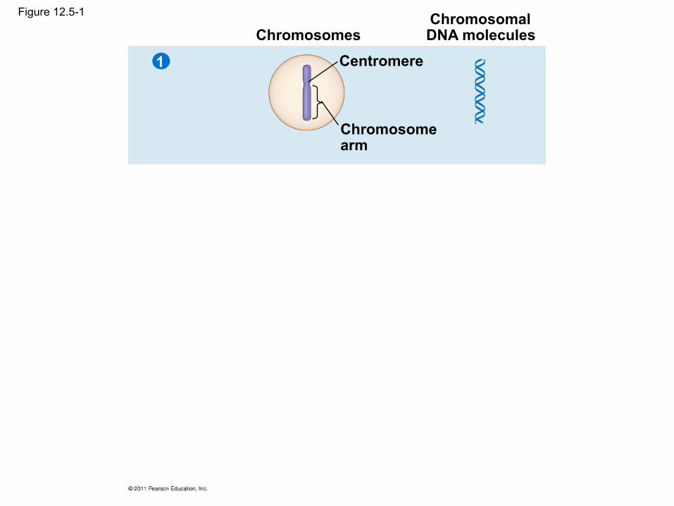
Figure 12.5-1
Chromosomes Chromosomal DNA molecules
Centromere
Chromosome arm
1

Figure 12.5-2
Chromosomes Chromosomal DNA molecules
Centromere
Chromosome arm
Chromosome duplication (including DNA replication) and condensation
Sister chromatids
1
2

Figure 12.5-3
Chromosomes Chromosomal DNA molecules
Centromere
Chromosome arm
Chromosome duplication (including DNA replication) and condensation
Sister chromatids
Separation of sister chromatids into two chromosomes
1
2
3
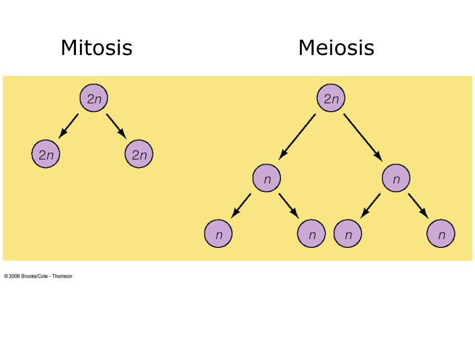
• Eukaryotic cell division consists of – Mitosis, the division of the genetic material in the
nucleus – Cytokinesis, the division of the cytoplasm
• Gametes are produced by a variation of cell division called meiosis
• Meiosis yields nonidentical daughter cells that have only one set of chromosomes, half as many as the parent cell
© 2011 Pearson Education, Inc.
Phases of the Cell Cycle
• The cell cycle consists of – Mitotic (M) phase (mitosis and cytokinesis) – Interphase (cell growth and copying of
chromosomes in preparation for cell division)
© 2011 Pearson Education, Inc.
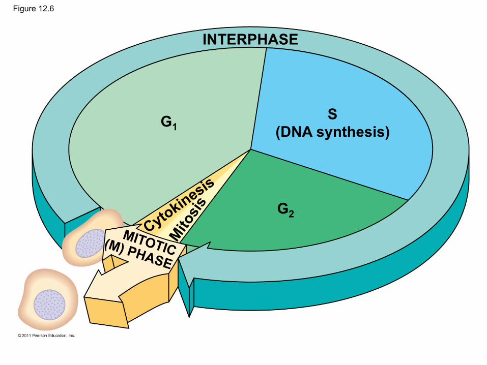
Figure 12.6
INTERPHASE
G1
G2
S (DNA synthesis)
MITOTIC (M) PHASE
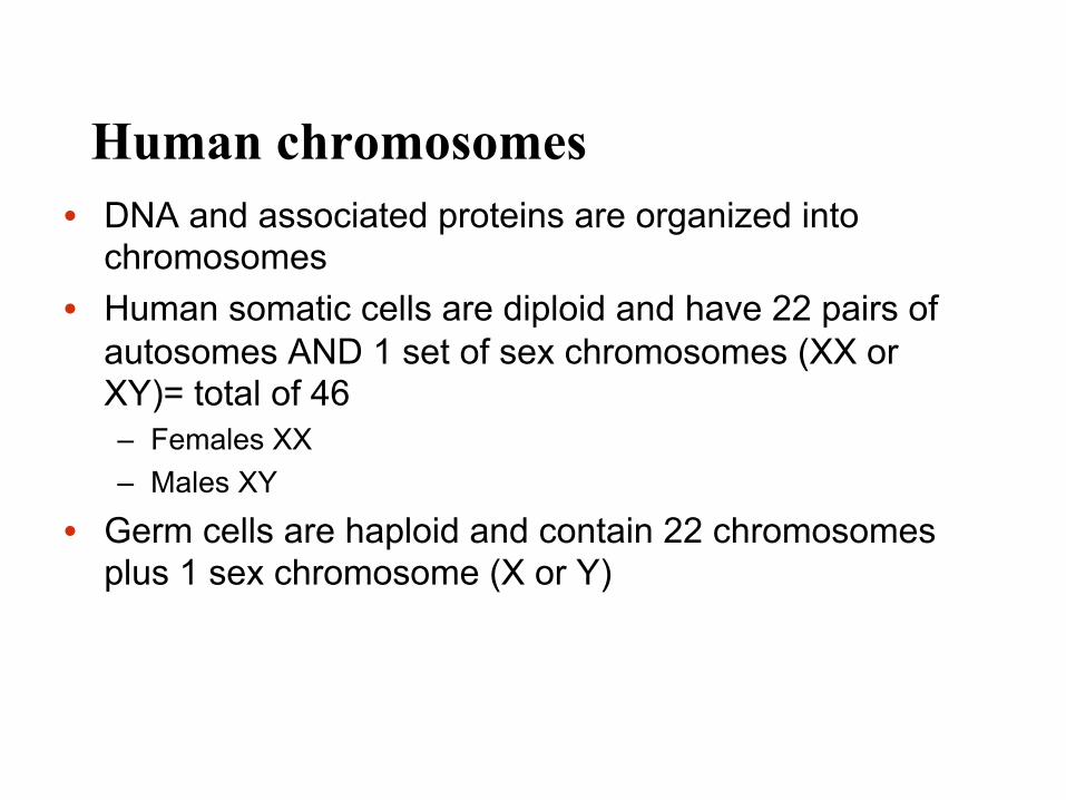
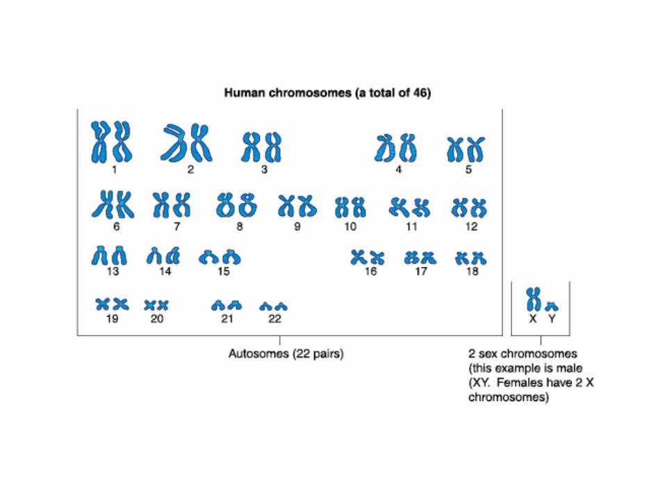
Human chromosomes • DNA and associated proteins are organized into
chromosomes • Human somatic cells are diploid and have 22 pairs of
autosomes AND 1 set of sex chromosomes (XX or XY)= total of 46 – Females XX – Males XY
• Germ cells are haploid and contain 22 chromosomes plus 1 sex chromosome (X or Y)
Mitosis Meiosis

Confocal image of cells at various stages of mitosis. The markers are microtubules (in red), DNA (in blue) and the Golgi apparatus (which temporarily fragments during mitosis, in green).
Credit: © Thomas Deerinck/Visuals Unlimited
900084

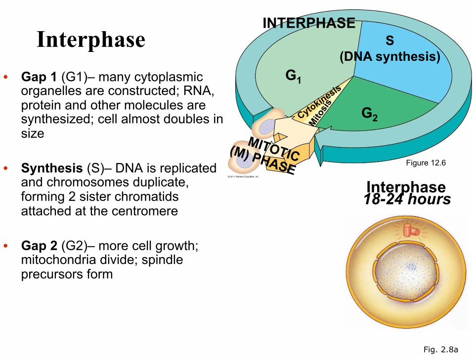
Interphase • Gap 1 (G1)– many cytoplasmic
organelles are constructed; RNA, protein and other molecules are synthesized; cell almost doubles in size
• Synthesis (S)– DNA is replicated
and chromosomes duplicate, forming 2 sister chromatids attached at the centromere
• Gap 2 (G2)– more cell growth; mitochondria divide; spindle precursors form
Interphase 18-24 hours
Fig. 2.8a
Figure 12.6
INTERPHASE
G1
G2
S (DNA synthesis)
MITOTIC (M) PHASE
One chromosome (unreplicated)
One chromosome (replicated) A chromatid
Its sister chromatid
Centromere

Mitosis • Produces identical daughter cells
– (46 chromosomes) • It must be accurate for cells to function properly • Continuous process but divided into distinct
steps: – Prophase – Metaphase – Anaphase – Telophase

Stepped Art
(a) Cell at interphase
Pair of centrioles
Nuclear envelope Chromosome
Mitosis
(b) Early Prophase (c) Late Prophase (d) Transition to Metaphase
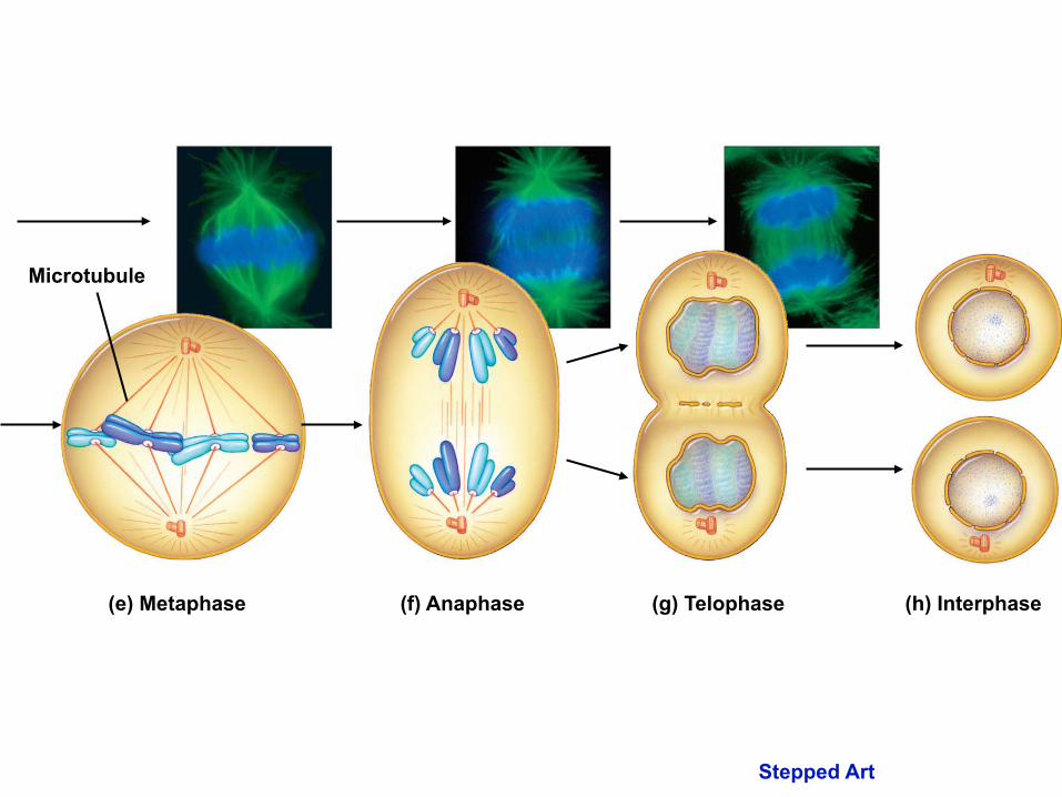
Microtubule
(e) Metaphase (f) Anaphase (h) Interphase (g) Telophase
Stepped Art
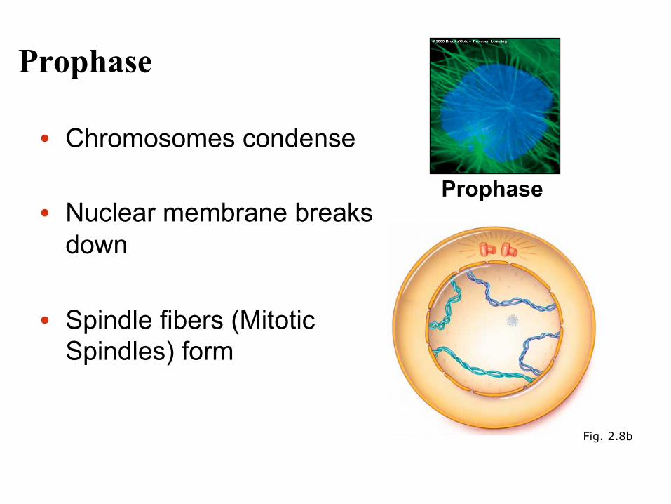
Prophase
• Chromosomes condense
• Nuclear membrane breaks down
• Spindle fibers (Mitotic Spindles) form
Fig. 2.8b
Prophase
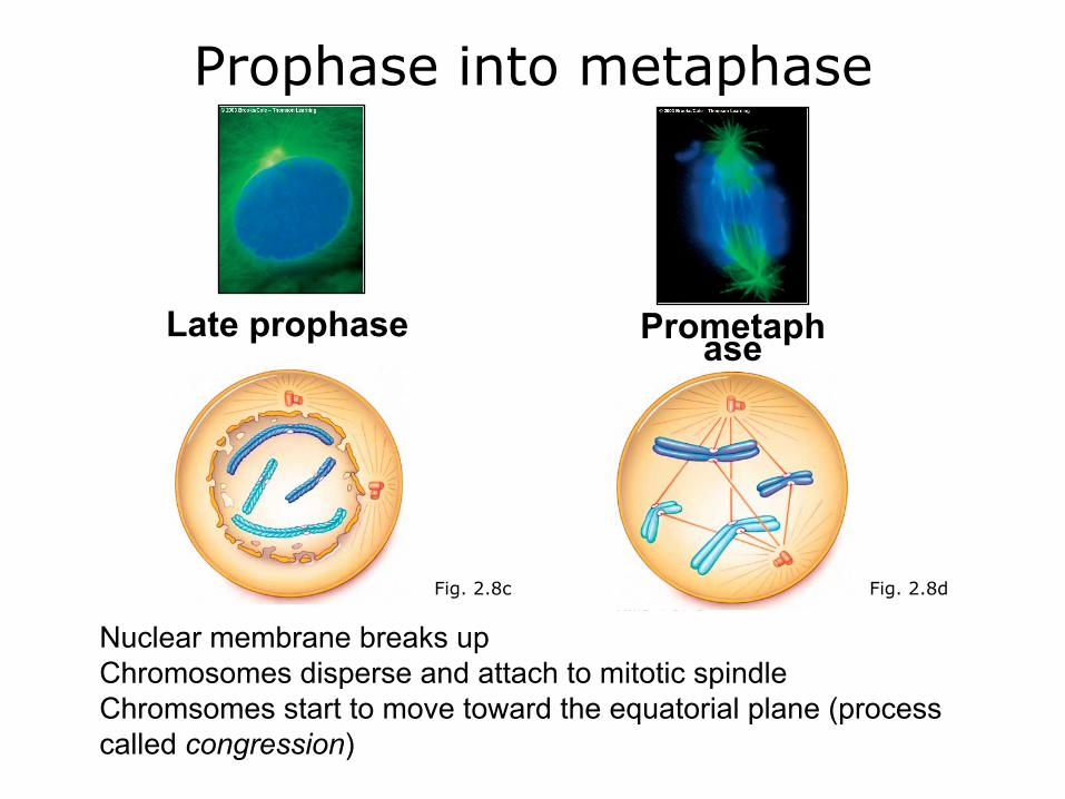
Late prophase Prometaphase
Prophase into metaphase
Fig. 2.8d Fig. 2.8c
Nuclear membrane breaks up Chromosomes disperse and attach to mitotic spindle Chromsomes start to move toward the equatorial plane (process called congression)

The Mitotic Spindle: A Closer Look
• The mitotic spindle is a structure made of microtubules that controls chromosome movement during mitosis
• In animal cells, assembly of spindle microtubules begins in the centrosome, the microtubule organizing center
• The centrosome replicates during interphase, forming two centrosomes that migrate to opposite ends of the cell during prophase and prometaphase
© 2011 Pearson Education, Inc.
• An aster (a radial array of short microtubules) extends from each centrosome
• The spindle includes the centrosomes, the spindle microtubules, and the asters
© 2011 Pearson Education, Inc.

• During prometaphase, some spindle microtubules attach to the kinetochores of chromosomes and begin to move the chromosomes
• Kinetochores are protein complexes associated with centromeres
• At metaphase, the chromosomes are all lined up at the metaphase plate, an imaginary structure at the midway point between the spindle’s two poles
© 2011 Pearson Education, Inc.
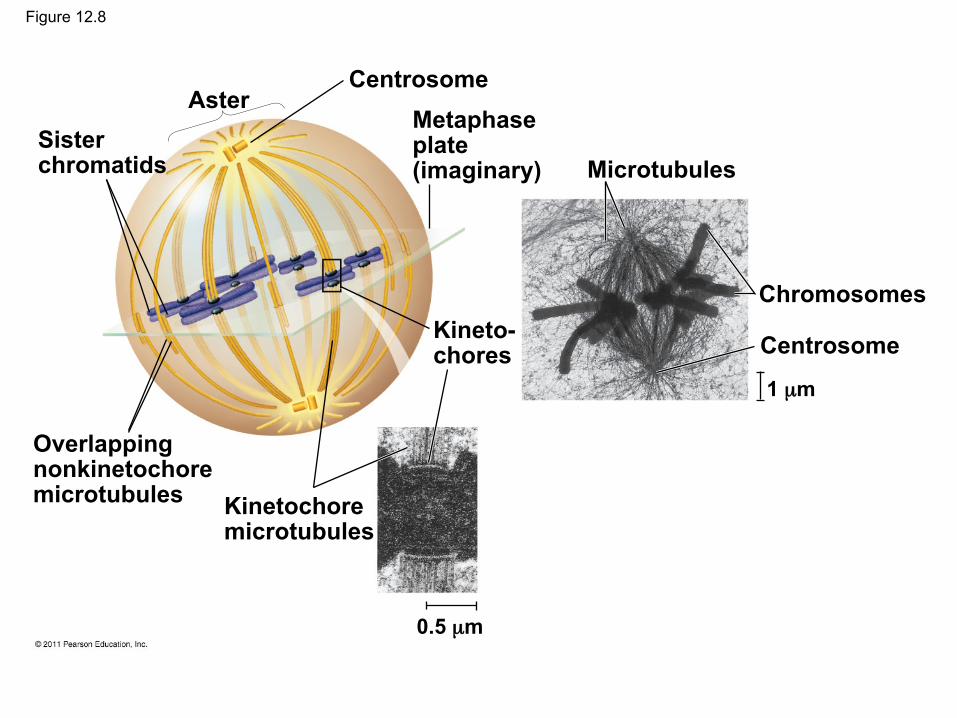
Figure 12.8
Sister chromatids
Aster Centrosome
Metaphase plate (imaginary)
Kineto- chores
Overlapping nonkinetochore microtubules Kinetochore
microtubules
Microtubules
Chromosomes
Centrosome
0.5 µm
1 µm
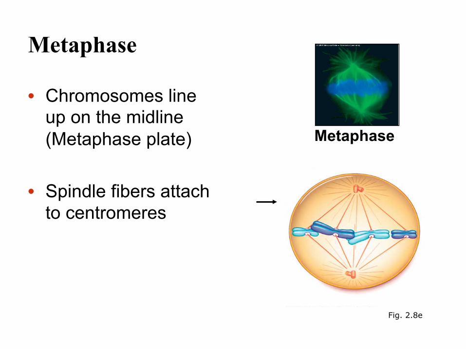
Metaphase
• Chromosomes line up on the midline (Metaphase plate)
• Spindle fibers attach to centromeres
Fig. 2.8e
Metaphase

Anaphase
• Centromeres divide • Spindle fibers shorten • Sister chromatids separate and
move along the kinetochore microtubules to opposite poles
• The microtubules shorten by depolymerizing at their kinetochore ends
Fig 2.8f
Anaphase
Chromosome movement
Microtubule
Motor protein
Chromosome
Kinetochore
Tubulin subunits
CONCLUSION
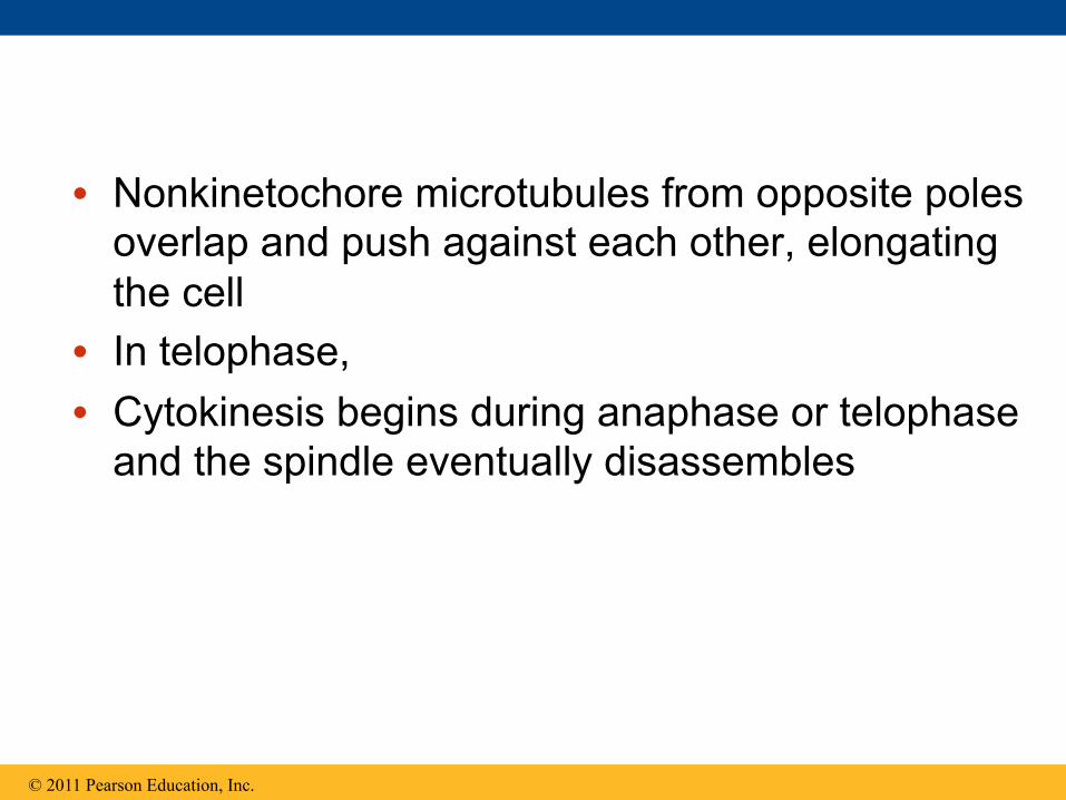
• Nonkinetochore microtubules from opposite poles overlap and push against each other, elongating the cell
• In telophase, • Cytokinesis begins during anaphase or telophase
and the spindle eventually disassembles
© 2011 Pearson Education, Inc.
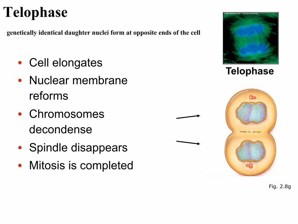
Telophase genetically identical daughter nuclei form at opposite ends of the cell
• Cell elongates • Nuclear membrane
reforms • Chromosomes
decondense • Spindle disappears • Mitosis is completed Fig. 2.8g
Telophase
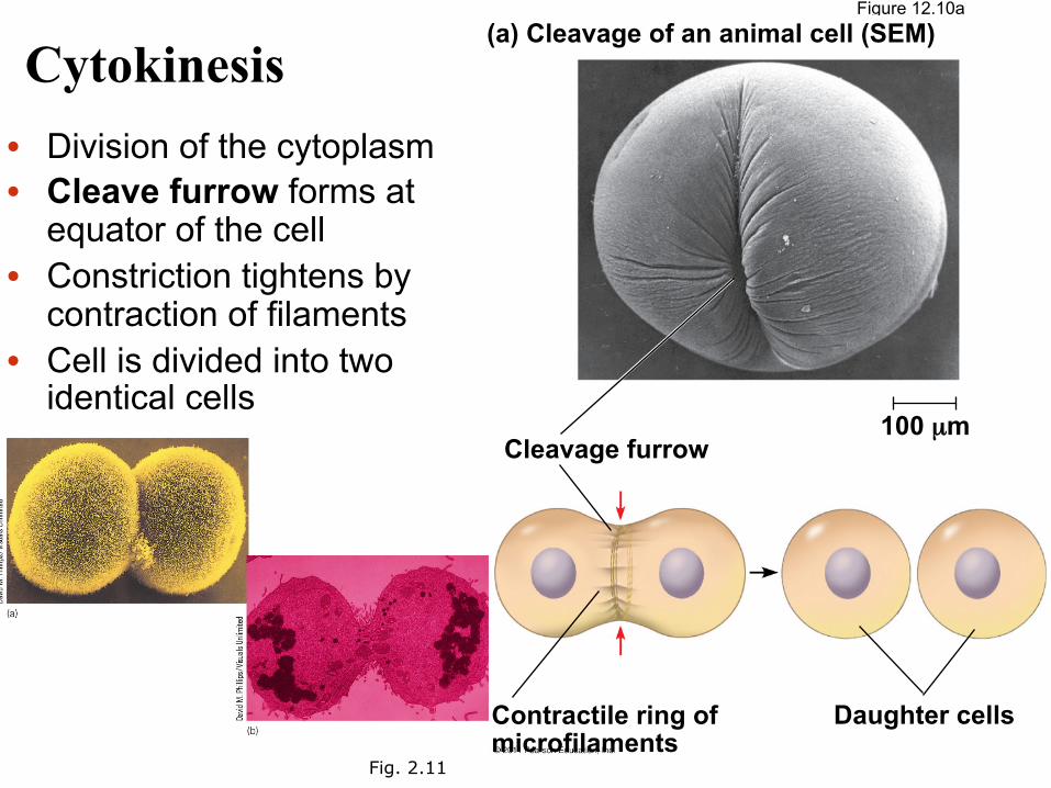
Cytokinesis • Division of the cytoplasm • Cleave furrow forms at
equator of the cell • Constriction tightens by
contraction of filaments • Cell is divided into two
identical cells
Fig. 2.11
Figure 12.10a (a) Cleavage of an animal cell (SEM)
Cleavage furrow
Contractile ring of microfilaments
Daughter cells
100 µm
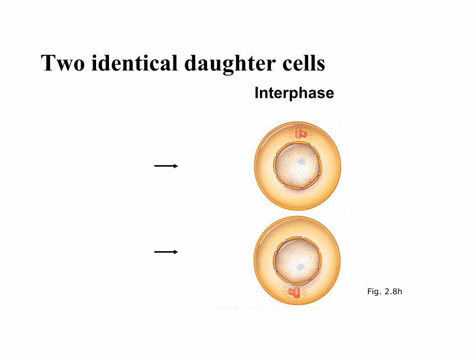
Two identical daughter cells Interphase
Fig. 2.8h
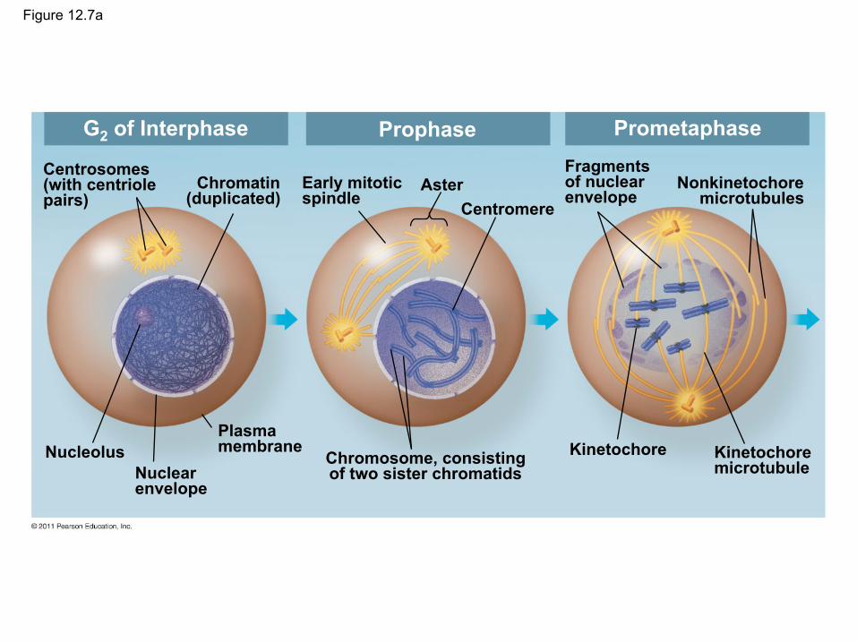
Figure 12.7a
G2 of Interphase Prophase Prometaphase
Centrosomes (with centriole pairs)
Chromatin (duplicated)
Nucleolus Nuclear envelope
Plasma membrane
Early mitotic spindle
Aster Centromere
Chromosome, consisting of two sister chromatids
Fragments of nuclear envelope
Nonkinetochore microtubules
Kinetochore Kinetochore microtubule
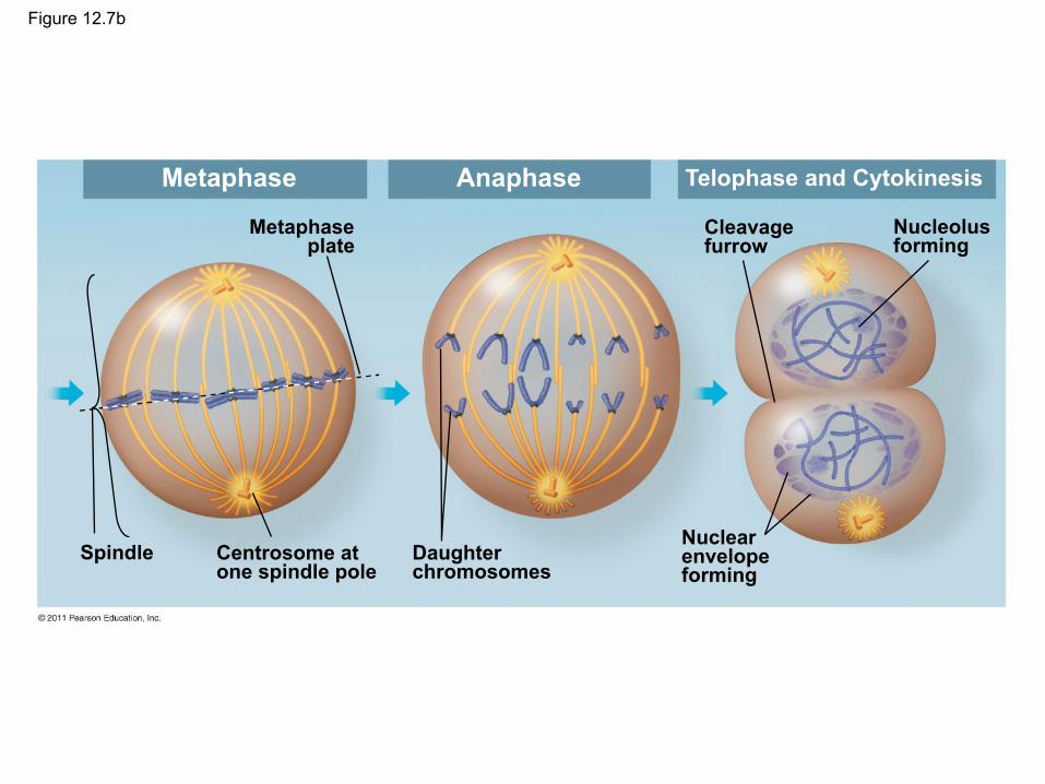
Figure 12.7b
Metaphase
Metaphase plate
Anaphase Telophase and Cytokinesis
Spindle Centrosome at one spindle pole
Daughter chromosomes
Cleavage furrow
Nucleolus forming
Nuclear envelope forming
Mitosis functions in growth and cell replacement • Cells from adults can divide only ~10–30 times • Most cells in the adult human body undergo mitosis;
however, brain and spinal cord nervous tissues do not. – Highly differentiated cells (such as neurons) are stuck in G0 in the
cell cycle and are terminally differentiated. • Cell division is tightly controlled. The cell cycle is
governed by a series of checkpoints—these monitor the quality of the process. – If damage is detected, then the cell may undergo programmed cell
death (apoptosis). – If checkpoints do not function properly, then uncontrolled cell
growth can occur (cancer)
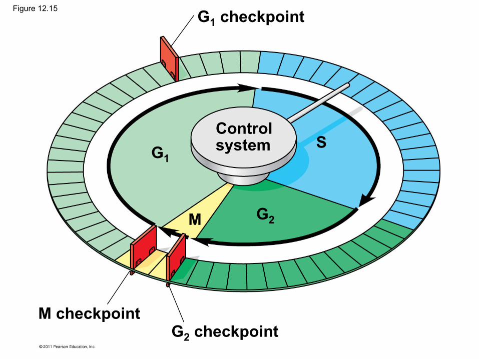
G1 checkpoint
G1
G2
G2 checkpoint M checkpoint
M
S Control system
Figure 12.15
• For many cells, the G1 checkpoint seems to be the most important
• If a cell receives a go-ahead signal at the G1 checkpoint, it will usually complete the S, G2, and M phases and divide
• If the cell does not receive the go-ahead signal, it will exit the cycle, switching into a nondividing state called the G0 phase
© 2011 Pearson Education, Inc.

Figure 12.16
G1 checkpoint
G1 G1
G0
(a) Cell receives a go-ahead signal.
(b) Cell does not receive a go-ahead signal.
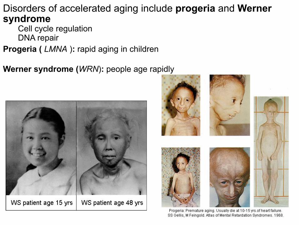
Disorders of accelerated aging include progeria and Werner syndrome
Cell cycle regulation DNA repair
Progeria ( LMNA ): rapid aging in children Werner syndrome (WRN): people age rapidly

Review of terms
• Haploid cells – (1n) with one copy of each chromosome
• Diploid cells – (2n) with two copies of each chromosome
• Somatic – non-germline (body) cells
• Gametes – sex cells (eggs and sperm)
Concept 13.3: Meiosis reduces the number of chromosome sets from diploid to haploid
• Like mitosis, meiosis is preceded by the replication of chromosomes
• Meiosis takes place in two sets of cell divisions, called meiosis I and meiosis II
• The two cell divisions result in four daughter cells, rather than the two daughter cells in mitosis
• Each daughter cell has only half as many chromosomes as the parent cell
© 2011 Pearson Education, Inc.
The Stages of Meiosis
• After chromosomes duplicate, two divisions follow
– Meiosis I (reductional division): homologs pair up and separate, resulting in two haploid daughter cells with replicated chromosomes
– Meiosis II (equational division) sister chromatids separate
• The result is four haploid daughter cells with unreplicated chromosomes
© 2011 Pearson Education, Inc.

Figure 13.7-1
Pair of homologous chromosomes in diploid parent cell
Duplicated pair of homologous chromosomes
Chromosomes duplicate
Sister chromatids Diploid cell with
duplicated chromosomes
Interphase
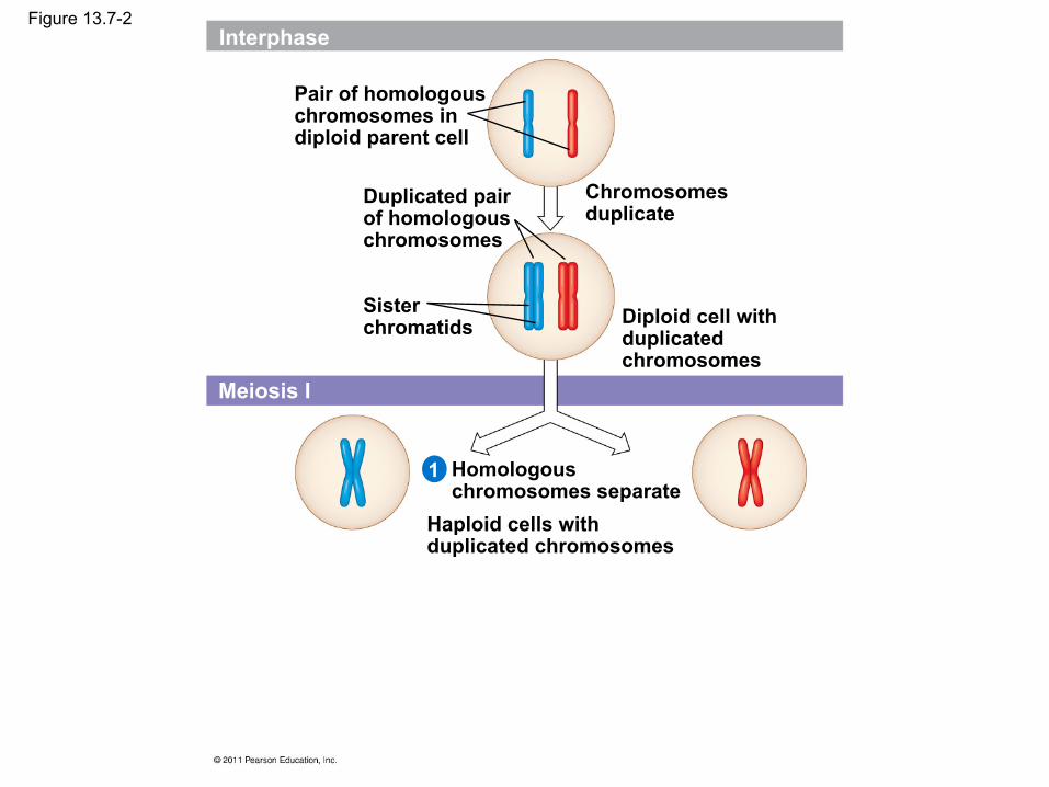
Figure 13.7-2
Pair of homologous chromosomes in diploid parent cell
Duplicated pair of homologous chromosomes
Chromosomes duplicate
Sister chromatids Diploid cell with
duplicated chromosomes
Homologous chromosomes separate
Haploid cells with duplicated chromosomes
Meiosis I
1
Interphase

Figure 13.7-3
Pair of homologous chromosomes in diploid parent cell
Duplicated pair of homologous chromosomes
Chromosomes duplicate
Sister chromatids Diploid cell with
duplicated chromosomes
Homologous chromosomes separate
Haploid cells with duplicated chromosomes
Sister chromatids separate
Haploid cells with unduplicated chromosomes
Interphase
Meiosis I
Meiosis II 2
1

• Meiosis I is preceded by interphase, when the chromosomes are duplicated to form sister chromatids
• The sister chromatids are genetically identical and joined at the centromere
• The single centrosome replicates, forming two centrosomes
© 2011 Pearson Education, Inc.
• Division in meiosis I occurs in four phases – Prophase I – Metaphase I – Anaphase I – Telophase I and cytokinesis
© 2011 Pearson Education, Inc.


Meiosis I
• Reductional division • A diploid cell divides and forms two haploid
cells (2n n) • In humans, the chromosome number is
reduced from 46 to 23 chromosomes • Occurs in testis and ovary in the primary
spermatocytes and primary oocytes • Interphase:
– Chromosomes replicate

Prophase I • Chromosomes condense • Nuclear membrane
breaks down • Homologous
chromosomes pair • Recombination occurs • Nucleolus disappears • Meiotic spindle begins to
form
newly forming microtubules
Prophase I
Fig. 2.13a
Stepped Art
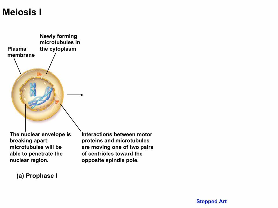
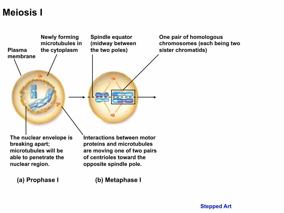
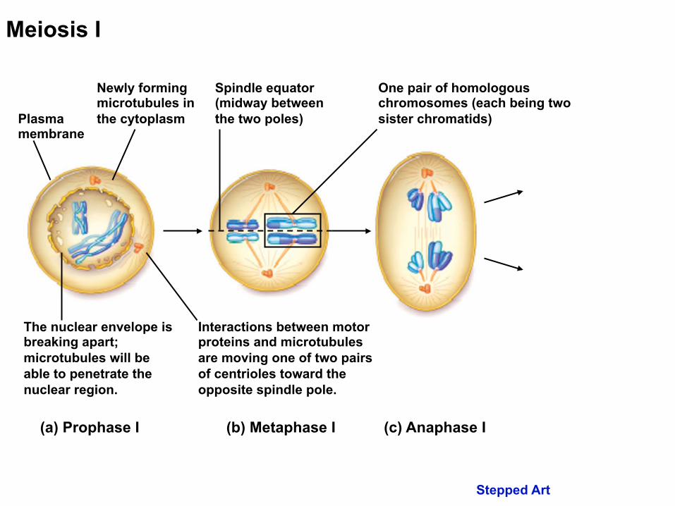
Plasma membrane
(a) Prophase I
Meiosis I
Newly forming microtubules in the cytoplasm
The nuclear envelope is breaking apart; microtubules will be able to penetrate the nuclear region.
Interactions between motor proteins and microtubules are moving one of two pairs of centrioles toward the opposite spindle pole.
Stepped Art
Plasma membrane
(b) Metaphase I (a) Prophase I
Meiosis I
Newly forming microtubules in the cytoplasm
The nuclear envelope is breaking apart; microtubules will be able to penetrate the nuclear region.
Interactions between motor proteins and microtubules are moving one of two pairs of centrioles toward the opposite spindle pole.
Spindle equator (midway between the two poles)
One pair of homologous chromosomes (each being two sister chromatids)
Stepped Art
Plasma membrane
(c) Anaphase I (b) Metaphase I (a) Prophase I
Meiosis I
Newly forming microtubules in the cytoplasm
The nuclear envelope is breaking apart; microtubules will be able to penetrate the nuclear region.
Interactions between motor proteins and microtubules are moving one of two pairs of centrioles toward the opposite spindle pole.
Spindle equator (midway between the two poles)
One pair of homologous chromosomes (each being two sister chromatids)
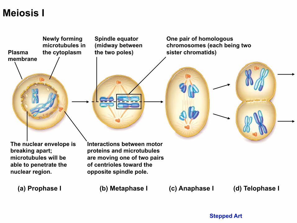
Stepped Art
Plasma membrane
(d) Telophase I (c) Anaphase I (b) Metaphase I (a) Prophase I
Meiosis I
Newly forming microtubules in the cytoplasm
The nuclear envelope is breaking apart; microtubules will be able to penetrate the nuclear region.
Interactions between motor proteins and microtubules are moving one of two pairs of centrioles toward the opposite spindle pole.
Spindle equator (midway between the two poles)
One pair of homologous chromosomes (each being two sister chromatids)
Stepped Art
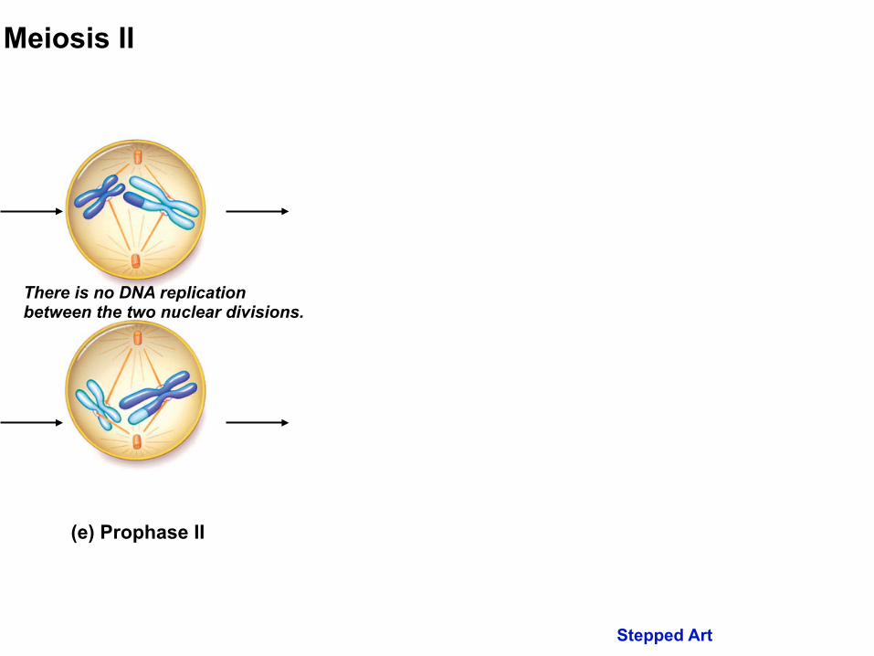
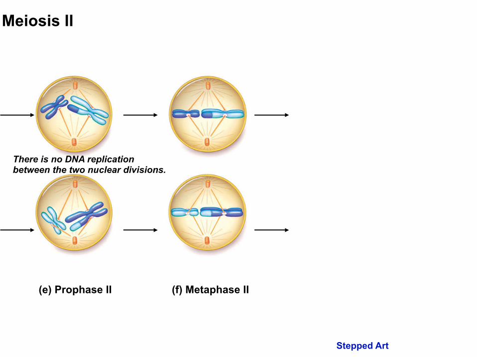
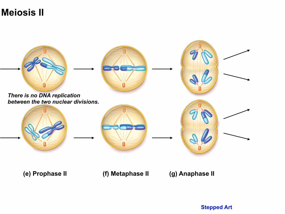
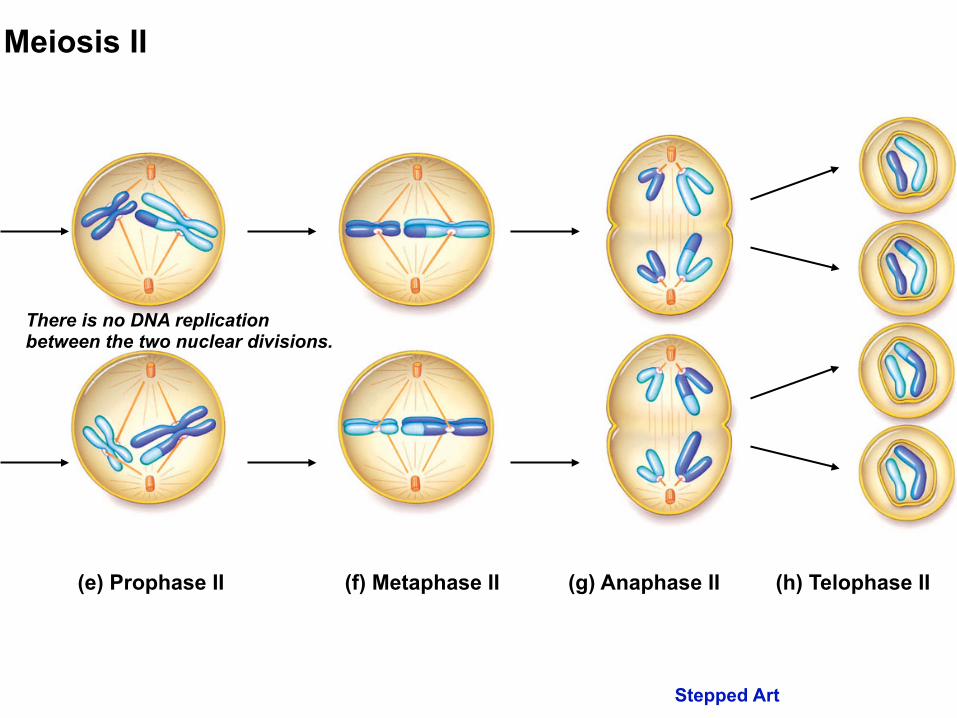
There is no DNA replication between the two nuclear divisions.
(e) Prophase II
Meiosis II
Stepped Art
There is no DNA replication between the two nuclear divisions.
(f) Metaphase II (e) Prophase II
Meiosis II
Stepped Art
There is no DNA replication between the two nuclear divisions.
(g) Anaphase II (f) Metaphase II (e) Prophase II
Meiosis II
Stepped Art
There is no DNA replication between the two nuclear divisions.
(h) Telophase II (g) Anaphase II (f) Metaphase II (e) Prophase II
Meiosis II
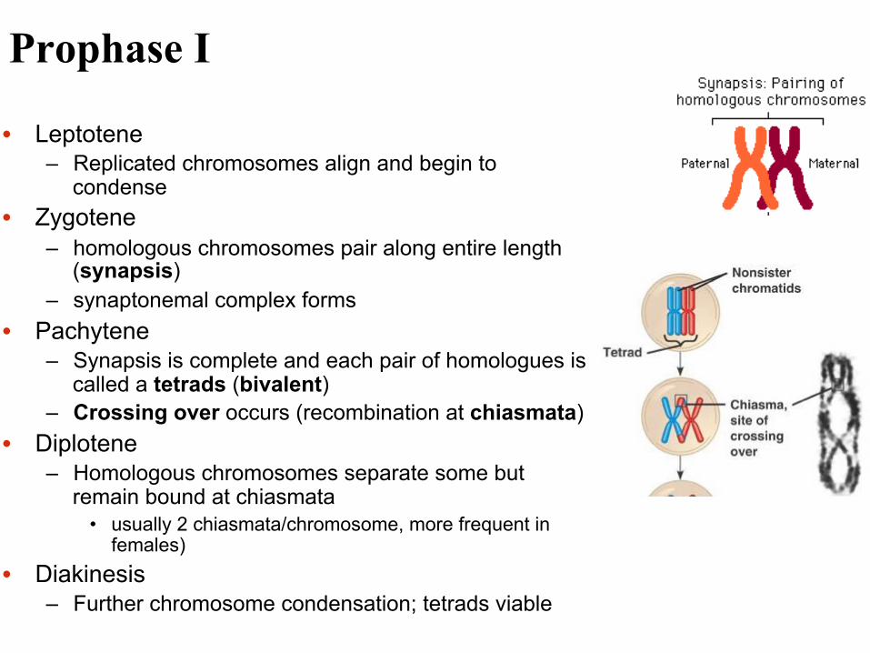
Prophase I
• Leptotene – Replicated chromosomes align and begin to
condense • Zygotene
– homologous chromosomes pair along entire length (synapsis)
– synaptonemal complex forms • Pachytene
– Synapsis is complete and each pair of homologues is called a tetrads (bivalent)
– Crossing over occurs (recombination at chiasmata) • Diplotene
– Homologous chromosomes separate some but remain bound at chiasmata
• usually 2 chiasmata/chromosome, more frequent in females)
• Diakinesis – Further chromosome condensation; tetrads viable

A tetrad showing chiasmata
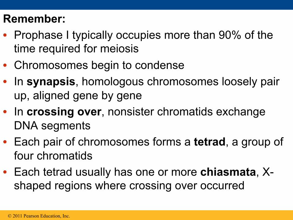
Remember: • Prophase I typically occupies more than 90% of the
time required for meiosis • Chromosomes begin to condense • In synapsis, homologous chromosomes loosely pair
up, aligned gene by gene • In crossing over, nonsister chromatids exchange
DNA segments • Each pair of chromosomes forms a tetrad, a group of
four chromatids • Each tetrad usually has one or more chiasmata, X-
shaped regions where crossing over occurred
© 2011 Pearson Education, Inc.

Metaphase I
• Chromosome pairs line up on the midline
• Spindles are fully formed and attached to centromeres
Metaphase I
spindle equator
one pair of homologous chromosomes
Fig. 2.13b

Anaphase I
• Centromeres do not divide
• Homologous chromosomes move to opposite poles and separate (disjunction)
Anaphase I
Fig. 2.13c
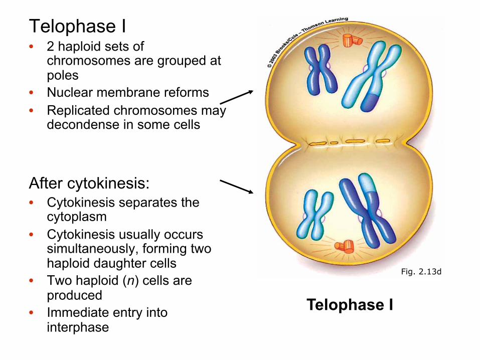
Telophase I • 2 haploid sets of
chromosomes are grouped at poles
• Nuclear membrane reforms • Replicated chromosomes may
decondense in some cells ! After cytokinesis: • Cytokinesis separates the
cytoplasm • Cytokinesis usually occurs
simultaneously, forming two haploid daughter cells
• Two haploid (n) cells are produced
• Immediate entry into interphase
Telophase I
Fig. 2.13d

Interkinesis Interphase II
• No DNA synthesis occurs
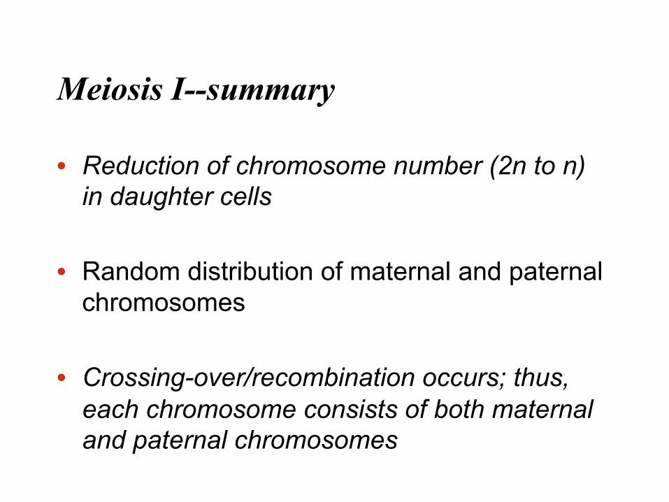
Meiosis I--summary
• Reduction of chromosome number (2n to n) in daughter cells
• Random distribution of maternal and paternal chromosomes
• Crossing-over/recombination occurs; thus, each chromosome consists of both maternal and paternal chromosomes
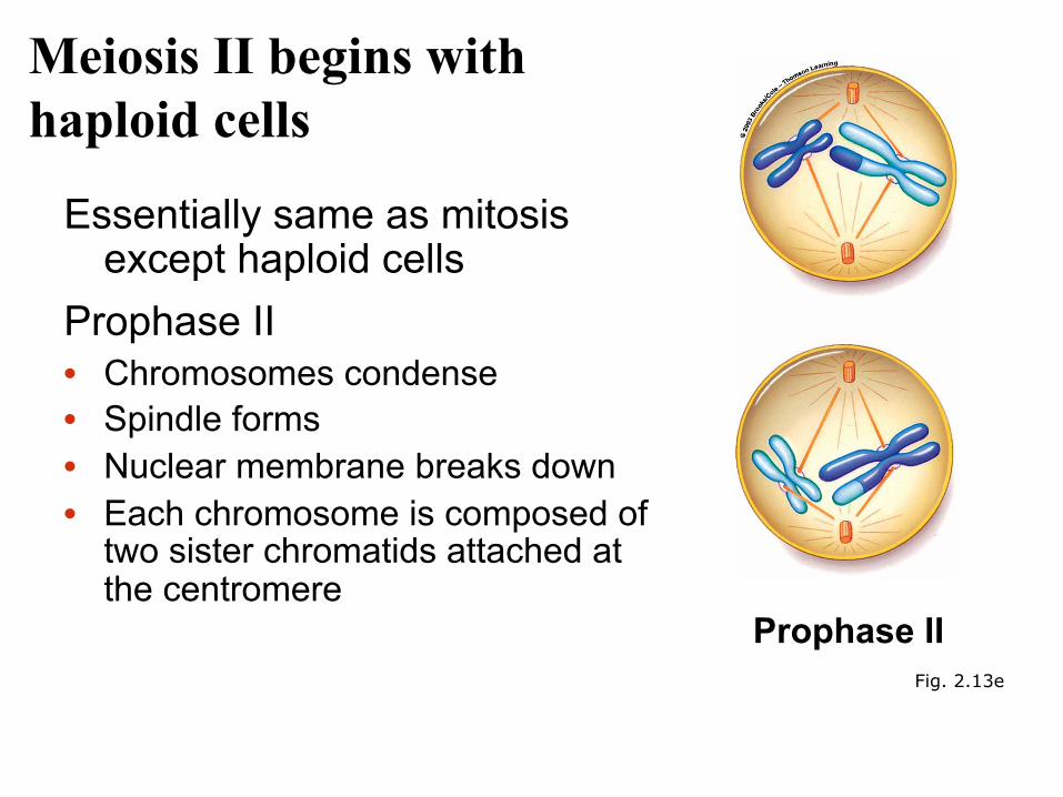
Meiosis II begins with haploid cells
Essentially same as mitosis except haploid cells
Prophase II • Chromosomes condense • Spindle forms • Nuclear membrane breaks down • Each chromosome is composed of
two sister chromatids attached at the centromere
Prophase II Fig. 2.13e

Metaphase II • Chromosomes line up on
the midline and centromeres attach to spindle fibers
Metaphase II
Fig. 2.13f
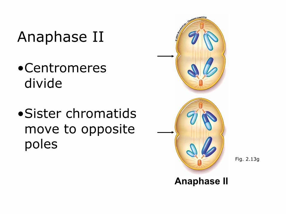
Anaphase II • Centromeres divide
• Sister chromatids move to opposite poles
Anaphase II
Fig. 2.13g

Telophase II
• Nuclear membrane forms • Chromosomes
decondense After cytokinesis: • Four unique haploid (n)
cells are produced with 23 chromosomes each
Telophase II
Fig. 2.13h
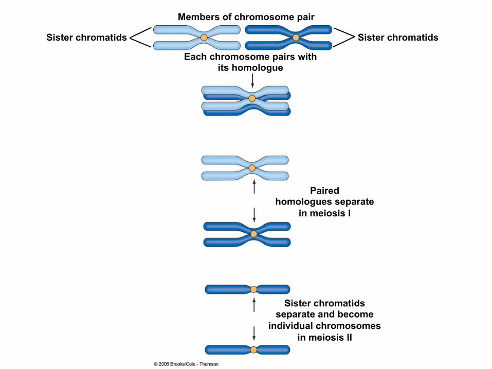
Members of chromosome pair
Each chromosome pairs with its homologue
Paired homologues separate
in meiosis I
Sister chromatids separate and become
individual chromosomes in meiosis II
Sister chromatids Sister chromatids
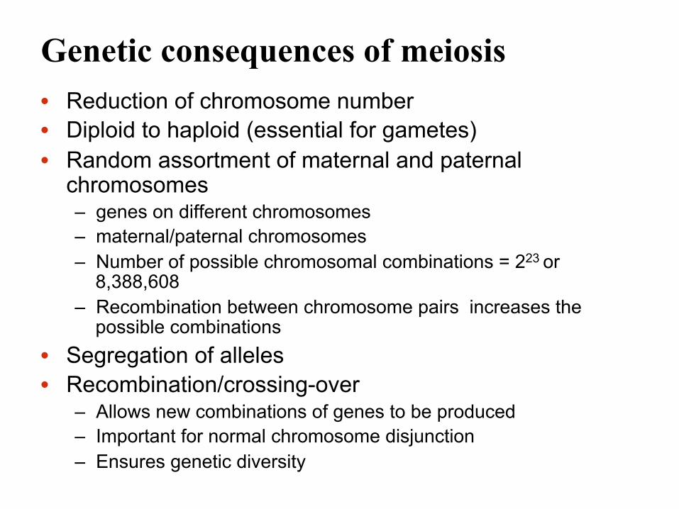
Genetic consequences of meiosis • Reduction of chromosome number • Diploid to haploid (essential for gametes) • Random assortment of maternal and paternal
chromosomes – genes on different chromosomes – maternal/paternal chromosomes – Number of possible chromosomal combinations = 223 or
8,388,608 – Recombination between chromosome pairs increases the
possible combinations • Segregation of alleles • Recombination/crossing-over
– Allows new combinations of genes to be produced – Important for normal chromosome disjunction – Ensures genetic diversity
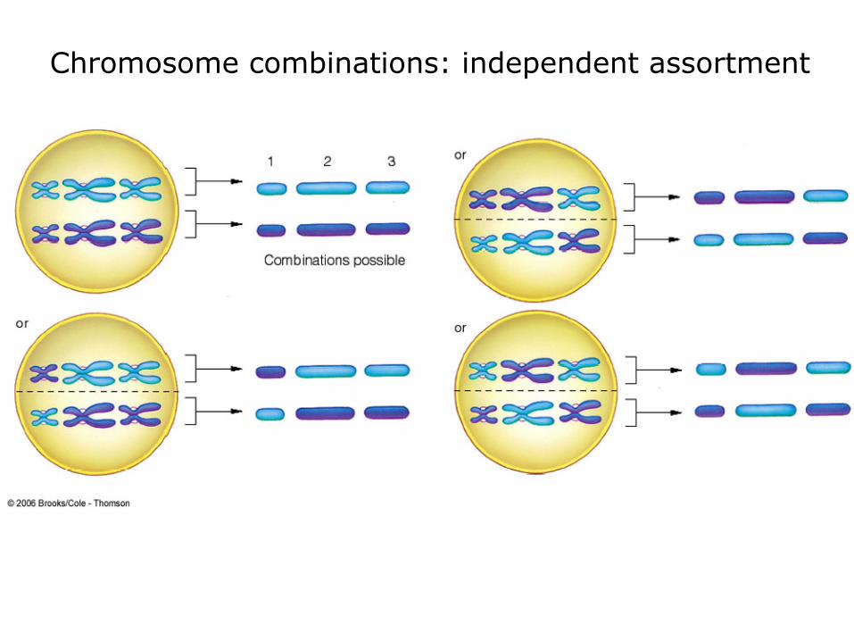
Chromosome combinations: independent assortment
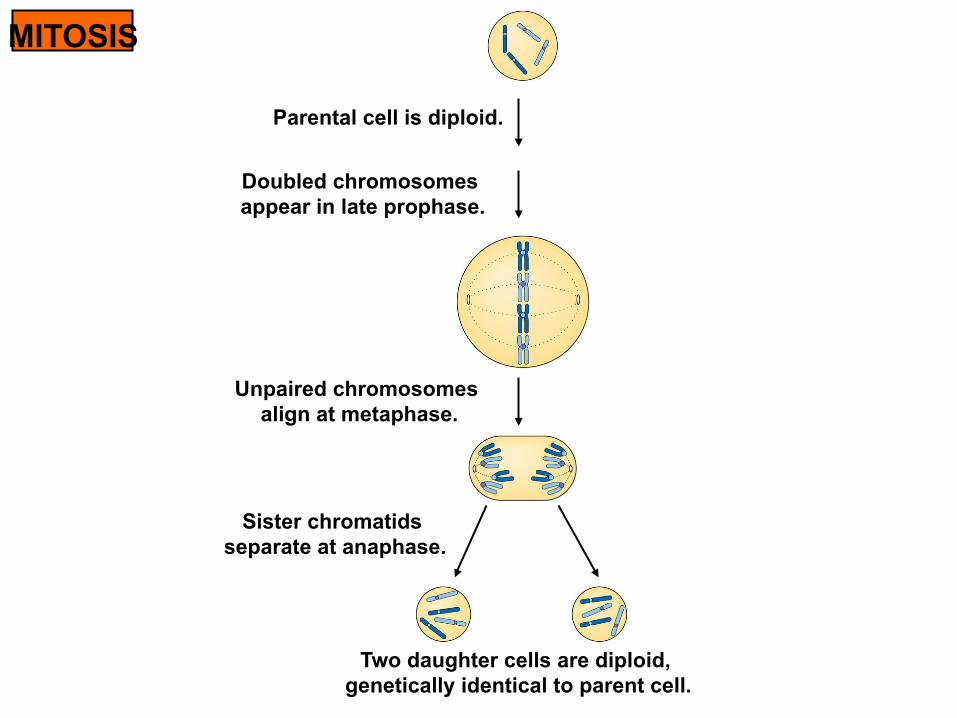
MITOSIS
Parental cell is diploid.
Doubled chromosomes appear in late prophase.
Unpaired chromosomes align at metaphase.
Sister chromatids separate at anaphase.
Two daughter cells are diploid, genetically identical to parent cell.

MEIOSIS
Four daughter cells are haploid, not genetically identical to parent cell.
Sister chromatids separate at anaphase II.
Paired homologous chromosomes align at metaphase I, then separate at anaphase I.
Parental cell is diploid.
Doubled chromosomes appear in late prophase.
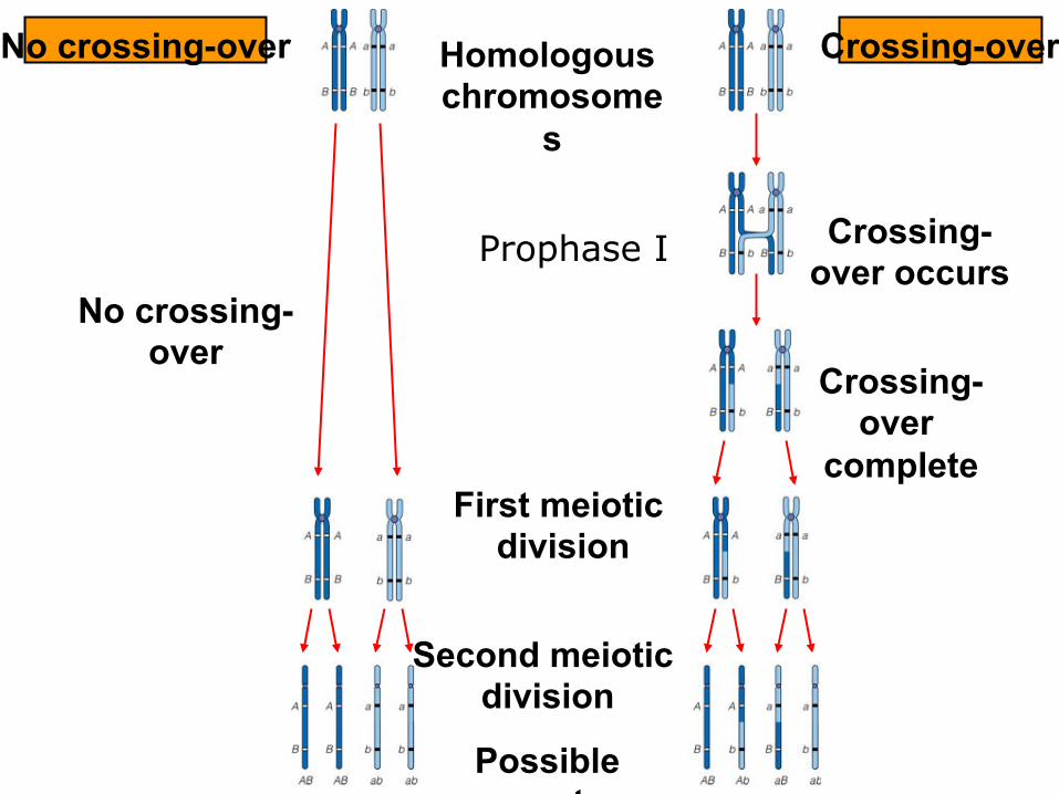
No crossing-over Crossing-over Homologous chromosome
s
Crossing-over occurs
Crossing-over
complete First meiotic
division
Second meiotic division
Possible gametes
No crossing-over
Prophase I

Summary of mitosis and meiosis
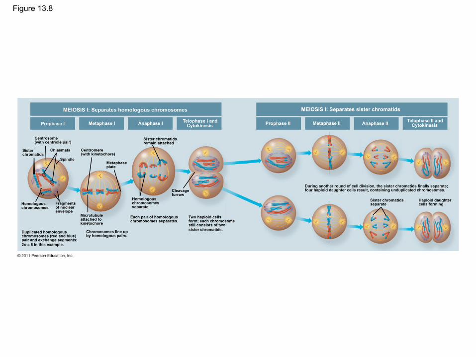
Figure 13.8
MEIOSIS I: Separates homologous chromosomes
Prophase I Metaphase I Anaphase I Telophase I and Cytokinesis
Centrosome (with centriole pair)
Sister chromatids
Chiasmata
Spindle
Homologous chromosomes
Fragments of nuclear envelope
Duplicated homologous chromosomes (red and blue) pair and exchange segments; 2n = 6 in this example.
Centromere (with kinetochore)
Metaphase plate
Microtubule attached to kinetochore
Chromosomes line up by homologous pairs.
Sister chromatids remain attached
Homologous chromosomes separate
Each pair of homologous chromosomes separates.
Cleavage furrow
Two haploid cells form; each chromosome still consists of two sister chromatids.
MEIOSIS I: Separates sister chromatids
Prophase II Metaphase II Anaphase II Telophase II and Cytokinesis
Sister chromatids separate
Haploid daughter cells forming
During another round of cell division, the sister chromatids finally separate; four haploid daughter cells result, containing unduplicated chromosomes.
A Comparison of Mitosis and Meiosis
• Mitosis conserves the number of chromosome sets, producing cells that are genetically identical to the parent cell
• Meiosis reduces the number of chromosomes sets from two (diploid) to one (haploid), producing cells that differ genetically from each other and from the parent cell
© 2011 Pearson Education, Inc.
Figure 13.9
Prophase
Duplicated chromosome
MITOSIS
Chromosome duplication
Parent cell
2n = 6
Metaphase
Anaphase Telophase
2n 2n
Daughter cells of mitosis
MEIOSIS
MEIOSIS I
MEIOSIS II
Prophase I
Metaphase I
Anaphase I Telophase I
Haploid n = 3
Chiasma
Chromosome duplication Homologous
chromosome pair
Daughter cells of
meiosis I
Daughter cells of meiosis II n n n n
SUMMARY
Property Mitosis Meiosis
DNA replication
Number of divisions
Synapsis of homologous chromosomes
Number of daughter cells and genetic composition
Role in the animal body
Occurs during interphase before mitosis begins
One, including prophase, metaphase, anaphase, and telophase
Does not occur
Two, each diploid (2n) and genetically identical to the parent cell
Enables multicellular adult to arise from zygote; produces cells for growth, repair, and, in some species, asexual reproduction
Occurs during interphase before meiosis I begins
Two, each including prophase, metaphase, anaphase, and telophase
Occurs during prophase I along with crossing over between nonsister chromatids; resulting chiasmata hold pairs together due to sister chromatid cohesion
Four, each haploid (n), containing half as many chromosomes as the parent cell; genetically different from the parent cell and from each other
Produces gametes; reduces number of chromosomes by half and introduces genetic variability among the gametes

Figure 13.9a
Prophase
Duplicated chromosome
MITOSIS
Chromosome duplication
Parent cell
2n = 6
Metaphase
Anaphase Telophase
2n 2n Daughter cells
of mitosis
MEIOSIS
MEIOSIS I
MEIOSIS II
Prophase I
Metaphase I
Anaphase I Telophase I
Haploid n = 3
Chiasma
Chromosome duplication Homologous
chromosome pair
Daughter cells of
meiosis I
Daughter cells of meiosis II n n n n
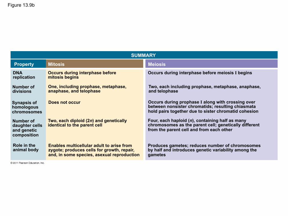
Figure 13.9b
SUMMARY
Property Mitosis Meiosis DNA replication
Number of divisions
Synapsis of homologous chromosomes
Number of daughter cells and genetic composition
Role in the animal body
Occurs during interphase before mitosis begins
One, including prophase, metaphase, anaphase, and telophase
Does not occur
Two, each diploid (2n) and genetically identical to the parent cell
Enables multicellular adult to arise from zygote; produces cells for growth, repair, and, in some species, asexual reproduction
Occurs during interphase before meiosis I begins
Two, each including prophase, metaphase, anaphase, and telophase
Occurs during prophase I along with crossing over between nonsister chromatids; resulting chiasmata hold pairs together due to sister chromatid cohesion
Four, each haploid (n), containing half as many chromosomes as the parent cell; genetically different from the parent cell and from each other
Produces gametes; reduces number of chromosomes by half and introduces genetic variability among the gametes

• Three events are unique to meiosis, and all three occur in meiosis l – Synapsis and crossing over in prophase I:
Homologous chromosomes physically connect and exchange genetic information
– At the metaphase plate, there are paired homologous chromosomes (tetrads), instead of individual replicated chromosomes
– At anaphase I, it is homologous chromosomes, instead of sister chromatids, that separate
© 2011 Pearson Education, Inc.
• Sister chromatid cohesion allows sister chromatids of a single chromosome to stay together through meiosis I
• Protein complexes called cohesins are responsible for this cohesion
• In mitosis, cohesins are cleaved at the end of metaphase
• In meiosis, cohesins are cleaved along the chromosome arms in anaphase I (separation of homologs) and at the centromeres in anaphase II (separation of sister chromatids)
© 2011 Pearson Education, Inc.
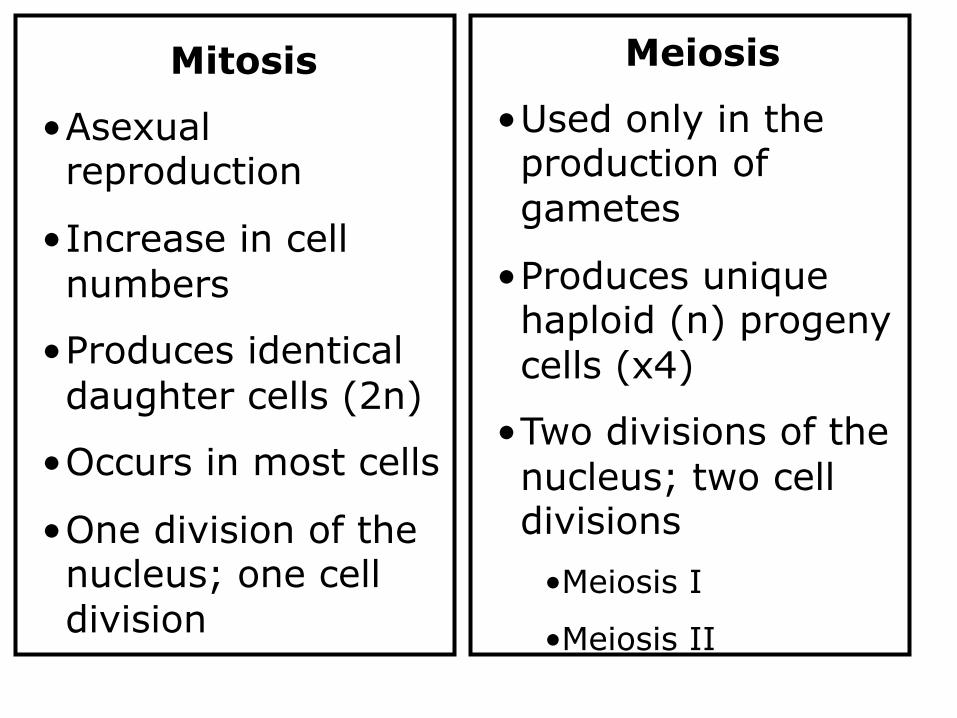
Meiosis
• Used only in the production of gametes
• Produces unique haploid (n) progeny cells (x4)
• Two divisions of the nucleus; two cell divisions
• Meiosis I
• Meiosis II
Mitosis
• Asexual reproduction
• Increase in cell numbers
• Produces identical daughter cells (2n)
• Occurs in most cells
• One division of the nucleus; one cell division

Inheritance of Genes
• Genes are the units of heredity, and are made up of segments of DNA
• Genes are passed to the next generation via reproductive cells called gametes (sperm and eggs)
• Each gene has a specific location called a locus on a certain chromosome
• Most DNA is packaged into chromosomes
© 2011 Pearson Education, Inc.
Comparison of Asexual and Sexual Reproduction
• In asexual reproduction, a single individual passes genes to its offspring without the fusion of gametes
• A clone is a group of genetically identical individuals from the same parent
• In sexual reproduction, two parents give rise to offspring that have unique combinations of genes inherited from the two parents
© 2011 Pearson Education, Inc.

Sets of Chromosomes in Human Cells
• Human somatic cells (any cell other than a gamete) have 23 pairs of chromosomes
• A karyotype is an ordered display of the pairs of chromosomes from a cell
• The two chromosomes in each pair are called homologous chromosomes, or homologs
• Chromosomes in a homologous pair are the same length and shape and carry genes controlling the same inherited characters
© 2011 Pearson Education, Inc.
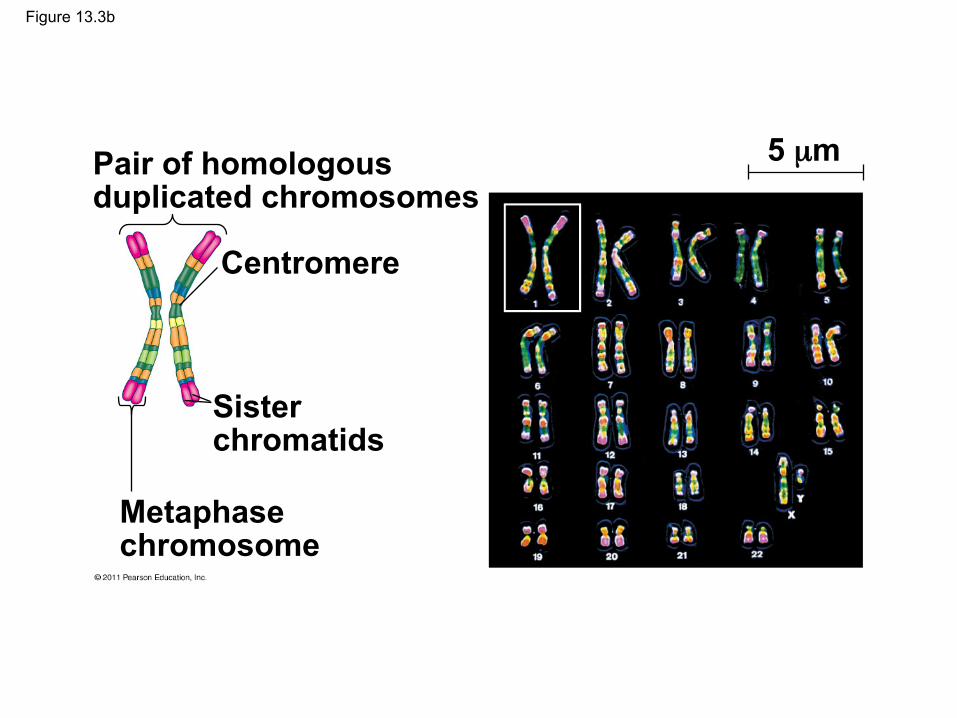
Figure 13.3b
Pair of homologous duplicated chromosomes
Centromere
Sister chromatids
Metaphase chromosome
5 µm
• The sex chromosomes, which determine the sex of the individual, are called X and Y
• Human females have a homologous pair of X chromosomes (XX)
• Human males have one X and one Y chromosome
• The remaining 22 pairs of chromosomes are called autosomes
© 2011 Pearson Education, Inc.
• Each pair of homologous chromosomes includes one chromosome from each parent
• The 46 chromosomes in a human somatic cell are two sets of 23: one from the mother and one from the father
• A diploid cell (2n) has two sets of chromosomes
• For humans, the diploid number is 46 (2n = 46)
© 2011 Pearson Education, Inc.
• In a cell in which DNA synthesis has occurred, each chromosome is replicated
• Each replicated chromosome consists of two identical sister chromatids
© 2011 Pearson Education, Inc.

Figure 13.4
Sister chromatids of one duplicated chromosome
Key Maternal set of chromosomes (n = 3) Paternal set of chromosomes (n = 3)
Key
2n = 6
Centromere
Two nonsister chromatids in a homologous pair
Pair of homologous chromosomes (one from each set)
• A gamete (sperm or egg) contains a single set of chromosomes, and is haploid (n)
• For humans, the haploid number is 23 (n = 23) • Each set of 23 consists of 22 autosomes and a
single sex chromosome • In an unfertilized egg (ovum), the sex
chromosome is X • In a sperm cell, the sex chromosome may be
either X or Y
© 2011 Pearson Education, Inc. © 2011 Pearson Education, Inc.

• Fertilization is the union of gametes (the sperm and the egg)
• The fertilized egg is called a zygote and has one set of chromosomes from each parent
• The zygote produces somatic cells by mitosis and develops into an adult
Behavior of Chromosome Sets in the Human Life Cycle
© 2011 Pearson Education, Inc.
• At sexual maturity, the ovaries and testes produce haploid gametes
• Gametes are the only types of human cells produced by meiosis, rather than mitosis
• Meiosis results in one set of chromosomes in each gamete
• Fertilization and meiosis alternate in sexual life cycles to maintain chromosome number
© 2011 Pearson Education, Inc.

Figure 13.5 Key
Haploid (n) Diploid (2n)
Egg (n)
Haploid gametes (n = 23)
Sperm (n)
Ovary Testis
Mitosis and development
Diploid zygote (2n = 46)
Multicellular diploid adults (2n = 46)
MEIOSIS FERTILIZATION

The Variety of Sexual Life Cycles
• The alternation of meiosis and fertilization is common to all organisms that reproduce sexually
• The three main types of sexual life cycles differ in the timing of meiosis and fertilization
© 2011 Pearson Education, Inc.

• Gametes are the only haploid cells in animals • They are produces by meiosis and undergo no
further cell division before fertilization • Gametes fuse to form a diploid zygote that
divides by mitosis to develop into a multicellular organism
© 2011 Pearson Education, Inc.

Concept 13.4: Genetic variation produced in sexual life cycles contributes to evolution
• Mutations (changes in an organism’s DNA) are the original source of genetic diversity
• Mutations create different versions of genes called alleles
• Reshuffling of alleles during sexual reproduction produces genetic variation
© 2011 Pearson Education, Inc.
Origins of Genetic Variation Among Offspring
• The behavior of chromosomes during meiosis and fertilization is responsible for most of the variation that arises in each generation
• Three mechanisms contribute to genetic variation
– Independent assortment of chromosomes – Crossing over – Random fertilization
© 2011 Pearson Education, Inc.
Independent Assortment of Chromosomes
• Homologous pairs of chromosomes orient randomly at metaphase I of meiosis
• In independent assortment, each pair of chromosomes sorts maternal and paternal homologues into daughter cells independently of the other pairs
© 2011 Pearson Education, Inc.
• The number of combinations possible when chromosomes assort independently into gametes is 2n, where n is the haploid number
• For humans (n = 23), there are more than 8 million (223) possible combinations of chromosomes
© 2011 Pearson Education, Inc.

Figure 13.10-1
Possibility 1 Possibility 2
Two equally probable arrangements of chromosomes at
metaphase I
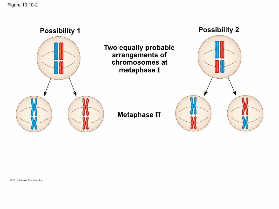
Figure 13.10-2
Possibility 1 Possibility 2
Two equally probable arrangements of chromosomes at
metaphase I
Metaphase II

Figure 13.10-3
Possibility 1 Possibility 2
Two equally probable arrangements of chromosomes at
metaphase I
Metaphase II
Daughter cells
Combination 1 Combination 2 Combination 3 Combination 4
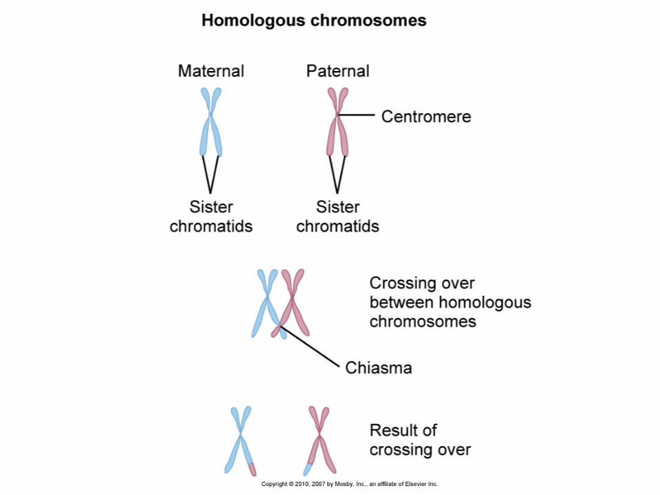
Crossing Over
• Crossing over produces recombinant chromosomes, which combine DNA inherited from each parent
• Crossing over begins very early in prophase I, as homologous chromosomes pair up gene by gene
© 2011 Pearson Education, Inc.
• In crossing over, homologous portions of two nonsister chromatids trade places
• Crossing over contributes to genetic variation by combining DNA from two parents into a single chromosome
© 2011 Pearson Education, Inc.

Figure 13.11-1 Prophase I of meiosis
Nonsister chromatids held together during synapsis
Pair of homologs

Figure 13.11-2 Prophase I of meiosis
Nonsister chromatids held together during synapsis
Pair of homologs
Chiasma
Centromere
TEM

Figure 13.11-3 Prophase I of meiosis
Nonsister chromatids held together during synapsis
Pair of homologs
Chiasma
Centromere
TEM Anaphase I
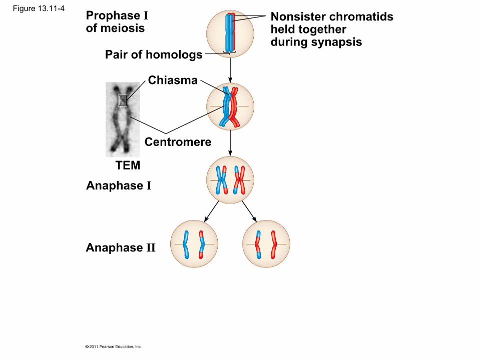
Figure 13.11-4 Prophase I of meiosis
Nonsister chromatids held together during synapsis
Pair of homologs
Chiasma
Centromere
TEM Anaphase I
Anaphase II

Figure 13.11-5 Prophase I of meiosis
Nonsister chromatids held together during synapsis
Pair of homologs
Chiasma
Centromere
TEM Anaphase I
Anaphase II
Daughter cells
Recombinant chromosomes

Figure 13.11a
Chiasma
Centromere
TEM

Random Fertilization
• Random fertilization adds to genetic variation because any sperm can fuse with any ovum (unfertilized egg)
• The fusion of two gametes (each with 8.4 million possible chromosome combinations from independent assortment) produces a zygote with any of about 70 trillion diploid combinations
© 2011 Pearson Education, Inc.

• Crossing over adds even more variation • Each zygote has a unique genetic identity
© 2011 Pearson Education, Inc.
Animation: Genetic Variation
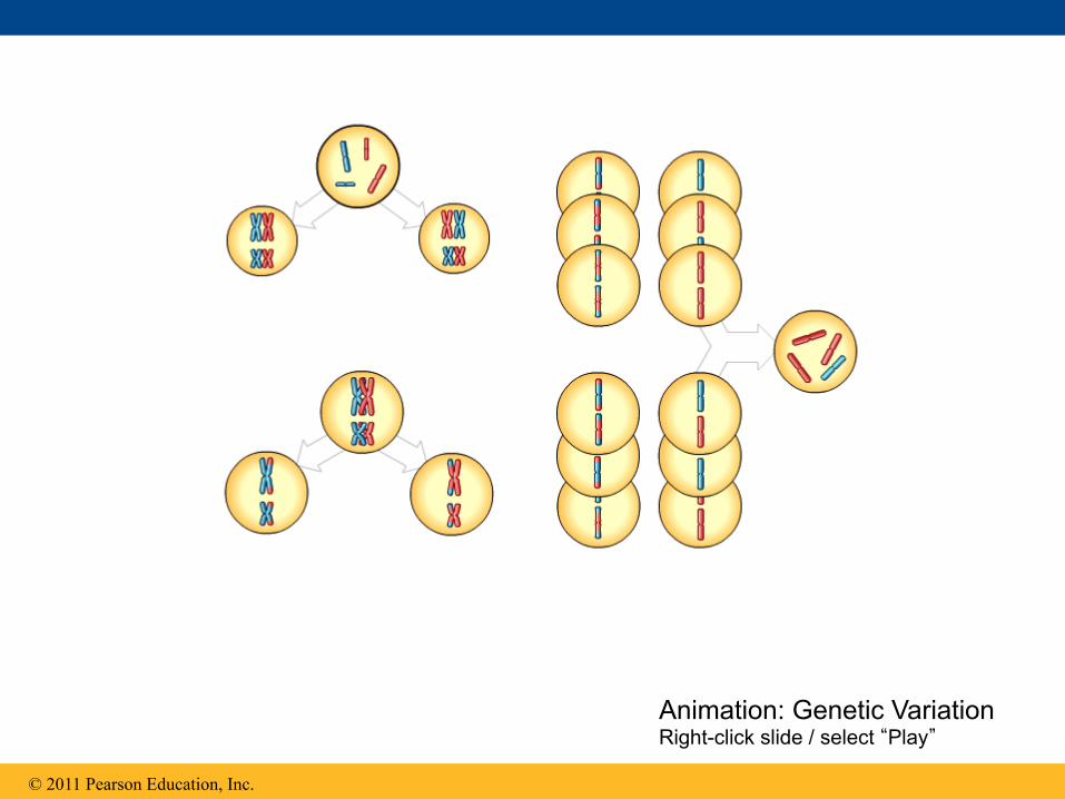
© 2011 Pearson Education, Inc.
Animation: Genetic Variation Right-click slide / select “Play”

Prophase I: Each homologous pair undergoes synapsis and crossing over between nonsister chromatids with the subsequent appearance of chiasmata.
Metaphase I: Chromosomes line up as homologous pairs on the metaphase plate.
Anaphase I: Homologs separate from each other; sister chromatids remain joined at the centromere.
Figure 13.UN01
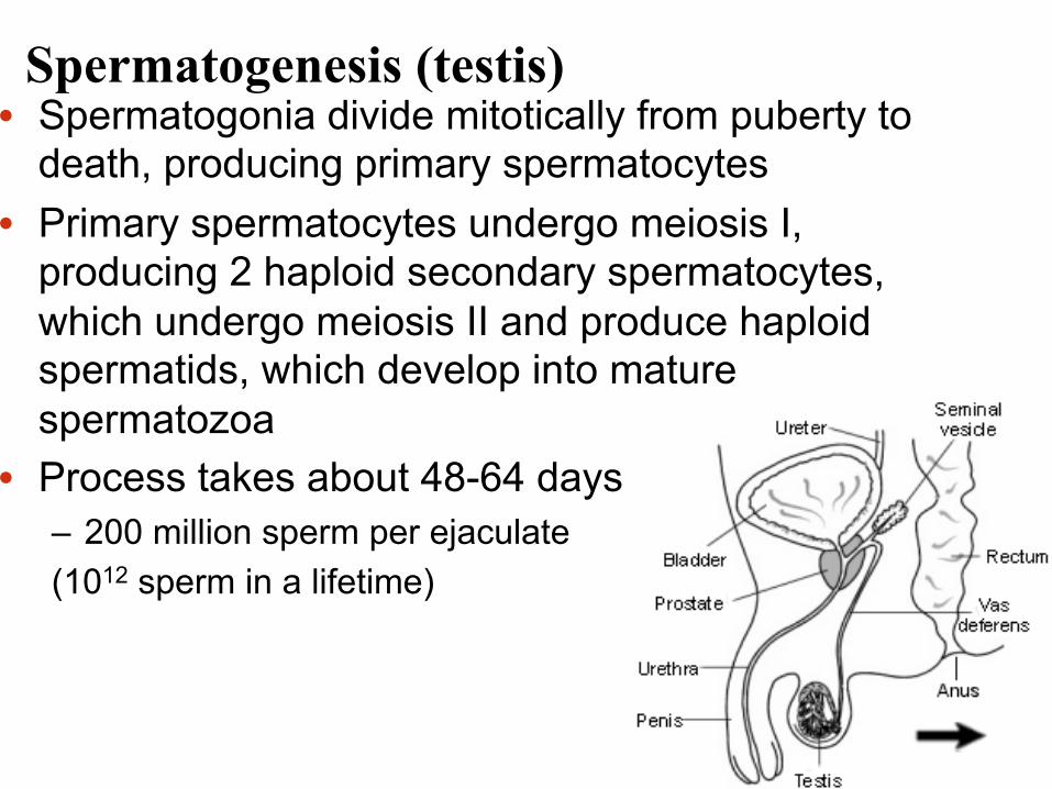
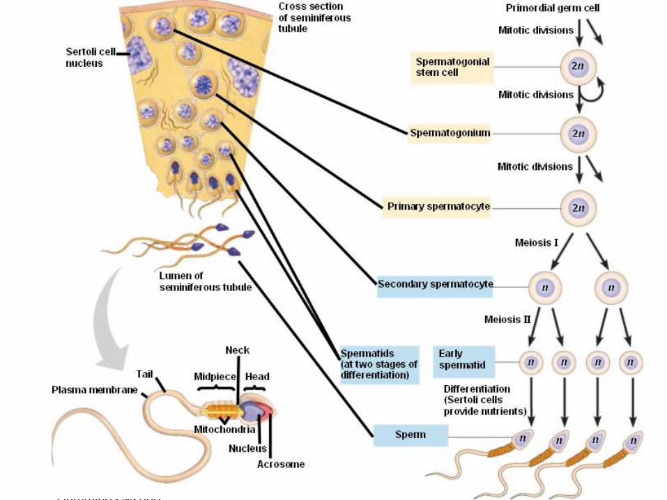
Spermatogenesis (testis) • Spermatogonia divide mitotically from puberty to
death, producing primary spermatocytes • Primary spermatocytes undergo meiosis I,
producing 2 haploid secondary spermatocytes, which undergo meiosis II and produce haploid spermatids, which develop into mature spermatozoa
• Process takes about 48-64 days – 200 million sperm per ejaculate (1012 sperm in a lifetime)
Chapter 1 Human Heredity by Michael Cummings ©2006 Brooks/Cole-Thomson Learning
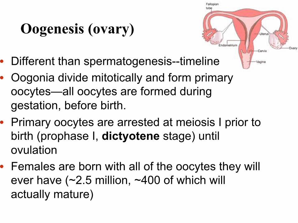
Oogenesis (ovary)
• Different than spermatogenesis--timeline • Oogonia divide mitotically and form primary
oocytes—all oocytes are formed during gestation, before birth.
• Primary oocytes are arrested at meiosis I prior to birth (prophase I, dictyotene stage) until ovulation
• Females are born with all of the oocytes they will ever have (~2.5 million, ~400 of which will actually mature)
Oogenesis (ovary)
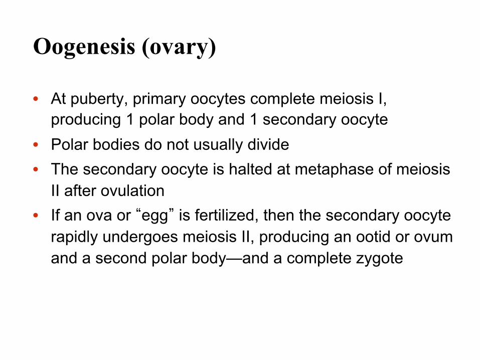
• At puberty, primary oocytes complete meiosis I, producing 1 polar body and 1 secondary oocyte
• Polar bodies do not usually divide • The secondary oocyte is halted at metaphase of meiosis
II after ovulation • If an ova or “egg” is fertilized, then the secondary oocyte
rapidly undergoes meiosis II, producing an ootid or ovum and a second polar body—and a complete zygote

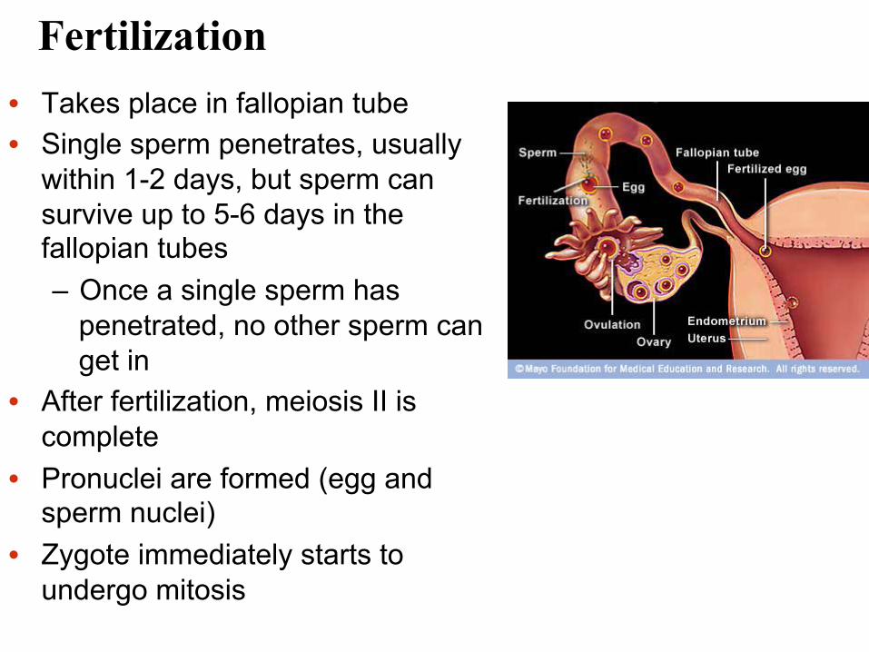
Fertilization • Takes place in fallopian tube • Single sperm penetrates, usually
within 1-2 days, but sperm can survive up to 5-6 days in the fallopian tubes – Once a single sperm has
penetrated, no other sperm can get in
• After fertilization, meiosis II is complete
• Pronuclei are formed (egg and sperm nuclei)
• Zygote immediately starts to undergo mitosis


Figure 2.11

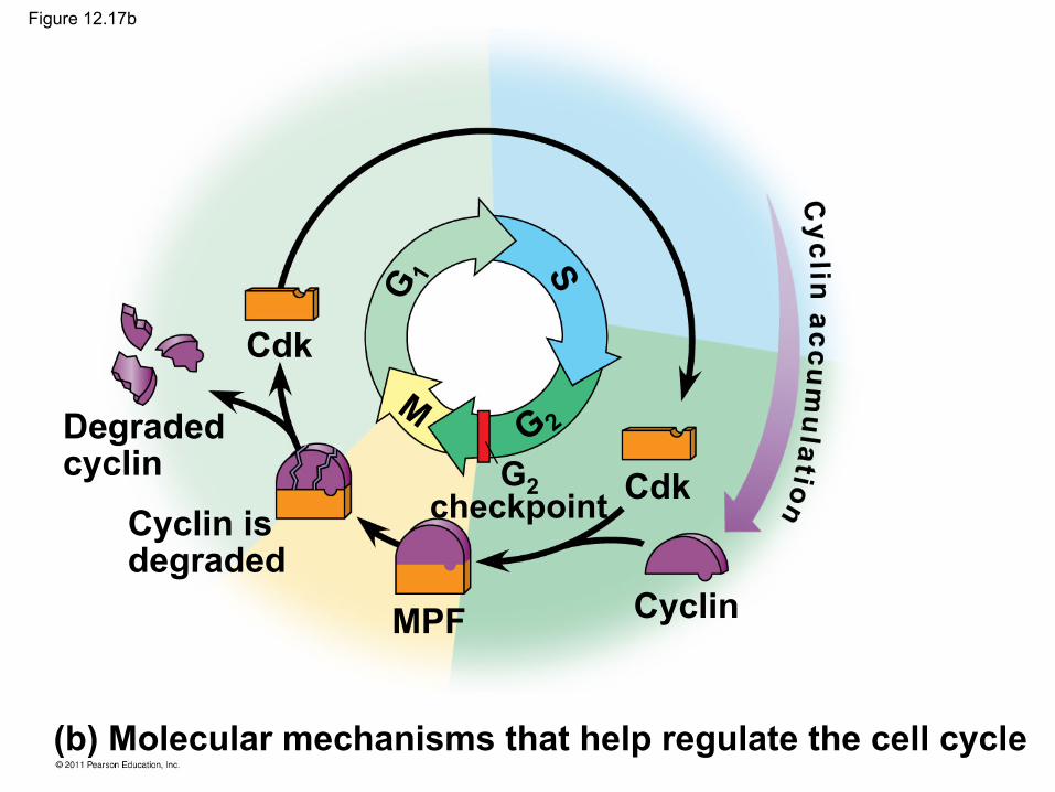
The Cell Cycle Clock: Cyclins and Cyclin-Dependent Kinases
• Two types of regulatory proteins are involved in cell cycle control: cyclins and cyclin-dependent kinases (Cdks)
• Cdks activity fluctuates during the cell cycle because it is controled by cyclins, so named because their concentrations vary with the cell cycle
• MPF (maturation-promoting factor) is a cyclin-Cdk complex that triggers a cell’s passage past the G2 checkpoint into the M phase
© 2011 Pearson Education, Inc.

Figure 12.17
(a) Fluctuation of MPF activity and cyclin concentration during the cell cycle
(b) Molecular mechanisms that help regulate the cell cycle
MPF activity Cyclin concentration
Time
M M M S S G1 G2 G1 G2 G1
Cdk
Degraded cyclin
Cyclin is degraded
MPF
G2 checkpoint Cdk
Cyclin
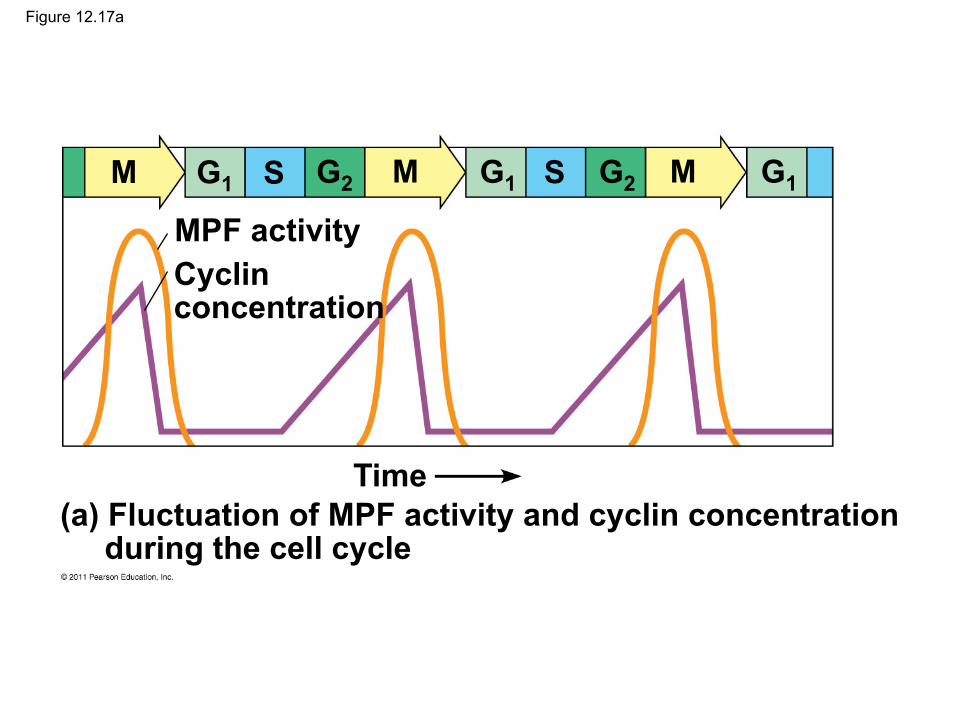
Figure 12.17a
(a) Fluctuation of MPF activity and cyclin concentration during the cell cycle
MPF activity Cyclin concentration
Time
M M M S S G1 G2 G1 G2 G1
(b) Molecular mechanisms that help regulate the cell cycle
Cdk
Degraded cyclin
Cyclin is degraded
MPF
G2 checkpoint Cdk
Cyclin
Figure 12.17b
Stop and Go Signs: Internal and External Signals at the Checkpoints
• An example of an internal signal is that kinetochores not attached to spindle microtubules send a molecular signal that delays anaphase
• Some external signals are growth factors, proteins released by certain cells that stimulate other cells to divide
• For example, platelet-derived growth factor (PDGF) stimulates the division of human fibroblast cells in culture
© 2011 Pearson Education, Inc.

Figure 12.18
A sample of human connective tissue is cut up into small pieces.
Enzymes digest the extracellular matrix, resulting in a suspension of free fibroblasts.
Cells are transferred to culture vessels.
Scalpels
Petri dish
PDGF is added to half the vessels.
Without PDGF With PDGF
10 µm
1
2
3 4
Loss of Cell Cycle Controls in Cancer Cells
• Cancer cells do not respond normally to the body’s control mechanisms
• Cancer cells may not need growth factors to grow and divide
– They may make their own growth factor – They may convey a growth factor’s signal without
the presence of the growth factor – They may have an abnormal cell cycle control
system
© 2011 Pearson Education, Inc.
• A normal cell is converted to a cancerous cell by a process called transformation
• Cancer cells that are not eliminated by the immune system, form tumors, masses of abnormal cells within otherwise normal tissue
• If abnormal cells remain at the original site, the lump is called a benign tumor
• Malignant tumors invade surrounding tissues and can metastasize, exporting cancer cells to other parts of the body, where they may form additional tumors
© 2011 Pearson Education, Inc.
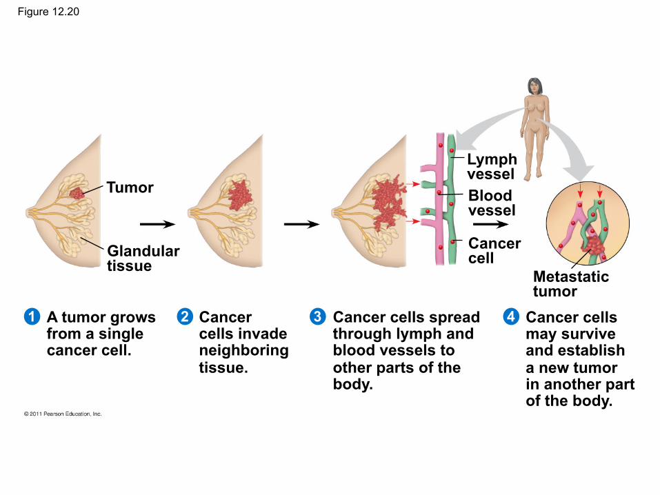
Figure 12.20
Glandular tissue
Tumor
Lymph vessel Blood vessel
Cancer cell
Metastatic tumor
A tumor grows from a single cancer cell.
Cancer cells invade neighboring tissue.
Cancer cells spread through lymph and blood vessels to other parts of the body.
Cancer cells may survive and establish a new tumor in another part of the body.
4 3 2 1