atıcı, ali eren-yeni.pdf
TRANSCRIPT
PROVIDING ANTIMICROBIAL PROPERTY TO SURFACES OF ULTRA HIGH MOLECULAR WEIGHT POLYETHYLENE
(UHMWPE) VIA GRAFTING BY UV INDUCED RAFT POLYMERIZATION
UV İLE BAŞLATILAN AŞILAMA YÖNTEMİ İLE RAFT POLİMERİZASYONU KULLANILARAK ULTRA MOLEKÜL
AĞIRLIKLI POLİETİLEN (UMAPE) YÜZEYİNE ANTİMİKROBİYAL ÖZELLİK KAZANDIRILMASI
ALİ EREN ATICI
PROF. DR. OLGUN GÜVEN Supervisor
ASSOC. PROF. DR. EBRU ORAL
Co-Supervisor
Submitted to
Graduate School of Science and Engineering of Hacettepe University
as a Partial Fullfillment to the Requirements
for the Award of the Degree of Doctor of Philosopy
in Chemistry
2020
Tarifi zor bir sevgi beslediğim eşim, oğlum, annem, babam ve kardeşime
Ve canım anneanneme
26/02/2020
i
ABSTRACT
PROVIDING ANTIMICROBIAL PROPERTY TO SURFACES OF
ULTRA HIGH MOLECULAR WEIGHT POLYETHYLENE (UHMWPE) VIA GRAFTING BY UV INDUCED RAFT POLYMERIZATION
Ali Eren Atıcı
Doctor of Philosophy, Chemistry Department Supervisor: Prof. Dr. Olgun Güven
Co-supervisor: Assoc. Prof. Dr. Ebru Oral February 2020, 165 Pages
Annually over 1 million patients in the US receive total joint replacements.
Ultrahigh molecular weight polyethylene (UHMWPE) has been used as a load-
bearing articular surface in majority of total joint arthroplasty. Periprosthetic
infection (PJI) is the most threatening complication facing total joint patients.
Although it occurs in 1-2% of cases, PJI is the reason of 30% of revisions and is a
severe healthcare burden. Most importantly, PJI is tremendously painful and
difficult for the patients. Recurrence of PJI prolongs the hospitalization with
additional series of surgeries. With recurrence, treatment becomes less effective,
some procedures such as arthrodesis amputation are often performed.
Approaches are needed to improve the efficacy of PJI treatment. Irrigation and
debridement (I&D), liner exchange, and one-stage revision are currently used
options to treat PJI. The gold standard treatment is removal of all components and
the placement of antibiotic-impregnated PMMA bone cement in the joint space in a
first surgery, followed by the placement of all new implant components after an
ii
intended 6-8 weeks of antibiotic treatment. However, the bioavailability of systemic
antibiotics in the bone/implant interface is very low and inefficient. On the other
hand, patients are largely immobilized during treatment due to PMMA spacers not
being able to bear the full weight of the patients. The 5-year success rates of I&D
followed by liner exchange and two stage surgery are 38 % and 80 % of the time.
As one strategy, therapeutic agents, such as antibiotics, can be incorporated into
ultra-high molecular weight polyethylene (UHMWPE) implants typically used in
total joint arthroplasty for local delivery of these therapeutic agents. Because of its
superior mechanical strength and markedly improved wear resistance in
comparison to bone cement UHMWPE is a better candidate than PMMA bone
cement as an articulating spacer and a delivery device eluting antibiotic.
Two important aspects have vital importance for an effective PJI treatment;
appropriate dosage control and sustainable antibiotic treatment. They both require
great attention otherwise could be devastating for patients and eventually turned
into an immense public health problem. Antibiotic dosage control must be so
delicate that it is not above toxicity levels and not below MIC which could lead to
antibiotic resistance. Sustainable antibiotic release is essential for implants to
avoid bacterial colonization which will fail patients to an additional joint
replacement surgery. Therefore, it is vital to tailor a drug-releasing implant which
ensures delicate dosage control and sustainable antibiotic release.
The aim of this thesis is to functionalize nonpolar UHMWPE by grafting 2-
hydroxyethyl methacrylate monomer and blend resulting copolymer (UHMPWE-g-
PHEMA) with commonly used antibiotic, gentamicin sulfate. RAFT polymerization
was also used to synthesize UHMWPE-g-PHEMA with controlled the molecular
weight and the molecular weight distribution of PHEMA to control the rate and the
sustainability of gentamicin sulfate release. Alterations in the chemical properties
after grafting PHEMA to UHMWPE have been investigated by using surface
characterization methods, ATR-FTIR, elemental analysis, X-ray photoelectron
spectroscopy (XPS), contact angle. Subsequently, antibiotic release studies from
antibiotic-loaded UHMWPE and UHMWPE-g-PHEMA (prepared by conventional
polymerization or RAFT polymerization) were conducted. Antimicrobial efficacy of
said polymers was tested in two ways:
1. Planktonic kill in the eluent media,
iii
2. Anticolonizing properties of polymeric surfaces.
Synthesized/prepared drug loaded polymers were tested to evaluate their
mechanical strength and wear resistance by using tensile testing, IZOD impact
testing and pin-on-disc wear testing.
The graft copolymer of UHMWPE-g-PHEMA showed significant increase for the
GS release rate in comparison to virgin UHMWPE. The antibacterial performance
of UHMWPE-g-PHEMA became more effective in parallel with release rate
improvement. HEMA grafting from UHMWPE reduced its mechanical properties
such as ultimate tensile strength, elongation at break and IZOD impact strength.
UHMWPE-g-PHEMA synthesized via UV-initiated RAFT polymerization increased
the GS release rate in a more sustainable trend compared to copolymer prepared
via conventional grafting. Thus, UHMWPE-g-PHEMA exhibited better planktonic
bacterial kill and non-adherent surface.
Keywords: UHMWPE, UV initiated grafting, RAFT polymerization, Periprosthetic
Joint Infection (PJI), knee implant, gentamicin sulfate
iv
ÖZET
UV ile Başlatılan Aşılama Yöntemi ile RAFT Polimerizasyonu
Kullanılarak Ultra Molekül Ağırlıklı Polietilen (UMAPE) Yüzeyine Antimikrobiyal Özellik Kazandırılması
Ali Eren Atıcı
Doktora, Kimya Bölümü Tez danışmanı: Prof. Dr. Olgun Güven
Eş danışman: Doç. Dr. Ebru Oral Şubat 2020, 165 Sayfa
ABD’de yılda 1 milyondan fazla hastaya total eklem değişimi
gerçekleştirilmektedir. Ultra molekül ağırlıklı polietilen (UMAPE), toplam eklem
artroplastisinin çoğunda yük taşıyan eklem yüzeyi olarak kullanılmaktadır.
Periprostetik eklem enfeksiyonu (PPEE) total eklem hastalarının karşılaştığı en
tehdit edici komplikasyondur. Vakaların %1-2'sinde görülmesine rağmen,
revizyonların % 30'unun nedeni PPEE’dir ve ciddi bir sağlık yüküdür. En önemlisi,
PPEE hastalar için son derece acı verici ve zordur. Tekrarlanması durumunda
hastanede yatış süresini uzatmaktadır. Tekrarlayan enfeksiyon vakalarında tedavi
daha az etkili olur, genellikle kurtarma için artrodez amputasyonu gibi bazı
prosedürler gerçekleştirilmektedir.
PPEE tedavisinin etkinliğini arttırmak için bazı yaklaşımlara ihtiyaç vardır.
Antiseptik su ile yıkama ve debridman, liner değişimi ve tek aşamalı revizyon şu
anda PPEE tedavisinde kullanılan prosedürlerdir. Altın standart tedavi ise tüm
protez bileşenlerinin çıkarılması ve ilk ameliyatta antibiyotik emdirilmiş PMMA
v
kemik çimentosunun eklem boşluğuna yerleştirilmesi ve ardından 6-8 haftalık bir
antibiyotik tedavisinden sonra tüm yeni protez bileşenlerinin yerleştirilmesidir.
Fakat, kemik / implant arayüzündeki sistemik antibiyotiklerin biyoelverişliliği çok
düşük ve verimsizdir. Ayrıca, PMMA aralayıcının hastaların tam ağırlığını
taşıyamaması nedeniyle hastalar tedavi sırasında büyük ölçüde
hareketsizleşmektedir. Bununla birlikte PPEE'yi ortadan kaldırmada 5 yıllık başarı
oranı sırasıyla antiseptik su ile yıkama ve debridman ve liner değişiminin için %
38, altın standart olan iki aşamalı tedavi için % 80'dir.
Başka bir strateji olarak, antibiyotikler gibi tedavi edici ajanlar, bu tedavi edici
ajanların lokal salımı için tipik olarak toplam eklem artroplastisinde kullanılan ultra
molekül ağırlıklı polietilen (UMAPE) implantların içerisine katılmaktadır. Üstün
mekanik dayanımı ve kemik çimentosu ile karşılaştırıldığında belirgin şekilde
geliştirilmiş aşınma direnci nedeniyle UMAPE, eklem aralayıcı olarak kullanılan
PMMA kemik çimentosundan ve antibiyotikleri salan bir ilaç salım sisteminden
daha iyi bir adaydır.
Etkili bir PPEE tedavisi için iki önemli husus hayati öneme sahiptir; uygun doz
kontrolü ve sürdürülebilir antibiyotik tedavisi. Her ikisi de büyük bir dikkat gerektirir,
aksi takdirde hastalar için yıkıcı etkileri olabilir ve sonunda büyük bir halk sağlığı
sorununa dönüşebilir. Antibiyotik doz kontrolü, antibiyotik direncine yol açabilecek
toksisite seviyelerinin üzerinde ve minimum inhibitör konsantrasyonun altında
olmayacak kadar hassas olmalıdır. Hastaların ek bir eklem değişim operasyonuna
daha girmesini engellemek için, implantlarda bakteri kolonizasyonundan kaçınmak
için sürdürülebilir antibiyotik salınımı şarttır. Bu nedenle, hassas doz kontrolü ve
sürdürülebilir antibiyotik salınımı sağlayan bir ilaç salan implantın uyarlanması
hayati önem taşır.
Bu tezin amacı, polar yapıda olmayan UMAPE'ye 2-hidroksietil metakrilat
monomeri aşılayarak elde edilen kopolimeri (UMAPE-g-PHEMA) yaygın olarak
kullanılan bir antibiyotik olan, gentamisin sülfat ile harmanlayarak
işlevselleştirmektir. RAFT polimerizasyonu, UMAPE-g-PHEMA’nın sentezinde
PHEMA'nın molekül ağırlığı ve molekül ağırlığı dağılımını kontrol edebilmek ve
gentamisin sülfatın salım oranını ve sürdürülebilirliğini kontrol etmek için kullanıldı.
PHEMA'nın UMAPE’ye aşılanmasından sonra kimyasal özelliklerinde meydana
gelen değişiklikler yüzey karakterizasyon yöntemleri, ATR-FTIR, elemental analizi,
vi
X-ışınları fotoelektron spektroskopisi (XPS) ve temas açısı kullanılarak
incelenmiştir. Daha sonra, antibiyotik yüklenen UMAPE ve UMAPE-g-PHEMA'dan
(geleneksel polimerizasyon veya RAFT polimerizasyonu ile hazırlanan) antibiyotik
salım çalışmaları gerçekleştirimiştir. Adı geçen polimerlerin antimikrobiyal etkinliği
iki şekilde test edilmiştir:
1. Eluent ortamda planktonik öldürme,
2. Polimerik yüzeylerin antikolonize edici özellikleri.
Sentezlenmiş/hazırlanmış ilaç yüklü polimerlerin çekme dayanımı ve aşınma
direncini incelemeleri mekanik testler ve aşınma testleri yapılmıştır.
UMAPE-g-PHEMA aşı kopolimeri GS salım oranını işlenmemiş UMAPE’ye kıyasla
önemli miktarda arttırmıştır. UMAPE-g-PHEMA aşı kopolimerinin antibakteriyel
performansı da salım oranındaki artışa parallel olarak daha etkili olmuştur.
UMAPE’ye HEMA aşılanması azami çekme mukavemeti, kopmadaki uzama ve
IZOD darbe dayanımı gibi mekanik özelliklerinin azalmasına sebep olmuştur. UV
ile başlatılan RAFT polimerizasyonuyla sentezlenmiş UMAPE-g-PHEMA
kopolimerler ise GS salımını geleneksel metodla aşılanarak hazırlanan kopolimere
kıyasla daha sürdürülebilir şekilde artırmıştır. Böylelikle, UMAPE-g-PHEMA daha
iyi planktonic bakteri öldürme ve yapışmaz yüzey özellikleri göstermiştir.
Anahtar Kelimeler: UMAPE, UV ile başlatılan aşılama, RAFT polimerizasyonu,
Periprostetik eklem enfeksiyonu (PPEE), diz implant, gentamisin sülfat
vii
TEŞEKKÜR
Danışmanım Prof. Dr. Olgun GÜVEN’e bana kattıklarından dolayı
teşekkürü borç bilirim.
Doktora çalışmalarım boyunca verdiği katkılardan ve
yönlendirmelerden dolayı Doç. Dr. Ebru ORAL’a teşekkür ederim.
Çalışmalarım sırasında verdiği enerji ve tavsiyelerle hevesimi canlı
tutan Prof. Dr. Orhun K. MURATOĞLU’na teşekkür ederim.
Massachusetts Genel Hastanesi’nde yer alan Harris Ortopedi
Labaratuvarı’ndaki çalışma arkadaşlarıma teşekkür ederim.
Bu süreçte benim hissettiklerimi paylaşan, beni yalnız bırakmayan
ailem Osman BAKAR, Müzeyyen BAKAR, Ece BAKAR ve Mine
BAKAR’a çok teşekkür ederim.
viii
TABLE OF CONTENTS
Abstract ....................................................................................................................i Özet ........................................................................................................................ iv Acknowledgement ................................................................................................. vii Table of Contents ................................................................................................. viii List of Tables .......................................................................................................... xi List of Figures ........................................................................................................ xii Symbols and Abbreviations ................................................................................. xvii 1. Introduction ......................................................................................................... 1 2. Total Joint Arthroplasty ....................................................................................... 4
2.1 UHMWPE ...................................................................................................... 6 2.1.1 Synthesis of UHMWPE ........................................................................... 7 2.1.2 Adaption of UHMWPE to The Field of Orthopaedics .............................. 9
2.2 Metals .......................................................................................................... 10 2.3 Ceramics ..................................................................................................... 11
2.3.1 Ceramic Oxides .................................................................................... 11 2.3.1.1 Alumina .......................................................................................... 11 2.3.1.2 Zirconia .......................................................................................... 12
2.3.2 Calcium Phosphate ............................................................................... 12 2.3. Reasons for revision surgery ...................................................................... 13
3. Periprosthetic Joint Infection ............................................................................. 15 3.1 Pathogenesis of Prosthetic Joint Infection ................................................... 16 3.2 Bacterial Strains in PJI ................................................................................ 18 3.3 Risk Factors in PJI ...................................................................................... 20
3.3.1 Patient-Related Risk Factors ................................................................ 20 3.3.2 Prophylactic Measures for PJI .............................................................. 21
3.4 Current Treatment for PJI ............................................................................ 21 3.4.1 Use of antibiotic-loaded bone cements as a PJI treatment ................... 24
4. Antimicrobial Polymeric Biomaterials ................................................................ 28 4.1 Antimicrobial Groups in Polymers ............................................................... 28
4.1.1 N-Halamines ......................................................................................... 28 4.1.2 Peptide-like structures........................................................................... 29
ix
4.1.3 Quaternary ammonium/phosphonium .................................................... 30 4.2 Functionalization of Polymers ...................................................................... 32
4.2.1 Methods for polymer functionalization ................................................... 32 4.3 Chemical Modifications of Biomaterials with Antimicrobial Functionality ...... 33
4.3.1 Dual Functionality Antimicrobial surfaces, coatings ............................... 36 5. Composite biomaterials ..................................................................................... 39 6. Living Radical Polymerization ............................................................................ 42
6.1 RAFT Polymerization ................................................................................... 42 6.2 Copolymer Synthesis by RAFT Polymerization ............................................ 44 6.3 Use of RAFT Technique for Drug Delivery Applications ............................... 45
7. Experimental ..................................................................................................... 47 7.1 Chemicals .................................................................................................... 47 7.2 Grafting 2-hydroxyethyl methacrylate from UHMWPE ................................. 47 7.3 RAFT mediated grafting of PHEMA from UHMWPE via preirradiation ......... 49 7.4 Antibiotic Blending with UHMWPE and UHMWPE-g-P(HEMA) ................... 49 7.5 Compression molding of the blends ............................................................ 50 7.6 Characterization .......................................................................................... 51
7.6.2 Structural characterization of PolyHEMA grafted UHMWPE ................. 51 7.6.2.1. FT-IR/ATR ..................................................................................... 51 7.6.2.2 CHNS/O Analysis ........................................................................... 51 7.6.2.3 X-ray Photoelectron Spectroscopy ................................................. 51 7.6.2.4 Contact Angle Measurements......................................................... 51 7.6.2.5 Tensile Strength Testing ................................................................. 52 7.6.2.6 IZOD Impact Strength Testing ........................................................ 52 7.6.2.7 Pin-on-Disc (POD) Wear Testing .................................................... 52 7.6.2.8 Differential Scanning Calorimetry ................................................... 52 7.6.2.9 Scanning Electron Microscopy (SEM) - EDAX Analysis ................. 53 7.6.2.10 Gravimetric Analysis of Grafting Degree ....................................... 53 7.6.2.11 Size Exclusion Chromatography-Multi-Angle Light Scattering ...... 53
7.7 Drug release studies .................................................................................... 54 7.7.1 Gentamicin Release Studies .................................................................. 54 7.7.2 Gentamicin release quantification by fluorescence spectroscopy .......... 54
7.7.2.1 Buffer solution preparation .............................................................. 55 7.7.2.2 Preparation of OPA solution ........................................................... 55
7.8 Antibacterial properties ................................................................................ 55
x
7.8.1 Bacterial Colonization Assessment ....................................................... 55 7.8.2 Antibacterial efficacy against planktonic bacteria .................................. 56 7.8.3 Determination of minimum inhibitory concentration (MIC) .................... 56
8. Results and Discussion .................................................................................... 57 8.1. Antibiotic Selection ..................................................................................... 58 8.2. Preparation of UHMWPE/GS Tibial Spacers .............................................. 59 8.3. Dehydration of UHMWPE/GS ..................................................................... 62
8.4.1. Drug Release from UHMWPE/GS ....................................................... 64 8.4.2. Antimicrobial Performance of UHMWPE/GS ........................................ 70 8.4.3. Mechanical Strength of UHMWPE/GS ................................................. 73 8.4.4. Bidirectional Wear Test of UHMWPE/GS Samples .............................. 75
8.5. 2-hydroxyethyl Methacrylate Grafting from UHMWPE ............................... 77
8.6. 2-hydroxyethyl Methacrylate Grafting from UHMWPE via RAFT Polymerization ........................................................................................... 92
9. Conclusions .................................................................................................... 110 9. References ..................................................................................................... 117
xi
LIST OF TABLES
Table 2.1. Physical properties of HDPE and UHMWPE (Edidin and Kurtz 2000).6
Table 3.1. Microorganisms in periprosthetic hip and knee infection .................. 18
Table 3.2. PJI causing bacteria and its antibiotic agent .................................... 22
Table 8.1. Grafting percentage of UHMWPE-g-PHEMA synthesized by
conventional radical polymerization by using 10 % and 20 %
monomer feed for 60, 90, 120 min ................................................... 82
Table 8.2. Melting temperatures and crystallinity percentages of virgin
UHMWPE and UHMWPE-g-PHEMA with increasing degree of
grafting ............................................................................................. 85
Table 8.3. Grafting percentage of UHMWPE-g-PHEMA (20 %) synthesized by
RAFT polymerization with various [HEMA]/[DDMAT] for 60 and 120
min ................................................................................................... 95
Table 8.4. C, H, N, S analysis of R-UHMWPE-g-PHEMA samples synthesized
in 120 min with various [HEMA]/[DDMAT] ........................................ 95
Table 8.5. Monomer to polymer conversion, theoretical and experimental Mns
and polydispersity of PHEMA homopolymer formed in the grafting
medium .......................................................................................... 100
Table 8.6. Images (top and cross-section) and molding conditions of
consolidated, tibial shape UHMWPE blocks loaded with GS/GS+VH111
xii
LIST OF FIGURES
Figure 2. 1. Total Knee and Hip Implant Components (Copyright AAOS) ............ 4
Figure 2. 2. Bicondylar knee designs, A) Geometric, B) Townley, C) Freeman-
Swanson total knee prostheses with UHMWPE components
(Reprinted from (Kurtz 2016)). ........................................................... 5
Figure 2. 3. TEM micrograph of semicrystalline UHMWPE (Bellare,
Schnablegger, and Cohen 1995) ....................................................... 7
Figure 2. 4. UHMWPE processing steps UHMWPE fine powder (A), extruded
UHMWPE powder (B), machining of the compression molded
UHMWPE (C), molded and machined final shape of the UHMWPE
acetabular heads (Biomet, Inc., Warsaw, Indiana, USA. ................... 8
Figure 2. 5. Biolox(R) acetabular head made of alumina .................................... 11
Figure 2. 6. Hydroxyapatite implant coatings (APS Materials, Daytona OH, USA).
........................................................................................................ 12
Figure 2. 7. Prevalence of various reasons for total hip arthroplasty in the
Swedish Knee Arthroplasty Register (Bozic et al. 2012). ................ 13
Figure 3.1. Biofilm related failure of titanium hip acetabular implant (Lentino
2003). .............................................................................................. 16
Figure 3.2. Pre-made spacers made with gentamicin sulfate-loaded bone
cement (InterSpace(R) Knee, Exactech, Inc., Fl USA) .................... 26
Figure 4.1. Structure of some n-Halamines and n-Halamine precursors (Demir et
al. 2017) .......................................................................................... 29
Figure 4.2. Representative structures of three main antimicrobial peptide
categories; LL-37 and human lactoferricin represent ɑ-helical
peptides, human β-defensin 1 represents β sheet peptides,
indolicidin represents coiled peptides (Mahlapuu et al. 2016) ......... 30
Figure 4.3. Structures of quaternary ammonium examples (Joyce et al. 2016) . 31
xiii
Figure 4.4. Dual functional repelling-releasing system (Ho et al. 2004) ............. 34
Figure 4.5. Surface with contact-killing and repelling functions that kills bacteria
below 35 °C (a) and repel above 35 °C (b) (Laloyaux et al. 2010) ... 35
Figure 4.6. Cellulose based coating with bactericidal DDA groups (Bieser,
Thomann, and Tiller 2011) ............................................................... 36
Figure 4.7. Structure of quaternary ammonium moiety containing polymeric
brush (Krishnamoorthy et al. 2014) .................................................. 37
Figure 4.8. Polymeric brushes with bactericidal functionalities (Krishnamoorthy
et al. 2014) ...................................................................................... 38
Figure 5.1. Classification of drug delivery devices depending on drug payload
and boundary conditions. (Siepmann and Siepmann 2008) ............. 39
Figure 6.1. Types of RAFT Agents ..................................................................... 43
Figure 6.2. RAFT polymerization mechanism..................................................... 44
Figure 6.3. Preparation of controlled drug delivery systems by using RAFT
polymerizations (Boyer et al. 2009) .................................................. 45
Figure 7.1. Chemical structures of a) Polyethylene, b) 2-hydroxyethyl
methacrylate, c) Benzophenone, d) Gentamicin Sulfate isomers, e)
S-dodecyl-S’-(α,α’-dimethyl-α’’-acetic acid)trithiocarbonate) ............ 47
Figure 7.2. a) Osram Vitalux UV lamp with copper cooler, b) Ace Glass jacketed
UV reactor ........................................................................................ 48
Figure 7.3. Components of the aluminum-bronze alloy mold; mold (left), insert
plate (middle), plunger (right). .......................................................... 50
Figure 7.4. Chemical structure of o-phthaldialdehyde ........................................ 55
Figure 8.1. SEM image of spherical gentamicin sulfate ...................................... 60
Figure 8.2. Color change of UHMWPE loaded with 8 wt. % gentamicin sulfate
molded at 170 °C for 8 min (left), 20 min (middle) and 2 hour (right)60
xiv
Figure 8.3. Isothermal TGA curves of gentamicin sulfate and vancomycin
hydrochloride at 170 °C for 110 min ................................................ 61
Figure 8.4. Chemical structure of gentamicin sulfate ......................................... 62
Figure 8.5. Image of cross-sectional area of tibial insert made of UHMWPE/GS
(top), O1s spectra of UHMWPE/GS sample from two spots ............ 62
Figure 8.6. Effect of dehydration on UHMWPE blocks with 8% gentamicin sulfate64
Figure 8.7. Image of dehydrated and molded UHMWPE/GS and its O1s spectra
of collected from two spots specified in the cross section ............... 64
Figure 8.8. The display of UHMWPE/GS with various concentrations. From left
to right; virgin UHMWPE, 1, 2, 4, 6, 8 and 10 % GS........................ 65
Figure 8.9. Daily GS release from UHMWPE/GS with various GS concentrations.66
Figure 8.10. SEM and EDX images of UHMWPE/GS (8 %) with carbon, sulfur,
nitrogen and oxygen maps .............................................................. 67
Figure 8.11. Optical images of UHMWPE/GS with weight percentages 1, 2, 4, 6,
8, 10. ............................................................................................... 68
Figure 8.12. Cumulative GS release from UHMWPE/GS samples with different
GS percentages .............................................................................. 69
Figure 8.13. Daily GS release from GS-loaded PMMA, UHMWPE/GS 4% and 8%70
Figure 8.14. Antibacterial performance of GS in eluents with respect to time ..... 71
Figure 8.15. Adherent bacterial count on drug release test strips ....................... 72
Figure 8.16. The trend in ultimate tensile strength and elongation at break of
UHMWPE/GS with increasing GS percentage ................................ 74
Figure 8.17. IZOD impact strength of UHMWPE/GS with increasing GS weight
percentage ...................................................................................... 75
Figure 8.18. Pin-on-disc wear test of UHMWPE/GS with increasing GS weight
percentage ...................................................................................... 76
Figure 8.19. Scheme of 2-hydroxyethyl methacrylate grafting from UHMWPE by
UV-initiated radical polymerization .................................................. 78
xv
Figure 8.20. Change in carbonyl and hydroxyl peak intensities of UHMWPE-g-
PHEMA with various initial monomer feed irradiated for 60 min. From
bottom to top 10, 20, and 30 % HEMA concentration ...................... 79
Figure 8.21. Change in carbonyl and hydroxyl peak intensities in regard to
irradiation time from bottom to top; 60 min, 90 min, 120 min (with 20
% (v/v) initial monomer concentration) ............................................. 80
Figure 8.22. XPS surveys and C1s spectra of a, b) Virgin UHMWPE, c, d)
UHMWPE-g-PHEMA (with 20 % (v/v) initial monomer concentration,
irradiated for 120 min) ...................................................................... 81
Figure 8.23. Contact angle of virgin UHMWPE, UHMWPE-g-PHEMA with grafting
degree 12.13, 16.73, 30.11 and PHEMA .......................................... 83
Figure 8.24. DSC curves of virgin UHMWPE and UHMWPE-g-PHEMA with
various grafting percentage .............................................................. 84
Figure 8.25. Ultimate tensile strength of virgin UHMWPE and UHMWPE-g-
PHEMA (21 % and 30 %) with/without GS ....................................... 86
Figure 8.26. Elongation at break of virgin UHMWPE and UHMWPE-g-PHEMA (21
% and 30 %) with/without GS ........................................................... 87
Figure 8.27. IZOD impact strength of virgin UHMWPE and UHMWPE-g-PHEMA
(21 % and 30 %) with/without GS ..................................................... 88
Figure 8.28. Cumulative (a) and daily (b) GS release from UHMWPE-g-PHEMA
(21 %) loaded with 2 , 4, 6 % GS ..................................................... 89
Figure 8.29. Cumulative (a) and daily (b) GS release from UHMWPE-g-PHEMA
with grafting degree of 30 % and loaded with 2, 4, 6 % GS ............. 90
Figure 8.30. Optical images of UHMWPE-g-PHEMA/GS (30 %) and
UHMWPE/GS ................................................................................... 91
Figure 8.31. UV-Vis spectra of DDMAT after various irradiation time (Bakar Atıcı
2018). ............................................................................................... 93
Figure 8.32. The XPS survey and sulfur spectra of UHMWPE-g-PHEMA (20 %)
prepared via RAFT polymerization with [HEMA]/[DDMAT] (350:1 (a),
475:1 (b), 800:1 (c)).......................................................................... 98
xvi
Figure 8.33. GPC chromatograms of PHEMA homopolymer formed in the grafting
medium ............................................................................................ 99
Figure 8.34. Ultimate tensile strength, elongation at break and IZOD impact
strength of UHMWPE-g-PHEMA (15.6 %) prepared via RAFT
polymerization loaded with 2, 4, 6 % GS ....................................... 101
Figure 8.35. UTS, EAB and IZOD impact strength comparison of UHMWPE-g-
PHEMA with grafting degrees 15.6 % (RAFT) and 16.7 %
(conventional) ................................................................................ 102
Figure 8.36. Wear rates of UHMWPE-g-PHEMA with similar grafting degree with
and without GS prepared via conventional radical polymerization vs
RAFT polymerization (the solid red line denotes the wear rate of
UHMWPE) ..................................................................................... 103
Figure 8.37. Cumulative (a) and daily (b) GS release from GS-loaded UHMWPE-
g-PHEMA (degree of grafting 15.6 %) prepared via RAFT
polymerization ............................................................................... 105
Figure 8.38. Cumulative (a) and daily (b) GS release from GS-loaded UHMWPE-
g-PHEMA (degree of grafting 16.7 %) prepared via RAFT
polymerization ............................................................................... 106
Figure 8.39. Adherent bacteria count for 12 weeks for the UHMWPE-g-
PHEMA/GS (grafting degrees 15.6 and 16.7 %) prepared via
conventional radical polymerization vs RAFT polymerization ........ 107
xvii
SYMBOLS AND ABBREVIATIONS
Symbols
�m Micrometer
�S Microsiemens
Å Angstrom
Cm Centimeter
H Entalpy
H Hour
kGy Kilogray
kJ Kilojoule
M Molar
mm2 Square milimeter
mm3 Cubic milimeter
Mpa Megapascal
Nm Nanometer
°C Degree celcius
Rpm Revolution per minute
W Watt
Abbreviations
AA Acrylic Acid
AB Aluminum broze
ALBC Antibiotic-loaded bone cement
ATR-FTIR spectroscopy Attenuated total reflectance-Fourier Transfer Infrared
spectroscopy
ATRP Atom transfer radical polymerization
xviii
CFU Colony forming units
CPC Calcium phosphate ceramic
CRP C-reactive protein
DDMAT S-dodecyl-S′-(α,α′-dimethyl-α′′-asetic
asid)trithiocarbonate
DSC Diferantial taramalı kalorimetre
EAB Elongation at break
ESR Erythrocyte Sedimentation Rate
FDA Food and Drug Administration
FRP Free radical polymerization
FT-IR/ATR Fourier-transform Infrared/Attenuated Total Reflectance
GS Gentamicin sulfate
HDPE High Density Polyethylene
HXLPE Highly crosslinked polyethylene
I&G Irrigation and debridement
ITP Iodine transfer polymerization
LCST Lower critical solution temperature
LDPE Low Density Polyethylene
LLDPE Linear low density polyethylene
MADIX Macromolecular design via the interchange of
xanthates
MALLS Multi-angle laser light scattering detector
MIC Minimum inhibitory concentration
MPC 2-methacryloyloxyethyl phosphorylcholine
MRSA Methicillin-resistant Staphylococcus Aureus
MSIS Musculoskeletal Infection Society
NMP Nitroxide mediated polymerization
xix
OEGMA Oligoethylene glycol
OPA o-phthaldialdehyde
PAA Poly(acrylic acid)
PAM Polyacryamide
PBS Phosphate buffer saline
PDI Polydispersity index
PDMAEMA Poly(2-(dimethylamino)ethyl methacrylate)
PEI Polyethylene imine
PET Polyethylene therephthalate
PJI Periprosthetic joint infection
PMMA Polymethyl methacrylate
PMN Polymorphonuclear
PNIPAAM Poly(N-isopropylacrylamide)
POD wear test Pin-on-disc wear test
PTFE Poly(tetrafluoroethylene)
PVA Polyvinyl alcohol
RAFT Polymerization Reversible Addition Fragmentation Chain Transfer
Polymerization
SEC Size exclusion chromatography
SEM-EDX spectroscopy Scanning electron microscopy-energy dispersive x-ray
spectroscopy
SET-DTLRP Single electron transfer-degenerative transfer living
polymerization
TGA Termogravimetrik analiz
THA Total hip arthroplasty
TJA Total joint arthroplasty
TSB Tryptic soy broth
xx
UHMWPE Ultrahigh Molecular Weight Polyethylene
UTS Ultimate tensile strength
UVIRP UV-initiated radical polymerization
VAc Vinilasetat
VH Vancomycin hydrochloride
XPS X-ray Photoelectron Spectroscopy
1
1. INTRODUCTION
Total joint replacement is a surgical procedure in which parts of a damaged joint
are replaced with implant materials. Metals, polymers or ceramics are used in the
design of different components of implant materials. The implant materials help to
relieve the pain of the patient and to improve the loss function. Total joint
arthroplasty in the hip, knee, shoulder, and other joints, produces very successful
outcomes. More than 1 million new replacement and implant surgeries are
performed each year in US. According to a recent study conducted by the Mayo
Clinic more than 7.2 million individuals in the United States currently live with
biomedical implants as a result of joint replacement surgeries (Maradit-Kremers et
al. 2014). The total number of individuals with implants confirms the usefulness of
these medical interventions, as these high numbers indicate increased quality of
life and longevity as a result of advanced implant procedures and novel biomedical
materials.
However, a joint replacement may fail for a variety of reasons within years. When
this happens, the patient suffers from pain and surgical procedure must be done
again to replace the joint replacement. One of the major reasons for revision is the
infection of the reconstructed joint. The implants used during surgery are metallic,
ceramic, or polymeric in nature and are prone to colonizing bacteria. One way to
reduce the rate of infection is to improve the surfaces of the implants. For
example, antibiotic coated\loaded materials can be used to inhibit bacterial
adhesion and colonization. Antibiotic loaded polymethyl methacrylate (PMMA)
bone cement has been in clinical use in total joint arthroplasty surgery to
prophylactically reduce infections. Antibiotic loaded bone cement has been
somewhat successful in reducing the infection rate. However, once infected, the
implants have to be removed the joint be debrided, and in some cases temporary
articulating or static spacer implants be implanted. These spacer implants are
manufactured from PMMA bone cement and contain various antibiotics. Temporal
release of antibiotics to the surgical site helps clear the infection (Stevens CM,
Tetsworth KD, Calhoun JH, Mader JT. An articulated antibiotic spacer used for
infected total knee arthroplasty: a comparative in vitro elution study of Simplex and
2
Palacos bone cements. J Orthop Res Off Publ Orthop Res Soc. 2005;23(1):27–
33.). The spacer implants are temporary in nature and they are typically replaced
within six months of implantation with permanent implants. However, PMMAs
present several disadvantages such as the occurrence of chemical necrosis
caused by non-polymerized monomer residues, and low toughness (Belt, H. V. D.,
Neut, D., Schenk, W., Horn, J. R. V., Mei, H. C. V. D., & Busscher, H. J. (2001).
Infection of orthopedic implants and the use of antibiotic-loaded bone cements: a
review. Acta Orthopaedica Scandinavica, 72(6), 557-571.). Patients are largely
immobilized during treatment due to PMMA spacers not being able to bear the full
weight of the patients.
As one strategy, therapeutic agents, such as antibiotics, can be incorporated into
ultra-high molecular weight polyethylene (UHMWPE) implants typically used in
total joint arthroplasty for local delivery of these therapeutic agents. UHMWPE is a
better candidate than PMMA bone cement as an articulating spacer and a delivery
device eluting antibiotics because of its superior mechanical strength and
markedly improved wear resistance in comparison to bone cement.
Typically, acetabular liners in total hips, tibial inserts in total knees, glenoid
components in total shoulders are fabricated from UHMWPE. Prophylaxis, to
reduce acute and chronic infections, can also be carried out by using therapeutic
agent containing UHMWPE implants not only in revision surgery but also in
primary surgery.
Polymeric implants in contact with body fluids are prone to be infected by various
microorganisms. In this thesis, it was aimed to compatibilize non-polar UHMWPE
bulk with polar functional moieties by grafting 2-hydroxyethyl methacrylate
monomer and blend resulting copolymer with commonly used antibiotic,
gentamicin sulfate. The UHMWPE, the UHMPWE-g-PHEMA (prepared by
conventional grafting and grafting via RAFT polymerization) powder is blended
with gentamicin sulfate powder. Subsequently, antibiotic release studies from
antibiotic-loaded UHMWPE and UHMWPE-g-PHEMA were conducted.
Antimicrobial efficacy of said polymers was tested in two ways:
3. Planktonic kill in the eluent media,
3
4. Anticolonizing properties of polymeric surfaces.
Synthesized/prepared drug loaded polymers were tested to evaluate their
mechanical strength and wear resistance by using tensile testing and pin-on-disc
wear testing.
4
2. TOTAL JOINT ARTHROPLASTY
Osteoarthritis (OA) is the most prevalent subset of the arthritic conditions all
across the world. Almost 27 million adults in the US and 8.5 million adults in the
UK suffer from OA (Lawrence et al. 2008; National Collaborating Centre for
Chronic Conditions (Great Britain) 2008). Projections predicts that symptomatic
osteoarthritis will affect nearly 67 million individuals in USA by 2030 (Hootman and
Helmick 2006). Prosthetic joint replacement is the golden standard for end stage
treatment of osteoarthritis and is performed on more than one million patients
annually for hip and knee replacement in the USA (Ayers and Franklin 2014).
Polymeric materials are ubiquitously used as articular components in replacement
devices. It was reported that the very first contemporary implant materials were
suggested by McKee (in 1951) and Charnley (in 1958) (McKee and Watson-Farrar
1966; John Charnley 1979, 1973). Sir Charnley used Teflon as the acetabular
component for an acetabulum-femur configuration of the hip implant, he then
transitioned to ultra-high molecular weight polyethylene (UHMWPE) due to
Teflon’s high wear rate.
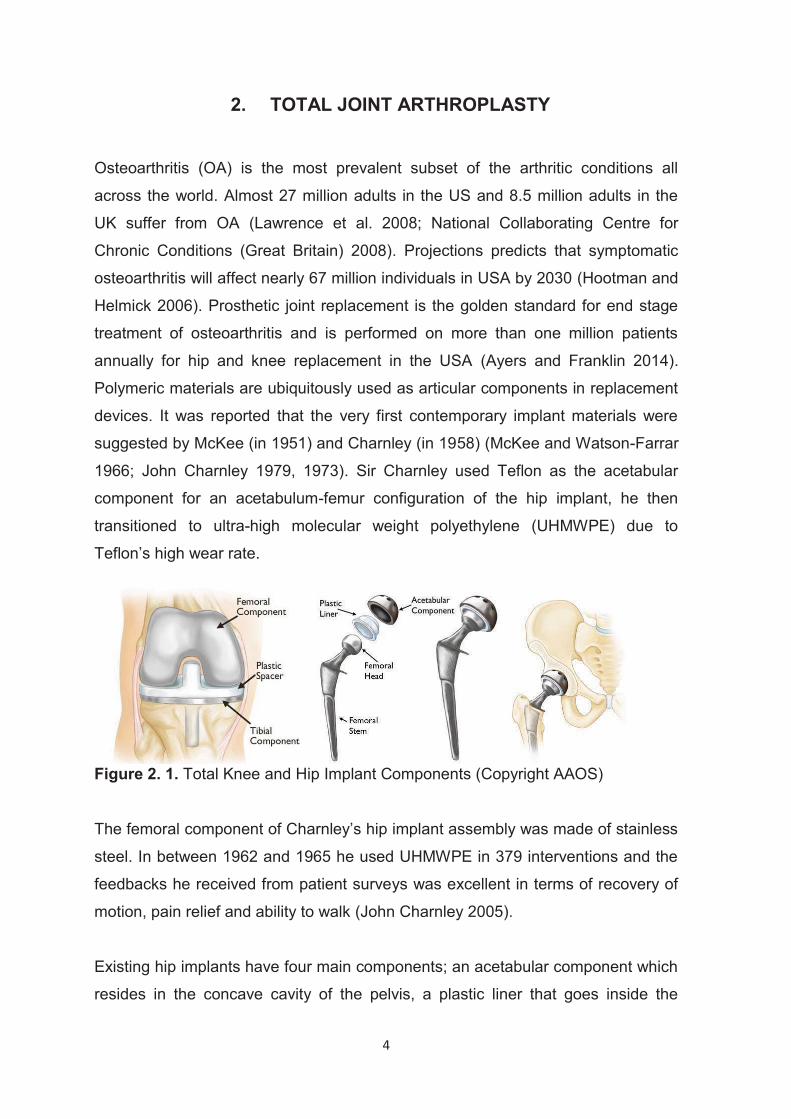
Figure 2. 1. Total Knee and Hip Implant Components (Copyright AAOS)
The femoral component of Charnley’s hip implant assembly was made of stainless
steel. In between 1962 and 1965 he used UHMWPE in 379 interventions and the
feedbacks he received from patient surveys was excellent in terms of recovery of
motion, pain relief and ability to walk (John Charnley 2005).
Existing hip implants have four main components; an acetabular component which
resides in the concave cavity of the pelvis, a plastic liner that goes inside the
5
acetabular component, a femoral head and a femoral stem (Figure 2.1). The
acetabular component and the femoral stem are made of metal whereas the
articular surfaces could alternatively be made of ceramic, metal or plastic
(UHMWPE). UHMWPE is most preferred liner for primary joint replacements
(Muratoglu et al. 2004).
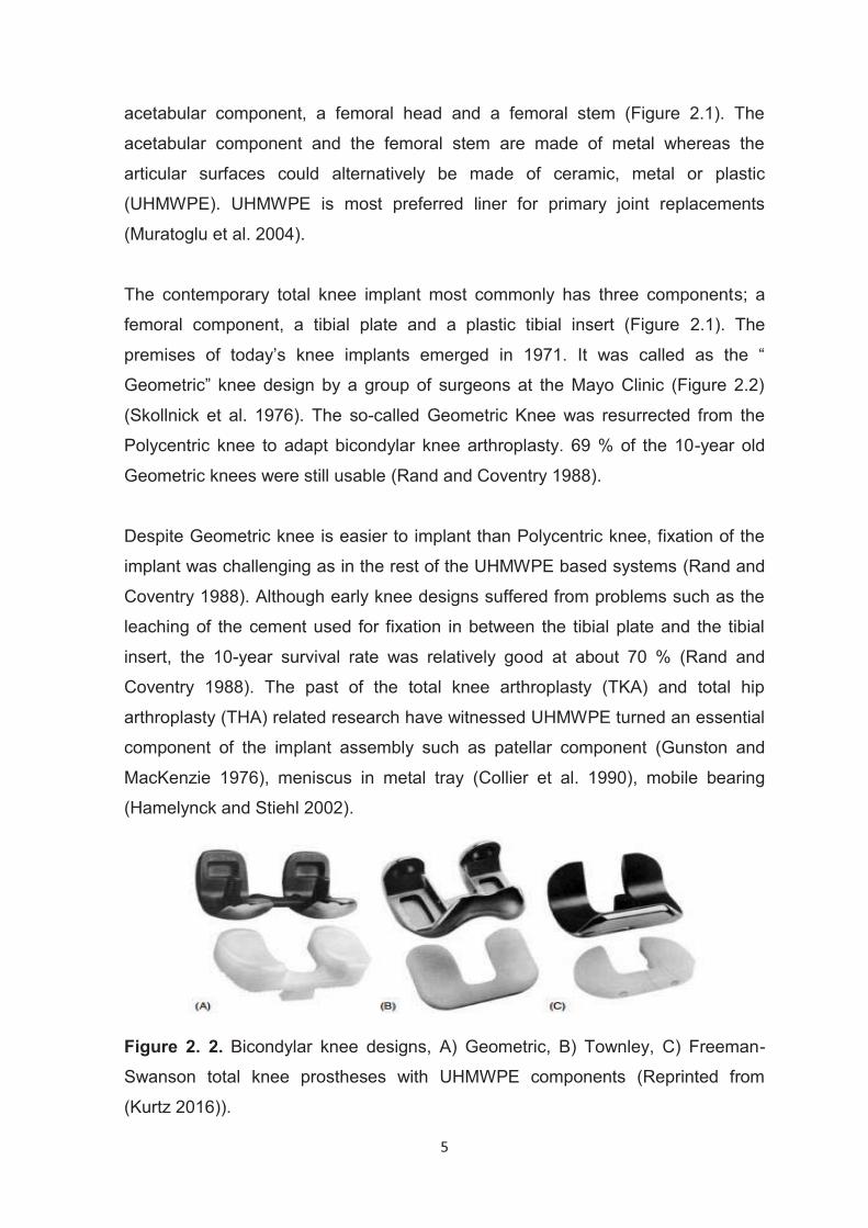
The contemporary total knee implant most commonly has three components; a
femoral component, a tibial plate and a plastic tibial insert (Figure 2.1). The
premises of today’s knee implants emerged in 1971. It was called as the “
Geometric” knee design by a group of surgeons at the Mayo Clinic (Figure 2.2)
(Skollnick et al. 1976). The so-called Geometric Knee was resurrected from the
Polycentric knee to adapt bicondylar knee arthroplasty. 69 % of the 10-year old
Geometric knees were still usable (Rand and Coventry 1988).
Despite Geometric knee is easier to implant than Polycentric knee, fixation of the
implant was challenging as in the rest of the UHMWPE based systems (Rand and
Coventry 1988). Although early knee designs suffered from problems such as the
leaching of the cement used for fixation in between the tibial plate and the tibial
insert, the 10-year survival rate was relatively good at about 70 % (Rand and
Coventry 1988). The past of the total knee arthroplasty (TKA) and total hip
arthroplasty (THA) related research have witnessed UHMWPE turned an essential
component of the implant assembly such as patellar component (Gunston and
MacKenzie 1976), meniscus in metal tray (Collier et al. 1990), mobile bearing
(Hamelynck and Stiehl 2002).
Figure 2. 2. Bicondylar knee designs, A) Geometric, B) Townley, C) Freeman-
Swanson total knee prostheses with UHMWPE components (Reprinted from
(Kurtz 2016)).
6
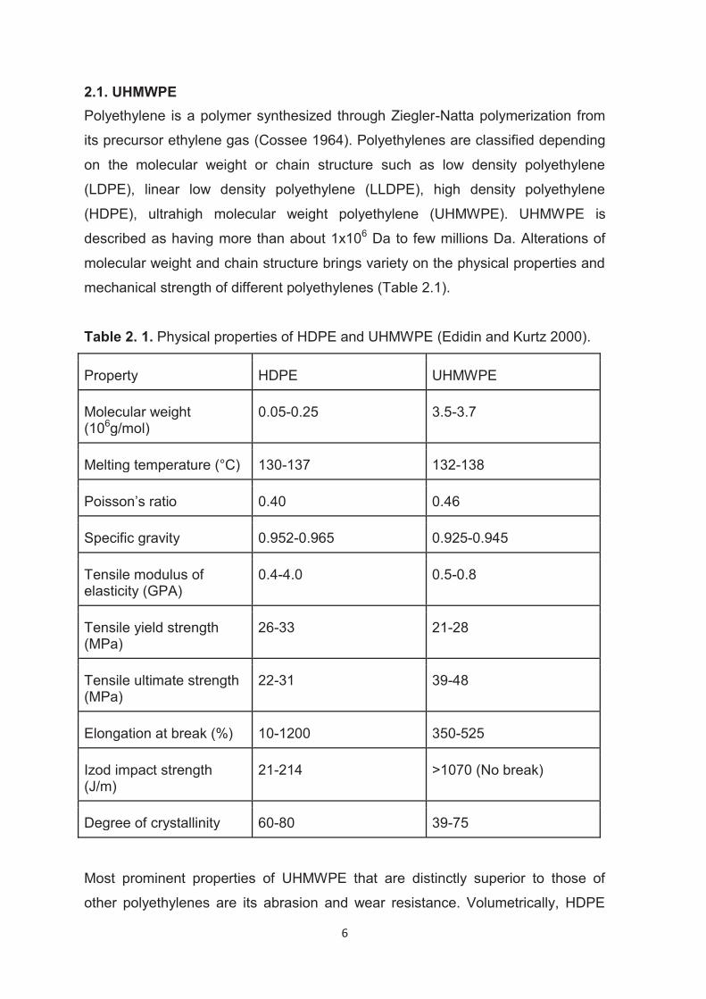
2.1. UHMWPE Polyethylene is a polymer synthesized through Ziegler-Natta polymerization from
its precursor ethylene gas (Cossee 1964). Polyethylenes are classified depending
on the molecular weight or chain structure such as low density polyethylene
(LDPE), linear low density polyethylene (LLDPE), high density polyethylene
(HDPE), ultrahigh molecular weight polyethylene (UHMWPE). UHMWPE is
described as having more than about 1x10⁶ Da to few millions Da. Alterations of
molecular weight and chain structure brings variety on the physical properties and
mechanical strength of different polyethylenes (Table 2.1).
Table 2. 1. Physical properties of HDPE and UHMWPE (Edidin and Kurtz 2000).
Property HDPE UHMWPE
Molecular weight (106g/mol)
0.05-0.25 3.5-3.7
Melting temperature (°C) 130-137 132-138
Poisson’s ratio 0.40 0.46
Specific gravity 0.952-0.965 0.925-0.945
Tensile modulus of elasticity (GPA)
0.4-4.0 0.5-0.8
Tensile yield strength (MPa)
26-33 21-28
Tensile ultimate strength (MPa)
22-31 39-48
Elongation at break (%) 10-1200 350-525
Izod impact strength (J/m)
21-214 >1070 (No break)
Degree of crystallinity 60-80 39-75
Most prominent properties of UHMWPE that are distinctly superior to those of
other polyethylenes are its abrasion and wear resistance. Volumetrically, HDPE
7
has a wear rate that is more than 4 times of the UHMWPE wear on multidirectional
hip simulator (Edidin and Kurtz 2000).
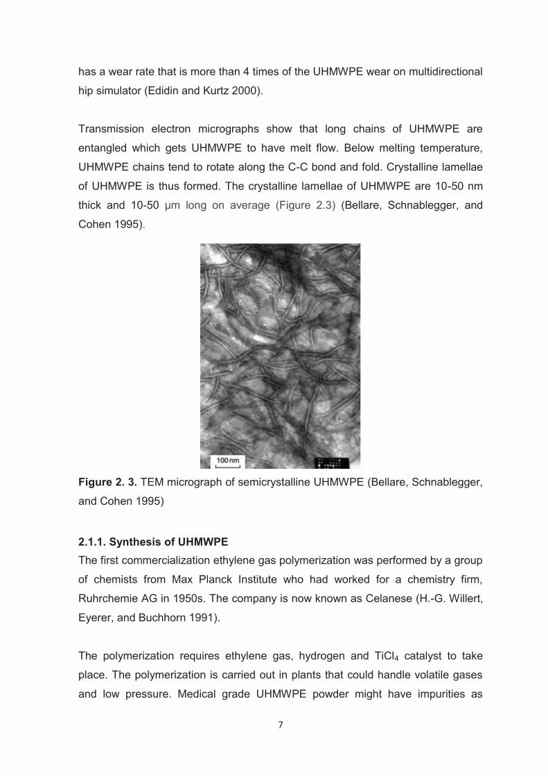
Transmission electron micrographs show that long chains of UHMWPE are
entangled which gets UHMWPE to have melt flow. Below melting temperature,
UHMWPE chains tend to rotate along the C-C bond and fold. Crystalline lamellae
of UHMWPE is thus formed. The crystalline lamellae of UHMWPE are 10-50 nm
thick and 10-50 μm long on average (Figure 2.3) (Bellare, Schnablegger, and
Cohen 1995).
Figure 2. 3. TEM micrograph of semicrystalline UHMWPE (Bellare, Schnablegger,
and Cohen 1995)
2.1.1. Synthesis of UHMWPE The first commercialization ethylene gas polymerization was performed by a group
of chemists from Max Planck Institute who had worked for a chemistry firm,
Ruhrchemie AG in 1950s. The company is now known as Celanese (H.-G. Willert,
Eyerer, and Buchhorn 1991).
The polymerization requires ethylene gas, hydrogen and TiCl4 catalyst to take
place. The polymerization is carried out in plants that could handle volatile gases
and low pressure. Medical grade UHMWPE powder might have impurities as
8
titanium, aluminum, chlorine which comes from catalyst up to the specified
concentrations specified in ASTM standard F648. Foremost medical grade resins
are GUR type of resins such as GUR 1020 and GUR 1050 are produced by
Celanese (Irving, TX, USA). GUR stands for “Granular”, “UHMWPE”,
“Ruhrchemie” and the adduct numbers stand for properties of resin. First number
is for showing bulk density where second for existence of calcium stearate, third
for average molecular weight and fourth for internal code designation.
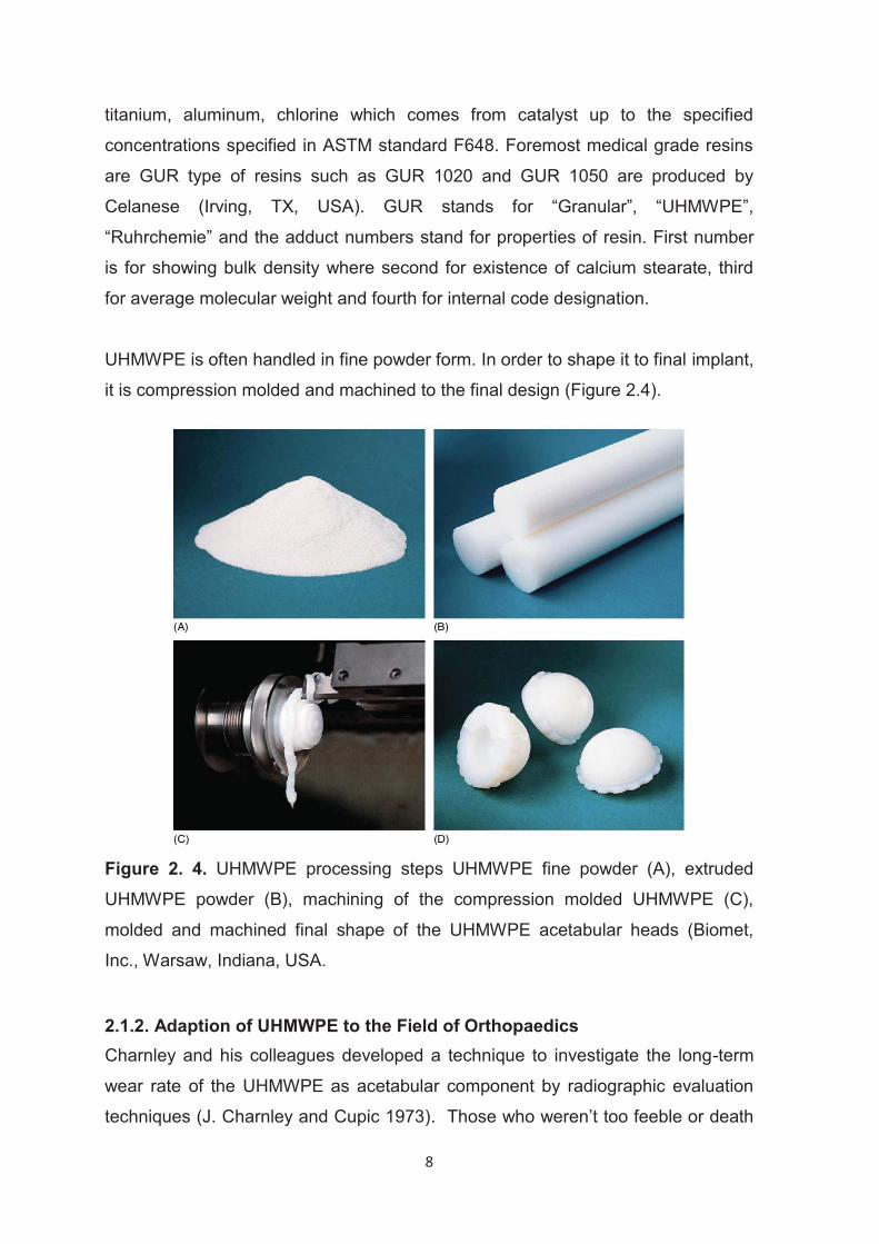
UHMWPE is often handled in fine powder form. In order to shape it to final implant,
it is compression molded and machined to the final design (Figure 2.4).
Figure 2. 4. UHMWPE processing steps UHMWPE fine powder (A), extruded
UHMWPE powder (B), machining of the compression molded UHMWPE (C),
molded and machined final shape of the UHMWPE acetabular heads (Biomet,
Inc., Warsaw, Indiana, USA.
2.1.2. Adaption of UHMWPE to the Field of Orthopaedics Charnley and his colleagues developed a technique to investigate the long-term
wear rate of the UHMWPE as acetabular component by radiographic evaluation
techniques (J. Charnley and Cupic 1973). Those who weren’t too feeble or death
9
and made to the follow-up examination let researchers evaluate the condition of
the implants again. 106 out of 185 acetabular cups were able to be examined after
9-10 years of implantation. Certain complications were listed as infection,
mechanical loosening and late dislocation (J. Charnley and Cupic 1973). The main
causes for revision surgeries in TKA were aseptic loosening; infection; fracture;
joint stiffness; tibio/femoral instability; patellar complications; periprosthetic
fracture; and wear of UHMWPE component (Sharkey et al. 2002; Lombardi,
Berend, and Adams 2014; Fehring et al. 2001; Schroer et al. 2013). Cross-linking
of the UHMWPE was proposed to decrease polymeric debris caused by the wear
of the inserts in TJA (Kurtz and Patel 2016). Many attempts had been made to
improve UHMWPE for TJA to reduce wear and thereby increase the longevity.
Cross-linking by ionizing radiation (gamma and e-beam) has largely reduced the
wear of UHMWPE. The UHMWPEs are referred as “conventional” if the amount of
irradiation dose was 25-40 kGy and was referred as “highly cross-linked UHMWPE
(HXLPE)” if the dose was >40 kGy (Kurtz and Patel 2016). UHMWPE cross-linking
affected some properties in unpremeditated manner such as reduction in ductility,
fatigue and fracture resistance (H. G. Willert, Bertram, and Buchhorn 1990). In
their seminal paper of 1995, the effect of nitrogen implantation of UHMWPE on the
wear resistance of it articulating against titanium was investigated (Allen, Bloyce,
and Bell 1995). While cross-linking improves wear resistance, in vivo oxidation of
cross-linked UHMWPE further lowers its ductility and its ultimate strength (Kurtz et
al. 2006; Medel, Rimnac, and Kurtz 2009; Wannomae et al. 2006). In vivo
oxidation is caused by the subsequent reactions of residual free radicals after
gamma irradiation and from in vivo lipid absorption present in the synovial fluid
(Muratoglu et al. 2010; Oral et al. 2012; Regis et al. 2014). Efforts to prevent in
vivo oxidation gave rise to the incorporation of the antioxidant vitamin E in the
polymer, which is a breakthrough in the improvement of UHMWPE (Bracco and
Oral 2011). Vitamin E-infused and cross-linked UHMWPE (VE-HXLPE), both
oxidatively and mechanically remained stable after accelerated in vitro oxidation
(Bracco and Oral 2011; Kurtz et al. 2015). In another study to prevent UHMWPE
oxidation, UHMWPE was incorporated with Europium stearate which has
outstanding binding affinity to oxygen (Gallardo et al. 2011). In contrast, HXLPE
without VE showed a 5-fold increase in oxidation and 70-80 % decrease in
mechanical strength (Kurtz et al. 2015; Bracco and Oral 2011). Vitamin E-infused
10
and cross-linked UHMWPE was approved by FDA in 2007 for hip and in 2008 for
knee and its clinical performance is under investigation (Oral and Muratoglu 2015).
The incidence of osteolysis dropped drastically with the use of HXLPE even after
ten years of implantation, which is relatively short compared to the eventual
lifetime of the implants (“National Joint Replacement Registry; Hip, Knee &
Shoulder Arthroplasty” 2016a). That said, the use of HXLPE did not change the
incidence of periprosthetic joint infection (PJI) compared to that when conventional
UHMWPE was used (“National Joint Replacement Registry; Hip, Knee & Shoulder
Arthroplasty” 2016b). HXLPE has involved in revision less than UHMWPE in total
knee arthroplasty (“National Joint Replacement Registry; Hip, Knee & Shoulder
Arthroplasty” 2016b). As in THA, the main reasons for revision of HXLPE were
aseptic loosening, pain, instability, and periprosthetic joint infection which is similar
to UHMWPE (“National Joint Replacement Registry; Hip, Knee & Shoulder
Arthroplasty” 2016b). PJI is a major reason for the TKA revision for the patients
implanted with HXLPE (“National Joint Replacement Registry; Hip, Knee &
Shoulder Arthroplasty” 2016b). Thus, it is substantial to develop better materials
and techniques to battle with PJI problem.
2.2. Metals Biomechanical properties of metals made them appropriate to use as an implant
material. Besides, metallic implants can be sterilized by the common sterilization
procedures that makes them easy to use. Cobalt chromium alloys, nickel alloys,
gold alloys, titanium and titanium based alloys and stainless steel are used as
metallic implant materials in orthopaedic applications. However, TJA components
are widely made from cobalt chromium alloys. Cobalt chromium alloys have low
rate of corrosion that provide long-term stability. Metal-on-metal configuration
generates 13,500 times higher number of particles when compared with
polyethylene particles which produced in metal-on-polyethylene configuration.
However, total volumetric wear of a metal-on-metal configuration is lower than the
volumetric wear of metal-on-polyethylene configuration because of the smaller
size of metal particles (<50nm) compared to polyethylene particles (<0.1μm).
Metal wear debris particles are found within synovial fluids and tissues and
because of very small size the true extent of dissemination is not known yet.
11
Studies shows that orthopaedic metals induce immunological effects which cause
cell mediated hypersensitivity response.
2.3. Ceramics Ceramics are inorganic based solid materials. In orthopaedics, ceramics basically
used in two forms:
1. Ceramic oxides which are used as ball heads or inserts in hip arthroplasty.
2. Calcium phosphate ceramics which are used to coat metal components for
osteoconductivity.
2.3.1. Ceramic Oxides Ceramic oxides have mainly two types, alumina and zirconia. They both exhibit
corrosion resistance, hardness and stiffness. They are both chemically inert and
have excellent wear resistance. Their wear debris is biocompatible (H. Liu et al.
2008).
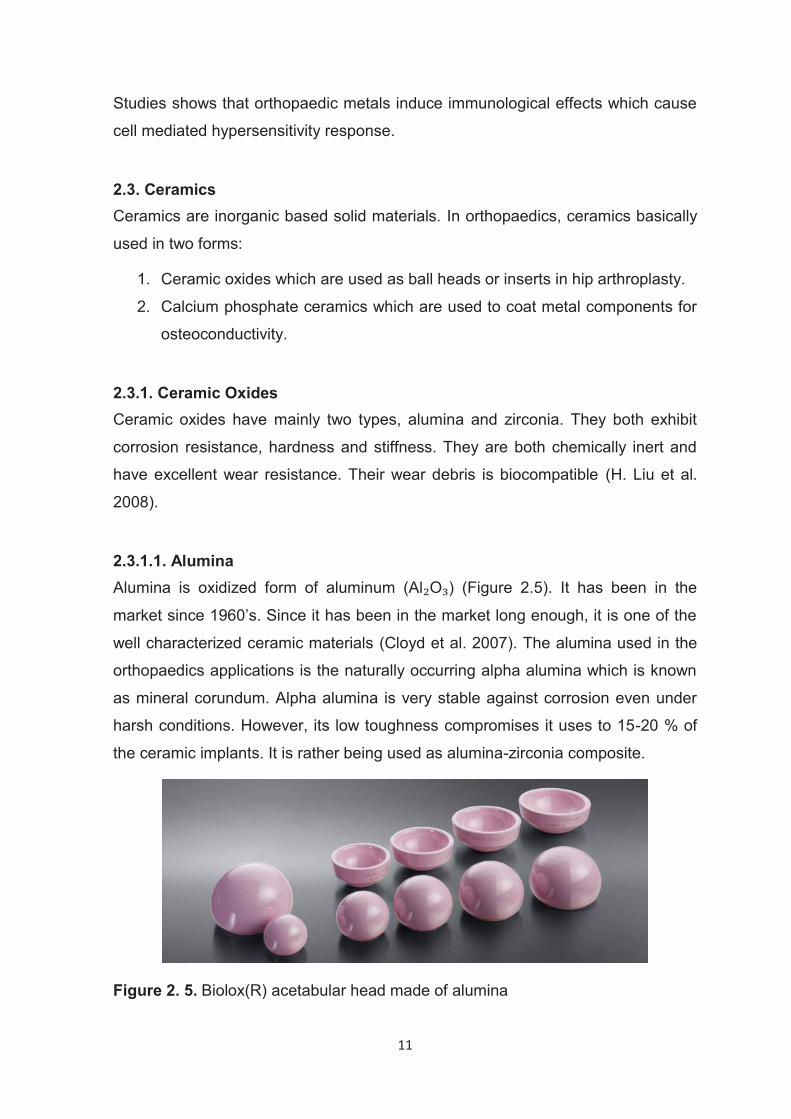
2.3.1.1. Alumina Alumina is oxidized form of aluminum (Al O ) (Figure 2.5). It has been in the
market since 1960’s. Since it has been in the market long enough, it is one of the
well characterized ceramic materials (Cloyd et al. 2007). The alumina used in the
orthopaedics applications is the naturally occurring alpha alumina which is known
as mineral corundum. Alpha alumina is very stable against corrosion even under
harsh conditions. However, its low toughness compromises it uses to 15-20 % of
the ceramic implants. It is rather being used as alumina-zirconia composite.
Figure 2. 5. Biolox(R) acetabular head made of alumina

12
2.3.1.2. Zirconia Zirconia was introduced in 1980’s to improve toughness of alumina. Most of the
effort was put to develop Magnesia and Yttria stabilized zirconia. The tetragonal-
monoclinic (t-m) phase transition of zirconia helps ceramic to dissipate the fracture
stress. The t-m transformation upgrades material to a tougher ceramic at
macroscopic level. It was reported that zirconia was applied to UHMWPE to
improve its long term stability by improving plastic deformation and wear of it
(Bianchi et al. 2015).
2.3.2. Calcium Phosphate Calcium phosphate ceramics (CPC) are known for their osteoconductive property.
It was used on metallic components to increase bone-metal integration (Figure
2.6) (Peppas and Langer 1994). One important aspect which determines CPC
property is calcium to phosphate ratio. The most stable CPC, Hydroxyapatite has
Ca/P as 5/3 (Sims et al. 1998). The osteoconductive CPC is deposited on metallic
surfaces by plasma spray technique which must take control of many crucial
parameters into account such as polymorph, oxidation reactions, phase separation
i.e (C. Liu, Xia, and Czernuszka 2007).
Figure 2. 6. Hydroxyapatite implant coatings (APS Materials, Daytona OH, USA).
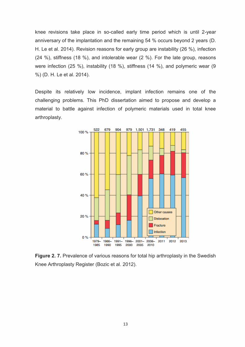
2.3. Reasons for revision surgery Under various circumstances knee and hip implants may fail, thus, may require
revision of the materials (Figure 2.7). According to a literature review, 46 % of the
13
knee revisions take place in so-called early time period which is until 2-year
anniversary of the implantation and the remaining 54 % occurs beyond 2 years (D.
H. Le et al. 2014). Revision reasons for early group are instability (26 %), infection
(24 %), stiffness (18 %), and intolerable wear (2 %). For the late group, reasons
were infection (25 %), instability (18 %), stiffness (14 %), and polymeric wear (9
%) (D. H. Le et al. 2014).
Despite its relatively low incidence, implant infection remains one of the
challenging problems. This PhD dissertation aimed to propose and develop a
material to battle against infection of polymeric materials used in total knee
arthroplasty.
Figure 2. 7. Prevalence of various reasons for total hip arthroplasty in the Swedish
Knee Arthroplasty Register (Bozic et al. 2012).
14
3. PERIPROSTHETIC JOINT INFECTION
Joint arthroplasty is procedure that increases the quality of life. In cases it is
performed successfully, the patient takes advantages of pain relief and ease of
joint motion. Despite the majority of joint arthroplasties are successful, there are
cases where the prosthesis fails. The prosthesis failure usually requires another
surgery. Reasons for failure could be either aseptic or infection related. The
aseptic failure could arise from loss of bone-cement integration at the prosthesis-
bone-cement interface, fracture of the implant, wear, malpositioning of the implant,
materials fatigue. Periprosthetic joint infection (PJI) is the infection in the vicinity of
implant which also involves the surrounding tissue (A. J. Tande and Patel 2014).
As defined by the Musculoskeletal Infection Society (MSIS), PJI is the condition of
meeting following criteria;
(1) There is a sinus tract communicating with the prosthesis; or
(2) A pathogen is isolated by culture from at least two separate tissue or fluid
samples obtained from the affected prosthetic joint; or
(3) Four of the following six criteria exist:
(a) Elevated serum erythrocyte sedimentation rate (ESR) and serum C-
reactive protein (CRP) concentration,
(b) Elevated synovial leukocyte count,
(c) Elevated synovial neutrophil percentage (PMN%),
(d) Presence of purulence in the affected joint,
(e) Isolation of a microorganism in one culture of periprosthetic tissue or
fluid,
(f) Greater than five neutrophils per high-power field in five high-power
fields observed from histologic analysis of periprosthetic tissue at 9400
magnification” (Parvizi et al. 2011).
Although the rate of total joint arthroplasty (TJA) infection is as low as 1 %- 2%,
the increase in the total number of operations and the growing aging population
15
are increasing prevalence of PJI. There are 7,100-15,000 patients that are
affected by PJI yearly in USA and it is expected to peak 55,000-75,500 by the year
2030 (Kurtz et al. 2012). Between 2001-2009 the cost of knee and hip
arthroplasties to the hospitals in United States of America rose from $320 million
to $566 million annually (Berend et al. 2013). The occurrence of PJI in two-year
period after implantation is greatest which is 60-70 % of all PJI cases (Pulido et al.
2008; Kurtz et al. 2010). Once the infected joint lost its functionality, it’s
challenging to revert back to its original state (Toulson et al. 2009). In all respects,
PJI is a severe problem and a battle that none of the sides win. Hence, mitigating
both the economic and the health burden caused by PJI is a worthwhile area of
focus.
3.1. Pathogenesis of Prosthetic Joint Infection The accounted causation is in three ways; bacteria seeding on the implant,
hematogenous spread from rest of the body or recurrence of a previous infection
(Kapadia et al. 2016). Cure for an infection related to foreign body part is
compelling due to weakened immune system (W. Zimmerli, Lew, and Waldvogel
1984). The most conspicuous thing that emerges from bacterial harbouring is
biofilm formation which reduces the susceptibility to antibiotics (Kapadia et al.
2016).
Biofilm is the structure where bacteria are embedded in their secreted extracellular
matrix secreted by itself (Shah et al. 2015). Regardless of the type, foreign devices
made of biomaterials increase susceptibility to infection (Figure 3.1). It is known
from previous studies that the vulnerability of biomaterials to infection is partly
because of granulocyte defect (W. Zimmerli et al. 1982; Costerton et al. 1995).
The biofilm is totally a complex structure where bacteria in different layers interact
with each other through quorum sensing to keep biofilm stable (Costerton,
Stewart, and Greenberg 1999).
16
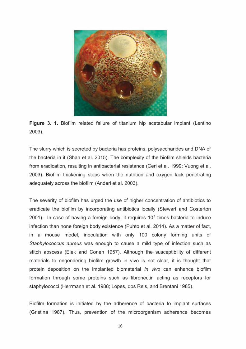
Figure 3. 1. Biofilm related failure of titanium hip acetabular implant (Lentino
2003).
The slurry which is secreted by bacteria has proteins, polysaccharides and DNA of
the bacteria in it (Shah et al. 2015). The complexity of the biofilm shields bacteria
from eradication, resulting in antibacterial resistance (Ceri et al. 1999; Vuong et al.
2003). Biofilm thickening stops when the nutrition and oxygen lack penetrating
adequately across the biofilm (Anderl et al. 2003).
The severity of biofilm has urged the use of higher concentration of antibiotics to
eradicate the biofilm by incorporating antibiotics locally (Stewart and Costerton
2001). In case of having a foreign body, it requires 10 times bacteria to induce
infection than none foreign body existence (Puhto et al. 2014). As a matter of fact,
in a mouse model, inoculation with only 100 colony forming units of
Staphylococcus aureus was enough to cause a mild type of infection such as
stitch abscess (Elek and Conen 1957). Although the susceptibility of different
materials to engendering biofilm growth in vivo is not clear, it is thought that
protein deposition on the implanted biomaterial in vivo can enhance biofilm
formation through some proteins such as fibronectin acting as receptors for
staphylococci (Herrmann et al. 1988; Lopes, dos Reis, and Brentani 1985).
Biofilm formation is initiated by the adherence of bacteria to implant surfaces
(Gristina 1987). Thus, prevention of the microorganism adherence becomes
17
crucial. Adherence depends on both nonspecific physical factors (surface tension,
hydrophilicity, electrostatic interactions i.e.) or specific adhesive proteins such as
fibronectin (Werner Zimmerli 2014). Subsequently, other signaling molecules such
as intercellular adhesion (ica) operon contributes to the adherent bacteria
transforming into biofilm (Laverty, Gorman, and Gilmore 2013). Biofilms consist of
microbes embedded in a secreted polymeric matrix, which is organized in complex
structures similar to multicellular organisms. At the stage when the diffusion and
local concentration of nutrients are limited by the growth of bacteria, quorum
sensing genes are activated to stop overgrowth (Simonetti et al. 2013). This
sheltered structure of biofilms makes them 1000-fold more resistant to antibiotics
than planktonic bacteria (Stewart and Costerton 2001). The presence of biofilm
dramatically reduces the chance from 80-90% to 30-60% for the cases that
treatment is started 3-4 weeks after infection (Deirmengian et al. 2003; Laffer et al.
2006; Giulieri et al. 2004; Marculescu et al. 2006).
3.2. Bacterial Strains in PJI 68-94 % of cultures obtained from PJI patients’ cultures test positive for a
causative organism (Peel et al. 2012; Werner Zimmerli, Trampuz, and Ochsner
2004) and 90-95 % of those cases originate from gram-positive bacteria (Werner
Zimmerli, Trampuz, and Ochsner 2004). Most of the infection episodes are caused
by Staphylococci; S. aureus and coagulase-negative staphylococci, particularly S.
epidermidis, or S. lugdunensis (Aaron J. Tande and Patel 2014). Gram-negative
bacteria such as E. Coli, P. acnes and other rare bacterial strains, including
Mycobacterium tuberculosis (Harwin et al. 2013), nontuberculous mycobacteria
(S.-X. Wang et al. 2011), Mycoplasma (Sneller et al. 1986), Legionella
(Fernández-Cruz et al. 2011) and fungal agents (Kuiper et al. 2013) can be
involved in PJI. Table 3.1. outlines common microorganisms that are involved in
TKA and THA.
18
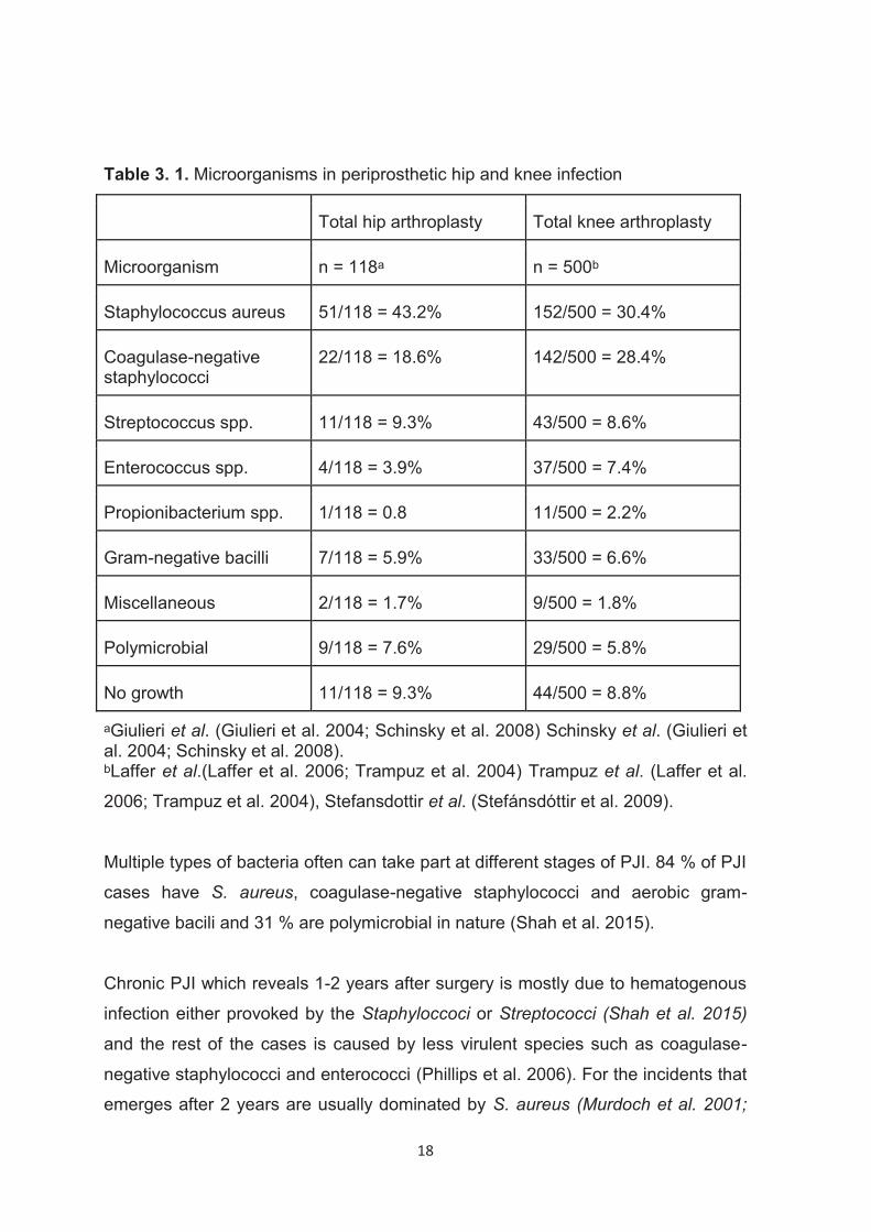
Table 3. 1. Microorganisms in periprosthetic hip and knee infection
Total hip arthroplasty Total knee arthroplasty
Microorganism n = 118ᵃ n = 500ᵇ
Staphylococcus aureus 51/118 = 43.2% 152/500 = 30.4%
Coagulase-negative staphylococci
22/118 = 18.6% 142/500 = 28.4%
Streptococcus spp. 11/118 = 9.3% 43/500 = 8.6%
Enterococcus spp. 4/118 = 3.9% 37/500 = 7.4%
Propionibacterium spp. 1/118 = 0.8 11/500 = 2.2%
Gram-negative bacilli 7/118 = 5.9% 33/500 = 6.6%
Miscellaneous 2/118 = 1.7% 9/500 = 1.8%
Polymicrobial 9/118 = 7.6% 29/500 = 5.8%
No growth 11/118 = 9.3% 44/500 = 8.8%
ᵃGiulieri et al. (Giulieri et al. 2004; Schinsky et al. 2008) Schinsky et al. (Giulieri et al. 2004; Schinsky et al. 2008). ᵇLaffer et al.(Laffer et al. 2006; Trampuz et al. 2004) Trampuz et al. (Laffer et al.
2006; Trampuz et al. 2004), Stefansdottir et al. (Stefánsdóttir et al. 2009).
Multiple types of bacteria often can take part at different stages of PJI. 84 % of PJI
cases have S. aureus, coagulase-negative staphylococci and aerobic gram-
negative bacili and 31 % are polymicrobial in nature (Shah et al. 2015).
Chronic PJI which reveals 1-2 years after surgery is mostly due to hematogenous
infection either provoked by the Staphyloccoci or Streptococci (Shah et al. 2015)
and the rest of the cases is caused by less virulent species such as coagulase-
negative staphylococci and enterococci (Phillips et al. 2006). For the incidents that
emerges after 2 years are usually dominated by S. aureus (Murdoch et al. 2001;
19
Parham Sendi et al. 2011).
Current state of the art prosthetic joint assembly for TKA is comprised in most
cases of metal a (Co-Cr or Ti) and polymeric material (UHMWPE) articulating pair
in the USA. Bacterial count on infected hip joints showed that UHMWPE has the
highest amount of bacteria on it, femoral head and acetabular head has less and
femoral stem has the least number of colonizing bacteria (Holinka et al. 2012).
This suggests that all components of the joint implant are involved in the initiation
and propagation of a bacterial infection.
3.3. Risk Factors in PJI
3.3.1. Patient-Related Risk Factors
Hematogenous seeding is obscure causation for PJI that usually generates
symptoms such as dental or urinary tract infection (Gehrke and Parvizi 2014;
Cruess, Bickel, and vonKessler 1975; Schmalzried et al. 1992). Patients with
aforementioned symptoms of current infection should be examined and be treated
immediately.
Glycaemic control is essential due diabetic patients who are in high risk group for
PJI (Dowsey and Choong 2009). In a study, correlation between blood glucose
level and infection occurrence was investigated and showed that blood glucose
level of >200 mg/dL doubles the risk of PJI (Mraovic et al. 2011). Diabetes likely to
induce PJI through impaired wound healing (McMurry 1984) and susceptibility of
biofilm formation due to high glucose levels in blood (Seneviratne et al. 2013).
Meeting the conditions of dropped levels of serum albumin, lymphocyte count,
serum transferrin or serum prealbumin attributes to malnourishment (Devoto et al.
2006; Cross et al. 2014). A considerable amount of literature has been published
on the correlation between lack of nutrition and probability of undergoing PJI (Paul
et al. 2015; Greene, Wilde, and Stulberg 1991). Collagen and proteoglycan
synthesis are notably being affected by poor nutrition intake. This may result in
20
collapsing the wound healing process and sustained drainage may increase the
risk of infection (Cross et al. 2014).
Studies have shown that high body mass index (BMI) has an effect on surgical site
infection (SSI) (Dowsey and Choong 2008) and obese people have 1.5-3 times
higher risk of reoperation after hip replacements (Jameson et al. 2014). In obese
patients, fraction of oxygen (FiO ) need is much higher than people in normal BMI
limits (Fleischmann et al. 2005).
It has been demonstrated that alcohol consumption impacted infection in non-
cardiac surgeries (Bradley et al. 2011). The infection risk inflation is conceived to
be arisen from weakened immune system which is adversely affected by alcohol
consumption (Tønnesen et al. 2009).
3.3.2. Prophylactic Measures for PJI Wound healing mechanism for those who has diabetes is more impaired than
those who are non-diabetics (Mraovic et al. 2011). It was found that high blood
sugar gets monocytes more susceptible to apoptosis (Komura et al. 2010) and
deteriorated neutrophil functioning alongside with deteriorated chemotactic,
phagocytic and bactericidal capability (Turina, Fry, and Polk 2005). In order to
improve neutrophil phagocytic function, a strict low glucose diet must be adapted
by the patient (Rassias et al. 1999). As opposed to glucose level controlling, a
massive study with 40.000 patients, showed there’s no additional risk for diabetic
patients (Adams et al. 2013).
As corticosteroids used to treat rheumatoid arthritis delay wound healing, they
increase the incidence of wound infection (Wicke et al. 2000). A relationship
between use of disease-modifying antirheumatic drugs (DMARD) and increased
risk of prosthetic joint infection was shown (Moucha et al. 2011). However, the
British Society for Rheumatoid (BSR) advised not to stop DMARD treatment
(Luqmani et al. 2009).
Other effective steps prior the PJI operation are skin site preparation with
antiseptic chlorhexidine gluconate (Johnson et al. 2013), mupirocin nasal ointment
since it collapses nasal carriers of methicillin-sensitive Staphylococcus aureus
21
(MSSA) (Breier et al. 2011). The ultimate risk of having PJI drops 8 % in case of
antibiotic prophylaxis introduction (AlBuhairan, Hind, and Hutchinson 2008).
Between 57 - 87 % of organisms are being vanished when pulsatile lavage is used
(Dunne et al. 2008).
3.4. Current Treatment for PJI The present-day practice to cure PJI involves 2-stage revision, 1-stage revision
and debridement, antibiotic and implant retention (DAIR) and any worse case is
treated with excision arthroplasty (Drago 2017).
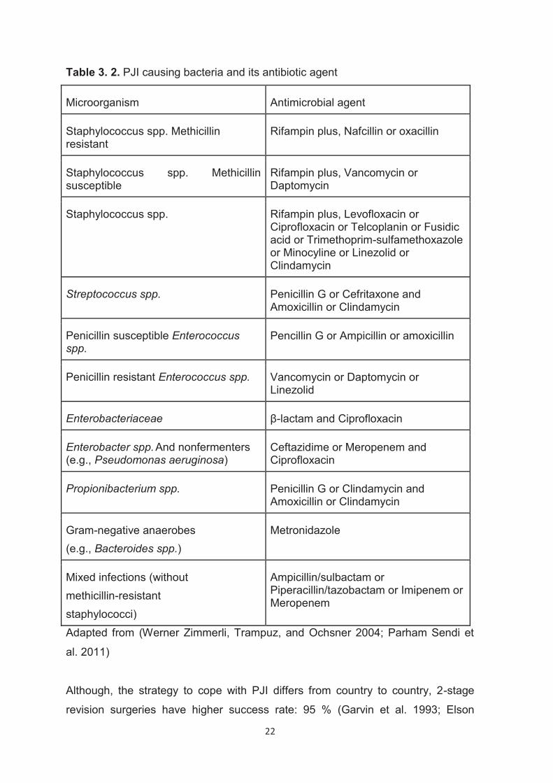
The standard procedure involves intravenous (IV) antibiotics route to avoid enteral
resorption and ensure the highest antibiotic level in tissues. Table 3.2. summarizes
bacteria-specific antibiotics (Werner Zimmerli, Trampuz, and Ochsner 2004; P.
Sendi and Zimmerli 2012; Osmon et al. 2013). The duration of antibiotic therapy
subject to variation from case to case depending on several parameters. Long-
term treatments are planned based on the assumption of incapability of host
immune to eradicate the microorganisms. Therefore, 3-month long course is
suggested for debridement, antibiotic and implant retention (DAIR, explained in
further sections) and one-stage, 2-3 weeks is suggested for two-stage for PJI
(Werner Zimmerli, Trampuz, and Ochsner 2004; P. Sendi and Zimmerli 2012;
Osmon et al. 2013; Petersen 2010). Knee PJI requires as long as 6-month long
course (Werner Zimmerli, Trampuz, and Ochsner 2004; P. Sendi and Zimmerli
2012; Osmon et al. 2013; Petersen 2010),(Werner Zimmerli et al. 1998).
22
Table 3. 2. PJI causing bacteria and its antibiotic agent
Microorganism Antimicrobial agent
Staphylococcus spp. Methicillin resistant
Rifampin plus, Nafcillin or oxacillin
Staphylococcus spp. Methicillin susceptible
Rifampin plus, Vancomycin or Daptomycin
Staphylococcus spp. Rifampin plus, Levofloxacin or Ciprofloxacin or Telcoplanin or Fusidic acid or Trimethoprim-sulfamethoxazole or Minocyline or Linezolid or Clindamycin
Streptococcus spp. Penicillin G or Cefritaxone and Amoxicillin or Clindamycin
Penicillin susceptible Enterococcus spp.
Pencillin G or Ampicillin or amoxicillin
Penicillin resistant Enterococcus spp. Vancomycin or Daptomycin or Linezolid
Enterobacteriaceae β-lactam and Ciprofloxacin
Enterobacter spp. And nonfermenters (e.g., Pseudomonas aeruginosa)
Ceftazidime or Meropenem and Ciprofloxacin
Propionibacterium spp. Penicillin G or Clindamycin and Amoxicillin or Clindamycin
Gram-negative anaerobes (e.g., Bacteroides spp.)
Metronidazole
Mixed infections (without methicillin-resistant staphylococci)
Ampicillin/sulbactam or Piperacillin/tazobactam or Imipenem or Meropenem
Adapted from (Werner Zimmerli, Trampuz, and Ochsner 2004; Parham Sendi et
al. 2011)
Although, the strategy to cope with PJI differs from country to country, 2-stage
revision surgeries have higher success rate: 95 % (Garvin et al. 1993; Elson
23
1993).
DAIR treatment is most suitable in the early post-operative phase or in the case of
an acute hematogenous infection with a well-functioning implant in following month
after surgery. This is because complete biofilm formation can occur between 36 h
to 3 weeks (Parvizi, Zmistowski, and Adeli 2010). To hinder infection recurrence
antibiotics should be used for months after the treatment surgery (Osmon et al.
2013). Synergic use of antibiotic rifampin with other antibiotics such as β-lactam,
glycopeptide, or fluoroquinolones was shown to leveraged efficacy of DAIR
treatment against S. aureus (Senneville et al. 2011; Vilchez et al. 2011).
Frequently encountered DAIR treatment failure reasons are involvement of
methicillin-resistant S. aureus (MRSA) (Soriano et al. 2006; Bradbury et al. 2009),
vancomycin-resistant enterococci (VRE) and fluoroquinolone-resistant gram-
negative bacilli (Jaén et al. 2012). The overall success rate of DAIR treatment for
knee is 33 % and for hip is 52 % (Romanò et al. 2012; Silva, Tharani, and
Schmalzried 2002).
In one-stage surgery, concurrent implication of infected implant removal and
implantation of new prosthesis with antibiotic-eluting bone cement (ALBC) fixing
element is performed (Shah et al. 2015). A long term antibiotic treatment is
afterwards characterized, first intravenously for 4-6 weeks, then orally for 3-12
months to nullify any remaining species (Klouche et al. 2012; Ure et al. 1998;
Buechel, Femino, and D’Alessio 2004). One-stage surgery is used dominantly for
hip joint PJI (Klouche et al. 2012; Ure et al. 1998; Buechel, Femino, and D’Alessio
2004) and rarely for knee joint PJI (Shah et al. 2015). If there’s no sinus tract
infection, absence of antibiotic resistant bacteria species, soft tissue that in good
shape and presence of sufficient volume of bone, the necessary conditions are
provided for one-stage revision surgery (Klouche et al. 2012; Ure et al. 1998; Raut,
Siney, and Wroblewski 1995, 1994).
In two-stage surgery, primarily infected implant, necrotic tissue and some native
tissue are removed to ensure as much as bacteria left. Subsequently, antibiotic-
eluting bone cement (ALBC) spacer is implanted to maintain joint function, aid
patient’s immune system to tackle with planktonic bacteria and let bone repair
24
completed in health conditions (Berend et al. 2013; Puhto et al. 2014). Besides
ALBC, a systemic antibiotic treatment is given for 2-6 weeks under normal
conditions (Mahmud et al. 2012; Bejon et al. 2010). If patient unveils any sign for
detectable bacteria, debridement may be iterated and systemic antibiotic
administration recommences (Mahmud et al. 2012; Bejon et al. 2010). When all
detectable bacteria is cleared out of the body, a permanent prostheses is
implanted by fixing it with ALBC (Mahmud et al. 2012; Bejon et al. 2010). To
assure joint is bacteria-free, a local tissue biopsy is performed meanwhile, IV
antibiotic is administered (Mahmud et al. 2012; Bejon et al. 2010).
3.4.1. Use of antibiotic-loaded bone cements as a PJI treatment Poly(methyl methacrylate) (PMMA) is one of the well-recognized biomaterials (Belt
et al. 2001). One of the very first fields of use for PMMA is dentistry (Munson and
Heron 1941). Thereafter, it was used for hip endoprosthesis manufacture for
coxarthrosis (Scales and Herschell 1945). Sir Charnley conformed PMMA to fixate
hip implants and dissipate mechanical force between bone and implant material
(John Charnley 1970).
The inceptive studies which conceived impregnation of antibiotics in PMMA is
comprised of mixing Palacos bone cement with Gentamicin Sulfate (GS)
(BUCHHOLZ and HW 1970). Bone cement is the generic name of the mixture of
variational copolymer of methyl acrylate, styrene and butyl methacrylate with
methyl methacrylate monomer and radiopaque (Belt et al. 2001). Often, property
differences in different bone cements have its source in variability in the ratio of
copolymers and methyl methacrylate monomer (Joseph, Chen, and Di Cesare
2003).
There are few acrylic bone cement brands available in the market with either
premixed antibiotics or antibiotics added by surgeons in the operating room.
Certain bone cement brands are Palacos (Smith & Nephew, Memphis, TN),
Simplex (Howmedica, Rutherford, NJ), CMW (DePuy, Warsaw, IN), and Zimmer
(Zimmer, Warsaw, IN) are the ones to be mixed with antibiotics by surgeons
(Joseph, Chen, and Di Cesare 2003). Premixed bone cements are AKZ (Simplex
P with colistin and erythromycin), Refobacin-Palacos R (Palacos R with GS), and
Septopal (beads of Palacos R with GS) (Joseph, Chen, and Di Cesare 2003).
25
The way of bone cement preparation is composed of adding the liquid monomer to
powder mixture (copolymer, radiopaque, antibiotic) and stirring thoroughly to
proceed polymerization of methyl methacrylate (Frommelt and Kühn 2005). As
polymerization continues, viscosity of the mixture increases and when it reaches to
adequate viscosity to be casted, ALBC is formed into a temporary PJI device or is
used for fixation of metal stem to bone (Scott, Higham, and Dumbleton 1999).
Polymerization of methyl methacrylate is an exothermic reaction in its nature.
During the polymerization, the temperature of the reaction milieu is elevated to 60-
80 °C which urges thermostable antibiotic use (Scott, Higham, and Dumbleton
1999).
Any antibiotic candidate for ALBC must fulfill the following requirements:
1. Must have activity above both minimum inhibitory concentration (MIC) and
minimum bactericidal concentration (MBC).
2. Must defeat breakpoint sensitivity of species to avoid bacterial resistance
which usually takes 3-4 weeks.
3. The concentration of antibiotic must not be as high to cause toxicity (Belt et
al. 2001).
Most of the antibiotics that are used for implant related infection treatment can be
incorporated in bone cement but Rifampin. Rifampin inhibits methyl methacrylate
polymerization (Edward McPherson 2011).
To treat bacterial infections even more effectively, synergistic approach of multiple
antibiotic is exploited; to eradicate S. aureus, S. epidermidis, E. coli, and P.
aeruginosa, GS is incorporated in ALBC together with vancomycin hydrochloride
(VH) (Bertazzoni Minelli et al. 2004). Multiple antibiotic incorporation brings better
anti-colonization (Gallo et al. 2005), increase in releasing rate of each antibiotic
compared to single antibiotic containing ALBC (Penner, Masri, and Duncan 1996)
and prolonged antibiotic release (Lewis and Janna 2006).
There are two types of PJI implants made of ALBC depending on their use; static
26
spacer, a load bearing implant and articulating spacer (Figure 3.2) (Shah et al.
2015). The expected functions of spacers are load bearing aid and antibiotic
delivery to the surgical site. Local antibiotic delivery through ALBC gains
advantage over systemic antibiotic administration in terms of effective antibiotic
concentration (Masri, Duncan, and Beauchamp 1998; Sterling et al. 2003).
Figure 3. 2. Pre-made spacers made with gentamicin sulfate-loaded bone cement
(InterSpace(R) Knee, Exactech, Inc., Fl USA)
The primary undesired property of ALBC is its weak mechanical strength (Dunne
et al. 2008). More the antibiotics incorporated in ALBC, less mechanical strength
ALBC will have (Daniëlle Neut et al. 2003). It was suggested that addition of 2 g of
antibiotic in 40 g of ALBC is adequate to treat PJI (Moojen et al. 2008). When it
comes to prophylactic use of bone cement, it is usually less than 2 g of antibiotic
added in 40 g of bone cement (Belt et al. 2001). Although the ingredients are
same, bone cement which is prepared by surgeon has weaker mechanical
strength than those are sold commercially (Jiranek, Hanssen, and Greenwald
2006). A comprehensive study showed that Palamed (17 wt. % antibiotic) bone
cement releases most antibiotic than Palacos (8.4 wt % antibiotic) and CMW1 (4-
5.3 wt % antibiotic) (Meinardi et al. 2016). High porosity and surface roughness in
accordance with surface area augment the antibiotic agent release which may
entail toxicity or may shorten drug release duration (Moojen et al. 2008; Bertazzoni
Minelli et al. 2004).
Another major detriment that use of ALBC arises is bacterial colonization on its
surface: Upon dropping below MIC and MBC concentrations, bacteria can start
attaching to the ALBC surface to form biofilm which may end up developing
27
antibiotic resistance (D. Neut et al. 2001). More Gentamicin resistant bacteria (88
%) was found in patients with ALBC in comparison to patients with mere bone
cement spacer (16 %) (Anagnostakos et al. 2008; Hendriks et al. 2005).
Briefly, ALBC has two major functions; planktonic kill of bacteria by releasing
antibiotics and inhibiting bacterial colonization on the surface.
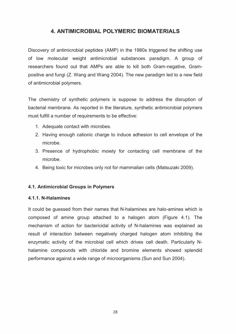
28
4. ANTIMICROBIAL POLYMERIC BIOMATERIALS
Discovery of antimicrobial peptides (AMP) in the 1980s triggered the shifting use
of low molecular weight antimicrobial substances paradigm. A group of
researchers found out that AMPs are able to kill both Gram-negative, Gram-
positive and fungi (Z. Wang and Wang 2004). The new paradigm led to a new field
of antimicrobial polymers.
The chemistry of synthetic polymers is suppose to address the disruption of
bacterial membrane. As reported in the literature, synthetic antimicrobial polymers
must fulfill a number of requirements to be effective:
1. Adequate contact with microbes.
2. Having enough cationic charge to induce adhesion to cell envelope of the
microbe.
3. Presence of hydrophobic moiety for contacting cell membrane of the
microbe.
4. Being toxic for microbes only not for mammalian cells (Matsuzaki 2009).
4.1. Antimicrobial Groups in Polymers
4.1.1. N-Halamines
It could be guessed from their names that N-halamines are halo-amines which is
composed of amine group attached to a halogen atom (Figure 4.1). The
mechanism of action for bactericidal activity of N-halamines was explained as
result of interaction between negatively charged halogen atom inhibiting the
enzymatic activity of the microbial cell which drives cell death. Particularly N-
halamine compounds with chloride and bromine elements showed splendid
performance against a wide range of microorganisms (Sun and Sun 2004).
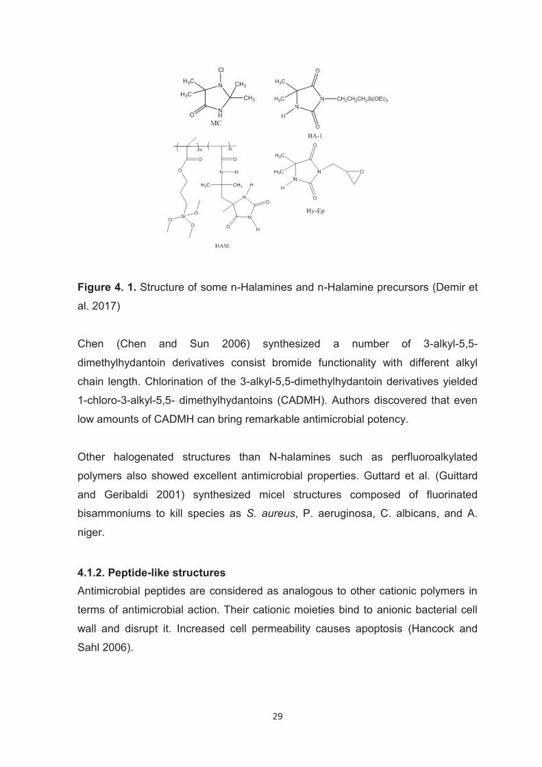
29
Figure 4. 1. Structure of some n-Halamines and n-Halamine precursors (Demir et
al. 2017) Chen (Chen and Sun 2006) synthesized a number of 3-alkyl-5,5-
dimethylhydantoin derivatives consist bromide functionality with different alkyl
chain length. Chlorination of the 3-alkyl-5,5-dimethylhydantoin derivatives yielded
1-chloro-3-alkyl-5,5- dimethylhydantoins (CADMH). Authors discovered that even
low amounts of CADMH can bring remarkable antimicrobial potency.
Other halogenated structures than N-halamines such as perfluoroalkylated
polymers also showed excellent antimicrobial properties. Guttard et al. (Guittard
and Geribaldi 2001) synthesized micel structures composed of fluorinated
bisammoniums to kill species as S. aureus, P. aeruginosa, C. albicans, and A.
niger.
4.1.2. Peptide-like structures Antimicrobial peptides are considered as analogous to other cationic polymers in
terms of antimicrobial action. Their cationic moieties bind to anionic bacterial cell
wall and disrupt it. Increased cell permeability causes apoptosis (Hancock and
Sahl 2006).
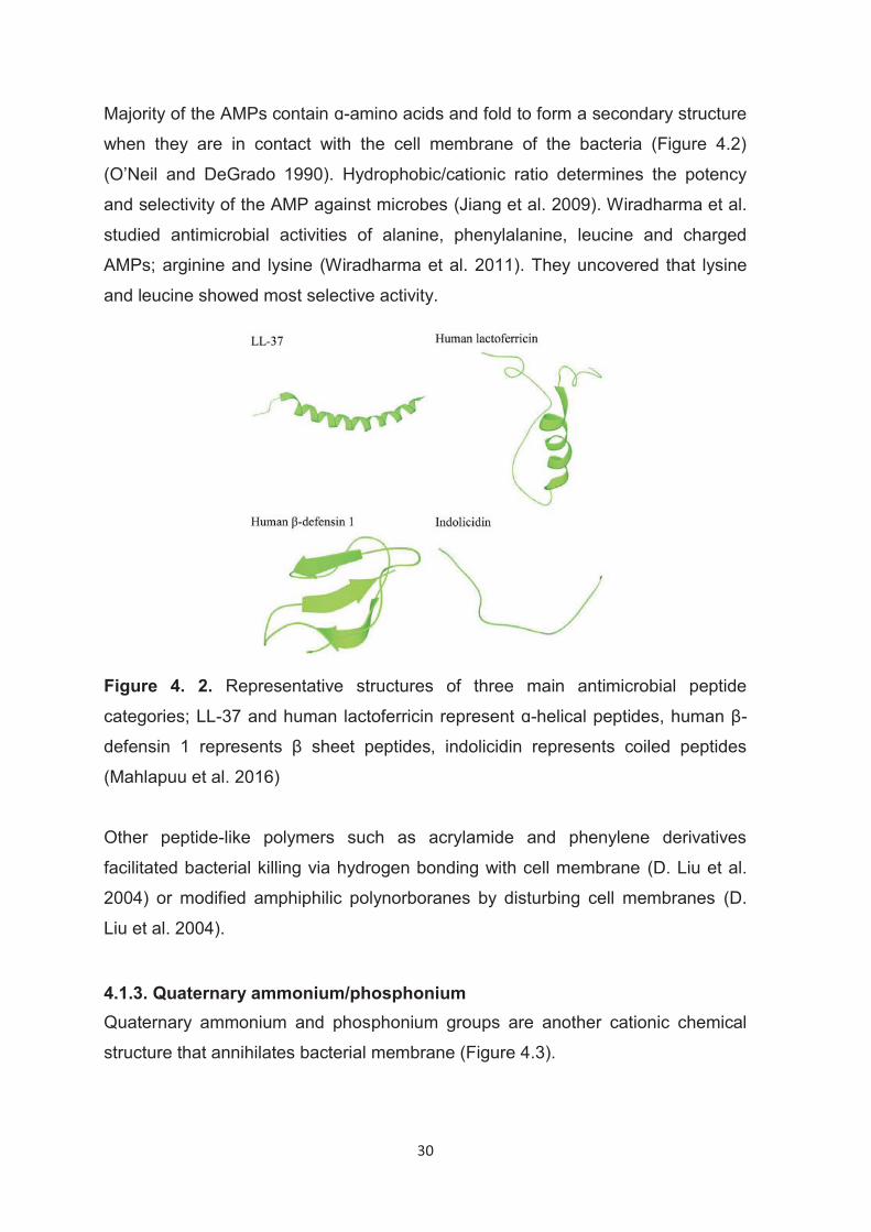
30
Majority of the AMPs contain ɑ-amino acids and fold to form a secondary structure
when they are in contact with the cell membrane of the bacteria (Figure 4.2)
(O’Neil and DeGrado 1990). Hydrophobic/cationic ratio determines the potency
and selectivity of the AMP against microbes (Jiang et al. 2009). Wiradharma et al.
studied antimicrobial activities of alanine, phenylalanine, leucine and charged
AMPs; arginine and lysine (Wiradharma et al. 2011). They uncovered that lysine
and leucine showed most selective activity.
Figure 4. 2. Representative structures of three main antimicrobial peptide
categories; LL-37 and human lactoferricin represent ɑ-helical peptides, human β-
defensin 1 represents β sheet peptides, indolicidin represents coiled peptides
(Mahlapuu et al. 2016)
Other peptide-like polymers such as acrylamide and phenylene derivatives
facilitated bacterial killing via hydrogen bonding with cell membrane (D. Liu et al.
2004) or modified amphiphilic polynorboranes by disturbing cell membranes (D.
Liu et al. 2004).
4.1.3. Quaternary ammonium/phosphonium Quaternary ammonium and phosphonium groups are another cationic chemical
structure that annihilates bacterial membrane (Figure 4.3).
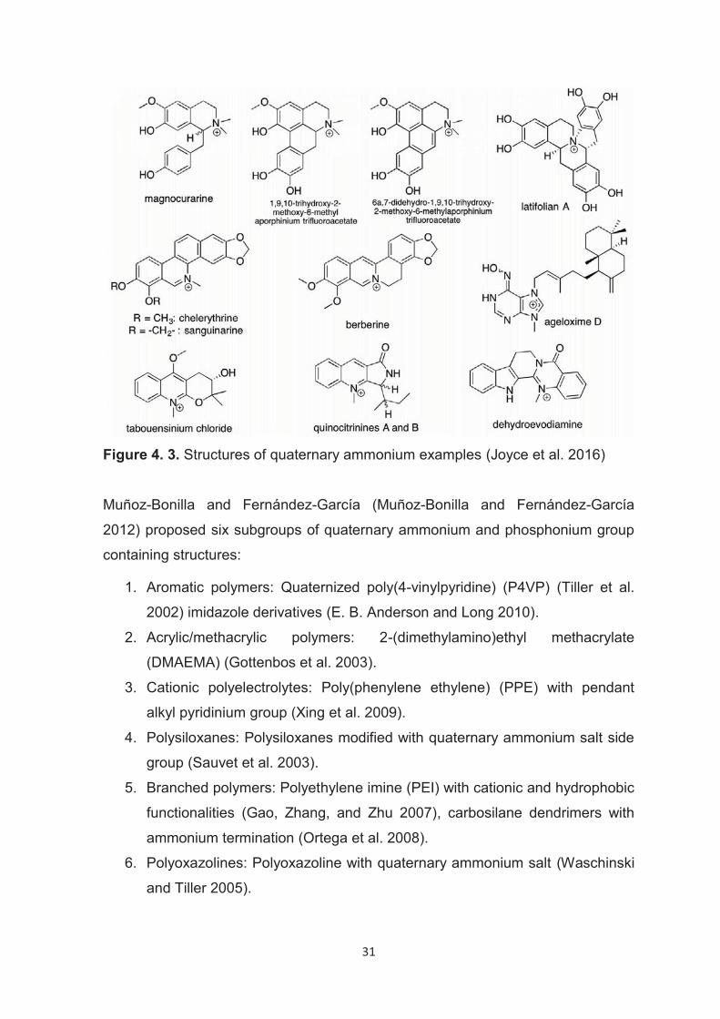
31
Figure 4. 3. Structures of quaternary ammonium examples (Joyce et al. 2016)
Muñoz-Bonilla and Fernández-García (Muñoz-Bonilla and Fernández-García
2012) proposed six subgroups of quaternary ammonium and phosphonium group
containing structures:
1. Aromatic polymers: Quaternized poly(4-vinylpyridine) (P4VP) (Tiller et al.
2002) imidazole derivatives (E. B. Anderson and Long 2010).
2. Acrylic/methacrylic polymers: 2-(dimethylamino)ethyl methacrylate
(DMAEMA) (Gottenbos et al. 2003).
3. Cationic polyelectrolytes: Poly(phenylene ethylene) (PPE) with pendant
alkyl pyridinium group (Xing et al. 2009).
4. Polysiloxanes: Polysiloxanes modified with quaternary ammonium salt side
group (Sauvet et al. 2003).
5. Branched polymers: Polyethylene imine (PEI) with cationic and hydrophobic
functionalities (Gao, Zhang, and Zhu 2007), carbosilane dendrimers with
ammonium termination (Ortega et al. 2008).
6. Polyoxazolines: Polyoxazoline with quaternary ammonium salt (Waschinski
and Tiller 2005).
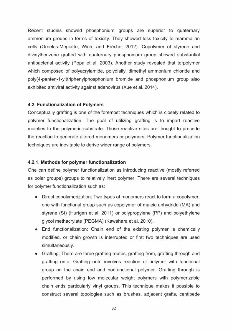
32
Recent studies showed phosphonium groups are superior to quaternary
ammonium groups in terms of toxicity. They showed less toxicity to mammalian
cells (Ornelas-Megiatto, Wich, and Fréchet 2012). Copolymer of styrene and
divinylbenzene grafted with quaternary phosphonium group showed substantial
antibacterial activity (Popa et al. 2003). Another study revealed that terpolymer
which composed of polyacrylamide, polydiallyl dimethyl ammonium chloride and
poly(4-penten-1-yl)triphenylphosphonium bromide and phosphonium group also
exhibited antiviral activity against adenovirus (Xue et al. 2014).
4.2. Functionalization of Polymers Conceptually grafting is one of the foremost techniques which is closely related to
polymer functionalization. The goal of utilizing grafting is to impart reactive
moieties to the polymeric substrate. Those reactive sites are thought to precede
the reaction to generate altered monomers or polymers. Polymer functionalization
techniques are inevitable to derive wider range of polymers.
4.2.1. Methods for polymer functionalization One can define polymer functionalization as introducing reactive (mostly referred
as polar groups) groups to relatively inert polymer. There are several techniques
for polymer functionalization such as:
● Direct copolymerization: Two types of monomers react to form a copolymer,
one with functional group such as copolymer of maleic anhydride (MA) and
styrene (St) (Hurtgen et al. 2011) or polypropylene (PP) and polyethylene
glycol methacrylate (PEGMA) (Kawahara et al. 2010).
● End functionalization: Chain end of the existing polymer is chemically
modified, or chain growth is interrupted or first two techniques are used
simultaneously.
● Grafting: There are three grafting routes; grafting from, grafting through and
grafting onto. Grafting onto involves reaction of polymer with functional
group on the chain end and nonfunctional polymer. Grafting through is
performed by using low molecular weight polymers with polymerizable
chain ends particularly vinyl groups. This technique makes it possible to
construct several topologies such as brushes, adjacent grafts, centipede
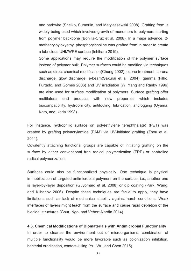
33
and barbwire (Sheiko, Sumerlin, and Matyjaszewski 2008). Grafting from is
widely being used which involves growth of monomers to polymers starting
from polymer backbone (Bonilla-Cruz et al. 2008). In a major advance, 2-
methacryloyloxyethyl phosphorylcholine was grafted from in order to create
a lubricious UHMWPE surface (Ishihara 2019).
Some applications may require the modification of the polymer surface
instead of polymer bulk. Polymer surfaces could be modified via techniques
such as direct chemical modification(Chung 2002), ozone treatment, corona
discharge, glow discharge, e-beam(Sakurai et al. 2004), gamma (Filho,
Furtado, and Gomes 2006) and UV irradiation (W. Yang and Ranby 1996)
are also used for surface modification of polymers. Surface grafting offer
multilateral end products with new properties which includes
biocompatibility, hydrophilicity, antifouling, lubrication, antifogging (Uyama,
Kato, and Ikada 1998).
For instance, hydrophilic surface on poly(ethylene terephthalate) (PET) was
created by grafting polyacrylamide (PAM) via UV-initiated grafting (Zhou et al.
2011).
Covalently attaching functional groups are capable of initiating grafting on the
surface by either conventional free radical polymerization (FRP) or controlled
radical polymerization.
Surfaces could also be functionalized physically. One technique is physical
immobilization of targeted antimicrobial polymers on the surface, i.e., another one
is layer-by-layer deposition (Guyomard et al. 2008) or dip coating (Park, Wang,
and Klibanov 2006). Despite these techniques are facile to apply, they have
limitations such as lack of mechanical stability against harsh conditions. Weak
interfaces of layers might leach from the surface and cause rapid depletion of the
biocidal structures (Gour, Ngo, and Vebert-Nardin 2014).
4.3. Chemical Modifications of Biomaterials with Antimicrobial Functionality In order to cleanse the environment out of microorganisms, combination of
multiple functionality would be more favorable such as colonization inhibition,
bacterial eradication, contact-killing (Yu, Wu, and Chen 2015).
34
Bacteria repelling and bactericidal agent releasing materials embodies an inherent
low adhesive polymeric material that demonstrate diminished bacterial adhesion
(up to few orders of magnitude). Such materials are poly(vinyl alcohol)(PVA),
PEG-bound copolymers, poly(acrylic acid) derivatives hydrogels. Those hydrogels
are loaded with antibiotics or other bactericides to kill bacteria simultaneously
while repelling them (Rodríguez-Hernández 2017). Another approach in this
category is an antimicrobial coating with silver ion releasing property (Ho et al.
2004). In this approach, glass surface covalently functionalized silanoyl groups
which is then tied to poly(ethylene imine) (PEI) groups to complex with silver
nanoparticles. Bacterial repelling is employed by poly(ethylene glycol) (PEG) layer
attached to poly(2-hydroxyethylacrylate) network (Figure 4.4).
Figure 4. 4. Dual functional repelling-releasing system (Ho et al. 2004)
While PEG ends of the network repels microorganisms, PEI releases silver
nanoions to kill planktonic S.aureus.
Another double functionality material is temperature labile polymer brushes which
transition from being bactericidal to repellent (Figure 4.5) (Laloyaux et al. 2010).
The structure is consisted of an antimicrobial peptide (Magainin) grafted
oligo(ethylene glycol) methacrylate (OEGMA). At room temperature OEGMA
chains are elongated so, Magainin groups are accessible by microbes in contact.
Nevertheless, at elevated temperatures than 35 °C OEGMA chains shrink, peptide
35
structures become inaccessible thereby OEGMA repels bacteria away. This on/off
system could be reversibly activated by heating/cooling cycles.
Figure 4. 5. Surface with contact-killing and repelling functions that kills bacteria
below 35 °C (a) and repel above 35 °C (b) (Laloyaux et al. 2010)
Another dual function structure is bactericidal compound releasing and contact-
killing type of materials. It was reported that antimicrobial N,N-dimethyl-
dodecylammonium (DDA) grafted poly(2-ethyl-1,3-oxazoline (PEtOₓ) coated by
cellulose is able to kill approaching bacteria (Bieser, Thomann, and Tiller 2011).
The system works as follows: When bacteria in contact with bactericidal moiety of
the structure die, it unleashes an enzyme called cellulase. The cellulase enzyme
degrades the cellulose layer and subsequently more DDA is released to kill the
bacteria again (Figure 4.6).
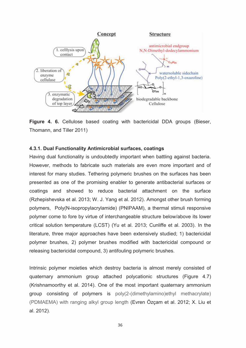
36
Figure 4. 6. Cellulose based coating with bactericidal DDA groups (Bieser,
Thomann, and Tiller 2011)
4.3.1. Dual Functionality Antimicrobial surfaces, coatings Having dual functionality is undoubtedly important when battling against bacteria.
However, methods to fabricate such materials are even more important and of
interest for many studies. Tethering polymeric brushes on the surfaces has been
presented as one of the promising enabler to generate antibacterial surfaces or
coatings and showed to reduce bacterial attachment on the surface
(Rzhepishevska et al. 2013; W. J. Yang et al. 2012). Amongst other brush forming
polymers, Poly(N-isopropylacrylamide) (PNIPAAM), a thermal stimuli responsive
polymer come to fore by virtue of interchangeable structure below/above its lower
critical solution temperature (LCST) (Yu et al. 2013; Cunliffe et al. 2003). In the
literature, three major approaches have been extensively studied; 1) bactericidal
polymer brushes, 2) polymer brushes modified with bactericidal compound or
releasing bactericidal compound, 3) antifouling polymeric brushes.
Intrinsic polymer moieties which destroy bacteria is almost merely consisted of
quaternary ammonium group attached polycationic structures (Figure 4.7)
(Krishnamoorthy et al. 2014). One of the most important quaternary ammonium
group consisting of polymers is poly(2-(dimethylamino)ethyl methacrylate)
(PDMAEMA) with ranging alkyl group length (Evren Özçam et al. 2012; X. Liu et
al. 2012).
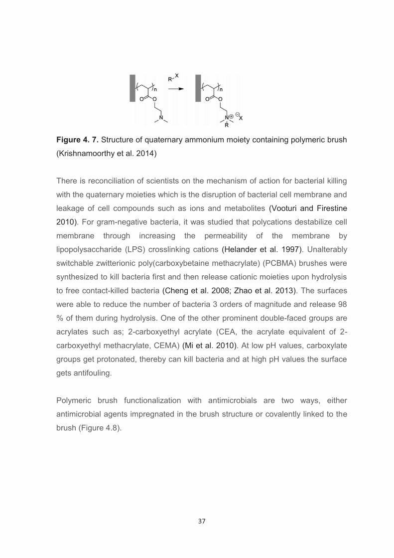
37
Figure 4. 7. Structure of quaternary ammonium moiety containing polymeric brush
(Krishnamoorthy et al. 2014)
There is reconciliation of scientists on the mechanism of action for bacterial killing
with the quaternary moieties which is the disruption of bacterial cell membrane and
leakage of cell compounds such as ions and metabolites (Vooturi and Firestine
2010). For gram-negative bacteria, it was studied that polycations destabilize cell
membrane through increasing the permeability of the membrane by
lipopolysaccharide (LPS) crosslinking cations (Helander et al. 1997). Unalterably
switchable zwitterionic poly(carboxybetaine methacrylate) (PCBMA) brushes were
synthesized to kill bacteria first and then release cationic moieties upon hydrolysis
to free contact-killed bacteria (Cheng et al. 2008; Zhao et al. 2013). The surfaces
were able to reduce the number of bacteria 3 orders of magnitude and release 98
% of them during hydrolysis. One of the other prominent double-faced groups are
acrylates such as; 2-carboxyethyl acrylate (CEA, the acrylate equivalent of 2-
carboxyethyl methacrylate, CEMA) (Mi et al. 2010). At low pH values, carboxylate
groups get protonated, thereby can kill bacteria and at high pH values the surface
gets antifouling.
Polymeric brush functionalization with antimicrobials are two ways, either
antimicrobial agents impregnated in the brush structure or covalently linked to the
brush (Figure 4.8).
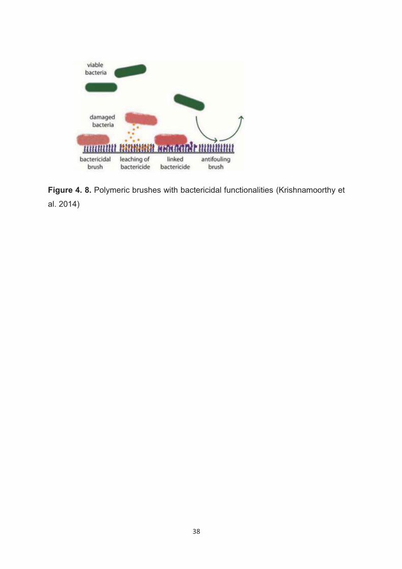
38
Figure 4. 8. Polymeric brushes with bactericidal functionalities (Krishnamoorthy et
al. 2014)
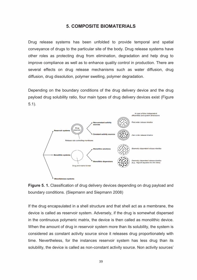
39
5. COMPOSITE BIOMATERIALS
Drug release systems has been unfolded to provide temporal and spatial
conveyance of drugs to the particular site of the body. Drug release systems have
other roles as protecting drug from elimination, degradation and help drug to
improve compliance as well as to enhance quality control in production. There are
several effects on drug release mechanisms such as water diffusion, drug
diffusion, drug dissolution, polymer swelling, polymer degradation.
Depending on the boundary conditions of the drug delivery device and the drug
payload drug solubility ratio, four main types of drug delivery devices exist (Figure
5.1).
Figure 5. 1. Classification of drug delivery devices depending on drug payload and
boundary conditions. (Siepmann and Siepmann 2008) If the drug encapsulated in a shell structure and that shell act as a membrane, the
device is called as reservoir system. Adversely, if the drug is somewhat dispersed
in the continuous polymeric matrix, the device is then called as monolithic device.
When the amount of drug in reservoir system more than its solubility, the system is
considered as constant activity source since it releases drug proportionately with
time. Nevertheless, for the instances reservoir system has less drug than its
solubility, the device is called as non-constant activity source. Non activity sources’
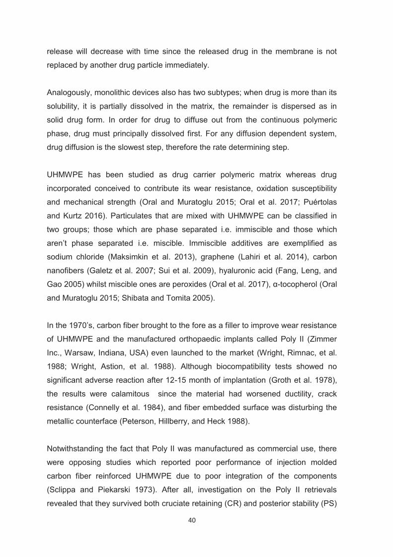
40
release will decrease with time since the released drug in the membrane is not
replaced by another drug particle immediately.
Analogously, monolithic devices also has two subtypes; when drug is more than its
solubility, it is partially dissolved in the matrix, the remainder is dispersed as in
solid drug form. In order for drug to diffuse out from the continuous polymeric
phase, drug must principally dissolved first. For any diffusion dependent system,
drug diffusion is the slowest step, therefore the rate determining step.
UHMWPE has been studied as drug carrier polymeric matrix whereas drug
incorporated conceived to contribute its wear resistance, oxidation susceptibility
and mechanical strength (Oral and Muratoglu 2015; Oral et al. 2017; Puértolas
and Kurtz 2016). Particulates that are mixed with UHMWPE can be classified in
two groups; those which are phase separated i.e. immiscible and those which
aren’t phase separated i.e. miscible. Immiscible additives are exemplified as
sodium chloride (Maksimkin et al. 2013), graphene (Lahiri et al. 2014), carbon
nanofibers (Galetz et al. 2007; Sui et al. 2009), hyaluronic acid (Fang, Leng, and
Gao 2005) whilst miscible ones are peroxides (Oral et al. 2017), α-tocopherol (Oral
and Muratoglu 2015; Shibata and Tomita 2005).
In the 1970’s, carbon fiber brought to the fore as a filler to improve wear resistance
of UHMWPE and the manufactured orthopaedic implants called Poly II (Zimmer
Inc., Warsaw, Indiana, USA) even launched to the market (Wright, Rimnac, et al.
1988; Wright, Astion, et al. 1988). Although biocompatibility tests showed no
significant adverse reaction after 12-15 month of implantation (Groth et al. 1978),
the results were calamitous since the material had worsened ductility, crack
resistance (Connelly et al. 1984), and fiber embedded surface was disturbing the
metallic counterface (Peterson, Hillberry, and Heck 1988).
Notwithstanding the fact that Poly II was manufactured as commercial use, there
were opposing studies which reported poor performance of injection molded
carbon fiber reinforced UHMWPE due to poor integration of the components
(Sclippa and Piekarski 1973). After all, investigation on the Poly II retrievals
revealed that they survived both cruciate retaining (CR) and posterior stability (PS)
41
(Kurtz 2009).
Hyaluronan, simplest glycosaminoglycan of extracellular matrix, incorporated as
micro filler to UHMWPE (Zhang et al. 2006, 2007). Hyaluronan is the lubricity
source of natural cartilage. By no means, incorporation of hyaluronan into
UHMWPE is not a downright task due to their water affinity difference. It was
reported that when hyaluronan is complexed with quaternary ammonium cations
and consequently silylated, it gains ease of diffusion into porous UHMWPE matrix
(Zhang et al. 2007). Diffused modified hyaluronan is then chemically crosslinked in
situ and hydrolyzed to hyaluronan (Zhang et al. 2007). In the last step, the
microcomposite is compression molded and machined to manufacture pin for pin-
on-disk (POD) test which was subsequently done and showed moderate wear
reduction for multidirectional POD wear test (Zhang et al. 2007).
Along with growing interest of most studies in improvement of wear resistance and
mechanical strength, biomimicking was another concern for polymeric bone
substitutes (Kurtz 2009). To do that, few exploratory studies were adopted for
hydroxyapatite (HA) incorporation in high density polyethylene (HDPE) (Bonfield
1988). Hydroxyapatite with its low molecular weight was used to prepare
microcomposites with high density polyethylene. Given the low molecular weight
components, HA/HDPE composites have multilateral practicality in terms of
processability including injection molding (M. Wang et al. 2000). Studies showed
HA/UHMWPE is both bioactive and biocompatible (Di Silvio, Dalby, and Bonfield
2002; S. M. Rea, Best, and Bonfield 2004; Susan M. Rea et al. 2004). One major
drawback of HA/UHMWPE microcomposites is it exhibits failure under high load
bearing conditions. Incorporating HA in UHMWPE was more challenging when it
comes to homogenous dispersion. Referring to the literature, it was managed to
disperse HA in UHMWPE matrix by various methods such as ball milling, extrusion
of swollen UHMWPE with pharmaceutical grade paraffin oil (Fang, Leng, and Gao
2006). Molded UHMWPE/HA samples showed superior mechanical strength; 7
GPa of elastic modulus and 375 % of elongation at break (Fang, Leng, and Gao
2006). For UHMWPE/HA bioactive composites, to be a predicate implant device, it
is required biocompatibility tests.
42
6. LIVING RADICAL POLYMERIZATION
Living radical polymerization (LRP) is one of the subsets of free radical
polymerizations that generates radicalic terminals which don’t terminate or transfer
and continue polymerization as monomer is fed in the reaction medium. Through
its inherent nature, radical polymerizations terminate where as LRP techniques
have minimum termination step which renders the technique to have control over
molecular weight and molecular weight distribution of the polymer. LRP techniques
have several wished aspects such as, plausibility for wide range monomers,
tolerance for various functionalities and can be carried out under mild reaction
conditions. Conventional free radical polymerizations are not suitable for
synthesizing polymers with complex architecture or with site specific functionality
as LRP techniques are. LRPs are categorized in three groups: 1) Atom Transfer
Radical Polymerization (ATRP), 2) Reversible Addition Fragmentation Chain
Transfer Polymerization (RAFT), 3) Nitroxide-mediated Polymerization (NMP).
Iodine transfer polymerization (ITP), single electron transfer-degenerative transfer
living polymerization (SET-DTLRP), organotellurium mediated living radical
polymerization (OMLRP) are other living radical polymerization techniques
although to a lesser extent.
6.1. RAFT Polymerization The RAFT polymerization has changed the era of controlled radical
polymerization. It was invented simultaneously by a research team in
Commonwealth Scientific and Industrial Research Organization (CSIRO) in
Australia and by a group of researchers in France. The group in France called the
technique as macromolecular design via the interchange of xanthates (MADIX)
(Charmot et al. 1999). Despite NMP and ATRP, RAFT technique is based on a
degenerative chain transfer and doesn’t rely on sustained radicalic species. Drug
and gene delivery, tissue engineering, regenerative medicine, membrane science,
bioconjugation are the areas where RAFT technique is used (Barner-Kowollik
2008).

43
RAFT polymerization is governed by a versatile RAFT agent. Figure 6.1 shows the
chemical structures of four generic RAFT agents. The R group is the site where
monomers start to add up and grow subsequently the Z group activates thiol bond
toward monomer addition and makes attached radical stable. Thereby two
polymeric chains grow on both sides concurrently.
Figure 6. 1. Types of RAFT Agents
In the mechanism of RAFT polymerization, the first step is comprised of initiation
through generation of a free radical (step 1) (Figure 6.2) by one of the various
sources such as thermal (T. P. Le et al. 1998), UV (Lu, Yang, and Cai 2005), γ
radiation (Millard et al. 2006) and pulsed laser irradiation (Junkers et al. 2005).
Oligomeric radicals are formed and attack RAFT agent (step 2). The radicalic
intermediate (2) has two reversible paths which it can either transform back to
RAFT agent and oligomeric radical or can proceed to an oligomeric RAFT agent
(3) and reinitiating R radical. Homopolymer chains grow as monomer adding up
(step 3). Upon initiation, polymer chains grow on both sides of the intermediate 4
is propagation step (step 4). Termination for the RAFT polymerization occurs in
two ways; 1) combination, 2) disproportionation (step 5).
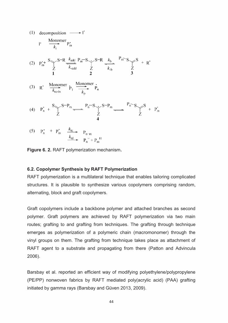
44
Figure 6. 2. RAFT polymerization mechanism.
6.2. Copolymer Synthesis by RAFT Polymerization RAFT polymerization is a multilateral technique that enables tailoring complicated
structures. It is plausible to synthesize various copolymers comprising random,
alternating, block and graft copolymers.
Graft copolymers include a backbone polymer and attached branches as second
polymer. Graft polymers are achieved by RAFT polymerization via two main
routes; grafting to and grafting from techniques. The grafting through technique
emerges as polymerization of a polymeric chain (macromonomer) through the
vinyl groups on them. The grafting from technique takes place as attachment of
RAFT agent to a substrate and propagating from there (Patton and Advincula
2006).
Barsbay et al. reported an efficient way of modifying polyethylene/polypropylene
(PE/PP) nonwoven fabrics by RAFT mediated poly(acrylic acid) (PAA) grafting
initiated by gamma rays (Barsbay and Güven 2013, 2009).
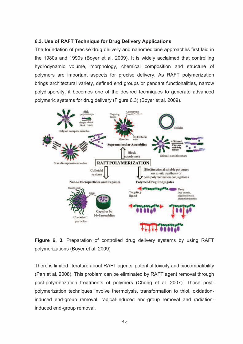
45
6.3. Use of RAFT Technique for Drug Delivery Applications The foundation of precise drug delivery and nanomedicine approaches first laid in
the 1980s and 1990s (Boyer et al. 2009). It is widely acclaimed that controlling
hydrodynamic volume, morphology, chemical composition and structure of
polymers are important aspects for precise delivery. As RAFT polymerization
brings architectural variety, defined end groups or pendant functionalities, narrow
polydispersity, it becomes one of the desired techniques to generate advanced
polymeric systems for drug delivery (Figure 6.3) (Boyer et al. 2009).
Figure 6. 3. Preparation of controlled drug delivery systems by using RAFT
polymerizations (Boyer et al. 2009)
There is limited literature about RAFT agents’ potential toxicity and biocompatibility
(Pan et al. 2008). This problem can be eliminated by RAFT agent removal through
post-polymerization treatments of polymers (Chong et al. 2007). Those post-
polymerization techniques involve thermolysis, transformation to thiol, oxidation-
induced end-group removal, radical-induced end-group removal and radiation-
induced end-group removal.
46
PJI treatment could have devastating results unless two important aspects of it
were taken care of; appropriate dosage control and sustainable antibiotic
treatment. They both require great attention otherwise could be disastrous for
patients and eventually turned into an immense public health problem. Antibiotic
dosage control must be so delicate that it is not above toxicity levels and not below
MIC which could lead to antibiotic resistance. Sustainable antibiotic release is
essential for implants to avoid bacterial colonization which will fail patients to an
additional joint replacement surgery.
Under consideration above reasons, it is vital to tailor a drug-releasing implant
which ensures delicate dosage control and sustainable antibiotic release. In this
thesis study, RAFT polymerization was used to synthesize UHMWPE-g-PHEMA
with controlled the molecular weight and the molecular weight distribution of
PHEMA to control the release rate and sustainability of gentamicin sulfate.
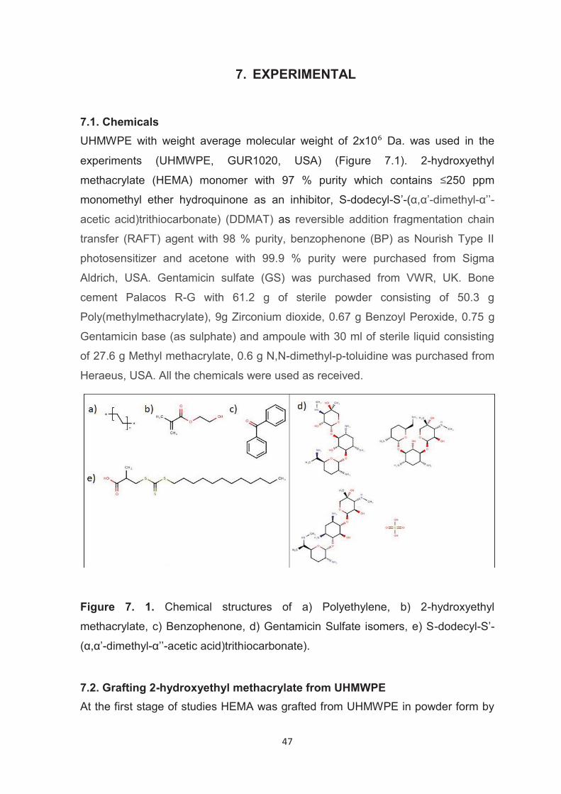
47
7. EXPERIMENTAL
7.1. Chemicals UHMWPE with weight average molecular weight of 2x10 Da. was used in the
experiments (UHMWPE, GUR1020, USA) (Figure 7.1). 2-hydroxyethyl
methacrylate (HEMA) monomer with 97 % purity which contains ≤250 ppm
monomethyl ether hydroquinone as an inhibitor, S-dodecyl-S’-(α,α’-dimethyl-α’’-
acetic acid)trithiocarbonate) (DDMAT) as reversible addition fragmentation chain
transfer (RAFT) agent with 98 % purity, benzophenone (BP) as Nourish Type II
photosensitizer and acetone with 99.9 % purity were purchased from Sigma
Aldrich, USA. Gentamicin sulfate (GS) was purchased from VWR, UK. Bone
cement Palacos R-G with 61.2 g of sterile powder consisting of 50.3 g
Poly(methylmethacrylate), 9g Zirconium dioxide, 0.67 g Benzoyl Peroxide, 0.75 g
Gentamicin base (as sulphate) and ampoule with 30 ml of sterile liquid consisting
of 27.6 g Methyl methacrylate, 0.6 g N,N-dimethyl-p-toluidine was purchased from
Heraeus, USA. All the chemicals were used as received.
Figure 7. 1. Chemical structures of a) Polyethylene, b) 2-hydroxyethyl
methacrylate, c) Benzophenone, d) Gentamicin Sulfate isomers, e) S-dodecyl-S’-
(α,α’-dimethyl-α’’-acetic acid)trithiocarbonate).
7.2. Grafting 2-hydroxyethyl methacrylate from UHMWPE At the first stage of studies HEMA was grafted from UHMWPE in powder form by
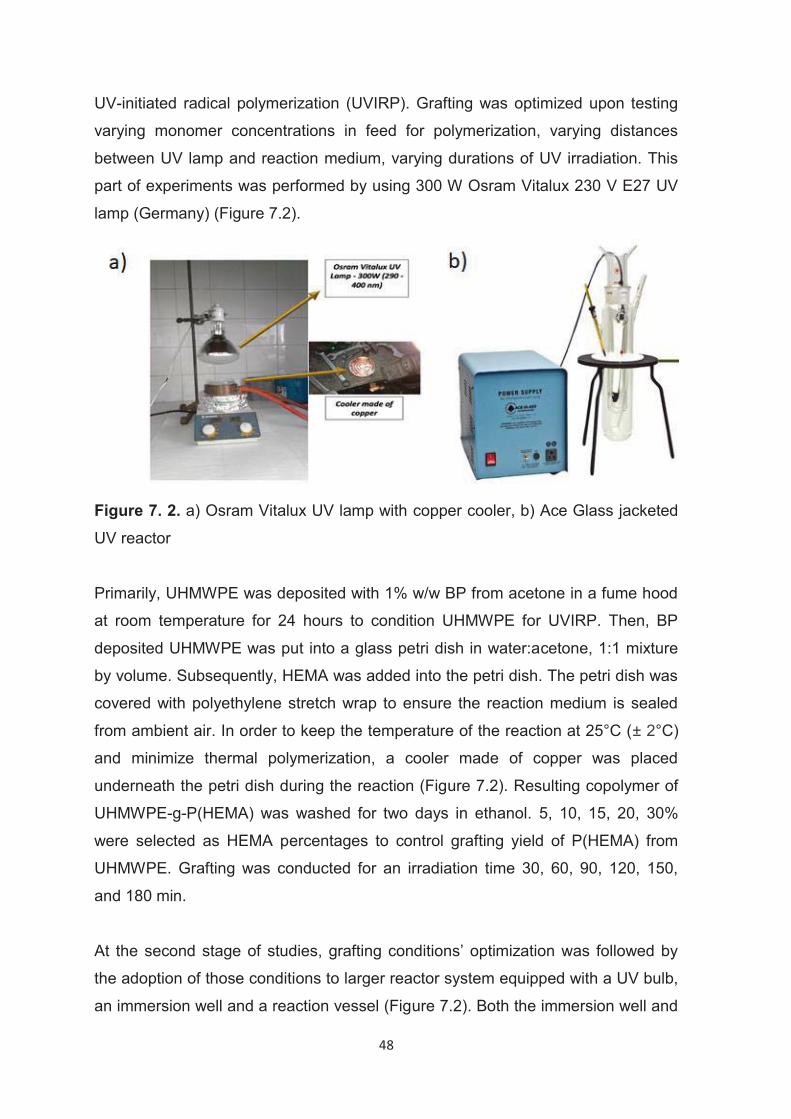
48
UV-initiated radical polymerization (UVIRP). Grafting was optimized upon testing
varying monomer concentrations in feed for polymerization, varying distances
between UV lamp and reaction medium, varying durations of UV irradiation. This
part of experiments was performed by using 300 W Osram Vitalux 230 V E27 UV
lamp (Germany) (Figure 7.2).
Figure 7. 2. a) Osram Vitalux UV lamp with copper cooler, b) Ace Glass jacketed
UV reactor
Primarily, UHMWPE was deposited with 1% w/w BP from acetone in a fume hood
at room temperature for 24 hours to condition UHMWPE for UVIRP. Then, BP
deposited UHMWPE was put into a glass petri dish in water:acetone, 1:1 mixture
by volume. Subsequently, HEMA was added into the petri dish. The petri dish was
covered with polyethylene stretch wrap to ensure the reaction medium is sealed
from ambient air. In order to keep the temperature of the reaction at 25°C (± 2°C)
and minimize thermal polymerization, a cooler made of copper was placed
underneath the petri dish during the reaction (Figure 7.2). Resulting copolymer of
UHMWPE-g-P(HEMA) was washed for two days in ethanol. 5, 10, 15, 20, 30%
were selected as HEMA percentages to control grafting yield of P(HEMA) from
UHMWPE. Grafting was conducted for an irradiation time 30, 60, 90, 120, 150,
and 180 min.
At the second stage of studies, grafting conditions’ optimization was followed by
the adoption of those conditions to larger reactor system equipped with a UV bulb,
an immersion well and a reaction vessel (Figure 7.2). Both the immersion well and
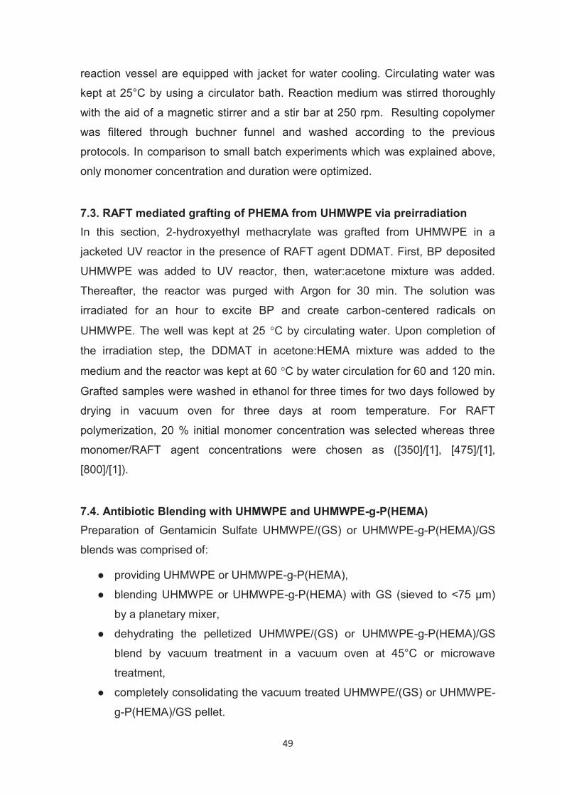
49
reaction vessel are equipped with jacket for water cooling. Circulating water was
kept at 25°C by using a circulator bath. Reaction medium was stirred thoroughly
with the aid of a magnetic stirrer and a stir bar at 250 rpm. Resulting copolymer
was filtered through buchner funnel and washed according to the previous
protocols. In comparison to small batch experiments which was explained above,
only monomer concentration and duration were optimized.
7.3. RAFT mediated grafting of PHEMA from UHMWPE via preirradiation In this section, 2-hydroxyethyl methacrylate was grafted from UHMWPE in a
jacketed UV reactor in the presence of RAFT agent DDMAT. First, BP deposited
UHMWPE was added to UV reactor, then, water:acetone mixture was added.
Thereafter, the reactor was purged with Argon for 30 min. The solution was
irradiated for an hour to excite BP and create carbon-centered radicals on
UHMWPE. The well was kept at 25 �C by circulating water. Upon completion of
the irradiation step, the DDMAT in acetone:HEMA mixture was added to the
medium and the reactor was kept at 60 �C by water circulation for 60 and 120 min.
Grafted samples were washed in ethanol for three times for two days followed by
drying in vacuum oven for three days at room temperature. For RAFT
polymerization, 20 % initial monomer concentration was selected whereas three
monomer/RAFT agent concentrations were chosen as ([350]/[1], [475]/[1],
[800]/[1]).
7.4. Antibiotic Blending with UHMWPE and UHMWPE-g-P(HEMA) Preparation of Gentamicin Sulfate UHMWPE/(GS) or UHMWPE-g-P(HEMA)/GS
blends was comprised of:
● providing UHMWPE or UHMWPE-g-P(HEMA),
● blending UHMWPE or UHMWPE-g-P(HEMA) with GS (sieved to <75 μm)
by a planetary mixer,
● dehydrating the pelletized UHMWPE/(GS) or UHMWPE-g-P(HEMA)/GS
blend by vacuum treatment in a vacuum oven at 45°C or microwave
treatment,
● completely consolidating the vacuum treated UHMWPE/(GS) or UHMWPE-
g-P(HEMA)/GS pellet.
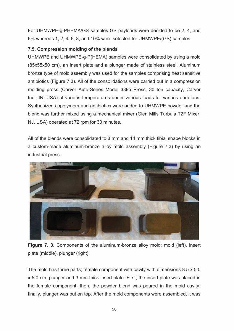
50
For UHMWPE-g-PHEMA/GS samples GS payloads were decided to be 2, 4, and
6% whereas 1, 2, 4, 6, 8, and 10% were selected for UHMWPE/(GS) samples.
7.5. Compression molding of the blends UHMWPE and UHMWPE-g-P(HEMA) samples were consolidated by using a mold
(85x55x50 cm), an insert plate and a plunger made of stainless steel. Aluminum
bronze type of mold assembly was used for the samples comprising heat sensitive
antibiotics (Figure 7.3). All of the consolidations were carried out in a compression
molding press (Carver Auto-Series Model 3895 Press, 30 ton capacity, Carver
Inc., IN, USA) at various temperatures under various loads for various durations.
Synthesized copolymers and antibiotics were added to UHMWPE powder and the
blend was further mixed using a mechanical mixer (Glen Mills Turbula T2F Mixer,
NJ, USA) operated at 72 rpm for 30 minutes.
All of the blends were consolidated to 3 mm and 14 mm thick tibial shape blocks in
a custom-made aluminum-bronze alloy mold assembly (Figure 7.3) by using an
industrial press.
Figure 7. 3. Components of the aluminum-bronze alloy mold; mold (left), insert
plate (middle), plunger (right).
The mold has three parts; female component with cavity with dimensions 8.5 x 5.0
x 5.0 cm, plunger and 3 mm thick insert plate. First, the insert plate was placed in
the female component, then, the powder blend was poured in the mold cavity,
finally, plunger was put on top. After the mold components were assembled, it was
51
placed between press platens which were already heated to 170 °C followed by
closing platens to apply 20 MPa pressure. The molding recipe was comprised of
two segments; the first segment involved heating of the mold for 8 minutes for 3
mm thick blocks and 45 minutes for 14 mm thick blocks. The second segment
involved cooling down to 20 °C by water circulating in the platens for 15 minutes.
As the molding was complete consolidated block was taken out and used for
further experiments.
7.6. Characterization 7.6.2. Structural characterization of PolyHEMA grafted UHMWPE 7.6.2.1. FT-IR/ATR UHMWPE samples were structurally characterized by FT-IR/ATR (Perkin Elmer
Spectrum Model 2) after grafting poly(HEMA). ATR attachment is equipped with
single-reflecting diamond crystal. Spectra were collected with scan number 16 and
resolution 4 cm-1. To ascertain the abundance of poly(HEMA), carbonyl peaks at
around 1720 cm-1 were compared after the C-H stretching peaks at around 2920
cm-1 were normalized.
7.6.2.2. CHNS/O Analysis Elemental composition of poly(HEMA) grafted UHMWPE was analyzed by Flash
2000 NC (Thermo Scientific) elemental analyzer equipped with Al/As 3000 series
autosampler. Samples were combusted in tin pans with 2,5-Bis(5-tert-butyl-2-
benzoxazolyl)thiophene (BBOT) as calibration standard and vanadium pentoxide
(V2O5) as catalyst.
7.6.2.3. X-ray Photoelectron Spectroscopy X-ray photoelectron spectroscopy (XPS) was performed on an XPS spectrometer
(Thermo Scientific K-Alpha) with monochromatic Al Kα as X-ray source. The
Chamber of the equipment were kept at pressure of 2 x 10⁻⁹ Torr and at a
temperature of 25OC.
52
7.6.2.4. Contact Angle Measurements Water contact angle of all pre-consolidated and powder specimens were
measured from glass-contact surfaces with a goniometric contact angle
measurement device (Easy Drop Gonyo-meter, Krüss, Hamburg, Germany). 10 μl
deionized water was used in every measurement. Contact angles were obtained
from three different spots of the sample surfaces. PHEMA grafted samples were
pressed for 2 mins at 10 ton/cm2 by a manual hydraulic press prior to contact
angle measurements.
7.6.2.5. Tensile Strength Testing Tensile specimens (type V) were stamped out of 3.2-mm-thick dog bone-shaped
sections in agreement with the American Society for Testing Materials (ASTM)
D638. Specimens were tested on an Insight 2 model MTS machine (Minneapolis,
Minnesota, USA) at a crosshead speed of 10 mm/min. In order to determine the
elongation at break (EAB), a laser extensometer was used to determine gauge
displacement by reading reflective tapes mounted on the specimen. Ultimate
tensile strength (UTS) and yield strength (YS) and elastic modulus (E) were
calculated in accordance with ASTM D-638 standard.
7.6.2.6. IZOD Impact Strength Testing Sample bars of 6.35 x 12.7 x 63.5 mm were double notched to a depth of 4.57
0.08 mm according to the ASTM D-256 and tested according to ASTM F-648 with
CEAST Instron model impact tester. All of the specimens were tested at room
temperature. The impact toughness was reported as the average value of n = 3
specimens in kJ/m2.
7.6.2.7. Pin-on-Disc (POD) Wear Testing Cylindrical pins (9-mm diameter and 13-mm long, n=3 each) were machined using
a CNC mill (ShopBot Tools Inc., NC, USA) from the materials that were supposed
to be POD tested. The pins were wear tested against polished Co-Cr-Mo discs at
2 Hz in undiluted, preserved bovine serum as a lubricant that contains Penicillin-
Streptomycin (Sigma, USA) and EDTA (Fisher Chemical, USA). Wear rates were
calculated gravimetrically by weighing the samples every 0.15 million cycle for 1.1
53
MC totally. The wear rate was determined by a linear regression of the weight loss
as a function of number of cycles from 0.5 to 1.1 MC.
7.6.2.8. Differential Scanning Calorimetry The specimens for differential scanning calorimetry were prepared by cutting thin
slabs (8-10 mg) for testing. The DSC specimens were weighed with a Sartorius
(Sartorius Corporation, Edgewood, NY) CP 225D balance to a resolution of 0.01
mg and placed in aluminum pans. The pan was covered with an aluminum cover
and placed in a Q-1000 Differential Scanning Calorimeter (TA Instruments,
Newark, USA). The sample and the reference were then heated at a heating rate
of 10° C/min from 20°C to 180°C, cooled to 20°C at 10°C/min, and subjected to
another heating cycle from 20°C to 180°C at 10°C/ min in nitrogen gas
atmosphere. Heat flow as a function of time and temperature was recorded, and
the cycles were referred to as first heat, first cool, and second heat, respectively.
The melting enthalpy of 100 % UHMWPE was accepted as 291 J/g and crystalline
percentages of modified samples were calculated accordingly (Oral et al. 2006).
The mass of PHEMA was obtained from elemental analysis and it was used to
calculate the mass of UHMWPE in UHMWPE-g-PHEMA.
7.6.2.9. Scanning Electron Microscopy (SEM) - EDAX Analysis All of the samples to be SEM-imaged were sputter coated with a layer of
gold/palladium and imaged with a Zeiss ULTRA 55 (Zeiss, Oberkochen, Germany)
type of microscope equipped with EDAX detector. Oxygen and nitrogen profiles
were obtained.
7.6.2.10. Gravimetric Analysis of Grafting Degree Weight gain upon polyHEMA grafting from UHMWPE indicates that the grafting
took place. UHMWPE samples that were modified with polyHEMA, were weighed
before and after modification. Samples were washed three times in ethanol before
they were weighed. The degree of grafting was calculated by using the Equation
7.1.
Equation 7.1
54
Where w1 is the weight of pre-consolidated virgin UHMWPE and w2 is the weight of
polyHEMA-grafted UHMWPE.
7.6.2.11. Size Exclusion Chromatography-Multi-Angle Light Scattering Molecular weight and polydispersity index of poly(HEMA) homopolymers formed
simultaneously with grafting in reaction medium were determined by Size
Exclusion Chromatography equipped with Multi-Angle Laser Light Scattering
techniques. Instrument has both refractive index (Wyatt Optilab T-rex) and Multi-
Angle Light Scattering (Wyatt Dawn Heleos II) detectors. Measurements were
carried out in N,N-dimethylformamide (DMF) containing 0.5 mol LiBr, at a flow rate
of 1.0 mL/ min (25 °C) using PL Mixed-C columns. Samples were passed through
0.1 μm PTFE filters to eliminate any contaminant. The poly(HEMA) homopolymer
obtained from grafting experiments was precipitated in diethyl ether and then dried
in vacuum oven at 40°C prior to molecular weight determination.
Theoretical number average molecular weights of p(HEMA) were calculated as
described in Equation 7.2.
Equation 7.2
7.7. Drug release studies
7.7.1. Gentamicin Release Studies
Gentamicin release over time was studied by using gentamicin containing strips
(n=3, 3x5x10 mm) that were machined by CNC device (ShopBot Desktop, NC,
USA). Prior to start the drug release studies, dimensions and weight of strips were
individually recorded. Each sample was placed in a 3 ml sterile, disposable syringe
in 1.5 ml of PBS. 1.35 ml of PBS was chosen to meet sink conditions as well as to
cover the entire strip in the syringe. Syringes were placed in a 96 well plate upright
and were kept in an incubator shaker at 37oC with constant shaking at 100 rpm.
At pre-determined time periods, syringes were taken out, PBS solution was
transferred to an HPLC vial and syringe was filled with fresh PBS to conduct drug
release to next measurement time.
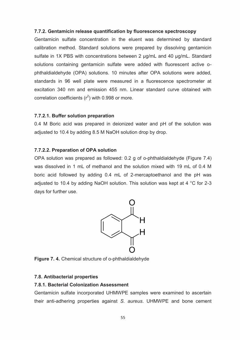
55
7.7.2. Gentamicin release quantification by fluorescence spectroscopy Gentamicin sulfate concentration in the eluent was determined by standard
calibration method. Standard solutions were prepared by dissolving gentamicin
sulfate in 1X PBS with concentrations between 2 μg/mL and 40 μg/mL. Standard
solutions containing gentamicin sulfate were added with fluorescent active o-
phthaldialdehyde (OPA) solutions. 10 minutes after OPA solutions were added,
standards in 96 well plate were measured in a fluorescence spectrometer at
excitation 340 nm and emission 455 nm. Linear standard curve obtained with
correlation coefficients (r2) with 0.998 or more.
7.7.2.1. Buffer solution preparation 0.4 M Boric acid was prepared in deionized water and pH of the solution was
adjusted to 10.4 by adding 8.5 M NaOH solution drop by drop.
7.7.2.2. Preparation of OPA solution OPA solution was prepared as followed: 0.2 g of o-phthaldialdehyde (Figure 7.4)
was dissolved in 1 mL of methanol and the solution mixed with 19 mL of 0.4 M
boric acid followed by adding 0.4 mL of 2-mercaptoethanol and the pH was
adjusted to 10.4 by adding NaOH solution. This solution was kept at 4 °C for 2-3
days for further use.
Figure 7. 4. Chemical structure of o-phthaldialdehyde
7.8. Antibacterial properties 7.8.1. Bacterial Colonization Assessment Gentamicin sulfate incorporated UHMWPE samples were examined to ascertain
their anti-adhering properties against S. aureus. UHMWPE and bone cement
56
samples (n=10) with and without gentamicin sulfate were prepared with
dimensions of 3x5x10 mm and were put in PBS (1X) in 3mL sterile syringes. The
syringes then were placed in a shaking incubator upright at 37oC. The samples
were removed from syringes at weeks 1, 2, 4, 8, 12 and placed into 1.7 mL
Eppendorf tubes individually. 1 mL of 5x10⁴ CFU/mL of Xen29 in BDTM Tryptic Soy
Broth was added to each tube and they were incubated overnight at 37oC. After
incubation strips were removed and rinsed in 5 ml of PBS and placed in new
Eppendorf tubes with 1 mL of PBS followed by 40-minute of sonication.
Subsequently, sonicated solutions were diluted to 2-10 CFU/μL then were placed
onto agar plate and spread with L-shaped bar. Upon overnight incubation at 37oC,
colonies formed were counted.
7.8.2. Antibacterial efficacy against planktonic bacteria Eluted GS aliquots were sterilized under UV for 30 minutes. Antimicrobial activity
of the samples against planktonic MSSA (ATCC 12600) was evaluated. Briefly,
bacterial isolates were cultured in tryptic soy broth (TSB) at 35 ± 2 °C until
reaching an early-stationary phase. The resulting bacterial suspension was diluted
with fresh TSB to 105 CFU/mL. Then, tested samples were transferred into 1.7 mL
sterile tubes containing 1 mL of bacterial suspension. The analyzed samples were
then statically incubated at 35 ± 2 °C. 18 hours later aliquots of the bacterial
suspension were collected and analyzed by the spread-plate method in
accordance with the International Standard ISO 4833-2. A total number of three
replicates were performed for each measurement.
7.8.3. Determination of minimum inhibitory concentration (MIC)
The antibacterial potency of gentamicin eluted from UHMWPE was evaluated
using the MIC test. Briefly, eluents were collected, and gentamicin concentration
was determined using the OPA method as outlined above. Concentrations of
gentamicin in the tested solutions were adjusted to the same level using PBS. MIC
test was conducted according to the CLSI protocol (M07-A10). Antibacterial
studies were conducted against methicillin-sensitive Staphylococcus Aureus
(MSSA, ATCC 12600). Total number of 3 replicates were measured for each
sample.
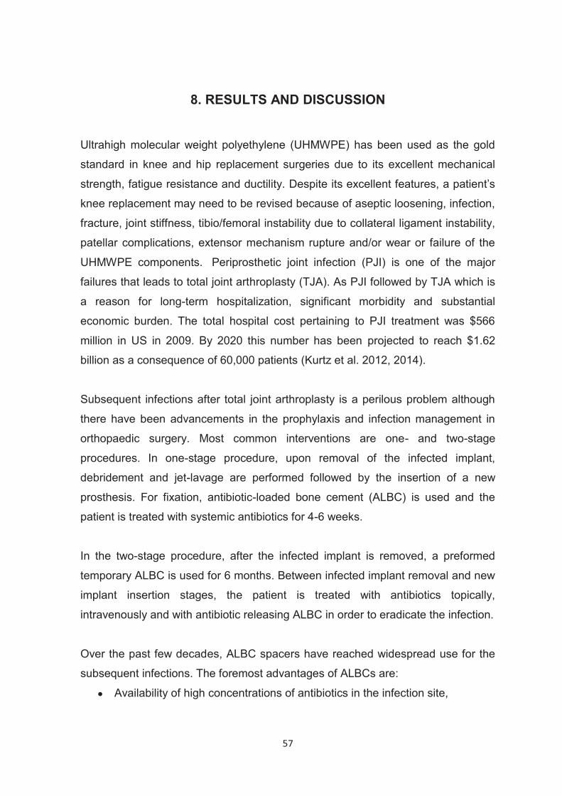
57
8. RESULTS AND DISCUSSION
Ultrahigh molecular weight polyethylene (UHMWPE) has been used as the gold
standard in knee and hip replacement surgeries due to its excellent mechanical
strength, fatigue resistance and ductility. Despite its excellent features, a patient’s
knee replacement may need to be revised because of aseptic loosening, infection,
fracture, joint stiffness, tibio/femoral instability due to collateral ligament instability,
patellar complications, extensor mechanism rupture and/or wear or failure of the
UHMWPE components. Periprosthetic joint infection (PJI) is one of the major
failures that leads to total joint arthroplasty (TJA). As PJI followed by TJA which is
a reason for long-term hospitalization, significant morbidity and substantial
economic burden. The total hospital cost pertaining to PJI treatment was $566
million in US in 2009. By 2020 this number has been projected to reach $1.62
billion as a consequence of 60,000 patients (Kurtz et al. 2012, 2014).
Subsequent infections after total joint arthroplasty is a perilous problem although
there have been advancements in the prophylaxis and infection management in
orthopaedic surgery. Most common interventions are one- and two-stage
procedures. In one-stage procedure, upon removal of the infected implant,
debridement and jet-lavage are performed followed by the insertion of a new
prosthesis. For fixation, antibiotic-loaded bone cement (ALBC) is used and the
patient is treated with systemic antibiotics for 4-6 weeks.
In the two-stage procedure, after the infected implant is removed, a preformed
temporary ALBC is used for 6 months. Between infected implant removal and new
implant insertion stages, the patient is treated with antibiotics topically,
intravenously and with antibiotic releasing ALBC in order to eradicate the infection.
Over the past few decades, ALBC spacers have reached widespread use for the
subsequent infections. The foremost advantages of ALBCs are:
● Availability of high concentrations of antibiotics in the infection site,
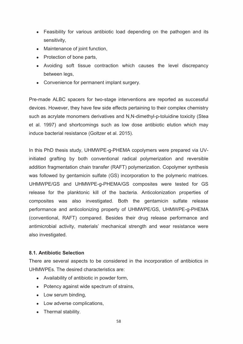
58
● Feasibility for various antibiotic load depending on the pathogen and its
sensitivity,
● Maintenance of joint function,
● Protection of bone parts,
● Avoiding soft tissue contraction which causes the level discrepancy
between legs,
● Convenience for permanent implant surgery.
Pre-made ALBC spacers for two-stage interventions are reported as successful
devices. However, they have few side effects pertaining to their complex chemistry
such as acrylate monomers derivatives and N,N-dimethyl-p-toluidine toxicity (Stea
et al. 1997) and shortcomings such as low dose antibiotic elution which may
induce bacterial resistance (Goltzer et al. 2015).
In this PhD thesis study, UHMWPE-g-PHEMA copolymers were prepared via UV-
initiated grafting by both conventional radical polymerization and reversible
addition fragmentation chain transfer (RAFT) polymerization. Copolymer synthesis
was followed by gentamicin sulfate (GS) incorporation to the polymeric matrices.
UHMWPE/GS and UHMWPE-g-PHEMA/GS composites were tested for GS
release for the planktonic kill of the bacteria. Anticolonization properties of
composites was also investigated. Both the gentamicin sulfate release
performance and anticolonizing property of UHMWPE/GS, UHMWPE-g-PHEMA
(conventional, RAFT) compared. Besides their drug release performance and
antimicrobial activity, materials’ mechanical strength and wear resistance were
also investigated.
8.1. Antibiotic Selection There are several aspects to be considered in the incorporation of antibiotics in
UHMWPEs. The desired characteristics are:
● Availability of antibiotic in powder form,
● Potency against wide spectrum of strains,
● Low serum binding,
● Low adverse complications,
● Thermal stability.
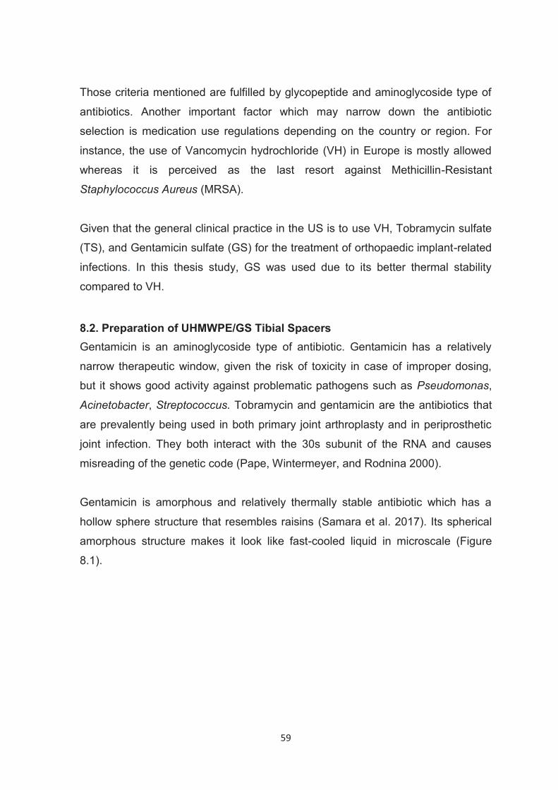
59
Those criteria mentioned are fulfilled by glycopeptide and aminoglycoside type of
antibiotics. Another important factor which may narrow down the antibiotic
selection is medication use regulations depending on the country or region. For
instance, the use of Vancomycin hydrochloride (VH) in Europe is mostly allowed
whereas it is perceived as the last resort against Methicillin-Resistant
Staphylococcus Aureus (MRSA).
Given that the general clinical practice in the US is to use VH, Tobramycin sulfate
(TS), and Gentamicin sulfate (GS) for the treatment of orthopaedic implant-related
infections. In this thesis study, GS was used due to its better thermal stability
compared to VH.
8.2. Preparation of UHMWPE/GS Tibial Spacers Gentamicin is an aminoglycoside type of antibiotic. Gentamicin has a relatively
narrow therapeutic window, given the risk of toxicity in case of improper dosing,
but it shows good activity against problematic pathogens such as Pseudomonas,
Acinetobacter, Streptococcus. Tobramycin and gentamicin are the antibiotics that
are prevalently being used in both primary joint arthroplasty and in periprosthetic
joint infection. They both interact with the 30s subunit of the RNA and causes
misreading of the genetic code (Pape, Wintermeyer, and Rodnina 2000).
Gentamicin is amorphous and relatively thermally stable antibiotic which has a
hollow sphere structure that resembles raisins (Samara et al. 2017). Its spherical
amorphous structure makes it look like fast-cooled liquid in microscale (Figure
8.1).
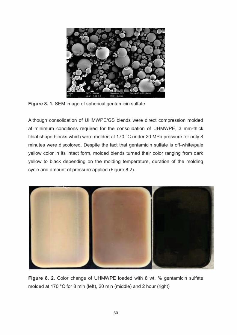
60
Figure 8. 1. SEM image of spherical gentamicin sulfate
Although consolidation of UHMWPE/GS blends were direct compression molded
at minimum conditions required for the consolidation of UHMWPE, 3 mm-thick
tibial shape blocks which were molded at 170 °C under 20 MPa pressure for only 8
minutes were discolored. Despite the fact that gentamicin sulfate is off-white/pale
yellow color in its intact form, molded blends turned their color ranging from dark
yellow to black depending on the molding temperature, duration of the molding
cycle and amount of pressure applied (Figure 8.2).
Figure 8. 2. Color change of UHMWPE loaded with 8 wt. % gentamicin sulfate
molded at 170 °C for 8 min (left), 20 min (middle) and 2 hour (right)
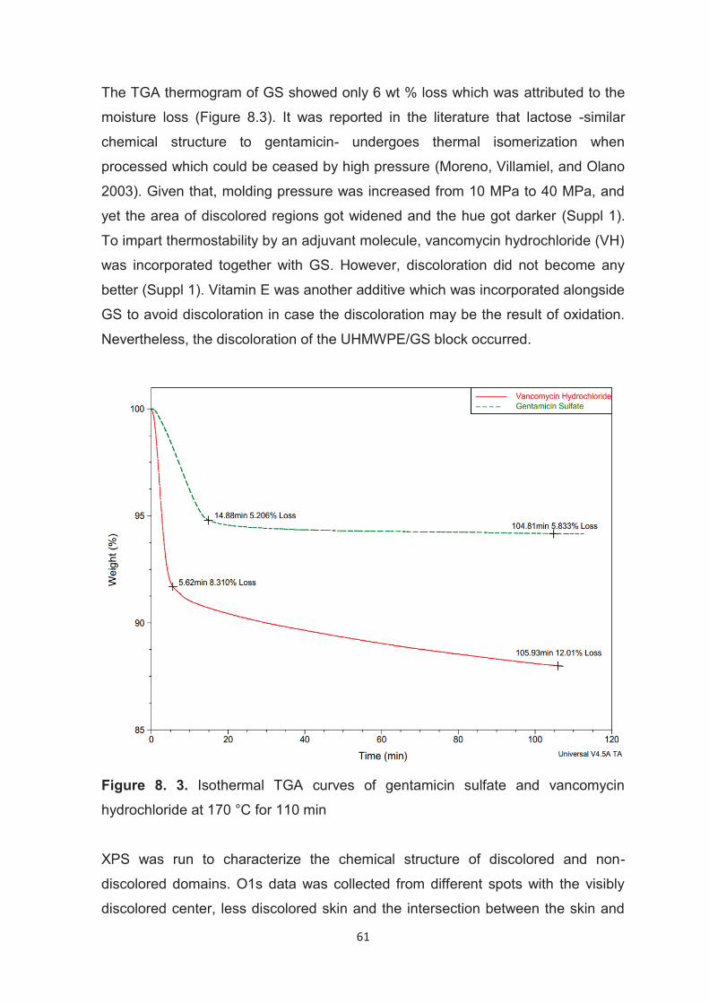
61
The TGA thermogram of GS showed only 6 wt % loss which was attributed to the
moisture loss (Figure 8.3). It was reported in the literature that lactose -similar
chemical structure to gentamicin- undergoes thermal isomerization when
processed which could be ceased by high pressure (Moreno, Villamiel, and Olano
2003). Given that, molding pressure was increased from 10 MPa to 40 MPa, and
yet the area of discolored regions got widened and the hue got darker (Suppl 1).
To impart thermostability by an adjuvant molecule, vancomycin hydrochloride (VH)
was incorporated together with GS. However, discoloration did not become any
better (Suppl 1). Vitamin E was another additive which was incorporated alongside
GS to avoid discoloration in case the discoloration may be the result of oxidation.
Nevertheless, the discoloration of the UHMWPE/GS block occurred.
Figure 8. 3. Isothermal TGA curves of gentamicin sulfate and vancomycin
hydrochloride at 170 °C for 110 min
XPS was run to characterize the chemical structure of discolored and non-
discolored domains. O1s data was collected from different spots with the visibly
discolored center, less discolored skin and the intersection between the skin and
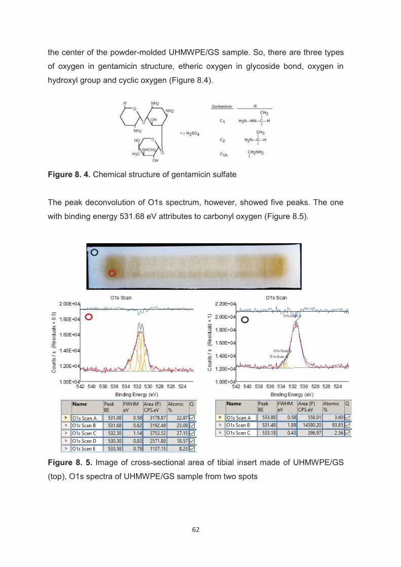
62
the center of the powder-molded UHMWPE/GS sample. So, there are three types
of oxygen in gentamicin structure, etheric oxygen in glycoside bond, oxygen in
hydroxyl group and cyclic oxygen (Figure 8.4).
Figure 8. 4. Chemical structure of gentamicin sulfate
The peak deconvolution of O1s spectrum, however, showed five peaks. The one
with binding energy 531.68 eV attributes to carbonyl oxygen (Figure 8.5).
Figure 8. 5. Image of cross-sectional area of tibial insert made of UHMWPE/GS
(top), O1s spectra of UHMWPE/GS sample from two spots
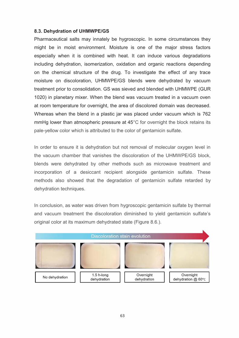
63
8.3. Dehydration of UHMWPE/GS Pharmaceutical salts may innately be hygroscopic. In some circumstances they
might be in moist environment. Moisture is one of the major stress factors
especially when it is combined with heat. It can induce various degradations
including dehydration, isomerization, oxidation and organic reactions depending
on the chemical structure of the drug. To investigate the effect of any trace
moisture on discoloration, UHMWPE/GS blends were dehydrated by vacuum
treatment prior to consolidation. GS was sieved and blended with UHMWPE (GUR
1020) in planetary mixer. When the blend was vacuum treated in a vacuum oven
at room temperature for overnight, the area of discolored domain was decreased.
Whereas when the blend in a plastic jar was placed under vacuum which is 762
mmHg lower than atmospheric pressure at 45°C for overnight the block retains its
pale-yellow color which is attributed to the color of gentamicin sulfate.
In order to ensure it is dehydration but not removal of molecular oxygen level in
the vacuum chamber that vanishes the discoloration of the UHMWPE/GS block,
blends were dehydrated by other methods such as microwave treatment and
incorporation of a desiccant recipient alongside gentamicin sulfate. These
methods also showed that the degradation of gentamicin sulfate retarded by
dehydration techniques.
In conclusion, as water was driven from hygroscopic gentamicin sulfate by thermal
and vacuum treatment the discoloration diminished to yield gentamicin sulfate’s
original color at its maximum dehydrated state (Figure 8.6.).
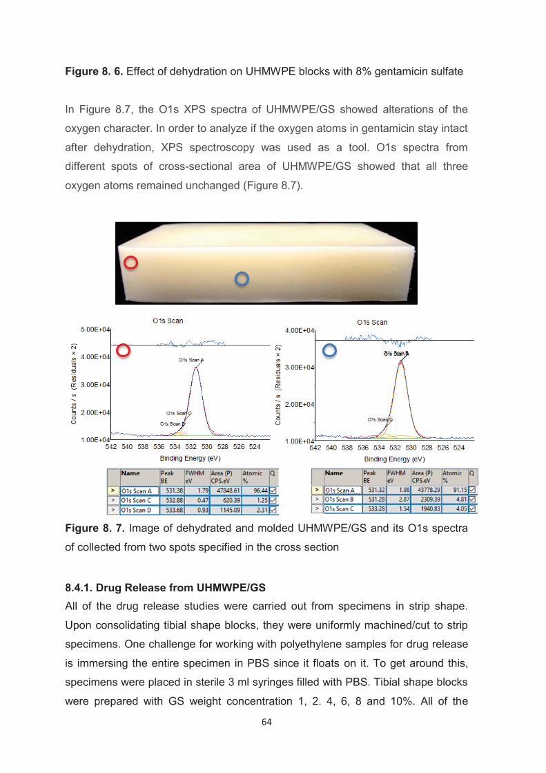
64
Figure 8. 6. Effect of dehydration on UHMWPE blocks with 8% gentamicin sulfate
In Figure 8.7, the O1s XPS spectra of UHMWPE/GS showed alterations of the
oxygen character. In order to analyze if the oxygen atoms in gentamicin stay intact
after dehydration, XPS spectroscopy was used as a tool. O1s spectra from
different spots of cross-sectional area of UHMWPE/GS showed that all three
oxygen atoms remained unchanged (Figure 8.7).
Figure 8. 7. Image of dehydrated and molded UHMWPE/GS and its O1s spectra
of collected from two spots specified in the cross section
8.4.1. Drug Release from UHMWPE/GS All of the drug release studies were carried out from specimens in strip shape.
Upon consolidating tibial shape blocks, they were uniformly machined/cut to strip
specimens. One challenge for working with polyethylene samples for drug release
is immersing the entire specimen in PBS since it floats on it. To get around this,
specimens were placed in sterile 3 ml syringes filled with PBS. Tibial shape blocks
were prepared with GS weight concentration 1, 2. 4, 6, 8 and 10%. All of the
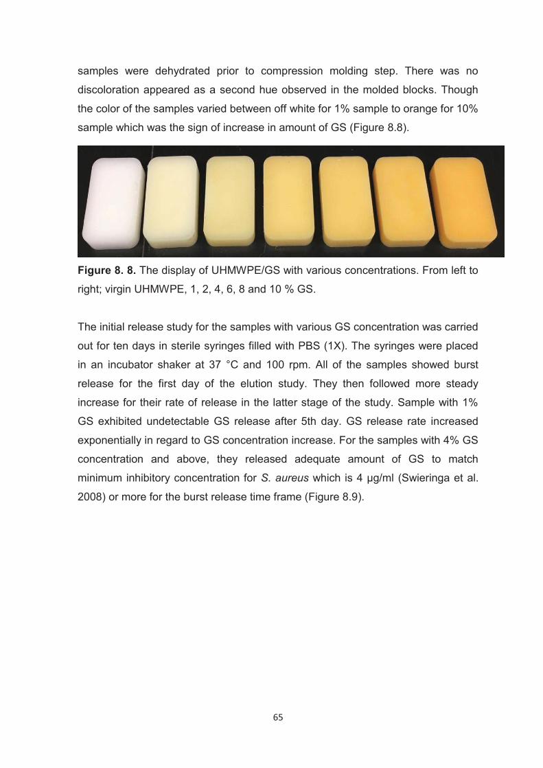
65
samples were dehydrated prior to compression molding step. There was no
discoloration appeared as a second hue observed in the molded blocks. Though
the color of the samples varied between off white for 1% sample to orange for 10%
sample which was the sign of increase in amount of GS (Figure 8.8).
Figure 8. 8. The display of UHMWPE/GS with various concentrations. From left to
right; virgin UHMWPE, 1, 2, 4, 6, 8 and 10 % GS.
The initial release study for the samples with various GS concentration was carried
out for ten days in sterile syringes filled with PBS (1X). The syringes were placed
in an incubator shaker at 37 °C and 100 rpm. All of the samples showed burst
release for the first day of the elution study. They then followed more steady
increase for their rate of release in the latter stage of the study. Sample with 1%
GS exhibited undetectable GS release after 5th day. GS release rate increased
exponentially in regard to GS concentration increase. For the samples with 4% GS
concentration and above, they released adequate amount of GS to match
minimum inhibitory concentration for S. aureus which is 4 μg/ml (Swieringa et al.
2008) or more for the burst release time frame (Figure 8.9).
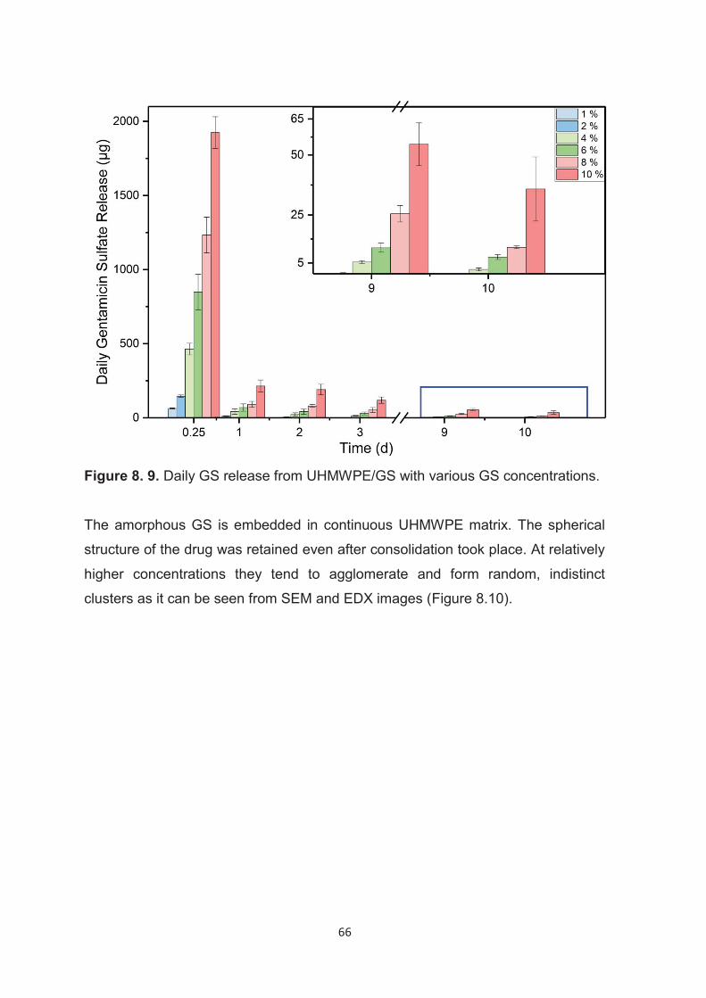
66
Figure 8. 9. Daily GS release from UHMWPE/GS with various GS concentrations.
The amorphous GS is embedded in continuous UHMWPE matrix. The spherical
structure of the drug was retained even after consolidation took place. At relatively
higher concentrations they tend to agglomerate and form random, indistinct
clusters as it can be seen from SEM and EDX images (Figure 8.10).
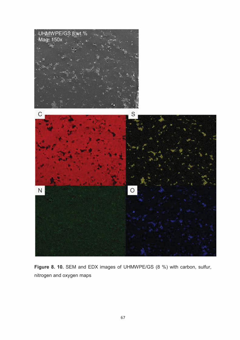
67
Figure 8. 10. SEM and EDX images of UHMWPE/GS (8 %) with carbon, sulfur,
nitrogen and oxygen maps
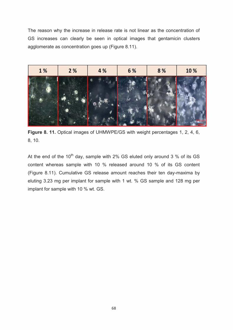
68
The reason why the increase in release rate is not linear as the concentration of
GS increases can clearly be seen in optical images that gentamicin clusters
agglomerate as concentration goes up (Figure 8.11).
Figure 8. 11. Optical images of UHMWPE/GS with weight percentages 1, 2, 4, 6,
8, 10.
At the end of the 10th day, sample with 2% GS eluted only around 3 % of its GS
content whereas sample with 10 % released around 10 % of its GS content
(Figure 8.11). Cumulative GS release amount reaches their ten day-maxima by
eluting 3.23 mg per implant for sample with 1 wt. % GS sample and 128 mg per
implant for sample with 10 % wt. GS.
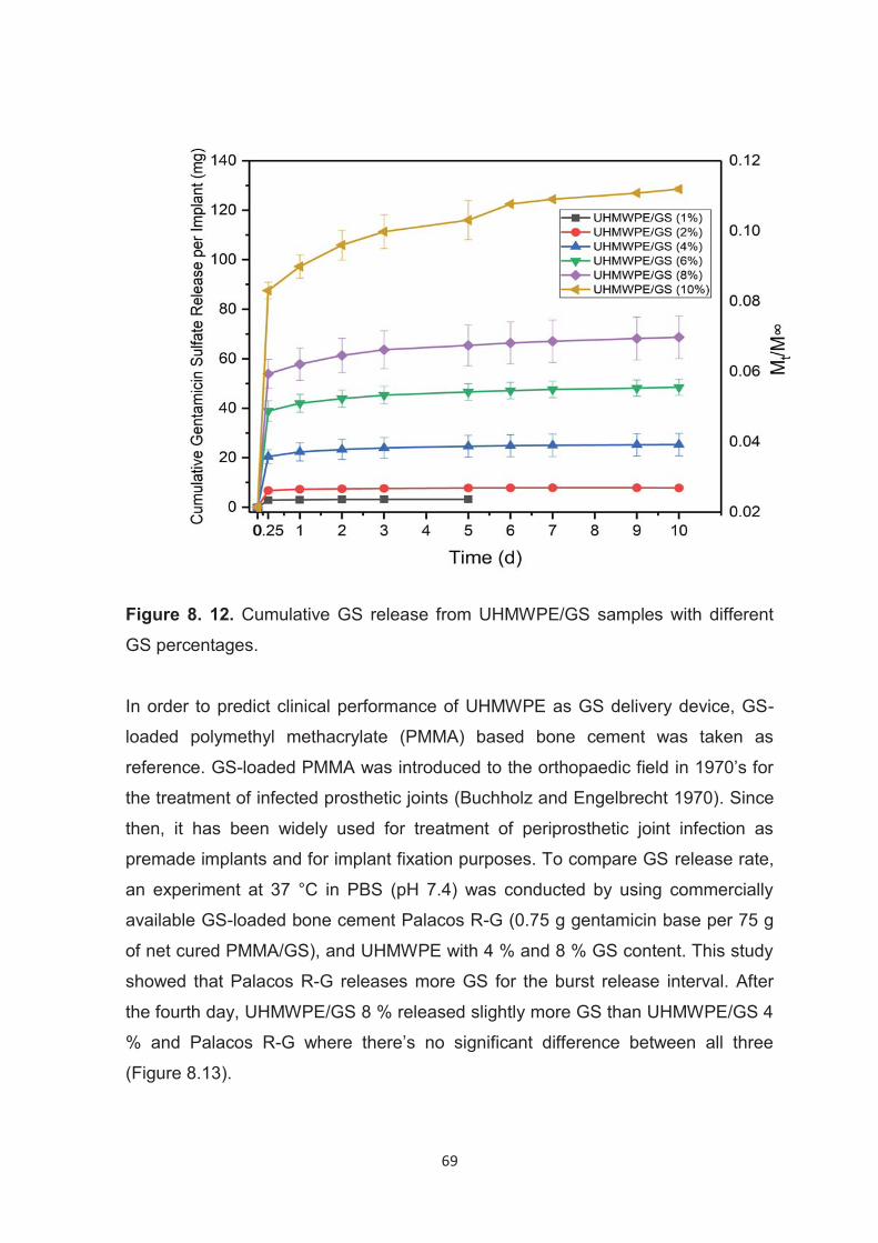
69
Figure 8. 12. Cumulative GS release from UHMWPE/GS samples with different
GS percentages.
In order to predict clinical performance of UHMWPE as GS delivery device, GS-
loaded polymethyl methacrylate (PMMA) based bone cement was taken as
reference. GS-loaded PMMA was introduced to the orthopaedic field in 1970’s for
the treatment of infected prosthetic joints (Buchholz and Engelbrecht 1970). Since
then, it has been widely used for treatment of periprosthetic joint infection as
premade implants and for implant fixation purposes. To compare GS release rate,
an experiment at 37 °C in PBS (pH 7.4) was conducted by using commercially
available GS-loaded bone cement Palacos R-G (0.75 g gentamicin base per 75 g
of net cured PMMA/GS), and UHMWPE with 4 % and 8 % GS content. This study
showed that Palacos R-G releases more GS for the burst release interval. After
the fourth day, UHMWPE/GS 8 % released slightly more GS than UHMWPE/GS 4
% and Palacos R-G where there’s no significant difference between all three
(Figure 8.13).
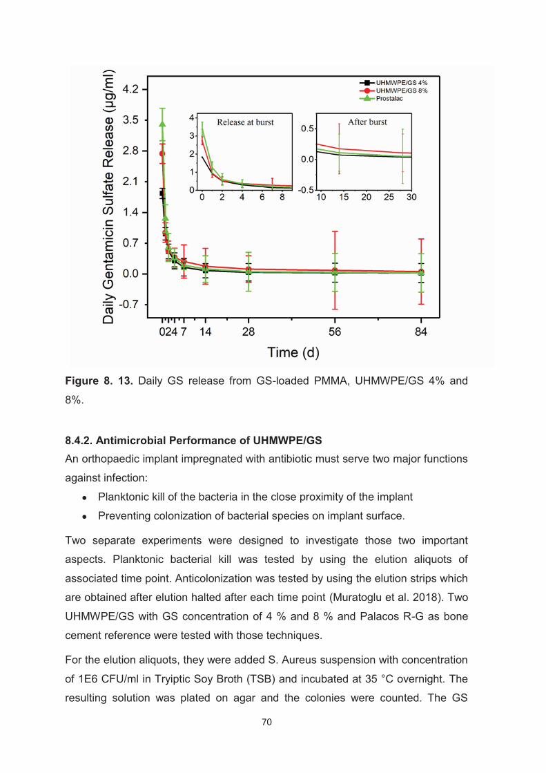
70
Figure 8. 13. Daily GS release from GS-loaded PMMA, UHMWPE/GS 4% and
8%.
8.4.2. Antimicrobial Performance of UHMWPE/GS An orthopaedic implant impregnated with antibiotic must serve two major functions
against infection:
● Planktonic kill of the bacteria in the close proximity of the implant
● Preventing colonization of bacterial species on implant surface.
Two separate experiments were designed to investigate those two important
aspects. Planktonic bacterial kill was tested by using the elution aliquots of
associated time point. Anticolonization was tested by using the elution strips which
are obtained after elution halted after each time point (Muratoglu et al. 2018). Two
UHMWPE/GS with GS concentration of 4 % and 8 % and Palacos R-G as bone
cement reference were tested with those techniques.
For the elution aliquots, they were added S. Aureus suspension with concentration
of 1E6 CFU/ml in Tryiptic Soy Broth (TSB) and incubated at 35 °C overnight. The
resulting solution was plated on agar and the colonies were counted. The GS
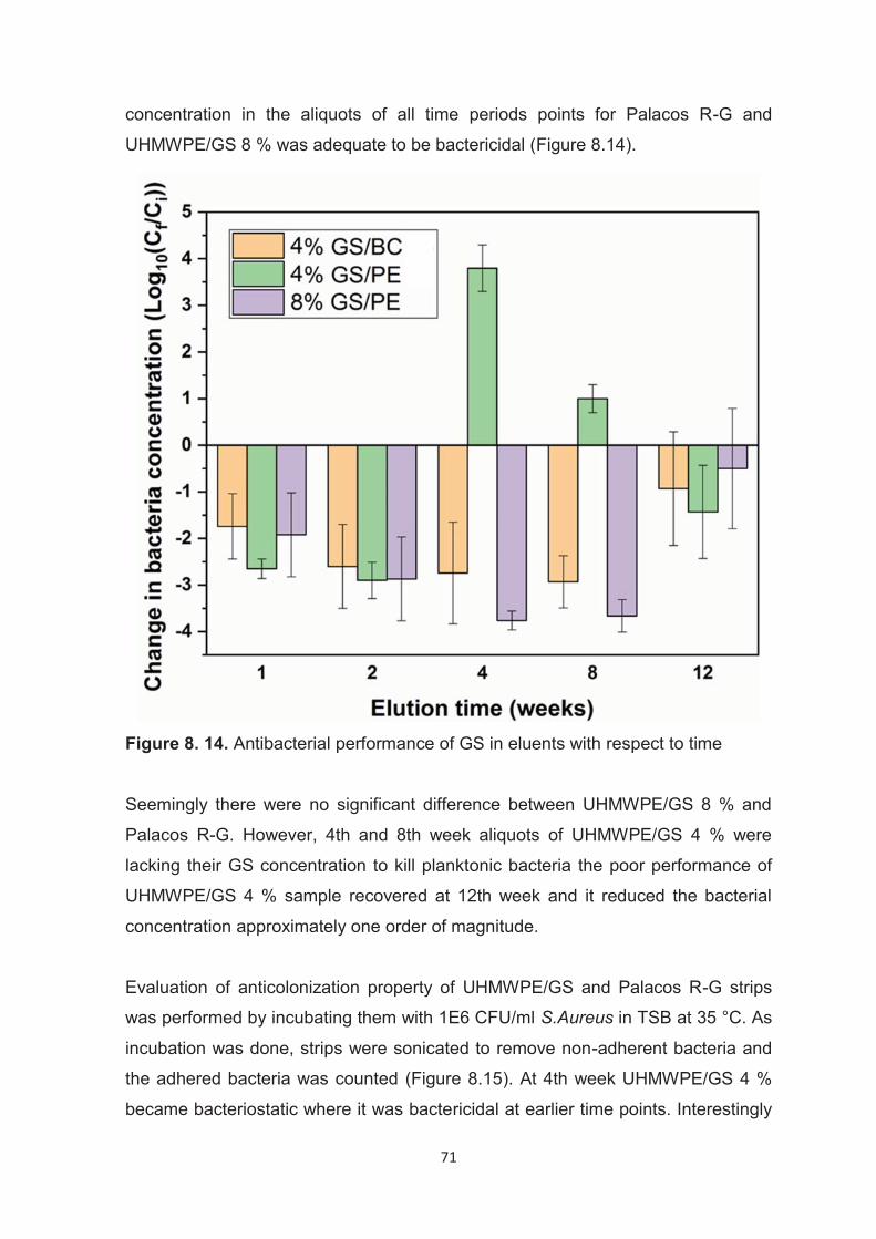
71
concentration in the aliquots of all time periods points for Palacos R-G and
UHMWPE/GS 8 % was adequate to be bactericidal (Figure 8.14).
Figure 8. 14. Antibacterial performance of GS in eluents with respect to time
Seemingly there were no significant difference between UHMWPE/GS 8 % and
Palacos R-G. However, 4th and 8th week aliquots of UHMWPE/GS 4 % were
lacking their GS concentration to kill planktonic bacteria the poor performance of
UHMWPE/GS 4 % sample recovered at 12th week and it reduced the bacterial
concentration approximately one order of magnitude.
Evaluation of anticolonization property of UHMWPE/GS and Palacos R-G strips
was performed by incubating them with 1E6 CFU/ml S.Aureus in TSB at 35 °C. As
incubation was done, strips were sonicated to remove non-adherent bacteria and
the adhered bacteria was counted (Figure 8.15). At 4th week UHMWPE/GS 4 %
became bacteriostatic where it was bactericidal at earlier time points. Interestingly
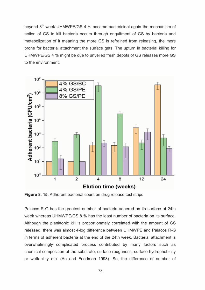
72
beyond 8th week UHMWPE/GS 4 % became bactericidal again the mechanism of
action of GS to kill bacteria occurs through engulfment of GS by bacteria and
metabolization of it meaning the more GS is refrained from releasing, the more
prone for bacterial attachment the surface gets. The upturn in bacterial killing for
UHMWPE/GS 4 % might be due to unveiled fresh depots of GS releases more GS
to the environment.
Figure 8. 15. Adherent bacterial count on drug release test strips
Palacos R-G has the greatest number of bacteria adhered on its surface at 24th
week whereas UHMWPE/GS 8 % has the least number of bacteria on its surface.
Although the planktonic kill is proportionately correlated with the amount of GS
released, there was almost 4-log difference between UHMWPE and Palacos R-G
in terms of adherent bacteria at the end of the 24th week. Bacterial attachment is
overwhelmingly complicated process contributed by many factors such as
chemical composition of the substrate, surface roughness, surface hydrophobicity
or wettability etc. (An and Friedman 1998). So, the difference of number of
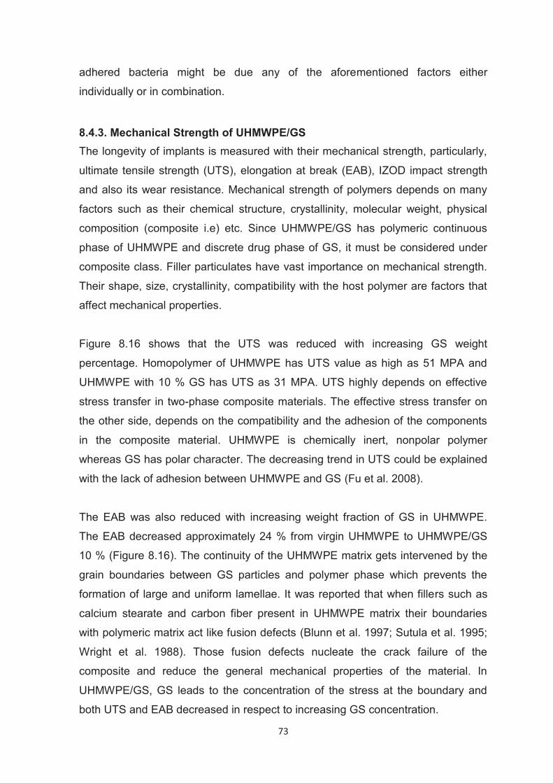
73
adhered bacteria might be due any of the aforementioned factors either
individually or in combination.
8.4.3. Mechanical Strength of UHMWPE/GS The longevity of implants is measured with their mechanical strength, particularly,
ultimate tensile strength (UTS), elongation at break (EAB), IZOD impact strength
and also its wear resistance. Mechanical strength of polymers depends on many
factors such as their chemical structure, crystallinity, molecular weight, physical
composition (composite i.e) etc. Since UHMWPE/GS has polymeric continuous
phase of UHMWPE and discrete drug phase of GS, it must be considered under
composite class. Filler particulates have vast importance on mechanical strength.
Their shape, size, crystallinity, compatibility with the host polymer are factors that
affect mechanical properties.
Figure 8.16 shows that the UTS was reduced with increasing GS weight
percentage. Homopolymer of UHMWPE has UTS value as high as 51 MPA and
UHMWPE with 10 % GS has UTS as 31 MPA. UTS highly depends on effective
stress transfer in two-phase composite materials. The effective stress transfer on
the other side, depends on the compatibility and the adhesion of the components
in the composite material. UHMWPE is chemically inert, nonpolar polymer
whereas GS has polar character. The decreasing trend in UTS could be explained
with the lack of adhesion between UHMWPE and GS (Fu et al. 2008).
The EAB was also reduced with increasing weight fraction of GS in UHMWPE.
The EAB decreased approximately 24 % from virgin UHMWPE to UHMWPE/GS
10 % (Figure 8.16). The continuity of the UHMWPE matrix gets intervened by the
grain boundaries between GS particles and polymer phase which prevents the
formation of large and uniform lamellae. It was reported that when fillers such as
calcium stearate and carbon fiber present in UHMWPE matrix their boundaries
with polymeric matrix act like fusion defects (Blunn et al. 1997; Sutula et al. 1995;
Wright et al. 1988). Those fusion defects nucleate the crack failure of the
composite and reduce the general mechanical properties of the material. In
UHMWPE/GS, GS leads to the concentration of the stress at the boundary and
both UTS and EAB decreased in respect to increasing GS concentration.
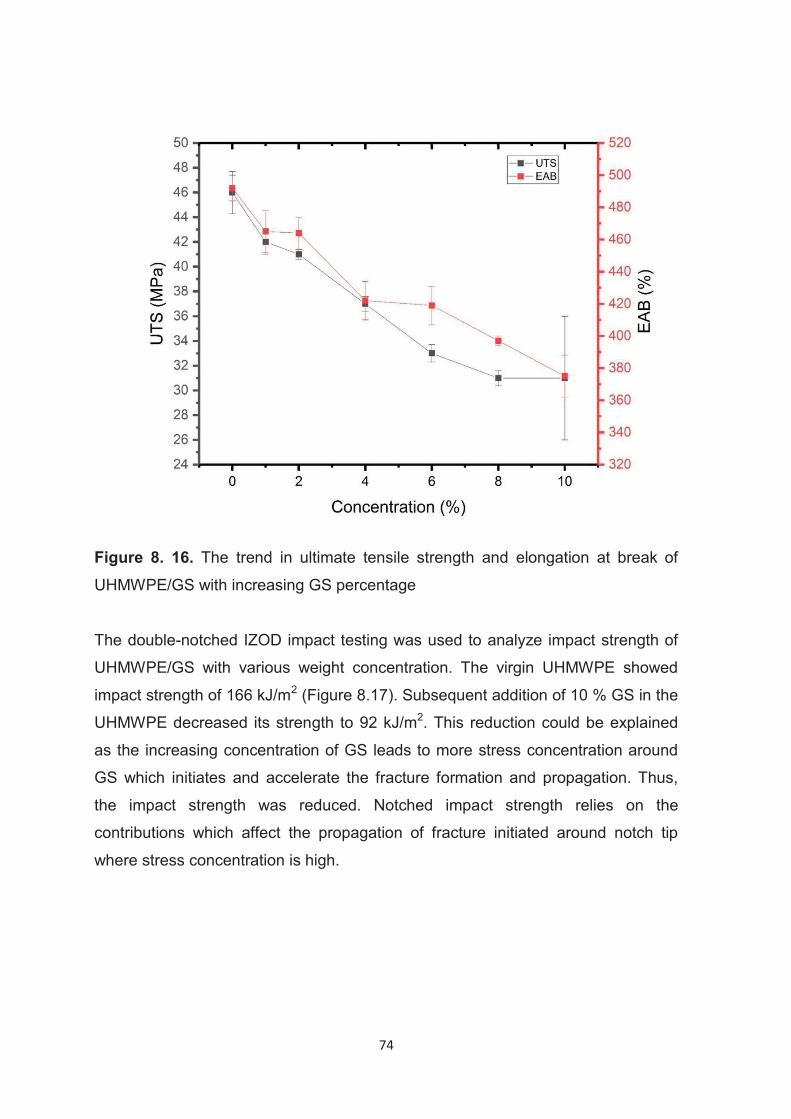
74
Figure 8. 16. The trend in ultimate tensile strength and elongation at break of
UHMWPE/GS with increasing GS percentage
The double-notched IZOD impact testing was used to analyze impact strength of
UHMWPE/GS with various weight concentration. The virgin UHMWPE showed
impact strength of 166 kJ/m2 (Figure 8.17). Subsequent addition of 10 % GS in the
UHMWPE decreased its strength to 92 kJ/m2. This reduction could be explained
as the increasing concentration of GS leads to more stress concentration around
GS which initiates and accelerate the fracture formation and propagation. Thus,
the impact strength was reduced. Notched impact strength relies on the
contributions which affect the propagation of fracture initiated around notch tip
where stress concentration is high.
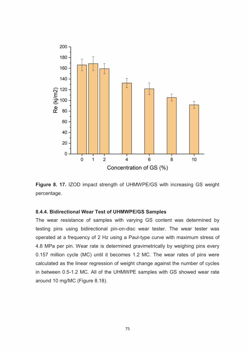
75
Figure 8. 17. IZOD impact strength of UHMWPE/GS with increasing GS weight
percentage.
8.4.4. Bidirectional Wear Test of UHMWPE/GS Samples The wear resistance of samples with varying GS content was determined by
testing pins using bidirectional pin-on-disc wear tester. The wear tester was
operated at a frequency of 2 Hz using a Paul-type curve with maximum stress of
4.8 MPa per pin. Wear rate is determined gravimetrically by weighing pins every
0.157 million cycle (MC) until it becomes 1.2 MC. The wear rates of pins were
calculated as the linear regression of weight change against the number of cycles
in between 0.5-1.2 MC. All of the UHMWPE samples with GS showed wear rate
around 10 mg/MC (Figure 8.18).
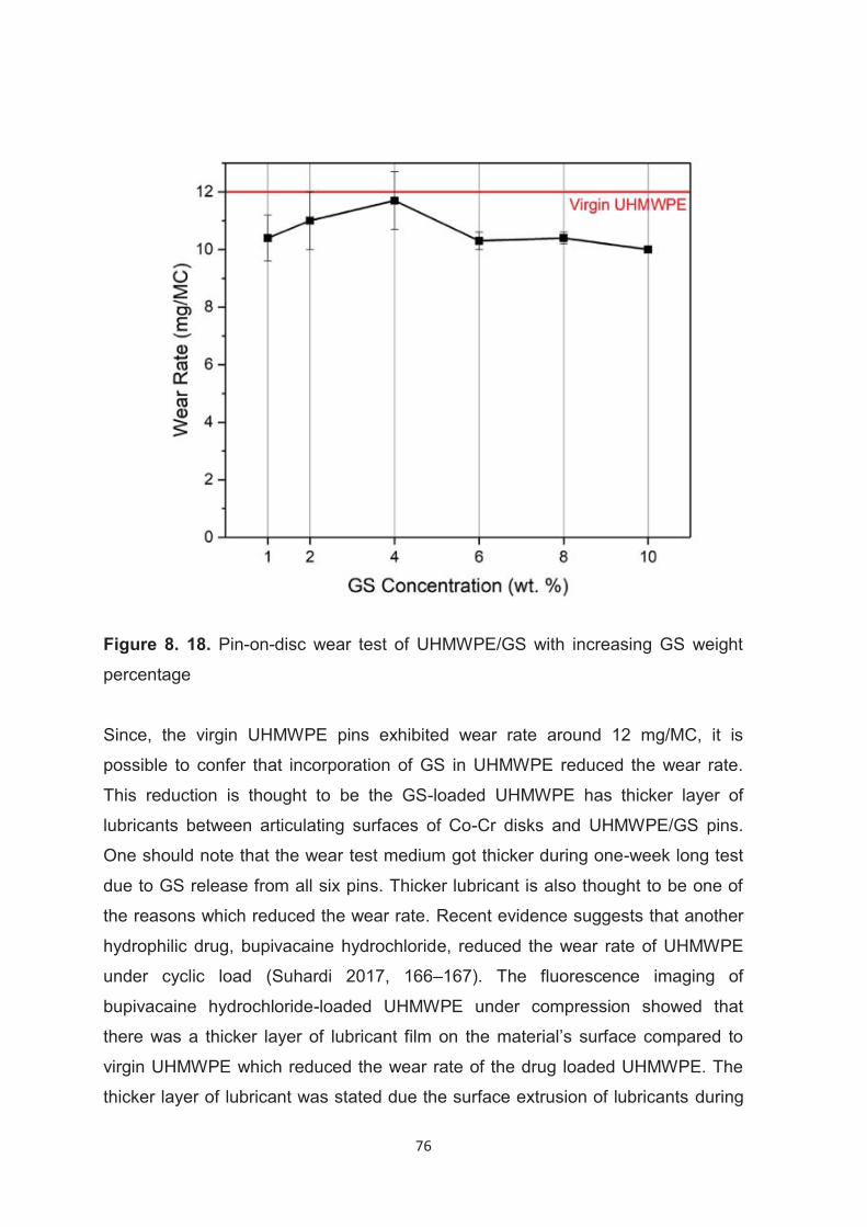
76
Figure 8. 18. Pin-on-disc wear test of UHMWPE/GS with increasing GS weight
percentage
Since, the virgin UHMWPE pins exhibited wear rate around 12 mg/MC, it is
possible to confer that incorporation of GS in UHMWPE reduced the wear rate.
This reduction is thought to be the GS-loaded UHMWPE has thicker layer of
lubricants between articulating surfaces of Co-Cr disks and UHMWPE/GS pins.
One should note that the wear test medium got thicker during one-week long test
due to GS release from all six pins. Thicker lubricant is also thought to be one of
the reasons which reduced the wear rate. Recent evidence suggests that another
hydrophilic drug, bupivacaine hydrochloride, reduced the wear rate of UHMWPE
under cyclic load (Suhardi 2017, 166–167). The fluorescence imaging of
bupivacaine hydrochloride-loaded UHMWPE under compression showed that
there was a thicker layer of lubricant film on the material’s surface compared to
virgin UHMWPE which reduced the wear rate of the drug loaded UHMWPE. The
thicker layer of lubricant was stated due the surface extrusion of lubricants during
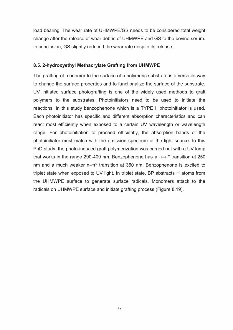
77
load bearing. The wear rate of UHMWPE/GS needs to be considered total weight
change after the release of wear debris of UHMWPE and GS to the bovine serum.
In conclusion, GS slightly reduced the wear rate despite its release.
8.5. 2-hydroxyethyl Methacrylate Grafting from UHMWPE
The grafting of monomer to the surface of a polymeric substrate is a versatile way
to change the surface properties and to functionalize the surface of the substrate.
UV initiated surface photografting is one of the widely used methods to graft
polymers to the substrates. Photoinitiators need to be used to initiate the
reactions. In this study benzophenone which is a TYPE II photoinitiator is used.
Each photoinitiator has specific and different absorption characteristics and can
react most efficiently when exposed to a certain UV wavelength or wavelength
range. For photoinitiation to proceed efficiently, the absorption bands of the
photoinitiator must match with the emission spectrum of the light source. In this
PhD study, the photo-induced graft polymerization was carried out with a UV lamp
that works in the range 290-400 nm. Benzophenone has a π–π* transition at 250
nm and a much weaker n–π* transition at 350 nm. Benzophenone is excited to
triplet state when exposed to UV light. In triplet state, BP abstracts H atoms from
the UHMWPE surface to generate surface radicals. Monomers attack to the
radicals on UHMWPE surface and initiate grafting process (Figure 8.19).
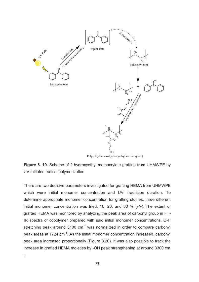
78
Figure 8. 19. Scheme of 2-hydroxyethyl methacrylate grafting from UHMWPE by
UV-initiated radical polymerization
There are two decisive parameters investigated for grafting HEMA from UHMWPE
which were initial monomer concentration and UV irradiation duration. To
determine appropriate monomer concentration for grafting studies, three different
initial monomer concentration was tried; 10, 20, and 30 % (v/v). The extent of
grafted HEMA was monitored by analyzing the peak area of carbonyl group in FT-
IR spectra of copolymer prepared with said initial monomer concentrations. C-H
stretching peak around 3100 cm-1 was normalized in order to compare carbonyl
peak areas at 1724 cm-1. As the initial monomer concentration increased, carbonyl
peak area increased proportionally (Figure 8.20). It was also possible to track the
increase in grafted HEMA moieties by -OH peak strengthening at around 3300 cm-
1.
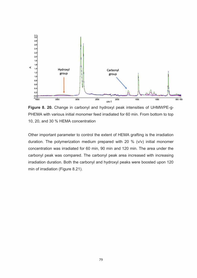
79
Figure 8. 20. Change in carbonyl and hydroxyl peak intensities of UHMWPE-g-
PHEMA with various initial monomer feed irradiated for 60 min. From bottom to top
10, 20, and 30 % HEMA concentration
Other important parameter to control the extent of HEMA grafting is the irradiation
duration. The polymerization medium prepared with 20 % (v/v) initial monomer
concentration was irradiated for 60 min, 90 min and 120 min. The area under the
carbonyl peak was compared. The carbonyl peak area increased with increasing
irradiation duration. Both the carbonyl and hydroxyl peaks were boosted upon 120
min of irradiation (Figure 8.21).
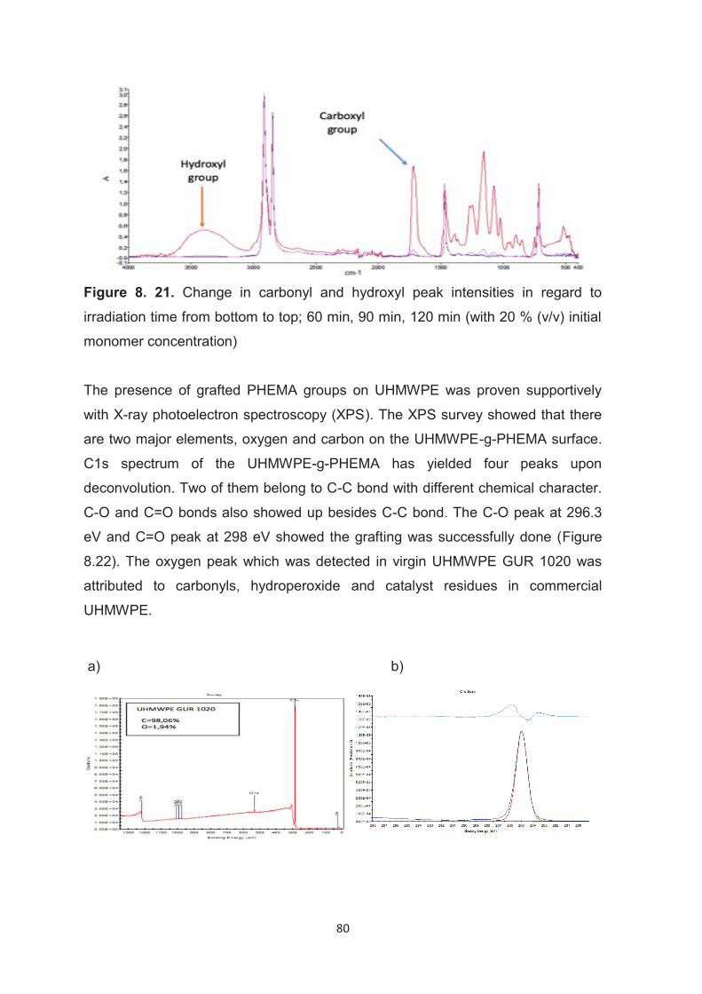
80
Figure 8. 21. Change in carbonyl and hydroxyl peak intensities in regard to
irradiation time from bottom to top; 60 min, 90 min, 120 min (with 20 % (v/v) initial
monomer concentration) The presence of grafted PHEMA groups on UHMWPE was proven supportively
with X-ray photoelectron spectroscopy (XPS). The XPS survey showed that there
are two major elements, oxygen and carbon on the UHMWPE-g-PHEMA surface.
C1s spectrum of the UHMWPE-g-PHEMA has yielded four peaks upon
deconvolution. Two of them belong to C-C bond with different chemical character.
C-O and C=O bonds also showed up besides C-C bond. The C-O peak at 296.3
eV and C=O peak at 298 eV showed the grafting was successfully done (Figure
8.22). The oxygen peak which was detected in virgin UHMWPE GUR 1020 was
attributed to carbonyls, hydroperoxide and catalyst residues in commercial
UHMWPE.
a) b)
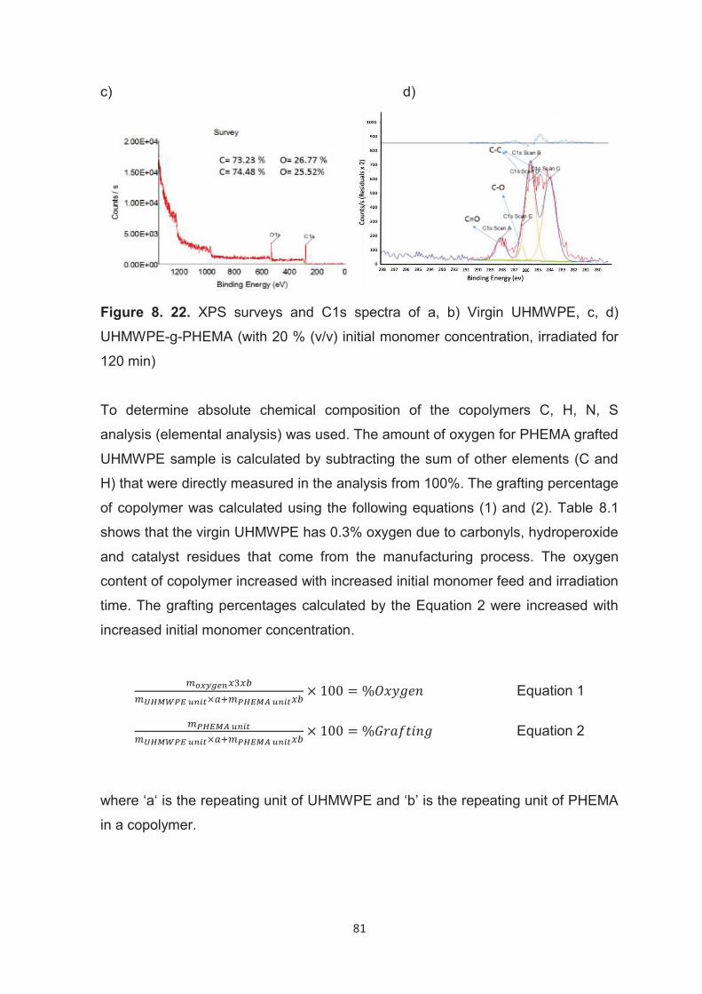
81
c) d)
Figure 8. 22. XPS surveys and C1s spectra of a, b) Virgin UHMWPE, c, d)
UHMWPE-g-PHEMA (with 20 % (v/v) initial monomer concentration, irradiated for
120 min)
To determine absolute chemical composition of the copolymers C, H, N, S
analysis (elemental analysis) was used. The amount of oxygen for PHEMA grafted
UHMWPE sample is calculated by subtracting the sum of other elements (C and
H) that were directly measured in the analysis from 100%. The grafting percentage
of copolymer was calculated using the following equations (1) and (2). Table 8.1
shows that the virgin UHMWPE has 0.3% oxygen due to carbonyls, hydroperoxide
and catalyst residues that come from the manufacturing process. The oxygen
content of copolymer increased with increased initial monomer feed and irradiation
time. The grafting percentages calculated by the Equation 2 were increased with
increased initial monomer concentration.
Equation 1
Equation 2
where ‘a‘ is the repeating unit of UHMWPE and ‘b’ is the repeating unit of PHEMA
in a copolymer.
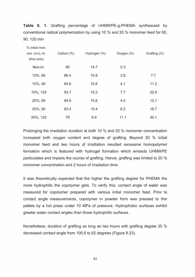
82
Table 8. 1. Grafting percentage of UHMWPE-g-PHEMA synthesized by
conventional radical polymerization by using 10 % and 20 % monomer feed for 60,
90, 120 min
% Initial mon.
con. (v/v), irr.
time (min)
Carbon (%) Hydrogen (%) Oxygen (%) Grafting (%)
Non-irr. 85 14.7 0.3
10%, 60 86.4 10.8 2.8 7.7
10%, 90 84.6 10.6 4.1 11.2
10%, 120 83.7 10.2 7.7 20.8
20%, 60 84.9 10.6 4.5 12.1
20%, 90 83.4 10.4 6.2 16.7
20%, 120 79 9.9 11.1 30.1
Prolonging the irradiation duration at both 10 % and 20 % monomer concentration
increased both oxygen content and degree of grafting. Beyond 20 % initial
monomer feed and two hours of irradiation resulted excessive homopolymer
formation which is featured with hydrogel formation which embeds UHMWPE
particulates and impairs the course of grafting. Hence, grafting was limited to 20 %
monomer concentration and 2 hours of irradiation time.
It was theoretically expected that the higher the grafting degree for PHEMA the
more hydrophilic the copolymer gets. To verify this, contact angle of water was
measured for copolymer prepared with various initial monomer feed. Prior to
contact angle measurements, copolymer in powder form was pressed to thin
pellets by a hot press under 10 MPa of pressure. Hydrophobic surfaces exhibit
greater water contact angles than those hydrophilic surfaces.
Nonetheless, duration of grafting as long as two hours with grafting degree 30 %
decreased contact angle from 100.6 to 62 degrees (Figure 8.23).
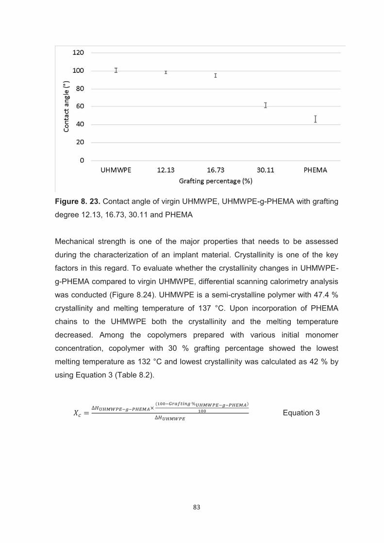
83
Figure 8. 23. Contact angle of virgin UHMWPE, UHMWPE-g-PHEMA with grafting
degree 12.13, 16.73, 30.11 and PHEMA Mechanical strength is one of the major properties that needs to be assessed
during the characterization of an implant material. Crystallinity is one of the key
factors in this regard. To evaluate whether the crystallinity changes in UHMWPE-
g-PHEMA compared to virgin UHMWPE, differential scanning calorimetry analysis
was conducted (Figure 8.24). UHMWPE is a semi-crystalline polymer with 47.4 %
crystallinity and melting temperature of 137 °C. Upon incorporation of PHEMA
chains to the UHMWPE both the crystallinity and the melting temperature
decreased. Among the copolymers prepared with various initial monomer
concentration, copolymer with 30 % grafting percentage showed the lowest
melting temperature as 132 °C and lowest crystallinity was calculated as 42 % by
using Equation 3 (Table 8.2).
Equation 3
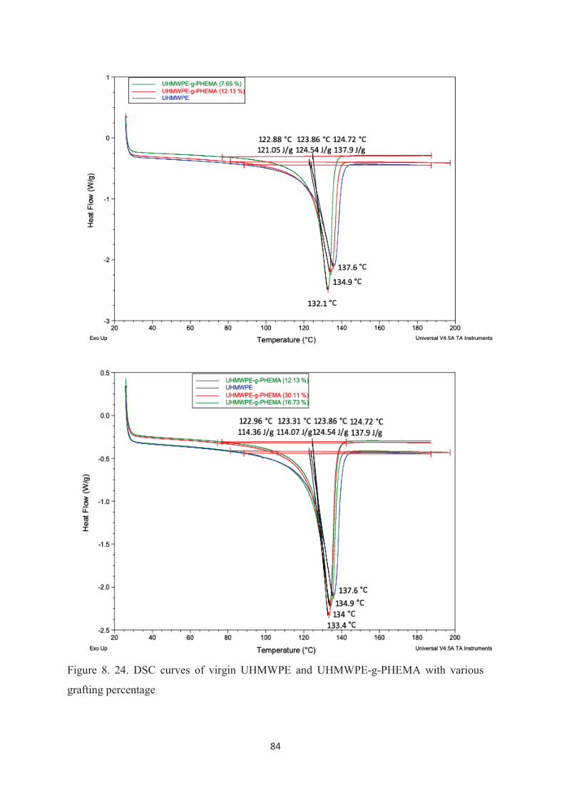
84
Figure 8. 24. DSC curves of virgin UHMWPE and UHMWPE-g-PHEMA with various
grafting percentage
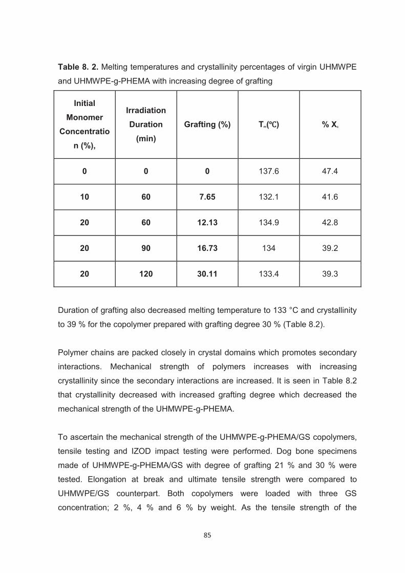
85
Table 8. 2. Melting temperatures and crystallinity percentages of virgin UHMWPE
and UHMWPE-g-PHEMA with increasing degree of grafting
Initial Monomer
Concentration (%),
Irradiation Duration
(min) Grafting (%) Tm( ) % Xc
0 0 0 137.6 47.4
10 60 7.65 132.1 41.6
20 60 12.13 134.9 42.8
20 90 16.73 134 39.2
20 120 30.11 133.4 39.3
Duration of grafting also decreased melting temperature to 133 °C and crystallinity
to 39 % for the copolymer prepared with grafting degree 30 % (Table 8.2).
Polymer chains are packed closely in crystal domains which promotes secondary
interactions. Mechanical strength of polymers increases with increasing
crystallinity since the secondary interactions are increased. It is seen in Table 8.2
that crystallinity decreased with increased grafting degree which decreased the
mechanical strength of the UHMWPE-g-PHEMA.
To ascertain the mechanical strength of the UHMWPE-g-PHEMA/GS copolymers,
tensile testing and IZOD impact testing were performed. Dog bone specimens
made of UHMWPE-g-PHEMA/GS with degree of grafting 21 % and 30 % were
tested. Elongation at break and ultimate tensile strength were compared to
UHMWPE/GS counterpart. Both copolymers were loaded with three GS
concentration; 2 %, 4 % and 6 % by weight. As the tensile strength of the
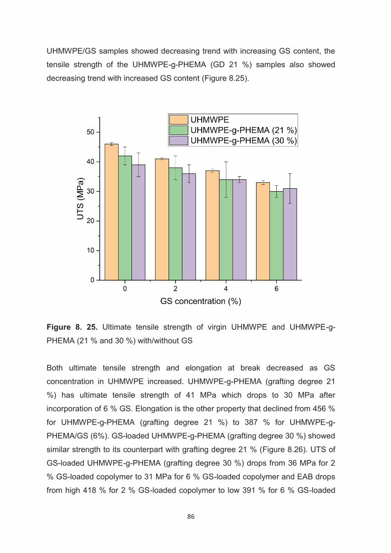
86
UHMWPE/GS samples showed decreasing trend with increasing GS content, the
tensile strength of the UHMWPE-g-PHEMA (GD 21 %) samples also showed
decreasing trend with increased GS content (Figure 8.25).
Figure 8. 25. Ultimate tensile strength of virgin UHMWPE and UHMWPE-g-
PHEMA (21 % and 30 %) with/without GS
Both ultimate tensile strength and elongation at break decreased as GS
concentration in UHMWPE increased. UHMWPE-g-PHEMA (grafting degree 21
%) has ultimate tensile strength of 41 MPa which drops to 30 MPa after
incorporation of 6 % GS. Elongation is the other property that declined from 456 %
for UHMWPE-g-PHEMA (grafting degree 21 %) to 387 % for UHMWPE-g-
PHEMA/GS (6%). GS-loaded UHMWPE-g-PHEMA (grafting degree 30 %) showed
similar strength to its counterpart with grafting degree 21 % (Figure 8.26). UTS of
GS-loaded UHMWPE-g-PHEMA (grafting degree 30 %) drops from 36 MPa for 2
% GS-loaded copolymer to 31 MPa for 6 % GS-loaded copolymer and EAB drops
from high 418 % for 2 % GS-loaded copolymer to low 391 % for 6 % GS-loaded
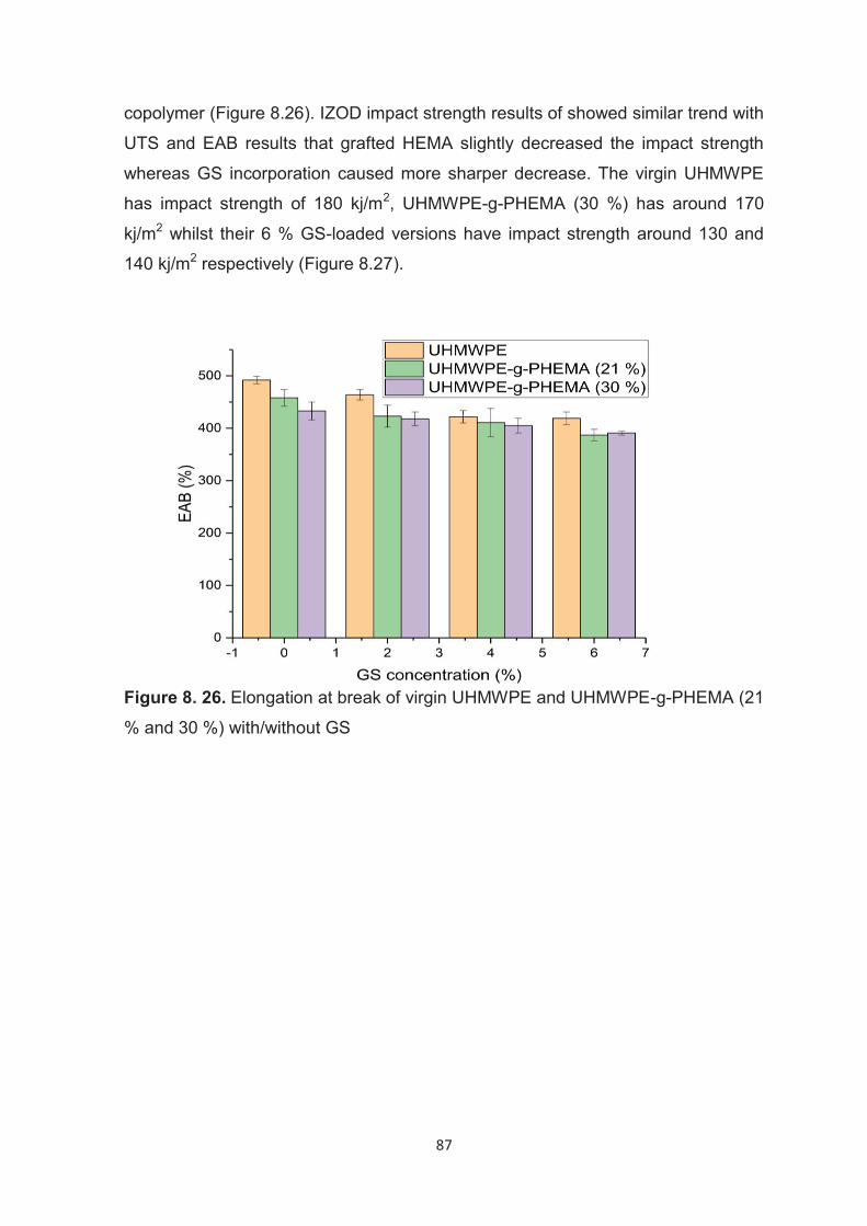
87
copolymer (Figure 8.26). IZOD impact strength results of showed similar trend with
UTS and EAB results that grafted HEMA slightly decreased the impact strength
whereas GS incorporation caused more sharper decrease. The virgin UHMWPE
has impact strength of 180 kj/m2, UHMWPE-g-PHEMA (30 %) has around 170
kj/m2 whilst their 6 % GS-loaded versions have impact strength around 130 and
140 kj/m2 respectively (Figure 8.27).
Figure 8. 26. Elongation at break of virgin UHMWPE and UHMWPE-g-PHEMA (21
% and 30 %) with/without GS
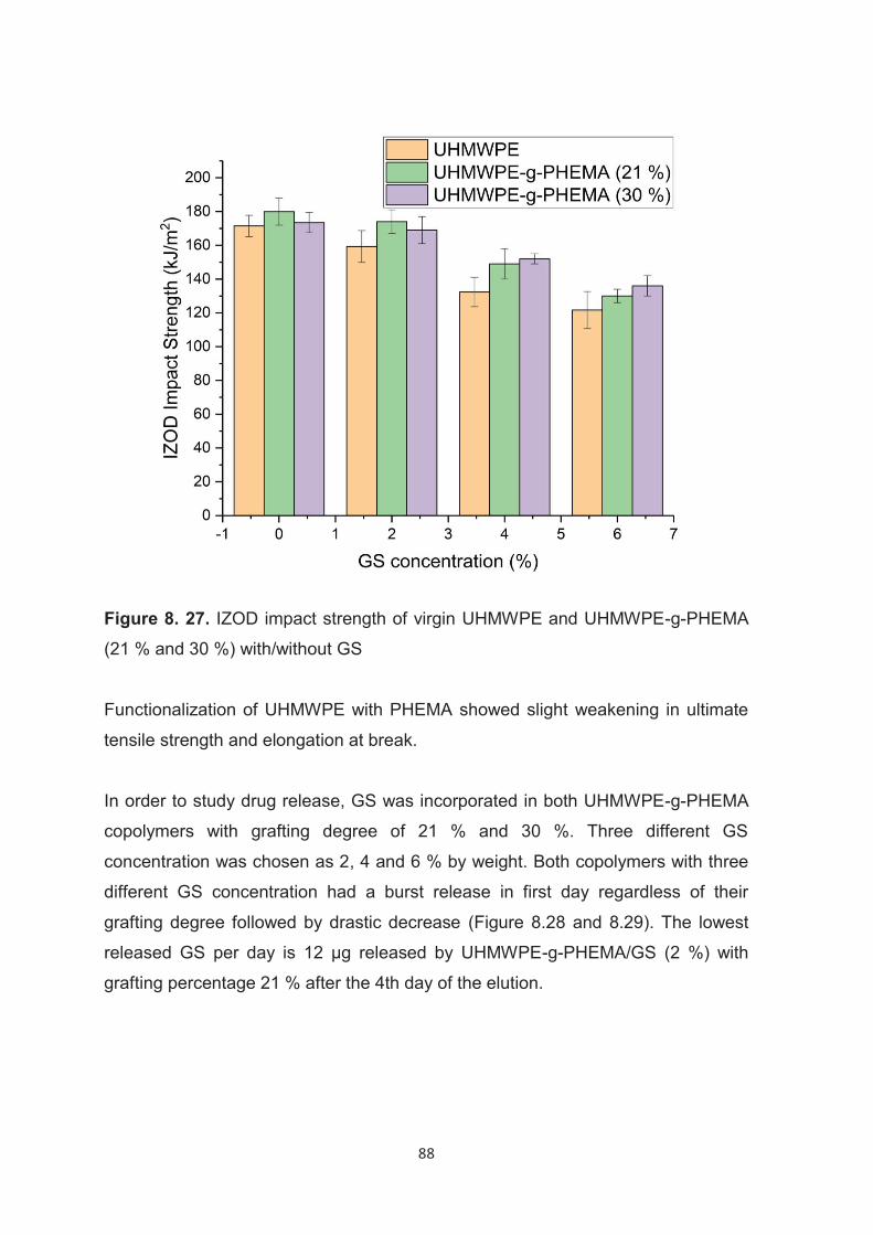
88
Figure 8. 27. IZOD impact strength of virgin UHMWPE and UHMWPE-g-PHEMA
(21 % and 30 %) with/without GS
Functionalization of UHMWPE with PHEMA showed slight weakening in ultimate
tensile strength and elongation at break.
In order to study drug release, GS was incorporated in both UHMWPE-g-PHEMA
copolymers with grafting degree of 21 % and 30 %. Three different GS
concentration was chosen as 2, 4 and 6 % by weight. Both copolymers with three
different GS concentration had a burst release in first day regardless of their
grafting degree followed by drastic decrease (Figure 8.28 and 8.29). The lowest
released GS per day is 12 μg released by UHMWPE-g-PHEMA/GS (2 %) with
grafting percentage 21 % after the 4th day of the elution.
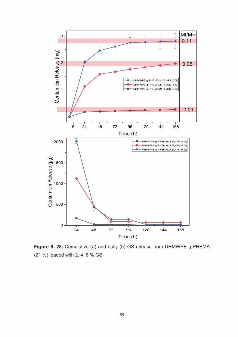
89
Figure 8. 28. Cumulative (a) and daily (b) GS release from UHMWPE-g-PHEMA
(21 %) loaded with 2, 4, 6 % GS
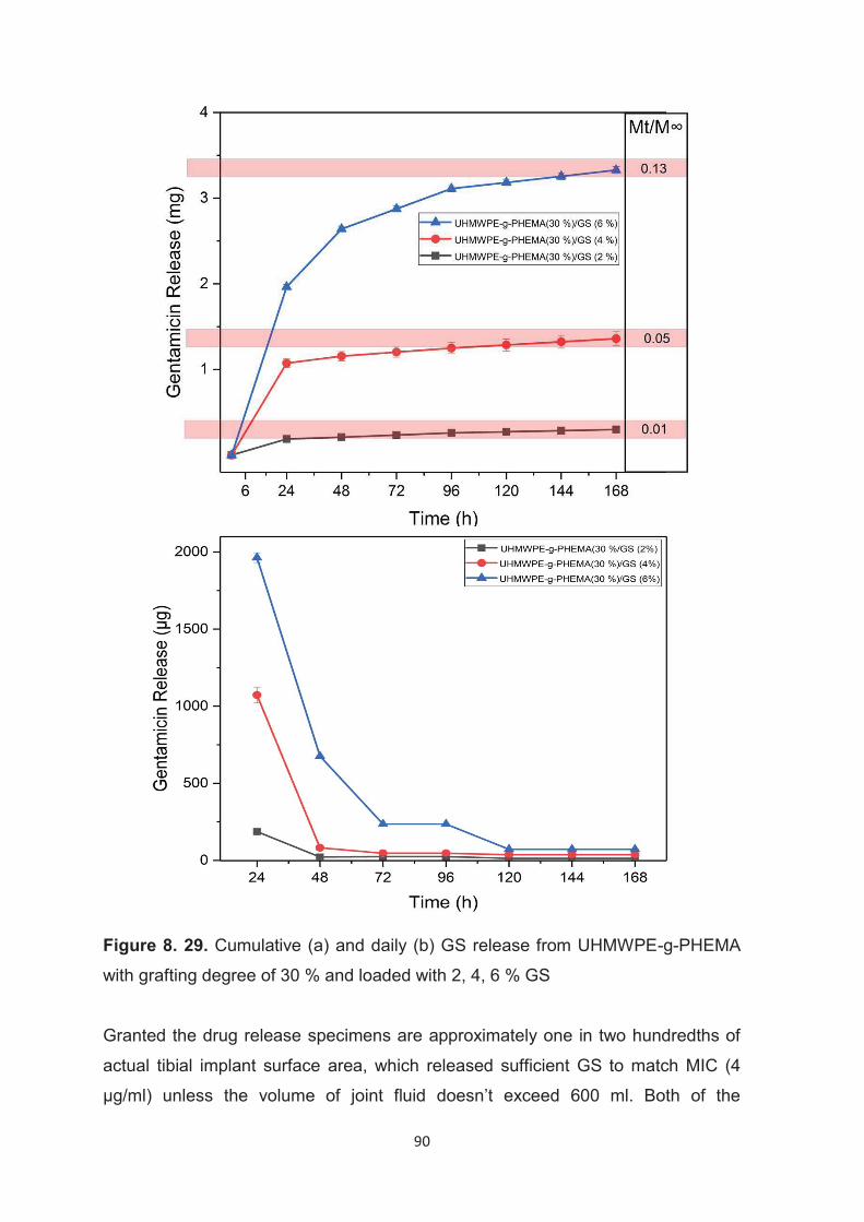
90
Figure 8. 29. Cumulative (a) and daily (b) GS release from UHMWPE-g-PHEMA
with grafting degree of 30 % and loaded with 2, 4, 6 % GS
Granted the drug release specimens are approximately one in two hundredths of
actual tibial implant surface area, which released sufficient GS to match MIC (4
μg/ml) unless the volume of joint fluid doesn’t exceed 600 ml. Both of the
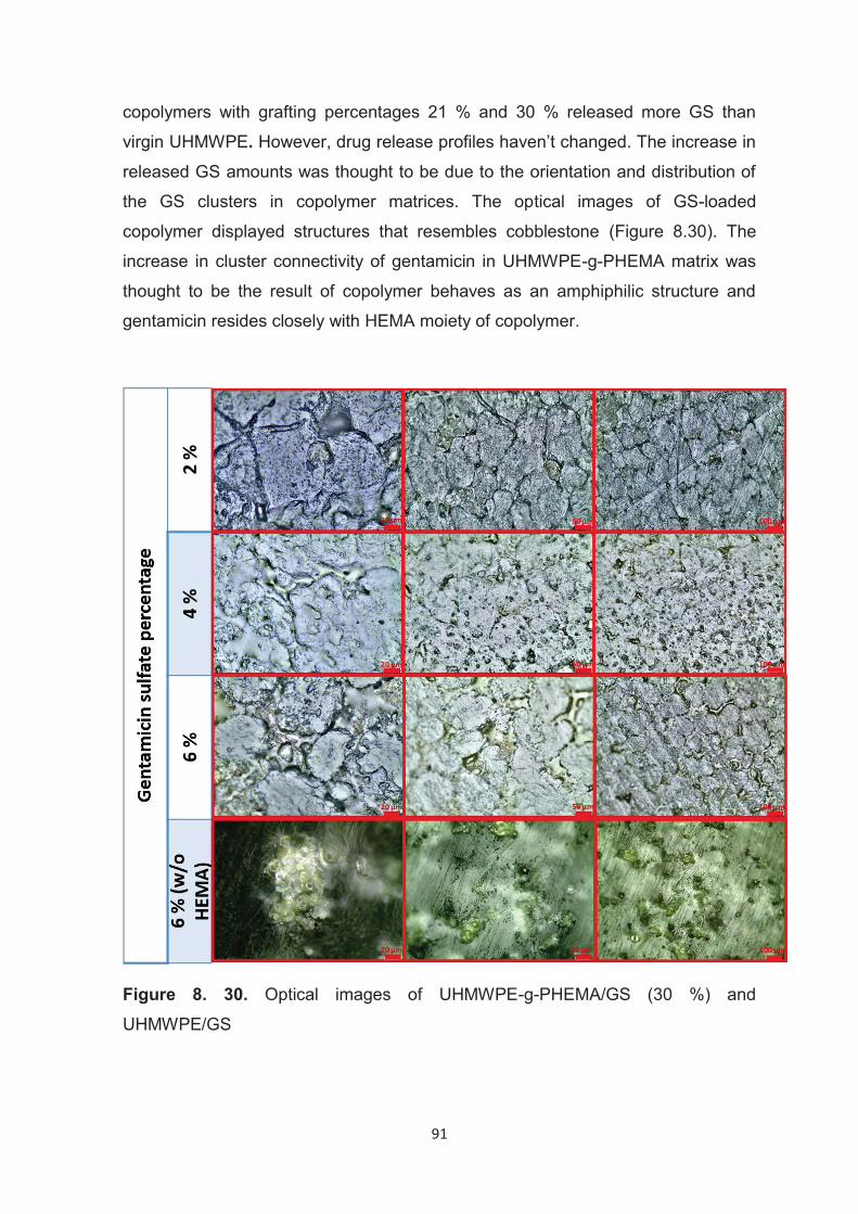
91
copolymers with grafting percentages 21 % and 30 % released more GS than
virgin UHMWPE. However, drug release profiles haven’t changed. The increase in
released GS amounts was thought to be due to the orientation and distribution of
the GS clusters in copolymer matrices. The optical images of GS-loaded
copolymer displayed structures that resembles cobblestone (Figure 8.30). The
increase in cluster connectivity of gentamicin in UHMWPE-g-PHEMA matrix was
thought to be the result of copolymer behaves as an amphiphilic structure and
gentamicin resides closely with HEMA moiety of copolymer.
Figure 8. 30. Optical images of UHMWPE-g-PHEMA/GS (30 %) and
UHMWPE/GS
92
8.6. 2-hydroxyethyl Methacrylate Grafting from UHMWPE via RAFT Polymerization Introduction and advancement of living radical polymerization (LRP) techniques
recently represented a major avenue in polymer chemistry. These techniques
brought countless opportunities to synthesize tailor-made structures, thereby it is
likely to have significant progress in future applications of polymeric materials.
Adaption of CRP techniques to industry depends on their feasibility, versatility and
their extra cost for the target material. Among those CRP techniques, RAFT
polymerization has particular importance since it combines the advantages of
conventional polymerization and CRP. The RAFT polymerization could be applied
with many monomers. The RAFT polymerization is also very effective on
controlling average molecular weight and molecular weight distribution. It is also
useful to control chain-end functionality and macromolecular architecture. In this
study, RAFT polymerization was particularly used to control molecular weight and
its distribution so the interaction between gentamicin sulfate and the polymeric
matrix can be improved which would enable the release of gentamicin sulfate in a
controlled manner. More specifically, 2-hydroxyethyl methacrylate is grafted from
UHMWPE and copolymer was incorporated with gentamicin sulfate. Given the
functional groups of P(HEMA) of UHMWPE-g-P(HEMA) (UHMWPE-g-PHEMA
prepared via RAFT polymerization is denoted as R-UHMWPE-g-PHEMA beyond
this part) and gentamicin sulfate will have secondary interactions, release
mechanism was investigated.
Some of the RAFT agents have chromophore moieties that absorb UV light (Lu et
al. 2006). It was reported that DDMAT has two absorption peaks in UV region
(Figure 8.31) (Bakar Atıcı 2018).
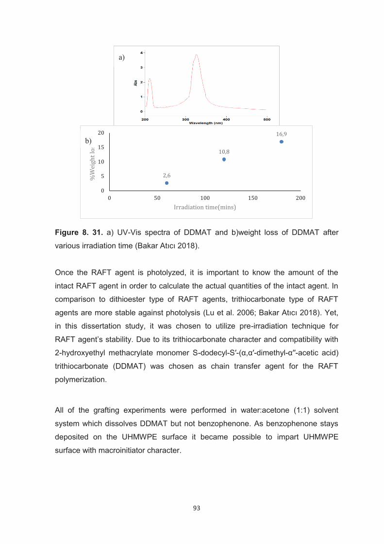
93
Figure 8. 31. a) UV-Vis spectra of DDMAT and b)weight loss of DDMAT after
various irradiation time (Bakar Atıcı 2018).
Once the RAFT agent is photolyzed, it is important to know the amount of the
intact RAFT agent in order to calculate the actual quantities of the intact agent. In
comparison to dithioester type of RAFT agents, trithiocarbonate type of RAFT
agents are more stable against photolysis (Lu et al. 2006; Bakar Atıcı 2018). Yet,
in this dissertation study, it was chosen to utilize pre-irradiation technique for
RAFT agent’s stability. Due to its trithiocarbonate character and compatibility with
2-hydroxyethyl methacrylate monomer S-dodecyl-S′-(α,α′-dimethyl-α′′-acetic acid)
trithiocarbonate (DDMAT) was chosen as chain transfer agent for the RAFT
polymerization.
All of the grafting experiments were performed in water:acetone (1:1) solvent
system which dissolves DDMAT but not benzophenone. As benzophenone stays
deposited on the UHMWPE surface it became possible to impart UHMWPE
surface with macroinitiator character.
2,6
10,8
16,9
0
5
10
15
20
0 50 100 150 200
%W
eig
ht
loss
Irradiation time(mins)
a)
b)
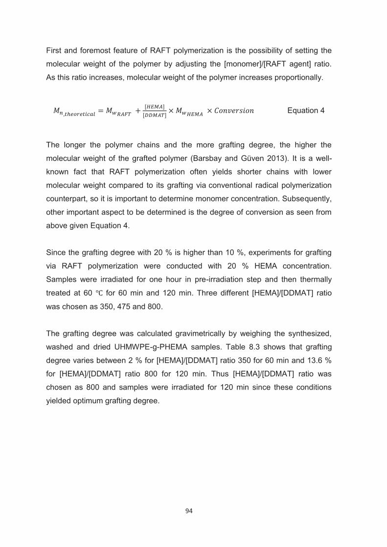
94
First and foremost feature of RAFT polymerization is the possibility of setting the
molecular weight of the polymer by adjusting the [monomer]/[RAFT agent] ratio.
As this ratio increases, molecular weight of the polymer increases proportionally.
Equation 4
The longer the polymer chains and the more grafting degree, the higher the
molecular weight of the grafted polymer (Barsbay and Güven 2013). It is a well-
known fact that RAFT polymerization often yields shorter chains with lower
molecular weight compared to its grafting via conventional radical polymerization
counterpart, so it is important to determine monomer concentration. Subsequently,
other important aspect to be determined is the degree of conversion as seen from
above given Equation 4.
Since the grafting degree with 20 % is higher than 10 %, experiments for grafting
via RAFT polymerization were conducted with 20 % HEMA concentration.
Samples were irradiated for one hour in pre-irradiation step and then thermally
treated at 60 for 60 min and 120 min. Three different [HEMA]/[DDMAT] ratio
was chosen as 350, 475 and 800.
The grafting degree was calculated gravimetrically by weighing the synthesized,
washed and dried UHMWPE-g-PHEMA samples. Table 8.3 shows that grafting
degree varies between 2 % for [HEMA]/[DDMAT] ratio 350 for 60 min and 13.6 %
for [HEMA]/[DDMAT] ratio 800 for 120 min. Thus [HEMA]/[DDMAT] ratio was
chosen as 800 and samples were irradiated for 120 min since these conditions
yielded optimum grafting degree.
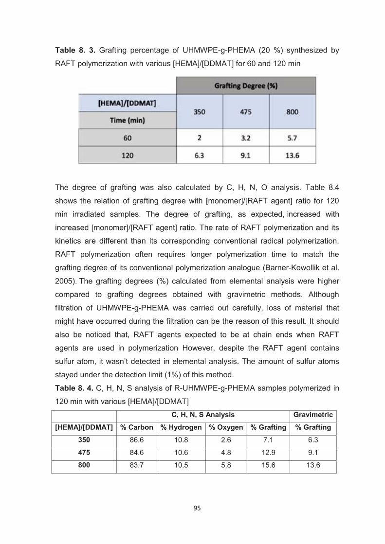
95
Table 8. 3. Grafting percentage of UHMWPE-g-PHEMA (20 %) synthesized by
RAFT polymerization with various [HEMA]/[DDMAT] for 60 and 120 min
The degree of grafting was also calculated by C, H, N, O analysis. Table 8.4
shows the relation of grafting degree with [monomer]/[RAFT agent] ratio for 120
min irradiated samples. The degree of grafting, as expected, increased with
increased [monomer]/[RAFT agent] ratio. The rate of RAFT polymerization and its
kinetics are different than its corresponding conventional radical polymerization.
RAFT polymerization often requires longer polymerization time to match the
grafting degree of its conventional polymerization analogue (Barner-Kowollik et al.
2005). The grafting degrees (%) calculated from elemental analysis were higher
compared to grafting degrees obtained with gravimetric methods. Although
filtration of UHMWPE-g-PHEMA was carried out carefully, loss of material that
might have occurred during the filtration can be the reason of this result. It should
also be noticed that, RAFT agents expected to be at chain ends when RAFT
agents are used in polymerization However, despite the RAFT agent contains
sulfur atom, it wasn’t detected in elemental analysis. The amount of sulfur atoms
stayed under the detection limit (1%) of this method.
Table 8. 4. C, H, N, S analysis of R-UHMWPE-g-PHEMA samples polymerized in
120 min with various [HEMA]/[DDMAT] C, H, N, S Analysis Gravimetric
[HEMA]/[DDMAT] % Carbon % Hydrogen % Oxygen % Grafting % Grafting
350 86.6 10.8 2.6 7.1 6.3
475 84.6 10.6 4.8 12.9 9.1
800 83.7 10.5 5.8 15.6 13.6
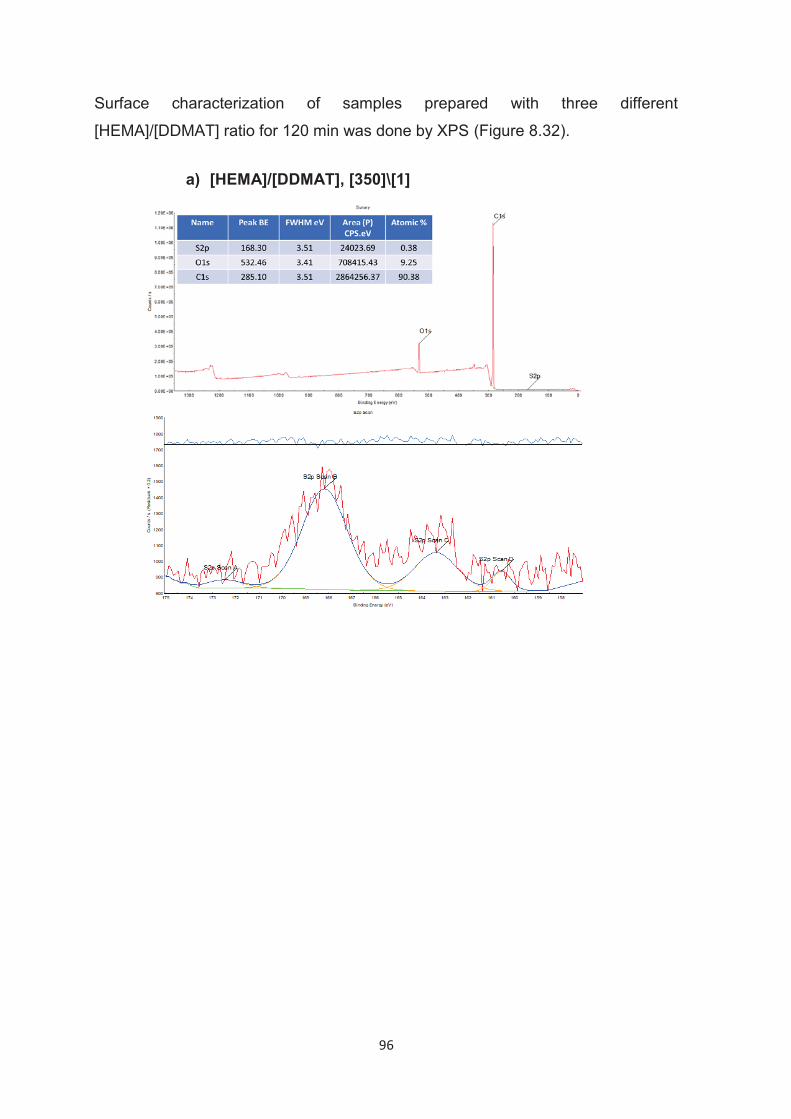
96
Surface characterization of samples prepared with three different
[HEMA]/[DDMAT] ratio for 120 min was done by XPS (Figure 8.32).
a) [HEMA]/[DDMAT], [350]\[1]
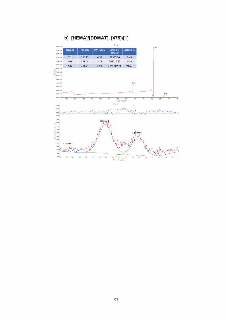
97
b) [HEMA]/[DDMAT], [475]\[1]
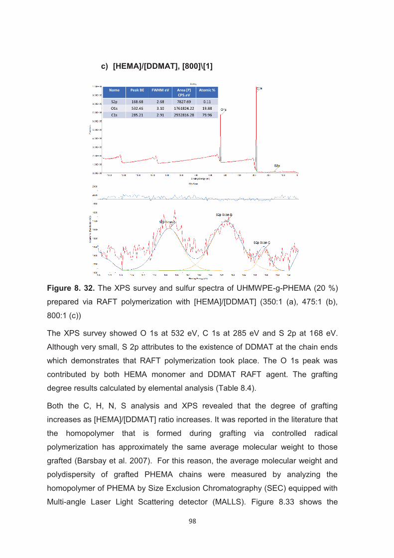
98
Figure 8. 32. The XPS survey and sulfur spectra of UHMWPE-g-PHEMA (20 %)
prepared via RAFT polymerization with [HEMA]/[DDMAT] (350:1 (a), 475:1 (b),
800:1 (c))
The XPS survey showed O 1s at 532 eV, C 1s at 285 eV and S 2p at 168 eV.
Although very small, S 2p attributes to the existence of DDMAT at the chain ends
which demonstrates that RAFT polymerization took place. The O 1s peak was
contributed by both HEMA monomer and DDMAT RAFT agent. The grafting
degree results calculated by elemental analysis (Table 8.4).
Both the C, H, N, S analysis and XPS revealed that the degree of grafting
increases as [HEMA]/[DDMAT] ratio increases. It was reported in the literature that
the homopolymer that is formed during grafting via controlled radical
polymerization has approximately the same average molecular weight to those
grafted (Barsbay et al. 2007). For this reason, the average molecular weight and
polydispersity of grafted PHEMA chains were measured by analyzing the
homopolymer of PHEMA by Size Exclusion Chromatography (SEC) equipped with
Multi-angle Laser Light Scattering detector (MALLS). Figure 8.33 shows the
c) [HEMA]/[DDMAT], [800]\[1]
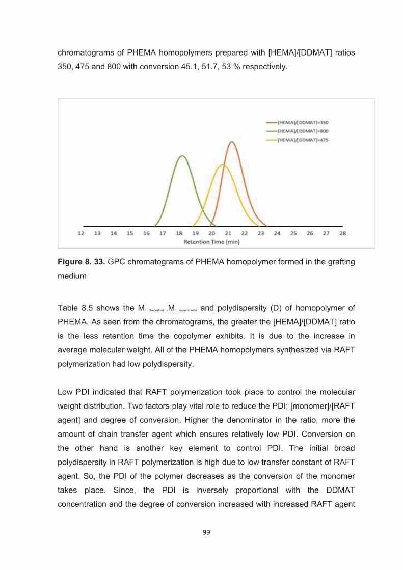
99
chromatograms of PHEMA homopolymers prepared with [HEMA]/[DDMAT] ratios
350, 475 and 800 with conversion 45.1, 51.7, 53 % respectively.
Figure 8. 33. GPC chromatograms of PHEMA homopolymer formed in the grafting
medium
Table 8.5 shows the Mn, theoretical ,Mn, experimental and polydispersity (D) of homopolymer of
PHEMA. As seen from the chromatograms, the greater the [HEMA]/[DDMAT] ratio
is the less retention time the copolymer exhibits. It is due to the increase in
average molecular weight. All of the PHEMA homopolymers synthesized via RAFT
polymerization had low polydispersity.
Low PDI indicated that RAFT polymerization took place to control the molecular
weight distribution. Two factors play vital role to reduce the PDI; [monomer]/[RAFT
agent] and degree of conversion. Higher the denominator in the ratio, more the
amount of chain transfer agent which ensures relatively low PDI. Conversion on
the other hand is another key element to control PDI. The initial broad
polydispersity in RAFT polymerization is high due to low transfer constant of RAFT
agent. So, the PDI of the polymer decreases as the conversion of the monomer
takes place. Since, the PDI is inversely proportional with the DDMAT
concentration and the degree of conversion increased with increased RAFT agent
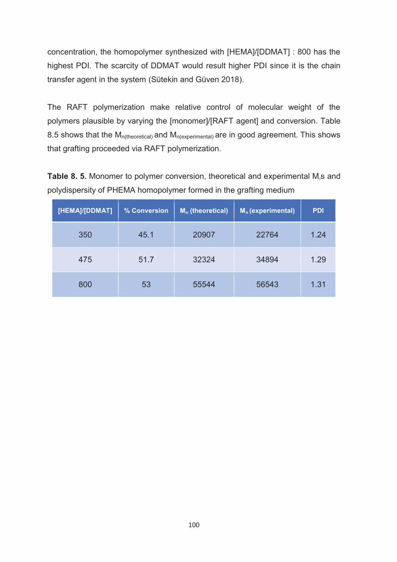
100
concentration, the homopolymer synthesized with [HEMA]/[DDMAT] : 800 has the
highest PDI. The scarcity of DDMAT would result higher PDI since it is the chain
transfer agent in the system (Sütekin and Güven 2018).
The RAFT polymerization make relative control of molecular weight of the
polymers plausible by varying the [monomer]/[RAFT agent] and conversion. Table
8.5 shows that the Mn(theoretical) and Mn(experimental) are in good agreement. This shows
that grafting proceeded via RAFT polymerization.
Table 8. 5. Monomer to polymer conversion, theoretical and experimental Mns and
polydispersity of PHEMA homopolymer formed in the grafting medium
[HEMA]/[DDMAT] % Conversion Mn (theoretical) Mn (experimental) PDI
350 45.1 20907 22764 1.24
475 51.7 32324 34894 1.29
800 53 55544 56543 1.31
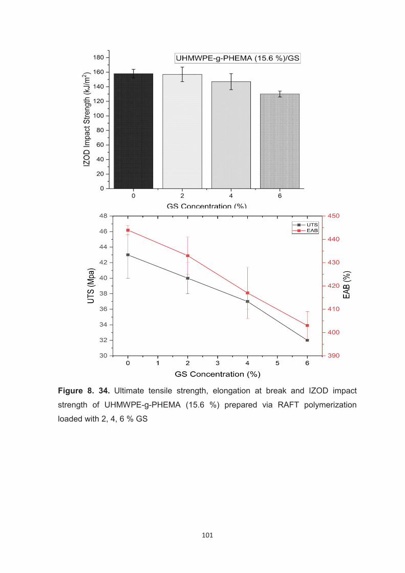
101
Figure 8. 34. Ultimate tensile strength, elongation at break and IZOD impact
strength of UHMWPE-g-PHEMA (15.6 %) prepared via RAFT polymerization
loaded with 2, 4, 6 % GS
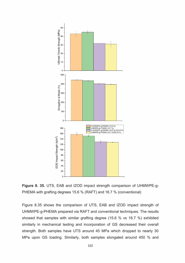
102
Figure 8. 35. UTS, EAB and IZOD impact strength comparison of UHMWPE-g-
PHEMA with grafting degrees 15.6 % (RAFT) and 16.7 % (conventional)
Figure 8.35 shows the comparison of UTS, EAB and IZOD impact strength of
UHMWPE-g-PHEMA prepared via RAFT and conventional techniques. The results
showed that samples with similar grafting degree (15.6 % vs 16.7 %) exhibited
similarly in mechanical testing and incorporation of GS decreased their overall
strength. Both samples have UTS around 45 MPa which dropped to nearly 30
MPa upon GS loading. Similarly, both samples elongated around 450 % and
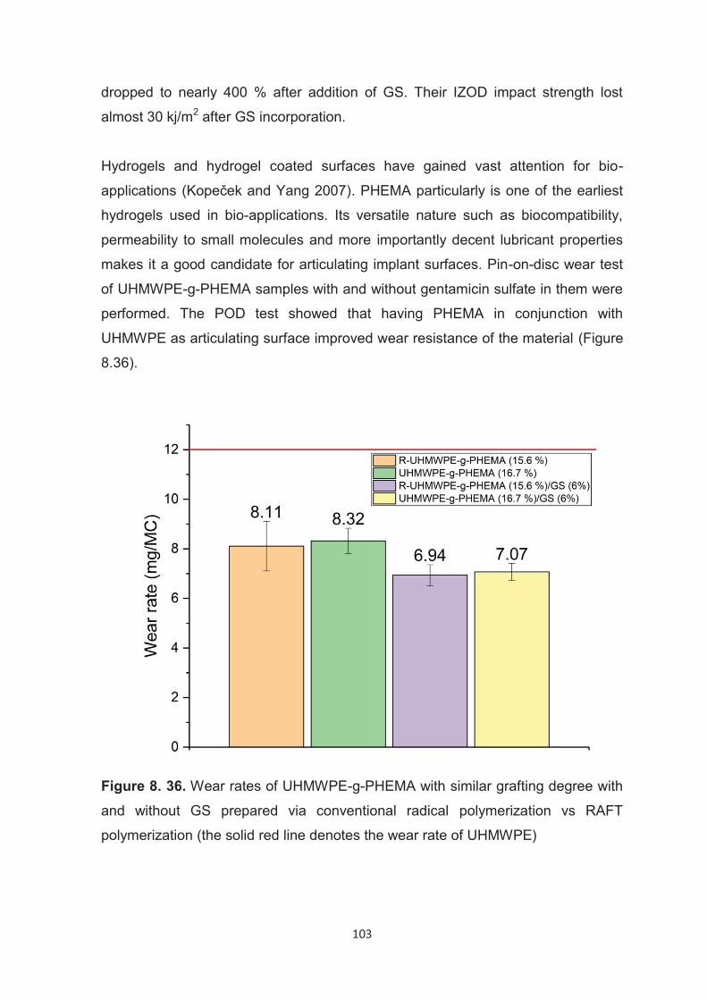
103
dropped to nearly 400 % after addition of GS. Their IZOD impact strength lost
almost 30 kj/m2 after GS incorporation.
Hydrogels and hydrogel coated surfaces have gained vast attention for bio-
applications (Kopeček and Yang 2007). PHEMA particularly is one of the earliest
hydrogels used in bio-applications. Its versatile nature such as biocompatibility,
permeability to small molecules and more importantly decent lubricant properties
makes it a good candidate for articulating implant surfaces. Pin-on-disc wear test
of UHMWPE-g-PHEMA samples with and without gentamicin sulfate in them were
performed. The POD test showed that having PHEMA in conjunction with
UHMWPE as articulating surface improved wear resistance of the material (Figure
8.36).
Figure 8. 36. Wear rates of UHMWPE-g-PHEMA with similar grafting degree with
and without GS prepared via conventional radical polymerization vs RAFT
polymerization (the solid red line denotes the wear rate of UHMWPE)

104
UHMWPE-g-PHEMA with similar grafting degree showed similar wear rate around
8 mg/MC (Figure 8.36). Incorporation of GS decreased the wear rate even beyond
the copolymers itself to 7 mg/MC as it was observed in GS loaded virgin
UHMWPE. Overall the reduction of wear rate was thought to be because of
PHEMA has ability to reduce the contact stress and promote fluid film lubrication
(Bavaresco et al. 2008).
UHMWPE-g-PHEMA samples with similar grafting degrees prepared with RAFT
and conventional techniques were chosen in order to compare drug release and
antibacterial properties. The UHMWPE-g-PHEMA (grafting degree 15.6 %)
prepared with [HEMA]/[DDMAT] : 800 ratio with UV induced preirradiation and
RAFT mediated grafting was chosen for drug release and antibacterial efficacy
studies. This sample was compared with UHMWPE-g-PHEMA (grafting degree
16.7 %) prepared with conventional technique. Both copolymers were loaded with
three GS concentration; 2, 4, 6 %. The amount of GS released for a week were 8
%, 4 %, 1 % for the copolymer with GS mass percentage 6, 4, 2 % respectively
(Figure 8.37).
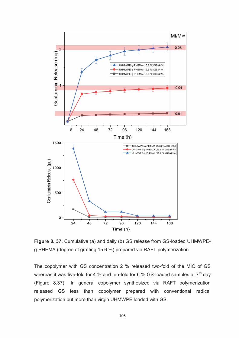
105
Figure 8. 37. Cumulative (a) and daily (b) GS release from GS-loaded UHMWPE-
g-PHEMA (degree of grafting 15.6 %) prepared via RAFT polymerization
The copolymer with GS concentration 2 % released two-fold of the MIC of GS
whereas it was five-fold for 4 % and ten-fold for 6 % GS-loaded samples at 7th day
(Figure 8.37). In general copolymer synthesized via RAFT polymerization
released GS less than copolymer prepared with conventional radical
polymerization but more than virgin UHMWPE loaded with GS.
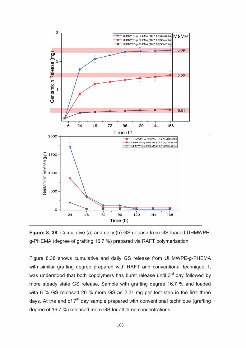
106
Figure 8. 38. Cumulative (a) and daily (b) GS release from GS-loaded UHMWPE-
g-PHEMA (degree of grafting 16.7 %) prepared via RAFT polymerization
Figure 8.38 shows cumulative and daily GS release from UHMWPE-g-PHEMA
with similar grafting degree prepared with RAFT and conventional technique. It
was understood that both copolymers has burst release until 3rd day followed by
more steady state GS release. Sample with grafting degree 16.7 % and loaded
with 6 % GS released 20 % more GS as 2.21 mg per test strip in the first three
days. At the end of 7th day sample prepared with conventional technique (grafting
degree of 16.7 %) released more GS for all three concentrations.
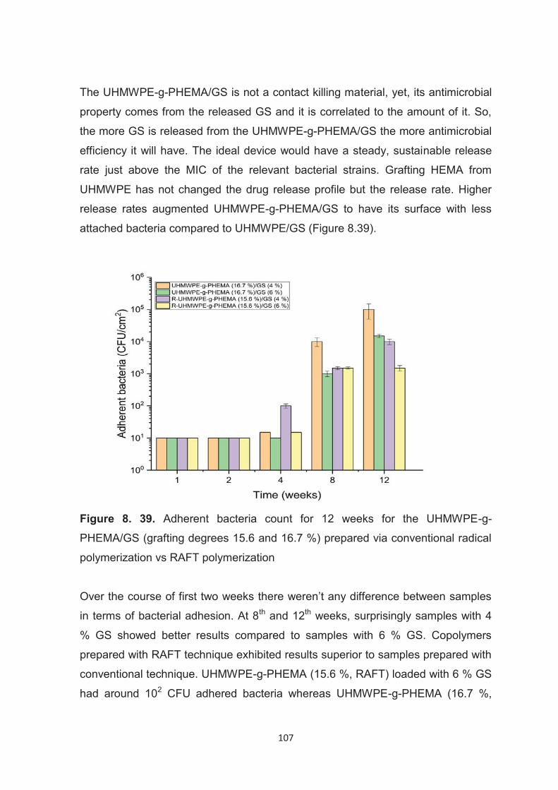
107
The UHMWPE-g-PHEMA/GS is not a contact killing material, yet, its antimicrobial
property comes from the released GS and it is correlated to the amount of it. So,
the more GS is released from the UHMWPE-g-PHEMA/GS the more antimicrobial
efficiency it will have. The ideal device would have a steady, sustainable release
rate just above the MIC of the relevant bacterial strains. Grafting HEMA from
UHMWPE has not changed the drug release profile but the release rate. Higher
release rates augmented UHMWPE-g-PHEMA/GS to have its surface with less
attached bacteria compared to UHMWPE/GS (Figure 8.39).
Figure 8. 39. Adherent bacteria count for 12 weeks for the UHMWPE-g-
PHEMA/GS (grafting degrees 15.6 and 16.7 %) prepared via conventional radical
polymerization vs RAFT polymerization
Over the course of first two weeks there weren’t any difference between samples
in terms of bacterial adhesion. At 8th and 12th weeks, surprisingly samples with 4
% GS showed better results compared to samples with 6 % GS. Copolymers
prepared with RAFT technique exhibited results superior to samples prepared with
conventional technique. UHMWPE-g-PHEMA (15.6 %, RAFT) loaded with 6 % GS
had around 102 CFU adhered bacteria whereas UHMWPE-g-PHEMA (16.7 %,
108
conventional) loaded with 6 % GS had two orders of magnitude more bacteria
(Figure 8.39).
In overall antimicrobial performance comparison of UHMWPE-g-PHEMA/GS to
UHMWPE/GS, it was understood that grafting PHEMA from UHMWPE surfaces
improved the GS release rate and hence bacterial colonization was inhibited more
in those copolymers. Another factor that aided the antimicrobial efficacy of
copolymer could be PHEMA is inherently able to inhibit bacterial colonization (Lee
et al. 2018).
109
CONCLUSIONS
The main goal of this dissertation was to create an articulating surface for tibial
implants which avoids bacterial colonization for revision surgeries. Virgin
UHMWPE has been succesfully used in the total joint arthroplasty for decades
now. However, it is known that synthetic materials are vulnerable to infection.
Gentamicin sulfate is one of the foremost antibiotics that is used in approaches to
treat PJI. In this study, we developed gentamicin sulfate-releasing UHMWPE and
UHMWPE-g-PHEMA as a temporary tibial spacer for knee arthroplasty. We
investigated the gentamicin sulfate release and antibacterial performances of
gentamicin sulfate-loaded virgin UHMWPE and UHMWPE-g-PHEMA prepared via
conventional and RAFT grafting techniques.
The UHMWPE/GS and UHMWPE-g-PHEMA/GS blends were dehydrated prior to
compression molding for reduce the water stress on gentamicin sulfate. Since,
high temperature (170°C) was used to fabricate gentamicin sulfate-loaded
UHMWPE and gentamicin sulfate-loaded UHMWPE-g-PHEMA, we showed that
the chemical structure of gentamicin sulfate was not comprimised by XPS.
Gentamicin sulfate and UHMWPE were observed as phase seperated based on
the optical microscope images. The gentamicin sulfate clusters showed tendency
to agglomerate in the polymeric matrix as the concentration increased.
UHMWPE/GS with 1, 2, 4, 6, 8, 10 wt. % GS were prepared and used for drug
release, antibacterial and mechanical testing studies. All of the GS-loaded virgin
UHMWPE showed a burst release in the first six hours followed by more steady
state release. UHMWPE/GS (1 wt. %) released 3.23 mg GS and UHMWPE/GS
(10 wt. %) released 128 mg GS from full size tibial implant in five days which is
approximately 800 and 32000-fold of the MIC respectively for S. aureus.
The UHMWPE/GS formulation with 8 wt. % GS exhibited similar drug release
profile to clinically used gentamicin sulfate-loaded bone cement (Palacos R-G).
Both UHMWPE/GS (8 wt. %) and Palacos R-G have remained as bactericidal
against 105 CFU/ml for 12 weeks.
The UHMWPE/GS had fewer adherent bacteria on its surface than gentamicin
sulfate-loaded bone cement over the course of the 24th week of the test.
The UHMWPE-g-PHEMA was synthesized via UV-initiated grafting with two
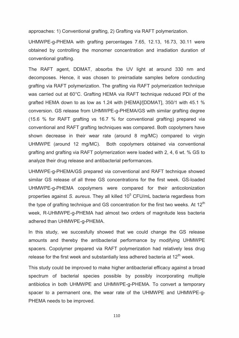
110
approaches: 1) Conventional grafting, 2) Grafting via RAFT polymerization.
UHMWPE-g-PHEMA with grafting percentages 7.65, 12.13, 16.73, 30.11 were
obtained by controlling the monomer concentration and irradiation duration of
conventional grafting.
The RAFT agent, DDMAT, absorbs the UV light at around 330 nm and
decomposes. Hence, it was chosen to preirradiate samples before conducting
grafting via RAFT polymerization. The grafting via RAFT polymerization technique
was carried out at 60°C. Grafting HEMA via RAFT technique reduced PDI of the
grafted HEMA down to as low as 1.24 with [HEMA]/[DDMAT], 350/1 with 45.1 %
conversion. GS release from UHMWPE-g-PHEMA/GS with similar grafting degree
(15.6 % for RAFT grafting vs 16.7 % for conventional grafting) prepared via
conventional and RAFT grafting techniques was compared. Both copolymers have
shown decrease in their wear rate (around 8 mg/MC) compared to virgin
UHMWPE (around 12 mg/MC). Both copolymers obtained via conventional
grafting and grafting via RAFT polymerization were loaded with 2, 4, 6 wt. % GS to
analyze their drug release and antibacterial performances.
UHMWPE-g-PHEMA/GS prepared via conventional and RAFT technique showed
similar GS release of all three GS concentrations for the first week. GS-loaded
UHMWPE-g-PHEMA copolymers were compared for their anticolonization
properties against S. aureus. They all killed 105 CFU/mL bacteria regardless from
the type of grafting technique and GS concentration for the first two weeks. At 12th
week, R-UHMWPE-g-PHEMA had almost two orders of magnitude less bacteria
adhered than UHMWPE-g-PHEMA.
In this study, we succesfully showed that we could change the GS release
amounts and thereby the antibacterial performance by modifying UHMWPE
spacers. Copolymer prepared via RAFT polymerization had relatively less drug
release for the first week and substantially less adhered bacteria at 12th week.
This study could be improved to make higher antibacterial efficacy against a broad
spectrum of bacterial species possible by possibly incorporating multiple
antibiotics in both UHMWPE and UHMWPE-g-PHEMA. To convert a temporary
spacer to a permanent one, the wear rate of the UHMWPE and UHMWPE-g-
PHEMA needs to be improved.
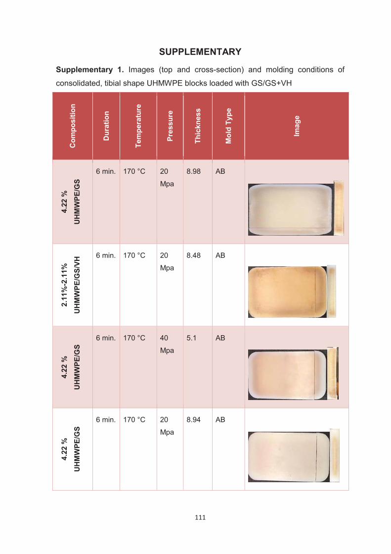
111
SUPPLEMENTARY
Supplementary 1. Images (top and cross-section) and molding conditions of
consolidated, tibial shape UHMWPE blocks loaded with GS/GS+VH C
ompo
sitio
n
Dur
atio
n
Tem
pera
ture
Pres
sure
Thic
knes
s
Mol
d Ty
pe
Imag
e
4.22
%
UH
MW
PE/G
S
6 min. 170 °C 20
Mpa 8.98 AB
2.11
%-2
.11%
UH
MW
PE/G
S/VH
6 min. 170 °C 20
Mpa 8.48 AB
4.22
%
UH
MW
PE/G
S
6 min. 170 °C 40
Mpa 5.1 AB
4.22
%
UH
MW
PE/G
S
6 min. 170 °C 20
Mpa 8.94 AB
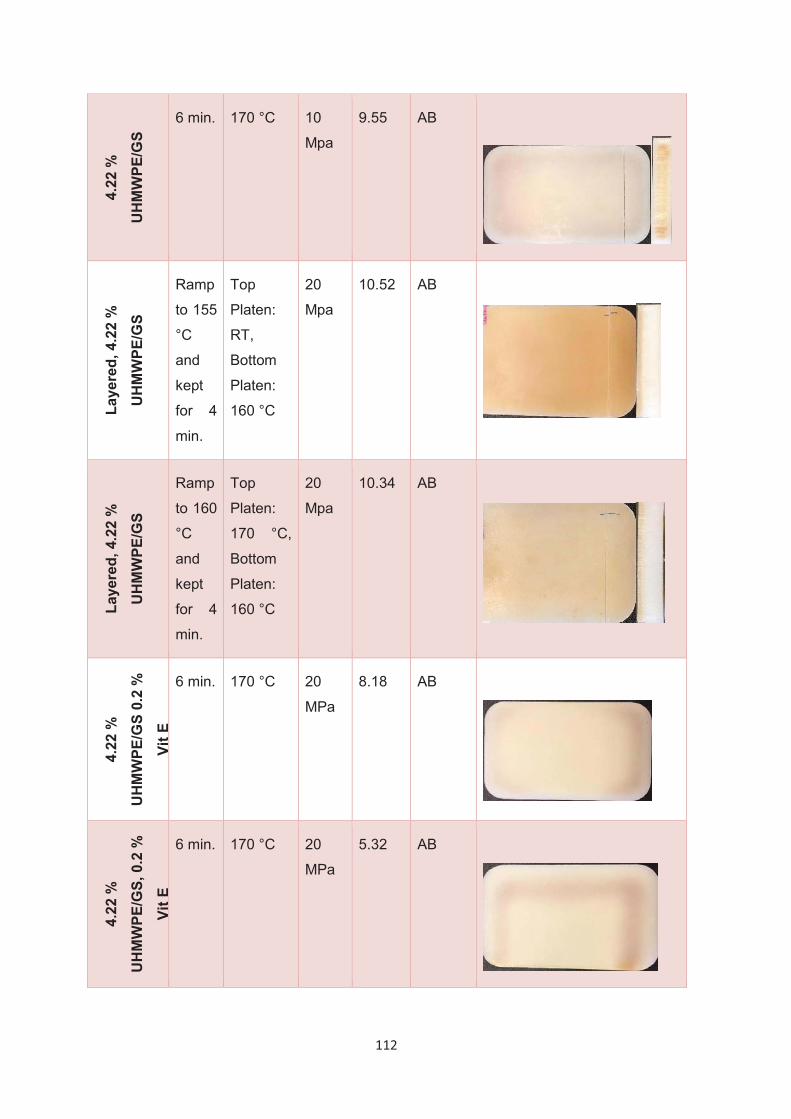
112
4.22
%
UH
MW
PE/G
S 6 min. 170 °C 10
Mpa 9.55 AB
Laye
red,
4.2
2 %
UH
MW
PE/G
S
Ramp
to 155
°C
and
kept
for 4
min.
Top
Platen:
RT,
Bottom
Platen:
160 °C
20
Mpa 10.52 AB
Laye
red,
4.2
2 %
UH
MW
PE/G
S
Ramp
to 160
°C
and
kept
for 4
min.
Top
Platen:
170 °C,
Bottom
Platen:
160 °C
20
Mpa 10.34 AB
4.22
%
UH
MW
PE/G
S 0.
2 %
Vit E
6 min. 170 °C 20
MPa 8.18 AB
4.22
%
UH
MW
PE/G
S, 0
.2 %
Vit E
6 min. 170 °C 20
MPa 5.32 AB
113
4.22
%
UH
MW
PE/G
S, 0
.2
% V
it E
6 min. 170 °C 20
MPa 8.2 AB
4.22
%
UH
MW
PE/G
S
6 min. 170 °C 20
MPa 7.25 AB
4.22
%
UH
MW
PE/G
S
6 min. 170 °C 20
MPa 8 AB
4.22
%
UH
MW
PE/V
H 6 min. 170 °C 20
MPa 9.03 AB
114
REFERENCES
Adams, Annette L., Elizabeth W. Paxton, Jean Q. Wang, Eric S. Johnson, Elizabeth A. Bayliss, Assiamira Ferrara, Cynthia Nakasato, Stefano A. Bini, and Robert S. Namba. 2013. “Surgical Outcomes of Total Knee Replacement according to Diabetes Status and Glycemic Control, 2001 to 2009.” The Journal of Bone and Joint Surgery. American Volume 95 (6): 481–87.
AlBuhairan, B., D. Hind, and A. Hutchinson. 2008. “Antibiotic Prophylaxis for Wound Infections in Total Joint Arthroplasty: A Systematic Review.” The Journal of Bone and Joint Surgery. British Volume 90 (7): 915–19.
Allen, C., A. Bloyce, and T. Bell. 1995. “The Effect of Ion Implantation on the Sliding Wear Behaviour of Ultrahigh Molecular Weight Polyethylene against an Oxidised Titanium Alloy Ti-6Al-4V.” Tribology Letters 1 (1): 35–38.
An, Y. H., and R. J. Friedman. 1998. “Concise Review of Mechanisms of Bacterial Adhesion to Biomaterial Surfaces.” Journal of Biomedical Materials Research 43 (3): 338–48.
Anagnostakos, Konstantinos, Philip Hitzler, Dietrich Pape, Dieter Kohn, and Jens Kelm. 2008. “Persistence of Bacterial Growth on Antibiotic-Loaded Beads: Is It Actually a Problem?” Acta Orthopaedica 79 (2): 302–7.
Anderl, Jeff N., Jeff Zahller, Frank Roe, and Philip S. Stewart. 2003. “Role of Nutrient Limitation and Stationary-Phase Existence in Klebsiella Pneumoniae Biofilm Resistance to Ampicillin and Ciprofloxacin.” Antimicrobial Agents and Chemotherapy 47 (4): 1251–56.
Anderson, Brian C., Paul D. Bloom, K. G. Baikerikar, Valerie V. Sheares, and Surya K. Mallapragada. 2002. “Al-Cu-Fe Quasicrystal/ultra-High Molecular Weight Polyethylene Composites as Biomaterials for Acetabular Cup Prosthetics.” Biomaterials 23 (8): 1761–68.
Anderson, Emily B., and Timothy E. Long. 2010. “Imidazole- and Imidazolium-Containing Polymers for Biology and Material Science Applications.” Polymer 51 (12): 2447–54.
Ayers, David C., and Patricia D. Franklin. 2014. “Joint Replacement Registries in the United States: A New Paradigm.” The Journal of Bone and Joint Surgery. American Volume 96 (18): 1567–69.
Bakar Atıcı, Ayşe. 2018. “Kalça Eklem Protezleri Yapımında Kullanılacak Olan Ultra Molekül Ağırlıklı Polietilen (Umape) Yüzeylere Kayganlık Kazandırmak İçin Kontrollü Olarak Poli (Akrilik Asit)(Paa) Aşılanması.” http://www.openaccess.hacettepe.edu.tr:8080/xmlui/handle/11655/5766.
115
Barner-Kowollik, Christopher, Michael Buback, Mark Egorov, Takeshi Fukuda, Atsushi Goto, Oskar Friedrich Olaj, Gregory T. Russell, Philipp Vana, Bunichiro Yamada, and Per B. Zetterlund. 2005. “Critically Evaluated Termination Rate Coefficients for Free-Radical Polymerization: Experimental Methods.” Progress in Polymer Science 30 (6): 605–43.
Barner-Kowollik, Christopher. 2008. Handbook of RAFT Polymerization. John Wiley & Sons.
Barsbay, Murat, and Olgun Güven. 2009. “A Short Review of Radiation-Induced Raft-Mediated Graft Copolymerization: A Powerful Combination for Modifying the Surface Properties of Polymers in a Controlled Manner.” Radiation Physics and Chemistry 78 (12): 1054–59.
Barsbay, Murat, and Olgun Güven. 2013. “RAFT Mediated Grafting of Poly(acrylic Acid) (PAA) from Polyethylene/polypropylene (PE/PP) Nonwoven Fabric via Preirradiation.” Polymer. https://doi.org/10.1016/j.polymer.2013.06.059.
Barsbay, Murat, Olgun Güven, Martina H. Stenzel, Thomas P. Davis, Christopher Barner-Kowollik, and Leonie Barner. 2007. “Verification of Controlled Grafting of Styrene from Cellulose via Radiation-Induced RAFT Polymerization.” Macromolecules 40 (20): 7140–47.
Bavaresco, V. P., C. A. C. Zavaglia, M. C. Reis, and J. R. Gomes. 2008. “Study on the Tribological Properties of pHEMA Hydrogels for Use in Artificial Articular Cartilage.” Wear: An International Journal on the Science and Technology of Friction Lubrication and Wear 265 (3): 269–77.
Bejon, P., A. Berendt, B. L. Atkins, N. Green, H. Parry, S. Masters, P. McLardy-Smith, R. Gundle, and I. Byren. 2010. “Two-Stage Revision for Prosthetic Joint Infection: Predictors of Outcome and the Role of Reimplantation Microbiology.” The Journal of Antimicrobial Chemotherapy 65 (3): 569–75.
Bellare, A., H. Schnablegger, and R. E. Cohen. 1995. “A Small-Angle X-Ray Scattering Study of High-Density Polyethylene and Ultrahigh Molecular Weight Polyethylene.” Macromolecules 28 (23): 7585–88.
Belt, Hilbrand van de, Daniëlle Neut, Willem Schenk, Jim R. van Horn, Henny C. van der Mei, and Henk J. Busscher. 2001. “Infection of Orthopedic Implants and the Use of Antibiotic-Loaded Bone Cements: A Review.” Acta Orthopaedica Scandinavica 72 (6): 557–71.
Berend, Keith R., Adolph V. Lombardi Jr, Michael J. Morris, Adam G. Bergeson, Joanne B. Adams, and Michael A. Sneller. 2013. “Two-Stage Treatment of Hip Periprosthetic Joint Infection Is Associated with a High Rate of Infection Control but High Mortality.” Clinical Orthopaedics and Related Research 471 (2): 510–18.
Bertazzoni Minelli, E., A. Benini, B. Magnan, and P. Bartolozzi. 2004. “Release of Gentamicin and Vancomycin from Temporary Human Hip Spacers in Two-
116
Stage Revision of Infected Arthroplasty.” The Journal of Antimicrobial Chemotherapy 53 (2): 329–34.
Bianchi, Michele, Marco Boi, Nicola Lopomo, Maria Cristina Maltarello, Fabiola Liscio, Silvia Milita, Andrea Visani, Alessandro Russo, and Maurilio Marcacci. 2015. “NANOMECHANICAL CHARACTERIZATION OF ZIRCONIA THIN FILMS DEPOSITED ON UHMWPE BY PULSED PLASMA DEPOSITION.” Journal of Mechanics in Medicine and Biology 15 (05): 1550070.
Bieser, Arno M., Yi Thomann, and Joerg C. Tiller. 2011. “Contact-Active Antimicrobial and Potentially Self-Polishing Coatings Based on Cellulose.” Macromolecular Bioscience 11 (1): 111–21.
Blunn, G. W., A. B. Joshi, R. J. Minns, L. Lidgren, P. Lilley, L. Ryd, E. Engelbrecht, and P. S. Walker. 1997. “Wear in Retrieved Condylar Knee Arthroplasties. A Comparison of Wear in Different Designs of 280 Retrieved Condylar Knee Prostheses.” The Journal of Arthroplasty 12 (3): 281–90.
Bonfield, W. 1988. “Composites for Bone Replacement.” Journal of Biomedical Engineering 10 (6): 522–26.
Bonilla-Cruz, José, Enrique Saldívar-Guerra, José Román Torres-Lubián, Ramiro Guerrero-Santos, Bruno López-Carpy, and Gabriel Luna-Bárcenas. 2008. “Controlled Grafting-From of Polystyrene on Polybutadiene: Mechanism and Spectroscopic Evidence of the Functionalization of Polybutadiene with 4-Oxo-TEMPO.” Macromolecular Chemistry and Physics. https://doi.org/10.1002/macp.200800367.
Boyer, Cyrille, Volga Bulmus, Thomas P. Davis, Vincent Ladmiral, Jingquan Liu, and Sébastien Perrier. 2009. “Bioapplications of RAFT Polymerization.” Chemical Reviews 109 (11): 5402–36.
Bozic, Kevin J., Edmund Lau, Steven Kurtz, Kevin Ong, Harry Rubash, Thomas P. Vail, and Daniel J. Berry. 2012. “Patient-Related Risk Factors for Periprosthetic Joint Infection and Postoperative Mortality Following Total Hip Arthroplasty in Medicare Patients.” The Journal of Bone and Joint Surgery. American Volume 94 (9): 794–800.
Bracco, Pierangiola, and Ebru Oral. 2011. “Vitamin E-Stabilized UHMWPE for Total Joint Implants: A Review.” Clinical Orthopaedics and Related Research 469 (8): 2286–93.
Bradbury, Thomas, Thomas K. Fehring, Michael Taunton, Arlen Hanssen, Khalid Azzam, Javad Parvizi, and Susan M. Odum. 2009. “The Fate of Acute Methicillin-Resistant Staphylococcus Aureus Periprosthetic Knee Infections Treated by Open Debridement and Retention of Components.” The Journal of Arthroplasty 24 (6 Suppl): 101–4.
117
Bradley, Katharine A., Anna D. Rubinsky, Haili Sun, Chris L. Bryson, Michael J. Bishop, David K. Blough, William G. Henderson, et al. 2011. “Alcohol Screening and Risk of Postoperative Complications in Male VA Patients Undergoing Major Non-Cardiac Surgery.” Journal of General Internal Medicine 26 (2): 162–69.
Breier, Ann-Christin, Christian Brandt, Dorit Sohr, Christine Geffers, and Petra Gastmeier. 2011. “Laminar Airflow Ceiling Size: No Impact on Infection Rates Following Hip and Knee Prosthesis.” Infection Control and Hospital Epidemiology: The Official Journal of the Society of Hospital Epidemiologists of America 32 (11): 1097–1102.
BUCHHOLZ, and HW. 1970. “Uber Die Depotwirkung Einiger Antibiotika Bei Vermischung Mit Dem Kunstharz Palacos.” Der Chirurg; Zeitschrift Fur Alle Gebiete Der Operativen Medizen 40: 511–15.
Buchholz, H. W., and H. Engelbrecht. 1970. “Depot Effects of Various Antibiotics Mixed with Palacos Resins.” Der Chirurg; Zeitschrift Fur Alle Gebiete Der Operativen Medizen 41 (11): 511–15.
Buechel, Frederick F., Frank P. Femino, and Jerry D’Alessio. 2004. “Primary Exchange Revision Arthroplasty for Infected Total Knee Replacement: A Long-Term Study.” American Journal of Orthopedics 33 (4): 190–98; discussion 198.
Ceri, H., M. E. Olson, C. Stremick, R. R. Read, D. Morck, and A. Buret. 1999. “The Calgary Biofilm Device: New Technology for Rapid Determination of Antibiotic Susceptibilities of Bacterial Biofilms.” Journal of Clinical Microbiology 37 (6): 1771–76.
Charmot, D., P. Corpart, D. Michelet, S. Z. Zard, and T. Biadatti. 1999. “PCT Int. Appl. WO 9858974, 1998.” In Chem Abstr, 130:82018.
Charnley, J., and Z. Cupic. 1973. “The Nine and Ten Year Results of the Low-Friction Arthroplasty of the Hip.” Clinical Orthopaedics and Related Research, no. 95 (September): 9–25.
Charnley, John. 1970. “Acrylic Cement in Orthopaedic Surgery.” In Acrylic Cement in Orthopaedic Surgery.
Charnley, John. 1973. “Arthroplasty of the Hip: A New Operation*.” Clinical Orthopaedics and Related Research 95: 4–8.
Charnley, John. 1979. “Low Friction Principle.” In Low Friction Arthroplasty of the Hip: Theory and Practice, edited by John Charnley, 3–15. Berlin, Heidelberg: Springer Berlin Heidelberg.
Charnley, John. 2005. “The Long-Term Results of Low-Friction Arthroplasty of the Hip Performed as a Primary Intervention. 1970.” Clinical Orthopaedics and Related Research, no. 430 (January): 3–11; discussion 2.
118
Chen, Zhaobin, and Yuyu Sun. 2006. “N-Halamine-Based Antimicrobial Additives for Polymers: Preparation, Characterization and Antimicrobial Activity.” Industrial & Engineering Chemistry Research 45 (8): 2634–40.
Cheng, Gang, Hong Xue, Zheng Zhang, Shengfu Chen, and Shaoyi Jiang. 2008. “A Switchable Biocompatible Polymer Surface with Self-Sterilizing and Nonfouling Capabilities.” Angewandte Chemie 47 (46): 8831–34.
Chong, Yen K., Graeme Moad, Ezio Rizzardo, and San H. Thang. 2007. “Thiocarbonylthio End Group Removal from RAFT-Synthesized Polymers by Radical-Induced Reduction.” Macromolecules 40 (13): 4446–55.
Chung, T. C. 2002. “Synthesis of Functional Polyolefin Copolymers with Graft and Block Structures.” Progress in Polymer Science 27 (1): 39–85.
Cloyd, Jordan M., Neil R. Malhotra, Lihui Weng, Weiliam Chen, Robert L. Mauck, and Dawn M. Elliott. 2007. “Material Properties in Unconfined Compression of Human Nucleus Pulposus, Injectable Hyaluronic Acid-Based Hydrogels and Tissue Engineering Scaffolds.” European Spine Journal: Official Publication of the European Spine Society, the European Spinal Deformity Society, and the European Section of the Cervical Spine Research Society 16 (11): 1892–98.
Collier, J. P., M. B. Mayor, V. A. Surprenant, H. P. Surprenant, L. A. Dauphinais, and R. E. Jensen. 1990. “The Biomechanical Problems of Polyethylene as a Bearing Surface.” Clinical Orthopaedics and Related Research, no. 261 (December): 107–13.
Connelly, G. M., C. M. Rimnac, T. M. Wright, R. W. Hertzberg, and J. A. Manson. 1984. “Fatigue Crack Propagation Behavior of Ultrahigh Molecular Weight Polyethylene.” Journal of Orthopaedic Research: Official Publication of the Orthopaedic Research Society 2 (2): 119–25.
Cossee, P. 1964. “Ziegler-Natta Catalysis I. Mechanism of Polymerization of α-Olefins with Ziegler-Natta Catalysts.” Journal of Catalysis 3 (1): 80–88.
Costerton, J. W., P. S. Stewart, and E. P. Greenberg. 1999. “Bacterial Biofilms: A Common Cause of Persistent Infections.” Science 284 (5418): 1318–22.
Costerton, J. W., Z. Lewandowski, D. E. Caldwell, D. R. Korber, and H. M. Lappin-Scott. 1995. “Microbial Biofilms.” Annual Review of Microbiology 49: 711–45.
Cross, Michael Brian, Paul Hyunsoo Yi, Charlotte F. Thomas, Jane Garcia, and Craig J. Della Valle. 2014. “Evaluation of Malnutrition in Orthopaedic Surgery.” The Journal of the American Academy of Orthopaedic Surgeons 22 (3): 193–99.
119
Cruess, R. L., W. S. Bickel, and K. L. vonKessler. 1975. “Infections in Total Hips Secondary to a Primary Source Elsewhere.” Clinical Orthopaedics and Related Research, no. 106 (January): 99–101.
Cunliffe, David, Carolina de las Heras Alarcón, Vanessa Peters, James R. Smith, and Cameron Alexander. 2003. “Thermoresponsive Surface-Grafted Poly(N−isopropylacrylamide) Copolymers: Effect of Phase Transitions on Protein and Bacterial Attachment.” Langmuir: The ACS Journal of Surfaces and Colloids 19 (7): 2888–99.
Deirmengian, Carl, Jordan Greenbaum, Paul A. Lotke, Robert E. Booth Jr, and Jess H. Lonner. 2003. “Limited Success with Open Debridement and Retention of Components in the Treatment of Acute Staphylococcus Aureus Infections after Total Knee Arthroplasty.” The Journal of Arthroplasty 18 (7 Suppl 1): 22–26.
Demir, Buket, Roy M. Broughton, Mingyu Qiao, Tung-Shi Huang, and S. D. Worley. 2017. “N-Halamine Biocidal Materials with Superior Antimicrobial Efficacies for Wound Dressings.” Molecules 22 (10). https://doi.org/10.3390/molecules22101582.
Devoto, Gianluigi, Fabrizio Gallo, Concetta Marchello, Omar Racchi, Roberta Garbarini, Stefano Bonassi, Giorgio Albalustri, and Enrico Haupt. 2006. “Prealbumin Serum Concentrations as a Useful Tool in the Assessment of Malnutrition in Hospitalized Patients.” Clinical Chemistry 52 (12): 2281–85.
Di Silvio, L., M. J. Dalby, and W. Bonfield. 2002. “Osteoblast Behaviour on HA/PE Composite Surfaces with Different HA Volumes.” Biomaterials 23 (1): 101–7.
Dowsey, Michelle M., and Peter F. M. Choong. 2008. “Early Outcomes and Complications Following Joint Arthroplasty in Obese Patients: A Review of the Published Reports.” ANZ Journal of Surgery 78 (6): 439–44.
Dowsey, Michelle M., and Peter F. M. Choong. 2009. “Obese Diabetic Patients Are at Substantial Risk for Deep Infection after Primary TKA.” Clinical Orthopaedics and Related Research 467 (6): 1577–81.
Drago, Lorenzo, ed. 2017. A Modern Approach to Biofilm-Related Orthopaedic Implant Infections: Advances in Microbiology, Infectious Diseases and Public Health Volume 5. Advances in Experimental Medicine and Biology.
Dunne, N. J., J. Hill, P. McAfee, R. Kirkpatrick, S. Patrick, and M. Tunney. 2008. “Incorporation of Large Amounts of Gentamicin Sulphate into Acrylic Bone Cement: Effect on Handling and Mechanical Properties, Antibiotic Release, and Biofilm Formation.” Proceedings of the Institution of Mechanical Engineers. Part H, Journal of Engineering in Medicine 222 (3): 355–65.

120
Edidin, A. A., and S. M. Kurtz. 2000. “Influence of Mechanical Behavior on the Wear of 4 Clinically Relevant Polymeric Biomaterials in a Hip Simulator.” The Journal of Arthroplasty 15 (3): 321–31.
Edward McPherson, M. D. Facs. 2011. “Deactivation of Palacos R Bone Cement with the Addition of Rifampin Antibiotic Powder An In-Vivo Experience -Case Report.” Reconstructive Review 1 (1). https://doi.org/10.15438/rr.v1i1.4.
Elek, S. D., and P. E. Conen. 1957. “The Virulence of Staphylococcus Pyogenes for Man; a Study of the Problems of Wound Infection.” British Journal of Experimental Pathology 38 (6): 573–86.
Elson, R. A. 1993. “Exchange Arthroplasty for Infection. Perspectives from the United Kingdom.” The Orthopedic Clinics of North America 24 (4): 761–67.
Evren Özçam, A., Kristen E. Roskov, Richard J. Spontak, and Jan Genzer. 2012. “Generation of Functional PET Microfibers through Surface-Initiated Polymerization.” Journal of Materials Chemistry 22 (12): 5855–64.
Fang, Liming, Yang Leng, and Ping Gao. 2005. “Processing of Hydroxyapatite Reinforced Ultrahigh Molecular Weight Polyethylene for Biomedical Applications.” Biomaterials 26 (17): 3471–78.
Fang, Liming, Yang Leng, and Ping Gao. 2006. “Processing and Mechanical Properties of HA/UHMWPE Nanocomposites.” Biomaterials 27 (20): 3701–7.
Fehring, T. K., S. Odum, W. L. Griffin, J. B. Mason, and M. Nadaud. 2001. “Early Failures in Total Knee Arthroplasty.” Clinical Orthopaedics and Related Research, no. 392 (November): 315–18.
Fernández-Cruz, Ana, Mercedes Marín, Laura Castelo, Rafael Usubillaga, Pablo Martín-Rabadán, Emilio Bouza, and GAIO (Group for the Assessment of Osteoarticular Infections) Study Group. 2011. “Legionella Micdadei, a New Cause of Prosthetic Joint Infection.” Journal of Clinical Microbiology 49 (9): 3409–10.
Filho, A. A. M. Furtado, A. A. Furtado, and A. S. Gomes. 2006. “Copolymerization of Styrene onto Polyethersulfone Films Induced By Gamma Ray Irradiation.” Polymer Bulletin. https://doi.org/10.1007/s00289-006-0574-7.
Fleischmann, Edith, Andrea Kurz, Monika Niedermayr, Karl Schebesta, Oliver Kimberger, Daniel I. Sessler, Barbara Kabon, and Gerhard Prager. 2005. “Tissue Oxygenation in Obese and Non-Obese Patients during Laparoscopy.” Obesity Surgery 15 (6): 813–19.
Frommelt, Lars, and Klaus-Dieter Kühn. 2005. “Antibiotic-Loaded Cement.” In The Well-Cemented Total Hip Arthroplasty: Theory and Practice, edited by
121
Steffen Breusch and Henrik Malchau, 86–92. Berlin, Heidelberg: Springer Berlin Heidelberg.
Fu, Shao-Yun, Xi-Qiao Feng, Bernd Lauke, and Yiu-Wing Mai. 2008. “Effects of Particle Size, Particle/matrix Interface Adhesion and Particle Loading on Mechanical Properties of Particulate–polymer Composites.” Composites Part B Engineering 39 (6): 933–61.
Galetz, M. C., T. Bla$\beta$, H. Ruckdäschel, Jkw Sandler, V. Altstädt, and U. Glatzel. 2007. “Carbon Nanofibre-Reinforced Ultrahigh Molecular Weight Polyethylene for Tribological Applications.” Journal of Applied Polymer Science 104 (6): 4173–81.
Gallardo, Luis A., Ilenia Carpentieri, Michel P. Laurent, Luigi Costa, and Markus A. Wimmer. 2011. “Europium Stearate Additives Delay Oxidation of UHMWPE for Orthopaedic Applications: A Pilot Study.” Clinical Orthopaedics and Related Research 469 (8): 2294–2301.
Gallo, Jirí, Milan Kolár, Anthony V. Florschütz, Radek Novotný, Roman Pantůcek, and Michaela Kesselová. 2005. “In Vitro Testing of Gentamicin-Vancomycin Loaded Bone Cement to Prevent Prosthetic Joint Infection.” Biomedical Papers of the Medical Faculty of the University Palacky, Olomouc, Czechoslovakia 149 (1): 153–58.
Gao, Baojiao, Xin Zhang, and Yong Zhu. 2007. “Studies on the Preparation and Antibacterial Properties of Quaternized Polyethyleneimine.” Journal of Biomaterials Science. Polymer Edition 18 (5): 531–44.
Garvin, K. L., R. H. Fitzgerald Jr, E. A. Salvati, B. D. Brause, O. A. Nercessian, S. L. Wallrichs, and D. M. Ilstrup. 1993. “Reconstruction of the Infected Total Hip and Knee Arthroplasty with Gentamicin-Impregnated Palacos Bone Cement.” Instructional Course Lectures 42: 293–302.
Gehrke, Thorsten, and Javad Parvizi. 2014. “Proceedings of the International Consensus Meeting on Periprosthetic Joint Infection.” The Journal of Arthroplasty 29 (2): 4.
Giulieri, S. G., P. Graber, P. E. Ochsner, and W. Zimmerli. 2004. “Management of Infection Associated with Total Hip Arthroplasty according to a Treatment Algorithm.” Infection 32 (4): 222–28.
Goltzer, Oren, Alex McLaren, Derek Overstreet, Christopher Galli, and Ryan McLemore. 2015. “Antimicrobial Release From Prefabricated Spacers Is Variable and the Dose Is Low.” Clinical Orthopaedics and Related Research 473 (7): 2253–61.
Gottenbos, Bart, Henny C. van der Mei, Flip Klatter, Dirk W. Grijpma, Jan Feijen, Paul Nieuwenhuis, and Henk J. Busscher. 2003. “Positively Charged Biomaterials Exert Antimicrobial Effects on Gram-Negative Bacilli in Rats.” Biomaterials 24 (16): 2707–10.
122
Gour, Nidhi, Kien Xuan Ngo, and Corinne Vebert-Nardin. 2014. “Anti-Infectious Surfaces Achieved by Polymer Modification.” Macromolecular Materials and Engineering 299 (6): 648–68.
Greene, K. A., A. H. Wilde, and B. N. Stulberg. 1991. “Preoperative Nutritional Status of Total Joint Patients. Relationship to Postoperative Wound Complications.” The Journal of Arthroplasty 6 (4): 321–25.
Gristina, A. G. 1987. “Biomaterial-Centered Infection: Microbial Adhesion versus Tissue Integration.” Science 237 (4822): 1588–95.
Groth, H., G. Shen, L. Gilbertson, G. Farling, and J. M. Shilling. 1978. “Postmortem Analysis of Carbon-Fiber Reinforced UHMWPE and UHMWPE Prostheses Retrieved from a Single Subject after a Service Life of 12 to 15 Months.” Trans Orthop Res Soc 24: 186.
Guittard, F., and S. Geribaldi. 2001. “Highly Fluorinated Molecular Organised Systems: Strategy and Concept.” Journal of Fluorine Chemistry 107 (2): 363–74.
Gunston, F. H., and R. I. MacKenzie. 1976. “Complications of Polycentric Knee Arthroplasty.” Clinical Orthopaedics and Related Research, no. 120 (October): 11–17.
Guofang, Gong, Yang Huayong, and Fu Xin. 2004. “Tribological Properties of Kaolin Filled UHMWPE Composites in Unlubricated Sliding.” Wear: An International Journal on the Science and Technology of Friction Lubrication and Wear 256 (1): 88–94.
Guyomard, Aurélie, Emmanuelle Dé, Thierry Jouenne, Jean-Jacques Malandain, Guy Muller, and Karine Glinel. 2008. “Incorporation of a Hydrophobic Antibacterial Peptide into Amphiphilic Polyelectrolyte Multilayers: A Bioinspired Approach to Prepare Biocidal Thin Coatings.” Advanced Functional Materials 18 (5): 758–65.
Hamelynck, K. J., and J. B. Stiehl. 2002. “LCS Mobile Bearing Knee Arthroplasty 25 Years of World Wide Experience.” Revision Total Knee Arthroplasty. New York: Springer, 175–81.
Hancock, Robert E. W., and Hans-Georg Sahl. 2006. “Antimicrobial and Host-Defense Peptides as New Anti-Infective Therapeutic Strategies.” Nature Biotechnology 24 (12): 1551–57.
Harwin, Steven F., Samik Banerjee, Kimona Issa, Bhaveen H. Kapadia, Robert Pivec, Harpal S. Khanuja, and Michael A. Mont. 2013. “Tubercular Prosthetic Knee Joint Infection.” Orthopedics 36 (11): e1464–69.
Helander, I. M., H. L. Alakomi, K. Latva-Kala, and P. Koski. 1997. “Polyethyleneimine Is an Effective Permeabilizer of Gram-Negative Bacteria.” Microbiology 143 ( Pt 10) (October): 3193–99.
123
Hendriks, J. G. E., D. Neut, J. R. van Horn, H. C. van der Mei, and H. J. Busscher. 2005. “Bacterial Survival in the Interfacial Gap in Gentamicin-Loaded Acrylic Bone Cements.” The Journal of Bone and Joint Surgery. British Volume 87 (2): 272–76.
Herrmann, M., P. E. Vaudaux, D. Pittet, R. Auckenthaler, P. D. Lew, F. Schumacher-Perdreau, G. Peters, and F. A. Waldvogel. 1988. “Fibronectin, Fibrinogen, and Laminin Act as Mediators of Adherence of Clinical Staphylococcal Isolates to Foreign Material.” The Journal of Infectious Diseases 158 (4): 693–701.
Ho, C. H., J. Tobis, C. Sprich, R. Thomann, and J. C. Tiller. 2004. “Nanoseparated Polymeric Networks with Multiple Antimicrobial Properties.” Advanced Materials 16 (12): 957–61.
Holinka, Johannes, Magdalena Pilz, Alexander M. Hirschl, Wolfgang Graninger, Reinhard Windhager, and Elisabeth Presterl. 2012. “Differential Bacterial Load on Components of Total Knee Prosthesis in Patients with Prosthetic Joint Infection.” The International Journal of Artificial Organs 35 (10): 735–41.
Hootman, Jennifer M., and Charles G. Helmick. 2006. “Projections of US Prevalence of Arthritis and Associated Activity Limitations.” Arthritis and Rheumatism 54 (1): 226–29.
Hurtgen, Marie, Antoine Debuigne, Charles-André Fustin, Christine Jérôme, and Christophe Detrembleur. 2011. “Organometallic-Mediated Radical Polymerization: Unusual Route toward (Quasi-) Diblock Graft Copolymers Starting from a Mixture of Monomers of Opposed Reactivity.” Macromolecules 44 (12): 4623–31.
Ishihara, K. 2019. “Revolutionary Advances in 2 methacryloyloxyethyl Phosphorylcholine Polymers as Biomaterials.” Journal of Biomedical Materials Research. Part A. https://onlinelibrary.wiley.com/doi/abs/10.1002/jbm.a.36635.
Jaén, Nieves, Juan C. Martínez-Pastor, Ernesto Muñoz-Mahamud, Sebastián García-Ramiro, Jordi Bosch, Josep Mensa, and Alex Soriano. 2012. “Long-Term Outcome of Acute Prosthetic Joint Infections due to Gram-Negative Bacilli Treated with Retention of Prosthesis.” Revista Espanola de Quimioterapia: Publicacion Oficial de La Sociedad Espanola de Quimioterapia 25 (3).
Jameson, Simon S., James M. Mason, Paul N. Baker, David W. Elson, David J. Deehan, and Mike R. Reed. 2014. “The Impact of Body Mass Index on Patient Reported Outcome Measures (PROMs) and Complications Following Primary Hip Arthroplasty.” The Journal of Arthroplasty 29 (10): 1889–98.

124
Jiang, Ziqing, Adriana I. Vasil, John Hale, Robert E. W. Hancock, Michael L. Vasil, and Robert S. Hodges. 2009. “Effects of Net Charge and the Number of Positively Charged Residues on the Biological Activity of Amphipathic α-Helical Cationic Antimicrobial Peptides.” Advances in Experimental Medicine and Biology. https://doi.org/10.1007/978-0-387-73657-0_246.
Jiranek, William A., Arlen D. Hanssen, and A. Seth Greenwald. 2006. “Antibiotic-Loaded Bone Cement for Infection Prophylaxis in Total Joint Replacement.” The Journal of Bone and Joint Surgery. American Volume 88 (11): 2487–2500.
Johnson, Aaron J., Bhaveen H. Kapadia, Jacqueline A. Daley, Christine B. Molina, and Michael A. Mont. 2013. “Chlorhexidine Reduces Infections in Knee Arthroplasty.” The Journal of Knee Surgery 26 (3): 213–18.
Joseph, Thomas N., Andrew L. Chen, and Paul E. Di Cesare. 2003. “Use of Antibiotic-Impregnated Cement in Total Joint Arthroplasty.” The Journal of the American Academy of Orthopaedic Surgeons 11 (1): 38–47.
Joyce, Maureen D., Megan C. Jennings, Celina N. Santiago, Madison H. Fletcher, William M. Wuest, and Kevin Pc Minbiole. 2016. “Natural Product-Derived Quaternary Ammonium Compounds with Potent Antimicrobial Activity.” The Journal of Antibiotics 69 (4): 344–47.
Junkers, Thomas, Alexander Theis, Michael Buback, Thomas P. Davis, Martina H. Stenzel, Philipp Vana, and Christopher Barner-Kowollik. 2005. “Chain Length Dependent Termination in Butyl Acrylate Free-Radical Polymerization Studied via Stationary and Pulsed Laser Initiated RAFT Polymerization.” Macromolecules 38 (23): 9497–9508.
Kapadia, Bhaveen H., Richard A. Berg, Jacqueline A. Daley, Jan Fritz, Anil Bhave, and Michael A. Mont. 2016. “Periprosthetic Joint Infection.” The Lancet 387 (10016): 386–94.
Kawahara, Nobuo, Junji Saito, Shingo Matsuo, Hideyuki Kaneko, Tomoaki Matsugi, and Norio Kashiwa. 2010. “Synthesis and Hydrophilic Property of Polypropylene-Graft-Poly(polyethylene Glycol-Methacrylate) (PP-G-P(PEGMA)).” Polymer Bulletin 64 (7): 657–66.
Klouche, S., P. Leonard, V. Zeller, L. Lhotellier, W. Graff, P. Leclerc, P. Mamoudy, and E. Sariali. 2012. “Infected Total Hip Arthroplasty Revision: One- or Two-Stage Procedure?” Orthopaedics & Traumatology, Surgery & Research: OTSR 98 (2): 144–50.
Komura, Takuya, Yoshio Sakai, Masao Honda, Toshinari Takamura, Kouji Matsushima, and Shuichi Kaneko. 2010. “CD14+ Monocytes Are Vulnerable and Functionally Impaired under Endoplasmic Reticulum Stress in Patients with Type 2 Diabetes.” Diabetes 59 (3): 634–43.
125
Kopeček, Jindřich, and Jiyuan Yang. 2007. “Hydrogels as Smart Biomaterials.” Polymer International, ACS Symposium, 56 (9): 1078–98.
Krishnamoorthy, Mahentha, Shoghik Hakobyan, Madeleine Ramstedt, and Julien E. Gautrot. 2014. “Surface-Initiated Polymer Brushes in the Biomedical Field: Applications in Membrane Science, Biosensing, Cell Culture, Regenerative Medicine and Antibacterial Coatings.” Chemical Reviews 114 (21): 10976–26.
Kuiper, Jesse W. P., Michel P. J. van den Bekerom, Jurgen van der Stappen, Peter A. Nolte, and Sascha Colen. 2013. “2-Stage Revision Recommended for Treatment of Fungal Hip and Knee Prosthetic Joint Infections: An Analysis of 164 Patients, 156 from the Literature and 8 Own Cases.” Acta Orthopaedica 84 (6): 517–23.
Kurtz, Steven M. 2009. “Composite UHMWPE Biomaterials and Fibers.” In UHMWPE Biomaterials Handbook, 249–58. Elsevier.
Kurtz, Steven M. 2016. “The Origins and Adaptations of UHMWPE for Knee Replacements.” UHMWPE Biomaterials Handbook. https://doi.org/10.1016/b978-0-323-35401-1.00008-9.
Kurtz, Steven M., and Jasmine D. Patel. 2016. “6 – The Clinical Performance of Highly Cross-Linked UHMWPE in Hip Replacements.” UHMWPE Biomaterials Handbook (Third Edition), 57–71.
Kurtz, Steven M., Edmund Lau, Heather Watson, Jordana K. Schmier, and Javad Parvizi. 2012. “Economic Burden of Periprosthetic Joint Infection in the United States.” The Journal of Arthroplasty 27 (8 Suppl): 61–65.e1.
Kurtz, Steven M., Kevin L. Ong, Edmund Lau, and Kevin J. Bozic. 2014. “Impact of the Economic Downturn on Total Joint Replacement Demand in the United States: Updated Projections to 2021.” The Journal of Bone and Joint Surgery. American Volume 96 (8): 624–30.
Kurtz, Steven M., Kevin L. Ong, Edward Lau, Kevin J. Bozic, Daniel Berry, and Javad Parvizi. 2010. “Prosthetic Joint Infection Risk after TKA in the Medicare Population.” Clinical Orthopaedics and Related Research 468 (1): 52–56.
Kurtz, Steven M., Pierangiola Bracco, Luigi Costa, Ebru Oral, and Orhun K. Muratoglu. 2015. “Vitamin E-Blended UHMWPE Biomaterials.” In UHMWPE Biomaterials Handbook (Third Edition), 293–306. Elsevier.
Kurtz, Steven M., William J. Hozack, James J. Purtill, Michele Marcolongo, Matthew J. Kraay, Victor M. Goldberg, Peter F. Sharkey, Javad Parvizi, Clare M. Rimnac, and Avram A. Edidin. 2006. “2006 Otto Aufranc Award Paper: Significance of in Vivo Degradation for Polyethylene in Total Hip Arthroplasty.” Clinical Orthopaedics and Related Research 453 (December): 47–57.
126
Laffer, R. R., P. Graber, P. E. Ochsner, and W. Zimmerli. 2006. “Outcome of Prosthetic Knee-Associated Infection: Evaluation of 40 Consecutive Episodes at a Single Centre.” Clinical Microbiology and Infection: The Official Publication of the European Society of Clinical Microbiology and Infectious Diseases 12 (5): 433–39.
Lahiri, Debrupa, Francois Hec, Mikael Thiesse, Andriy Durygin, Cheng Zhang, and Arvind Agarwal. 2014. “Nanotribological Behavior of Graphene Nanoplatelet Reinforced Ultra High Molecular Weight Polyethylene Composites.” Tribology International 70 (February): 165–69.
Laloyaux, Xavier, Emilie Fautré, Thomas Blin, Viswas Purohit, Jérôme Leprince, Thierry Jouenne, Alain M. Jonas, and Karine Glinel. 2010. “Temperature-Responsive Polymer Brushes Switching from Bactericidal to Cell-Repellent.” Advanced Materials 22 (44): 5024–28.
Laverty, Garry, Sean P. Gorman, and Brendan F. Gilmore. 2013. “Biomolecular Mechanisms of Staphylococcal Biofilm Formation.” Future Microbiology 8 (4): 509–24.
Lawrence, Reva C., David T. Felson, Charles G. Helmick, Lesley M. Arnold, Hyon Choi, Richard A. Deyo, Sherine Gabriel, et al. 2008. “Estimates of the Prevalence of Arthritis and Other Rheumatic Conditions in the United States. Part II.” Arthritis and Rheumatism 58 (1): 26–35.
Le, David H., Stuart B. Goodman, William J. Maloney, and James I. Huddleston. 2014. “Current Modes of Failure in TKA: Infection, Instability, and Stiffness Predominate.” Clinical Orthopaedics and Related Research 472 (7): 2197–2200.
Le, T. P., G. Moad, E. Rizzardo, and S. H. Thang. 1998. “PCT Int. Appl. WO 9801478 A1 980115.” In Chem. Abstr, 128:115390.
Lee, Bor-Shiunn, Yu-Jia Chen, Ta-Chin Wei, Tien-Li Ma, and Che-Chen Chang. 2018. “Comparison of Antibacterial Adhesion When Salivary Pellicle Is Coated on Both Poly(2-Hydroxyethyl-Methacrylate)- and Polyethylene-Glycol-Methacrylate-Grafted Poly(methyl Methacrylate).” International Journal of Molecular Sciences 19 (9). https://doi.org/10.3390/ijms19092764.
Lentino, Joseph R. 2003. “Prosthetic Joint Infections: Bane of Orthopedists, Challenge for Infectious Disease Specialists.” Clinical Infectious Diseases: An Official Publication of the Infectious Diseases Society of America 36 (9): 1157–61.
Lewis, Gladius, and Si Janna. 2006. “Estimation of the Optimum Loading of an Antibiotic Powder in an Acrylic Bone Cement: Gentamicin Sulfate in SmartSet HV.” Acta Orthopaedica 77 (4): 622–27.
127
Liu, C., Z. Xia, and J. T. Czernuszka. 2007. “Design and Development of Three-Dimensional Scaffolds for Tissue Engineering.” Chemical Engineering Research and Design 85 (7): 1051–64.
Liu, Dahui, Sungwook Choi, Bin Chen, Robert J. Doerksen, Dylan J. Clements, Jeffrey D. Winkler, Michael L. Klein, and William F. DeGrado. 2004. “Nontoxic Membrane-Active Antimicrobial Arylamide Oligomers.” Angewandte Chemie 43 (9): 1158–62.
Liu, Haifeng, Hongbin Fan, Siew L. Toh, and James C. H. Goh. 2008. “A Comparison of Rabbit Mesenchymal Stem Cells and Anterior Cruciate Ligament Fibroblasts Responses on Combined Silk Scaffolds.” Biomaterials 29 (10): 1443–53.
Liu, Xiao, Hua Zhang, Zhicheng Tian, Ayusman Sen, and Harry R. Allcock. 2012. “Preparation of Quaternized Organic--Inorganic Hybrid Brush Polyphosphazene-Co-Poly [2-(dimethylamino) Ethyl Methacrylate] Electrospun Fibers and Their Antibacterial Properties.” Polymer Chemistry 3 (8): 2082–91.
Lombardi, A. V., Jr, K. R. Berend, and J. B. Adams. 2014. “Why Knee Replacements Fail in 2013: Patient, Surgeon, or Implant?” The Bone & Joint Journal 96-B (11 Supple A): 101–4.
Lopes, J. D., M. dos Reis, and R. R. Brentani. 1985. “Presence of Laminin Receptors in Staphylococcus Aureus.” Science 229 (4710): 275–77.
Lu, Lican, Haijia Zhang, Nianfa Yang, and Yuanli Cai. 2006. “Toward Rapid and Well-Controlled Ambient Temperature RAFT Polymerization under UV- Vis Radiation: Effect of Radiation Wave Range.” Macromolecules 39 (11): 3770–76.
Lu, Lican, Nianfa Yang, and Yuanli Cai. 2005. “Well-Controlled Reversible Addition-Fragmentation Chain Transfer Radical Polymerisation under Ultraviolet Radiation at Ambient Temperature.” Chemical Communications , no. 42 (November): 5287–88.
Luqmani, Raashid, Sheena Hennell, Cristina Estrach, Damian Basher, Fraser Birrell, Ailsa Bosworth, Frank Burke, et al. 2009. “British Society for Rheumatology and British Health Professionals in Rheumatology Guideline for the Management of Rheumatoid Arthritis (after the First 2 Years).” Rheumatology 48 (4): 436–39.
Mahlapuu, Margit, Joakim Håkansson, Lovisa Ringstad, and Camilla Björn. 2016. “Antimicrobial Peptides: An Emerging Category of Therapeutic Agents.” Frontiers in Cellular and Infection Microbiology 6 (December): 194.
Mahmud, Tahir, Matthew C. Lyons, Douglas D. Naudie, Steven J. Macdonald, and Richard W. McCalden. 2012. “Assessing the Gold Standard: A Review of
128
253 Two-Stage Revisions for Infected TKA.” Clinical Orthopaedics and Related Research 470 (10): 2730–36.
Maksimkin, A. V., S. D. Kaloshkin, V. V. Tcherdyntsev, D. I. Chukov, and A. A. Stepashkin. 2013. “Technologies for Manufacturing Ultrahigh Molecular Weight Polyethylene-Based Porous Structures for Bone Implants.” Biomedical Engineering 47 (2): 73–77.
Maradit-Kremers, H., C. S. Crowson, D. Larson, W. A. Jiranek, and D. J. Berry. 2014. “Prevalence of Total Hip (THA) and Total Knee (TKA) Arthroplasty in the United States.” In Abstract Presented at: AAOS Annual Meeting.
Marculescu, C. E., E. F. Berbari, A. D. Hanssen, J. M. Steckelberg, S. W. Harmsen, J. N. Mandrekar, and D. R. Osmon. 2006. “Outcome of Prosthetic Joint Infections Treated with Debridement and Retention of Components.” Clinical Infectious Diseases: An Official Publication of the Infectious Diseases Society of America 42 (4): 471–78.
Masri, B. A., C. P. Duncan, and C. P. Beauchamp. 1998. “Long-Term Elution of Antibiotics from Bone-Cement: An in Vivo Study Using the Prosthesis of Antibiotic-Loaded Acrylic Cement (PROSTALAC) System.” The Journal of Arthroplasty 13 (3): 331–38.
Matsuzaki, Katsumi. 2009. “Control of Cell Selectivity of Antimicrobial Peptides.” Biochimica et Biophysica Acta 1788 (8): 1687–92.
McKee, G. K., and J. Watson-Farrar. 1966. “Replacement of Arthritic Hips by the McKee-Farrar Prosthesis.” The Journal of Bone and Joint Surgery. British Volume 48 (2): 245–59.
McMurry, J. F., Jr. 1984. “Wound Healing with Diabetes Mellitus. Better Glucose Control for Better Wound Healing in Diabetes.” The Surgical Clinics of North America 64 (4): 769–78.
Medel, Francisco J., Clare M. Rimnac, and Steven M. Kurtz. 2009. “On the Assessment of Oxidative and Microstructural Changes after in Vivo Degradation of Historical UHMWPE Knee Components by Means of Vibrational Spectroscopies and Nanoindentation.” Journal of Biomedical Materials Research. Part A 89 (2): 530–38.
Meinardi, Joris E., Edward R. Valstar, Paul Van Der Voort, Bart L. Kaptein, Marta Fiocco, and Rob G. H. H. Nelissen. 2016. “Palacos Compared to Palamed Bone Cement in Total Hip Replacement: A Randomized Controlled Trial.” Acta Orthopaedica 87 (5): 473–78.
Mi, Luo, Matthew T. Bernards, Gang Cheng, Qiuming Yu, and Shaoyi Jiang. 2010. “pH Responsive Properties of Non-Fouling Mixed-Charge Polymer Brushes Based on Quaternary Amine and Carboxylic Acid Monomers.” Biomaterials 31 (10): 2919–25.
129
Millard, Pierre-Eric, Leonie Barner, Martina H. Stenzel, Thomas P. Davis, Christopher Barner-Kowollik, and Axel H. E. Müller. 2006. “RAFT Polymerization ofN-Isopropylacrylamide and Acrylic Acid Underγ-Irradiation in Aqueous Media.” Macromolecular Rapid Communications 27 (11): 821–28.
Moojen, Dirk Jan F., Bram Hentenaar, H. Charles Vogely, Abraham J. Verbout, René M. Castelein, and Wouter J. A. Dhert. 2008. “In Vitro Release of Antibiotics from Commercial PMMA Beads and Articulating Hip Spacers.” The Journal of Arthroplasty 23 (8): 1152–56.
Moreno, F. Javier, Mar Villamiel, and Agustín Olano. 2003. “Effect of High Pressure on Isomerization and Degradation of Lactose in Alkaline Media.” Journal of Agricultural and Food Chemistry 51 (7): 1894–96.
Moucha, Calin S., Terry A. Clyburn, Richard P. Evans, and Laura Prokuski. 2011. “Modifiable Risk Factors for Surgical Site Infection.” Instructional Course Lectures 60: 557–64.
Mraovic, Boris, Donghun Suh, Christina Jacovides, and Javad Parvizi. 2011. “Perioperative Hyperglycemia and Postoperative Infection after Lower Limb Arthroplasty.” Journal of Diabetes Science and Technology 5 (2): 412–18.
Muñoz-Bonilla, Alexandra, and Marta Fernández-García. 2012. “Polymeric Materials with Antimicrobial Activity.” Progress in Polymer Science. https://doi.org/10.1016/j.progpolymsci.2011.08.005.
Munson, Frederick T., and David F. Heron. 1941. “Facial Reconstruction with Acrylic Resin.” American Journal of Surgery 53 (2): 291–95.
Muratoglu, O. K., E. Oral, D. Gil, A. Atici, and R. Connolly. 2018. “GENTAMICIN SULFATE-ELUTING UHMWPE SPACER FOR TWO-STAGE REVISION.” Orthopaedic Proceedings, October. https://online.boneandjoint.org.uk/doi/abs/10.1302/1358-992X.2018.12.060.
Muratoglu, Orhun K., Evan S. Greenbaum, Charles R. Bragdon, Murali Jasty, Andrew A. Freiberg, and William H. Harris. 2004. “Surface Analysis of Early Retrieved Acetabular Polyethylene Liners: A Comparison of Conventional and Highly Crosslinked Polyethylenes.” The Journal of Arthroplasty 19 (1): 68–77.
Muratoglu, Orhun K., Keith K. Wannomae, Shannon L. Rowell, Brad R. Micheli, and Henrik Malchau. 2010. “Ex Vivo Stability Loss of Irradiated and Melted Ultra-High Molecular Weight Polyethylene.” The Journal of Bone and Joint Surgery. American Volume 92 (17): 2809–16.
Murdoch, D. R., S. A. Roberts, V. G. Fowler Jr, M. A. Shah, S. L. Taylor, A. J. Morris, and G. R. Corey. 2001. “Infection of Orthopedic Prostheses after
130
Staphylococcus Aureus Bacteremia.” Clinical Infectious Diseases: An Official Publication of the Infectious Diseases Society of America 32 (4): 647–49.
National Collaborating Centre for Chronic Conditions (Great Britain). 2008. Osteoarthritis: National Clinical Guidelines for Care and Management in Adults. Royal College of Physicians.
“National Joint Replacement Registry; Hip, Knee & Shoulder Arthroplasty.” 2016a. Australian Orthopaedic Association.
“National Joint Replacement Registry; Hip, Knee & Shoulder Arthroplasty.” 2016b. Australian Orthopaedic Association.
Neut, D., H. van de Belt, I. Stokroos, J. R. van Horn, H. C. van der Mei, and H. J. Busscher. 2001. “Biomaterial-Associated Infection of Gentamicin-Loaded PMMA Beads in Orthopaedic Revision Surgery.” The Journal of Antimicrobial Chemotherapy 47 (6): 885–91.
Neut, Daniëlle, Hilbrand van de Belt, Jim R. van Horn, Henny C. van der Mei, and Henk J. Busscher. 2003. “The Effect of Mixing on Gentamicin Release from Polymethylmethacrylate Bone Cements.” Acta Orthopaedica Scandinavica 74 (6): 670–76.
O’Neil, K. T., and W. F. DeGrado. 1990. “How Calmodulin Binds Its Targets: Sequence Independent Recognition of Amphiphilic Alpha-Helices.” Trends in Biochemical Sciences 15 (2): 59–64.
Oral, Ebru, Arnaz S. Malhi, and Orhun K. Muratoglu. 2006. “Mechanisms of Decrease in Fatigue Crack Propagation Resistance in Irradiated and Melted UHMWPE.” Biomaterials 27 (6): 917–25.
Oral, Ebru, and Orhun K. Muratoglu. 2015. “Highly Cross-Linked UHMWPE Doped with Vitamin E.” In UHMWPE Biomaterials Handbook (Third Edition), 307–25. Elsevier.
Oral, Ebru, Bassem W. Ghali, Andrew Neils, and Orhun K. Muratoglu. 2012. “A New Mechanism of Oxidation in Ultrahigh Molecular Weight Polyethylene Caused by Squalene Absorption.” Journal of Biomedical Materials Research. Part B, Applied Biomaterials 100 (3): 742–51.
Oral, Ebru, Brinda N. Doshi, Rizwan M. Gul, Andrew L. Neils, Sanem Kayandan, and Orhun K. Muratoglu. 2017. “Peroxide Cross-Linked UHMWPE Blended with Vitamin E.” Journal of Biomedical Materials Research. Part B, Applied Biomaterials 105 (6): 1379–89.
Ornelas-Megiatto, Cátia, Peter R. Wich, and Jean M. J. Fréchet. 2012. “Polyphosphonium Polymers for siRNA Delivery: An Efficient and Nontoxic Alternative to Polyammonium Carriers.” Journal of the American Chemical Society 134 (4): 1902–5.
131
Ortega, Paula, Jose Luis Copa-Patiño, Ma Angeles Muñoz-Fernandez, Juan Soliveri, Rafael Gomez, and F. Javier de la Mata. 2008. “Amine and Ammonium Functionalization of Chloromethylsilane-Ended Dendrimers. Antimicrobial Activity Studies.” Organic & Biomolecular Chemistry 6 (18): 3264–69.
Osmon, Douglas R., Elie F. Berbari, Anthony R. Berendt, Daniel Lew, Werner Zimmerli, James M. Steckelberg, Nalini Rao, Arlen Hanssen, Walter R. Wilson, and Infectious Diseases Society of America. 2013. “Diagnosis and Management of Prosthetic Joint Infection: Clinical Practice Guidelines by the Infectious Diseases Society of America.” Clinical Infectious Diseases: An Official Publication of the Infectious Diseases Society of America 56 (1): e1–25.
Pan, Huaizhong, Monika Sima, Pavla Kopecková, Kuangshi Wu, Songqi Gao, Jihua Liu, Dong Wang, Scott C. Miller, and Jindrich Kopecek. 2008. “Biodistribution and Pharmacokinetic Studies of Bone-Targeting N-(2-Hydroxypropyl)methacrylamide Copolymer-Alendronate Conjugates.” Molecular Pharmaceutics 5 (4): 548–58.
Pape, T., W. Wintermeyer, and M. V. Rodnina. 2000. “Conformational Switch in the Decoding Region of 16S rRNA during Aminoacyl-tRNA Selection on the Ribosome.” Nature Structural Biology 7 (2): 104–7.
Park, Daewon, Jun Wang, and Alexander M. Klibanov. 2006. “One-Step, Painting-like Coating Procedures to Make Surfaces Highly and Permanently Bactericidal.” Biotechnology Progress 22 (2): 584–89.
Parvizi, Javad, Benjamin Zmistowski, and Bahar Adeli. 2010. “Periprosthetic Joint Infection: Treatment Options.” Orthopedics 33 (9): 659.
Parvizi, Javad, Benjamin Zmistowski, Elie F. Berbari, Thomas W. Bauer, Bryan D. Springer, Craig J. Della Valle, Kevin L. Garvin, Michael A. Mont, Montri D. Wongworawat, and Charalampos G. Zalavras. 2011. “New Definition for Periprosthetic Joint Infection: From the Workgroup of the Musculoskeletal Infection Society.” Clinical Orthopaedics and Related Research 469 (11): 2992–94.
Patton, Derek L., and Rigoberto C. Advincula. 2006. “A Versatile Synthetic Route to Macromonomers via RAFT Polymerization.” Macromolecules 39 (25): 8674–83.
Paul, H. Yi, Rachel M. Frank, Elliott Vann, Kevin A. Sonn, Mario Moric, and Craig J. Della Valle. 2015. “Is Potential Malnutrition Associated with Septic Failure and Acute Infection after Revision Total Joint Arthroplasty?” Clinical Orthopaedics and Related Research 473 (1): 175–82.
Peel, Trisha N., Allen C. Cheng, Kirsty L. Buising, and Peter F. M. Choong. 2012. “Microbiological Aetiology, Epidemiology, and Clinical Profile of Prosthetic
132
Joint Infections: Are Current Antibiotic Prophylaxis Guidelines Effective?” Antimicrobial Agents and Chemotherapy 56 (5): 2386–91.
Penner, Murray J., Basam A. Masri, and Clive P. Duncan. 1996. “Elution Characteristics of Vancomycin and Tobramycin Combined in Acrylic Bone—cement.” The Journal of Arthroplasty 11 (8): 939–44.
Peppas, N. A., and R. Langer. 1994. “New Challenges in Biomaterials.” Science 263 (5154): 1715–20.
Petersen, Kyle. 2010. “Mandell, Douglas, and Bennett’s Principles and Practice of Infectious Diseases, 7th Edition Edited by Gerald L. Mandell, John E. Bennett, and Raphael Dolin Philadelphia, PA: Churchill Livingstone Elsevier, 2009. 4320 Pp, Illustrated. $415.00 (hardcover).” Clinical Infectious Diseases: An Official Publication of the Infectious Diseases Society of America 51 (5): 636–37.
Peterson, C. D., B. M. Hillberry, and D. A. Heck. 1988. “Component Wear of Total Knee Prostheses Using Ti-6A1-4V, Titanium Nitride Coated Ti-6A1-4V, and Cobalt-Chromium-Molybdenum Femoral Components.” Journal of Biomedical Materials Research 22 (10): 887–903.
Phillips, J. E., T. P. Crane, M. Noy, T. S. J. Elliott, and R. J. Grimer. 2006. “The Incidence of Deep Prosthetic Infections in a Specialist Orthopaedic Hospital: A 15-Year Prospective Survey.” The Journal of Bone and Joint Surgery. British Volume 88 (7): 943–48.
Popa, Adriana, C. M. Davidescu, R. Trif, Gh Ilia, Smaranda Iliescu, and Gh Dehelean. 2003. “Study of Quaternary ‘onium’ Salts Grafted on Polymers: Antibacterial Activity of Quaternary Phosphonium Salts Grafted on ‘gel-Type’ Styrene–divinylbenzene Copolymers.” Reactive and Functional Polymers. https://doi.org/10.1016/s1381-5148(02)00224-9.
Puértolas, José Antonio, and Steven M. Kurtz. 2016. “UHMWPE Matrix Composites.” In UHMWPE Biomaterials Handbook, 369–97. Elsevier.
Puhto, Ari-Pekka, Teija M. Puhto, Tuukka T. Niinimäki, Juhana I. Leppilahti, and Hannu P. T. Syrjälä. 2014. “Two-Stage Revision for Prosthetic Joint Infection: Outcome and Role of Reimplantation Microbiology in 107 Cases.” The Journal of Arthroplasty 29 (6): 1101–4.
Pulido, Luis, Elie Ghanem, Ashish Joshi, James J. Purtill, and Javad Parvizi. 2008. “Periprosthetic Joint Infection: The Incidence, Timing, and Predisposing Factors.” Clinical Orthopaedics and Related Research 466 (7): 1710–15.
Puukilainen, Esa, Hanna Saarenpää, and Tapani A. Pakkanen. 2007. “Compression-Molded, Lubricant-Treated UHMWPE Composites.” Journal of Applied Polymer Science 104 (3): 1762–68.
133
Ramasubramanian, N., R. Krishnamurthy, and S. K. Malhotra. 1993. “Tribological Characteristics of Filled Ultrahigh Molecular Weight High Density Polyethene.” Wear: An International Journal on the Science and Technology of Friction Lubrication and Wear 162-164 (April): 631–35.
Rand, J. A., and M. B. Coventry. 1988. “Ten-Year Evaluation of Geometric Total Knee Arthroplasty.” Clinical Orthopaedics and Related Research, no. 232 (July): 168–73.
Rassias, A. J., C. A. Marrin, J. Arruda, P. K. Whalen, M. Beach, and M. P. Yeager. 1999. “Insulin Infusion Improves Neutrophil Function in Diabetic Cardiac Surgery Patients.” Anesthesia and Analgesia 88 (5): 1011–16.
Raut, V. V., P. D. Siney, and B. M. Wroblewski. 1994. “One-Stage Revision of Infected Total Hip Replacements with Discharging Sinuses.” The Journal of Bone and Joint Surgery. British Volume 76 (5): 721–24.
Raut, V. V., P. D. Siney, and B. M. Wroblewski. 1995. “One-Stage Revision of Total Hip Arthroplasty for Deep Infection. Long-Term Followup.” Clinical Orthopaedics and Related Research, no. 321 (December): 202–7.
Rea, S. M., S. M. Best, and W. Bonfield. 2004. “Bioactivity of Ceramic-Polymer Composites with Varied Composition and Surface Topography.” Journal of Materials Science. Materials in Medicine 15 (9): 997–1005.
Rea, Susan M., Roger A. Brooks, Andreas Schneider, Serena M. Best, and William Bonfield. 2004. “Osteoblast-like Cell Response to Bioactive composites?Surface-Topography and Composition Effects.” Journal of Biomedical Materials Research 70B (2): 250–61.
Regis, M., P. Bracco, L. Giorgini, S. Fusi, P. Dalla Pria, L. Costa, and C. Schmid. 2014. “Correlation between in Vivo Stresses and Oxidation of UHMWPE in Total Hip Arthroplasty.” Journal of Materials Science. Materials in Medicine 25 (9): 2185–92.
Rodríguez-Hernández, Juan. 2017. Polymers Against Microorganisms. Edited by Juan Rodríguez-Hernández. Cham: Springer International Publishing.
Romanò, Carlo L., Giovanni Manzi, Nicola Logoluso, and Delia Romanò. 2012. “Value of Debridement and Irrigation for the Treatment of Peri-Prosthetic Infections. A Systematic Review.” Hip International: The Journal of Clinical and Experimental Research on Hip Pathology and Therapy 22 Suppl 8 (July): S19–24.
Rzhepishevska, Olena, Shoghik Hakobyan, Rohit Ruhal, Julien Gautrot, David Barbero, and Madeleine Ramstedt. 2013. “The Surface Charge of Anti-Bacterial Coatings Alters Motility and Biofilm Architecture.” Biomaterials Science 1 (6): 589–602.
134
Sakurai, K., Y. Kondo, K. Miyazaki, T. Okamoto, S. Irie, and T. Sasaki. 2004. “Ultrahigh-Molecular-Weight-Polyethylene-Fiber Surface Treatment by Electron-Beam-Irradiation-Induced Graft Polymerization and Its Effect on Adhesion in a Styrene-Butadiene Rubber Matrix.” Journal of Polymer Science Part B: Polymer Physics. https://doi.org/10.1002/polb.20139.
Samara, E., T. F. Moriarty, L. A. Decosterd, R. G. Richards, E. Gautier, and P. Wahl. 2017. “Antibiotic Stability over Six Weeks in Aqueous Solution at Body Temperature with and without Heat Treatment That Mimics the Curing of Bone Cement.” Bone & Joint Research 6 (5): 296–306.
Sauvet, Georges, Witold Fortuniak, Krzysztof Kazmierski, and Julian Chojnowski. 2003. “Amphiphilic Block and Statistical Siloxane Copolymers with Antimicrobial Activity.” Journal of Polymer Science. Part A, Polymer Chemistry 41 (19): 2939–48.
Scales, J. T., and W. Herschell. 1945. “Perspex in Orthopaedics.” British Medical Journal 2 (4421): 423–24.
Schinsky, Mark F., Craig J. Della Valle, Scott M. Sporer, and Wayne G. Paprosky. 2008. “Perioperative Testing for Joint Infection in Patients Undergoing Revision Total Hip Arthroplasty.” The Journal of Bone and Joint Surgery. American Volume 90 (9): 1869–75.
Schmalzried, T. P., H. C. Amstutz, M. K. Au, and F. J. Dorey. 1992. “Etiology of Deep Sepsis in Total Hip Arthroplasty. The Significance of Hematogenous and Recurrent Infections.” Clinical Orthopaedics and Related Research, no. 280 (July): 200–207.
Schroer, William C., Keith R. Berend, Adolph V. Lombardi, C. Lowry Barnes, Michael P. Bolognesi, Michael E. Berend, Merrill A. Ritter, and Ryan M. Nunley. 2013. “Why Are Total Knees Failing Today? Etiology of Total Knee Revision in 2010 and 2011.” The Journal of Arthroplasty 28 (8 Suppl): 116–19.
Schwarzkopf, Ran. 2014. Modern Techniques in Total Hip Arthroplasty: From Primary to Complex. JP Medical Ltd.
Sclippa, E., and K. Piekarski. 1973. “Carbon Fiber Reinforced Polyethylene for Possible Orthopedic Uses.” Journal of Biomedical Materials Research 7 (1): 59–70.
Scott, C. P., P. A. Higham, and J. H. Dumbleton. 1999. “Effectiveness of Bone Cement Containing Tobramycin. An in Vitro Susceptibility Study of 99 Organisms Found in Infected Joint Arthroplasty.” The Journal of Bone and Joint Surgery. British Volume 81 (3): 440–43.
Sendi, P., and W. Zimmerli. 2012. “Antimicrobial Treatment Concepts for Orthopaedic Device-Related Infection.” Clinical Microbiology and Infection:
135
The Official Publication of the European Society of Clinical Microbiology and Infectious Diseases 18 (12): 1176–84.
Sendi, Parham, Florian Banderet, Peter Graber, and Werner Zimmerli. 2011. “Periprosthetic Joint Infection Following Staphylococcus Aureus Bacteremia.” The Journal of Infection 63 (1): 17–22.
Seneviratne, C. J., J. W. Y. Yip, J. W. W. Chang, C. F. Zhang, and L. P. Samaranayake. 2013. “Effect of Culture Media and Nutrients on Biofilm Growth Kinetics of Laboratory and Clinical Strains of Enterococcus Faecalis.” Archives of Oral Biology 58 (10): 1327–34.
Senneville, Eric, Donatienne Joulie, Laurence Legout, Michel Valette, Hervé Dezèque, Eric Beltrand, Bernadette Roselé, et al. 2011. “Outcome and Predictors of Treatment Failure in Total Hip/knee Prosthetic Joint Infections due to Staphylococcus Aureus.” Clinical Infectious Diseases: An Official Publication of the Infectious Diseases Society of America 53 (4): 334–40.
Shah, Neel B., Aaron J. Tande, Robin Patel, and Elie F. Berbari. 2015. “Anaerobic Prosthetic Joint Infection.” Anaerobe 36 (December): 1–8.
Sharkey, Peter F., William J. Hozack, Richard H. Rothman, Shani Shastri, and Sidney M. Jacoby. 2002. “Why Are Total Knee Arthroplasties Failing Today?” Clinical Orthopaedics and Related Research 404 (November): 7.
Sheiko, Sergei S., Brent S. Sumerlin, and Krzysztof Matyjaszewski. 2008. “Cylindrical Molecular Brushes: Synthesis, Characterization, and Properties.” Progress in Polymer Science 33 (7): 759–85.
Shibata, Nobuyuki, and Naohide Tomita. 2005. “The Anti-Oxidative Properties of Alpha-Tocopherol in Gamma-Irradiated UHMWPE with Respect to Fatigue and Oxidation Resistance.” Biomaterials 26 (29): 5755–62.
Siepmann, J., and F. Siepmann. 2008. “Mathematical Modeling of Drug Delivery.” International Journal of Pharmaceutics 364 (2): 328–43.
Silva, Mauricio, Ravi Tharani, and Thomas P. Schmalzried. 2002. “Results of Direct Exchange or Debridement of the Infected Total Knee Arthroplasty.” Clinical Orthopaedics and Related Research, no. 404 (November): 125–31.
Simonetti, Oriana, Oscar Cirioni, Federico Mocchegiani, Ivana Cacciatore, Carmela Silvestri, Leonardo Baldassarre, Fiorenza Orlando, et al. 2013. “The Efficacy of the Quorum Sensing Inhibitor FS8 and Tigecycline in Preventing Prosthesis Biofilm in an Animal Model of Staphylococcal Infection.” International Journal of Molecular Sciences 14 (8): 16321–32.
Sims, C. D., P. E. Butler, Y. L. Cao, R. Casanova, M. A. Randolph, A. Black, C. A. Vacanti, and M. J. Yaremchuk. 1998. “Tissue Engineered Neocartilage
136
Using Plasma Derived Polymer Substrates and Chondrocytes.” Plastic and Reconstructive Surgery 101 (6): 1580–85.
Skollnick, M. D., R. S. Bryan, L. F. Peterson, J. J. Combs Jr, and D. M. Ilstrup. 1976. “Polycentric Total Knee Arthroplasty. A Two-Year Follow-up Study.” The Journal of Bone and Joint Surgery. American Volume 58 (6): 743–48.
Sneller, M., F. Wellborne, M. F. Barile, and P. Plotz. 1986. “Prosthetic Joint Infection with Mycoplasma Hominis.” The Journal of Infectious Diseases 153 (1): 174–75.
Soriano, A., S. Garcia, G. Bori, M. Almela, X. Gallart, F. Macule, J. Sierra, J. A. Martínez, S. Suso, and J. Mensa. 2006. “Treatment of Acute Post-Surgical Infection of Joint Arthroplasty.” Clinical Microbiology and Infection: The Official Publication of the European Society of Clinical Microbiology and Infectious Diseases 12 (9): 930–33.
Stea, S., D. Granchi, C. Zolezzi, G. Ciapetti, M. Visentin, D. Cavedagna, and A. Pizzoferrato. 1997. “High-Performance Liquid Chromatography Assay of N,N-Dimethyl-P-Toluidine Released from Bone Cements: Evidence for Toxicity.” Biomaterials 18 (3): 243–46.
Stefánsdóttir, Anna, Daniel Johansson, Kaj Knutson, Lars Lidgren, and Otto Robertsson. 2009. “Microbiology of the Infected Knee Arthroplasty: Report from the Swedish Knee Arthroplasty Register on 426 Surgically Revised Cases.” Scandinavian Journal of Infectious Diseases 41 (11-12): 831–40.
Sterling, G. J., S. Crawford, J. H. Potter, G. Koerbin, and R. Crawford. 2003. “The Pharmacokinetics of Simplex-Tobramycin Bone Cement.” The Journal of Bone and Joint Surgery. British Volume 85 (5): 646–49.
Stewart, P. S., and J. W. Costerton. 2001. “Antibiotic Resistance of Bacteria in Biofilms.” The Lancet 358 (9276): 135–38.
Suhardi, Vincentius Jeremy. 2017. “Drug Eluting Prosthetic Joints through Drug Cluster Morphology Control.” Massachusetts Institute of Technology. https://dspace.mit.edu/handle/1721.1/111323?show=full.
Sui, G., W. H. Zhong, X. Ren, X. Q. Wang, and X. P. Yang. 2009. “Structure, Mechanical Properties and Friction Behavior of UHMWPE/HDPE/carbon Nanofibers.” Materials Chemistry and Physics 115 (1): 404–12.
Sun, Yuyu, and Gang Sun. 2004. “Novel Refreshable N-Halamine Polymeric Biocides: N-Chlorination of Aromatic Polyamides.” Industrial & Engineering Chemistry Research 43 (17): 5015–20.
Sütekin, S. Duygu, and Olgun Güven. 2018. “Radiation-Induced Controlled Polymerization of Acrylic Acid by RAFT and RAFT-MADIX Methods in Protic Solvents.” Radiation Physics and Chemistry 142 (January): 82–87.
137
Sutula, L. C., J. P. Collier, K. A. Saum, B. H. Currier, J. H. Currier, W. M. Sanford, M. B. Mayor, R. E. Wooding, D. K. Sperling, and I. R. Williams. 1995. “The Otto Aufranc Award. Impact of Gamma Sterilization on Clinical Performance of Polyethylene in the Hip.” Clinical Orthopaedics and Related Research, no. 319 (October): 28–40.
Swieringa, Anne J., Jon H. M. Goosen, Frank G. A. Jansman, and Niek J. A. Tulp. 2008. “In Vivo Pharmacokinetics of a Gentamicin-Loaded Collagen Sponge in Acute Periprosthetic Infection: Serum Values in 19 Patients.” Acta Orthopaedica 79 (5): 637–42.
Tande, A. J., and R. Patel. 2014. “Prosthetic Joint Infection.” Clinical Microbiology Reviews. https://doi.org/10.1128/cmr.00111-13.
Tande, Aaron J., and Robin Patel. 2014. “Prosthetic Joint Infection.” Clinical Microbiology Reviews 27 (2): 302–45.
Tiller, Joerg C., Sang Beom Lee, Kim Lewis, and Alexander M. Klibanov. 2002. “Polymer Surfaces Derivatized with Poly(vinyl-N-Hexylpyridinium) Kill Airborne and Waterborne Bacteria.” Biotechnology and Bioengineering. https://doi.org/10.1002/bit.10299.
Tønnesen, H., P. R. Nielsen, J. B. Lauritzen, and A. M. Møller. 2009. “Smoking and Alcohol Intervention before Surgery: Evidence for Best Practice.” British Journal of Anaesthesia 102 (3): 297–306.
Toulson, Charles, Sarah Walcott-Sapp, John Hur, Eduardo Salvati, Mathias Bostrom, Barry Brause, and Geoffrey H. Westrich. 2009. “Treatment of Infected Total Hip Arthroplasty with a 2-Stage Reimplantation Protocol: Update on ‘our Institution’s’ Experience from 1989 to 2003.” The Journal of Arthroplasty 24 (7): 1051–60.
Trampuz, Andrej, Arlen D. Hanssen, Douglas R. Osmon, Jayawant Mandrekar, James M. Steckelberg, and Robin Patel. 2004. “Synovial Fluid Leukocyte Count and Differential for the Diagnosis of Prosthetic Knee Infection.” The American Journal of Medicine 117 (8): 556–62.
Turina, Matthias, Donald E. Fry, and Hiram C. Polk Jr. 2005. “Acute Hyperglycemia and the Innate Immune System: Clinical, Cellular, and Molecular Aspects.” Critical Care Medicine 33 (7): 1624–33.
Ure, K. J., H. C. Amstutz, S. Nasser, and T. P. Schmalzried. 1998. “Direct-Exchange Arthroplasty for the Treatment of Infection after Total Hip Replacement. An Average Ten-Year Follow-Up.” The Journal of Bone and Joint Surgery. American Volume 80 (7): 961–68.
Uyama, Yoshikimi, Koichi Kato, and Yoshito Ikada. 1998. “Surface Modification of Polymers by Grafting.” In Grafting/Characterization Techniques/Kinetic Modeling, edited by H. Galina, Y. Ikada, K. Kato, R. Kitamaru, J.
138
Lechowicz, Y. Uyama, and C. Wu, 1–39. Berlin, Heidelberg: Springer Berlin Heidelberg.
Vilchez, F., J. C. Martínez-Pastor, S. Garcia-Ramiro, G. Bori, F. Maculé, J. Sierra, L. Font, J. Mensa, and A. Soriano. 2011. “Outcome and Predictors of Treatment Failure in Early Post-Surgical Prosthetic Joint Infections due to Staphylococcus Aureus Treated with Debridement.” Clinical Microbiology and Infection: The Official Publication of the European Society of Clinical Microbiology and Infectious Diseases 17 (3): 439–44.
Vooturi, S. K., and S. M. Firestine. 2010. “Synthetic Membrane-Targeted Antibiotics.” Current Medicinal Chemistry 17 (21): 2292–2300.
Vuong, Cuong, Christiane Gerke, Greg A. Somerville, Elizabeth R. Fischer, and Michael Otto. 2003. “Quorum-Sensing Control of Biofilm Factors in Staphylococcus Epidermidis.” The Journal of Infectious Diseases 188 (5): 706–18.
Wang, M., N. H. Ladizesky, K. E. Tanner, I. M. Ward, and W. Bonfield. 2000. “Hydrostatically Extruded HAPEXTM.” Journal of Materials Science 35 (4): 1023–30.
Wang, Shu-Xiang, Chang-Jen Yang, Yu-Chuan Chen, Chorng-Jang Lay, and Chen-Chi Tsai. 2011. “Septic Arthritis Caused by Mycobacterium Fortuitum and Mycobacterium Abscessus in a Prosthetic Knee Joint: Case Report and Review of Literature.” Internal Medicine 50 (19): 2227–32.
Wang, Zhe, and Guangshun Wang. 2004. “APD: The Antimicrobial Peptide Database.” Nucleic Acids Research 32 (Database issue): D590–92.
Wannomae, Keith K., Shayan Bhattacharyya, Andrew Freiberg, Daniel Estok, William H. Harris, and Orhun Muratoglu. 2006. “In Vivo Oxidation of Retrieved Cross-Linked Ultra–High-Molecular-Weight Polyethylene Acetabular Components with Residual Free Radicals.” The Journal of Arthroplasty 21 (7): 1005–11.
Waschinski, Christian J., and Joerg C. Tiller. 2005. “Poly(oxazoline)s with Telechelic Antimicrobial Functions.” Biomacromolecules 6 (1): 235–43.
Wicke, C., B. Halliday, D. Allen, N. S. Roche, H. Scheuenstuhl, M. M. Spencer, A. B. Roberts, and T. K. Hunt. 2000. “Effects of Steroids and Retinoids on Wound Healing.” Archives of Surgery 135 (11): 1265–70.
Willert, H. G., H. Bertram, and G. H. Buchhorn. 1990. “Osteolysis in Alloarthroplasty of the Hip. The Role of Bone Cement Fragmentation.” Clinical Orthopaedics and Related Research, no. 258 (September): 108–21.
139
Willert, Hans-Georg, Peter Eyerer, and G. Buchhorn. 1991. Ultra-High Molecular Weight Polyethylene as Biomaterial in Orthopedic Surgery. Hogrefe & Huber Pub.
Wiradharma, Nikken, Ulung Khoe, Charlotte A. E. Hauser, See Voon Seow, Shuguang Zhang, and Yi-Yan Yang. 2011. “Synthetic Cationic Amphiphilic α-Helical Peptides as Antimicrobial Agents.” Biomaterials 32 (8): 2204–12.
Wright, T. M., C. M. Rimnac, P. M. Faris, and M. Bansal. 1988. “Analysis of Surface Damage in Retrieved Carbon Fiber-Reinforced and Plain Polyethylene Tibial Components from Posterior Stabilized Total Knee Replacements.” The Journal of Bone and Joint Surgery. American Volume 70 (9): 1312–19.
Wright, T. M., D. J. Astion, M. Bansal, C. M. Rimnac, T. Green, J. N. Insall, and R. P. Robinson. 1988. “Failure of Carbon Fiber-Reinforced Polyethylene Total Knee-Replacement Components. A Report of Two Cases.” The Journal of Bone and Joint Surgery. American Volume 70 (6): 926–32.
Xie, X. L., C. Y. Tang, Kathy Y. Y. Chan, X. C. Wu, C. P. Tsui, and C. Y. Cheung. 2003. “Wear Performance of Ultrahigh Molecular Weight Polyethylene/quartz Composites.” Biomaterials 24 (11): 1889–96.
Xing, Chengfen, Qingling Xu, Hongwei Tang, Libing Liu, and Shu Wang. 2009. “Conjugated Polymer/porphyrin Complexes for Efficient Energy Transfer and Improving Light-Activated Antibacterial Activity.” Journal of the American Chemical Society 131 (36): 13117–24.
Xue, Yan, Yuanfeng Pan, Huining Xiao, and Yi Zhao. 2014. “Novel Quaternary Phosphonium-Type Cationic Polyacrylamide and Elucidation of Dual-Functional Antibacterial/antiviral Activity.” RSC Advances 4 (87): 46887–95.
Yang, Wantai, and Bengt Ranby. 1996. “Bulk Surface Photografting Process and Its Applications. I. Reactions and Kinetics.” Journal of Applied Polymer Science. https://doi.org/3.0.co;2-#">10.1002/(sici)1097-4628(19961017)62:3<533::aid-app11>3.0.co;2-#.
Yang, Wen Jing, Koon-Gee Neoh, En-Tang Kang, Serina Siew Chen Lee, Serena Lay-Ming Teo, and Daniel Rittschof. 2012. “Functional Polymer Brushes via Surface-Initiated Atom Transfer Radical Graft Polymerization for Combating Marine Biofouling.” Biofouling 28 (9): 895–912.
Yu, Qian, Janghwan Cho, Phanindhar Shivapooja, Linnea K. Ista, and Gabriel P. López. 2013. “Nanopatterned Smart Polymer Surfaces for Controlled Attachment, Killing, and Release of Bacteria.” ACS Applied Materials & Interfaces 5 (19): 9295–9304.
Yu, Qian, Zhaoqiang Wu, and Hong Chen. 2015. “Dual-Function Antibacterial Surfaces for Biomedical Applications.” Acta Biomaterialia 16 (April): 1–13.
140
Zhang, Min, Philippe Pare, Richard King, and Susan P. James. 2007. “A Novel Ultra High Molecular Weight Polyethylene-Hyaluronan Microcomposite for Use in Total Joint Replacements. II. Mechanical and Tribological Property Evaluation.” Journal of Biomedical Materials Research. Part A 82 (1): 18–26.
Zhang, Min, Richard King, Mark Hanes, and Susan P. James. 2006. “A Novel Ultra High Molecular Weight Polyethylene–hyaluronan Microcomposite for Use in Total Joint Replacements. I. Synthesis and Physical/chemical Characterization.” Journal of Biomedical Materials Research, Urethane Chem, 78A (1): 86–96.
Zhao, Jie, Lingjie Song, Qiang Shi, Shifang Luan, and Jinghua Yin. 2013. “Antibacterial and Hemocompatibility Switchable Polypropylene Nonwoven Fabric Membrane Surface.” ACS Applied Materials & Interfaces 5 (11): 5260–68.
Zhou, Chao, Hong-Wen Zhang, Yan Jiang, Wen-Jun Wang, and Qiang Yu. 2011. “Grafting of Polyacrylamide from Poly(ethylene Terephthalate) Films.” Journal of Applied Polymer Science. https://doi.org/10.1002/app.33107.
Zimmerli, W., F. A. Waldvogel, P. Vaudaux, and U. E. Nydegger. 1982. “Pathogenesis of Foreign Body Infection: Description and Characteristics of an Animal Model.” The Journal of Infectious Diseases 146 (4): 487–97.
Zimmerli, W., P. D. Lew, and F. A. Waldvogel. 1984. “Pathogenesis of Foreign Body Infection. Evidence for a Local Granulocyte Defect.” The Journal of Clinical Investigation 73 (4): 1191–1200.
Zimmerli, Werner, Andreas F. Widmer, Marianne Blatter, R. Frei, Peter E. Ochsner, and for the Foreign-Body Infection (FBI) Study Group. 1998. “Role of Rifampin for Treatment of Orthopedic Implant–Related Staphylococcal Infections : A Randomized Controlled Trial.” JAMA: The Journal of the American Medical Association 279 (19): 1537–41.
Zimmerli, Werner, Andrej Trampuz, and Peter E. Ochsner. 2004. “Prosthetic-Joint Infections.” The New England Journal of Medicine 351 (16): 1645–54.
Zimmerli, Werner. 2014. Bone and Joint Infections: From Microbiology to Diagnostics and Treatment. John Wiley & Sons.