aspects fonctionnels et structuraux des systèmes thiorédoxine
TRANSCRIPT
HAL Id: tel-01748118https://hal.univ-lorraine.fr/tel-01748118
Submitted on 29 Mar 2018
HAL is a multi-disciplinary open accessarchive for the deposit and dissemination of sci-entific research documents, whether they are pub-lished or not. The documents may come fromteaching and research institutions in France orabroad, or from public or private research centers.
L’archive ouverte pluridisciplinaire HAL, estdestinée au dépôt et à la diffusion de documentsscientifiques de niveau recherche, publiés ou non,émanant des établissements d’enseignement et derecherche français ou étrangers, des laboratoirespublics ou privés.
Aspects fonctionnels et structuraux des systèmesthiorédoxine et glutarédoxine de peuplier et de leursenzymes cibles, les peroxyrédoxines et les méthionine
sulfoxyde réductases de type ANicolas Rouhier
To cite this version:Nicolas Rouhier. Aspects fonctionnels et structuraux des systèmes thiorédoxine et glutarédoxine depeuplier et de leurs enzymes cibles, les peroxyrédoxines et les méthionine sulfoxyde réductases de typeA. Biologie végétale. Université Henri Poincaré - Nancy 1, 2003. Français. �NNT : 2003NAN10104�.�tel-01748118�
AVERTISSEMENT
Ce document est le fruit d'un long travail approuvé par le jury de soutenance et mis à disposition de l'ensemble de la communauté universitaire élargie. Il est soumis à la propriété intellectuelle de l'auteur. Ceci implique une obligation de citation et de référencement lors de l’utilisation de ce document. D'autre part, toute contrefaçon, plagiat, reproduction illicite encourt une poursuite pénale. Contact : [email protected]
LIENS Code de la Propriété Intellectuelle. articles L 122. 4 Code de la Propriété Intellectuelle. articles L 335.2- L 335.10 http://www.cfcopies.com/V2/leg/leg_droi.php http://www.culture.gouv.fr/culture/infos-pratiques/droits/protection.htm
UFR Sciences et Techniques BiologiquesE.D. Ressources, Procédés, Produits et EnvironnementUMR 1136 UHP-INRA Interaction Arbres Microorganismes
Thèse présentée pour l’obtention du grade de
par Nicolas Nicolas RouhierRouhier
Soutenue le 4 juillet 2003
Aspects fonctionnels et structuraux des systèmesthiorédoxine et glutarédoxine de peuplier
et de leurs enzymes cibles,les peroxyrédoxines et les méthionine sulfoxyde
réductases de type A
Docteur de l’Université Henri Poincaré, Nancy I en Biologie Forestière
Professeur, Université de Berkeley, USAProfesseur, Université Paris -Sud, OrsayIngénieur/Chercheur CEA, CadaracheDirecteur de Recherche CNRS, PerpignanProfesseur, Université Henri Poincaré, NancyIProfesseur, Université Henri Poincaré, NancyI
Bob BuchananPierre GadalPascal ReyYves MeyerGuy BranlantJean-Pierre Jacquot
Composition du jury :
Président du jury :Rapporteurs :
Examinateurs :
Directeur de thèse :
Sommaire
Abréviations……………………………………………...……..……………………………………1
Introduction……………………………………………...……..…………………………………….3
1.Les molécules réductrices non enzymatiques……………………………………..……7
1.1 Le glutathion………………………………………………………………..…….8
1.2 L’ascorbate……………………………………………………………………….9
2. Les différents systèmes réducteurs, thiorédoxine et glutarédoxine………………...10
Article 1 : Redox control by dithiol-disulfide exchange in plants: I. The
chloroplastic systems. …………………………………………………………………..….15
Article 2 : Redox control by dithiol-disulfide exchange in plants: II. The
cytosolic and mitochondrial systems. …………………………………………………….28
Article 3 : Thioredoxins and related proteins in photosynthetic organisms:
molecular basis for thiol dependent regulation. …………………………………………38
3. Les protéines cibles et enzymes utilisant les glutarédoxines et thiorédoxines
comme donneur de pouvoir réducteur……………………………………………………..…..….44
3.1 Les peroxyrédoxines……………………………………………………….….47
Article 4 : Plant peroxiredoxins : alternative hydroperoxide scavenging
enzymes………………………………………………………………………….….47
3.2 Les méthionine sulfoxyde réductases…………………..………………..….61
3.2.1 Les MsrA……………………………………………...………..…….61
3.2.1.1 Les différentes familles……………………..………...…61
3.2.1.2 La structure ……………………………………………....65
3.2.1.3 Le mécanisme catalytique………………………………66
3.2.2 Les MsrB………………………………………………………….….67
3.2.2.1 Les différentes familles………………………..…………67
3.2.2.2 La structure………………………………………..…..….70
3.2.2.3 Le mécanisme catalytique……………………….………70
3.3 La réduction des MetSO libres…………………………………………..……72
3.4 Expression et fonctions des méthionine sulfoxyde réductases…………...72
3.5 Les protéines de fusion………………………………………………………..74
3.6 Les méthionine sulfoxyde réductases végétales……………………...……75
4. Mon travail de thèse ………………………………………………………………….…79
Résultats……………………………………………...……..………………………………………81
Chapitre I : Les systèmes réducteurs………………………………………………..……81
Article 5 : Isolation and characterization of an extended thioredoxin h
from poplar. …………………………….……………………………………..……82
Article 6 : Identification and characterization of a third thioredoxin h in
poplar.…………………………………………………………………………..……90
Sommaire
Article 7 : Enhancement of poplar glutaredoxin expression by optimization of
the cDNA sequence. ………………………………………………………..……………..98
Article 8 : Exploring the active site of plant glutaredoxin by site-directed
mutagenesis. ……………………………………………………………..…………….….107
Article 9 : Characterization of the redox properties of poplar glutaredoxin.
……………………………………………………………………………………………….113
Article 10 : Crystallization and preliminary X ray data of poplar glutaredoxin.
……………………………………………………………………….………………………122
Chapitre II : Les protéines cibles……………………………………..………………….127
Article 11 : Isolation and characterization of a new peroxiredoxin from poplar
sieve tubes that uses either glutaredoxin or thioredoxin as a proton donor………...128
Article 12 : Glutaredoxin-dependent peroxiredoxin from poplar: protein-protein
interaction and catalytic mechanism. ……………………………………………….…..140
Article 13 : Crystallization and preliminary X-ray data of a bifunctional
peroxiredoxin from poplar. ………………………………………………………….……148
Article 14 : Active site mutagenesis and phospholipid hydroperoxide
reductase activity of type II poplar peroxiredoxin………………………………………151
Article 15 : Further characterization of plant peroxiredoxins : molecular
characterization of chloroplastic poplar peroxiredoxin Q and its involvement in the
pathogenic response. ……………………………………………………………..……...175
Article 16 : Functional and structural aspects of poplar type A methionine
sulfoxide reductase. ………………………………………………………………….…...208
Discussion……………………………………………...……..…………………………………...245
1. Le rôle des espèces réactives chez les plantes…………………..…………………245
2. Intervention des Trx, Grx, Prx et MsrA dans la lutte antioxydante………..……….247
2.1 Spécificité ou redondance des systèmes réducteurs ? ………….………247
2.2 Les connexions entre les systèmes Trx et Grx…………………….……...250
2.3 Les mécanismes de réduction catalytiques des Prx et des MsrA……….251
3. Expression et localisation de ces protéines………………………………………….254
4. Les structures tridimensionnelles……………………………………………...……...256
Perspectives……………………………………………...……..…………………………………259
Références……………………………………………...……..…………………………………..261
Abréviations
ADN : acide désoxyribonucléique
ADNc : ADN complémentaire
Ahp : alkyl hydroperoxyde réductase
Apx : ascorbate peroxydase
ARNt : acide ribonucléique de transfert
Asc : ascorbate
BCP : bacterioferritin comigratory protein
CDSP : chloroplastic drought-induced protein
CUOOH : cumene hydroperoxide
DHA : déhydroascorbate
DHAR : déhydroascorbate réductase
DTT : dithiothreitol
EDTA : ethylene diamine tetra acetic acid
EST : expressed sequence tag
FBPase : fructose 1, 6 bisphosphatase
GFP : green fluorescent protein
Gpx : glutathion peroxydase
GR : glutathion réductase
Grx : glutarédoxine
GSH : glutathion réduit
GSSG : glutathion oxydé
HED : hydroxyéthyl disulfide
H2O2 : peroxyde d’hydrogène
kDa : kilodalton
mBBr : monobromobimane
MDH à NADP : malate déshydrogénase à NADP
MDHA : monodéhydroascorbate
MDHAR : monodéhydroascorbate réductase
Met : méthionine
MetSO : méthionine sulfoxyde
Msr : méthionine sulfoxyde réductase
MsrA : méthionine sulfoxyde réductase de type A
MsrB : méthionine sulfoxyde réductase de type B
NAD(P)H : nicotinamide adénine dinucléotide (phosphate) réduit
Abréviations
NTR : NADPH thiorédoxine réductase
PAGE : polyacrylamide gel electrophoresis
PAPS : 3’-phosphoadénosine 5’-phosphosulfate
PCOOH : phosphatidylcholine hydroperoxide
PCR : polymerase chain reaction
PDI : protéine disulfide isomérase
PHGpx : phospholipide hydroperoxyde glutathion peroxydase
PICOT-HD : Protein Kinase C Interacting Cousin of Thioredoxin Homology Domain
Prx : peroxyrédoxine
QTL : quantitative trait loci
RMN : résonance magnétique nucléaire
RNR : ribonucléotide réductase
RNS : reactive nitrogen species
ROS : reactive oxygen species
SDS : sodium dodecyl sulfate
SOD : superoxyde dismutase
SOH : acide sulfénique
t-BOOH : tertiary butyl hydroperoxide
TR : thiorédoxine réductase
Tris : Tris [hydroxyméthyl] aminoéthane
Trx : thiorédoxine
UV : ultra violet
WT : wild type

Introduction
Introduction
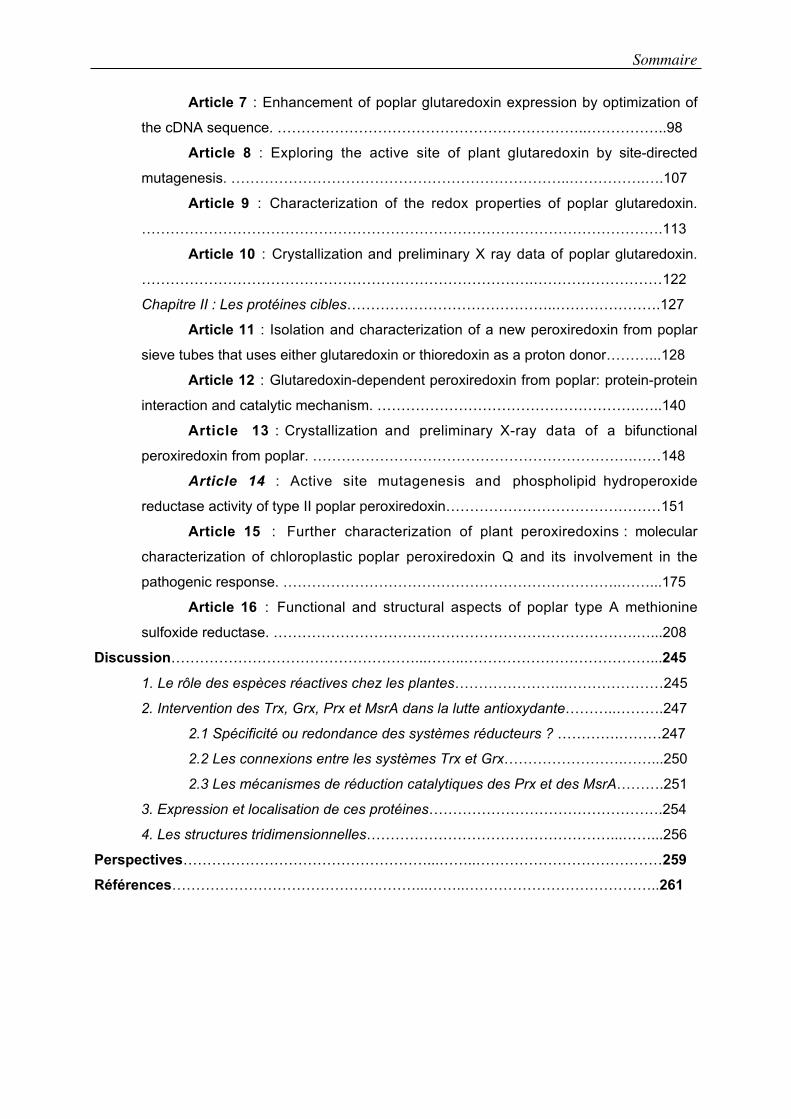
Dans un métabolisme cellulaire basal, le fonctionnement des chaînes de
transfert d’électrons et de certaines oxydases génère la formation d’espèces
réactives oxygénées (ROS) ou nitrées (RNS) (Figure 1).
Les concentrations de ces produits sont plus élevées lors de stress biotiques ou
abiotiques. A faible concentration, ces espèces radicalaires semblent être impliquées
dans les mécanismes de signalisation cellulaire, alors que de fortes concentrations
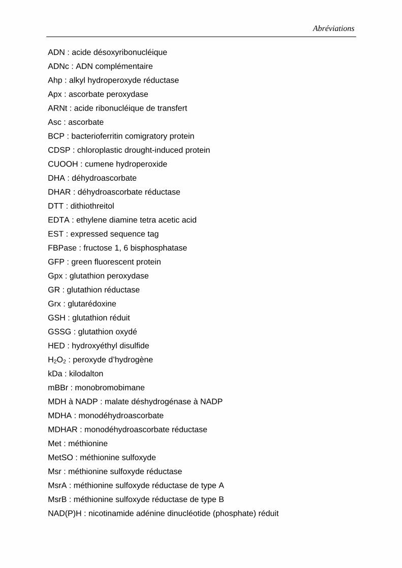
induisent un stress oxydatif qui va engendrer notamment des dommages au niveau
des macromolécules telles que les lipides, les acides nucléiques et les protéines
(Figures 2, 3 et 4) (Halliwell & Gutteridge, 1990 ; Beckman et al., 1997 ; Berlett et al.,
1997 ; Neill et al., 2002 ; Kohler et al., 2003).
Figure 1 : Voies de production des espèces oxygénées et nitrées réactives (d’après Nathan & Shiloh, 2000).
A B
Figure 2 : Phosphatidylcholine hydroperoxyde (PCOOH) (A) et oxydation des acides gras en acides gras hydroperoxydes (B) (d’après Baier & Dietz, 1999c ; Yamamoto, 2000).
Introduction
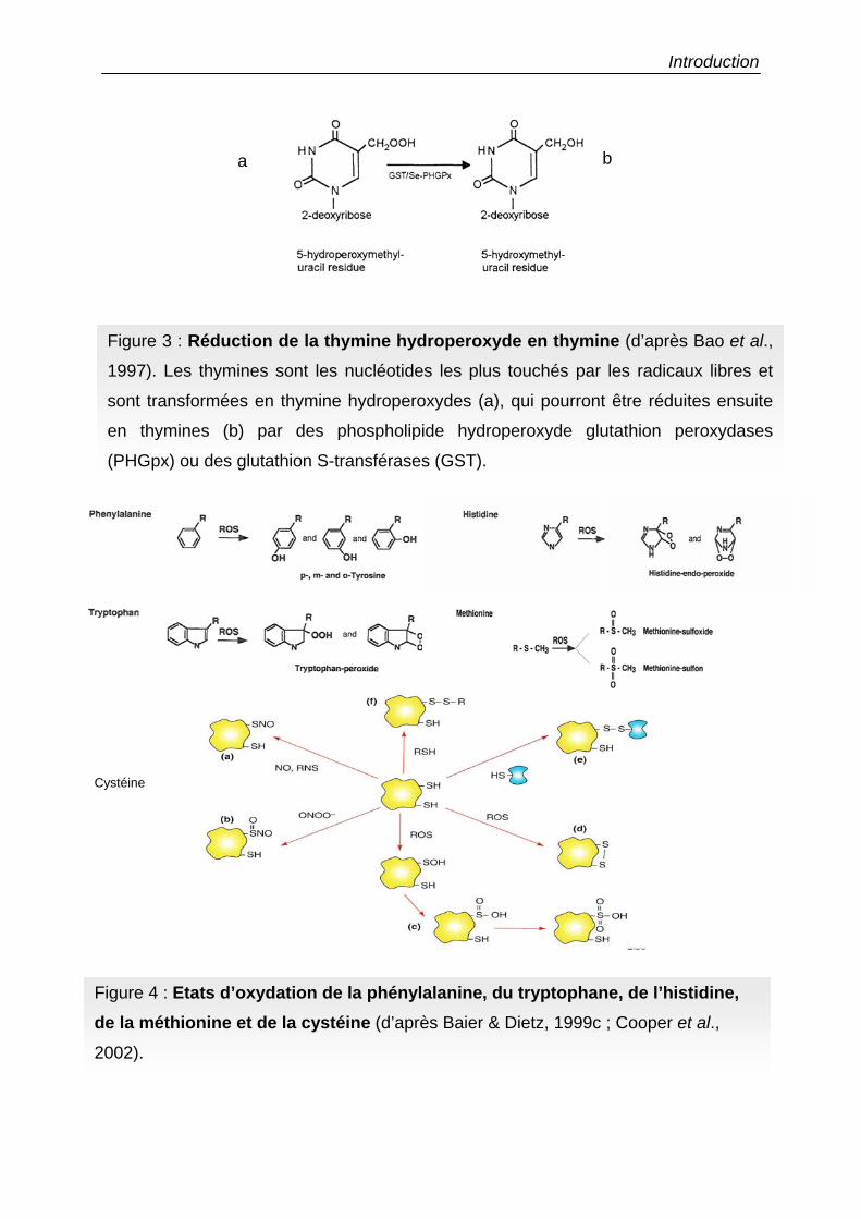
Figure 3 : Réduction de la thymine hydroperoxyde en thymine (d’après Bao et al.,
1997). Les thymines sont les nucléotides les plus touchés par les radicaux libres et
sont transformées en thymine hydroperoxydes (a), qui pourront être réduites ensuite
en thymines (b) par des phospholipide hydroperoxyde glutathion peroxydases
(PHGpx) ou des glutathion S-transférases (GST).
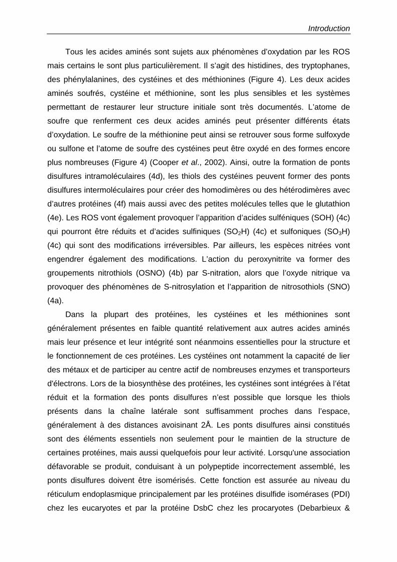
Figure 4 : Etats d’oxydation de la phénylalanine, du tryptophane, de l’histidine, de la méthionine et de la cystéine (d’après Baier & Dietz, 1999c ; Cooper et al.,
2002).
Cystéine
a b
Introduction
Tous les acides aminés sont sujets aux phénomènes d’oxydation par les ROS
mais certains le sont plus particulièrement. Il s’agit des histidines, des tryptophanes,
des phénylalanines, des cystéines et des méthionines (Figure 4). Les deux acides
aminés soufrés, cystéine et méthionine, sont les plus sensibles et les systèmes
permettant de restaurer leur structure initiale sont très documentés. L’atome de
soufre que renferment ces deux acides aminés peut présenter différents états
d’oxydation. Le soufre de la méthionine peut ainsi se retrouver sous forme sulfoxyde
ou sulfone et l’atome de soufre des cystéines peut être oxydé en des formes encore
plus nombreuses (Figure 4) (Cooper et al., 2002). Ainsi, outre la formation de ponts
disulfures intramoléculaires (4d), les thiols des cystéines peuvent former des ponts
disulfures intermoléculaires pour créer des homodimères ou des hétérodimères avec
d’autres protéines (4f) mais aussi avec des petites molécules telles que le glutathion
(4e). Les ROS vont également provoquer l’apparition d’acides sulféniques (SOH) (4c)
qui pourront être réduits et d’acides sulfiniques (SO2H) (4c) et sulfoniques (SO3H)
(4c) qui sont des modifications irréversibles. Par ailleurs, les espèces nitrées vont
engendrer également des modifications. L’action du peroxynitrite va former des
groupements nitrothiols (OSNO) (4b) par S-nitration, alors que l’oxyde nitrique va
provoquer des phénomènes de S-nitrosylation et l’apparition de nitrosothiols (SNO)
(4a).
Dans la plupart des protéines, les cystéines et les méthionines sont
généralement présentes en faible quantité relativement aux autres acides aminés
mais leur présence et leur intégrité sont néanmoins essentielles pour la structure et
le fonctionnement de ces protéines. Les cystéines ont notamment la capacité de lier
des métaux et de participer au centre actif de nombreuses enzymes et transporteurs
d'électrons. Lors de la biosynthèse des protéines, les cystéines sont intégrées à l’état
réduit et la formation des ponts disulfures n’est possible que lorsque les thiols
présents dans la chaîne latérale sont suffisamment proches dans l’espace,
généralement à des distances avoisinant 2Å. Les ponts disulfures ainsi constitués
sont des éléments essentiels non seulement pour le maintien de la structure de
certaines protéines, mais aussi quelquefois pour leur activité. Lorsqu'une association
défavorable se produit, conduisant à un polypeptide incorrectement assemblé, les
ponts disulfures doivent être isomérisés. Cette fonction est assurée au niveau du
réticulum endoplasmique principalement par les protéines disulfide isomérases (PDI)
chez les eucaryotes et par la protéine DsbC chez les procaryotes (Debarbieux &
Introduction
Beckwith, 1999 ; Frand et al., 2000 ; Collet et al., 2002). Ces deux protéines de la
famille des oxydoréductases réalisent ces isomérisations de ponts disulfures du fait
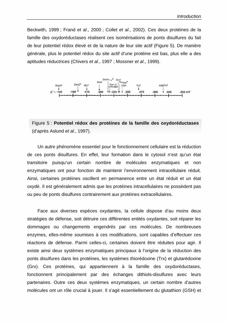
de leur potentiel rédox élevé et de la nature de leur site actif (Figure 5). De manière
générale, plus le potentiel rédox du site actif d’une protéine est bas, plus elle a des
aptitudes réductrices (Chivers et al., 1997 ; Mossner et al., 1999).
Un autre phénomène essentiel pour le fonctionnement cellulaire est la réduction
de ces ponts disulfures. En effet, leur formation dans le cytosol n’est qu’un état
transitoire puisqu’un certain nombre de molécules enzymatiques et non
enzymatiques ont pour fonction de maintenir l’environnement intracellulaire réduit.
Ainsi, certaines protéines oscillent en permanence entre un état réduit et un état
oxydé. Il est généralement admis que les protéines intracellulaires ne possèdent pas
ou peu de ponts disulfures contrairement aux protéines extracellulaires.
Face aux diverses espèces oxydantes, la cellule dispose d’au moins deux
stratégies de défense, soit détruire ces différentes entités oxydantes, soit réparer les
dommages ou changements engendrés par ces molécules. De nombreuses
enzymes, elles-même soumises à ces modifications, sont capables d’effectuer ces
réactions de défense. Parmi celles-ci, certaines doivent être réduites pour agir. Il
existe ainsi deux systèmes enzymatiques principaux à l’origine de la réduction des
ponts disulfures dans les protéines, les systèmes thiorédoxine (Trx) et glutarédoxine
(Grx). Ces protéines, qui appartiennent à la famille des oxydoréductases,
fonctionnent principalement par des échanges dithiols-disulfures avec leurs
partenaires. Outre ces deux systèmes enzymatiques, un certain nombre d’autres
molécules ont un rôle crucial à jouer. Il s’agit essentiellement du glutathion (GSH) et
Figure 5 : Potentiel rédox des protéines de la famille des oxydoréductases(d’après Aslund et al., 1997).
Introduction
de l’ascorbate, mais aussi de l’α-tocophérol par exemple. La synthèse
bibliographique qui suit, articulée autour de quatre articles de revue résumant
l’essentiel des données acquises chez les végétaux, a pour but de décrire non
seulement le fonctionnement de ces systèmes réducteurs (Trx et Grx) mais aussi
leur capacité à régénérer deux types d’enzymes cibles impliquées dans la lutte anti-
oxydante. Ce sont des peroxydases Trx ou Grx dépendantes appelées
peroxyrédoxines (Prx), qui réduisent le peroxyde d’hydrogène et différents alkyles ou
lipides hydroperoxydes, et des méthionine sulfoxyde réductases (Msr) qui ont pour
fonction essentielle de réduire des méthionine sulfoxydes (MetSO) en méthionine
(Met).
Ce manuscrit décrira en priorité les informations fournies par les études sur
les plantes, en incluant, lorsque les articles de synthèse sont devenus incomplets,
les avancées récentes dans le domaine végétal. Mettant à profit le séquençage
complet du génome d’Arabidopsis thaliana, les séquences végétales présentées
pour décrire les différentes familles de protéines seront celles d’A. thaliana, trouvées
dans la base de donnée MATDB (MIPS (Munich information center for protein
sequences) Arabidopsis thaliana database), à l’adresse suivante :
http://mips.gsf.de/proj/thal/db/index.html. Par ailleurs, les Msr végétales étant très
peu caractérisées, les informations obtenues chez divers autres organismes seront
principalement décrites.
1. Les molécules réductrices non enzymatiques
Ces molécules, à fort pouvoir anti-oxydant, telles que le GSH, l’acide
ascorbique (vitamine C) ou l’α-tocophérol (vitamine E) agissent soit directement sur
les espèces oxydantes, soit indirectement en activant un certain nombre d’enzymes
aux propriétés anti-oxydantes (Noctor & Foyer, 1998). De plus, l’équilibre entre les
états de réduction ou d’oxydation de ces molécules est gouverné par le cycle de
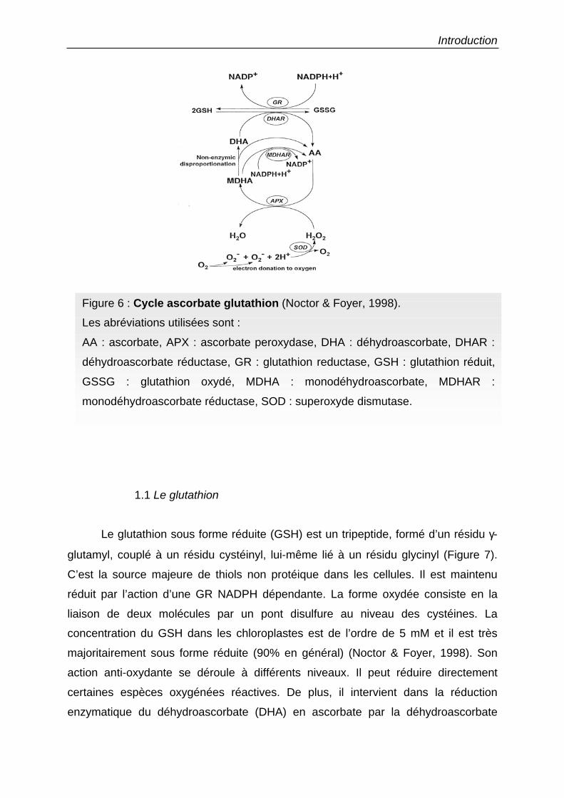
régénération ascorbate-glutathion (Figure 6) (Asada, 1999).
Introduction
1.1 Le glutathion
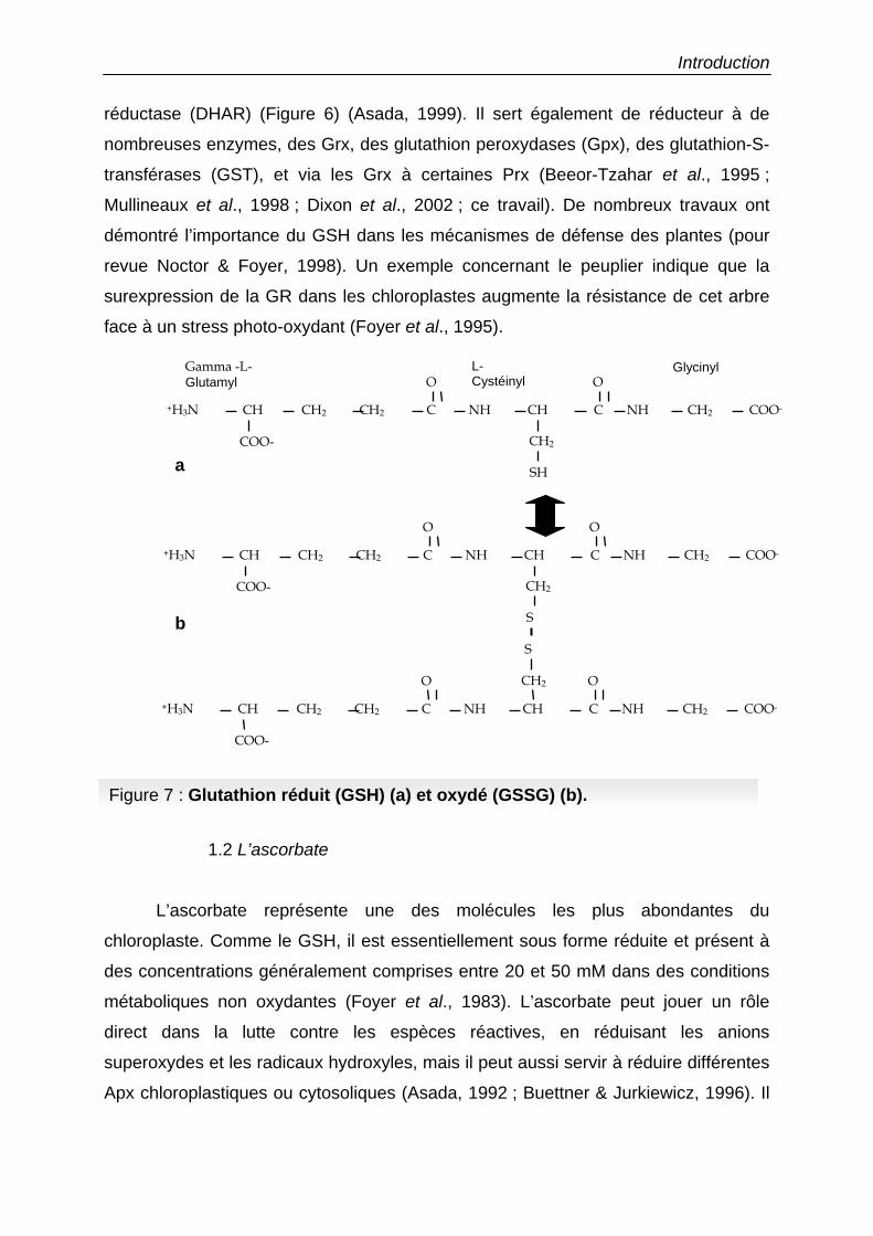
Le glutathion sous forme réduite (GSH) est un tripeptide, formé d’un résidu γ-
glutamyl, couplé à un résidu cystéinyl, lui-même lié à un résidu glycinyl (Figure 7).
C’est la source majeure de thiols non protéique dans les cellules. Il est maintenu
réduit par l’action d’une GR NADPH dépendante. La forme oxydée consiste en la
liaison de deux molécules par un pont disulfure au niveau des cystéines. La
concentration du GSH dans les chloroplastes est de l’ordre de 5 mM et il est très
majoritairement sous forme réduite (90% en général) (Noctor & Foyer, 1998). Son
action anti-oxydante se déroule à différents niveaux. Il peut réduire directement
certaines espèces oxygénées réactives. De plus, il intervient dans la réduction
enzymatique du déhydroascorbate (DHA) en ascorbate par la déhydroascorbate
Figure 6 : Cycle ascorbate glutathion (Noctor & Foyer, 1998).
Les abréviations utilisées sont :
AA : ascorbate, APX : ascorbate peroxydase, DHA : déhydroascorbate, DHAR :
déhydroascorbate réductase, GR : glutathion reductase, GSH : glutathion réduit,
GSSG : glutathion oxydé, MDHA : monodéhydroascorbate, MDHAR :
monodéhydroascorbate réductase, SOD : superoxyde dismutase.
Introduction
réductase (DHAR) (Figure 6) (Asada, 1999). Il sert également de réducteur à de
nombreuses enzymes, des Grx, des glutathion peroxydases (Gpx), des glutathion-S-
transférases (GST), et via les Grx à certaines Prx (Beeor-Tzahar et al., 1995 ;
Mullineaux et al., 1998 ; Dixon et al., 2002 ; ce travail). De nombreux travaux ont
démontré l’importance du GSH dans les mécanismes de défense des plantes (pour
revue Noctor & Foyer, 1998). Un exemple concernant le peuplier indique que la
surexpression de la GR dans les chloroplastes augmente la résistance de cet arbre
face à un stress photo-oxydant (Foyer et al., 1995).
1.2 L’ascorbate
L’ascorbate représente une des molécules les plus abondantes du
chloroplaste. Comme le GSH, il est essentiellement sous forme réduite et présent à
des concentrations généralement comprises entre 20 et 50 mM dans des conditions
métaboliques non oxydantes (Foyer et al., 1983). L’ascorbate peut jouer un rôle
direct dans la lutte contre les espèces réactives, en réduisant les anions
superoxydes et les radicaux hydroxyles, mais il peut aussi servir à réduire différentes
Apx chloroplastiques ou cytosoliques (Asada, 1992 ; Buettner & Jurkiewicz, 1996). Il
Figure 7 : Glutathion réduit (GSH) (a) et oxydé (GSSG) (b).
Gamma -L-Glutamyl
L-Cystéinyl
Glycinyl
CH CH2 C
O
NH CH
SH
C NH COO- CH2 CH2
CH2 COO-
+H3N
O
CH CH2 C
O
NH CH
S
C NH COO-CH2 CH2
CH2 COO-
+H3N
O
CH CH2 C
O
NH CH
S
C NH COO-CH2 CH2
CH2
COO-
+H3N
O
a
b
Introduction
peut également participer à la régénération du radical tocophérol en α-tocophérol
(Padh, 1990). En effet, l’α-tocophérol peut agir soit sur les anions superoxydes ou
radicaux hydroxyles, soit sur les lipides hydroperoxydes, se retrouvant ensuite
transformé en un cation radicalaire (Liebler et al., 1986). A la suite de son action,
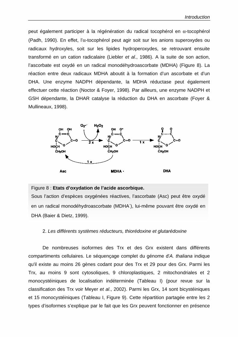
l’ascorbate est oxydé en un radical monodéhydroascorbate (MDHA) (Figure 8). La
réaction entre deux radicaux MDHA aboutit à la formation d’un ascorbate et d’un
DHA. Une enzyme NADPH dépendante, la MDHA réductase peut également
effectuer cette réaction (Noctor & Foyer, 1998). Par ailleurs, une enzyme NADPH et
GSH dépendante, la DHAR catalyse la réduction du DHA en ascorbate (Foyer &
Mullineaux, 1998).
2. Les différents systèmes réducteurs, thiorédoxine et glutarédoxine
De nombreuses isoformes des Trx et des Grx existent dans différents
compartiments cellulaires. Le séquençage complet du génome d'A. thaliana indique
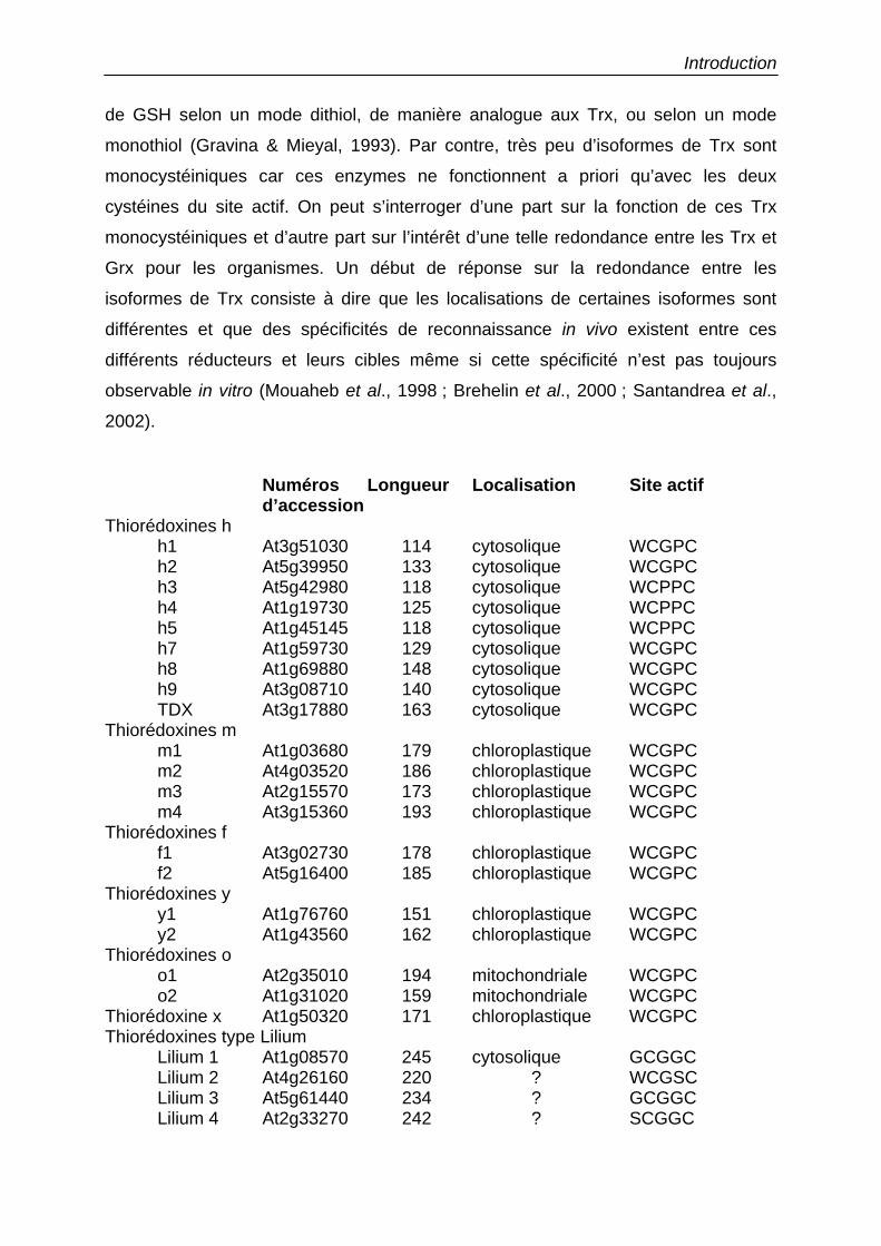
qu'il existe au moins 26 gènes codant pour des Trx et 29 pour des Grx. Parmi les
Trx, au moins 9 sont cytosoliques, 9 chloroplastiques, 2 mitochondriales et 2
monocystéiniques de localisation indéterminée (Tableau I) (pour revue sur la
classification des Trx voir Meyer et al., 2002). Parmi les Grx, 14 sont bicystéiniques
et 15 monocystéiniques (Tableau I, Figure 9). Cette répartition partagée entre les 2
types d’isoformes s’explique par le fait que les Grx peuvent fonctionner en présence
Figure 8 : Etats d’oxydation de l’acide ascorbique. Sous l’action d’espèces oxygénées réactives, l’ascorbate (Asc) peut être oxydé
en un radical monodéhydroascorbate (MDHA.), lui-même pouvant être oxydé en
DHA (Baier & Dietz, 1999).
Introduction
de GSH selon un mode dithiol, de manière analogue aux Trx, ou selon un mode
monothiol (Gravina & Mieyal, 1993). Par contre, très peu d’isoformes de Trx sont
monocystéiniques car ces enzymes ne fonctionnent a priori qu’avec les deux
cystéines du site actif. On peut s’interroger d’une part sur la fonction de ces Trx
monocystéiniques et d’autre part sur l’intérêt d’une telle redondance entre les Trx et
Grx pour les organismes. Un début de réponse sur la redondance entre les
isoformes de Trx consiste à dire que les localisations de certaines isoformes sont
différentes et que des spécificités de reconnaissance in vivo existent entre ces
différents réducteurs et leurs cibles même si cette spécificité n’est pas toujours
observable in vitro (Mouaheb et al., 1998 ; Brehelin et al., 2000 ; Santandrea et al.,
2002).
Numéros Longueur Localisation Site actif d’accession
Thiorédoxines h h1 At3g51030 114 cytosolique WCGPC h2 At5g39950 133 cytosolique WCGPC h3 At5g42980 118 cytosolique WCPPC h4 At1g19730 125 cytosolique WCPPC h5 At1g45145 118 cytosolique WCPPC h7 At1g59730 129 cytosolique WCGPC h8 At1g69880 148 cytosolique WCGPC h9 At3g08710 140 cytosolique WCGPC TDX At3g17880 163 cytosolique WCGPC
Thiorédoxines m m1 At1g03680 179 chloroplastique WCGPC m2 At4g03520 186 chloroplastique WCGPC m3 At2g15570 173 chloroplastique WCGPC m4 At3g15360 193 chloroplastique WCGPC
Thiorédoxines f f1 At3g02730 178 chloroplastique WCGPC f2 At5g16400 185 chloroplastique WCGPC
Thiorédoxines y y1 At1g76760 151 chloroplastique WCGPC y2 At1g43560 162 chloroplastique WCGPC
Thiorédoxines o o1 At2g35010 194 mitochondriale WCGPC o2 At1g31020 159 mitochondriale WCGPC
Thiorédoxine x At1g50320 171 chloroplastique WCGPC Thiorédoxines type Lilium
Lilium 1 At1g08570 245 cytosolique GCGGC Lilium 2 At4g26160 220 ? WCGSC Lilium 3 At5g61440 234 ? GCGGC Lilium 4 At2g33270 242 ? SCGGC
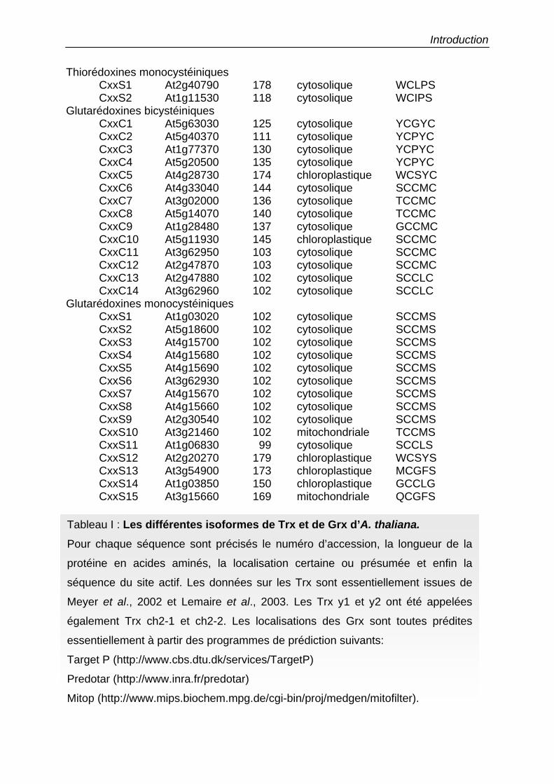
Introduction
Thiorédoxines monocystéiniques CxxS1 At2g40790 178 cytosolique WCLPS CxxS2 At1g11530 118 cytosolique WCIPS
Glutarédoxines bicystéiniques CxxC1 At5g63030 125 cytosolique YCGYC CxxC2 At5g40370 111 cytosolique YCPYC CxxC3 At1g77370 130 cytosolique YCPYC CxxC4 At5g20500 135 cytosolique YCPYC CxxC5 At4g28730 174 chloroplastique WCSYC CxxC6 At4g33040 144 cytosolique SCCMC CxxC7 At3g02000 136 cytosolique TCCMC CxxC8 At5g14070 140 cytosolique TCCMC CxxC9 At1g28480 137 cytosolique GCCMC CxxC10 At5g11930 145 chloroplastique SCCMC CxxC11 At3g62950 103 cytosolique SCCMC CxxC12 At2g47870 103 cytosolique SCCMC CxxC13 At2g47880 102 cytosolique SCCLC CxxC14 At3g62960 102 cytosolique SCCLC
Glutarédoxines monocystéiniques CxxS1 At1g03020 102 cytosolique SCCMS CxxS2 At5g18600 102 cytosolique SCCMS CxxS3 At4g15700 102 cytosolique SCCMS CxxS4 At4g15680 102 cytosolique SCCMS CxxS5 At4g15690 102 cytosolique SCCMS CxxS6 At3g62930 102 cytosolique SCCMS CxxS7 At4g15670 102 cytosolique SCCMS CxxS8 At4g15660 102 cytosolique SCCMS CxxS9 At2g30540 102 cytosolique SCCMS CxxS10 At3g21460 102 mitochondriale TCCMS CxxS11 At1g06830 99 cytosolique SCCLS CxxS1 2 At2g20270 179 chloroplastique WCSYS CxxS1 3 At3g54900 173 chloroplastique MCGFS CxxS1 4 At1g03850 150 chloroplastique GCCLG CxxS1 5 At3g15660 169 mitochondriale QCGFS
Tableau I : Les différentes isoformes de Trx et de Grx d’A. thaliana. Pour chaque séquence sont précisés le numéro d’accession, la longueur de la
protéine en acides aminés, la localisation certaine ou présumée et enfin la
séquence du site actif. Les données sur les Trx sont essentiellement issues de
Meyer et al., 2002 et Lemaire et al., 2003. Les Trx y1 et y2 ont été appelées
également Trx ch2-1 et ch2-2. Les localisations des Grx sont toutes prédites
essentiellement à partir des programmes de prédiction suivants:
Target P (http://www.cbs.dtu.dk/services/TargetP)
Predotar (http://www.inra.fr/predotar)
Mitop (http://www.mips.biochem.mpg.de/cgi-bin/proj/medgen/mitofilter).
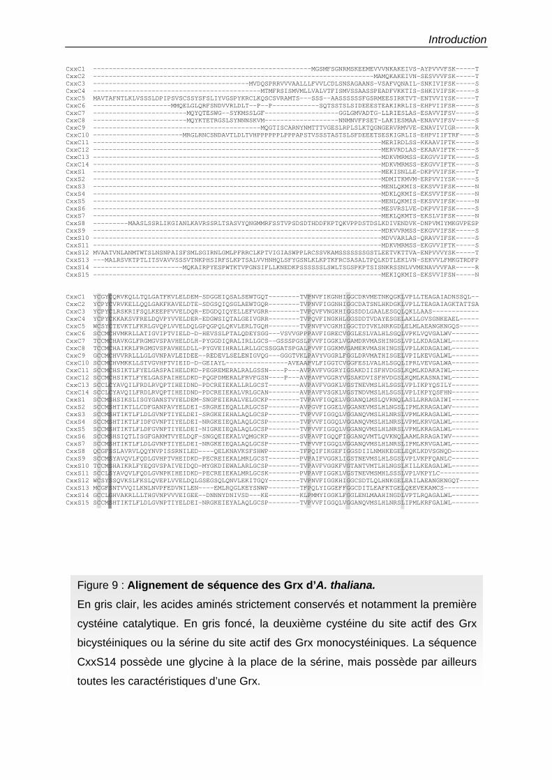
Introduction CxxC1 --------------------------------------------------------MGSMFSGNRMSKEEMEVVVNKAKEIVS-AYPVVVFSK-----TCxxC2 ------------------------------------------------------------------------MAMQKAKEIVN-SESVVVFSK-----TCxxC3 ----------------------------------------MVDQSPRRVVVAALLLFVVLCDLSNSAGAANS-VSAFVQNAIL-SNKIVIFSK-----SCxxC4 -------------------------------------------MTMFRSISMVMLLVALVTFISMVSSAASSPEADFVKKTIS-SHKIVIFSK-----SCxxC5 MAVTAFNTLKLVSSSLDPIPSVSCSSYSFSLIYVGSPYKRCLKQSCSVRAMTS---SSS--AASSSSSSFGSRMEESIRKTVT-ENTVVIYSK-----TCxxC6 --------------------MMQELGLQRFSNDVVRLDLT--P--P------------SQTSSTSLSIDEEESTEAKIRRLIS-EHPVIIFSR-----SCxxC7 ------------------------MQYQTESWG--SYKMSSLGF-------------------GGLGMVADTG-LLRIESLAS-ESAVVIFSV-----SCxxC8 ------------------------MQYKTETRGSLSYNNNSKVM-------------------NNMNVFPSET-LAKIESMAA-ENAVVIFSV-----SCxxC9 -------------------------------------------MQGTISCARNYNMTTTVGESLRPLSLKTQGNGERVRMVVE-ENAVIVIGR-----RCxxC10 -----------------------MRGLRNCSNDAVTLDLTVHPPPPPPLPPPAPSTVSSSTASTSLSFDEEETSESKIGRLIS-EHPVIIFTRF----SCxxC11 --------------------------------------------------------------------------MERIRDLSS-KKAAVIFTK-----SCxxC12 --------------------------------------------------------------------------MERVRDLAS-EKAAVIFTK-----SCxxC13 --------------------------------------------------------------------------MDKVMRMSS-EKGVVIFTK-----SCxxC14 --------------------------------------------------------------------------MDKVMRMSS-EKGVVIFTK-----SCxxS1 --------------------------------------------------------------------------MEKISNLLE-DKPVVIFSK-----TCxxS2 --------------------------------------------------------------------------MDMITKMVM-ERPVVIYSK-----SCxxS3 --------------------------------------------------------------------------MENLQKMIS-EKSVVIFSK-----NCxxS4 --------------------------------------------------------------------------MDKLQKMIS-EKSVVIFSK-----NCxxS5 --------------------------------------------------------------------------MENLQKMIS-EKSVVIFSK-----NCxxS6 --------------------------------------------------------------------------MESVRSLVE-DKPVVIFSK-----SCxxS7 --------------------------------------------------------------------------MEKLQKMTS-EKSLVIFSK-----NCxxS8 ---------MAASLSSRLIKGIANLKAVRSSRLTSASVYQNGMMRFSSTVPSDSDTHDDFKPTQKVPPDSTDSLKDIVENDVK-DNPVMIYMKGVPESPCxxS9 --------------------------------------------------------------------------MDKVVRMSS-EKGVVIFSK-----SCxxS10 --------------------------------------------------------------------------MDVVARLAS-QRAVVIFSK-----SCxxS11 --------------------------------------------------------------------------MDKVMRMSS-EKGVVIFTK-----SCxxS12 MVAATVNLANMTWTSLNSNPAISFSMLSGIRNLGMLPFRRCLKPTVIGIASWPPLRCSSVKAMSSSSSSSGSTLEETVKTTVA-ENPVVVYSK-----TCxxS13 ---MALRSVKTPTLITSVAVVSSSVTNKPHSIRFSLKPTSALVVHNHQLSFYGSNLKLKPTKFRCSASALTPQLKDTLEKLVN-SEKVVLFMKGTRDFPCxxS14 -----------------------MQKAIRPYESPWTKTVPGNSIFLLKNEDKPSSSSSSLSWLTSGSPKPTSISNKRSSNLVVMENAVVVFAR-----RCxxS15 --------------------------------------------------------------------------MEKIQKMIS-EKSVVIFSN-----N
CxxC1 YCGYCQRVKQLLTQLGATFKVLELDEM-SDGGEIQSALSEWTGQT--------TVPNVFIKGNHIGGCDRVMETNKQGKLVPLLTEAGAIADNSSQL--CxxC2 YCPYCVRVKELLQQLGAKFKAVELDTE-SDGSQIQSGLAEWTGQR--------TVPNVFIGGNHIGGCDATSNLHKDGKLVPLLTEAGAIAGKTATTSACxxC3 YCPYCLRSKRIFSQLKEEPFVVELDQR-EDGDQIQYELLEFVGRR--------TVPQVFVNGKHIGGSDDLGAALESGQLQKLLAAS------------CxxC4 YCPYCKKAKSVFRELDQVPYVVELDER-EDGWSIQTALGEIVGRR--------TVPQVFINGKHLGGSDDTVDAYESGELAKLLGVSGNKEAEL-----CxxC5 WCSYCTEVKTLFKRLGVQPLVVELDQLGPQGPQLQKVLERLTGQH--------TVPNVFVCGKHIGGCTDTVKLNRKGDLELMLAEANGKNGQS-----CxxC6 SCCMCHVMKRLLATIGVIPTVIELD-D-HEVSSLPTALQDEYSGG---VSVVGPPPAVFIGRECVGGLESLVALHLSGQLVPKLVQVGALWV-------CxxC7 TCCMCHAVKGLFRGMGVSPAVHELDLH-PYGGDIQRALIRLLGCS--GSSSPGSLPVVFIGGKLVGAMDRVMASHINGSLVPLLKDAGALWL-------CxxC8 TCCMCHAIKRLFRGMGVSPAVHELDLL-PYGVEIHRALLRLLGCSSGGATSPGALPVVFIGGKMVGAMERVMASHINGSLVPLLKDAGALWL-------CxxC9 GCCMCHVVRRLLLGLGVNPAVLEIDEE--REDEVLSELENIGVQG---GGGTVKLPAVYVGGRLFGGLDRVMATHISGELVPILKEVGALWL-------CxxC10 SCCMCHVMKKLLSTVGVHPTVIEID-D-GEIAYL----------------AVEAAPVLFIGGTCVGGFESLVALHLSGQLIPRLVEVGALWA-------CxxC11 SCCMCHSIKTLFYELGASPAIHELDKD-PEGREMERALRALGSSN----P---AVPAVFVGGRYIGSAKDIISFHVDGSLKQMLKDAKAIWL-------CxxC12 SCCMCHSIKTLFYELGASPAIHELDKD-PQGPDMERALFRVFGSN----P---AVPAVFVGGRYVGSAKDVISFHVDGSLKQMLKASNAIWL-------CxxC13 SCCLCYAVQILFRDLRVQPTIHEIDND-PDCREIEKALLRLGCST--------AVPAVFVGGKLVGSTNEVMSLHLSGSLVPLIKPYQSILY-------CxxC14 SCCLCYAVQILFRDLRVQPTIHEIDND-PDCREIEKALVRLGCAN--------AVPAVFVSGKLVGSTNDVMSLHLSGSLVPLIKPYQSFHN-------CxxS1 SCCMSHSIKSLISGYGANSTVYELDEM-SNGPEIERALVELGCKP--------TVPAVFIGQELVGGANQLMSLQVRNQLASLLRRAGAIWI-------CxxS2 SCCMSHTIKTLLCDFGANPAVYELDEI-SRGREIEQALLRLGCSP--------AVPGVFIGGELVGGANEVMSLHLNGSLIPMLKRAGALWV-------CxxS3 SCCMSHTIKTLFLDLGVNPTIYELDEI-SRGKEIEHALAQLGCSP--------TVPVVFIGGQLVGGANQVMSLHLNRSLVPMLKRAGALWL-------CxxS4 SCCMSHTIKTLFIDFGVNPTIYELDEI-NRGKEIEQALAQLGCSP--------TVPVVFIGGQLVGGANQVMSLHLNRSLVPMLKRVGALWL-------CxxS5 SCCMSHTIKTLFLDFGVNPTIYELDEI-NIGREIEQALAQLGCSP--------TVPVVFIGGQLVGGANQVMSLHLNRSLVPMLKRAGALWL-------CxxS6 SCCMSHSIQTLISGFGAKMTVYELDQF-SNGQEIEKALVQMGCKP--------SVPAVFIGQQFIGGANQVMTLQVKNQLAAMLRRAGAIWV-------CxxS7 SCCMSHTIKTLFLDLGVNPTIYELDEI-NRGKEIEQALAQLGCSP--------TVPVVFIGGQLVGGANQVMSLHLNRSLIPMLKRVGALWL-------CxxS8 QCGFSSLAVRVLQQYNVPISSRNILED----QELKNAVKSFSHWP--------TFPQIFIKGEFIGGSDIILNMHKEGELEQKLKDVSGNQD-------CxxS9 SCCMSYAVQVLFQDLGVHPTVHEIDKD-PECREIEKALMRLGCST--------PVPAIFVGGKLIGSTNEVMSLHLSGSLVPLVKPFQANLC-------CxxS10 TCCMSHAIKRLFYEQGVSPAIVEIDQD-MYGKDIEWALARLGCSP--------TVPAVFVGGKFVGTANTVMTLHLNGSLKILLKEAGALWL-------CxxS11 SCCLSYAVQVLFQDLGVNPKIHEIDKD-PECREIEKALMRLGCSK--------PVPAVFIGGKLVGSTNEVMSMHLSSSLVPLVKPYLC----------CxxS12 WCSYSSQVKSLFKSLQVEPLVVELDQLGSEGSQLQNVLEKITGQY--------TVPNVFIGGKHIGGCSDTLQLHNKGELEAILAEANGKNGQT-----CxxS13 MCGFSNTVVQILKNLNVPFEDVNILEN----EMLRQGLKEYSNWP--------TFPQLYIGGEFFGGCDITLEAFKTGELQEEVEKAMCS---------CxxS14 GCCLGHVAKRLLLTHGVNPVVVEIGEE--DNNNYDNIVSD---KE--------KLPMMYIGGKLFGGLENLMAAHINGDLVPTLRQAGALWL-------CxxS15 SCCMSHTIKTLFLDLGVNPTIYELDEI-NRGKEIEYALAQLGCSP--------TVPVVFIGGQLVGGANQVMSLHLNRSLIPMLKRFGALWL-------
Figure 9 : Alignement de séquence des Grx d’A. thaliana. En gris clair, les acides aminés strictement conservés et notamment la première
cystéine catalytique. En gris foncé, la deuxième cystéine du site actif des Grx
bicystéiniques ou la sérine du site actif des Grx monocystéiniques. La séquence
CxxS14 possède une glycine à la place de la sérine, mais possède par ailleurs
toutes les caractéristiques d’une Grx.

Introduction
Les trois articles de revue suivants traitent de façon non exhaustive des
différents modes de réduction des Trx et Grx de plantes, de leurs structures et de
leurs fonctions. Ces systèmes sont très bien caractérisés chez les organismes
bactériens et animaux mais les connaissances acquises chez ces organismes ne
seront pas détaillées dans ce manuscrit.
Introduction
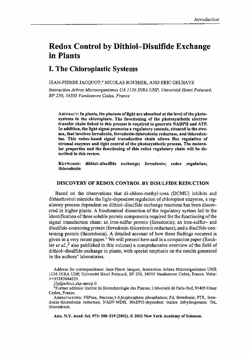
Cet article présente les voies de régulation rédox par échange dithiol-disulfure
dans les chloroplastes. Il décrit la composition et le fonctionnement d'une chaîne de
régulation appelée le système ferrédoxine-thiorédoxine qui comprend le transport
photosynthétique d'électrons, et trois protéines solubles, la ferrédoxine, la
ferrédoxine-thiorédoxine réductase, et les Trx. La structure de ces protéines et leur
mode d’interaction sont détaillés. Le mécanismes enzymatique qui est impliqué dans
ces régulations est basé sur des cascades d'échanges dithiol-disulfure dans
lesquelles une protéine se trouve oxydée alors que le partenaire est réduit. Dans la
deuxième partie de cet article sont détaillées les cibles moléculaires de ces systèmes
de régulation chloroplastique. Une attention particulière est accordée aux enzymes
pour lesquelles il existe à la fois des données biochimiques et structurales (NADP-
MDH et FBPase). Il apparait que la régulation rédox est apparue au cours de
l'évolution et de l'avènement de la photosynthèse oxygènique. Les biocatalyseurs
régulés ont été modifiés par addition ou insertion de séquences régulatrices de petite
taille ou par intégration sélective de cystéines adaptées à créer des ponts disulfures.
Après réduction, les enzymes cibles des Trx doivent subir une étape de changement
conformationnel pour aboutir à une configuration active.
Par contre, aucune Grx chloroplastique n’a été caractérisée chez les végétaux
jusqu’à présent, mais la GR et le GSH sont présents en grande quantité dans ce
compartiment cellulaire et certaines séquences d’A. thaliana possèdent une
extension N-terminale susceptible de diriger la protéine vers le chloroplaste (Tableau
I).
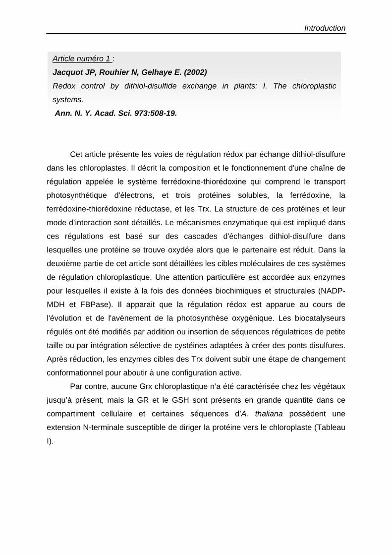
Article numéro 1 :
Jacquot JP, Rouhier N, Gelhaye E. (2002) Redox control by dithiol-disulfide exchange in plants: I. The chloroplastic
systems.
Ann. N. Y. Acad. Sci. 973:508-19.
Introduction
Redox Control by Dithiol-Disulfide Exchangein Plants
1. The Chloroplastic Systems
JEAN-PIERRE JACQUOT,a NICOLAS ROUHIER, AND ERIC GELHAYE
Interaction Arbres Microorganismes UA 1136 INRA UHP, Université Henri Poincaré,BP 230,54505 Vandoeuvre Cedex, France
ABSTRACT: In plants, the photons of light are absorbed at the level of the photosystems in the chloroplasts. The' functioning of the photosynthetic electrontransfer chain linked to this process is required to generate NADPH and ATP.In addition, the light signal promotes a regulatory cascade, situated in the stro~
ma, that involves ferredoxin, ferredoxin-thioredoxin reductase, and thioredoxins. This redox-based signal transduction chain allows fine regulation ofstromal enzymes and tlght control of the photosynthetic process. The molecular properties and the functioning of this redox regulatory chain will b-e described in tbis review.
KEYWORDS: dithiol-disulfide exchange; ferredoxin; redox regulation;thioredoxin
DISCOVERY OF REDOX CONTROL DY DISULFIDE REDUCTION
Based on the observations that di-chloro-methyl-urea (DeMU) inhibits anddithiothreitol mimicks the light-dependent regulation ofchioroplast enzymes, a regulatory process dependent on dithiol-disulfide exchange reactions has been discovered in higher plants. A biochemical dissection of the regulatory system led to theidentification ofthree soluble protein components required for the functioning ofthesignal transduction chain: an iron-sulfur protein (ferredoxin); an iron-sulfur- anddisulfide-containing protein (ferredoxin-thioredoxin reductase); and a disulfide-containing protein (thioredoxin). A detailed account of how these findings occurred isgiven in a very recent paper. 1 We will present here and in a companion paper (Rouhier et al.,2 also published in this volume) a comprehensive overview of the field ofdithiol-disulfide exchange in plants, with special emphasis on the results generatedin the authors' Iaboratories.
Address for correspondence: Jean-Pierre Jacquot, Interaction Arbres Microorganismes UMR1136 INRA UHP, Université Henri Poincaré, BP 230, 54505 Vandoeuvre Cedex, France. Vaice:++33383684225.
[email protected] address: Institut de Biotechnologie des Plantes; Université de Paris-Sud, 91405 Orsay
Cedex, France.ABBREVIATIONS: FBPase, fructose,I-6,bisphosphate phosphatase; Pd, ferredoxin; FfR, ferre
doxin-thioredoxin reductase; NADP-MDH, NADPH-dependent malate dehydrogenase; Trx,thioredoxin.
Ann. N.Y. Acad. Sei. 973: 508-519 (2002). © 2002 New York Academy of Sciences.
JACQUOT et al.: CHLOROPLASTIC THIOREDOXIN SYSTEM
STROMA
Introduction
509
Photosystem 1
Other enzymes
Fm,&
\
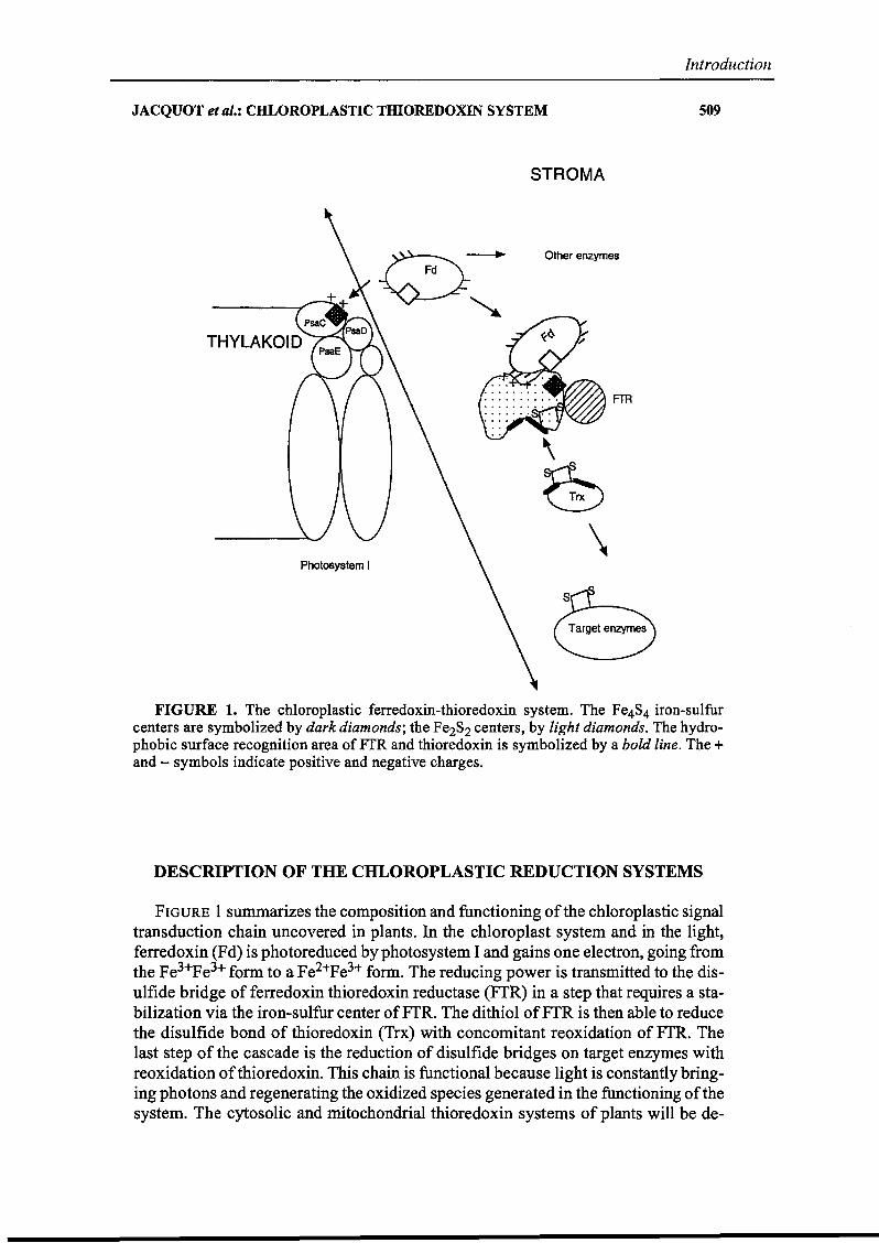
FIGURE 1. The ehloroplastic ferredoxin-thioredoxin system. The Fe4S4 iron-sulfurcenters are symbolized by dark diamonds; the Fe2S2 centers, by light diamonds. The hydrophobie surface recognition area ofFfR and thioredoxin is symbolized by a bold line. The +and - symbols indicate positive and negative charges.
DESCRIPTION OF THE CHLOROPLASTIC REDUCTION SYSTEMS
FIGURE 1 summarizes the composition and functioning of the chloroplastic signaltransduction chain uncovered in plants. In the chloroplast system and in the light,ferredoxin (Fd) is photoreduced by photosystem 1and gains one electron, going fromthe Fe3+Pe3+ form to a Fe2+Fe3+ farm. The reducing power is transmitted to the disulfide bridge of ferredoxin thioredoxin reductase (FfR) in a step that requires a stabilization via the iron-sulfur center ofFfR. The dithiol ofFfR is then able to reducethe disulfide bond of thioredoxin (Trx) with concomitant reoxidation of FfR. Thelast step of the cascade is the reduction of disulfide bridges on target enzymes withreoxidation ofthioredoxin. This chain is functional because light is constantly bringing photons and regenerating the oxidized species generated in the functioning ofthesystem. The cytosolic and mitochondrial thioredoxin systems of plants will he de-
510
Introduction
ANNALS NEW YORK ACADEMY OF SCIENCES
scribed in the companion paper by Rouhier et al.2 Several reviews have been devotedto the subject ofredox regulation via dithiol-disulfide exchange in plants.3,4 This review will detail the molecular mechanisms involved in the transmission of the redoxsignal in the chloroplastic redox regulatory chain. The description of the structures,functions, and protein-protein interaction properties of each of the individual components will be especially detailed.
FERREDOXIN: A KEY COMPONENT CONTROLLING THEDISTRIBUTION OF REDUCING POWER
Plant stromatic soluble ferredoxin belongs ta a subclass of iron-sulfur proteins.The chloroplastic protein is a very small protein (ca. 98 amino acids) encoded by nuclear genes as a higher molecular weight precursor that is processed upon entry inthe chloroplast. The sequence ofFd is extremely conserved among species from cyanobacteria to higher plants.5 This protein contains a Fe2S2 iron-sulfur center witha very low redox potential (ca. -420 mV). In higher plants, it is able to accept oneelectron from photosystem l, shifting from the oxidized state (Fe3+Pe 3+) to a reduced state (Fe2+Fe 3+). Ferredoxin is composed of a central pleated p sheet surrounded by three <X helices, each of these helices being surface accessible andcontaining conserved negative charges. The iron-sulfur center is situated close to theC terminal helix a3. Ferredoxin is a stro~atic soluble protein component that is ableta dock ta PSI by making molecular contacts with the subunits encoded by the psaC,psaD, andpsaE genes.6 The PsaC subunit is very important because it contains thethe two terminal electron acceptors ofPSI, the iron-sulfur centers FA and FB. A critical Lys residue of PsaC has been shawn ta be responsible for an adequate bindingofferredoxin to PSI.7 It is generally assumed that the interaction offerredoxin withPSI is based on electrostatic interactions, with negative charges offerredoxin matching positive charges of PSI. When reduced, ferredoxin is able to separate from PSIand bind ta its many electron acceptors. Ferredoxin is indeed a key component forthe distribution of the reducing power in the chloroplast, since it donates electronsta ferredoxin NADP reductase for NADPH production, to sulfite reductase for theincorporation of sulfur into organic compounds, to nitrite reductase and GOGAT(two enzymes involved in inorganic nitrogen assimilation), and ta ferredoxin-thioredoxin reductase, ta name just a few. Several of these protein-protein interactionshave been described in detail through site-directed mutagenesis or via the elucidation of the three-dimensional structure of the complexes.5,S-11 AlI results point outto the major role played by the electrostatic interactions in these contacts. It is believed that once ferredoxin has become close ta its protein partners through thestrong electrostatic interactions, a "fine tuning' of the fit between the two proteinscan accur through additional hydrophobie interactions. 12
THE ELECTRONIC SIGNAL IS CONVERTED INTO A DISULFIDEREDUCTION AT THE LEVEL OF FfR
FTR is a heterodimeric enzyme present exclusively in photosynthetic organismsranging from cyanobacteria ta higher plants. 13 Both subunits are encoded by nuclear
JACQUOT et al.: CHLOROPLASTIC THIOREDOXIN SYSTEM
Introduction
SIl
genes. One subunit has a constant size (ca. 13 kDa) and bears the catalytic sites; thesecond subunit varies in size according to the species (from 7 kDa in eyanobacteriato Il kDa in higher plants).14 The three-dimensional structure of the Synechocystisenzyme has recently been solved. 13 The enzyme has the shape of a biconcave discwith binding sites to ferredoxin on one side and to thioredoxin on the other side. Asfor other ferredoxin-dependent enzymes, it was shown by cross-linking experimentsand site-directed mutagenesis that the Fd/FTR interaction involves specifie electrostatie interactions, with positive charges of FTR matching the negative charges ofFd. The presence of a crucial Glu residue necessary for an adequate binding in theC tenninal a3 heIix of Fd has been elearly demonstrated. 15 The corresponding positive charges ofFTR have not yet been identified. It is speculated that when Fd bindsta FTR through these electrostatic interactions, this brings the iron-sulfur center offerredoxin into close proximity with the redox-active center of FTR (see FIG. 1).This center is composed of a high redox potential (above +420 mV) Fe4S4 iron-sulfur center and of a disulfide bridge (with a redox potential of- 320 fiV). Because ofits electropositive value, the iron-sulfur center of FTR cannot participate directly incatalysis. Rather, it helps stabilize the semireduced form of the disulfide bridge ofFTR in a redox mechanism unique to this class of enzymes. FTR is a unique enzymebecause it converts a monoelectronic signal from Fd (which carries only one electron) into a dielectronic signal needed for the reduction of the disulfide bridge. It isthus postulated that the enzyme will tirst "stock" one electron from Fd and then usea second Fd molecule to perform the full reduction of the disulfide. 16 The three-dimensional crystallographic structure reveals that the Fe4S4 center and the disulfidebridge are situated in close proximity, in agreement with the postulated mechanism.In addition, the disulfide bridge is close to the surface of the Trx interaction site.Overall, the structure of FTR seems ta be extremely weIl adapted to the electrontransfer with defined contact sites with the two interacting partner proteins and anoptimization of the distances between the catalytic sites.
THE DITHIOL-DISULFIDE EXCHANGE REACTIONS START AT THELEVEL OF THE FrR-THIOREDOXIN INTERACTION
Based on amine aeid sequences and target specificity, there are at least two typesofthioredoxins in higher plant chloroplasts. One type, ealled thioredoxin f, is the selective activator of the chloroplastic fructose-I,6-bisphosphatase; the other type,thioredoxin m, is a good activator of NADP-malate dehydrogenase. 17 In addition,new and possibly chloroplastic sequences have emerged that have been called thioredoxin x. 18 Like aIl thioredoxins, the chloroplastic thioredoxins contain about 110amino acids and are encoded by nuclear genes in higher plants. These proteins areknown to he extremely stable, presumably because of a high degree of secondarystructure relative to the overaillength of the proteine The ehloroplastic thioredoxinshave a very conserved active site (WCGPC[R/K]) that shows a very high reactivityin the reduction ofdisulfide bridges on other proteins. 19 Several crystallographic andNMR structures have been described for thioredoxins in higher plants and green aIgae.20,21 AlI proteins share the same foId, with a central pleated ~ sheet containingfive strands surrounded by ex helices. The active site is situated between the second-
512
Introduction
ANNALS NEW YORK ACADEMY OF SCIENCES
~~SH ~~SH S
HS
HS
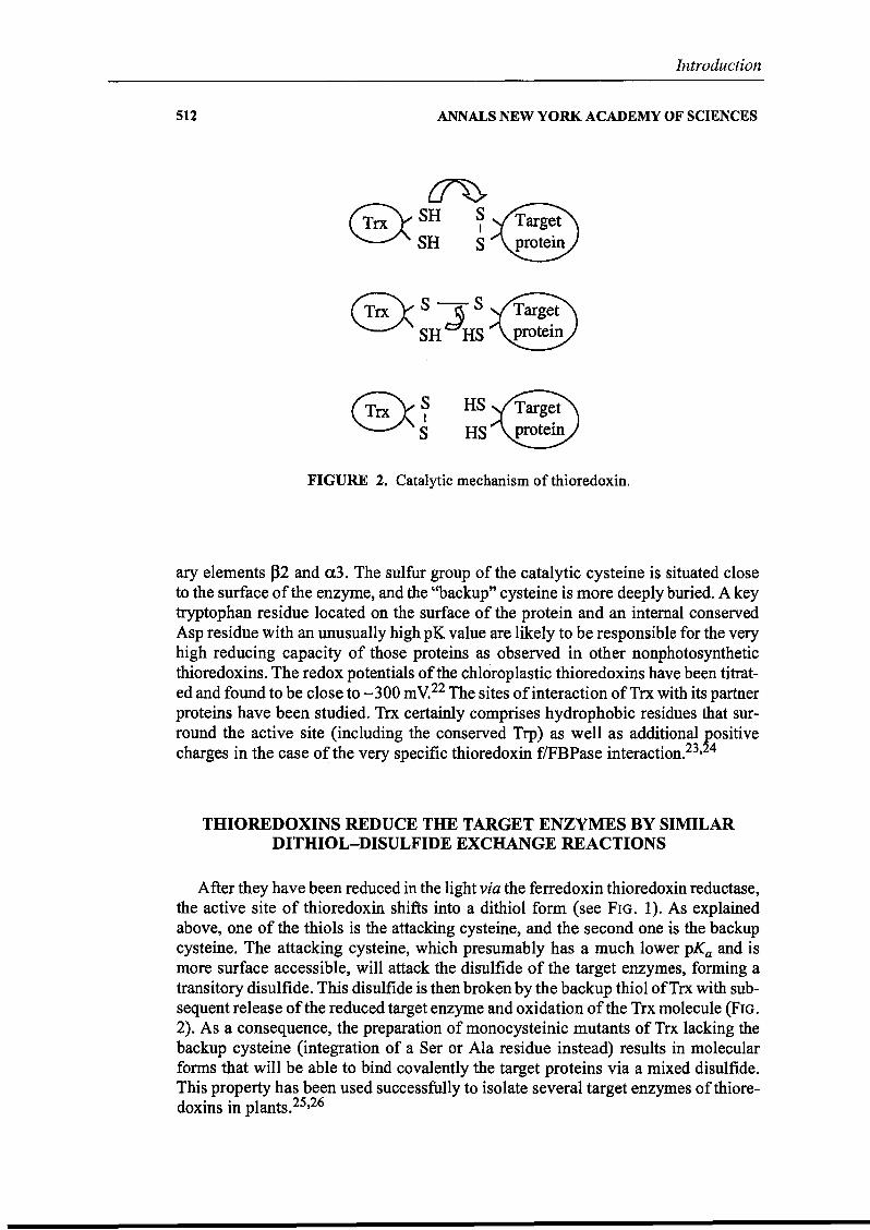
FIGURE 2. Catalytic mechanism of thioredoxin.
ary elements f32 and a3. The sulfur group of the catalytic cysteine is situated closeto the surface ofthe enzyme, and the "backup" cysteine is more deeply buried. A keytryptophan residue located on the surface of the protein and an internaI conservedAsp residue with an unusually high pK value are likely to be responsible for the veryhigh reducing capacity of those proteins as observed in other nonphotosyntheticthioredoxins. The redox potentials of the chloroplastic thioredoxins have been titrated and found to be close to -300 mV.22 The sites of interaction ofTrx with its partnerproteins have been studied. Trx certainly comprises hydrophobie residues that surround the active site (including the conserved Trp) as wel1 as additional ~ositivecharges in the case of the very specifie thioredoxin flFBPase interaction.23, 4
THIOREDOXINS REDUCE THE TARGET ENZYMES BY SIMILARDITHIOL-DI8ULFIDE EXCHANGE REACTIONS
After they have been reduced in the light via the ferredoxin thioredoxin reductase,the active site of thioredoxin shifts into a dithiol fonn (see FIG. 1). As explainedabove, one of the thiols is the .attacking cysteine, and the second one is the backupcysteine. The attacking cysteine, which presumably has a much lower pKa and ismore surface accessible, will attack the disulfide of the target enzymes, forming atransitory disulfide. This disulfide is then broken by the backup thiol ofTrx with subsequent release ofthe reduced target enzyme and oxidation ofthe Trx molecule (FIG.2). As a consequence, the preparation of monocysteinic mutants of Trx lacking thebackup cysteine (integration of a Ser or Ala residue instead) results in molecularfonns that will be able to bind covalently the target proteins via a rnixed disuifide.This property has been used successfully to isolate several target enzymes of thioredoxins in plants.25,26
JACQUOT et al.: CHLOROPLASTIC THIOREDOXIN SYSTEM
Sequence addition
Introduction
513
i 11Sequence insertion
__i _Insertion of selected cysteines
~__ï......--:t__i~~~_S_1 _
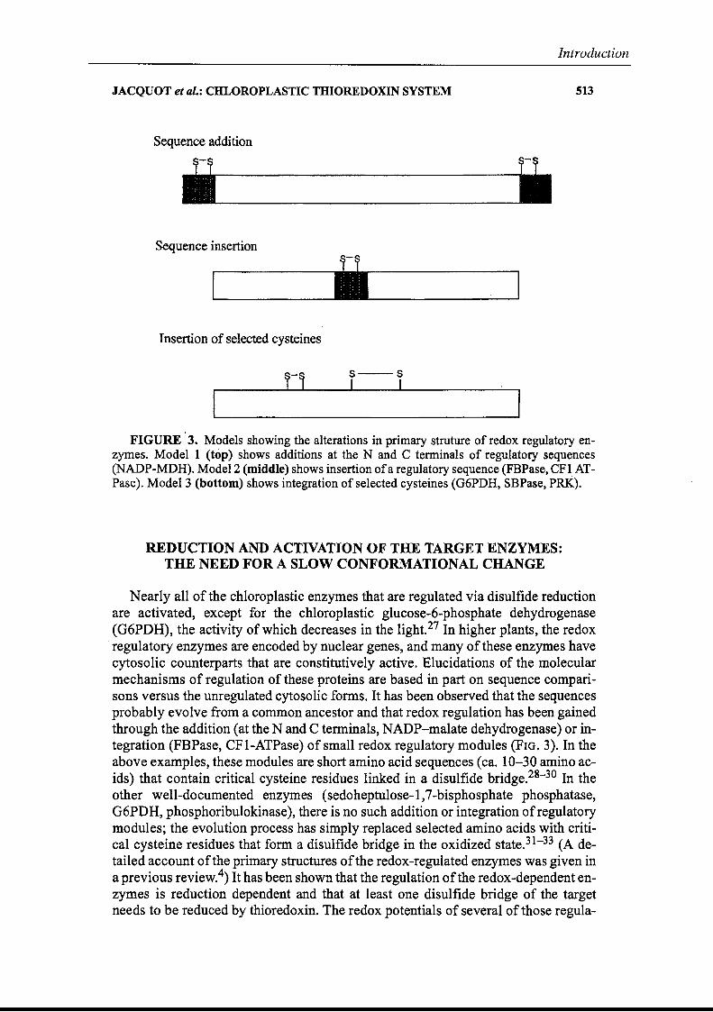
FIGURE' 3. Models showing the alterations in primary struture of redox regulatory enzymes. Model 1 (top) shows additions at the N and C terminaIs of regulatory sequences(NADP-MDH). Mode12 (middle) shows insertion ofa regulatory sequence (FBPase, CFt ATPase). Mode13 (bottom) shows integration ofselected cysteines (G6PDH, SBPase, PRK).
REDUCTION AND ACTIVATION OF THE TARGET ENZYMES:THE NEED FOR A SLOW CONFORMATIONAL CHANGE
Nearly aIl of the chloroplastic enzymes that are regulated via disulfide reductionare activated, except for the chloroplastic glucose-6-phosphate dehydrogenase(G6PDH), the activity ofwhich decreases in the light.27 In higher plants, the redoxregulatory enzymes are encoded by nuclear genes, and many of these enzymes havecytosolic counterparts that are constitutively active. Elucidations of the molecularmechanisms of regulation of these proteins are based in part on sequence comparisons versus the unregulated cytosolic fonns. It has been observed that the sequencesprobably evolve from a common ancestor and that redox regulation has been gainedthrough the addition (at the N and C terminaIs, NADP-malate dehydrogenase) or integration (FBPase, CFI-ATPase) of small redox regulatory modules (FIG. 3). In theabove examples, these modules are short amino acid sequences (ca. 10-30 amino acids) that contain critical cysteine residues linked in a disulfide bridge.28- 30 In theother well-documented enzymes (sedoheptulose-I,7-bisphosphate phosphatase,G6PDH, phosphoribulokinase), there is no such addition or integration ofregulatorymodules; the evolution process has simply replaced selected amino acids with critical cysteine residues that form a disulfide bridge in the oxidized state.31- 33 (A detailed account ofthe primary structures ofthe redox-regulated enzymes was given ina previous review.4) It has been shown that the regulation ofthe redox-dependent enzymes is reduction dependent and that at least one disulfide bridge of the targetneeds to be reduced by thioredoxin. The redox potentials of several of those regula-
514
Introduction
ANNALS NEW YORK ACADEMY OF SCIENCES
Reduction
Activity
5 10 15
Time (min)
FIGURE 4. Rates of reduction and catalysis of redox regulatory enzymes.
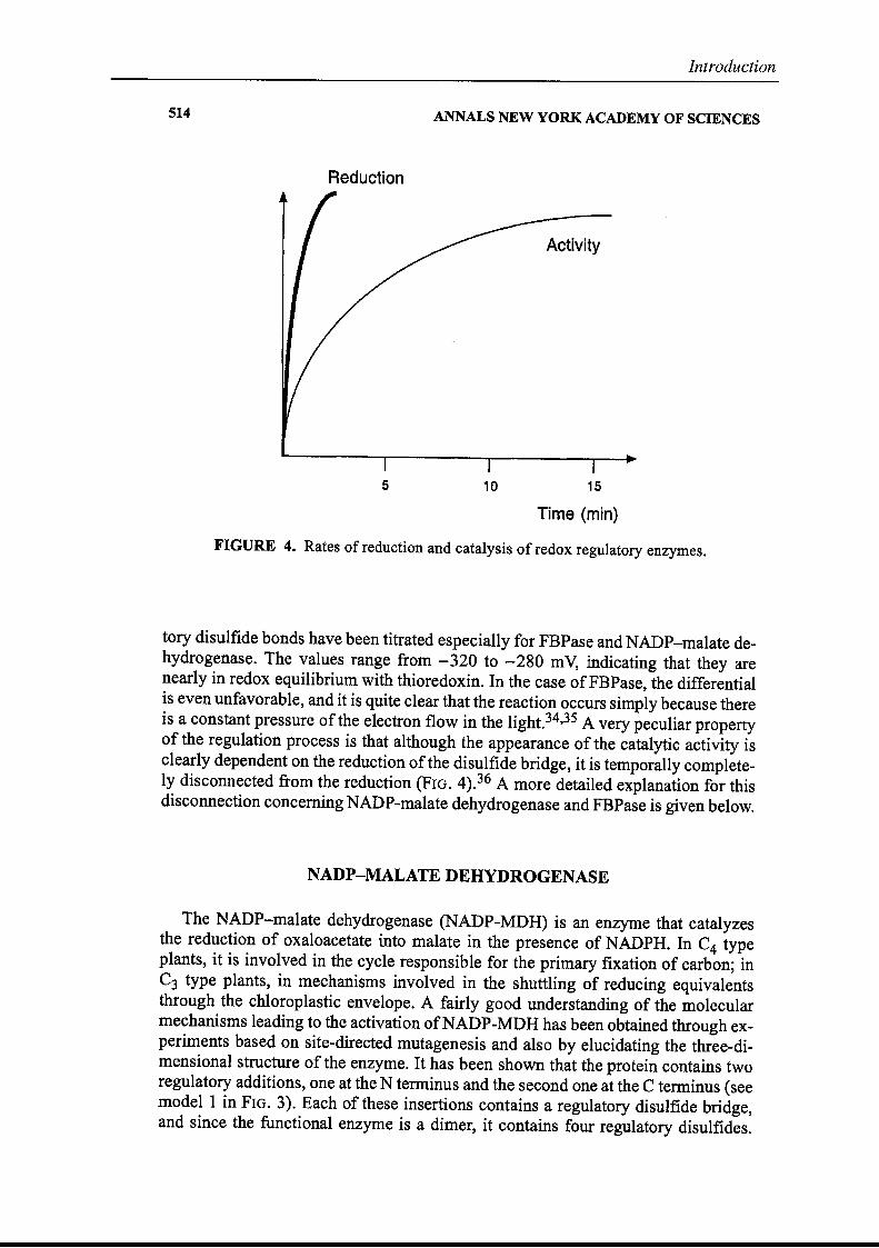
tory disulfide bonds have been titrated especially for FBPase and NADP-malate dehydrogenase. The values range from - 320 to - 280 mV, indicating that they arenearly in redox equilibrium with thioredoxin. In the case ofFBPase, the differentialis even unfavorable, and it is quite clear that the reaction occurs simply because thereis a constant pressure of the electron flow in the light.34,35 A very peculiar propertyof the regulation process is that although the appearance of the catalytic activity isclearly dependent on the reduction ofthe disulfide bridge, it is temporally completeIy disconnected°from the reduction (FIG. 4).36 A more detailed explanation for thisdisconnection conceming NADP-malate dehydrogenase and FBPase is given below.
NADP-MALATE DEHYDROGENASE
The NADP-malate dehydrogenase (NADP-MDH) is an enzyme that catalyzesthe reduction of oxaloacetate into malate in the presence of NADPH. In C4 typeplants, it is involved in the cycle responsible for the primary fixation of carbon; inC3 type plants, in mechanisms involved in the shuttling of reducing equivalentsthrough the chloroplastic envelope. A fairly good understanding of the molecularmechanisms leading to the activation ofNADP-MDH has been obtained through experiments based on site-directed mutagenesis and a180 by elucidating the three-dimensional structure of the enzyme. It has been shawn that the protein contains tworegulatory additions, one at the N terminus and the second one at the Ctenninus (seemode! 1 in FIG. 3). Each ofthese insertions contains a regulatory disulfide bridge,and since the functional enzyme is a dimer, it cantains four regulatory disulfides.
JACQUOT et al.: CHLOROPLASTIC THIOREDOXIN SYSTEM
Introduction
515
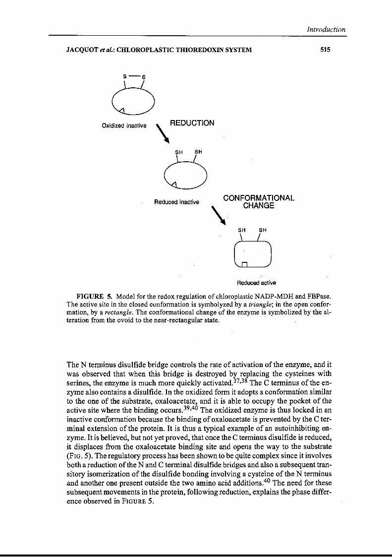
Oxidized inactive \ REDUCTION
SH SH
\Reduced inactive CONFORMATIONAL
CHANGE
5H SH
Reduced active
FIGURE 5. Model for the redox regulation of chloroplastic NADP-MDH and FBPase.The active site in the closed conformation is symbolyzed by a triangle; in the open conformation, by a rectangle. The conformational change of the enzyme is symbolized by the alteration from the ovoid to the near-rectangular state.
The N termi~us disulfide bridge contraIs the rate of activation of the enzyme, and itwas observed that when this bridge is destroyed by replacing the cysteines withserines, the enzyme is much more quickly activated.37,38 The C terminus of the enzyme also contains a disulfide. In the oxidized form it adopts a conformation similarto the one of the substrate, oxaloacetat~, and it is able to occupY the pocket of theactive site where the binding occurs.39,40 The oxidized enzyme is thus Iocked in aninactive confo.rmation because the binding ofoxaloacetate is prevented by the C terminal extension of the proteine It is thus a typical example of an autoinhibiting enzyme. It is believed, but not yet proved, that once the C terminus disulfide is reduced,it displaces from the oxaloacetate binding site and opens the way to the substrate(FIG. 5). The regulatory process has been shawn to be quite complex since it involvesboth a reduction ofthe N and C terminal disulfide bridges and also a subsequent transitory isomerization of the disulfide bonding involving a cysteine of the N tenninusand another one present outside the two amino acid additions.40 The need for thesesubsequent movements in the protein, following reduction, explains the phase difference observed in FIGURE 5.
516
Introduction
ANNALS NEW YORK ACADEMY OF SCIENCES
FRUCTOSE-l,6-BISPHOSPHATE PHOSPHATASE
Fructose-I,6-bisphosphate phosphatase (FBPase) is a protein that is a key regulatory component of the photosynthetic carbon reduction cycle, otherwise known asthe Calvin cycle. In the presence ofMg2+, this enzyme converts fructose-I,6-bisphosphate into fructose-6-phosphate with the concomitant release of inorganic phosphate. FBPase is a rather unusual regulatory enzyme because it is active either in thereduced state or in the oxidized state. When the Mg2+ concentration is low in the reaction medium, only the reduced farm is active; but when the Mg2+ concentration isincreased to unphysiologicallevels (above 10 mM), the oxidized enzyme becomescatalytically competent. The physiological activator ofFBPase is thioredoxin f. (Theinteraction sites between the two proteins have been studied in detail.) In addition tothe previously mentioned hydrophobie contacts, the selectivity of the Trx f-FBPaseinteraction relies also on complementary electrostatic charges, positive on thioredoxin f and negative onFBPase.24,41 Analysis of the primary structure of the regulated·enzyme versus the cytosolic, constitutively active form reveals an insertion ofca. 30 amino acids, an insertion that contains three conserved Cys residues. Site-directed mutagenesis combined with the elucidation of the crystallographic structurehave indicated that two of these residues, separated by 20 amino acids, are indeedengaged in a disulfide bond.42,43 The chloroplastic protein is a homotetramer withan overall molecular mass of 160 kDa. Each of the regulatory insertions is situatedon a solvent-accessible flexible loop, distant from the active site. A comparison ofthe structures of the oxidized inactive and reduced active enzymes helps explain theregulation of the enzyme.43,44 It is postulated that once the enzyme is reduced viathioredoxin f, the regulatory loop changes orientation, and this in tum induces amovement of two ~ strands (~1 and ~2) that shift from a parallel to an antiparallelconformation. This is likely to cause a displacement ofthe side chain ofa Val residuethat is replaced by a glutamate. As this residue, unlike the Val, possesses a chargedcarboxyl group on its laterai chain, it is able to bind electrostatically the catalytic ionand bring it into close proximity to the other substrate fructose'-1,6-bisphosphate.Following those movements ofsecondary structures and amino acid side chains, theenzyme becomes catalytically competent. This postulated mechanism of activationexplains why the rates of reduction and of appearance of catalytic competence arenot in phase for FBPase as weIl.
CONCLUDING REMARKS
Several other chloroplastic enzymes are known to be regulated via the reductionofdisufide bridges, but the molecular nature ofthe modifications that occur on thoseenzymes is not as weIl documented as the above examples. A quite detailed accountof these findings is reported in a paper by Meyer et al.3 The molecular evolution ofthe thioredoxin systems in plants is also of great interest. It gives insights on the rationale behind these light-dependent regulation reactions that seem to be strictlylinked to oxygenic photosynthesis, presumably for fighting oxidizing species. Another explanation is the need to avoid futile cycles in eukaryotic compartmented organisms. These aspects, which cannot be treated here, are developed in severa!review papers.3,4
JACQUOT et al.: CHLOROPLASTIC THIOREDOXIN SYSTEM
REFERENCES
Introduction
517
1. BUCHANAN, B.B., P. SCHÜRMANN, R.A. WOLOSIUK & J.P. JACQUOT. 2002. The ferredoxin-thioredoxin system: from discovery to molecular structures and beyond. Phot.Res. 73: 215-222.
2. ROUHIER, N., E. GELHAYE & J.P. JACQUOT. 2002. Redox control by dithiol-disulfideexchange in plants. II. The cytosolic and mitochondiral systems. Ann. N.Y. Acad.Sei. This volume.
3. MEYER, Y., L. VERDOUCQ & F. VIGNOLS. 1999. Plant thioredoxins and glutaredoxins:identity and putative roles. Trends Plant Sei. 4: 387-394.
4. SCHÜRMANN, P. & J.P. JACQUOT. 2000. Plant thioredoxin systems revisited. Annu. Rev.Plant Physiol. Plant Mol. Biol. 51: 371-400.
5. TSUKIHARA, T., K. FUKUYAMA, M. MIZUSHIMA, et al. 1990. Structure of the [2Fe2S]ferredoxin 1 from the blue-green alga Aphanothece sacrum at 2.2 Aresolution. J.MoL Biol. 216: 399-410.
6. VASSILIEV, 1.R., Y.S. JUNG, F. YANG & J.H. GOLBECK. 1998. PsaC subunit ofphotosystem 1 is oriented with iron-sulfur cluster F(B) as the immediate eleetron donor toferredoxin and flavodoxin. Biophys. J. 74: 2029-2035.
7. FISCHER, N., M. HIPPLER, P. SETIF, et al. 1998. The PsaC subunit ofphotosystem 1providesan essentiallysine residue for fast electron transfer to ferredoxin. EMBO J. 17: 849-858.
8. KNAFF, D.B. & M. HIRASAWA. 1991. Ferredoxin-dependent chloroplast enzymes. Biochim. Biophys. Acta 1056: 93-125.
9. BINDA, C., A. CODA, A. ALIVERTI, et al. 1998. Structure of the mutant E92K of [2Fe28] ferredoxin 1 from Spinacia oleracea at 1.7 A resolution. Acta Crystallogr. DBiol. Crystallogr. 54: 1353-1358.
10. HURLEY J.K. t J.T. HAZZARD, M. MARTINEz-JULVEZ, et al. 1999. Electrostatic forcesinvolved in orienting Anabaena ferredoxin during binding to Anabaena ferredoxin:NADP+ reductase: site-specifie mutagenesis, transient kinetic measurements,and electrostatic surface potentials. Protein Sei. 8: 1614-1622.
Il. GARCIA-SANCHEZ, M.I., A. DIAZ..QUINTANA, C. GOTOR, et al. 2000. Homology predicted structure and funtionai interaction of ferredoxin from the eukaryotic aigaChlamydomonas reinhardti with nitrite reductase and glutamate synthase. J. Biol.Inorg. Chem. 5: 713-719.
12. MARTINEz-JULVEZ, M., 1. NOGUES, M. FARO, et al. 2001. Role of a cluster ofhydrophobic residues near the FAD cofactor in Anabaena PCC 7119 ferredoxin-NADP+reductase for optimal complex formation and electron transfer to ferredoxin. J. Biol.Chem. 276: 27498-27510.
13. DAI, S., C. SCHWENDTMAYER, K. JOHANSSON, et al. 2000. How does light regulate chloroplast enzymes? Structure-function studies of the ferredoxin/thioredoxin system. Q.Rev. Biophys. 33: 67-108.
14. GAYMARD, E., L. FRANCHINI, W. MANIERI, et al. 2000. A dieistronic construet for theexpression of functional spinach chloroplast ferredoxin:thioredoxin reductase inEscherichia coli. Plant Sci. 158: 107-113.
15. JACQUOT, J.P., M. STEIN, A. SUZUKI, et al. 1997. Residue Glu-91 of Chlamydomonasreinhardtii ferredoxin is essential for eleetron transfer to ferredoxin-thioredoxinreductase. FEBS Lett. 400: 293-296.
16. STAPLES, C.R., E. GAYMARD, A.L. STRITT-EYER, et al. 1998. Role of the [Fe4S4] cIuster in mediating disulfide reduction in spinach ferredoxin:thioredoxin reductase. Biochemistry 37: 4612-4620.
17. STEIN, M., J.P. JACQUOT, E. JEANNETTE, et al. 1995. Chlamydomonas reinhardtii thioredoxins: structure of the genes coding for the chloroplastic ID and cytosolic h isoforms; expression in Escherichia coli of the recombinant proteins, purification andbiochemical properties. Plant Mol. Biol. 28: 487-503.
18. MESTRES-ORTEGA, D. & Y. MEYER. 1999. The Arabidopsis thaliana genome encodes atIeast four thioredoxins fi and a new prokaryotic-like thioredoxin. Gene 240: 307-316.
19. LANCELIN, J.M., M. STEIN & J.P. JACQUOT. 1993. Secondary structure and protein folding of recombinant eh1oroplastic thioredoxin Ch2 from the green aiga Chlamydomonas reinhardtii as determined by lH NMR. J. Biochem. 114: 421-431.
Introduction
518 ANNALS NEW YORK ACADEMY OF SCIENCES
20. CAPITANI, G., Z. MARKOVIC-HoUSLEY, G. DELVAL, et al. 2000. Crystal structures oftwo functionally different thioredoxins in spinach chloroplasts. J. Mol. Biol. 302:135-154.
21. LANCELIN, J.M., L. GUILHAUDIS, M. STEIN, et al. 2000. NMR structures ofthioredoxinm from the green alga Chlamydomomas reinhardtii. Prot. Struct. Funct. Genet. 41:334-349.
22. HIRASAWA, M., P. SCHÜRMANN, J.P. JACQUOT, et al. 1999. Oxidation-reduction properties of chloroplast thioredoxins, ferredoxin-thioredoxin reductase and fructose bisphosphatase. Biochemistry 38: 5200-5205.
23. KATTI, S.K., D.M. LEMASTER & H. EKLUND. 1990. Crystal structure of thioredoxinfrom Escherichia coli at 1.68 Ar.esolution. J. Mol. Biol. 212: 167-184.
24. GECK, M.K., F.W. LARIMER & F.C. HARTMAN. 1996. Identification ofresidues of spinach thioredoxin fthat influence interactions with target enzymes. J. Biol. Chem. 271:24736-24740.
25. GOYER, A., C. HALESKOS, M. MIGINIAC-MASLOW, et al. 2002. Isolation and characterization of a thioredoxin-dependent peroxidase from Chlamydomonas reinhardtii.Eur. J. Biochem. 269: 1-11.
26. MOTOHASHI, K., A. KONDOH, M.T. STUMPP & T. HISABORI. 2001. Comprehensive survey of proteins targeted by chloroplast thioredoxin. Proc. Natl. Acad. Sei. USA 98:11224-11229.
27. VON SCHAEWEN, A., G. LANGENKAMPER, K. GRAEVE, et al. 1995. Molecular characterization of the plastidic glueose-6-phosphate dehydrogenase from potato in comparison to its·cytosolic counterpart. Plant Physiol. 109: 13.27-1335.
28. ISSAKIDIS, E., M. LEMAIRE, P. DECOTTIGNIES, et al. 1996. Direct evidence for the different roles of the N- and C-terminal regulatory disulfides of sorghum leaf NADPmalate dehydrogenase in its activation by reduced thioredoxin. FEBS Lett. 392: 121124.
29. RAINES, C.A., J.C. LLOYD, M. LONGSTAFF, et al. 1988. Chloroplast fructose .. l,6-bisphosphatase: the product of a mosaic genet Nucleic Acids Res. 16: 7931-7942.
30. MIKI, J., M. MAEDA, Y. MUKOHATA & M. FUTAI. 1988. The gamma-subunit of ATPsynthase from spinach chloroplasts. Primary structure deduced from the clonedcDNA sequence. FEBS Lett. 232: 221-226.
31. DUNFORD, R.P., M.A. CATLEY, C.A. RAINES, et al. 1998. Purification of active chloropIast sedoheptulose-l,7-bisphosphatase expressed in Escherichia coli. Protein Expr.Purif. 14: 139-145.
32. BRANDES, H.K., F.C. HARTMAN, T.Y. Lu & F.W. LARIMER. 1996. Efficient expression ofthe gene for spinach phosphoribulokinase in Pichia pastoris and utilization of therecombinant enzyme to explore the raIe of regulatory cysteinyl residues by sitedirected mutagenesis. J. Biol. Chem. 271: 6490-6496.
33. WENDEROTH, 1., R. SCHEIBE & A. VON SCHAEWEN. 1997. Identification of the cysteineresidues involved in redox modification of plant plastidic glucose-6-phosphate dehy..drogenase. J. Biol. Chem. 272: 26985-26990.
34. HIRASAWA, M., E. RUELLAND, 1. SCHEPENS, et al. 2000. Oxidation-reduction propertiesof the regulatory disulfides of sorghum chloroplast nicotinamide adenine dinucleotide phosphate-malate dehydrogenase. Biochemistry 39:· 3344-3350.
35. BALMER, Y., A.L. STRITT-EvER, M. HIRASAWA, et al. 2001. Oxidation..reduction andactivation properties of chloroplast fructose 1,6-bisphosphatase with mutated regulatory site. Biochemistry 40: 15444-15450.
36. MIGINIAC-MASLOW, M., P. DECOTTIGNIES, J.P. JACQUOT & P. GADAL. 1990. Regulationof corn leaf NADP-malate dehydrogenase light-activation by the photosyntheticelectron flow. Effect of photoinhibition studied in a reconstituted system. Biochim.Biophys. Acta 1017: 273-279.
37. MIGINIAC-MASLOW, M., K. JOHANSSON, E. RUELLAND, et al. 2000. Light activation ofNADP..malate dehydrogenase: a highly controlled process for an optimized function.Physiol. Plant 110: 322-329.
38. JOHANSSON, K., S. RAMASWAMY, M. LEMAIRE-CHAMLEY, et al. 1999. Structural basisfor light activation of a chloroplast enzyme. The structure of sorghum NADP-malatedehydrogenase in its oxidized forme Biochemistry 38: 4319-4326.
JACQUOT et al.: CHLOROPLASTIC THIOREDOXIN SYSTEM
Introduction
519
39. RUELLAND, E., K. JOHANSSON, P. DECOTTIGNIES, et al. 1998. The autoinhibition of sorghum NADP malate dehydrogenase is mediated by aC-terminaI negative charge. J.Biol. Chem. 273: 33482-33488.
40. RUELLAND, E. & M. MIGINIAC-MASLOW. 1999. Regulation of chloroplast enzyme aetivities by thioredoxins: activation or relieffrom inhibition? Trends Plant Sei. 4: .136-141.
41. WANGENSTEEN, O.S., A. CHUECA, M. HIRASAWA, et al. 2001. Binding features of chloroplast fruetose-l ,6-bisphosphatase-thioredoxin interaction. Biochim. Biophys. Acta1547: 156-166.
42. JACQUOT, J.P., J. LOPEZ-JARAMILLO, M. MIGINIAC-MASLOW, et al. 1997. Cysteine 153 isrequired for redox regulation of pea chloroplast fructose-l,6-bisphosphatase. FEBSLeU. 401: 143-147. .
43. CHIADMI, M., A. NAVAZA, M. MIGINIAC-MASLOW, et al. 1999. Redox signalling in thechloroplast: structure of oxidized pea fructose-I,6-bisphosphatase. EMBO J; 18:6809-6815.
44. VILLERET, V., S. HUANG, Y. ZHANG, et al. 1995. Crystal structure ofspinach chloroplastfructose-l,6-bisphosphatase at 2.8 Aresolution. Biochemistry 34: 4299-4306.
Introduction
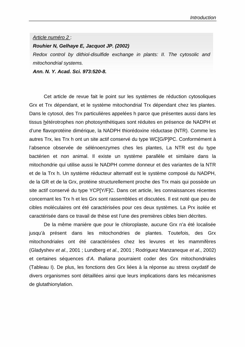
Cet article de revue fait le point sur les systèmes de réduction cytosoliques
Grx et Trx dépendant, et le système mitochondrial Trx dépendant chez les plantes.
Dans le cytosol, des Trx particulières appelées h parce que présentes aussi dans les
tissus hétérotrophes non photosynthétiques sont réduites en présence de NADPH et
d’une flavoprotéine dimérique, la NADPH thiorédoxine réductase (NTR). Comme les
autres Trx, les Trx h ont un site actif conservé du type WC[G/P]PC. Conformément à
l’absence observée de sélénoenzymes ches les plantes, La NTR est du type
bactérien et non animal. Il existe un système parallèle et similaire dans la
mitochondrie qui utilise aussi le NADPH comme donneur et des variantes de la NTR
et de la Trx h. Un système réducteur alternatif est le système composé du NADPH,
de la GR et de la Grx, protéine structurellement proche des Trx mais qui possède un
site actif conservé du type YCP[Y/F]C. Dans cet article, les connaissances récentes
concernant les Trx h et les Grx sont rassemblées et discutées. Il est noté que peu de
cibles moléculaires ont été caractérisées pour ces deux systèmes. La Prx isolée et
caractérisée dans ce travail de thèse est l’une des premières cibles bien décrites.
De la même manière que pour le chloroplaste, aucune Grx n’a été localisée
jusqu’à présent dans les mitochondries de plantes. Toutefois, des Grx
mitochondriales ont été caractérisées chez les levures et les mammifères
(Gladyshev et al., 2001 ; Lundberg et al., 2001 ; Rodriguez Manzaneque et al., 2002)
et certaines séquences d’A. thaliana pourraient coder des Grx mitochondriales
(Tableau I). De plus, les fonctions des Grx liées à la réponse au stress oxydatif de
divers organismes sont détaillées ainsi que leurs implications dans les mécanismes
de glutathionylation.
Article numéro 2 :
Rouhier N, Gelhaye E, Jacquot JP. (2002) Redox control by dithiol-disulfide exchange in plants: II. The cytosolic and
mitochondrial systems.
Ann. N. Y. Acad. Sci. 973:520-8.
Introduction
Redox Control by Dithiol-Disulfide Exchangein Plants
ll. The Cytosolic and Mitochondrial Systems
NICOLAS RüUHIER, ERIC GELHAYE, AND JEAN-PIERRE JACQUOT
Unité Mixte de Recherches 1136 INRA URP (Interaction Arbres Microorganismes),Université Henri Poincaré BP 239,54506 Vandoeuvre Cedex, France
ABSTRACT: This paper describes the existence of two pathways efficient in thereduction of disulfide bridges on selected proteins and mitochondria of photosynthetic organisms. The tirst is constituted by NADPH, the flavoenzymeNADPH thioredoxin reductase, and thioredoxio; and the second by NADPH,glutathione reductase, glutathione, and glutaredoxin. Molecular details concerning the proteins participating in these redox regulatory cascades are provided, and their molecular targets and functions are described.
KEYWORDS: disulfide; glutaredoxin; glutathione; peroxiredoxin; thioredoxin
INTRODUCTION
Many enzymes are regulated by posttranslational modifications, and one of themûst extensively decribed mechanisms is the phosphorylation-dephosphorylation ofhydroxylated residues. Another important biochemical modification is the reversibleoxidoreduction of critical cysteine residues via dithiol-disulfide exchange. In addition to the chloroplastic system described by Jacquot et al., l two major systemsmaintain the thiol redox state in the cytosol, the thioredoxin (Trx) system, and theglutathione/glutaredoxin (GSH/Grx) systems. Both systems present many analogieswith those ofnonphotosynthetic bacteria and animal systems. The two systems wereoriginally discovered in Escherichia coli as alternative electron dOllars to ribonucleotide reductase, one of the key enzymes of DNA biosynthesis.2,3 In bath cases,NADPH is the reductant, and the transmission of the redox signal requires a flavoprotein, either NADPH thioredoxin reductase (NTR) or glutathione reductase(GR). In the following text, we will examine in more detail the structure and functionof these two systems. Sorne of the functions of the two reducing systems are redundant, as demonstrated notably by mutational studies.4 In addition, the reaction mech-
Address for correspondence: Nicolas Rouhier, Unité Mixte de Recherches 1136 INRA URP(Interaction Arbres Microorganismes), Université Henri Poincaré BP 239, 54506 VandoeuvreCedex, France. Voice: ++33383684225.
[email protected]: Grx: glutaredoxin, GR: glutathione reductase, GSH: reduced glutathione,
OSSG: oxidized glutathione, NTR: NADPH thioredoxin reductase, Prx: peroxiredoxins, Trx:thioredoxin.
Ann. N.Y. Acad. Sci. 973: 520-528 (2002). © 2002 New York Academy of Sciences.
ROUHIER et al.: THIOREDOXIN AND GLUTAREDOXIN SYSTEMS
Introduction
521
anisms of Trx and Grx differ, essentially in one aspect. The two proteins are able toreduce disulfide bonds of target proteins with the participation of the two active sitecysteines, as is the case for the reduction of the ribonucleotide reductase; but onlyGrx can function in an alternative pathway called the monothiol pathway. In the latter case, only the most N terminal cysteine of the active site is involved in catalyzingthe reduction of GSH-containing rnixed disulfides.5
CYTOSOLIC AND MITOCHONDRIAL TRX SYSTEMS
The NADPH Thioredoxin Reductase ofthe Cytosol and Mitochondria
The NTR is a homodimeric flavoprotein that is present both in the cytosol and themitochondria.6,7 Both proteins are encoded by nuclear genes, and the mitochondrialsequence contains a transit peptide that is subsequently cleaved. The subunits havea conserved size of 35 kDa. The crystallographic structure of the Arabidopsisthaliana cytosolic enzyme has been detennined.8 Each subunit contains two domains: one that is able ta bind the NADPH; the other, the flavine adenine dinucleotide (FAD) molecule. The NADPH binding domain comprises the central part ofthe sequence; the FAD binding domain, the combination ofthe N and C termini. Theprotein exhibits mobility at the level of a hinge consisting oftwo ~ strands. Following that rotation, the NADPH binding domain is postulated ta be able ta get closerto the isoalloxazine ring of the FAD. Right in front of the isoalloxazine ring lies thedisulfide bridge ofNTR with a very conserved sequence, CAVe, which exhibits thesame spacing between the two Cys residues as Trx. One ofthe sulfurs ofthe disulfidebridge is actually closer to the FAD reactive center, and this corresponds to the catalytic cysteine ofNTR, the mast C terminal one being the "backup" cysteine. Unlikethe iron-sulfur centers, FAD is able ta transfer two electrons at a time, and thus themechanism for the reduction of the disulfide bridge ofNTR is more simple than theone of the chloroplastic homologue, ferredoxin thioredoxin reductase (see Jacquotet al. 1). The redox potential of the disulfide bridge of NTR is unknown at present.
The Cytosolic and Mitochondrial Thioredoxins in Plants
It is now quite clear that multiple isoforms of thioredoxins exist in aIl organismsfrom bacteria to mammals. This property has been documented very early in plants,and it is assumed that there are nearly 35 genes cading for thioredoxin-like proteinsin the very simple model Arabidopsis thaliana. About half ofthose genes encode forcytosolic Trx (aiso called Trx h, where h stands for heterotrophic, because cystolicTrx is found in heterotrophic tissues as weIl) and two for mitochondrial Trx (theydiffer essentially by the presence of an N terminus transit peptide). Cytosolic thioredoxins have been described quite extensively, and their three-dimensional structureis fairly weIl known. The structures ofthe wild-type Chlamydomonas reinhardtii Trxh, as weIl as those of several mutants, have been solved both by NMR and X-raycrystal10graphy.9,10 Trx h can be identified not only by analysis of the amine aeidsequences and the presence of the conserved active site WC[G/P]PC, but aisa byunique spectral characteristics due to a Trp residue present in the first a helix of theprotein (in general this residue is absent in the sequences of the other thioredoxin
522
Introduction
ANNALS NEW YORK ACADEMY OF SCIENCES
types). Il Thioredoxins h have a three-dimensional structure very similar to theirchloroplastic counterparts, and the NMR and crystallographic structures match quiteweIl. The pKa values of the redox active cysteines as weil as the one of a conservedAsp residue essential for catalysis have been titrated by NMR, and the values agreequite weIl with those published for other thioredoxins. 12 The redox potential ofthioredoxin h has aiso been titrated at - 290 mV, and physicochemical parameters ofits cold and heat denaturation properties have been measured. 13
Molecular Targets ofCytosolic and Mitochondrial Thioredoxins
The construction of monocysteinic mutants of Trx h has allowed the identification of one potential molecular target, a peroxiredoxin (Prx). This has been doneboth by in vivo complementation in yeast and by the successful isolation ofthis typeofprotein'by using columns where a monocysteini~ mutant is covalently bound. 14,15
These results should, however, be treated cautiously because one of the targets is achloroplastic sequence, and the other one is a protein from yeast. Nevertheless, theexistence of peroxiredoxins in plants homologous to mammalian cytosolic Prx supports the proposaI that Prx could indeed be a physiological target. 16 Other potentialtargets for crosolic Trx h could inc1ude transcription factors, as in animal and bacterial ceIls.l The mitochondriai Trx could play a role in the redox regulation of thealternative oxidase, an enzyme present in the plant mitochondrial inner membranethat reduces molecular oxygen with no formation of ATP. 18
A SECOND CYTOSOLIC PATHWAY REQUIRES GLUTATHIONEREDUCTASE AND GLUTATHIONE
In paraiiel to the NTR/Trx h system, the cytosol of higher plants aiso contains aso-called GSH/Grx system. The functioning ofthis system is schematized in FIGURE
1. In this altemate system, NADPH is the donor, and the electrons flow to a flavoprotein, the glutathione reductase. GR is a very well-known enzyme that shows both sequence and struturaI homology to NTR. As with NTR, it contains a flavin and adisulfide bridge, and the latter is reduced through the isoalloxazine ring ofthe flavineWhen reduced, GR can in turn reduce oxidized glutathione (GSSG) into two GSHmolecules by cleavage of the disuifide bridge. The reduced form of glutathione (thetripeptide ')Glu-Cys-Gly) thén reduces a disulfide bridge on Grx, and the latter protein can in tum reduce selected target enzymes. Up to now, no plant "Grx has beenfound in the mitochondria, unlike in mammals. 19 In many respects, the properties ofthe GSH/Grx system differ from those of the Trx system, and this results mostlyfrom the molecular properties of the Grx that will be detailed below.
Mo/ecular Properties ofPlant Glutaredoxins
Orx are proteins of low molecular weight (ca. 110 amino acids), and they containa reactive disulfide bridge. The overwhelmingly conserved sequence of the activesite is YCP[Y/F]C. In addition to this redox center, Grx possess a binding site forgiutathione.20 The three-dimensional structure ofGrx is known only in bacteria andmammals. The protein is structurally homologous to Trx, and the active site is quite
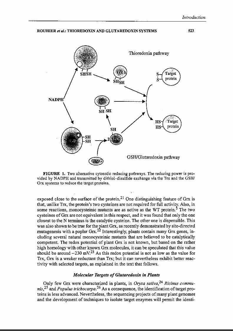
Introduction
ROUHIER et al.: THIOREDOXIN AND GLUTAREDOXIN SYSTEMS 523
Thioredoxin pathway
GSHIGlutaredoxin pathway
S1S
HSHS
ra.""'.;-,:S~L
~SHSH
\
NADPH·
FIGURE 1. Two alternative cytosolic reducing pathways. The reducing power is provided by NADPH and transmitted by dithiol-disulfide exchange via the Trx and the GSHIGrx systems to reduce the target proteins.
;, .
exposed close to the surface of the protein.21 One distinguishing feature of Grx isthat, unlike Trx, the protein's two cysteines are not required for full activity. Also, insorne reactions, monocysteinic mutants are as active as the WT protein.5 The twocysteines ofGrx are not equivalent in this respect, and it was found that only the oneclosest to the N terminus is the catalytic cysteine. The other one is dispensible. Thiswas also shown to be true for the ~lant Orx, as recently demonstrated by site-directedmutagenesis with a poplar Grx.2 Interestingly, plants contain many Grx genes, including several natural monocysteinic mutants that are believed to be catalyticallycompetent. The redox potential of plant Grx is not known, but based on the ratherhigh homology with other known Orx molecules, it can be speculated that this valueshould be around -230 mV.23 As this redox potential is not as low as the value forTrx, Orx is a weaker reductant than Trx; but it can nevertheless exhibit better reactivity with selected targets, as explained in the text that follows.
Molecular Targets ofGlutaredoxin in Plants
Only few Orx were characterized in plants, in Oryza sativa,24 Ricinus communis,25 and Populus trichocarpa.26 As a consequence, the identification oftarget proteins is less advanced. Nevertheless, the sequencing projects ofmany plant geriomesand the development of techniques to isolate target enzymes will permit the identi-
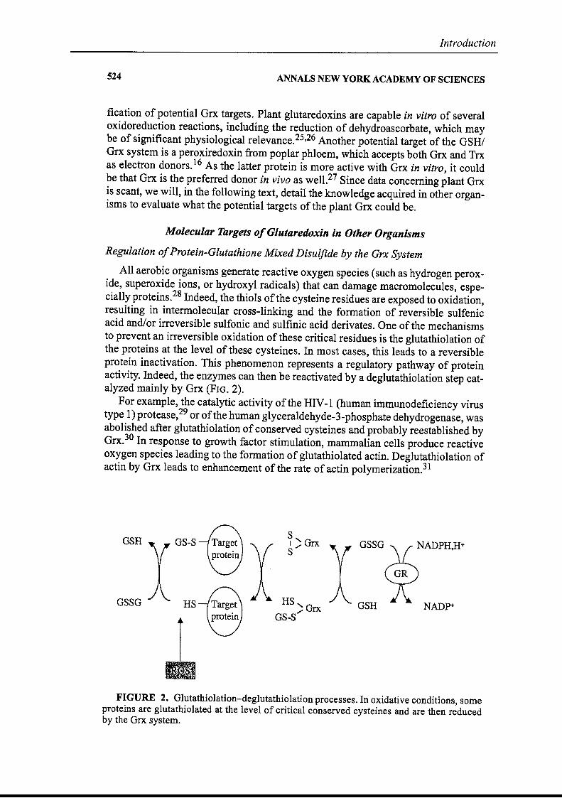
Introduction
524 ANNALS NEW YORK ACADEMY OF SCIENCES
fication of potential Orx targets. Plant glutaredoxins are capable in vitro of severaloxidoreduction reactions, including the reduction of dehydroascorbate, which maybe of significant physiological relevance.25,26 Another potential target of the GSHIGrx system is a peroxiredoxin from poplar phloem, which accepts both Orx and Trxas electron donors. 16 As the latter protein is more active with Grx in vitro, it couldbe that Grx is the preferred donor in vivo as welI.27 Since data conceming plant Grxis seant, we will, in the following text, detail the knowledge acquired in other organisms to evaluate what the potential targets of the plant Grx could be.
Molecular Targets ofGlutaredoxin in Other Organisms
Regulation ofProtein-Glutathione Mixed Disulfide by the Grx System
AlI aerobic organisms generate reactive oxygen species (such as hydrogen peroxide, superoxide ions, or hydroxyl radicals) that can damage macromolecules, especially proteins.28 Indeed, the thiols ofthe cysteine residues are exposed to oxidation,resulting in intermolecular cross-linking and the formation of reversible sulfenicaeid and/or irreversible sulfonic and sulfinic acid derivates. One of the mechanismsto prevent an irreversible oxidation ofthese critical residues is the glutathiolation ofthe proteins at the level of these cysteines. In most cases, this leads ta a reversibleprotein inactivation. This phenomenon represents a regulatory pathway of proteinactivity. Indeed, the enzymes can then he reactivated by a deglutathiolation step catalyzed mainly by Grx (FIG. 2).
For example, the catalytic activity of the HIV-I (human immunodeficiency virustype 1) protease,29 or ofthe human glyceraldehyde-3-phosphate dehydrogenase, wasabolished after glutathiolation of conserved cysteines and probably reestablished byGrx.30 In response to growth factor stimulation, mammalian cells produce reactiveoxygen species leading ta the formation of glutathiolated actin. Deglutathiolation ofactin by Grx leads to enhancement of the rate of actin polymerization.31
NADP+
GSSG
GSH
Sl 'GrxS/
HS'G/ IXGS-S
HS
GS-S
r•
GSH
osso
FIGURE 2. Glutathiolation-deglutathiolation processes. In oxidative conditions, sorneproteins are glutathiolated at the level of critical conserved cysteines and are then reducedby the Orx system.
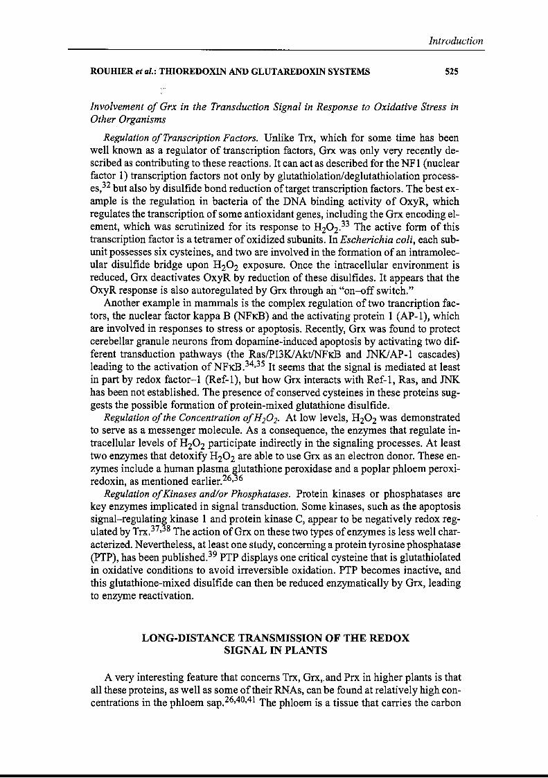
ROUHIER et al.: THIOREDOXIN AND GLUTAREDOXIN SYSTEMS
Introduction
525
Involvement of Grx in the Transduction Signal in Response to Oxidative Stress inOther.Organisms
Regulation ofTranscription Factors. Unlike Trx, which for sorne time has beenweil known as a regulator of transcription factors, ~rx was only very recently described as contributing to these reaetions. It can aet as described for the NFl (nuclearfactor 1) transcription factors not only by glutathiolation/deglutathiolation processes,32 but aisa by disulfide bond reduction oftarget transcription factors. The best example is the regulation in bacteria of the DNA binding activity of OxyR, whichregulates the transcription ofsorne antioxidant genes, including the Grx encoding element, which was scrutinized for its response to H20 2.33 The active form of thistranscription factor is a tetramer of oxidized subunits. In Escherichia coli, each subunit possesses six cysteines, and two are involved in the formation of an intramolecular disulfide bridge upon H20 2 exposure. Once the intracellular environment isreduced, Grx deactivates OxyR by reduction of these disulfides. It appears that theOxyR response is also autoregulated by Grx through an "on-off ·switch."
Another example in mammals is the complex regulation of two trancription factors, the nuclear factor kappa B (NFKB) and the activating protein 1 (AP-l), whichare involved in responses ta stress or apoptosis. Recently, Orx was found to protectcerebellar granule neuronsfrom dopamine-induced apoptosis by activating two different transduction pathways (the Ras/PI3K1AktINFKB and JNKlAP-l cascades)leading to the activation ofNFKB.34,35 It seems that the sig-nal is mediated at leastin part by redox factor-l (Ref-l), but how Grx interacts with Ref-l, Ras, and JNKhas been not established. The presence of conserved cy·steines in these proteins suggests the possible formation of protein-mixed glutathione disulfide.
Regulation ofthe Concentration ofH20 2. At low levels, H20 2 was demonstratedto serve as a messenger molecule. As a consequence, the enzymes that regulate intracellular levels of H20 2 participate indirectly in the signaling processes. At leasttwo enzymes that detoxify H20 2 are able to use Grx as an electron donor. These enzymes inciude a human plasma glutathione peroxidase and a poplar phloem peroxiredoxin, as mentioned earlier.26,36
Regulation ofKinases and/or Phosphatases. Protein kinases or phosphatases arekey enzymes implicated in signal transduction. Sorne kinases, such as the apoptosissignal-regulating kinase 1 and protein kinase C, appear to be negatively redox regulated by Trx.37,38 The action ofGrx on these two types ofenzymes is less weIl characterîzed. Nevertheless, at least one study, concerning a protein tyrosine phosphatase(PTP), has been published.39 PTP displays one critical cysteine that is glutathiolatedin oxidative conditions to avoid irreversible oxidation. PTP beeomes inactive, andthis glutathione-mixed disulfide can then be reduced enzymatieally by Orx, leadingto enzyme reactivation.
LONG-DISTANCE TRANSMISSION OF THE REDOXSIGNAL IN PLANTS
A very interesting feature that concerns Trx, Grx,. and Prx in higher plants is thataIl these proteins, as weIl as sorne oftheir RNAs, can be found at relatively high concentrations in the phloem sap.26,40,41 The phloem is a tissue that carries the carbon
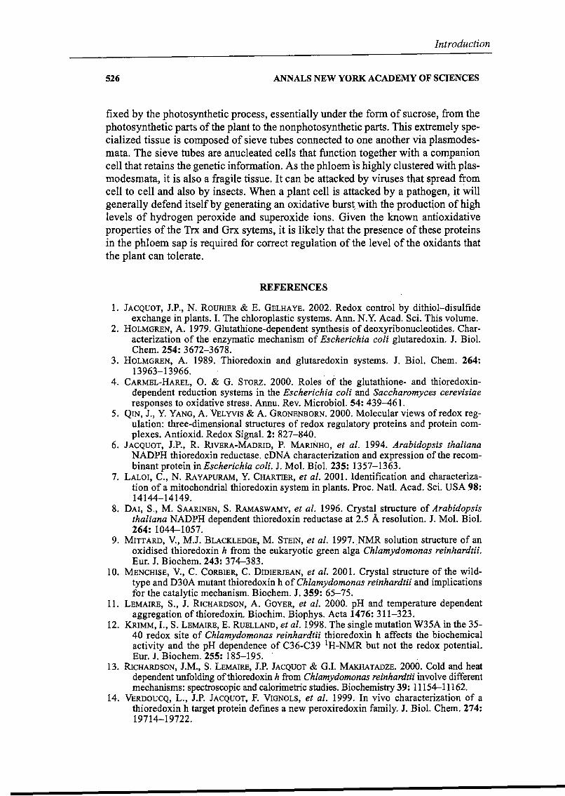
526
Introduction
ANNALS NEW YORK ACADEMY OF SCIENCES
fixed by the photosynthetic process, essentially under the form of sucrose, from thephotosynthetic parts of the plant to the nonphotosynthetic parts. This extremely specialized tissue is composed of sieve tubes connected ta one another via plasmodesmata. The sieve tubes are anucleated cells that function together with a companioncell that retains the genetic information. As the phloem tS highly clustered with plasmodesmata, it is aiso a fragile tissue. It can he attacked by viroses that spread ~omcell to celi and aIso by insects. When a plant cell is attacked by a pathogen, i.t willgeneraIly defend itselfby generating an oxidative burs~..with the product~onofhighleveis ôf hydrogen peroxide and superoxide ions. Given the known antioxidativeproperties of the Trx and Grx sytems, it is likely that the pres'ence of these proteinsin the phloem sap is required for correct regulation of the level of the oxidants thatthe plant can tolerate.
REFERENCES
1. JACQUOT, J.P., N. ROUHIER & E. GELHAYE. 2002. Redox control by dithiol-disulfideexchange in plants. J. The chloroplastic systems. Ann. N.Y. Acad. Sei. This volume.
2. HOLMGREN, A. 1979. Glutathione-dependent synthesis of deoxyribonucleotides. Characterization of the enzymatic mechanism of Escherichia coli glutaredoxin. J. Biol.Chem. 254: 3672-3678.
3. HOLMGREN, A. 1989. Thioredoxin and glutaredoxin systems. J. Biol. Chem. 264:13963-13966.
4. CARMEL-HAREL, O. & G. STORZ. 2000. Roles' of the glutathione- and thioredoxindependent reduction systems in the Escherichia coli and Saccharomyces cerevisiaeresponses to oxidative stress. Annu. Rev. Microbiol. 54: 439-461.
5. QIN, J., Y. YANG, A. VELYVIS & A. GRONENBORN. 2000. Molecular views ofredox regulation: three-dimensional structures of redox regulatory proteins and protein complexes. Antioxid. Redox Signal. 2: 827-840.
6. JACQUOT, J.P., R. RIVERA-MADRID, P. MARINHO, et al. 1994. Arabidopsis thalianaNADPH thioredoxin reductase. cDNA characterization and expression of the recombinant protein in Escherichia coli. J. Mol. Biol. 235: 1357-1363.
7. LALOI, C., N. RAVAPURAM, Y. CHARTIER, et al. 2001. Identification and characterization of a mitochondrial thioredoxin system in plants. Proc. Natl. Acad. Sei. USA 98:14144-14149.
8. DAI, S., M. SAARINEN, S. RAMASWAMY, et al. 1996. Crystal structure of Arabidopsisthaliana NADPH dependent thioredoxin reductase at 2.5 Â resolution. J. Mol. Biol.264: 1044-1057.
9. MITTARD, V., M.J. BLACKLEDGE, M. STEIN, et al. 1997..NMR solution structure of anoxidised thioredoxin h from the eukaryotic green aiga Chlamydomonas reinhardtii.Eur. J. Biochem. 243: 374-383.
10. MENCHISE, V., C. CORBIER, C. DIDIERJEAN, et al. 2001. Crystal structure of the wildtype and D30A mutant thioredoxin h of Chlamydomonas reinhardtii and implicationsfor the catalytic mechanism. Biochem. J. 359: 65-75.
Il. LEMAIRE, S., J. RICHARDSON, A. GOYER, et al. 2000. pH and temperat~re dependentaggregation ofthioredoxin. Biochim. Biophys. Acta 1476: 311-323.
12. KRIMM, 1., S. LEMAIRE, E. RUELLAND, et al. 1998. The single mutation W35A in the 3540 redox site of Chlamydomonas reinhardtii thioredoxin h affect$ the biochemicalactivity and the pH dependence of C36-C39 IH-NMR but not the redox potentiaI.Eur. J. Biochem. 255: 185-195. . .
13. RICHARDSON, J.M., S. LEMAIRE, J.P. JACQUOT & G.I. MAKHATADZE. 2000. Cold and heatdependent unfolding of thioredoxin h from Chlamydomonas reinhardtii involve differentmechanisms: spectroscopie and calorimetric studies. Biochemistry 39: 11154-11162.
14. VERDOUCQ, L., J.P. JACQUOT, F. VIGNOLS, et al. 1999. In vivo characterization of athioredoxin h target protein defines a new peroxiredoxin family. J. Biol. Chem. 274:19714-19722.
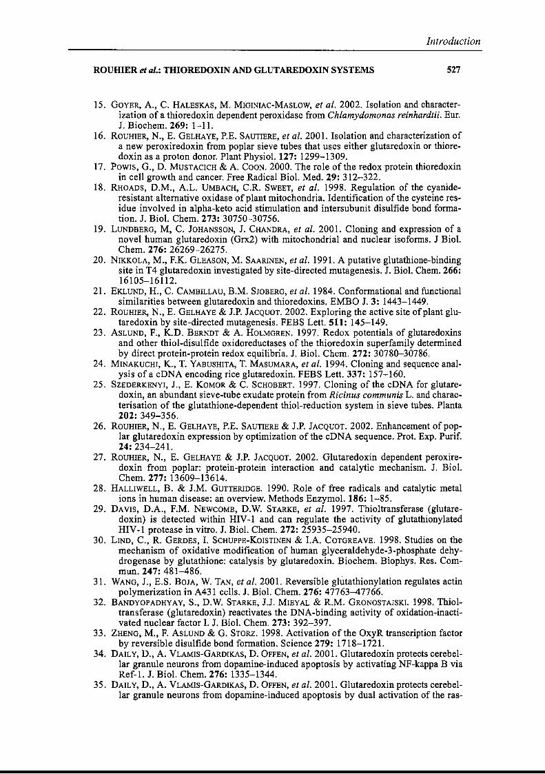
ROUHIER et al.: THIOREDOXIN AND GLUTAREDOXIN SYSTEMS
Introduction
527
15. GOYER, A., C. HALESKAS, M. MIGINIAC-MASLOW, et al. 2002. Isolation and characterization of a thioredoxin dependent peroxidase from Chlamydomonas reinhardtii. Eur.J. Biochem. 269: 1-11.
16. ROUHIER, N., E. GELHAYE, P.E. SAUTIERE, et al. 2001. Isolation and characterization ofa new peroxiredoxin from poplar sieve tubes that uses either glutaredoxin or thioredoxin as a proton donor. Plant Physiol. 127: 1299-1309.
17. POWIS, G., D. MUSTACICH & A. COON. 2000. The raIe of the redox protein thioredoxinin cell growth and cancer. Free Radical Biol. Med. 29: 312-322.
18. RHOADS, D.M., A.L. UMBACH, C.R. SWEET, et al. 1998. Regulation of the cyanideresistant alternative oxidase of plant mitochondria. Identification of the cysteine residue involved in alpha-keto acid stimulation and intersubunit disulfide bond formation. J. Biol. Chem. 273: 30750-30756.
19. LUNDBERG, M, C. JOHANSSON, J. CHANDRA, et al. 2001. Cloning and expression of anovel human glutaredoxin (Grx2) with mitochondrial and nuclear isoforms. J Biol.Chem. 276: 26269-26275.
20. NIKKOLA, M., F.K. GLEASON, M. SAARINEN, et al. 1991. A putative glutathione-bindingsite in T4 glutaredoxin investigated by site-directed mutagenesis. J. Biol. Chem. 266:16105-16112.
21. EKLUND, H., C. CAMBILLAU, B.M. SJOBERG, et al. 1984. Conformational and functionalsimilarities between glutaredoxin and thioredoxins. EMBO J. 3: 1443-1449.
22. ROUHIER, N., E. GELHAYE & J.P. JACQUOT. 2002. Exploring the active site ofplant glutaredoxin by site-directed mutagenesis. FEBS Lett. 511: 145-149.
23. ASLUND, F., K.D. BERNDT & A. HOLMGREN. 1997. Redox potentials of glutaredoxinsand other thiol-disulfide oxidoreductases of the thioredoxin ·superfamily detenninedby direct protein-protein redox equilibria. J. Biol. Chem. 272: 30780-30786.
24. MINAKUCHI, K., T. YABUSHITA, T. MASUMARA, et al. 1994. Cloning and sequence analysis ofa cDNA encoding rice glutaredoxin. FEBS Lett. 337: 157-160.
25. SZEDERKENYI, J., E. KOMOR & C. SCHOBERT. 1997. Cloning of the cDNA for glutaredoxin, an abundant sieve-tube exudate protein from Ricinus communis L. and characterisation of the glutathione-dependent thiol-reduction system in sieve tubes. Planta282: 349-356.
26. ROUHIER, N., E. GELHAYE, P.E. SAUTIERE & J.P. JACQUOT. 2002. Enhancement of poplar glutaredoxin expression by optimization of the cDNA sequence. Prat. Exp. Purif.24: 234-241.
27. ROUHIER, N., E. GELHAYE & J.P. JACQUOT. 2002. Glutaredoxin dependent peroxiredoxin from poplar: protein-protein interaction and catalytic mechanism. J. Biol.Chem. 277: 13609-13614.
28. HALLIWELL, B. & J.M. GUTTERIDGE. 1990. Role of free radicals and catalytic metalions in human disease: an overview. Methods Enzymol. 186: 1-85.
29. DAVIS, D.A., F.M. NEWCOMB, D.W. STARKE, et al. 1997. Thioltransferase (glutaredoxin) is detected within HIV-1 and can regulate the activity of glutathionylatedHIV-I protease in vitro. J. Biol. Chem. 272: 25935-25940.
30. LIND, C., R. GERDES, 1. SCHUPPE-KoISTINEN & I.A. COTGREAVE. 1998. Studies on themechanism of oxidative modification of human glyceraldehyde-3-phosphate dehydrogenase by glutathione: catalysis by glutaredoxin. Biochem. Biophys. Res. Commun. 247: 481-486.
31. WANG, J., E.S. BOJA, W. TAN, et al. 2001. Reversible glutathionylation regulates actinpolymerization in A431 cells. J. Biol. Chem. 276: 47763-47766.
32. BANDYOPADHYAY, S., D.W. STARKE, J.J. MIEYAL & R.M. GRONOSTAJSKI. 1998. Thioltransferase (glutaredoxin) reactivates the DNA-binding activity of oxidation..inactivated nuclear factor l. J. Biol. Chem. 273: 392-397.
33. ZHENG, M., F. AsLUND & G. STORZ. 1998. Activation of the OxyR transcription factorby reversible disulfide bond formation. Science 279: 1718-1721.
34. DAILY, D., A. VLAMIS-GARDIKAS, D. OFFEN, et al. 2001. Glutaredoxin protects cerebellar granule neurons from dopamine-induced apoptosis by activating NF-kappa B viaRef-1. J. Biol. Chem. 276: 1335-1344.
35. DAILY, D., A. VLAMIs-GARDIKAS, D. OFFEN, et al. 2001. Glutaredoxin protects cerebellar granule neurons from dopamine-induced apoptosis by dual activation of the ras-
528
Introduction
ANNALS NEW YORK ACADEMY OF SCIENCES
phosphoinositide 3-kinase and jun n-tenninal kinase pathways. J. Biol. Chem. 276:21618-21626.
36. BJORNSTEDT, M., J. XUE, W. HUANG, et al. 1994. The thioredoxin and glutaredoxin systems are efficient electron dODors to human plasma glutathione peroxidase. J. Biol.Chem. 269: 29382-29384.
37. SAITOH, M., H. NISHITOH, M. FUJII, et al. 1998. Mammalian thioredoxin is a directinhibitor of apoptosis signal-regulating kinase (A8K) 1. EMBO J. 17: 2596-2606.
38. WATSON, J.A., M.G. RUMSBY & R.G. WOLOWACZ. 1999. Phage display identifiesthioredoxin and superoxide dismutase as novel protein kinase C-interacting proteins:thioredoxin inhibits protein kinase C-mediated phosphorylation of histone. Biochem.J. 343: 301-305.
39. BARRETT, W.C., J.P. DEGNORE, Y.F. KENG, et al. 1999. Roles of superoxide radicalanion in signal transduction mediated by reversible regulation of protein-tyrosinephosphatase lB. J. Biol. Chem. 274: 34543-34546.
40. ISHIWATARI, Y., C. HONDA, 1. KAWASHIMA, et al. 1995. Thioredoxin h is one of themajor proteins in rice phloem sap. Planta 195: 456-463.
41. SASAKI, T., M. CHINO, H. HAYASHI & T. FUJIWARA. 1998. Detection ofseveral mRNAspecies in rice phloem sap. Plant Cel!. Physiol. 39: 895-897.
Introduction
Le troisième article aborde l'étude des Trx végétales d'un point de vue plus
structural et mécanistique. L’organisation du « thioredoxin fold » y est décrite et une
comparaison d’une structure tridimensionnelle de Trx h de Chlamydomonas
reinhardtii obtenue par cristallographie et par résonance magnétique nucléaire
(RMN) est présentée. Il présente également un certain nombre de protéines
homologues aux Trx, appelées "thioredoxin-like proteins", dont l'architecture est
basée soit sur une répétition d’unités Trx soit sur la fusion entre un module Trx et un
autre module d’activité différente. La géométrie du site actif, les acides aminés
invariants comprenant le tryptophane du site actif et un aspartate enfoui nécessaires
pour les reconnaissances protéine-protéine et la catalyse sont discutés dans cet
article. Les Prx et les MsrA, deux des cibles caractérisées, sont également des
enzymes qui possèdent des analogies structurales avec les Trx (Schröder & Pontig,
1998 ; Gladyshev, 2002).
Article numéro 3 :
Jacquot JP, Gelhaye E, Rouhier N, Corbier C, Didierjean C, Aubry A. (2002) Thioredoxins and related proteins in photosynthetic organisms: molecular basis
for thiol dependent regulation.
Biochem. Pharmacol. 64(5-6):1065-9.
Introduction
ELSEVIER Biochemical Pharmacology 64 (2002) 1065-1069
BlochemlcalPharmacololV
Thioredoxins and related proteins in photosynthetic organisllls:molecular basis for thiol dependent regulation
Jean-Pierre Jacquota,*, Eric Gelhayea, Nicolas Rouhiera
, Catherine Corbierb,
Claude Didierjeanb, André Aubryb
aUMR INRA UHP Interaction Arbres Microorganismes, Université Henri Poincaré, 54506 Vandoeuvre Cedex, FrancebLCM3B Groupe Biocristallographie, 54506 Vandoeuvre Cedex, France
Received 25 February 2002; accepted 22 March 2002
Abstract
Thioredoxins are small molecular weight disulfide oxidoreductases specialized in the reduction of disulfide bonds on other proteins.Generally, the enzymes which are selectively and reversibly reduced by these proteins oscillate between an oxidized and inactiveconformation and a reduced and active conformation. Thioredoxin constitutes the archetype of a family of protein disulfideoxidoreductases which comprises glutaredoxin and protein disulfide isomerase. Thioredoxin and glutaredoxin serve many roles inthe cell, including the redox regulation of target enzymes and transcription factors. They can also serve as hydrogen donors toperoxiredoxins, recently discovered heme free peroxidases, the function of which is to get rid of hydroperoxides in the cell. This reviewdescribes the molecular basis for the functioning and interaction between these enzymes in photosynthetic organisms.© 2002 Elsevier Science Inc. AlI rights reserved.
Keywords: Disulfide bridge reduction; Redox regulation; Thioredoxin
1. Reactions catalyzed by protein disulfideoxidoreductases
The presence of disulfide bonds is one of the majorstructural determinants in proteins. It is generally recognized that extracellular proteins possess disulfide bridgesthat help stabilize their structure, while intracellularenzymes are devoid of such features [1,2]. AlI polypeptidesare however synthesized with free cysteine residues and inthe same time that specialized proteins such as chaperonesor prolyl isomerases help for the folding process, otherenzymes known as protein disulfide isomerases (PDI) areneeded for the creation of those disulfide bonds [3]. Conversely, sorne proteins which contain disulfide bridgesneed to be reduced to a dithiol form to gain activity andthese reactions are catalyzed by thioredoxins (Trx) andother molecular variants of this protein such as glutaredoxins (Grx) [4,5]. PDI, Trx and Grx are thus membersof a superfamily whose function is either to create or
* Corresponding author. Tel.: +33-3-83-91-22-53.E-mail address:[email protected] (J.-P. Jacquot).Abbreviations: Grx, glutaredoxin; PDI, protein disulfide isomerase;
Prx, peroxiredoxin; Trx, thioredoxin.
reduce disulfide bonds on other proteins according tothe equation:
/s /SH~
/SH /8P1 1 + P2 P1 + P2 1
"'5 "'SH~
'" SH "'-..8
2. Molecular and functional characteristicsof plant thioredoxins
Plant thioredoxins are small molecular weight (ca.12 kDa) and generally extremely thermostable proteins.They contain in general around 110 amino acids in theirmature form (excluding the transit peptides of the nuclearencoded chloroplastic and mitochondrial isoforms) [6,7].AlI thioredoxins have a very conserved active site with thesequence WC [GIP]PC. AU available structures demonstrate that the two sulfur atoms of the Cys residues ofthe active site are indeed linked in a disulfide bridge in theoxidized state. The 3D architecture of the protein is conserved throughout the evolution with a succession ofsecondary elements as follows: ~1, ex1, ~2, ex2, ~3, ex3,~4, ~5, ex4. The active site is exposed and situated in the
0006-2952/02/$ - see front matter © 2002 Elsevier Science Inc. AlI rights reserved.PlI: S 0 006 - 2 9 5 2 ( 0 2 ) 0 1 1 7 7 - 2
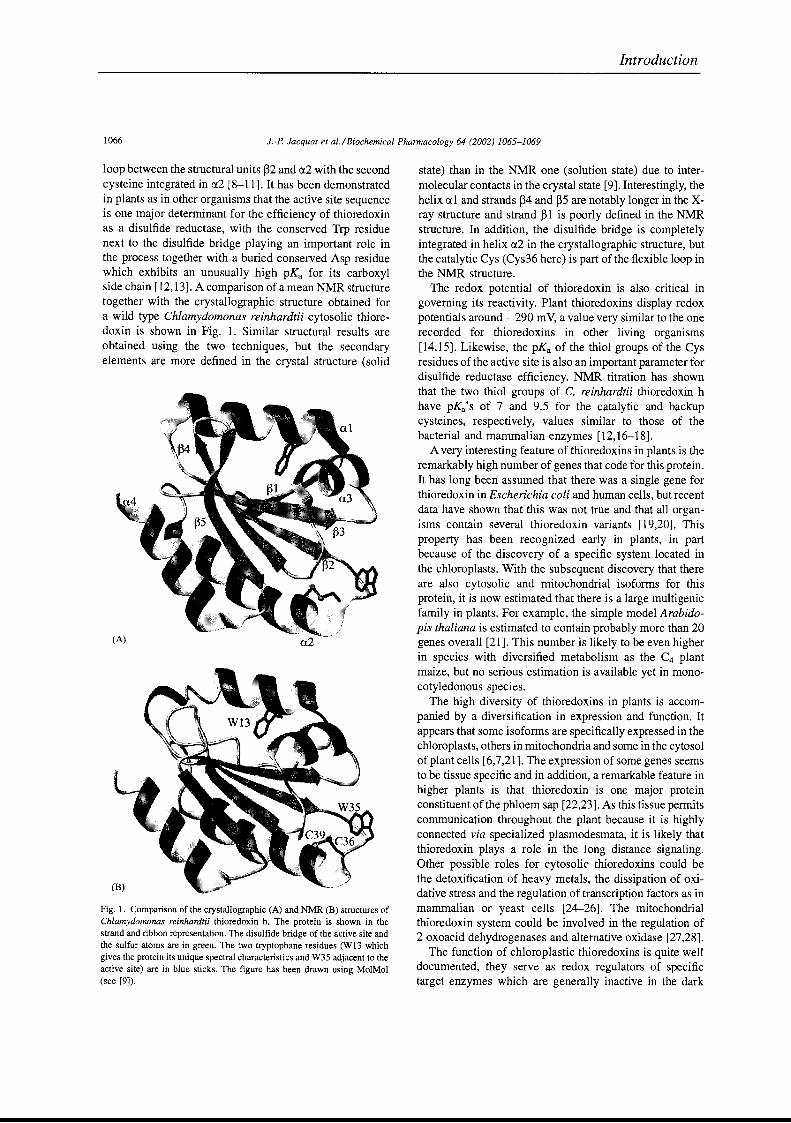
1066 J.-P. Jacquot et al.IBiochemical Pharmacology 64 (2002) 1065-1069
Introduction
loop between the structural units ~2 and r:x2 with the secondcysteine integrated in rJ..2 [8-11]. It has been demonstratedin plants as in other organisms that the active site sequenceis one major determinant for the efficiency of thioredoxinas a disulfide reductase, with the conserved Trp residuenext to the disulfide bridge playing an important role inthe process together with a buried conserved Asp residuewhich exhibits an unusually high pKa for its carboxylside chain [12,13]. A comparison of a mean NMR structuretogether with the crystallographic structure obtained fora wild type Chlamydomonas reinhardtii cytosolic thioredoxin is shown in Fig. 1. Similar structural results areobtained using the two techniques, but the secondaryelements are more defined in the crystal structure (solid
Fig. 1. Comparison of the crystallographic (A) and NMR (B) structures ofChlamydomonas reinhardtii thioredoxin h. The protein is shown in thestrand and ribbon representation. The disulfide bridge of the active site andthe sulfur atoms are in green. The two tryptophane residues (W13 whichgives the protein its unique spectral characteristics and W35 adjacent to theactive site) are in blue sticks. The figure has been drawn using MolMol(see [9]).
state) than in the NMR one (solution state) due to intermolecular contacts in the crystal state [9]. Interestingly, thehelix rJ..l and strands ~4 and ~5 are notably longer in the Xray structure and strand ~1 is poorly defined in the NMRstructure. In addition, the disulfide bridge is completelyintegrated in helix r:x2 in the crystallographic structure, butthe catalytic Cys (Cys36 here) is part of the flexible loop inthe NMR structure.
The redox potential of thioredoxin is also critical ingoveming its reactivity. Plant thioredoxins display redoxpotentials around - 290 mV, a value very similar to the onerecorded for thioredoxins in other living organisms[14,15]. Likewise, the pKa of the thiol groups of the Cysresidues of the active site is also an important parameter fordisulfide reductase efficiency. NMR titration has shownthat the two thiol groups of C. reinhardtii thioredoxin hhave pKa's of 7 and 9.5 for the catalytic and backupcysteines, respectively, values similar to those of thebacterial and mammalian enzymes [12,16-18].
A very interesting feature of thioredoxins in plants is theremarkably high number of genes that code for this proteineIt has long been assumed that there was a single gene forthioredoxin in Escherichia coli and human cells, but recentdata have shown that this was not true and that all organisms contain several thioredoxin variants [19,20]. Thisproperty has been recognized early in plants, in partbecause of the discovery of a specifie system located inthe chloroplasts. With the subsequent discovery that thereare also cytosolic and mitochondrial isoforms for thisprotein, it is now estimated that there is a large multigenicfamily in plants. For example, the simple model Arabidopis thaliana is estimated to contain probably more than 20genes overall [21]. This number is likely to be even higherin species with diversified metabolism as the C4 plantmaize, but no serious estimation is available yet in monocotyledonous species.
The high diversity of thioredoxins in plants is accompanied by a diversification in expression and function. Itappears that sorne isoforms are specifically expressed in thechloroplasts, others in mitochondria and sorne in the cytosolof plant cells [6,7,21]. The expression of sorne genes seemsto be tissue specifie and in addition, a remarkable feature inhigher plants is that thioredoxin is one major proteinconstituent ofthe phloem sap [22,23]. As this tissue permitscommunication throughout the plant because it is highlyconnected via specialized plasmodesmata, it is likely thatthioredoxin plays a role in the long distance signaling.Other possible roles for cytosolic thioredoxins could bethe detoxification of heavy metals, the dissipation of oxidative stress and the regulation of transcription factors as inmammalian or yeast cells [24-26]. The mitochondrialthioredoxin system could he involved in the regulation of2 oxoacid dehydrogenases and alternative oxidase [27,28].
The function of chloroplastic thioredoxins is quite welldocumented, they serve as redox regulators of specifietarget enzymes which are generally inactive in the dark
J.-P. Jacquot et al. / Biochemical Pharmacology 64 (2002) 1065-1069
Introduction
1067
and active in the light, with the notable exception ofglucose-6-phosphate dehydrogenase which is oppositelyregulated. Through this redox regulation, thioredoxins helpcontrol the rate of carbon fixation through the reducingpentose phosphate pathway otherwise known as Calvincycle [5,29]. Several of the regulatory target enzymesdisplay additional regulatory sequences that contain critical cysteines and these cysteines can be either in adisulfide bond (oxidized form) or as dithiols (reducedstate). Generally, the conversion of the oxidized inactiveenzymes into reduced active catalysts occurs in two steps,first a fast reduction that produces a reduced inactiveenzymes and second a slow conformational change thatleads to the active catalyst [30]. The chloroplastic thioredoxin system is unique in at least two ways. First, unlikethe mitochondrial and cytosolic systems where the primaryreductant is NADPH relayed via a ftavoprotein NADPHthioredoxin reductase, in chloroplasts the donor is photoreduced ferredoxin through a specifie iron-sulfur containing enzyme called ferredoxin thioredoxin reductase [31].In addition, there are two molecular variants for thioredoxin, named thioredoxin m and thioredoxin f whichdisplay differential selectivity. More specifically, thioredoxin f is strictly required for the reductive activation offructose-l,6-bisphosphatase and other thioredoxins cannotmake the job [32]. This is in marked contrast with othersystems where thioredoxins are generally considered as apromiscuous enzymes (Le. they show no marked specificity). Details about the functioning and composition of theregulatory chloroplastic system can be found in severalreviews [5,29,33,34]. In chloroplasts, thioredoxins are alsoinvolved in the removal of peroxides via specifie chloroplastic thiol peroxidases or peroxiredoxins (Prx) [35].
3. Glutaredoxins, peroxiredoxins and protein disulfideisomerases share molecular determinants withthioredoxin
The 3D organization of the thioredoxin molecule is weIlestablished and referred to as the thioredoxin fold. Basedon this architecture, several catalysts have evolved. Bykeeping basically the same secondary structural units andchanging the active site to YCP[Y/F]C, one obtains aglutaredoxin molecule that is still a reductant but with aslightly higher redox potential. Glutaredoxin is reduced viaa cascade that comprises NADPH, glutathione reductaseand the tripeptide glutathione [36,37]. The combination oftwo thioredoxin modules together with the alteration of theactive site sequence to WCGHC leads to protein disulfideisomerase, an oxidizing protein whose function is to createdisulfide bonds on other proteins as discussed in the firstsection [38]. An even more complex version callednucleoredoxin results of the combination of three successive thioredoxin modules, the N- and C-terminus onesbearing each an active site [39]. In addition to these weIl
documented evolutions of thioredoxin, several sequencesin the databases feature natural fusion products between agiven enzyme (APS reductase or peroxiredoxin for exampIe) together with a glutaredoxin module [40,41]. Inaddition, many natural monocysteinic variants of thioredoxin and glutaredoxin are present in the protein databanks. These proteins are likely to be catalytically competent as it was demonstrated that engineered monocysteinic glutaredoxins are also functional catalysts [42,43].Finally, another enzyme which has a fold similar to thioredoxin is peroxiredoxin even though that protein contains asingle catalytic cysteine which is transformed into a sulfenic acid upon catalysis [44]. Other examples of structurally related proteins in plants can be found in [5].
4. Functions of thioredoxin related catalystsin plant cells
The function of protein disulfide isomerase is linked tothe folding pathway of the ER, PDI sequences containingconsistently a KDEL retention sequence that targets themto this compartment [45]. It has been shown however in C.reinhardtii that the related protein RB60 which also contains the KDEL signal is targeted both to the ER and to thechloroplast [46]. It has been proposed to play a role as aregulator of chloroplast translational activation [47]. Onthe other hand, the function of nucleoredoxin is poorlydocumented but believed to be related to the redox regulation of transcription factors [39]. In plants, glutaredoxinsseem to be restricted to the cytosol. This protein has beenshown to possess the capacity to reduce dehydroascorbatewhich could be of physiological significance [43]. Anotherrole proposed for glutaredoxin is to serve as an electrondonor to a subclass of peroxiredoxin [48]. In this respect,both the thioredoxin and glutaredoxin pathways could beimplicated in the removal of hydroperoxides and the fightagainst oxidative stress.
5. Future challenges of redox regulation in plants
The high number of genes coding for thioredoxins andrelated proteins poses the problem of the specificity andpossible redundancy of each of these products. This question is starting to be addressed using the double hybridtechnique, the yeast and Synechocystis complementationapproach and the biochemical isolation of targets. Indeed,the use of engineered monocysteinic mutants has helpedisolate targets for given thioredoxin types, but the specificity of this approach needs to be assessed carefully withother parameters such as the putative subcellular localization of the partner proteins [49]. Using the yeast complementation, sorne specificity is observed between thevarious isoforms notably conceming the growth on sulfateor methionine sulfoxide and the resistance to hydrogen
1068 J.-P. Jacquot et al./Biochemical Pharmacology 64 (2002) 1065-1069
Introduction
peroxide but it is not clear yet that this is of physiologicalsignificance [50-53]. Recent attempts to obtain knockoutmutants in Arabidopsis thaliana by successive PCRscreening of transposon transformed pools, have beensuccessful, but generally no clear phenotype appears underphysiological conditions. Clearly, sorting out the physiological function for each of these proteins will be achallenge for the years to come.
References
[1] Brumlik MJ, van der Goot FG, Wong KR, Buckley JT. The disulfidebond in the Aeromonas hydrophila lipase/acyltransferase stabilizes thestructure but is not required for secretion or activity. J Bacteriol1997;179:3116-21.
[2] Fra AM, Fagioli C, Finazzi D, Sitia R, Alberini C. Quality control ofER synthesized proteins: an exposed thiol group as a three-way switchmediating assembly, retention and degradation. EMBO J1993;12:4755-61.
[3] Winter J, Klappa P, Freedman RB, Lilie H, Rudolph R. Catalyticactivity and chaperone function of human protein-disulfide isomeraseare required for the efficient refolding of proinsulin. J Biol Chem2002;277:310-7.
[4] Holmgren A. Antioxidant function of thioredoxin and glutaredoxinsystems. Antioxid Redox Signal 2000;2:811-20.
[5] SchürInann P, Jacquot JP. Plant thioredoxin systems revisited. AnnRev Plant Physiol Plant Mol Biol 2000;51:371-400.
[6] Laloi C, Rayapuram N, Chartier Y, Grienenberger JM, Bonnard G,Meyer Y. Identification and characterization of a rnitochondrialthioredoxin system in plants. Proc Natl Acad Sci USA2001;98:14144-9.
[7] Stein M, Jacquot JP, Jeannette E, Decottignies P, Hodges M, LancelinJM, Mittard V, Schrnitter JM, Miginiac-Maslow M. Chlamydomonasreinhardtii thioredoxins: structure of the genes coding for the chIoroplastic m and cytosolic h isoforms; expression in Escherichia coli ofthe recombinant proteins, purification and biochernical properties.Plant Mol Biol 1995;28:487-503.
[8] Capitani G, Markovic-Housley Z, DelVal G, Morris M, Jansonius JN,Schürmann P. Crystal structures of two functionally different thioredoxins in spinach chloroplasts. J Mol Biol 2000;302:135-54.
[9] Menchise V, Corbier C, Didierjean C, Saviano M, Benedetti E, JacquotJP, Aubry A. Crystal structure of the wild-type and D30A mutantthioredoxin h of Chlamydomonas reinhardtii and implications for thecatalytic mechanism. Biochem J 2001;359:65-75.
[10] Mittard V, Blackledge MJ, Stein M, Jacquot JP, Marion D, LancelinJM. NMR solution structure of an oxidised thioredoxin h from theeukaryotic alga Chlamydomonas reinhardtii. Eur J Biochem1997;243:674-8.
[11] Lancelin JM, Guilhaudis L, Krimm l, Blackledge MJ, Marion D,Jacquot JP. NMR structures of thioredoxin m from the green algaChlamydomonas reinhardtii. Proteins 2000;41:334-49.
[12] Krimm l, Lemaire S, Ruelland E, Miginiac-Maslow M, Jacquot Ip,Hirasawa M, Knaff DB, Lancelin JM. The single mutation W35A inthe 35-40 redox site of Chlamydomonas reinhardtii thioredoxinh affects the biochemical activity and the pH dependence of C36C39 1H-NMR but not the redox potential. Eur J Biochem 1998;255:185-95.
[13] Menchise V, Corbier C, Didierjean C, Jacquot JP, Benedetti E, AubryA, Saviano M. Crystal structure of the W35A mutant thioredoxin h ofChlamydomonas reinhardtii: the substitution of the conserved activesite Trp leads to modifications in the environment of the two catalyticcysteines. Biopolymers 2000;56: 1-7.
[14] Hirasawa M, Schürmann P, Jacquot JP, Manieri W, Jacquot P, KeryerE, Hartman F, Knaff DB. Oxidation-reduction properties of chloroplast thioredoxins, ferredoxin-thioredoxin reductase and fructosebisphosphatase. Biochernistry 1999;38:5200-5.
[15] Balmer Y, Stritt-Etter AL, Hirasawa M, Jacquot JP, Keryer E, KnaffDB, Schurmann P. Oxidation-reduction and activation properties ofchloroplast fructose-1,6-bisphosphatase with mutated regulatory site.Biochernistry 2001 ;40: 15444-50.
[16] Dyson HJ, Jeng MF, Tennant LL, Slaby 1, Lindell M, Cui DS, KuprinS, Holmgren A. Effects of buried charged groups on cysteine thiolionization and reactivity in Escherichia coli thioredoxin: structuraland functional characterization of mutants of Asp 26 and Lys 57.Biochernistry 1997;36:2622-36.
[17] Huber-Wunderlich M, Glockshuber R. A single dipeptide sequencemodulates the redox properties of a whole enzyme farnily. Fold Des1998;3: 161-71.
[18] Kortemme T, Creighton TE. Ionisation of cysteine residues at thetermini of model alpha-helical peptides. Relevance to unusual thiolpKa values in proteins of the thioredoxin farnily. J Mol Biol1995;253:799-812.
[19] Lillig CH, Prior A, Schwenn JD, Aslund F, Ritz D, Vlamis-GardikasA, Holmgren A. New thioredoxins and glutaredoxins as electrondonors of 3'-phosphoadenylylsulfate reductase. J Biol Chem 1999;274:7695-8.
[20] Miranda-Vizuete A, Damdimopoulos AB, Spyrou G. The rnitochondrial thioredoxin system. Antioxid Redox Signal 2000;2:801-10.
[21] Mestres-Ortega D, Meyer Y. The Arabidopsis thaliana genome encodes at least four thioredoxins m and a new prokaryotic-like thioredoxin. Gene 1999;240:307-16.
[22] Brugidou C, Marty l, Chartier Y, Meyer Y. The Nicotiana tabacumgenome encodes two cytoplasmic thioredoxin genes which are differently expressed. Mol Gen Genet 1993;238:285-93.
[23] Ishiwatari ~ Honda C, Kawashima l, Nakamura S, Hirano H, Mori S,Fujiwara T, Hayashi H, Chino M. Thioredoxin h is one of the majorproteins in rice phloem sap. Planta 1995;195:456-63.
[24] Nishinaka Y, Masutani H, Nakamura H, Yodoi J. Regulatory roIes ofthioredoxin in oxidative stress-induced cellular responses. Redox Rep2001;6:289-95.
[25] Lemaire S, Keryer E, Stein M, Schepens l, Issakidis-Bourguet E,Gerard-Hirne C, Miginiac-Maslow M, Jacquot JP. Heavy-metal regulation of thioredoxin gene expression in Chlamydomonas reinhardtii.Plant Physiol 1999;120:773-8.
[26] Kuge S, Arita M, Murayama A, Maeta K, Izawa S, InDue ~ NomotoA. Regulation of the yeast Yaplp nuclear export signal is mediated byredox signal-induced reversible disulfide bond formation. Mol CellBiol 2001;21:6139-50.
[27] Bunik V, Raddatz G, Lemaire S, Meyer y, Jacquot JP, Bisswanger H.Interaction of thioredoxins with target proteins: role of particularstructural elements and electrostatic properties of thioredoxins in theirinterplay with 2-oxoacid dehydrogenase complexes. Protein Sci 1999;8:65-74.
[28] Vanlerberghe GC, McIntosh L, Yip JY. Molecular localization of aredox-modulated process regulating plant rnitochondrial electrontransport. Plant Cell 1998;10:1551-60.
[29] Buchanan BB, Schurmann P, Jacquot JP. Thioredoxin and metabolicregulation. Sernin Cell Biol 1994;5:285-93.
[30] Miginiac-Maslow M, Decottignies P, Jacquot JP, Gadal P. Regulationof corn leaf NADP-Malate dehydrogenase light-activation by thephotosynthetic electron flow. Effect of photoinhibition studiedin a reconstituted system. Biochim Biophys Acta 1990;1017:273-9.
[31] Dai S, Schwendtmayer C, Schurmann P, Ramaswamy S, Eklund H.Redox signaling in chloroplasts: cleavage of disulfides by an ironsulfur cluster. Science 2000;287:655-8.
[32] Wangensteen OS, Chueca A, Hirasawa M, Sahrawy M, Knaff DB,Lopez Gorge J. Binding features of chloroplast fructose-1,6-bispho-
J.-P. Jacquot et al. / Biochemical Pharmacology 64 (2002) 1065-1069
Introduction
1069
sphatase-thioredoxin interaction. Biochim Biophys Acta 2001;154:156-66.
[33] Dai S, Schwendtmayer Ct Johansson K, Ramaswamy S, Schurmann P,Eklund H. How does light regulate chloroplast enzymes? Structurefunction studies of the ferredoxinlthioredoxin system. Q Rev Biophys2000;33:67-108.
[34] Miginiac-Maslow Mt Johansson K, Ruelland E, Issakidis-Bourguet E,Schepens 1, Goyer A, Lemaire-CharnIey M, Jacquot JP, Le Maréchal P,Decottignies P. Light activation of NADP-malate dehydrogenase: ahighly controlled process for an optimized function. Physiol Plant2000;110:322-9.
[35] Baier M, Noctor G, Foyer CH, Dietz KJ. Antisense suppression of2-cysteine peroxiredoxin in Arabidopsis specifically enhances theactivities and expression of enzymes associated with ascorbatemetabolism but not glutathione metabolism. Plant Physiol 2000;124:823-32.
[36] Nordstrand K, Sandstrom A, Aslund F, Holmgren A, Otting G, BerndtKD. NMR structure of oxidized glutaredoxin 3 from Escherichia coli.J Mol Biol 2000;303:423-32.
[37] Wells WW, Yang Y, Deits TL, Gan ZR. Thioltransferases. AdvEnzymol Relat Areas Mol Biol 1993;66:149-201.
[38] Kemmink J, Darby NJ, Dijkstra K, Nilges M, Creighton TE. Thefolding catalyst protein disulfide isomerase is constructed of activeand inactive thioredoxin modules. CUIT Biol 1997;7:239~5.
[39] Laughner BJ, Sehnke PC, Ferl RJ. A novel nuclear member of thethioredoxin superfamily. Plant Physiol 1998;118:987-96.
[40] Bick JA, Aslund F, Chen y, Leustek T. Glutaredoxin function for thecarboxyl-terminal domain of the plant-type 5'-adenylylsulfate reductase. Proc Nat! Acad Sei USA 1998;95:8404-9.
[41] Vergauwen B, Pauwels F, Jacquemotte F, Meyer TE, Cusanovich MA,Bartsch RG, Van Beeumen 11. Characterization of clutathione amidereductase from Chromatium gracile. Identification of a novel thiolperoxidase (PrxlGrx) fueled by glutathione amide redox cycling. JBiol Chem 2001;276:20890-7.
[42] Yang Y, Jao S, Nanduri S, Starke DW, Mieyal 11, Qin J. Reactivity ofthe human thioltransferase (glutaredoxin) C7S, C25S, C78S, C82Smutant and NMR solution structure of its glutathionyl mixed disulfideintermediate reflect catalytic specificity. Biochemistry 1998;37:17145-56.
[43] Rouhier N, Gelhaye E, Jacquot JP. Exploring the active site of plantglutaredoxin by site-directed mutagenesis. FEBS Lett 2002;511:145-9.
[44] Declercq JP, Evrard C, Clippe A, Stricht DV, Bernard A, Knoops B.Crystal structure of human peroxiredoxin 5, a novel type of mammalian peroxiredoxin at 1.5 Â. resolution. J Mol Biol 2001;311:751-9.
[45] Ciaffi M, Paolacci AR, Dominici L, Tanzarella OA, Porceddu E.Molecular characterization of gene sequences coding for proteindisulfide isomerase (PDI) in durum wheat (Triticum turgidumssp. durum). Gene 2001;265:147-56.
[46] Trebitsh T, Meiri E, Ostersetzer 0, Adam Z, Danon A. The proteindisulfide isomerase-like RB60 is partitioned between stroma andthylakoids in Chlamydomonas reinhardtii chloroplasts. J Biol Chem2001 ;276:4564-9.
[47] Yohn CB, Cohen A, Danon A, Mayfie1d SP. A poly(A) binding proteinfunctions in the chloroplast as a message-specific translation factor.Proc Nat! Acad Sci USA 1998;95:2238-43.
[48] Rouhier N, Gelhaye E, Sautiere PE, Brun A, Laurent P, Tagu D, GerardJ, de Fay E, Meyer Y, Jacquot JP. Isolation and characterization of anew peroxiredoxin from poplar sieve tubes that uses either glutaredoxin or thioredoxin as a proton donor. Plant Physiol 2001;127:1299-309.
[49] Goyer A, Halesk6s C, Miginiac-Maslow M, Klein U, Le Maréchal P,Jacquot JP, Decottignies P. Isolation and characterization of a thioredoxin dependent peroxidase from Chlamydomonas reinhardtii. Eur JBiochem 2002;269: 1-11.
[50] Issakidis-Bourguet E, Mouaheb N, Meyer y, Miginiac-Maslow M.Heterologous complementation of yeast reveals a new putative function for chloroplast rn-type thioredoxin. Plant J 2001 ;25: 127-35.
[51] Brehelin C, Mouaheb N, Verdoucq L, Lancelin JM, Meyer Y. Characterization of determinants for the speciflcity of Arabidopsis thioredoxins h in yeast complementation. J Biol Chem 2000;275:31641-7.
[52] Verdoucq L, Vignols F, Jacquot JP, Chartier Yt Meyer Y. In vivocharacterization of a thioredoxin h target protein defines a newperoxiredoxin family. J Biol Chem 1999;274:19714-22.
[53] Mouaheb N, Thomas D, Verdoucq L, Monfort P, Meyer Y. In vivofunctional discrimination between plant thioredoxins by heterologousexpression in the yeast Saccharomyces cerevisiae. Proc Natl Acad SciUSA 1998;95:3312-7.
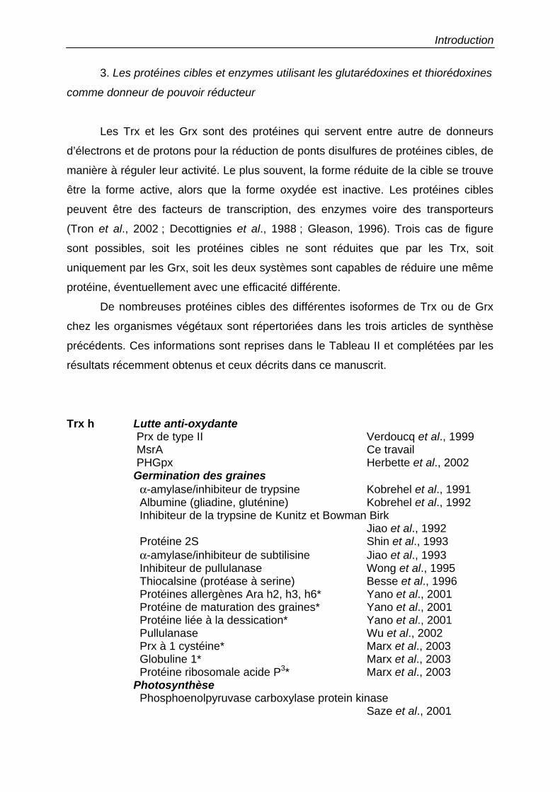
Introduction
3. Les protéines cibles et enzymes utilisant les glutarédoxines et thiorédoxines
comme donneur de pouvoir réducteur
Les Trx et les Grx sont des protéines qui servent entre autre de donneurs
d’électrons et de protons pour la réduction de ponts disulfures de protéines cibles, de
manière à réguler leur activité. Le plus souvent, la forme réduite de la cible se trouve
être la forme active, alors que la forme oxydée est inactive. Les protéines cibles
peuvent être des facteurs de transcription, des enzymes voire des transporteurs
(Tron et al., 2002 ; Decottignies et al., 1988 ; Gleason, 1996). Trois cas de figure
sont possibles, soit les protéines cibles ne sont réduites que par les Trx, soit
uniquement par les Grx, soit les deux systèmes sont capables de réduire une même
protéine, éventuellement avec une efficacité différente.
De nombreuses protéines cibles des différentes isoformes de Trx ou de Grx
chez les organismes végétaux sont répertoriées dans les trois articles de synthèse
précédents. Ces informations sont reprises dans le Tableau II et complétées par les
résultats récemment obtenus et ceux décrits dans ce manuscrit.
Trx h Lutte anti-oxydante Prx de type II Verdoucq et al., 1999 MsrA Ce travail PHGpx Herbette et al., 2002 Germination des graines α-amylase/inhibiteur de trypsine Kobrehel et al., 1991
Albumine (gliadine, gluténine) Kobrehel et al., 1992 Inhibiteur de la trypsine de Kunitz et Bowman Birk Jiao et al., 1992 Protéine 2S Shin et al., 1993
α-amylase/inhibiteur de subtilisine Jiao et al., 1993 Inhibiteur de pullulanase Wong et al., 1995 Thiocalsine (protéase à serine) Besse et al., 1996 Protéines allergènes Ara h2, h3, h6* Yano et al., 2001
Protéine de maturation des graines* Yano et al., 2001 Protéine liée à la dessication* Yano et al., 2001
Pullulanase Wu et al., 2002 Prx à 1 cystéine* Marx et al., 2003 Globuline 1* Marx et al., 2003 Protéine ribosomale acide P3* Marx et al., 2003 Photosynthèse
Phosphoenolpyruvase carboxylase protein kinase Saze et al., 2001
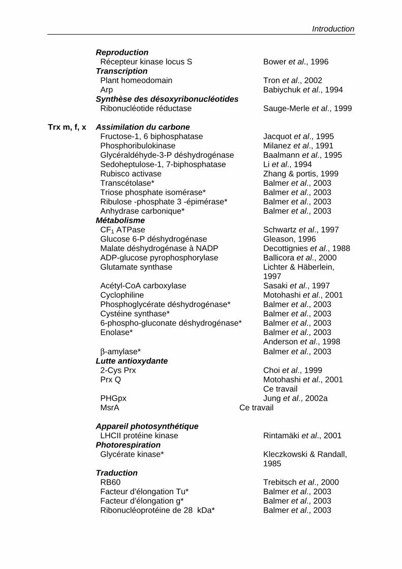
Introduction
Reproduction Récepteur kinase locus S Bower et al., 1996 Transcription Plant homeodomain Tron et al., 2002 Arp Babiychuk et al., 1994 Synthèse des désoxyribonucléotides Ribonucléotide réductase Sauge-Merle et al., 1999 Trx m, f, x Assimilation du carbone
Fructose-1, 6 biphosphatase Jacquot et al., 1995 Phosphoribulokinase Milanez et al., 1991 Glycéraldéhyde-3-P déshydrogénase Baalmann et al., 1995 Sedoheptulose-1, 7-biphosphatase Li et al., 1994 Rubisco activase Zhang & portis, 1999 Transcétolase* Balmer et al., 2003 Triose phosphate isomérase* Balmer et al., 2003 Ribulose -phosphate 3 -épimérase* Balmer et al., 2003 Anhydrase carbonique* Balmer et al., 2003 Métabolisme CF1 ATPase Schwartz et al., 1997
Glucose 6-P déshydrogénase Gleason, 1996 Malate déshydrogénase à NADP Decottignies et al., 1988 ADP-glucose pyrophosphorylase Ballicora et al., 2000 Glutamate synthase Lichter & Häberlein, 1997 Acétyl-CoA carboxylase Sasaki et al., 1997 Cyclophiline Motohashi et al., 2001 Phosphoglycérate déshydrogénase* Balmer et al., 2003 Cystéine synthase* Balmer et al., 2003 6-phospho-gluconate déshydrogénase* Balmer et al., 2003 Enolase* Balmer et al., 2003 Anderson et al., 1998 β-amylase* Balmer et al., 2003 Lutte antioxydante 2-Cys Prx Choi et al., 1999 Prx Q Motohashi et al., 2001
Ce travail PHGpx Jung et al., 2002a MsrA Ce travail
Appareil photosynthétique
LHCII protéine kinase Rintamäki et al., 2001 Photorespiration
Glycérate kinase* Kleczkowski & Randall, 1985 Traduction
RB60 Trebitsch et al., 2000 Facteur d’élongation Tu* Balmer et al., 2003 Facteur d’élongation g* Balmer et al., 2003
Ribonucléoprotéine de 28 kDa* Balmer et al., 2003
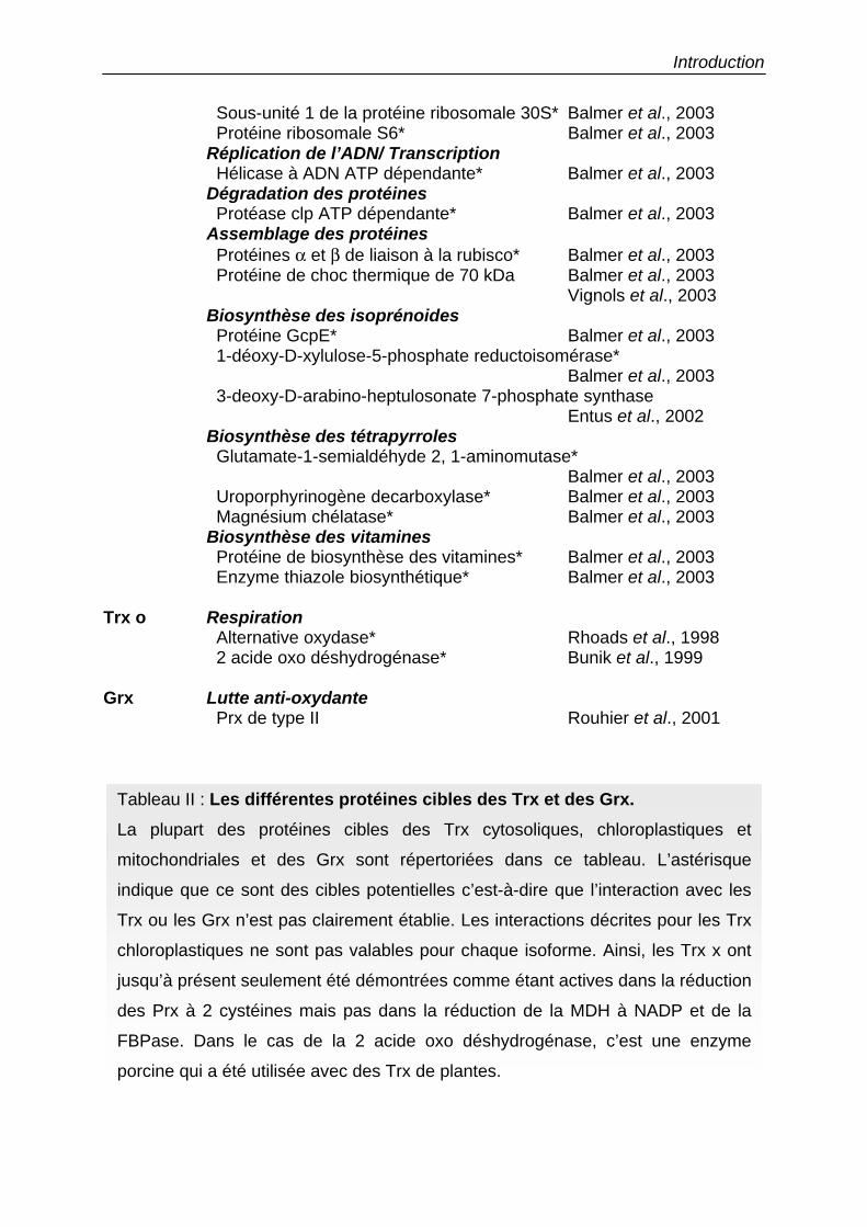
Introduction
Sous-unité 1 de la protéine ribosomale 30S* Balmer et al., 2003 Protéine ribosomale S6* Balmer et al., 2003 Réplication de l’ADN/ Transcription Hélicase à ADN ATP dépendante* Balmer et al., 2003 Dégradation des protéines Protéase clp ATP dépendante* Balmer et al., 2003 Assemblage des protéines Protéines α et β de liaison à la rubisco* Balmer et al., 2003 Protéine de choc thermique de 70 kDa Balmer et al., 2003
Vignols et al., 2003 Biosynthèse des isoprénoides Protéine GcpE* Balmer et al., 2003 1-déoxy-D-xylulose-5-phosphate reductoisomérase*
Balmer et al., 2003 3-deoxy-D-arabino-heptulosonate 7-phosphate synthase Entus et al., 2002
Biosynthèse des tétrapyrroles Glutamate-1-semialdéhyde 2, 1-aminomutase*
Balmer et al., 2003 Uroporphyrinogène decarboxylase* Balmer et al., 2003 Magnésium chélatase* Balmer et al., 2003 Biosynthèse des vitamines Protéine de biosynthèse des vitamines* Balmer et al., 2003 Enzyme thiazole biosynthétique* Balmer et al., 2003 Trx o Respiration
Alternative oxydase* Rhoads et al., 1998 2 acide oxo déshydrogénase* Bunik et al., 1999 Grx Lutte anti-oxydante
Prx de type II Rouhier et al., 2001
Tableau II : Les différentes protéines cibles des Trx et des Grx. La plupart des protéines cibles des Trx cytosoliques, chloroplastiques et
mitochondriales et des Grx sont répertoriées dans ce tableau. L’astérisque
indique que ce sont des cibles potentielles c’est-à-dire que l’interaction avec les
Trx ou les Grx n’est pas clairement établie. Les interactions décrites pour les Trx
chloroplastiques ne sont pas valables pour chaque isoforme. Ainsi, les Trx x ont
jusqu’à présent seulement été démontrées comme étant actives dans la réduction
des Prx à 2 cystéines mais pas dans la réduction de la MDH à NADP et de la
FBPase. Dans le cas de la 2 acide oxo déshydrogénase, c’est une enzyme
porcine qui a été utilisée avec des Trx de plantes.
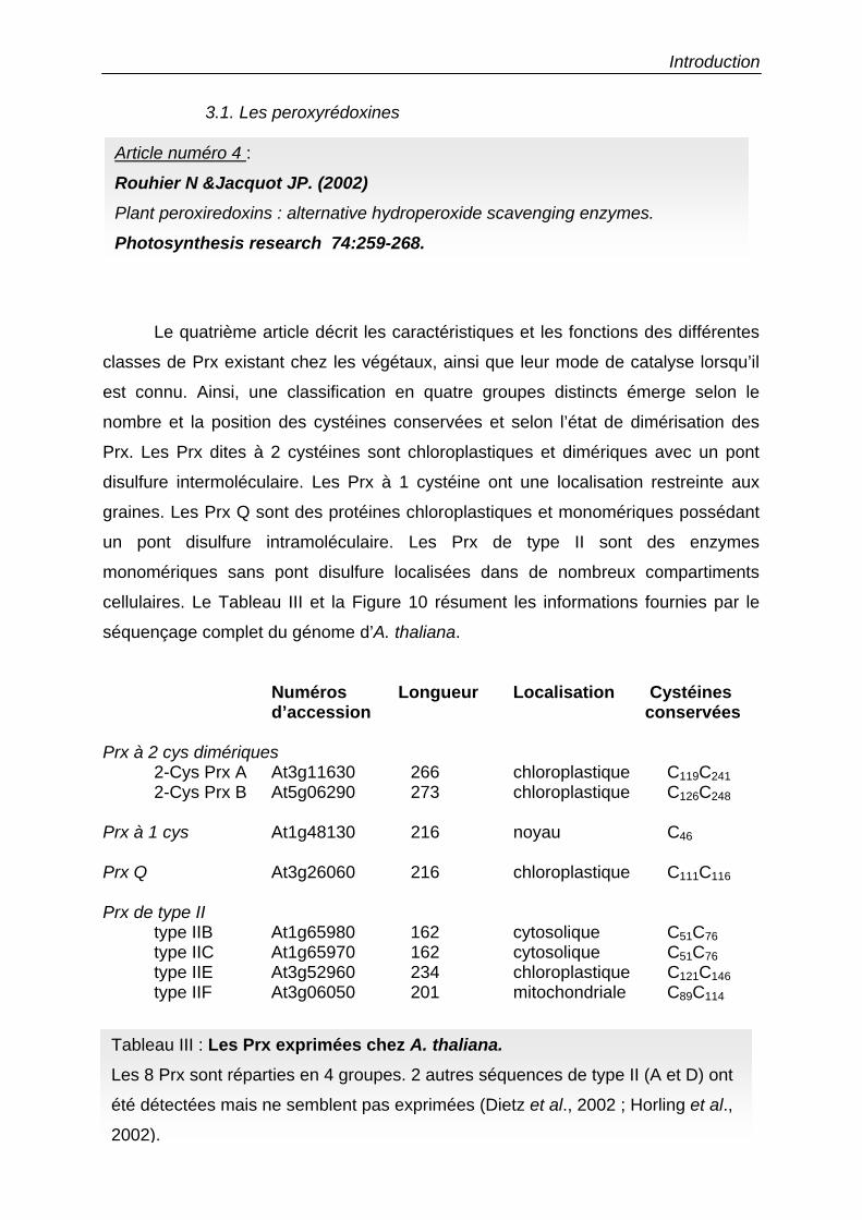
Introduction
3.1. Les peroxyrédoxines
Le quatrième article décrit les caractéristiques et les fonctions des différentes
classes de Prx existant chez les végétaux, ainsi que leur mode de catalyse lorsqu’il
est connu. Ainsi, une classification en quatre groupes distincts émerge selon le
nombre et la position des cystéines conservées et selon l’état de dimérisation des
Prx. Les Prx dites à 2 cystéines sont chloroplastiques et dimériques avec un pont
disulfure intermoléculaire. Les Prx à 1 cystéine ont une localisation restreinte aux
graines. Les Prx Q sont des protéines chloroplastiques et monomériques possédant
un pont disulfure intramoléculaire. Les Prx de type II sont des enzymes
monomériques sans pont disulfure localisées dans de nombreux compartiments
cellulaires. Le Tableau III et la Figure 10 résument les informations fournies par le
séquençage complet du génome d’A. thaliana.
Numéros Longueur Localisation Cystéines d’accession conservées
Prx à 2 cys dimériques
2-Cys Prx A At3g11630 266 chloroplastique C119C241 2-Cys Prx B At5g06290 273 chloroplastique C126C248
Prx à 1 cys At1g48130 216 noyau C46 Prx Q At3g26060 216 chloroplastique C111C116 Prx de type II type IIB At1g65980 162 cytosolique C51C76 type IIC At1g65970 162 cytosolique C51C76 type IIE At3g52960 234 chloroplastique C121C146 type IIF At3g06050 201 mitochondriale C89C114
Tableau III : Les Prx exprimées chez A. thaliana. Les 8 Prx sont réparties en 4 groupes. 2 autres séquences de type II (A et D) ont
été détectées mais ne semblent pas exprimées (Dietz et al., 2002 ; Horling et al.,
2002).
Article numéro 4 :
Rouhier N &Jacquot JP. (2002) Plant peroxiredoxins : alternative hydroperoxide scavenging enzymes.
Photosynthesis research 74:259-268.
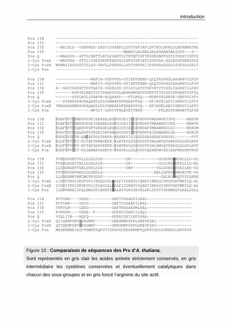
Introduction
Prx IIB -------------------------------------------------------------Prx IIC -------------------------------------------------------------Prx IIE --MATSLS--VSRFMSS-SATVISVAKPLLSPTVSFTAPLSFTRSLAPNLSLKFRNRRTNSPrx IIF -----------------------------MAMSILKLRNLSALRSAANSARIGVS---S--Prx Q --MAASSS--SFTLCNHTTLRTLPLRKTLVTKTQFSVPTKSSESNFFGSTLTHSSYISPVS2-Cys PrxA --MASVAS--STTLISSPSSRVFPAKSSLSSPSVSFLRTLSSPSAS-ASLRSGFARRSSLS2-Cys PrxB MSMASIASSSSTTLLSS-SRVLLPSKSSLLSPTVSFPRIIPSSSASSSSLCSGFSSLGSLT1-Cys Prx -------------------------------------------------------------
Prx IIB ---------------MAPIA-VGDVVPD-GTISFFDEND-QLQTASVHSLAAGKKVILFGVPrx IIC ---------------MAPIT-VGDVVPD-GTISFFDEND-QLQTVSVHSIAAGKKVILFGVPrx IIE A--SATTRSFATTPVTASIS-VGDKLPD-STLSYLDPSTGDVKTVTVSSLTAGKKTILFAVPrx IIF -------RGFSKLAEGTDITSAAPGVSLQKARSWDEGVSSKFSTTPLSDIFKGKKVVIFGLPrx Q -------SSSLKGLIFAKVN-KGQAAPD---FTLKDQ---NGKPVSLKKYK-GKPVVLYFY2-Cys PrxA ---STSRRSFAVKAQADDLPLVGNKAPDFEAEAVFDQ---EFIKVKLSDYIGKKYVILFFY2-Cys PrxB TNRSASRRNFAVKAQADDLPLVGNKAPDFEAEAVFDQ---EFIKVKLSEYIGKKYVILFFY1-Cys Prx ---------------MPGIT-LGDTVPNLEVETTHDK-------FKLHDYFANSWTVLFSH
Prx IIB PGAFTPTCSMKHVPGFIEKAEELKSKGVDEIICFSVNDPFVMKAWGKTYPE-----NKHVKPrx IIC PGAFTPTCSMSHVPGFIGKAEELKSKGIDEIICFSVNDPFVMKAWGKTYPE-----NKHVKPrx IIE PGAFTPTCSQKHVPGFVSKAGELRSKGIDVIACISVNDAFVMEAWRKDLGI-----NDEVMPrx IIF PGAYTGVCSQQHVPSYKSHIDKFKAKGIDSVICVSVNDPFAINGWAEKLGA-----KDAIEPrx Q PADETPGCTK-QACAFRDSYEKFK-KAGAEVIGISGDDSASHKAFASKYKL-------PYT2-Cys PrxA PLDFTFVCPT-EITAFSDRHSEFE-KLNTEVLGVSVDSVFSHLAWVQTDRKSGGLGDLNYP2-Cys PrxB PLDFTFVCPT-EITAFSDRYEEFE-KLNTEVLGVSVDSVFSHLAWVQTDRKSGGLGDLNYP1-Cys Prx PGDFTPVCTT-ELGAMAKYAHEFD-KRGVKLLGLSCDDVQSHKDWIKDIEAFNHGSKVNYP
Prx IIB FVADGSGEYTHLLGLELDLK----------DK------------GLGVRSRRFALLLD-DLPrx IIC FVADGSGEYTHLLGLELDLK----------DK------------GLGIRSRRFALLLD-NLPrx IIE LLSDGNGEFTGKLGVELDLR----------DKP----------VGLGVRSRRYAILAD-DGPrx IIF FYGDFDGKFHKSLGLDKDLS----------------------AALLGPRSERWSAYVE-DGPrx Q LLSDEGNKVRKDWGVPGDLF-------------------------GALPG-RQTYVLDKNG2-Cys PrxA LISDVTKSISKSFGVLIHDQGIALRGLFIIDKEGVIQHSTINNLGIGRSVDETMRTLQ-AL2-Cys PrxB LVSDITKSISKSFGVLIPDQGIALRGLFIIDKEGVIQHSTINNLGIGRSVDETMRTLQ-AL1-Cys Prx IIADPNKEIIPQLNMIDPIENGPSRALHIVGPDSKIKLSFLYPSTTGRNMDEVLRALDSLL
Prx IIB KVTVAN----VESG----------GEFTVSSADDILKAL-------------------Prx IIC KVTVAN----VESG----------GEFTVSSAEDILKAL-------------------Prx IIE VVKVLN----LEEG----------GAFTNSSAEDMLKAL-------------------Prx IIF KVKAVN----VEEA--P-------SDFKVTGAEVILGQI-------------------Prx Q VVQLIYN---NQFQ----------PEKHIDETLKFLKAA-------------------2-Cys PrxA QYIQENPDEVCPAGWKP-------GEKSMKPDPKLSKEYFSAI---------------2-Cys PrxB QYVQENPDEVCPAGWKP-------GEKSMKPDPKLSKEYFSAI---------------1-Cys Prx MASKHNNKIATPVNWKPDQPVVISPAVSDEEAKKMFPQGFKTADLPSKKGYLRHTEVS
Figure 10 : Comparaison de séquences des Prx d’A. thaliana. Sont représentés en gris clair les acides aminés strictement conservés, en gris
intermédiaire les cystéines conservées et éventuellement catalytiques dans
chacun des sous-groupes et en gris foncé l’arginine du site actif.
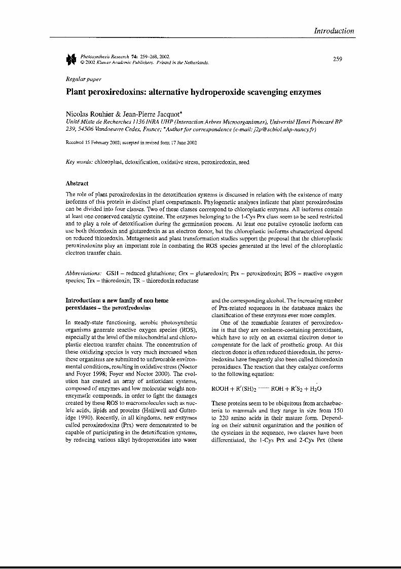
..-. Photosynthesis Research 74: 259-268,2002.
.,~ © 2002 Kluwer Academie Publishers. Printed in the Netherlands.
Regular paper
Introduction
259
Plant peroxiredoxins: alternative hydroperoxide scavenging enzymes
Nicolas Rouhier & Jean-Pierre Jacquot*Unité Mixte de Recherches 1136 INRA UHP (Interaction Arbres Microorganismes), Université Henri Poincaré BP239,54506 Vandoeuvre Cedex, France; *Author for correspondence (e-mail: [email protected])
Received 15 February 2002; accepted in revised form 17 June 2002
Key words: chloroplast, detoxification, oxidative stress, peroxiredoxin, seed
Abstract
The role of plant peroxiredoxins in the detoxification systems is discussed in relation with the existence of manyisoforms of this protein in distinct plant compartments. Phylogenetic analyses indicate that plant peroxiredoxinscan be divided into four classes. Two of these classes correspond to chloroplastic enzymes. AIl isoforms containat least one conserved catalytic cysteine. The enzymes belonging to the l-Cys Prx class seem to be seed restrictedand to play a role of detoxification during the germination process. At least one putative cytosolic isoform canuse both thioredoxin and glutaredoxin as an electron donor, but the chloroplastic isoforms characterized dependon reduced thioredoxin. Mutagenesis and plant transformation studies support the proposaI that the chloroplasticperoxiredoxins play an important role in combating the ROS species generated at the level of the chloroplasticelectron transfer chain.
Abbreviations: GSH - reduced glutathione; Grx - glutaredoxin; Prx - peroxiredoxin; ROS - reactive oxygenspecies; Trx - thioredoxin; TR - thioredoxin reductase
Introduction: a new famUy of non hemeperoxidases - the peroxiredoxins
In steady-state functioning, aerobic photosyntheticorganisms generate reactive oxygen species (ROS),especially at the level of the mitochondrial and chloroplastic electron transfer chains. The concentration ofthese oxidizing species is very much increased whenthese organisms are submitted to unfavorable environmental conditions, resulting in oxidative stress (Noctorand Foyer 1998; Foyer and Noctor 2000). The evolution has created an array of antioxidant systems,composed of enzymes and low molecular weight nonenzymatic compounds, in order to fight the damagescreated by these ROS to macromolecules such as nucleic acids, lipids and proteins (HaIliweIl and Gutteridge 1990). Recently, in aIl kingdoms, newenzymescaIled peroxiredoxins (Prx) were demonstrated to becapable of participating in the detoxification systems,by reducing various alky1 hydroperoxides into water
and the corresponding alcohoL The increasing numberof Prx-related sequences in the databases makes theclassification of these enzymes ever more complexe
One of the remarkable features of peroxiredoxins is that they are nonheme-containing peroxidases,which have to rely on an external electron donor tocompensate for the lack of prosthetic group. As thiselectron donor is often reduced thioredoxin, the peroxiredoxins have frequently also been called thioredoxinperoxidases. The reaction that they catalyze conformsto the foIlowing equation:
ROOH + R'(SH)2 ROH + R'S2 + H20
These proteins seem to be ubiquitous from archaebacteria to mammals and they range in size from 150to 220 amino acids in their mature forme Depending on their subunit organization and the position ofthe cysteines in the sequence, two classes have beendifferentiated, the l-Cys Prx and 2-Cys Prx (these
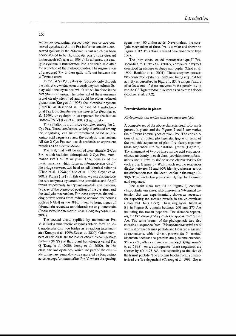
260
sequences containing, respectively, one or two conserved cysteines). AlI the Prx isoforms contain a conserved cysteine in the N-terminus part which has beendemonstrated to be the catalytic one by site-directedmutagenesis (Chae et al. 1994a). In aIl cases, the catalytic cysteine is transformed into a sulfenic acid afterthe reduction of the hydroperoxides. The regenerationof a reduced Prx is then quite different between thedifferent classes.
ln the 1-Cys Prx, catalysis proceeds only throughthe catalytic cysteine even though they sometimes display additional cysteines, which are not involved in thecatalytic mechanism. The reductant of these enzymesis not clearly identified and couId be either reducedglutathione (Kang et al. 1998), the thioredoxin system(TrxtrR) as deseribed in the case of a mitochondrial Prx from Saccharomyces cerevisiae (Pedrajas etal. 1999), or cyclophilin as reported for the humanisoform Prx VI (Lee et al. 2001) (Figure lA).
The situation is a bit more complex among the 2Cys Prx. Three subclasses, widely distributed amongthe kingdoms, can be differentiated based on theamino acid sequences and the catalytic mechanism.AlI the 2-Cys Prx can use thioredoxin or equivalentproteins as an eleetron donor.
The first, that will be called here dimeric 2-CysPrx, which includes chloroplastic 2-Cys Prx, mammalian Prx 1 to IV or yeast TSA, consists of dimeric enzymes which form an intermolecular disulfide bridge between two head-to-tail identical subunits(Chae et al. 1994a; Chae et al. 1999; Goyer et al.2002) (Figure 1, BI). In this class, we can also includethe rare enzymes trypanothione peroxidase and AhpCfound respectively in trypanosomatids and bacteria,because of the conserved position of the cysteines andthe catalytic mechanism. For these enzymes, the reducing power cornes from reduced adenine nucleotidessuch as NADH or NADPH, linked by homologues ofthioredoxin reduetase and thioredoxin or glutaredoxin(Poole 1996; Montemartini et al. 1998; Reynolds et al.2002).
The second class, typified by mammalian PrxV, includes monomeric enzymes which form an intramolecular disulfide bridge as a reaction intermediate (Knoops et al. 1999; Seo et al. 2000). Other members of this class are the bacterioferritin co-migratoryproteins (BCP) and their plant homologues called PrxQ (Kong et al. 2000; Jeong et al. 2000). In thisclass, the two cysteines, which are part of the disulfide bridge, are generally only separated by four aminoacids, except for mammalian Prx V, where the spacing
Introduction
spans over 100 amino acids. Nevertheless, the catalytic mechanism of these Prx is similar and shown inFigure 1, B2. This class is named here monomeric type1 Prx.
The third class, called monomeric type II Prx,according to Dietz et al (2002), comprises enzymesdescribed in chinese cabbage and poplar (Choi et al.1999; Rouhier et al. 2001). These enzymes possesstwo conserved cysteines, only one being required foractivity as described in Figure 1, B3. A unique featureof at least one of these enzymes is the possibility touse the GSH/glutaredoxin system as an electron donor(Rouhier et al. 2002).
Peroxiredoxins in plants
Phylogenetic and amino acid sequences analysis
A complete set of the above characterized isoforms ispresent in plants and the Figures 2 and 3 summarizethe different known types of plant Prx. The construction of an unrooted phylogenetic tree with most ofthe available sequences of plant Prx clearly separatesthese sequences into four distinct groups (Figure 2).The alignment of two of these amino aeid sequences,chosen randomly in each class, provides more informations and allows to define sorne characteristics foreach group (Figure 3). Within each set, the sequencesdisplay between 75 and 90% identity, whereas acrossthe different classes, the identities faU in the range 1030%. Thus, each class is very weIl defined by its aminoacid sequence.
The main class (set Blin Figure 2) containschloroplastic enzymes, which present a N-terminal extension that was experimentally shown as necessaryfor exporting the mature protein in the chloroplasts(Baier and Dietz 1997). These sequences, listed asBlin Figure 3, contain between 260 and 275 AAincluding the transit peptides. The distance separating the two conserved cysteines is approximately 120AA. The same branch of the phylogenetic tree alsocontains a sequence from Chlamydomonas reinhardtiiwith a shortened transit peptide and from red algae andcyanobacteria, which do not possess the N-terminalextension because the proteins are plastome encoded,whereas the others are nuclear encoded (Klughammeret al. 1998). As a consequence, these sequences areshorter by 60 to 75 AA, corresponding to the size ofthe transit peptide. The proteins biochemically characterized are Trx dependent (Cheong et al. 1999; Goyer
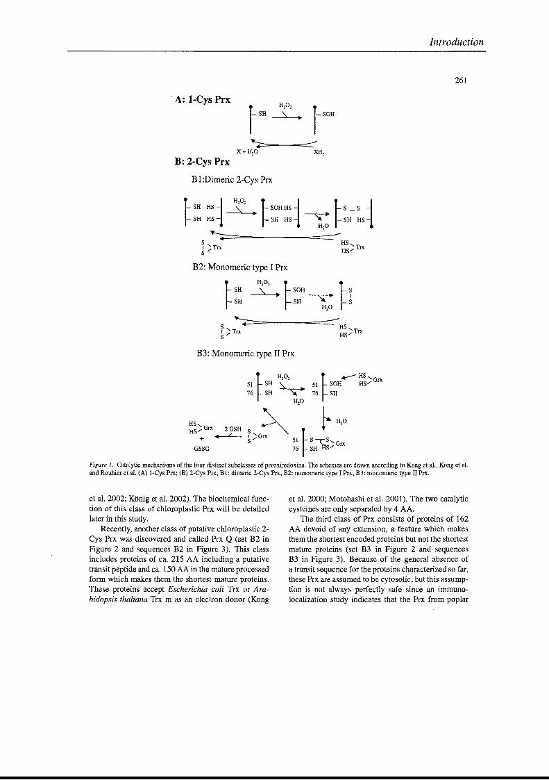
Introduction
261
, A: l-Cys Prx
r- SOH
B: 2-Cys Prx
BI :Dimeric 2-Cys Prx
S"---1 )TrxS
B2: Monomeric type 1Prx
~SH~SH
S ~~..:::;=----===-=:::::-=----HS~ ) Trx HS) Trx
B3: Monomeric type II Prx
HS,HS"'" Grx 2 GSH
+ +-L--GSSa
~H202 ~.---HS,O
51 SH~..... 51 SOH HS"'" ex76 SH ~ 76 SH
H20
~ ~H'O~)Grx 51~S-SS '- 'Grx
76 SH HS'"
Figure 1. Catalytic mechanisms of the four distinct subclasses of peroxiredoxins. The schemes are drawn according to Kang et al., Kong et al.and Rouhier et al. (A) l-Cys Prx; (B) 2-Cys Prx, BI: dimeric 2-Cys Prx, B2: monomeric type 1Prx, B3: monomeric type II Prx.
et al. 2002; Konig et al. 2002). The biochemical function of this class of chloroplastic Prx will be detailedlater in this study.
Recently, another class of putative chloroplastic 2Cys Prx was discovered and called Prx Q (set B2 inFigure 2 and sequences B2 in Figure 3). This classincludes proteins of ca. 215 AA including a putativetransit peptide and ca. 150 AA in the mature processedform which makes them the shortest mature proteins.These proteins accept Escherichia coli Trx or Arabidopsis thaliana Trx m as an electron donor (Kong
et al. 2000; Motohashi et al. 2001). The two catalyticcysteines are only separated by 4 AA.
The third class of Prx consists of proteins of 162AA devoid of any extension, a feature which makesthem the shortest encoded proteins but not the shortestmature proteins (set B3 in Figure 2 and sequencesB3 in Figure 3). Because of the general absence ofa transit sequence for the proteins characterlzed so far,these Prx are assumed to be cytosolic, but this assumption is not always perfectly safe since an immunolocalization study indicates that the Prx from poplar
Introduction
262
Figure 2. Unrooted phylogenetic tree of the different plant peroxiredoxins constructed using the Clustalw and Treeview softwares. A: l-CysPrx; BI: dimeric 2-Cys Prx; BZ: monomeric type 1 Prx; Set B3: monomeric type II Prx.
is located in plastid-like structures contained in thephloem sieve tubes (Rouhier et al. 2001). Moreover,at least two strongly homologous sequences found inthe protein databank, from Arabidopsis thaliana andOryza sativa, present an N-terminal extension, whichis predicted to export the proteins either in chloroplastor in mitochondria (accession numbers NP_190864and BAA82377). Although these proteins possess twoconserved cysteines distant of 25 AA, they functioncatalytically as 1-Cys Prx (see below).
Finally, the last class of plant Prx comprises proteins of 216 to 220 AA and are referred to as 1-CysPrx (set A in Figure 2 and sequences A in Figure 3).These proteins do not display a N-terminal extensionbut rather a C-terminal one, which is predicted to contain an NLS signal exporting the protein to the nucleus(Stacy et al. 1999).
Function ofperoxiredoxins in plants
Up to now, the functions of the different plant Prxhave not been evenly characterized in aIl subclasses,but aIl. enzymes appear to have a common functionof alkyl hydroperoxide detoxification in the differentplant compartments. The putative in vivo functions of
the various plant types are detailed below, except forthose of set B2, for which no additional information isyet available.
The chloroplastic detoxification systems: is there arole for the dimeric 2-Cys peroxiredoxins?
The major source of production of the ROS in thechloroplast is located at the level of PS 1 whereOz combines with the reduced species generatedby the functioning of this photosystem, producingsuperoxide ions and then hydrogen peroxide (Figure 4). A complex chloroplastic detoxification system, called the HaIliweIl and Asada cycle fightsagainst these ROS and is composed of glutathione andascorbate and of known enzymes such as superoxide dismutases, monodehydroascorbate reductases anddehydroascorbate reductases (Asada 1999).
A high number of hydroperoxide scavenging enzymes also exists in the chloroplast. First, thereare two different ascorbate peroxidases (APx), onestromal and another one thylakoid-bound, which playa crucial role in the reduction of hydrogen peroxidebut not of alkyl hydroperoxides (Asada 1999). Addi-
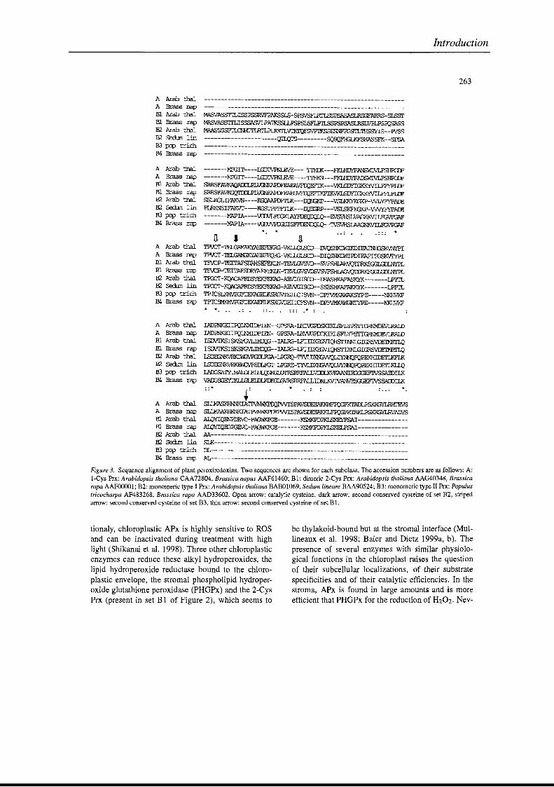
Introduction
263
.::: *
A Arab thalA Brass napEl Arab thalm Brass rapB2 Arab tbalB2 sedum linB3 IXJP trichB4 Brass :rap
A Arab thalA Brass napID Arab thalB1 Brass rap132 Arab thalB2 S€dun linB3 PJP trich134 Brass. rap
A Arab tha1A Brass napEl Arab thalEl Brass rapB2 Arab thalB2 Sa:lJm linB3 IXJP trichB4 Brass rap
MASVASSITLISSPSSRVFPAKSSLS-SPSVSFIRI'LSSPSASASLRSGFARRS-SLSSrMASVASSITLISSSASVLPA'IKSSLLPSPSISFLPrLSSPSPSASLRSLVPLPSmsASSMAASSSSFI'lŒHITLRI'LPLRlcrLVTI<1QF&VPrKSSESNFFGSrrJllISSYIS--PVSS----------------------QIlQI'S---------~F'lG...l<F'SfJASF'K--SPSA
-------MffiIT----LGIJIVR.JLE.VE----TIHOK---FKI.RDYFANSWIVLFSHFGDF-------MroIT----LGIJIVR.JLE.VE----TIHKN---FKI.RDYFAnS'WIVLFSHRDFSRRSFAVKAQAIDLPLVGNKAPDFE'AFA~EFIK---VKI.SDYIGKKYVILFFYPIDF
SRRSFAVKGQIDJLPLVi3NI<APDF'EAEGEFIKFIKVKI..SDYIGKKYVILFFLPlDFSSLKGLIFAKVN----KGQAAPDFTLK---~---VSU<KYKGKP-WLYFYPADE
PLRKNSIFAKVI'----KGSl'PppFI'LK---m&;RP---VSlSKFKGKP-WVYFYPADE-------MAPIA----~IQ_-E.VSVHSLVAGKK.VILF'G\i'
-------MAPIA----VGlJJVPI:GSIS~I.Q_-~
* *n 1 iTP'iJCT-TEl.GAMN<YAHEF!VKLI.GIS:D--DVQSHKIWIKDlFAFNf{;SKVNYPITPVCT-TElGAM:;KYAHEF'EQRG-VKLI.GIS:D--DIQSHKIJflIPDlFAF'rffiSKVI'YPITFVCP-TEITAFSDRHSEFEKLN-TEVLGVSVD--SVF~INYPL
TFVCP-TEITAFSDRYAEFEKlN LTIO:T-~-AEVIGISGD--OOASHKAFASKYK-------LPYTL
TIG:T-KQACAFRœYEKFKKAAEVVGISGD--SSESHKAFAKKYK-------LPFI'LTPICSLKHVEGFI~II.CISVN--DPFVMKAWAKSYPE-----NKHVKF
TP1CSMKH\.1ffiFIEKAEELKSKGVDEIIŒ'SVN--D~-----NKHVKF
* * ::.. . ::: .* :
: :*
A Arab thalA Brass ŒIp
Bl Arab thalID Brass rapB2 Arab thalB2 SErlJm linB3 IXJP trichB4 Brass :t:ap
A Arab thalA Brass napm Al:ab thalm Brass rapB2 Arab thalB2 SOOum linB3 p:p trichB4 Brass rap
IADmKEIIEQLNMIDPIEN--GPSRA-UII\,{;PDSKIl<ISFLYPSI'IGRNMDEVLRAIDIADFNKEIIl?Ç,1llJMIDPIEN--GPSRA-lRVVGPOCK.IKI.SYPSI'IGRNMDEVLRAI.DISDVrKSISKSFGVLIHI:>'J3-IALRG-LFIIDKEI.iVIQHSI'INNIJ3IGRSVDEIMRTI.QISDVrKSISKSFGVLIIirQ:;--IALRG-LFIIDKEI;vIQmrIXNI..GIGRSVDE'IMRTI.Q~-LB3RQ-~~FQPEKIDDEI'LKFLK
~SDLFGr-LroRE-~L~FQPEKHIDErLKLIJJ
I.AI:GSA~ALLVDDLKVKAANI~ILK
VAI:G93EYTKLLGLELDLKDKGI.GVRSRRF~SADDIIK
~.
~~PV\TISPAVSDEFAI<KMFFQ8FADLPSKKGYLRHrEVS
Slll1AAKHKNKIA~SPAVSDEE'AKKLF~
ALQYIQENPDEVC-PAGWKPGE-------KSMKPDPKLSKEYFSAI--------------ALQYIQEGPGEVC-PAGWKPGE-------KSMKPDPKLSKELFSAI---------------M----------------------------------------------------------SIl<---------------------------------------------------------DL----------------------------------------------------------AL-----------------------------------------------------------
Figure 3. Sequence alignment of plant peroxiredoxins. Two sequences are shown for each subclass. The accession numbers are as follows: A:I-Cys Prx: Arabidopsis thaliana CAA72804, Brassica napus AAF61460; BI: dimeric 2-Cys Prx: Arabidopsis thaliana AAG40348, Brassicarapa AAFOOOOl; B2: monomeric type 1Prx: Arabidopsis thaliana BABOI069, Sedum lineare BAA90524; B3: monomeric type II Prx: Populustricocharpa AF483268, Brassica rapa AAD33602. Open arrow: catalytic cysteine, dark arrow: second conserved cysteine of set B2, stripedarrow: second conserved cysteine of set B3, thin arrow: second conserved cysteine of set BI.
tionaly, chloroplastic APx is highly sensitive to ROSand can be inactivated during treatment with highlight (Shikanai et al. 1998). Three other chloroplasticenzymes can reduce these alkyl hydroperoxides, thelipid hydroperoxide reductase bound to the chloroplastic envelope, the stromal phospholipid hydroperoxide glutathione peroxidase (PHGPx) and the 2-CysPrx (present in set BI of Figure 2), which seems to
be thylakoid-bound but at the stromal interface (Mullineaux et al. 1998; Baier and Dietz 1999a, b). Thepresence of several enzymes with similar physiological functions in the chloroplast raises the questionof their subcellular localizations, of their substratespecificities and of their catalytic efficiencies. In thestroma, APx is found in large amounts and is moreefficient that PHGPx for the reduction of H202. Nev-
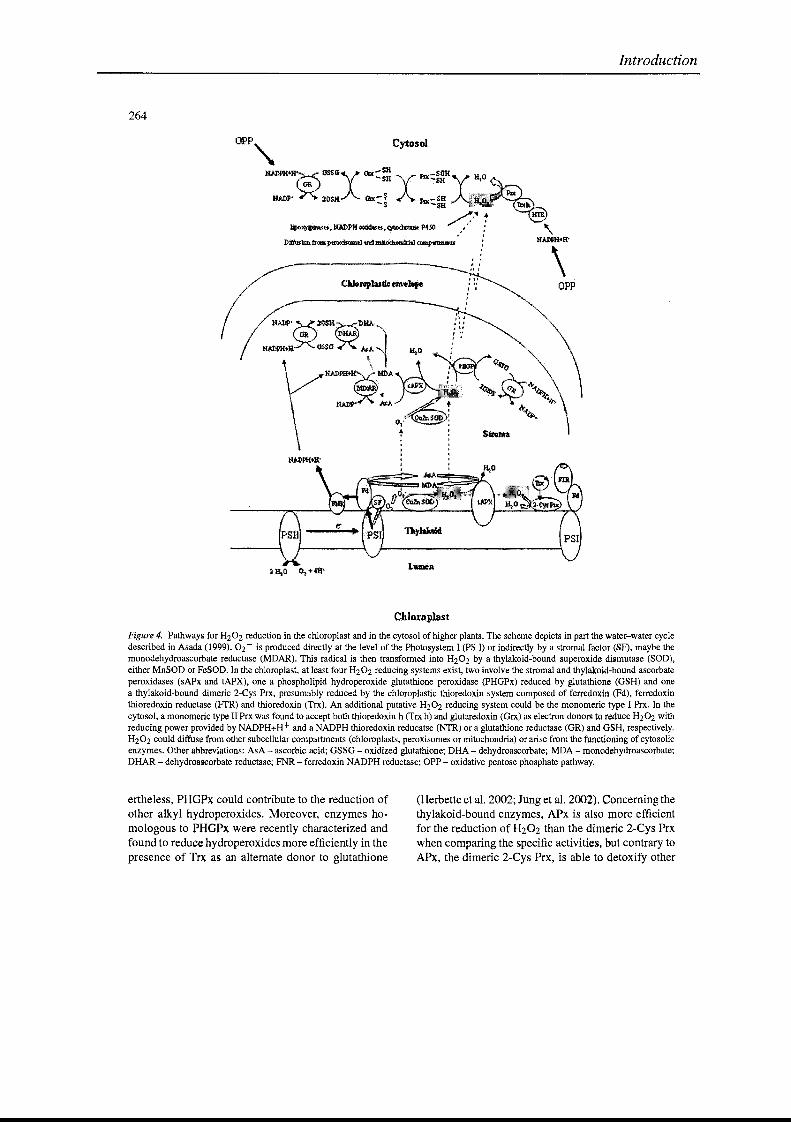
Introduction
264
opp
N.... :ADP.............•........•.•.~...•..........•........... .......• ·•.•.·...••....'OS.·.··..K,'GR
~H·· ·G$SO'
OPP, Cyto$oI
N~fH~tJSSttX········· lb::: X··~::I~JI
_.X :1./ !lilc:=J ...•. 1'JlI~= oI!· ..... l@!lŒ''I''!m..._
~,1fAJ)l'll'~.~l'4jl) ?'J,'"
(
Chloroplast
Figure 4. Pathways for H202 reduction in the chloroplast and in the cytosol of higher plants. The scheme depicts in part the water-water cycledescribed in Asada (1999). 02 - is produced directly at the level of the Photosystem 1 (PS 1) or indirectly by a stroma! factor (SF), maybe themonodehydroascorbate reductase (MDAR). This radical is then transformed into H202 by a thylakoid-bound superoxide dismutase (SOD),either MnSOD or FeSOD. In the chloroplast, at least four H202 reducing systems exist, two involve the stroma! and thylakaid~bound ascorbateperoxidases (sAPx and tAPX), one a phospholipid hydroperoxide glutathione peroxidase (PHGPx) reduced by glutathione (GSH) and onea thylakoid-bound dimeric 2-Cys Prx, presumably reduced by the chloroplastic thioredoxin system composed of ferredoxin (Fd), ferredoxinthioredoxin reductase (FfR) and thioredoxin (Trx). An additional putative H202 reducing system could be the monomeric type 1 Prx. In thecytosol, a monomeric type II Prx was found to accept both thioredoxin h (Trx h) and glutaredoxin (Grx) as electron donors ta reduce H202 withreducing power provided by NADPH+H+ and a NADPH thioredoxin reducatse (NTR) or a glutathione reductase (GR) and GSH, respectively.H202 could diffuse from other subcellular compartments (chloroplasts, peroxisomes or mitochondria) or arise from the functioning of cytosolicenzymes. Other abbreviations: AsA - ascorbic acid; GSSG - oxidized glutathione; DHA - dehydroascorbate; MDA - monodehydroascorbate;DHAR - dehydroascorbate reductase; FNR - ferredoxin NADPH reductase; OPP - oxidative pentose phosphate pathway.
ertheless, PHGPx could contribute to the reduction ofother alkyl hydroperoxides. Moreover, enzymes homologous to PHGPx were recently characterized andfound to reduce hydroperoxides more efficiently in thepresence of Trx as an alternate donor to glutathione
(Herbette et al. 2002; Jung et al. 2002). Concerningthethylakoid-bound enzymes, APx is also more efficientfor the reduction of H202 than the dimeric 2-Cys Prxwhen comparing the specifie activities, but contrary toAPx, the dimeric 2-Cys Prx, is able to detoxify other
hydroperoxides in vitro such as cumene hydroperoxide or tert-butyl hydroperoxide (Konig et al. 2002).The thylakoid-bound localization of the Prx suggestsalso that these proteins could combat lipid peroxidation, which causes damage to the membranes of thethylakoids. Konig et al (2002) propose that the 2-CysPrx could act not only in the water-water cycle pathway for energy dissipation in photosynthesis, but alsoin the peroxide detoxification in the plastids during thedark phase.
The function of the 2-Cys Prx was verified in vivoby complementing an Escherichia coli deficient strainfor the AhpC gene, the bacterial equivalent of 2-CysPrx (Baier et al. 1997). In addition, the Prx Q (setB2 in Figure 2) are also assumed to be chloroplasticand to detoxify the alkyl hydroperoxides in vitro butthe localization of these proteins has not yet been described. The reduction of hydroperoxides by these twotypes of chloroplastic Prx is presumed to be directlyassociated with the photosynthetic electron transferchain because of the thioredoxin dependence of thePrx (Konig et al. 2002).
ln order to investigate the importance of the 2Cys Prx in the detoxification process, the effect of asuppression in Arabidopsis thaliana was studied byengineering antisense plants (Baier and Dietz 1999a).The results suggest an essential role of the 2-Cys Prxin the protection of the chloroplastic structure againstoxidative damage because the levels of DI protein,ribulose-l ,5-bisphosphate carboxylase/oxygenase, thelight-harvesting protein complex associated with Photosystem II and the subunit CF! of the chloroplast ATPsynthase decrease. It was also concluded that neitherthe lipid hydroperoxide reductase, nor the glutathioneperoxidase can substitute for dimeric 2-Cys Prx probably because of different suborganellar localization ofthese proteins, which leads to specific physiologicalfunctions. The antisense suppression of the 2-Cys Prxdoes not induce a change in the transcriptional expression of the enzymes involved in glutathione metabolism, or of the chloroplastic glutathione peroxidase,and it has a weak effect on the abundance of transcriptsfor glutathione reductase, superoxide dismutase andcatalase (Baier et al. 2000). On the contrary, thelevel of transcripts and the enzymatic activities ofthe stromal and thylakoidal ascorbate peroxidases andof monodehydroascorbate reductase increase. In thismutant, the pool of ascorbate is highly oxidized. Theimportance of reduced ascorbate in the redox regulation of the 2-Cys Prx was recently studied in Ricciafluitans (Horling et al. 2001). When the level of ROS
Introduction
265
decreases, the pool of reduced ascorbate increases andinduces a decrease of the 2-Cys Prx transcript leveland the redox signaling cascade from the chloroplastto the nucleus couId be linked in part to a proteinkinase.
The 2-Cys Prx belongs to a multigenic family andis expressed in aIl organs of the mature plant, except in the roots (Cheong et al. 1999; Genot et al.2001). There is a gradient of expression of the 2-CysPrx gene from the young leaves to the mature leaves,whereas the expression decreases in aged tissues and,as a consequence, this protein could play an important role in the early stages of plant development whenthe cells are dividing or elongating (Baier and Dietz1996; Cheong et al. 1999). An additional proof of theimportance of the 2-Cys Prx is the severe effect onthe growth of a strain of Synechocystis sp deleted forthis gene (Klughammer et al. 1998). The expression ofthe 2-Cys Prx is induced in response to H202 and toillumination but quite weakly by methyl viologen ormetals, and it is not induced in response to ozone, orabscisic acid and giberellic acid treatments (Baier andDietz 1997; Cheong et al. 1999; Genot et al. 2001;Goyer et al. 2002).
A last piece of information about this type of enzymes is its possible endosymbiotic origine Indeed,in the red algae Porphyra purpurea, the 2-Cys Prxis plastome-encoded whereas in Arabidopsis thaliana,the 2-Cys Prx is nuclear encoded and the transit peptide is encoded by a separate exon supporting thehypothesis that this extension has been added duringevolution (Klughammer et al. 1998).
l-Cys Prx: a function restricted to seeds?
The l-Cys Prx are 216 to 220 AA long and comparedto the other classes of Prx, these proteins possess a Cterminal extension, which contains a putative nuclearsignal. An immunolocalization experiment confirmedthe presence of the protein in the nucleus of immatureembryos and aleurone cells and mast predominantlyin nucleoli (Stacy et al. 1999). The presence of thesel-Cys Prx was, however, also observed in the cytosolof these cells.
AlI the proteins of this type are not weIl biochemically characterized, and in particular the identity of theelectron donor is not known, as it is generally replacedby the chemical reductant dithiothreitol in vitro. It willbe thus of great interest to determine the nature of thisdonor.
266
The l-Cys Prx gene is only expressed in seeds andbelongs to a monogenic family (Stacy et al. 1996;Haslekas et al. 1998). The expression was shown tobe spatialIy and temporalIy regulated. The transcriptsincrease in the aleurone layer and in the imbibed embryos of dormant seeds exclusively, which suggests arole for these Prx in the control of dormancy, as H202was found to be able to break the phenomenon ofdormancy (Stacy et al. 1996). In addition, many stepsof the germination process, such as the dessicationand imbibition, are critical because of the formation ofROS. As the l-Cys Prx possess an in vitro peroxidaseactivity, another hypothesis is that the Prx protects theonly two tissues which survive the dessication step.Given that other seed antioxidants, such as superoxide dismutase and catalase, are found throughout theplant, the seed-specificity of the Prx suggests a keyrole for this enzyme in this tissue (Haslekas et al.1998). To investigate the exact function of the l-CysPrx, transgenic tobacco plants constitutively overexpressing the rice l-Cys Prx gene have been generated(Lee et al. 2000). The frequency of germination ofthese plants was similar to the control plant but thetransgenic Hnes exhibited a higher resistance againstoxidative stress, suggesting that the essential role ofl-Cys Prx is protection against oxidants.
Monomeric type II Prx
A poplar Prx with two conserved cysteines is assumed to function as al-Cys Prx because site-directedmutagenesis has demonstrated that only the catalyticcysteine of the poplar Prx is essential for hydroperoxide reduction (Rouhier et al. 2002). Moreover, thisenzyme is the only plant Prx so far characterized, thatcan accept both glutaredoxin (Grx) and thioredoxin asan electron donor (Rouhier et al. 2001). The only otherbiochemically characterized Prx of this group is fromBrassica rapa, which was shown to be Trx dependent,but Grx was probably not tested (Choi et al. 1999).It will also be of interest to characterize the proteinsof this class putatively routed to the chloroplast, inorder to verify the specificity of the electron donorbecause no glutaredoxins have so far been found inthe chloroplast. Additional informations about the Grxdependence of the Prx is provided by the numerousexisting fusion sequences in the nucleotide databasesbetween Grx and Prx modules and by the characterization of this type of protein in Chromatium gracile(Vergauwen et al. 2001).
Introduction
Perspectives
Different Prx isoforms have been characterized in several plant compartments such as the chloroplast, thenucleus and presumably the cytosol. Nevertheless, theincreasing number of plant sequencing projects couldwelllead to the description of new plant Prx isoformslocalized in the mitochondria or in the peroxisome, asalready shown in mammals or in yeast (Knoops et al.1999, Yamashita et al. 1999, Pedrajas et al. 1999). Thepresence of a complete thioredoxin system in plant mitochondria indicates that a functional electron donorsystem is present for any putative Prx present (Laloi etal. 2001). Interestingly, Dietz et al (2002) describe theexistence in Arabidopsis thaliana of a sequence whosegene product is presumed to be mitochondrial.
One domain to explore in plants is related to thebroad substrate specificities of Prx. Indeed, in additionto hydroperoxides, bacterial Prx have been demonstrated to be able to reduce different reactive nitrogenspecies, and particularly peroxynitrites, into nitrites(Chen et al. 1998, Bryk et al. 2000). Recently, a Prxfrom Leishmania chagasi was also found to detoxifyhydroxylradicals (Barr and Gedamu 2001). Moreover,a thioredoxin peroxidase from yeast was proposed toeHminate reactive sulfur species (Chae et al. 1994b)
Another challenge for plant Prx is to determine thein vivo functions of these proteins. Sorne indicationsprovided by the studies in mammalian cells suggestthat Prx are involved in cell differentiation and proliferation (Nemoto et al. 1990; Prosperi et al. 1993) andin apoptosis (Zhang et al. 1997; Kowaltowski et al.2000; Berggren et al. 2001). AlI these functions seemto be linked to the antioxidative properties of the Prx.
References
Asada K (1999) The water-water cycle in chloroplasts: scavengingof active oxygen and dissipation of excess photons. Annu RevPlant Physiol Plant Mol Biol 50: 601-639
Baier M and Dietz KJ (1996) Primary stmcture and expression of plant homologues of animal and fungal thioredoxindependent peroxide reductases and bacterial alkyl hydroperoxidereductases. Plant Mol Biol 31: 553-564
Baier M and Dietz KJ (1997) The plant 2-Cys peroxiredoxin BAS1is a nuclear-encoded chloroplast protein: its expressional regulation, phylogenetic origin, and implications for its specifiephysiological function in plants. Plant J 12: 179-190
Baier M and Dietz KJ (1999a) Protective function of chloropIast 2-cysteine peroxiredoxin in photosynthesis. Evidence fromtransgenic Arabidopsis. Plant Physioll19: 1407-1414
Baier M and Dietz KJ (1999b) Alkyl hydroperoxide reductases: theway out of the oxidative breakdown of lipids in chloroplasts.Trends Plant Sei 4: 166-168
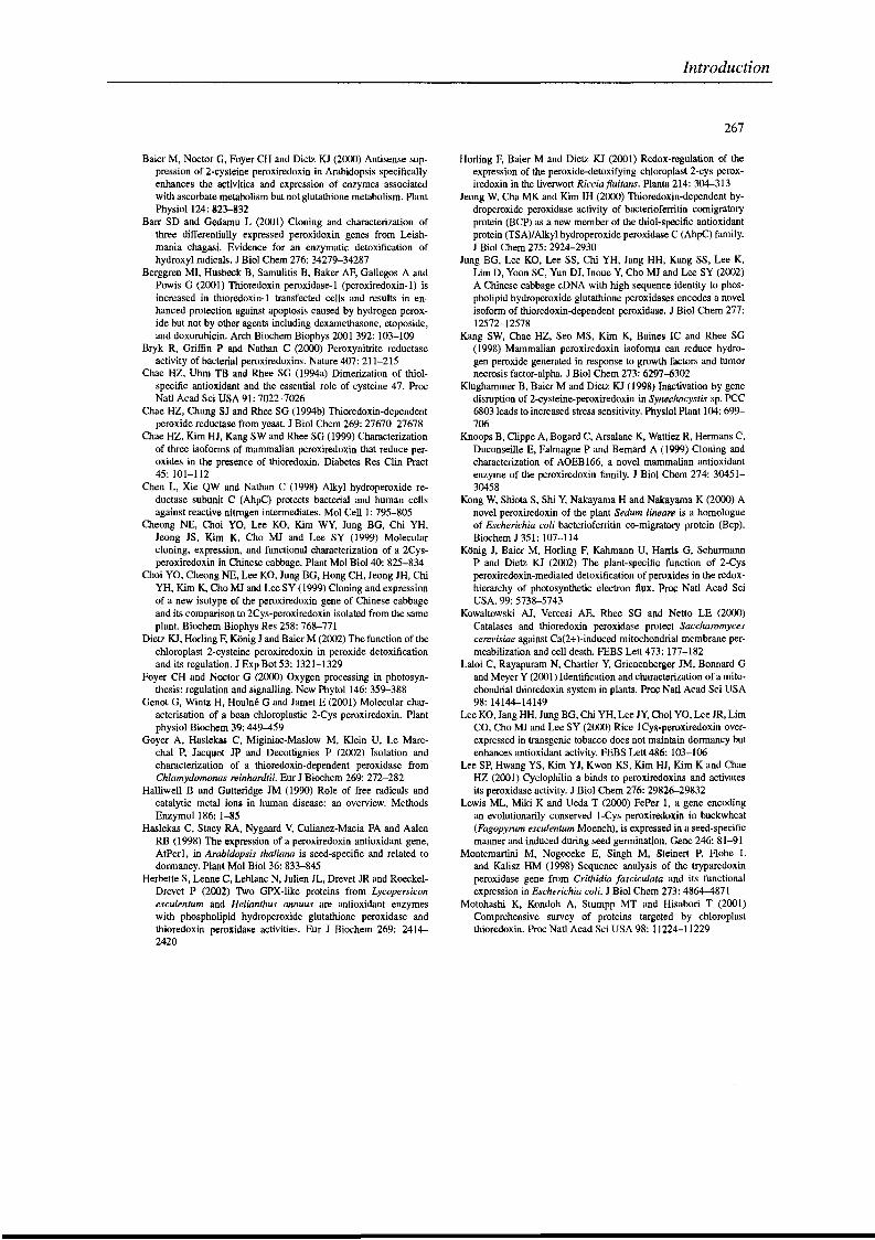
Baier M, Noctor G, Foyer CH and Dietz KJ (2000) Antisense suppression of 2-cysteine peroxiredoxin in Arabidopsis specificallyenhances the activities and expression of enzymes assoeiatedwith ascorbate metabolism but not glutathione metabolism. PlantPhysiol 124: 823-832
Barr SD and Gedamu L (2001) Cloning and characterization ofthree differentially expressed peroxidoxin genes from Leishmania chagasi. Evidence for an enzymatic detoxification ofhydroxyl radicals. J Biol Chem 276: 34279-34287
Berggren MI, Husbeck B, Samulitis B, Baker AF, Gallegos A andPowis G (2001) Thioredoxin peroxidase-1 (peroxiredoxin-1) isincreased in thioredoxin-1 transfected cells and results in enhanced protection against apoptosis caused by hydrogen peroxide but not by other agents including dexamethasone, etoposide,and doxorubicin. Arch Biochem Biophys 2001 392: 103-109
Bryk R, Griffin P and Nathan C (2000) Peroxynitrite reductaseactivity ofbacterial peroxiredoxins. Nature 407: 211-215
Chae HZ, Uhm TB and Rhee SG (1994a) Dimerization of thiolspecifie antioxidant and the essential role of cysteine 47. ProcNat! Acad Sci USA 91: 7022-7026
Chae HZ, Chung SJ and Rhee SG (1994b) Thioredoxin-dependentperoxide reductase from yeast. J Biol Chem 269: 27670-27678
Chae HZ, Kim HJ, Kang SW and Rhee SG (1999) Characterizationof three isofonns of mammalian peroxiredoxin that reduce peroxides in the presence of thioredoxin. Diabetes Res Clin Pract45: 101-112
Chen L, Xie QW and Nathan C (1998) Alkyl hydroperoxide reductase subunit C (AhpC) protects bacterial and human cellsagainst reactive nitrogen intermediates. Mol Cell 1: 795-805
Cheong NE, Choi YO, Lee KO, Kim WY, Jung BG, Chi YH,Jeong JS, Kim K, Cho MJ and Lee SY (1999) Molecularcloning, expression, and functional characterization of a 2Cysperoxiredoxin in Chinese cabbage. Plant Mol Biol 40: 825-834
Choi YO, Cheong NE, Lee KO, Jung BG, Hong CH, Jeong JH, ChiYH, Kim K, Cho MJ and Lee SY (1999) Cloning and expressionof a new isotype of the peroxiredoxin gene of Chinese cabbageand its comparison ta 2Cys-peroxiredoxin isolated from the sameplant. Biochem Biophys Res 258: 768-771
Dietz KJ, Horling F, Konig J and Baier M (2002) The function of thechloroplast 2-cysteine peroxiredoxin in peroxide detoxificationand its regulation. J Exp Bot 53: 1321-1329
Foyer CH and Noctor G (2000) Oxygen processing in photosynthesis: regulation and signalling. New Phytol 146: 359-388
Genot G, Wintz H, Houlné Gand Jamet E (2001) Molecular characterisation of a bean chloroplastic 2-Cys peroxiredoxin. Plantphysiol Biochem 39: 449-459
Goyer A, Haslekas C, Miginiac-Maslow M, Klein U, Le Marechal P, Jacquot JP and Decottignies P (2002) Isolation andcharacterization of a thioredoxin-dependent peroxidase fromChlamydomonas reinhardtii. Eur J Biochem 269: 272-282
Halliwell Band Gutteridge JM (1990) Role of free radicals andcatalytic metal ions in human disease: an overview. MethodsEnzymol 186: 1-85
Haslekas C, Stacy RA, Nygaard V, Culianez-Maeia FA and AalenRB (1998) The expression of a peroxiredoxin antioxidant gene,AtPer1, in Arabidopsis thaliana is seed-specific and related todormancy. Plant Mol Biol 36: 833-845
Herbette S, Lenne C, Leblanc N, Julien JL, Drevet JR and RoeckelDrevet P (2002) Two GPX-like proteins from Lycopersiconesculentum and Helianthus annuus are antioxidant enzymeswith phospholipid hydroperoxide glutathione peroxidase andthioredoxin peroxidase activities. Eur J Biochem 269: 24142420
Introduction
267
Horling F, Baier M and Dietz KJ (2001) Redox-regulation of theexpression of the peroxide-detoxifying chloroplast 2-cys peroxiredoxin in the liverwort Ricciafluitans. Planta 214: 304-313
Jeong W, Cha MK and Kim IH (2000) Thioredoxin-dependent hydroperoxide peroxidase activity of bacterioferritin comigratoryprotein (BCP) as a new member of the thiol-specific antioxidantprotein (TSA)/Alkyl hydroperoxide peroxidase C (AhpC) family.J Biol Chem 275: 2924-2930
Jung BG, Lee KO, Lee SS, Chi YH, Jang HH, Kang SS, Lee K,Lim D, Yoon SC, Yun DJ, Inoue Y, Cho MJ and Lee SY (2002)A Chinese cabbage cDNA with high sequence identity to phospholipid hydroperoxide glutathione peroxidases encodes a novelisoform of thioredoxin-dependent peroxidase. J Biol Chem 277:12572-12578
Kang SW, Chae HZ, Seo MS, Kim K, Baines le and Rhee SG(1998) Mammalian peroxiredoxin isofonns can reduce hydrogen peroxide generated in response to growth factors and tumornecrosis factor-alpha. J Biol Chem 273: 6297-6302
Klughammer B, Baier M and Dietz KJ (1998) Inactivation by genedisruption of 2-cysteine-peroxiredoxin in Synechocystis sp. PCC6803 leads to increased stress sensitivity. Physiol Plant 104: 699706
Knoops B, Clippe A, Bogard C, Arsalane K, Wattiez R, Hermans C,Duconseille E, Falmagne P and Bernard A (1999) Cloning andcharacterization of AOEB166, a novel mammalian antioxidantenzyme of the peroxiredoxin family. J Biol Chem 274: 3045130458
Kong W, Shiota S, Shi Y, Nakayama H and Nakayama K (2000) Anovel peroxiredoxin of the plant Sedum lineare is a homologueof Escherichia coli bacterioferritin co-migratory protein (Bep).Biochem J 351: 107-114
Konig J, BaierM, Horling F, Kahmann U, Hanis G, SchurmannP and Dietz KJ (2002) The plant-specific function of 2-Cysperoxiredoxin-mediated detoxification of peroxides in the redoxhierarchy of photosynthetic electron flux. Proc Natl Acad SeiUSA. 99: 5738-5743
Kowaltowski AJ, Vercesi AE, Rhee SG and Netto LE (2000)Catalases and thioredoxin peroxidase protect Saccharomycescerevisiae against Ca(2+)-induced mitochondrial membrane permeabilization and cell death. FEBS Lett 473: 177-182
Laloi C, Rayapuram N, Chartier Y, Grienenberger lM, Bonnard Gand Meyer Y (2001) Identification and characterization of a mitochondrial thioredoxin system in plants. Proc Natl Acad Sei USA98: 14144-14149
Lee KO, Jang HH, Jung BG, Chi YH, Lee JY, Choi YO, Lee JR, LimCO, Cho MJ and Lee SY (2000) Rice lCys-peroxiredoxin overexpressed in transgenic tobacco does not maintain dormancy butenhances antioxidant activity. FEBS Lett 486: 103-106
Lee SP, Hwang YS, Kim YJ, Kwon KS, Kim HJ, Kim K and ChaeHZ (2001) Cyclophilin a binds to peroxiredoxins and activatesits peroxidase activity. J Biol Chem 276: 29826-29832
Lewis ML, Miki K and Ueda T (2000) FePer 1, a gene encodingan evolutionarily conserved 1-Cys peroxiredoxin in buckwheat(Fagopyrum esculentum Moench), is expressed in a seed-specificmanner and induced during seed germination. Gene 246: 81-91
Montemartini M, Nogoceke E, Singh M, Steinert P, Flohe Land Kalisz HM (1998) Sequence analysis of the tryparedoxinperoxidase gene from Crithidia fasciculata and its functionalexpression in Escherichia coli. J Biol Chem 273: 4864-4871
Motohashi K, Kondoh A, Stumpp MT and Hisabori T (2001)Comprehensive survey of proteins targeted by chloroplastthioredoxin. Proc Nat! Acad Sei USA 98: 11224-11229
268
Mullineaux PM, Karpinski S, Jimenez A, Cleary SP, RobinsonC and Creissen GP (1998) Identification of cDNAs encodingplastid-targeted glutathione peroxidase. Plant J 13: 375-379
Nemoto Y, Yamamoto T, Takada S, Matsui Y and Obinata M (1990)Antisense RNA of the latent period gene (MER5) inhibits thedifferentiation of murine erythroleukemia cells. Gene 91: 261265
Noctor G and Foyer CH (1998) Ascorbate and glutathione: keepingactive oxygen under control. Annu Rev Plant Physiol Plant MolBiol 49: 249-279
Pedrajas JR, Kosmidou E, Miranda-Vizuete A, Gustafsson JA,Wright AP and Spyrou G (1999) Identification and functionalcharacterization of a novel mitochondrial thioredoxin system inSaccharomyces cerevisiae. J Biol Chem 274: 6366-6373
Poole LB (1996) Flavin-dependent alkyl hydroperoxide reductasefrom Salmonella typhimurium. 2. Cystine disulfides involved incatalysis of peroxide reduction. Biochemistry 35: 65-75
Prosperi MT, Ferbus D, Karczinski 1and Goubin G (1993) A humancDNA corresponding to a gene overexpressed during cell proliferation encodes a product sharing homology with amoebic andbacterial proteins. J Biol Chem 268: 11050-11056
Reynolds CM, Meyer J and Poole LB (2002) An NADH-dependentbacteria1 thioredoxin reductase-like protein in conjunction witha glutaredoxin homologue form a unique peroxiredoxin (AhpC)reducing system in Clostridium pasteurianum. Biochemistry 41:1990-2001
Rouhier N, Gelhaye E, Sautière PE, Brun A, Laurent P, Tagu D,de Fay E, Meyer Y and Jacquot JP (2001) Isolation and characterization of a new peroxiredoxin from poplar sieve tubes thatuses either glutaredoxin or thioredoxin as a proton donor. PlantPhysiol127: 1299-1309
Rouhier N, Gelhaye E and Jacquot JP (2002) Glutaredoxin-
Introduction
dependent Peroxiredoxin from Poplar. Protein-protein interaction and cata1ytic mechanism. J Biol Chem 277: 13609-13614
Seo MS, Kang SW, Kim K, Baines IC, Lee TH and Rhee SG (2000)Identification of a new type of mammalian peroxiredoxin thatforms an intramolecular disulfide as a reaction intermediate. JBiol Chem 275: 20346-20354
Shikanai T, Takeda T, Yamauchi H, Sano S, Tomizawa KI, YokotaA and Shigeoka S (1998) Inhibition of ascorbate peroxidaseunder oxidative stress in tobacco having bacterial catalase inchloroplasts. FEBS Lett 428: 47-51
Stacy RA, Munthe E, Steinum T, Sharma B and Aalen RB (1996)A peroxiredoxin antioxidant is encoded by a dormancy-relatedgene, Per1, expressed during late development in the aleuroneand embryo ofbarley grains. Plant Mol Biol 31: 1205-1216
Stacy RA, Nordeng TW, Culianez-Macia FA and Aalen RB (1999)The dormancy-related peroxiredoxin anti-oxidant, PER1, is localized to the nucleus of barley embryo and aleurone cells. PlantJ 19: 1-"8
Vergauwen B, Pauwels F, Jacquemotte F, Meyer TE, CusanovichMA, Bartsch RG and Van Beeumen 11 (2001) Characterization ofglutathione amide reductase from Chromatium gracile. Identification of a novel thiol peroxidase (PrxlGrx) fueled by glutathioneamide redox cycling. J Biol Chem 276: 20890-20897
Yamashita H, Avraham S, Jiang S, London R, Van VeldhovenPP, Subramani S, Rogers RA and Avraham H (1999) Characterization of human and murine PMP20 peroxisomal proteinsthat exhibit antioxidant activity in vitro. J Biol Chem 274:29897-29904
Zhang P, Liu B, Kang SW, Seo MS, Rhee SG and Obeid LM (1997)Thioredoxin peroxidase is a novel inhibitor of apoptosis with amechanism distinct from that of Bcl-2. J Biol Chem 272: 3061530618
Introduction
Les travaux récents sur les Prx de plante qui ne sont pas inclus dans l’article
de synthèse précédent sont résumés dans cette partie.
Les Prx à 2 cystéines sont des protéines chloroplastiques impliquées dans la
protection des membranes des thylakoides et semblent obtenir avec une efficacité
variable leur pouvoir réducteur de différentes isoformes de Trx chloroplastiques, les
Trx m, les Trx f, les Trx x ou une Trx-like protéine appelée CDSP32 (chloroplastic
drought-induced stress protein of 32 kDa), vraisemblablement au travers du transfert
d’électrons photosynthétique et de la ferrédoxine (Broin et al., 2002 ; König et al.,
2002 ; Collin et al., 2003). CDSP 32 est une protéine chloroplastique avec un module
Trx actif en position C-terminale qui, comme son nom l’indique, est impliquée dans la
réponse à différents stress oxydants et notamment à un stress hydrique sévère (Rey
et al., 1998 ; Broin et al., 2000). L’attachement de la Prx à 2 cystéines aux
membranes des thylakoides s’effectue suite à une oligomérisation de la protéine
lorsqu’elle est réduite ou dans un état d’oxydation irréversible, en présence
d’ascorbate ou de fortes concentrations en sels (König et al., 2002 ; König et al.,
2003). Par ailleurs, cette Prx à 2 cystéines possède un large spectre de substrats
allant du peroxyde d’hydrogène aux phospholipide hydroperoxydes en passant par
des alkyl hydroperoxydes (König et al., 2003). De plus, elle est capable de lutter
contre les espèces nitrées réactives. Cette aptitude a été mise en évidence in vivo
par des études de complémentation de souches de levures mutées au niveau des
homologues cytosoliques des Prx à 2 cystéines (Tsa1 et Tsa2) et sensibles aux
RNS. Cette capacité a été confirmée in vitro en suivant l’inhibition par cette Prx de
l’oxydation de la dihydrorhodamine 123 par les peroxynitrites (Sakamoto et al.,
2003).
Un important travail de caractérisation des différentes isoformes de Prx
exprimées chez A. thaliana a été réalisé. Au niveau biochimique, toutes les
isoformes testées possèdent une activité de réduction du peroxyde d’hydrogène
(Horling et al., 2003). Par ailleurs, l’expression des différentes isoformes a été suivie
au niveau des transcrits et des protéines en fonction des différents tissus et de leur
âge, de stress salin ou oxydant, de l’alternance entre faible et forte intensité
lumineuse ou d’un apport en ascorbate (Horling et al., 2002 ; Horling et al., 2003).
Les résultats obtenus démontrent que parmi les isoformes chloroplastiques, les
profils d’expression sont sensiblement identiques quels que soient les traitements
réalisés, mais que les Prx, situées dans les différents compartiments cellulaires

Introduction
semblent être régulées de manière différente. Par ailleurs, l’analyse du protéome de
mitochondrie d’A. thaliana en réponse à des stress oxydant provoqués par le
peroxyde d’hydrogène ou la ménadione indique que l’expression de la Prx IIF
mitochondriale est augmentée plus de 3 fois (Sweetlove et al., 2002).
Enfin, alors que toutes les Prx à 1 cystéine étudiées jusqu’à présent étaient
localisées dans les noyaux des cellules des graines, une isoforme issue de
Xerophyta viscosa est localisée dans les noyaux des cellules des tissus végétatifs et
son expression est induite lors de divers stress abiotiques (Mowla et al., 2002).
Introduction
3.2 Les méthionine sulfoxyde réductases
Les méthionine sulfoxyde réductases végétales ont été très peu étudiées
jusqu’à présent et de ce fait les données concernant ces enzymes sont rares. Je vais
donc décrire dans ce chapitre les quelques informations existant chez les plantes et
surtout détailler les résultats obtenus chez d’autres organismes.
Sous l’action de radicaux toxiques tels que le peroxyde d’hydrogène (H2O2), les
ions superoxydes (O2 .-) et les radicaux hydroxyles (OH .), la méthionine (Met) peut
être oxydée en méthionine sulfoxyde (MetSO), phénomène réversible, ou en
méthionine sulfone, a priori irréversiblement (Figure 4). La réduction des MetSO en
Met est effectuée par des enzymes ubiquitaires appelées méthionine sulfoxyde
réductases (Msr) ou peptide méthionine sulfoxyde réductases (pMSR). Ces enzymes
sont capables de réduire les résidus MetSO libres ou intégrés dans les protéines.
Elles nécessitent l’apport de pouvoir réducteur fourni par le système Trx (Russel &
Model, 1986 ; Boschi-Muller et al., 2001). La première mise en évidence de la
fonction de ces enzymes a été réalisée à partir d’études sur une souche
d’Escherichia coli (E. coli) auxotrophe pour la Met mais pouvant se développer en
présence de MetSO (Ejiri et al., 1979). Les auteurs ont pu vérifier qu’un type de Msr,
appelé par la suite méthionine sulfoxyde réductase de type A (MsrA), était capable
de réduire les MetSO en Met. Par la suite, le séquençage complet de certains
génomes, de récentes études de stéréospécificité et la caractérisation de mutants
MsrA- ont mis en évidence l’existence d’un second type de Msr appelé MsrB (type B)
(Grimaud et al., 2001 ; Singh et al., 2001).
3.2.1 Les MsrA
3.2.1.1 les différentes familles
Les MsrA sont des enzymes monomériques dont la masse moléculaire varie
entre 25 et 35 kDa. La présence de cette enzyme a été démontrée aussi bien chez
les eucaryotes que chez les eubactéries et les archébactéries. La comparaison de
séquences de MsrA permet de distinguer 6 six sous-classes en fonction du nombre
et de la position des cystéines conservées (Figure 11, Tableau IV). Les positions de
Introduction
référence sont 51, 54, 198 et 206, selon la numérotation des cystéines de l’enzyme
d’E. coli.
POSITION DES CYSTEINES
51
54
198
206
Escherichia coli, Bos taurus,
Arabidopsis thaliana
+
0
+
+
Deinoccocus radiodurans, Sulfolobus solfataricus
+
+
+
0
Neisseria meningitidis, Saccharomyces cerevisiae
+
0
+
0
Bacillus subtilis, Staphylococcus aureus
+
+
0
0
Rhodobacter capsulatus
+
0
0
0
Gracilaria gracilis
+
0
0
+
Ec 1 ---------------------MSLFDKKHLVSPADALPGRNTPMPVATLHAVNG-HSM--TN 39Ech 1 --------------------MIANFDKTQPIGQSDALPGRTTPMPVARLHVVNQ-HSM--TH 39Hs 1 MLSATRRACQLLLLHSLFPVPRMGNSASNIVSPQEALPGRKEQTPVAAKHHVNGNRTV--EP 60Bt 1 MLSVTRRALQ--LFHSLFPIPRMGDSAAKIVSPQEALPGRKEPLVVAAKHHVNGNRTV--EP 58Os 1 ----------------------MSWLGKLGLGGLGGSP-RASAASAALAQGPDE------DR 33At 1 ----------------------MNILNKLGIG---SSR-QTNMDPSPIAQVIDD------EA 30Dr 1 -------------------------MGWHPLGEHGAPHNSSMTQQTNSQ----G------TQ 28Ss 1 --------------------------------------------------------------Sc 1 -------------------------------------------MSSLISKTIKY-------D 12Nm 131 AQSLNISVYPSWALIGKDGDVQRIVKGSINEAQALALIRDPNADLGSLKHSFYKPDTQKKDS 193Bs 1 --------------------------------------------------------------Sa 1 --------------------------------------------------------------Rc 1 -------------------------MRRLVLALALALATPAFAEEFRVIPPPAVD-----AA 32Gg 226 GSDDVHGLDARNELIPMLTSAAASVSSPAATEVLSAEEEQTETDTALPADVVSE------EK 282
Tableau IV : Position des cystéines essentielles dans les différentes familles
de MsrA. + : présence ; 0 : absence
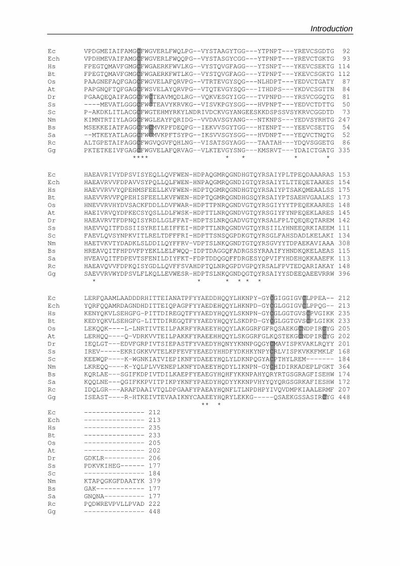
Introduction Ec VPDGMEIAIFAMGCFWGVERLFWQLPG--VYSTAAGYTGG---YTPNPT---YREVCSGDTG 92Ech VPDHMEVAIFAMGCFWGVERLFWQQPG--VYSTASGYCGG---YTPNPT---YREVCTGKTG 93Hs FPEGTQMAVFGMGCFWGAERKFWVLKG--VYSTQVGFAGG---YTSNPT---YKEVCSEKTG 114Bt FPEGTQMAVFGMGCFWGAERKFWTLKG--VYSTQVGFAGG---YTPNPT---YKEVCSGKTG 112Os PAAGNEFAQFGAGCFWGVELAFQRVPG--VTRTEVGYSQG---NLHDPT---YEDVCTGATY 87At PAPGNQFTQFGAGCFWSVELAYQRVPG--VTQTEVGYSQG---ITHDPS---YKDVCSGTTN 84Dr PGAAQEQAIFAGGCFWCTEAVMQDLRG--VQKVESGYIGG---TVPNPD---YRSVCGGQTG 81Ss ----MEVATLGGGCFWCTEAVYKRVKG--VISVKPGYSGG---HVPNPT---YEDVCTDTTG 50Sc P-AKDKLITLACGCFWGTEHMYRKYLNDRIVDCKVGYANGEESKKDSPSSVSYKRVCGGDTD 73Nm KIMNTRTIYLAGGCFWGLEAYFQRIDG--VVDAVSGYANG---NTKNPS---YEDVSYRHTG 247Bs MSEKKEIATFAGGCFWCMVKPFDEQPG--IEKVVSGYTGG---HTENPT---YEEVCSETTG 54Sa --MTKEYATLAGGCFWCMVKPFTSYPG--IKSVVSGYSGG---HVDNPT---YEQVCTNQTG 52Rc ALTGPETAIFAGGCFWGVQGVFQHLNG--VISATSGYAGG---TAATAH---YDQVSGGETG 86Gg PKTETKEIVFGAGCFWGVELAFQRVAG--VLKTEVGYSNG---KMSRVT---YDAICTGATG 335
**** * * * *
Ec HAEAVRIVYDPSVISYEQLLQVFWEN-HDPAQGMRQGNDHGTQYRSAIYPLTPEQDAAARAS 153Ech HAEAVRVVFDPAVVSYPQLLQLFWEN-HNPAQGMRQGNDIGTQYRSAIYTLTTEQETAAKES 154Hs HAEVVRVVYQPEHMSFEELLKVFWEN-HDPTQGMRQGNDHGTQYRSAIYPTSAKQMEAALSS 175Bt HAEVVRVVFQPEHISFEELLKVFWEN-HDPTQGMRQGNDHGSQYRSAIYPTSAEHVGAALKS 173Os HNEVVRVHYDVSACKFDDLLDVFWAR-HDPTTPNRQGNDVGTQYRSGIYYYTPEQEKAARES 148At HAEIVRVQYDPKECSYQSLLDLFWSK-HDPTTLNRQGNDVGTQYRSGIYFYNPEQEKLARES 145Dr HAEAVRVTFDPNQISYRDLLGLFFAT-HDPTSLNRQGADVGTQYRSALFPLTQEQEQTAREM 142Ss HAEVVQITFDSSIISYREILEIFFEI-HDPTTLNRQGNDVGTQYRSIILYHNEEQRKIAEEM 111Sc FAEVLQVSYNPKVITLRELTDFFFRI-HDPTTSNSQGPDKGTQYRSGLFAHSDADLKELAKI 134Nm HAETVKVTYDADKLSLDDILQYFFRV-VDPTSLNKQGNDTGTQYRSGVYYTDPAEKAVIAAA 308Bs HREAVQITFHPDVFPYEKLLELFWQQ-IDPTDAGGQFADRGSSYRAAIFYHNDKQKELAEAS 115Sa HVEAVQITFDPEVTSFENILDIYFKT-FDPTDDQGQFFDRGESYQPVIFYHDEHQKKAAEFK 113Rc HAEAVQVVFDPKQISYGDLLQVFFSVAHDPTQLNRQGPDVGPQYRSALFPVTEDQARIAKAY 148Gg SAEVVRVWYDPSVLFLKQLLEVWESR-HDPTSLNKQGNDQGTQYRSAIYYSDEEQAEEVRRW 396
* * * * * *
Ec LERFQAAMLAADDDRHITTEIANATPFYYAEDDHQQYLHKNPY-GYCGIGGIGVCLPPEA-- 212Ech YQRFQQAMRDAGNDHDITTEIQPAGPFYYAEDEHQQYLHKNPD-GYCGLGGIGVCLPPQG-- 213Hs KENYQKVLSEHGFG-PITTDIREGQTFYYAEDYHQQYLSKNPN-GYCGLGGTGVSCPVGIKK 235Bt KEDYQKVLSEHGFG-LITTDIREGQTFYYAEDYHQQYLSKDPD-GYCGLGGTGVSCPLGIKK 233Os LEKQQK----L-LNRTIVTEILPAKRFYRAEEYHQQYLAKGGRFGFRQSAEKGCNDPIRCYG 205At LERHQQ----Q-VDRKVVTEILPAKKFYRAEEHHQQYLSKGGRFGLKQSTEKGCNDPIRCYG 202Dr IEQLGT---EDVFGRPIVTSIEPASTFYVAEDYHQNYYKNNPGQGYCMAVISPKVAKLRQYY 201Ss IREV-----EKRIGKKVVTELKPFEVFYEAEDYHHDFYDKHKYNPYCRLVISPKVKKFMKLF 168Sc KEEWQP----K-WGNKIATVIEPIKNFYDAEEYHQLYLDKNPQGYACPTHYLREM------- 184Nm LKREQQ----K-YQLPLVVENEPLKNFYDAEEYHQDYLIKNPN-GYCHIDIRKADEPLPGKT 364Bs KQRLAE---SGIFKDPIVTDILKAEPFYEAEGYHQHFYKKNPAHYQRYRTGSGRAGFISEHW 174Sa KQQLNE---QGIFKKPVITPIKPYKNFYPAEDYHQDYYKKNPVHYYQYQRGSGRKAFIESHW 172Rc IDQLGR---ARAFDAAIVTQLDPGAAFYPAEAYHQNFLTLNPDHPYIVQVDMPKIAALERMF 207Gg ISEAST----R-HTKEIVTEVAAIKNYCAAEEYHQRYLEKKG-----QSAEKGSSASIRCYG 448
** *Ec --------------- 212Ech --------------- 213Hs --------------- 235Bt --------------- 233Os --------------- 205At --------------- 202Dr GDKLR---------- 206Ss PDKVKIHEG------ 177Sc --------------- 184Nm KTAPQGKGFDAATYK 379Bs GAK------------ 177Sa GNQNA---------- 177Rc PQDWREVPVLLPVAD 222Gg --------------- 448
Introduction
Le premier type est représenté par les MsrA d’E. coli et de mammifères qui
possèdent les trois cystéines catalytiques 51, 198 et 206, mais pas la cystéine 54.
Ce groupe renferme la plupart des MsrA d’origine végétale même si elles diffèrent
d’une part en position C-terminale par la nature des acides aminés environnant les
deux cystéines (absence dans les séquences végétales des nombreuses glycines) et
d’autre part par la présence de seulement 5 acides aminés entre ces deux cystéines
au lieu de 7 pour l’enzyme d’E. coli. Une deuxième sous-classe représentée entre
autres par la protéine de Deinococcus radiodurans, ne possède pas la cystéine 206,
mais présente les cystéines 51, 54 et 198. Les enzymes identiques à celle de
Saccharomyces cerevisiae ne possèdent pas cette fois les cystéines 54 et 206, et
pourraient donc former un autre groupe. La protéine PILB de Neisseria meningitidis
est incluse dans ce groupe bien que possédant un module MsrB en fusion et une
extension N-terminale. Un quatrième type de MsrA est représenté par l’enzyme de
Bacillus subtilis qui ne possède pas les cystéines 198 et 206, mais comporte les 2
résidus en position 51 et 54. Une cinquième classe représentée par une séquence
putative de Rhodobacter capsulatus, possède uniquement la cystéine 51. Enfin,
l’enzyme de l’algue rouge Gracilaria gracilis est particulière non seulement parce
qu’elle est fusionnée à deux modules Grx en position N-terminale, mais aussi parce
qu’elle ne possède que les cystéines 51 et 206, mais pas les cystéines 54 et 198.
Cette comparaison permet également d’identifier, outre les cystéines
catalytiques, un certain nombre de résidus essentiels. Ainsi, toutes les enzymes
Figure 11 : Alignement des 6 sous-classes de MsrA. Rc : Rhodobacter capsulatus, Bs : Bacillus subtilis, Sa : Staphylococcus aureus,
Dr : Deinococcus radiodurans, Ss : Sulfolobus solfataricus, Ec : Escherichia coli,
Ech : Erwinia chrysanthemi, Hs : Homo sapiens, Bt : Bos taurus, Os : Oryza
sativa, At : Arabidopsis thaliana, Gg : Gracilaria gracilis, Sc : Saccharomyces
cerevisiae, Nm : Neisseria meningitidis. Les séquences d’H. sapiens, E.
chrysanthemi et O . sativa ne sont pas reportées dans le tableau IV mais
appartiennent au groupe 1. Les astérisques correspondent aux acides aminés
conservés. Les cystéines en fond gris représentent les cystéines en position
conservée à l’intérieur de chaque sous-classe (numérotation d’E. coli : 51, 54,
198 et 206) et susceptibles de participer au mécanisme catalytique.
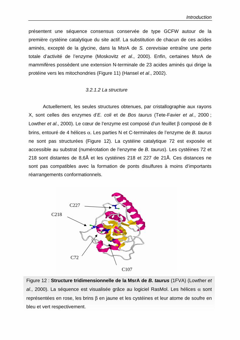
Introduction
présentent une séquence consensus conservée de type GCFW autour de la
première cystéine catalytique du site actif. La substitution de chacun de ces acides
aminés, excepté de la glycine, dans la MsrA de S. cerevisiae entraîne une perte
totale d’activité de l’enzyme (Moskovitz et al., 2000). Enfin, certaines MsrA de
mammifères possèdent une extension N-terminale de 23 acides aminés qui dirige la
protéine vers les mitochondries (Figure 11) (Hansel et al., 2002).
3.2.1.2 La structure
Actuellement, les seules structures obtenues, par cristallographie aux rayons
X, sont celles des enzymes d’E. coli et de Bos taurus (Tete-Favier et al., 2000 ;
Lowther et al., 2000). Le cœur de l’enzyme est composé d’un feuillet β composé de 8
brins, entouré de 4 hélices α. Les parties N et C-terminales de l’enzyme de B. taurus
ne sont pas structurées (Figure 12). La cystéine catalytique 72 est exposée et
accessible au substrat (numérotation de l’enzyme de B. taurus). Les cystéines 72 et
218 sont distantes de 8,6Å et les cystéines 218 et 227 de 21Å. Ces distances ne
sont pas compatibles avec la formation de ponts disulfures à moins d’importants
réarrangements conformationnels.
Figure 12 : Structure tridimensionnelle de la MsrA de B. taurus (1FVA) (Lowther et
al., 2000). La séquence est visualisée grâce au logiciel RasMol. Les hélices α sont
représentées en rose, les brins β en jaune et les cystéines et leur atome de soufre en
bleu et vert respectivement.
C227
C218
C107
C72
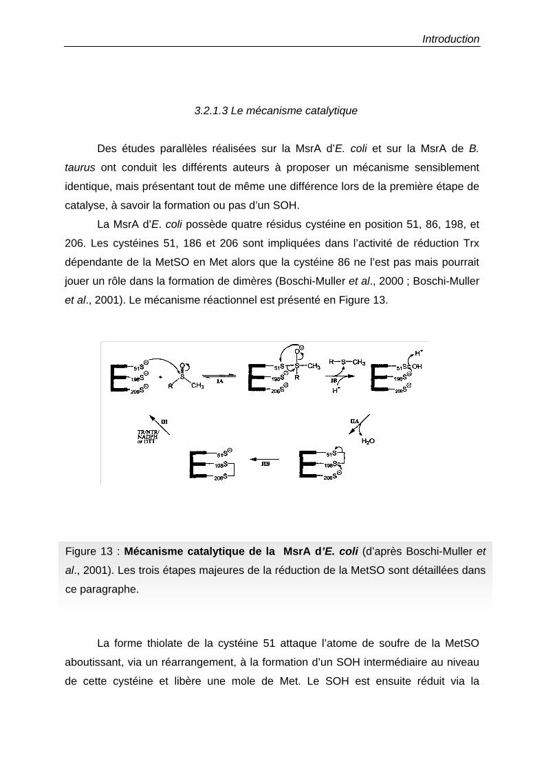
Introduction
3.2.1.3 Le mécanisme catalytique
Des études parallèles réalisées sur la MsrA d’E. coli et sur la MsrA de B.
taurus ont conduit les différents auteurs à proposer un mécanisme sensiblement
identique, mais présentant tout de même une différence lors de la première étape de
catalyse, à savoir la formation ou pas d’un SOH.
La MsrA d’E. coli possède quatre résidus cystéine en position 51, 86, 198, et
206. Les cystéines 51, 186 et 206 sont impliquées dans l’activité de réduction Trx
dépendante de la MetSO en Met alors que la cystéine 86 ne l’est pas mais pourrait
jouer un rôle dans la formation de dimères (Boschi-Muller et al., 2000 ; Boschi-Muller
et al., 2001). Le mécanisme réactionnel est présenté en Figure 13.
La forme thiolate de la cystéine 51 attaque l’atome de soufre de la MetSO
aboutissant, via un réarrangement, à la formation d’un SOH intermédiaire au niveau
de cette cystéine et libère une mole de Met. Le SOH est ensuite réduit via la
Figure 13 : Mécanisme catalytique de la MsrA d’E. coli (d’après Boschi-Muller et
al., 2001). Les trois étapes majeures de la réduction de la MetSO sont détaillées dans
ce paragraphe.
Introduction
formation d’un pont disulfure entre les cystéines 51 et 198, suivi par la formation d’un
second pont disulfure entre les cystéines 198 et 206, libérant la cystéine 51, à
nouveau libre pour l’attaque d’une deuxième molécule de MetSO. Le pont disulfure
entre les cystéines 198 et 206 est ensuite réduit par les Trx. Ainsi, pour une mole
d’enzyme, deux moles de MetSO sont réduites en Met, puisque la cystéine peut
effectuer une seconde attaque nucléophile, corroborant les observations issues de
l’étude des mutants monocystéiniques obtenus par mutagenèse dirigée. En effet,
aucune mole de Met n’est formée avec le mutant C51S dans lequel la cystéine est
mutée en sérine et une seule mole de Met est formée avec les mutants C198S et
C206S au lieu des deux attendues dans le cas de la protéine sauvage. Les auteurs
qui ont travaillé sur la MsrA de B. taurus proposent un mécanisme catalytique
légèrement différent dans lequel un pont disulfure intermédiaire entre la cystéine 72,
équivalente à la cystéine 51 d’E. coli, et la MetSO est formé et non pas un SOH
(Lowther et al., 2000).
3.2.2 Les MsrB
3.2.2.1 les différentes familles
Les MsrB sont des enzymes de masse moléculaire de l’ordre de 12 à 15 kDa
présentes chez tous les organismes. Chez les vertébrés, les MsrB sont plus connues
sous le nom de SelR ou SelX, du fait de la présence d’un résidu sélénocystéine dans
la partie C-terminale à la place d’une des cystéines catalytiques (Kryukov et al.,
2002). La présence de cette sélénocystéine dans les enzymes de mammifère est
essentielle à leur activité et son remplacement par une cystéine diminue
énormément leur efficacité catalytique (Bar-Noy & Moskovitz, 2002). Ces enzymes
ne possèdent pas d’identité de séquence avec les MsrA. L‘alignement de diverses
séquences de MsrB permet de répartir ces enzymes selon 5 sous-groupes en
fonction d’une part de la présence d’un résidu sélénocystéine ou d’un à 2 résidus
cystéinyls et d’autre part de la présence ou l’absence de 2 motifs CxxC permettant la
fixation d’un atome de zinc (Figure 14 et Tableau V). Des études récentes sur la
MsrB de Drosophila melanogaster ont montré que la mutation d’une de ces cystéines
ne permet plus la fixation d’un atome de zinc, et entraîne également une perte
d’activité de l’enzyme (Kumar et al., 2002). Dans ce type de protéine, le zinc jouerait
un rôle de stabilisateur de la structure protéique.
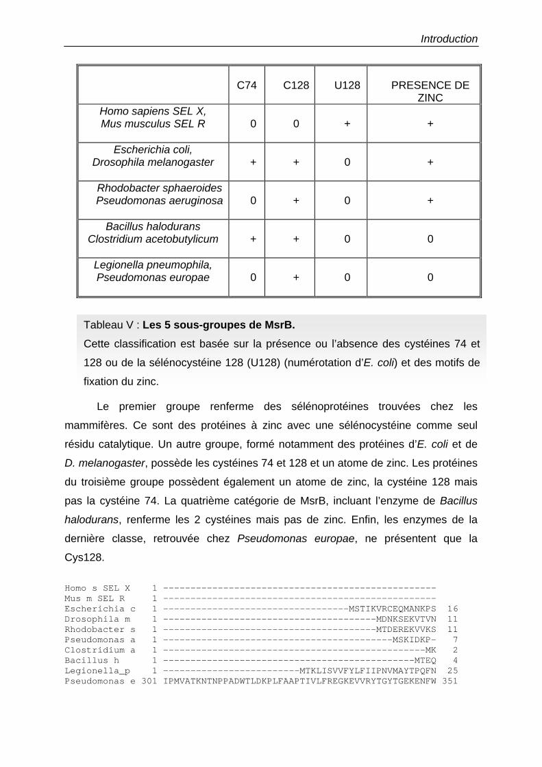
Introduction
C74
C128
U128
PRESENCE DE
ZINC Homo sapiens SEL X, Mus musculus SEL R
0
0
+
+
Escherichia coli, Drosophila melanogaster
+
+
0
+
Rhodobacter sphaeroides Pseudomonas aeruginosa
0
+
0
+
Bacillus halodurans Clostridium acetobutylicum
+
+
0
0
Legionella pneumophila, Pseudomonas europae
0
+
0
0
Le premier groupe renferme des sélénoprotéines trouvées chez les
mammifères. Ce sont des protéines à zinc avec une sélénocystéine comme seul
résidu catalytique. Un autre groupe, formé notamment des protéines d’E. coli et de
D. melanogaster, possède les cystéines 74 et 128 et un atome de zinc. Les protéines
du troisième groupe possèdent également un atome de zinc, la cystéine 128 mais
pas la cystéine 74. La quatrième catégorie de MsrB, incluant l’enzyme de Bacillus
halodurans, renferme les 2 cystéines mais pas de zinc. Enfin, les enzymes de la
dernière classe, retrouvée chez Pseudomonas europae, ne présentent que la
Cys128.
Homo s SEL X 1 --------------------------------------------------Mus m SEL R 1 --------------------------------------------------Escherichia c 1 ----------------------------------MSTIKVRCEQMANKPS 16Drosophila m 1 ---------------------------------------MDNKSEKVTVN 11Rhodobacter s 1 ---------------------------------------MTDEREKVVKS 11Pseudomonas a 1 ------------------------------------------MSKIDKP- 7Clostridium a 1 ------------------------------------------------MK 2Bacillus h 1 ----------------------------------------------MTEQ 4Legionella_p 1 -------------------------MTKLISVVFYLFIIPNVMAYTPQFN 25Pseudomonas e 301 IPMVATKNTNPPADWTLDKPLFAAPTIVLFREGKEVVRYTGYTGEKENFW 351
Tableau V : Les 5 sous-groupes de MsrB. Cette classification est basée sur la présence ou l’absence des cystéines 74 et
128 ou de la sélénocystéine 128 (U128) (numérotation d’E. coli) et des motifs de
fixation du zinc.
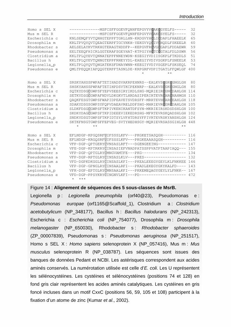
Introduction Homo s SEL X -------------MSFCSFFGGEVFQNHFEPGVYVCAKCGYELFS----- 32Mus m SEL R -------------MSFCSFFGGEVFQNHFEPGVYVCAKCSYELFS----- 32Escherichia c KNLSEMQFYVTQNHGTEPPFTGRLLHN-KRDGVYHCLICDAPLFHAEELK 65Drosophila m KRLTPVQYQVTQEAGTERPFTGCYNKH-YEKGVYQCIVCHQDLFSKEELR 60Rhodobacter s AELSELAYKVTRKHGTERAGTHDDFP--KEPGVFRCVGCGAPLFDDAEWR 59Pseudomonas a EELTEEQFHICRLGGTERAFSGEYHAT-KTPGIYHCTCCGTALFDLDSWR 56Clostridium a KKLTPLQYEVTQNNATEPPFNNEYWDN-KSEGIYVDIISGKPLFTKDDLS 51Bacillus h KKLTPLQYEVTQNNGTEPPFRNEYYDL-EAEGIYVDIVSGKPLFSNEELK 53Legionella_p KKLTPLQYQVTQNGATEKSFDNAYWNN-KEEGIYVDIVSGEPLFSKSEQL 74Pseudomonas e QMLTPEQQKIAFQQGTERPFTASNLDE-KRPGRFVDPITGATLFFQWLGF 400
* **
Homo s SEL X SRSKYAHSSPWPAFTETIHADSVAKRPEHNRS--EALKVSCGKCGNGLGH 80Mus m SEL R SHSKYAHSSPWPAFTETIHPDSVTKCPEKNRP--EALKVSCGKCGNGLGH 80Escherichia c SQTKYDSGCGWPSFYEPVSEESIRYIKDLSHG-MQRIEIRCGNCDAHLGH 114Drosophila m SETKYDSGCGWPAFNDVLDKGKVTLHRDASIPERIRTEVRCARCNAHMGH 110Rhodobacter s QAQKFESGTGWPSFWAPIDPEAVETSVDRSFF-MRRTEVHCARCEAHLGH 118Pseudomonas a SDAKYDSGSGWPSYFQPVDAEAVRELDDFSHG-MHRIEVRCGRCDAHLGH 105Clostridium a SLDKFDSGCGWPSFTKPIVKENIKANTDFSYN-MKRIEIRSKDSDSHLGH 100Bacillus h SKDKYDAGCGWPSFTKPIDEEEVIEKEDRSHG-MFRTEVRSKQADSHLGH 102Legionella_p SNDKYDSGTGWPSFTKPIDTSYLVFHTDRSYFFIVRTEVRSKYANSHLGH 124Pseudomonas e SKTKFNSGTGWPSFFEPVEG-SVTYHEDHSGY-MQRIEVRSASSGIHLGH 448
* ** **
Homo s SEL X EFLNDGP-KPGQSRFUIFSSSLKFV---PKGKETSASQGH---------- 116Mus m SEL R EFLNDGP-KRGQSRFUIFSSSLKFV---PKGKEAAASQGH---------- 116Escherichia c VFP-DGP-QPTGERYCVNSASLRFT---DGENGEEING------------ 147Drosophila m VFE-DGP-KPTRKRYCINSASIEFVNADPATSSPPVATPTAAPIAQQ--- 155Rhodobacter s VFP-DGP-QPTGLRYCMNGVAMTFE---PKG------------------- 134Pseudomonas a VFP-DGP-RPTGLRYCINSASLKLV---PRES------------------ 132Clostridium a VFD-DGPKDKGGLRYCINSASLKFI---PKEALEEEGYGEYLKLFNKKEE 146Bacillus h VFP-DGP-GPNGLRYCINSAALRFI---PKADLEKEGYGKYKALFD---- 143Legionella_p VFK-DGP-EPTGLRYCMNSAALKFI---PKKEMEQAGYGEYLYLFNKK-- 167Pseudomonas e VFD-DGP-PPSYKRYCINGNVLKFI---PD-------------------- 472
* ***
Figure 14 : Alignement de séquences des 5 sous-classes de MsrB. Legionella p : Legionella pneumophila (orf40@23), Pseudomonas e :
Pseudomonas europae (orf1165@Scaffold_1), Clostridium a : Clostridium
acetobutylicum (NP_348177), Bacillus h : Bacillus halodurans (NP_242313),
Escherichia c : Escherichia coli (NP_754077), Drosophila m : Drosophila
melanogaster (NP_650030), Rhodobacter s : Rhodobacter sphaeroides
(ZP_00007839), Pseudomonas s : Pseudomonas aeruginosa (NP_251517),
Homo s SEL X : Homo sapiens selenoprotein X (NP_057416), Mus m : Mus
musculus selenoprotein R (NP_038787). Les séquences sont issues des
banques de données Pedant et NCBI. Les astérisques correspondent aux acides
aminés conservés. La numérotation utilisée est celle d’E. coli. Les U représentent
les sélénocystéines. Les cystéines et sélénocystéines (positions 74 et 128) en
fond gris clair représentent les acides aminés catalytiques. Les cystéines en gris
foncé incluses dans un motif CxxC (positions 56, 59, 105 et 108) participent à la
fixation d’un atome de zinc (Kumar et al., 2002).
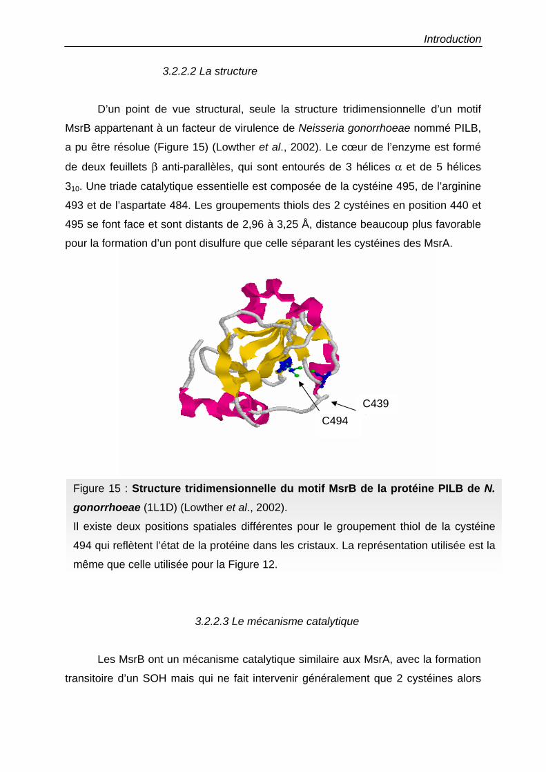
Introduction
C439 C494
3.2.2.2 La structure
D’un point de vue structural, seule la structure tridimensionnelle d’un motif
MsrB appartenant à un facteur de virulence de Neisseria gonorrhoeae nommé PILB,
a pu être résolue (Figure 15) (Lowther et al., 2002). Le cœur de l’enzyme est formé
de deux feuillets β anti-parallèles, qui sont entourés de 3 hélices α et de 5 hélices
310. Une triade catalytique essentielle est composée de la cystéine 495, de l’arginine
493 et de l’aspartate 484. Les groupements thiols des 2 cystéines en position 440 et
495 se font face et sont distants de 2,96 à 3,25 Å, distance beaucoup plus favorable
pour la formation d’un pont disulfure que celle séparant les cystéines des MsrA.
3.2.2.3 Le mécanisme catalytique
Les MsrB ont un mécanisme catalytique similaire aux MsrA, avec la formation
transitoire d’un SOH mais qui ne fait intervenir généralement que 2 cystéines alors
Figure 15 : Structure tridimensionnelle du motif MsrB de la protéine PILB de N. gonorrhoeae (1L1D) (Lowther et al., 2002).
Il existe deux positions spatiales différentes pour le groupement thiol de la cystéine
494 qui reflètent l’état de la protéine dans les cristaux. La représentation utilisée est la
même que celle utilisée pour la Figure 12.
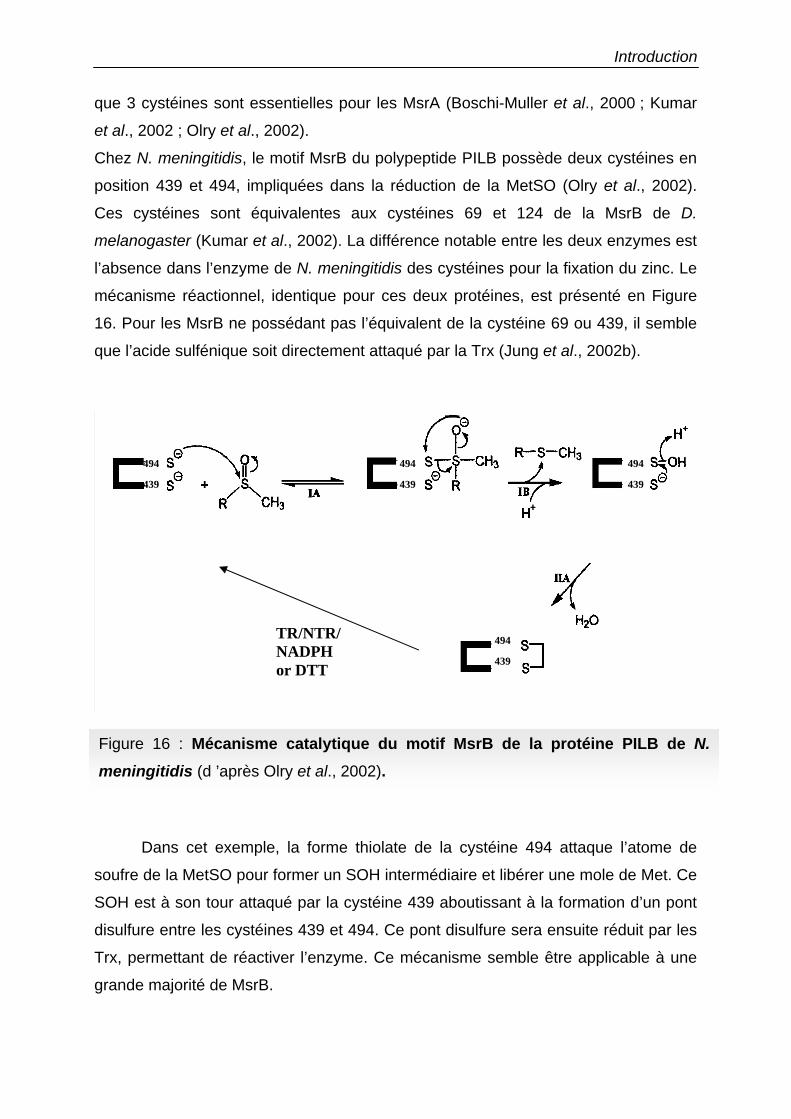
Introduction
que 3 cystéines sont essentielles pour les MsrA (Boschi-Muller et al., 2000 ; Kumar
et al., 2002 ; Olry et al., 2002).
Chez N. meningitidis, le motif MsrB du polypeptide PILB possède deux cystéines en
position 439 et 494, impliquées dans la réduction de la MetSO (Olry et al., 2002).
Ces cystéines sont équivalentes aux cystéines 69 et 124 de la MsrB de D.
melanogaster (Kumar et al., 2002). La différence notable entre les deux enzymes est
l’absence dans l’enzyme de N. meningitidis des cystéines pour la fixation du zinc. Le
mécanisme réactionnel, identique pour ces deux protéines, est présenté en Figure
16. Pour les MsrB ne possédant pas l’équivalent de la cystéine 69 ou 439, il semble
que l’acide sulfénique soit directement attaqué par la Trx (Jung et al., 2002b).
Dans cet exemple, la forme thiolate de la cystéine 494 attaque l’atome de
soufre de la MetSO pour former un SOH intermédiaire et libérer une mole de Met. Ce
SOH est à son tour attaqué par la cystéine 439 aboutissant à la formation d’un pont
disulfure entre les cystéines 439 et 494. Ce pont disulfure sera ensuite réduit par les
Trx, permettant de réactiver l’enzyme. Ce mécanisme semble être applicable à une
grande majorité de MsrB.
Figure 16 : Mécanisme catalytique du motif MsrB de la protéine PILB de N. meningitidis (d ’après Olry et al., 2002).
TR/NTR/NADPH or DTT
494 494 494
494
439
439
439 439
Introduction
Cependant, les comparaisons de séquences indiquent notamment que la
MsrB humaine hCBS1 ne possède pas la seconde cystéine catalytique dans une
position conservée, mais que celle-ci se trouve en position légèrement décalée. La
mutation de cette cystéine n’influence pas l’activité de l’enzyme au contraire de celle
de la cystéine d’attaque en position 169, équivalent à la cystéine 494 de PILB (Jung
et al., 2002b). Ces travaux suggèrent que cette enzyme fonctionne selon un
mécanisme catalytique différent des autres MsrB, vraisemblablement par la réduction
du SOH directement par les Trx. Cette hypothèse est renforcée par l’existence de
nombreuses séquences qui ne possèdent pas la cystéine équivalente à celle située
en position 494 de PILB ou 69 de la protéine de D. melanogaster (voir § 3.2.2.1).
3.3 La réduction des MetSO libres
L’oxydation chimique de l’atome de soufre asymétrique de la méthionine
produit deux épimères de MetSO, les formes R ou S, qu’elles soient libres ou
intégrées dans un polypeptide. Les MsrA et la fSMsr, une enzyme spécifique des
formes libres de la MetSO, sont capables de réduire l’énantiomère libre S-MetSO
mais pas la R-MetSO (Moskovitz et al., 1996a). De plus, les MsrB ont une activité
très faible envers la R-MetSO dans des extraits d’E. coli et la forme libre R-MetSO
est un mauvais substrat in vitro pour cette enzyme, suggérant l’existence d’enzymes
spécifiques pour cette réduction (Grimaud et al., 2001). L’utilisation de double
mutants MsrA- et MsrB- d’E. coli a permis récemment de purifier partiellement une
fRMsr et de mettre en évidence une activité de réduction de l’isomère R-MetSO libre
(Etienne et al., 2003). De plus, l’utilisation de ce mutant a aussi permis de
caractériser au niveau membranaire une activité de réduction de la R ou S-MetSO
libre ou intégrée dans des protéines (Spector et al., 2003). Les séquences codant
pour ces enzymes doivent être divergentes par rapport aux séquences existantes car
le séquençage complet du génome d’E. coli n’a mis en évidence qu’une seule
isoforme de MsrA et de MsrB (Kryukov et al., 2002).
3.4 Expression et fonctions des méthionine sulfoxyde réductases
D’un point de vue fonctionnel, la caractéristique première des MsrA et MsrB
est leur diastéréosélectivité envers les isomères R et S de la L-MetSO, l’isomère L
Introduction
étant la forme unique de la MetSO in vivo. En effet, les MsrA ne réduisent que les
isomères de type S et les MsrB les isomères de type R (Sharov et al., 1999 ;
Moskovitz et al., 2002).
L’oxydation de la Met en MetSO est un phénomène connu pour être impliqué
dans plusieurs pathologies telles que la maladie d’Alzheimer (Gabbita et al., 1999)
et pour exercer des effets régulateurs sur les protéines, modifiant leurs propriétés
biologiques (pour revue voir Brot & Weissbach, 2000).
D’une part, ce phénomène peut conduire à une inhibition de l’activité de
certaines protéines. C’est le cas par exemple de l’inhibiteur α-protéinase, dont
l’oxydation de résidus Met diminue l’affinité pour ses protéases cibles (Vogt et al.,
1995). De même, l’oxydation de certaines Met de la calmoduline induit des
changements dans la structure tertiaire de la protéine qui entraîneraient une
diminution de sa capacité à activer la Ca2+ATPase de la membrane plasmique (Yao
et al., 1996 ; Sun et al., 1999).
D’autre part, l’oxydation de Met peut être une forme d’activation, c’est
notamment le cas pour le composant C5 du complément. Ce composé est le point de
départ de la formation du complexe d’attaque membranaire qui aboutira à la lyse de
corps étrangers. L’oxydation de certaines Met par des radicaux hydroxyles active ce
composant C5 (Vogt et al., 1995). De plus, l’oxydation d’une Met en MetSO régule
l’inactivation d’un canal potassique voltage dépendant (Ciorba et al., 1997 ; Ciorba et
al., 1999).
Par ailleurs, l’oxydation et la réduction cyclique des résidus Met exposés à la
surface des protéines serviraient de première ligne de défense anti-oxydante contre
des dommages liés aux ROS et aux RNS (Moskovitz et al., 1999 ; Levine et al.,
2000 ; St John et al., 2001 ; Stadtman et al., 2002). Ainsi, l’oxydation de plusieurs
résidus Met n’altère pas par exemple l‘activité de la glutamine synthétase (Levine et
al., 1999). De plus, l’étude de mutants MsrA- d’E. coli, d’Erwinia chrysanthemi et de
levure a montré que ceux-ci étaient plus sensibles au stress oxydatif que les souches
sauvages et par conséquent que les MsrA joueraient un rôle dans la résistance au
stress oxydatif (Moskovitz et al., 1995 ; Moskovitz et al., 1997 ; Moskovitz et al.,
1998 ; Hassouni et al., 1999). En luttant contre ce stress, les MsrA participeraient à
l’augmentation de la durée de vie des organismes (Moskovitz et al., 2001 ;
Petropoulos et al., 2001 ; Ruan et al., 2002).
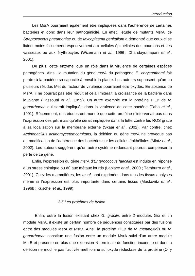
Introduction
Les MsrA pourraient également être impliquées dans l’adhérence de certaines
bactéries et donc dans leur pathogénicité. En effet, l’étude de mutants MsrA- de
Streptococcus pneumoniae ou de Mycoplasma genitalium a démontré que ceux-ci se
liaient moins facilement respectivement aux cellules épithéliales des poumons et des
vaisseaux ou aux érythrocytes (Wizemann et al., 1996 ; Dhandayuthapani et al.,
2001).
De plus, cette enzyme joue un rôle dans la virulence de certaines espèces
pathogènes. Ainsi, la mutation du gène msrA du pathogène E. chrysanthemi fait
perdre à la bactérie sa capacité à envahir la plante. Les auteurs supposent qu’un ou
plusieurs résidus Met du facteur de virulence pourraient être oxydés. En absence de
MsrA, il ne pourrait pas être réduit et cela limiterait la croissance de la bactérie dans
la plante (Hassouni et al., 1999). Un autre exemple est la protéine PILB de N.
gonorrhoeae qui serait impliquée dans la virulence de cette bactérie (Taha et al.,
1991). Récemment, des études ont montré que cette protéine n’intervenait pas dans
l’expression des pili, mais qu’elle serait impliquée dans la lutte contre les ROS grâce
à sa localisation sur la membrane externe (Skaar et al., 2002). Par contre, chez
Actinobacillus actinomycetemcomitans, la délétion du gène msrA ne provoque pas
de modification de l’adhérence des bactéries sur les cellules épithéliales (Mintz et al.,
2002). Les auteurs suggèrent qu’un autre système redondant pourrait compenser la
perte de ce gène.
Enfin, l’expression du gène msrA d’Enterococcus faecalis est induite en réponse
à un stress chimique ou dû aux métaux lourds (Laplace et al., 2000 ; Tamburro et al.,
2001). Chez les mammifères, les msrA sont exprimées dans tous les tissus analysés
même si l’expression est plus importante dans certains tissus (Moskovitz et al.,
1996b ; Kuschel et al., 1999).
3.5 Les protéines de fusion
Enfin, outre la fusion existant chez G. gracilis entre 2 modules Grx et un
module MsrA, il existe un certain nombre de séquences constituées par des fusions
entre des modules MsrA et MsrB. Ainsi, la protéine PILB de N. meningitidis ou N.
gonorrhoeae constitue une fusion entre un module MsrA suivi d’un autre module
MsrB et présente en plus une extension N-terminale de fonction inconnue et dont la
délétion ne modifie pas l’activité méthionine sulfoxyde réductase de la protéine (Olry
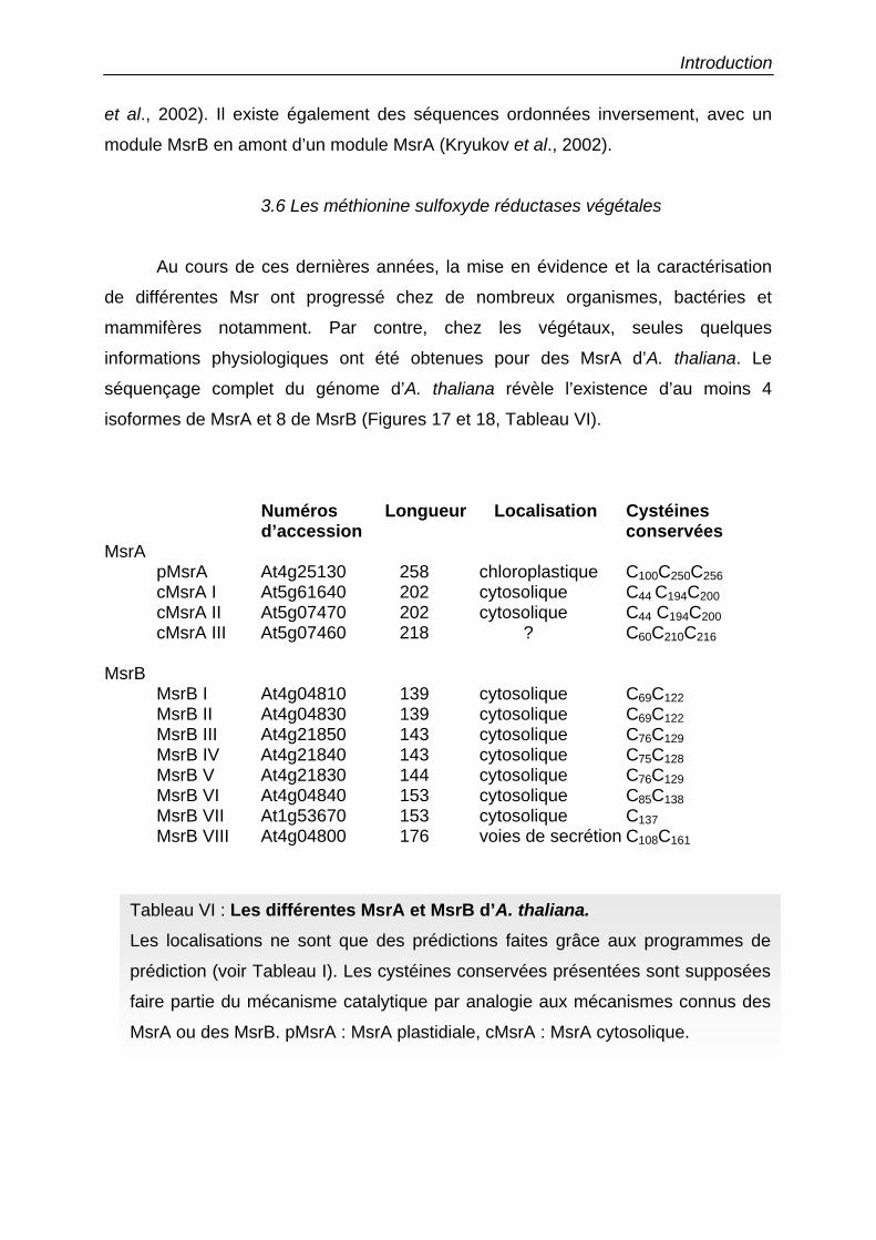
Introduction
et al., 2002). Il existe également des séquences ordonnées inversement, avec un
module MsrB en amont d’un module MsrA (Kryukov et al., 2002).
3.6 Les méthionine sulfoxyde réductases végétales
Au cours de ces dernières années, la mise en évidence et la caractérisation
de différentes Msr ont progressé chez de nombreux organismes, bactéries et
mammifères notamment. Par contre, chez les végétaux, seules quelques
informations physiologiques ont été obtenues pour des MsrA d’A. thaliana. Le
séquençage complet du génome d’A. thaliana révèle l’existence d’au moins 4
isoformes de MsrA et 8 de MsrB (Figures 17 et 18, Tableau VI).
Numéros Longueur Localisation Cystéines d’accession conservées
MsrA pMsrA At4g25130 258 chloroplastique C100C250C256 cMsrA I At5g61640 202 cytosolique C44 C194C200 cMsrA II At5g07470 202 cytosolique C44 C194C200 cMsrA III At5g07460 218 ? C60C210C216 MsrB MsrB I At4g04810 139 cytosolique C69C122 MsrB II At4g04830 139 cytosolique C69C122 MsrB III At4g21850 143 cytosolique C76C129 MsrB IV At4g21840 143 cytosolique C75C128 MsrB V At4g21830 144 cytosolique C76C129 MsrB VI At4g04840 153 cytosolique C85C138
MsrB VII At1g53670 153 cytosolique C137 MsrB VIII At4g04800 176 voies de secrétion C108C161
Tableau VI : Les différentes MsrA et MsrB d’A. thaliana. Les localisations ne sont que des prédictions faites grâce aux programmes de
prédiction (voir Tableau I). Les cystéines conservées présentées sont supposées
faire partie du mécanisme catalytique par analogie aux mécanismes connus des
MsrA ou des MsrB. pMsrA : MsrA plastidiale, cMsrA : MsrA cytosolique.

Introduction cMsrAI 1 -----------------------------------------------------cMsrAII 1 -----------------------------------------------------cMsrAIII 1 ------------------------------------MDSSLKTQEPQVVETSP 17pMsrA 1 MQVLVVSPPLIAAASLSKPLNSLSKAALSFSRAKPICPFPQTSRRPISVYKSP 53
cMsrAI MNIL-NKLGIGSSRQTNMDPS--PIAQVIDDEAPAPGNQFTQFGAGCFWSVEL 50cMsrAII MNIL-NRLGLGSSGQTNMDPS--PIAQGNDDDTPAPGNQFAQFGAGCFWGVEL 50cMsrAIII -SPVAQEPPQ-VADKPAIVPS--PIAQEPDNDVPAPGNEFAEFAAGCFWGVEL 66pMsrA MNNLFNRLGFGSRPQAQADPSSAAIAQGPDDDVPSSGQQFAQFGAGCFWGVEL 106
** *** * * * * * ***** ***
cMsrAI AYQRVPGVTQTEVGYSQGITHDPSYKDVCSGTTNHAEIVRVQYDPKECSYQSL 103cMsrAII AFQRVPGVTQTEAGYTQGTVDNPSYGDVCSGTTGHSEVVRVQYDLNDCTYESL 103cMsrAIII AFQRIPGVTVTEVGYTHGISHNPSYEDVCTNTTNHAEVVRVQYDPKECTYETL 119pMsrA AYQRVPGVTKTEVGYSHGIVHNPSYEDVCTGTTGHNEVVRVQYDPKECSFESL 159
* ** **** ** ** * *** *** ** * * ****** * **
cMsrAI LDLFWSKHDPTTLNRQGNDVGTQYRSGIYFYNPEQEKLARESLERHQQQVDRK 156cMsrAII LDLFWSRHDPTTLNRQGNDVGTQYRSGIYFYTPEQEKLARESLERHQQQMERK 156cMsrAIII LDLFWSRHNPTTLNRQGELLGAQYRSGIYFYTPEQEKLARESLEKEQKKLEDK 172pMsrA LDVFWNRHDPTTLNRQGGDVGTQYRSGIYYYTDEQERIAREAVEKQQKILNKR 212
** ** * ******** * ******* * *** *** * *
cMsrAI VVTEILPAKKFYRAEEHHQQYLSKGGRFGLKQSTEKGCNDPIRCYG 202cMsrAII IMTEILPAKKFYRAEEHHQQYLSKGGRFGQGQSTAKGCNDPIRCYG 202cMsrAIII IVTEILPAKKFYKAEEYHQQYLVKGGMHGNAQSPAKSCKDPIRCYG 218pMsrA IVTEILPATKFYRAENYHQQYLAKGGRMGLRQSAEKGCKDPIRCYG 258
****** *** ** ***** *** * ** * * *******
Figure 17 : Comparaison de séquences des MsrA d’A. thaliana. Les cystéines en fond gris sont les cystéines catalytiques présumées d’après le
mécanisme catalytique d’E. coli. Les astérisques représentent les acides aminés
strictement conservés.
Introduction MsrB I 1 ---------------------------------------MADLVTVVKKTE---- 12MsrB II 1 ---------------------------------------MAASPLVVQKTE---- 12MsrB III 1 --------------------------------MPTSATAVAPSTGSVQKKD---- 19MsrB IV 1 ---------------------------------MAMTAAAVPSSGSFQKQD---- 18MsrB V 1 --------------------------------MAAMTAAAVPATGSFQKQD---- 19MsrB VI 1 -----------------------MNTSPKMEMEMKMETKAAPEAGMIKKSN---- 28MsrB VII 1 ----------------------------------MGSSSSSPKPDNVQEAEKNEF 21MsrB VIII 1 MNIVNSKILFLSFTLLLLLQSSIVESDSICLSSGVASTVAMAAPGSVQKGD---- 51
MsrB I ----E-EWRAVLSPEQFRILRQKGTETPGTEEYDKFFEEGIFSCIGCKTPLYKST 62MsrB II ----E-EWRAVLSPEQFRILRQKGTEKPGTGEYDKFFEEGIFDCVGCKTPLYKST 62MsrB III ----Q-DWRAILSPEQFRVLREKGTENRGKGEYTKLFDDGIYSCAGCATPLYKST 69MsrB IV ----E-EWRAVLSPEQFRVLRLKGTDKRGKGEFTKKFEEGTYSCAGCGTALYKST 68MsrB V ----E-EWRAVLSPEQFRVLRLKGTDKRGKGEFTKKFEEGTYSCAGCGTALYKST 69MsrB VI ----E-EWRTVLSPEQFKILREKSIEKRGSGEYVKLFEEGIYCCVGCGNPVYKST 78MsrB VII ASLSENEWKKRLTPEQYYITRQKGTERAFTGEYWNSKTPGVYNCVCCDTPLFDSS 76MsrB VIII ----E-EWRAILSPEQFRILRQKGTEYPGTGEYVNFDKEGVYGCVGCNAPLYKST 101
* * *** * * * * * * *
MsrB I TKFDAGCGWPAFFEGLPGAINRAPDPD---GRRTEITCAVCDGHLGHVHKGEGYS 114MsrB II TKFDSGCGWPAFFEGLPGAINRTPDPD---GRRTEITCAACDGHLGHVFKGEGYG 114MsrB III TKFDSGCGWPSFFDAIPGAIKQTPEAG---GRRMEITCAACDGHLGHVVKGEGFP 121MsrB IV TKFDSGCGWPAFFDAIPGAIKQTPEAG---GRRMEITCAVCDGHLGHVFKGEGYS 120MsrB V TKFDSGCGWPAFFDAIPGAIKQTPEAG---GRRMEITCAVCDGHLGHVFKGEGYS 121MsrB VI TKFDSGCGWPAFFDAIPGAINRTEERA---GLRYEITCTKCDGHLGHVLKNEGFP 130MsrB VII TKFDSGTGWPSYYQPIGNNVKTKLDLSIIFMPRQEVVCAVCNAHLGHVFD-DG-P 129MsrB VIII TKFNAGCGWPAFFEGIPGAITRTTDPD---GRRIEINCATCGGHLGHVFKGEGFA 153
*** * *** * * * * ***** *
MsrB I TPTDERLCVNSVSINFNPAKPSSIT 139MsrB II NPTDERHCVNSVSISFNPAKSSSII 139MsrB III TATDERHCVNSVSLKFSEISSQ--- 143MsrB IV TPTDQRHCVNSVSLKFASADSSK-- 143MsrB V TPTDQRHCVNSVSLKFSSAGSSQ-- 144MsrB VI TPTDERHCVNSVALKFSSAITSQ-- 153MsrB VII RPTGKRYCLNSAALKLNALEKTRD- 153MsrB VIII TPTDERHCVNSVSLKFTPAASSL-- 176
** * * ** *
Figure 18 : Comparaison de séquences des MsrB d’A. thaliana. Les cystéines en fond gris sont les cystéines catalytiques présumées et de
fixation du zinc d’après le mécanisme catalytique de la protéine PILB de N.
meningitidis. Les astérisques représentent les acides aminés strictement
conservés.
Introduction
Aucune MsrB végétale n’a pour l’instant été caractérisée. Parmi les 8 séquences de
MsrB d’A. thaliana, toutes possèdent les motifs CxxC capables de fixer un atome de
zinc. MsrB III présente une extension N-terminale qui pourrait permettre la sécrétion
de la protéine. Enfin, la MsrB VII possède la cystéine catalytique mais pas la
deuxième cystéine conservée.
Concernant les MsrA, deux isoformes sur les 4 existant, une plastidiale et une
cytosolique ont été étudiées. Ces deux protéines, exprimées chez E. coli et purifiées,
présentent une activité de réduction des MetSO mais en présence d’un réducteur
artificiel, le DTT (Sadanandom et al., 1996 ; Sadanandom et al., 2000). La MsrA
chloroplastique possède une masse moléculaire de 28 kDa et un pH optimum de 8.0,
en accord avec sa localisation stromatique. La MsrA cytosolique a pour sa part une
masse moléculaire de 27 kDa et un pH optimum compris entre 7.0 et 7.2. Ces
travaux démontrent également une forte expression de la forme cytosolique dans les
racines et les graines et son implication dans la défense contre les pathogènes
(Sadanandom et al., 2000). La forme chloroplastique est fortement régulée par la
lumière dans les tissus photosynthétiques et semble protéger l’activité chaperone
d’une protéine heat shock (hsp21) en réduisant des MetSO en Met (Sadanandom et
al., 2000 ; Gustavsson et al., 2002).
Enfin, une étude récente a montré l’existence de sélénoprotéines chez l’algue
verte Chlamydomonas reinhardtii, alors que ce type de protéines n’était présent que
chez les archébactéries, les virus et les mammifères (Novoselov et al., 2002). Parmi
les 10 protéines identifiées se trouvent des protéines connues chez les animaux
comme étant des sélénoenzymes, telles qu’une TR ou des PHGpx et de manière
plus surprenante une MsrA. En effet, certaines MsrB sont connues pour posséder un
résidu sélénocystéine mais pas les MsrA. Cette enzyme est longue de 159 acides
aminés, possède une sélénocystéine en position conservée à la place de la cystéine
catalytique (C51 pour l’enzyme d’E. coli). Par contre, elle ne possède aucune des
deux autres cystéines essentielles situées dans la partie C-terminale (C196 et C202
pour l’enzyme d’E. coli).
Introduction
4. Mon travail de thèse
Le travail expérimental présenté dans cette thèse sous la forme d’articles de
recherche a pour objet la caractérisation, tant biochimique que structurale, des
protéines impliquées dans le maintien de l’homéostasie rédox intracellulaire chez les
végétaux que sont les Trx et les Grx en utilisant comme modèle d'étude le peuplier
(Populus x interamericana Brockh.) clone ‘Beaupré’. Ces travaux ont fait l’objet de 6
articles. Les 2 articles portant sur les Trx ont permis de caractériser deux isoformes
de Trx, l’une cytosolique (Identification and characterization of a third thioredoxin h in poplar) et l’autre mitochondriale (Isolation and characterization of an extended thioredoxin h from poplar). Les 4 autres articles décrivent la
caractérisation des propriétés rédox d’une isoforme de Grx de peuplier et les
résultats préliminaires de la structure cristallographique de cette Grx en complexe
avec le GSH et s’intitulent (i) Enhancement of poplar glutaredoxin expression by optimization of the cDNA sequence, (ii) Exploring the active site of plant glutaredoxin by site-directed mutagenesis, (iii) Characterization of the redox properties of poplar glutaredoxin et (iv) Crystallization and preliminary X ray data of poplar glutaredoxin.
De plus, une attention particulière a été portée sur deux types d’enzymes
cibles des Trx ou des Grx du fait de leur implication potentielle dans la réponse aux
stress chez les plantes, les Prx et les MsrA.
Deux isoformes de Prx, une Prx de type IIC et une Prx Q, ont ainsi été
caractérisées en terme d’expression et de localisation dans les différents organes de
la plante. Leurs propriétés catalytiques, leurs spécificités de substrats et leurs
capacités à accepter des électrons de divers donneurs ont également été examinées
notamment par des expériences de mutagenèse dirigée et par détermination de leur
structure. Le travail sur la Prx de type II a fait l’objet de 3 articles intitulés : (i) Isolation and characterization of a new peroxiredoxin from poplar sieve tubes that uses either glutaredoxin or thioredoxin as a proton donor, (ii) Glutaredoxin-dependent peroxiredoxin from poplar: protein-protein interaction and catalytic mechanism, (iii) Crystallisation and preliminary X-ray data of a bifunctional peroxiredoxin from poplar et d’un quatrième en cours de révision (iv) Active site mutagenesis and phospholipid hydroperoxide reductase activity of poplar type II peroxiredoxin. Le travail sur la Prx Q est présenté dans un article en

Introduction
préparation : Molecular characterization of chloroplastic poplar peroxiredoxin Q and its involvement in the pathogenic response. Deux isoformes de MsrA, plastidiale et cytosolique, ont également été
étudiées d’un point de vue fonctionnel et structural. Cette étude est présentée sous
la forme d’un article en préparation intitulé : Functional and structural aspects of poplar cytosolic and plastidial type A methionine sulfoxide reductases.

Résultats

Chapitre I : les systèmes réducteurs
Résultats chapitre I : Les systèmes réducteurs
Les deux systèmes réducteurs enzymatiques principaux dans une cellule sont
les sytèmes Trx et Grx. Chez les plantes, il semble que ces systèmes soient
représentés par un grand nombre d’isoformes dans les différents compartiments
cellulaires.
Le système Trx chloroplastique est le mieux connu, mais de nouvelles
séquences de Trx chloroplastiques, dont on ne connaît pas la fonction précise,
émergent (Meyer et al., 2002). Par ailleurs, les enzymes composant le système
mitochondrial viennent seulement d’être caractérisées chez A. thaliana (Laloi et al.,
2002). Au niveau cytosolique, au moins 8 isoformes de Trx h coexistent chez A.
thaliana (Meyer et al., 2002).
Au début de mon travail de thèse, très peu de données étaient disponibles sur
les Grx végétales alors que leurs homologues bactériens et animaux étaient
beaucoup mieux caractérisés. Pourtant, le séquençage du génome d’A. thaliana
démontre l’existence potentielle de multiples isoformes dans le cytosol, les
chloroplastes et les mitochondries (voir tableau I de l’introduction).
Avant mon arrivée dans le laboratoire, une isoforme cytosolique de Trx de
peuplier appelée popTrx h1 a été caractérisée (Behm & Jacquot, 2000). Cette
protéine est en réalité l’homologue d’un point de vue séquence primaire du site actif
(WCPPC) des Trx h3, h4 ou h5 d’A. thaliana. Deux autres isoformes présumées
cytosoliques ont ensuite été isolées et caractérisées et font l’objet des articles 5 et 6.
Par ailleurs, une isoforme de Grx bicystéinique, homologue à la séquence
CxxC4 d’A. thaliana (voir Tableau I et Figure 9 de l’introduction) a également été au
centre de mon travail de recherche. La caractérisation structurale et fonctionnelle de
cette enzyme est reportée dans les articles 7 à 10.
Résultats chapitre I : Les systèmes réducteurs
Cet article présente l'isolement et la caractérisation d'une deuxième Trx de
peuplier (popTrx h2) de 143 acides aminés. Par rapport à la première protéine isolée,
cette deuxième variante présente une extension du côté N-terminal et la protéine
recombinante est naturellement clivée dans E. coli. Son site actif (WCGPC) diffère de
celui de popTrx h1 (WCPPC). Les caractéristiques physico-chimiques et catalytiques
de la protéine recombinante ont été comparées à celles de popTrx h1. De récentes
expériences de fusion avec la GFP démontrent que popTrx h2 est
vraisemblablement une protéine mitochondriale alors qu’on présumait qu’elle était
localisée dans le cytosol. Cette protéine, différente des deux isoformes de Trx
mitochondriales d’A. thaliana appelées AtTrx o1 et o2, ne possède que 25%
d’identité avec ces Trx o. Pour ces raisons, cette isoforme devrait être appelée
dorénavant popTrx o3 plutôt que popTrx h2 si les isoformes équivalentes à AtTrx o1
et o2 existent chez le peuplier.
Article numéro 5 :
Gelhaye E, Rouhier N, Laurent P, Sautière PE, Martin F, Jacquot JP. (2002)
Isolation and characterization of an extended thioredoxin h from poplar.
Physiol. Plant. 114(2):165-171.
PHYSIOLOGIA PLANTARUM 114: 165-171. 2002Printed in Denmark - ail rights reserved
Résultats chapitre l : Les systèmes réducteurs
Copyright © Physiologia Plantarum 2002
ISSN 0031-9317
Isolation and characterization of an extended thioredoxin h from poplar
Eric Gelhayea, Nicolas Rouhiera , Pascal Laurenta , Pierre-Eric SautièreC
, Francis Martinb and Jean-Pierre Jacquota ,*
aUMR IaM 1136 INRA-Université H. Poincaré-Nancy 1 Faculté des Sciences F-54506 Vandoeuvre, FrancebUMR IaM 1136 INRA-Université H. Poincaré-Nancy 1 Centre INRA de Nancy., F-54280 Champenoux, FrancecLaboratoire d'Endocrinologie des Annélides UPRESA 8017. Université des Sciences et Technologies de Lille,F-59655 Villeneuve d'Ascq Cedex, France*Corresponding author, e-mail: [email protected]
Received 2 April; revised 13 August 2001
A cDNA coding for a thioredoxin h has been isolated froma xylemlphloem poplar cDNA library by RACE-PCR. Thenucleotide sequence called popTrx-h2 is homologous to otherthioredoxins h isolated from plants but differs from the otherthioredoxins h by presenting a 30 amino acid long N-terminus extension. A variant of this cDNA lacking the N-terminaI extension was also generated by PCR. Both cDNAshave been introduced loto an expression plasmid (pET-3d)and the recombinant proteins have been expressed to a highlevel and purified from Escherichia coli ceUs. Protein sequencing showed that a part of the N-terminal extensionwas cleaved in the E. coli ceUs, with the tirst 19 amino acids
Introduction
In plants, thioredoxins are a multigenic family of redoxproteins containing a disulphide bridge involved in several metabolic funetions inside and outside the cells (Fora review, see Schürmann and Jacquot 2000). Sequenceanalyses have revealed three different thioredoxin groupsin the plant cells. The tirst group contains thioredoxinssimilar to the prokaryotic thioredoxins, the prototype ofwhich is the Escherichia coli thioredoxin, a protein verywell characterized both kinetically and structurally(Holmgren 1968, Katti et al. 1990, Jeng et al. 1994).These thioredoxins are nucleus-encoded (Stein et al.1995) but localized in the chloroplasts and reduced by aferredoxin-thioredoxin reductase (FTR). These thioredoxins are efficient catalysts in vitro of the reduction/activation of the chloroplast NADP-malate dehydrogenase (NADP-MDH) and have been accordingly calledthioredoxins m. The second group of nucleus-encodedchloroplast thioredoxins, also reduced by FTR and
missing, suggesting the presence of a putative cleavage sitein the N-terminal extension of popTrx-h2. Both recombinantproteins display unusual catalytic properties compared toother thioredoxins h characterized so far, i.e. a weak reduction by Arabidopsis thaliana NADPH·dependent thioredoxinreductase, and a weak activation of the chloroplasticNADP-malate dehydrogenase, a non-physiological target enzyme. Northern blot experiments indicate that the transcripts of popTrx-h2 are present in leaves and roots, albeitat a lower level compared to the earlier characterizedpopTrx-hl.
called thioredoxins f, are very specifie activators of thefructose-l,6-bisphosphatase of the Calvin cycle. Thistype of thioredoxin has also been described in terms ofreactivity and structure (Hirasawa et al. 1999, Capitaniet al. 2000). Finally, plants contain a third type of thioredoxins, the thioredoxins h (for heterotrophic), that arepresumably located in the cytosol. In this cytosolic system, the reducing power of NADPH is transferred via aflavoprotein-the NADPH-thioredoxin reductase (NTR)(Florencio et al. 1988). A number of thioredoxin h sequences have since been isolated from both non-photosynthetic and photosynthetic tissues, most notably fromArabidopsis thaliana (Rivera-Madrid et al. 1995). Primary structure analyses indicate that all h type proteinsare closely related on an evolutionary standpoint (Jacquot et al. 1997). The cytosolic thioredoxin displays atleast one specific characteristic that allows its identification: the presence of a conserved Trp residue (W13
Abbreviations - DTT, Dithiotreitol; DTNB, 5,5' dithiobis nitrobenzoic acid; EST, expressed sequence tag; NADP-MDH, NADP-dependent malatedehydrogenase; NTR, NADPH-thioredoxin reductase; Trx, thioredoxin.
Physiol. Plant. 114,2002 165
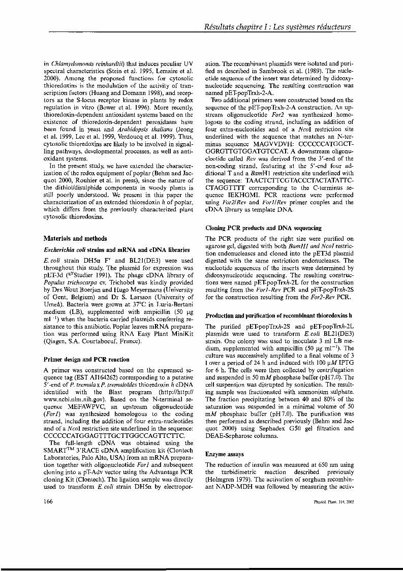
in Chlamydomonas reinhardtii) that induces peculiar UVspectral characteristics (Stein et al. 1995, Lemaire et al.2000). Among the proposed functions for cytosolicthioredoxins is the modulation of the activity of transcription factors (Huang and Domann 1998), and receptors as the S-locus receptor kinase in plants by redoxregulation in vitro (Bower et al. 1996). More recent1y,thioredoxin-dependent antioxidant systems based on theexistence of thioredoxin-dependent peroxidases havebeen found in yeast and Arabidopsis thaliana (Jeonget al. 1999, Lee et al. 1999, Verdoucq et al. 1999). Thus,cytosolic thioredoxins are likely to be involved in signalling pathways, developmental processes, as weIl as antioxidant systems.
In the present study, we have extended the characterization of the redox equipment of poplar (Behm and Jacquot 2000, Rouhier et al. in press), since the nature ofthe dithiol/disulphide components in woody plants isstill poorly understood. We present in this paper thecharacterization of an extended thioredoxin h of poplar,which differs from the previously characterized plantcytosolic thioredoxins.
Materials and methods
Escherichia coli strains and mRNA and cDNA libraries
E. coli strain DH5a F' and BL21(DE3) were usedthroughout this study. The plasmid for expression waspET-3d (q3'Studier 1991). The phage cDNA library ofPopulus trichocarpa cv. Trichobel was kindly providedby Drs Wout Boerjan and Hugo Meyermans (Universityof Gent, Belgium) and Dr S. Larsson (University ofUmeâ). Bacteria were grown at 37°C in Luria-Bertanimedium (LB), supplemented with ampicillin (50 Jigml- 1) when the bacteria carried plasmids conferring resistance to this antibiotic. Poplar leaves mRNA preparation was performed using RNA Easy Plant MiniKit(Qiagen, S.A. Courtaboeuf, France).
Primer design and peR reaction
A primer was constructed based on the expressed sequence tag (EST AI164262) corresponding to a putative5'-end of P. tremulaxP. tremuloïdes thioredoxin h cDNAidentified with the Blast program (http://http://www.ncbi.nlm.nih.gov). Based on the N-terminal sequence MEFAWPVC, an upstream oligonucleotide(For}) was synthesized homologous to the codingstrand, including the addition of four extra-nucleotidesand of a NcoI restriction site underlined in the sequence:CCCCCCATGGAGTTTGCTTGGCCAGTTCTTC.
The full-Iength cDNA was obtained using theSMART™ 3'RACE cDNA amplification kit (ClontechLaboratories, Palo Alto, USA) from an mRNA preparation together with oligonucleotide For} and subsequentcloning into a pT-Adv vector using the Advantage PCRcloning Kit (Clontech). The ligation sample was directlyused to transform E. coli strain DH5a by electropor-
166
Résultats chapitre l : Les systèmes réducteurs
ation. The recombinant plasmids were isolated and purified as described in Sambrook et al. (1989). The nucleotide sequence of the insert was determined by dideoxynucleotide sequencing. The resulting construction wasnamed pEJ:popTrxh-2-A.
Two additional primers were constructed based on thesequence of the pEJ:popTrxh-2-A construction. An upstream oligonucleotide For2 was synthesized homologous to the coding strand, including an addition offour extra-nucleotides and of a NcoI restriction siteunderlined with the sequence that matches an N-terminus sequence MAGVVDVH: CCCCCCATGGCTGGRGTTGTGGATGTCCAT. A downstream oligonucleotide called Rev was derived from the 3'-end of thenon-coding strand, featuring at the 5'-end four additional T and a BamH1 restriction site underlined withthe sequence: TAACTCTTCGTACCCTACTATATTCCTAGGTTTT corresponding to the C-terminus sequence IEKHGMI. PCR reactions were performedusing For21Rev and For}1Rev primer couples and thecDNA library as template DNA.
Cloning PCR products and DNA sequencing
The PCR products of the right size were purified onagarose gel, digested with both BamHl and NcoI restriction endonucleases and cloned into the pET3d plasmiddigested with the same restriction endonucleases. Thenucleotide sequences of the inserts were determined bydideoxynucleotide sequencing. The resulting constructions were named pEJ:popTrxh-2L for the constructionresulting from the For}-Rev PCR and pET-popTrxh-2Sfor the construction resulting from the For2-Rev PCR.
Production and purification of recombinant thioredoxins h
The purified pET-popTrxh-2S and pET-popTrxh-2Lplasmids were used to transform E. coli BL21(DE3)strain. One colony was used to inoculate 3 ml LB medium, supplemented with ampicillin (50 Jig ml- 1). Theculture was successively amplified to a final volume of 3lover a period of 24 h and induced with 100 JiM IPTGfor 6 h. The cells were then collected by centrifugationand suspended in 50 mM phosphate buffer (pH 7.0). Thecell suspension was disrupted by sonication. The resulting sample was fractionated with ammonium sulphate.The fraction precipitating between 40 and 800/0 of thesaturation was suspended in a minimal volume of 50mM phosphate buffer (pH 7.0). The purification wasthen performed as described previously (Behm and Jacquot 2000) using Sephadex G50 gel filtration andDEAE-Sepharose columns.
Enzyme assays
The reduction of insulin was measured at 650 nm usingthe turbidimetric reaction described previously(Holmgren 1979). The activation of sorghum recombinant NADP-MDH was followed by measuring the activ-
Physiol. Plant. 114,2002
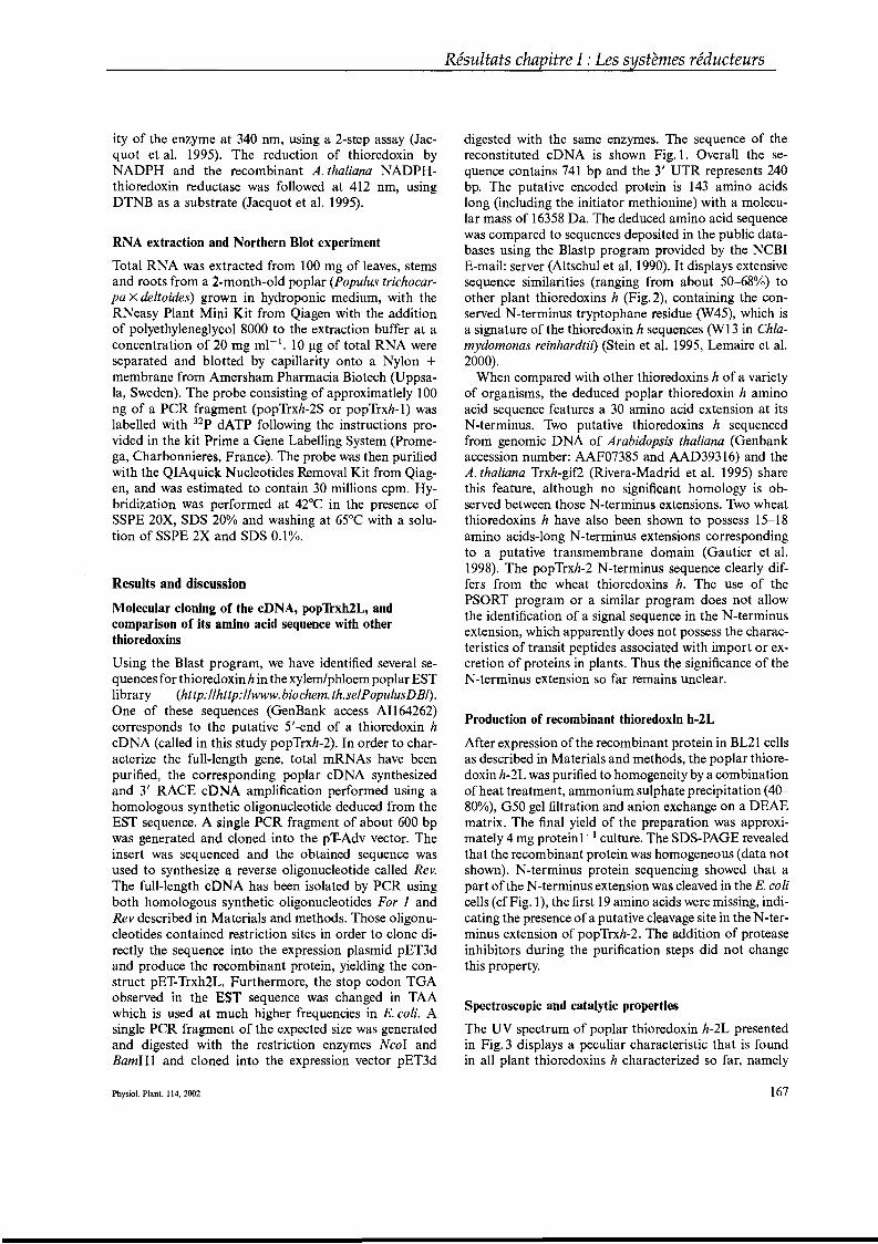
ity of the enzyme at 340 nm, using a 2-step assay (Jacquot et al. 1995). The reduction of thioredoxin byNADPH and the recombinant A. thaliana NADPHthioredoxin reductase was followed at 412 nm, usingDTNB as a substrate (Jacquot et al. 1995).
RNA extraction and Northern Blot experiment
Total RNA was extracted from 100 mg of leaves, stemsand roots from a 2·month-old poplar (Populus trichocarpa X deltoides) grown in hydroponic medium, with theRNeasy Plant Mini Kit from Qiagen with the additionof polyethyleneglycol 8000 to the extraction buffer at aconcentration of 20 mg ml-le 10 Jlg of total RNA wereseparated and blotted by capillarity onto a Nylon +membrane from Amersham Pharmacia Biotech (Uppsala, Sweden). The probe consisting of approximatlely 100ng of a PCR fragment (popTrxh-2S or popTrxh-1) waslabelled with 32p dATP following the instructions provided in the kit Prime a Gene Labelling System (Promega, Charbonnieres, France). The probe was then purifiedwith the QIAquick Nucleotides Removal Kit from Qiagen, and was estimated to contain 30 millions cpm. Hybridization was performed at 42°C in the presence ofSSPE 20X, SDS 200/0 and washing at 65°C with a solution of SSPE 2X and SDS 0.10/0.
Results and discussion
Molecular cloning of the cDNA, popTrxh2L, andcomparison of its amino acid sequence with otherthioredoxins
Using the Blast program, we have identified several sequences for thioredoxin h in the xylemlphloem poplar ESTlibrary (http: //http://www.biochem.th.seIPopulusDBI).One of these sequences (GenBank access A1164262)corresponds to the putative 5'-end of a thioredoxin hcDNA (called in this study popTrxh-2). In order to characterize the full-length gene, total mRNAs have beenpurified, the corresponding poplar cDNA synthesizedand 3' RACE cDNA amplification performed using ahomologous synthetic oligonucleotide deduced from theEST sequence. A single PCR fragment of about 600 bpwas generated and cloned into the pT-Adv vector. Theinsert was sequenced and the obtained sequence wasused to synthesize a reverse oligonucleotide called Rev.The full-Iength cDNA has been isolated by PCR usingboth homologous synthetic oligonucleotides For 1 andRev described in Materials and methods. Those oligonucleotides contained restriction sites in order to clone directly the sequence into the expression plasmid pET3dand produce the recombinant protein, yielding the construct pET-Trxh2L. Furthermore, the stop codon TGAobserved in the EST sequence was changed in TAAwhich is used at much higher frequencies in E. coli. Asingle PCR fragment of the expected size was generatedand digested with the restriction enzymes Ncol andBamH1 and cloned into the expression vector pET3d
Physiol. Plant. 114,2002
Résultats chapitre l : Les systèmes réducteurs
digested with the same enzymes. The sequence of thereconstituted cDNA is shown Fig. 1. Overall the sequence contains 741 bp and the 3' UTR represents 240bp. The putative encoded protein is 143 amino acidslong (including the initiator methionine) with a molecular mass of 16358 Da. The deduced amino acid sequencewas compared to sequences deposited in the public databases using the Blastp program provided by the NCBIE-mail: server (Altschul et al. 1990). It displays extensivesequence similarities (ranging from about 50-680/0) toother plant thioredoxins h (Fig.2), containing the conserved N-terminus tryptophane residue (W45), which isa signature of the thioredoxin h sequences (W13 in Chlamydomonas reinhardtii) (Stein et al. 1995, Lemaire et al.2000).
When compared with other thioredoxins h of a varietyof organisms, the deduced poplar thioredoxin h aminoacid sequence features a 30 amino acid extension at itsN-terminus. Two putative thioredoxins h sequencedfrom genomic DNA of Arabidopsis thaliana (Genbankaccession number: AAF07385 and AAD39316) and theA. thaliana Trxh-gif2 (Rivera-Madrid et al. 1995) sharethis feature, although no significant homology is observed between those N-terminus extensions. Two wheatthioredoxins h have also been shown ta possess 15-18amino acids-Iong N-terminus extensions correspondingto a putative transmembrane domain (Gautier et al.1998). The popTrxh-2 N-terminus sequence clearly differs from the wheat thioredoxins h. The use of thePSORT program or a similar program does not allowthe identification of a signal sequence in the N-terminusextension, which apparently does not possess the characteristics of transit peptides associated with import or excretion of proteins in plants. Thus the significance of theN-terminus extension so far remains unclear.
Production of recombinant thioredoxin h-2L
After expression of the recombinant protein in BL21 cellsas described in Materials and methods, the poplar thioredoxin h-2L was purified to homogeneity by a combinationofheat treatment, ammonium sulphate precipitation (40800/0), G50 gel filtration and anion exchange on a DEAEmatrix. The final yield of the preparation was approximately 4 mg proteinl- l culture. The SnS-PAGE revealedthat the recombinant protein was homogeneous (data notshown). N-terminus protein sequencing showed that apart of the N-terminus extension was cleaved in the E. colicells (cf Fig. 1), the first 19 amino acids were missing, indicating the presence of a putative cleavage site in the N-terminus extension of popTrxh-2. The addition of proteaseinhibitors during the purification steps did not changethis property.
Spectroscopie and catalytic properties
The UV spectrum of poplar thioredoxin h-2L presentedin Fig. 3 displays a peculiar characteristic that is foundin all plant thioredoxins h characterized so far, namely
167
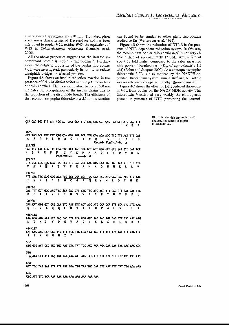
Résultats chapitre l : Les systèmes réducteurs
a shoulder at approximately 290 nm. This absorptionspectrum is characteristic of Trp residues and has beenattributed to poplar h-2L residue W45, the equivalent ofW13 in Chlamydomonas reinhardtii (Lemaire et al.2000).
AlI the above properties suggest that the isolated recombinant protein is indeed a thioredoxin h. Furthermore, the catalytic properties of the poplar thioredoxinh-2L were investigated, particularly its ability to reducedisulphide bridges on selected proteins.
Figure 4A shows an insulin reduction reaction in thepresence of 0.5 mM dithiothreitol and 2.8 flM recombinant thioredoxin h. The increase in absorbancy at 650 nmindicates the precipitation of the insulin chains due tothe reduction of the disulphide bonds. The efficiency ofthe recombinant poplar thioredoxin h-2L in this reaction
was found to be similar to other plant thioredoxinsstudied so far (Wetterauer et al. 1992).
Figure 4B shows the reduction of DTNB in the presence of NTR dependent reduction system. In this test,the recombinant poplar thioredoxin h-]L is not very efficient (Km of approximately 12 JlM), with a Km ofabout 10 fold higher compared to the value measuredwith poplar thioredoxin h-1 (Km of approximately 1.5flM) (Behm and Jacquot 2000). As a consequence poplarthioredoxin h-2L is also reduced by the NADPH-dependent thioredoxin system from A. thaliana, but with aweaker efficiency compared to other thioredoxins h.
Figure 4C shows the effect of DTT reduced thioredoxin h-2L from poplar on the NADP-MDH activity. Thisthioredoxin h activated very weakly the chloroplasticprotein in presence of DTT, preventing the determi-
1CGA CACi·TGCTlTGTTTGG··IGT··M,A.G(A nCTAG CTACGTGAG TC. ·GCTATGGAG/TTT
NEF
Fig. 1. Nucleotide and amino aciddeduced sequences of poplarthioredoxin h·2.
5814GGr ···TGGC:CAGTTCfTCAGCGAGGAAGAACA··GtG ·€AAACAAGC l'TC .•.. Trt ·Mt TrrGATA W P V L q R G R T V Q TSF F H F 0
RetOlÎl' ·popTr~;;.2L+
116ï23~<:TCC.~TGGA ··TTTCa ,.~ .ACAAAGCGAGCTGcrGGG .GTT mG GAT .. GTCCATTCTH S N G F P C r K P A A G V V D V H S
Popfrlh;.;;2S -----..M174/42GIA GGtGCA·TG~ .GlJCCT,TllC·.GAG>GCC .kACMG ··CM··1(AC/4.f:..··1J4ATTG ·ctG,t,V GAI R S y F E A N K q N N K L L V
Z32161AU GAATTCACG GeI.CAl" TGT·G·~C(T TG(CGATAC ATGGA,GCAGACCATG.Mti1 E F T A T i c , p dRY M E Q T M K
Z9I1S1~CTITG(:T .·~ÇCAAG"~C· ..lC.J....·~C •• ~TTGTG nC.AICAGG ATTG4(:Gj'[ (i~rGAATTG
D F A A K V T 0 V V F l R l D V 0 E L
348l99.CA,(iQTG)"GG(:T··CAG< ..CQ.·nc MT GtG AtT ACCAtO·CCAGCA .nTTCA crCTTGAAGQ H V A q q F N VTT M PAF S l L K
4061118~GGG>lAG .AtAGllGAC· GAGGTAGCA ·GGG>GJtAAGAAG AGr6AG ·CTTCAGMC MGK G K 1 V DEY A G V K K SEL Q N K
fi4t131~TTGA'>MG CAT ···GGG.ATG ATATGATTGCCACGA TACTTAACTATTAATGCC··.ATG.· CCCl E K H G NI·
5ZZATCiGCGAAT CCC·TGCTGG AATGTA TAT TCCAGC·AfiAACiA.··.<;M.···GAA TAAAACAAG Gee
580T(~>AAA·GCA ATTTG(TGA GGCAAAAAT AAGGCCATC CTTTTTTCTTTTCTTCTTCTT
638GATTGCT1T TATlTAA'TA"fACGTAnc; TAATGC ..(AA GTTAAT·rrr TATTTAAGA.AM
696crCATrUC TtA···.u.A··.üA.·· ••~···AAAAAA.··MAMAMA
168 Physiol. Plant. 114, 2002
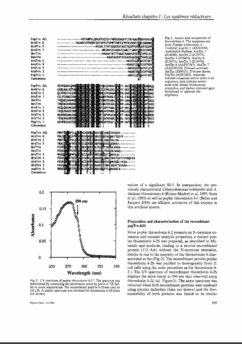
Résultats chapitre l : Les systèmes réducteurs
PopTrxh2lArdJr)( 6AraTrx ··ZAraTrx7DurTrxAesTrxArdTrx 1ArâT'rx4A,raTrx 5Aralrx 3Pop"frx 1Consensus
f'op'Trxh2lAraTrx 6ArdTrx2Ar(}Trx 7DurTrxAesTrxAral"rx 1Ara7f,X 4AràTrx 5AroTrx3PgpTrx 1.Côns.ensus
RopTrx ftZlAroTrx6A.rttTrx ZAraTrx7OUrTrxAesTr:1.AralrxlAray,.x··4AraTrxSArttTrx. ·3poPTrx1Consensus
_··_·_·__··-·__·.-_·.-.-·*-MEFAWPVl~RGRTV9TS.F ..FNFDlISNGFPCTKPAA.._.......... _.- •·..··_·..·-.MtiANVSTPDQRFqYTH.F.~S1"KPffTP~p~·.~yr·~.~v~~~----- ..--------- --- ...---...MGGALSTVFGSGEDATMGTESEPS• .......·....• .... --,.·--...- ·-----,.·...........M<iSNVSSVHQVASSMEXT$NG-.-.--..-..-----------..----~-~Wl'4~T~ffl~!~~~__ .W' ----'''" -.- -.__ - - -.- - .._ MAI\SB1ATATAAAVGA.;;, - _..- ..._.__._..- - -.-._--- _-- _..;,., ........ -_. -;",.-- ...,.;;.··"".,;,·;,.,··_... ·_·_··""·..:r.tASEE.~ _ _ -..-.- _..., ..,.--.--.-.-..- -..-.-.-.-.-----..- ._.-.-_._·--·· r-tAAE- - -_._",.."".""'."": "".""..,,,. ,,. - - - _., ·_·· · · -·---""·""· MAGEG·- --"" -,--.- -- -.- --------- ;..::...--_ · ·-J4AAEG.............................................. "" - "' __ _ _ ·-~E.:E·
KHGMI--- .. _,;.-,;.-YTQSFf...... ..., - ... :...L.RA.-- ... --- - _...RV--- -_ ;;.. ...
lHAAA-- -- ·- ...I..:tfMQ_ • ...KHLA-- ..."'--"",-_·""KliTGVTtvvNQFEA·HG(jtVASA;;;.~ ... _"
KHKTYV~';;'--...",,·-
KliATA-··...------:,.;o·....
Fig. 2. Amino acid comparison ofthioredoxins h. The sequences arefrom Populus trichocarpa cv.Trichobel, popTrx 1 (AI161830);Arabidopsis thaliana, AraTrx 1(Z14084), AraTrx 2 (Z35475),AraTrx 3 (Z35474), AraTrx 4(Z35473), AraTrx 5 (Z35476),AraTrx 6 (AAF07385), AraTrx 7(AAD39316); Triticum aestivum,AesTrx (X69915); Triticum durum,DurTrx (AJ001903). Asterisksindicate conserved amino acids in aIlsequences, dots indicate aminoacids with similar biochemicalproperties, and dashes represent gapsintroduced to optimise thealignment.
Preparation and characterization of the recombinantpopTrx-h2S
Since poplar thioredoxin h-2 presents an N-terminus extension and unusual catalytic properties, a mutant popIar thioredoxin h-2S was prepared, as described in Materials and methods, Ieading to a shorter recombinantprotein (113 AA) without the N ·terminus extension,similar in size to the majority of the thioredoxins h characterized so far (Fig. 2). The recombinant protein poplarthioredoxin h-2S was purified to homogeneity from E.coli cens using the same procedure as for thioredoxin h2 1. The UV spectrum of recombinant thioredoxin h-2Sdisplays the same bump at 290 nm that observed usingthioredoxin h-2L (cf. Figure3). The same spectrum wasobtained when both recombinant proteins were analysedusing circular dichroïsm (data not shown) and the thermostability of both proteins was found to be similar
350275 300 325
Wavelength .(Dm)
nation of a significant SO.5. In comparison, the previously characterized Chlamydomonas reinhardtii and A.thaliana thioredoxins h (Rivera-Madrid et al. 1995, Steinet al., 1995) as well as poplar thioredoxin h-1 (Behm andJacquot 2000) are efficient activators of this enzyme inthis artificial system.
Fig. 3. UV spectrum of poplar thioredoxin h-2 1. The spectrum wasdetermined by measuring the absorbance point by point in TE buffer at room temperature. The recombinant popTrx h-2Lwas used at2.8 JlM. A similar spectrum was obtained for thioredoxin h-2S (datanot shown)
Physiol. Plant. 114,2002 169
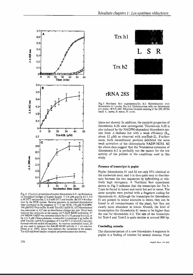
Résultats chapitre l : Les systèmes réducteurs
O.7-r---.---r---r-----r--~--"'"'
A
o.~
0.3
O~2
O_[Q_:rc~Dl'l:tl1J~=--10102030405060
TiJlle .•..•Clî1i..l
Trxlil~ ;:::l~
~ ,,~)!
A~ "":«:":
L
(data not shown). In addition, the catalytic properties ofthioredoxin h-2S were investigated. Thioredoxin h-2S isalso reduced by the NADPH-dependent thioredoxin system from A. thaliana but with a weak efficiency (Kmabout 12 JlM) as observed with popTrxh-2L. Furthermore, both recombinant proteins exhibited the sameweak activation of the chloroplastic NADP-MDH. AlIthe above data suggest that the N...terminus extension ofthioredoxin h-2 is probably not the reason for the 10wactivity of the protein in the conditions used in thisstudy.
Fig. 5. Northern blot experimentsTrx h·l: Hybridization withthioredoxin h-1 probe; Trx h-2: Hybridization with the thioredoxinh-2 probe: rRNA 28S: Ethidium bromide staining if the 28S rRNAband. L: leaves, S: stems; R: roots.
Presence of transcripts in poplar
Poplar thioredoxins hl and h2 are only 530/0 identical atthe nucleotide level, and it is thus quite easy to discriminate between the two sequences by hybridizing at relatively high stringency. A Northern blot experimentshown in Fig. 5 indicates that the transcripts for Trx h2 can be found in leaves and roots but not in stems. Thesame samples were probed with a fragment coding forthioredoxin hl. Although the transcripts for thioredoxinhl are present in minor amounts in stems, they can befound in all compartments of the plant, but they areclearly more abundant in leaves. Overall, the level oftranscription for thioredoxin hl seems to be higher thanthe one for thioredoxin h-2. The size of the transcriptsfor Trxh-l and Trxh-2 is quite similar at around 800 bp.
Concluding remarks
The characterization of a new thioredoxin h sequence inpoplar is a finding of interest for severa! reasons. First
5020 3040TJt~~b~L·(pM)
lU
S 10 1.5 20 25IncuBaltoutime•(mil'i)
O~7
(l.I
o.]
o.sB
Fig. 4. Catalytic properties ofpoplar thioredoxin h-21. (a) Reductionof disulphide bridges of human insulin. 0 2.8 JlM popTrx h-2 1, 0.5mM DTT and insulin; 00.5 mM DTT and insulin. (b) DTNB reduction by the NTR system. Various amounts of oxidised thioredoxinswere incubated in the presence of 77.5 nM NTR, 150 JlM NADPH,100 JlM DTNB in buffer 30 mM Tris-HCl (pH 8.0). DTNB reductionwas measured at 412 nm as absorbance change per min. Error barsindicate the variations in the assays. (c) NADP-MDH activation. 27nM MDH-NADP was activated either by (0) 35 JlM popTrx h-2L orby (0) 9 JlM Chlamydomonas reinhardtii thioredoxin h in buffer 50mM Tris-HCl (pH 8.0) in presence of5 mM DTT. Control (0) was performed without added thioredoxin. At various incubation times, 20Jll was used to measure the NADP-MDH activity in 1 ml reaction(Stein et al. 1995). Error bars indicate the variations in the assays.Trx-h2S exhibited similar catalytic properties (data not shown).
170 Physiol. Plant. 114, 2002
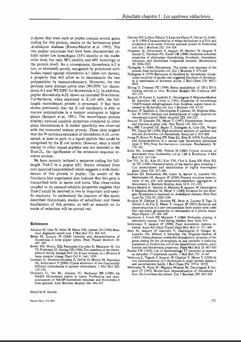
it shows that trees such as poplar contain several genescoding for this protein, similar to the herbaceous plantArabidopsis thaliana (Rivera-Madrid et al. 1995). Thetwo poplar sequences that have been characterized exhibit rather 10w homologies (530/0 identity at the nucleotide level, but only 380/0 identity and 600/0 homology atthe protein level). As a consequence, thioredoxin h-2 isnot, or extremely poorly, recognised by polyclonal antibodies raised against thioredoxin h-l (data not shown),a property that will allow us to discriminate the twopolypeptides by immunochemistry. Moreover, the twoproteins have distinct active sites (WCPPC for thioredoxin h-l and WCGPC for thioredoxin h-2). In addition,poplar thioredoxin h~2L shows an extended N-terminus.Furthermore, when expressed in E. coli cells, the fuBlength recombinant protein is processed. It has beenshown previously that the E. coli machinery is able tomature polypeptides in a manner analogous to higherplants (Jacquot et al. 1991). The recombinant proteindisplays unusual catalytic properties compared to otherplant thioredoxins h. A similar specificity was observedwith the truncated mutant proteine These data suggestthat the N-terminus extension of thioredoxin h-2L correspond, at least in part, to a signal peptide that could berecognised by the E. coli system. However, since a motifsimilar to other transit peptides was not detected in theTrxh-2L, the significance of the extension sequence remains unclear.
We have recently isolated a sequence coding for fullIength Trxh-2 in a poplar EST library obtained fromroot material (unpublished) which suggests a wide distribution of this protein in poplar. The results of theNorthern blot experiment also indicate that this gene istranscribed both in leaves and roots. This observationcoupled to its unusual catalytic properties suggests thatTrxh-2 could be involved in vivo in important and specifie reactions. To understand the function of this newlydescribed thioredoxin, studies of subcellular and tissue10calization of this protein, as weB as research on itsmode of reduction will be carried out.
References
Altschul SF: Gish W, Miller W, Myers EW, Lipman DJ (1990) Basiclocal alignment search tool. J Mol Biol 215: 403-410
Behm M, Jacquot JP (2000) Isolation and characterization ofthioredoxin h from poplar xylem. Plant Physiol Biochem 38:363-369
Bower MS, Matias DO, Fernandes-Carvalho E, Mazzurco M, GuTS, Rothstein SJ, Goring DR (1996) Two members of the thioredoxin-h family interact with the kinase domain of a Brassica Slocus receptor kinase. Plant Cell 8: 1641-1650
Capitani G, Markovic·Housley Z, DelVal G, Morris M, JansoniusJN, Schurmann P (2000) Crystal structures of two functionallydifferent thioredoxins in spinach chloroplasts. J Mol Biol 302:135-154
Florencio FJ, Yee BC, Johnson TC, Buchanan BB (1988) AnNAD~ Ithioredoxin system in leaves. Purification and characterization of NADP-thioredoxin reductase and thioredoxin hfrom spinach. Arch Biochem Biophys 266: 496-507
Edited by R. Scheibe
Physiol. Plant. 114, 2002
Résultats chapitre l : Les systèmes réducteurs
Gautier MF: Lullien-Pellerin V, Lamotte-Guery F: Guirao A, Joudrier P (1998) Characterization of wheat thioredoxin h cDNA andproduction of an active Triticum aestivum protein in Escherichiacoli. Eur J Biochem 252: 314-324
Hirasawa M, Schurmann ~ Jacquot JR Manieri W, Jacquot P,Keryer E, Hartman FC, Knaff DB (1999) Oxidation-reductionproperties of chloroplast thioredoxins, ferredoxin. thioredoxinreductase, and thioredoxin f-regulated enzymes. Biochemistry38: 5200-5205
Holmgren A (1968) Thioredoxin. The amino acid sequence of theprotein from Escherichia coli. Eur J Biochem 6,475-484
Holmgren A (1979) Reduction of disulfides by thioredoxin. Exceptional reactivity of insulin and suggested functions of thioredoxin in mechanism of hormone action. J Biol Chem 254: 96279632
Huang Y, Domann FE (1998) Redox modulation of AP-2 DNAbinding activity in vitro. Biochem Biophys Res Commun 249:307-312
Jacquot J~ Keryer E, Issakidis E, Decottignies ~ Miginiac-MaslowM, Schmitter JM, Crétin C (1991) Properties of recombinantNADP-malate dehydrogenases from Sorghum vulgare leaves expressed in Escherichia coli. Eur J Biochem 199: 47-51
Jacquot JP, Issakidis E, Decottignies ~ Lemaire M, Miginiac-Maslow M (1995) Analysis and manipulation of target enzymes forthioredoxin control. Meth Enzymol 252: 240-252
Jacquot J~ Lancelin JM, Meyer Y (1997) Thioredoxins: Structureand function in plant cells. New Phytol 136: 543-570
Jeng MF: Campbell A~ Begley T, Holmgren A, Case DA, WrightPE, Dyson HJ (1994) High-resolution solution of oxidized andreduced Escherichia coli thioredoxin. Structure 2: 853-868
Jeong JS, Kwon SJ, Kang Sw, Rhee SG, Kim K (1999) Purificationand characterization of a second type thioredoxin peroxidase(type II TPx) from Saccharomyces cerevisiae. Biochemistry 38:776-783
Katti SK, Lemaster DM, Eklund H (1990) Crystal structure ofthioredoxin from Escherichia coli at 1.68 A Resolution. J MolBiol 212: 167-184
Lee TH, Yu SL, Kim SU, Kim YM, Choi l, Kang Sw, Rhee SG,Yu DY (1999) Characterization of the murine gene encoding 1Cys peroxiredoxin and identification of highly homologousgenes. Gene 234: 337-344
Lemaire SD, Richardson JM, Goyer A, K.eryer E, Lancelin JM,Makhatadze GI, Jacquot JP (2000) Primary structure determinants of the pH- and temperature-dependent aggregation ofthioredoxin h. Biochem Biophys Acta 1476: 311-323
Rivera-Madrid R, Mestres D, Marinho R Jacquot J~ Decottignies~ Miginiac-Maslow M, Meyer Y (1995) Evidence for five divergent thioredoxin h sequences in Arabidopsis thaliana. Proc Nat!Acad Sci USA 92: 5260-5264
Rouhier N, Gelhaye E, Sautiere PE, Brun A, Laurent ~ Tagu D,Gerard J, de Fay E, Meyer Y, Jacquot JP (2001) Isolation andcharacterization of a new peroxiredaxin from poplar sieve tubesthat uses either glutaredoxin or thioredoxin as a proton donor.Plant Physiol 127: 299-309
Sambrook J, Frisch EF: Maniatis T (1989) Molecular cloning: Alaboratory manual. Cold Spring Harbor, New York, NY
Schürmann P, Jacquot JP (2000) Plant thioredoxin systems revisited. Annu Rev Plant Physiol Plant Mol Biol 51: 371-400
Stein M, Jacquot J~ Jeannette E, Decottignies ~ Hodges M,Lancelin JM, Mittard V, Schmitter JM, Miginiac-Maslow M(1995) Chlamydomonas reinhardtii thioredoxins: structure of thegenes coding for the chloroplastic m and cytosolic h isoforms;expression in Escherichia coli of the recombinant proteins, purification and biochemical properties. Plant Mol Biol 28: 487-503
Studier FW (1999) Use of bacteriophage T7 lysozyme to improvean inducible T7 expression system. J Mol Biol 219: 37-44
Verdoucq L, Vignols F: Jacquot J~ Chartier Y, Meyer Y (1999) Invivo characterization of a thioredoxin h target protein defines anew peroxiredoxin family. J Biol Chem 274: 19714-19722
Wetterauer B, Véron M, Miginiac-Maslow M, Decottignies ~ Jacquot JP (1992) Biochemical characterization of thioredoxin 1from Dictyostelium discoideum. Eur J Biochem 209: 643-649
171
Résultats chapitre I : Les systèmes réducteurs
Afin de compléter les informations sur les thiorédoxines extra-chloroplastisques
du peuplier, une troisième isoforme de Trx h nommée popTrx h3 a été clonée et
surproduite dans E. coli. Cette isoforme diffère notablement de popTrx h2 (40%
d’identité) mais elle est relativement proche de popTrx h1 au niveau de sa séquence
primaire (63% d’identité). Néanmoins, son site actif (WCGPC) est différent de celui de
popTrx h1 (WCPPC). Par rapport aux séquences d'A. thaliana, elle semble être
l’isoforme équivalente de AtTrx h1. Des mutants de popTrx h1 et popTrx h3 ont été
générés au niveau du site actif de manière à transformer l'un en l'autre. PopTrx h3 est
réduite efficacement par la NTR d'A. thaliana, et c’est un meilleur donneur que popTrx
h1 pour la Prx de type II de peuplier. En revanche, popTrx h3 est un donneur moins
efficace que popTrx h1 pour la RNR d’E. coli. Ces résultats indiquent donc une certaine
spécificité entre les diverses classes de Trx en dépit des homologies quelquefois
élevées.
Article numéro 6 :
Gelhaye E, Rouhier N, Vlamis-Gardikas A, Girardet JM, Sautière PE, Sauzet M, Martin F, Jacquot JP. (2003) Identification and characterization of a third thioredoxin h in poplar.
Plant Physiol. Biochem., sous presse.
Résultats chapitre l : Les systèmes réducteurs
ARTICLE IN PRESS
ELSEVIER
Available online at www.sciencedirect.com
SCIENCI! @DIRECTO
Plant Physiology and Biochemistry 00 (2003) 000-000
PlantPhysiology
andBiochemistry
www.elsevier.com/locatelplaphy
Original article
2 Identification and characterization of a third thioredoxin h in poplar
3 Eric Gelhaye a,*, Nicolas Rouhier a, Alexios Vlamis-Gardikas b, Jean-Michel Girardet c,4 Pierre-Eric Sautière d, Michel Sauzet d, Francis Martin a, Jean-Pierre Jacquot a
PLAPHY-00000068
In plants, each thioredoxin type and especially the cytoso- 38lic one, is represented by a small multigenic family. Nine 39thioredoxins h have been detected in Arabidopsis thaliana, 40three of which having the unusual active site sequence 41WCPPC [13,14]. In poplar, five thioredoxins h have also 42been detected ([1,5], unpublished data), the first one named 43popTrxhl exhibiting the unusual active site WCPPC [1]. It 44has been demonstrated that the redox activity of the active 45center depends on amino acid residues situated between the 46two cysteines [3]. Among the proposed functions for cytoso- 47lic thioredoxins is the reduction of non-heme peroxidases 48called peroxiredoxins (Prx), which catalyze the reduction of 49various peroxides [15,20]. 50
Thioredoxins have a particularly stable three-dimensional 51structure, which is conserved among all thioredoxin struc- 52tures from prokaryotes to eukaryotic algae, higher plants, and 53mammals [4]. Nevertheless, yeast complementation experi- 54ments have demonstrated that A. thaliana thioredoxins are 55active only on sorne of the yeast thioredoxin targets [2], this 56specificity being probably related to the nature of the resi- 57
5 a Interactions Arbres-Micro-organismeslnra, Faculté des Sciences, Université H. Poincaré-Nancy 1 (UMR 1136),54506 Vandoeuvre-les-Nancy, France6 b Department ofMedical Biochemistry and Biophysics, Karolinska Institutet, Medical Nobel Institute for Biochemistry, 171 77 Stockholm, Sweden7 C Laboratoire des BioSciences de l'Aliment Inra, Faculté des Sciences (UC885), Université H. Poincaré-Nancy 1,54506 Van1r;,uvre-les-Nancy, France8 d Laboratoire d'Endocrinologie des Annélides, Université des Sciences et Technologies de Lille (UPRESA 8017), 59655 Villt(I\~~~'Jd'Ascq cedex, France
9 Received 13 December 2002; accepted 21 January 2003
Abbreviations: CDP, cytidine diphosphate; DEAE, diethyl amino ethyl;DTNB, 5,5' dithiobis nitrobenzoic acid; EST, expressed sequence tag; Grx,glutaredoxin; NTR, NADPH thioredoxin reductase; Prx, peroxiredoxin;WT, wild type.
* Corresponding author. Fax: +33-3-83-91-25-64.E-mail address:[email protected] (E. Gelhaye).
© 2003 Éditions scientifiques et médicales Elsevier SAS. AH rights reserved.DOl: 1 0 . 1 0 1 6 / S a9 8 1 - 9 4 2 8 ( a3 ) 0 a0 6 3 - 9
25 1. Introduction
26 Plant thioredoxins are small 18Iê~\<weight (ca. 1227 kDa) and generally extremel~lhWmost,fle proteins. They28 reduce disulfide bonds thro~gft:t <\"J,ij(ox active site with29 the conserved sequence~~G!I]PC. Plants possess two30 well-described thiore{fOxi'R;~'jTltlH1s: a cytoplasmic system31 including several t~\.<:;ii h and a NADPH-dependent32 thioredoxin re~~~taslllf'~i a specifie chloroplastic system33 characterized bY;~f~n:~'oxin-dependent thioredoxin reduc34 tase. Furthermoré;:;If\VO thioredoxins and a NADPH-35 dependent thioredoxin reductase have been described re36 cently as a functional plant mitochondrial thioredoxin system37 [11].
12 Three full-Iength sequences encoding thioredoxin h have been isolated in a leaf/root cv. Trichobel expressed13 sequence tag (EST) library. One of these, popCXXS 1 exhibits the nontypical active sil; to atCXXS 1. The second one,14 named popTrxh4 is related to atTrxh9 which forms with several other plant thioredoxin ha subgroup of thioredoxins h. The third one,15 named popTrxh3, displays 66% identity and also a high degree ofhomology (81 % . the iously described popTrxhl. Nevertheless the16 active sites ofboth proteins differ, since the active site ofpopTrxhl (WCPPC) ij;~t of the canonical WCGPC found in popTrxh3. The17 cDNA sequence of popTrxh3 has been introduced in an expression plas . (pj'!în order to express the corresponding recombinant18 polypeptide. The protein has been expressed to a high level, purified fr~~;r c ricnlTlicoli cells with a high yield and its catalytic properties19 compared to popTrxhl. Furthermore two mutants, popTrxh1P40G t,nd<' 3G41P have been engineered in arder to explore the20 importance of the active site residues in the thioredoxin h catalytic,fdPslties. e results are discussed in relation with known biochemical21 properties of thioredoxins h. .
22 © 2003 Éditions scientifiques et médicales Elsevier SAS. Ali23 Keywords: Populus; Redox regulation; Recombinant; Thiored24
10
Il Abstract
Résultats chapitre l : Les systèmes réducteurs
1
ARTICLE IN PRESS2 E. Gelhaye et al. / Plant Physiology and Biochemistry 00 (2003) 000-000
122123
124125126127128129130131132133134135136137138139140141142143144145146147148
149
150151152153154155156157158159
2.5. Enzymatic activities
The reduction of HzOz by poplar Prx in the presence ofdifferent popTrxh was followed spectrophotometrically using a Cary 50' spectrophotometer as described previously[15]. The reduction of thioredoxin by NADPH and the recombinant A. thaliana NADPH-thioredoxin reductas,e wasfollowed at 412 nm using DTNB as a substrate [10]. Reduction of cytidine diphosphate (CDP) by E. coli RRla wasperformed essentially as described in [6]. Reducing powerwas provided by 2 mM DTT. All assays were performed in atotal volume of 120 Jll. The catalytic effect of the thioredox-
2.4. Expressionproteins
GGGGGGATCCTTATGCAGTTGCGTGCTTTG 3'. Mu- 107tagenic oligonucleotides were as follows: direct 5' ACTGCT- 108TCATGGTGTGGACCATGTAAAATGATT 3'; and reverse 1095' AATCATTTTACATGGTCCACACCATGAAGCAGT 3'. 110The popTrxh3 mutant was obtained using the cloning oligo- 111nucleotides described in Section 2.2 and the mutagenic oli- 112gonucleotides were as follows: G41P direct 5' GCTGCT- 113TCATGGTGTCCTCCGTGCCGTGTCATT 3'; G41P 114reverse 5' AATGACACGGCACGGAGGACACCATGA- 115AGCAGC 3'. 116
The mutated PCR products that contained the restriction 117sites were cloned into the expression plasmid pET-3d, yield- 118ing the construction pET popTrxh1 ~~~P,~d pET popTrxh3 119G41P. The sequences of the reco~biJant plasmids were 120verified by DNA sequencing. p , 121
'\i&~4j71'''''\;I,;~pression and purification of A.thaliana NADnft, ,~ductase,poplar Trxh1 and poplar Prx
; ëwhere [1,9,15]. For the expression ofpopTrxhl OG, d popTrxh3 WT, the recombinant plas
,,~d to transform E. coli BL21(DE3) strain. One
~~~_ ··~....t ...•....•.......... sed to inoculate 3 ml LB medium, supplecl~q,-tl 'YÎth ampicillin. The culture was successively ampli-
"fiell];~i~,ti.nal volume of 3 1over a period of 24 h and inducedith:IOO JlM IPTG for 6 h. The cells were then collected by
èentrifugation and suspended in TE buffer (30 mM Tris-HCl,1 mM EDTA, pH 8). The cell suspension was disrupted bysonication. The resulting sample was fractionated with am-monium sulfate. The fraction precipitating between 40% and80% of the saturation was suspended in a minimal volume ofTE buffer. The purification was then performed as describedpreviously [1] using Sephadex G50 gel filtration and DEAESepharose columns.
The last purification step was performed by size exclusionchromatography. A volume of 100 #LI protein (5.5 mg ml-l
)
obtained as described above was loaded on a Superose 12HRcolumn connected to a fast protein liquid chromatography(FPLC) system. The column was equilibrated with 30 mMTris-HCl (pH 8) buffer containing 0.15 M NaCl and theproteins were eluted in the same buffer at a flow rate of 0.25ml min-le
72 2. Methods
58 dues on the protein surface. It was proposed that plant thiore59 doxins h have a common origin from a totipotent ancestor60 and that after gene duplications, mutations appeared masking61 sorne determinants necessary for the interaction with particu62 lar targets and resulting in a specialization of the plant thiore63 doxins h [2].64 In this context, we have extended the characterization of65 the redox equipment ofpoplar [1,5,15,17]. We present in this66 paper three new nucleotidic sequences of putative thioredox67 ins h and the characterization of one of these called pop68 Trxh3. Furthermore, we have compared its biochemical69 properties to the previously described popTrxhl [1] focusing70 on the difference observed between popTrxhl and popTrxh371 in the sequences of the active sites (WCGPC vs. WCPPC).
73 2.1. Escherichia coli strains, cDNA library and plasmids
74 E. coli strains DH5a F' and BL21(DE3) were grown in LB75 medium at 37 oC supplemented with ampicillin (50 Jlg ml- l
).
76 The poplar thioredoxin h3 cDNA was isolated from a77 xylemlphloem cDNA library of Populus trichocarpa cv.78 Trichobel provided by Dr. W. Boerjan and Dr. H. Meyermans79 (University of Ghent, Belgium) and was cloned into the80 expression vector pET-3d.
99 2.3. Mutagenesis
82 Two oligonucleotides primers were constructe~i\ e83 the EST (unpublished results) corresponding ~;tatiMe
84 thioredoxin h full-length cDNA (369 bp) dy îllJj;the85 BLAST program. The upstream 24-base .ge>hucle-86 otide was synthesized homologous tQil!~ ln.g strand,87 including an addition of six C and rêstriction site88 underlined at the S'-end with u> nce: 5' CCC-89 CCCATGGCAGCAGAAGA1'1 e downstream oli-90 gonucleotide was 27 bases llng an. nved from the 5'-end91 of the non-coding stran 'j at the 5'-end four addi-92 tional C and a BamBin· on site (underlined) with the93 sequence: 5' CCCC 'CAAGCAGAAGCAGTAGC94 3'. After PCR reâ~l~~n;P!'fOrmed,the amplified fragment has95 been purified, dig~",;;:>~;;with the appropriate enzymes and96 cloned into the pET-3d expression plasmid as described pre97 viously [16] in order to obtain the construction named pET98 popTrxh3.
81 2.2. PCR cloning
100 Mutagenesis of the cloned cDNAs was effected by PCR. A101 strategy similar to the one in [17] was employed to generate102 two overlapping mutated fragments and in a second PCR103 reaction the full-length mutated sequence. The popTrxh104 1P40G was obtained from pET-popTrxhl [1] using the fol105 lowing cloning oligonucleotides: direct 5' GGGGCCATG106 GCCGAAGAAGGACAAGT 3'; and reverse 5'
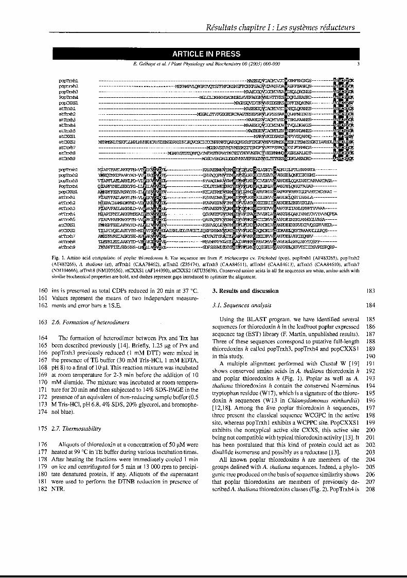
Résultats chapitre l : Les systèmes réducteurs
ARTICLE IN PRESSE. Gelhaye et al. / Plant Physiology and Biochemistry 00 (2003) 000-000 3
pq:il):xhlpcpt.rxh2pcpTl:xh3PcpTrxh4
PCP::XXSlatTrxhlatTrxh2atTrxh3at'lb<h4atTLxhSatCXXSlatCXXS2atTr.Kh7atTrxhBat'Trxh9
pq:il):xhlp:prrxh2pcpTl:xh3PcpTrxh4
PCP::XXSlatTrxh1atTrxh2atTrxh3atTrxh4atTrxhSatCXXSlatCXXS2atrri:J<h7atTr:xh8at'Irxh9
------------------------------------------------------------·~~11(}~~KVKL~
--------------------------------------------·~~ssv~~~~~~~~~KSl~~;S---------~·~~~
============================~~========~~,e, ".,
• -1IiIIr-~;~: ...' ~io•..-:-- -----------~ . "Ii"T K e- vrJ<' l'V e-AJ:I< l~LPr.LVAKHATA---------
4::: "i_~~=======~:~;K :::c:=~==r-'" • ~%~IVM" e-A.R\Jn<Me- ~. G3FVHI'nGYKAI--
• '~1 ~ "\1\M. ~ Ir e- x 'AKK ]Ioo:~IAKHIA--------
~ ......f"l~.r••~il~; e- :to: "'H e-AKK· Jto:~----------
wDiR<:'~~ --·---·----·--...:NI'\i1'AE:ESFK~MlirrtI~~e-l ..E:IK:KEl'i..:f\'Vii/e-AA1\AKKJEE·:..:~:IIANLEKHK'IWMA-----__________ .... : KA e_J-o.'\NlX ..v e_ANKE[~FEA
-------------QAV1JIJ ~ .... e_N.I<\N~J!"~XNEKI.MKfD:;INASA-----
"l-------------I<E.VASQ JO' rrn'l.N· .lé e_NAMIl.< ..V t!'ANPIJEZKE<RVIX;FVQSSRVVHIA-
'qLPIYQELASI'YTSl:" ·T> 3EJ+I\ISKLSDIGVKICLIQEF • li" t!'ROYl[]( ~V~~AAAANLLlRQS----
PJIEPRVREIASKYS-~ ~.• " • ,,< 1VI1 '. 'IV e-EE.: F..\N~Jï,.:...:.~----------~ -.. ----------- • C'Rfo:\/J]VJ\J\/e_VK\nJto:LERI<I..NKY'IQS-------
~- :' ~ --------------SDFSSSWD%Ioo'~> ~L'J..N,,,~IGKLV~ÎaQKKVI'SIIDSVP~
Fig. 1. Amino acid comparison of poplar thioredoxins h. The sequence are from P. trichocarpa cv. Trichobel (pop), popTrxh1 (AF483265), popTrxh2(AF483266), A. thaliana (at), atTrxhl (CAA78462), atTrxh2 (Z35474), atTrxh3 (CAA846l 1), atTrxh4 (CAA84613), atTrxh5 (CAA84610), atTrxh7(NM104666), atTrxh8 (NM105656), atCXXS1 (AF144390), atCXXS2 (ATU35639). ConserVed amino acids in aIl the sequences are white, amino acids withsimilar biochemical properties are bold, and dashes represent gaps introduced tO optimize the alignment.
160 ins is presented as total CDPs reduced in 20 min at 37 oC. 3. ResuUs and discussion 183161 Values represent the means of two independent measure-162 ments and error bars ± lS.E. 3.1. Sequences analysis 184
163 2.6. Formation of heterodimers Using the BLAST program, we have identified several 185sequences for thioredoxin h in the leaf/root poplar expressed 186sequence tag (EST) library (F. Martin, unpublished results). 187
164 The formation of heterodimer between Prx and Trx hasThree of these sequences correspond to putative full-iength 188
165 been described previously [14]. Briefly, 1.25 ~g of Prx andthioredoxins h called popTrxh3, popTrxh4 and popCXXS1 189
166 popTrxh3 previously reduced (1 mM DIT) were mixed inin this study. 190
167 the presence of TE buffer (30 mM Tris-HCI, 1 mM EDTA,A multiple alignment performed with Clustal W [19] 191
168 pH 8) to a final of 10 ~l. Thisreaction mixture was incubatedshows conserved amino acids in A. thaliana thioredoxin h 192
169 at room temperature for 2-3 min before the addition of 10and poplar thioredoxins h (Fig. 1). Poplar as well as A. 193
170 mM diamide. The mixture was incubated at room tempera-thaliana thioredoxins h contain the conserved N-terminus 194
171 ture for 20 min and then subjected to 14% SOS-PAGE in thetryptophan residue (W17), which is a signature of the thiore- 195
172 presence of an equivalent of non-reducing sample buffer (0.5doxin h sequences (W13 in Chlamydomonas reinhardtii) 196
173 M Tris-HCI, pH 6.8, 4% SDS, 20% glycerol, and bromophe-[12,18]. Among the five poplar thioredoxin h sequences, 197
174 nol blue).three present the classical sequence WCGPC in the active 198site, whereas popTrxhl exhibits a WCPPC site. PopCXXS1 199
175 2.7. Thermostability exhibits the nontypical active site CXXS, this active site 200being not compatible with typical thioredoxin activity [13]. It 201
176 Aliquots of thioredoxin at a concentration of 50 ~M were has been postulated that this kind of protein could act as 202177 heated at 99 oC in TE buffer during various incubation times. disulfide isomerase and possibly as a reductase [13]. 203178 After heating the fractions were immediately cooled 1 min AlI known poplar thioredoxins h are members of the 204179 on ice and centrifugated for 5 min at 13 000 rpm to precipi- groups defined with A. thaliana sequences. Indeed, a phylo- 205180 tate denatured protein, if any. Aliquots of the supematant genic tree produced on the basis ofsequence similarity shows 206181 were used to perform the DTNB reduction in presence of that poplar thioredoxins are members of previously de- 207182 NTR. scribed A. thaliana thioredoxins classes (Fig. 2). PopTrxh4 is 208
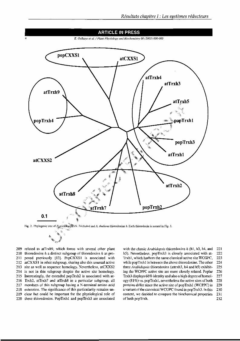
Résultats chapitre l : Les systèmes réducteurs
ARTICLE IN PRESS4
atCXXS2
0.1
E. Gelhaye et al. / Plant Physiology and Biochemistry 00 (2003) 000-000
frir/'J,Iîi:Ulmif":;it"V Trichobel and A. thaliana thioredoxins h. Each thioredoxin is named in Fig. 1.
209 related to atTrxh9, which forms with several other plant210 thioredoxins h a distinct subgroup of thioredoxins h as pro211 posed previously [13]. PopCXXS1 is associated with212 atCXXS1 in other subgroup, sharing also this unusual active213 site as weH as sequence homology. Nevertheless, atCXXS2214 is not in this subgroup despite the active site homology.215 Interestingly, the extended popTrxh2 is associated with at216 Trxh2, atTrxh7 and atTrxh8 in a particular subgroup, aH217 members of this subgroup having aN-terminal amino acid218 extension. The significance of this particularity remains UD
219 clear but could be important for the physiological role of220 these thioredoxins. PopTrxh1 and popTrxh3 are associated
with the classic Arabidopsis thioredoxins h (hl, h3, h4, and 221h5). Nevertheless, popTrxh3 is closely associated with at- 222Trxh1, which harbors the same classical active site WCGPC, 223while popTrxh1 is between the above thioredoxins. The other 224three Arabidopsis thioredoxins (attrxh3, h4 and h5) exhibit- 225ing the WCPPC active site are more closely related. Poplar 226Trxh3 displays 66% identity and also a high degree of homo1- 227ogy (81 %) vs. popTrxh1, nevertheless the active sites of both 228proteins differ since the active site of popTrxh1 (WCPPC) is 229a variant of the canonical WCGPC found in popTrxh3. In this 230context, we decided to compare the biochemical properties 231of both popTrxh. 232
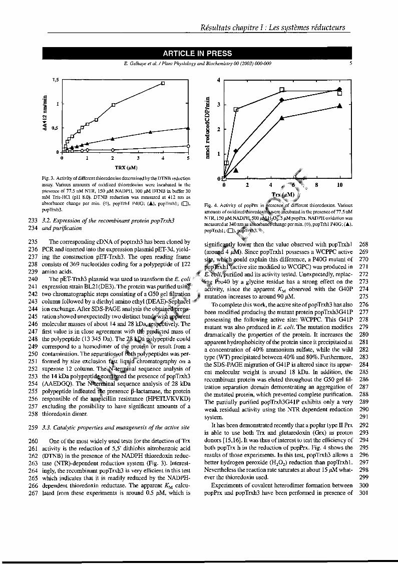
Résultats chapitre l : Les systèmes réducteurs
ARTICLE IN PRESSE. Gelhaye et al. / Plant Physiology and Biochemistry 00 (2003) 000-000 5
268269270271272273274275276277278279280281282283284285286287288289290291292293294295296297298299300301
8 10
thioredoxins. Various
cubated in the presence of 77.5 nM2 2~ 5 pM popPrx. NADPH oxidation was
t8change per min. (0), popTrh1 P40G; (Â),
2
2
3
1
4----r--~--~-~r__-__,___,
Fig. 4. Activity of popPrx in
amounts of oxidized thiorNTR,150pMNADPH;.
measured at 340 ~i'popTrxh1; (O)';i~pr
loWêlJ' then the value observed with popTrxh1(ar~?II). Since popTrxhl possesses a WCPPC activesi'_~ ~1I;~è.,ould explain this difference, a P40G mutant of
'f . factive site modified to WCGPC) was produced in• lJlILj;i urified and its activity tested. Unexpectedly, replac
P~o40 by a glycine residue has a strong effect on thesince the apparent KM observed with the G40P
mutation increases to around 90 JLM.To complete this work, the active site ofpopTrxh3 has also
been modified producing the mutant protein popTrxh3G41Ppossessing the following active site: WCPPC. This G41Pmutant was also produced in E. coli. The mutation modifiesdramatically the properties of the protein. It increases theapparent hydrophobicity of the protein since it precipitated ata concentration of 40% ammonium sulfate, while the wildtype (WT) precipitated between 40% and 80%. Furthermore,the SDS-PAGE migration of G41P is altered since its apparent molecular weight is around 18 kDa. In addition, therecombinant protein was eluted throughout the G50 gel filtration separation domain demonstrating an aggregation ofthe mutated protein, which prevented complete purification.The partially purified popTrxh3G41P exhibits only a veryweak residual activity using the NTR dependent reductionsystem.
It has been demonstrated recently that a poplar type il Prxis able to use both Trx and glutaredoxin (Grx) as protondonors [15,16]. It was thus ofinterest to test the efficiency ofboth popTrx h in the reduction of popPrx. Fig. 4 shows theresults of those experiments. In this test, popTrxh3 allows abetter hydrogen peroxide (H202) reduction than popTrxhl.Nevertheless the reaction rate saturates at about 15 JLM whatever the thioredoxin used.
Experiments of covalent heterodimer formation betweenpopPrx and popTrxh3 have been performed in presence of
1:1
11~
~Qu'etG)
!:i'ClIG)...
.....
1 2 3 4 5 ~TRX(/LM)
259 3.3. Catalytic properties and mutagenesis of the active site
1
260 One of the most widely used tests for the detection ofTrx261 activity is the reduction of 5,5' dithiobis nitrobenzoic acid262 (DTNB) in the presence of the NADPH thioredoxin reduc263 tase (NTR)-dependent reduction system (Fig. 3). Interest264 ingly, the recombinant popTrxh3 is very efficient in this test265 which indicates that it is readily reduced by the NADPH266 dependent thioredoxin reductase. The apparent KM calcu267 lated from these experiments is around 0.5 JLM, which is
~
:c<J 0,5
1,5 ---------------:------,
Fig. 3. Activity of different thioredoxins detennined by the DTNB reduction
assay. Various amounts of oxidized thioredoxins were incubated in thepresence of 77.5 nM NTR, 150 #lM NADPH, 100 #LM DTNB in buffer 30mM Tris-Hel (pH 8.0). DTNB reduction was measured at 412 nID asabsorbance change per min. (0), popTrh1 P40G; (Â), popTrxh1; (0),popTrxh3.
233 3.2. Expression of the recombinant protein popTrxh3234 and purification
235 The corresponding cDNA of poptrxh3 has been cloned by236 PCR and inserted into the expression plasmid pET-3d, yield237 ing the construction pET-Trxh3. The open reading frame238 consists of 369 nucleotides coding for a polypeptide of 122239 amino acids.240 The pET-Trxh3 plasmid was used to transform the E.241 expression strain BL21(DE3). The protein was purified u242 two chromatographie steps consisting of a G50 gel ~IVa243 column followed by a diethyl amino ethyl (DEAE) p~
244 ion exchange. After SDS-PAGE analysis the ob .245 ration showed unexpectedly two distinct ban246 molecular masses of about 14 and 28 kD247 first value is in close agreement with t"248 the polypeptide (13 345 Da). The~~ypeptide could249 correspond to a homodimer of t~l prot,~n or result from a250 contamination. The separati~.'8~'k?~~lypeptideswas per-251 formed by size exclusion f: .q~~d' 'chromatography on a252 superose 12 column. Th :nal sequence analysis of253 the 14 kDa polype~~~'. ed the presence of popTrxh3254 (AAEDGQ). The N~~::~al sequence analysis of 28 kDa255 polypeptide indî~~~~d ~;;F presence ~-lactamase, the protein256 responsible of thé:;;i_~lcillin resistance (HPETLVKVKD)257 excluding the possibility to have significant amounts of a258 thioredoxin dimer.
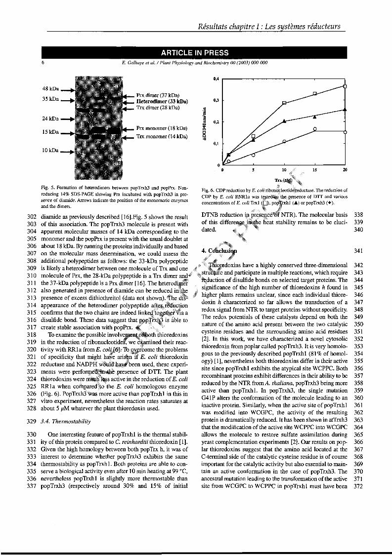
Résultats chapitre l : Les systèmes réducteurs
ARTICLE IN PRESS6 E. Gelhaye et al. / Plant Physiology and Biochemistry 00 (2003) 000-000
329 3.4. Thermostability
338339340
341
342343344345346347348349350351352353354355356357358359360361362363364365366367368369370371372
20IS
)rejrenc~e;t)l NTR). The molecular basisstability remains to be eluci-
5
0,2
0,4 r------,...--------,r-----...,.------w
0,1
0,3
DTNB reduction .of this C1lttere]lw~\
dated.
Fig. 6. CDP reduction by E. coli ribonl~lc~otildeileductasie.The reduction ofCDP by E. coli RNRla was of DTT and variousconcentrations of E. coli Trx1 (..) or popTrxh3 (.).
~~~,doxinshave a highly conserved three-dimensional, ,;~~ru{J'me and participate in multiple reactions, which require
lt;puction of disulfide bonds on selected target proteins. Thesignificance of the high number of thioredoxins h found inhigher plants remains unclear, since each individual thioredoxin h characterized so far allows the transduction of aredox signal from NTR to target proteins without specificity.The redox potentials of these catalysts depend on both thenature of the amino acid present between the two catalyticcysteine residues and the surrounding amino acid residues[2]. In this work, we have characterized a novel cytosolicthioredoxin from poplar called popTrxh3. It is very homologous to the previously described popTrxh1 (81 % of homology) [1], nevertheless both thioredoxins differ in their activesite since popTrxh1 exhibits the atypical site WCPPC. Bothrecombinant proteins exhibit differences in their ability to bereduced by the NTR from A. thaliana, popTrxh3 being moreactive than popTrxhl. In popTrxh3, the single mutationG41P alters the conformation of the molecule leading to aninactive proteine Similarly, when the active site of popTrxh1was modified into WCGPC, the activity of the resultingprotein is dramatically reduced. It has been shown in atTrxh3that the modification of the active site WCPPC into WCGPCallows the molecule to restore sulfate assimilation duringyeast complementation experiments [2]. Our results on poplar thioredoxins suggest that the amino acid located at theC-terminal side of the catalytic cysteine residue is of courseimportant for the catalytic activity but also essential to maintain an active conformation in the case of popTrxh3. Theancestral mutation leading to the transformation of the activesite from WCGPC to WCPPC in popTrxh1 must have been
PrxmonOlner(18.kDa)
Trx .m.onoIller (14kDa)
Pa dimer(37 kDa)Helerodimer-(33kDa)Tex dimer (28 kDa)
481<Da
35kDa
lOkOa
24kDa
15kDa
Fig. 5. Formation of heterodimers between popTrxh3 and popPrx. Nonreducing 14% SDS-PAGE showing Prx incubated with popTrxh3 in presence of diamide. Arrows indicate the position of the monomeric enzymesand the dimers.
diamide as previously described [16].Fig. 5 shows the resultof this association. The popTrxh3 molecule is present withapparent molecular masses of 14 kDa corresponding to themonomer and the popPrx is present with the usual doublet atabout 18 kDa. By running the proteins individually and basedon the molecular mass determination, we could assess theadditional polypeptides as follows: the 33-kDa polypeptideis likely a heterodimer between one molecule of Trx and onemolecule of Prx, the 28-kDa polypeptide is a Trx dimer andthe 37-kDa polypeptide is a Prx dimer [16]. The heterodiBfE~l}
also generated in presence of diamide can be reduce2iri'ltepresence of excess dithiothreitol (data not shown) , .h~appearance of the heterodimer polypeptide a(tt?itr "~i,hconfirms that the two chains are indeed linke" ',,; r ·-la adisulfide bond. These data suggest that is~ble tocreate stable association with popPrx.
To examine the possible involv~lJlell
in the reduction of ribonucleotideÎ:: we adiined their reactivity with RRla from E. col~~ome the problemsof specificity that might~aie ari,n' if E. coli thioredoxinreductase and NADP~~() .'been used, these experi-ments were perfo~e<l:;;!ll)\ ,.,.... .' presence of DTT. The plantthioredoxins w~.~~m ';~i.~ active in the reduction of E. coliRR1a when com e",o 'the E. coli homologous enzyme(Fig. 6). PopTrxh3aSlimore active than popTrxh1 in this invitro experiment, nevertheless the reaction rates saturates atabout 5 JLM whatever the plant thioredoxin used.
302303304305306307308309310311312313314315316317318319320321322323324325326327328
330 One interesting feature of popTrxh1 is the thermal stabil331 ity ofthis protein compared to C. reinhardtii thioredoxin [1].332 Given the high homology between both popTrx h, it was of333 interest to determine whether popTrxh3 exhibits the same334 thermostability as popTrxhl. Both proteins are able to con335 serve a biological activity even after 10 min heating at 99 oC,336 nevertheless popTrxh1 is slightly more thermostable than337 popTrxh3 (respectively around 30% and 15% of initial
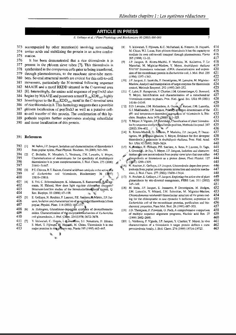
Résultats chapitre l : Les systèmes réducteurs
ARTICLE IN PRESSE. Gelhaye et al. / Plant Physiology and Biochemistry 00 (2003) 000-000 7
391 References
373 accompanied by other mutation(s) involving surrounding374 amino acids and stabilizing the protein in an active confor375 mation.376 It has been demonstrated that a rice thioredoxin h is377 present in the phloem sieve tubes [7]. This thioredoxin is378 synthesized in the companion cells prior to being transferred,379 through plasmodesmata, to the enucleate sieve-tube mem380 bers. Several structural motifs are critical for this cell-to-cell381 movement, particularly the N-terminal following sequence382 MAAEE and a motif RKDD situated in the C-terminal area383 [8]. Interestingly, the amino acid sequence of popTrxh3 also384 begins by MAAEE and possesses a motifRI04KDEI07 highly385 homologous to the RIOIKDDI04 motif in the C-terminal area386 of rice thioredoxin h. This homology suggests then a possible387 phloem localization of popTrxh3 as well as a putative cell388 to-cell transfer of this proteine The confirmation of this hy389 pothesis requires further experiments studying subcellular390 and tissue localization of this proteine
392 [1]393394 [2]395396397398 [3]399400401 [4]402403404405 [5]406407408 [6]409410411 [7]412413
M. Behm, J.P. Jacquot, Isolationand characterization ofthioredoxin hfrom poplar xylem, Plant Physiol. Biochem. 39 (2000) 363-369.
C. Brehelin, N. Mouaheb, L. Verdoucq, J.M. Lancelin, Y. Meyer,Characterization of determinants for the specificity of Arabidopsisthioredoxins h in yeast complementation, J. Biol. Chem. 275 (2000)31641-31647.
P.T. Chivers, R.T. Raines, General acidlbase catalysis in the activ~;,ite
of Escherichia coli thioredoxin, Biochemistry 36 (11~7)15810-15816. . '
S. Dai, C. Schwendtmayer, K. Johansson, S. Ramaswmann, H. Eklund, How does light regulateStructure-function studies of the len·ed()xinl!thi.o~1
Rev. Biophys. 33 (2000) 67-108.
E. Gelhaye, N. Rouhier, P. Laurent, P.E. S~\1(tier'~;:lfüM,artin,
quot, Isolation and characterization of aJl,~x;t~~l\lj~dthioredloxtn hlrOlmpoplar, Physiol. Plant. 114 (2002)
A. Holmgren, Glutathione-d~~i~..} of deoxyribonucle-otides. Characterization of t~r'e~i" echanism of Escherichiacoli glutaredoxin, J. Biol~;fH' 4 ''1978) 3672-3678.
Y. Ishiwatari, C. I;txw
a, S.I. Nakamura, H. Hirano,S. Mori, T.MglU, M. Chino, Thioredoxin h is onemajor proteins in sap, Planta 195 (1995) 463-465.
[8] Y. Ishiwatari, T. Fijiwara, K.C. McFarland, K. Nemoto, H. Hayashi, 414M. Chino, W.J. Lucas, Rice phloem thioredoxin h has the capaeity to 415mediate its own cell-to-cell transport through plasmodesma, Planta 416205 (1998) 12-22. 417
[9] J.P. Jacquot, R. Rivera-Madrid, P. Marinho, M. Kollarova, P. Le 418Marechal, M. Miginiac-Maslow, Y. Meyer, Arabidopsis thaliana 419NAPHP thioredoxin reductase. cDNA characterization and expres- 420sion of the recombinant protein in Escherichia coli, J. Mol. Biol. 235 421(1994) 1357-1363. 422
[10] J.P. Jacquot, E. Issakidis, P. Decottignies, M. Lemaire, M. Miginiac- 423Maslow, Analysis and manipulation of target enzymes for thioredoxin 424control, Methods Enzymol. 252 (1995) 240-252. 425
[11] C. Laloi, N. Rayapuram, Y. Chartier, J.M. Grienenberger, G. Bonnard, 426Y. Meyer, Identification and characterization of a mitochondrial 427thioredoxin system in plants, Proc. Natl. Sei. USA 98 (2001) 42814144-14149. 429
[12] S.D. Lemaire, J.M. Richardson, A. G,tll:iKeryer, J.M. Lancelin, 430G.I. Makhatadze, J.P. Jacquot, Pi: \~Wre determinants of the 431pH- and temperature-dependen~l~g onY'of thioredoxin h, Bio- 432chim. Biophys. Acta 1476.,'. ""B. 433
[13] Y Meyer, F. Vignols, J.p.léic ';' assification of plant thioredox- 434ins by sequence simi! ' inm~n position, Methods Enzymol. 347 435(2002) 394-402. 436
[14] R. Rivera-Madri 437tignies, Mi'.;'i:/ ~~aslow, Y. Meyer, Evidence for five divergent 438thioredoxiUi;i,quen~~s in Arabidopsis thaliana, Proc. Natl. Acad. 439Sci.. ~~A 9t'~'~~5) 5620-5624. 440
[15] ~;;;Riotlli.tr, E. Gelhaye, P.E. Sautiere, A. Brun, P. Laurent, D. Tagu, 441<. Jti;~erard;~p. de Fay, Y. Meyer, J.P. Jacquot, Isolation and character- 442
izàtilP ':l,a: new peroxiredoxin from poplar sieve tubes that uses either 443gïutfê(î~xin or thioredoxin as a proton donor, Plant Physiol. 127 444
;;i;t~i~l) 1299-1309. 445'''Pi: Rouhier, E. Gelhaye, J.P. Jacquot, Glutaredoxin-dependent perox- 446
iredoxin from poplar: protein-protein interaction and catalytic mecha- 447nism, J. Biol. Chem. 277 (2002) 13609-13614. 448
[17] N. Rouhier, E. Gelhaye, J.P. Jacquot, Exploring the active site of plant 449glutaredoxin by site-directed mutagenesis, FEBS Lett. 511 (2002) 450145-149. 451
[18] M. Stein, J.P. Jacquot, E. Jeannette, P. Decottignies, M. Hodges, 452J.M. Lancelin, V. Mittard, J.M. Schmitter, M. Miginiac-Maslow, 453Chlamydomonas reinhardtii thioredoxins: structure of the genes cod- 454ing for the chloroplastic m and cytosolic h isoforms; expression in 455Escherichia coli of the recombinant proteins, purification and bio- 456chemical properties, Plant Mol. Biol. 28 (1995) 487-503. 457
[19] J.D. Thompson, F. Plewniak, O. Poch, A comprehensive comparison 458of multiple sequence alignment programs, Nucleic acid Res. 27 459(1999) 2682-2690. 460
[20] L. Verdoucq, F. Vignols, J.P. Jacquot, Y. Chartier, Y. Meyer, In vivo 461characterization of a thioredoxin h target protein defines a snew 462peroxiredoxin family, J. Biol. Chem. 274 (1999) 19714-19722. 463
Résultats chapitre I : Les systèmes réducteurs
Cet article décrit l’isolement d’une Grx bicystéinique de 139 acides aminés
homologue à la Grx CxxC4 d'A. thaliana (Tableau I et Figure 9) ainsi que les
modifications de la séquence nucléotidique effectuées pour optimiser l’expression de
la protéine recombinante chez E. coli. En effet, la séquence initialement reconstituée
à partir de séquence EST était tronquée en C-terminal de 14 acides aminés. De plus,
cette séquence code pour une protéine qui possède une extension N-terminale
d’environ 27 acides aminés par rapport aux séquences fonctionnelles de Grx
préalablement caractérisées. Quelles que soient les modifications envisagées, la
protéine de 139 acides aminés n’a jamais pu être exprimée chez E. coli. Toutefois,
ce travail nous a permis de réaliser l’importance des codons initiaux pour l’efficacité
de la traduction d’une séquence chez E. coli. Ainsi, l’existence de ce biais de codon
chez E. coli c’est à dire que certains codons sont peu utilisés du fait de la faible
représentativité des ARNt correspondant, est à prendre en compte lorsqu’on veut
exprimer une séquence chez E. coli. En effet, en fonction de leur position dans la
séquence codante, deux des codons codant pour l’arginine, AGG et AGA, sont
réellement défavorables à une expression efficace de la protéine recombinante chez
E. coli. Ce problème peut être contourné en utilisant le plasmide pSBET qui code
pour l’ARNt reconnaissant ces deux codons AGG et AGA. Enfin, l’activité de cette
Grx a été démontrée envers un substrat artificiel le HED (hydroxyethyl disulfide) et le
DHA (déhydroascorbate).
Article numéro 7 :
Rouhier N, Gelhaye E, Sautière PE, Jacquot JP. (2002) Enhancement of poplar glutaredoxin expression by optimization of the cDN
sequence.
Protein Expr Purif. 24(2): 234-41.
Résultats chapitre l : Les systèmes réducteurs
Protein Expression and Purification 24, 234-241 (2002)
doï: 1O.1006/prep.2001.1574, available online at http://www.ïdealibrary.com on 1DE'tl®
Enhancement of Poplar Glutaredoxin Expression byOptimization of the cDNA Sequence
Nicolas Rouhier,* Eric Gelhaye,* Pierre-Eric Sautière,t and Jean-Pierre Jacquot*,1*Unité Mixte de Recherche 1136IaM (Interaction Arbres Microorganismes), INRA-UHP Nancy 1. Université HenriPoincaré, 54506 Vandoeuvre Cedex. France; tLaboratoire d'Endocrinologie des Annélides UPRESA 8017,Université des Sciences et Technologies de Lille, 59655 Villeneuve d'Ascq Cedex, France
Received July 26,2001, and in revised form October 26, 2001
Glutaredoxins are low-molecular-weight oxidoreductases that play an important role in redox regulation in eukaryotic and prokaryotic cells. Because oftheir low abundance, these proteins are poorly characterized in plants. Furthermore, very poor yields havebeen obtained with the expression systems preparedso far, and in addition, the recombinant products contain a His-tag which can interfere with the biochemicalcharacterization. In order to obtain more informationabout those important regulatory proteins in plants, acDNA coding for an extended glutaredoxin has beenintroduced into the expression plasmid pET-3d and theresulting construction has been used to transformEscherichia coli strain BL21 (DE3) in the presence ofplasmid helper pSBET or note Initially poor or ineffective protein expression has been improved by successively cloning aN-terminus truncated form of theprotein, introducing silent mutations both at the 5' andat the 3' ends of the nucleotide sequence, and finallyaltering the 3' end in order to change the C-terminusamino acid sequence of the proteine The first modifications have allowed us ta produce the protein in largeamounts but essentially in an insoluble form whichcould be resolubilized and purified. On the other hand,changing the C-terminus sequence resulted in proteinpreparations of high purity and in a soluble forme Therecombinant proteins were biochemically active andthe yield varied between 6 and 14 mg of homogeneousprotein per liter of culture. © 2002 Elsevier Science (USA)
1 To whom correspondence should be addressed. E-mai1:[email protected].
234
In the past years, the study of redox reactions involving the reduction of disulfide bridges has received evermore attention. It appears that maintaining a reducedintracellular state is essential to fight against an oxidizing extracellular environment and reactive oxygen species which are the product of normal aerobic metabolism. Indeed, the oxidation of proteins can lead to theirinactivation and to cellular dysfunction (1). The thioldisulfide oxidoreductases, which regroups proteinssuch as thioredoxins (Trx),2 glutaredoxins (Grx) (originally named thioltransferase or transhydrogenase),protein disulfide isomerases (PDI) , and nucleoredoxinsare present in aIl living organisms and are necessaryfor the maintaining of a proper redox state (2 -4). WhileTrx and Grx are effective reductants, PDI is clearly anoxidant whose function is to create disulfide bridges.AlI the above-mentioned proteins possess a conservedactive site C-X-X-C, required for their activities and asimilar basic structural fold called the thioredoxin fold.
Glutaredoxin can be differentiated from the otherabove-mentioned catalysts by the amino acid sequenceof its active site [CP(Y/F) C) that is highly conservedamong species. In Escherichia coli, glutaredoxins varyin size between 82 and 215 amino acids, but in mostother species, the medium size is of ca. 100 amino acidswith a molecular weight of around Il kDa (5). Grx isselectively reduced via NADPH, through a flavoproteinglutathione reductase and reduced glutathione (6). Inprokaryotic and animal cells, Grx acts as a reductantfor many enzymes including ribonucleotide reductase
2 Abbreviations used: DHA, dehydroascorbate; Grx, glutaredoxin;HED, hydroxyethyl disulfide; Trx, thioredoxins; PDI, protein disulfideisomerases; LB, Luria-Bertani; DTT, dithiothreitol; EST, expressedsequence tag.
1046-5928/02 $35.00© 2002 Elsevier Science (USA)
AlI rights reserved.
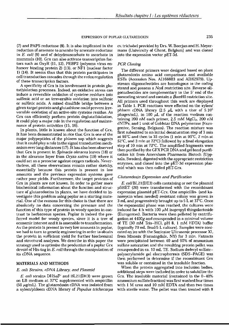
Résultats chapitre l : Les systèmes réducteurs
EXPRESSION OF POPLAR GLUTAREDOXIN 235
(7) and PAPS reductase (8). It is also implicated in thereduction of arsenate to arsenite by arsenate reductasein E. coli (9) and of dehydroascorbate to ascorbate inmammals (10). Grx can also activate transcription factors such as OxyR (11, 12), PEBP2 (polyoma virus enhancer binding protein 2) (13), or NFI (nuclear factor1) (14). It seems thus that this protein participates incell transduction cascades through the redox regulationof these transcription factors.
A specificity of Grx is its involvement in protein glutathiolation processes. Indeed, an oxidative stress caninduce a reversible oxidation of cysteine residues intosulfenic acid or an irreversible oxidation into sulfonicor sulfinic acids. A mixed disulfide bridge between agiven target protein and glutathione could prevent irreversible oxidation of an active-site cysteine residue. AsGrx can efficiently perform protein deglutathiolation,it could play a major role in the regulation and maintenance of protein activities (15, 16).
In plants, little is known about the function of Grx.It has been demonstrated in rice that Grx is one of themajor polypeptides of the phloem sap which suggeststhat it could play a role in the signal transduction mechanism over long distances (17). It has also been observedthat Grx is present in Spinacia oleracea leaves (18) orin the aleurone layer from Oryza sativa (19) where itcould act as a protector against oxygen radicals. Nevertheless, aIl these observations remain rather sketchy,essentially because this protein is present in lowamounts and the previous expression systems gaverather poor yields. Furthermore, the target proteins ofGrx in plants are not known. In order to gather morebiochemical information about the function and structure of glutaredoxins in plants, we have decided to investigate this problem using poplar as a starting material. One of the reasons for this choice is that there areabsolutely no data concerning the presence and thefunction of this type of protein in woody species in contrast to herbaceous species. Poplar is indeed the preferred model for woody species, since it is a tree ofeconomic interest and it is amenable to transformation.As the protein is present in very low amounts in poplar,we had to turn to genetie engineering in order to obtainthe protein in sufficient yield for further biochemicaland structural analyses. We describe in this paper thestrategy used to optimize the production of a poplar Grxdevoid of His-tag in E. colithrough the manipulation ofits cDNA sequence.
MATERIALS AND METHOOS
E. coli Strains, cDNA Library, and Plasmid
E. coli strains DH5aF' and BL21 (DE3) were grownon LB medium at 37QC supplemented with ampicillin(50 ,ag/mL). The glutaredoxin cDNA was isolated froma xylem/phloem cDNA library of Populus trichocarpa
cv. trichobel provided by Drs. W. Boerjan and H. Meyermans (University of Ghent, Belgium) and was clonedinto the expression vector pET-3d.
PCR Cloning
The different primers were designed based on plantglutaredoxin amino acid comparisons and availableESTs (Accession Nos. AI166603 and AII62979). Upstream oligonucleotides are homologous to the codingstrand and possess a NcoI restriction site. Reverse oligonucleotides are complementary to the 5' end of thenoncoding strand and contain a BamHI restriction site.AlI primers used throughout this work are displayedin Table 1. PCR reactions were effected on the xylemlphloem cDNA library (2.5 ,uL with a titer of 5.105
phages/mL), in 100 ,aL of the reaction medium containing 200 nM each primer, 2.5 mM MgC12, 200 nMdNTPs, and 1 unit of Goldstar DNA polymerase (Eurogentec, Seraing, Belgium). The reaction mixture wasfirst submitted to an initial denaturation step of 3 minat 95°C and then to 35 cycles (1 min at 95°C, 2 min at52°C, and 3 min at 72°C) followed by a final elongationstep of 10 min at 72°C. The amplified fragments werethen purified by the GFX PCR DNA and gel band purification kit from Amersham Pharmacia Biotech (Uppsala, Sweden), digested with the appropriate restrictionenzymes, and cloned into the pET-3d expression plasmid which was then called pET-Grx.
Clutaredoxin Expression and Purification
E. coli BL21 (DE3) cells containing or not the plasmidpSBET (20) were transformed with the recombinantexpression plasmid pET-Grx. One ampicillin- (and kanamycin when needed) resistant colony was grown in3 mL and progressively brought up to 5 L at 37°C. Oncethe exponential phase was reached, the cultures wereinduced for 4 h with 100 ,aM isopropyl thiogalactoside(Eurogentec). Bacteria were then pelleted by centrifugation at 4400g and resuspended in a minimal volumeof TE (30 mM Tris-HCI, pH 8, 1 mM EDTA) buffer(typically 70 mL final/5 L culture). Samples were sonicated on ice with the Sonicator Ultrasonic processor XLfrom Misonix (Farmingdale, NY) for 3 min. Extractswere precipitated between 40 and 90% of ammoniumsulfate saturation and the resulting protein pellet wasresuspended in ca. 10 mL TE. Sodium dodecyl sulfatepolyacrylamide gel electrophoresis (SDS-PAGE) wasthen performed to determine if the recombinant Grxwas soluble or contained in the insoluble fraction.
When the protein aggregated into inclusion bodies,additional steps were included in order to solubilize theGrx. The insoluble material (contained in the 0-40%ammonium sulfate fraction) was first washed four timeswith 1 M urea and 10 mM EDTA and then two timeswith sterile water. The pellet was then treated with 8
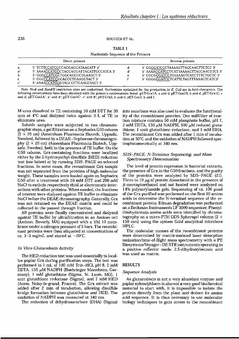
236
Résultats chapitre l : Les systèmes réducteurs
ROUHIER ET AL.
TABLE 1
Nucleotide Sequence of the Primers
aa'bcc'
Direct primers
5' TCTTCCATGGCGACGAGGATAAGATT3'5'AAAACCATGGCTACCAGGATTAGATTGCCATCA3'5'GGGCCATGGCTGGCAGCCCTGAAGCT3'5'GGGCCATGGGCAGCCTGAAGCTACT3'5'AAAACCATGGGTAGCCCTGAAGCTACT3'
dd'ef
Reverse primers
5'GGGGATCCCTAAAAGTTAGCAAGTTCTCC3'5'AAAAGGATCCTCATTAAAAGTTAGCAAGTTCT3'5'GGGGGGATCCTTAAAAGTCATCTTTCTGCTC3'5' GGGGGGATCCTCATTCTAGTTTAAAGTCATC3'
Note. NcoI and BamHI restriction sites are underlined. Nucleotides optimized for the production in E. Col} are in bold characters. Thefollowing constructions have been obtained with the primer's combinations listed: pET-GrxlA, a and d; pET-GrxlB, band d; pET-GrxlC, cand d; pET-GrxlA', a' and d'; pET-GrxlC', c' and d'; pET-Grx2, band e; pET-Grx3, band f.
M urea dissolved in TE containing 10 mM DTT for 30min at 4°C and dialyzed twice against 3 L of TE toeliminate urea.
Soluble sampIes were subjected to two chromatographic steps, a gel filtration on a Sephadex GSO column(S X 70 cm) (Amersham Pharmacia Biotech, Uppsala,Sweden), followed by a DEAE-Sepharose chromatography (2 X 20 cm) (Amersham Pharmacia Biotech, Uppsala, Sweden), both in the presence of TE buffer. On theGSO column, Grx-containing fractions were localizedeither by the 2-hydroxyethyl disulfide (HED) reductiontest (see below) or by running SDS-PAGE on selectedfractions. In sorne cases, the recombinant Grx samplewas not separated from the proteins of high molecularweight. These samples were loaded again on SephadexGSO after a treatment with 10 mM DTT and 200 mMNaCI to exclude respectively thiol or electrostatic interactions with other proteins. When needed, the fractionsof interest were dialyzed against TE buffer to eliminateNaCI before the DEAE chromatography. Generally, Grxwas not retained on the DEAE matrix and could becollected in the passed through fraction.
AlI proteins were finally concentrated and dialyzedagainst TE buffer by ultrafiltration in an Amicon cell(Amicon, Beverly, MA) equipped with a YM 10 membrane under a nitrogen pressure of S bars. The recombinant proteins were then aliquoted at concentrations ofca. 3-S mg/mL and stored at -20°C.
In Vitro Glutaredoxin Activity
The HED reduction test was used essentially to localize poplar Grx during purification steps. The test wasperformed in 1 mL of 100 mM Tris-HCI, pH 8,2 mMEDTA, ISO ,aM NADPH (Boehringer Mannheim, Germany) , 1 mM glutathione (Sigma, St. Louis, MO), 1unit glutathione reductase (Sigma), and 7 mM HED(Acros, Noisy-le-grand, France). The Grx extract wasadded after 2 min of incubation, allowing disulfidebridge formation between glutathione and HED. Theoxidation of NADPH was measured at 340 nm.
The reduction of dehydroascorbate (DHA) (Sigma)
into ascorbate was also used to evaluate the functionality of the recombinant proteins. One milliliter of reaction mixture contains SO mM phosphate buffer, pH 7,2 mM EDTA, lS0,aM NADPH, 500,aM reduced glutathione, 1 unit glutathione reductase, and 1 mM DHA.The recombinant Grx was added after 1 min of incubation at 30°C and the oxidation of NADPH followed spectrophotometrically at 340 nm.
SDS-PAGE, N-Terminus Sequencing, and MassSpectrometry Determination
The level of protein expression in bacterial extracts,the presence of Grx in the G50 fractions, and the purityof the proteins were analyzed by SDS-PAGE (21).Three to 10 ,ag of protein dissociated in the presence off3-mercaptoethanol and not heated were analyzed on14% polyacrylamide gels. Sequencing of ca. 100 pmolof the Grx purified was performed on the first 10 aminoacids to determine the N-terminal sequence of the recombinant proteine Edman degradation was performedon a Beckman Instruments LF 3000 sequencer. Phenylthiohydantoin-amino acids were identified by chromatography on a micro-PTH ODS Spherogel column (2 XISO mm) using the system Gold analytical microboreHPLC.
The molecular masses of the recombinant proteinswere determined by matrix-assisted laser desorptionionization/time-of-flight mass spectrometry with a PEBiosystemsNoyager-DE STR instruments operating ina positive reflector mode. 2.5-dihydroxybenzoic acidwas used as matrix.
RESULTS
Sequence Analysis
As glutaredoxin is not a very abundant enzyme andpoplar xylem/phloem is also not a very good biochemicalmaterial to start with, it is impossible to isolate theprotein directly from the plant and deduce its aminoacid sequence. It is thus necessary to use molecularbiology techniques to gain access to the recombinant
Résultats chapitre l : Les systèmes réducteurs
EXPRESSION OF POPLAR GLUTAREDOXIN 237
proteine A putative glutaredoxin sequence (Grx1) wasreconstituted by overlapping two poplar ESTs with respective accession numbers AI166603 and AI162979and cloned by PCR into the pET-3d plasmide Overall,the cDNA coding sequence is 378 bp long, resulting ina protein of 125 amino acids, including the initiatormethionine. The deduced amino acid sequence possesses the conserved active site YCPYC, characteristicof the glutaredoxins (Fig, 1). This figure also shows twoalternative C-termini ends assuming that there is anerror in the sequence of the EST, resulting in aframeshift (denominated later in this paper Grx2 andGrx3). The theoretical molecular weight, pl, and molarextinction coefficient of Grx1 have been determined bythe program Prot param tool from Expasy (http://www.expasy.org/tools/protparam.html) and are respectively 13,649 Da, 6.1, and 9650 M cm- l at 280 nm.An interesting feature of this sequence is that whencompared to most available plant Grx sequences, it displays an N-terminus extension of ca. 25 amino acids.The interrogation of the PSORT prediction site (http://psort.nibb.acJp/form.html) suggests that the proteincould have a N-terminus transit peptide targeting forexcretion outside the celL The cleavage site is predictedto be between alanine 27 and 28.
ATGGCGACijAGGATAAGATIGCCATCAA1CTT'GGCTACAGCAGTAA,CAlTAACAGTACTTMA T R IRl PS 1 t AT ,4 VT l T V l
[il , ftGCGGCATCACTCACCTGGGCTGCTGGCA6CCCTGAAGCTACTTTTGTC.AAAAAGACCATCA A S L T W A A G S P E A T f V K l T l
mJœ .....TCTTCTCATCAGATCGTCATCTTCTCWGTC AAGAAGGCTAAAS 5 If QI V' IF S K S KK AK
GGTGT1TICAAA'AACfGAACCAGACACCACATGTTGTCGAGCTCGATCAM;GAGAG<iATtlV'f 1< EL N QT P H V V EL DQ RE {)
GGGCACAACATTCAGGATGCCATGAGTGAAATTGTTGGGAGGCGCACCGTGCCTCAG,GTT~ INI QD A M S E IV , ft R T VP Q v
ITCATAAACG'GAAGCACAJT(jGTG'CTCAG.ATGACACCGT'(jAAGCATACGAAAGT'~FIN G KHI G , S DDT V E A Y E 5 ~
GAACTTGCTAACII··I···,AGE l ·ANF ...•~.... m
GAACTTGCTAAGCTTTTAGGAGTTGCITCAGAGCAGAAAGATGACTTTTAAELA K l L , V A S E Q K 0 D F •
4 iiiGAACTIGCTAAGCTnTAGGAGTIGCTTCAGAGCAGAAAGATGACTfTAAACTAGAArGAEL A K l l G V A S E Q K D DF K LE·
..lI)
FIG. 1. Nucleotide and deduced amino acid sequences of poplarglutaredoxin. The three aIternate C-termini of Grxl, 2, and 3 areindicated as weIl as the position of the primers used for PCR cloning.The active site is shade boxed.
As a consequence, we have decided to clone differentconstructions into pET-3d, a long sequence namedpET-Grx1A (MATRIRL) and two truncated sequenceswithout the putative transit peptide called respectivelypET-Grx1B (MAGSPEATFVKK) and pET-Grx1C(MGSPEATFVKK) (Fig. 1). In brackets is indicated theN-terminus sequence resulting from these constructions. The details of the sequences and combination ofthe cloning oligonucleotides are given in Table 1. Thesequences in the pET-Grx1B and pET-Grx1C constructions programmed respectively for proteins of 99 and98 amino acids. The theoretical molecular weights, pLand molar extinction coefficient of Grx1B and 1C arerespectively 10,957 or 10,886 Da, 6.1, and 3960 M cm- l
at 280 nm. The rationale behind this choice is the following: it is clear from the literature that the precursorsequences when overexpressed in E. coli are eithercleaved at a site close to the putative cleavage site orvery frequently unstable and degraded (22, 23). On theother hand, when the signal peptide is eliminated, ingeneral the protein is expressed with a high yield in asoluble forme In our case, whatever the constructionused, no glutaredoxin overexpression was observed inE. coli strain BL21(DE3) either using SDS-PAGE orwhen testing in the HED reduction (data not shown).
Cotransformation with pSBET
One possibility to explain this absence of productionis the existence of rare codons in the sequence that arenot adapted to the E. coli translation machinery. Inparticular, the AGG and AGA codons triplets for arginine are very poorly used in E, coli and could preventprotein expression. These codons are even more unfavorable when they are located in the 5' end of the cDNAsequence or when they are repeated in tandem (24).Two such unfavorable arginine codons are located in thesix first codons of the Grx1 sequence, and additionallythere is a tandem of Arg codons in positions 94 and95, the first one being unfavorable. To overcome thisproblem, the strain BL21 (DE3) was cotransformed withplasmid pSBET which encodes the tRNA recognizingthe AGG and AGA codons (20) and protein expressionwas analyzed as above.
When the cells containing either pET-Grx1A or pETGrx1C were cotransformed with pSBET, no expressionwas observed. This strategy was however clearly successful with the construction pET..Grx1B. The recombinant protein was generally soluble but the solubilityseemed to depend on the growth temperature, and onthe timing and length of the induction periode As theseparameters are difficult to manipulate aIl together, itwas not always possible to obtain a soluble recombinantproteine Moreover, N-terminal sequencing indicates aN-terminus clipping with a partial excision of the Nterminal methionine and degradation up to three or
238
Résultats chapitre l : Les systèmes réducteurs
ROUHIER ET AL.
four first amino acids. It is difficult to explain whyamong the two shorter constructions, only pET-GrxlBis a productive one. Interestingly, the only differencebetween constructions pET-GrxlB and pET-GrxlC isan additional alanine behind the initiator methioninein N-terminal for pET-GrxIB. One level of explanationfor the large fluctuations observed in expression between these three constructions is related to the nucleotide composition of the 5' and 3' ends as explainedbelow.
Introduction ofSilent Mutations at the 3' and the 5'End Nucleotide Sequence
Studies about the preferentially used stop codon demonstrate that in E. coli the UAA stop codon is moreefficient than the two others (UAG and UGA) as translation terminator and even more when present in theUAAU sequence (25). A new reverse oligonucleotidethat contains the two stop codons TAA and TGA intandem (named oligo d'in Table 1) was thus synthesized. In addition, Bucheler et al. have demonstratedthe importance of the third base of the codon in thefirst eight codons for a correct initiation of the translation step and as a consequence for the protein expression in E. coli (26). More specifically, it appears thatthe presence of A or T in this position favors, at leastin sorne cases, a better expression of the recombinantproteine It has also been shown that the nature of thecodon adjacent to the initial start codon is very important for the translation of the sequence because ofits intervention in the ribosomal-binding site structure(27). To take into account these biases, two new forwardoligonucleotides were constructed (named a' and c' inTable 1). In primer a', the second codon GCG was replaced by GCT, the third ACG by ACC, and the fifthATA by ATT (in addition to the above mentioned rule,ATA is a very poor codon in E. COll). In oligo c', thesecond codon GGC is replaced by GGT. In relation withthis modification, Looman et al. have described thatthe GGC codon is never used in second position whereasGGT is (27). Combining the use of primers a' and d'and of primers c' and d' resulted in two new constructions pET-Grx-1A' and pET-Grx-1C'.
Cotransformation of the strain BL21 (DE3) by pSBETtogether with these new constructions leads to proteinoverexpression only for pET-Grx-1 C' and further analysis showed that the corresponding recombinant proteinwas insoluble. Different treatments, described underMaterials and Methods, allow us to solubilize and partially purify the recombinant Grx. N-terminus sequencing confirms the sequence MGSPEATFVKK of the protein and indicates that the N-terminal methionine ispartially cleaved (ca. 75%). The presence of glycinewhich has a very small side chain as the adjacent residue generally favors the cleavage of the methionine
(28). Clearly, the optimization of the coding sequencehelped solve the expression problem in the case of pETGrx1C', but did not improve the solubility problem.
Modifications of the 3' Nucleotide Sequence Leadingto Alternate C-Terminal Amino Acid SequencesThe recent availability in the GenBank database of
a glutaredoxin nucleotide sequence from Arabidopsisthaliana (Accession No. AJ271472), strongly homologous to the poplar sequence (ca. 75% identity) ,prompted us to reexamine the 3' end sequence. It isclear that ESTs are generally not completely secure interms of nucleotide sequence and this led us to modifythe 3' end sequence, taking into account deduced aminoacid comparisons (data not shown). As a consequence,two reverse primers, e and f, were constructed and usedto amplify from the cDNA library two alternate versionsof poplar Grx that end with sequences YESGELAKLLGVASEQKDDF and YESGELAKLLGVASEQKDDFKLE instead of YESGELANF (see Fig. 1). Two newrecombinant plasmids were engineered, leading to constructions pET-Grx2 (primers band e) and pET-Grx3(primers band f) (cf. Table 1). The proteins encoded bythese constructions were shortened on the N-terminusside, but elongated on the C-terminus side with respectively 110 and 113 amino acids. The theoretical molecular masses, pl, and molar extinction coefficient of Grx2and 3 are respectively 12,156 or 12,526 Da, 5.8, and3960 M cm- l at 280 nm.
Constructions pET-Grx2 and pET-Grx3 led to the overexpression of Grx in a soluble forme As a consequencethe protein could be purified relatively easily using acombination of ammonium sulfate fractionation, Sephadex G50 gel filtration and DEAE chromatography asdescribed under Materials and Methods. An exampleof the elution profile of the Sephadex G50 step is shownin Fig. 2. Because of the absence of Trp residues in thesequence, the UV absorption of the protein is quite lowand no additional peak or shoulder peak is detectedon the chromatogram (Fig. 2A). On the other hand,SDS-PAGE of selected fractions clearly shows the distribution of the recombinant protein through the various fractions and the separation of the bulk of the protein from high-molecular-weight contaminants (Fig.2B). Up to 9.5 mg of pure recombinant protein could beobtained per liter of culture. The molecular masses ofGrx2 and Grx3 were analyzed by mass spectrometryand found to be 12,023 and 12,393 Da, respectively.Comparing these values to those of the theoreticalmasses obtained directly from the amino acid sequence(12,156 and 12,526 Da) indicates that both proteins aresmaller by 133 Da, a value that would fit weIl withthe removal of the N-terminus methionine. This wasconfirmed for Grx2 by analysis of the N-terminus sequence which was found to be AGSPEATFVK, indicating a complete excision of the methionine. In addition,
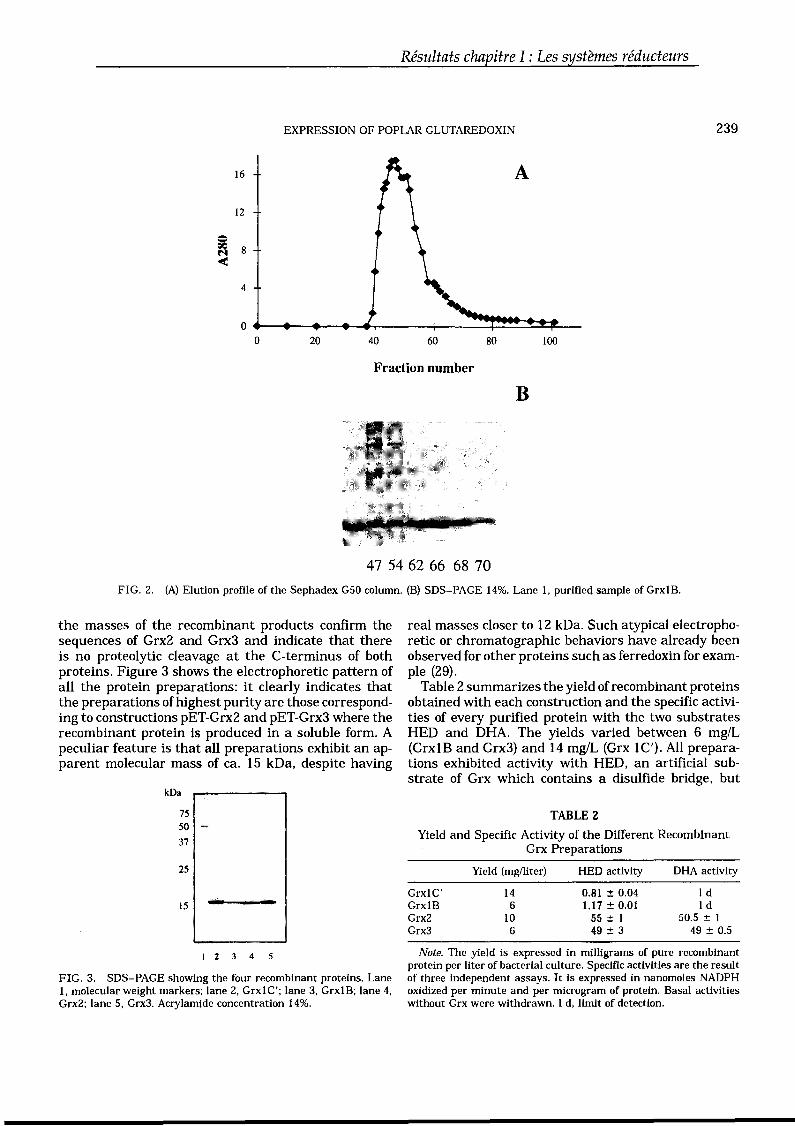
Résultats chapitre l : Les systèmes réducteurs
EXPRESSION OF POPLAR GLUTAREDOXIN 239
16 A
12
i 8M<
4
00 20 40 60 80 100
Fraction number
B
47 54 62 66 68 70
FIG. 2. (A) Elution profile of the Sephadex G50 column. (B) SDS-PAGE 14%. Lane 1, purified sample of GrxiB.
the masses of the recombinant products confirm thesequences of Grx2 and Grx3 and indicate that thereis no proteolytic cleavage at the C-terminus of bothproteins. Figure 3 shows the electrophoretic pattern ofaIl the protein preparations: it clearly indicates thatthe preparations ofhighest purity are those corresponding to constructions pET-Grx2 and pET-Grx3 where therecombinant protein is produced in a soluble forme Apeculiar feature is that aIl preparations exhibit an apparent molecular mass of ca. 15 kDa, despite having
kDa
7550
37
25
15
1 2 3 4 5
FIG. 3. SDS-PAGE showing the four recombinant proteins. Lane1, molecular weight markers; lane 2, GrxlC'; lane 3, GrxlB; lane 4,Grx2; lane 5, Grx3. Acrylamide concentration 14%.
real masses closer to 12 kDa. Such atypical electrophoretic or chromatographie behaviors have already beenobserved for other proteins such as ferredoxin for exampIe (29).
Table 2 summarizes the yield of recombinant proteinsobtained with each construction and the specifie activities of every purified protein with the two substratesHED and DHA. The yields varied between 6 mg/L(GrxlB and Grx3) and 14 mg/L (Grx IC'). AlI preparations exhibited activity with HED, an artificial substrate of Grx which contains a disulfide bridge, but
TABLE 2
Yield and Specifie Activity of the Different RecombinantGrx Preparations
Yield (mg/liter) HED activity DHA activity
Grx1C' 14 0.81 ± 0.04 1 dGrxlB 6 1.17::0.01 IdGrx2 10 55 ± 1 50.5 ± 1Grx3 6 49 ± 3 49 ± 0.5
Note. The yield is expressed in milligrams of pure recombinantprotein per liter of bacterial culture. Specific activities are the resultof three independent assays. It is expressed in nanomoles NADPHoxidized per minute and per microgram of protein. Basal activitieswithout Grx were withdrawn. 1 d, limit of detection.
240
Résultats chapitre l : Les systèmes réducteurs
ROUHIER ET AL.
Grx2 and Grx3 were much more efficient catalysts thanGrxlB and 1C'. Alternatively, when dehydroascorbatewas used as a substrate, only Grx2 and Grx3 wereactive with similar efficiencies, and both GrxlB andGrx1C' were inactive, even at higher concentrations.
DISCUSSION
The initiallack of expression for recombinant glutaredoxin in E. coli has been overcome by successivelyshortening the 5' nucleotide sequence, cotransformingwith a plasmid rescuing the Arg unfavorable codons,optimizing the 5' and 3' ends of the cDNAs and finallyelongating the 3' end which resulted in a soluble protein, shorter on the N-terminus side and elongated onthe C-terminus side compared to the first versionstested. Because of the relative variations observed inthe sequence length of several glutaredoxins, it is notalways easy to predict where a given protein shouldstart and end, more especially so because the N and Cterminal ends are generally the most variable regionsin proteins. Structurally, it has been observed that theshortest forms of glutaredoxin (E. coli or T4) lack an œhelix on the N-terminus (30). In this respect, eventhough they have been shortened on the N-terminusside, the constructions Grx1B, Grx1C, Grx2, and Grx3are aIl longer than the short glutaredoxins and arepredicted to possess this additional N-terminus helix.Overall, aIl constructions which contain the N-terminusextension that presumably leads to excretion were unsuccessful.
The situation is a bit more complex on the C-terminusside, because of low similarity to the existing structuralmodels. It appears nevertheless that the C-terminusend of constructions GrxlB or GrxlC is likely part ofaC-terminus œ-helix that may have been shortened.Because ofthe low homologies in the C-terminus region,structure predictions do not allow to predict whetherthe C-terminal ends of constructions Grx2 and Grx3are indeed involved in a longer œ-helix or in anotherstructural unit. Nevertheless, it appears likely that thesolubility problems encountered in the beginning ofthisstudy are the result of a truncation of the protein onthe C-terminus side which results in increased hydrophobicity and partial insolubilization of the polypeptide.
AlI biochemical evidence supports the proposaI thatthe longest C-terminus forms may represent a betterand more stable version of plant Grx. AlI versions ofpoplar Grx constructed in this study were active withHED, although the two C-terminus elongated formswere ca. SO-fold more active. In addition, only the twoelongated forms were found to be able to reduce DHA.There is indeed sorne controversy in the literature concerning the reduction of DHA by plant glutaredoxin. Areport by Szederkenyi et al. shows that the Ricinus
communis glutaredoxin is active in that reaction (17).However Minakuchi et al. and MoreIl et al. did notdetect any activity for seed rice and spinach leaf glutaredoxins, respectively (18, 19). Our results clearly support the first observation.
As poplar Grx is now obtained in a very good purity,in a soluble form and with a good yield, this opens theway to structural and biochemical studies, includingthe preparation of variants by site-directed mutagenesis that will help identify the targets of this proteine
ACKNOWLEDGMENTS
The authors thank Dr. Ricart for molecular mass determinations,Arnaud Javelle for helpful tips for cloning, and Sibel Catakli andDenis Tagu for critical reading and helpful suggestions.
REFERENCES
1. Halliwell, B., and Gutteridge, J. M. (1990) Role of free radicalsand catalytic metal ions in human disease: An overview. MethodsEnzymol. 186, 1-85.
2. Holmgren, A. (1989) Thioredoxin and glutaredoxin systems. J.Biol. Chem. 264, 13963-13966.
3. Kim, H. T., Russell, R. L., Raina, A. K., Harris, P. L., Siedlak, S.L., Zhu, X., Petersen, R. B., Shimohama, S., Smith, M. A., andPerry, G. (2000) Protein disulfide isomerase in Alzheimer disease.Antioxid Redox Signal. 2, 485-489.
4. Laughner, B. J., Sehnke, P. C., and Ferl, R. J. (1998) A novelnuclear member of the thioredoxin superfamily. Plant PhysioJ.118, 987-996.
5. Vlamis-Gardikas, A., Aslund, F., Spyrou, G., Bergman, T., andHolmgren, A. (1997) Cloning, overexpression, and characterization of glutaredoxin 2, an atypical glutaredoxin from Escherichiacoli. J. Biol. Chem. 272, 11236-11243.
6. Holmgren, A., and Aslund, F. (1995) Glutaredoxin. MethodsEnzymol. 252, 283-292.
7. Holmgren, A. (1979) Glutathione-dependent synthesis ofdeoxyribonucleotides: Characterization of the enzymatic mechanism ofEscherichia coli glutaredoxin. J. Biol. Chem. 254, 3672-3678.
8. Lillig, C. H., Prior, A., Schwenn, J. D., Aslund, F., Ritz, D., VlamisGardikas, A., and Holmgren, A. (1999) New thioredoxins andglutaredoxins as electron donors of 3'-phosphoadenylylsulfatereductase. J. Biol. Chem. 274, 7695-7698.
9. Liu, J., and Rosen, B. P. (1997) Ligand interactions of the ArsCarsenate reductase. J. Biol. Chem. 272, 21084-21089.
10. Wells, W. W., Xu, D. P., Yang, Y. F., and Rocque, P. A. (1990)Mammalian thioltransferase (glutaredoxin) and protein disulfideisomerase have dehydroascorbate reductase activity. J. Biol.Chem. 265, 15361-15364.
11. Aslund, F., and Beckwith, J. (1999) Bridge over troubled waters:Sensing stress by disulfide bond formation. Ce1196, 751-753.
12. Demple, B. (1998) A bridge to control. Science 279, 1655-1656.13. Nakamura, T., Ohno, T., Hirota, K., Nishiyama, A., Nakamura,
H., Wada, H., and Yodoi, J. (1999) Mouse glutaredoxin-cDNAcloning, high level expression in E. coli and its possible implication in redox regulation of the DNA binding activity in transcription factor PEBP2. Free Rad. Res. 31, 357-365.
14. Bandyopadhyay, S., Starke, D. W., Mieyal, J. J., and Gronostajski,R. M. (1998) Thioltransferase (glutaredoxin) reactivates the
Résultats chapitre l : Les systèmes réducteursEXPRESSION OF POPLAR GLUTAREDOXIN 241
DNA-binding activity of oxidation-inactivated nuclear factor I.J. Biol. Chem. 273, 392-397.
15. Barrett, W. C., DeGnore, J. P., Konig, S., Fales, H. M., Keng, Y.F., Zhang, Z. Y., Yim, M. B., and Chock, P. B. (1999) Regulationof PTPIB via glutathionylation of the active site cysteine 215.Biochemistry 38, 6699-6705.
16. Davis, D. A., Newcomb, F. M., Starke, D. W., Ott, D. E., Mieyal,J. J., and Yarchoan, R. (1997) Thioltransferase (glutaredoxin) isdetected within HIV-l and can regulate the activity of glutathionylated HIV-l protease in vitro. J. Biol. Chem. 272, 25935-25940.
17. Szederkenyi, J., Komor, E., and Schobert, C. (1997) Cloning of thecDNA for glutaredoxin, an abundant sieve-tube exudate proteinfrom Ricinus communis L., and characterisation of the glutathione-dependent thiol-reduction system in sieve tubes. Planta202, 349-356.
18. MoreIl, S., Follmann, H., and Haberlein, I. (1995) Identificationand localization of the first glutaredoxin in leaves of a higherplant. FEBS Lett. 369, 149-152.
19. Minakuchi, K., Yabushita, T., Masumura, T., Ichihara, K., andTanaka, K. (1994) Claning and sequence analysis of a cDNAencoding rice glutaredaxin. FEBS Lett. 337, 157-160.
20. Schenk, P. M., Baumann, S., Mattes, R., and Steinbiss, H. H.(1995) Improved high-Ievel expression system for eukaryoticgenes in Escherichia coli using T7 RNA polymerase and rare ArgtRNA's. Biotechniques 19, 196-200.
21. Laemmli, U. K. (1970) Cleavage of structural proteins during theassemblyofthe head ofbacteriophage T4. Nature 227, 680-685.
22. Jacquot, J. P., Keryer, E., Issakidis, E., Decottignies, P., MiginiacMalow, M., Schmitter, J. M., and Crétin, C. (1991) Properties ofrecombinant NADP-Malate dehydrogenases from Sorghum vulgare leaves expressed in Escherichia coli cells. Eur. J. Biochem.199, 47-51.
23. Hodges, M., Miginiac-Maslow, M., Decottignies, P., Jacquot, J.P., Stein, M., Lepiniec, L., Crétin, C., and Gada!, P. (1994) Purification and characterization of pea thioredoxin f expressed inEscherichia coli. Plant Mol. Biol. 26, 225-234.
24. Makrides, S. C. (1996) Strategies for achieving high-Ievel expression of genes in Escherichia coli. Microbiol. Rev. 60, 512-538.
25. Poole, E. S., Brown, C. M., and Tate, W. P. (1995) The identityof the base following the stop codon determines the efficiency ofin vivo translational termination in Escherichia coli. EMBD J.14, 151-158.
26. Bucheler, U. S., Werner, D., and Schirmer, R. H. (1990) Randomsilent mutagenesis in the initial triplets of the coding region: atechnique for adapting human glutathione reductase-encodingcDNA to expression in Escherichia coli. Gene 96, 271-276.
27. Looman, A. C., Bodlaender, J., Comstock, L. J., Eaton, D.,Jhurani, P., De Boer, H. A., and Van Knippenberg, P. H. (1987)Influence of the codon following the AUG initiation codon on theexpression of a modified lacZ gene in Escherichia coli. EMBD J.6, 2489-2492.
28. Hirel, P. H., Schmitter, M. J., Dessen, P., Fayat, G., and Blanquet,S. (1989) Extent of N-terminal methionine excision from Escherichia coli proteins is governed by the side-chain length of thepenultimate amino acid. Proc. Natl, Acad. Sei. USA 86, 82478251.
29. Schmitter, J. M., Jacquot, J. P., de Lamotte-Guéry, F., Beauvallet,C., Dutka, S., Gada!, P., and Decottignies, P. (1988) Purification,properties and complete amino acid sequence of ferredoxin froma green alga, Chlamydomonas reinhardtii. Eur. J. Biochem.172, 405-412.
30. Nordstrand, K., Aslund, F., Holmgren,A., Otting, G., and Berndt,K. D. (1999) NMR structure of Escherichia coli glutaredoxin 3glutathione mixed disulfide complex: Implications for the enzymatic mechanism. J. Mol. Biol. 286, 541-552.
Résultats chapitre I : Les systèmes réducteurs
Le travail effectué dans cet article décrit la mutation de tous les acides aminés
du site actif de cette Grx de peuplier, d’une part pour connaître l’importance de
chaque résidu dans la catalyse et d’autre part pour transformer le site actif de cette
Grx (26YCPYC30) en celui d’une Trx (WCPPC). Six mutations ont été introduites
aboutissant aux sites actifs modifiés ACPYC, YSPYC, YCPFC, YCPPC, YCPYS et
WCPPC. Toutes ces protéines ont été exprimées chez E. coli, purifiées et leur
activité a été évaluée Les résultats obtenus indiquent que la cystéine 27 est
indispensable à la catalyse, au contraire de la cystéine 30 démontrant que cette Grx
peut fonctionner selon un mode de catalyse monothiol. La mutation de la tyrosine 26
en alanine réduit faiblement l’activité de la protéine pour chacun des tests
enzymatiques utilisés suggérant que ce résidu n’est pas essentiel à l’activité de la
protéine. La mutation de la tyrosine 29 en un autre acide aminé aromatique, la
phénylalanine ne modifie pas l’activité de la protéine, alors que la mutation en proline
abolit complètement ou presque son activité. Enfin, le mutant WCPPC présentant un
site actif de Trx n’est plus actif quel que soit le test d’activité utilisé suggérant que le
déterminisme structural de reconnaissance des Trx envers ses protéines cibles n’est
pas uniquement contenu dans le site actif.
Article numéro 8 :
Rouhier N, Gelhaye E, Jacquot JP. (2002) Exploring the active site of plant glutaredoxin by site-directed mutagenesis.
FEBS Lett. 511(1-3):145-9.
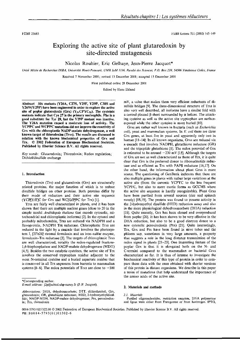
FEBS 25683
Résultats chapitre l : Les systèmes réducteurs
FEBS Letters 511 (2002) 145-149
Exploring the active site of plant glutaredoxin bysite-directed mutagenesis
Nicolas Rouhier, Eric Gelhaye, Jean-Pierre Jacquot*Unité Mixte de Recherches INRA, Université Henri-Poincaré, UMR [aM 1136, Faculté des Sciences, P.O. Box 239,54506 Vandoeuve Cedex, France
Received 7 November 2001; revised 13 December 2001; accepted 13 December 2001
First published online 28 December 2001
Edited by Hans Eklund
Abstract Six mutants (Y26A, C27S, Y29F, Y29P, C30S andY26W1Y29P) have been engineered in order to explore the activesite of poplar glutaredoxin (Grx) (Y26CPYC30). The cysteinicmutants indicate that Cys 27 is the primary nucleophile. Phe is agood substitute for Tyr 29, but the Y29P mutant was inactive.The Y26A mutation caused a moderate loss of activity. TheYCPPC and WCPPC mutations did not improve the reactivity ofGrx with the chloroplastic NADP-malate dehydrogenase, a weIlknown target of thioredoxins (Trxs). The results are discussed inrelation with the known biochemical properties of Grx andTrx. © 2002 Federation of European Biochemical Societies.Published by Elsevier Science B.V. AlI rights reserved.
Key words: Glutaredoxin; Thioredoxin; Redox regulation;DithioVdisulfide exchange
1. Introduction
Thioredoxin (Trx) and glutaredoxin (Grx) are structurallyrelated proteins, the major function of which is to reducedisulfide bridges on other proteins. Both proteins differ bytheir mode of reduction and their active site sequence(YCP[Y/F]C for Grx and WC[G/P]PC for Trx) [1].
Trxs are fairly weIl characterized in plants, and it has beenshown that there are multiple nuclear genes (close to 20 in thesimple model Arabidopsis thaliana that encode cytosolic, mitochondrial and chloroplastic isoforms) [2]. In the cytosol andprobably mitochondria, Trxs are reduced via NADPH and aflavoprotein, NADPH Trx reductase. Chloroplastic Trxs arereduced in the light by a cascade that involves the photosystem l, [2Fe2S] stromal ferredoxin and an iron-sulfur enzyme,ferredoxin-Trx reductase [3]. The targets of chloroplastic Trxsare weIl characterized, notably the redox-regulated fructose1,6-bisphosphatase and NADP-malate dehydrogenase (MDH)[4,5]. Besides the two catalytic cysteines, the active site of Trxinvolves the conserved tryptophan residue adjacent to themost N-terminal cysteine and a buried aspartate residue thatis conserved in aIl Trx sequences from bacteria to mammaliansystems [6-8]. The redox potentials of Trxs are close to -300
*Corresponding author.E-mail address:[email protected] (J.-P. Jacquot).
Abbreviations: DHA, dehydroascorbate; DTT, dithiothreitol; Grx,glutaredoxin; GR, glutathione reductase; HED, 2-hydroxyethyldisulfide; NADP-MDH, NADP·malate dehydrogenase; Prx, peroxiredoxin; Trx, thioredoxin
mV, a value that makes them very efficient reductants of disulfide bridges [9]. The three-dimensional structure of Trxs isalso very weIl described, aIl isoforms have a similar fold witha central pleated ~ sheet surrounded by a helices. The attacking cysteine as weIl as the active site tryptophan are surfaceexposed while the other cysteine is more buried [10].
Grxs are rather weIl known in bacteria (such as Escherichiacoli), yeast and mammalian systems. In E. coli there are threeGrx genes, at least five in yeast and apparently only two inhuman [11-14]. In aIl known organisms, Grxs are reduced viaa cascade that involves NADPH, glutathione reductase (GR)and the tripeptide glutathione [1]. The redox potential of Grxis estimated to be around -230 mV [15]. Although the targetsof Grx are not as weIl characterized as those of Trx, it is quiteclear that Grx is the preferred donor to ribonucleotide reductase and as efficient as Trx with PAPS reductase [16,17]. Onthe other hand, the information about plant Grxs is morescarce. The questioning of GenBank indicates that there arealso multiple genes in plants with rather large variations at theactive site (from the canonic YCPYC to the less frequentYCPFC, but also to more exotic forms as GCCMS wherethe active site sequence is hardly recognizable). Plant Grxshave been purified from several sources (rice and spinachmostly) [18,19]. The protein was found to possess activity inthe 2-hydroxyethyl disulfide (HED) reduction assay and alsoin the more physiological dehydroascorbate (DHA) reduction[18]. Quite recently, Grx has been cloned and overproducedfrom poplar [20]. It has been shown to be very effective in theDHA reduction, but also to be a good electron donor to anew cytosolic peroxiredoxin (Prx) [21]. Quite interestingly,Trx, Grx and Prx have been found in sieve tubes and thephloem sap, sometimes in very large amounts, a propertythat suggests a role in the long distance transmission of theredox signal in plants [21-23]. One interesting feature of thepoplar Grx is that it is elongated both on the N- andC-termini compared to the mammalian or bacterial Grxscharacterized so far. It is thus of interest to investigate thebiochemical reactivity of this type of protein in order to compare those data with the ones obtained with shorter versionsof this protein in distant organisms. We describe in this papera series of mutations that help understand the importance ofthe amino acids of the active site.
2. Materials and methods
2.1. MaterialsPurified oligonucleotides, restriction enzymes, DNA polymerase
and ligase were either from Eurogentec or from Invitrogen. IPTG,
0014-5793/02/$22.00 © 2002 Federation of European Biochemical Societies. Published by Elsevier Science B.V. AIl rights reserved.PlI: S 0 0 1 4 - 5 7 9 3 (0 1 ) 0 3 3 0 2 - 6
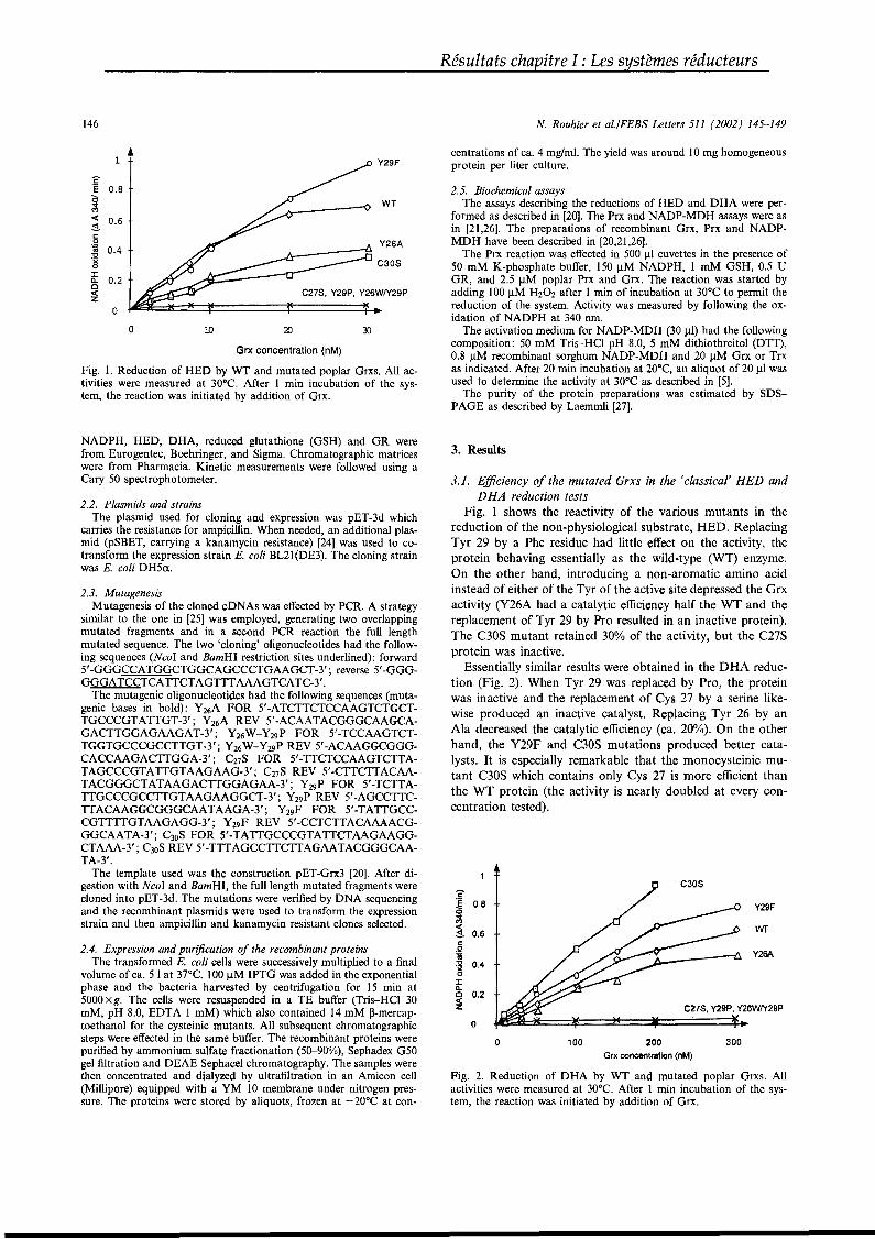
Résultats chapitre l : Les systèmes réducteurs
146
1 Y29F
I?'Ë 0.8èS WT~« 0.6Sc:0 Y26A~ 0.4'0'x C30S0:I
0.2'l.0
C27S, Y29P, Y26WIY29P«z0
0 10 20 30
Grx concentration (nM)
Fig. 1. Reduction of HED by WT and mutated poplar Grxs. AlI activities were measured at 30°C. After 1 min incubation of the system, the reaction was initiated by addition of Grx,
N. Rouhier et al.lFEBS Letters 511 (2002) 145-149
centrations of ca. 4 mg/ml. The yield was around 10 mg homogeneousprotein per liter culture.
2.5. Biochemical assaysThe assays describing the reductions of HED and DHA were per
formed as described in [20]. The Prx and NADP-MDH assays were asin [21,26]. The preparations of recombinant Grx, Prx and NADPMDH have been described in [20,21,26].
The Prx reaction was effected in 500 J..tl cuvettes in the presence of50 mM K-phosphate buffer, 150 J..tM NADPH, 1 mM GSH, 0.5 UGR, and 2.5 J..tM poplar Prx and Grx. The reaction was started byadding 100 J..tM H202 after 1 min of incubation at 30°C to permit thereduction of the system. Activity was measured by following the oxidation of NADPH at 340 nm.
The activation medium for NADP-MDH (30 J..tl) had the fol1owingcomposition: 50 mM Tris-HCI pH 8.0, 5 mM dithiothreitol (DTT),0.8 JlM recombinant sorghum NADP-MDH and 20 JlM Grx or Trxas indicated. After 20 min incubation at 20°C, an aliquot of 20 JlI wasused to determine the activity at 30°C as described in [5].
The purity of the protein preparations was estimated by SDSPAGE as described by Laemmli [27].
C30S
3. Results
Fig. 2. Reduction of DHA by WT and mutated poplar Grxs. AlIactivities were measured at 30°C. After 1 min incubation of the system, the reaction was initiated by addition of Grx.
wr
Y29f
Y26A
300
C27S, Y29P. Y26WIY29P
200Grx.concentration ·(nM)
100
c'Ë 0,80~
~ 0.6c::.Q16~ 0.40J:'l.
0.20«z0
0
3.1. Efficiency of the mutated Grxs in the i classical' HED andDHA reduction tests
Fig. 1 shows the reactivity of the various mutants in thereduction of the non-physiological substrate, HED. ReplacingTyr 29 by a Phe residue had little effect on the activity, theprotein behaving essentially as the wild...type (WT) enzyme.On the other hand, introducing a non...aromatic amino acidinstead of either of the Tyr of the active site depressed the Grxactivity (Y26A had a catalytic efficiency half the WT and thereplacement of Tyr 29 by Pro resulted in an inactive protein).The C30S mutant retained 30% of the activity, but the C27Sprotein was inactive.
Essentially similar results were obtained in the DHA reduction (Fig. 2). When Tyr 29 was replaced by Pro, the proteinwas inactive and the replacement of Cys 27 by a serine likewise produced an inactive catalyst. Replacing Tyr 26 by anAla decreased the catalytic efficiency (ca. 200/0). On the otherhand, the Y29F and C30S mutations produced better catalysts. It is especially remarkable that the monocysteinic mutant C30S which contains only Cys 27 is more efficient thanthe WT protein (the activity is nearly doubled at every concentration tested).
2.4. Expression and purification of the recombinant proteinsThe transformed E. coli ceUs were successively multiplied to a final
volume of ca. 51 at 37°C. 100)lM IPTG was added in the exponentialphase and the bacteria harvested by centrifugation for 15 min at5000Xg. The cells were resuspended in a TE buffer (Tris-HCl 30mM, pH 8.0, EDTA 1 mM) which also contained 14 mM ~-mercap
toethanol for the cysteinic mutants. AlI subsequent chromatographiesteps were effected in the same buffer. The recombinant proteins werepurified by ammonium sulfate fractionation (50-900/0), Sephadex G50gel filtration and DEAE Sephacel chromatography. The samples werethen concentrated and dialyzed by ultrafiltration in an Amicon cell(Millipore) equipped with a YM 10 membrane under nitrogen pressure. The proteins were stored by aliquots, frozen at -20°C at con-
2.2. Plasmids and strainsThe plasmid used for cloning and expression was pET-3d which
carries the resistance for ampicillin. When needed, an additional plasmid (pSBET, carrying a kanamycin resistance) [24] was used to cotransform the expression strain E. coli BL21(DE3). The cloning strainwas E. coli DH5a.
NADPH, HED, DHA, reduced glutathione (GSH) and GR werefrom Eurogentec, Boehringer, and Sigma. Chromatographic matriceswere from Pharmacia. Kinetic measurements were followed using aCary 50 spectrophotometer.
2.3. MutagenesisMutagenesis of the cloned cDNAs was effected by PCR. A strategy
similar to the one in [25] was employed, generating two overlappingmutated fragments and in a second PCR reaction the full lengthmutated sequence. The two 'c1oning' oligonucleotides had the following sequences (NcoI and BamHI restriction sites underlined): forward5'-GGGCCATGGCTGGCAGCCCTGAAGCT-3'; reverse 5'-GGGGGGATCCTCATTCTAGTTTAAAGTCATC-3'.
The mutagenic oligonucleotides had the following sequences (mutagenic bases in bold): Y26A FOR 5'-ATCTTCTCCAAGTCTGCTTGCCCGTATTGT-3'; Y26A REV 5'-ACAATACGGGCAAGCAGACTTGGAGAAGAT-3'; Y26W-Y29P FOR 5'-TCCAAGTCTTGGTGCCCGCCTTGT-3'; Y26W-Y29P REV 5'-ACAAGGCGGGCACCAAGACTTGGA-3' ; C27S FOR 5'-TTCTCCAAGTCTTATAGCCCGTATTGTAAGAAG·3'; C27S REV 5'-CTTCTTACAATACGGGCTATAAGACTTGGAGAA-3'; Y29 P FOR 5'-TCTTATTGCCCGCCTTGTAAGAAGGCT-3'; Y29 P REV 5'-AGCCTTCTTACAAGGCGGGCAATAAGA-3'; Y29F FOR 5'-TATTGCCCGTTTTGTAAGAGG-3'; Y29F REV 5'-CCTCTTACAAAACGGGCAATA-3'; C30S FOR 5'·TATTGCCCGTATTCTAAGAAGGCTAAA-3'; C30S REV 5'-TTTAGCCTTCTTAGAATACGGGCAATA-3'.
The template used was the construction pET-Grx3 [20]. After digestion with NcoI and BamHI, the fulliength mutated fragments werecloned into pET-3d. The mutations were verified by DNA sequencingand the recombinant plasmids were used to transform the expressionstrain and thenampicillin and kanamycin resistant clones selected.

N. Rouhier et al.lFEBS Letters 511 (2002) 145-149
120
100
t!- 80~>ts 60<VQ)>~ 40"i)0::
20
c:~
~(J) u- a.. ~ a.. (J):s
§,.....
0'> 0'> <0 0'> 0"0 C\I
~ ~ ~ ~C")
<V (.) U0Z
Fig. 3. Activity of Prx with WT and mutated Grxs. The activity isexpressed in % of the WT proteine 1000/0 activity is 0.55 dOD/min.Details of the reaction are given in Section 2.
3.2. Grx efficiency vs plant type C PrxIt has been demonstrated recently that type C Prx of plants
uses both Trx and Grx as proton donors [21]. It was thus ofinterest to determine if the various mutants generated here areable to sustain the catalytic activity of Prx in this newly described reaction. Fig. 3 shows the results of those experiments.The WT and Y29F mutant show similar activity. On the otherhand, replacing the two Tyr residues by non-aromatic sidechains decreased the catalytic efficiency. FinaIly, the C30Smutant retains most of its catalytic efficiency and the C27Sprotein is inactive.
3.3. Activation of NADP·MDH by mutated GrxsTwo of the mutations that we have engineered in this work
transform the Grx active site into a Trx-like active site(YCPPC and WCPPC). It was expected that these mutationsshould increase the reactivity of Grx vs the NADP-MDH, aweIl characterized target enzyme of Trx. Fig. 4 shows thereactivity of the various mutants with NADP-MDH. It isinteresting that the WT Grx is able to activate the enzymein the presence of DTT with reasonably good efficiency(nearly half the capacity of the non-physiological Trx h atthe same concentration of 20 JlM). AlI mutants tested showedcatalytic activity, but aIl mutations decreased the efficiencyexcept for Y26A. The two mutations that were intended tomimic the Trx active site (Y29P and Y26W/Y29P) did notimprove the reactivity at aIl. Interestingly, both the C27Sand C30S mutants retained the capacity to activate theNADP-MDH but their efficiencies were strongly reduced (toca. 250/0 of the WT).
4. Discussion
4.1. Mutations of the active site cysteinesln the Grx dependent reactions (HED and DHA reduction
and Prx activation), the mutation of Cys 27 leads to a virtuaIly inactive enzyme. On the other hand, the mutation of Cys30 into Ser had either a strongly negative effect (HED), amildly negative effect (Prx) or a positive effect (DHA). AlIthese data strongly suggest that Cys 27 is the primary nucleophile of the reaction and that Cys 30 is the backup cysteine.Similar results have been obtained with the E. coli and humanGrx or aIl Trxs where the catalytic cysteine is always located
,on the N-terminus side [28-30]. It seems thus that the N- andC-termini extensions of plant Grx do not alter that property.
Résultats chapitre l : Les systèmes réducteurs
147
AdditionaIly, these site-directed mutagenesis experiments indicate that Grx can be quite efficient as a single cysteine catalyst, confirming that it can act in the so-caIled monothiolpathway [31]. Moreover, these experiments suggest that thenumerous natural monocysteinic versions of Grx that existin the databanks are almost certainly functional catalysts.
4.2. Importance of the aromatic residues of the active siteThe replacement of the Tyr residue present between the two
catalytic cysteines by a proline leads to a drastic decline inreactivity. It is clearly essential to keep an aromatic residue inthis position as the Y29F mutant is a very good catalyst in aIlGrx dependent reactions tested. This Tyr residue has beenimplicated as one of the ligands necessary for the fixation ofglutathione to E. coli Grx3 or T4 Grx [11,32]. On the otherhand, it is not necessary to have an aromatic residue in position 26 (N-terminus to the catalytic cysteine) as the Y26Amutant kept at least half of the reactivity of the WT proteineThis is very much in contrast with the case of Trx where theremoval of the Trp adjacent to the catalytic cysteine has avery strong negative effect [6,33].
4.3. The NADP-MDH is activated by plant Grx and ilsmutants
The NADP-MDH has been recognized as a target of Trxssince a long time. The chloroplastic Trx f is the most efficient,foIlowed by chloroplastic Trx m, and the cytosolic Trx h [34].Mammalian Trxs are very poor reductants/activators of thisenzyme [35]. AlI Trxs which have been demonstrated as capable of activating this enzyme have either a WCGPC or aWCPPC active site. By making the Y29P and Y26WIY29Pmutations, we have engineered proteins with active sites thathave been transformed into YCPPC and WCPPC. It was thusexpected that these mutations could result in Grxs with betterreactivity vs the NADP-MDH. The results in Fig. 4 indicateclearly that this is not the case as only the Y26A mutant hasan activity comparable to the WT protein and aIl other mutations decrease the reactivity. It is surprising to observe thatWT Grx is able to induce the activation of the enzyme in thepresence of DTT (see Fig. 4). This activation is dependent onthe concentration of added Grx and on DTT (data notshown). The WT poplar Grx is not as efficient as the cytosolicTrx h but more efficient than the human Trx or the Trxs ofDictyostelium discoideum [35,36]. We have checked that thisreactivity is not due to contaminating E. coli Trx in the recombinant Grx preparations. We have indeed quantitated the
250
200~c
~.S; 150:um
~ 100~mID0::
50
o
Fig. 4. Activity of NADP-MDH with WT and mutated Grxs. Activities are expressed in % of the WT Grx. 1000/0 activity is OAdODImin. Experimental details are given in Section 2.
148
contamination of those preparations by the NTR/DTNB system and found that it could not exceed 0.30/0. At a Grx concentration of 20 JlM in Fig. 4, the E. coli Trx concentration isthus lower than 60 nM, a value at which no detectableNADP-MDH activation occurs [37]. This activation ofNADP-MDH by Grx is likely to be non-physiological sinceGSH could not replace DTT, even in the presence of NADPHand GR (data not shown). Three additional observations support the proposaI that Grx is indeed not a physiological activator of NADP-MDH. First, it seems that there are nochloroplastic sequences for Grx in protein databases. Second,the redox potential of Grx (normally around -230 mV) is notadequate with respect to the ones of the NADP-MDH disulfide bridges (-280 and -300 mV) for an efficient reaction totake place [38]. Third, the fact that the C30S mutant keeps alevel of activity similar to the C27S mutant suggests that therole of Grx in this reaction is non-catalytic. We propose thusthat Grx acts here in a non-catalytic way to modify the accessibility of the active site to DIT which has the right redoxpotential. Such a structural role has already been observed inthe case of the interaction between the T7 DNA polymeraseand the E. coli Trx [39]. The results obtained with the C27Smutant seem to support such a hypothesis since Cys 30 iswidely recognized as the non-catalytic one, a property alsoverified in the other reactions described here.
4.4. Concluding remarksMutations similar to those described here have already been
performed either on E. coli Trx to transform it into a Grx or aDsbA protein [40,41], or on DsbA to transform it into a Grxor a Trx [42]. In most of these mutants, essentially physicochemical characteristics as pKa values and redox potentialshave been determined but few kinetic experiments have beenperformed except in [40] where the creation of a CGHC sitehas been shown to be accompanied by an increase in PDI-likeactivity. In this study, nearly all the mutants that we havegenerated are less active than the WT protein, in contrast toinitial expections. Transfonning the Grx active site into a Trxlike active site did not improve the reactivity with NADPMDH or Trx reductase (data not shown). Similarly to whatis described here, simulations of Trx or Grx-like active sites intryparedoxin 2 did not result in Trx or Grx-like activities [43].Overall, these data strongly suggest that the structural determinants for Trx or Grx reactivity go beyond the sequence ofthe active site.
Nevertheless, the series of mutants generated here help tounderstand the functioning of plant Grxs. The catalytic cysteine as well as the importance of an aromatic residue insidethe regulatory sequence and adjacent to the backup cysteinehave been uncovered. An additional interesting finding of thisstudy is the capacity of Grx to activate the NADP-MDH.This enzyme has always been reputed as promiscuous as itaccepts many different Trxs as regulators, in contrast to fructose-1,6-biphosphatase that requires selectively Trx f Thedata presented here indicate that the DTT NADP-MDH activation test should be used cautiously, as Grx is also active inthis process despite the fact that it probably does not act in acatalytic way.
References
[1] Holmgren, A. (2000) Antioxid. Redox Signal 2, 811-820.
Résultats chapitre l : Les systèmes réducteurs
N. Rouhier et al.lFEBS Letters 511 (2002) 145-149
[2] Meyer, Y., Verdoucq, L. and Vignols, F. (1999) Trends Plant Sei.4, 388-394.
[3] Schürmann, P. and Jacquot, J.P. (2000) Annu. Rev. Plant Physiol. Plant Mol. Biol. 51, 371-400.
[4] Chiadmi, M., Navaza, A., Miginiac-Maslow, M., Jacquot, J.P.and Cherfils, J. (1999) EMBO J. 18, 6809-6815.
[5] Miginiac-Maslow, M., Johansson, K., Ruelland, E., IssakidisBourguet, E., Schepens, 1., Goyer, A., Lemaire-ChamIey, M.,Jacquot, J.P., Le Maréchal, P. and Decottignies, P. (2000) Physiol. Plant. 110, 322-329.
[6] Krause, G. and Holmgren, A. (1991) J. Biol. Chem. 266, 40564066.
[7] Dyson, H.J., Jeng, M.F., Tennant, L.L., Slaby, 1., Lindell, M.,Cui, O.S., Kuprin, S. and Holmgren, A. (1997) Biochemistry 36,2622-2626.
[8] Menchise, V., Corbier, C., Didierjean, C., Saviano, M., Benedetti, E., Jacquot, J.P. and Aubry, A. (2001) Biochem. J. 359, 65-75.
[9] Hirasawa, M., Schürmann, P., Jacquot, J.P., Manieri, W., Jacquot, P., Keryer, E., Hartman, F. and Knaff, D.B. (1999) Biochemistry 38, 5200-5205.
[10] Jeng, M.F., Campbell, A.P., Begley, T., Holmgren, A., Case,D.A., Wright, P.E. and Dyson, H.J. (1994) Structure 2, 853-868.
[Il] Nordstrand, K., Sandstrom, A., Aslund, F., Holmgren, A., Otting, G. and Berndt, K.D. (2000) J. Mol. Biol. 303,423-432.
[12] Lundberg, M., Johansson, C., Chandra, J., Enoksson, M., Jacobsson, G., Ljung, J., Johansson, M. and Holmgren, A.(2001) J. Biol. Chem. 276, 26269-26275.
[13] Gladyshev, V.N., Liu, A., Novoselov, S.V., Krysan, K., Sun,Q.A., Kryukov, V.M., Kryukov, G.V. and Lou, M.F. (2001)J. Biol. Chem. 276, 30374-30380.
[14] Rodriguez-Manzaneque, M.T., Ros, J., Cabiscol, E., Sorribas, A.and Herrero, E. (1999) Mol. Cell. Biol. 19, 8180-8190.
[15] Aslund, F., Berndt, K.D. and Holmgren, A. (1997) J. Biol.Chem. 272, 30780-30786.
[16] Berardi, M.J. and Bushweller, J.H. (1999) J. Mol. Biol. 292, 151161.
[17] Lillig, C.H., Prior, A., Schwenn, J.O., Aslund, F., Ritz, D., Vlamis-Gardikas, A. and Holmgren, A. (1999) J. Biol. Chem. 274,7695-7698.
[18] Sha, S., Minakuchi, K., Higaki, N., Sato, K., Ohtsuki, K., Kurata, A., Yoshikawa, H., Kotaru, M., Masumura, T., Ichihara,K. and Tanaka, K. (1997) J. Biochem. 121, 842-848.
[19] MoreIl, S., Follmann, H. and Haberlein, J. (1995) FEBS Lett.369, 149-152.
[20] Rouhier, N., Gelhaye, E., Sautiere, P,E. and Jacquot, J.P. (2002)Prot. Expr. Purif., in press.
[21] Rouhier, N., Gelhaye, E., Sautiere, P.E., Brun, A., Laurent, P.,Tagu, D., Gerard, J., de Fay, E., Meyer, Y. and Jacquot, J.P.(2001) Plant Physiol. 127, 1-11.
[22] Szederkenyi, J., Komor, E. and Schobert, C. (1997) Planta 202,349-356.
[23] Ishiwatari, Y., Fujiwara, T., McFarland, K.C., Nemoto, K.,Hayashi, H., Chino, M. and Lucas, W,J. (1998) Planta 205,12-22.
[24] Schenk, P.M., Baumann, S., Mattes, R. and Steinbiss, H.H.(1995) Biotechniques 19, 196-200.
[25] Jacquot, J.P., Stein, M., Suzuki, A., Liottet, S., Sandoz, G. andMiginiac-Maslow, M. (1997) FEBS Lett. 400, 293-296.
[26] Ruelland, E., Johansson, K., Decottignies, P., Djukic, N. andMiginiac-Maslow, M. (1998) J. Biol. Chem. 273, 33482-33488.
[27] LaemmIi, V.K. (1970) Nature 227,680-685.[28] Bushweller, J.H., Aslund, F., Wuthrich, K. and Holmgren, A.
(1992) Biochemistry 31, 9288-9293.[29] Foloppe, N., Sagemark, J., Nordstrand, K., Berndt, K.D. and
Nilsson, L. (2001) J. Mol. Biol. 310,449-470.[30] Padilla, C.A., Spyrou, G. and Holmgren, A. (1996) FEBS Lett.
378, 69-73.[31] Yang, Y., Jao, Sc., Nanduri, S., Starke, D.W., Mieyal, J.J. and
Qin, J. (1998) Biochemistry 37, 17145-17156.[32] Nikkola, M., Gleason, F.K., Saarinen, M., Joelson, T., Bjorn
berg, O. and Eklund, H. (1991) J. Biol. Chem. 266, 1610516112.
[33] Krimm, 1., Lemaire, S., Ruelland, E., Miginiac-Maslow, M., Jacquot, J.P., Hirasawa, M., Knaff, D.B. and Lancelin, J.M. (1998)Eur. J. Biochem. 255, 185-195.

N. Rouhier et al.lFEBS Letters 511 (2002) 145-149
[34] Aguilar, F., Brunner, B., Gardet-Salvi, L., Stutz, E. and Schürmann, P. (1992) Plant Mol. Biol. 20, 301-306.
[35] Jacquot, J.P., de Lamotte..Guéry, F., Fontecave, M., Schürmann,P., Decottignies, P., Miginiac-Maslow, M. and Wollmann, E.(1990) Biochem. Biophys. Res. Commun. 173, 1375-1381.
[36] Wetterauer, B., Véron, M., Miginiac-Maslow, M., Decottignies,P. and Jacquot, J.P. (1992) Eur. J. Biochem. 209, 643-649.
[37] de Lamotte-Guery, F., Miginiac-Maslow, M., Decottignies, P.,Stein, M., Minard, P. and Jacquot, J.P. (1991) Eur. J. Biochem.196, 287-294.
[38] Hirasawa, M., Ruelland, E., Schepens, I., Issakidis-Bourguet, E.,Miginiac-Maslow, M. and Knaff, D.B. (2000) Biochemistry 39,3344-3350.
Résultats chapitre l : Les systèmes réducteurs
149
[39] Huber, H.E., Russel, M., Model, P. and Richardson, C.C. (1986)J. Biol. Chem. 261, 15006-15012.
[40] Lundstrom, J., Krause, G. and Holmgren, A. (1992) J. Biol.Chem. 267, 9047-9052.
[41] Mëssner, E., Huber-Wunderlich, M. and Glockshuber, R. (1998)Protein Sei. 7, 1233-1244.
[42] Huber-Wunderlich, M. and Glockshuber, R. (1998) Fold. Des. 3,161-171.
[43] Steinert, P., Plank-Schumacher, K., Montemartini, M., Hecht,H.J. and Flohe, L. (2000) Biol. Chem. 381, 211-219.
Résultats chapitre I : Les systèmes réducteurs
Jusqu’à présent, la seule protéine cible connue des Grx de plantes est la Prx
de type II (voir articles 11 et 12). Chez les bactéries notamment, un certain nombre
de protéines cibles incluant la PAPS réductase ou la RNR sont connues. La Grx de
peuplier est capable de servir de donneur d’électrons à ces deux enzymes d’E. coli
suggérant qu’elle pourrait jouer un rôle analogue pour la RNR de plantes par
exemple. Par contre, aucune séquence codant une PAPS réductase n’a été trouvée
chez les plantes supérieures et l’équivalent fonctionnel de cette enzyme est une APS
réductase, localisée dans les chloroplastes et qui possède un motif Grx en position
C-terminale. Par ailleurs, cet article montre que cette Grx a une fonction au moins
indirecte dans la réponse au stress oxydant en réduisant la Prx de type II mais
qu’elle n’a pas la capacité de réduire directement les hydroperoxydes comme cela a
été montré pour la Grx d’Oryza sativa et la Grx 1 de S. cerevisiae. Cette situation est
d’autant plus surprenante qu’il existe chez O. sativa au moins trois isoformes de Prx
de type II (nos d’accession Genbank AAG40130, BAA82377, BAA90363) et chez S.
cerevisiae au moins 3 Gpx et 5 Prx capables de catalyser ces réactions (Carmel-
Harel & Storz, 2000).
Article numéro 9 :
Rouhier N, Vlamis-Gardikas A, Lillig CH, Berndt C, Schwenn JD, Holmgren A, Jacquot JP. (2003) Characterization of the redox properties of poplar glutaredoxin.
Antioxid. Redox Signal. 5(1):15-22.
Résultats chapitre l : Les systèmes réducteurs
ANTIOXIDANTS & REDOX SIGNALINGVolume s, Number 1, 2003© Mary Ann Liebert, Ine.
Forum Original Research Communication
Characterization of the Redox Properties of Poplar Glutaredoxin
NICOLAS ROUHIER,1 ALEXIOS VLAMIS-GARDIKAS,2 CHRISTOPHER HORST LILLIG,2CARSTEN BERNDT,3 JENS-DIRK SCHWENN,3 ARNE HOLMGREN,2
and JEAN-PIERRE JACQUOTI
ABSTRACT
The presence of glutaredoxins in plants is now weil recognized, but their functions and natural substrates remain largely unknown. Recently, a poplar glutaredoxin has been biochemically characterized and several mutants have been engineered in order to explore its reactivity. This work focuses on sorne physiological functions of the enzyme. According to our findings, the poplar glutaredoxin can serve as an electron donor to thebacterial 3'-phosphoadenylylsulfate reductase as it supports both the catalysis by the enzyme in vitro andcomplements a methionine auxotroph strain of Escherichia coli. In addition, poplar glutaredoxin is able to reduce the Escherichia coli ribonucleotide reductase la (in vitro reduction of cytidine diphosphate). Althoughthis glutaredoxin is described as an electron donor to a phloem-Iocated peroxiredoxin, whose function is todetoxify hydroperoxides, we found that it does not directly reduce hydrogen peroxide or other alkyl hydroperoxides as described for yeast and rice glutaredoxins. However, the poplar glutaredoxin may be involvedin the response to oxidative stress as its overexpression in Escherichia coli resulted in a higher resistance toward hydrogen peroxide, menadione, and tert-butyl hydroperoxide. Antioxid. Redox Signal. 5, 15-22.
INTRODUCTION
THE THIOL REDOX STATUS OF THE CYTOSOL is maintained bythe thioredoxin (Trx) and the glutathione/glutaredoxin
(GSH/Grx) systems. Trx and Grx are small (--10-12 kDa),heat-stable disulfide oxidoreductases, with the conserved active site CXXC [typically CP(Y/F)C for Grx and WC(G/P)PCfor Trx] (18,19). These two proteins are constituted by a characteristic succession of ~ strands and Cl helices, the ~ strandsbeing organized into a central pleated ~ sheet surrounded by Cl
helices, the whole folding pattern known as the Trx/Grx fold.Initial reducing equivalents for both systems are provided byNADPH. In the Trx system, NADPH reduces Trx reductase,which then reduces Trx. Trx reductase is replaced by glutathione reductase and GSH in the Grx system. Grxs can re-
duce disulfides with a dithiol mechanism using both theiractive-site cysteine residues (33). An example is the reductionof the disulfide of the Escherichia coli ribonucleotide reductase la (E. coli RNRla), which forms when the enzyme reduces ribonucleotides to deoxyribonucleotides under aerobicconditions (1). However, Grxs can utilize a monothiol mechanism for the reduction of mixed disulfides between proteinsand GSH (protein-S-SG substrates) (26, 33).
Trx and Grx species can reduce both RNR and 3' -phosphoadenylylsulfate (PAPS) via PAPS reductase (23, 33, 43).Other substrates include a plasma glutathione reductase and apoplar type II peroxiredoxin (6, 36). Monothiol Grxs containing only one active-site cysteine are natural catalysts for thedeglutathionylation of protein-S-SG substrates and may bespecific for the assembly of iron sulfur enzymes (34, 40, 43).
IUnité Mixte de Recherche 1136 IaM (Interaction arbres microorganismes), INRA-UHP Nancy I. Université Henri Poincaré, 54506 Vandoeuvre Cedex France.
2Medical Nobel Institute for Biochemistry, Department of Medical Biochemistry and Biophysics, Karolinska Institutet, S-171 77 Stockholm,Sweden.
3Biochemistry of Plants, Faculty of Biology, Ruhr-University, 44780 Bochum, Germany.
15
16
Sultbydryls of cysteine residues are particularly exposed toreactive oxygen species (ROS) such as hydrogen peroxide(H20 2) or superoxide anions, which are formed during normal aerobic growth or during oxidative stress (14). Formationof intermolecular disulfides or overoxidation of thiols tosulfenic, sulfinic or sulfonic acids can occur and thus lead toloss of enzymatic activity, aging, pathological disease, andcell death (3, 12). To challenge the overoxidation of thiols,cells possess a large battery of antioxidant enzymes, in whichTrx and Grx play key roles. For example, Trx and/or Grx canreduce peroxiredoxins, non-heme peroxidases that catalyzethe reduction of hydroperoxides (8, 36). Trx and Grx are alsoable to reduce protein...S-SG substrates forming under conditions of oxidative stress (9, 20).
Analysis of the transcriptional regulation and overexpressionldeletion of different Trx and Grx isoforms under conditions of hydroperoxide stress has highlighted specifie rolesfor particular genes (7, 13, 16, 24, 31). Two recent studiesdemonstrate that yeast Grx1 and a Grx from rice are able toreduce directly different alkyl hydroperoxides into alcohol, areaction generally catalyzed by peroxidases (10, 22).
Whereas there are at least three "classical" dithiol Grxs in E.coli (CPYC active site), in yeast there are two ofthese proteinsand three atypical ones with one active site cysteine (33, 34,43). Apparently, there are only two classical Grxs in human (15,25). In the genome of Arabidopsis thaliana, there are at leastfour classical Grxs and many other Grx-like proteins, but theseproteins were not biochemically characterized and thus the understanding ofthe functions ofGrxs in plants is not as advancedas in bacteria, yeast, or mammalian cells. The first plant Grxswere identified in spinach leaves, in the phloem sap of castorbean, and in the aleurone layer of rice (28, 29, 42). Until recently, the substrates of plant Grxs were unknown, whereas therange of substrates for bacterial Grxs includes redox-sensitivetranscription factors (2, Il, 41). Plant Grxs possess dehydroascorbate reduction activity, and the Grx from poplar wasdemonstrated to he an efficient electron donor for a peroxiredoxin involved in the reduction ofhydroperoxides (28, 35).
In this work, we describe the capacity for the poplar Grx toreduce E. coli RNRla and PAPS reductase and to restore thegrowth of methionine auxotroph strains ofE. coli, suggestingthat plant Grxs can perform functions similar to those described in other organisms. Moreover, the involvement of thepoplar Grx in the resistance to ROS was investigated by overexpressing the gene in an E. coli BL21(DE3) strain submittedto oxidative stress and by measuring the glutathione peroxidase activity of this Grx.
MATERIALS AND METHODS
Chemicals and reagents
NADPH was obtained from Boehringer Mannheim (Germany). Diamide, H20 2, tert-butyl hydroperoxide, menadione,dithiothreitol (DTT), glutathione reductase, and GSH werefrom Sigma (St. Louis, MO, U.S.A.). Isopropyl f3-Dthiogalactoside (IPTG), kanamycin, and ampicillin were fromEuromedex (France). AlI the recombinant Grx were purifiedas described (35).
Résultats chapitre l .. Les systèmes réducteurs
ROUHIER ET AL.
Cloning in pBAD and E. coli mutantstrains complementation
A fragment encoding the poplar Grx was inserted betweenthe restriction sites NcoI andXhoI ofthe plasmid pBADHisMycand called pBAD Grx. This plasmid was used to transform theE. coli strains DHB4 trxA - grxA -, DHB4 gor- trxA -, andDHB4 grxA-grxB-grxC- (32, 44). The same strains transformed with the plasmid without insert were used as controls.Transformed strains were grown on M9 medium, supplied withLeu, Ile, and Met (50 Jlg/ml) and ampicil1in (100 Jlg/ml). Single colonies were streaked on the same medium lacking Metbut supplied with 0.2% arabinose and kept at 37°C for 48 h.
Reduction ofcytidine diphosphate (CDP)by E. coli RNR
Reduction of CDP by E. coli RNRla was performed essentially as described (17). Reducing power was provided by4 mM GSH, 1 mM NADPH, and 6 Jlg/ml yeast glutathione reductase. AlI assays were performed in a total volume of120 Jll. The catalytic rates reported represent the mean valueof two independent measurements ± 1 SE.
E. coli PAPS reductase activity measurement
PAPS reductase activity was measured as described (23)by following the formation of [35S] S032- from [35S]PAPS.The reaction mixture contained 100 mM Tris-HCl, pH 8.0,3.3 nM PAPS reductase, 10 mM Na2S03, 60-100 ~[35S]PAPS (specific radioactivity: 1,700 Bq/nmol), and various amounts (0.5-70 JlM) of the wild-type (WT) poplar Grxand of an active-site mutant Grx Y29P, mimicking the activesite of sorne plant Trxs. The Grxs were kept reduced by 5 mMGSH and 5 mM DTT.
In vitro peroxidase activity ofGrx
The peroxidase activity of poplar Grx was followed usingtwo different spectrophotometric measurements with a Cary50 spectrophotometer. Firstly, the consumption of H20 2 wasevaluated in a coupled reaction by fol1owing the oxidation ofNADPH at 340 nm in the presence of the GSH/Grx system at30°C. A reaction mixture of 500 Jll contained 50 mM TrisHCl, pH 8.0, 2 mM EDTA, 150 JlM NADPH, 300 JlM GSH,0.5 units of yeast glutathione reductase, and various concentrations ofeither WT Grx or of the two monocysteinic mutantsC27S and C30S. It was incubated for 1 min before the reactionwas started by addition of250 JlMH20r In the second test, themeasurement of H20 2 consumption was estimated using thePeroxoquant Quantitative Peroxide Assay from Pierce. A50 JlL reaction mixture comprising 30 mM Tris-HCl, pH 8.0,500 JlM DTT, 500 JlM H20 2, and various Grx concentrations(1-50 JlM) was incubated for 4-10 min at ambient temperature. An aliquot of 5 Jll was then added to the working reagent,and the absorbancy was measured 20 min later at 560 nm.
Resistance ofa Grx overexpressing E. colistrain to oxidants
The E. coli strain BL21(DE3), transformed with thehelper plasmid pSBET, was cotransformed with the different
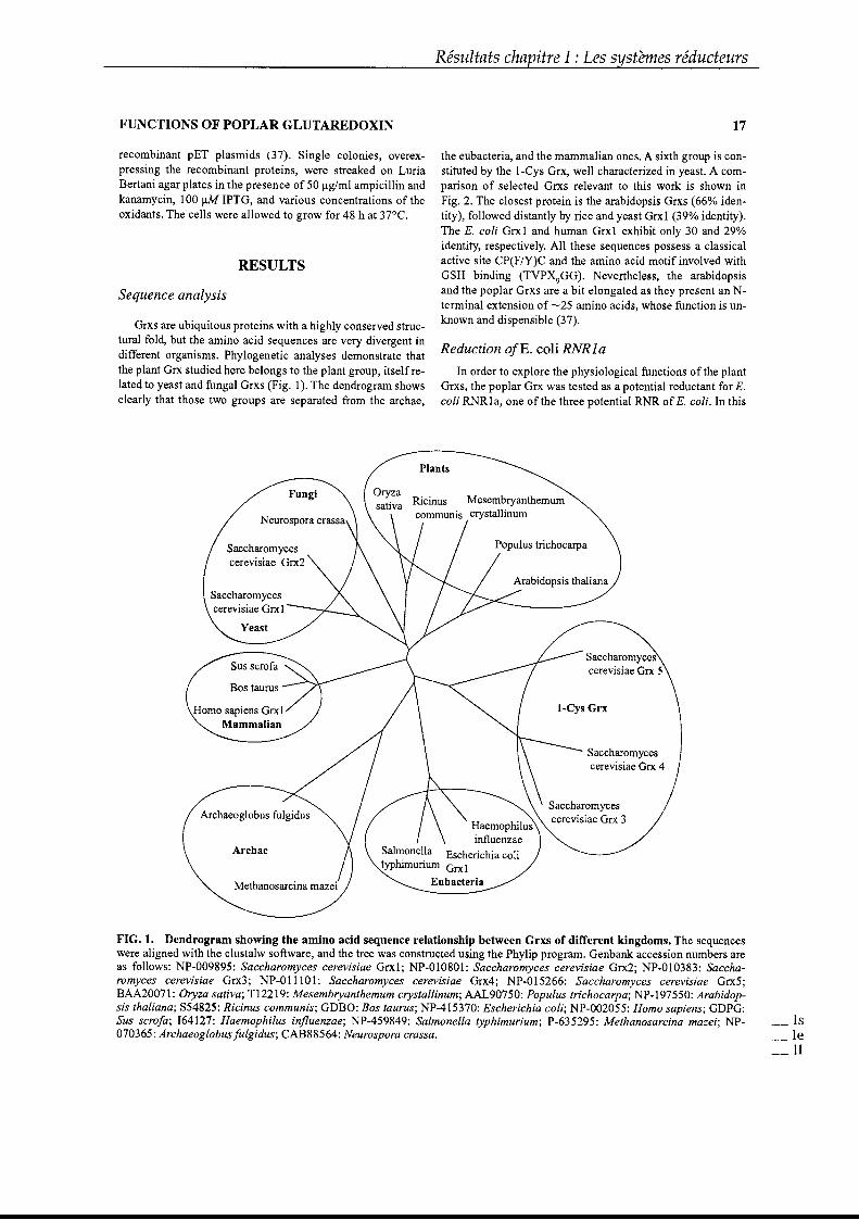
Résultats chapitre l : Les systèmes réducteurs
FUNCTIONS OF POPLAR GLUTAREDOXIN 17
recombinant pET plasmids (37). Single colonies, overexpressing the recombinant proteins, were streaked on LuriaBertani agar plates in the presence of 50 Jlg/ml ampicillin andkanamycin, 100 JlM IPTG, and various concentrations of theoxidants. The cel1s were al10wed to grow for 48 h at 37°C.
RESULTS
Sequence analysis
Grxs are ubiquitous proteins with a highly conserved structural fold, but the amino acid sequences are very divergent indifferent organisms. Phylogenetic analyses demonstrate thatthe plant Grx studied here belongs to the plant group, itself related to yeast and fungal Grxs (Fig. 1). The dendrogram showsclearly that those two groups are separated from the archae,
the eubacteria, and the mammalian ones. A sixth group is constituted by the I-Cys Grx, well characterized in yeast. A comparison of selected Grxs relevant to this work is shown inFig. 2. The closest protein is the arabidopsis Grxs (66% identity), followed distantly by rice and yeast Grx! (39% identity).The E. coli Grx1 and human Grx1 exhibit only 30 and 29%identity, respectively. Al1 these sequences possess a classicalactive site CP(F/Y)C and the amino acid motif involved withGSH binding (TVPX9GG). Nevertheless, the arabidopsisand the poplar Grxs are a bit elongated as they present an Nterminal extension of -25 amino acids, whose function is unknown and dispensible (37).
Reduction ofE. coli RNRla
ln order to explore the physiological functions of the plantGrxs, the poplar Grx was tested as a potential reductant for E.coli RNRla, one of the three potential RNR ofE. coli. In this
Saccharomycescerevisiae Grx 4
Saccharomycescerevisiae Grx S
Saccharomycescerevisiae Grx 3
Arabidopsis thaliana
Haemophilusinfluenzae
Salmonella Escherichia colityphimurium Grx1
EubacteriaMethanosarcina mazei
Archae
Saccharomycescerevisiae Grx2
Archaeoglobus fulgidus
Saccharomycescerevisiae Grx1
Yeast
Homo sapiens Grx1Mammalian
Neurospora crassa
Fungi
Plants
FIG. 1. Dendrogram showing the amino acid sequence relationship between Grxs of different kingdoms. The sequenceswere aligned with the clustalw software, and the tree was constructed using the Phylip program. Genbank accession numbers areas fol1ows: NP-009895: Saccharomyces cerevisiae Grxl; NP-OI0801: Saccharomyces cerevisiae Grx2; NP·OI0383: Saccharomyces cerevisiae Grx3; NP-OIIIOI: Saccharomyces cerevisiae Grx4; NP-015266: Saccharomyces cerevisiae Grx5;BAA20071: Oryza sativa; T12219: Mesembryanthemum crystallinum; AAL90750: Populus trichocarpa; Np·!97550: Arabidopsis thaliana; S54825: Ricinus communis; GDBO: Bos taurus; NP-415370: Escherichia coli; NP-002055: Homo sapiens; GDPG:Sus scrofa; 164127: Haemophilus injluenzae; NP-459849: Salmonella typhimurium; P-635295: Methanosarcina mazei; NP070365: Archaeoglobusfulgidus; CAB88564: Neurospora crassa.
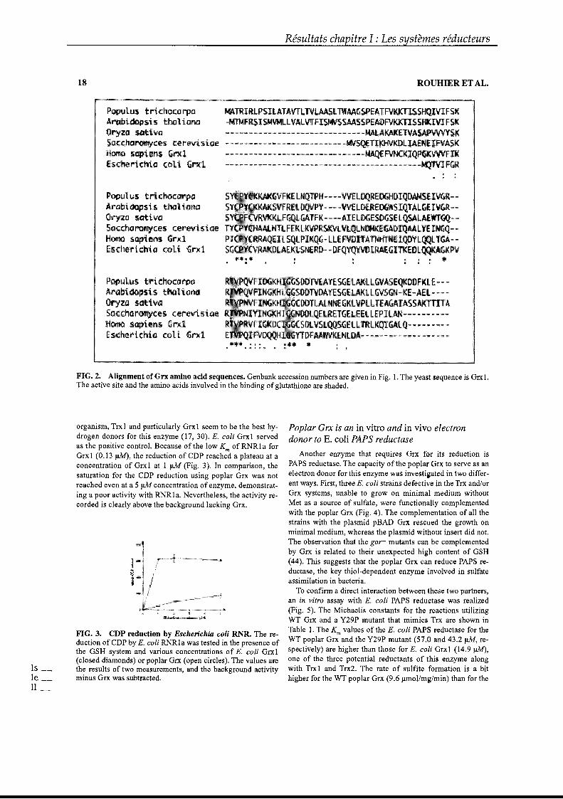
18
l,ustrichoca.rpaAfI1IbiidoP5\.stbal iOn<J
Oryza 5ativGSClcchorotTlYces; çerev'Î5i<JeH0fn05Dpi'eul5GrxlEschelrt.cma coli Gr:wcl
Plustrichoca.rpaArtltJiidops,i.s tlrtt111QOOOryza sattvGSCbccharomyces cerl"'i s iOIHomt:tsapi'irti GrxlElchlrrithia coli Gr>:.·J
PopLilult:r\;chocarpabi~ \.1 thaliatll
Or~l,a Itltiva$accharomycll cerlviltalHoma·sopiJlrtlEschf!lrichla li Grxl
Résultats chapitre l : Les systèmes réducteurs
ROUDIER ET AL.
MATRIRLPS:IlATAVfLlVL l\t'AAGSPEATFV1OITI'SSHqIVIF5K~f ISMiNLLVAL.VTFISMYS5AASSPEAOF\lKKllSSRKIVl:FSI
....._....._. __ ....... __ ..,..._._ ..........,_ ..,..,.,................................... n~·KD:LI.A:Ei
--._ .._. -,- -,_.._,_. - .._._.. _--<--._.. __ ._.. -_..._._-_..-- ... ···..)KIQ.P-- .__.-_._.. _._ .._.. _.__ .. _,_ .._.. __ ........ __ ..- -_.._-_ .._..--_._-:-...._..--._.. __ ._.._~ ....:
: : .
FIG. 2. Alignment of Grx amino acid sequences. Genbank accession numbers are given in Fig. 1. The yeast sequence is Grx1.The active site and the amino acids involved in the binding ofglutathione are shaded.
organism, Trx1 and particularly Grx1 seem to be the best hydrogen donors for this enzyme (17, 30). E. coli Grx1 servedas the positive control. Because of the low K of RNR1a forGrx1 (0.13 J1M), the reduction of CDP reached a plateau at aconcentration of Grx1 at 1~ (Fig. 3). In comparison, thesaturation for the CDP reduction using poplar Grx was notreached even at a 5~ concentration of enzyme, demonstrating a poor activity with RNR1a. Nevertheless, the activity recorded is clearly above the background lacking Grx.
FIG. 3. CDP reduction by Escherichia coli RNR. The reduction of CDP by E. coli RNR1a was tested in the presence ofthe GSH system and various concentrations of E. coli Grx1(closed diamonds) or poplar Grx (open circles). The values arethe results of two measurements, and the background activityminus Grx was subtracted.
Poplar Crx is an in vitro and in vivo electrondonor to E. coli PAPS reductase
Another enzyme that requires Grx for its reduction isPAPS reductase. The capacity of the poplar Orx to serve as anelectron donor for this enzyme was investigated in two different ways. First, three E. coli strains defective in the Trx and/orGrx systems, unable to grow on minimal medium withoutMet as a source of sulfate, were functionally complementedwith the poplar Grx (Fig. 4). The complementation of aIl thestrains with the plasmid pBAD Grx rescued the growth onminimal medium, whereas the plasmid without insert did not.The observation that the gor- mutants can he complementedby Grx is related to their unexpected high content of GSH(44). This suggests that the poplar Grx can reduce PAPS reductase, the key thiol-dependent enzyme involved in sulfateassimilation in bacteria.
To confirm a direct interaction between these two partners,an in vitro assay with E. coli PAPS reductase was realized(Fig. 5). The Michaelis constants for the reactions utilizingWT Grx and a Y29P mutant that mimics Trx are shown inTable 1. The Km values of the E. coli PAPS reductase for theWT poplar Grx and the Y29P mutant (57.0 and 43.2 JlM, respectively) are higher than those for E. coli Grx1 (14.9 J.1M),one of the three potential reductants of this enzyme alongwith Trx1 and Trx2. The rate of sulfite formation is a bithigher for the WT poplar Grx (9.6 Jlmollmg/min) than for the
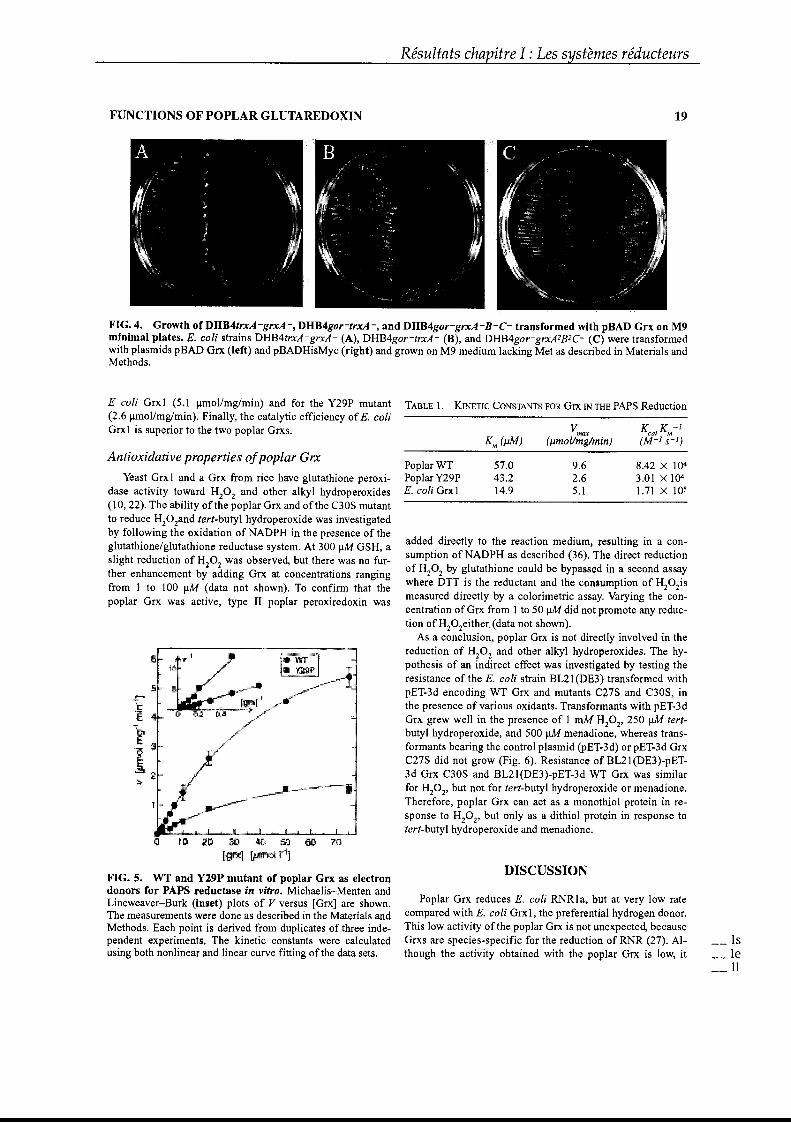
Résultats chapitre l : Les systèmes réducteurs
FUNCTIONS OF POPLAR GLUTAREDOXIN 19
FIG. 4. Growtb of DHB4trxA -grxA -, DHB4gor-trxA -, and DHB4gor-grxA -B-C- transformed witb pBAD Grx on M9minimal plates. E. coli strains DHB4trxA -grxA - (A), DHB4gor-trxA- (B), and DHB4gor-grxA1B1C- (C) were transformedwith plasmids pBAD Grx (left) and pBADHisMyc (right) and grown on M9 medium lacking Met as described in Materials andMethods.
added directly to the reaction medium, resulting in a consumption of NADPH as described (36). The direct reductionof H20 2 by glutathione could be bypassed in a second assaywhere DTT is the reductant and the consumption of H20 2ismeasured directly by a colorimetrie assay. Varying the concentration of Grx from 1 to 50 JlM did not promote any reduction of H20 2either (data not shown).
As a conclusion, poplar Grx is not directly involved in thereduction of H20 2 and other alkyl hydroperoxides. The hypothesis of an indirect effect was investigated by testing theresistance of the E. coli strain BL21(DE3) transformed withpET-3d encoding WT Grx and mutants C27S and C30S, inthe presence of various oxidants. Transformants with pET-3dGrx grew well in the presence of 1 mM H20 2, 250 JlM tertbutyI hydroperoxide, and 500 JlM menadione, whereas transformants bearing the control plasmid (PET-3d) or pET-3d GrxC27S did not grow (Fig. 6). Resistance of BL21(DE3)-pET3d Grx C30S and BL21(DE3)-pET-3d WT Grx was similarfor H20 2, but not for tert-butyl hydroperoxide or menadione.Therefore, poplar Grx can act as a monothiol protein in response to H20 2, but only as a dithiol protein in response totert-butyl hydroperoxide and menadione.
V K K-1max cal m
Km (j1M) (j1mol/mg/min) (M-1 S-l)
PoplarWT 57.0 9.6 8.42 X 10⁴PoplarY29P 43.2 2.6 3.01 X 10⁴
E. coli Grxl 14.9 5.1 1.71 X 10⁵
Antioxidative properties ofpoplar Grx
Yeast Grx1 and a Grx from rice have glutathione peroxidase activity toward H20 2 and other alkyl hydroperoxides(10,22). The ability ofthe poplar Grx and ofthe C30S mutantto reduce H20 2and tert...butyl hydroperoxide was investigatedby following the oxidation of NADPH in the presence of theglutathione/glutathione reductase system. At 300 JlM GSH, aslight reduction of H20 2 was observed, but there was no further enhancement by adding Grx at concentrations rangingfrom 1 to 100 JlM (data not shown). To confirm that thepoplar Grx was active, type II poplar peroxiredoxin was
E coli Grx1 (5.1 J,lmol/mg/min) and for the Y29P mutant TABLE 1. KINETIC CONSTANTS FOR Grx IN THE PAPS Reduction(2.6 Jlmollmg/min). Finally, the catalytic efficiency ofE. coliGrx1 is superior to the two poplar Grxs.
(j '020 :JJ ~ 5J 10 1[1
Ig~J !\WM:
FIG. 5. WT and Y29P mutant of poplar Grx as electrondonors for PAPS reductase in vitro. Michaelis-Menten andLineweaver-Burk (ioset) plots of V versus [Grx] are shown.The measurements were done as described in the Materials andMethods. Each point is derived from duplicates of three independent experiments. The kinetic constants were calculatedusing both nonlinear and linear curve fitting ofthe data sets.
DISCUSSION
Poplar Grx reduces E. coli RNRla, but at very low ratecompared with E. coli Grx l, the preferential hydrogen donor.This low activity ofthe poplar Grx is not unexpected, becauseGrxs are species-specific for the reduction of RNR (27). AIthough the activity obtained with the poplar Grx is low, it
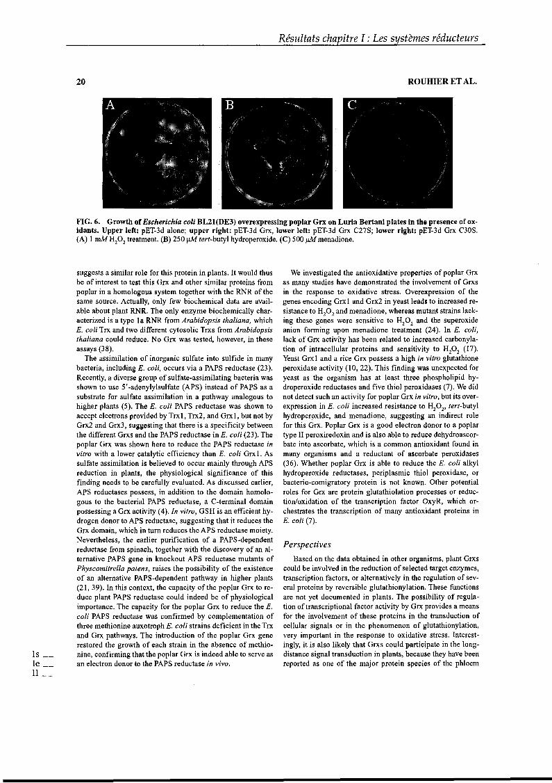
20
Résultats chapitre l : Les systèmes réducteurs
ROUHIER ET AL.
FIG. 6. Growth of Escherichia coli BL21(DE3) overexpressing poplar Grx on Luria Bertani plates in the presence of oxidants. Upper 1eR: pET-3d alone; upper right: pET-3d Grx, lower left: pET-3d Grx C27S; lower right: pET-3d Grx C30S.(A) 1 mM H20 2 treatment. (H) 250 JlM tert-butyl hydroperoxide. (C) 500~menadione.
suggests a similar raIe for this protein in plants. It would thusbe of interest to test this Grx and other similar proteins frompoplar in a homologous system together with the RNR of thesame source. ActuaUy, only few biochemical data are available about plant RNR. The only enzyme biochemically characterized is a type 1a RNR from Arabidopsis thaliana, whichE. coli Trx and two ditferent cytosolic Trxs from Arabidopsisthaliana could reduce. No Grx was tested, however, in theseassays (38).
The assimilation of inorganic sulfate into sulfide in manybacteria, including E. coli, occurs via a PAPS reductase (23).Recently, a diverse group of sulfate-assimilating bacteria wasshown to use 5'-adenylylsulfate (APS) instead of PAPS as asubstrate for sulfate assimilation in a pathway analogous tohigher plants (5). The E. coli PAPS reductase was shown toaccept electrons provided by Trx1, Trx2, and Grx1, but not byGrx2 and Grx3, suggesting that there is a specificity betweenthe different Grxs and the PAPS reductase in E. coli (23). Thepoplar Grx was shawn here to reduce the PAPS reductase invitro with a lower catalytic efficiency than E. coli Grx1. Assulfate assimilation is believed to occur mainly through APSreduction in plants, the physiological significance of thisfinding needs to he carefully evaluated. As discussed earlier,APS reductases possess, in addition to the domain homologous to the bacterial PAPS reductase, aC-terminal domainpossessing a Grx activity (4). In vitro, GSH is an efficient hydrogen donor to APS reductase, suggesting that it reduces theGrx domain, which in turn reduces the APS reductase moiety.Nevertheless, the earlier purification of a PAPS-dependentreductase from spinach, together with the discovery of an alternative PAPS gene in lmockout APS reductase mutants ofPhyscomitrella patens, raises the possibility of the existenceof an alternative PAPS-dependent pathway in higher plants(21, 39). In this context, the capacity of the poplar Grx to reduce plant PAPS reductase could indeed be of physiologicalimportance. The capacity for the poplar Grx to reduce the E.coli PAPS reductase was confirmed by complementation ofthree methionine auxotroph E. coli strains deficient in the Trxand Grx pathways. The introduction of the poplar Grx generestored the growth of each strain in the absence of methionine, confirming that the poplar Grx is indeed able to serve asan electron donor to the PAPS reductase in vivo.
We investigated the antioxidative properties of poplar Grxas many studies have demonstrated the involvement of Grxsin the response to oxidative stress. Overexpression of thegenes encoding Grx1 and Grx2 in yeast leads to increased resistance to H
20
2and menadione, whereas mutant strains lack
ing these genes were sensitive to H20 2 and the superoxideanion forming upon menadione treatment (24). In E. coli,lack of Grx activity has been related ta increased carbonylation of intracellular proteins and sensitivity to H20 2 (17).Yeast Grx1 and a rice Grx possess a high in vitro glutathioneperoxidase activity (10, 22). This finding was unexpected foryeast as the organism has at least three phospholipid hydroperoxide reductases and five thiol peroxidases (7). We didnot detect such an activity for poplar Grx in vitro, but its overexpression in E. coli increased resistance to H20 2, tert-butylhydroperoxide, and menadione, suggesting an indirect rolefor this Grx. Poplar Grx is a good electron donor to a poplartype II peroxiredoxin and is also able to reduce dehydroascorbate into ascorbate, which is a common antioxidant found inmany organisms and a reductant of ascorbate peroxidases(36). Whether poplar Grx is able to reduce the E. coli alkylhydroperoxide reductases, periplasmic thiol peroxidase, orbacterio-comigratory protein is not known. Other potentialroles for Grx are protein glutathiolation processes or reduction/oxidation of the transcription factor OxyR, which orchestrates the transcription of many antioxidant proteins inE. coli (7).
Perspectives
Based on the data obtained in other organisms, plant Grxscould be involved in the reduction of selected target enzymes,transcription factors, or alternatively in the regulation of several proteins by reversible glutathionylation. These functionsare not yet documented in plants. The possibility of regulation oftranscriptional factor activity by Grx provides a meansfor the involvement of these proteins in the transduction ofcellular signaIs or in the phenomenon of glutathionylation,very important in the response to oxidative stress. Interestingly, it is also likely that Grxs could participate in the longdistance signal transduction in plants, hecause they have beenreported as one of the major protein species of the phloem
FUNCTIONS OF POPLAR GLUTAREDOXIN
sap together with Trx (42). Given the sensitivity ofthis tissueto pathogen attacks, it will be of interest to test whether Grxand Trx are also involved in pathogen-resistance mechanisms.
ACKNOWLEDGMENTS
This research was supported by grants to Jean-PierreJacquot from the Ministère de l'Education Nationale, and toArne Holmgren from the Swedish Medical Research Council(13x-3529) and the Swedish Cancer Society (961).
ABBREVIATIONS
APS, 5'-adenylylsulfate; CDP, cytidine diphosphate; DTT,dithiothreitol; Grx, glutaredoxin; GSH, reduced glutathione;H20 2, hydrogen peroxide; IPTG, isopropylB-n-thiogalactopyranoside; PAPS, 3'.phosphoadenylylsulfate; RNR, ribonucleotide reductase; ROS, reactive oxygen species; Trx, thioredoxin; WT, wild type.
REFERENCES
1. Aslund F, Ehn B, Miranda-Vizuete A, Pueyo C, and Holmgren A. Two additional glutaredoxins exist in Escherichiacoli: glutaredoxin 3 is a hydrogen donor for ribonucleotidereductase in a thioredoxinlglutaredoxin 1 double mutant.Proc NatlAcad Sci USA 91: 9813-9817, 1994.
2. Aslund F, Zheng M, Beckwith J, and Storz G. Regulationofthe OxyR transcription factor by hydrogen peroxide andthe cellular thiol..disulfide status. Proc Natl Acad Sci USA96:6161-6165,1999.
3. Bedett BS and Stadtman ER. Protein oxidation in aging,disease, and oxidative stress. J Biol Chem 272: 2031320316, 1997.
4. Bick JA, Aslund F, Chen Y, and Leustek T. Glutaredoxinfunction for the carboxyl-terminal domain of the planttype 5' -adenylylsulfate reductase. Proc Natl Acad Sci U SA 95: 8404-8409, 1998.
5. Bick JA, Dennis JJ, Zylstra GJ, Nowack J, and Leustek T.Identification of a new class of 5'-adenylylsulfate (APS)reductases from sulfate-assimilating bacteria. J Bacteriol182: 135-142,2000.
6. Bjornstedt M, Xue J, Huang W, Akesson B, and HolmgrenA. The thioredoxin and glutaredoxin systems are efficientelectron donors to human plasma glutathione peroxidase. JBiol Chem 269: 29382-29384, 1994.
7. Carmel-HarelO and Storz G. Roles ofthe glutathione- andthioredoxin-dependent reduction systems in the Escherichiacoli and Saccharomyces cerevisiae responses to oxidativestress. Annu Rev Microbiol 54: 439-446, 2000.
8. Chae HZ, Chung SJ, and Rhee SG. Thioredoxin-dependentperoxide reductase from yeast. J Biol Chem 269: 2767027678, 1994.
9. Chrestensen CA, Starke D~ and Mieyal J1. Acute cadmium exposure inactivates thioltransferase (glutaredoxin),inhibits intracellular reduction of protein-glutathionyl-
Résultats chapitre l : Les systèmes réducteurs
21
mixed disulfides, and initiates apoptosis. J Biol Chem 275:26556-26565,2000.
10. Collinson EJ, Wheeler GL, Garrido EO, Avery AM, AveryS~ and Grant CM. The yeast glutaredoxins are active asglutathione peroxidases. J Biol Chem 277: 16712-16717,2002.
Il. Daily D, Vlamis-Gardikas A, Offen D, Mittelman L,Melamed E, Holmgren A, and Barzilai A. Glutaredoxinprotects cerebellar granule neurons from dopamineinduced apoptosis by activating NF...kappa B via Ref-l. JBiol Chem 276: 1335-1344,2001.
12. Finkel T and Holbrook N1. Oxidants, oxidative stress andthe biology ofaging. Nature 408: 239-247, 2000.
13. Garrido EO and Grant CM. Role of thioredoxins in the response of Saccharomyces cerevisiae to oxidative stress induced by hydroperoxides. Mol Microbiol 43: 993-1003,2002.
14. Gille G and Sigler K. Oxidative stress and living cells.Folia Microbiol40: 131-152, 1995.
15. Gladyshev VN, Liu A, Novoselov S~ Krysan K, Sun QA,Kryukov VM, Kryukov G~ and Lou MF. Identificationand characterization of a new mammalian glutaredoxin(thioltransferase), Grx2. J Biol Chem 276: 30374-30380,2001.
16. Grant CM. Role of the glutathione/glutaredoxin andthioredoxin systems in yeast growth and response to stressconditions. Mol Microbiol39: 533-541,2001.
17. Holmgren A. Glutathione-dependent synthesis of deoxyribonucleotides. Characterization of the enzymatic mechanism of Escherichia coli glutaredoxin. J Biol Chem 254:3672-3678, 1979.
18. Holmgren A. Thioredoxin and glutaredoxin systems. JBiol Chem 264: 13963-13966, 1989.
19. Holmgren A. Antioxidant function of thioredoxin andglutaredoxin systems. Antioxid Redox Signal 2: 811-820,2000.
20. Klatt P and Lamas S. Regulation of protein function byS-glutathiolation in response to oxidative and nitrosativestress. Eur J Biochem 267: 4928-4944, 2000.
21. KoprivovaA, Meyer AJ, Schween G, Herschbach C, ReskiR, and Kopriva S. Functional knockout of the adenosine5'-phosphosulfate reductase gene in Physcomitrellapatens revives an old route of sulfate assimilation. J BiolChem 277: 32195-32201, 2002.
22. Lee KO, Lee JR, Yoo JY, Jang HH, Moon JC, Jung BG, ChiYH, Park SK, Lee SS, Lim CO, Yun DJ, Cho MJ, and LeeSV. GSH-dependent peroxidase activity of the rice (Oryzasativa) glutaredoxin, a thioltransferase. Biochem BiophysRes Commun 296: 1152-1156,2002.
23. Lillig CH, Prior A, Schwenn JD, Aslund F, Ritz D, VlamisGardikas A, and Holmgren A. New thioredoxins andglutaredoxins as electron donors of 3' ..phosphoadenylylsulfate reductase. J Biol Chem 274: 7695-7698, 1999.
24. Luikenhuis S, Perrone G, Dawes I~ and Grant CM. Theyeast Saccharomyces cerevisiae contains two glutaredoxingenes that are required for protection against reactive oxygen species. Mol Biol Ce1l9: 1081-1091, 1998.
25. Lundberg M, Johansson C, Chandra J, Enoksson M,Jacobsson G, Ljung J, Johansson M, and Holmgren A.Cloning and expression of a novel human glutaredoxin
15 __le __Il __
22
with mitochondrial and nuclear isoforms. J Biol Chem276:26269-26275,2001.
26. Lundstrom-Ljung J, Vlamis-Gardikas A, Aslund F, andHolmgren A. Reactivity of glutaredoxins 1, 2 and 3 fromEscherichia coli and protein disulfide isomerase towardsglutathionyl-mixed disulfides in ribonuclease A. FEBSLett 443: 85-88, 1999.
27. Luthman M, Eriksson S, Holmgren A, and Thelander L.Glutathione-dependent hydrogen donor system for calfthymus ribonucleoside-diphosphate reductase. Proc NatlAcad Sci USA 76: 2158-2162, 1979.
28. Minakuchi K, Yabushita T, Masumura T, Ichihara K, andTanaka K. Cloning and sequence analysis of a cDNA encoding rice glutaredoxin. FEBS Lett 337: 157-160, 1994.
29. Morell S, Follmann H, and Haberlein I. Identification andlocalization of the first glutaredoxin in leaves of a higherplant. FEBS Lett 369: 149-152, 1995.
30. Potamitou A, Holmgren A, and Vlamis-Gardikas A. Protein levels of Escherichia coli thioredoxins and glutaredoxins and their relation to null mutants, growth phase,and function. J Biol Chem 277: 18561-18567,2002.
31. Prieto-Alamo MJ, Jurado J, Gallardo-Madueno R, MonjeCasas F, Holmgren A, and Pueyo C. Transcriptional regulation of glutaredoxin and thioredoxin pathways and related enzymes in response to oxidative stress. J Biol Chem275: 13398-13405,2000.
32. Prinz WA, Aslund F, Holmgren A, and Beckwith 1. Therole of the thioredoxin and glutaredoxin pathways in reducing protein disulfide bonds in the Escherichia coli cytoplasm. J Biol Chem 272: 15661-15667, 1997.
33. Qin J, Yang Y, Velyvis A, and Gronenborn A. Molecularviews of redox regulation: three-dimensional structures of
-redox regulatory proteins and protein complexes. AntioxidRedox Signal 2: 827-840, 2000.
34. Rodriguez-Manzaneque MT, Ros J,. Cabiscol E, SorribasA, and Herrero E. Grx5 glutaredoxin plays a central role inprotection against protein oxidative damage in Saccharomyces cerevisiae. Mol Cell Biol 19: 8180-8190, 1999.
35. Rouhier N, Gelhaye E, and Jacquot J~ Exploring the activesite of plant glutaredoxin by site-directed mutagenesis.FEBS Lett 511: 145-149,2002.
36. Rouhier N, Gelhaye E, and Jacquot J~ Glutaredoxindependent peroxiredoxin from poplar. Protein-protein interaction andcatalytic mechanism. J Biol Chem 277:13609-13614,2002.
Résultats chapitre l : Les systèmes réducteurs
ROUHIER ET AL.
37. Rouhier N, Gelhaye E, Sautiere PE, and Jacquot J~ Enhancement of poplar glutaredoxin expression by optimization of the cDNA sequence. Protein Expr Purif 24:234-241, 2002.
38. Sauge-Merle S, Falconet D, and Fontecave M. An active ribonucleotide reductase from Arabidopsis thaliana cloning,expression and characterization of the large subunit. Eur JBiochem 266: 62-69, 1999.
39. Schwenn JD. Sulphate assimilation in higher plants: athioredoxin PAPS reductase from spinach leaves. Z Naturforsch [Cl 4: 504-508, 1989.
40. Shenton D, Perrone G, Quinn KA, Dawes I~ and GrantCM. Regulation of protein S-thiolation by glutaredoxin 5in the yeast Saccharomyces cerevisiae. J Biol Chem 277:16853-16859,2002.
41. Shi J, Vlamis-Gardikas A, Aslund F, Holmgren A, andRosen B~ Reactivity of glutaredoxins 1, 2, and 3 from Escherichia coli shows that glutaredoxin 2 is the primary hydrogen donor to ArsC-catalyzed arsenate reduction. J BiolChem 274: 36039-36042, 1999.
42. Szederkenyi J, Komor E, and Schobert C. Cloning of thecDNA for glutaredoxin, an abundant sieve-tube exudateprotein from Ricinus communis L. and characterisation ofthe glutathione-dependent thiol-reduction system in sievetubes. Planta 202: 349-356, 1997.
43. Vlamis-Gardikas A and Holmgren A. Thioredoxin andglutaredoxin isoforms. Methods Enzymol 347: 286-296,2002.
44. Vlamis-Gardikas A, Potamitou A, Zarivach R, HochmanA, and Holmgren A. Characterization of Escherichia colinull mutants for glutaredoxin 2. J Biol Chem 277:10861-10868,2002.
Address reprint requests to:Dr. Jean-Pierre Jacquot
Unité Mixte de Recherche 1136 IaM(Interaction arbres microorganismes)
INRA -UHP Nancy I. Université Henri Poincaré54506 Vandoeuvre Cedex, France
E-mail: [email protected]
Received for publication September 10, 2002; accepted October 7,2002.
Résultats chapitre I : Les systèmes réducteurs
Les seules structures tridimensionnelles de Grx connues pour l’instant sont 4
protéines d’E. coli, 3 Grx bicystéiniques et une protéine homologue NrdH, une Grx du
phage T4, et enfin les Grx porcine et humaine. Ces structures ont été résolues soit par
RMN soit par cristallographie. Différentes conditions ont été testées pour la Grx de
peuplier, mais ni la protéine sauvage ni aucun des mutants du site actif sous forme réduite
ou oxydée n’ont cristallisé correctement. Finalement, la solution a été de fixer du GSH de
manière covalente sur le mutant de la deuxième cystéine du site actif, Grx C30S. Ensuite,
une fois les conditions de cristallisation connues, la protéine mutante a été exprimée dans
une souche d’E. coli auxotrophe pour la méthionine, sur milieu minimum et en présence
de sélénométhionine. En effet, la faible identité de séquence entre la Grx de peuplier et les
Grx de structure connue imposait d’avoir recours au marquage par la sélénométhionine
puisque la méthode de remplacement moléculaire n’a pas permis de résoudre la structure.
L’incorporation de la sélénométhionine de manière homogène dans toutes les molécules a
été vérifiée par spectrométrie de masse en mode électrospray (augmentation de masse de
47.1 Da). Les cristaux obtenus diffractent à 1.73Å, permettant de déterminer la première
structure 3D de Grx de plantes. La description de cette structure fera l’objet d’un article
séparé.
Article numéro 10 :
d'Ambrosio K, Kauffmann B, Rouhier N, Benedetti E, Jacquot JP, Aubry A, Corbier C. Crystallization and preliminary X ray data of poplar glutaredoxin.
Acta Crystallogr. D Biol. Crystallogr., sous presse.
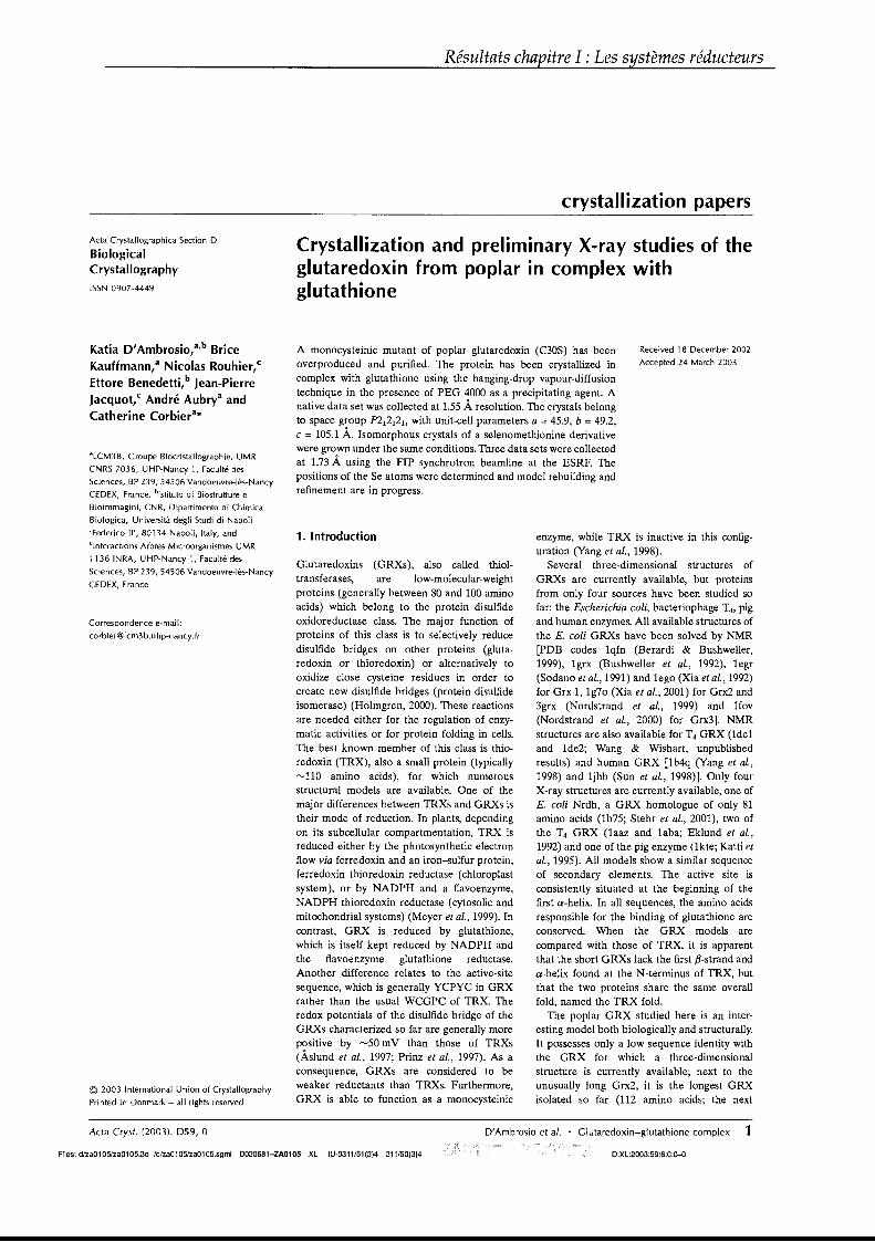
Résultats chapitre l : Les systèmes réducteurs
crystallization papers
Acta Crystallographica Section D
BiologicalCrystallography
ISSN 0907-4449
Crystallization and preliminary X-ray studies of theglutaredoxin from poplar in complex withglutathione
A monocysteinic mutant of poplar glutaredoxin (C30S) has beenoverproduced and purified. The protein has been crystallized incomplex with glutathione using the hanging-drop vapour-diffusiontechnique in the presence of PEG 4000 as a precipitating agent. Anative data set was collected at 1.55 À resolution. The crystals belongto space group n 1212J, with unit-cell parameters a = 45.9, b = 49.2,c = 105.1 À. Isomorphous crystals of a selenomethionine derivativewere grown under the same conditions. Three data sets were colIectedat 1.73 À using the FIP synchrotron beamline at the ESRF. Thepositions of the Se atoms were determined and model rebuilding andrefinement are in progress.
Katia D'Ambrosio,a,b Brice
Kauffmann,a Nicolas Rouhier,C
Ettore Benedetti,b Jean-Pierre
Jacquot,C André Aubrya and
Catherine Corbiera*
aLCM3B, Groupe Biocristallographie, UMR
CNRS 7036, UHP-Nancy 1, Faculté des
Sciences, BP 239,54506 Vandoeuvre-lès-Nancy
CEDEX, France, blstituto di Biostrutture e
Bioimmagini, CNR, Dipartimento di Chimica
Biologica, Università degli Studi di Napoli
'Federico 11', 80134 Napoli, Italy, and
Clnteractions Arbres Microorganismes UMR
1136 INRA, UHP-Nancy 1, Faculté des
Sciences, BP 239, 54506 Vandoeuvre-lès-Nancy
CEDEX, France
Correspondence e-mail :
© 2003 International Union of Crystallography
Printed in Denmark - ail rights reserved
1. 1ntroduction
Glutaredoxins (GRXs), also called thioltransferases, are low-molecular-weightproteins (generally between 80 and 100 aminoacids) which belong to the protein disulfideoxidoreductase class. The major function ofproteins of this class is to selectively reducedisulfide bridges on other proteins (glutaredoxin or thioredoxin) or alternatively tooxidize close cysteine residues in order tocreate new disulfide bridges (protein disulfideisomerase) (Holmgren, 2000). These reactionsare needed either for the regulation of enzymatie activities or for protein folding in cells.The best known member of this class is thioredoxin (TRX), also a small protein (typically""110 amino acids), for which numerousstructural models are available. One of themajor differences between TRXs and GRXs istheir mode of reduction. In plants, dependingon its subcellular compartmentation, TRX isreduced either by the photosynthetic electronflow via ferredoxin and an iron-sulfur protein,ferredoxin thioredoxin redu'ctase (chloroplastsystem), or by NADPH and a flavoenzyme,NADPH thioredoxin reductase (cytosolic andmitochondrial systems) (Meyer et al., 1999). Incontrast, GRX is reduced by glutathione,which is itself kept reduced by NADPH andthe flavoenzyme glutathione reductase.Another difference relates to the active-sitesequence, which is generally YCPYC in GRXrather than the usual WCGPC of TRX. Theredox potentials of the disulfide bridge of theGRXs characterized so far are generally morepositive by ""50 mV than those of TRXs(Àslund et al., 1997; Prinz et al., 1997). As aconsequence, GRXs are considered to beweaker reductants than TRXs. Furthermore,GRX is able to function as a monocysteinic
Received 18 December 2002
Accepted 24 March 2003
enzyme, while TRX is inactive in this configuration (Yang et al., 1998).
Several three-dimensional structures ofGRXs are currently available, but proteinsfrom only four sources have been studied sofar: the Escherichia coli, bacteriophage T4, pigand human enzymes. AlI available structures ofthe E. coli GRXs have been solved by NMR[PDB codes 1qfn (Berardi & BushweIler,1999), 19rx (Bushweller et al., 1992), 1egr(Sodano et al., 1991) and 1ego (Xia et al., 1992)for Grx 1, 1970 (Xia et al., 2001) for Grx2 and3grx (Nordstrand et al., 1999) and 1fov(Nordstrand et al., 2000) for Grx3]. NMRstructures are also available for T4 GRX (lde1and 1de2; Wang & Wishart, unpublishedresults) and human GRX [lb4q (Yang et al.,1998) and 1jhb (Sun et al., 1998)]. Only fourX-ray structures are currently available, one ofE. coli Nrdh, a GRX homologue of only 81amino acids (lh75; Stehr et al., 2001), two ofthe T4 GRX (laaz and 1aba; Eklund et al.,1992) and one of the pig enzyme (lkte; Katti etal., 1995). AlI models show a similar sequenceof secondary elements. The active site isconsistently situated at the beginning of thefirst a-helix. In aIl sequences, the amino acidsresponsible for the binding of glutathione areconserved. When the GRX models arecompared with those of TRX, it is apparentthat the short GRXs lack the first ,B-strand anda-helix found at the N-terminus of TRX, butthat the two proteins share the same overallfold, named the TRX fold.
The poplar GRX studied here is an interesting model both biologicalIy and structuraIly.It possesses only a low sequence identity withthe GRX for which a three-dimensionalstructure is currently available; next to theunusually long Grx2, it is the longest GRXisolated so far (112 amino acids; the next
Acta Cryst. (2003). 059, a D'Ambrosio et al. • Glutaredoxin-glutathione complex 1
Files: d/za0105/za0105.3d Id/za0105/za0105.sgml 0030681-ZA010S XL IU-0311/51(3)4 311/50(3)4 O:XL:2003:59:6:0:0-0
Résultats chapitre l : Les systèmes réducteurs
crystallization papers
Table 1Statistics of the X-ray diffraction data for the SeMet GRX crystals.
Values in parentheses refer to the highest resolution shell.
Figure 1Crystals of C30S GRX in complex with glutathione(approximate dimensions 0.5 x 0.1 x 0.02 mm).
Remote
0.97241.73 (1.77-1.73)1694624695497.9 (93.0)3.614.7 (36.6)13.7 (2.0)
Edge
0.98041.73 (1.77-1.73)1674344628596.4 (89.6)3.624.0 (19.8)17.0 (2.3)
3. Results and discussion
ln our hands, crystallization trials usingstandard procedures failed to produce crystaIs from the wild-type protein or for severalvariants of the active site. However, the useof a monocysteinic mutant (C30S) allowedus to obtain a stable mixed disulfide complex(C30S-g1utathione) that crystallized within1 d under the conditions described above(Fig. 1). Crystals belong to space groupn l212h with unit-cell parameters a = 45.7,b = 49.09, c = 104.81 Â (Table 2). A nativedata set was collected at 1.55 Â resolution.Molecular-replacement trials using theprogram AMoRe and performed using the
for the selenomethionyl derivative werecollected on beamline BM30A (FIP; Roth etal., 2002) at the ESRF, using a MARResearch 345 image-plate detector. For thelatter data set, three wavelengths werechosen from the fluorescence spectrumcorresponding to the peak, inflection pointand high-energy remote. The data wereprocessed using DENZO and scaled withSCALEPACK (Otwinowski & Minor,1997). Statistics of the data are presented inTable 1.
Peak
0.98021.73 (1.77-1.73)1434004633696.1 (92.0)3.094.1 (13.3)18.2 (3.4)0.3860.899
Wavelength (Â)Resolution (Â)Observed reflectionsUnique reflectionsCompleteness (%)MultiplicityRsym (%)I/(J'(I)Mean figure of merit before solvent flatteningMean figure of merit after solvent flattening
replaced by Ser using the PCR strategydescribed in Rouhier et al. (2002a). TheC30S mutated protein (40 mg ml-l) wasmixed with an equal volume of 100 mMphosphate buffer containing 1 mM EDTA atpH 8.0. The sample was reduced with100 mM DTT for 15 min at 277 K. Glutathione disulfide (GSSG) was subsequentlyadded to a concentration of 500 mM. After30 min incubation at 277 K, excess GSSGwas removed by gel filtration using aSephadex G25 column pre-equilibrated with50 mM phosphate buffer pH 6.0. Theconcentration was determined by UVabsorption at 276 nm, using a molar extinction coefficient of 4350 M-I cm-l.
2.3. Crystallization
Crystallization experiments were carriedout using the hanging-drop vapour-diffusionmethod (McPherson, 1999). The search forinitial crystallization conditions wasperformed using Hampton Research CrystalScreen kits 1 and II (Jancarik & Kim, 1991;Cudney et al., 1994) and Clear StrategyScreens (Molecular Dimensions Ltd). Thewells contained 800 Jil of precipitant solution and the drops were composed of 2 Jil ofreservoir solution and 2 Jil of protein solution at various concentrations. The mostpromising crystallization conditions wererefined by variation of protein or precipitantconcentrations, pH or the ratio of protein toreservoir in the droplet. Finally, the bestcrystals were obtained at a protein concentration of 6.5 mg ml- l from a solutioncontaining 28% PEG 4000 and 0.2 M(NH4hS04 at 285 K.
2.4. Data collection
X-ray diffraction experiments werecarried out at 100 K. Since the crystallizationsolution was suitable to provide cryoprotection, the crystals were quickly washedin the reservoir solution and immediatelyflash-frozen in a nitrogen-gas stream at100 K. The data sets for the native protein or
2.2. Site-directed mutagenesis andpreparation of the mixed glutaredoxinglutathione complex
ln order to isolate a stable mixed disulfideintermediate between GRX and glutathione,the second active-site cysteine has been
longest is the mammalian type with 106residues; Rouhier et al., 2002). This is thefirst GRX from a plant that has been crystallized so far and the first X-ray structure ofa GRX in complex with glutathione. Interestingly, this protein has been demonstratedto be an efficient donor to a peroxiredoxin,an enzyme which is able to selectivelyremove various hydroperoxides (Rouhier etal., 2001, 2002b). This property has not beenreported in other species so far; the variousperoxiredoxins studied instead use TRX asan electron donor. It is thus of great interestto study the molecular interaction betweenthe two proteins in this unique system. Wehave recently reported the crystallizationand preliminary X-ray data of the peroxiredoxin component (Echalier et al., 2002).This paper reports parallel work on GRX,the other partner in this model proteinprotein interaction.
2. Material and methods
2.1. Cloning, expression and purification
The cloning, expression and purificationof wild-type and C30S mutant poplar GRXhas been described in Rouhier et al. (2002,2002a). Typically, 100 mg of homogeneousprotein was obtained and was stored frozenin 1 ml aliquots in 30 mM Tris-HClcontaining 1 mM EDTA and 14 mMfJ-mercaptoethanol at a concentration of15 mg ml-l at 252 K. The purity of thefractions was monitored by SDS-PAGE.
For the production of the selenomethione-substituted GRX, a special straindeficient in methionine synthesis[BL21(DE3)Met-] was cotransformed bypET-Grx and pSBET. One ampicillin- andkanamycin-resistant colony was amplified toIl in M9 medium supplemented with aIl 19regular amino acids and selenomethionineeach at 50 mg ml-l. When the culturereached the exponential stage, proteinsynthesis was induced by the addition of100 JiM IPTG. The subsequent purificationprocedure was identical to that for thenative protein. Selenomethionine substitution was checked by electrospray massspectrometry analysis. A mass increase of47.1 Da was observed compared with thenative protein, as expected from thepresence of one SeMet residue per molecule.
2 D'Ambrosio et al. • Glutaredoxin-glutathione complex Acta Cryst. (2003). 059, 0
Files: d/za0105/za0105.3d Id/za0105/za0105.sgml 0030681-ZA010S XL IU-0311151(3)4 311/50(3)4 O:XL:2003:59:6:0:0-0
Résultats chapitre l : Les systèmes réducteurs
crystallization papers
Figure 2Histogram of the number of trials and their corresponding minimalfunction values Rmin obtained with Shake-and-Bake (SnB). Using700 reflections selected in the resolution range 25-1.66 Â. and 230invariants, a maximum of four sites were searched for 1000 trials of20 cycles each. Two sites were found in 118 successful trials. Thebest value of Rmin is 0.006.
Table 2Crystallization conditions of the GRX-glutathionecomplex and X-ray characterization.
coordinates of the closest GRXs as searchmodels were unsuccessful. A SeMet-substituted protein was therefore prepared andcrystallized under the same conditions.Crystals only appeared after one month, butcould be obtained within one week by theuse of microseeding techniques. TInee datasets were collected at three wavelengths to1.73 Â. resolution (Table 1). A search for thepositions of two Se atoms per asymmetricunit was performed with the data setcollected at the peak of the selenium K edgeusing either the Patterson method (CNS;Brünger et al., 1998) or direct methods(Shake-and-Bake version 2.1; Weeks &Miller, 1999) (Fig. 2). Both methods unambiguously allowed the identification of thesame two sites, as expected from thepresence of one molecule of SeMet permolecule and two molecules per asymmetric
Holmgren, A. (2000). Antioxid. Redox Signal. 2,811-820.
Jancarik, 1 & Kim, S.-H. (1991). J. Appl. Cryst. 24,409-411.
Katti, S. K., Robbins, A. H., Yang, Y. & Wells,W. W. (1995). Protein Sei. 4, 1998-2005.
La Fortelle, E. de & Bricogne, G. (1997). MethodsEnzymol. 276, 472-494.
McPherson, A. (1999). Crystallization of Biological Macromolecules. New York: Cold SpringHarbor Laboratory Press.
Meyer, Y., Verdoucq, L. & Vignols, P. (1999).Trends Plant Sei. 4, 388-394.
Nordstrand, K., Aslund, P., Holmgren, A., Otting,G. & Berndt, K. D. (1999). J. Mol. Biol. 286,541-552.
Nordstrand, K., Sandstrom, A., Aslund, P.,Holmgren, A., Otting, G. & Berndt, K. D.(2000). J. Mol. Biol. 303, 423-432.
Otwinowski, Z. & Minor, W. (1997). MethodsEnzymol. 276, 307-326.
Perrakis, A., Morris, R. 1 & Lamzin, V. (1999).Nature Struct. Biol. 6,458-463.
Prinz, W. A., Aslund, P., Holmgren, A. & Beckwith, 1 (1997). J. Biol. Chem. 27, 15661-15667.
Roth, M., Carpentier, :P., Kaikati, O., Joly, l,Charrault, P., Pirocchi, M., Kahn, R., Fanchon,E., Jacquamet, L., Borel, P., Bertoni, A., IsraelGouy, :P. & Ferrer, 1 L. (2002). Acta Cryst. D58,805-814.
Rouhier, N., Gelhaye, E. & Jacquot, 1 :P. (2002a).FEBS LeU. 511, 145-149.
Rouhier, N., Gelhaye, E. & Jacquot, 1 P. (2002b).J. Biol. Chem. 277, 13609-13614.
Rouhier, N., Gelhaye, E., Sautiere, :P. E., Brun, A.,Laurent, :P., Tagu, D., de Fay, E., Meyer, Y. &Jacquot, 1 :P. (2001). Plant Physiol. 127, 12991309.
Rouhier, N., Gelhaye, E., Sautiere, :P. E. &Jacquot, 1. :P. (2002). Protein Expr. Purif 24,234-24l.
Sodano, :P., Xia, T. H., Bushweller, 1 H., Bjornberg, O., Holmgren, A., Billeter, M. &Wuthrich, K. (1991). J. Mol. Biol. 221, 13111324.
Stehr, M., Schneider, G., Aslund, P., Holmgren, A.& Lindqvist, Y. (2001). J. Biol. Chem. 276,35836-3584l.
Sun, C., Berardi, M. 1 & Bushweller, 1 H. (1998).J. Biol. Chem. 280, 687-70l.
Weeks, C. M. & Miller, R. (1999). Acta Cryst. D55,492-500.
Xia, B., Vlamis-Gardikas, A., Holmgren, A.,Wright, :P. E. & Dyson, H. 1 (2001). J. Mol.Biol. 310, 907-918.
Xia, T. H., Bushweller, 1 H., Sodano,:P., Billeter,M., Bjornberg, O., Holmgren, A. & Wuthrich,K. (1992). Protein Sei. 1, 310-321.
Yang, Y., Jao, S., Nanduri, S., Starke, D. W., Mieyal,1 1 & Qin, 1 (1998). Biochemistry, 37, 1714517156.
unit. Peak-wavelength structurefactors were phased withSHARP (de La Fortelle &Bricogne, 1997) to 1.73 Â. resolution and the phases wereimproved by solvent flatteningusing DM (CollaborativeComputational Project, Number4, 1994) and SOLOMON(Abrahams & Leslie, 1996).Automatic model building withARP/wARP (Perrakis et al.,1999) led to the repositioning of95 of 112 residues, with RandRfree values of 20 and 250/0 at thebeginning of the refinementprocess. Model completion andrefinement are currently inprogress.
We would like to thank 1. L. Ferrer at theESRF for his help during data collection. Weare very grateful to Dr Claude Schauber fordetermining the molecular weight of thenative GRX and of the SeMet derivative.
References
Abrahams, 1 :P. & Leslie, A. G. W. (1996). ActaCryst. D52, 30-42.
Àslund, P., Berndt, K. D. & Holmgren, A. (1997).J. Biol. Chem. 272,30780-30786.
Berardi, M. 1 & Bushweller, 1 H. (1999). J. Mol.Biol. 292, 151-16l.
Brünger, A. T., Adams, :P. D., Clore, G. M.,DeLano, W. L., Gros, :P., Grosse-Kunstleve,R. W., Jiang, 1 S., Kuszewski, l, Nilges, M.,Pannu, N. S., Read, R. l, Rice, L. M., Simonson,T. & Warren, G. L. (1998). Acta Cryst. D54, 90592l.
Bushweller, 1 H., Aslund, P., Wuthrich, K. &Holmgren, A. (1992). Biochemistry, 31, 92889293.
Collaborative Computational Project, Number 4(1994). Acta Cryst. D50, 760-763.
Cudney, R., Patel, S., Weisgraber, K., Newhouse,Y. & McPherson, A. (1994). Acta Cryst. D50,414-423.
Echalier, A., Corbier, c., Rouhier, N., Jacquot, 1. :P.& Aubry, A. (2002). Acta Cryst. D58, 15011503.
Eklund, H., Ingelman, M., Soderberg, B. O., Dhlin,T., Nordlund, :P., Nikkola, M., Sonnerstam, D.,Joelson, T. & Petratos, K. (1992). J. Mol. Biol.228, 596-618.
6.528% PEG 4000,
0.2 M (NH4)zS040.5 x 0.2 x 0.02a = 45.7, b = 49.09,
c = 104.81n 12121
22.49491.55
0.2331 0.800/0.289 0.856
Minimal function range (Rmin)
180··160140120
~ 100··~ 80·
60··4020··o0.00610.063
Protein concentration (mg ml-1)
Crystallization solution
Crystal dimensions (mm)Unit-cell parameters (A)
Space groupMolecules per asymmetric unitVM (A3 Da-1
)
Solvent content (%)Diffraction Iimit (Â.)
Acta Cryst. (2003). 059, 0 D'Ambrosio et al. • Glutaredoxin-glutathione complex 3
Files: d/za0105lza0105.3d /d/za0105/za0105.sgml 0030681-ZA010S XL IU-0311/51(3)4 311/50(3)4 O:XL:2003:59:6:0:0-0


Chapitre II : les protéines cibles
Résultats chapitre II : Les protéines cibles
Un des objectifs de mon travail de thèse était d’identifier des protéines cibles
des Trx cytosoliques ou de la Grx de peuplier isolées par notre équipe. Différentes
approches sont possibles, de la technique du double hybride, à l’utilisation de
monobromobimane (mBBr), un produit fluorescent qui réagit avec les groupements
thiols ou à la création de mutants monocystéiniques pouvant être utilisés soit pour
créer des colonnes d’affinité et retenir de manière covalente des protéines cibles soit
pour réaliser des expériences d’immunoprécipitation (Verdoucq et al., 1999 ;
Motohashi et al., 2001 ; Yano et al., 2001 ; Balmer et al., 2003 ; Vignols et al., 2003).
Par ailleurs, Trx et Grx ayant une fonction et un mode d’action commun, il n’est pas
illusoire de penser qu’elles peuvent réduire les même protéines. En effet, même si
leurs potentiels rédox et leurs séquences primaires sont différents, leurs structures
tridimensionnelles sont très voisines. Ainsi, plusieurs enzymes ou protéines
possèdent cette capacité d’être réduite par les Trx ou les Grx. C’est le cas
notamment de la PAPS réductase ou de la RNR d’E. coli et d’une Gpx ou de divers
facteurs de transcription tels que AP 1 ou Nf-kB de mammifères (Bjornstedt et al.,
1994 ; Prinz et al., 1997 ; Lillig et al., 1999 ; Hirota et al., 2000).
Actuellement, un certain nombre de protéines cibles des Trx de plantes ont
été identifiées mais cela concerne surtout des protéines chloroplastiques (voir
tableau II de l’introduction). Deux types de protéines cibles, connues pour être Trx
dépendantes, ont été isolées et caractérisées lors de mon travail de thèse. Il s’agit de
deux isoformes de Prx, de type II et de type Q, et de deux isoformes de MsrA, l’une
étant cytosolique et l’autre plastidiale. Ces deux types d’enzymes possèdent en outre
une propriété commune retrouvée dans un nombre limité d’enzymes qui est la
transformation du groupement thiol des cystéines en SOH lors de la catalyse (pour
revue sur les SOH voir Claiborne et al., 1999 ; Claiborne et al., 2001).
La caractérisation de la Prx de type II est reportée dans les articles 11 à 14,
alors que celle de la Prx Q et des deux isoformes de MsrA fait l’objet des deux
articles en préparation 15 et 16.
Résultats chapitre II : Les protéines cibles
Cet article présente la mise en évidence d’une Prx de type II de peuplier
(appelée à l’origine type C avant qu’une classification plus claire ne soit définie). La
protéine recombinante exprimée chez E. coli et purifiée est une protéine
monomérique qui présente de manière surprenante un doublet sur gel d’acrylamide
quel que soit son état de réduction. L’activité anti-oxydante de cette enzyme a tout
d’abord été vérifiée par un test de protection de plasmide en présence d’un système
générateur de radicaux oxygénés. Ensuite, une activité de réduction de divers
hydroperoxydes (H2O2, t-BOOH, CUOOH) a été établie en présence des deux
systèmes réducteurs que sont les systèmes Trx et Grx. Par ailleurs, l’étude de
l’expression et de la localisation de cette enzyme a été entreprise. Les transcrits
codant pour la Prx de type II et la protéine ont été détectés dans les trois organes
testés avec une prédominance dans les feuilles par rapport aux racines et aux tiges.
Des études d’immunolocalisation sur des coupes de feuilles ont montré la présence
de cette protéine dans des structures plastidiques présentes dans les tubes criblés
du phloème. Cette localisation est inattendue puisque la protéine ne possède pas de
peptide de transit apparent. Elle pourrait être expliquée par le fait qu’il existe une
isoforme de Prx II présumée plastidiale chez A. thaliana et que l’homologue de cette
isoforme existe certainement chez le peuplier.
Article numéro 11 :
Rouhier N, Gelhaye E, Sautière PE, Brun A, Laurent P, Tagu D, Gérard J, de Faÿ E, Meyer Y, Jacquot JP. (2001) Isolation and characterization of a new peroxiredoxin from poplar sieve tubes
that uses either glutaredoxin or thioredoxin as a proton donor.
Plant Physiol. 127(3):1299-309.
Résultats chapitre II : Les protéines cibles
Isolation and Characterization of a New Peroxiredoxinfrom Poplar Sieve Tubes That Uses Either Glutaredoxin orThioredoxin as a Proton Donor1
Nicolas Rouhier, Eric Gelhaye, Pierre-Eric Sautiere, Annick Brun, Pascal Laurent, Denis Tagu,Joelle Gerard, Elisabeth de Fay, Yves Meyer, and Jean-Pierre Jacquot*
Unité Mixte de Recherche Interaction Arbres Microorganisms, Institut National de la RechercheAgronomique-Université Henri Poincaré Nancy I. Biochimie et Biologie Moléculaire Végétales, UniversitéHenri Poincaré, 54506 Vandoeuvre cedex, France (N.R., E.G., A.B., P.L., D.T., J.G., E.d.F., J.-P.J.); Laboratoired'Endocrinologie des Annélides Equipe Enseignement Supérieur Associé 97, Université des Sciences etTechnologies de Lille, 59655 Villeneuve d'Ascq cedex, France (P.-E.S.); and Laboratoire de Physiologie et deBiologie Moléculaire des Plantes, Unité Mixte de Recherche 5545, Université de Perpignan, 66025 Perpignancedex, France (Y.M.)
A sequence coding for a peroxiredoxin (Prx) was isolated from a xylem/phloem cDNA library from Populus trichocarpa andsubsequently inserted into an expression plasmid yielding the construction pET-Prx. The recombinantprotein was producedin Escherichia coli cells and purified to homogeneity with a high yield. The poplar Prx is composed of 162 residues, a propertythat makes it the shortest plant Prx sequence isolated so far. It was shown that the protein is monomeric and possesses twoconserved cysteines (Cys). The Prx degrades hydrogen peroxide and alkyl hydroperoxides in the presence of an exogenousproton donor that can be either thioredoxin or glutaredoxin (Grx). Based on this finding, we propose that the poplar proteinrepresents a new type of Prx that differs from the so-called 2-Cys and l-Cys Prx, a suggestion supported by the existenceof natural fusion sequences constituted of a Prx motif coupled to a Grx motif. The protein was shown to be highly expressedin sieve tubes where thioredoxin h and Grx are also major proteins.
Reactive oxygen, sulfur, and nitrogen intermediates can cause serious damage to macromoleculessuch as proteins, lipids, or nucleic acids, eventuallyleading to pathological processes (Halliwell and Gutteridge, 1990; Beckman and Ames, 1997; Berlett andStadtman, 1997). Among the antioxidant defensemechanisms developed by aerobic organisms, nonenzymatic reactions dependent on glutathione, ascorbate, or a-tocopherol play an important role. Alternatively, enzymatic detoxication reactions thatinvolve catalases, superoxide dismutases, and ascorbate or glutathione peroxidases have been extensively studied and documented (Rhee et al., 1994).
A new family of antioxidative enzymes, called peroxiredoxins (Prx), recently has been characterized inaIl phyla (Rhee et al., 1999). These enzymes catalyzethe reduction of either hydrogen peroxide (HzOz) orvarious alkyl hydroperoxides to water and the corresponding alcohol in the presence of a hydrogen
1 This work was supported by Centre NaHonal de la RechercheScientifique grant no. PCV 98-099 and by the credit d'installationfrom the Ministère de l'Education Nationale, de la Recherche et dela Technologie.
* Corresponding author; e-mail [email protected]; fax33383912243.
Article, publication date, and citation information can be foundat www.plantphysiol.org/cgi/doi/10.1104/pp.010586.
donor, which is in turn converted to the oxidizedform according to the equation:
ROOH + R'(SH)2 - water + HzO + R'S-S
Prx associated with catalases or other peroxidasesare believed to participate in signal transduction byregulating the intracellular concentration of HzOz,which in turn controls gene transcription and cellsignaling through phosphorylation cascades (Jin etal., 1997).
Based .on amino acid comparisons, Prx can bedivided into two to three main groups: the so-called2-Cys Prx (referred to as type A in this manuscript),the 1-Cys Prx (type B), and a new isotype that we willcalI type C. AlI these isoforms can be differentiatedby the position of the Cys (Choi et al., 1999; Verdoucqet al., 1999; Seo et al., 2000). Prx differ from otherperoxidases not only by their unique primary sequences but also by lacking prosthetic groups containing a metal ion, normally needed for the catalyticreaction to occur. Prx overcome this problem by using the conserved N terminus catalytic Cys that isconverted into a sulfenic acid and regenerated via aproton donor. Site-directed mutagenesis has clearlyidentified this Cys residue (Cys 47 in yeast [Saccharomyces cerevisiae] thiol-specific antioxidant) as thecatalytic residue (Chae et al., 1994a). In the case of1-Cys Prx, the hydrogen donor remains essentially
Plant Physiology, November 2001, Vol. 127, pp. 1299-1309, www.plantphysiol.org © 2001 American Society of Plant Biologists 1299
Rouhier et al.
unidentified, but could be Trx for the mammalianmitochondrial enzyme (Pedrajas et al., 2000). For the2-Cys Prx, the donor is clearly thioredoxin (Trx) andthe mechanism involves another conserved Cys ofPrx (Kang et al., 1998). The hydrogen donor of thenew isotype is also believed to be Trx (Chae et al.,1994b). The 2-Cys Prx exhibits a dimeric structure,with two identical subunits linked by a disulfidebridge (Choi et al., 1999). Biochemical evidence indicates that 1-Cys Prx are monomeric proteins with anintramolecular disulfide in the oxidized state (Kanget al., 1998). The molecular organization of type C Prxis stililargely unknown. Nevertheless, it appears thataIl subunits are organized in a fold similar to Trxwith a central pleated J3-sheet surrounded bya-helices (Schroder and Ponting, 1998).
In mammals, Prx are associated with diverse cellular functions including apoptosis (Zhang et al., 1997;Kim et al., 2000), cell proliferation, and differentiation (Yamamoto et al., 1989; Prosperi et al., 1993). Inthe case of tumor necrosis factor-a-induced apoptosis, it was demonstrated recently that in the presenceof reactive oxygen intermediates, human Trx peroxidase appears to be regulated by glutathione conjugation (Sullivan et al., 2000). This protein-glutathionemixed disulfide could then be reduced by oxidoreductases such as Trx, glutaredoxins (Grx) or protein disulfide isomerases. This regulatory mechanismof Cys oxidation reduction might prevent an irreversible oxidation of the concemed Cys into sulfonic orsulfinic acid.
As a general rule, many Prx sequences coexist in asingle organisme For example, there are at least threePrx in Escherichia coli, five in yeast, and six isoformscharacterized in mammalian cells (Zhou et al., 2000).One of the reasons explaining this multiplicity is themultiple subcellular localization of these proteins including the cytosol, peroxisomes, mitochondria, andpossibly the nucleus (Stacy et al., 1999; Pedrajas et al.,2000; Zhou et al., 2000).
The systematic sequencing of Arabidopsis also indicates that there are many Prx genes in plants. Itappears that the expression of sorne Prx genes isregulated both temporally and spatially, accountingfor the high number of isoforms. In plants, the threetypes of Prx described above have been identified.The 1-Cys Prx, apparently encoded by a single gene,is expressed specifically in the aleurone layer andembryo of developing seeds of dicotyledonous andmonocotyledonous plants (Haslekas et al., 1998;Stacy et al., 1999; Lewis et al., 2000). Its proposedfunction is to protect these tissues from reactive oxygen species produced during desiccation or as aby-product of respiration during imbibition of seeds.Moreover, there is indirect evidence suggesting thatPrx are involved in maintenance of dormancy (Lewiset al., 2000). Apparently in plants, the 2-Cys Prxcharacterized so far are nuclear encoded, but localized in the chloroplasts (Baier and Dietz, 1997). The
1300
Résultats chapitre II.' Les protéines cibles
chloroplastic 2-Cys Prx belong to a multigenic familyand seem to be expressed in most plant tissues butthe roots (Cheong et al., 1999). It is assumed to detoxify products of chloroplastic electron transport(Baier and Dietz, 1999). A recent report indicates thatantisense plants suppressed for 2-Cys Prx compensate by overexpressing ascorbate peroxidase and dehydroascorbate reductase, suggesting that a subtleequilibrium exists between diverse antioxidant enzymes in chloroplasts (Baier et al., 2000).
Prx of the C type have been isolated recently froma flower bud cDNA library of Brassica rapa (Choi etal., 1999) and also from Arabidopsis (Verdoucq et al.,1999). In a very classic assay, the Prx from Chinesecabbage (Brassica campestris L. subsp. pekinensis) protects GIn synthetase from oxidation by radicals generated by the Fenton reaction. Both the Arabidopsisand B. rapa enzymes have been described to reduceH20 2 in the presence of NADPH, Trx reductase, andTrx. We describe here the isolation and characterization of a Prx of the type C from poplar (Populustrichocarpa) phloem and show that this protein isbifunctional, i.e. it can use either Trx or Grx as ahydrogen donor. This is discussed in the light ofexisting homologous prokaryotic sequences that contain a Grx motif fused to type C Prx. The protein wasshown to be present in the plant and highly expressed in the sieve tubes of the phloem, which alsocontains high amounts of Trx and Grx.
RESULTS
Sequence Analysis and Cloning
Based on two overlapping expressed sequence tags(ESTs; AI163857 and AI162101) of respectively 458and 553 nucleotides, a putative full-Iength sequencecoding for a poplar Prx has been reconstituted. Thecorresponding cDNA sequence has been cloned byPCR and inserted into the expression plasmid pET3d, yielding the construction pET-Prx. The openreading frame consists of 489 nucleotides coding fora polypeptide of 162 amino acids. Figure 1 shows anamino acid sequence comparison that includes plantPrx of the three types (A, B, and C). The sequenceisolated in this study is strongly associated with thetype C Prx from plants. It is clear that aIl type A plantPrx possess a N terminus extension of about 75amino acids that presumably codes for a chloroplastic transit peptide as described in Baier and Dietz(1999). In general, Prx of the Band C type do natpossess similar extensions and are thus probably notrouted to mitochondria, chloroplasts, or to the vacuolar and extracellular compartments. A remarkablefeature of the poplar sequence and aIl type C plantPrx is that they are aIl considerably shorter on theC-terminal end (type A and B show about 36 and 56amino acids extensions, respectively). As a consequence, the type C sequences are among the shortestisolated for a Prx so far. The poplar Prx isolated in
Plant Physiol. Vol. 127, 2001
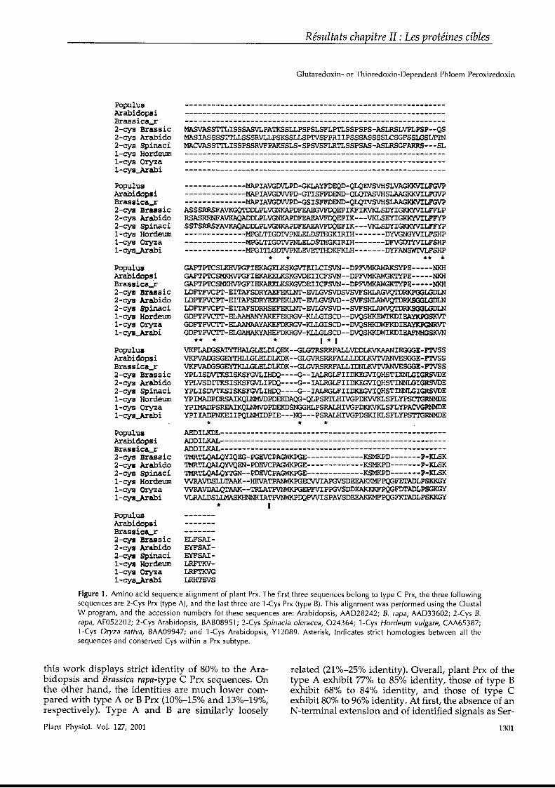
Résultats chapitre II : Les protéines cibles
Glutaredoxin- or Thioredoxin-Dependent Phloem Peroxiredoxin
PopulusArabidopsiBrassicA_r2-cys Brassie2-cys Arabido2-cys Spinaeil-cys Hordeuml-cys Oryzal-cys..,Arabi
PopulusArabidopsiBrassicil_r2-cys Brassic2-cys Arabido2-cys Spinacil-cys Hordeuml-cys Oryzal-cys.-Arabi
PopulusArabidopsiBrassica~r2-cys Brassic2-cys Arabido2-cys Spinacil-cys Hordeuml-cys Oryzal-cys...Arabi
PopulusArabidopsiBrassicA_r2-cys Brassic2-cys Arabido2-cys Spinacil-cys Hordeuml-cys Oryzal-cys.."Arabi
PopulusArabidopsiBrassica..r2-cys Brassie2-cys Arabido2-cys Spinacil-cys Hordeuml-cys Oryzal-cys.-Arabi
PopulusArabidopsiBrassic--.r2-cys Br.ssie2-cys Arabido2-cys Spinacil-cys Hordeuml-cys Oryzal-cys~abi
MASVASSTTLISSSASVLPATKSSLLPSPSLSFLPTLSSPSPS-ASLRSLVPLPSP--QSMASIASSSSTrLLSSSRVLLPSKSSLLSP'IVSFPRIIPSSSASSSSLCSGFSSIGSLTTNMACVASST'rLISSPSSRVFPAKSSLS-SPSVSFLRTLSSPSAS-ASLRSGFARRS---SL
--------------MAPIAVGDVLPD-GKLAYFDEQD-QLQEVSVHSLVAGKKVILFGVP--------------MAPIAVGDVVPD-GTISFFDEND-QLQTASVHSLAAGIOOlILFGVP--------------MAPIAVGDVVPD-GSISFFDEND-QLQTVSVHSLAAGIOOlILlGVPASSSRRSFAVKGQTDDLPLVGNKAPDFEAEGVFDQEFIKFIKVKLSDYIGKK'YVILFFLPRSASRRNFAVKAQADDLPLVGNKAPDFFAEAVFDQEFIK-- -VKLSEYIGKK'YVILFFYPSSTSRRSFAVKAQADDLPLVGNKAPDFFAEAVFDQEFIK---VKLSDYIGKK'YVILP'FYP--------------MPGLTIGDTVPNLELDSTHGKIRIH-------DYVGNGYVILFSHP--------------MPGLTIGDTVPNLELDSTHGKIRIH-------DFV~LFSHP
- - - - - - - - - - - - - -MPGITLGDTVPNLEV'E'M'HDKFKLH- - - - - - - DYFAN5WI'VLFSHP* * ** *
GAFTPTCSLKHVPGFIEKAGELKSKGVTEILCISVN--DPFVMKAWAKSYPE-----N!<HGAFTPl'CSMKHVPGFIEKAEELKSKGVDEIICFSVN--DPFVMKAWGKTYPE-----NKHGAFTPI'CSMKHVPGFIEKAEELKSKGVDEIICFSVN--DPFVMKAWGKTYPE-----NKHLDFTFVCPT-EITAFSDRYAEFEKLNT-EVU:NSVDSVSVFSHLAGVQTDPJ<FOOLGDLNLDFTFVCPT-EITAFSDRYEEFEKLNT-EVU:NSVD--SVFSHLAWVQTDRKSOOUIDLNLDFTFVCPT-EITAFSDRHSEFEKLNT-EVU:NSVD--SVFSHLAWVQTDRKSOOIGDLNGDFTPVCTl'-ELAAMANYAKEFEKRGV-KLLGISCD--DVQSHlŒWI'KDlEAYKPGSKVTGDFTPVCTr-ELAAMAAYAKEFDKRGV-KLLGISCD--DVQSHI<I'MFKDIEAYKPGNRVI'GDFTPVCTr-EWAMAKYAHEFDKRGV-KLLGLSCD--DVQSHI<IMIKDlEAFNHGSKVN
** * * 1 * 1VKFLADGSATYTHALGLELDLQEK--GLGTRSRRFALLVDDLKVKAANIEGGGE-FTVSSVKFVAOOSGEYTm,LGLELDLKDK--GLGVRSRRFALLLDDLKVTVANVESGG!-PTVSSVKFVAOOSGEYTKLLGLELDLKDK--GLGVRSRRFALLIDNLKVTVANVESGGE-F'1'VSSYPLISDVTKSISKSFGVLIHDQ----G--IALRGLFIIDKEGVIQHSTIXNLGIGRSVDEYPLVSDITKSISKSFGVLIPDQ----G--IALRGLFIIDKEGVIQHSTINNLGIGRSVDEYPLISDVTKSISKSFGVLIHDQ----G--IALRGLFIIDKEGVIQHSTINNLGIGRSVDEYPIMADPDRSAIKQLNMVDPDEKDAQG-QLPSRTLHIVGPDKVVKLSFLYPSCTGRNMDEYPIMADPSREAIKQLNMVDPDEKDSNGG~PSRALHIVGPDKKVKLSFLYPACVG»DIDE
YPlIADPNKEIIPQLNMIDPIE---NG---PSRALHIVGPDSKIKLSFLYFSTTGRNMDE* * *
AEDILKDL---------------------------------------------------ADDILKAL----------------------------------------------_·---ADDILKAL----------------------------------------------------TMRTLQALQYIQEG-PGEVCPAGWKPGE-------------KSMKPD-------P-XLSKTMRTLQALQYVQEN-PDEVCPAGWKPGE-------------KSMKPD-------P-KLSKTMRTLQALQYTGN--PDEVCPAGWKPGE-------------KSMKPD-------P-KLSKWRAVDSLLTAAK--HKVATPANWKPGECWIAPGVSDEEAKKMFPQGFETADLPSKKGYWRAVDAJ.J;[rAA'K--TRLATPVNWKPGEPFVIPPGVSDDEAKEKFPQGFDTADLPSGKGYVLRALDSLLMASKHNNKIATPVNWKPDQPVVISPAVSDEEAKKMFPQGFKTADLPSKKGY
* 1
ELFSAIEYFSAIEYFSAILRFTKVLRFTKVGLRHTEVS
Figure 1. Amino acid sequence alignment of plant Prx. The first three sequences belong to type C Prx, the three followingsequences are 2-Cys Prx (type A), and the last three are l-Cys Prx (type B). This alignment was performed using the ClustalW program, and the accession numbers for these sequences are: Arabidopsis, AAD28242; B. rapa, AAD336D2; 2-Cys B.rapa, AF052202; 2-Cys Arabidopsis, BAB08951; 2-Cys Spinacia o/eracea, 024364; l-Cys Hordeum vu/gare, CAA65387;l-Cys Oryza saliva, BAA09947; and l-Cys Arabidopsis, Y12D89. Asterisk, Indicates strict homologies between ail thesequences and conserved Cys within a Prx subtype.
this work displays strict identity of 80% to the Arabidopsis and Brassica rapa-type C Prx sequences. Onthe other hand, the identities are much lower compared with type A or B Prx (10%-150/0 and 13%-19%,respectively). Type A and B are similarly loosely
Plant Physiol. Vol. 127, 2001
related (210/0-25% identity). Overall, plant Prx of thetype A exhibit 770/0 to 85% identity, those of type Bexhibit 68% to 84% identity, and those of type Cexhibit 800/0 to 96% identity. At first, the absence of anN-terminal extension and of identified signaIs as Ser-
1301
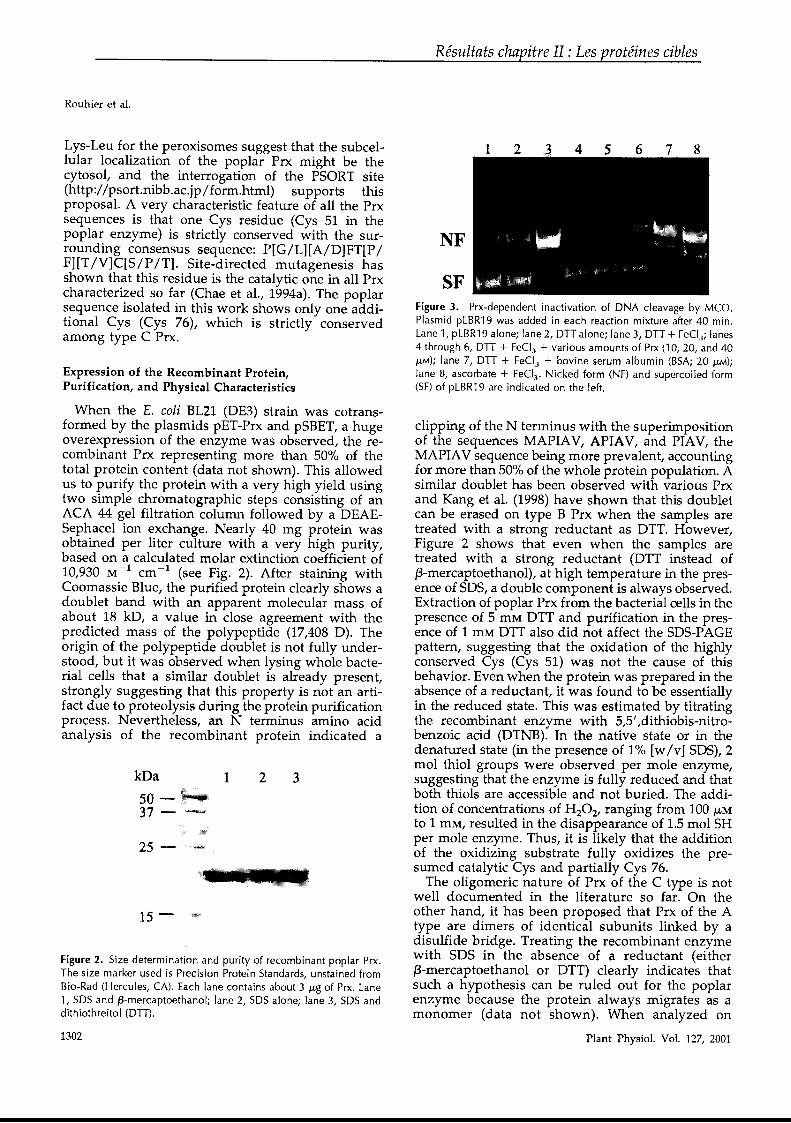
Résultats chapitre II : Les protéines cibles
Rouhier et al.
1302
Figure 2. Size determination and purity of recombinant poplar Prx.The size marker used is Precision Protein Standards, unstained fromBio-Rad (Hercules, CA). Each lane contains about 3 J..tg of Prx. Lanel, SOS and l3-mercaptoethanol; lane 2, SOS alone; lane 3, SOS anddithiothreitol (OTT).
7 864 5321
Figure 3. Prx-dependent inactivation of DNA c1eavage by Mca.Plasmid pLBR19 was added in each reaction mixture after 40 min.Lane l, pLBR19 alone; lane 2, OTT alone; lane 3, OTT + FeCI3 ; lanes4 through 6, OTT + FeCI 3 + various amounts of Prx (10, 20, and 40J..tM); lane 7, OTT + FeCI3 + bovine serum albumin (BSA; 20 ILM);lane 8, ascorbate + FeCI3 • Nicked form (NF) and supercoiled form(SF) of pLBR19 are indicated on the left.
clipping of the N terminus with the superimpositionof the sequences MAPIAV, APIAV, and PIAV, theMAPIAV sequence being more prevalent, accountingfor more than 50% of the whole protein population. Asimilar doublet has been observed with various Prxand Kang et al. (1998) have shown that this doubletcan be erased on type B Prx when the samples aretreated with a strong reductant as DTT. However,Figure 2 shows that even when the samples aretreated with a strong reductant (DTT instead off3-mercaptoethanol), at high temperature in the presence of SDS, a double component is always observed.Extraction of poplar Prx from the bacterial cells in thepresence of 5 mM DTT and purification in the presence of 1 mM DTT also did not affect the SDS-PAGEpattern, suggesting that the oxidation of the highlyconserved Cys (Cys 51) was not the cause of thisbehavior. Even when the protein was prepared in theabsence of a reductant, it was found to be essentiallyin the reduced state. This was estimated by titratingthe recombinant enzyme with 5,5' ,dithiobis-nitrobenzoic acid (DTNB). In the native state or in thedenatured state (in the presence of 1% [wIv] SDS), 2mol thiol groups were observed per mole enzyme,suggesting that the enzyme is fully reduced and thatboth thiols are accessible and not buried. The addition of concentrations of H20 2, ranging from 100 J-LMto 1 mM, resulted in the disappearance of 1.5 mol SHper mole enzyme. Thus, it is likely that the additionof the oxidizing substrate fully oxidizes the presumed catalytic Cys and partially Cys 76.
The oligomeric nature of Prx of the C type is notweIl documented in the literature so far. On theother hand, it has been proposed that Prx of the Atype are dimers of identical subunits linked by adisulfide bridge. Treating the recombinant enzymewith SDS in the absence of a reductant (eitherf3-mercaptoethanol or DTT) clearly indicates thatsuch a hypothesis can be ruled out for the poplarenzyme because the protein always migrates as amonomer (data not shown). When analyzed on
Plant Physiol. Vol. 127, 2001
321
15-
25
kDa
5037 -
Lys-Leu for the peroxisomes suggest that the subcellular localization of the poplar Prx might be thecytosol, and the interrogation of the PSORT site(http://psort.nibb.ac.jp/ form.html) supports thisproposal. A very characteristic feature of aIl the Prxsequences is that one Cys residue (Cys 51 in thepoplar enzyme) is strictly conserved with the surrounding consensus sequence: P[G/L][A/D]FT[P/F][T/V]C[S/P /T]. Site-directed mutagenesis hasshown that this residue is the catalytic one in aIl Prxcharacterized so far (Chae et al., 1994a). The poplarsequence isolated in this work shows only one additional Cys (Cys 76), which is strictly conservedamong type C Prx.
Expression of the Recombinant Protein,Purification, and Physical Characteristics
When the E. coli BL21 (DE3) strain was cotransformed by the plasmids pET-Prx and pSBET, a hugeoverexpression of the enzyme was observed, the recombinant Prx representing more than 50% of thetotal protein content (data not shown). This allowedus to purify the protein with a very high yield usingtwo simple chromatographic steps consisting of anACA 44 gel filtration column followed by a DEAESephacel ion exchange. Nearly 40 mg protein wasobtained per liter culture with a very high purity,based on a calculated molar. extinction coefficient of10,930 M-
1 cm -1 (see Fig. 2). After staining withCoomassie Blue, the purified protein clearly shows adoublet band with an apparent molecular mass ofabout 18 kD, a value in close agreement with thepredicted mass of the polypeptide (17,408 D). Theorigin of the polypeptide doublet is not fully understood, but it was observed when lysing whole bacterial cells that a similar doublet is already present,strongly suggesting that this property is not an artifact due to proteolysis during the protein purificationprocess. Nevertheless, an N terminus amino acidanalysis of the recombinant protein indicated a
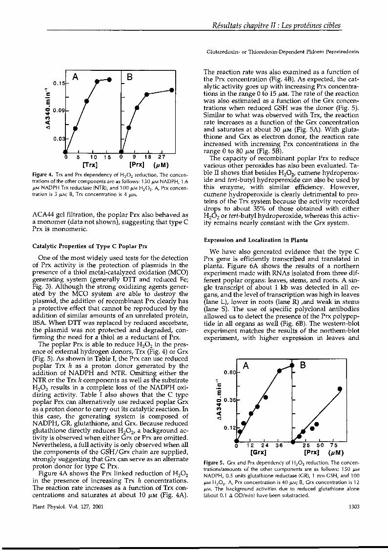
Résultats chapitre II : Les protéines cibles
Glutaredoxin- or Thioredoxin-Dependent Phloem Peroxiredoxin
ACA44 gel filtration, the poplar Prx also behaved asa monomer (data not shown), suggesting that type CPrx is monomeric.
Figure 4. Trx and Prx dependency of H2 0 2 reduction. The concentrations of the other components are as follows: 150 /LM NADPH, 1.6/LM NADPH Trx reductase (NTR), and 100 /LM H2 0 2 • A, Prx concentration is 5 /LM; B, Trx concentration is 4 /LM.
0.15
cE~ 0.09C')
ca:<:]
0.0
o
A
5 10[Trx]
1 5 o
B
9 18
[Prx]27
(pM)
The reaction rate was also examined as a function ofthe Prx concentration (Fig. 4B). As expected, the catalytic activity goes up with increasing Prx concentrations in the range 0 to 15 /LM. The rate of the reactionwas also estimated as a function of the Grx concentrations when reduced GSH was the donor (Fig. 5).Similar to what was observed with Trx, the reactionrate increases as a function of the Grx concentrationand saturates at about 30 J,LM (Fig. SA). With glutathione and Grx as electron donor, the reaction rateincreased with increasing Prx concentrations in therange 0 to 80 /LM (Fig. SB).
The capacity of recombinant poplar Prx to reducevarious other peroxides has also been evaluated. Table II shows that besides H20 21 cumene hydroperoxide and tert-butyl hydroperoxide can also be used bythis enzyme, with similar efficiency. However,cumene hydroperoxide is clearly detrimental to proteins of the Trx system because the activity recordeddrops to about 35% of those obtained with eitherH 20 2 or tert-butyl hydroperoxide, whereas this activity remains nearly constant with the Grx system.
1303
Figure 5. Grx and Prx dependency of H20 2 reduction. The concentrations/amounts of the other components are as follows: 150 /LMNADPH, 0.5 units glutathione reductase (GR), 1 mM GSH, and 100/LM H20 2 • A, Prx concentration is 40 /LM; B, Grx concentration is 12/LM. The background activities due ta reduced glutathione alone(about 0.1 Ll OD/min) have been substracted.
Expression and Localization in Planta
We have also generated evidence that the type CPrx gene is efficiently transcribed and translated inplanta. Figure 6A shows the results of a northernexperiment made with RNAs isolated from three different poplar organs: leaves, stems, and roots. A single transcript of about 1 kb was detected in aIl organs, and the level of transcription was high in leaves(lane L), lower in roots (lane R) ,and weak in stems(lane S). The use of specifie polyclonal antibodiesallowed us to detect the presence of the Prx polypeptide in aIl organs as weIl (Fig. 6B). The western-blotexperiment matches the results of the northern-blotexperiment, with higher expression in leaves and
BA0.60
...cEci 0.36~Cf)
c(<:]
Catalytic Properties of Type C Poplar Prx
One of the most widely used tests for the detectionof Prx activity is the protection of plasmids in thepresence of a thiol metal-catalyzed oxidation (MCO)generating system (generally DTT and reduced Fe;Fig. 3). Although the strong oxidizing agents generated by the MCO system are able to destroy theplasmid, the addition of recombinant Prx clearly hasa protective effect that cannot be reproduced by theaddition of similar amounts of an unrelated protein,BSA. When DTT was replaced by reduced ascorbate,the plasmid was not protected and degraded, confirming the need for a thiol as a reductant of Prx.
The poplar Prx is able to reduce H 20 2 in the presence of external hydrogen donors, Trx (Fig. 4) or Grx(Fig. 5). As shown in Table l, the Prx can use reducedpoplar Trx h as a proton donor generated by theaddition of NADPH and NTR. Omitting either theNTR or the Trx h components as weIl as the substrateH20 2 results in a complete loss of the NADPH oxidizing activity. Table 1 al80 shows that the C typepoplar Prx can alternatively use reduced poplar Grxas a proton donor to carry out its catalytic reaction. Inthis case, the generating system is composed ofNADPH, GR, glutathione, and Grx. Because reducedglutathione directly reduces H 20 2, a background activity is observed when either Grx or Prx are omitted.Nevertheless, a full activity is only observed when aIlthe components of the GSH/Grx chain are supplied,strongly suggesting that Grx can serve as an alternateproton donor for type C Prx.
Figure 4A shows the Prx linked reduction of H20 2in the presence of increasing Trx h concentrations.The reaction rate increases as a function of Trx concentrations and saturates at about 10 /LM (Fig. 4A).
Plant Physiol. Vol. 127, 2001
Résultats chapitre II : Les protéines cibles
Rouhier et al.
Proton Donor
Table Il. Comparison of various a/ky/hydroperoxides as substratesof pop/ar Prx
Reactions are performed at 30°C in 50 mM phosphate buffer, pH 7,with 1sa /LM NADPH, 100 /-LM substrates, and in the presence of 1.6/LM NTR, 15 /LM Trx, and 25 /LM Prx for the Trx system and 0.5 unitsof GR, 1 mM GSH, 12.5 /-LM Grx, and 25 /-LM Prx for the Grx system.Reactions were started after 1 min by adding substrates.
release their protein content at the level of the sieveplate (Knoblauch and van Bel, 1998). Because type CPrx is specifically located in those structures, it istempting to speculate that is should be involved inthe response to oxidative stress generated in suchconditions.
A high expression of Prx was obtained by cotransformation with the plasmid pSBET and the proteincould then be purified with a very high yield. Thenative enzyme is monomeric in solution and its twoCys residues are in the reduced form, irrespective ofits mode of purification (inclusion of a reductant asDTT or not). It is interesting that type A Prx arereported to be dimeric enzymes with a disulfidebridge connecting the catalytic Cys of one subunitand another Cys of the other subunit. Because thisadditional Cys is in the C terminus extension, a similar organization cannot apply to the type C Prxdescribed in this work.
We propose that the short length of the sequence isa first criterion of a type C Prx. Other criteria are thepresence of an additional invariant Cys at position 76and the capacity to use either Trx or Grx as a protondonor for the reaction. The poplar enzyme reducesalkyl hydroperoxides, and especially H20 2, in thepresence of an exogenous proton donor. This wasdemonstrated both kinetically, following theNADPH oxidation linked to the catalytic process,and by the observation that externally added H20 2 isable to oxidize critical Cys residues, even at lowconcentrations. The capacity to reduce H20 2 was alsoconfirmed with a widely used plasmid protectionassay. The biochemical data suggest that the poplarPrx is able to use either Trx or Grx as proton donors,a result that has not been reported before (Fig. 8).This raises the question of the identity of the physiological proton donor of this new type C Prx.
The biochemical data presented here do not permitus to determine the identity of the physiological proton donor in plants because both systems (Trx andGrx) display similar efficiencies. A further complication is the high number of Trx h variants in plants(Buchanan et al., 1994; Rivera-Madrid et al., 1995),because the different isoforms could display varyingaffinities toward type C Prx. We could not test aIlisoforms as proton donors at this point, but could
Plant Physiol. Vol. 127, 2001
0.3110.2780.256
Grx system
0.4530.1740.409
Trx systemSubstrate
H20 2
Cumene hydroperoxidetert-butyl hydroperoxide
Complete 0.522 Complete 0.662-NTR 0.006 -GR 0.000-Trx h 0.005 -GSH 0.000-Prx 0.004 -Grx 0.095-H2 0 2 0.016 -Prx 0.095
-H2 0 2 0.011
Table 1. Proton donor requirements for the peroxiredoxin-linkedreduction of H2 0 2
Ali reactions were carried out at 30°C in a total volume of 500 /LLand followed spectrophotometrically using a Cary sa spectrophometer (Varian Mulgrave, Victoria, Australia). The results are expressedin dA340 per minute. The concentrations/amounts of the variouscomponents were as follows: A, 1.6 /LM NTR, 15 /LM Trx h, 17 /LM
Prx, 100 /-LM H20 2 and B, 0.5 units GR, 1 mM GSH, 50 /LM Grx, 43/-LM Prx; 100 /LM H2 0 2 •
roots. It was estimated that there is approximately500 ng Prx mg protein -1 in leaves based on a standard curve established with increasing amounts ofthe recombinant protein (data not shown). A singlepolypeptide was detected in plant extracts with a sizeidentical to the recombinant proteine Again, this indicates that there is no cleavage of a signal sequencein the proteine Nevertheless, immunolocalization experiments in leaves indicate the presence of largeamounts of type C Prx in the sieve tubes of thephloem. The gold particle labeling was extremelyspecific and essentially restricted to plastid-likestructures present in these cells (Fig. 7).
DISCUSSION
A B
The Prx sequence isolated here is neither a socalled 2-Cys Prx (referred to in this work as type A),nor al-Cys Prx (referred here as type B). The poplarPrx is the shortest among the many isotypes characterized so far (only 162 amino acids). It lacks especially a C-terminal extension consistently present inisotypes A and Band there is no N-terminal extension. The presence in aIl ESTs of a stop codon inframe 9 triplets upstream of the initiation codon precludes the existence of a transit peptide in this proteint Despite the apparent absence of a signal sequence, the protein was shown by immunochemistryto be located in plastids of the sieve elements of thephloem. Thus, either there is a signal internaI tothe sequence or the protein is transported throughthe vesicular pathway from the companion cell viathe branched plasmodesmata. It is interesting thatboth Trx and Grx are major soluble protein components of the phloem sap (Szederkenyi et al., 1997;Ishiwatari et al., 1998). This has prompted speculation that both proteins are involved in redox basedregulation in phloem cells. It is clear that the Prxisolated here could participate to such a regulationbecause it uses both Trx and Grx as a proton donor.It is interesting that it has been proposed that uponinjury, the sieve element plastids disintegrate and
1304
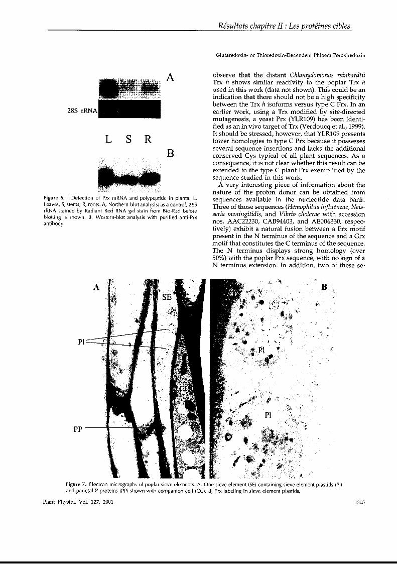
Figure 6. : Detection of Prx mRNA and polypeptide in planta. L,Leaves, S, stems; R, roots. A, Northern-blot analysis: as a control, 28SrRNA stained by Radiant Red RNA gel stain from Bio-Rad beforeblotting is shown. B, Western-blot analysis with purified anti-Prxantibody.
285 rRNA
L s R
A
B
Résultats chapitre II : Les protéines cibles
Glutaredoxin- or Thioredoxin-Dependent Phloem Peroxiredoxin
observe that the distant Chlamydomonas reinhardtiiTrx h shows similar reactivity to the poplar Trx hused in this work (data not shown). This could be anindication that there should not be a high specificitybetween the Trx h isoforms versus type C Prx. In anearlier work, using a Trx modified by site-directedmutagenesis, a yeast Prx (YLR109) has been identified as an in vivo target of Trx (Verdoucq et al., 1999).It should be stressed, however, that YLR109 presentslower homologies to type C Prx because it possessesseveral sequence insertions and lacks the additionalconserved Cys typical of aIl plant sequences. As aconsequence, it is not clear whether this result can beextended to the type C plant Prx exemplified by thesequence studied in this work.
A very interesting piece of information about thenature of the proton donor can be obtained fromsequences available in the nucleotide data bank.Three of those sequences (Hemophilus influenzae, Neisseria meningitidis, and Vibrio cholerae with accessionnos. AAC22230, CAB94403, and AE004330, respectively) exhibit a natural fusion between a Prx motifpresent in the N terminus of the sequence and a Grxmotif that constitutes the C terminus of the sequence.The N terminus displays strong homology (over50%) with the poplar Prx sequence, with no sign of aN terminus extension. In addition, two of these se-
Figure 7. Electron micrographs of poplar sieve elements. A, One sieve element (SE) containing sieve element plastids (PI)and parietal P proteins (PP) shown with companion cel! (CC). B, Prx labeling in sieve element plastids.
Plant Physiol. Vol. 127, 2001 1305
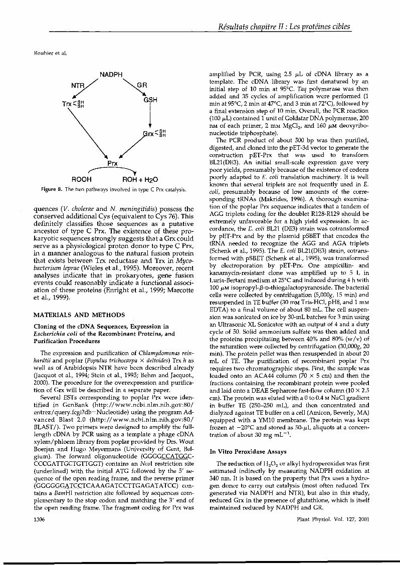
Rouhier et al.
NADPH
7~T .... SH GIHrx 'SH
/rxq~
Prx
ROOH ROH + H20Figure 8. The two pathways involved in type C Prx catalysis.
quences (V. cholerae and N. meningitidis) possess theconserved additional Cys (equivalent to Cys 76). Thisdefinitely classifies those sequences as a putativeancestor of type C Prx. The existence of these prokaryotic sequences strongly suggests that a Grx couldserve as a physiological proton donor to type C Prx,in a manner analogous to the natural fusion proteinthat exists between Trx reductase and Trx in Mycobacterium leprae (Wieles et al., 1995). Moreover, recentanalyses indicate that in prokaryotes, gene fusionevents could reasonably indicate a functional association of these proteins (Enright et al., 1999; Marcotteet al., 1999).
MATERIALS AND METHODS
Cloning of the cDNA Sequences, Expression inEscherichia coli of the Recombinant Proteins, andPurification Procedures
The expression and purification of Chlamydomonas reinhardtii and poplar (Populus trichocarpa X deltoides) Trx h asweIl as of Arabidopsis NTR have been described already(Jacquot et al., 1994; Stein et al., 1995; Behm and Jacquot.,2000). The procedure for the overexpression and purification of Grx will be described in a separate paper.
Several ESTs corresponding to poplar Prx were identified in GenBank (http://www.ncbi.nlm.nih.gov:801entrez/query.fcgi?db=Nucleotide) using the program Advanced Blast 2.0 (http://www .ncbi.nlm.nih.gov:80 /BLAST1). Two primers were designed to amplify the fulllength cDNA by PCR using as a template a phagecDNAxylem/phloem library from poplar provided by Drs. WoutBoerjan and Hugo Meyermans (University of Gent, Belgium). The forward oligonucleotide (GGGGCCATGGCCCCGATTGCTGTTGGT) contains an NcoI restriction site(underlined) with the initial ATG followed by the 5' sequence of the open reading frame, and the reverse primer(GGGGGGATCCTCAAAGATCCTTGAGATATCC) OO~
tains a BamHI restriction site followed by sequences complementary to the stop codon and matching the 3' end ofthe open reading frame. The fragment coding for Prx was
1306
Résultats chapitre II : Les protéines cibles
amplified by PCR, using 2.5 JLL of cDNA library as atemplate. The cDNA library was first denatured by aninitial step of 10 min at 95°C. Taq polymerase was thenadded and 35 cycles of amplification were performed (1min at 95°C, 2 min at 47°C, and 3 min at 72°C), followed bya final extension step of 10 min. Overall, the PCR reaction(100 JLL) contained 1 unit of Goldstar DNA polymerase, 200nM of each primer, 2 mM MgClz, and 160 JLM deoxyribonucleotide triphosphate).
The PCR product of about 500 bp was then purified,digested, and cloned into the pET-3d vector to generate theconstruction pET-Prx that was used to transformBL21(DE3). An initial small-scale expression gave verypoor yields, presumably because of the existence of codonspoorly adapted to E. coli translation machinery. It is weIlknown that several triplets are not frequently used in E.coli, presumably because of low amounts of the corresponding tRNAs (Makrides, 1996). A thorough examination of the poplar Prx sequence indicates that a tandem ofAGG triplets coding for the doublet R128-R129 should beextremely unfavorable for a high yield expression. In accordance, the E. coli BL21 (DE3) strain was cotransformedby pET-Prx and by the plasmid pSBET that encodes thetRNA needed to recognize the AGG and AGA triplets(Schenk et al., 1995). The E. coli BL21(DE3) strain, cotransformed with pSBET (Schenk et al., 1995), was transformedby electroporation by pET-Prx. One ampicillin- andkanamycin-resistant clone was amplified up to 5 L inLuria-Bertani medium at 25°C and induced during 4 h with100 JLM isopropyl-/3-D-thiogalactopyranoside. The bacterialcells were collected by centrifugation (5,OOOg, 15 min) andresuspended in TE buffer (30 mM Tris-HCI, pH8, and 1 mMEDTA) to a final volume of about 80 mL. The cell suspension was sonicated on ice by 30-mL batches for 3 min usingan Ultrasonic XL Sonicator with an output of 4 and a dutYcycle of 50. Solid ammonium sulfate was then added andthe proteins precipitating between 40% and 80% (w Iv) ofthe saturation were collected by centrifugation (30,OOOg, 20min). The protein pellet was then resuspended in about 20mL of TE. The purification of recombinant poplar Prxrequires two chromatographic steps. First, the sample wasloaded onto an ACA44 column (70 X 5 cm) and then thefractions containing the recombinant protein were pooledand laid onto a DEAE Sepharose fast-flow column (10 X 2.5cm). The protein was eluted with a 0 to 0.4 MNaCI gradientin buffer TE (250-250 mL), and then concentrated anddialyzed against TE buffer on a cell (Amicon, Beverly, MA)equipped with a YM10 membrane. The protein was keptfrozen at -20°C and stored as 50-JLL aliquots at a concentration of about 30 mg mL -1.
In Vitro Peroxidase Assays
The reduction of HzOz or alkyl hydroperoxides was firstestimated indirectly by measuring NADPH oxidation at340 nm. It is based on the property that Prx uses ahydrogen donor to carry out catalysis (most often reduced Trxgenerated via NADPH and NTR), but also in this study,reduced Grx in the presence of glutathione, which is itselfmaintained reduced by NADPH and GR.
Plant Physiol. Vol. 127, 2001
The reaction mixture (500 JLL) for the Trx-dependentassay was as follows: 50 mM Na-K phosphate buffer, pH7.0; 150 J.LM NADPH; 0 to 50 JLM poplar or C. reinhardtii Trxh; 0 to 50 JLM Prx; and 1.5 JLM Arabidopsis Trx reductase.The reaction was initiated by adding 1 mM H20 2 and theNADPH oxidation was followed spectrophotometrically at340 nm using a Cary 50 apparatus at 30°C.
When Grx was tested as the proton donor, the reactionmedium had the following composition: 50 mM Na-K phosphate buffer, pH 7.0; 150 JLM NADPH; 1 mM reducedglutathione; 0.5 units GR; 0 to 50 JLM Grx; and 0 to 80 JLMPrx. As described above, the reaction was carried out in 500JLL, initiated with H20 2, and its rate followed spectrophotometrically at 340 nm.
A second possible test is the protection of plasmidsagainst radicals generated by MCO. H20 2 can be formed bya reaction occurring between DIT, Fe3
+, and 02' and thentransformed into hydroxyl radical by the Fenton reaction(Halliwell and Gutteridge, 1990). These radicals are able todamage nucleic acids or inactivate enzymes as GInsynthetase.
Plasmid pLBR19 was used to measure the capacity of Prxto eliminate H20 2 before its tranformation in radicals. Areaction mixture of 20 ILL includes 3.3 JLM FeCI3, 10 mMDTT for the thiol MCO system, or 10 mM ascorbate for thenon-thiol MCO system, different Prx concentrations (10-40JLM), or 20 JLM BSA, in 50 mM HEPES [4-(2-hydroxyethyl)1-piperazineethanesulfonic acid]-[NaOH], pH 7.2. The reaction was initiated by incubating the mixture for 40 minbefore adding about 1 JLg of plasmid for 7 h at 37°C. Theplasmid degradation was evaluated on 1% (w Iv) agarosegel after staining with ethidium bromide (Sambrook et al.,1989).
Thiol Content Titration
The free thiol content of recombinant poplar Prx wasestimated using DTNB as described by Jacquot et al. (1984).One milligram of Prx dissolved in 1 mL of Tris-HCI (100mM, pH 8.0) was reacted with 10 JLL of 20 mM DTNB in thepresence of various H20 2 concentrations (ranging from 100JLM to 1 mM). An assay was performed in the presence of1% (w Iv) SDS to denature the protein and estimatewhether it contains buried thiol groups. The cleavage ofDTNB to TNB- was followed by measuring the A412 after30 min reaction in the dark, and the thiol content wasdetermined using a molar extinction coefficient of 13,600M- 1 cm- 1 at 412 nm.
Protein Extraction from Poplar Tissues
Four-hundred milligrams of 2-month-old poplar leaves,stems, and roots was used for protein extraction. Tissueswere ground into powder in the presence of liquid nitrogenand suspended in the extraction buffer (50 mM Tris-HCI,pH 7.5; 14 mM phenylmethylsulfonyl fluoride; and 0.05%[v Iv] f3-mercaptoethanoI). Soluble proteins were obtainedin the supernatant by two successive centrifugation steps(14,000g, 10 min) and then precipitated in the presence of
Plant Physiol. Vol. 127, 2001
Résultats chapitre II : Les protéines cibles
Glutaredoxin- or Thioredoxin-Dependent Phloem Peroxiredoxin
acetone (4 volumes). Proteins were pelleted by centrifugation, the pellet was then washed by a novel centrifugationstep in the presence of acetone, and dried after eliminatingthe supernatant. Proteins were resuspended in a solubilization buffer composed of 125 mM Tris HCI, pH 6.8; 20%(v Iv) glycerol; 2% (w Iv) SDS; 50/0 (v Iv) f3-mercaptoethanol; and 0.05% (w Iv) bromophenol blue. Protein concentrations in the extracts were determined by the Bradfordreaction as described in the Bio-Rad kit.
Antibody Purification
The antibodies have been purified from the serum byimmuno-adsorption on a Prx-Sepharose column generatedby coupling 35 mg of Prx to 2.5 g of CNBr Sepharose gel(Amersham Pharmacia Biotech, Uppsala). Fractions of 4mL of the rabbit serum were applied onto the column (1 X5 cm) and the antibodies were allowed to react with thematrix for 30 min. After washing the column with TEbuffer, antibodies were eluted with 50 mM acetic acid infractions of 1 mL in tubes containing 200 JLL of 1 M TrisHCI, pH 8. Fractions containing the antibodies were pooledand dialyzed against TE buffer.
SOS-PAGE and Western Blotting
Proteins from poplar tissues (10 ILg) were separated by14% (w Iv) SDS-PAGE following the procedure of Laemmli(1970) and transferred onto a polyvinylidene difluoridemembrane during the night in the presence of transferbuffer (25 mM Tris, 192 mM Gly, and 20% [v Iv] ethanol).Blocking of the membrane was achieved at room temperature during 4 h with the blocking buffer (20 mM Tris-HCI,pH 7.5; 500 mM NaCI; 0.05% [w Iv] milk powder; and 0.05%[v Iv] Tween 20). The membrane was then incubated withpurified anti-Prx antibodies (equivalent to 1.10-3 aD at 280nm) in 15 mL of blocking buffer during at least 2 h, andextensively washed before reaction with anti-rabbit secondary antibodies provided in the Immune Star Goat AntiRabbit Detection Kit from Bio-Rad. The bioluminescencereaction was done according to the instructions of themanufacturer.
RNA Extraction and Northem Blotting
Total RNA was extracted from 100 mg of leaves, stems,and roots from 2-month-old poplar grown in hydroponicmedium, with the RNeasy Plant Mini Kit (Qiagen USA,Valencia, CA) with addition of polyethylene glycol 8000 tothe extraction buffer at a concentration of 20 mg mL -1. Tenmicrograms of total RNA was separated and blotted bycapillarity onto a nylon membrane from Amersham Pharmacia Biotech. The probe, consisting of about 100 ng of aPCR fragment (identical to the Prx fragment cloned), waslabeled with 32p dATP following the instructions providedin the kit Prime a Gene Labeling System (Promega, Madison, WI). The probe was then purified with the QIAquickNucleotides Removal Kit from Qiagen, and was estimatedto contain 30 million cpm. Hybridization was performed at
1307
Rouhier et al.
42°C in the presence of 5x SSPE and 0.5% (w Iv) SDS, andwashing at 65°C with a solution of 2x SSPE and 0.1%
(w/v) SDS.
Immunolocalization Procedures
Samples were dissected from young poplar leaves in adrop of fixation solution and then fixed by immersion for3 h at 4°C in 4% (w Iv) paraformaldehyde and 0.5% (v Iv)glutaraldehyde in 0.1 M cacodylate buffer, pH 7.4. Leafsamples were subsequently rinsed in 0.1 M of the abovebuffer, treated with 2% (w Iv) OS04, dehydrated in a seriesof alcohol and propylene oxide, embedded in Durcupan,and finally polymerized at 56° for 48 h.
Ultrathin sections were successively floated for 15 min ina 0.05 M Gly, 0.05 M Tris.-phosphate-buffered saline (TPBS)solution, 30 min on a blocking solution of 1% (w Iv) BSA inTPBS (TPBS/BSA), and then treated for 2 h with the purified rabbit Prx-antibodies (diluted 1:100 [v Iv] in TPBSIB5A). This step was omitted in the controls. After washing,sections were incubated for 2 h in a solution of 0.05 M
Tris-HCI buffer containing 0.05% (w Iv) polyethylene glycol with gold-Iabeled goat anti-rabbit IgG (10 nm, dilution1:20 [v Iv], Sigma, St. Louis). Sections were then rinsed,dried, and stained with uranyl acetate and lead citrate.
ACKNOWLEDGMENTS
The authors wouid like to thank Matthieu Behm forinitial cloning experiments, Eliane Keryer for DNA sequencing, Marine Wasniewski for invaluable help with thenorthern experiments, Arnaud Javelle for helpful discussions, and Dr. Hugo Meyermans for providing the poplarcDNA library.
Received July 2, 2001; returned for revision July 10, 2001;accepted August 15, 2001.
LITERATURE CITED
Baier M, Dietz KJ (1997) The plant 2-Cys peroxiredoxinBA51 is a nuclear-encoded chloroplast protein: its expressional regulation, phylogenetic origin, and implications for its specific physiological function in plants.Plant J 12: 179-190
Baier M, Dietz KJ (1999) Protective function of chloroplast2-cysteine peroxiredoxin in photosynthesis: evidencefrom transgenic Arabidopsis. Plant Physiol 119:1407-1414
Baier M, Noctor G, Foyer CH, Dietz KJ (2000) Antisensesuppression of 2-cysteine peroxiredoxin in Arabidopsisspecifically enhances the activities and expression of enzymes associated with ascorbate metabolism but not glutathione metabolism. Plant Physiol 124: 823-832
Beckman KB, Ames BN (1997) Oxidative decay of DNA.J Biol Chem 272: 19633-19636
Behm M, Jacquot JP (2000) Isolation and characterizationof thioredoxin h from poplar xylem. Plant Physiol Biochem 38: 363-369
1308
Résultats chapitre II : Les protéines cibles
Berlett BS, Stadtman ER (1997) Protein oxidation in aging,disease, and oxidative stress. J Biol Chem 272:20313-20316
Buchanan BB, 5chürmann P, Decottignies P, Lozano RM(1994) Thioredoxin: a multifunctional regulatory proteinwith a bright future in technology and medicine. ArchBiochem Biophys 314: 257-260
Chae HZ, Chung SJ, Rhee SG (1994a) Thioredoxindependent peroxide reductase from yeast. J Biol Chem269: 27670-27678
Chae HZ, Uhm TB, Rhee SG (1994b) Dimerization ofthiol-specific antioxidant and the essential role of cysteine 47. Proc Nat! Acad Sci USA 91: 7022-7026
Cheong NE, Choi YO, Lee KO, Kim WY, Jung BG, ChiYH, Jeong J5, Kim K, Cho MJ, Lee SY (1999) Molecularcloning, expression, and functional characterization of a2 Cys-peroxiredoxin in Chinese cabbage. Plant Mol Biol40: 825-834
Choi YO, Cheong NE, Lee KO, Jung DG, Hong CH, JeongJH, Chi YH, Kim K, Cho MJ, Lee SY (1999) Cloning andexpression of a new isotype of the peroxiredoxin gene ofChinese cabbage and its comparison to 2 Cysperoxiredoxin isolated from the same plant. BiochemBiophys Res Commun 258: 768-771
Enright AJ, Iliopoulos 1, Kyrpides NC, Ouzounis CA(1999) Protein interaction maps for complete genomesbased on gene fusion events. Nature 402: 86-90
Halliwell B, Gutteridge JM (1990) Role of free radicals andcatalytic metal ions in human disease: an overview.Methods Enzymol 186: 1-85
Haslekas C, Stacy RA, Nygaard V, Culianez-Macia FA,Aalen RB (1998) The expression of a peroxiredoxin antioxidant gene, AtPerl, in Arabidopsis thaliana is seedspecific and related to dormancy. Plant Mol Biol 36:833-845
Jacquot JP, Gadal P, Nishizawa AN, Yee BC, CrawfordNA, Buchanan BB (1984) Enzyme regulation in C4 photosynthesis: mechanism of activation of NADP-malatedehydrogenase by reduced thioredoxin. Arch BiochemBiophys 228: 170-178
Jacquot JP, Rivera-Madrid R, Marinho P, Kollarova M, LeMarechal P, Miginiac-Maslow M, Meyer Y (1994) Arabidopsis thaliana NAPHP thioredoxin reductase. cDNAcharacterization and expression of the recombinant protein in Escherichia coli. J Mol Biol. 235: 1357-1363
Ishiwatari Y, Fujiwara T, McFarland KC, Nemoto K, Hayashi H, Chino M, Lucas WJ (1998) Rice phloem thioredoxin h has the capacity to mediate its own cell-to-celltransport through plasmodesmata. Planta 205: 12-22
Jin DY, Chae HZ, Rhee SG, Jeang KT (1997) Regulatoryrole for a novel human thioredoxin peroxidase in NFkappaB activation. J Biol Chem 272: 30952-30961
Kang SW, Baines IC, Rhee SG (1998) Characterization of amammalian peroxiredoxin that contains one conservedcysteine. J Biol Chem 273: 6303-6311
Kim H, Lee TH, Park ES, Suh JM, Park 5J, Chung HK,Kwon OY, Kim YK, Ro HK, Shong M (2000) Role ofperoxiredoxins in regulating intracellular hydrogen peroxide and hydrogen peroxide-induced apoptosis in thyroid cells. J Biol Chem 275: 18266-18270
Plant Physiol. Vol. 127, 2001
Knoblauch M, van Bel AJ (1998) Sieve tubes in action.Plant Cell 10: 35-50
Laemmli UK (1970) Cleavage of structural proteins duringthe assembly of the head of bacteriophage T4. Nature227: 680-685
Lewis ML, Miki K, Ueda T (2000) FePer l, a gene encodingan evolutionarily conserved 1-Cys peroxiredoxin inbuckwheat (Fagopyrum esculentum Moench) is expressedin a seed-specific manner and induced during seed germination. Gene 246: 81-91
Makrides SC (1996) Strategies for achieving high-Ievel expression of genes in Escherichia coli. Microbiol Rev 60:512-538
Marcotte EM, Pellegrini M, Ng HL, Rice DW, Yeates TO,Eisenberg D (1999) Detecting protein function andprotein-protein interactions from genome sequences. Science 285: 751-753
Pedrajas JR, Miranda-Vizuete A, Javanmardy N, Gustafsson JA, Spyrou G (2000) Mitochondria of Saccharomycescerevisiae contain one-conserved cysteine type peroxiredoxin with thioredoxin peroxidase activity. J Biol Chem275: 16296-16301
Prosperi MT, Ferbus D, Karczinski 1, Goubin G (1993) Ahuman cDNA corresponding to a gene overexpressedduring cell proliferation encodes a product sharing homology with amoebic and bacterial proteins. JBiol Chem268: 11050-11056
Rhee SG, Kang SW, Netto LE, Seo MS, Stadtman ER(1999) A family of novel peroxidases, peroxiredoxins.Biofactors 10: 207-209
Rhee SG, Kim KH, Chae HZ, Yim MB, Uchida K, NettoLE, Stadtman ER (1994) Antioxidant defense mechanisms: a new thiol-specifie antioxidant enzyme. Ann N YAcad Sci 738: 86-92
Rivera-Madrid R, Mestres D, Marinho P, Jacquot JP, Decottignies P, Miginiac-Maslow M, Meyer Y (1995) Evidence for five divergent thioredoxin h sequences in Arabidopsis thaliana. Proc Nat! Acad Sci USA 92: 5620-5624
Sambrook J, Fritsch EF, Maniatis T (1989) Molecular cloning: A Laboratory Manual. Cold Spring Harbor Laboratory Press, Cold Spring Harbor, NY
Schenk PM, Baumann S, Mattes R, Steinbiss HH (1995)Improved high-Ievel expression system for eukaryoticgenes in Escherichia coli using T7 RNA polymerase andrare ArgtRNAs. Biotechniques 19: 196-200
Schroder E, Ponting CP (1998) Evidence that peroxiredoxins are novel members of the thioredoxin fold superfamily. Protein Sci 7: 2465-2468
Plant Physiol. Vol. 127, 2001
Résultats chapitre II : Les protéines cibles
Glutaredoxin- or Thioredoxin-Dependent Phloem Peroxiredoxin
Seo MS, Kang SW, Kim K, Baines IC, Lee TH, Rhee SG(2000) Identification of a new type of mammalian peroxiredoxin that forms an intramolecular disulfide as a reaction intermediate. J Biol Chem 381: 20346-20354
Stacy RA, Nordeng TW, Culianez-Macia FA, Aalen RB(1999) The dormancy-related peroxiredoxin anti-oxidant,PERI, is localized to the nucleus of barley embryo andaleurone cells. Plant J 19: 1-8
Stein M, Jacquot JP, Jeannette E, Decottignies P, HodgesM, Lancelin JM, Mittard V, Schmitter JM, MiginiacMaslow M (1995) Chlamydomonas reinhardtii thioredoxins: structure of the genes coding for the chloroplastic mand cytosolic h isoforms; expression in Escherichia coli ofthe recombinant proteins, purification and biochemicalproperties. Plant Mol Biol 28: 487-503
Sullivan DM, Wehr NB, Fergusson MM, Levine RL,Finkel T (2000) Identification of oxidant-sensitive proteins: TNF-alpha induces protein glutathiolation. Biochemistry 39: 11121-11128
Szederkenyi J, Komor E, Schobert C (1997) Cloning of thecDNA for glutaredoxin, an abundant sieve-tube exudateprotein from Ricinus communis L. and characterization ofthe glutathione-dependent thiol-reduction system insieve tubes. Planta 202: 349-356
Verdoucq L, Vignols F, Jacquot JP, Chartier Y, Meyer Y(1999) In vivo characterization of a thioredoxin h targetprotein defines a new peroxiredoxin family. JBiol Chem274: 19714-19722
Wieles B, Van Noort J, Drijfhout JW, Offringa R,Holmgren A, Ottenhoff TH (1995) Purification and functional analysis of the Mycobacterium leprae thioredoxin/thioredoxin reductase hybrid proteine J Biol Chem 270:25604-25606
Yamamoto T, Matsui Y, Natori S, Obinata M (1989) Cloning of a housekeeping-type gene (MERS) preferentiallyexpressed in murine erythroleukemia cells. Gene 80:337-343
Zhang P, Liu B, Kang SW, Seo MS, Rhee SG, Obeid LM(1997) Thioredoxin peroxidase is a novel inhibitor ofapoptosis with a mechanism distinct from that of Bcl-2.J Biol Chem 272: 30615-30618
Zhou Y, Kok KH, Chun AC, Wong CM, Wu HW, Lin MC,Fung PC, Kung H, Jin DY (2000) Mouse peroxiredoxin V is a thioredoxin peroxidase that inhibits p53induced apoptosis. Biochem Biophys Res Commun 268:921-927
1309
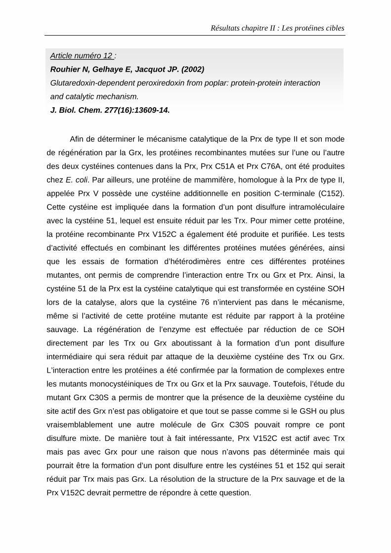
Résultats chapitre II : Les protéines cibles
Afin de déterminer le mécanisme catalytique de la Prx de type II et son mode
de régénération par la Grx, les protéines recombinantes mutées sur l’une ou l’autre
des deux cystéines contenues dans la Prx, Prx C51A et Prx C76A, ont été produites
chez E. coli. Par ailleurs, une protéine de mammifère, homologue à la Prx de type II,
appelée Prx V possède une cystéine additionnelle en position C-terminale (C152).
Cette cystéine est impliquée dans la formation d’un pont disulfure intramoléculaire
avec la cystéine 51, lequel est ensuite réduit par les Trx. Pour mimer cette protéine,
la protéine recombinante Prx V152C a également été produite et purifiée. Les tests
d’activité effectués en combinant les différentes protéines mutées générées, ainsi
que les essais de formation d’hétérodimères entre ces différentes protéines
mutantes, ont permis de comprendre l’interaction entre Trx ou Grx et Prx. Ainsi, la
cystéine 51 de la Prx est la cystéine catalytique qui est transformée en cystéine SOH
lors de la catalyse, alors que la cystéine 76 n’intervient pas dans le mécanisme,
même si l’activité de cette protéine mutante est réduite par rapport à la protéine
sauvage. La régénération de l’enzyme est effectuée par réduction de ce SOH
directement par les Trx ou Grx aboutissant à la formation d’un pont disulfure
intermédiaire qui sera réduit par attaque de la deuxième cystéine des Trx ou Grx.
L’interaction entre les protéines a été confirmée par la formation de complexes entre
les mutants monocystéiniques de Trx ou Grx et la Prx sauvage. Toutefois, l’étude du
mutant Grx C30S a permis de montrer que la présence de la deuxième cystéine du
site actif des Grx n’est pas obligatoire et que tout se passe comme si le GSH ou plus
vraisemblablement une autre molécule de Grx C30S pouvait rompre ce pont
disulfure mixte. De manière tout à fait intéressante, Prx V152C est actif avec Trx
mais pas avec Grx pour une raison que nous n’avons pas déterminée mais qui
pourrait être la formation d’un pont disulfure entre les cystéines 51 et 152 qui serait
réduit par Trx mais pas Grx. La résolution de la structure de la Prx sauvage et de la
Prx V152C devrait permettre de répondre à cette question.
Article numéro 12 :
Rouhier N, Gelhaye E, Jacquot JP. (2002) Glutaredoxin-dependent peroxiredoxin from poplar: protein-protein interaction
and catalytic mechanism.
J. Biol. Chem. 277(16):13609-14.
THE JOURNAL OF BIOLOGICAL CHEMISTRY© 2002 by The American Society for Biochemistry and Molecular Biology, Inc.
Résultats chapitre II : Les protéines cibles
Vol. 277, No. 16, Issue of April 19, pp. 13609-13614,2002Printed in U.8.A.
Glutaredoxin-dependent Peroxiredoxin frOID PoplarPROTEIN-PROTEIN INTERACTION AND CATALYTIC MECHANI8M*
Received for publication, December 3, 2001, and in revised form, January 28,2002Published, JBC Papers in Press, February 6, 2002, DOl 10.10741jbc.M1l1489200
Nicolas Rouhier, Eric Gelhaye, and Jean Pierre Jacquot:f:
From the Unité mixte de recherche IaM INRA-UHP Nancy I, Biochimie et Biologie Moléculaire Végétales,Université Henri Poincaré, 54506 Vandoeuvre Cedex, France
Recently, a poplar phloem peroxiredoxin (Prx) wasfound to accept both glutaredoxin (Grx) and thioredoxin(Trx) as proton donors. To investigate the catalyticmechanism of the Grx·dependent reduction of hydroperoxides catalyzed by Prx, a series of cysteinic mutants was constructed. Mutation of the most N-terminalconserved cysteine of Prx (Cys-51) demonstrates that itis the catalytic one. The second cysteine (Cys-76) is notessential for peroxiredoxin activity because the C76Amutant retained -25% ofthe wild type Prx activity. Onlyone cysteine of the Grx active site (Cys-27) is essentialfor peroxiredoxin catalysis, indicating th~t Grx can actin this reaction either via a dithiol or a monothiol path.way. The creation ofcovalent heterodimers between Prxand Grx mutants confirms that Prx Cys.51 and GrxCys·27 are the two residues involved in the catalyticmechanism. The integration of a third cysteine in position 152 of the Prx, making it similar in sequence to theTrx.dependent human Prx V, resulted in a protein thathad no detectable activity with Grx but kept activitywith Trx. Based on these experimental results, a catalytic mechanism is proposed to explain the Grx- andTrx-dependent activities of poplar Prx.
Peroxiredoxins (PrxS)l constitute a recently discovered family of non-heme peroxidases present in ail organisms fromprokaryotes to eukaryotes, and they catalyze the reduction ofvarious hydroperoxides into the corresponding alcohol and water (1). Currently, these proteins are the subject of numerousstudies because their function seems to be particularly important in the detoxification of reactive oxygen species that cancause serious damage to the nucleic acids, proteins, and lipids(2-4). Prx is also involved in the control of signal transductionby modulating the reactive oxygen species-mediated cellularresponses and by regulating transcription factors (5-7).
AlI the Prx isoforms have in common a conserved catalyticcysteine, localized in the N-terminal part of the protein, that isconverted into a sulfenic acid by hydroperoxides (8). This cysteine was demonstrated by site-directed mutagenesis to beessential for catalysis (9). Based mainly on the number of
*The costs of publication of this article were defrayed in part by thepayment ofpage charges. This article must therefore be hereby marked"advertisement" in accordance with 18 U.S.C. Section 1734 solely toindicate this facto
:j: To whom correspondence should be adressed: Unité mixte de recherche IaM INRA-UHP Nancy l, Biochimie et Biologie MoléculaireVégétales, Université Henri Poincaré, BP 239 Blvd. des Aiguillettes,54506 Vandoeuvre Cedex, France. Tel.: 33-3-839-12253; E-mail:[email protected].
1 The abbreviations used are: Prx, peroxiredoxin; Grx, glutaredoxin;Trx, thioredoxin; WT, wiId type; DTNB, 5,5'-dithiobis(nitrobenzoicacid).
conserved cysteines, a classification has been proposed for themultiple existing Prxs. The 1-Cys Prxs mediate the reduction ofH20 2 with the use of an unknown proton donor, which could beTrx for a mitochondrial1-Cys Prx from Saccharomyces cerevisiae (l0, 11). Recently, human cyclophilin A was identified asan electron donor to the mammalian Prx VI, the only mammalian 1-Cys Prx characterized, and also to aIl known mantmalianPrxs (12). Among the Prxs with two eonserved cysteines, atleast three classes can be distinguished, according to the position of the cysteines. The frrst class, eomprising mantmalianPrx V, includes monomeric enzymes that form an intramolecular disulfide bridge as a reaction intermediate (13, 14). Thesecond class is formed by the homologues of the bacterioferritinco-migratory protein, which are also shown to be monomericenzymes with an intramolecular disulfide bridge in the oxidized state (15, 16). In this second class, the spacing betweenthe two cysteines that are part of the disulfide bridge is considerably shorter, consistently containing 4 amino acids, instead of the -100 antino acids for the first class. The thirdclass, which includes mammalian Prx 1 to IV, consists of dimeric enzymes that form an intermolecular disulfide bridgebetween two identieal subunits (l4, 17). Despite these differences, the three types of 2-Cys Prx use Trx as a proton donor.
In a previous report, a poplar phloem Prx was characterized(l8). This Prx is a smail protein of 162 amino acids that contains only two cysteines in position 51 and 76. The primarysequence is quite different from that ofthe 1-Cys Prxs and mostof the 2-Cys Prxs, especiaily because of the distance that separates the two cysteines. An unexpected finding was that thisPrx could use Trx but also Grx as a proton donor for its catalysis (18). This Prx has been referred to as type C Prx, whereasthe other 2-Cys Prxs, whieh use only Trx, were referred to astype A Prx, and 1-Cys Prxs were referred to as type B Prx.Among the biochemically well-charaeterized Prxs, one of theclosest proteins is the mantmalian Prx V. This enzyme alsocomprises 162 amino acids and displays 40% identity to poplarPrx at the amino acid leveI. A notable difference is the presenceof an additional eysteine in position 152 that is linked togetherwith Cys-51 to form an intramolecular disulfide bridge in Prx V(14). A Prx from Chinese cabbage, highly homologous to poplarPrx, was also shown to be a Trx-dependent enzyme, but noattempt was made to evaluate the potential for Grx as a protondonor in this work (19). To get a better understanding of thecatalytic mechanism of this Grx-dependent Prx, cysteinic mutants of Grx and Prx have been created by site-directed mutagenesis. Based on kinetie measurements and the creation ofheterodimers, a new mechanism for the Grx-dependent Prxactivity is proposed.
EXPERIMENTAL PROCEDURESMaterials-NADPH was obtained from Roche Molecular Biochemi
cals; diamide, J'l-mercaptoethanol, glutathione reductase, and reduced
This paper is available on line at http://www.jbc.org 13609
13610
Résultats chapitre II : Les protéines cibles
Glutaredoxin-dependent Poplar Peroxiredoxin
FIG. 1. Multiple alignment of related sequences to poplar Prx. Thiscomparison was realized using CLUSTALW software. The GenBank™ accessionnumbers of the sequences aligned withpoplar Prx are as foIlows: O. sativa,AAG40130; C. boidinii, AAA34357; human Prx V AOB166, P30044; B. rapa,AAD33602; and A. thaliana, AAG48827.The asterisk corresponds to strict identity, the colon corresponds to functionalhomology, and the period corresponds tostructural homology.
Arabidopsis thalBrassi ca rapaPopul us tri choco-yza_SBt i vacandi da boi di ni iHlItan Prx V
Arabidopsi. th.lPcassi ca rapaPopul us tri chocO"y:z:a_sat i vacandi da boi di ni iHl/ran Prx V
Arab;dopsi. th.!Brassi ca ripaPopul us t ri choca-yzB_sati vacandi da bOl di ni iHllran Pr", V
Arabi dopsi s th.lBrassi ca rapaPopul us l r i choco-YZO_Slt i v.candi da boi di ni iH.man Prx V
Mo.PI AVŒM!PDJrl SFfD. - ENJCI.,QI'ASVHlL.AAŒOOlI LF<lIIPGAFT 48Mo.PI AVŒM!Pr:Œl SFfD. • EN:Q.,Ql'VSVHlL.AAŒOOlI LF<lIIPGAFT 48M'.PI AVŒM.PD3KLAITJ). -~VHlLVAŒIM LF<lIIPGAFT 48Mo.PVA~·~~<lIIPGAFT48MJ>I KRc:IEFPTl'lDVYYI FPEOOEP<PLELSlŒ'VKfIO(FWVSVPGAFT 50MJ>IJaI'tDIIl p. ·AVEVFE-··Œ:P~<lIIPGAFT45•••: ••.• •• :: ..•••••*
49 PT~ClFIEKAEELIŒlI«lVIEI~SVNPFVMCAVŒTYP"ENK9649 PT MŒVPŒl EKAEELl<SI«lVIEI 1 SVNP.FVM{I(.;vœ:r.yp.•ENK 9649 PT LKHVPCIFI EKAŒLl<SK!WI'E1 SVNPFVMCAVAKSYP- - ENK 9649 PT i'QiVPCIFI NQAEq,I<AIŒ\IlD VNPFVMCAVAKSYP- - ENK 9651 pp GYIKNLPRILS SQU'FVLIaJYlŒŒLGAAI 10046 P ICI'HLPŒVEQIIEALKAKO'lQVV: VN:lAl'VT(lEV(JRA11(. - AEG 93
.. *: *: **: : : : *.* .• ••.•• ..-
97 H\lKFVAlXEIJEY'l'llLLG..ELII LGVR- SRR!'ALLJ.ID. LKVI'VAN 14397 H\lKFVAlXEIJE~LGVR·SRR!'ALUmLKVI'VAN 14397 HllIŒ'LAI:ŒAT~Lal'R. SRR!'/>JLVID- LKVKAAN 14397 HVKFLAIXlLGI'YTKALCLELILIlEK<J. La! R. BRR!'AI..L.Am LKVI'VAN 143
101 KLVFVSIPI-LKLTKKLœTI ILSAI (J.. LGJ'R. BœLALI VNRSGl VEYM 14994 KVRI..LAil'TGAi'<l!ŒTIX.LLttllLVSIF<NUUJ<RFSWVQD- <JVKALN 142
: : :* :* . :.. *: : : : •
144 VE-§jSAmLKAL 162144 VE-S SAmLKAL 162144 lE- SAElILIa::L 162144 1E- E <lAEEI LKAL 162150 IE- TAQ:llAI<L 167143 VEPDJI'CLT LAPN[IS~ 162
.... * ,.. . "': ..
glutathione were from Sigma. Dithiothreitol, isopropyl-1-thio-f3-D-galactopyranoside, kanamycin, and ampicillin were from Fermentas.
Cloning and Mutations of Grx and Prx-The procedures for theisolation of the cDNAs and their subsequent cloning in expressionplasmids are described in Refs. 18, 20, and 21. The mutagenesis ofpoplar Grx was effected as we described (31). The Prx mutants C51A,C76A, and V152C were generated by PCR using the oligonucleotidesshown below (NcoI and BamID sites are underlined, mutagenic basesare in bold).
Cloning oligonucleotides were as follows: direct, 5'-GGGGCCATGGCCCCGATTGCTGTrGGT-3'; and reverse, 5'-GGGGGGGATCCTCAAAGATCCTTGAGGATATCCTCGGCACT-3'. Mutagenic oligonucleotides were as follows: C51A direct, 5'-GCCTTCACCCCCACCGCCAGCTTGAAGCATGTG-3'; C51A reverse, 5'-CACATGCTTCAAGCTGGC·GGTGGGGGTGAAGGC-3'; C76A direct, 5'-GTTACTGAAA'ITTTGGC.CATCAGCGTCAACGAC-3'; C76A reverse, 5'-GTCGTTGACGCTGATGGCCAAAATTTCAGTAAC.3'; V152C direct, 5'-GGGGGTGGAGAATTCACTTGCTCCAGTGCCGAGGATATC-3'; and V152C reverse,5'-GATATCCTCGGCACTGGAGCAAGTGAATTCTCCACCCCC-3'.
The mutated PCR products that contained the restriction sites werein turn cloned into the expression plasmid pET-3d, yielding the constructions pET Prx C51A, pET Prx C76A, and pET Prx V152C. Thesequences of the recombinant plasmids were verified by sequencing.
Expression and Purification of the Recombinant Proteins-AlI procedures for the expression and purification of Arabidopsis thalianaNADPH Trx reductase, poplar Trx hl, and WT and mutant Grx aredescribed elsewhere (20, 21, 23, 31). For the expression of Prx, therecombinant plasmids were used to transform Escherichia coli strainBL21(DE3), which was also co-transformed with the plasmid helperpSBET as described previously (l8, 24). Because preliminary experimenta indicated that Prx is susceptible to oxidation, aIl purificationsteps for the wild type and the three mutant proteins were done in thepresence of 14 mM f3-mercaptoethanol.
Thiol Content Titration-The thiol content of each protein preparation was measured using the DTNB procedure. To eliminate the f3-mercaptoethanol, 1 mg of each protein was incubated with 10% trichloroacetic acid for 30 min on ice. The mixture was then centrifuged for 15min at 13,000 rpm, and the pellet was washed three times with 1 ml of2% trichloroacetic acid. The pellet was resuspended in 100 mM TrisHCI, pH 8, and 1 mM EDTA, and the protein concentration was determined by measuring the absorbance at 280 nm. SDS was then added toa final concentration of 1%, and the reaction was started by adding 100ILM DTNB. The reaction mixture was stored in the dark for 20 min, andthe absorbance at 412 nm was measured. A second measurement wasperformed after a 30-min incubation in the dark. Both measurementsgave identical results. Similar results were obtained when we used 80%acetone as a precipitant instead of trich1oroacetic acid.
Prx Activity Measurement-The reduction of H20 2 by poplar Prx inthe presence of the Trx or Grx system was foIlowed spectrophotometricaIly using a Cary 50 spectrophotometer as described in Ref. 18. Theactivities were measured at a fixed concentration ofPrx (2.5 ILM) and atvariable concentrations of Grx or Trx.
Formation and Separation of the Heterodimers-1.25 ILg ofPrx andGrx were mixed in the presence ofTE buffer (30 mM Tris-HCI, pH 8, and1 mM EDTA) to a final volume of 10 ILL This reaction mixture wasincubated at room temperature for 2-3 min before the addition of 10 mM
diamide. The mixture was incubated at room temperature for 20 minand then subjected to 14% SDS-PAGE in the presence of an equivalentvolume ofa nonreducing sample buffer (0.5 MTris-HCI, pH 6.8, 4% SDS,20% glycerol, and bromphenol blue) (25). When needed, 10 mM dithiothreitol or 20-50 mM glutathione was added after incubation in thepresence of diamide to reduce the covalent disulfide adducts betweenPrx and Grx.
Western Blotting-2 ILl ofthe mixture described above were subjectedto 14% SDS-PAGE before transfer. The immunodetection using theImmune Star Goat Anti Rabbit Detection Kit from Bio-Rad was performed as described in Ref. 18. Rabbit polyclonal antibodies againstTrx, Grx, and Prx were purified onto affinity columns according to theprocedure described for Prx antibodies in Rer. 18.
RESULTS
Homologues ofPoplar Prx among Other Speeies-A previousstudy has indieated that poplar Prx differs from the otherperoxiredoxins characterized thus far, essentially by the position of the conserved cysteine residues and by the overalllength of the sequence (18). As detailed in the "Introduction,"this class of new Prxs was called type C Prx, whereas 2-CysPrxs were named type A Pa, and 1-Cys Pas were called typeB Prx. A number ofsequences similar to type C poplar Prx havenow appeared in the literature, and an amine acid comparisonof sorne of the closest relatives is given in Fig. 1. Poplar Pa isstrongly homologous to the other plant Pas of its class (76%,81%, and 82% amino acid identity with Oryza sativa, Brassiearapa, and A. thaliana, respectively) and also to the well-characterized PMP20 protein from Candida boidinii and to thehuman Prx V (42% and 40% amine acid identity, respectively)(Fig. 1). Nevertheless, an important difference is the presenceof an additional cysteine in human Pa in position 152; thepoplar and aIl other nonmammalian sequences do not possess
Résultats chapitre II : Les protéines cibles
Glutaredoxin-dependent Poplar Peroxiredoxin 13611
TABLE 1Relative activities of the various Prx preparations with Trx and GrxData are expressed in ~ol ofNADPH oxidized/minlmg protein. The
measurements were effected at 30 oC with 2.5 IJ.M Prx in the presence of2.5 IJ.M Trx or Grx. Initial Grx corresponds to the construction pET-GrxlB, and improved Grx corresponds to pET-Grx 3 as described in Ref. 20.l.d., limit of detection.
!<Da
25- ---15- --10·
this additional cysteine. This particularity is examined here interms of catalytic efficiency.
Improvement of the Purification of Wild Type Prx andGrx-In a previous work, we have shown that type C Prx coulduse both Grx and Trx as proton donors for catalysis. In thisinitial work, rather high concentrations of Trx or Grx wererequired in the in vitro measurement of peroxidase activity(18). Moreover, the specifie activity of the enzyme remainedlow. We have improved this system in two respects: (i) thetruncated form of poplar Grx, which was used in the earlierexperiments, was replaced by a C-terminal-extended protein,which was both more stable and more active (20); and (ü)because we observed that Prx was susceptible to oxidation, itspurification and subsequent storage were carried out in thepresence of f3-mercaptoethanol to avoid an irreversible oxidation of the catalytic cysteine. These changes resulted in aconsiderable enhancement of enzymatic activity, as shown inTable 1. At a saturating Grx concentration (~10 MM), the maximal specifie activity of the enzyme is 8 /Lmol NADPH oxidizedlmin- 1/mg protein-1. This value is similar ta those reported forhuman peroxiredoxins and the E. coli bacterioferritin co-migratory protein (l0, 14, 15). The specific activities recorded underthe conditions of Table 1 suggest that Grx might be a betterdonor than Trx for poplar Prx. The maximal specific activity ata saturating Trx concentration estimated from the results inFig. 3, 1.5 /Lmol NADPH oxidizedlmin-1/mg protein-l, alsoagrees with that proposaI. However, these data should betreated with caution because the assays for Prx activity areindirect and involve the coupling of several components.
Production of Mutant Proteins by Site-directed Mutagenesis-Two monocysteinic mutants, called C27S and C30S, havebeen created to modify the active site of poplar Grx (31). Inaddition, the mutants C51A, C76A, and V152C have been engineered to explore the reactivity of poplar Prx. Ail these cysteinic mutants were purified in the presence of f3-mercaptoethanol to avoid undesired dimerizations. Fig. 2 shows that all Grxand Trx hl preparations are highly homogeneous. As observedpreviously, all Prx preparations exhibited a protein doubletthat cannot be eliminated by the addition of an excess of reductant (l8). The reason for this behavior is unknown, but aUbiochemical and structural evidence gathered otherwise indicates that the protein is nevertheless highly homogeneous.Several reports in the literature indicate that the catalyticcysteine of peroxiredoxins can be transformed into sulfenic,sulfonic, or sulfinic acids, and this has been proposed as thereason for the formation of apparent protein doublets (10). Thetitration results with DTNB do not really support such a hypothesis for the poplar protein (see below). Besides, as observedpreviously, if such a modification occurs, it is not the result ofthe purification procedure because similar protein doubletswere observed after direct lysis of freshly harvested bacteria(data not shown).
Thiol Titration of the Recombinant Proteins-The thiol content of aU protein preparations has been estimated using theDTNB method. Ail titrations were made on enzymes that werefreed from reductant and after the addition of SDS (see "Ex-
Prx oxidizing conditionsPrx reducing conditions
Initial Grx
l.d.l.d.
Improved Grx
3.554.02
Trx
0.550.82
abcde fgh
FIG. 2. Analysis of purified recombinant proteins by SDSPAGE. 14% SDS-PAGE under reducing conditions showing recombinant Trx, Grx, and Prx samples. Lane a, WT Trx hl; lane b, WT Prx;lane c, Prx C51A; lane d, Prx C76A; lane e, Prx V152C; lane f, WT Grx;lane g, Grx C27S; lane h, Grx C30S.
perimental Procedures"). Consequently, aU thiols are titrated,regardless ofwhether or not they were accessible in the nativeprotein. A summary ofthese data is shown in Table II. For Grx,the results are quite simple; nearly two thiols are titrated forthe reduced WT protein, and no SH group is present in theoxidized WT protein, suggesting that the two Cys residues areindeed linked in a disulfide bridge. The monocysteinic mutantsof Grx give values around 1 SHlmol protein, as expected. ForPrx, the results are more complex to analyze and depend onwhether the protein was isolated in the presence or absence ofa reductant. When prepared under nonreducing conditions, theWT Prx shows nearly 2 SHlenzyme monomer, and it appearsthat the C51A and C76A mutants are partially oxidized because values lower than 1 SHlmol were recorded. The V152Cmutant gives a value of 2.6 SHlmol, nearly in agreement withthe expected 3 SHlmoI. When the Prx preparations were madein the presence of a the monothiol reductant f3-mercaptoethanol, very contrasting results were obtained. The C51A mutantshowed nearly 1 SHlmol, but aU other preparations had a thiolcontent that was reduced by about 1 SH group compared withthe expected theoretical value. We interpret this as the resultof a likely interaction between the thiol group of Cys-51 andf3-mercaptoethanol that can give rise to a mixed disulfide thatcannot be titrated with DTNB. As Prx preparation treated withthe dithiol reductant dithiothreitol titrate as the enzyme prepared under oxidizing conditions, this formation of mixed disulfide is postulated to be specific for l3-mercaptoethanoI. Sucha behavior has already been described for the bovine 1-Cysperoxiredoxin (26). Alternatively, because the thiol titrationsare not always in perfect agreement with the theoretical values, this could also indicate that a portion of the peroxiredoxinmolecules is in a denatured oxidized form with an internaldisulfide bridge as described by Kang et al. for human 1-CysPrx (l0) or that a portion of the catalytic cysteine is in anoxidized form.
Peroxidase Activity of the Mutant Peroxiredoxins-The catalytic capacity of aU Prx mutants has been evaluated in thepresence of the various engineered Grxs as proton donors. Thishas been tested in a coupIed enzymatic reaction where theperoxiredoxin-catalyzed conversion of H20 2 is linked toNADPH oxidation via glutathione reductase, glutathione, andglutaredoxin. We have shown previously, using the 2-hydroethyldisulfide and dehydroascorbate reduction tests, that Cys-27is the catalytic residue of Grx, and Cys-30 is generally dispensable in those reactions (31).
Fig. 3 shows the H20 2-dependent NADPH oxidizing activities of the various engineered peroxiredoxins as a function ofGrx concentrations ranging from 0.5 to 20 /LM. Only four combinations were found to promote catalysis. The best reactivitywas obtained when WT Prx was associated with WT Grx. Themutation of Cys-30 of Grx (C30S) has little effect because therate of catalysis with this mutant is nearly identical to the oneobtained with WT Grx. The other associations that are catalyticaUy competent are those between Prx C76A and Grx WT or
13612
Résultats chapitre II : Les protéines cibles
Glutaredoxin-dependent Poplar Peroxiredoxin
TABLE IIThiol content of Grx and Prx purifted under oxidizing or
reducing conditionsThiols were titrated using the reduction ofDTNB as described under
"Experimental Procedures." Data are expressed in mol SHlmol enzyme.n.d., not determined. The S.D. is typically ± 0.2 SHlmol SH.
GrxWTGrx C27SGrx C30SPrxWTPrx C5lAPrx C76APrx V152C
Oxidizing conditions
0.05n.d.n.d.l.7
0.280.332.6
Reducing conditions
1.670.880.930.90.870.191.4
O.ZS
1
"E0.15
.,;'l'
'"C<l
O.OS
10
Trx concentration (pM)
30
FIG. 3. Grx.dependent Prx activities. HzOz consumption by poplar Prx in the presence of the Grx system was followed in the coupledreaction system by NADPH oxidationlmin as a function of Grx concentration. The WT and mutant Prx were associated in this test with eachof the Grxs: WT Prx associated with WT Grx (0 ) or Grx C30S (""), PrxC76A associated with WT Grx (0) or Grx C30S (0). Prx C5lA associated with any Grx, Prx V152C associated with any Grx. WT Prx associated with Grx C27S, and Prx C76A associated with Grx C27S (X).
C30S. However, the activities of the C76S mutant are reducedby approximately 75% compared with those recorded with WTPrx. Another observation is that the Prx C5lA and V152Cenzymes are completely inactive with Grx as a proton donor. Alast piece of information is that the C27S mutant of Grx isunable ta promote catalysis for ail Prxs tested.
Fig. 4 presents the catalytic activity of the Prx enzymes, inthe presence ofWT Trx as a proton donor. In this case, the Grxgenerating system was replaced by the Trx system, which iscomposed oftheA. thaliana NADPH thioredoxin reductase andpoplar Trx hl (l8). The reduction of poplar Trx by the Arabidopsis NADPH thioredoxin reductase was previously shown tobe functional using the DTNB reduction test (21). In general,similar results were obtained with the Trx system: the WT Prxwas the most active catalyst, the mutation C51A abolishedcatalysis, and C76A had a depressing effect. The most strikingdifference is that the V152C protein, which was inactive withGrx as a proton donor, was catalyticaily active with Trx (Fig. 4,.). No saturation of Prx V152C enzyme activity could be recorded with Trx up to concentrations of 50 IJM (data not shown).
From the data in Figs. 3 and 4, Km values can be determinedto characterize interactions between Prx and Grx or Trx. Similar values were obtained for Grx and Trx (2.5 and 3 /LM,respectively). These values are in good agreement with otherparameters published in the literature for Trx (14).
Heterodimer Formation-The mutation of the cysteine thatis potentiaily involved in the breaking of the mixed disulfideintermediates stabilizes the heterodimers between Trx or Grxand their interacting partners (27). We have taken advantage
0.8
7 0.6.!"!E.,; 0.4'l'
'"-<<l
O.Z
10
Gu cODcentradoD (pM)
lS zo
FIG. 4. Trx.dependent Prx activities. HoOz consumption by poplarPrx in the presence ofthe Trx system was followed in a coupled reactionsystem by NADPH oxidationlmin. Activities are plotted as a function ofpoplar Trx hl concentration. WT Trx with WT Prx, "'; Prx V152C, .;Prx C76A, .; Prx C5lA, •.
of that property to create heterodimers between the peroxiredoxin and glutaredoxin molecules. The covalent heterodimerformation was greatly improved using the oxidant diamide.The four different Prx preparations (WT, C5lA, C76A, andV152C) were incubated with three different versions of the Grx(WT, C27S, and C30S). Fig. 5 shows the results of these associations. In ail assays, the Grx polypeptide is present with anapparent molecular mass of 15 kDa and the Prx is present withthe usual doublet at -18 kDa. By running the proteins individuaily and based on the molecular mass determinations, wecould assess the additional polypeptides as foilows: the 30-kDapolypeptide is likely a Grx dimer, the 33-kDa polypeptide aheterodimer, and the 37-kDa polypeptide is a Prx dimer. Fig. 5shows that a heterodimer is efficiently created when using GrxC30S together with either WT Prx (lane c) or Prx C76A (lane i)and is created much less efficiently with Prx V152C (lane k).The Grx C27S mutant was also able to create heterodimerswith the same Prx preparations, but with a much reducedefficiency (lanes b, h, and k). Strikingly, Prx C5lA is the onlypreparation that does not form any dimer with ail Grx (lanesd-f), indicating that this residue is an essential partner in theredox interaction. The WT Grx is unable to create any stableassociation with any Prx (lanes a, d, g, and}). Finaily, a majoradditional polypeptide of -48 kDa is present in the interactionbetween Grx C30S and Prx V152C. This high molecular masscould correspond to a Prx linked with two Grx molecules.
The nature of these polypeptides was confirmed by Westernblotting experiments. As shown in Fig. 6, anti-Grx antibodiesdo not react with Prx (lane f) and vice versa (lane g). Thisexperiment reveals that the band at 30 kDa is indeed a Grxdimer (lane d), the one at 33 kDa is a heterodimer between Grxand Prx (lanes e and h), and the one at 37 kDa is a dimer ofPrx(lane i).
A further confirmation of the nature of the heterodimer isshown in Fig. 7. The heterodimer generated in the presence ofdiamide can be reduced in the presence of excess dithiothreitolor reduced glutathione (Fig. 7, Lanes b and d). The disappearance of the heterodimer polypeptide after reduction confirmsthat the two chains are indeed linked together via a disulfidebond.
DISCUSSION
The poplar Prx described here is the only characterized Prxthat accepts both Grx and Trx as an electron donor, whereasmammalian peroxiredoxins were shown to use only Trx or an
Résultats chapitre II : Les protéines cibles
Glutaredoxin-dependent Poplar Peroxiredoxin 13613
kDa wt CSIA C76A VI52C
50 - -37- ~ - - ...- Pnc dim...-- -~- =.-..,~+- Heterodimer- +- Grx dîmer
::~_::abcdefghi jkl
A
GSSG
31 l SH r' _31 l SOH--- ::::Gnc
76 ~SH ~ 76 ~SHH,O
S~ tH'os)Grx 31 l S---;;:S,Grx
76 ~ SH Hli~
FIG. 5. Formation of heterodimers between poplar Prx andGrx. Nonreducing 14% SDS-PAGE showing each Prx incubated witheach Grx in the presence ofdiamide. Lanes a-c, WTPrx; lanesd-f, PrxC51A; lanes g-i, Prx C76A; lanes j-I, Prx V152C. These Prxs wereincubated with WTGrx (Ianesa, d,g, and)), Grx C27S (Ianes b, e, h, andh), or Grx caos (Ianes c, f, i, and 1). Arrows indicate the position of themonomeric enzymes and the dimers.
A B C37 - .-- ...- Prx dîmer- _tI!' ...- Heterodimc:r...- On: dim...23 -
~ -.. +- Pnc
I:l- -- --- ...- On:
kDo. o. b • d • f 9 h i
FIG. 6. Immunodetection of the heterodimers. A, CoomassieBlue staining ofnonreducing 14% SDS-PAGE. Lane a, Grx caos alone+ diamide; lane b, Grx caOS + WT Prx + diamide; lane c, WT Prx alone+ diamide. B, Western blot experiment with anti-Grx antibodies. C,Western blot experiment with anti-Prx antibodies. Lanes d-fandg-icorrespond to lanes a-c described in A.
kDa
37 -
15- .....
a b d
FIG. 7. Dissociation of the heterodimers by reductants. Theheterodimers between Prx and Grx C568 were prepared in the presenceof 1 mM diamide (Iane a) and then tentatively reduced with 10 mMdithiothreitol (Iane b), 20 mM glutathione (lane cl, or 50 mM glutathione(Iane dl, always in the presence of diamide.
unidentified donor as a proton source (14, 18). The poplarenzyme studied in this report is clearly a homologue ofhumanPrx V (14). Both proteins are monomeric and contain 162 aminoacids. The major difference between the two enzymes is theircysteine content. The poplar Prx possesses two cysteines atpositions 51 and 76. These residues are conserved in humanPrx V, but there is an additional cysteine in position 152. Thiscysteine residue is present only in the mammalian sequencesand not in the plant homologues ofpoplar Prx or the C. boidiniisequence (Fig. 1). Mutagenesis studies on human Prx V haveshown that this protein belongs to a new class of Prxs (14).Site-directed mutagenesis has clearly shown that Cys-47 is thecatalytic residue on this protein, similar to ail other peroxiredoxins. The catalytic mechanism of human Prx V necessitatesboth Cys-47 and Cys-152 when the donor is thioredoxin ornecessitates Cys-47 alone with dithiothreitol as a donor. Mutagenesis studies indicate that Cys-72 is not involved in thereactivity of this protein.
Given the strong similarity between human Prx V and poplar
B 31 lSH~ 31 lSOH---HS,Grx
76 ~SH ~ 76 ~SH
SH " H,O t H,O
2arf ...~ ~S-S,Gnc~ ~S-8,GrxOSSG 76 SH \
or
HS ,~_ • G~H GSS GSR or HS, Orx~.~ 'Gn:
+GSSG
FIG. 8. Mechanisms for Grx.dependant Prx catalysis. Proposedcatalytic mechanism of the WT Prx reduction supported by WT Grx (A)or by the mutant Grx caOS (B). Closed circles indicate the N terminusofPrx.
Prx and also the specificity of the plant enzyme (to use also Grxas a proton donor), it was ofinterest to examine the reasons forthe unique properties of the plant protein. We have investigated this by a combination of site-directed mutagenesis, kinetic measurements, and the construction of covalently linkedhybrid molecules between Prx and Grx. The results presentedhere indicate clearly that Cys-51 ofPrx is the catalytic cysteine.The mutation of Cys-76 into an alanine does reduce the catalytic efficiency of the enzyme by approximately 75%, but nevertheless, the protein remains active with Grx as a protondonor, a behavior strikingly different from that of the C51Amutant, which is completely inactive. The biochemical datagathered in this study suggest that poplar Prx is a monomericenzyme (very little dimer is present in the preparations analyzed by nonreducing SnS-PAGE irrespective of the conditionsused for preparation of the protein). Moreover, the thiol titrations do not support the presence of a disulfide bridge betweenCys-51 and Cys-76 on this protein. We postulate thus that thedecrease in catalytic efficiency ofthe C76A mutant is linked taa modification orthe microenvironment orthe catalytic cysteinebecause of the introduction of the alanine residue in place ofCys-76. It is noteworthy that a slightly different mutation hasbeen made in the case of human Prx V (C72S instead of C76Ahere), and this might be a reason for the different behavior ofthe plant mutant. Another line of evidence that Cys-76 isprobably not involved in the redox mechanism of this Prx typeis that this residue is absent in the two very closely related O.satiua and C. boidinii sequences (see Fig. 1). Moreover, a threedimensional structure was recently published for the humanPrx V (28). It shows that the sulfur atom of Cys-47 is 7.5 Aaway from that of Cys-72, suggesting that the two residues areindeed not linked by a disulfide bridge unless there is animportant conformational change.
The mutagenesis experiments presented here also provideexperimental evidence for the Grx-dependent catalytic mechanism of poplar Prx. From the heterodimer experiments, it isclear that the two residues that interact between Prx and Grxare Cys-51 and Cys-27, respectively. The absence of het-
13614
Résultats chapitre II : Les protéines cibles
Glutaredoxin-dependent Poplar Peroxiredoxin
erodimer formation in the presence of the WT Grx is a clearindication that Cys-30 of Grx is the backup cysteine responsiblefor the breaking of the heterodisulfide. A scheme for the reaction ofPrx in the presence ofWT Grx is shown in Fig. SA. As forthe other Prx, the catalysis proceeds through the formation ofa sulfenic acid on Cys-5I. The latter is reduced, and a disulfidebond is formed with Cys-27 of Grx with the release ofone watermolecule. The heterodisulfide is then broken by Cys-30 of Grx,and the reoxidized Grx molecule is regenerated by reducedglutathione. Fig. 8B shows the alternative catalytic cycle in thepresence of a monocysteinic Grx (C30S). As in Fig. SA, thesulfenic acid of Cys-51 is reduced via Cys-27 of Grx. The heterodimer is then broken by either glutathione or another monocysteinic Grx molecule. The glutathionylated Grx or the dimerie Grx is then reduced via glutathione.
Given the rather high similarity between poplar Prx andhuman Prx V, it is rather surprising that the latter protein wasnot found to use Grx as a donor as well (14). However, there isone important difference between the human and the poplarenzyme: the presence of Cys-152, which participates in theformation of an intramolecular disulfide bridge in the humanenzyme, and its absence in the plant protein. Our resultsclearly indicate that WT Prx and mutant C76A can both useTrx and Grx as donors. Both these enzymes, unlike the mammalian protein, lack Cys·152. When we engineered the plantprotein and produced the V152C mutant that is similar to themammalian protein, this enzyme conserved activity with Trxas a donor but lost its capacity to use Grx as a reductant. Themaximal activity obtained with the Prx V152C mutant is similar to that described for human Prx V. The poplar protein hasthus acquired a behavior similar to that of the mammalianprotein. Because we could observe that Grx C27S is still able tobind to the V152C mutant but produces higher molecular masscomplexes that are likely to contain two Grx molecules attached/monomer Prx, we postulate that the reason for theabsence of catalytic activity of the V152C mutant with Grx is adue to a steric hindrance because of the fixation of the two Grxmolecules.
It has been shown that ribonucleotide reductase cannot usemonocysteinic Grx as a proton donor. Other reactions catalyzedby Grx (dehydroascorbate, 2-hydroethyldisulfide reduction) require either a monocysteinic Grx or the protein that containstwo thiols (29). Clearly, both versions of Grx are able to promote the catalysis ofPrx. On the other hand, the monocysteinicmutant of thioredoxins is essentially inactive with the targetsthat have been characterized thus far (30). We have alSO generated a single cysteinic mutant ofpoplar Trx hl in which thenoncatalytic cysteine is replaced by a serine (C42S), and weobserved that it is unable to promote catalysis with any Prx(data not shown). Thus, Trx is also unable to function in amonothiol pathway in this system. This is a further indicationthat the monothiol pathway discovered with Grx is very specificfor this type ofprotein, Interestingly, there are many naturallyoccurring monocysteinic Grxs in plants. Based on these observations, it will be of very high interest to compare the relativeefficiencies of the corresponding proteins for Prx catalysis. Theidentity of the ultimate physiological donor for poplar Prx isthus still an open question. It is noteworthy that several sequences in the data bases indicate the presence of fusion proteins between a close homologue of poplar Prx and Grx but notTrx (18,22). In most ofthese sequences, the position of the twocysteines is conserved relative to poplar Prx. Based on theamino acid comparisons, it is relatively safe to assume that the
Brassica and Arabidopsis proteins should be able to use Grx asa donor as well. Whether this type is restricted to plants is stillan open question.
Because the mutagenesis results suggest that the interactionsites for Grx and Trx are different on the plant Prx, it will bevery interesting to map the areas of contact between the molecules, possibly by crystallizing the covalent complexes andsolving its three-dimensional structure. The successful creationof disulfide-linked heterodimers between WT Prx and Trx hlC42S should also help in this respect (data not shown). Thesolution of the three-dimensional structure of poplar Prxshould also help us to understand why this protein accepts twodonors, but human Prx V does not. The results obtained in thisstudy suggest that Cys-152 is a critical residue for this property, but other domains of the proteins may be equally important as well.
Ackrwwledgment-We thank Dr. Francis Martin for valuable helpand discussions.
REFERENCES
1. Rhee, S. G., Kang, S. W., Netto, L. E., Seo, M. B., and Stadtman, E. R. (1999)Bioractors 10, 207-209
2. Halliwell, B., and Gutteridge, J. M. (1990) MethodB Enzymol. 186, 1-853. Beckman, K. B., and Ames, B. N. (1997) J. Biol. Chem. 272, 19633-196364. Ber1ett, B. S., and Stadtman, E. R. (1997) J. Biol. Chem. 272, 20313-203165. Jin, D. Y., Chae, H. Z., Rhee, S. G., and Jeang, K. T. (1997) J. Biol. Chem. 272,
30952-309616. Kim, H., Lee, T. H., Park, E. S., Suh, J. M., Park, B. J., Chung, H. K., Kwon,
O. Y., Kim, Y. K., Ro, H. K., and Shong, M. (2000) J. Biol. Chem. 275,18266-18270
7. Zhou, Y., Kok, K. H., Chun, AC., Wong, C. M., Wu, H. W., Lin, M. C., Fung,P. C., Kung, H., and Jin, D. Y. (2000) Biochem. Biophys. Res. Commun. 268,921-927
8. Rhee, S. G., Kim, K. H., Chae, H. Z., Yim, M. B., Uchida, K., Netto, L. E., andStadtman, E. R. (1994) Ann. N. Y. Acad. Sei. 788, 86-92
9. Chae, H. Z., Uhm, T. B., and Rhee, S. G. (1994) Proc. Natl. Acad. Sei. U. S. A.91, 7022-7026
10. Kang, S. W., Baines, I. C., and Rhee, B. G. (1998) J. Biol. Chem. 273,6303-6311
11. Pedrajas, J. R., Miranda-Vizuete, A, Javanmardy, N., Gustafsson, J. A, andSpyrou, G. (2000) J. Biol. Chem. 275, 16296-16301
12. Lee, S. P., Hwang, Y. S., Kim, Y. J., Kwon, K. B., Kim, H. J., Kim, K., and Chae,H. Z. (2001) J. Biol. Chem. 276, 29826-29832
13. Knoops, B., Clippe, A, Bagard, C., Arsalane, K., Wattiez, R., Hermans, C.,Duconseille, E., Falmagne, P., and Bernard, A. (1999) J. Biol. Chem. 274,30451-30458
14. Seo, M. S., Kang, S. W., Kim, K., Baines, I. C., Lee, T. H., and Rhee, S. G. (2000)J. Biol. Chem. 275, 20346-20354
15. Jeong, W., Cha, M. K., and Kim, I. H. (2000) J. Biol. Chem. 275, 2924-293016. Kong, W., Shiota, S., Shi, Y., Nakayama, H., and Nakayama, K. (2000) Bio
chem. J. 351, 107-11417. Chae, H. Z., Kim, H. J., Kang, S. W., and Rhee, B. G. (1999) Diabetes Res. Clin.
Pract. 45, 101-11218. Rouhier, N., Gelbaye, E., Sautière, P. E., Brun, A, Laurent, P., Tagu, D.,
Gerard, J., de Fay, E., Meyer, Y., and Jacquot, J. P. (2001) Plant Physiol.127, 1299-1309
19. Chai, Y. O., Cheong, N. E., Lee, K. O., Jung, B. G., Hong, C. H., Jeong, J. H.,Chi, Y. H., Kim, K., Cha, M. J., and Lee, S. Y. (1999) Biochem. Biophys. Res.Commun. 258, 768-771
20. Rouhier, N., Gelbaye, E., and Jacquot, J. P. (2002) Prat. Expression Purif. 24,234-241
21. Behm, M., and Jacquot, J. P. (2000) Plant Physiol. Biochem. 38,363-36922. Vergauwen, B., Pauwels, F., Jacquemotte, F., Meyer, T. E., Cusanovich, M. A,
Bartsch, R. G., and Van Beeumen J. J. (2001) J. Biol. Chem. 276,20890-20897
23. Jacquot, J. P., Rivera-Madrid, R., Marinho, P., Kollarova, M., Le Marechal, P.,Miginiac-Maslow, M., and Meyer, Y. (1994) J. Mol. Biol. 235, 1357-1363
24. Schenk, P. M., Baumann, S., Mattes, R., and BteinbiBs, H. H. (1995) BioTech-niques 19, 196-200
25. Laemmli, U. K. (1970) Nature 227, 680-68526. Peshenko, I. V., and Shichi, H. (2001) Free Radie. Biol. Med. 31, 292-30327. Verdoucq, L., Vignols, F., Jacquot, J. P., Chartier, Y., and Meyer, Y. (1999)
J. Biol. Chem. 274, 19714-1972228. Declercq, J. P., Evrard, C., Clippe, A, Stricht, D. V., Bernard, A, and Knoops,
B. (2001) J. Mol. Biol. 311,751-75929. Qin, J., Yang, Y., Velyvis, A, and Gronenbom, A (2000) Antioxid. Redox
Signal. 2, 827-84030. Goyer, A, Decottignies, P., Lemaire, S., Ruelland, E., Issakidis-Bourguet, E.,
Jacquot, J. P., and Miginiac-Maslow, M. (1999) FEBS LeU. 444, 165-16931. Rouhier, N., Gelbaye, E., and Jacquot, J. P. (2002) FEBS Leu. 511, 145-149
Résultats chapitre II : Les protéines cibles
Parmi les 4 groupes distincts de Prx chez les plantes, aucune structure n’a été
obtenue jusqu’à présent que ce soit par RMN ou cristallographie. Seules 9 structures
de Prx ont été résolues. Aux 6 structures déjà décrites dans cet article, s’ajoutent
maintenant une autre forme de la Prx V (code PDB 1HD2), une Ahpc de Salmonella
thyphimurium (code PDB 1KYG) et une protéine de fusion d’Haemophilus influenzae
entre des modules Grx et Prx (code PDB 1NM3). Deux formes de cristaux ont été
obtenues pour la Prx sauvage et diffractent à 1.62Å et 2.48Å et une seule forme pour
Prx V152C qui diffracte à 2.17Å. La structure de la Prx de type II a été déterminée
par remplacement moléculaire par rapport à la structure de la Prx V humaine puisque
les deux protéines possèdent une identité de séquence de 42% et sera présentée
dans un prochain article.
Article numéro 13 :
Echalier A, Corbier C, Rouhier N, Jacquot JP, Aubry A. (2002) Crystallization and preliminary X-ray data of a bifunctional peroxiredoxin from
poplar. Acta Crystallogr. D Biol. Crystallogr. 58(Pt 9):1501-3.

Acta Crystallographica Section D
BiologicalCrystallography
ISSN 0907-4449
Résultats chapitre II : Les protéines cibles
crystallization papers
Crystallization and preliminary X-ray data of abifunctional peroxiredoxin from poplar
Aude Echalier,a CatherineCorbier,a Nicolas Rouhier,bJean-Pierre Jacquotb and AndréAubrya*
aLCM3B, Groupe Biocristallographie,
UMR 7036, UHP, Faculté des Sciences,
54506 Vandoeuvre CEDEX, France, and
blnteraction Arbres Microorganismes,
UMR 1136 INRA UHP, Faculté des Sciences,
54506 Vandoeuvre CEDEX, France
Correspondence e-mail:
Two variants (wild type and V152C mutant) of a bifunctional poplarperoxiredoxin have been overexpressed in Escherichia coli cells. Thetwo recombinant enzymes were purified and crystallized using thehanging-drop vapour-diffusion technique. Data sets were collected to1.62 and 2.48 Â resolution using X-ray synchrotron-source radiationfrom two crystal forms of wild-type peroxiredoxin which belonged tothe monoclinic space group nt (with unit-cell parameters a =59.26,b = 68.80, c = 75.71 Â, f3 = 93.45°) and to the orthorhombic spacegroup n t 2t 2 (with unit-cell parameters a = 64.70, b = 130.73,c = 35.59 Â), respectively. Data were also collected to 2.17 Àresolution using a home X-ray source from a V152C peroxiredoxincrystal which belongs to the triclinic space group (Pl), with unit-cellparameters a = 36.65, b = 41.53, c = 58.06 Â, ex = 70.52, f3 = 93.45,Y = 64.31°. Phases have been obtained using molecular replacementwith the structure of human peroxiredoxin V (PDB code 1hd2) as asearch mode!. Refinement of the structures is in progresse
Received 11 April 2002
Accepted 3 July 2002
© 2002 International Union of Crystallography
Printed in Denmark - ail rights reserved
Acta Cryst. (2002). 058, 1501-1503
1. Introduction
Peroxiredoxins (PRX) are non-haem peroxidases which catalyse the conversion ofalkylhydroperoxides into the correspondingalcohol and water in the presence of anadequate electron/proton donor according tothe equation
ROOH + R'(SH)2 ~ ROH + H 20 + R'SS.
Peroxiredoxins are ubiquitous enzymes frombacteria to mammals and are generally overexpressed when the cells are subjected tooxidative stress, a condition where H20 2 isproduced in massive amounts (Kang, Chae etal., 1998). They also play a role in apoptosis orin the regulation of transcription factors(Zhang et al., 1997; Zhou et al., 2000). As theproton donor is mostly the small ubiquitousprotein thioredoxin, it has also often beencalled thioredoxin peroxidase. Other variantshave been named AhpC and tryparedoxinperoxidase (Poole, 1996; Montemartini et al.,1998). Several classes of peroxiredoxins canbe differentiated based on their catalyticmechanisms and sequences. All the peroxiredoxins possess a conserved catalytic cysteinesurrounded by conserved amino acids with theconsensus sequence Ff(P/F)(V/GIT)C(PIT/S).In the course of catalysis, this catalytic cysteineis transformed into a sulfenic acid which isregenerated into a thiol group, generally in thepresence of thioredoxin as an electron donor(Kang, Baines et al., 1998). In the so-called2-Cys peroxiredoxins, catalysis proceeds
through the formation of either an intramolecular disulfide between the catalytic cysteineand another conserved cysteine or of anintermolecular disuifide between two head-totail peroxiredoxin monomers (Kong et al. ,2000). In another type, called the 1-Cysperoxiredoxins, only the catalytic cysteine isnecessary and the thiol-regenerating system iseither thioredoxin or an unidentified thiolcomponent that can be replaced by dithiothreitol in vitro (Kang, Baines et al., 1998;Pedrajas et al., 2000). A third type, recentlycharacterized in poplar (Populus trichocarpa),is an atypical 2-Cys peroxiredoxin whichcontains the conserved catalytic Cys andanother Cys, the position of which is conservedin this third group. 1t differs however from theposition of the second Cys of the 2-Cysperoxiredoxins and, in addition, it is notnecessary for catalysis (Rouhier et al., 2002).This protein is a bifunctional enzyme whichcan use thioredoxin as a proton donor butexhibits preference for a related protein,glutaredoxin, which is itself reduced byNADPH, glutathione reductase and glutathione, instead of NADPH and thioredoxinreductase as required for thioredoxin (Rouhieret al., 2001).
In mammals, there are at least six genescoding for peroxiredoxin and the corresponding proteins play a prominent role infighting oxidizing species, since they areproduced at very high levels in several tissues(Seo et al., 2000). Currently, six structures areknown for peroxiredoxins: three are of human
Echalier et al. • Peroxiredoxin 1501
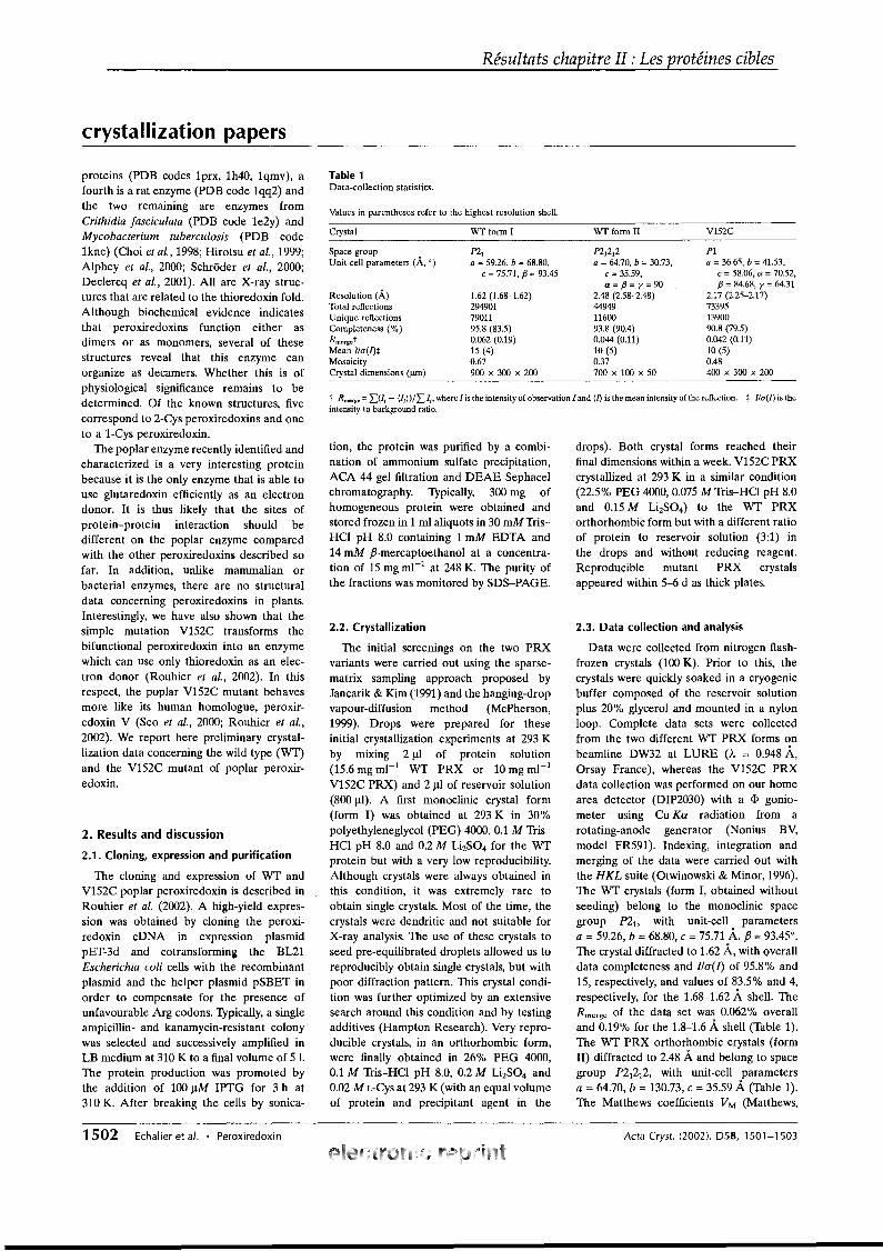
Résultats chapitre II : Les protéines cibles
crystallization papers
Table 1Data-collection statistics.
t Rmerge =L(Ii - (/i})/L li' where 1 is the intensity of observation 1 and {/} is the mean intensity of the reftection. :j: l/a(I) is theintensity to background ratio.
Values in parentheses refer to the highest resolution sheU.
tion, the protein was purified by a combination of ammonium sulfate precipitation,ACA 44 gel filtration and DEAE Sephacelchromatography. Typically, 300 mg ofhomogeneous protein were obtained andstored frozen in 1 ml aliquots in 30 mM TrisHCI pH 8.0 containing 1 mM EDTA and14 mM fJ-mercaptoethanol at a concentration of 15 mg ml-1 at 248 K. The purity ofthe fractions was monitored by SDS-PAGE.
V152C
Pla = 36.65, b = 41.53,
c =58.06, a =70.52,{3 =84.68, Y =64.31
2.17 (2.25-2.17)733951390090.8 (79.5)0.042 (0.11)10 (5)0.48400 x 300 x 200
WT fonu II
n t 212a =64.70, b =30.73,
c = 35.59,a={3=y=90
2.48 (2.58-2.48)449491160093.8 (90.4)0.044 (0.11)10 (5)0.37700 x 100 x 50
2.3. Data collection and analysis
Data were collected from nitrogen flashfrozen crystals (100 K). Prior to this, thecrystals were quickly soaked in a cryogenicbuffer composed of the reservoir solutionplus 200/0 glycerol and mounted in a nylonloop. Complete data sets were collectedfrom the two different WT PRX forms onbeamline DW32 at LURE (À = 0.948 A,Orsay France), whereas the V152C PRXdata collection was performed on our homearea detector (DIP2030) with a cI> goniometer using Cu Ka radiation from arotating-anode generator (Nonius BV,model FR591). Indexing, integration andmerging of the data were carried out withthe HKL suite (Otwinowski & Minor, 1996).The WT crystals (form l, obtained withoutseeding) belong to the monoclinic spacegroup nit with unit-cell parametersa =59.26, b =68.80, c ;::; 75.71 A, fJ =93.45°.The crystal diffracted to 1.62 A, with overalldata completeness and I/a(l) of 95.80/0 and15, respectively, and values of 83.50/0 and 4,respectively, for the 1.68-1.62 A shell. TheRmerge of the data set was 0.062% overalland 0.19% for the 1.8-1.6 A. shell (Table 1).The WT PRX orthorhombic crystals (formII) diffracted to 2.48 Aand belong to spacegroup n 1212, with unit-cell parametersa =64.70, b =130.73, c =35.59 A. (Table 1).The Matthews coefficients VM (Matthews,
drops). Both crystal forms reached theirfinal dimensions within a week. V152C PRXcrystallized at 293 K in a similar condition(22.5% PEG 4000,0.075 M Tris-HCI pH 8.0and 0.15 M LhS04) to the WT PRXorthorhombic form but with a different ratioof protein to reservoir solution (3:1) inthe drops and without reducing reagent.Reproducible mutant PRX crystalsappeared within 5-6 d as thick plates.
WT form 1
nta = 59.26, b = 68.80,
c = 75.71, {3 = 93.45
1.62 (1.68-1.62)2949017901195.8 (83.5)0.062 (0.19)15 (4)0.67900 x 300 x 200
Crystal
Resolution (À)Total refleetionsUnique refleetionsCompleteness (%)Rmerget
Mean I/a(l)*MosaieityCrystal dimensions (Jlm)
2.2. Crystallization
The initial screenings on the two PRXvariants were carried out using the sparsematrix sampling approach proposed byJancarik & Kim (1991) and the hanging-dropvapour-diffusion method (McPherson,1999). Drops were prepared for theseinitial crystallization experiments at 293 Kby mixing 2 JlI of protein solution(15.6 mg ml-1 WT PRX or 10 mg ml-1
V152C PRX) and 2 JlI of reservoir solution(800 JlI). A first monoclinic crystal form(form 1) was obtained at 293 K in 300/0polyethyleneglycol (PEG) 4000, 0.1 M TrisHCI pH 8.0 and 0.2 M LhS04 for the WTprotein but with a very low reproducibility.Although crystals were always obtained inthis condition, it was extremely rare toobtain single crystals. Most of the time, thecrystals were dendritic and not suitable forX-ray analysis. The use of these crystals toseed pre-equilibrated droplets allowed us toreproducibly obtain single crystals, but withpoor diffraction pattern. This crystal condition was further optimized by an extensivesearch around this condition and by testingadditives (Hampton Research). Very reproducible crystals, in an orthorhombic form,were finally obtained in 26% PEG 4000,0.1 M Tris-HCI pH 8.0, 0.2 M LhS04 and0.02 ML-Cys at 293 K (with an equal volumeof protein and precipitant agent in the
Spaee groupUnit-eeU parameters (À, 0)
2. Results and discussion
2.1. Cloning, expression and purification
The cloning and expression of WT andV152C poplar peroxiredoxin is described inRouhier et al. (2002). A high-yield expression was obtained by cloning the peroxiredoxin cDNA in expression plasmidpET-3d and cotransforming the BL21Escherichia coli cells with the recombinantplasmid and the helper plasmid pSBET inorder to compensate for the presence ofunfavourable Arg codons. Typically, a singleampicillin- and kanamycin-resistant colonywas selected and successively amplified inLB medium at 310 K to a final volume of 5 1.The protein production was promoted bythe addition of 100 JlM IPTG for 3 h at310 K. After breaking the ceIls by sonica-
proteins (PDB codes 1prx, 1h40, 1qmv), afourth is a rat enzyme (PDB code 1qq2) andthe two remaining are enzymes fromCrithidia fasciculata (PDB code 1e2y) andMycobacterium tuberculosis (PDB code1knc) (Choi et al., 1998; Hirotsu et al., 1999;Alphey et al., 2000; Schrôder et al., 2000;Declercq et al., 2001). AlI are X-ray structures that are related to the thioredoxin fold.Although biochemical evidence indicatesthat peroxiredoxins function either asdimers or as monomers, several of thesestructures reveal that this enzyme canorganize as decamers. Whether this is ofphysiological significance remains to bedetermined. Of the known structures, fivecorrespond to 2-Cys peroxiredoxins and oneto al-Cys peroxiredoxin.
The poplar enzyme recently identified andcharacterized is a very interesting proteinbecause it is the only enzyme that is able touse glutaredoxin efficiently as an electrondonor. It is thus likely that the sites ofprotein-protein interaction should bedifferent on the poplar enzyme comparedwith the other peroxiredoxins described sofar. In addition, unlike mammalian orbacterial enzymes, there are no structuraldata concerning peroxiredoxins in plants.Interestingly, we have also shown that thesimple mutation V152C transforms thebifunctional peroxiredoxin into an enzymewhich can use only thioredoxin as an electron donor (Rouhier et al., 2002). In thisrespect, the poplar V152C mutant behavesmore like its human homologue, peroxiredoxin V (Seo et al., 2000; Rouhier et al.,2002). We report here preliminary crystallization data concerning the wild type (WT)and the V152C mutant of poplar peroxiredoxin.
1502 Echalier et al. • Peroxiredoxin Acta Cryst. (2002). D58, 1501-1503
Résultats chapitre II : Les protéines cibles
crystallization papers
1968) for the monoclinic and the orthorhombic forms were 2.22 and 2.17 À3 Da-l,respectively, which correspond to 42.4 and41.1 % solvent content, respectively,assuming four and two molecules perasymmetric unit. The mutant V152C PRXcrystals belong to the triclinic space group(Pl), with unit-celI parameters a = 36.65,b = 41.53, c =58.06 À, a =70.52, fJ =93.45,Y = 64.31 0
• They diffracted to 2.17 À resolution (Table 1). Assuming two moleculesper asymmetric unit, the Matthews coefficient VM is 2.10 À3 Da-1 (39.2% solventcontent).
2.4. Phase determination
Molecular-replacement trials using theprogram AMoRe (Navaza, 1994) wereperformed with different modifications ofhuman red blood cell PRX coordinates(PDB code 1qmv) or rat PRX coordinates(PDB code 1qq2) used as a model (28 and25% identity, respectively), but wereunsuccessful. Several attempts were alsomade to obtain· either xenon or bromidederivatives. FinaIly, release of the coordinates of human peroxiredoxin V (PDB code1hd2; 420/0 identity) allowed the determination of the phases by molecular replacementusing AMoRe. The search model used is the
Acta Cryst. (2002). 058, 1501-1503
human PRX refined structure in which aIlthe non-aligned residues (except Gly andPro residues) between the poplar PRX andhuman PRX V have been replaced byalanines. Using data in the resolution range12-4 À and an integration radius of 25 À, aclear solution was obtained with two molecules in the asymmetric unit for the WTorthorhombic form, with a correlationcoefficient and an R factor of 44.9 and44.50/0, respectively (next best solution: 33.8and 48.8%, respectively). Model buildingand refinement of these three structures iscurrently under way.
References
Alphey, M. S., Bond, C. S., Tetaud, E., Fairlamb,A. H. & Hunter, W. N. (2000). J. Mol. Biol. 300,903-916.
Choi, H. 1., Kang, S. W., Yang, C. H., Rhee, S. G. &Ryu, S. E. (1998). Nature Struct. Biol. 5, 400406.
Declercq, 1. ~, Evrard, C., Clippe, A., Stricht, D. V.,Knoops, A. & Bernard, B. (2001). J. Mol. Biol.311, 751-759.
Hirotsu, S., Abe, Y., Okada, K., Nagahara, N.,Hori, H., Nishino, T. & Hakoshima, T. (1999).Proc. Natl Acad. Sei. USA, 96,12333-12338.
Jancarik, 1. & Kim, S.-H. (1991). J. Appl. Cryst.24,409-411.
Kang, S. W., Baines, 1. C. & Rhee, S. G. (1998). J.Biol. Chem. 273, 6303-6311.
Kang, S. W., Chae, H. Z., Seo, M. S., Kim, K.,Baines, 1. C. & Rhee, S. G. (1998). J. Biol. Chem.273, 6297-6302.
Kong, W., Shiota, S., Shi, Y., Nakayama, H. &Nakayama, K. (2000). Biochem. J. 351, 107-114.
McPherson, A. (1999). Crystallization of Biological Macromolecules. New York: Cold SpringHarbor Laboratory Press.
Matthews, B. W. (1968). J. Mol. Biol. 33, 491-497.Montemartini, M., Nogoceke, E., Singh, M.,
Steinert, ~, Flohe, L. & Kalisz, H. M. (1998).J. Biol. Chem. 273, 4864-4871.
Navaza, 1. (1994). Acta Cryst. A50, 157-163.Otwinowski, Z. & Minor, W. (1996). Methods
Enzymol. 276, 307-326.Pedrajas, 1. R., Miranda-Vizuete, A., Javanmardy,
N., Gustafsson, J. A. & Spyrou, G. (2000). J.Biol. Chem. 275, 16296-16301.
Poole, L. B. (1996). Biochemistry, 35, 65-75.Rouhier, N., Gelhaye, E. & Jacquot, 1. ~ (2002). J.
Biol. Chem. 277, 13609-13614.Rouhier, N., Gelhaye, E., Sautière, ~ E., Brun, A.,
Laurent, ~, Tagu, D., de Fay, E., Meyer, Y. &Jacquot, 1. ~ (2001). Plant Physiol. 127, 12991309.
Schrôder, E., Littlechild, 1. A., Lebedev, A. A.,Errington, N., Vagin, A. A. & Isupov, M. N.(2000). Structure Fold. Des. 8, 605-615.
Seo, M. S., Kang, S. W., Kim, K., Baines, I. C., Lee,T. H. & Rhee, S. G. (2000). J. Biol. Chem. 275,20346-20354.
Zhang,~, Liu, B., Kang, S. W., Seo, M. S., Rhee,S. G. & Obeid, L. M. (1997). J. Biol. Chem. 272,30615-30618.
Zhou, Y., Kok, K. H., Chun, A. C., Wong, C. M.,Wu, H. W., Lin, M. C., Fung, P. C., Kung, H. &Jin, D. Y. (2000). Biochem. Biophys. Res.Commun. 268, 921-927.
Echalier et al. • Peroxiredoxin 1503
Résultats chapitre II : Les protéines cibles
Deux nouvelles mutations ont été effectuées sur la Prx de type II, en se basant
sur un alignement de séquences des différents types de Prx existant et sur la structure
d’une tryparédoxine peroxidase de Crithidia fasciculata. En effet, ces deux approches
indiquent que la thréonine en position 48, la cystéine 51 et l’arginine 129 (numérotation
de l’enzyme de peuplier) sont les trois seuls acides aminés conservés parmi toutes les
séquences. Ainsi, les protéines recombinantes Prx T48V et R129Q ont été produites et
purifiées et leur activité comparée à celle de la protéine sauvage. Ces deux mutants
sont soit faiblement actifs ou totalement inactifs. Cette perte d’activité serait due plutôt à
une perte de reconnaissance du substrat que du donneur d’électrons (Trx ou Grx)
puisque ces protéines mutantes sont légèrement actives en présence CUOOH. Par
ailleurs, cet article fait état pour la première fois d’une activité de réduction des
phospholipide hydroperoxydes et plus précisément des phosphatidylcholine
hydroperoxydes (PCOOH), pour une Prx de plante alors que cette activité était déjà
décrite pour des Prx d’autres organismes. Cette activité de réduction des phospholipide
hydroperoxydes vient d’être mise en évidence également pour les Prx à 2 cystéines
(König et al., 2003). D’un point de vue physiologique, cette activité est importante
puisque les ROS vont notamment endommager les membranes des cellules par ces
phénomènes de peroxydation des lipides.
Article numéro 14 :
Rouhier N, Gelhaye E, Corbier C, Jacquot JP. Active site mutagenesis and phospholipid hydroperoxide reductase activity of
type II poplar peroxiredoxin.
Physiol. Plant., accepté.
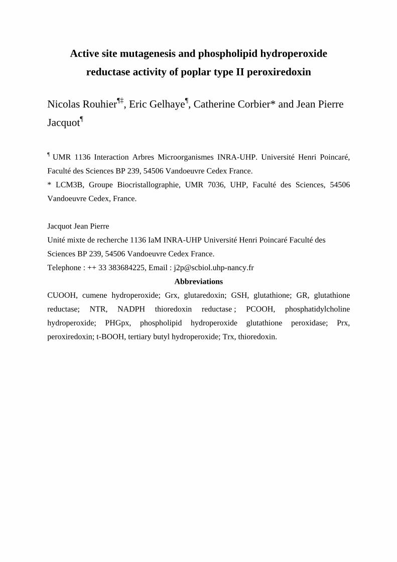
Active site mutagenesis and phospholipid hydroperoxide
reductase activity of poplar type II peroxiredoxin
Nicolas Rouhier¶‡, Eric Gelhaye¶, Catherine Corbier* and Jean Pierre
Jacquot¶
¶ UMR 1136 Interaction Arbres Microorganismes INRA-UHP. Université Henri Poincaré,
Faculté des Sciences BP 239, 54506 Vandoeuvre Cedex France.
* LCM3B, Groupe Biocristallographie, UMR 7036, UHP, Faculté des Sciences, 54506
Vandoeuvre Cedex, France.
Jacquot Jean Pierre
Unité mixte de recherche 1136 IaM INRA-UHP Université Henri Poincaré Faculté des
Sciences BP 239, 54506 Vandoeuvre Cedex France.
Telephone : ++ 33 383684225, Email : [email protected]
Abbreviations
CUOOH, cumene hydroperoxide; Grx, glutaredoxin; GSH, glutathione; GR, glutathione
reductase; NTR, NADPH thioredoxin reductase ; PCOOH, phosphatidylcholine
hydroperoxide; PHGpx, phospholipid hydroperoxide glutathione peroxidase; Prx,
peroxiredoxin; t-BOOH, tertiary butyl hydroperoxide; Trx, thioredoxin.
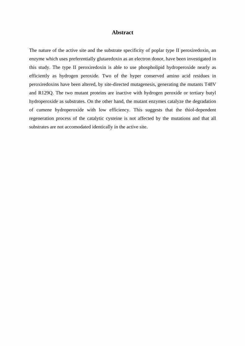
Abstract
The nature of the active site and the substrate specificity of poplar type II peroxiredoxin, an
enzyme which uses preferentially glutaredoxin as an electron donor, have been investigated in
this study. The type II peroxiredoxin is able to use phospholipid hydroperoxide nearly as
efficiently as hydrogen peroxide. Two of the hyper conserved amino acid residues in
peroxiredoxins have been altered, by site-directed mutagenesis, generating the mutants T48V
and R129Q. The two mutant proteins are inactive with hydrogen peroxide or tertiary butyl
hydroperoxide as substrates. On the other hand, the mutant enzymes catalyze the degradation
of cumene hydroperoxide with low efficiency. This suggests that the thiol-dependent
regeneration process of the catalytic cysteine is not affected by the mutations and that all
substrates are not accomodated identically in the active site.
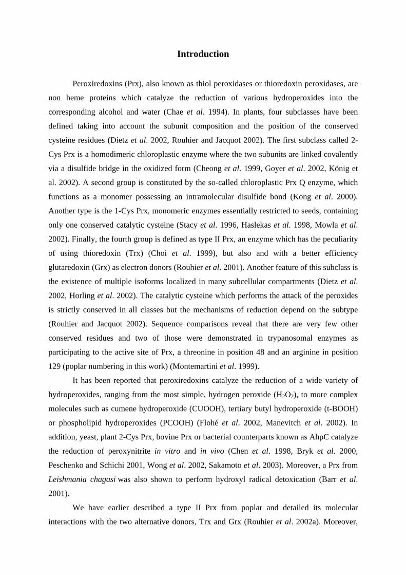
Introduction
Peroxiredoxins (Prx), also known as thiol peroxidases or thioredoxin peroxidases, are
non heme proteins which catalyze the reduction of various hydroperoxides into the
corresponding alcohol and water (Chae et al. 1994). In plants, four subclasses have been
defined taking into account the subunit composition and the position of the conserved
cysteine residues (Dietz et al. 2002, Rouhier and Jacquot 2002). The first subclass called 2-
Cys Prx is a homodimeric chloroplastic enzyme where the two subunits are linked covalently
via a disulfide bridge in the oxidized form (Cheong et al. 1999, Goyer et al. 2002, König et
al. 2002). A second group is constituted by the so-called chloroplastic Prx Q enzyme, which
functions as a monomer possessing an intramolecular disulfide bond (Kong et al. 2000).
Another type is the 1-Cys Prx, monomeric enzymes essentially restricted to seeds, containing
only one conserved catalytic cysteine (Stacy et al. 1996, Haslekas et al. 1998, Mowla et al.
2002). Finally, the fourth group is defined as type II Prx, an enzyme which has the peculiarity
of using thioredoxin (Trx) (Choi et al. 1999), but also and with a better efficiency
glutaredoxin (Grx) as electron donors (Rouhier et al. 2001). Another feature of this subclass is
the existence of multiple isoforms localized in many subcellular compartments (Dietz et al.
2002, Horling et al. 2002). The catalytic cysteine which performs the attack of the peroxides
is strictly conserved in all classes but the mechanisms of reduction depend on the subtype
(Rouhier and Jacquot 2002). Sequence comparisons reveal that there are very few other
conserved residues and two of those were demonstrated in trypanosomal enzymes as
participating to the active site of Prx, a threonine in position 48 and an arginine in position
129 (poplar numbering in this work) (Montemartini et al. 1999).
It has been reported that peroxiredoxins catalyze the reduction of a wide variety of
hydroperoxides, ranging from the most simple, hydrogen peroxide (H2O2), to more complex
molecules such as cumene hydroperoxide (CUOOH), tertiary butyl hydroperoxide (t-BOOH)
or phospholipid hydroperoxides (PCOOH) (Flohé et al. 2002, Manevitch et al. 2002). In
addition, yeast, plant 2-Cys Prx, bovine Prx or bacterial counterparts known as AhpC catalyze
the reduction of peroxynitrite in vitro and in vivo (Chen et al. 1998, Bryk et al. 2000,
Peschenko and Schichi 2001, Wong et al. 2002, Sakamoto et al. 2003). Moreover, a Prx from
Leishmania chagasi was also shown to perform hydroxyl radical detoxication (Barr et al.
2001).
We have earlier described a type II Prx from poplar and detailed its molecular
interactions with the two alternative donors, Trx and Grx (Rouhier et al. 2002a). Moreover,

we have shown that poplar type II Prx is able to catalyze the reduction of various
hydroperoxides with similar efficiency. We present here new data concerning the catalytic
mechanism and substrate specificity of this protein, two domains which have not been
extensively explored for plant enzymes.
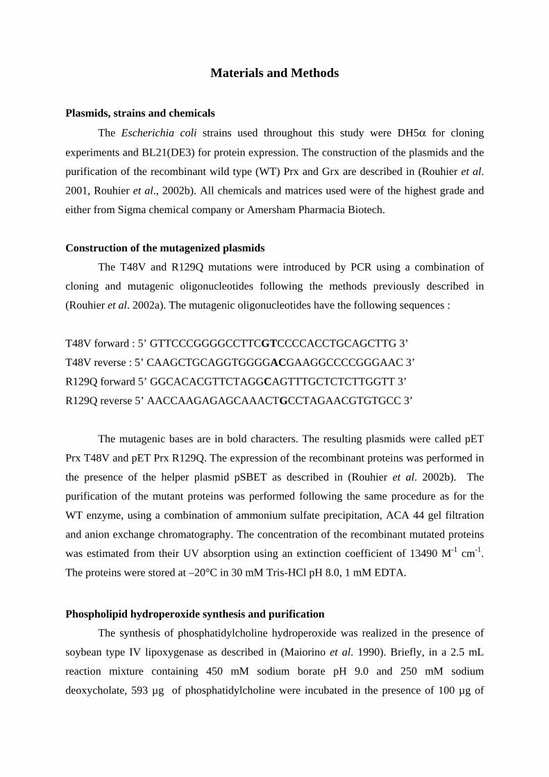
Materials and Methods
Plasmids, strains and chemicals
The Escherichia coli strains used throughout this study were DH5α for cloning
experiments and BL21(DE3) for protein expression. The construction of the plasmids and the
purification of the recombinant wild type (WT) Prx and Grx are described in (Rouhier et al.
2001, Rouhier et al., 2002b). All chemicals and matrices used were of the highest grade and
either from Sigma chemical company or Amersham Pharmacia Biotech.
Construction of the mutagenized plasmids
The T48V and R129Q mutations were introduced by PCR using a combination of
cloning and mutagenic oligonucleotides following the methods previously described in
(Rouhier et al. 2002a). The mutagenic oligonucleotides have the following sequences :
T48V forward : 5’ GTTCCCGGGGCCTTCGTCCCCACCTGCAGCTTG 3’
T48V reverse : 5’ CAAGCTGCAGGTGGGGACGAAGGCCCCGGGAAC 3’
R129Q forward 5’ GGCACACGTTCTAGGCAGTTTGCTCTCTTGGTT 3’
R129Q reverse 5’ AACCAAGAGAGCAAACTGCCTAGAACGTGTGCC 3’
The mutagenic bases are in bold characters. The resulting plasmids were called pET
Prx T48V and pET Prx R129Q. The expression of the recombinant proteins was performed in
the presence of the helper plasmid pSBET as described in (Rouhier et al. 2002b). The
purification of the mutant proteins was performed following the same procedure as for the
WT enzyme, using a combination of ammonium sulfate precipitation, ACA 44 gel filtration
and anion exchange chromatography. The concentration of the recombinant mutated proteins
was estimated from their UV absorption using an extinction coefficient of 13490 M-1 cm-1.
The proteins were stored at –20°C in 30 mM Tris-HCl pH 8.0, 1 mM EDTA.
Phospholipid hydroperoxide synthesis and purification
The synthesis of phosphatidylcholine hydroperoxide was realized in the presence of
soybean type IV lipoxygenase as described in (Maiorino et al. 1990). Briefly, in a 2.5 mL
reaction mixture containing 450 mM sodium borate pH 9.0 and 250 mM sodium
deoxycholate, 593 µg of phosphatidylcholine were incubated in the presence of 100 µg of
type IV soybean lipoxygenase (36000 units) for two hours at room temperature with
continuous shaking. The purification of PCOOH was then performed using Sep pak C18
columns from Waters using 100% methanol as an eluent. The concentration of PCOOH was
estimated by a colorimetric reaction using the ferrous oxidation of xylenol orange reagent
(FOX 2) by reference to a calibration curve realized with known H2O2 concentrations (Wolff
1994). PCOOH was kept frozen at –20°C at a concentration of 1.7 mM.
In vitro hydroperoxide reduction by type II poplar Prx
The H2O2, t-BOOH or CUOOH (250 µM) reduction were measured in a coupled
reaction by following the oxidation of NADPH at 340 nm via NADPH/GR/GSH/Grx or
NADPH/NTR/Trx in a 500 µL reaction medium at 30°C as described in (Rouhier et al.
2002a). A slight modification is the addition of 15 µL of 20% Triton X-100 for the
measurement of PCOOH reduction (250 µM unless otherwise indicated) by recombinant type
II Prx. A second assay consists in the time course measurement of the disappearance of
PCOOH and other hydroperoxides using the FOX 2 or FOX 1 reagents respectively. The
reaction medium (50 to 300 µL) had the following composition : 50 mM Tris HCl pH 8.0,
500 µM DTT, 250 µM PCOOH and varying amounts of Prx, between 0 to 3 µM. The reaction
was started by adding PCOOH after one minute incubation at ambient temperature. For each
time point, 50 µL of the reaction mixture were added to 950 µL FOX 2 reagent. The
absorbance at 560 nm was read after one hour of incubation. All kinetics were repeated three
times with two different enzyme preparations and the indicated values represent the average
of the three measurements. The percentages presented in Table I are computed from the kcat
values obtained for each enzyme.
Results
Prx sequence analysis and nature of the active site
When aligning the poplar type II Prx sequence with homologous enzymes from
different sources (mammals, yeast, bacteria, trypanosoma), seven amino acid residues appear
to be very conserved, P44, T48, C51, S78, D103, G122 and R129 (poplar numbering). If
additional sequences are taken into account, out of those seven residues, only three amino
acids remain strictly conserved, the catalytic cysteine, C51, one threonine, T48, and one
arginine, R129 (data not shown). These properties suggest that T48 and R129 are likely to
play an important role in catalysis as demonstrated in the case of trypanosomal enzymes
(Montemartini et al. 1999, Flohé et al. 2002).
We have recently obtained crystals of poplar type II Prx that diffract to 1.6Å (Echalier
et al. 2002) and solved the 3D structure of the protein (unpublished data). The structure
confirms that the threonine and arginine residues are indeed in close proximity to the cysteine
of the active site. Fig. 1 shows a blow up of the active site region of the poplar enzyme with
the positioning of the three above mentioned residues. The partial structure presented here,
fits well the hypothesis of the catalytic triad. The positions of two additional side chains
which are more distant from the active site (W88 and C76) are also shown in Fig. 1. Cys76,
which is the second cysteine of type II Prx, is dispensable for activity (Rouhier et al. 2002)
and W88 is a conserved aromatic residue found also in the Leshmania donovani structure
(Flohé et al. 2002).
Active site mutagenesis
Thus, despite the low sequence identity (20%) between the poplar and the Leishmania
enzymes, these data suggest a common catalytic mechanism for all peroxiredoxins. In order to
confirm that T48 and R129 are indeed implicated in the catalysis of type II Prx, site-directed
mutagenesis was performed to create the two single mutants, T48V, in which the OH group is
removed, and R129Q where the positive charge is replaced by a non charged side chain. Fig.
2 shows the electrophoretic behavior of the various protein preparations generated. As
observed before, despite being highly homogeneous, all preparations present a polypeptide
doublet with a molecular mass of ca 17 kDa irrespective of the reducing conditions (Rouhier
et al. 2001). The 3D structure indicates that the C-terminus is complete and N-terminal
sequencing shows that all amino acids are present and no cleavage of the polypeptide
occured, except for a partial processing of the initial methionine which could perhaps explain
the presence of the doublet (data not shown). In addition, mass analysis suggests that no post-
translational modification occured in E. coli (data not shown). In oxidizing conditions, no
evidence for a dimer was observed, indicating that the proteins do not contain an
intermolecular disulfide bridge irrespective of the nature of the mutations.
Catalytic efficiencies of the WT and mutant enzymes
The catalytic activities of the WT and of the mutated enzymes were first determined
by using a coupled system in which the reduction of peroxides is coupled to NADPH
oxidation via the Grx system. Table I shows the relative efficiencies of the WT and mutant
peroxiredoxins, using various hydroperoxides as substrates. The T48V and R129Q mutants
are marginally active with no more than 4% of the activity of the WT protein, when using
H2O2 or t-BOOH. Likewise, with CUOOH as a substrate, the activities of the mutants are
depressed, but they retain a higher level of activity compared to the WT enzyme (7.8 and
16.3%). It should be mentioned that CUOOH does not behave as the two other substrates
since the reaction catalysed by the WT enzyme is quickly inhibited, but the rates remain linear
with the mutants. With the two other substrates the reactions are linear as a function of time
whatever the enzyme used (data not shown).
The catalytic impairement of the mutants was confirmed by using a different activity
measurement that relies on the direct determination of the remaining peroxides (Fig. 3). As
observed in the coupled assay, the T48V and R129Q mutations resulted in catalysts that were
essentially inactive or severely impaired with H2O2 and t-BOOH. The results with CUOOH
were not as clear cut as the mutants were more active than the WT enzyme. The discrepancy
between this result and the one obtained using the coupled assay results from the above
mentioned inhibition of the WT enzyme in the presence of CUOOH that occurs after ca 20
sec catalysis and the selected time of analysis in this second experiment (3 minutes).
We have also studied whether poplar type II Prx can use an additional substrate of
physiological importance, phospholipid hydroperoxides. The direct determination of
remaining PCOOH by FOX 2 after catalysis in the presence of the WT type II Prx and DTT is
shown in Fig. 4. PCOOH is obviously a substrate of the enzyme, and the rate and extent of its
removal is directly proportional to the concentration of enzyme present. The time course of
PCOOH reduction indicates that the reaction, is complete after 45 seconds incubation in the
presence of 500 µM DTT and 3 µM Prx. When the Prx concentration is lowered, the catalytic
rates are reduced accordingly. DTT alone did not induce any noticeable degradation of
PCOOH. Fig. 5 presents a plot showing the reaction rate as a function of PCOOH

concentration using the coupled assay. The reaction saturates at a PCOOH concentration of ca
50 µM. These data indicate that the Km for PCOOH is 23 µM.
Discussion
In plants as in other biological organisms, ROS can be extremely damaging for
biological macromolecules and especially for phospholipids. It is often observed that the
peroxidation of the membrane lipids leads to the destruction of those barriers and ultimately
to the death of the cells. In order to fight these disastrous effects, several enzymes are able to
reduce lipid hydroperoxides. Prominent among those, are the phospholipid hydroperoxide
glutathione peroxidases (PHGpx) which are more efficient with phospholipid hydroperoxides
than with other hydroperoxides (Maiorino et al. 1990). For some of the plant enzymes,
homologous to mammalian PHGpx, glutathione is a poor donor which can be replaced more
efficiently by Trx (Herbette et al. 2002, Jung et al. 2002). Surprisingly, it has been reported
that the enzymes from Lycopersicon esculentum and Heliantus annuus exhibit catalytic
activity with H2O2 only when Trx was used as a donor (Herbette et al. 2002). This type of
PHGpx could thus constitute a fifth type of peroxiredoxin unrecognized so far.
It has also been shown that mammalian, bacterial or trypanosomal peroxiredoxins can
reduce phospholipid hydroperoxides (Maiorino et al. 1990, Hillas et al. 1997, Nogoceke et al.
1997). A recent report indicates that this is also true for barley 2-Cys Prx, which exibits a low
catalytic turnover with linoleic acid hydroperoxide and phosphatidylcholine dilinoleyl
hydroperoxide, but this has not been shown for other plant enzymes yet (König et al. 2003).
The data generated in this study indicate that type II Prx also possesses this additional
function. This observation is further supported by the relatively good efficiency of this
enzyme with PCOOH as kcat values are 2.6 s-1 (H2O2), 2.1 s-1 (PCOOH), 1.5 s-1 (t-BOOH)
and 0.89 s-1 (CUOOH). The kcat values recorded for type II Prx with H2O2 are very similar to
those obtained with human Prx V, a mammalian homolog (Seo et al. 2001). From the data
presented on tryparedoxin peroxidase from Crithidia fasciculata and on phospholipid
hydroperoxide glutathione peroxidase from Helianthus annuus, catalytic efficiencies can be
computed (Nogoceke et al. 1997, Herbette et al. 2002). The values of kcat/Km oscillate
between 4X10³ and 4X10⁴ M-1.sec-1. The poplar type II Prx possesses a catalytic efficiency of
8X10⁴ M-1.sec-1 using PCOOH as a substrate, which indicates that it is quite a good catalyst
for this reaction.
As the hydroperoxides used in this study display varied structures, it was of interest to
investigate the nature of the active site of type II Prx. Through site-directed mutagenesis, we
have obtained evidence that the type II Prx active site is indeed very similar to the active sites
present in other peroxiredoxins, consisting of a catalytic triad formed by the catalytic
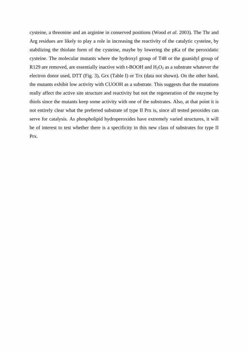
cysteine, a threonine and an arginine in conserved positions (Wood et al. 2003). The Thr and
Arg residues are likely to play a role in increasing the reactivity of the catalytic cysteine, by
stabilizing the thiolate form of the cysteine, maybe by lowering the pKa of the peroxidatic
cysteine. The molecular mutants where the hydroxyl group of T48 or the guanidyl group of
R129 are removed, are essentially inactive with t-BOOH and H2O2 as a substrate whatever the
electron donor used, DTT (Fig. 3), Grx (Table I) or Trx (data not shown). On the other hand,
the mutants exhibit low activity with CUOOH as a substrate. This suggests that the mutations
really affect the active site structure and reactivity but not the regeneration of the enzyme by
thiols since the mutants keep some activity with one of the substrates. Also, at that point it is
not entirely clear what the preferred substrate of type II Prx is, since all tested peroxides can
serve for catalysis. As phospholipid hydroperoxides have extremely varied structures, it will
be of interest to test whether there is a specificity in this new class of substrates for type II
Prx.
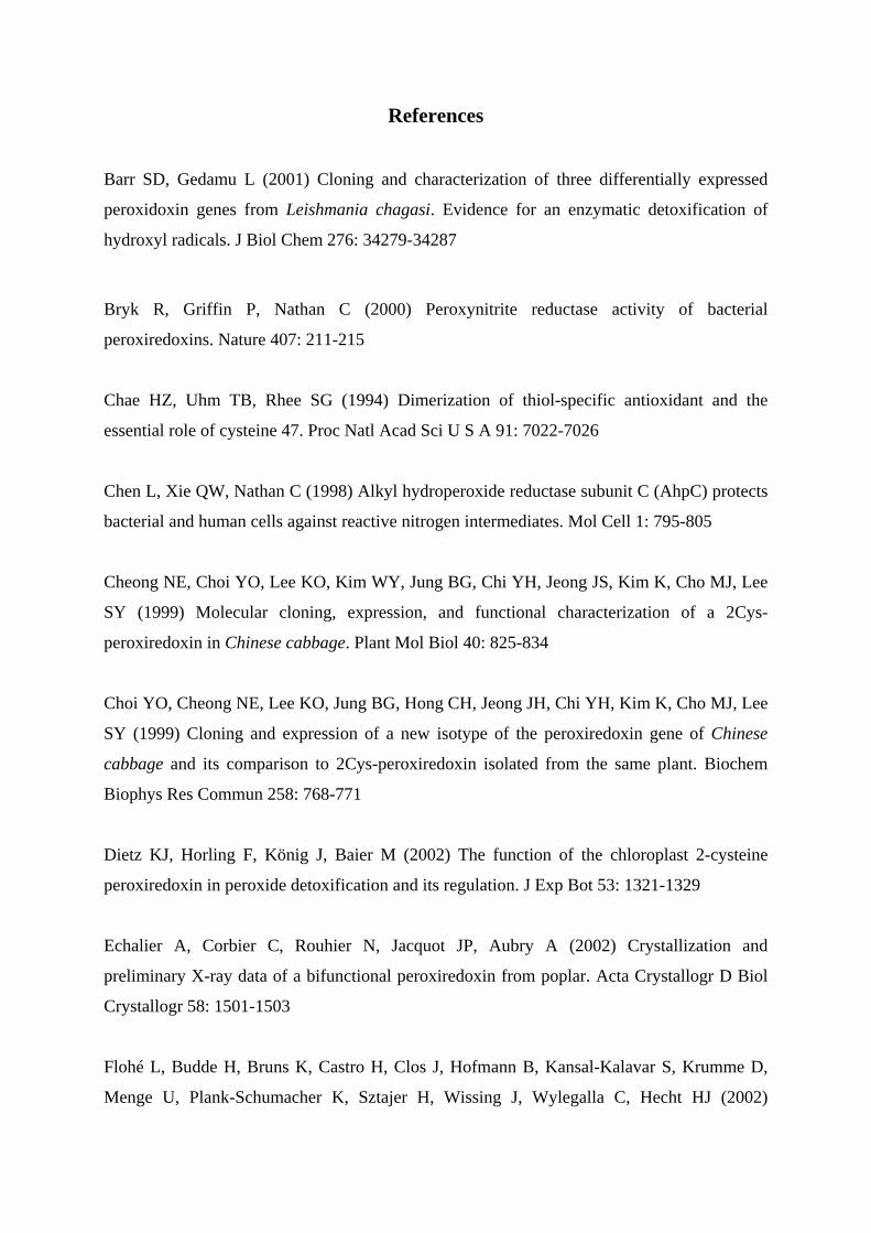
References
Barr SD, Gedamu L (2001) Cloning and characterization of three differentially expressed
peroxidoxin genes from Leishmania chagasi. Evidence for an enzymatic detoxification of
hydroxyl radicals. J Biol Chem 276: 34279-34287
Bryk R, Griffin P, Nathan C (2000) Peroxynitrite reductase activity of bacterial
peroxiredoxins. Nature 407: 211-215
Chae HZ, Uhm TB, Rhee SG (1994) Dimerization of thiol-specific antioxidant and the
essential role of cysteine 47. Proc Natl Acad Sci U S A 91: 7022-7026
Chen L, Xie QW, Nathan C (1998) Alkyl hydroperoxide reductase subunit C (AhpC) protects
bacterial and human cells against reactive nitrogen intermediates. Mol Cell 1: 795-805
Cheong NE, Choi YO, Lee KO, Kim WY, Jung BG, Chi YH, Jeong JS, Kim K, Cho MJ, Lee
SY (1999) Molecular cloning, expression, and functional characterization of a 2Cys-
peroxiredoxin in Chinese cabbage. Plant Mol Biol 40: 825-834
Choi YO, Cheong NE, Lee KO, Jung BG, Hong CH, Jeong JH, Chi YH, Kim K, Cho MJ, Lee
SY (1999) Cloning and expression of a new isotype of the peroxiredoxin gene of Chinese
cabbage and its comparison to 2Cys-peroxiredoxin isolated from the same plant. Biochem
Biophys Res Commun 258: 768-771
Dietz KJ, Horling F, König J, Baier M (2002) The function of the chloroplast 2-cysteine
peroxiredoxin in peroxide detoxification and its regulation. J Exp Bot 53: 1321-1329
Echalier A, Corbier C, Rouhier N, Jacquot JP, Aubry A (2002) Crystallization and
preliminary X-ray data of a bifunctional peroxiredoxin from poplar. Acta Crystallogr D Biol
Crystallogr 58: 1501-1503
Flohé L, Budde H, Bruns K, Castro H, Clos J, Hofmann B, Kansal-Kalavar S, Krumme D,
Menge U, Plank-Schumacher K, Sztajer H, Wissing J, Wylegalla C, Hecht HJ (2002)
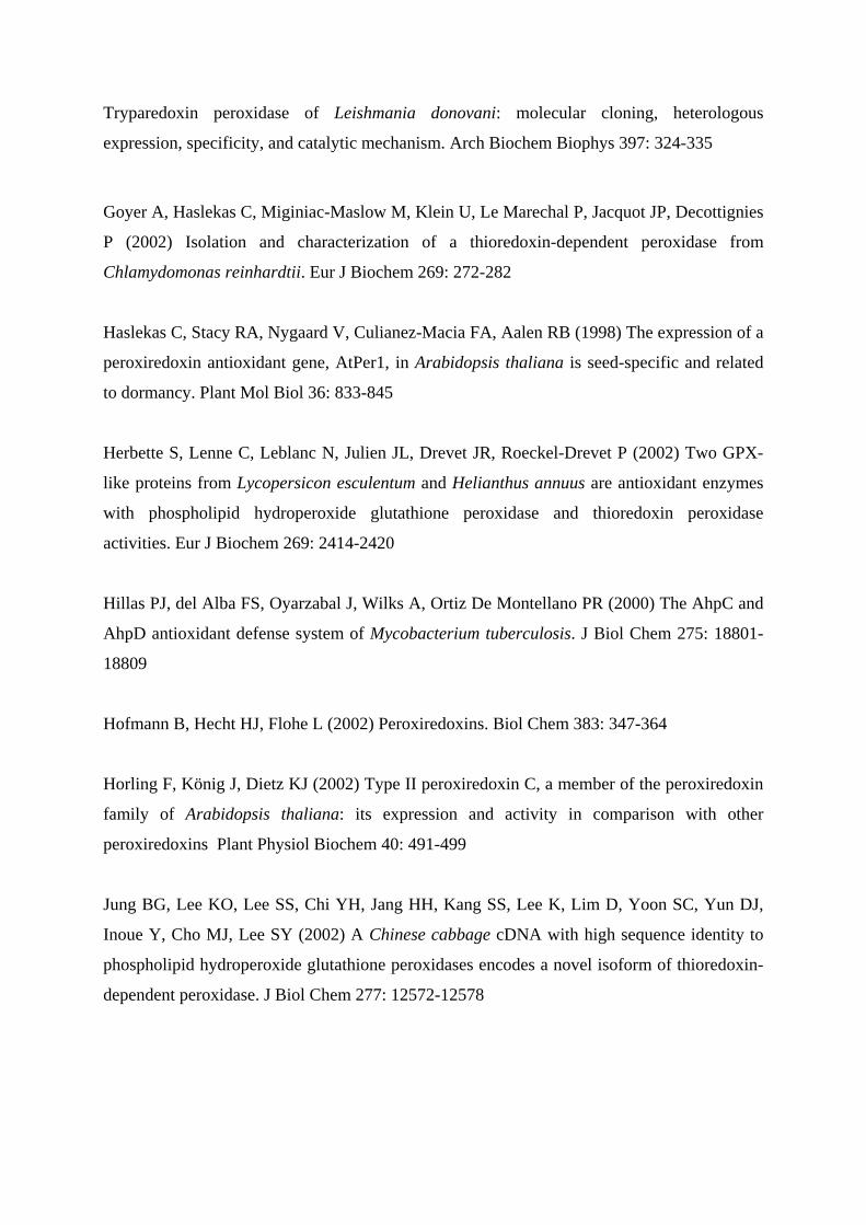
Tryparedoxin peroxidase of Leishmania donovani: molecular cloning, heterologous
expression, specificity, and catalytic mechanism. Arch Biochem Biophys 397: 324-335
Goyer A, Haslekas C, Miginiac-Maslow M, Klein U, Le Marechal P, Jacquot JP, Decottignies
P (2002) Isolation and characterization of a thioredoxin-dependent peroxidase from
Chlamydomonas reinhardtii. Eur J Biochem 269: 272-282
Haslekas C, Stacy RA, Nygaard V, Culianez-Macia FA, Aalen RB (1998) The expression of a
peroxiredoxin antioxidant gene, AtPer1, in Arabidopsis thaliana is seed-specific and related
to dormancy. Plant Mol Biol 36: 833-845
Herbette S, Lenne C, Leblanc N, Julien JL, Drevet JR, Roeckel-Drevet P (2002) Two GPX-
like proteins from Lycopersicon esculentum and Helianthus annuus are antioxidant enzymes
with phospholipid hydroperoxide glutathione peroxidase and thioredoxin peroxidase
activities. Eur J Biochem 269: 2414-2420
Hillas PJ, del Alba FS, Oyarzabal J, Wilks A, Ortiz De Montellano PR (2000) The AhpC and
AhpD antioxidant defense system of Mycobacterium tuberculosis. J Biol Chem 275: 18801-
18809
Hofmann B, Hecht HJ, Flohe L (2002) Peroxiredoxins. Biol Chem 383: 347-364
Horling F, König J, Dietz KJ (2002) Type II peroxiredoxin C, a member of the peroxiredoxin
family of Arabidopsis thaliana: its expression and activity in comparison with other
peroxiredoxins Plant Physiol Biochem 40: 491-499
Jung BG, Lee KO, Lee SS, Chi YH, Jang HH, Kang SS, Lee K, Lim D, Yoon SC, Yun DJ,
Inoue Y, Cho MJ, Lee SY (2002) A Chinese cabbage cDNA with high sequence identity to
phospholipid hydroperoxide glutathione peroxidases encodes a novel isoform of thioredoxin-
dependent peroxidase. J Biol Chem 277: 12572-12578
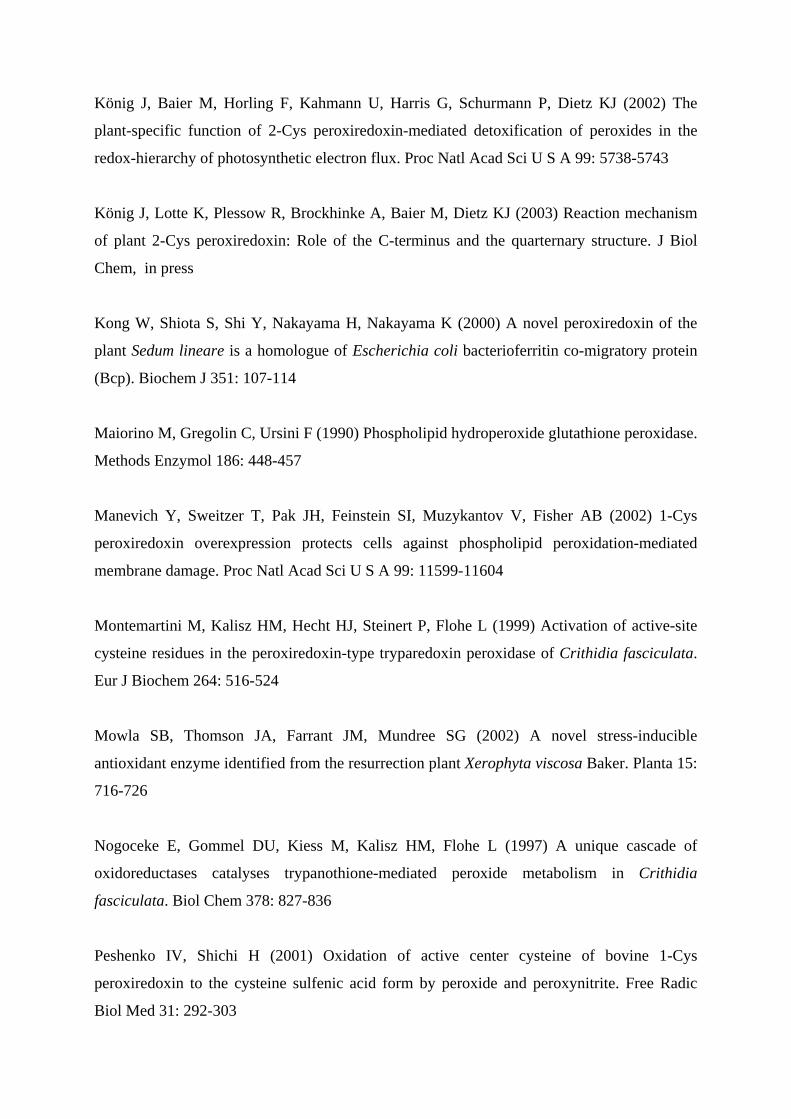
König J, Baier M, Horling F, Kahmann U, Harris G, Schurmann P, Dietz KJ (2002) The
plant-specific function of 2-Cys peroxiredoxin-mediated detoxification of peroxides in the
redox-hierarchy of photosynthetic electron flux. Proc Natl Acad Sci U S A 99: 5738-5743
König J, Lotte K, Plessow R, Brockhinke A, Baier M, Dietz KJ (2003) Reaction mechanism
of plant 2-Cys peroxiredoxin: Role of the C-terminus and the quarternary structure. J Biol
Chem, in press
Kong W, Shiota S, Shi Y, Nakayama H, Nakayama K (2000) A novel peroxiredoxin of the
plant Sedum lineare is a homologue of Escherichia coli bacterioferritin co-migratory protein
(Bcp). Biochem J 351: 107-114
Maiorino M, Gregolin C, Ursini F (1990) Phospholipid hydroperoxide glutathione peroxidase.
Methods Enzymol 186: 448-457
Manevich Y, Sweitzer T, Pak JH, Feinstein SI, Muzykantov V, Fisher AB (2002) 1-Cys
peroxiredoxin overexpression protects cells against phospholipid peroxidation-mediated
membrane damage. Proc Natl Acad Sci U S A 99: 11599-11604
Montemartini M, Kalisz HM, Hecht HJ, Steinert P, Flohe L (1999) Activation of active-site
cysteine residues in the peroxiredoxin-type tryparedoxin peroxidase of Crithidia fasciculata.
Eur J Biochem 264: 516-524
Mowla SB, Thomson JA, Farrant JM, Mundree SG (2002) A novel stress-inducible
antioxidant enzyme identified from the resurrection plant Xerophyta viscosa Baker. Planta 15:
716-726
Nogoceke E, Gommel DU, Kiess M, Kalisz HM, Flohe L (1997) A unique cascade of
oxidoreductases catalyses trypanothione-mediated peroxide metabolism in Crithidia
fasciculata. Biol Chem 378: 827-836
Peshenko IV, Shichi H (2001) Oxidation of active center cysteine of bovine 1-Cys
peroxiredoxin to the cysteine sulfenic acid form by peroxide and peroxynitrite. Free Radic
Biol Med 31: 292-303
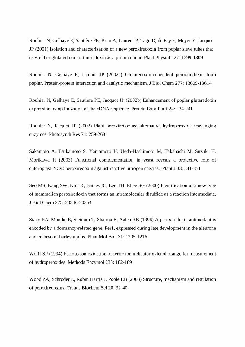
Rouhier N, Gelhaye E, Sautière PE, Brun A, Laurent P, Tagu D, de Fay E, Meyer Y, Jacquot
JP (2001) Isolation and characterization of a new peroxiredoxin from poplar sieve tubes that
uses either glutaredoxin or thioredoxin as a proton donor. Plant Physiol 127: 1299-1309
Rouhier N, Gelhaye E, Jacquot JP (2002a) Glutaredoxin-dependent peroxiredoxin from
poplar. Protein-protein interaction and catalytic mechanism. J Biol Chem 277: 13609-13614
Rouhier N, Gelhaye E, Sautiere PE, Jacquot JP (2002b) Enhancement of poplar glutaredoxin
expression by optimization of the cDNA sequence. Protein Expr Purif 24: 234-241
Rouhier N, Jacquot JP (2002) Plant peroxiredoxins: alternative hydroperoxide scavenging
enzymes. Photosynth Res 74: 259-268
Sakamoto A, Tsukamoto S, Yamamoto H, Ueda-Hashimoto M, Takahashi M, Suzuki H,
Morikawa H (2003) Functional complementation in yeast reveals a protective role of
chloroplast 2-Cys peroxiredoxin against reactive nitrogen species. Plant J 33: 841-851
Seo MS, Kang SW, Kim K, Baines IC, Lee TH, Rhee SG (2000) Identification of a new type
of mammalian peroxiredoxin that forms an intramolecular disulfide as a reaction intermediate.
J Biol Chem 275: 20346-20354
Stacy RA, Munthe E, Steinum T, Sharma B, Aalen RB (1996) A peroxiredoxin antioxidant is
encoded by a dormancy-related gene, Per1, expressed during late development in the aleurone
and embryo of barley grains. Plant Mol Biol 31: 1205-1216
Wolff SP (1994) Ferrous ion oxidation of ferric ion indicator xylenol orange for measurement
of hydroperoxides. Methods Enzymol 233: 182-189
Wood ZA, Schroder E, Robin Harris J, Poole LB (2003) Structure, mechanism and regulation
of peroxiredoxins. Trends Biochem Sci 28: 32-40

Wong CM, Zhou Y, Ng RW, Kung Hf HF, Jin DY (2002) Cooperation of yeast
peroxiredoxins Tsa1p and Tsa2p in the cellular defense against oxidative and nitrosative
stress. J Biol Chem 277: 5385-5394
Figure legends
Fig. 1. Reaction center of poplar type II Prx.
The figure was generated using PyMOL (Delano W, http://www.pymol.org). The side chains
of the catalytic triad that includes Thr48, Cys51 and Arg129 are shown in sticks with a ribbon
rendering of the closeby secondary structural units. The threonine and cysteine residues are
located respectively close to or in helix α2 and the arginine in the penultimate strand β9. The
additional cysteine (Cys76 of β4) and the conserved residue Trp88 of helix α3 are also
shown.
Fig. 2. SDS PAGE analysis of the recombinant type II peroxiredoxins.
SDS PAGE 15 % showing WT type II Prx (lanes a and d) and mutants T48V (lanes b and e)
and R129Q (lanes c and f) in reducing conditions (700 mM ß-mercaptoethanol, lanes a, b, c)
and in non reducing conditions (lanes d, e, f). The molecular weight marker (lane M) (15, 20,
25, 37, 50, 75 kDa) is the Precision Plus Protein Unstained Standard form BIORAD.
Fig. 3. DTT dependent activity of type II Prx WT, T48V, R129Q.
The reaction were carried out with 2 µM of Prx in the presence of 500 µM DTT and 500 µM
hydroperoxides. All experiments are triplicates.
Fig. 4. Time course of phosphatidylcholine hydroperoxide reduction.
The reduction of PCOOH by WT type II Prx was measured by following the disappearance of
250 µM PCOOH using 500 µM DTT as a reductant and the FOX 2 reagent. Close circles :
minus Prx, open circles :1 µM Prx, close triangles : 2 µM Prx, open triangles : 3 µM Prx.
Fig. 5. Kinetics of hydroperoxide reduction as a function of the substrate concentration.
The 500 µL reaction mixture contained 150 µM NADPH, 500 µM GSH, 0.5 unit GR, 2 µM
poplar Grx, 1 µM type II Prx and various concentrations of PCOOH, ranging from 25 to 250
µM. The catalytic activity was expressed as µmoles of oxidized NADPH per min per mg of
protein.
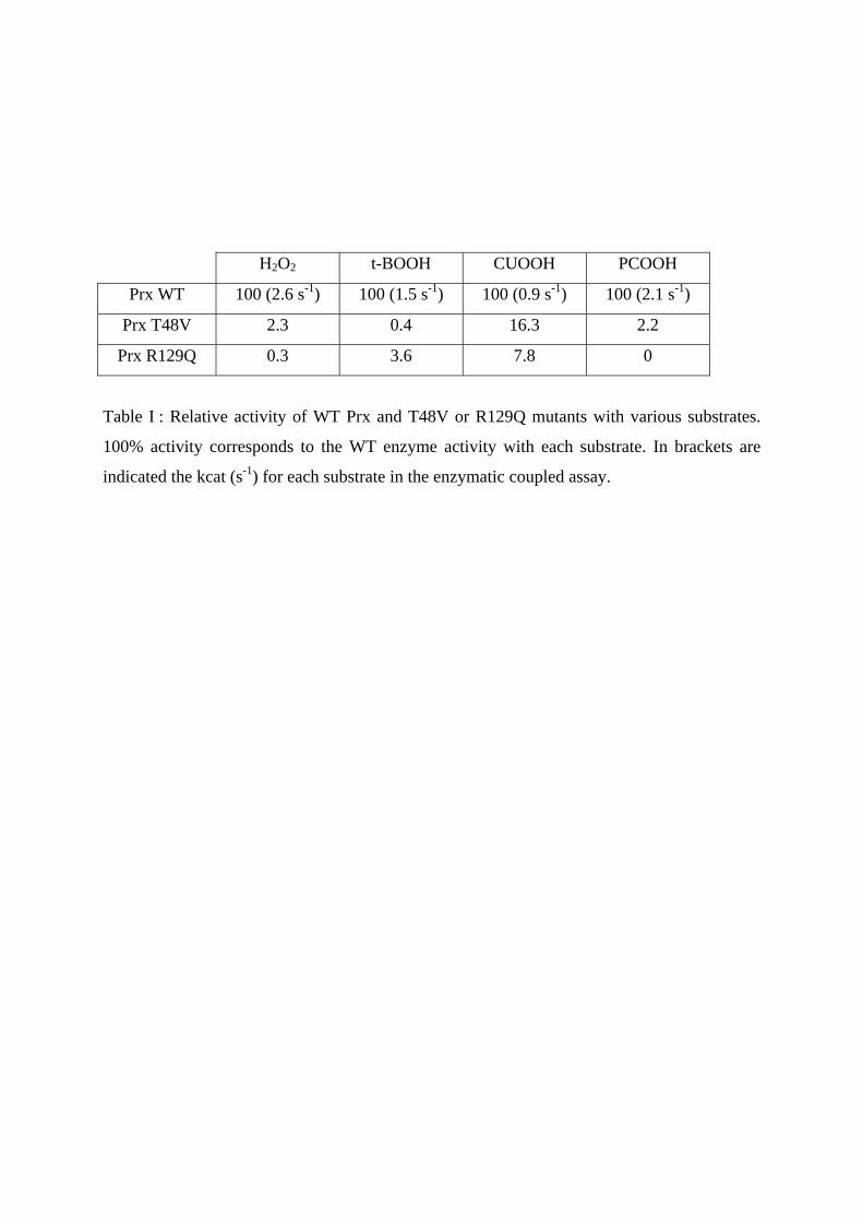
H2O2 t-BOOH CUOOH PCOOH
Prx WT 100 (2.6 s-1) 100 (1.5 s-1) 100 (0.9 s-1) 100 (2.1 s-1)
Prx T48V 2.3 0.4 16.3 2.2
Prx R129Q 0.3 3.6 7.8 0
Table I : Relative activity of WT Prx and T48V or R129Q mutants with various substrates.
100% activity corresponds to the WT enzyme activity with each substrate. In brackets are
indicated the kcat (s-1) for each substrate in the enzymatic coupled assay.
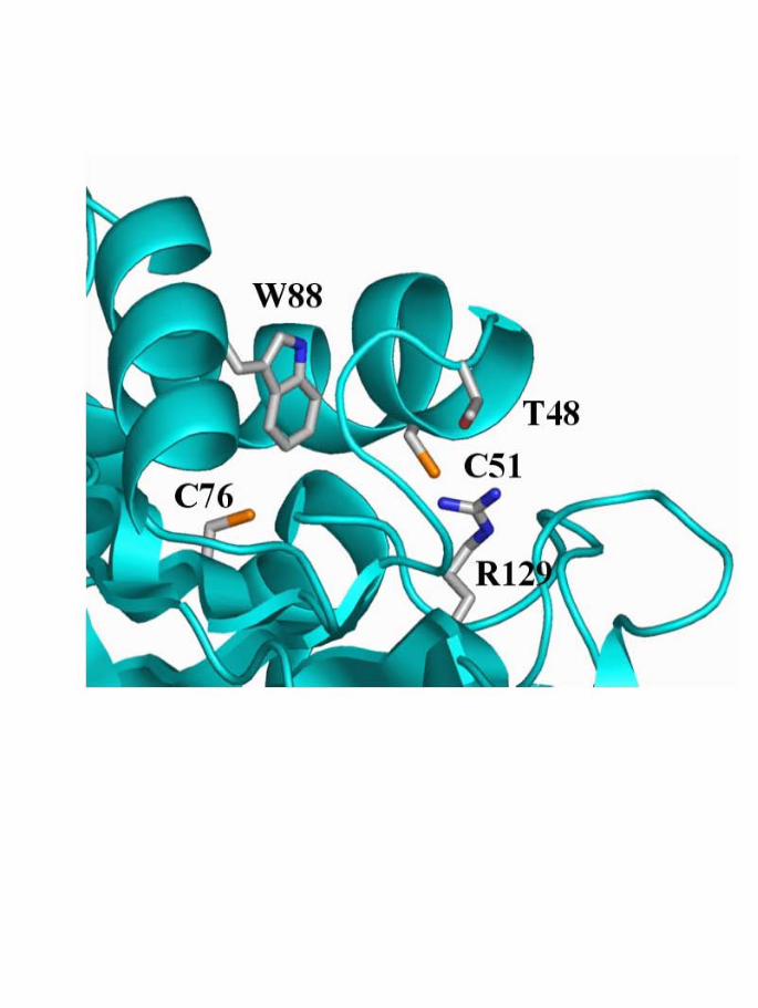
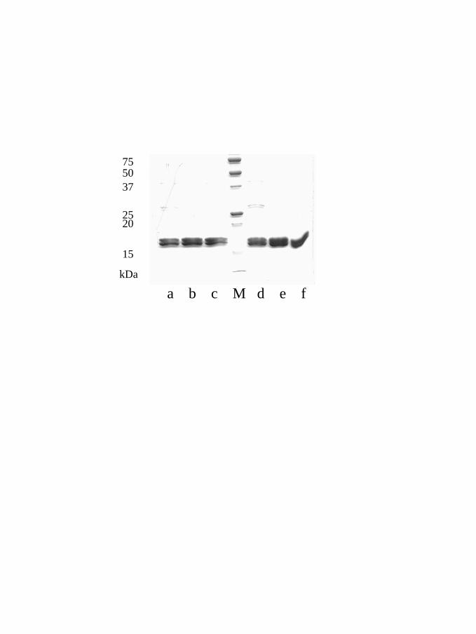
a b c M d e f
20
15
25
375075
kDa
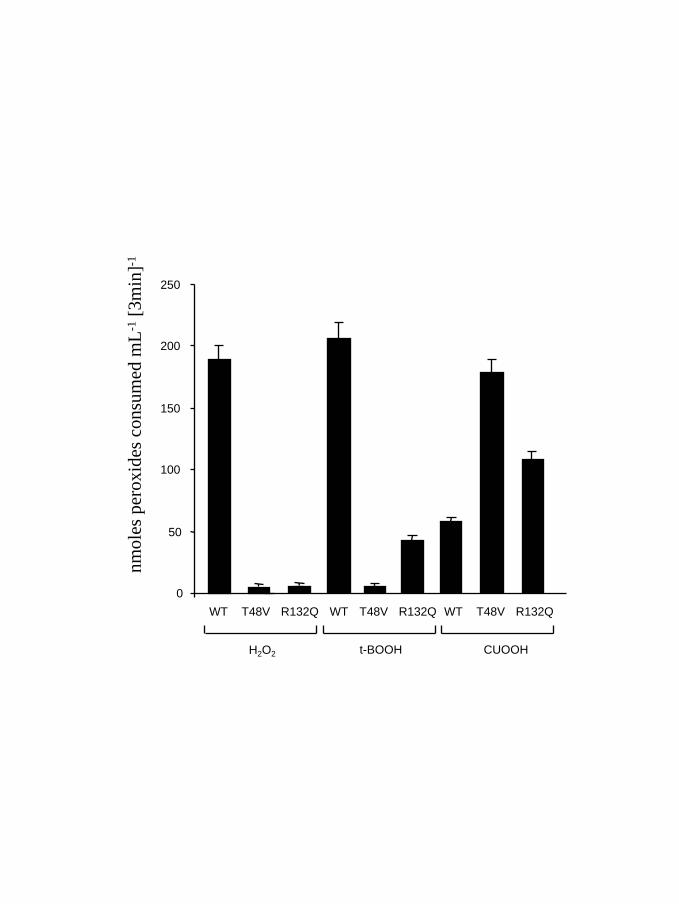
0
50
100
150
200
250
WT T48V
H2O2
R132Q
t-BOOH CUOOH
WT T48V R132Q WT T48V R132Q
nmol
espe
roxi
des c
onsu
med
mL-1
[3m
in]-1
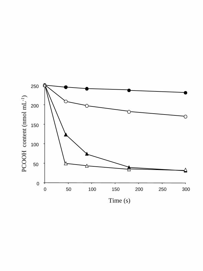
0
50
100
150
200
250
0 50 100 150 200 250 300
Time (s)
PCO
OH
con
tent
(nm
olm
L-1)
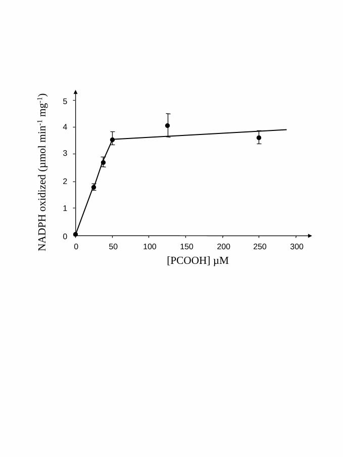
0
1
4
0 50 100 150 200 250 300
[PCOOH] µM
NA
DPH
oxid
ized
(µm
ol m
in-1
mg-1
)
5
3
2
Résultats chapitre II : Les protéines cibles
Les Prx Q représentent un des 4 groupes de Prx chez les plantes. Ces
enzymes possèdent une extension N-terminale par rapport à la protéine mature
isolée chez Sedum lineare. Afin de déterminer la fonction de cette extension, une
fusion de celle-ci avec la GFP (green fluorescent protein) a été réalisée. Elle indique
que c’est un peptide d’adressage de la protéine vers les chloroplastes. Des études
d’immunolocalisation démontrent que la protéine est présente dans les chloroplastes
des cellules du mésophylle et des stomates. Par ailleurs, la protéine ne semble être
exprimée que dans les feuilles. De plus, la quantité de Prx Q est régulée lors de
l’interaction du peuplier avec un champignon pathogène Melampsora larici-populina.
Ainsi, lors d’une réaction compatible, c’est-à-dire lorsque le champignon infecte la
feuille, la quantité de Prx Q décroît durant les 24 premières heures d’infection. Lors
d’une réaction incompatible, c’est-à-dire lorsque la plante lutte pour repousser le
champignon et qu’elle met en place une réponse hypersensible caractérisée par la
formation de ROS, la quantité de cette protéine augmente fortement dès la première
heure d’infection. Cette Prx semble donc jouer un rôle dans la réponse à une
infection par un pathogène. D’un point de vue biochimique, ce sont des enzymes
monomériques et leur mécanisme catalytique passe par la formation d’un pont
disulfure intramoléculaire entre les deux cystéines contenues dans la séquence, les
cystéines 46 et 51. Ce pont disulfure n’est réduit que par les Trx mais pas par les
Grx. Pourtant, le mutant Prx Q C51S est toujours actif avec les Trx comme donneur
d’électrons mais le devient aussi avec les Grx comme si l’absence de cette cystéine
permettait aux Grx de réduire directement le SOH formé lors de la catalyse. Le fait
que la Grx ne puisse pas réduire le pont disulfure pourrait s‘expliquer par le potentiel
rédox très négatif de la Prx, de l’ordre de -325 mV à pH 7. Enfin, il ne semble pas
qu’il existe de spécificité de reconnaissance in vitro entre les Prx Q et les différentes
isoformes de Trx puisque des Trx cytosoliques réduisent mieux la Prx Q que les Trx
chloroplastiques.
Article numéro 15 :
Rouhier N, Gelhaye E, Gualberto J, de Faÿ E, Jordy MN, Hirasawa M, Duplessis S, Martin F, Knaff D, Schürmann P, Frey P, Jacquot JP. Molecular characterization of chloroplastic poplar peroxiredoxin Q and its
involvement in the pathogenic response.
En préparation
Biological sciences : Biochemistry or Plant biology
Molecular characterization of chloroplastic peroxiredoxin Q from poplar
and its involvement in the pathogenic response
Rouhier Nicolas***, Gelhaye Eric*, Gualberto José M†, Jordy Marie-Noelle*, De Fay
Elisabeth*, Hirasawa Masa‡, Duplessis Sébastien*, Lemaire Stéphane§, Frey Pascal¶, Martin
Francis*, Manieri Wanda||, Knaff David‡, Jacquot Jean-Pierre*
* Unité Mixte de Recherche INRA-UHP 1136, Interactions Arbres/Micro-organismes,
Université Henri Poincaré, Faculté des Sciences, BP 239 54506 Vandoeuvre Cedex France. † Institut de Biologie Moléculaire des Plantes, CNRS 67084 Strasbourg Cedex ‡ Department of Chemistry and Biochemistry, and Center for Biotechnology and Genomics,
Texas Tech University, Lubbock, Texas 79409-1061, USA. § Institut de Biotechnologie des Plantes, Université de Paris Sud 91405 Orsay cedex. ¶ INRA, Unité Pathologie Forestière, F-54280 Seichamps, France ||Laboratoire de Biochimie Végétale CH-2007 Neuchatel, Switzerland
**To whom correspondence should be addressed:
Unité Mixte de Recherche INRA-UHP 1136, Interactions Arbres/Micro-organismes,
Université Henri Poincaré, Faculté des Sciences, BP 239 54506 Vandoeuvre Cedex
France.Telephone.++33383684225 email: [email protected]
Abbreviations : BCP, bacterioferritin comigratory protein ; CUOOH, cumene hydroperoxide ;
Grx, glutaredoxin ; NTR, NADPH thioredoxin reductase ; Prx peroxiredoxin ; tBOOH, tertio-
butyl hydroperoxide ; Trx, thioredoxin.
Abstract
Peroxiredoxins are ubiquitous thioredoxin or glutaredoxin dependent peroxidases, the
function of which is to destroy peroxides. Peroxiredoxin Q, one of the four plant subtypes, is
a homolog of the bacterial bacterioferritin comigratory proteins. We show here that the poplar
protein acts as a monomer with an intramolecular disulfide bridge between two conserved
cysteines. A broad spectrum of electron donors and substrates was tested. Unlike type II
peroxiredoxin, peroxiredoxin Q cannot use glutaredoxin but various cytosolic chloroplastic
and mitochondrial thioredoxins are efficient electron donors with no marked specificities. The
redox potential of the peroxiredoxin Q catalytic disulfide is -325 mV at pH 7.0, explaining
why the WT protein is reduced by thioredoxin (-290 mV) but not by glutaredoxin (ca -230
mV). Peroxiredoxin Q can reduce various alkyl hydroperoxides, but with a better efficiency
for cumene hydroperoxide than hydrogen peroxide and tertiary butyl hydroperoxide.
Additional evidence that Trx can be a donor was obtained by the formation of heterodimers
between peroxiredoxin Q and monocysteinic mutants of spinach thioredoxin m. The use of a
green fluorescence protein construct indicates that the transit sequence efficiently targets
peroxiredoxin Q to the chloroplasts and especially to those of the guard cells.
Immunolocalization confirms that peroxiredoxin Q is located in chloroplast. The expression
of this protein is modified in response to an infection by various races of Melampsora larici-
populina, the causative agent of the poplar rust. In the case of an hypersensitive response, the
peroxiredoxin expression increased whereas it decreased, during a compatible interaction.
Introduction
Organisms living under aerobic conditions have developped an array of antioxidative
systems to protect the cells from oxidative damages caused by reactive oxygen and nitrogen
species (ROS, RNS). An emerging family of non-heme peroxidases, called peroxiredoxins
(Prx) has been described in all kingdoms during the past years (1, 2, 3). These ubiquitous
proteins are able to reduce both organic and inorganic hydroperoxides with electrons provided
mainly by NADPH or NADH and different proteins such as thioredoxin (Trx), glutaredoxin
(Grx), cyclophilin A and subunit F of alkyl hydroperoxide reductase subunit F (AhpF) (1, 4,
5, 6). In addition to their documented activity as peroxidases, peroxiredoxins are involved in
resistance to RNS (7, 8). The C subunits of many bacterial alkyl hydroperoxide reductase
(AhpC) also possesses a peroxynitrite reductase activity (9). Recently, a complex reduction
system of AhpC was found to occur in Mycobacterium tuberculosis with an electron flow
starting from NADH and involving a dihydrolipoamide dehydrogenase, a dihydrolipoamide
succinyl-transferase and an alkyl hydroperoxide reductase subunit D (AhpD) (10). The
peroxiredoxins were first classified into two subclasses, the 1-Cys Prx and the 2-Cys Prx
according to the number of conserved cysteines. However, the increasing number of Prx
found in all organisms leads to a more complex hierarchy. Indeed, there are at least six Prx
isoforms in mammals, five in yeast, and eight out of the ten genes present in the genome of
Arabidopsis thaliana are expressed (3, 11, 12). The plant sequences can be organized into
four distinct subgroups (3, 13). The first one corresponds to the 1-Cys Prx. These are seed
restricted enzymes which are mainly localized in the nucleus of the embryo and the aleurone
layer (14). The second group, which comprises the dimeric 2-Cys Prx, is well characterized in
terms of function and catalysis (3, 15, 16). These proteins, homologous to the mammalian Prx
I to IV and bacterial AhpC, are chloroplastic enzymes. The third class is a large group of
monomeric type II Prx, with at least four expressed isoforms in A. thaliana. Two of these are
assumed to be cytosolic enzymes, the two other are probably chloroplastic and mitochondrial
(17-19). Finally, the last class is constituted by Prx Q, an enzyme homologous to the
bacterioferritin comigratory proteins (BCP). Until recently, this type of enzyme was only
found in prokaryotes and lower eukaryotes but an enzyme of Sedum lineare has been recently
characterized (20). Up to now, there are no homologous of Prx Q in mammalian cells.
Apparently, this enzyme is a monomer with an intramolecular disulfide bridge between two
cysteine residues separated by only four amino acids (20). The functions of the BCP and Prx
Q are not very well understood. BCP is involved in the resistance of Escherichia coli against
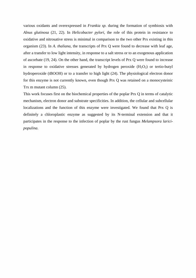
various oxidants and overexpressed in Frankia sp. during the formation of symbiosis with
Alnus glutinosa (21, 22). In Helicobacter pylori, the role of this protein in resistance to
oxidative and nitrosative stress is minimal in comparison to the two other Prx existing in this
organism (23). In A. thaliana, the transcripts of Prx Q were found to decrease with leaf age,
after a transfer to low light intensity, in response to a salt stress or to an exogenous application
of ascorbate (19, 24). On the other hand, the transcript levels of Prx Q were found to increase
in response to oxidative stresses generated by hydrogen peroxide (H2O2) or tertio-butyl
hydroperoxide (tBOOH) or to a transfer to high light (24). The physiological electron donor
for this enzyme is not currently known, even though Prx Q was retained on a monocysteinic
Trx m mutant column (25).
This work focuses first on the biochemical properties of the poplar Prx Q in terms of catalytic
mechanism, electron donor and substrate specificities. In addition, the cellular and subcellular
localizations and the function of this enzyme were investigated. We found that Prx Q is
definitely a chloroplastic enzyme as suggested by its N-terminal extension and that it
participates in the response to the infection of poplar by the rust fungus Melampsora larici-
populina.
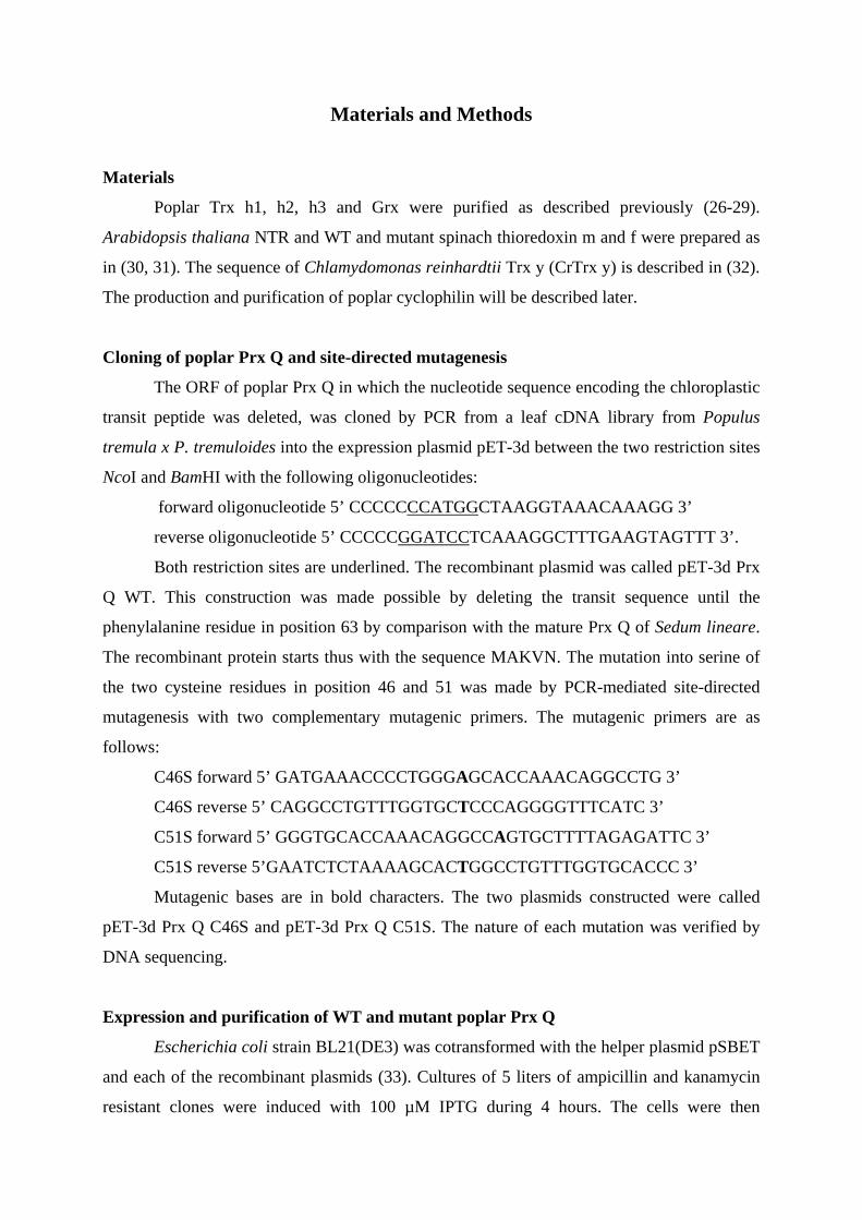
Materials and Methods
Materials Poplar Trx h1, h2, h3 and Grx were purified as described previously (26-29).
Arabidopsis thaliana NTR and WT and mutant spinach thioredoxin m and f were prepared as
in (30, 31). The sequence of Chlamydomonas reinhardtii Trx y (CrTrx y) is described in (32).
The production and purification of poplar cyclophilin will be described later.
Cloning of poplar Prx Q and site-directed mutagenesis
The ORF of poplar Prx Q in which the nucleotide sequence encoding the chloroplastic
transit peptide was deleted, was cloned by PCR from a leaf cDNA library from Populus
tremula x P. tremuloides into the expression plasmid pET-3d between the two restriction sites
NcoI and BamHI with the following oligonucleotides:
forward oligonucleotide 5’ CCCCCCCATGGCTAAGGTAAACAAAGG 3’
reverse oligonucleotide 5’ CCCCCGGATCCTCAAAGGCTTTGAAGTAGTTT 3’.
Both restriction sites are underlined. The recombinant plasmid was called pET-3d Prx
Q WT. This construction was made possible by deleting the transit sequence until the
phenylalanine residue in position 63 by comparison with the mature Prx Q of Sedum lineare.
The recombinant protein starts thus with the sequence MAKVN. The mutation into serine of
the two cysteine residues in position 46 and 51 was made by PCR-mediated site-directed
mutagenesis with two complementary mutagenic primers. The mutagenic primers are as
follows:
C46S forward 5’ GATGAAACCCCTGGGAGCACCAAACAGGCCTG 3’
C46S reverse 5’ CAGGCCTGTTTGGTGCTCCCAGGGGTTTCATC 3’
C51S forward 5’ GGGTGCACCAAACAGGCCAGTGCTTTTAGAGATTC 3’
C51S reverse 5’GAATCTCTAAAAGCACTGGCCTGTTTGGTGCACCC 3’
Mutagenic bases are in bold characters. The two plasmids constructed were called
pET-3d Prx Q C46S and pET-3d Prx Q C51S. The nature of each mutation was verified by
DNA sequencing.
Expression and purification of WT and mutant poplar Prx Q
Escherichia coli strain BL21(DE3) was cotransformed with the helper plasmid pSBET
and each of the recombinant plasmids (33). Cultures of 5 liters of ampicillin and kanamycin
resistant clones were induced with 100 µM IPTG during 4 hours. The cells were then
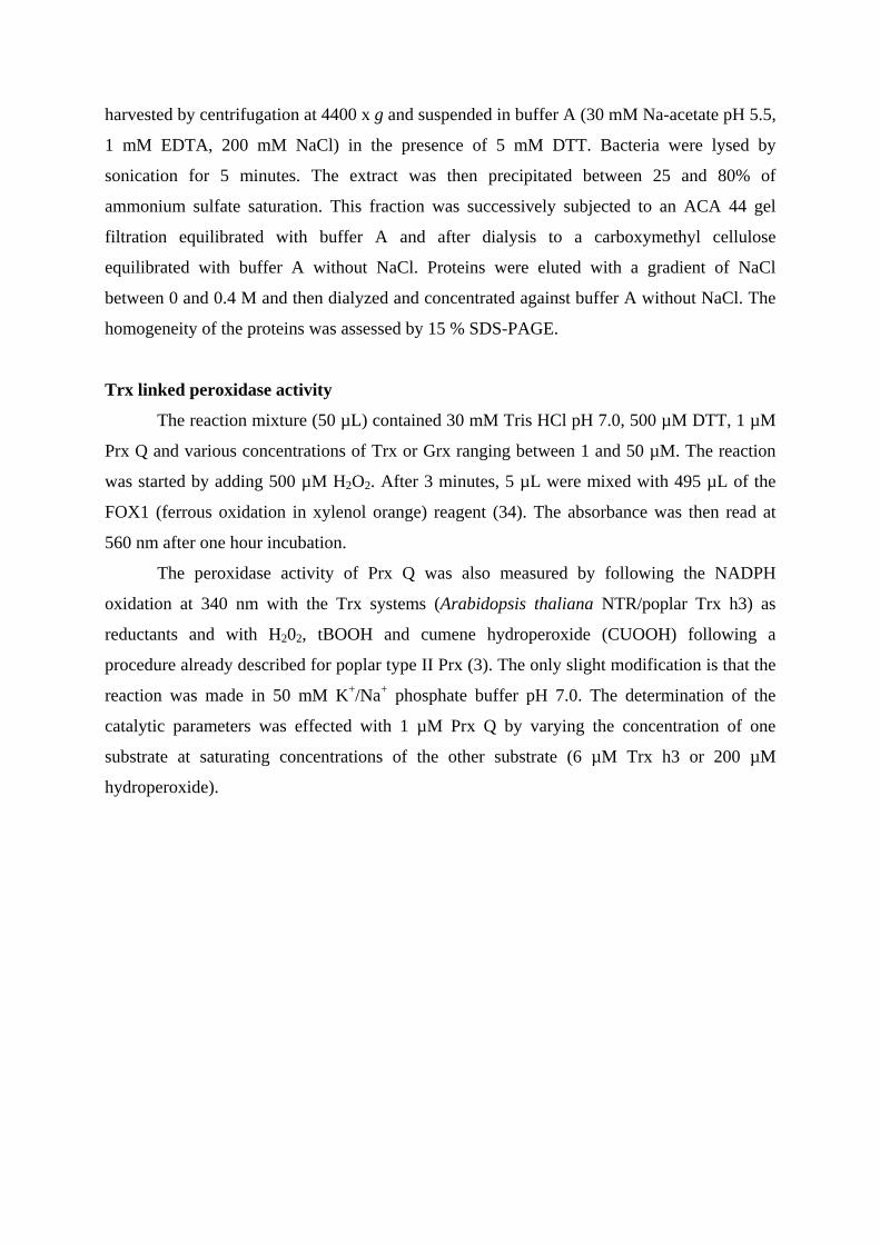
harvested by centrifugation at 4400 x g and suspended in buffer A (30 mM Na-acetate pH 5.5,
1 mM EDTA, 200 mM NaCl) in the presence of 5 mM DTT. Bacteria were lysed by
sonication for 5 minutes. The extract was then precipitated between 25 and 80% of
ammonium sulfate saturation. This fraction was successively subjected to an ACA 44 gel
filtration equilibrated with buffer A and after dialysis to a carboxymethyl cellulose
equilibrated with buffer A without NaCl. Proteins were eluted with a gradient of NaCl
between 0 and 0.4 M and then dialyzed and concentrated against buffer A without NaCl. The
homogeneity of the proteins was assessed by 15 % SDS-PAGE.
Trx linked peroxidase activity
The reaction mixture (50 µL) contained 30 mM Tris HCl pH 7.0, 500 µM DTT, 1 µM
Prx Q and various concentrations of Trx or Grx ranging between 1 and 50 µM. The reaction
was started by adding 500 µM H2O2. After 3 minutes, 5 µL were mixed with 495 µL of the
FOX1 (ferrous oxidation in xylenol orange) reagent (34). The absorbance was then read at
560 nm after one hour incubation.
The peroxidase activity of Prx Q was also measured by following the NADPH
oxidation at 340 nm with the Trx systems (Arabidopsis thaliana NTR/poplar Trx h3) as
reductants and with H202, tBOOH and cumene hydroperoxide (CUOOH) following a
procedure already described for poplar type II Prx (3). The only slight modification is that the
reaction was made in 50 mM K+/Na+ phosphate buffer pH 7.0. The determination of the
catalytic parameters was effected with 1 µM Prx Q by varying the concentration of one
substrate at saturating concentrations of the other substrate (6 µM Trx h3 or 200 µM
hydroperoxide).
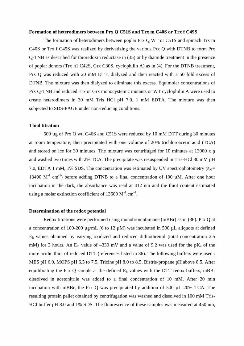
Formation of heterodimers between Prx Q C51S and Trx m C40S or Trx f C49S
The formation of heterodimers between poplar Prx Q WT or C51S and spinach Trx m
C40S or Trx f C49S was realized by derivatizing the various Prx Q with DTNB to form Prx
Q-TNB as described for thioredoxin reductase in (35) or by diamide treatment in the presence
of poplar donors (Trx h1 C42S, Grx C30S, cyclophilin A) as in (4). For the DTNB treatment,
Prx Q was reduced with 20 mM DTT, dialyzed and then reacted with a 50 fold excess of
DTNB. The mixture was then dialyzed to eliminate this excess. Equimolar concentrations of
Prx Q-TNB and reduced Trx or Grx monocysteinic mutants or WT cyclophilin A were used to
create heterodimers in 30 mM Tris HCl pH 7.0, 1 mM EDTA. The mixture was then
subjected to SDS-PAGE under non-reducing conditions.
Thiol titration
500 µg of Prx Q wt, C46S and C51S were reduced by 10 mM DTT during 30 minutes
at room temperature, then precipitated with one volume of 20% trichloroacetic acid (TCA)
and stored on ice for 30 minutes. The mixture was centrifuged for 10 minutes at 13000 x g
and washed two times with 2% TCA. The precipitate was resuspended in Tris-HCl 30 mM pH
7.0, EDTA 1 mM, 1% SDS. The concentration was estimated by UV spectrophotometry (εM=
13490 M-1 cm-1) before adding DTNB to a final concentration of 100 µM. After one hour
incubation in the dark, the absorbance was read at 412 nm and the thiol content estimated
using a molar extinction coefficient of 13600 M-1.cm-1.
Determination of the redox potential
Redox titrations were performed using monobromobimane (mBBr) as in (36). Prx Q at
a concentration of 100-200 µg/mL (6 to 12 µM) was incubated in 500 µL aliquots at defined
Eh values obtained by varying oxidized and reduced dithiothreitol (total concentration 2.5
mM) for 3 hours. An Em value of –330 mV and a value of 9.2 was used for the pKa of the
more acidic thiol of reduced DTT (references listed in 36). The following buffers were used :
MES pH 6.0, MOPS pH 6.5 to 7.5, Tricine pH 8.0 to 8.5, Bistris-propane pH above 8.5. After
equilibrating the Prx Q sample at the defined Eh values with the DTT redox buffers, mBBr
dissolved in acetonitrile was added to a final concentration of 10 mM. After 20 min
incubation with mBBr, the Prx Q was precipitated by addition of 500 µL 20% TCA. The
resulting protein pellet obtained by centrifugation was washed and dissolved in 100 mM Tris-
HCl buffer pH 8.0 and 1% SDS. The fluorescence of these samples was measured at 450 nm,
excited by 380 nm light, using an Aminco-Bowman Series 2 Luminsecence
Spectrofluorimeter. Fitting of the data was done as in (36).
Infection of the leaves, protein extraction, antibody purification and Western blot
analysis
Populus x interamericana 'Beaupré' plants were grown for 12 weeks in a greenhouse
from dormant cuttings in pots containing a sand-peat mixture. Fully expanded leaves were
detached and spray-inoculated on their abaxial surface with an urediniospore suspension in
water-agar (0.1 g.l-1) adjusted to 5000 urediniospores.ml-1 or with water-agar (control). Two
isolates of M. larici-populina were used: 98AG31 (pathotype 3-4-7) and 93ID6 (pathotype 3-
4), virulent and avirulent, respectively, on 'Beaupré' (37). The inoculated leaves were then
incubated with the abaxial surface uppermost, floating on deionized water in Petri dishes, at
20°C under artificial illumination, for various durations ranging from 1 hour to 10 days. 400
mg of leaves were ground with a mortar and pestle on ice and then 4 mL of the extraction
buffer (50 mM Tris HCl pH 7.5, 1 mM EDTA, PVP 2.5 mg/mL and 25 mM β-
mercaptoethanol) were added. The mixture was centrifuged twice for 10 minutes at 14000 x g
and the supernatant precipitated with 80% acetone. Proteins were pelleted by centrifugation
and washed with 80% acetone. Proteins were resuspended in 125 mM Tris-HCl buffer (pH
6.8) containing glycerol 20%, SDS 2%, β-mercaptoethanol 5% and bromophenol blue 0.05%
and their concentration was determined by the Bradford reaction kit from BIO-RAD. Protein
extraction of untreated leaves, stems and roots was carried out as described in (18). Polyclonal
antibodies raised in rabbit against poplar Prx Q were purified by affinity on a Prx Q
Sepharose column following the procedure described previously (18). Western blots were
performed using PVDF membranes from Millipore and the Immune Star Goat Anti Rabbit
Detection Kit from BIO-RAD.
Intracellular localization via GFP fusion
For in vivo intracellular localization, the first 195 bp of the poplar Prx Q cDNA,
corresponding to the N-terminal presequence (65 amino acids) were cloned into the NcoI and
BamHI sites of pCK-GFP3A using the two following primers:
5’ CCCCCATGGCTTCCATTTCTCTC 3’ and
5’ CCCCGGATCCTTGGCAAAAATGGTATTCTT 3’
The PCR amplified fragment was fused to GFP at the BamHI site, resulting in a
chimeric protein where the transit sequence of Prx Q is present on the N-terminus side of GFP
and under the control of a double 35S promoter (38). Nicotiana bentamiana cells were
transfected by bombardment of leaves with tungsten particles coated with plasmid DNA and
images were obtained with a Zeiss LSM510 confocal microscope.
Immunolocalization
Sections of 25 mm2 were cut from expanded leaves of 2 months old poplar and fixed
for 3h at 4°C in 4% paraformaldehyde (w/v) and 0.5% (v/v) glutaraldehyde in 0.1 M
cacodylate buffer, pH 7.4. The samples were subsequently rinsed in 0.1 M cacodylate, treated
with 2% OsO4, dehydrated in increasing alcohol and propylene oxide, embedded in Durcupan
(Fluka) and finally polymerized at 56°C for 48h.
For immunocytochemistry ultrathin sections (70 nm) were placed onto 300 mesh
nickel grids. Grids were floated for 15 min in a 50 mM glycine, 50 mM Tris phosphate–
buffered saline (TPBS) solution, transferred for 30 min on a blocking solution of 1% BSA in
Tris phosphate–buffered saline (TPBS/BSA) and then treated overnight at 4°C with purified
rabbit Prx Q antibodies (diluted 100x in TPBS/BSA). Controls were realized by omission of
this step. After washing, sections were incubated for 2h in a solution of 50 mM Tris-HCl
buffer containing 0.05% PEG with gold-labelled goat anti-rabbit IgG (10 nm, Sigma)
(dilution 1:20). After rinsing, the sections were dried and stained with uranyl acetate and lead
citrate. Observations were made using a Zeiss‚ TEM 902 electronic microscope.
For histochemical detection of Prx Q using fluorescence, 130 nm thin sections are
coated with polylysine (Sigma) on a glass slide. The resin was removed by dissolution for 20
min with ethanol saturated in NaOH washed with 100% ethanol and then with 100% acetone.
Slides were treated 15 min with in TPBS solution containing 50 mM glycine, washed with
TPBS and finally treated with TPBS/BSA for 2h30 before incubation with purified rabbit Prx
Q antibodies overnight (diluted 100 fold). After rinsing with TPBS and TPBS/BSA, the slides
were incubated for 2h with secondary antibodies coupled to FITC (fluorescein isothiocyanate)
diluted 160 fold. Control experiments were performed either by omitting primary antibodies
or by testing preimmune serum. The slides were rinsed and mounted in TPBS containing 50%
glycerol. Sections were viewed on epifluorescence microscope (Nikon Optiphot II coupled to
a mercury lamp and B2A filters) and images were captured using a Nikon D1 camera.
Results
Sequence analysis
The deduced amino acid sequence of the poplar enzyme studied here consists of 213
residues with a putative transit peptide of 64 residues, identified by analogy to the mature Prx
Q of Sedum lineare (20). The recombinant mature form of poplar Prx Q used in this study
contains 151 amino acids, including two residues (one methionine and one alanine) that have
been added to the N-terminus immediately after the putative cleavage site of the precursor. Its
calculated molecular mass and isoelectric point are 16831 Da and 9.3, respectively.
An amino acid sequence comparison of three plant Prx Q and four BCP from
cyanobacteria, bacteria, yeast and archaebacteria is shown in Fig. 1. All the plant sequences
contain a N-terminal extension of 60 to 70 amino acids, which was identified as a
chloroplastic target sequence by prediction softwares
(http://www.cbs.dtu.dk/services/TargetP/ and http://psort.nibb.ac.jp/form.html). The
Saccharomyces cerevisiae enzyme also possesses a N-terminal extension, but the cellular
location of the yeast enzyme has not yet been identified. The sequences of the mature
proteins, devoid of the transit peptides, are 149 to 156 amino acid long and all of them contain
the two putative active site cysteines in position 46 and 51 (poplar numbering of the shortened
construction). Cys 46 is the residu that is homologous to the catalytic cysteine present in all
plant peroxiredoxins, whatever their subtype (consensus sequence P-(X)3-T-[P/F]-(X)-C-
[T/S/P]) and which is involved in catalysis via the transient formation of a sulfenic acid. The
identity levels of the sequences of the mature plant proteins range from 84 to 88%.
Comparisons between plant and non-plant enzymes show much lower identity levels, ranging
from 35 to 55%.
Biochemical characterization
Expression, purification and mutagenesis
Using a combination of gel filtration and cation exchange, recombinant Prx Q WT,
C46S and C51S were purified to homogeneity (Fig. 2A). The overall yield was about 7 to 30
mg homogenous protein per liter of E. coli culture. The oxidized form of purified WT Prx Q
is a monomeric enzyme as judged by its behavior during gel filtration (data not shown) and its
migration in non-reducing 15% SDS PAGE (Fig. 2B, lane 4). The two monocysteinic mutants
are able to form measurable amounts of homodimer, presumably via disulfide bonds
involving the remaining cysteine (Fig. 2B, lane 5 and 6). Thiol titrations of the reduced
enzymes confirmed the presence of two thiols per mole enzyme for the reduced form of WT
Prx Q, while no thiols could be detected in the oxidized state (data not shown). This
observation suggests that there is an intramolecular disulfide in the monomer. Only one
cysteine was detected per mol enzyme for the two mutants (data not shown).
Electron donor and substrate specificity
Regardless of the conditions used, we could not observe DTT functioning as a direct
electron donor to Prx Q using the FOX detection method. Activity was only observed if Trx
was added to promote catalysis (Fig. 3). Thus, in order to investigate whether the enzyme
exhibits a preference for a given electron donor, the efficiency of several chloroplastic donors
including spinach Trx m and f and the recently described Chlamydomonas reinhardtii Trx y,
has been studied (32). In addition, one mitochondrial Trx (poplar Trx h2, unpublished data),
two cytosolic Trx (poplar Trx h1 and h3), cyclophilin A and Grx have been also tested as
electron donors. Fig. 3 shows that unlike type II Prx, WT Prx Q cannot use Grx as an electron
donor. Likewise, the poplar cyclophilin A used in this study was uneffective as a donor. On
the other hand, all Trx tested were active with an apparent efficiency order h1>y>h3>[m, f,
h2]. Experiments carried out with different Trx concentrations, indicated that the reactions
typically saturate at Trx concentrations between 5 and 10 µM. Experiments using NADPH
reduction of poplar Trx h3, catalyzed by Arabidopsis thaliana NTR provides a method for
monitoring spectrophotometrically the Trx dependent reduction of hydroperoxide by Prx Q.
The kinetic parameters, determined in this fashion, are shown in Table I. Several
hydroperoxides can be used as substrates by Prx Q in the presence of Trx h3. The reactions
saturate at a Trx concentration of ca 6 µM and the Km for Trx h3, the best electron donor is
around 1.5 µM. The preferred substrate is CUOOH followed by H2O2 and tBOOH when
considering the catalytic efficiencies (kcat/Km).
Characterization of the active site by site-directed mutagenesis
Fig. 4 shows the peroxidase activity of WT Prx Q, Prx Q C46S or C51S with H2O2 as
a substrate and in the presence of DTT and either WT Grx, or Grx C30S or Trx h1. As
observed before, the WT Prx Q is only reduced by Trx h1 (column 7) but not by Grx (col. 1
and 4) even at high concentration (50 µM). The C46S mutant does not possess any
biochemical activity, whatever the reductants (Trx, Grx) or the substrates (H2O2, tBOOH,
CUOOH) used (col. 2, 5, 8 and data not shown). On the other hand, the activity of C51S
mutant is not detectable at low Trx concentration (ca 5 µM) but it can be partially rescued by
increasing the Trx concentration to 50 µM (Fig. 4, col. 9). Unexpectedly, this mutant
possesses a peroxidase activity in the presence of WT Grx but it is weakly active with the
mutant Grx C30S (col. 3 and 6). These results were confirmed spectrophotometrically by
following the NADPH oxidation in the presence of the Trx or Grx systems (data not shown).
These results fully confirm that Cys 46 is the catalytic cysteine. In the WT enzyme it is
regenerated via the formation of a disulfide bridge with the adjacent Cys 51, which disulfide
bond is reduced by Trx but not Grx (Fig. 4, col. 1, 4, 7) . Nevertheless, it is likely that in the
C51S mutant, the sulfenic acid formed in Cys 46 can be reduced directly by Trx or Grx in the
absence of the second cysteine (Fig. 4, col. 3, 9).
Complex formation
The biochemical characteristics described above have been confirmed by establishing
that heterodimers can form between the donors and Prx Q C51S. While, in agreement with the
biochemical results, cyclophilin A was unable to form heterodimers with Prx Q (data not
shown). On the other hand, heterodimers were present when mixing Prx C51S and the various
monocysteinic Trx, either Trx f C49S or Trx h1 C42S (data not shown) or Trx m C40S (Fig.
2B, lane 2). The dissociation of this complex by excess DTT indeed proves that the
heterodimer was linked by a covalent disulfide bridge (lane 4). The size of the heterodimer
polypeptide (ca 30 kDa) is in good agreement with the addition of one Trx (13 kDa) to one
Prx Q (17 kDa). The observation that WT Grx does not form stable heterodimers with Prx Q
C51S, whereas it can support the activity of this mutant is consistent with the formation of a
transient intermolecular disulfide bridge that is subsequently reduced by the second active site
cysteine of Grx (data not shown). Grx C30S does not form dimers with Prx Q C51S while it
makes excellent heterodimeric structures with type II Prx in agreement with its lower catalytic
efficiency (4).
Redox potential
Fig. 5 summarizes the results of redox titrations of the Prx Q disulfide over the pH
range from pH 5.5 to 10.5. Each point represents the average of at least two determinations
and the average deviations suggest that the experimental uncertainty in Em is between 5 and
10 mV. Titrations at all pH values gave excellent fits to the Nernst Equation for a single two-
electron redox couple. Titrations at three different total DTT concentrations in the redox
buffers used (1 mM, 1.75 mM and 2.5 mM) and at two different redox equilibration times (2
and 3 hours) gave identical titration curves (within the experimental uncertainties), as
expected for titrations in which redox equilibrium has been established. The Em value for the
disulfide of Prx Q at pH 7.0, determined using the mBBr fluorescence method, is -325 ± 10
mV (Fig. 5A). A plot of Em vs pH contains two linear regions (Fig. 5B). At pH values
between 5.5 and 9.0, the slope is -59 mV per pH unit, the value expected for a process in
which a two-electron reduction is accompanied by the uptake of two protons (39). Between
pH 9.0 and 10.5, the Em vs pH slope is –29.5 mV per pH unit, indicating that the two-electron
reduction is accompanied by the uptake of only a single proton over this pH range (). The
intersection of these two straight-line segments corresponds to the pKa of an acid/base couple
with a protonation state that is linked to the redox state of Prx Q (39). Although the simplest
explanation for this data is that the more acidic active-site cysteine of reduced Prx Q has a
pKa value close to 9.0, thermodynamic measurements alone cannot identify the actual amino
acid(s) involved in this redox-coupled proton uptake.
Expression and localization of Prx Q
Localization of Prx Q
Several experiments aimed at investigating the cellular and subcellular localization of
Prx Q have been undertaken. First, a transient expression experiment was carried out using a
chimeric protein obtained by fusing the transit peptide of Prx Q to GFP. After bombardment
of tobacco leaf epidermis (which contain epidermal cells and underlaying parenchymal
photosynthetic cells) flurorescence was detected in the chloroplasts of the mesophyll cells
(data not shown) and also in the chloroplasts of the guard cells of stomata (Fig. 6, panel C).
Control experiments showing the natural fluorescence of chlorophyll indicate that the GFP
fluorescence coincides with the chloroplasts (Fig. 6, panel B). These results indicate that the
transit sequence at the N-terminus of mature Prx Q efficiently targets the protein to
chloroplasts and especially to those of the guard cells of stomata.
In a second series of experiments, semi-thin sections of poplar leaves have been
incubated with purified Prx Q antibodies and the presence of the polypeptide detected by
using anti rabbit antibodies coupled to FITC. As above with the GFP experiments, the
secondary fluorescence was found in the chloroplasts of mesophyll cells and also in the
chloroplasts of guard cells (data not shown). Immunocytochemistry experiments indicate that
Prx Q is present both in the stromal part of the chloroplasts and also somehow associated with
the thylakoid membranes in a distribution similar to the one observed for 2-Cys peroxiredoxin
(Fig. 7) (16).
Expression of Prx Q and involvement in the pathogenic response
The expression of Prx Q was followed both at the mRNA and protein levels in roots,
stems or leaves and leaves inoculated or not by the rust fungus Melampsora larici-populina
either in the case of a compatible or a incompatible reaction.
The expression of prx Q in plant organs was investigated by recording the number of
ESTs encoding this protein in the Genbank database. Only nine ESTs are present out of a total
of 114443, which suggests that the gene is transcribed at very low levels in leaves (4 hits),
bark (3 hits) and flowers (2 hits). Fig. 8A shows that the Prx Q polypeptide is only present in
detectable amounts in leaves but not in stems or roots.
On the other hand, the content in Prx Q polypeptide increased markedly with the time
of infection in the case of a hypersensitive response (Fig. 8B) with a maximum at ca 24 hours.
Conversely, in the compatible reaction, the Prx Q content decreased as infection proceeded
(Fig. 8C). After 10 days of infection, the Prx Q content is approximatively equivalent to the
one before infection. In non infected leaves (control), the relative amount of Prx Q remained
nearly unchanged (Fig. 8D).
Discussion
Peroxiredoxins from plants, like their counterparts in other biological organisms, are
subjects of great current interest because of their documented or sometimes postulated
protective role in oxidative or nitrosative stress. Two subtypes have been extensively studied,
the chloroplastic dimeric 2-Cys Prx and the type II Prx. Less is known about the 1-Cys Prx
and the Prx Q. We present here new data concerning the molecular mechanism, substrate and
donor specificity and in vivo function and localization of Prx Q in poplar, a woody species.
Catalytic mechanism
Using recombinant technology and site-directed mutagenesis, we have identified the
active site of the poplar enzyme as an intramolecular disulfide which involves Cys 46 and Cys
51. Based on several lines of evidence, Cys 46 is proposed to be the catalytic cysteine. First it
is in a conserved position with respect to all other Prx types where it has been shown to be the
peroxidatic cysteine. Secondly, the C46S mutant is inactive while the C51S mutant is partially
active at high electron donors concentration. We propose here that in the wild type form of the
enzyme, the regeneration of Cys 46 involves the transient formation of a disulfide with Cys
51 which is subsequently reduced by Trx but not Grx. In the C51S mutant, the sulfenic acid
can be reduced directly by Trx or Grx, albeit these results were not physiologically relevant
because these isoforms of Trx and Grx used here are not located in the chloroplast (Fig. 3).
These data for the WT or the Prx Q C51S mutant are depicted in the reaction scheme
presented in Fig. 9. These observations are in agreement with results obtained for the BCP
from E. coli, for which only the equivalent of Cys 46 is essential and the two other BCP
cysteines are dispensable (21). A complete alignment analysis of BCP is also consistent with
this observation. Whereas the first cysteine is totally conserved, the second is not present in
BCP from Helicobacter pylori or Synechocystis sp. (accession numbers AAD05701 and
BAA16704). Although site-directed mutagenesis experiments carried out with the Sedum
lineare enzyme have been interpreted in terms of two essential cysteines (20), the experiments
using mutated S. lineare enzyme did not investigate the possibility of activity at high Trx
concentrations (20).
Substrate specificity
From the biochemical analysis of poplar Prx Q, we found that its catalytic efficiency
vs H2O2 (kcat/Km of 7.98x10³ M-1 s-1) is comparable to the value obtained for the E. coli BCP
(2.45x10³ M-1 s-1) and to those of the 2-Cys Prx from A. thaliana (36x103 M-1 s-1) (16, 21). In
addition to reducing H2O2, Prx Q also catalyses the reduction of other artificial peroxides,
with a preference for CUOOH compared to tBOOH. The fact that Prx Q does not display the
same affinity vs the various peroxides tested (see Table II), indicates that the active site has a
remarkable flexibility (i.e. it accomodates substrates with very diverse structures) but also
possesses some selectivity as all substrates are not equally reduced. Given the important roles
postulated for H2O2, especially in the chloroplast metabolism, these findings are likely to be
of physiological importance.
Physiological electron donor
The chloroplast of photosynthetic organisms is a complex organelle which contains a
multiplicity of proteins with possible similar function. Indeed, in addition to many ascorbate
or glutathione peroxidases, four Prx isoforms are also found in the chloroplasts of A. thaliana;
two dimeric 2-Cys Prx, one type II Prx and one Prx Q (3). On the other hand, a large number
ofTrx isoforms are likely to be located in this compartment, four Trx m, two Trx f, two Trx y,
one Trx x and CDP32 (chloroplast drought-induced protein of 32 kDa), a thioredoxin-like
protein which is able to reduce the 2-Cys Prx (40, 41). Earlier studies with the Sedum enzyme
used the couple E. coli Trx/ E. coli Trx reductase as an electron donor for the reaction which
is not a physiological reductant (20). We have carried out an extensive study of the electron
donor specificity of Prx Q, testing a large number of chloroplastic and non-chloroplastic
thioredoxins. Although all Trx tested were able to serve as electron donors, their efficiencies
differed. The most efficient reductant is a cytosolic Trx suggesting there is no marked
specificity between the thioredoxins for the reduction of this enzyme (Fig. 3). On the other
hand, other documented donors such as glutaredoxin and cyclophilin A are totally inefficient
with the WT Prx Q (4, 5). This observation clearly separates Prx Q from type II Prx which
can use both Trx and Grx as electron donors (4).
As has been observed for dimeric 2-Cys Prx, DTT cannot serve as a direct donor for
Prx Q, in contrast to its ability to serve as a donor to type II Prx (24). As the redox potential of
DTT is sufficient to reduce Prx Q (see below), this observation suggests an important role for
the active site of Trx in the protein/protein recognition needed for the interaction with the
catalytic disulfide of Prx Q. While it will be necessary to obtain the 3D structure of Prx Q to
understand the detailed nature of these molecular contacts, some insight is provided by the
recently resolved structure of a fusion protein from Haemophilus influenzae between Prx and
Grx modules (42). The interactions between Prx and Grx derived from charged interactions
between three Asp residues of Prx in position 148, 154 and 156 and Lys and Arg of Grx
whereas the interactions between Prx and Trx are of hydrophobic nature. In poplar, the type II
Prx which was previously found to interact with Grx possess two Glu residues equivalent to
the Asp residues in position 148 and 156 partly explaining the interaction (4, 42). When
comparing all these proteins with poplar Prx Q, we can observe that Prx Q possesses two Glu
residues (at positions 200 and 205) in position equivalent to Asp148 and 154 of the Prx/Grx
fusion that might be involved in similar electrostatic interactions. These observations could
explain why Prx Q C51S is able to interact with Grx but not why the disulfide bond of WT
Prx Q is not reduced catalytically by Grx.
In order to assess whether a thermodynamic barrier exists for the reduction of Prx Q
by Grx, we carried out an extensive series of oxidation-reduction titrations of Prx Q. The Em
value of -325 mV, measured for Prx Q at pH 7.0, explains why Grx, with an Em value at pH
7.0 between –200 to –230 mV (43) is such a poor donor in this reaction. The Em value for
poplar Prx Q is more negative than those of other peroxiredoxins, which range from -320 mV
(2-Cys Prx B) to -288 mV (Prx II B) (16) and reduction of the poplar enzyme by Trx would
be slightly unfavorable. This situation is similar to those observed for reduction of the
regulatory disulfide of chloroplast FBPase (Em values of -305 and –315 mV have been
reported for the spinach and pea enzymes, respectively, while the Em for pea and spinach Trx
f is –290 mV) and the reduction of the C-terminal regulatory disulfide of sorghum NADP-
malate dehydrogenase (Em of -330 mV) by Trx m (Em of -300 mV) (44). Despite the slightly
unfavorable ∆Emvalues of 20 to 30 mV, reduction of these regulatory disulfides can occur
efficiently but the much greater ∆Em of ca 100 mV for reduction of Prx Q by Grx is probably
too high for the reaction to proceed.
Expression and localization of Prx Q
Among the three organs tested, leaves, stems and roots, Prx Q is only expressed in
leaves. The cellular and subcellular distribution of Prx Q in leaves has been investigated by
immunofluorescence or immunogold labelling. All techniques gave similar results, indicating
that the protein is localized in the chloroplasts of mesophyll cells and also in those of the
guard cells of stomata. This observation is in good agreement with the predictions made on
the basis of the transit sequence analysis. As H2O2 is known to be a compound which affects
stomatal closure, the intervention of Prx Q in this process is rather likely (45). In addition,
pathogen attacks of leaves very often involve the colonization of the fungus via the ostiole of
the stomata (46). This second property together with the important observation that Prx Q is
highly overproduced in poplar leaves inoculated with an incompatible isolate of Melampsora,
strongly suggests that Prx Q may play an important role to control the content of reactive
species in mesophyll cells and also especially at the stomatal level. The transcription of a type
II Prx in pepper leaf was shown to respond to bacterial infection by Xanthomonas campestris
pv. vesicatoria (47). Moreover, as in the case of animal cells, when plants are submitted to a
pathogenic attack, they respond to an host invasion by producing in the infected tissues H2O2
and nitric oxide (NO) during the hypersensitive response for direct intoxication of the
pathogen and induction of defence-related gene expression in adjacent cells (48-51).
Moreover, the peroxide dependent synthesis, deposition and assembly of lignin, cellulose,
suberin and other cell wall thickening materials during fungal infection results in papilla
hardening driven by cross-linking reactions (51). Subsequently, plant cells may need to be
protected against the damaging ROS and RNS and Prx Q could be a first line of defense for
the photosynthetic cells.
References
1. Chae, H.Z., Chung, S.J. & Rhee, S.G. (1994) J. Biol. Chem. 269, 27670-27678.
2. Chae, H.Z., Robison, K., Poole, L.B., Church, G., Storz, G. & Rhee, S.G. (1994) Proc.
Natl. Acad. Sci. USA 91, 7017-7021.
3. Dietz, K.J., Horling, F., König, J., & Baier, M. (2002) J. Exp. Bot. 53, 1321-1329.
4. Rouhier, N., Gelhaye, E., & Jacquot, J.P. (2002) J. Biol. Chem. 277, 13609-13614.
5. Lee, S.P., Hwang, Y.S., Kim, Y.J., Kwon, K.S., Kim, H.J., Kim, K., & Chae, H.Z. (2001)
J. Biol. Chem. 276, 29826-29832.
6. Niimura, Y., Poole, L.B., & Massey, V. (1995) J. Biol. Chem. 270, 25645-25650.
7. Sakamoto, A., Tsukamoto, S., Yamamoto, H., Ueda-Hashimoto, M., Takahashi, M.,
Suzuki, H., & Morikawa, H. (2003) Plant J. 33, 841-851.
8. Wong,, C.M., Zhou, Y., Kung, H.F., & Jin, D.Y. (2002) J. Biol. Chem. 277, 5385-5394.
9. Bryk, R., Griffin, P., & Nathan, C. (2000) Nature 407, 211-215.
10. Bryk, R., Lima, C.D., Erdjument-Bromage, H., Tempst, P., & Nathan, C. (2002) Science
295, 1073-1077.
11. Fujii, J. & Ikeda, Y. (2002) Redox Rep. 7, 123-130.
12. Park, S.-G., Cha, M.K., Jeong, W., & Kim, I.H. (2000) J. Biol. Chem. 275, 5723-5732.
13. Rouhier, N. & Jacquot, J.P. (2002) Photosynthesis research 74, 259-268.
14. Stacy, R.A., Nordeng, T.-W., Culianez-Macia, F.A., & Aalen, R.B. (1999) Plant J. 19, 1-
8.
15. Goyer, A., Haslekas, C., Miginiac-Maslow, M., Klein, U., Le Marechal, P., Jacquot, J.P.,
& Decottignies, P. (2002) Eur. J. Biochem. 269, 272-282.
16. König, J., Baier, M., Horling, F., Kahmann, U., Harris, G., Schurmann, P., & Dietz, K.J.
(2002) Proc. Natl. Acad. Sci. USA 99, 5738-5743.
17. Choi, Y.O., Cheong, N.E., Lee, K.O., Jung, B.G., Hong, C.H., Jeong, J.H., Chi, Y.H.,
Kim, K., Cho, M.J., & Lee, S.Y. (1999) Biochem. Biophys. Res. Commun. 258, 768-771.
18. Rouhier, N., Gelhaye, E., Sautiere, P.-E., Brun, A., Laurent, P., Tagu, D., Gerard, J., de
Fay, E., Meyer, Y., & Jacquot, J.P. (2001) Plant Physiol. 127, 1299-1309.
19. Horling, F., König, J. & Dietz, K.J. (2002) Plant Physiol. Biochem. 40, 491-499
20. Kong, W., Shiota, S., Shi, Y., Nakayama, H., & Nakayama, K. (2000) Biochem. J. 351,
107-114.
21. Jeong, W., Cha, M.-K., & Kim, I.H. (2000) J. Biol. Chem. 275, 2924-2930.
22. Hammad, Y., Marechal, J., Cournoyer, B., Normand, P., & Domenach, A.M. (2001) Can.
J. Microbiol. 47, 541-547.
23. Comtois, S.L., Gidley, M.D. & Kelly, D.J. (2003) Microbiology 149, 121-129.
24. Horling, F., Lamkemeyer, P., Konig, J., Finkemeier, I., Kandlbinder, A., Baier, M., &
Dietz, K.J. (2003) Plant Physiol. 131, 317-325.
25. Motohashi, K., Kondoh, A., Stumpp, M.T., & Hisabori, T. (2001) Proc. Natl. Acad. Sci.
USA 98, 11224-11229.
26. Behm , M., & Jacquot, J.P. (2000) Plant Physiol. Biochem. 38, 363-369.
27. Gelhaye, E., Rouhier, N., Vlamis-Gardikas, A., Girardet, J.M., Sautiere, P.E., Sauzet, M.,
Martin, F., & Jacquot, J.P. (2003) Plant Physiol. Biochem. In press
28. Gelhaye, E., Rouhier, N., Laurent, P., Sautiere, P.E., Martin, F., & Jacquot, J.P. (2002)
Physiol. Plant. 114, 165-171.
29. Rouhier, N., Gelhaye, E., Sautiere, P.E. & Jacquot, J.P. (2002) Protein Expr. Purif. 24,
234-241.
30. Jacquot, J.P., Rivera-Madrid, R., Marinho, P., Kollarova, M., Le Marechal, P., Miginiac-
Maslow, M., & Meyer, Y. (1994) J. Mol. Biol. 235, 1357-1363.
31. Capitani, G., Markovic-Housley, Z., DelVal, G., Morris, M., Jansonius, J.N., &
Schürmann, P. (2000) J. Mol. Biol. 302, 135-154.
32. Lemaire, S.D., Collin, V., Keryer, E., Issakidis-Bourguet, E., Lavergne, D. & Miginiac-
Maslow, M. (2003) Plant Physiol. Biochem., In press.
33. Schenk, P.M., Baumann, S., Mattes, R., & Steinbiss, H.H. (1995) Biotechniques 19, 196-
200.
34. Wolff, S.P. (1994) Methods Enzymol. 233, 182-189.
35. Wang, P.F., Veine, D.M., Ahn, S.H., & Williams, C.H. (1996) Biochemistry 35, 4812-
4819.
36. Krimm, I., Lemaire, S., Ruelland, E., Miginiac-Maslow, M., Jacquot, J.P., Hirasawa, M.,
Knaff, D.B., & Lancelin, J.M. (1998) Eur. J. Biochem. 255, 185-195.
37. Miot, S., Frey, P., & Pinon, J. (1999) Eur. J. Forest Pathol. 29, 411-423.
38. Menand, B., Marechal-Drouard, L., Sakamoto, W., Dietrich, A., & Wintz, H. (1998) Proc.
Natl. Acad. Sci. USA 95, 11014-11019.
39. Chivers, P.T., Prehoda, KE., & Raines, R.T. (1997) The CXXC motif: a rheostat in the
active site. Biochemistry 36, 4061-4066.
40. Meyer, Y., Vignols, F. & Reichheld, J.P. (2002) Methods Enzymol. 347, 394-402.
41. Broin, M., Cuine, S., Eymery, F. & Rey, P. (2002) Plant Cell 14, 1417-1432.
42. Kim, S.J., Woo, J.R., Hwang, Y.S., Jeong, D.G., Shin, D.H., Kim, K. & Ryu, S.E. (2003)
J. Biol. Chem. 278, 10790-10798.
43. Prinz, W.A., Aslund, F., Holmgren, A. & Beckwith, J. (1997) J. Biol. Chem. 272, 15661-
15667.
44. Schürmann, P. & Jacquot, J.P. (2000) Annu. Rev. Plant Physiol. Plant Mol. Biol. 51, 371-
400.
45. Zhang, X., Zhang, L., Dong, F., Gao, J., Galbraith, D.W. & Song, C.P. (2001) Plant
Physiol. 126, 1438-1448.
46. Mendgen K, Hahn M. (2002) Trends Plant Sci. 7, 352-356.
47. Do, H.M., Hong, J.K., Jung, H.W., Kim, S.H., Ham, J.H., Hwang, B.K. (2003) Mol. Plant
Microbe Interact. 16, 196-205.
48. Nathan, C. & Shiloh, M.U. (2000) Proc. Natl. Acad. Sci. USA 97, 8841-8848.
49. Levine, A., Tenhaken, R., Dixon, R. & Lamb, C. (1994) Cell 79, 583-593.
50. Delledonne, M., Zeier, J., Marocco, A. & Lamb, C. (2001) Proc. Natl. Acad. Sci. USA 98,
13454-13459.
51. Mellersh, D.G., Foulds, I.V., Higgins, V.J. & Heath, M.C. (2002) Plant J. 29, 257-268.
Figure legends
Fig. 1. Alignment of poplar Prx Q with homologous proteins using ClustalW. Accession
numbers are xxxxx for the Populus tremula x P. tremuloides Prx Q (Pop trem), BAA90524
for Sedum lineare (Sed line), BAB01069 for Arabidopsis thaliana (Ara thal), NP_487223 for
Nostoc sp. PCC 7120 (Nos pcc7), NP_343463 for Sulfolobus solfataricus (Sul solf), P23480
for Escherichia coli (Esc coli), CAA86239 for Saccharomyces cerevisiae (Sac cere). The
arrow represents the site of truncation of the poplar sequence.
Fig. 2. A : SDS PAGE 15% analysis of the recombinant Prx Q proteins. Poplar Prx Q WT
(lanes 1 and 4), Prx Q C46S (lanes 2 and 5) and Prx Q C51S (lanes 3 and 6) in reducing
conditions (10 mM DTT, lanes 1, 2, 3) and in non reducing conditions (lanes 4, 5, 6).
B : Non reducing SDS PAGE showing the formation of heterodimers between Prx Q C51S
and Trx m C40S. Lane 1 : Prx Q C51S-TNB, lane 2 : Prx Q C51S-TNB + Trx m C40S, lane
3 : Trx m C40S, lane 4 : Prx Q C51S-TNB + Trx m C40S + 10 mM DTT. The star represents
the heterodimer between Prx Q C51S and Trx m C40S, a and c are respectively monomer and
dimer of Trx m C40S, b and d are respectively monomer and dimer of Prx Q C51S.
Fig. 3. Time course of H2O2 reduction mediated by the recombinant WT Prx Q and various
Trx in the presence of DTT. Open squares : Trx h2, closed squares : Trx h1, open circles :
spinach Trx m, closed circles spinach Trx f, open triangles : Trx h1 and close triangles :
poplar Grx, cyclophilin A DTT alone, minus Trx, minus Prx Q.
Fig. 4. Trx or Grx dependent peroxidase activity of Prx Q WT, C51S or C46S.
The peroxidase activity was measured with 500 µM H2O2, 5 µM of either WT Prx Q
(columns 1, 4, 7) or Prx Q C46S (2, 5, 8) or Prx Q C51S (3, 6, 9), in the presence of 5 mM
DTT with 50 µM of either WT Grx (columns 1, 2, 3), or Grx C30S (4, 5, 6) or Trx h1 WT (7,
8, 9). The activity was stopped 5 min after H2O2 addition and expressed as a concentration of
consumed H2O2 during 5 minutes. Each experiment was repeated three times. Background
values of the reactions are in the order of 50 µM.
Fig. 5. Oxidation-reduction properties of Prx Q. A. Oxidation-reduction titration of Prx Q at
pH 7.0. Each reaction mixture contained 100 µg of Prx Q in 1.0 ml of 100 mM MOPS buffer
(pH 7.0) containing DTT at a total concentration of 2.5 mM (Eh was adjusted by varying the
ratio of reduced:oxidized DTT). Samples were equilibrated for 3.0 hours before the addition
of mBBr. B. Effect of pH on the Em value of Prx Q. Reaction conditions were as in (A) except
for the pH buffers used (see Methods).
Fig. 6. Chloroplast localization of the fusion protein comprising the N-terminal sequence of
Prx fused to GFP. 1- Image of stomata from Nicotiana bentamiana under visible light. Only
the lower guard cell was transfected. 2- Autofluorescence of chlorophyll (red). 3-
Fluorescence of the fusion protein (green). 4- Merged images.
Fig. 7. Immunogold labelling of Prx Q in a chloroplast of mesophyll cells from light grown
poplar leaves.
Fig. 8. Expression pattern of poplar Prx Q in plant organs or in response of Melampsora
larici-populina.
Western blot showing the expression of Prx Q in leaves (L), stems (S) or roots (R) (A), and
during an incompatible reaction between 0 and 72 hours (B), a compatible reaction between 0
and 24h and 10 days after infection (C) or in non treated plant (D).
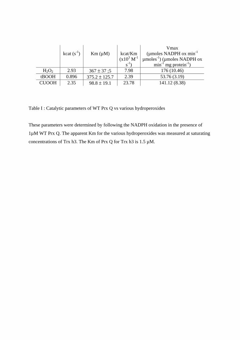
kcat (s-1)
Km (µM)
kcat/Km (x103 M-1
s-1)
Vmax (µmoles NADPH ox min-1
µmoles-1) (µmoles NADPH ox min-1 mg protein-1)
H2O2 2.93 367 ± 37 ;5 7.98 176 (10.46) tBOOH 0.896 375.2 ± 125.7 2.39 53.76 (3.19) CUOOH 2.35 98.8 ± 19.1 23.78 141.12 (8.38)
Table I : Catalytic parameters of WT Prx Q vs various hydroperoxides
These parameters were determined by following the NADPH oxidation in the presence of
1µM WT Prx Q. The apparent Km for the various hydroperoxides was measured at saturating
concentrations of Trx h3. The Km of Prx Q for Trx h3 is 1.5 µM.
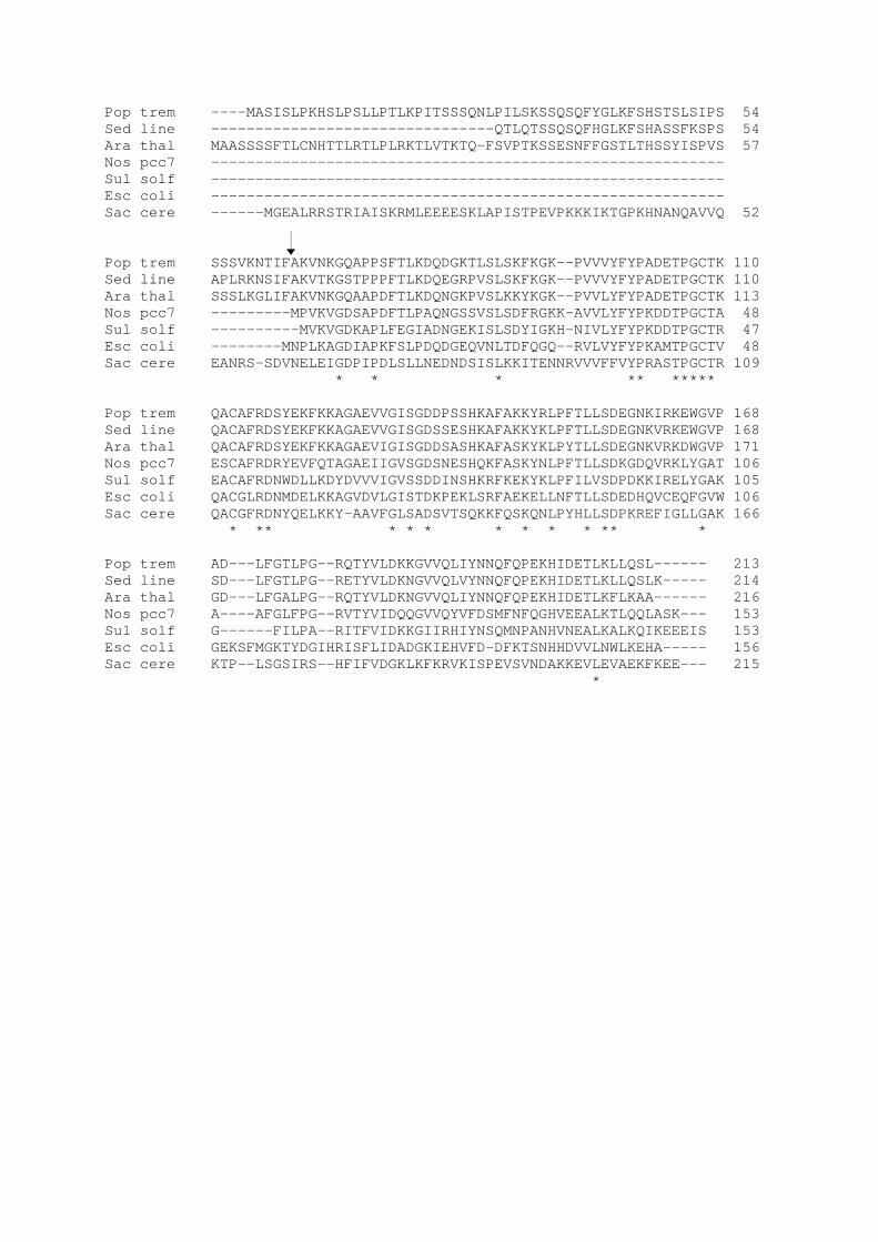
Pop trem ----MASISLPKHSLPSLLPTLKPITSSSQNLPILSKSSQSQFYGLKFSHSTSLSIPS 54Sed line --------------------------------QTLQTSSQSQFHGLKFSHASSFKSPS 54Ara thal MAASSSSFTLCNHTTLRTLPLRKTLVTKTQ-FSVPTKSSESNFFGSTLTHSSYISPVS 57Nos pcc7 ----------------------------------------------------------Sul solf ----------------------------------------------------------Esc coli ----------------------------------------------------------Sac cere ------MGEALRRSTRIAISKRMLEEEESKLAPISTPEVPKKKIKTGPKHNANQAVVQ 52
Pop trem SSSVKNTIFAKVNKGQAPPSFTLKDQDGKTLSLSKFKGK--PVVVYFYPADETPGCTK 110Sed line APLRKNSIFAKVTKGSTPPPFTLKDQEGRPVSLSKFKGK--PVVVYFYPADETPGCTK 110Ara thal SSSLKGLIFAKVNKGQAAPDFTLKDQNGKPVSLKKYKGK--PVVLYFYPADETPGCTK 113Nos pcc7 ---------MPVKVGDSAPDFTLPAQNGSSVSLSDFRGKK-AVVLYFYPKDDTPGCTA 48Sul solf ----------MVKVGDKAPLFEGIADNGEKISLSDYIGKH-NIVLYFYPKDDTPGCTR 47Esc coli --------MNPLKAGDIAPKFSLPDQDGEQVNLTDFQGQ--RVLVYFYPKAMTPGCTV 48Sac cere EANRS-SDVNELEIGDPIPDLSLLNEDNDSISLKKITENNRVVVFFVYPRASTPGCTR 109
* * * ** *****
Pop trem QACAFRDSYEKFKKAGAEVVGISGDDPSSHKAFAKKYRLPFTLLSDEGNKIRKEWGVP 168Sed line QACAFRDSYEKFKKAGAEVVGISGDSSESHKAFAKKYKLPFTLLSDEGNKVRKEWGVP 168Ara thal QACAFRDSYEKFKKAGAEVIGISGDDSASHKAFASKYKLPYTLLSDEGNKVRKDWGVP 171Nos pcc7 ESCAFRDRYEVFQTAGAEIIGVSGDSNESHQKFASKYNLPFTLLSDKGDQVRKLYGAT 106Sul solf EACAFRDNWDLLKDYDVVVIGVSSDDINSHKRFKEKYKLPFILVSDPDKKIRELYGAK 105Esc coli QACGLRDNMDELKKAGVDVLGISTDKPEKLSRFAEKELLNFTLLSDEDHQVCEQFGVW 106Sac cere QACGFRDNYQELKKY-AAVFGLSADSVTSQKKFQSKQNLPYHLLSDPKREFIGLLGAK 166
* ** * * * * * * * ** *
Pop trem AD---LFGTLPG--RQTYVLDKKGVVQLIYNNQFQPEKHIDETLKLLQSL------ 213Sed line SD---LFGTLPG--RETYVLDKNGVVQLVYNNQFQPEKHIDETLKLLQSLK----- 214Ara thal GD---LFGALPG--RQTYVLDKNGVVQLIYNNQFQPEKHIDETLKFLKAA------ 216Nos pcc7 A----AFGLFPG--RVTYVIDQQGVVQYVFDSMFNFQGHVEEALKTLQQLASK--- 153Sul solf G------FILPA--RITFVIDKKGIIRHIYNSQMNPANHVNEALKALKQIKEEEIS 153Esc coli GEKSFMGKTYDGIHRISFLIDADGKIEHVFD-DFKTSNHHDVVLNWLKEHA----- 156Sac cere KTP--LSGSIRS--HFIFVDGKLKFKRVKISPEVSVNDAKKEVLEVAEKFKEE--- 215
*
1 2 3 M 4 5 6
A
15
25
3750
kDa
B
M 1 2 3 4
15
25
3750
kDa
75
ab
cd
*
0
10
20
30
40
50
60
70
80
90
100
0 10 20 30 40 50
Electron donor concentration (µM)
H2O
2 co
nten
t (%
)
0
20
40
60
80
100
1 2 3 4 5 6 7 8 9
H2O
2co
nsum
ed(n
mol
es m
L-1m
in -1
)
Grx WT Grx C30S Trx h1 WT
WT
WT
WT
C46S C46SC46S
C51S
C51S
C51S
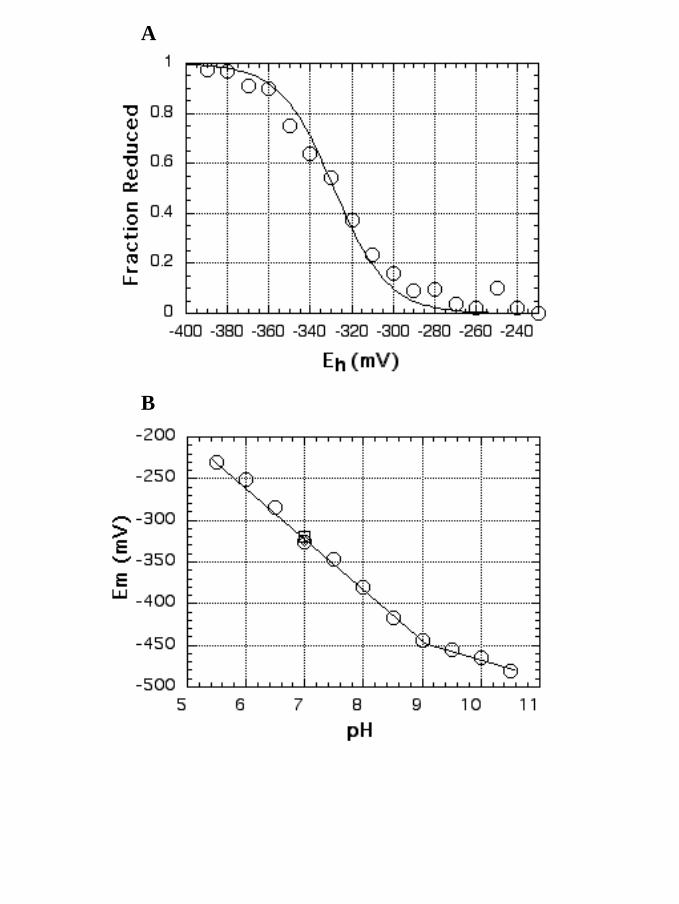
A
B
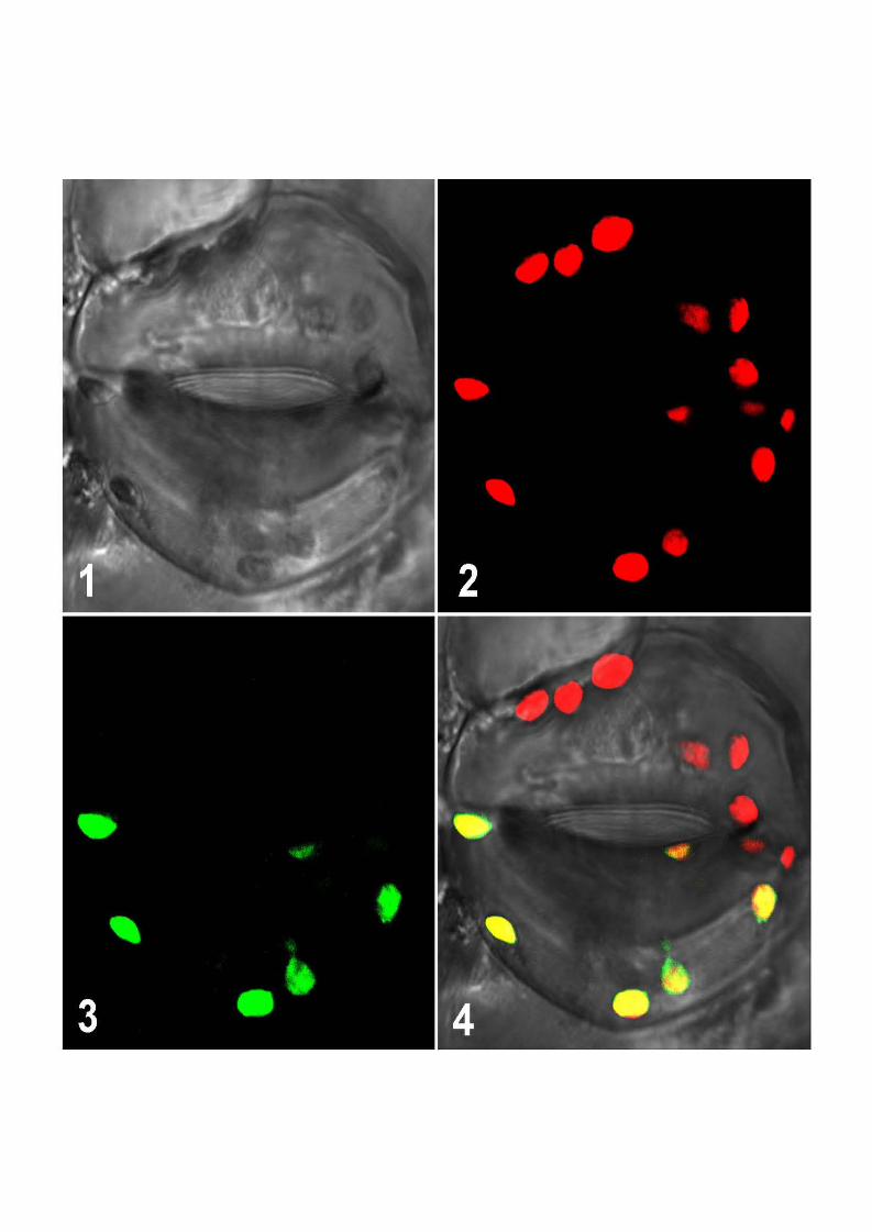
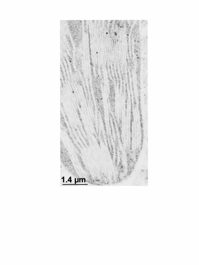
•
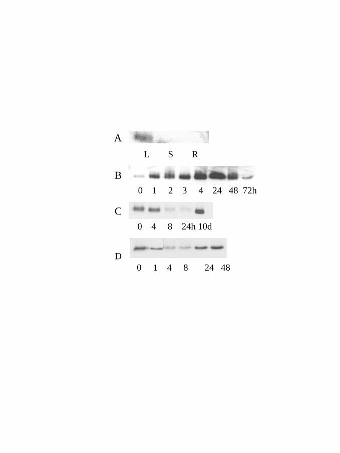
A
0 1 2 3 4 24 48 72hB
0 4 8 24h 10dC
0 1 4 8 �24 48
L S R
D
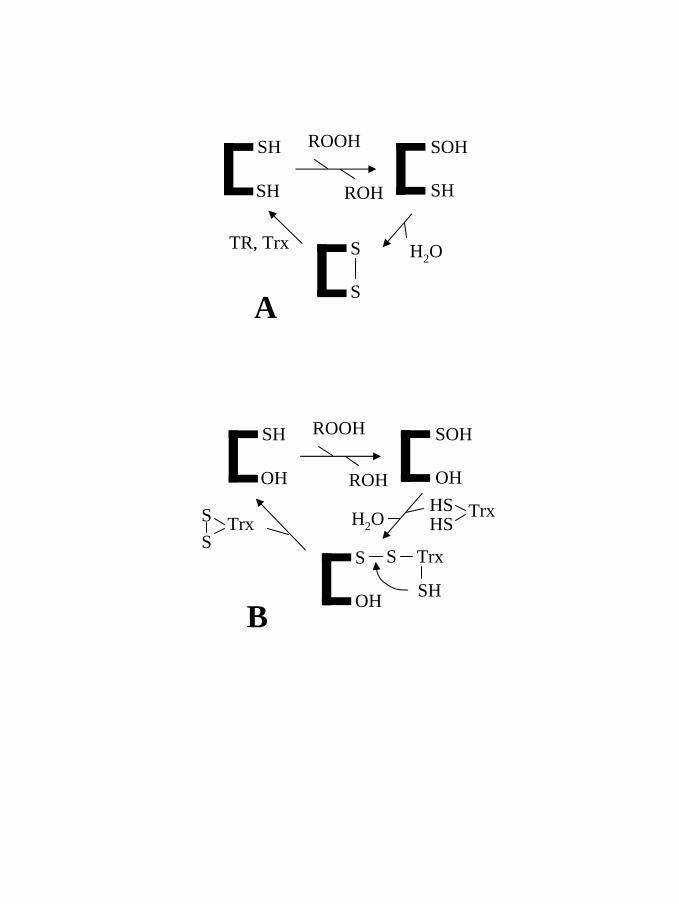
SH
SH
ROOH
S
ROH
SOH
SH
STR, Trx
A
SH
OH
ROOH
OH
ROH
SOH
OH
S
B
S Trx
SH
TrxSS
TrxHSHS
H2O
H2O
Résultats chapitre II : Les protéines cibles
Deux isoformes de MsrA de peuplier, une MsrA cytosolique (cMsrA) et une
plastidiale (pMsrA) sans son peptide de transit ont été exprimées chez E. coli. Les
protéines recombinantes sont monomériques et possèdent une activité de réduction
de la méthionine sulfoxyde (L-R,S MetSO). Différents types de MsrA existent selon le
nombre et la position des cystéines impliquées dans la catalyse (voir introduction).
Les deux MsrA de peuplier possèdent cinq cystéines en position conservée (31, 66,
85, 182 et 188 pour la cMsrA et 46, 81, 100, 196 et 202 pour la pMsrA). Les cinq
cystéines de la cMsrA ont été mutées une à une et les trois cystéines 46, 196 et 202
de la pMsrA également. Les protéines sauvages possèdent une activité de réduction
de la MetSO en Met dépendante des Trx cytosoliques Trx h1 et h3 mais pas des Grx.
Les mutants cMsrA C66S et C85S conservent également cette activité suggérant que
ces cystéines ne sont pas essentielles à la catalyse. Par contre, tous les autres
mutants sont inactifs avec les Trx. Par ailleurs, pour déterminer le rôle de chacune de
ces cystéines, le contenu en thiols libres de ces mutants a été estimé en présence et
en absence de MetSO et la stoechiométrie d’apparition de la Met de ces différents
mutants analysés en présence d’une concentration connue de MsrA réduite. Les
résultats obtenus indiquent que la cystéine 31 ou 46 selon l’isoforme considérée est
la cystéine catalytique et que les cystéines 182 et 188 ou 196 et 202 participent à la
régénération Trx dépendante de l’enzyme, selon un mécanisme identique à la MsrA
d’E. coli. Par ailleurs, la structure cristallographique de la pMsrA a pu être résolue.
Cette structure démontre que comme pour les autres structures de MsrA déterminées
jusqu’à présent, un important réarrangement est indispensable pour la formation des
ponts disulfures nécessaires à la réduction de l’enzyme.
Article numéro 16 :
Rouhier N, Kauffmann B, Tete-Favier F, Paladino P, Gans P, Boschi-Muller S, Branlant G, Aubry A, Jacquot JP.
Functional and structural aspects of poplar cytosolic and plastidial type A
methionine sulfoxide reductases.
En préparation
Functional and structural aspects of poplar cytosolic and plastidial type A methionine sulfoxide reductases
Nicolas Rouhier1*, Brice Kauffmann2*, Frédérique Tete-Favier2, Pasquale Paladino2, Jérémi
Couturier1, Pierre Gans4, Sandrine Boschi-Muller3, Guy Branlant3, André Aubry2 and Jean-
Pierre Jacquot1,5
1Unité Mixte de Recherches 1136 INRA Université Henri-Poincaré, Interactions Arbres
Microorganismes. Faculté des Sciences BP 239 54506 Vandoeuve Cedex France 2LCM3B, Groupe de Biocristallographie, UMR CNRS 7036, Université Henri-Poincaré,
Faculté des Sciences BP 239 54506 Vandoeuve Cedex France 3Laboratoire MAEM, Université Henri Poincaré, Faculté des Sciences BP 239 54506
Vandoeuve Cedex France 4Laboratoire de Résonance Magnétique Nucléaire. Institut de Biologie Structurale CEA-
CNRS-UJF "Jean-Pierre Ebel" 38027 Grenoble Cedex 1. 5Corresponding author : Tel ++ 33 3 83684225, email : [email protected] *These two authors have equally contributed to the work.
Running title: Structure and function of plant MsrA
Keywords : methionine sulfoxide reductase/mutagenesis/poplar/structure/thioredoxin
Abstract
This study presents new data concerning the molecular functioning and structure of
plant type A methionine sulfoxide reductases (MsrA). Both cytosolic and plastidial poplar
MsrA were analyzed. The enzymes are highly homologous, monomeric and active in the
reduction of methionine sulfoxide with either dithiothreitol or poplar thioredoxins as a
reductant. In both enzymes, five conserved cysteines are found in positions 31, 66, 85, 182
and 188 (cytosolic isoform numbering). Site-directed mutagenesis indicates that three
cysteines are involved in catalysis/regeneration by thioredoxin: Cys 31, the catalytic cysteine
and the two C-terminal cysteines Cys 182 and 188. As observed with other MsrA, in the
tridimensional structure of the plastidial enzyme, the three cysteinyl residues are far apart, but
molecular modelling suggests the presence of two hinges that could provide the needed
flexibility of the C-terminal part.
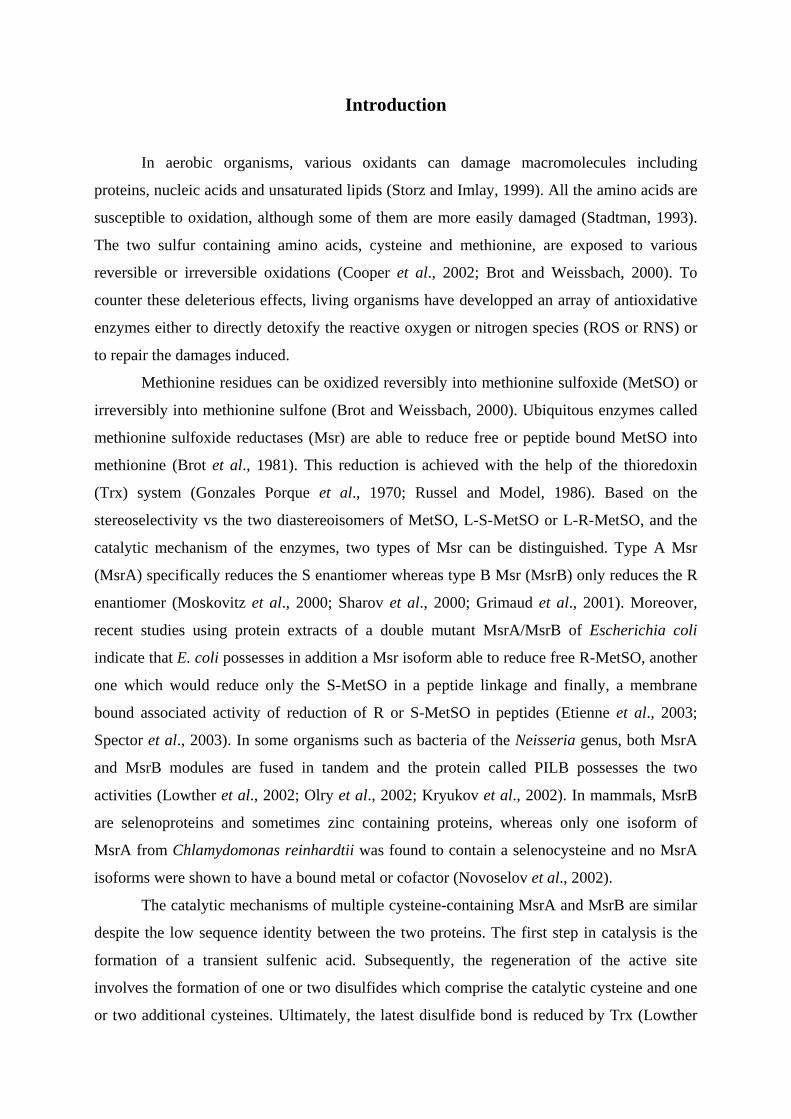
Introduction
In aerobic organisms, various oxidants can damage macromolecules including
proteins, nucleic acids and unsaturated lipids (Storz and Imlay, 1999). All the amino acids are
susceptible to oxidation, although some of them are more easily damaged (Stadtman, 1993).
The two sulfur containing amino acids, cysteine and methionine, are exposed to various
reversible or irreversible oxidations (Cooper et al., 2002; Brot and Weissbach, 2000). To
counter these deleterious effects, living organisms have developped an array of antioxidative
enzymes either to directly detoxify the reactive oxygen or nitrogen species (ROS or RNS) or
to repair the damages induced.
Methionine residues can be oxidized reversibly into methionine sulfoxide (MetSO) or
irreversibly into methionine sulfone (Brot and Weissbach, 2000). Ubiquitous enzymes called
methionine sulfoxide reductases (Msr) are able to reduce free or peptide bound MetSO into
methionine (Brot et al., 1981). This reduction is achieved with the help of the thioredoxin
(Trx) system (Gonzales Porque et al., 1970; Russel and Model, 1986). Based on the
stereoselectivity vs the two diastereoisomers of MetSO, L-S-MetSO or L-R-MetSO, and the
catalytic mechanism of the enzymes, two types of Msr can be distinguished. Type A Msr
(MsrA) specifically reduces the S enantiomer whereas type B Msr (MsrB) only reduces the R
enantiomer (Moskovitz et al., 2000; Sharov et al., 2000; Grimaud et al., 2001). Moreover,
recent studies using protein extracts of a double mutant MsrA/MsrB of Escherichia coli
indicate that E. coli possesses in addition a Msr isoform able to reduce free R-MetSO, another
one which would reduce only the S-MetSO in a peptide linkage and finally, a membrane
bound associated activity of reduction of R or S-MetSO in peptides (Etienne et al., 2003;
Spector et al., 2003). In some organisms such as bacteria of the Neisseria genus, both MsrA
and MsrB modules are fused in tandem and the protein called PILB possesses the two
activities (Lowther et al., 2002; Olry et al., 2002; Kryukov et al., 2002). In mammals, MsrB
are selenoproteins and sometimes zinc containing proteins, whereas only one isoform of
MsrA from Chlamydomonas reinhardtii was found to contain a selenocysteine and no MsrA
isoforms were shown to have a bound metal or cofactor (Novoselov et al., 2002).
The catalytic mechanisms of multiple cysteine-containing MsrA and MsrB are similar
despite the low sequence identity between the two proteins. The first step in catalysis is the
formation of a transient sulfenic acid. Subsequently, the regeneration of the active site
involves the formation of one or two disulfides which comprise the catalytic cysteine and one
or two additional cysteines. Ultimately, the latest disulfide bond is reduced by Trx (Lowther
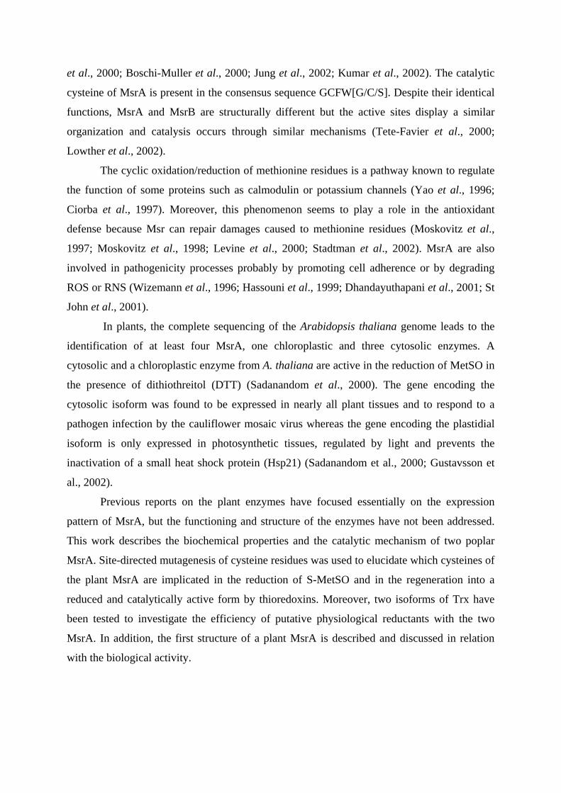
et al., 2000; Boschi-Muller et al., 2000; Jung et al., 2002; Kumar et al., 2002). The catalytic
cysteine of MsrA is present in the consensus sequence GCFW[G/C/S]. Despite their identical
functions, MsrA and MsrB are structurally different but the active sites display a similar
organization and catalysis occurs through similar mechanisms (Tete-Favier et al., 2000;
Lowther et al., 2002).
The cyclic oxidation/reduction of methionine residues is a pathway known to regulate
the function of some proteins such as calmodulin or potassium channels (Yao et al., 1996;
Ciorba et al., 1997). Moreover, this phenomenon seems to play a role in the antioxidant
defense because Msr can repair damages caused to methionine residues (Moskovitz et al.,
1997; Moskovitz et al., 1998; Levine et al., 2000; Stadtman et al., 2002). MsrA are also
involved in pathogenicity processes probably by promoting cell adherence or by degrading
ROS or RNS (Wizemann et al., 1996; Hassouni et al., 1999; Dhandayuthapani et al., 2001; St
John et al., 2001).
In plants, the complete sequencing of the Arabidopsis thaliana genome leads to the
identification of at least four MsrA, one chloroplastic and three cytosolic enzymes. A
cytosolic and a chloroplastic enzyme from A. thaliana are active in the reduction of MetSO in
the presence of dithiothreitol (DTT) (Sadanandom et al., 2000). The gene encoding the
cytosolic isoform was found to be expressed in nearly all plant tissues and to respond to a
pathogen infection by the cauliflower mosaic virus whereas the gene encoding the plastidial
isoform is only expressed in photosynthetic tissues, regulated by light and prevents the
inactivation of a small heat shock protein (Hsp21) (Sadanandom et al., 2000; Gustavsson et
al., 2002).
Previous reports on the plant enzymes have focused essentially on the expression
pattern of MsrA, but the functioning and structure of the enzymes have not been addressed.
This work describes the biochemical properties and the catalytic mechanism of two poplar
MsrA. Site-directed mutagenesis of cysteine residues was used to elucidate which cysteines of
the plant MsrA are implicated in the reduction of S-MetSO and in the regeneration into a
reduced and catalytically active form by thioredoxins. Moreover, two isoforms of Trx have
been tested to investigate the efficiency of putative physiological reductants with the two
MsrA. In addition, the first structure of a plant MsrA is described and discussed in relation
with the biological activity.
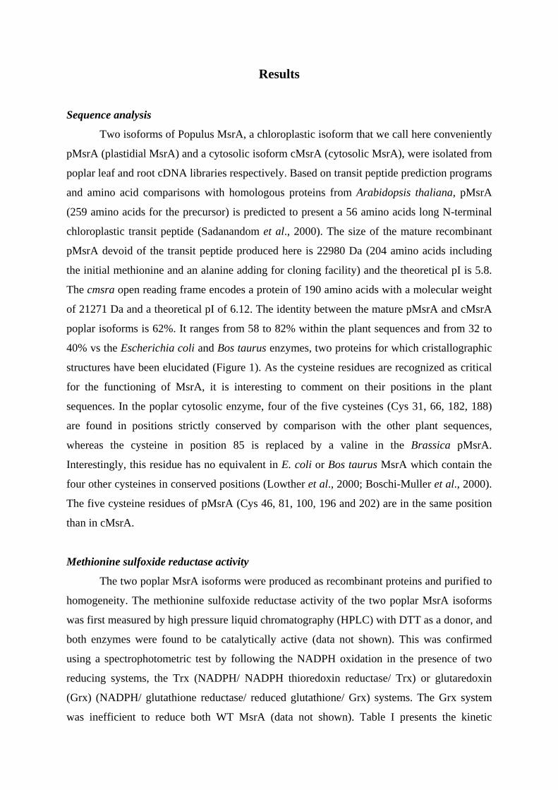
Results
Sequence analysis
Two isoforms of Populus MsrA, a chloroplastic isoform that we call here conveniently
pMsrA (plastidial MsrA) and a cytosolic isoform cMsrA (cytosolic MsrA), were isolated from
poplar leaf and root cDNA libraries respectively. Based on transit peptide prediction programs
and amino acid comparisons with homologous proteins from Arabidopsis thaliana, pMsrA
(259 amino acids for the precursor) is predicted to present a 56 amino acids long N-terminal
chloroplastic transit peptide (Sadanandom et al., 2000). The size of the mature recombinant
pMsrA devoid of the transit peptide produced here is 22980 Da (204 amino acids including
the initial methionine and an alanine adding for cloning facility) and the theoretical pI is 5.8.
The cmsra open reading frame encodes a protein of 190 amino acids with a molecular weight
of 21271 Da and a theoretical pI of 6.12. The identity between the mature pMsrA and cMsrA
poplar isoforms is 62%. It ranges from 58 to 82% within the plant sequences and from 32 to
40% vs the Escherichia coli and Bos taurus enzymes, two proteins for which cristallographic
structures have been elucidated (Figure 1). As the cysteine residues are recognized as critical
for the functioning of MsrA, it is interesting to comment on their positions in the plant
sequences. In the poplar cytosolic enzyme, four of the five cysteines (Cys 31, 66, 182, 188)
are found in positions strictly conserved by comparison with the other plant sequences,
whereas the cysteine in position 85 is replaced by a valine in the Brassica pMsrA.
Interestingly, this residue has no equivalent in E. coli or Bos taurus MsrA which contain the
four other cysteines in conserved positions (Lowther et al., 2000; Boschi-Muller et al., 2000).
The five cysteine residues of pMsrA (Cys 46, 81, 100, 196 and 202) are in the same position
than in cMsrA.
Methionine sulfoxide reductase activity
The two poplar MsrA isoforms were produced as recombinant proteins and purified to
homogeneity. The methionine sulfoxide reductase activity of the two poplar MsrA isoforms
was first measured by high pressure liquid chromatography (HPLC) with DTT as a donor, and
both enzymes were found to be catalytically active (data not shown). This was confirmed
using a spectrophotometric test by following the NADPH oxidation in the presence of two
reducing systems, the Trx (NADPH/ NADPH thioredoxin reductase/ Trx) or glutaredoxin
(Grx) (NADPH/ glutathione reductase/ reduced glutathione/ Grx) systems. The Grx system
was inefficient to reduce both WT MsrA (data not shown). Table I presents the kinetic
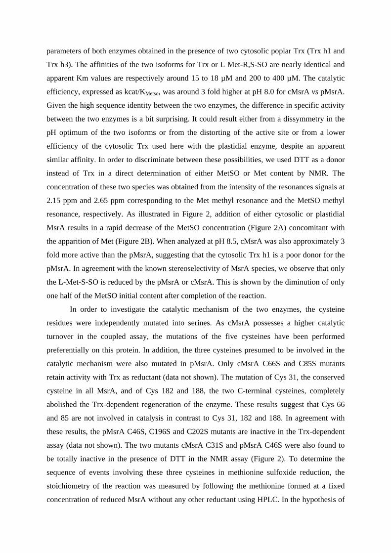
parameters of both enzymes obtained in the presence of two cytosolic poplar Trx (Trx h1 and
Trx h3). The affinities of the two isoforms for Trx or L Met-R,S-SO are nearly identical and
apparent Km values are respectively around 15 to 18 µM and 200 to 400 µM. The catalytic
efficiency, expressed as kcat/KMetso, was around 3 fold higher at pH 8.0 for cMsrA vs pMsrA.
Given the high sequence identity between the two enzymes, the difference in specific activity
between the two enzymes is a bit surprising. It could result either from a dissymmetry in the
pH optimum of the two isoforms or from the distorting of the active site or from a lower
efficiency of the cytosolic Trx used here with the plastidial enzyme, despite an apparent
similar affinity. In order to discriminate between these possibilities, we used DTT as a donor
instead of Trx in a direct determination of either MetSO or Met content by NMR. The
concentration of these two species was obtained from the intensity of the resonances signals at
2.15 ppm and 2.65 ppm corresponding to the Met methyl resonance and the MetSO methyl
resonance, respectively. As illustrated in Figure 2, addition of either cytosolic or plastidial
MsrA results in a rapid decrease of the MetSO concentration (Figure 2A) concomitant with
the apparition of Met (Figure 2B). When analyzed at pH 8.5, cMsrA was also approximately 3
fold more active than the pMsrA, suggesting that the cytosolic Trx h1 is a poor donor for the
pMsrA. In agreement with the known stereoselectivity of MsrA species, we observe that only
the L-Met-S-SO is reduced by the pMsrA or cMsrA. This is shown by the diminution of only
one half of the MetSO initial content after completion of the reaction.
In order to investigate the catalytic mechanism of the two enzymes, the cysteine
residues were independently mutated into serines. As cMsrA possesses a higher catalytic
turnover in the coupled assay, the mutations of the five cysteines have been performed
preferentially on this protein. In addition, the three cysteines presumed to be involved in the
catalytic mechanism were also mutated in pMsrA. Only cMsrA C66S and C85S mutants
retain activity with Trx as reductant (data not shown). The mutation of Cys 31, the conserved
cysteine in all MsrA, and of Cys 182 and 188, the two C-terminal cysteines, completely
abolished the Trx-dependent regeneration of the enzyme. These results suggest that Cys 66
and 85 are not involved in catalysis in contrast to Cys 31, 182 and 188. In agreement with
these results, the pMsrA C46S, C196S and C202S mutants are inactive in the Trx-dependent
assay (data not shown). The two mutants cMsrA C31S and pMsrA C46S were also found to
be totally inactive in the presence of DTT in the NMR assay (Figure 2). To determine the
sequence of events involving these three cysteines in methionine sulfoxide reduction, the
stoichiometry of the reaction was measured by following the methionine formed at a fixed
concentration of reduced MsrA without any other reductant using HPLC. In the hypothesis of
a mechanism similar to the one of E. coli MsrA, the stoichiometry is assumed to be two moles
of methionine formed per mole enzyme for cMsrA WT enzyme, cMSrA C66S and cMsrA
C85S, only one for cMsrA C182S and C188S and none for the C31S mutant. Table II shows
the results obtained with the poplar enzymes. The WT cMsrA and pMsrA present a
stoichiometry near 2 as expected based on the presumed catalytic mechanism. The cMsrA
C66S and C85S mutants reduce only 1.4 moles of Met per mole enzyme instead of the 2
expected presumably because part of these enzymes was in an oxidized form. The cMsrA
C31S mutant is totally inactive as it reduces 0 moles of Met per mole enzyme, whereas the
cMsrA C182S and C188S mutants possess a stoichiometry of 1.16 and 1.68 respectively
instead of the expected value of 1. From these data, Cys 31 is the catalytic cysteine but
whether Cys 182 and Cys 188 are both involved in the Trx dependent regeneration is not
clear. In order to gain additional information about the involvement of the Cys182 and
Cys188 in catalysis, the thiol content of each recombinant protein was estimated after
reduction in the presence or absence of an excess of L Met-R,S-SO (Table III). For the pMsrA
WT, C46S, C196S and C202S mutants, the decrease in free thiol groups matches well the
theoretical values assuming an attack of Cys 46 by MetSO to form a sulfenic acid which is
then reduced by disulfide bonding with Cys 196, the resulting disulfide bond being finally
reduced by Cys 202. For the cMsrA mutants, the results also essentially agree with the
postulated mechanism wihin the incertitude inherent to thiol measurement.
Overall structure of pMsrA
Crystallisation trials were performed for both chloroplastic and cytosolic MsrA, but
crystals of sufficient dimension for X-Ray analysis were obtained only for the pMsrA form.
The trigonal crystals (space group P31) contain one molecule per asymmetric unit and 45% of
solvent, and diffract to 1.7Å resolution (Table IV and V). The structure was solved and
refined with final R- and free-R-factors of 19.5 and 20.1%, respectively. Superimposition of
the poplar pMsrA with the E. coli and the bovine structures (PDB entries : 1FF3 and 1FVA,
respectively) (Tete-Favier et al., 2000; Lowther et al., 2000) shows that the cores of the three
enzymes possess the same overall fold as expected from the sequence identity between these
enzymes (Figure 3), while the long N and C-terminal ends display the following substantial
differences:
- The core of the MsrA structure starts at the first β-strand, S1, at the end of which is found
the GCFWG footprint containing the catalytic cysteine residue Cys46, and ends 3 amino
acids after the AExxHQxφ footprint, next to the last β-strand S6 (see Tete-Favier et al.,
2000 for secondary structure numbering). Selecting the corresponding amino acids, Gln37
to Lys182 in pMsrA, Met42 to Lys192 in the E. coli MsrA, and Thr63 to Lys212 in the
bovine MsrA, Cα atoms can be superimposed with a root mean square distance (rmsd) of
0.800 Å for the poplar/E. coli pair and with an rmsd value of 0.874 Å for the
poplar/bovine pair.
- In the E. coli MsrA, the long N-terminal end coils up around the core of the domain. It is
constituted of 41 amino acids, the first seven residues being observed in different
conformations in the three monomers of the asymmetric unit. In the bovine MsrA model,
in which amino acids 1 to 27 are not present, residues 28 to 62 display the same overall
conformation than in the E. coli enzyme. In the pMsrA structure, only residues Pro22 to
Gly36 of the N-terminal end were observed in the electron density. Their positions
strongly differ from those observed in the E. coli and bovine MsrA structures (Figure 3).
The coil is hydrogen-bonded via the main chains to the extended segment that links S5 to
S6. This different position of the N-terminal end arises from the sequence of the hinge
region connecting the N-terminal end to the core of the enzyme: 35Pro-Gly36 is present in
poplar, while it is 40Asp-Gly41 in E. coli, and 61Glu-Gly62 in the bovine enzyme. The
missing residues 1Met-Asp21 are very disordered since no electron density is observed
upstream from Pro22, and are probably situated in the large solvent cavity observed near
Pro22.
- The 193Asn-Ala211 C-terminal end of the E. coli MsrA was observed in only one monomer
among three in the asymmetric unit, leading to conclude to its flexibility. Despite the
presence of an intermolecular disulfide bridge involving Cys202 that could have forced
the C-terminal end to adopt an unlikely conformation in the E. coli model, it is surprising
to observe a similar position of the 213Asp-Pro228 C-terminal end in the bovine MsrA
structure. The extended coil runs above the active site and then antiparallel to α-helices
Η2 and Η3. In the pMsrA model, the 183Gly-Gly204 C-terminal end is folded in a
completely different way (Figure 3) : it consists of a coil that runs first parallel to the α-
helix Η1 and then, after a turn, antiparallel to it (Figure 4). Numerous hydrogen bonds are
observed inside this C-terminal part, and between the C-terminal part and the core of the
domain. This region is involved in the crystal packing, since several residues are observed
at the proximity of two symmetry-related monomers. However, the intermolecular
contacts seem weaker than the intramolecular contacts.
The active site and the cysteine residues
As previously observed in the E. coli and the bovine MsrA structures, the active site of
pMsrA is a large open cavity, which gives high accessibility to the catalytic cysteine residue
Cys46. This amino acid, as well as residues Cys196 and Cys202, clearly shows an extra-
density on its sulfur atom that was unambiguously modeled as a bonded β-mercaptoethanol,
present in the crystallisation solution. Cys81 also possesses an extra-density, however its
weaker weight and smallest size have been interpreted as a sulfenic acid (Figure 5). Only
Cys100 was observed in its reduced form. It is puzzling to note that neither in the E. coli
MsrA nor in the bovine enzyme structure, the three cysteine residues involved in the catalytic
mechanism are observed in a reduced form.
Two successive intramolecular disulfide bridges are formed in the mechanism of the
E. coli and the bovine enzymes (Boschi-Muller et al., 2000; Lowther et al., 2000). Sequence
alignments point to Cys196 as the residue of pMsrA involved in the first disulfide bridge with
Cys46, and Cys202 would then form the consecutive bridge with Cys196. The distance
between Sγ46 and Sγ196, 18.0Å, is far from being compatible with the formation of a disulfide
bridge. Therefore, the distances between the Sγ atoms of the five cysteine residues have been
measured (Table VI) in order to evaluate their ability to form disulfide bridges, considering
that their current position would be unchanged in an hypothetical structure with five reduced
cysteine residues. None of the cysteine pairs, even the narrowest one (Cys46-Cys202, for
which the distance observed between the Sγ atoms is 7.2Å), is compatible with the formation
of a disulfide bridge if no conformational change occurs.
Discussion
Most of the available data concerning plant MsrA are devoted essentially to expression
studies, but unlike in other organisms, little is known about their catalytic mechanism and
structural properties (Sadanandom et al., 2002). This paper fills the gap in knowledge
concerning the plant enzymes by providing site-directed mutagenesis data together with a first
tridimensional structure of a plant MsrA and a catalytic mechanism for poplar MsrA.
Catalytic mechanism of poplar MsrA
Using site-directed mutagenesis, all the five cysteines of cMsrA and the presupposed
essential cysteines of pMsrA have been mutated into serines. All the WT and mutated
recombinant enzymes are able to reduce MetSO with DTT as a reductant except the cMsrA
C31S and pMsrA C46S indicating that these cysteines are the catalytic ones. Moreover, the
Trx dependent regeneration was abolished for cMsrA C182S and C188S and unaffected for
the cMsrA C66S and C85S mutated proteins suggesting that C66 and C85 do not participate
to catalysis whereas the two C-terminal cysteines are involved in the cysteine sulfenic acid
reduction of C31. The stoichiometry of the reaction was checked for each recombinant
proteins. The results confirm previous observations but do not permit actually to ascertain
definitively the involvement of the cysteines C182 and C188 in the Trx dependent reduction.
Whether these two cysteines are implicated to form two successive disulfide bridges as for the
E. coli enzyme, was also assessed by measuring the decrease in free thiol groups of each
recombinant protein after MetSO treatment. The data obtained from this experiment confirm
with no doubt that C31 (C46 for pMsrA) is the catalytic cysteine which is reduced after attack
of the sulfenic acid intermediates by C182 (C196) forming a first disulfide bridge. This
disulfide is subsequently attacked by C188 (C202) forming a second disulfide bridge between
C182 and C188 (C196 and C202) which will be reduced by Trx but not by Grx as a
mechanism similar to the E. coli MsrA (Boschi-Muller et al., 2000). Why Grx is not able to
reduce this disulfide is not clear actually. Whether the redox potential of the C182-C188
disulfide is too low to be reduced by Grx or whether the interaction could be prevented by
steric hindrance or by unadapted surface protein recognition needs to be addresed.
Nevertheless, two independent observations are in the favor of a possible Grx dependent
regeneration of MsrA in other organisms. First, the growth on MetSO of a methionine
auxotroph Trx A- E. coli strain is restored by Grx 1 overexpresion (Stewart et al., 1998).
Second, a coding sequence, found in the red algae Gracilaria gracilis (accession number
AAD43253), consist of two Grx modules fused with a MsrA module. This module is highly
similar to other plant MsrA except for the absence of the cysteine equivalent to the C182 of
poplar cMsrA. Whether this protein use a different catalytic mechanism and is reduced by the
Grx modules in the presence of glutathione is not known actually.
To gain insight into the interaction between Trx and MsrA, 3D structures of each
partner must be resolved at least independently or in complex. Such attempts for linking
covalently the two proteins are under investigation. Some heterodimers are observed on non-
reducing SDS-PAGE when mixing MsrA and a monocysteinic Trx but the amount is too low
to isolate the complex (data not shown).
Structural comparison to other MsrA
Two structures of MsrA from E. coli and B. taurus are currently available (Lowther et
al., 2000 ; Tete-Favier et al., 2000). The amino acid sequences of these two enzymes are 57%
homologous and as a consequence their tridimensional crystallographic structures present
extensive homologies. Both enzymes are monomeric, they share a common fold and the core
of the protein is similar, but the N and C-termini differ in both structures and none of the
cysteines is close enough to the catalytic cysteine to accomodate for the formation of a
disulfide bridge. Recent structural data obtained by NMR, which gives a more dynamic
picture of the protein, have confirmed that the cysteinyl side chains are indeed not properly
located for the formation of the necessary disulfide bridges (Beraud et al., 2002). As a
consequence, for both enzymes it has been postulated that important conformational
rearrangements are needed or the Trx-mediated regeneration of these enzymes. In connection
with these existing data it is obviously of interest to obtain the structure of an additional
MsrA.
The structure of the pMsrA from poplar presented here is rather similar to the ones
described for the bacterial and mammalian enzymes. All enzymes share the same fold and the
catalytic cysteine of the active site is in a similar environment. On the other hand, the
orientation and the amino acid composition of the C-terminal part that contains the two
additional cysteines implicated in the regeneration process is radically different from the E.
coli and B. taurus MsrA. Indeed, the spacing between the two C-terminal located cysteinyl
residues is 5 amino acids for poplar pMsrA instead of 7 or 8 amino acids for the mammalian
and bacterial enzymes respectively. Moreover, the high content in glycines between these two
cysteines is not present in the poplar enzyme. The C-terminal loop of pMsrA is highly
organized with many hydrogen bonds connecting the two parts of the loop whereas it is
poorly structured in the two other models. However as in the other MsrA, the cysteines are in
positions that do not allow for the formation of a disulfide bridge. Due to the positions of the
monomers in the crystal, it is likely that the conformation of the loop is influenced by the
packing of the molecules. In order to understand how the necessary disulfide bridges can be
created in pMsrA, molecular modelling studies have been undertaken. The results suggest that
there are two hinges in the loop centered on Gly residues (Gly43 and Gly195) that would
allow the required movement of the loop for catalysis to occur.
Conclusion
The data generated here suggest that MsrA enzymes have kept a similar structure and
mode of catalysis along evolution. In several cysteine requiring enzymes such as thioredoxin
reductase of glutathione peroxidase, the mammalian catalysts have been altered into more
complex enzymes that contain selenocysteines. The presence of this residue is generally
correlated with a much higher specific activity of the enzyme. Plants are generally recognized
as lacking selenocysteines with the notable exception of the green alga Chlamydomonas
reinhardtii which does contain this unusual amino acid in many catalysts (Novoselov et al.,
2002). Analysis of the tridimensional structures of thioredoxin reductases indicates that the
plant enzyme (Arabidopsis thaliana) is similar to the bacterial enzyme (E. coli) but not to the
mammalian enzyme. Using this grid of evaluation, plants appear to be closer to bacteria than
to mammalians. Analysis of the MsrA structures leads to the conclusion that all models of this
protein have remained relatively constant along the lines of evolution. It could be correlated to
the presence of alternate catalysts as MsrB which seem to have varied more along evolution.
The need for several isoforms of MsrA in cytosol of plants is not yet completely clear. The
major sources for ROS in plants are likely to be the peroxisomes, chloroplasts and to a lesser
extent mitochondria, but few enzymatic systems generate those oxidants in the cystosol. In
connection with this observation, it is puzzling that the cytosolic enzyme exhibits a higher
catalytic turnover than the plastidial enzyme. It remains to be determined whether this could
be compensated by a higher level of expression of the plastidial isoform. Another question to
be investigated is whether those enzymes are able to reduce peptide bound MetSO and
whether they display a preference for substrates in vivo when using oxidized proteins as
substrates instead of free L-MetSO.
Materials and Methods
Cloning and site-directed mutagenesis
The nucleotidic sequences encoding cMsrA and pMsrA were cloned by PCR into the
expression plasmid pET-3d using as templates respectively a root cDNA library of Populus x
interamericana (clone Beaupré) (Kohler et al., in press) and a leaf cDNA library of Populus
tremula x tremuloides (Larsson et al., 1997). Both reactions also contained Pfu DNA
polymerase (Promega) and the forward and reverse MsrA oligonucleotides described in Table
VII. In the pMsrA cloning, a codon for alanine was inserted downstream the methionine
closest to the putative cleavage site and the corresponding N-terminal amino acid sequence
starts thus with MANIL. Various moncysteinic mutations were introduced in the cytosolic
MsrA (cMsrA C31S, C66S, C85S, C182S, C188S) and plastidic MsrA (pMsrA C46S, C196S,
C202S) using two complementary mutagenic primers per mutation (Table VII) following a
procedure previously described (Jacquot et al., 1997). The introduction of the mutation in the
cDNA sequence was verified by sequencing.
Mutagenesis, expressionand purification of the recombinant proteins
The recombinant plasmids were used to transform the BL21(DE3) E. coli strain which
also contains the helper plasmid pSBET (Schenk et al., 1995). Cultures of 5 L of a kanamycin
(50 µg/mL) and ampicillin (50 µg/mL) resistant colony were grown at 37 °C and induced by
100 µM IPTG in the exponential phase. Bacteria were harvested by centrifugation,
resuspended in buffer A (30 mM Tris-HCl, 1 mM EDTA, 200 mM NaCl) containing 20 mM
DTT and lysed by sonication. The soluble and insoluble fractions were separated by
centrifugation. For the pMsrA WT, the recombinant protein was in the soluble fraction and
precipitated between 0 and 50% of ammonium sulfate. All the other recombinant proteins
were produced essentially as inclusion bodies with only a small soluble part when cultures
were grown at 30°C without induction. When needed, the insoluble fraction was resuspended
in buffer A in the presence of 20 mM DTT and 8 M urea and then dialyzed against 1 L of
buffer A containing 500 mM urea for at least 5 hours at 5°C (all subsequent steps were
realized at that temperature). The extract was centrifuged and the soluble fraction dialyzed
against 1 L of buffer A for 5 hours and centrifuged. The resulting soluble fraction was
purified by exclusion size chromatography onto an ACA 44 column equilibrated in buffer A.
The fractions of interest were pooled, dialyzed to remove salts, and separated by DEAE
Sephacel chromatography. The recombinant proteins were eluted around 100 mM NaCl using
a linear gradient from 0 to 400 mM NaCl. The purified fractions were dialyzed to remove
salts, adjusted to 1.6 M NaCl and applied to a Phenyl Sepharose column equilibrated with
buffer B (30 mM Tris-HCl, 1 mM EDTA, 1.6 M NaCl). The recombinant proteins were eluted
by a decreasing gradient between 1.6 and 0 M NaCl, dialyzed and concentrated. The purity of
the proteins was assessed using 15% SDS-PAGE. The protein concentration was estimated
spectrophotometrically using a molar extinction coefficient of 25700 M-1 cm-1 for cMsrA and
pMsrA. The proteins were stored at –30°C in buffer A either in the presence of 14 mM β-
mercaptoethanol or 25 to 30 mM DTT.
Cristallization, X-ray data collection, structure determination and refinement
Crystallisation of pMsrA was achieved by hanging-drop vapour-diffusion method in
Linbro multiwell tissue-culture plates, at 277 K. The purified enzyme was concentrated to 40
mg/ml in a solution containing 30 mM Tris-HCl, pH 7.0, 14 mM β-mercaptoethanol, and 1
mM EDTA. The crystals were grown from 4 µl droplets composed of equal volumes of the
protein solution and of the precipitant solution (10% W/V polyethyleneglycol 6000, 2 M
NaCl), and equilibrated against 700 µL reservoirs. Long needles (1 mm) with a thin triangular
cross-section (0.03 mm) appeared after 6 weeks. Crystals were briefly soaked in a
cryoprotectant solution (10 % V/V methylpentanediol mixed with the precipitant solution)
and flash-frozen by fast immersion in nitrogen gas stream at 100 K, maintained during the X-
Ray diffraction experiments performed on beamline BM30A (FIP) at the ESRF.
Crystals belong to space group P31 with unit cell parameters a = b = 68.6Å , c = 40.7Å
and contain one monomer per asymmetric unit. Using a wavelength of 1.009 Å, one native
data set was collected up to 1.7Å resolution, and processed using DENZO (Otwinowski and
Minor, 1997). Further details are given in Table IV.
The structure was solved using the molecular replacement method implemented in
Molrep (Vagin and Teplyakov, 1997) of the CCP4 program suite. The initial model used in
Molrep consisted of the core (41Gly-Pro194) of the E. coli MsrA structure (PDB entry: 1FF3).
The molecular replacement solution was submitted to the Molrep mode and then to the
warpNtrace mode of the Arp/wArp5.1 automatic model building and refinement program
(Perrakis et al., 1999). It produced a model that contained 4 polypeptide chains representing
164 amino acids, with R and free-R factors of 20.6 and 25.8 %, respectively. Manual
corrections (in particular, building of the missing residues) and automatic CNS refinement
(Brünger et al., 1998) of the model were then performed in an iterative procedure, until the
model fulfilled satisfactory criteria. The final structure corresponds to 183 amino acids among
204 (residues 22Pro-Gly204), 183 water molecules, with R = 19.5 %, Rfree = 20.1%. Further
details are given in Table V.
Thiol content titration
250 µg of the recombinant proteins were treated either with 50 mM DTT or 50 mM L-
MetSO during one hour at room temperature. The proteins were then precipitated on ice by
addition of one volume of 20% trichloroacetic acid (TCA) for 30 minutes. The proteins were
pelleted by centrifugation and washed twice with 2% TCA. The pellets were resuspended in
30 mM Tris-HCl pH 8.0, 1 mM EDTA and 2% SDS. The concentration of the proteins were
determined spectrophotometrically at this stage and then DTNB was added to a final
concentration of 100 µM and the absorbance was read at 412 nm one hour later. The thiol
content was determined using a molar extinction coefficient of 13600 M-1 cm-1 for TNB-.
Enzymatic assays
HPLC detection and measurement of MsrA activity in the presence of DTT
The reaction mixture of 1 mL contained 10 mM DTT or various Trx amounts, 5 µM
pMsrA and 100 mM L-MetSO in buffer C. After 1, 2, 3 or 4 minutes, 2.5 µL of trifluoroacetic
acid (TFA) were added to aliquots of 250 µL and the methionine formed was measured by
HPLC on Sephasil C18 column (Boschi-Muller et al., 2000).
Thioredoxin dependent methionine sulfoxide reductase activity
The activity of cMsrA and pMsrA in the presence of Trx was measured by following
the NADPH oxidation at 340 nm in the presence of the Trx or Grx system. A 500 µL cuvette
is constituted of 30 mM Tris-HCl pH 8.0, 1 mM EDTA, 200 µM NADPH, 2 µM Arabidopsis
thaliana NADPH thioredoxin reductase, various concentrations of cytosolic thioredoxins and
10 mM L-MetSO. After one minute of incubation, MsrA was added to the reaction mixture.
Trx h1 and h3 and Grx from poplar were purified as described previously (Behm and Jacquot,
1999, Gelhaye et al., in press, Rouhier et al., 2002). The reaction was carried out at 30°C with
a Cary 50 spectrophotometer. The catalytic parameters for Trx or MetSO were determined at
saturating concentrations of the other enzyme substrate.
Stoichiometry of methionine formation in the absence of reductants
The different proteins were reduced by 50 mM DTT and dialyzed twice against 1 L of
30 mM Tris-HCl pH 8.0, 1 mM EDTA (buffer C). A typical 200 µL reaction mixture
containing 100 to 400 µM proteins and 100 mM L MetSO in buffer C was incubated at room
temperature for 10 minutes. 100 µL were then injected onto a Sephasil C18 column to
quantify the concentration of Met formed (Boschi-Muller et al., 2000).
NMR determination of activity
The reductase activity was determined by monitoring the reduction of MetSO to Met
by MsrA using DTT as reducing agent. The assay conditions were 100 mM phosphate buffer,
50 mM DTT, 20 mM L-MetR,S-SO at pH 8.5 in 90%/10% H2O/D2O. L-alanine (10 mM) was
used for internal concentration calibration. The enzyme was added directly in the NMR cell
and careful homogeneization of the sample was performed just before recording. NMR
spectra were recorded with 8 scans at 27 °C every 79 seconds on Varian Inova 400 MHz
spectrometer equipped with a triple-resonance (1H, 13C, 15N) probe including shielded z-
gradients. Data were processed using FELIX 97 (Accelrys).
References
Behm,M. and Jacquot,J.P. (2000) Isolation and characterization of thioredoxin h from poplar
xylem. Plant Physiol. Biochem., 38, 363-369.
Beraud,S., Bersch,B., Brutscher,B., Gans,P., Barras,F. and Blackledge,M. (2002) Direct
structure determination using residual dipolar couplings: reaction-site conformation of
methionine sulfoxide reductase in solution. J. Am. Chem. Soc., 124, 13709-13715.
Boschi-Muller,S., Azza,S., Sanglier-Cianferani,S., Talfournier,F., Van Dorsselear,A. and
Branlant,G. (2000) A sulfenic acid enzyme intermediate is involved in the catalytic
mechanism of peptide methionine sulfoxide reductase from Escherichia coli. J. Biol. Chem.,
275, 35908-35913.
Boschi-Muller,S., Azza,S. and Branlant,G. (2000) E. coli methionine sulfoxide reductase with
a truncated N terminus or C terminus, or both, retains the ability to reduce methionine
sulfoxide. Protein Sci., 10, 2272-2279.
Brot,N., Weissbach,L., Werth,J. and Weissbach,H. (1981) Enzymatic reduction of protein-
bound methionine sulfoxide. Proc. Natl. Acad. Sci. USA, 78, 2155-2158.
Brot,N. and Weissbach,H. (2000) Peptide methionine sulfoxide reductase: biochemistry and
physiological role. Biopolymers, 55, 288-296.
Brünger, A. T., Adams, P. D., Clore, G. M., DeLano, W. L., Gros, P., Grosse-Kunsteleve, R.
W., Jiang, J. S., Kuszewski, J., Nilges, M., Pannu, N. S., Read, R. J., Rice, L. M., Simonson,
T. and Warren, G. L. (1998) Crystallography & NMR system: a new software suite for
macromolecular structure determination. Acta Crystallogr. D Biol. Crystallogr., 54, 905-921.
Ciorba,M.A., Heinemann,S.H., Weissbach,H., Brot,N. and Hoshi,T. (1997) Modulation of
potassium channel function by methionine oxidation and reduction. Proc. Natl. Acad. Sci.
USA, 94, 9932-9937.
Cooper,C.E., Patel,R.P., Brookes,P.S. and Darley-Usmar,V.M. (2002) Nanotransducers in
cellular redox signaling: modification of thiols by reactive oxygen and nitrogen species.
Trends Biochem. Sci., 27, 489-492.
Dhandayuthapani,S., Blaylock,M.W., Bebear,C.M., Rasmussen,W.G. and Baseman,J.B.
(2001) Peptide methionine sulfoxide reductase (MsrA) is a virulence determinant in
Mycoplasma genitalium. J. Bacteriol., 183, 5645-5650.
Etienne,F., Spector,D., Brot,N. and Weissbach, H. (2003) A methionine sulfoxide reductase in
Escherichia coli that reduces the R enantiomer of methionine sulfoxide. Biochem. Biophys.
Res. Commun., 300, 378-382.
Gelhaye,E., Rouhier,N., Vlamis-Gardikas,A., Girardet,J.M., Sautière,P.E., Sauzet,M.,
Martin,F., Jacquot,J.P. Identification and characterization of a third thioredoxin h in poplar.
Plant Physiol. Biochem., In press
Gonzalez-Porque,P., Baldesten,A. and Reichard, P. (1970) The involvement of the
thioredoxin system in the reduction of methionine sulfoxide and sulfate. J. Biol. Chem., 245,
2371-2374.
Grimaud,R., Ezraty,B., Mitchell,J.K., Lafitte,D., Briand,C., Derrick,P.J. and Barras,F. (2001)
Repair of oxidized proteins. Identification of a new methionine sulfoxide reductase. J. Biol.
Chem., 276, 48915-48920.
Gustavsson,N., Kokke,B.P., Harndahl,U., Silow,M., Bechtold,U., Poghosyan,Z., Murphy,D.,
Boelens,W.C. and Sundby,C. (2002) A peptide methionine sulfoxide reductase highly
expressed in photosynthetic tissue in Arabidopsis thaliana can protect the chaperone-like
activity of a chloroplast-localized small heat shock protein. Plant J., 29, 545-553.
Hassouni,M.E., Chambost,J.P., Expert,D., Van Gijsegem,F. and Barras,F. (1999) The
minimal gene set member msrA, encoding peptide methionine sulfoxide reductase, is a
virulence determinant of the plant pathogen Erwinia chrysanthemi. Proc. Natl. Acad. Sci.
USA, 96, 887-892.
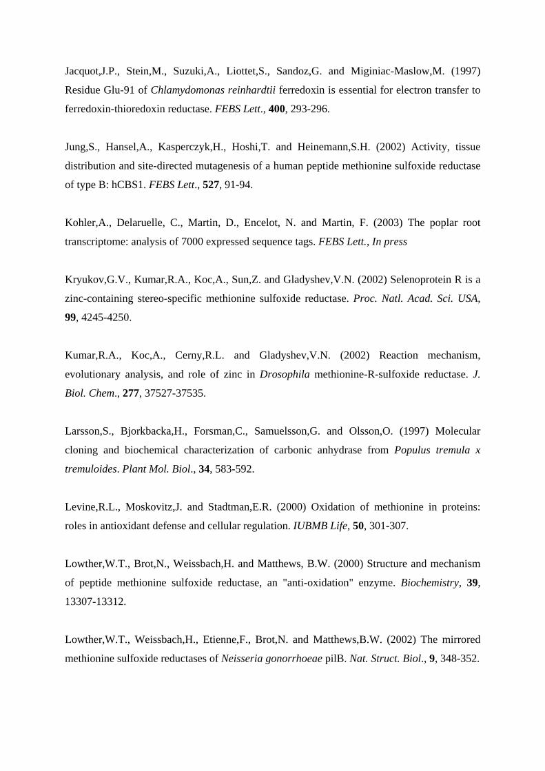
Jacquot,J.P., Stein,M., Suzuki,A., Liottet,S., Sandoz,G. and Miginiac-Maslow,M. (1997)
Residue Glu-91 of Chlamydomonas reinhardtii ferredoxin is essential for electron transfer to
ferredoxin-thioredoxin reductase. FEBS Lett., 400, 293-296.
Jung,S., Hansel,A., Kasperczyk,H., Hoshi,T. and Heinemann,S.H. (2002) Activity, tissue
distribution and site-directed mutagenesis of a human peptide methionine sulfoxide reductase
of type B: hCBS1. FEBS Lett., 527, 91-94.
Kohler,A., Delaruelle, C., Martin, D., Encelot, N. and Martin, F. (2003) The poplar root
transcriptome: analysis of 7000 expressed sequence tags. FEBS Lett., In press
Kryukov,G.V., Kumar,R.A., Koc,A., Sun,Z. and Gladyshev,V.N. (2002) Selenoprotein R is a
zinc-containing stereo-specific methionine sulfoxide reductase. Proc. Natl. Acad. Sci. USA,
99, 4245-4250.
Kumar,R.A., Koc,A., Cerny,R.L. and Gladyshev,V.N. (2002) Reaction mechanism,
evolutionary analysis, and role of zinc in Drosophila methionine-R-sulfoxide reductase. J.
Biol. Chem., 277, 37527-37535.
Larsson,S., Bjorkbacka,H., Forsman,C., Samuelsson,G. and Olsson,O. (1997) Molecular
cloning and biochemical characterization of carbonic anhydrase from Populus tremula x
tremuloides. Plant Mol. Biol., 34, 583-592.
Levine,R.L., Moskovitz,J. and Stadtman,E.R. (2000) Oxidation of methionine in proteins:
roles in antioxidant defense and cellular regulation. IUBMB Life, 50, 301-307.
Lowther,W.T., Brot,N., Weissbach,H. and Matthews, B.W. (2000) Structure and mechanism
of peptide methionine sulfoxide reductase, an "anti-oxidation" enzyme. Biochemistry, 39,
13307-13312.
Lowther,W.T., Weissbach,H., Etienne,F., Brot,N. and Matthews,B.W. (2002) The mirrored
methionine sulfoxide reductases of Neisseria gonorrhoeae pilB. Nat. Struct. Biol., 9, 348-352.
Moskovitz,J., Berlett,B.S., Poston,J.M. and Stadtman,E.R. (1997) The yeast peptide-
methionine sulfoxide reductase functions as an antioxidant in vivo. Proc. Natl. Acad. Sci.
USA, 94, 9585-9589.
Moskovitz,J., Flescher,E., Berlett,B.S., Azare,J., Poston,J.M. and Stadtman,E.R. (1998)
Overexpression of peptide-methionine sulfoxide reductase in Saccharomyces cerevisiae and
human T cells provides them with high resistance to oxidative stress. Proc Natl Acad Sci
USA, 95, 14071-14075.
Moskovitz,J., Poston,J.M., Berlett,B.S., Nosworthy,N.J., Szczepanowski,R. and
Stadtman,E.R. (2000) Identification and characterization of a putative active site for peptide
methionine sulfoxide reductase (MsrA) and its substrate stereospecificity. J. Biol. Chem., 275,
14167-14172.
Novoselov,S.V., Rao,M., Onoshko,N.V., Zhi,H., Kryukov,G.V., Xiang,Y., Weeks,D.P.,
Hatfield,D.L. and Gladyshev,V.N. (2002) Selenoproteins and selenocysteine insertion system
in the model plant cell system, Chlamydomonas reinhardtii. EMBO J., 21, 3681-3693.
Olry,A., Boschi-Muller,S., Marraud,M., Sanglier-Cianferani,S., Van Dorsselear,A. and
Branlant,G. (2002) Characterization of the methionine sulfoxide reductase activities of PILB,
a probable virulence factor from Neisseria meningitidis. J. Biol. Chem., 277, 12016-12022.
Otwinowski,Z., and Minor,W. (1997) Processing of X-ray diffraction data collected in
oscillation mode. Methods Enzymol., 276, 307-326.
Perrakis,A., Morris,J.R. and Lamzin,V.S. (1999) Automated protein model building combined
with iterative structure refinement. Nat. Struct. Biol., 6, 458-463.
Rouhier,N., Gelhaye,E., Sautiere,P.E. and Jacquot,J.P. (2002) Enhancement of poplar
glutaredoxin expression by optimization of the cDNA sequence. Protein Expr. Purif., 24, 234-
241.
Russel,M. and Model,P. (1986) The role of thioredoxin in filamentous phage assembly.
Construction, isolation, and characterization of mutant thioredoxins. J. Biol. Chem., 261,
14997-15005.
Sadanandom,A., Poghosyan,Z., Fairbairn,D.J. Murphy,D.J. (2000) Differential regulation of
plastidial and cytosolic isoforms of peptide methionine sulfoxide reductase in Arabidopsis.
Plant Physiol., 123, 255-264.
Saint-John,G., Brot,N., Ruan,J., Erdjument-Bromage,H., Tempst,P., Weissbach,H. and
Nathan,C. (2001) Peptide methionine sulfoxide reductase from Escherichia coli and
Mycobacterium tuberculosis protects bacteria against oxidative damage from reactive
nitrogen intermediates. Proc. Natl. Acad. Sci. USA, 98, 9901-9906.
Schenk,P.M., Baumann,S., Mattes,R. and Steinbiss,H.H. (1995) Improved high-level
expression system for eukaryotic genes in Escherichia coli using T7 RNA polymerase and
rare Arg tRNA’s. Biotechniques, 19, 196-200.
Sharov,V.S., Ferrington,D.A., Squier,T.C. and Schoneich,C. (1999) Diastereoselective
reduction of protein-bound methionine sulfoxide by methionine sulfoxide reductase. FEBS
Lett., 455, 247-250.
Spector,D., Etienne,F., Brot,N. and Weissbach,H. (2003) New membrane-associated and
soluble peptide methionine sulfoxide reductases in Escherichia coli. Biochem. Biophys. Res.
Commun., 302, 284-289.
Stadtman, E.R. (1993) Oxidation of free amino acids and amino acid residues in proteins by
radiolysis and by metal-catalyzed reactions. Annu. Rev. Biochem., 62, 797-821.
Stadtman,E.R., Moskovitz,J., Berlett,B.S. and Levine,R.L. (2002) Cyclic oxidation and
reduction of protein methionine residues is an important antioxidant mechanism. Mol. Cell.
Biochem., 234, 3-9.
Stewart,E.J., Aslund,F. and Beckwith,J. (1998) Disulfide bond formation in the Escherichia
coli cytoplasm: an in vivo role reversal for the thioredoxins. EMBO J., 17, 5543-5550.
Storz,G. and Imlay,J.A. (1999) Oxidative stress. Curr. Opin. Microbiol., 2, 188-194.
Tete-Favier,F., Cobessi,D., Boschi-Muller,S., Azza,S., Branlant,G. and Aubry,A. (2000)
Crystal structure of the Escherichia coli peptide methionine sulphoxide reductase at 1.9 A
resolution. Structure Fold Des., 8, 1167-1178.
Vagin,A., and Teplyakov,A. (1997) MOLREP: an automated program for molecular
replacement., J. Appl. Cryst., 30, 1022-1025.
Wizemann,T.M., Moskovitz,J., Pearce,B.J., Cundell,D., Arvidson,C.G., So,M., Weissbach,H.,
Brot,N. and Masure,H.R. (1996) Peptide methionine sulfoxide reductase contributes to the
maintenance of adhesins in three major pathogens. Proc. Natl. Acad. Sci. USA., 93, 7985-
7990.
Yao,Y., Yin,D., Jas,G.S., Kuczer,K., Williams,T.D., Schoneich,C. and Squier,T.C. (1996)
Oxidative modification of a carboxyl-terminal vicinal methionine in calmodulin by hydrogen
peroxide inhibits calmodulin-dependent activation of the plasma membrane Ca-ATPase.
Biochemistry, 35, 2767-2787.
Figure legends
Fig. 1. Amino acid comparison of the two poplar MsrA with known MsrA sequences. The
sequences were compared using ClustalW. The accession numbers (GenBank or MATDB)
are as follow : Populus tremula x tremuloides pMsrA (Pt pMsrA) : xxxxx, Arabidopsis
thaliana cMsrA (At cMsrA) : At5g61640, Arabidopsis thaliana pMsrA (At pMsrA) :
At4g25130, Brassica napus pMsrA (Bn pMsrA) : P54151, Populus tremula x tremuloides
cMsrA (Pt cMsrA) : xxxxx, Escherichia coli MsrA (Ec MsrA) : NP_418640, Bos taurus
MsrA (Bs MsrA) : P54149. The stars represent strict amino acid identities.
Fig. 2. MetSO reductive activity of wild-type and mutant cytosolic (cMsrA) and plastidial
(pMsrA) poplar sulfoxide methionine reductases. A) MetSO concentration evolution as a
function of time; cMsrA (full circles), pMsrA (full squares), C31S mutant of pMsrA (open
circles), C46S mutant of cMsrA (open squares). B) Met concentration evolution as a function
of time; cMsrA (full circles), pMsrA (full squares), C31S mutant of cMsrA (open squares),
C46S mutant of pMsrA (open circles). Reactions were recorded at 25 ∞C in the presence of
100 mM phosphate buffer with 50 mM DTT, 20 mM (L)Met(D,L)SO at pH 8.5. The enzyme
concentrations were for cMsrA, pMsrA, C31S and C46S : 4.7 M, 6.1 M, 4.7 M and 4.9 M
respectively. MetSO and Met concentrations were determined from the methyl peak intensity
of the MetSO at 2.65 ppm and the Met at 2.15 ppm.
Fig. 3. Cαtraces of the pMsrA (blue) and E. coli MsrA (red). The five cysteinyl residues of the
poplar enzyme are shown in balls and sticks.
Fig. 4. Schematic drawing of the pMsrA structure. The β-strands are represented as arrows
and the α-helices as springs. The trace of residues 167Lys-Gly204 is shown in red. The five
cysteinyl residues are shown in balls and sticks.
Fig. 5. 3Fo-2Fc electron density maps contoured at 1.2s in the Cys46 (a), Cys81 (b), Cys100
(c), Cys196 (d) and Cys202 (e) regions. Cys46, Cys196 and Cys202 are bonded to a β-
mercaptoethanol molecule via a disulfide bridge. Cys81 is observed as a sulfenic acid, while
Cys100 is in reduced form.

KTr
x h1
(µM
)
KTr
x h3
(µM
)
KM
etSO
(µM
)
kcat
(s-1
)
kcat
/ KM
etSO
(x10
3 M-1
s-1)
Vm
ax
(µm
oles
NA
DPH
ox/
min
/µm
oles
)
(µm
oles
NA
DPH
ox/
min
/mg
prot
ein)
cMsr
A
15.2
± 2
.9
17.5
± 3
.2
383
±101
1.
2 3.
13
73 (1
.55)
pMsr
A
17.9
± 8
n.
d.
239
± 33
0.
29
1.2
17.2
2 (0
.40)
Tab
le I.
Cat
alyt
ic p
aram
eter
s of p
Msr
A a
nd c
Msr
A. T
hese
par
amet
ers w
ere
dete
rmin
ed b
y fo
llow
ing
the
NA
DPH
oxi
datio
n in
the
pres
ence
of 1
0
mM
L-M
et-R
,S-S
O, p
opla
r cy
toso
lic T
rx h
1 an
d h3
as
elec
tron
dono
rs a
nd 0
.5 µ
M c
Msr
A o
r 3
µM p
Msr
A. T
he K
m f
or T
rx h
1, T
rx h
3 an
d
Met
SO w
ere
mea
sure
d at
satu
ratin
g co
ncen
tratio
n of
the
othe
r sub
stra
te. n
.d. :
not
det
erm
ined
.
Stoichiometry
cMsrA wt 1.72 (2)
cMsrA C31S 0 (0)
cMsrA C66S 1.4 (2)
cMsrA C85S 1.41 (2)
cMsrA C182S 1.16 (1)
cMsrA C188S 1.68 (1)
pMsrA wt 1.8 (2)
Table II. Stoichiometry of Met formed in the absence of reductant. The results are expressed
in mol Met formed /mol enzyme. In parenthesis, the theoretical ratio based on the catalytic
mechanism of E. coli.
Number of free thiols
without L-Met-R,S-SO
Number of free thiols
with L-Met-R,S-SO
Decrease in free
thiols
cMsrA wt 4.48 (5) 1.11 (2) 3.37 (3)
cMsrA C31S 3.9 (4) 3.7 (4) 0.2 (0)
cMsrA C66S 4.09 (4) 0.7 (1) 3.39 (3)
cMsrA C85S 3.9 (4) 0.4 (1) 3.5 (3)
cMsrA C182S 3.4 (4) 2.37 (3) 1.03 (1)
cMsrA C188S 3.85 (4) 1.5 (2) 2.35 (2)
pMsrA wt 4.25 (5) 1.33 (2) 2.92 (3)
pMsrA C46S 4.3 (4) 4.0 (4) 0.3 (0)
pMsrA C196S 3.5 (4) 2.4 (3) 1.1 (1)
pMsrA C202S 4.1 (4) 1.8 (2) 2.3 (2)
Table III. Thiol content of WT and mutants MsrA after treatment or not with L-Met-R,S-SO.
In parenthesis, the theoretical content of thiols based on the catalytic mechanism of E. coli
MsrA.
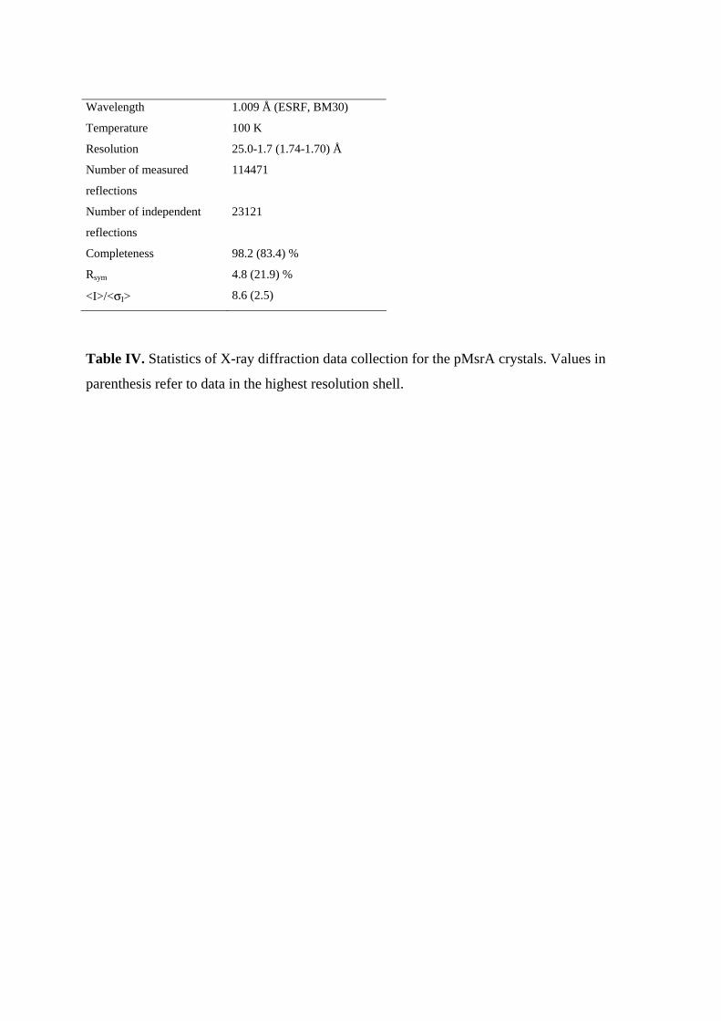
Wavelength 1.009 Å (ESRF, BM30)
Temperature 100 K
Resolution 25.0-1.7 (1.74-1.70) Å
Number of measured
reflections
114471
Number of independent
reflections
23121
Completeness 98.2 (83.4) %
Rsym 4.8 (21.9) %
<I>/<σI> 8.6 (2.5)
Table IV. Statistics of X-ray diffraction data collection for the pMsrA crystals. Values in
parenthesis refer to data in the highest resolution shell.
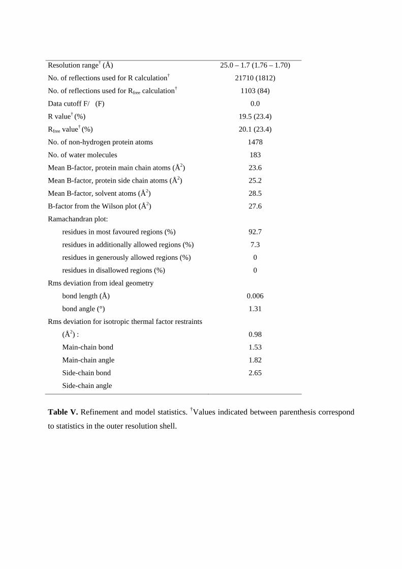
Resolution range† (Å) 25.0 – 1.7 (1.76 – 1.70)
No. of reflections used for R calculation† 21710 (1812)
No. of reflections used for Rfree calculation† 1103 (84)
Data cutoff F/�(F)
R value† (%)
0.0
19.5 (23.4)
Rfree value† (%) 20.1 (23.4)
No. of non-hydrogen protein atoms 1478
No. of water molecules 183
Mean B-factor, protein main chain atoms (Å2) 23.6
Mean B-factor, protein side chain atoms (Å2) 25.2
Mean B-factor, solvent atoms (Å2)
B-factor from the Wilson plot (Å2)
28.5
27.6
Ramachandran plot:
residues in most favoured regions (%)
residues in additionally allowed regions (%)
residues in generously allowed regions (%)
residues in disallowed regions (%)
92.7
7.3
0
0
Rms deviation from ideal geometry
bond length (Å)
bond angle (°)
Rms deviation for isotropic thermal factor restraints
(Å2) :
Main-chain bond
Main-chain angle
Side-chain bond
Side-chain angle
0.006
1.31
0.98
1.53
1.82
2.65
Table V. Refinement and model statistics. †Values indicated between parenthesis correspond
to statistics in the outer resolution shell.
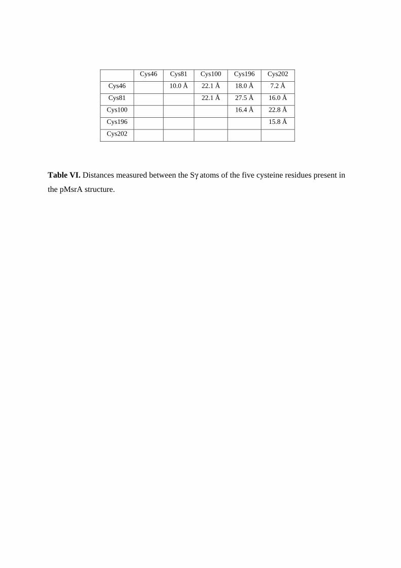
Cys46 Cys81 Cys100 Cys196 Cys202
Cys46 10.0 Å 22.1 Å 18.0 Å 7.2 Å
Cys81 22.1 Å 27.5 Å 16.0 Å
Cys100 16.4 Å 22.8 Å
Cys196 15.8 Å
Cys202
Table VI. Distances measured between the Sγ atoms of the five cysteine residues present in
the pMsrA structure.
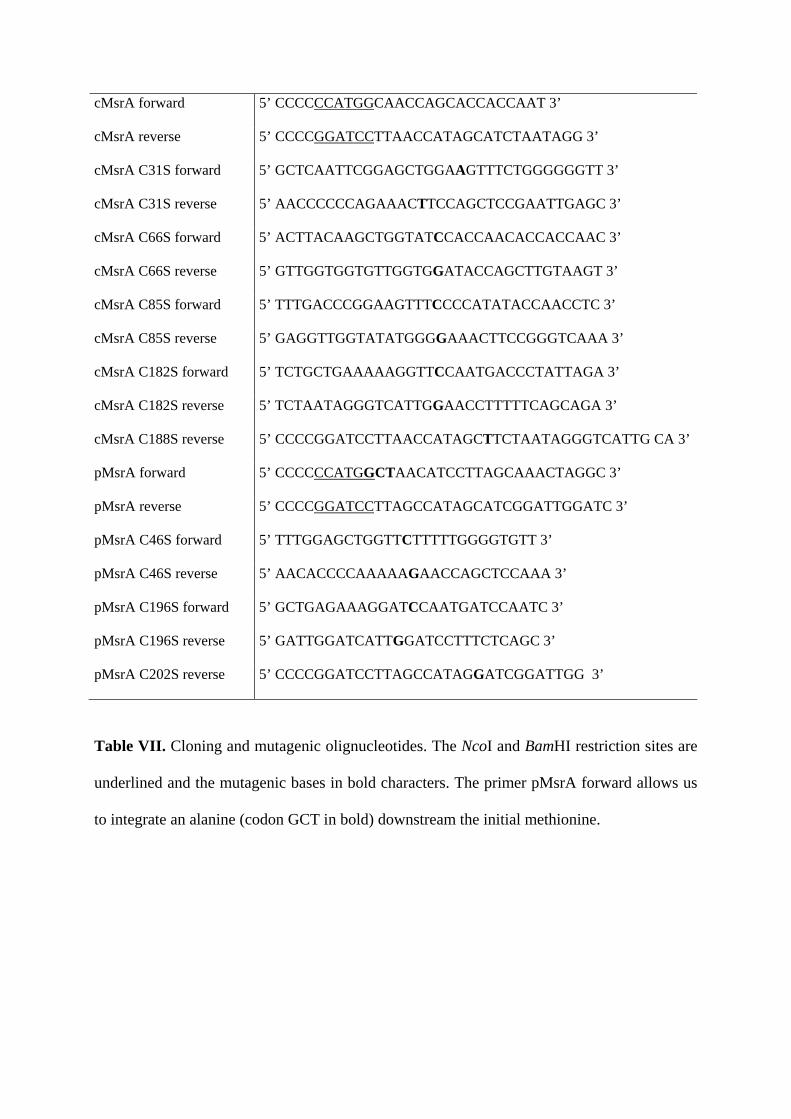
cMsrA forward 5’ CCCCCCATGGCAACCAGCACCACCAAT 3’
cMsrA reverse 5’ CCCCGGATCCTTAACCATAGCATCTAATAGG 3’
cMsrA C31S forward 5’ GCTCAATTCGGAGCTGGAAGTTTCTGGGGGGTT 3’
cMsrA C31S reverse 5’ AACCCCCCAGAAACTTCCAGCTCCGAATTGAGC 3’
cMsrA C66S forward 5’ ACTTACAAGCTGGTATCCACCAACACCACCAAC 3’
cMsrA C66S reverse 5’ GTTGGTGGTGTTGGTGGATACCAGCTTGTAAGT 3’
cMsrA C85S forward 5’ TTTGACCCGGAAGTTTCCCCATATACCAACCTC 3’
cMsrA C85S reverse 5’ GAGGTTGGTATATGGGGAAACTTCCGGGTCAAA 3’
cMsrA C182S forward 5’ TCTGCTGAAAAAGGTTCCAATGACCCTATTAGA 3’
cMsrA C182S reverse 5’ TCTAATAGGGTCATTGGAACCTTTTTCAGCAGA 3’
cMsrA C188S reverse 5’ CCCCGGATCCTTAACCATAGCTTCTAATAGGGTCATTG CA 3’
pMsrA forward 5’ CCCCCCATGGCTAACATCCTTAGCAAACTAGGC 3’
pMsrA reverse 5’ CCCCGGATCCTTAGCCATAGCATCGGATTGGATC 3’
pMsrA C46S forward 5’ TTTGGAGCTGGTTCTTTTTGGGGTGTT 3’
pMsrA C46S reverse 5’ AACACCCCAAAAAGAACCAGCTCCAAA 3’
pMsrA C196S forward 5’ GCTGAGAAAGGATCCAATGATCCAATC 3’
pMsrA C196S reverse 5’ GATTGGATCATTGGATCCTTTCTCAGC 3’
pMsrA C202S reverse 5’ CCCCGGATCCTTAGCCATAGGATCGGATTGG 3’
Table VII. Cloning and mutagenic olignucleotides. The NcoI and BamHI restriction sites are
underlined and the mutagenic bases in bold characters. The primer pMsrA forward allows us
to integrate an alanine (codon GCT in bold) downstream the initial methionine.
Pt pMsrA MLQTLSTHLSSTSTSTTTPLLLLSKPFLSPSAKSQLSHSKPFNFPRTLKPISYYKPMN-I 59At cMsrA --------------------------------------------------------MN-I 3At pMsrA MQVLVVSPPLIAAASLSKPLNSLSKAALSFS---RAKPICPFPQTSRRPISVYKSPMNNL 57Bn pMsrA MLSIVASPPVISAVSLSKPLQSLAKAALSLSK--RAKPTSPFPKTAR-SISVYKSPMNNL 57Pt cMsrA ------------------------------------------------------------Bt MsrA --------------------------MLSVTRRALQLFHSLFPIPRM---G--DSAAKIV 29Ec MsrA --------------------------------------MSLF-----------D-KKHLV 10
Pt pMsrA LSKLGFGTRSPDPSTMDPT----IPQGPDDDLPAPGQQFAQFGAGCFWGVELAFQRVPGV 115At cMsrA LNRLGLGSSGQ--TNMDPS---PIAQGNDDDTPAPGNQFAQFGAGCFWGVELAFQRVPGV 58At pMsrA FNRLGFGSRPQ--AQADPS-SAAIAQGPDDDVPSSGQQFAQFGAGCFWGVELAYQRVPGV 114Bn pMsrA FTRLGFGSRP----QPDPAASSAIAQGPDDDVPSPGQQFAQFGAGCFWGAELAYQRVPGV 113Pt cMsrA ---MATSTTNP---ALDPD----LGQ-PDN--P--NHEFAQFGAGCFWGVELAFQRLHGV 45Bt MsrA SPQEALPGRKE---PLVVAAKHHVNGNRTVEPFPEGTQMAVFGMGCFWGAERKFWTLKGV 86Ec MsrA SPADALPGRNT---PMPVATLHAVNG-HSMTNVPDGMEIAIFAMGCFWGVERLFWQLPGV 66
* * ***** * **
Pt pMsrA TKTEVGYTQGLLHNPTYEDVCTGTTNHNEVVRVQYDPKECSFDTLIDVLWARHDPTTLNR 175At cMsrA TQTEAGYTQGTVDNPSYGDVCSGTTGHSEVVRVQYDLNDCTYESLLDLFWSRHDPTTLNR 118At pMsrA TKTEVGYSHGIVHNPSYEDVCTGTTGHNEVVRVQYDPKECSFESLLDVFWNRHDPTTLNR 174Bn pMsrA TKTEVGYSHGFVDNPTYEDVCSETTGHNEIVRVQYDPKEVSFESLLDVFWKRHDPTTLNR 173Pt cMsrA VKTEVGYSQGNVPDPTYKLVCTKTTNHVEVVRVQFDPEVCPYTNLLSLFWSRHDPTTLNR 105Bt MsrA YSTQVGFAGGYTPNPTYKEVCSGKTGHAEVVRVVFQPEHISFEELLKVFWENHDPTQGMR 146Ec MsrA YSTAAGYTGGYTPNPTYREVCSGDTGHAEAVRIVYDPSVISYEQLLQVFWENHDPAQGMR 126
* * * * * ** * * * ** * * *** *
Pt pMsrA QGNDVGTQYRSGIYYYTPEQEKAAKESLERQQ----K-LLNRKIVTEILPAKKFYRAEEY 230At cMsrA QGNDVGTQYRSGIYFYTPEQEKLARESLERHQ----Q-QMERKIMTEILPAKKFYRAEEH 173At pMsrA QGGDVGTQYRSGIYYYTDEQERIAREAVEKQQ----K-ILNKRIVTEILPATKFYRAENY 229Bn pMsrA QGNDVGTRYRSGIYFYTDEQEKLAREAMEKQQ----K-ILNRKIVTEILPATKFYRAENY 228Pt cMsrA QGGDVGTQYRSGIYYYNEAQAKLAQESKEAKQ----LGLTDNTVVTEILPAKRFYRAEEY 161Bt MsrA QGNDHGSQYRSAIYPTSAEHVGAALKSKEDYQKVLSEHGFG-LITTDIREGQTFYYAEDY 205Ec MsrA QGNDHGTQYRSAIYPLTPEQDAAARASLERFQAAMLAADDDRHITTEIANATPFYYAEDD 186
** * * ** ** * * * * * ** **
Pt pMsrA HQQYLAKGGRFGFMQSAEKGCNDPIRCYG 259At cMsrA HQQYLSKGGRFGQGQSTAKGCNDPIRCYG 202At pMsrA HQQYLAKGGRMGLRQSAEKGCKDPIRCYG 258Bn pMsrA HQQYLAKGGRMGLSQSAEKGCNDPIRCYG 257Pt cMsrA HRQYLEKGGGRSAKQSAEKGCNDPIRCYG 190Bt MsrA HQQYLSKDP-DGYCGLGGTGVSCPLGIKK 233Ec MsrA HQQYLHKNP-YGYCGIGGIGVCLPPEA-- 212
* *** * * *
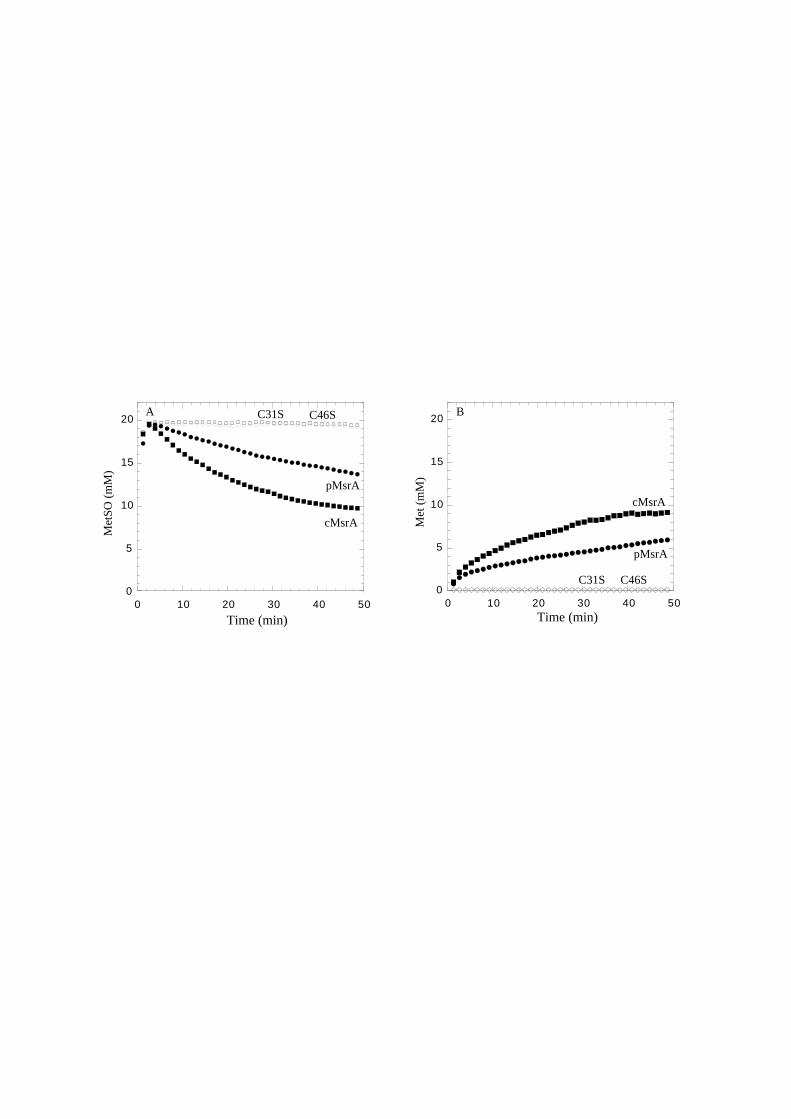
0
5
10
15
20
0 10 20 30 40 500
5
10
15
20
0 10 20 30 40 50Time (min) Time (min)
Met
SO (
mM
)
Met
(m
M)
A B
cMsrApMsrA
cMsrA
pMsrA
C31S C46S
C31S C46S
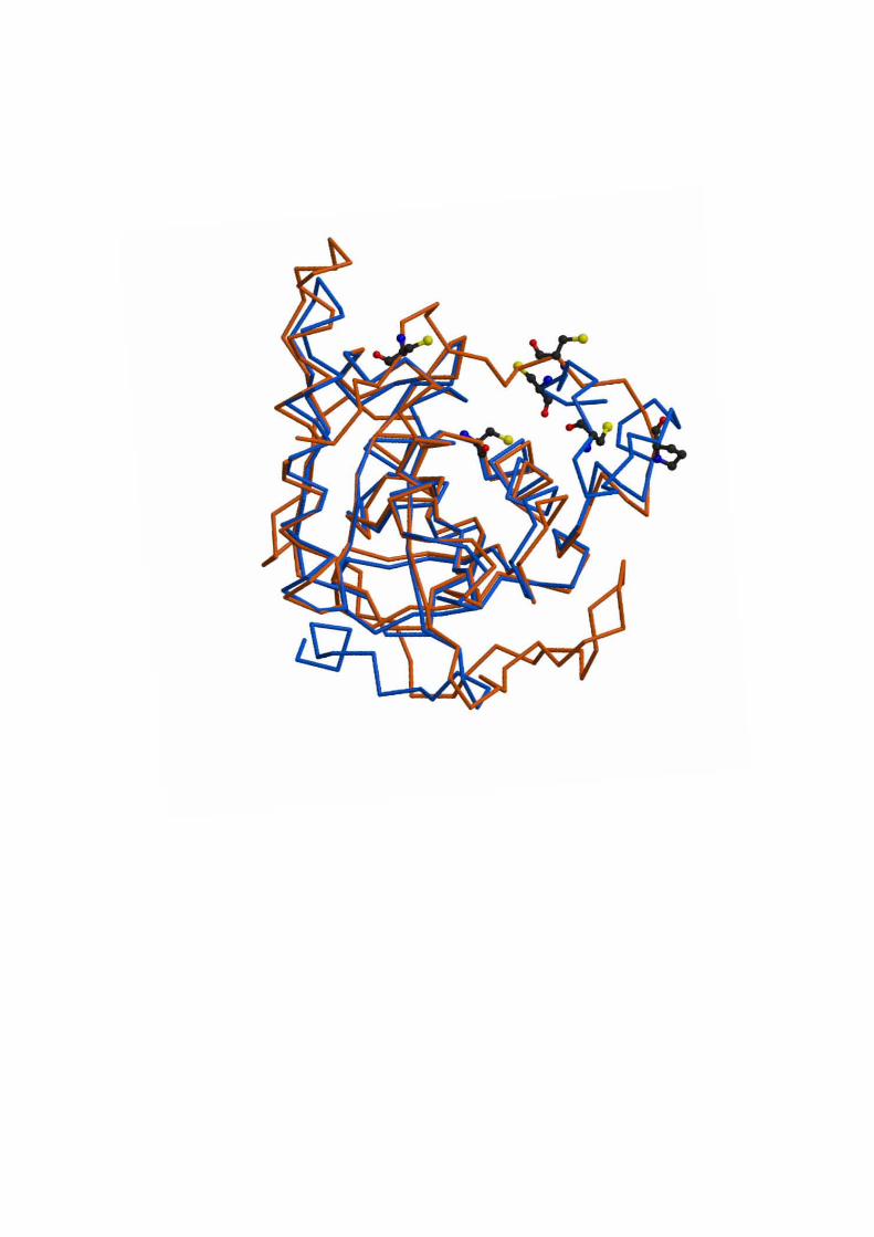
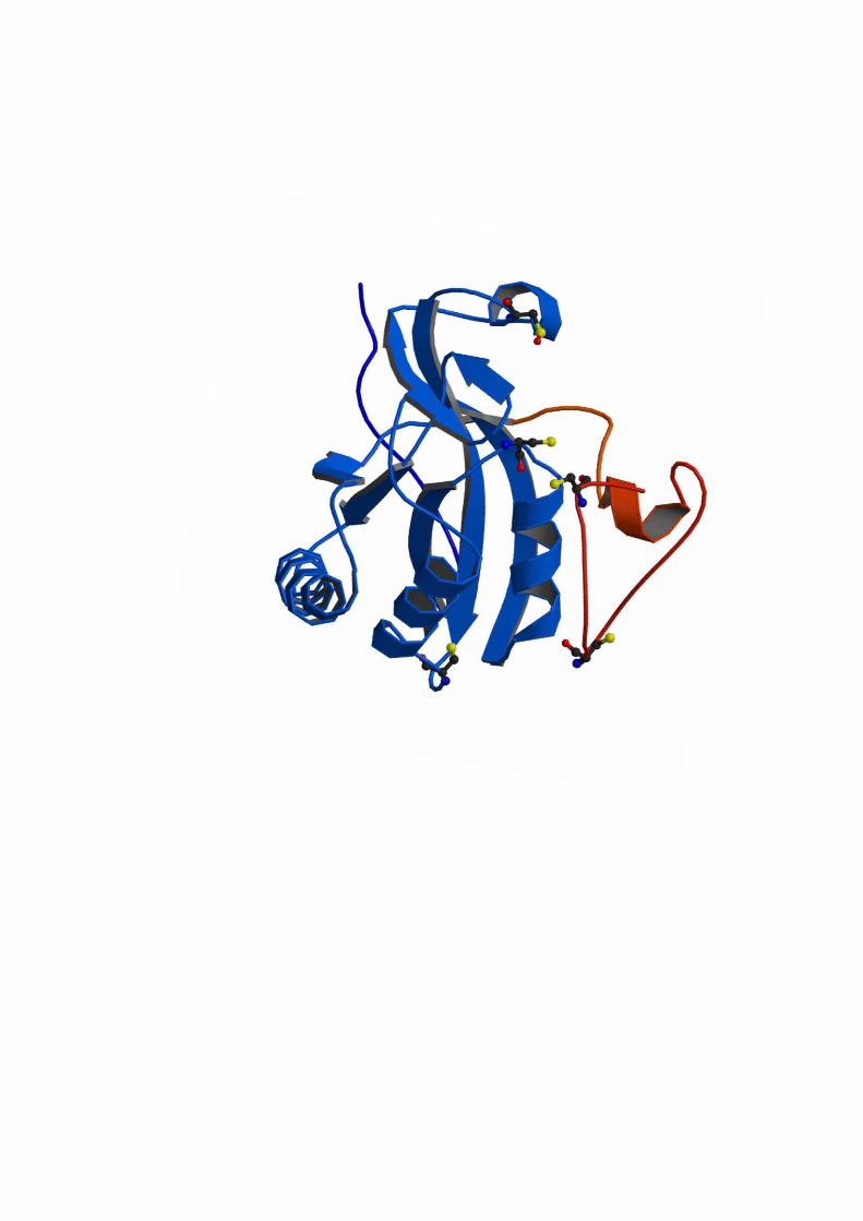
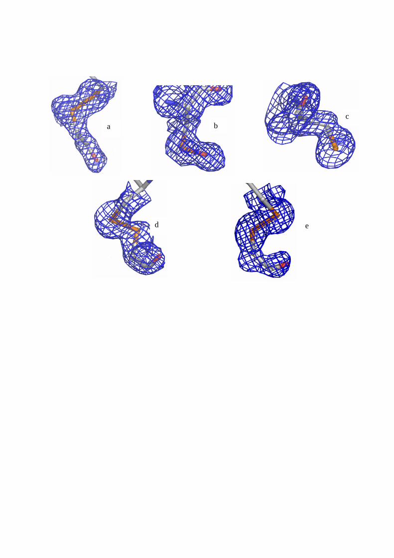
a b c
d e

Discussion
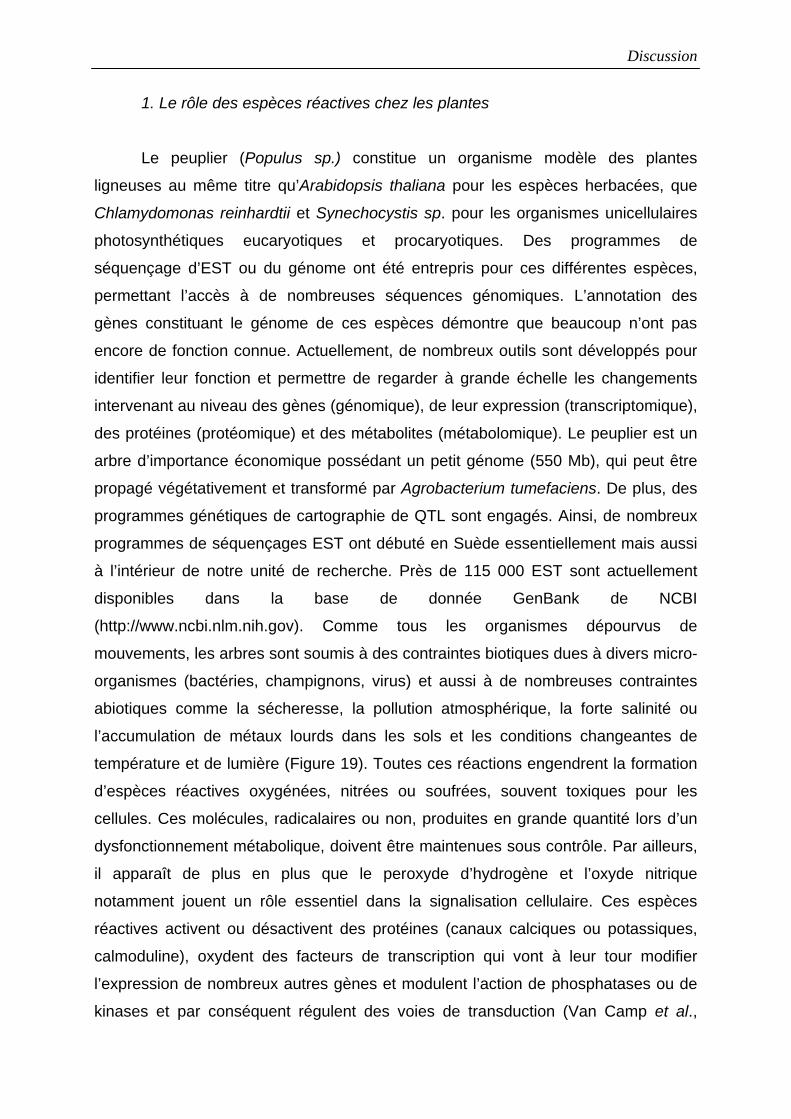
Discussion
1. Le rôle des espèces réactives chez les plantes
Le peuplier (Populus sp.) constitue un organisme modèle des plantes
ligneuses au même titre qu’Arabidopsis thaliana pour les espèces herbacées, que
Chlamydomonas reinhardtii et Synechocystis sp. pour les organismes unicellulaires
photosynthétiques eucaryotiques et procaryotiques. Des programmes de
séquençage d’EST ou du génome ont été entrepris pour ces différentes espèces,
permettant l’accès à de nombreuses séquences génomiques. L’annotation des
gènes constituant le génome de ces espèces démontre que beaucoup n’ont pas
encore de fonction connue. Actuellement, de nombreux outils sont développés pour
identifier leur fonction et permettre de regarder à grande échelle les changements
intervenant au niveau des gènes (génomique), de leur expression (transcriptomique),
des protéines (protéomique) et des métabolites (métabolomique). Le peuplier est un
arbre d’importance économique possédant un petit génome (550 Mb), qui peut être
propagé végétativement et transformé par Agrobacterium tumefaciens. De plus, des
programmes génétiques de cartographie de QTL sont engagés. Ainsi, de nombreux
programmes de séquençages EST ont débuté en Suède essentiellement mais aussi
à l’intérieur de notre unité de recherche. Près de 115 000 EST sont actuellement
disponibles dans la base de donnée GenBank de NCBI
(http://www.ncbi.nlm.nih.gov). Comme tous les organismes dépourvus de
mouvements, les arbres sont soumis à des contraintes biotiques dues à divers micro-
organismes (bactéries, champignons, virus) et aussi à de nombreuses contraintes
abiotiques comme la sécheresse, la pollution atmosphérique, la forte salinité ou
l’accumulation de métaux lourds dans les sols et les conditions changeantes de
température et de lumière (Figure 19). Toutes ces réactions engendrent la formation
d’espèces réactives oxygénées, nitrées ou soufrées, souvent toxiques pour les
cellules. Ces molécules, radicalaires ou non, produites en grande quantité lors d’un
dysfonctionnement métabolique, doivent être maintenues sous contrôle. Par ailleurs,
il apparaît de plus en plus que le peroxyde d’hydrogène et l’oxyde nitrique
notamment jouent un rôle essentiel dans la signalisation cellulaire. Ces espèces
réactives activent ou désactivent des protéines (canaux calciques ou potassiques,
calmoduline), oxydent des facteurs de transcription qui vont à leur tour modifier
l’expression de nombreux autres gènes et modulent l’action de phosphatases ou de
kinases et par conséquent régulent des voies de transduction (Van Camp et al.,
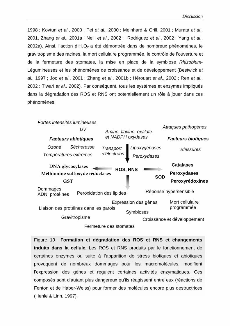
Discussion
1998 ; Kovtun et al., 2000 ; Pei et al., 2000 ; Meinhard & Grill, 2001 ; Murata et al.,
2001, Zhang et al., 2001a ; Neill et al., 2002 ; Rodriguez et al., 2002 ; Yang et al.,
2002a). Ainsi, l’action d’H2O2 a été démontrée dans de nombreux phénomènes, le
gravitropisme des racines, la mort cellulaire programmée, le contrôle de l’ouverture et
de la fermeture des stomates, la mise en place de la symbiose Rhizobium-
Légumineuses et les phénomènes de croissance et de développement (Bestwick et
al., 1997 ; Joo et al., 2001 ; Zhang et al., 2001b ; Hérouart et al., 2002 ; Ren et al.,
2002 ; Tiwari et al., 2002). Par conséquent, tous les systèmes et enzymes impliqués
dans la dégradation des ROS et RNS ont potentiellement un rôle à jouer dans ces
phénomènes.
Figure 19 : Formation et dégradation des ROS et RNS et changements induits dans la cellule. Les ROS et RNS produits par le fonctionnement de
certaines enzymes ou suite à l’apparition de stress biotiques et abiotiques
provoquent de nombreux dommages pour les macromolécules, modifient
l’expression des gènes et régulent certaines activités enzymatiques. Ces
composés sont d’autant plus dangereux qu’ils réagissent entre eux (réactions de
Fenton et de Haber-Weiss) pour former des molécules encore plus destructrices
(Henle & Linn, 1997).
Dommages ADN, protéines
Liaison des protéines dans les parois
Ozone Températures extrêmes
UVFortes intensités lumineuses
Sécheresse
Facteurs abiotiques
Blessures
Attaques pathogènes
Facteurs biotiques
Transport d’électrons
Amine, flavine, oxalate et NADPH oxydases
Peroxydases
Catalases Peroxydases
Peroxidation des lipides
Fermeture des stomates
Expression des gènes Mort cellulaire programmée
Réponse hypersensible
Croissance et développement
ROS, RNS SOD
Méthionine sulfoxyde réductasesDNA glycosylases
GST
Lipoxygénases
Gravitropisme
Peroxyrédoxines
Symbioses
Discussion
2. Intervention des Trx, Grx, Prx et MsrA dans la lutte antioxydante
Une approche de caractérisation des protéines (production de protéines
recombinantes, mutagenèse dirigée, activités enzymatiques) a été employée dans ce
travail pour étudier d’un point de vue fonctionnel et structural différentes isoformes de
Trx et de Grx mais aussi leur interaction avec deux type d’enzymes cibles, les Prx,
enzymes qui détruisent les alkyl ou phospholipide hydroperoxydes et les méthionine
sulfoxyde réductases, enzymes qui réduisent les MetSO libres ou intégrées dans des
peptides. Sous-jacent à ce travail, il serait intéressant de déterminer si ces multiples
isoformes de Trx, Grx, Prx et MsrA sont redondantes ou spécifiques de certaines
fonctions. En effet, il existe au moins 55 Trx ou Grx mono ou bicystéiniques chez A.
thaliana, de nombreuses protéines « thioredoxin like », 8 isoformes exprimées de Prx
dans différents compartiments cellulaires dont au moins 4 sont localisées dans le
chloroplaste et au moins 4 isoformes de MsrA dont 3 sont vraisemblablement
cytosoliques. Pour ce qui est des Prx, cette redondance est d’autant plus
surprenante qu’il existe également 6 isoformes de catalases, au moins 8 de Gpx et 7
d’Apx (Frugoli et al., 1996, résultats de blast dans MATDB). Cette étude visait donc à
déterminer la localisation et le rôle de ces isoformes dans les différents organes de la
plante et à isoler si possible des cibles spécifiques de ces isoformes et identifier leur
mode d’interaction.
2.1 Spécificité ou redondance des systèmes réducteurs ?
Le travail préalable mené sur les Trx h et les Grx de peuplier consiste à isoler
et caractériser les différentes isoformes constituant les systèmes de réduction du
peuplier.
Quatre isoformes de Trx cytosoliques bicystéiniques ont ainsi été isolées chez
le peuplier, popTrx h1, h3 et h4, une Trx monocystéinique (popTrx CxxS2), une Trx
mitochondriale (popTrx h2) et deux Grx, une bicystéinique et une monocystéinique.
Les isoformes popTrx h1 et h3 possèdent 2 sites actifs différents (WCPPC et
WCGPC respectivement) mais elles ont toutes deux la capacité de réduire les Prx (Q
et de type II) et les MsrA suggérant qu’il n’y a pas de spécificité in vitro des Trx
envers ces enzymes alors que la Grx ne réduit que la Prx de type II. Néanmoins,
certaines spécificités entre donneurs d’électrons et protéines cibles existent
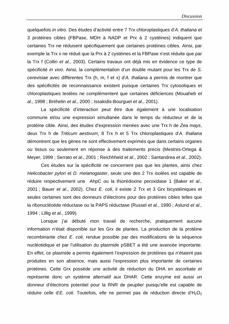
Discussion
quelquefois in vitro. Des études d’activité entre 7 Trx chloroplastiques d’A. thaliana et
3 protéines cibles (FBPase, MDH à NADP et Prx à 2 cystéines) indiquent que
certaines Trx ne réduisent spécifiquement que certaines protéines cibles. Ainsi, par
exemple la Trx x ne réduit que la Prx à 2 cystéines et la FBPase n’est réduite que par
la Trx f (Collin et al., 2003). Certains travaux ont déjà mis en évidence ce type de
spécificité in vivo. Ainsi, la complémentation d’un double mutant pour les Trx de S.
cerevisiae avec différentes Trx (h, m, f et x) d’A. thaliana a permis de montrer que
des spécificités de reconnaissance existent puisque certaines Trx cytosoliques et
chloroplastiques testées ne complémentent que certaines déficiences (Mouaheb et
al., 1998 ; Bréhelin et al., 2000 ; Issakidis-Bourguet et al., 2001).
La spécificité d’interaction peut être due également à une localisation
commune et/ou une expression simultanée dans le temps du réducteur et de la
protéine cible. Ainsi, des études d’expression menées avec une Trx h de Zea mays,
deux Trx h de Triticum aestivum, 8 Trx h et 5 Trx chloroplastiques d’A. thaliana
démontrent que les gènes ne sont effectivement exprimés que dans certains organes
ou tissus ou seulement en réponse à des traitements précis (Mestres-Ortega &
Meyer, 1999 ; Serrato et al., 2001 ; Reichhheld et al., 2002 ; Santandrea et al., 2002).
Ces études sur la spécificité ne concernent pas que les plantes, ainsi chez
Helicobacter pylori et D. melanogaster, seule une des 2 Trx isolées est capable de
réduire respectivement une AhpC ou la thiorédoxine peroxidase 1 (Baker et al.,
2001 ; Bauer et al., 2002). Chez E. coli, il existe 2 Trx et 3 Grx bicystéiniques et
seules certaines sont des donneurs d’électrons pour des protéines cibles telles que
la ribonucléotide réductase ou la PAPS réductase (Russel et al., 1990 ; Aslund et al.,
1994 ; Lillig et al., 1999).
Lorsque j’ai débuté mon travail de recherche, pratiquement aucune
information n’était disponible sur les Grx de plantes. La production de la protéine
recombinante chez E. coli, rendue possible par des modifications de la séquence
nucléotidique et par l’utilisation du plasmide pSBET a été une avancée importante.
En effet, ce plasmide a permis également l’expression de protéines qui n’étaient pas
produites en son absence, mais aussi l’expression plus importante de certaines
protéines. Cette Grx possède une activité de réduction du DHA en ascorbate et
représente donc un système alternatif aux DHAR. Cette enzyme est aussi un
donneur d’électrons potentiel pour la RNR de peuplier puisqu’elle est capable de
réduire celle d’E. coli. Toutefois, elle ne permet pas de réduction directe d’H2O2
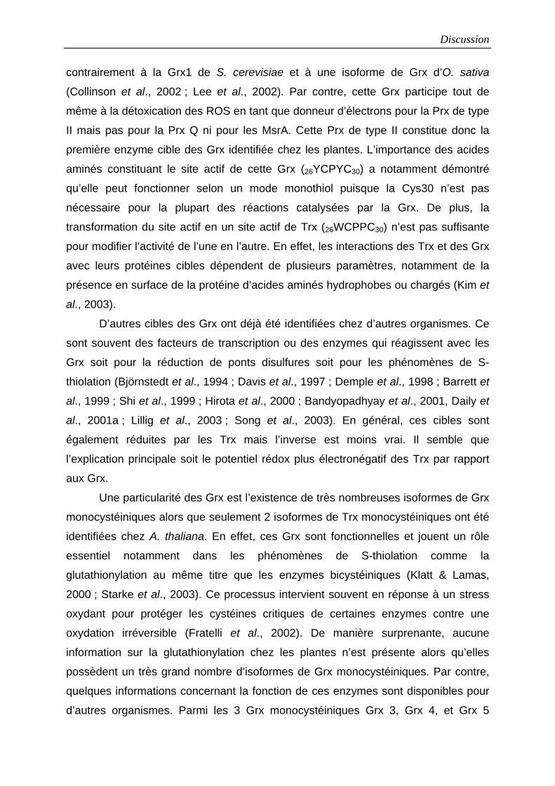
Discussion
contrairement à la Grx1 de S. cerevisiae et à une isoforme de Grx d’O. sativa
(Collinson et al., 2002 ; Lee et al., 2002). Par contre, cette Grx participe tout de
même à la détoxication des ROS en tant que donneur d’électrons pour la Prx de type
II mais pas pour la Prx Q ni pour les MsrA. Cette Prx de type II constitue donc la
première enzyme cible des Grx identifiée chez les plantes. L’importance des acides
aminés constituant le site actif de cette Grx (26YCPYC30) a notamment démontré
qu’elle peut fonctionner selon un mode monothiol puisque la Cys30 n’est pas
nécessaire pour la plupart des réactions catalysées par la Grx. De plus, la
transformation du site actif en un site actif de Trx (26WCPPC30) n’est pas suffisante
pour modifier l’activité de l’une en l’autre. En effet, les interactions des Trx et des Grx
avec leurs protéines cibles dépendent de plusieurs paramètres, notamment de la
présence en surface de la protéine d’acides aminés hydrophobes ou chargés (Kim et
al., 2003).
D’autres cibles des Grx ont déjà été identifiées chez d’autres organismes. Ce
sont souvent des facteurs de transcription ou des enzymes qui réagissent avec les
Grx soit pour la réduction de ponts disulfures soit pour les phénomènes de S-
thiolation (Björnstedt et al., 1994 ; Davis et al., 1997 ; Demple et al., 1998 ; Barrett et
al., 1999 ; Shi et al., 1999 ; Hirota et al., 2000 ; Bandyopadhyay et al., 2001, Daily et
al., 2001a ; Lillig et al., 2003 ; Song et al., 2003). En général, ces cibles sont
également réduites par les Trx mais l’inverse est moins vrai. Il semble que
l’explication principale soit le potentiel rédox plus électronégatif des Trx par rapport
aux Grx.
Une particularité des Grx est l’existence de très nombreuses isoformes de Grx
monocystéiniques alors que seulement 2 isoformes de Trx monocystéiniques ont été
identifiées chez A. thaliana. En effet, ces Grx sont fonctionnelles et jouent un rôle
essentiel notamment dans les phénomènes de S-thiolation comme la
glutathionylation au même titre que les enzymes bicystéiniques (Klatt & Lamas,
2000 ; Starke et al., 2003). Ce processus intervient souvent en réponse à un stress
oxydant pour protéger les cystéines critiques de certaines enzymes contre une
oxydation irréversible (Fratelli et al., 2002). De manière surprenante, aucune
information sur la glutathionylation chez les plantes n’est présente alors qu’elles
possèdent un très grand nombre d’isoformes de Grx monocystéiniques. Par contre,
quelques informations concernant la fonction de ces enzymes sont disponibles pour
d’autres organismes. Parmi les 3 Grx monocystéiniques Grx 3, Grx 4, et Grx 5
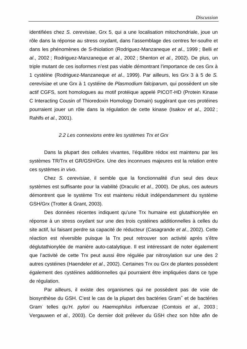
Discussion
identifiées chez S. cerevisiae, Grx 5, qui a une localisation mitochondriale, joue un
rôle dans la réponse au stress oxydant, dans l’assemblage des centres fer-soufre et
dans les phénomènes de S-thiolation (Rodriguez-Manzaneque et al., 1999 ; Belli et
al., 2002 ; Rodriguez-Manzaneque et al., 2002 ; Shenton et al., 2002). De plus, un
triple mutant de ces isoformes n’est pas viable démontrant l’importance de ces Grx à
1 cystéine (Rodriguez-Manzaneque et al., 1999). Par ailleurs, les Grx 3 à 5 de S.
cerevisiae et une Grx à 1 cystéine de Plasmodium falciparum, qui possèdent un site
actif CGFS, sont homologues au motif protéique appelé PICOT-HD (Protein Kinase
C Interacting Cousin of Thioredoxin Homology Domain) suggérant que ces protéines
pourraient jouer un rôle dans la régulation de cette kinase (Isakov et al., 2002 ;
Rahlfs et al., 2001).
2.2 Les connexions entre les systèmes Trx et Grx
Dans la plupart des cellules vivantes, l’équilibre rédox est maintenu par les
systèmes TR/Trx et GR/GSH/Grx. Une des inconnues majeures est la relation entre
ces systèmes in vivo.
Chez S. cerevisiae, il semble que la fonctionnalité d’un seul des deux
systèmes est suffisante pour la viabilité (Draculic et al., 2000). De plus, ces auteurs
démontrent que le système Trx est maintenu réduit indépendamment du système
GSH/Grx (Trotter & Grant, 2003).
Des données récentes indiquent qu’une Trx humaine est glutathionylée en
réponse à un stress oxydant sur une des trois cystéines additionnelles à celles du
site actif, lui faisant perdre sa capacité de réducteur (Casagrande et al., 2002). Cette
réaction est réversible puisque la Trx peut retrouver son activité après s’être
déglutathionylée de manière auto-catalytique. Il est intéressant de noter également
que l’activité de cette Trx peut aussi être régulée par nitrosylation sur une des 2
autres cystéines (Haendeler et al., 2002). Certaines Trx ou Grx de plantes possèdent
également des cystéines additionnelles qui pourraient être impliquées dans ce type
de régulation.
Par ailleurs, il existe des organismes qui ne possèdent pas de voie de
biosynthèse du GSH. C’est le cas de la plupart des bactéries Gram+ et de bactéries
Gram- telles qu’H. pylori ou Haemophilus influenzae (Comtois et al., 2003 ;
Vergauwen et al., 2003). Ce dernier doit prélever du GSH chez son hôte afin de
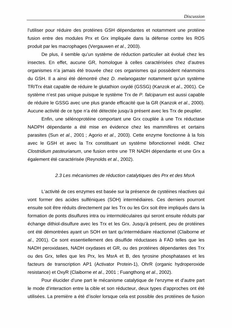
Discussion
l’utiliser pour réduire des protéines GSH dépendantes et notamment une protéine
fusion entre des modules Prx et Grx impliquée dans la défense contre les ROS
produit par les macrophages (Vergauwen et al., 2003).
De plus, il semble qu’un système de réduction particulier ait évolué chez les
insectes. En effet, aucune GR, homologue à celles caractérisées chez d’autres
organismes n’a jamais été trouvée chez ces organismes qui possèdent néanmoins
du GSH. Il a ainsi été démontré chez D. melanogaster notamment qu’un système
TR/Trx était capable de réduire le glutathion oxydé (GSSG) (Kanzok et al., 2001). Ce
système n’est pas unique puisque le système Trx de P. falciparum est aussi capable
de réduire le GSSG avec une plus grande efficacité que la GR (Kanzok et al., 2000).
Aucune activité de ce type n’a été détectée jusqu’à présent avec les Trx de peuplier.
Enfin, une sélénoprotéine comportant une Grx couplée à une Trx réductase
NADPH dépendante a été mise en évidence chez les mammifères et certains
parasites (Sun et al., 2001 ; Agorio et al., 2003). Cette enzyme fonctionne à la fois
avec le GSH et avec la Trx constituant un système bifonctionnel inédit. Chez
Clostridium pasteurianum, une fusion entre une TR NADH dépendante et une Grx a
également été caractérisée (Reynolds et al., 2002).
2.3 Les mécanismes de réduction catalytiques des Prx et des MsrA
L’activité de ces enzymes est basée sur la présence de cystéines réactives qui
vont former des acides sulféniques (SOH) intermédiaires. Ces derniers pourront
ensuite soit être réduits directement par les Trx ou les Grx soit être impliqués dans la
formation de ponts disulfures intra ou intermoléculaires qui seront ensuite réduits par
échange dithiol-disulfure avec les Trx et les Grx. Jusqu’à présent, peu de protéines
ont été démontrées ayant un SOH en tant qu’intermédiaire réactionnel (Claiborne et
al., 2001). Ce sont essentiellement des disulfide réductases à FAD telles que les
NADH peroxidases, NADH oxydases et GR, ou des protéines dépendantes des Trx
ou des Grx, telles que les Prx, les MsrA et B, des tyrosine phosphatases et les
facteurs de transcription AP1 (Activator Protein-1), OhrR (organic hydroperoxide
resistance) et OxyR (Claiborne et al., 2001 ; Fuangthong et al., 2002).
Pour élucider d’une part le mécanisme catalytique de l’enzyme et d’autre part
le mode d’interaction entre la cible et son réducteur, deux types d’approches ont été
utilisées. La première a été d’isoler lorsque cela est possible des protéines de fusion
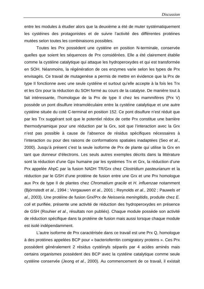
Discussion
entre les modules à étudier alors que la deuxième a été de muter systématiquement
les cystéines des protagonistes et de suivre l’activité des différentes protéines
mutées selon toutes les combinaisons possibles.
Toutes les Prx possèdent une cystéine en position N-terminale, conservée
quelles que soient les séquences de Prx considérées. Elle a été clairement établie
comme la cystéine catalytique qui attaque les hydroperoxydes et qui est transformée
en SOH. Néanmoins, la régénération de ces enzymes varie selon les types de Prx
envisagés. Ce travail de mutagenèse a permis de mettre en évidence que la Prx de
type II fonctionne avec une seule cystéine et surtout qu’elle accepte à la fois les Trx
et les Grx pour la réduction du SOH formé au cours de la catalyse. De manière tout à
fait intéressante, l’homologue de la Prx de type II chez les mammifères (Prx V)
possède un pont disulfure intramoléculaire entre la cystéine catalytique et une autre
cystéine située du coté C-terminal en position 152. Ce pont disulfure n’est réduit que
par les Trx suggérant soit que le potentiel rédox de cette Prx constitue une barrière
thermodynamique pour une réduction par la Grx, soit que l’interaction avec la Grx
n’est pas possible à cause de l’absence de résidus spécifiques nécessaires à
l’interaction ou pour des raisons de conformations spatiales inadaptées (Seo et al.,
2000). Jusqu’à présent c’est la seule isoforme de Prx de plante qui utilise la Grx en
tant que donneur d’électrons. Les seuls autres exemples décrits dans la littérature
sont la réduction d’une Gpx humaine par les systèmes Trx et Grx, la réduction d’une
Prx appelée AhpC par la fusion NADH TR/Grx chez Clostridium pasteurianum et la
réduction par le GSH d’une protéine de fusion entre une Grx et une Prx homologue
aux Prx de type II de plantes chez Chromatium gracile et H. influenzae notamment
(Björnstedt et al., 1994 ; Vergauwen et al., 2001 ; Reynolds et al., 2002 ; Pauwels et
al., 2003). Une protéine de fusion Grx/Prx de Neisseria meningitidis, produite chez E.
coli et purifiée, présente une activité de réduction des hydroperoxydes en présence
de GSH (Rouhier et al., résultats non publiés). Chaque module possède son activité
de réduction spécifique dans la protéine de fusion mais aussi lorsque chaque module
est isolé indépendamment.
L’autre isoforme de Prx caractérisée dans ce travail est une Prx Q, homologue
à des protéines appelées BCP pour « bacterioferritin comigratory proteins ». Ces Prx
possèdent généralement 2 résidus cystéinyls séparés par 4 acides aminés mais
certains organismes possèdent des BCP avec la cystéine catalytique comme seule
cystéine conservée (Jeong et al., 2000). Au commencement de ce travail, il existait
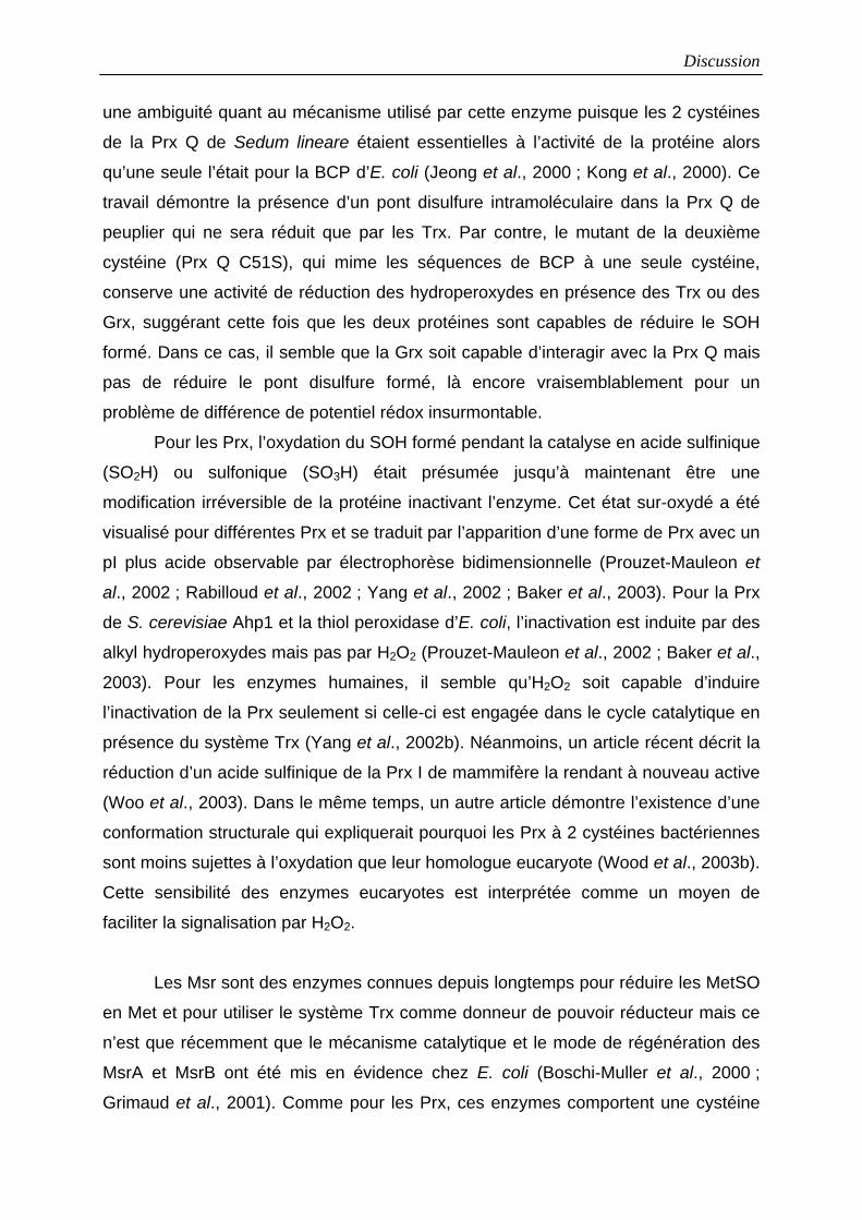
Discussion
une ambiguité quant au mécanisme utilisé par cette enzyme puisque les 2 cystéines
de la Prx Q de Sedum lineare étaient essentielles à l’activité de la protéine alors
qu’une seule l’était pour la BCP d’E. coli (Jeong et al., 2000 ; Kong et al., 2000). Ce
travail démontre la présence d’un pont disulfure intramoléculaire dans la Prx Q de
peuplier qui ne sera réduit que par les Trx. Par contre, le mutant de la deuxième
cystéine (Prx Q C51S), qui mime les séquences de BCP à une seule cystéine,
conserve une activité de réduction des hydroperoxydes en présence des Trx ou des
Grx, suggérant cette fois que les deux protéines sont capables de réduire le SOH
formé. Dans ce cas, il semble que la Grx soit capable d’interagir avec la Prx Q mais
pas de réduire le pont disulfure formé, là encore vraisemblablement pour un
problème de différence de potentiel rédox insurmontable.
Pour les Prx, l’oxydation du SOH formé pendant la catalyse en acide sulfinique
(SO2H) ou sulfonique (SO3H) était présumée jusqu’à maintenant être une
modification irréversible de la protéine inactivant l’enzyme. Cet état sur-oxydé a été
visualisé pour différentes Prx et se traduit par l’apparition d’une forme de Prx avec un
pI plus acide observable par électrophorèse bidimensionnelle (Prouzet-Mauleon et
al., 2002 ; Rabilloud et al., 2002 ; Yang et al., 2002 ; Baker et al., 2003). Pour la Prx
de S. cerevisiae Ahp1 et la thiol peroxidase d’E. coli, l’inactivation est induite par des
alkyl hydroperoxydes mais pas par H2O2 (Prouzet-Mauleon et al., 2002 ; Baker et al.,
2003). Pour les enzymes humaines, il semble qu’H2O2 soit capable d’induire
l’inactivation de la Prx seulement si celle-ci est engagée dans le cycle catalytique en
présence du système Trx (Yang et al., 2002b). Néanmoins, un article récent décrit la
réduction d’un acide sulfinique de la Prx I de mammifère la rendant à nouveau active
(Woo et al., 2003). Dans le même temps, un autre article démontre l’existence d’une
conformation structurale qui expliquerait pourquoi les Prx à 2 cystéines bactériennes
sont moins sujettes à l’oxydation que leur homologue eucaryote (Wood et al., 2003b).
Cette sensibilité des enzymes eucaryotes est interprétée comme un moyen de
faciliter la signalisation par H2O2.
Les Msr sont des enzymes connues depuis longtemps pour réduire les MetSO
en Met et pour utiliser le système Trx comme donneur de pouvoir réducteur mais ce
n’est que récemment que le mécanisme catalytique et le mode de régénération des
MsrA et MsrB ont été mis en évidence chez E. coli (Boschi-Muller et al., 2000 ;
Grimaud et al., 2001). Comme pour les Prx, ces enzymes comportent une cystéine
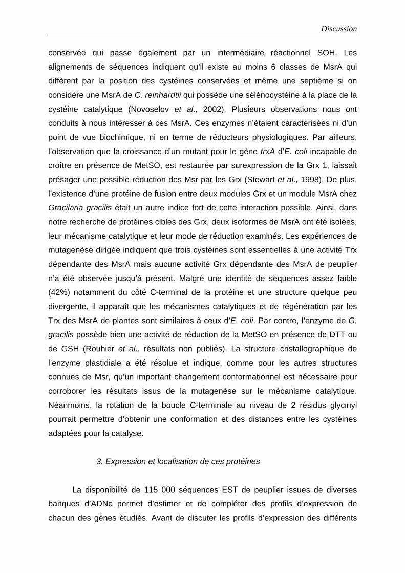
Discussion
conservée qui passe également par un intermédiaire réactionnel SOH. Les
alignements de séquences indiquent qu’il existe au moins 6 classes de MsrA qui
diffèrent par la position des cystéines conservées et même une septième si on
considère une MsrA de C. reinhardtii qui possède une sélénocystéine à la place de la
cystéine catalytique (Novoselov et al., 2002). Plusieurs observations nous ont
conduits à nous intéresser à ces MsrA. Ces enzymes n’étaient caractérisées ni d’un
point de vue biochimique, ni en terme de réducteurs physiologiques. Par ailleurs,
l’observation que la croissance d’un mutant pour le gène trxA d’E. coli incapable de
croître en présence de MetSO, est restaurée par surexpression de la Grx 1, laissait
présager une possible réduction des Msr par les Grx (Stewart et al., 1998). De plus,
l’existence d’une protéine de fusion entre deux modules Grx et un module MsrA chez
Gracilaria gracilis était un autre indice fort de cette interaction possible. Ainsi, dans
notre recherche de protéines cibles des Grx, deux isoformes de MsrA ont été isolées,
leur mécanisme catalytique et leur mode de réduction examinés. Les expériences de
mutagenèse dirigée indiquent que trois cystéines sont essentielles à une activité Trx
dépendante des MsrA mais aucune activité Grx dépendante des MsrA de peuplier
n’a été observée jusqu’à présent. Malgré une identité de séquences assez faible
(42%) notamment du côté C-terminal de la protéine et une structure quelque peu
divergente, il apparaît que les mécanismes catalytiques et de régénération par les
Trx des MsrA de plantes sont similaires à ceux d’E. coli. Par contre, l’enzyme de G.
gracilis possède bien une activité de réduction de la MetSO en présence de DTT ou
de GSH (Rouhier et al., résultats non publiés). La structure cristallographique de
l’enzyme plastidiale a été résolue et indique, comme pour les autres structures
connues de Msr, qu’un important changement conformationnel est nécessaire pour
corroborer les résultats issus de la mutagenèse sur le mécanisme catalytique.
Néanmoins, la rotation de la boucle C-terminale au niveau de 2 résidus glycinyl
pourrait permettre d’obtenir une conformation et des distances entre les cystéines
adaptées pour la catalyse.
3. Expression et localisation de ces protéines
La disponibilité de 115 000 séquences EST de peuplier issues de diverses
banques d’ADNc permet d’estimer et de compléter des profils d’expression de
chacun des gènes étudiés. Avant de discuter les profils d’expression des différents
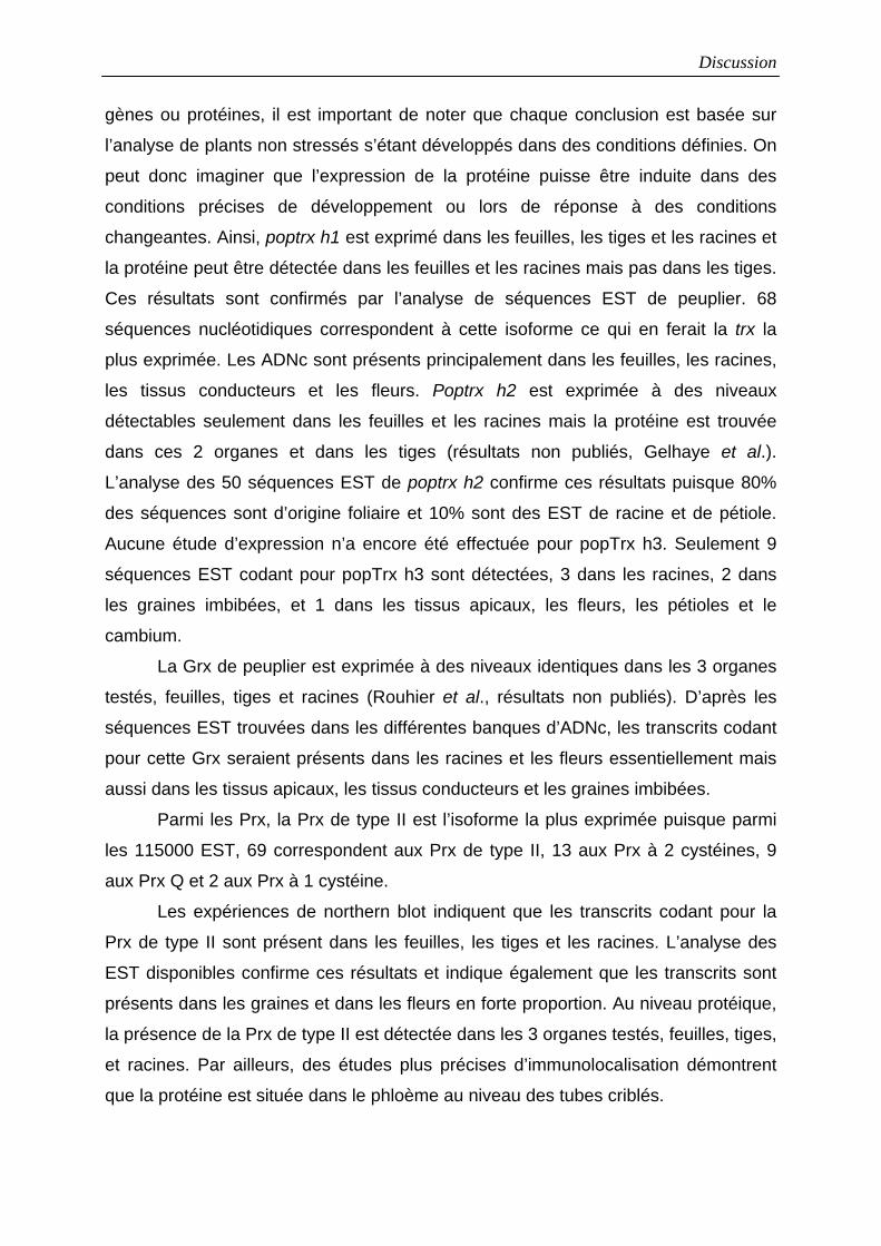
Discussion
gènes ou protéines, il est important de noter que chaque conclusion est basée sur
l’analyse de plants non stressés s’étant développés dans des conditions définies. On
peut donc imaginer que l’expression de la protéine puisse être induite dans des
conditions précises de développement ou lors de réponse à des conditions
changeantes. Ainsi, poptrx h1 est exprimé dans les feuilles, les tiges et les racines et
la protéine peut être détectée dans les feuilles et les racines mais pas dans les tiges.
Ces résultats sont confirmés par l’analyse de séquences EST de peuplier. 68
séquences nucléotidiques correspondent à cette isoforme ce qui en ferait la trx la
plus exprimée. Les ADNc sont présents principalement dans les feuilles, les racines,
les tissus conducteurs et les fleurs. Poptrx h2 est exprimée à des niveaux
détectables seulement dans les feuilles et les racines mais la protéine est trouvée
dans ces 2 organes et dans les tiges (résultats non publiés, Gelhaye et al.).
L’analyse des 50 séquences EST de poptrx h2 confirme ces résultats puisque 80%
des séquences sont d’origine foliaire et 10% sont des EST de racine et de pétiole.
Aucune étude d’expression n’a encore été effectuée pour popTrx h3. Seulement 9
séquences EST codant pour popTrx h3 sont détectées, 3 dans les racines, 2 dans
les graines imbibées, et 1 dans les tissus apicaux, les fleurs, les pétioles et le
cambium.
La Grx de peuplier est exprimée à des niveaux identiques dans les 3 organes
testés, feuilles, tiges et racines (Rouhier et al., résultats non publiés). D’après les
séquences EST trouvées dans les différentes banques d’ADNc, les transcrits codant
pour cette Grx seraient présents dans les racines et les fleurs essentiellement mais
aussi dans les tissus apicaux, les tissus conducteurs et les graines imbibées.
Parmi les Prx, la Prx de type II est l’isoforme la plus exprimée puisque parmi
les 115000 EST, 69 correspondent aux Prx de type II, 13 aux Prx à 2 cystéines, 9
aux Prx Q et 2 aux Prx à 1 cystéine.
Les expériences de northern blot indiquent que les transcrits codant pour la
Prx de type II sont présent dans les feuilles, les tiges et les racines. L’analyse des
EST disponibles confirme ces résultats et indique également que les transcrits sont
présents dans les graines et dans les fleurs en forte proportion. Au niveau protéique,
la présence de la Prx de type II est détectée dans les 3 organes testés, feuilles, tiges,
et racines. Par ailleurs, des études plus précises d’immunolocalisation démontrent
que la protéine est située dans le phloème au niveau des tubes criblés.
Discussion
Le gène codant la Prx Q paraît peu exprimé puisqu’on ne retrouve que 9 EST
dans la base de donnée, 4 sont issues de la banque d’ADNc de feuille, 3 de la
banque d’écorce et 2 de la banque de fleur. La protéine n’est détectée que dans les
feuilles par western blot mais pas dans les tiges et les racines. Par ailleurs, la
localisation chloroplastique de la protéine a été déterminée par des expériences de
fluorescence et de microscopie électronique. Enfin, cette protéine semble impliquée
dans les mécanismes de réponse à une attaque pathogène. En effet, la quantité de
protéine augmente en fonction du temps lors d’une interaction avec une race
incompatible de Melampsora larici-populina alors qu’elle décroît lors d’une réaction
avec une race compatible. Lors d’une réaction incompatible, un phénomène de mort
cellulaire programmée qu’on appelle réponse hypersensible se met en place. Lors de
cette réponse hypersensible, de grande quantités d’H2O2 sont produites et on peut
donc imaginer que la Prx Q ait un rôle à jouer dans le contrôle de ce phénomène.
Pour les msrA, l’analyse des séquences EST permet de distinguer 2 isoformes
qui sont celles que l’on a isolé, alors que 4 isoformes sont présentes chez A.
thaliana. Les 2 msrA paraissent peu exprimés, seulement 10 et 7 EST codant
respectivement pour la cMsrA et la pMsrA. Pour cmsrA, les transcrits sont présents
dans les racines, les fleurs, les feuilles et le bois et pour pmsrA, dans les feuilles, les
racines, les fleurs et le bois. Au niveau protéique, les premières expériences
indiquent que la pMsrA est exprimée dans les feuilles, les tiges et les racines alors
que la cMsrA est difficilement détectable avec les anticorps dirigés contre la pMsrA,
vraisemblablement parce que la protéine est peu exprimée car ces anticorps
reconnaissent parfaitement la protéine recombinante (Rouhier et al., résultats non
publiés). Par ailleurs, la quantité de ces protéines ne varie pas en réponse à une
attaque pathogène par M. larici-populina (Rouhier et al., résultats non publiés).
4. Les structures tridimensionnelles
Les Trx, Grx et Prx ont toutes une structure tridimensionnelle similaire basée
sur ce que l’on appelle en anglais le « thioredoxin fold » alors que ces protéines n’ont
pas du tout la même séquence primaire ni la même fonction excepté pour les Trx et
les Grx (Schröder & Ponting, 1998). Les MsrA ont aussi par certains aspects une
structure similaire aux Trx (Gladyshev et al., 2002).

Discussion
Au cours de ma thèse, la collaboration entreprise avec le laboratoire de
cristallographie de Nancy a d’ores et déjà permis de résoudre la structure de 3
protéines, la Grx en complexe avec le GSH, la Prx de type II et la pMsrA. Toutes trois
sont dans leur famille les premières structures pour des enzymes de plante. Ces
structures permettent d’appréhender notamment l’accessibilité et la conformation du
site actif de l’enzyme et la répartition des charges en surface de la protéine. Par
conséquent, elles représentent un outil indispensable pour étudier les interactions
protéine-protéine ou protéine-substrat.


Perspectives
Perspectives
Le travail de caractérisation entrepris sur les Trx, les Grx et leurs protéines
cibles ainsi que l’apparition récente dans la base de données GenBank de
nombreuses séquences EST ouvrent des perspectives intéressantes. Quelques-unes
d’entre elles sont listées ci-dessous :
- Isoler de nouvelles isoformes de Trx, Grx, Prx et MsrA afin de
comparer leurs propriétés biochimiques, leurs expressions et déterminer leurs
fonctions dans la cellule.
- Déterminer les interactions à l’origine de la reconnaissance entre les
Trx et les Grx et leurs protéines cibles soit par résolution de la structure des
protéines en complexe soit en étudiant au niveau structural et biochimique
d’éventuelles protéines de fusion.
- Poursuivre l’étude de l’expression de ces diverses protéines lors de la
croissance du peuplier ou en réponse à divers stress.
- Identifier de nouvelles protéines cibles des différentes Trx et Grx
isolées en utilisant les outils de la protéomique et les EST disponibles.
- Etudier par RMN les changements structuraux qui interviennent lors
de l’interaction Grx-Prx de type II.
- Etudier le transcriptome et le protéome de plants de peuplier infectés
par la rouille.
- Transformer des plants de peuplier par certains de gènes pour
augmenter leur résistance.


Références
Références
A
Abrahams JP, Leslie AGW. (1996) Methods used in the structure determination of
bovine mitochondrial F1 ATPase. Acta Crystallogr. D Biol. Crystallogr. 52 :30-42.
Agorio A, Chalar C, Cardozo S, Salinas G. (2003) Alternative mRNAs arising from
trans-splicing code for mitochondrial and cytosolic variants of Echinococcus
granulosus thioredoxin glutathione reductase. J. Biol. Chem. 278:12920-12928.
Aguilar F, Brunner B, Gardet-Salvi L, Stutz E, Schurmann P. (1992) Biosynthesis of
active spinach-chloroplast thioredoxin f in transformed E. coli. Plant Mol. Biol.
20:301-306.
Alphey MS, Bond CS, Tetaud E, Fairlamb AH, Hunter WN. (2000) The structure of
reduced tryparedoxin peroxidase reveals a decamer and insight into reactivity of
2Cys-peroxiredoxins. J. Mol. Biol. 300:903-916.
Altschul SF, Gish W, Miller W, Myers EW, Lipman DJ. (1990) Basic local alignment
search tool. J. Mol. Biol. 215:403-410.
Anderson LE, Li AD, Stevens FJ. (1998) The enolases of ice plant and Arabidopsis
contain a potential disulphide and are redox sensitive. Phytochemistry 47:707-713.
Asada K. (1992) Ascorbate peroxidase a hydrogen peroxide scavenging enzyme in
plants. Physiol. Plant. 85:235-241.
Asada K. (1999) The water-water cycle in chloroplasts: scavenging of active oxygen
and dissipation of excess photons. Annu. Rev. Plant Physiol. Plan Mol. Biol. 50:601-
639.
Aslund F, Ehn B, Miranda-Vizuete A, Pueyo C, Holmgren A. (1994) Two additional
glutaredoxins exist in Escherichia coli: glutaredoxin 3 is a hydrogen donor for
ribonucleotide reductase in a thioredoxin/glutaredoxin 1 double mutant. Proc. Natl.
Acad. Sci. USA 91:9813-9817.
Références
Aslund F, Berndt KD, Holmgren A. (1997) Redox potentials of glutaredoxins and
other thiol-disulfide oxidoreductases of the thioredoxin superfamily determined by
direct protein-protein redox equilibria. J. Biol. Chem. 272:30780-30786.
Aslund F, Beckwith J. (1999) Bridge over troubled waters : sensing stress by disulfide
bond formation. Cell 96:751-753.
Aslund F, Zheng M, Beckwith J, Storz G. (1999) Regulation of the OxyR transcription
factor by hydrogen peroxide and the cellular thiol-disulfide status. Proc. Natl. Acad.
Sci. USA 96:6161-6165.
B
Baalmann E, Backhausen JE, Rak C, Vetter S, Scheibe R. (1995) Reductive
modification and non reductive activation of purified spinach chloroplast NADP-
dependent glyceraldehyde-3-phosphate dehydrogenase. Arch. Biochem. Biophys.
324:201-208.
Babiychuk E, Kushnir S, Van Montagu M, Inze D. (1994) The Arabidopsis thaliana
apurinic endonuclease Arp reduces human transcription factors Fos and Jun. Proc.
Natl. Acad. Sci. USA 91:3299-3303.
Baier M, Dietz KJ. (1996) Primary structure and expression of plant homologues of
animal and fungal thioredoxin-dependent peroxide reductases and bacterial alkyl
hydroperoxide reductases. Plant Mol. Biol. 31:553-564.
Baier M, Dietz KJ. (1997) The plant 2-Cys peroxiredoxin BAS1 is a nuclear-encoded
chloroplast protein: its expressional regulation, phylogenetic origin, and implications
for its specific physiological function in plants. Plant J. 12:179-190.
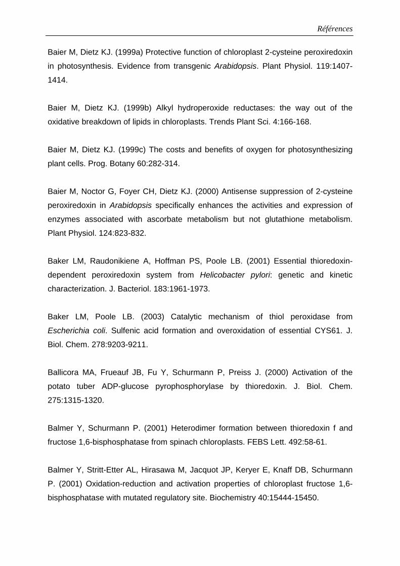
Références
Baier M, Dietz KJ. (1999a) Protective function of chloroplast 2-cysteine peroxiredoxin
in photosynthesis. Evidence from transgenic Arabidopsis. Plant Physiol. 119:1407-
1414.
Baier M, Dietz KJ. (1999b) Alkyl hydroperoxide reductases: the way out of the
oxidative breakdown of lipids in chloroplasts. Trends Plant Sci. 4:166-168.
Baier M, Dietz KJ. (1999c) The costs and benefits of oxygen for photosynthesizing
plant cells. Prog. Botany 60:282-314.
Baier M, Noctor G, Foyer CH, Dietz KJ. (2000) Antisense suppression of 2-cysteine
peroxiredoxin in Arabidopsis specifically enhances the activities and expression of
enzymes associated with ascorbate metabolism but not glutathione metabolism.
Plant Physiol. 124:823-832.
Baker LM, Raudonikiene A, Hoffman PS, Poole LB. (2001) Essential thioredoxin-
dependent peroxiredoxin system from Helicobacter pylori: genetic and kinetic
characterization. J. Bacteriol. 183:1961-1973.
Baker LM, Poole LB. (2003) Catalytic mechanism of thiol peroxidase from
Escherichia coli. Sulfenic acid formation and overoxidation of essential CYS61. J.
Biol. Chem. 278:9203-9211.
Ballicora MA, Frueauf JB, Fu Y, Schurmann P, Preiss J. (2000) Activation of the
potato tuber ADP-glucose pyrophosphorylase by thioredoxin. J. Biol. Chem.
275:1315-1320.
Balmer Y, Schurmann P. (2001) Heterodimer formation between thioredoxin f and
fructose 1,6-bisphosphatase from spinach chloroplasts. FEBS Lett. 492:58-61.
Balmer Y, Stritt-Etter AL, Hirasawa M, Jacquot JP, Keryer E, Knaff DB, Schurmann
P. (2001) Oxidation-reduction and activation properties of chloroplast fructose 1,6-
bisphosphatase with mutated regulatory site. Biochemistry 40:15444-15450.
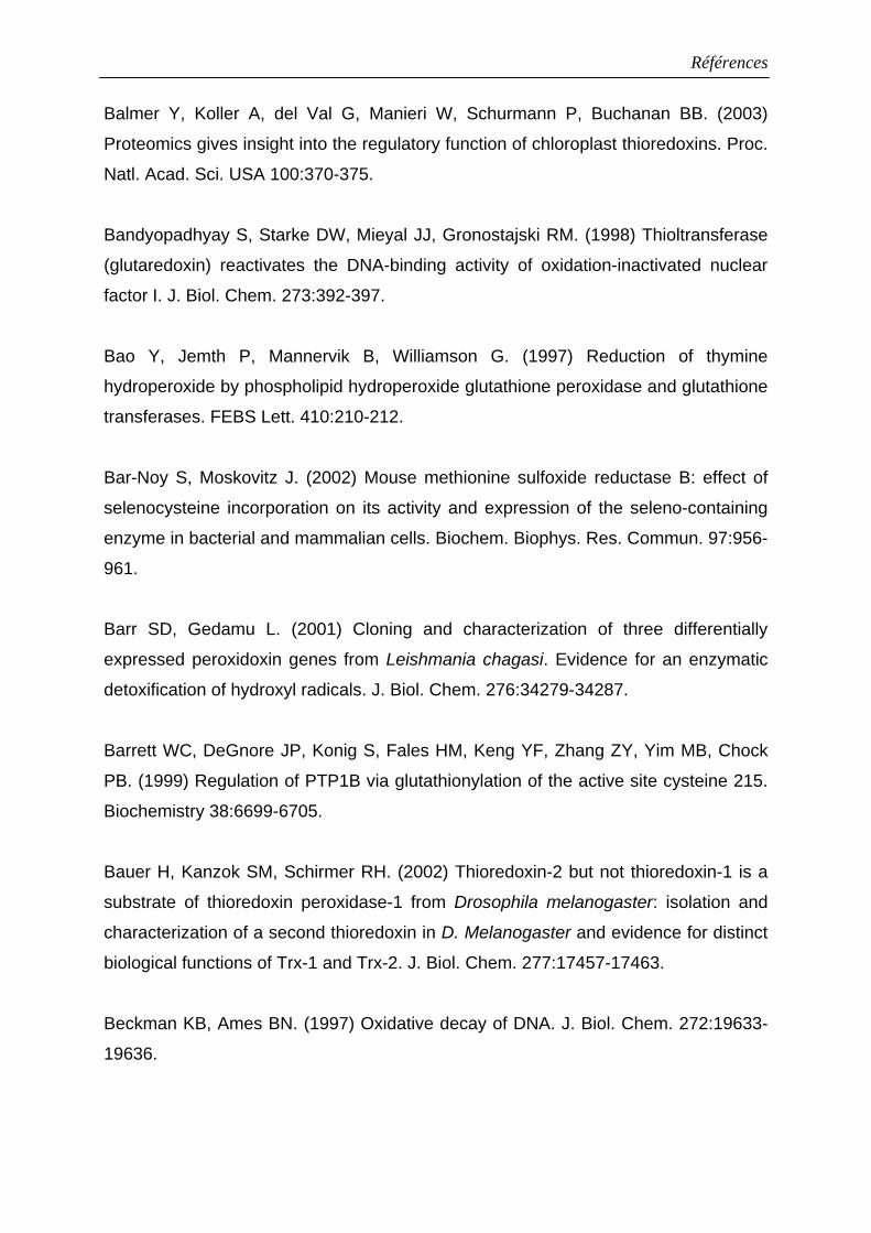
Références
Balmer Y, Koller A, del Val G, Manieri W, Schurmann P, Buchanan BB. (2003)
Proteomics gives insight into the regulatory function of chloroplast thioredoxins. Proc.
Natl. Acad. Sci. USA 100:370-375.
Bandyopadhyay S, Starke DW, Mieyal JJ, Gronostajski RM. (1998) Thioltransferase
(glutaredoxin) reactivates the DNA-binding activity of oxidation-inactivated nuclear
factor I. J. Biol. Chem. 273:392-397.
Bao Y, Jemth P, Mannervik B, Williamson G. (1997) Reduction of thymine
hydroperoxide by phospholipid hydroperoxide glutathione peroxidase and glutathione
transferases. FEBS Lett. 410:210-212.
Bar-Noy S, Moskovitz J. (2002) Mouse methionine sulfoxide reductase B: effect of
selenocysteine incorporation on its activity and expression of the seleno-containing
enzyme in bacterial and mammalian cells. Biochem. Biophys. Res. Commun. 97:956-
961.
Barr SD, Gedamu L. (2001) Cloning and characterization of three differentially
expressed peroxidoxin genes from Leishmania chagasi. Evidence for an enzymatic
detoxification of hydroxyl radicals. J. Biol. Chem. 276:34279-34287.
Barrett WC, DeGnore JP, Konig S, Fales HM, Keng YF, Zhang ZY, Yim MB, Chock
PB. (1999) Regulation of PTP1B via glutathionylation of the active site cysteine 215.
Biochemistry 38:6699-6705.
Bauer H, Kanzok SM, Schirmer RH. (2002) Thioredoxin-2 but not thioredoxin-1 is a
substrate of thioredoxin peroxidase-1 from Drosophila melanogaster: isolation and
characterization of a second thioredoxin in D. Melanogaster and evidence for distinct
biological functions of Trx-1 and Trx-2. J. Biol. Chem. 277:17457-17463.
Beckman KB, Ames BN. (1997) Oxidative decay of DNA. J. Biol. Chem. 272:19633-
19636.
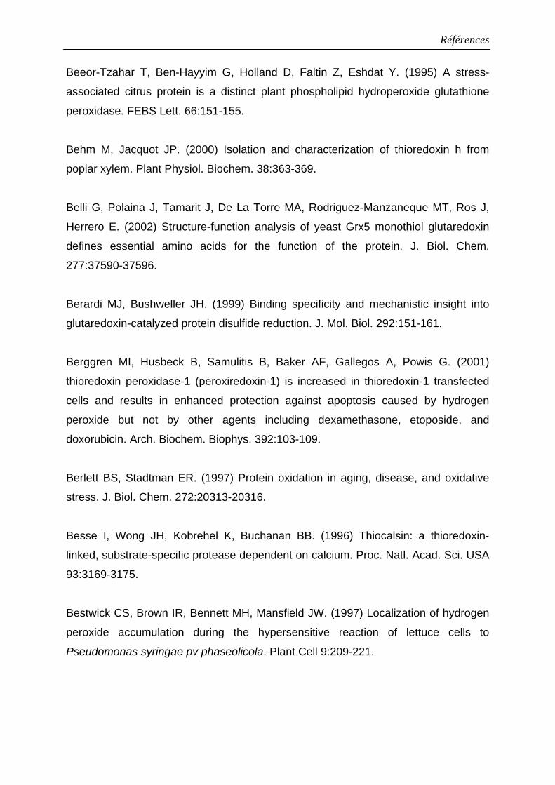
Références
Beeor-Tzahar T, Ben-Hayyim G, Holland D, Faltin Z, Eshdat Y. (1995) A stress-
associated citrus protein is a distinct plant phospholipid hydroperoxide glutathione
peroxidase. FEBS Lett. 66:151-155.
Behm M, Jacquot JP. (2000) Isolation and characterization of thioredoxin h from
poplar xylem. Plant Physiol. Biochem. 38:363-369.
Belli G, Polaina J, Tamarit J, De La Torre MA, Rodriguez-Manzaneque MT, Ros J,
Herrero E. (2002) Structure-function analysis of yeast Grx5 monothiol glutaredoxin
defines essential amino acids for the function of the protein. J. Biol. Chem.
277:37590-37596.
Berardi MJ, Bushweller JH. (1999) Binding specificity and mechanistic insight into
glutaredoxin-catalyzed protein disulfide reduction. J. Mol. Biol. 292:151-161.
Berggren MI, Husbeck B, Samulitis B, Baker AF, Gallegos A, Powis G. (2001)
thioredoxin peroxidase-1 (peroxiredoxin-1) is increased in thioredoxin-1 transfected
cells and results in enhanced protection against apoptosis caused by hydrogen
peroxide but not by other agents including dexamethasone, etoposide, and
doxorubicin. Arch. Biochem. Biophys. 392:103-109.
Berlett BS, Stadtman ER. (1997) Protein oxidation in aging, disease, and oxidative
stress. J. Biol. Chem. 272:20313-20316.
Besse I, Wong JH, Kobrehel K, Buchanan BB. (1996) Thiocalsin: a thioredoxin-
linked, substrate-specific protease dependent on calcium. Proc. Natl. Acad. Sci. USA
93:3169-3175.
Bestwick CS, Brown IR, Bennett MH, Mansfield JW. (1997) Localization of hydrogen
peroxide accumulation during the hypersensitive reaction of lettuce cells to
Pseudomonas syringae pv phaseolicola. Plant Cell 9:209-221.
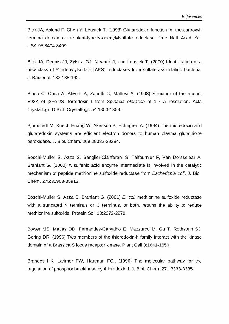
Références
Bick JA, Aslund F, Chen Y, Leustek T. (1998) Glutaredoxin function for the carboxyl-
terminal domain of the plant-type 5'-adenylylsulfate reductase. Proc. Natl. Acad. Sci.
USA 95:8404-8409.
Bick JA, Dennis JJ, Zylstra GJ, Nowack J, and Leustek T. (2000) Identification of a
new class of 5'-adenylylsulfate (APS) reductases from sulfate-assimilating bacteria.
J. Bacteriol. 182:135-142.
Binda C, Coda A, Aliverti A, Zanetti G, Mattevi A. (1998) Structure of the mutant
E92K of [2Fe-2S] ferredoxin I from Spinacia oleracea at 1.7 Å resolution. Acta
Crystallogr. D Biol. Crystallogr. 54:1353-1358.
Bjornstedt M, Xue J, Huang W, Akesson B, Holmgren A. (1994) The thioredoxin and
glutaredoxin systems are efficient electron donors to human plasma glutathione
peroxidase. J. Biol. Chem. 269:29382-29384.
Boschi-Muller S, Azza S, Sanglier-Cianferani S, Talfournier F, Van Dorsselear A,
Branlant G. (2000) A sulfenic acid enzyme intermediate is involved in the catalytic
mechanism of peptide methionine sulfoxide reductase from Escherichia coli. J. Biol.
Chem. 275:35908-35913.
Boschi-Muller S, Azza S, Branlant G. (2001) E. coli methionine sulfoxide reductase
with a truncated N terminus or C terminus, or both, retains the ability to reduce
methionine sulfoxide. Protein Sci. 10:2272-2279.
Bower MS, Matias DD, Fernandes-Carvalho E, Mazzurco M, Gu T, Rothstein SJ,
Goring DR. (1996) Two members of the thioredoxin-h family interact with the kinase
domain of a Brassica S locus receptor kinase. Plant Cell 8:1641-1650.
Brandes HK, Larimer FW, Hartman FC.. (1996) The molecular pathway for the
regulation of phosphoribulokinase by thioredoxin f. J. Biol. Chem. 271:3333-3335.
Références
Brehelin C, Mouaheb N, Verdoucq L, Lancelin JM, Meyer Y. (2000) Characterization
of determinants for the specificity of Arabidopsis thioredoxins h in yeast
complementation. J. Biol. Chem. 275:31641-31647.
Broin M, Cuine S, Peltier G, Rey P. (2000) Involvement of CDSP 32, a drought-
induced thioredoxin, in the response to oxidative stress in potato plants. FEBS Lett.
467:245-248.
Broin M, Cuine S, Eymery F, Rey P. (2002) The plastidic 2-cysteine peroxiredoxin is
a target for a thioredoxin involved in the protection of the photosynthetic apparatus
against oxidative damage. Plant Cell 14:1417-1432.
Brot N, Weissbach H. (2000) Peptide methionine sulfoxide reductase: biochemistry
and physiological role. Biopolymers 55:288-296.
Brugidou C, Marty I, Chartier and Meyer Y. (1993) The Nicotiana tabacum genome
encodes two cytoplasmic thioredoxin genes which are differently expressed. Mol.
Gen. Genet. 238:285-293.
Brumlik MJ, van der Goot FG, Wong KR, and Buckley JT. (1997) The disulfide bond
in the Aeromonas hydrophila lipase/acyltransferase stabilizes the structure but is not
required for secretion or activity. J. Bacteriol. 179:3116-3121.
Brunger AT, Adams PD, Clore GM, DeLano WL, Gros P, Grosse-Kunstleve RW,
Jiang JS, Kuszewski J, Nilges M, Pannu NS, Read RJ, Rice LM, Simonson T,
Warren GL. (1998) Crystallography & NMR system : A new software suite for
macromolecular structure determination. Acta Crystallogr. D Biol. Crystallogr. 54:905-
921.
Bryk R, Griffin P, Nathan C. (2000) Peroxynitrite reductase activity of bacterial
peroxiredoxins. Nature 407:211-215.
Références
Bryk R, Lima CD, Erdjument-Bromage H, Tempst P, Nathan C. (2002) Metabolic
enzymes of mycobacteria linked to antioxidant defense by a thioredoxin-like protein.
Science 295:1073-1077.
Buchanan BB, Schürmann P, Decottignies P, Lozano RM. (1994) Thioredoxin: a
multifunctional regulatory protein with a bright future in technology and medicine.
Arch. Biochem. Biophys. 314:257-260.
Buchanan BB, Schurmann P, Jacquot JP. (1994) Thioredoxin and metabolic
regulation. Semin. Cell. Biol. 5:285-293.
Buchanan BB, Schürmann P, Wolosiuk RA, Jacquot JP. (2002) The
ferredoxin/thioredoxin system: from discovery to molecular structures and beyond.
Photosynth. Res. 73:215-222.
Bucheler US, Werner D, Schirmer RH. (1990) Random silent mutagenesis in the
initial triplets of the coding region: a technique for adapting human glutathione
reductase-encoding cDNA to expression in Escherichia coli. Gene 96:271-276.
Buettner GR, Jurkiewicz BA. (1996) Chemistry and biochemistry of ascorbic acid.
Handbook of antioxidant, ed. E cadenas, L Packer, pp91-115. New York : Dekker.
Bunik V, Raddatz G, Lemaire S, Meyer Y, Jacquot JP, Bisswanger H. (1999)
Interaction of thioredoxins with target proteins: role of particular structural elements
and electrostatic properties of thioredoxins in their interplay with 2-oxoacid
dehydrogenase complexes. Protein Sci. 8:65-74.
Bushweller JH, Aslund F, Wuthrich K, Holmgren A. (1992) Structural and functional
characterization of the mutant Escherichia coli glutaredoxin (C14----S) and its mixed
disulfide with glutathione. Biochemistry 31:9288-9293.
C
Références
Capitani G, Markovic-Housley Z, DelVal G, Morris M, Jansonius JN, Schurmann P.
(2000) Crystal structures of two functionally different thioredoxins in spinach
chloroplasts. J. Mol. Biol. 302:135-154.
Carmel-Harel O, Storz G. (2000) Roles of the glutathione- and thioredoxin-dependent
reduction systems in the Escherichia coli and Saccharomyces cerevisiae responses
to oxidative stress. Annu. Rev. Microbiol. 54:439-461.
Casagrande S, Bonetto V, Fratelli M, Gianazza E, Eberini I, Massignan T, Salmona
M, Chang G, Holmgren A, Ghezzi P. (2002) Glutathionylation of human thioredoxin: a
possible crosstalk between the glutathione and thioredoxin systems. Proc. Natl.
Acad. Sci. USA. 99:9745-9749.
Chae HZ, Chung SJ, Rhee SG. (1994) Thioredoxin-dependent peroxide reductase
from yeast. J. Biol. Chem. 269:27670-27678.
Chae HZ, Uhm TB, Rhee SG. (1994) Dimerization of thiol-specific antioxidant and the
essential role of cysteine 47. Proc. Natl. Acad. Sci. USA 91:7022-7026.
Chae HZ, Kim HJ, Kang SW, Rhee SG. (1999) Characterization of three isoforms of
mammalian peroxiredoxin that reduce peroxides in the presence of thioredoxin.
Diabetes Res. Clin. Pract. 45:101-112.
Chen L, Xie QW, Nathan C. (1998) Alkyl hydroperoxide reductase subunit C (AhpC)
protects bacterial and human cells against reactive nitrogen intermediates. Mol. Cell.
1:795-805.
Cheong NE, Choi YO, Lee KO, Kim WY, Jung BG, Chi YH, Jeong JS, Kim K, Cho
MJ, Lee SY. (1999) Molecular cloning, expression, and functional characterization of
a 2Cys-peroxiredoxin in Chinese cabbage. Plant Mol. Biol. 40:825-834.
Chiadmi M, Navaza A, Miginiac-Maslow M, Jacquot JP, Cherfils J. (1999) Redox
signalling in the chloroplast: structure of oxidized pea fructose-1,6-bisphosphate
phosphatase. EMBO J. 18:6809-6815.
Références
Chivers PT, Prehoda KE, Raines RT. (1997) The CXXC motif: a rheostat in the active
site. Biochemistry 36:4061-4066.
Choi HJ, Kang SW, Yang CH, Rhee SG, Ryu SE. (1998) Crystal structure of a novel
human peroxidase enzyme at 2.0 Å resolution. Nat. Struct. Biol. 5:400-406.
Choi YO, Cheong NE, Lee KO, Jung BG, Hong CH, Jeong JH, Chi YH, Kim K, Cho
MJ, Lee SY. (1999) Cloning and expression of a new isotype of the peroxiredoxin
gene of Chinese cabbage and its comparison to 2Cys-peroxiredoxin isolated from the
same plant. Biochem. Biophys. Res. Commun. 258:768-771.
Chrestensen CA, Starke DW, Mieyal JJ. (2000) Acute cadmium exposure inactivates
thioltransferase (Glutaredoxin), inhibits intracellular reduction of protein-glutathionyl-
mixed disulfides, and initiates apoptosis. J. Biol. Chem. 275:26556-26565.
Ciaffi M, Paolacci AR, Dominici L, Tanzarella OA, Porceddu E. (2001) Molecular
characterization of gene sequences coding for protein disulfide isomerase (PDI) in
durum wheat (Triticum turgidum ssp. durum). Gene 265:147-156.
Ciorba MA, Heinemann SH, Weissbach H, Brot N, Hoshi T. (1997) Modulation of
potassium channel function by methionine oxidation and reduction. Proc. Natl. Acad.
Sci. USA 94:9932-9937.
Ciorba MA, Heinemann SH, Weissbach H, Brot N, Hoshi T. (1999) Regulation of
voltage-dependent K+ channels by methionine oxidation: effect of nitric oxide and
vitamin C. FEBS Lett. 442:48-52.
Claiborne A, Yeh JI, Mallett TC, Luba J, Crane EJ, Charrier V, Parsonage D. (1999)
Protein-sulfenic acids: diverse roles for an unlikely player in enzyme catalysis and
redox regulation. Biochemistry 38:15407-15416.
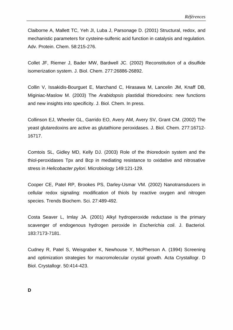
Références
Claiborne A, Mallett TC, Yeh JI, Luba J, Parsonage D. (2001) Structural, redox, and
mechanistic parameters for cysteine-sulfenic acid function in catalysis and regulation.
Adv. Protein. Chem. 58:215-276.
Collet JF, Riemer J, Bader MW, Bardwell JC. (2002) Reconstitution of a disulfide
isomerization system. J. Biol. Chem. 277:26886-26892.
Collin V, Issakidis-Bourguet E, Marchand C, Hirasawa M, Lancelin JM, Knaff DB,
Miginiac-Maslow M. (2003) The Arabidopsis plastidial thioredoxins: new functions
and new insights into specificity. J. Biol. Chem. In press.
Collinson EJ, Wheeler GL, Garrido EO, Avery AM, Avery SV, Grant CM. (2002) The
yeast glutaredoxins are active as glutathione peroxidases. J. Biol. Chem. 277:16712-
16717.
Comtois SL, Gidley MD, Kelly DJ. (2003) Role of the thioredoxin system and the
thiol-peroxidases Tpx and Bcp in mediating resistance to oxidative and nitrosative
stress in Helicobacter pylori. Microbiology 149:121-129.
Cooper CE, Patel RP, Brookes PS, Darley-Usmar VM. (2002) Nanotransducers in
cellular redox signaling: modification of thiols by reactive oxygen and nitrogen
species. Trends Biochem. Sci. 27:489-492.
Costa Seaver L, Imlay JA. (2001) Alkyl hydroperoxide reductase is the primary
scavenger of endogenous hydrogen peroxide in Escherichia coli. J. Bacteriol.
183:7173-7181.
Cudney R, Patel S, Weisgraber K, Newhouse Y, McPherson A. (1994) Screening
and optimization strategies for macromolecular crystal growth. Acta Crystallogr. D
Biol. Crystallogr. 50:414-423.
D
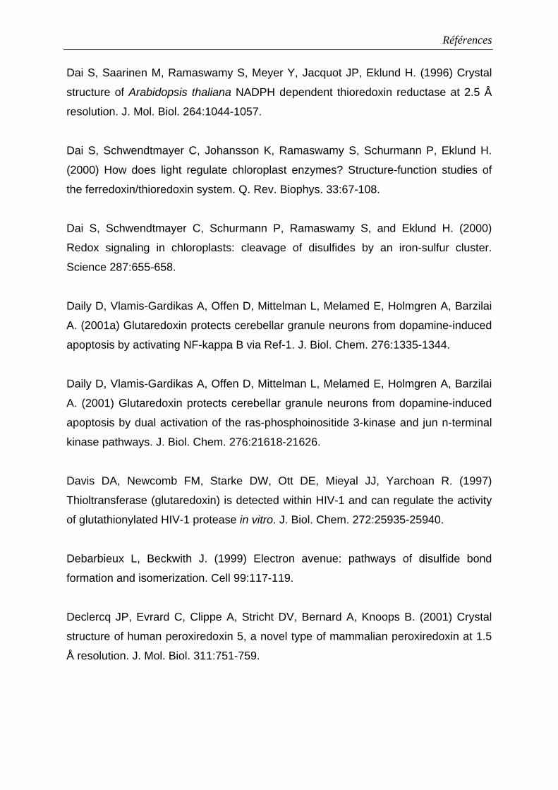
Références
Dai S, Saarinen M, Ramaswamy S, Meyer Y, Jacquot JP, Eklund H. (1996) Crystal
structure of Arabidopsis thaliana NADPH dependent thioredoxin reductase at 2.5 Å
resolution. J. Mol. Biol. 264:1044-1057.
Dai S, Schwendtmayer C, Johansson K, Ramaswamy S, Schurmann P, Eklund H.
(2000) How does light regulate chloroplast enzymes? Structure-function studies of
the ferredoxin/thioredoxin system. Q. Rev. Biophys. 33:67-108.
Dai S, Schwendtmayer C, Schurmann P, Ramaswamy S, and Eklund H. (2000)
Redox signaling in chloroplasts: cleavage of disulfides by an iron-sulfur cluster.
Science 287:655-658.
Daily D, Vlamis-Gardikas A, Offen D, Mittelman L, Melamed E, Holmgren A, Barzilai
A. (2001a) Glutaredoxin protects cerebellar granule neurons from dopamine-induced
apoptosis by activating NF-kappa B via Ref-1. J. Biol. Chem. 276:1335-1344.
Daily D, Vlamis-Gardikas A, Offen D, Mittelman L, Melamed E, Holmgren A, Barzilai
A. (2001) Glutaredoxin protects cerebellar granule neurons from dopamine-induced
apoptosis by dual activation of the ras-phosphoinositide 3-kinase and jun n-terminal
kinase pathways. J. Biol. Chem. 276:21618-21626.
Davis DA, Newcomb FM, Starke DW, Ott DE, Mieyal JJ, Yarchoan R. (1997)
Thioltransferase (glutaredoxin) is detected within HIV-1 and can regulate the activity
of glutathionylated HIV-1 protease in vitro. J. Biol. Chem. 272:25935-25940.
Debarbieux L, Beckwith J. (1999) Electron avenue: pathways of disulfide bond
formation and isomerization. Cell 99:117-119.
Declercq JP, Evrard C, Clippe A, Stricht DV, Bernard A, Knoops B. (2001) Crystal
structure of human peroxiredoxin 5, a novel type of mammalian peroxiredoxin at 1.5
Å resolution. J. Mol. Biol. 311:751-759.
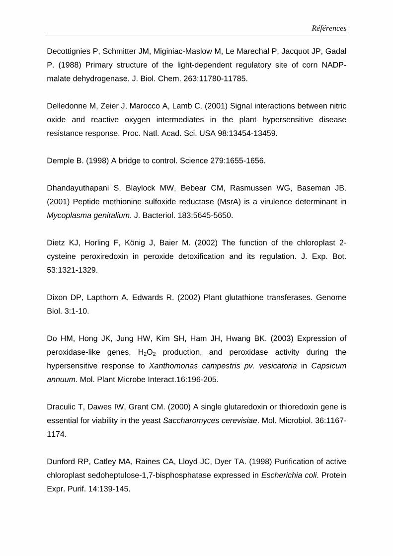
Références
Decottignies P, Schmitter JM, Miginiac-Maslow M, Le Marechal P, Jacquot JP, Gadal
P. (1988) Primary structure of the light-dependent regulatory site of corn NADP-
malate dehydrogenase. J. Biol. Chem. 263:11780-11785.
Delledonne M, Zeier J, Marocco A, Lamb C. (2001) Signal interactions between nitric
oxide and reactive oxygen intermediates in the plant hypersensitive disease
resistance response. Proc. Natl. Acad. Sci. USA 98:13454-13459.
Demple B. (1998) A bridge to control. Science 279:1655-1656.
Dhandayuthapani S, Blaylock MW, Bebear CM, Rasmussen WG, Baseman JB.
(2001) Peptide methionine sulfoxide reductase (MsrA) is a virulence determinant in
Mycoplasma genitalium. J. Bacteriol. 183:5645-5650.
Dietz KJ, Horling F, König J, Baier M. (2002) The function of the chloroplast 2-
cysteine peroxiredoxin in peroxide detoxification and its regulation. J. Exp. Bot.
53:1321-1329.
Dixon DP, Lapthorn A, Edwards R. (2002) Plant glutathione transferases. Genome
Biol. 3:1-10.
Do HM, Hong JK, Jung HW, Kim SH, Ham JH, Hwang BK. (2003) Expression of
peroxidase-like genes, H2O2 production, and peroxidase activity during the
hypersensitive response to Xanthomonas campestris pv. vesicatoria in Capsicum
annuum. Mol. Plant Microbe Interact.16:196-205.
Draculic T, Dawes IW, Grant CM. (2000) A single glutaredoxin or thioredoxin gene is
essential for viability in the yeast Saccharomyces cerevisiae. Mol. Microbiol. 36:1167-
1174.
Dunford RP, Catley MA, Raines CA, Lloyd JC, Dyer TA. (1998) Purification of active
chloroplast sedoheptulose-1,7-bisphosphatase expressed in Escherichia coli. Protein
Expr. Purif. 14:139-145.
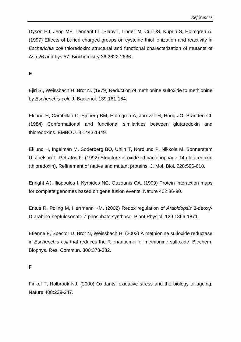
Références
Dyson HJ, Jeng MF, Tennant LL, Slaby I, Lindell M, Cui DS, Kuprin S, Holmgren A.
(1997) Effects of buried charged groups on cysteine thiol ionization and reactivity in
Escherichia coli thioredoxin: structural and functional characterization of mutants of
Asp 26 and Lys 57. Biochemistry 36:2622-2636.
E
Ejiri SI, Weissbach H, Brot N. (1979) Reduction of methionine sulfoxide to methionine
by Escherichia coli. J. Bacteriol. 139:161-164.
Eklund H, Cambillau C, Sjoberg BM, Holmgren A, Jornvall H, Hoog JO, Branden CI.
(1984) Conformational and functional similarities between glutaredoxin and
thioredoxins. EMBO J. 3:1443-1449.
Eklund H, Ingelman M, Soderberg BO, Uhlin T, Nordlund P, Nikkola M, Sonnerstam
U, Joelson T, Petratos K. (1992) Structure of oxidized bacteriophage T4 glutaredoxin
(thioredoxin). Refinement of native and mutant proteins. J. Mol. Biol. 228:596-618.
Enright AJ, Iliopoulos I, Kyrpides NC, Ouzounis CA. (1999) Protein interaction maps
for complete genomes based on gene fusion events. Nature 402:86-90.
Entus R, Poling M, Herrmann KM. (2002) Redox regulation of Arabidopsis 3-deoxy-
D-arabino-heptulosonate 7-phosphate synthase. Plant Physiol. 129:1866-1871.
Etienne F, Spector D, Brot N, Weissbach H. (2003) A methionine sulfoxide reductase
in Escherichia coli that reduces the R enantiomer of methionine sulfoxide. Biochem.
Biophys. Res. Commun. 300:378-382.
F
Finkel T, Holbrook NJ. (2000) Oxidants, oxidative stress and the biology of ageing.
Nature 408:239-247.
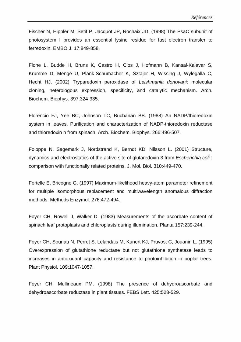
Références
Fischer N, Hippler M, Setif P, Jacquot JP, Rochaix JD. (1998) The PsaC subunit of
photosystem I provides an essential lysine residue for fast electron transfer to
ferredoxin. EMBO J. 17:849-858.
Flohe L, Budde H, Bruns K, Castro H, Clos J, Hofmann B, Kansal-Kalavar S,
Krumme D, Menge U, Plank-Schumacher K, Sztajer H, Wissing J, Wylegalla C,
Hecht HJ. (2002) Tryparedoxin peroxidase of Leishmania donovani: molecular
cloning, heterologous expression, specificity, and catalytic mechanism. Arch.
Biochem. Biophys. 397:324-335.
Florencio FJ, Yee BC, Johnson TC, Buchanan BB. (1988) An NADP/thioredoxin
system in leaves. Purification and characterization of NADP-thioredoxin reductase
and thioredoxin h from spinach. Arch. Biochem. Biophys. 266:496-507.
Foloppe N, Sagemark J, Nordstrand K, Berndt KD, Nilsson L. (2001) Structure,
dynamics and electrostatics of the active site of glutaredoxin 3 from Escherichia coli :
comparison with functionally related proteins. J. Mol. Biol. 310:449-470.
Fortelle E, Bricogne G. (1997) Maximum-likelihood heavy-atom parameter refinement
for multiple isomorphous replacement and multiwavelength anomalous diffraction
methods. Methods Enzymol. 276:472-494.
Foyer CH, Rowell J, Walker D. (1983) Measurements of the ascorbate content of
spinach leaf protoplasts and chloroplasts during illumination. Planta 157:239-244.
Foyer CH, Souriau N, Perret S, Lelandais M, Kunert KJ, Pruvost C, Jouanin L. (1995)
Overexpression of glutathione reductase but not glutathione synthetase leads to
increases in antioxidant capacity and resistance to photoinhibition in poplar trees.
Plant Physiol. 109:1047-1057.
Foyer CH, Mullineaux PM. (1998) The presence of dehydroascorbate and
dehydroascorbate reductase in plant tissues. FEBS Lett. 425:528-529.
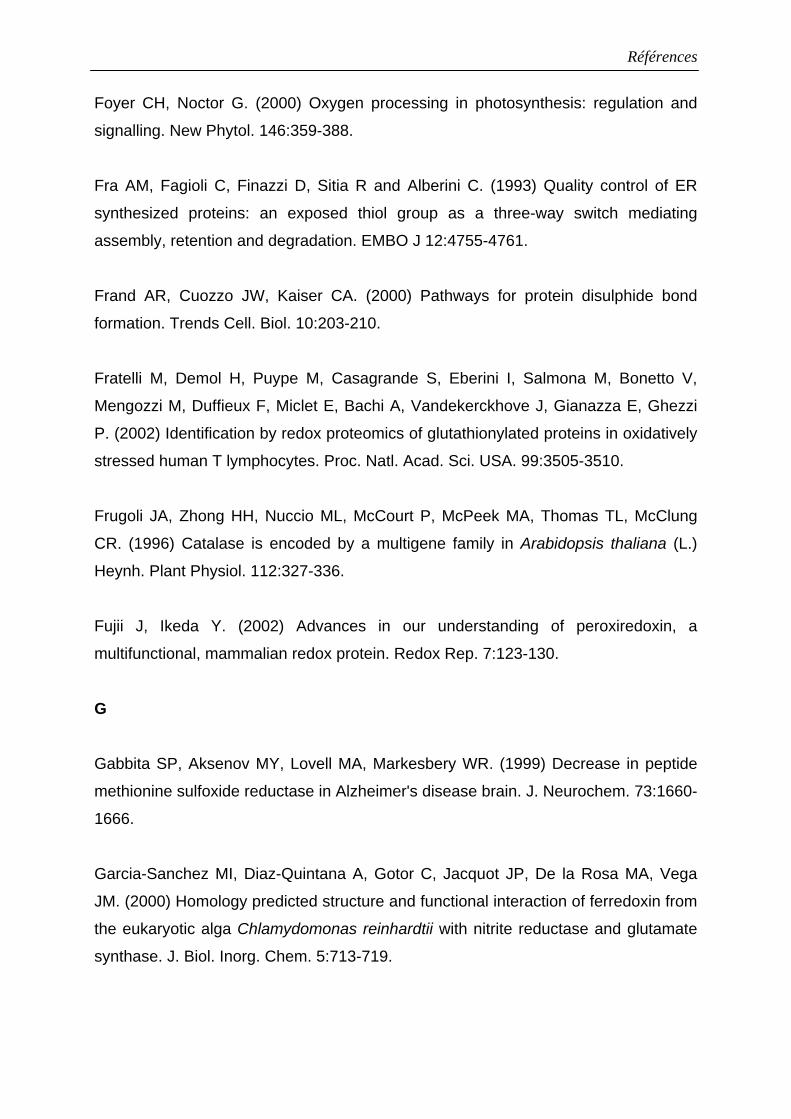
Références
Foyer CH, Noctor G. (2000) Oxygen processing in photosynthesis: regulation and
signalling. New Phytol. 146:359-388.
Fra AM, Fagioli C, Finazzi D, Sitia R and Alberini C. (1993) Quality control of ER
synthesized proteins: an exposed thiol group as a three-way switch mediating
assembly, retention and degradation. EMBO J 12:4755-4761.
Frand AR, Cuozzo JW, Kaiser CA. (2000) Pathways for protein disulphide bond
formation. Trends Cell. Biol. 10:203-210.
Fratelli M, Demol H, Puype M, Casagrande S, Eberini I, Salmona M, Bonetto V,
Mengozzi M, Duffieux F, Miclet E, Bachi A, Vandekerckhove J, Gianazza E, Ghezzi
P. (2002) Identification by redox proteomics of glutathionylated proteins in oxidatively
stressed human T lymphocytes. Proc. Natl. Acad. Sci. USA. 99:3505-3510.
Frugoli JA, Zhong HH, Nuccio ML, McCourt P, McPeek MA, Thomas TL, McClung
CR. (1996) Catalase is encoded by a multigene family in Arabidopsis thaliana (L.)
Heynh. Plant Physiol. 112:327-336.
Fujii J, Ikeda Y. (2002) Advances in our understanding of peroxiredoxin, a
multifunctional, mammalian redox protein. Redox Rep. 7:123-130.
G
Gabbita SP, Aksenov MY, Lovell MA, Markesbery WR. (1999) Decrease in peptide
methionine sulfoxide reductase in Alzheimer's disease brain. J. Neurochem. 73:1660-
1666.
Garcia-Sanchez MI, Diaz-Quintana A, Gotor C, Jacquot JP, De la Rosa MA, Vega
JM. (2000) Homology predicted structure and functional interaction of ferredoxin from
the eukaryotic alga Chlamydomonas reinhardtii with nitrite reductase and glutamate
synthase. J. Biol. Inorg. Chem. 5:713-719.
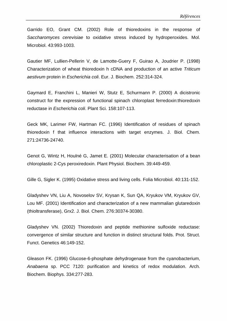
Références
Garrido EO, Grant CM. (2002) Role of thioredoxins in the response of
Saccharomyces cerevisiae to oxidative stress induced by hydroperoxides. Mol.
Microbiol. 43:993-1003.
Gautier MF, Lullien-Pellerin V, de Lamotte-Guery F, Guirao A, Joudrier P. (1998)
Characterization of wheat thioredoxin h cDNA and production of an active Triticum
aestivum protein in Escherichia coli. Eur. J. Biochem. 252:314-324.
Gaymard E, Franchini L, Manieri W, Stutz E, Schurmann P. (2000) A dicistronic
construct for the expression of functional spinach chloroplast ferredoxin:thioredoxin
reductase in Escherichia coli. Plant Sci. 158:107-113.
Geck MK, Larimer FW, Hartman FC. (1996) Identification of residues of spinach
thioredoxin f that influence interactions with target enzymes. J. Biol. Chem.
271:24736-24740.
Genot G, Wintz H, Houlné G, Jamet E. (2001) Molecular characterisation of a bean
chloroplastic 2-Cys peroxiredoxin. Plant Physiol. Biochem. 39:449-459.
Gille G, Sigler K. (1995) Oxidative stress and living cells. Folia Microbiol. 40:131-152.
Gladyshev VN, Liu A, Novoselov SV, Krysan K, Sun QA, Kryukov VM, Kryukov GV,
Lou MF. (2001) Identification and characterization of a new mammalian glutaredoxin
(thioltransferase), Grx2. J. Biol. Chem. 276:30374-30380.
Gladyshev VN. (2002) Thioredoxin and peptide methionine sulfoxide reductase:
convergence of similar structure and function in distinct structural folds. Prot. Struct.
Funct. Genetics 46:149-152.
Gleason FK. (1996) Glucose-6-phosphate dehydrogenase from the cyanobacterium,
Anabaena sp. PCC 7120: purification and kinetics of redox modulation. Arch.
Biochem. Biophys. 334:277-283.
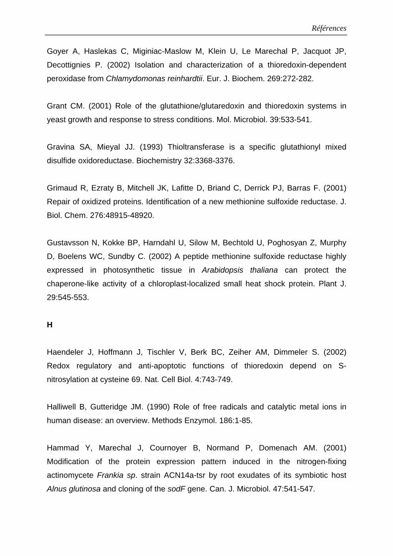
Références
Goyer A, Haslekas C, Miginiac-Maslow M, Klein U, Le Marechal P, Jacquot JP,
Decottignies P. (2002) Isolation and characterization of a thioredoxin-dependent
peroxidase from Chlamydomonas reinhardtii. Eur. J. Biochem. 269:272-282.
Grant CM. (2001) Role of the glutathione/glutaredoxin and thioredoxin systems in
yeast growth and response to stress conditions. Mol. Microbiol. 39:533-541.
Gravina SA, Mieyal JJ. (1993) Thioltransferase is a specific glutathionyl mixed
disulfide oxidoreductase. Biochemistry 32:3368-3376.
Grimaud R, Ezraty B, Mitchell JK, Lafitte D, Briand C, Derrick PJ, Barras F. (2001)
Repair of oxidized proteins. Identification of a new methionine sulfoxide reductase. J.
Biol. Chem. 276:48915-48920.
Gustavsson N, Kokke BP, Harndahl U, Silow M, Bechtold U, Poghosyan Z, Murphy
D, Boelens WC, Sundby C. (2002) A peptide methionine sulfoxide reductase highly
expressed in photosynthetic tissue in Arabidopsis thaliana can protect the
chaperone-like activity of a chloroplast-localized small heat shock protein. Plant J.
29:545-553.
H
Haendeler J, Hoffmann J, Tischler V, Berk BC, Zeiher AM, Dimmeler S. (2002)
Redox regulatory and anti-apoptotic functions of thioredoxin depend on S-
nitrosylation at cysteine 69. Nat. Cell Biol. 4:743-749.
Halliwell B, Gutteridge JM. (1990) Role of free radicals and catalytic metal ions in
human disease: an overview. Methods Enzymol. 186:1-85.
Hammad Y, Marechal J, Cournoyer B, Normand P, Domenach AM. (2001)
Modification of the protein expression pattern induced in the nitrogen-fixing
actinomycete Frankia sp. strain ACN14a-tsr by root exudates of its symbiotic host
Alnus glutinosa and cloning of the sodF gene. Can. J. Microbiol. 47:541-547.
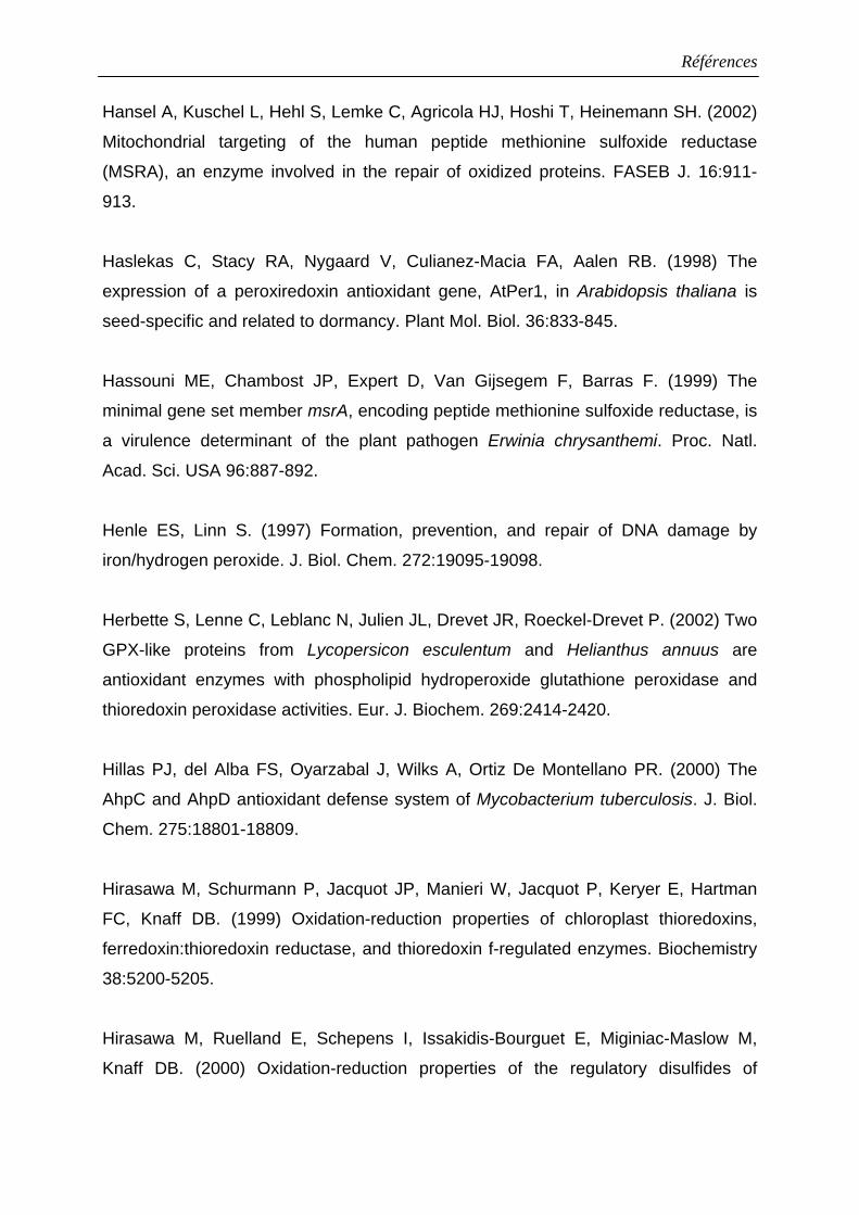
Références
Hansel A, Kuschel L, Hehl S, Lemke C, Agricola HJ, Hoshi T, Heinemann SH. (2002)
Mitochondrial targeting of the human peptide methionine sulfoxide reductase
(MSRA), an enzyme involved in the repair of oxidized proteins. FASEB J. 16:911-
913.
Haslekas C, Stacy RA, Nygaard V, Culianez-Macia FA, Aalen RB. (1998) The
expression of a peroxiredoxin antioxidant gene, AtPer1, in Arabidopsis thaliana is
seed-specific and related to dormancy. Plant Mol. Biol. 36:833-845.
Hassouni ME, Chambost JP, Expert D, Van Gijsegem F, Barras F. (1999) The
minimal gene set member msrA, encoding peptide methionine sulfoxide reductase, is
a virulence determinant of the plant pathogen Erwinia chrysanthemi. Proc. Natl.
Acad. Sci. USA 96:887-892.
Henle ES, Linn S. (1997) Formation, prevention, and repair of DNA damage by
iron/hydrogen peroxide. J. Biol. Chem. 272:19095-19098.
Herbette S, Lenne C, Leblanc N, Julien JL, Drevet JR, Roeckel-Drevet P. (2002) Two
GPX-like proteins from Lycopersicon esculentum and Helianthus annuus are
antioxidant enzymes with phospholipid hydroperoxide glutathione peroxidase and
thioredoxin peroxidase activities. Eur. J. Biochem. 269:2414-2420.
Hillas PJ, del Alba FS, Oyarzabal J, Wilks A, Ortiz De Montellano PR. (2000) The
AhpC and AhpD antioxidant defense system of Mycobacterium tuberculosis. J. Biol.
Chem. 275:18801-18809.
Hirasawa M, Schurmann P, Jacquot JP, Manieri W, Jacquot P, Keryer E, Hartman
FC, Knaff DB. (1999) Oxidation-reduction properties of chloroplast thioredoxins,
ferredoxin:thioredoxin reductase, and thioredoxin f-regulated enzymes. Biochemistry
38:5200-5205.
Hirasawa M, Ruelland E, Schepens I, Issakidis-Bourguet E, Miginiac-Maslow M,
Knaff DB. (2000) Oxidation-reduction properties of the regulatory disulfides of
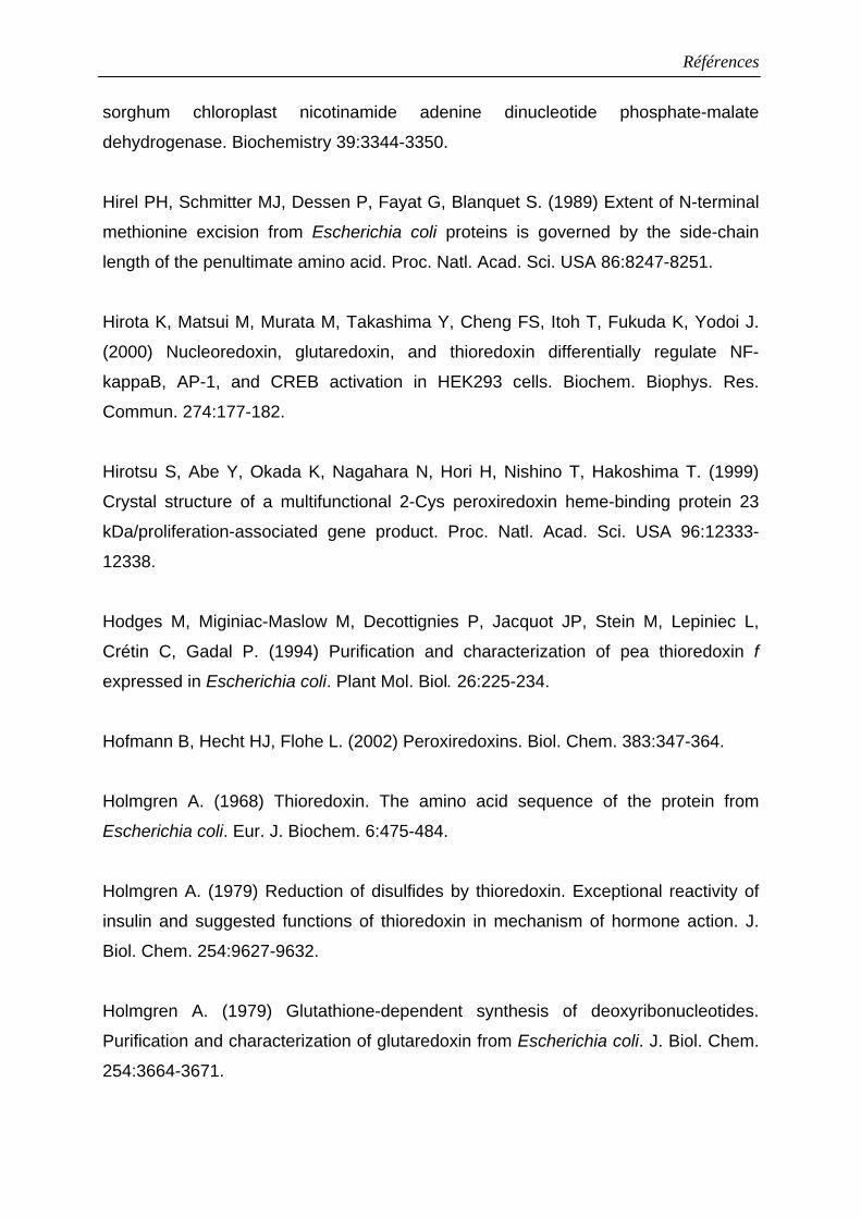
Références
sorghum chloroplast nicotinamide adenine dinucleotide phosphate-malate
dehydrogenase. Biochemistry 39:3344-3350.
Hirel PH, Schmitter MJ, Dessen P, Fayat G, Blanquet S. (1989) Extent of N-terminal
methionine excision from Escherichia coli proteins is governed by the side-chain
length of the penultimate amino acid. Proc. Natl. Acad. Sci. USA 86:8247-8251.
Hirota K, Matsui M, Murata M, Takashima Y, Cheng FS, Itoh T, Fukuda K, Yodoi J.
(2000) Nucleoredoxin, glutaredoxin, and thioredoxin differentially regulate NF-
kappaB, AP-1, and CREB activation in HEK293 cells. Biochem. Biophys. Res.
Commun. 274:177-182.
Hirotsu S, Abe Y, Okada K, Nagahara N, Hori H, Nishino T, Hakoshima T. (1999)
Crystal structure of a multifunctional 2-Cys peroxiredoxin heme-binding protein 23
kDa/proliferation-associated gene product. Proc. Natl. Acad. Sci. USA 96:12333-
12338.
Hodges M, Miginiac-Maslow M, Decottignies P, Jacquot JP, Stein M, Lepiniec L,
Crétin C, Gadal P. (1994) Purification and characterization of pea thioredoxin f
expressed in Escherichia coli. Plant Mol. Biol. 26:225-234.
Hofmann B, Hecht HJ, Flohe L. (2002) Peroxiredoxins. Biol. Chem. 383:347-364.
Holmgren A. (1968) Thioredoxin. The amino acid sequence of the protein from
Escherichia coli. Eur. J. Biochem. 6:475-484.
Holmgren A. (1979) Reduction of disulfides by thioredoxin. Exceptional reactivity of
insulin and suggested functions of thioredoxin in mechanism of hormone action. J.
Biol. Chem. 254:9627-9632.
Holmgren A. (1979) Glutathione-dependent synthesis of deoxyribonucleotides.
Purification and characterization of glutaredoxin from Escherichia coli. J. Biol. Chem.
254:3664-3671.
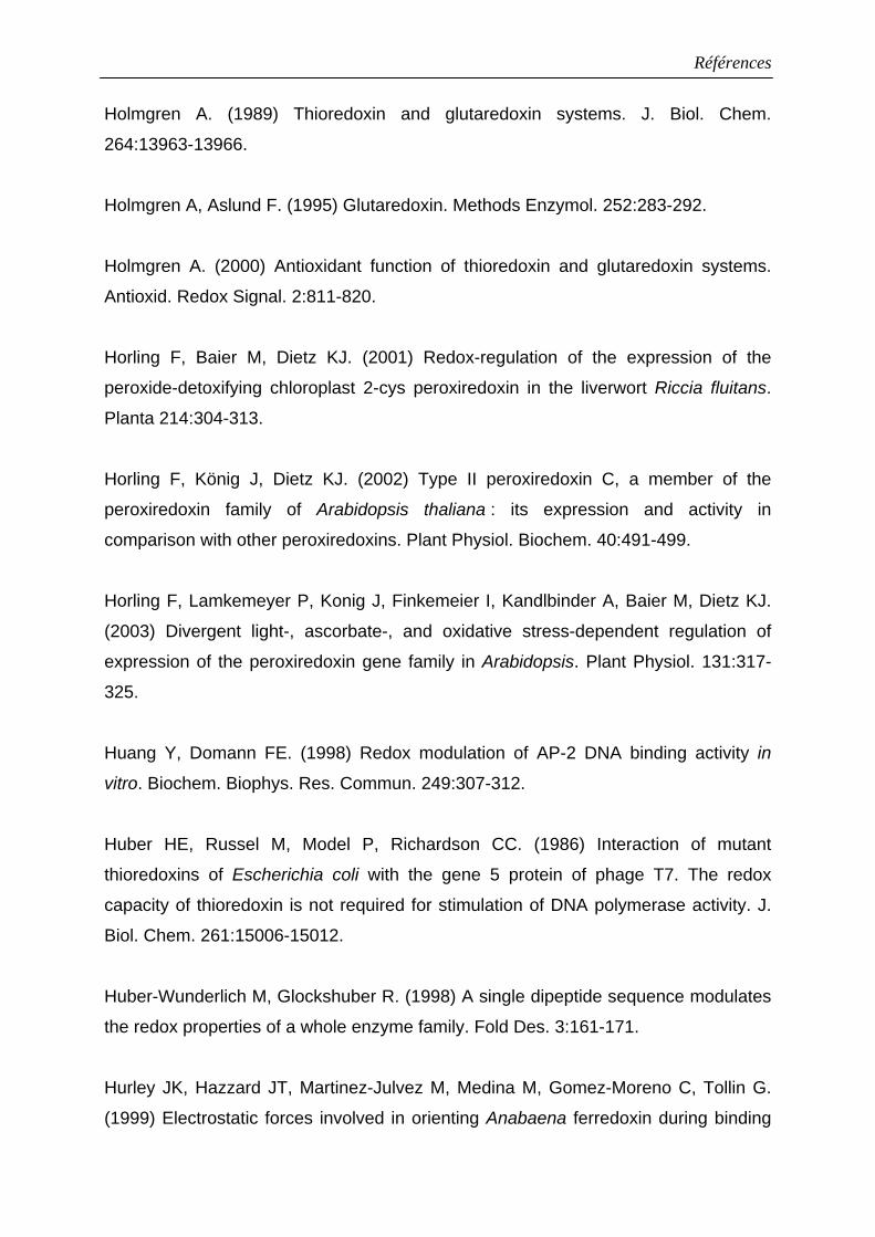
Références
Holmgren A. (1989) Thioredoxin and glutaredoxin systems. J. Biol. Chem.
264:13963-13966.
Holmgren A, Aslund F. (1995) Glutaredoxin. Methods Enzymol. 252:283-292.
Holmgren A. (2000) Antioxidant function of thioredoxin and glutaredoxin systems.
Antioxid. Redox Signal. 2:811-820.
Horling F, Baier M, Dietz KJ. (2001) Redox-regulation of the expression of the
peroxide-detoxifying chloroplast 2-cys peroxiredoxin in the liverwort Riccia fluitans.
Planta 214:304-313.
Horling F, König J, Dietz KJ. (2002) Type II peroxiredoxin C, a member of the
peroxiredoxin family of Arabidopsis thaliana : its expression and activity in
comparison with other peroxiredoxins. Plant Physiol. Biochem. 40:491-499.
Horling F, Lamkemeyer P, Konig J, Finkemeier I, Kandlbinder A, Baier M, Dietz KJ.
(2003) Divergent light-, ascorbate-, and oxidative stress-dependent regulation of
expression of the peroxiredoxin gene family in Arabidopsis. Plant Physiol. 131:317-
325.
Huang Y, Domann FE. (1998) Redox modulation of AP-2 DNA binding activity in
vitro. Biochem. Biophys. Res. Commun. 249:307-312.
Huber HE, Russel M, Model P, Richardson CC. (1986) Interaction of mutant
thioredoxins of Escherichia coli with the gene 5 protein of phage T7. The redox
capacity of thioredoxin is not required for stimulation of DNA polymerase activity. J.
Biol. Chem. 261:15006-15012.
Huber-Wunderlich M, Glockshuber R. (1998) A single dipeptide sequence modulates
the redox properties of a whole enzyme family. Fold Des. 3:161-171.
Hurley JK, Hazzard JT, Martinez-Julvez M, Medina M, Gomez-Moreno C, Tollin G.
(1999) Electrostatic forces involved in orienting Anabaena ferredoxin during binding
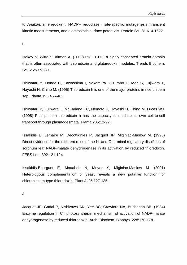
Références
to Anabaena ferredoxin : NADP+ reductase : site-specific mutagenesis, transient
kinetic measurements, and electrostatic surface potentials. Protein Sci. 8:1614-1622.
I
Isakov N, Witte S, Altman A. (2000) PICOT-HD: a highly conserved protein domain
that is often associated with thioredoxin and glutaredoxin modules. Trends Biochem.
Sci. 25:537-539.
Ishiwatari Y, Honda C, Kawashima I, Nakamura S, Hirano H, Mori S, Fujiwara T,
Hayashi H, Chino M. (1995) Thioredoxin h is one of the major proteins in rice phloem
sap. Planta 195:456-463.
Ishiwatari Y, Fujiwara T, McFarland KC, Nemoto K, Hayashi H, Chino M, Lucas WJ.
(1998) Rice phloem thioredoxin h has the capacity to mediate its own cell-to-cell
transport through plasmodesmata. Planta 205:12-22.
Issakidis E, Lemaire M, Decottignies P, Jacquot JP, Miginiac-Maslow M. (1996)
Direct evidence for the different roles of the N- and C-terminal regulatory disulfides of
sorghum leaf NADP-malate dehydrogenase in its activation by reduced thioredoxin.
FEBS Lett. 392:121-124.
Issakidis-Bourguet E, Mouaheb N, Meyer Y, Miginiac-Maslow M. (2001)
Heterologous complementation of yeast reveals a new putative function for
chloroplast m-type thioredoxin. Plant J. 25:127-135.
J
Jacquot JP, Gadal P, Nishizawa AN, Yee BC, Crawford NA, Buchanan BB. (1984)
Enzyme regulation in C4 photosynthesis: mechanism of activation of NADP-malate
dehydrogenase by reduced thioredoxin. Arch. Biochem. Biophys. 228:170-178.
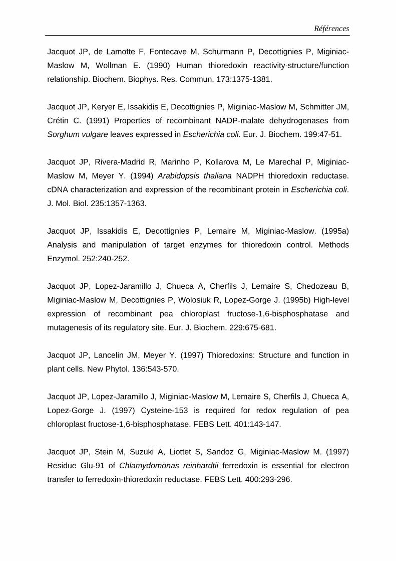
Références
Jacquot JP, de Lamotte F, Fontecave M, Schurmann P, Decottignies P, Miginiac-
Maslow M, Wollman E. (1990) Human thioredoxin reactivity-structure/function
relationship. Biochem. Biophys. Res. Commun. 173:1375-1381.
Jacquot JP, Keryer E, Issakidis E, Decottignies P, Miginiac-Maslow M, Schmitter JM,
Crétin C. (1991) Properties of recombinant NADP-malate dehydrogenases from
Sorghum vulgare leaves expressed in Escherichia coli. Eur. J. Biochem. 199:47-51.
Jacquot JP, Rivera-Madrid R, Marinho P, Kollarova M, Le Marechal P, Miginiac-
Maslow M, Meyer Y. (1994) Arabidopsis thaliana NADPH thioredoxin reductase.
cDNA characterization and expression of the recombinant protein in Escherichia coli.
J. Mol. Biol. 235:1357-1363.
Jacquot JP, Issakidis E, Decottignies P, Lemaire M, Miginiac-Maslow. (1995a)
Analysis and manipulation of target enzymes for thioredoxin control. Methods
Enzymol. 252:240-252.
Jacquot JP, Lopez-Jaramillo J, Chueca A, Cherfils J, Lemaire S, Chedozeau B,
Miginiac-Maslow M, Decottignies P, Wolosiuk R, Lopez-Gorge J. (1995b) High-level
expression of recombinant pea chloroplast fructose-1,6-bisphosphatase and
mutagenesis of its regulatory site. Eur. J. Biochem. 229:675-681.
Jacquot JP, Lancelin JM, Meyer Y. (1997) Thioredoxins: Structure and function in
plant cells. New Phytol. 136:543-570.
Jacquot JP, Lopez-Jaramillo J, Miginiac-Maslow M, Lemaire S, Cherfils J, Chueca A,
Lopez-Gorge J. (1997) Cysteine-153 is required for redox regulation of pea
chloroplast fructose-1,6-bisphosphatase. FEBS Lett. 401:143-147.
Jacquot JP, Stein M, Suzuki A, Liottet S, Sandoz G, Miginiac-Maslow M. (1997)
Residue Glu-91 of Chlamydomonas reinhardtii ferredoxin is essential for electron
transfer to ferredoxin-thioredoxin reductase. FEBS Lett. 400:293-296.
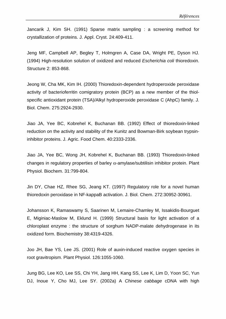
Références
Jancarik J, Kim SH. (1991) Sparse matrix sampling : a screening method for
crystallization of proteins. J. Appl. Cryst. 24:409-411.
Jeng MF, Campbell AP, Begley T, Holmgren A, Case DA, Wright PE, Dyson HJ.
(1994) High-resolution solution of oxidized and reduced Escherichia coli thioredoxin.
Structure 2: 853-868.
Jeong W, Cha MK, Kim IH. (2000) Thioredoxin-dependent hydroperoxide peroxidase
activity of bacterioferritin comigratory protein (BCP) as a new member of the thiol-
specific antioxidant protein (TSA)/Alkyl hydroperoxide peroxidase C (AhpC) family. J.
Biol. Chem. 275:2924-2930.
Jiao JA, Yee BC, Kobrehel K, Buchanan BB. (1992) Effect of thioredoxin-linked
reduction on the activity and stability of the Kunitz and Bowman-Birk soybean trypsin-
inhibitor proteins. J. Agric. Food Chem. 40:2333-2336.
Jiao JA, Yee BC, Wong JH, Kobrehel K, Buchanan BB. (1993) Thioredoxin-linked
changes in regulatory properties of barley α-amylase/subtilisin inhibitor protein. Plant
Physiol. Biochem. 31:799-804.
Jin DY, Chae HZ, Rhee SG, Jeang KT. (1997) Regulatory role for a novel human
thioredoxin peroxidase in NF-kappaB activation. J. Biol. Chem. 272:30952-30961.
Johansson K, Ramaswamy S, Saarinen M, Lemaire-Chamley M, Issakidis-Bourguet
E, Miginiac-Maslow M, Eklund H. (1999) Structural basis for light activation of a
chloroplast enzyme : the structure of sorghum NADP-malate dehydrogenase in its
oxidized form. Biochemistry 38:4319-4326.
Joo JH, Bae YS, Lee JS. (2001) Role of auxin-induced reactive oxygen species in
root gravitropism. Plant Physiol. 126:1055-1060.
Jung BG, Lee KO, Lee SS, Chi YH, Jang HH, Kang SS, Lee K, Lim D, Yoon SC, Yun
DJ, Inoue Y, Cho MJ, Lee SY. (2002a) A Chinese cabbage cDNA with high
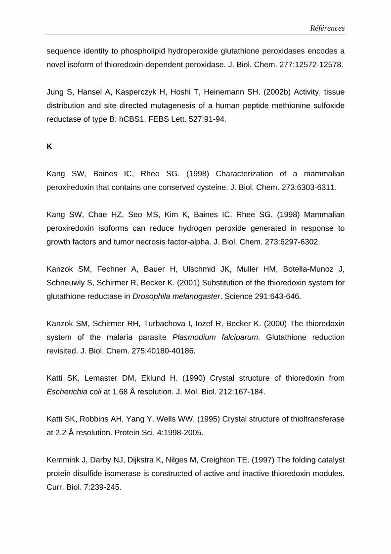
Références
sequence identity to phospholipid hydroperoxide glutathione peroxidases encodes a
novel isoform of thioredoxin-dependent peroxidase. J. Biol. Chem. 277:12572-12578.
Jung S, Hansel A, Kasperczyk H, Hoshi T, Heinemann SH. (2002b) Activity, tissue
distribution and site directed mutagenesis of a human peptide methionine sulfoxide
reductase of type B: hCBS1. FEBS Lett. 527:91-94.
K
Kang SW, Baines IC, Rhee SG. (1998) Characterization of a mammalian
peroxiredoxin that contains one conserved cysteine. J. Biol. Chem. 273:6303-6311.
Kang SW, Chae HZ, Seo MS, Kim K, Baines IC, Rhee SG. (1998) Mammalian
peroxiredoxin isoforms can reduce hydrogen peroxide generated in response to
growth factors and tumor necrosis factor-alpha. J. Biol. Chem. 273:6297-6302.
Kanzok SM, Fechner A, Bauer H, Ulschmid JK, Muller HM, Botella-Munoz J,
Schneuwly S, Schirmer R, Becker K. (2001) Substitution of the thioredoxin system for
glutathione reductase in Drosophila melanogaster. Science 291:643-646.
Kanzok SM, Schirmer RH, Turbachova I, Iozef R, Becker K. (2000) The thioredoxin
system of the malaria parasite Plasmodium falciparum. Glutathione reduction
revisited. J. Biol. Chem. 275:40180-40186.
Katti SK, Lemaster DM, Eklund H. (1990) Crystal structure of thioredoxin from
Escherichia coli at 1.68 Å resolution. J. Mol. Biol. 212:167-184.
Katti SK, Robbins AH, Yang Y, Wells WW. (1995) Crystal structure of thioltransferase
at 2.2 Å resolution. Protein Sci. 4:1998-2005.
Kemmink J, Darby NJ, Dijkstra K, Nilges M, Creighton TE. (1997) The folding catalyst
protein disulfide isomerase is constructed of active and inactive thioredoxin modules.
Curr. Biol. 7:239-245.
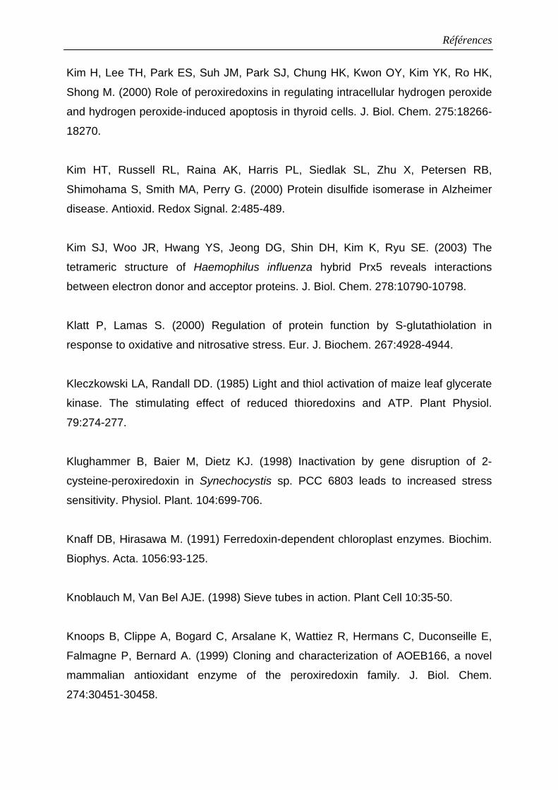
Références
Kim H, Lee TH, Park ES, Suh JM, Park SJ, Chung HK, Kwon OY, Kim YK, Ro HK,
Shong M. (2000) Role of peroxiredoxins in regulating intracellular hydrogen peroxide
and hydrogen peroxide-induced apoptosis in thyroid cells. J. Biol. Chem. 275:18266-
18270.
Kim HT, Russell RL, Raina AK, Harris PL, Siedlak SL, Zhu X, Petersen RB,
Shimohama S, Smith MA, Perry G. (2000) Protein disulfide isomerase in Alzheimer
disease. Antioxid. Redox Signal. 2:485-489.
Kim SJ, Woo JR, Hwang YS, Jeong DG, Shin DH, Kim K, Ryu SE. (2003) The
tetrameric structure of Haemophilus influenza hybrid Prx5 reveals interactions
between electron donor and acceptor proteins. J. Biol. Chem. 278:10790-10798.
Klatt P, Lamas S. (2000) Regulation of protein function by S-glutathiolation in
response to oxidative and nitrosative stress. Eur. J. Biochem. 267:4928-4944.
Kleczkowski LA, Randall DD. (1985) Light and thiol activation of maize leaf glycerate
kinase. The stimulating effect of reduced thioredoxins and ATP. Plant Physiol.
79:274-277.
Klughammer B, Baier M, Dietz KJ. (1998) Inactivation by gene disruption of 2-
cysteine-peroxiredoxin in Synechocystis sp. PCC 6803 leads to increased stress
sensitivity. Physiol. Plant. 104:699-706.
Knaff DB, Hirasawa M. (1991) Ferredoxin-dependent chloroplast enzymes. Biochim.
Biophys. Acta. 1056:93-125.
Knoblauch M, Van Bel AJE. (1998) Sieve tubes in action. Plant Cell 10:35-50.
Knoops B, Clippe A, Bogard C, Arsalane K, Wattiez R, Hermans C, Duconseille E,
Falmagne P, Bernard A. (1999) Cloning and characterization of AOEB166, a novel
mammalian antioxidant enzyme of the peroxiredoxin family. J. Biol. Chem.
274:30451-30458.
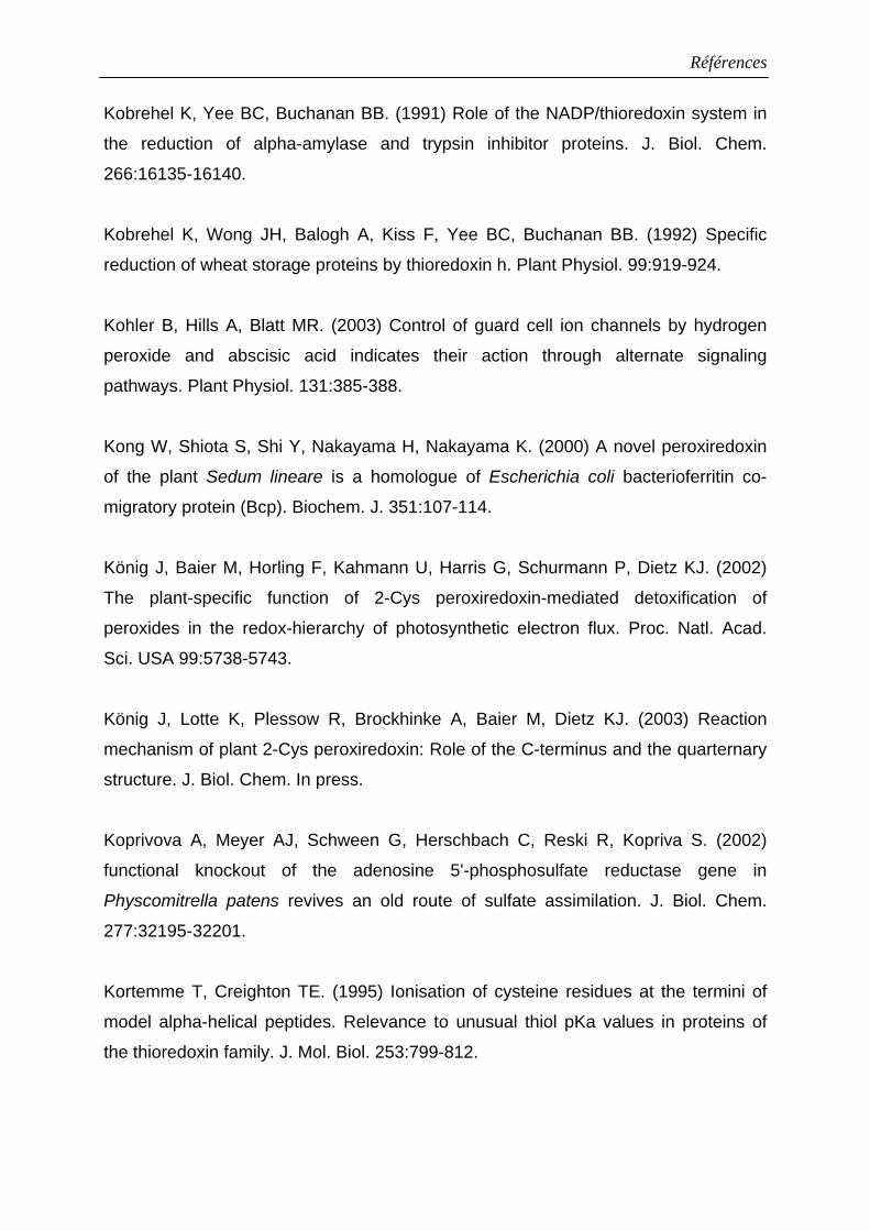
Références
Kobrehel K, Yee BC, Buchanan BB. (1991) Role of the NADP/thioredoxin system in
the reduction of alpha-amylase and trypsin inhibitor proteins. J. Biol. Chem.
266:16135-16140.
Kobrehel K, Wong JH, Balogh A, Kiss F, Yee BC, Buchanan BB. (1992) Specific
reduction of wheat storage proteins by thioredoxin h. Plant Physiol. 99:919-924.
Kohler B, Hills A, Blatt MR. (2003) Control of guard cell ion channels by hydrogen
peroxide and abscisic acid indicates their action through alternate signaling
pathways. Plant Physiol. 131:385-388.
Kong W, Shiota S, Shi Y, Nakayama H, Nakayama K. (2000) A novel peroxiredoxin
of the plant Sedum lineare is a homologue of Escherichia coli bacterioferritin co-
migratory protein (Bcp). Biochem. J. 351:107-114.
König J, Baier M, Horling F, Kahmann U, Harris G, Schurmann P, Dietz KJ. (2002)
The plant-specific function of 2-Cys peroxiredoxin-mediated detoxification of
peroxides in the redox-hierarchy of photosynthetic electron flux. Proc. Natl. Acad.
Sci. USA 99:5738-5743.
König J, Lotte K, Plessow R, Brockhinke A, Baier M, Dietz KJ. (2003) Reaction
mechanism of plant 2-Cys peroxiredoxin: Role of the C-terminus and the quarternary
structure. J. Biol. Chem. In press.
Koprivova A, Meyer AJ, Schween G, Herschbach C, Reski R, Kopriva S. (2002)
functional knockout of the adenosine 5'-phosphosulfate reductase gene in
Physcomitrella patens revives an old route of sulfate assimilation. J. Biol. Chem.
277:32195-32201.
Kortemme T, Creighton TE. (1995) Ionisation of cysteine residues at the termini of
model alpha-helical peptides. Relevance to unusual thiol pKa values in proteins of
the thioredoxin family. J. Mol. Biol. 253:799-812.
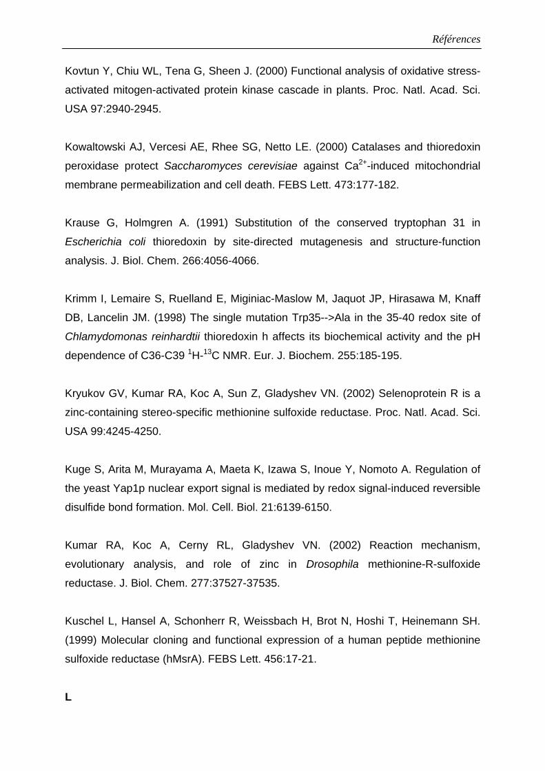
Références
Kovtun Y, Chiu WL, Tena G, Sheen J. (2000) Functional analysis of oxidative stress-
activated mitogen-activated protein kinase cascade in plants. Proc. Natl. Acad. Sci.
USA 97:2940-2945.
Kowaltowski AJ, Vercesi AE, Rhee SG, Netto LE. (2000) Catalases and thioredoxin
peroxidase protect Saccharomyces cerevisiae against Ca2+-induced mitochondrial
membrane permeabilization and cell death. FEBS Lett. 473:177-182.
Krause G, Holmgren A. (1991) Substitution of the conserved tryptophan 31 in
Escherichia coli thioredoxin by site-directed mutagenesis and structure-function
analysis. J. Biol. Chem. 266:4056-4066.
Krimm I, Lemaire S, Ruelland E, Miginiac-Maslow M, Jaquot JP, Hirasawa M, Knaff
DB, Lancelin JM. (1998) The single mutation Trp35-->Ala in the 35-40 redox site of
Chlamydomonas reinhardtii thioredoxin h affects its biochemical activity and the pH
dependence of C36-C39 1H-13C NMR. Eur. J. Biochem. 255:185-195.
Kryukov GV, Kumar RA, Koc A, Sun Z, Gladyshev VN. (2002) Selenoprotein R is a
zinc-containing stereo-specific methionine sulfoxide reductase. Proc. Natl. Acad. Sci.
USA 99:4245-4250.
Kuge S, Arita M, Murayama A, Maeta K, Izawa S, Inoue Y, Nomoto A. Regulation of
the yeast Yap1p nuclear export signal is mediated by redox signal-induced reversible
disulfide bond formation. Mol. Cell. Biol. 21:6139-6150.
Kumar RA, Koc A, Cerny RL, Gladyshev VN. (2002) Reaction mechanism,
evolutionary analysis, and role of zinc in Drosophila methionine-R-sulfoxide
reductase. J. Biol. Chem. 277:37527-37535.
Kuschel L, Hansel A, Schonherr R, Weissbach H, Brot N, Hoshi T, Heinemann SH.
(1999) Molecular cloning and functional expression of a human peptide methionine
sulfoxide reductase (hMsrA). FEBS Lett. 456:17-21.
L
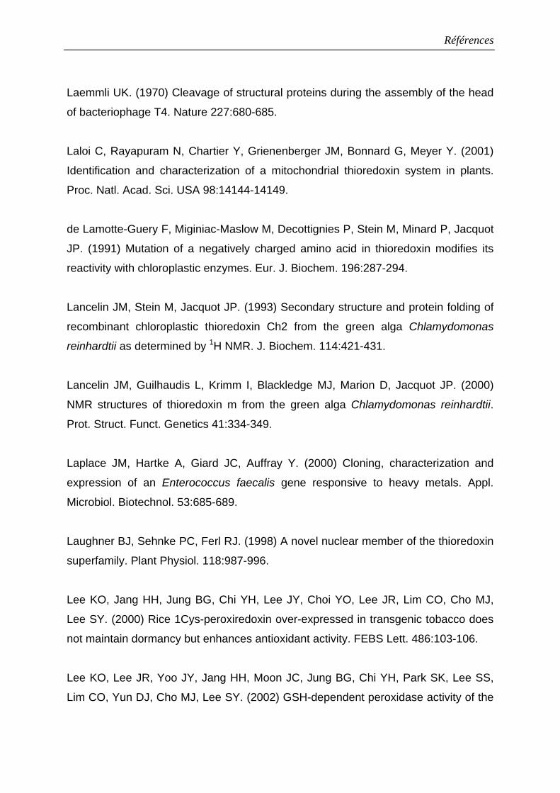
Références
Laemmli UK. (1970) Cleavage of structural proteins during the assembly of the head
of bacteriophage T4. Nature 227:680-685.
Laloi C, Rayapuram N, Chartier Y, Grienenberger JM, Bonnard G, Meyer Y. (2001)
Identification and characterization of a mitochondrial thioredoxin system in plants.
Proc. Natl. Acad. Sci. USA 98:14144-14149.
de Lamotte-Guery F, Miginiac-Maslow M, Decottignies P, Stein M, Minard P, Jacquot
JP. (1991) Mutation of a negatively charged amino acid in thioredoxin modifies its
reactivity with chloroplastic enzymes. Eur. J. Biochem. 196:287-294.
Lancelin JM, Stein M, Jacquot JP. (1993) Secondary structure and protein folding of
recombinant chloroplastic thioredoxin Ch2 from the green alga Chlamydomonas
reinhardtii as determined by 1H NMR. J. Biochem. 114:421-431.
Lancelin JM, Guilhaudis L, Krimm I, Blackledge MJ, Marion D, Jacquot JP. (2000)
NMR structures of thioredoxin m from the green alga Chlamydomonas reinhardtii.
Prot. Struct. Funct. Genetics 41:334-349.
Laplace JM, Hartke A, Giard JC, Auffray Y. (2000) Cloning, characterization and
expression of an Enterococcus faecalis gene responsive to heavy metals. Appl.
Microbiol. Biotechnol. 53:685-689.
Laughner BJ, Sehnke PC, Ferl RJ. (1998) A novel nuclear member of the thioredoxin
superfamily. Plant Physiol. 118:987-996.
Lee KO, Jang HH, Jung BG, Chi YH, Lee JY, Choi YO, Lee JR, Lim CO, Cho MJ,
Lee SY. (2000) Rice 1Cys-peroxiredoxin over-expressed in transgenic tobacco does
not maintain dormancy but enhances antioxidant activity. FEBS Lett. 486:103-106.
Lee KO, Lee JR, Yoo JY, Jang HH, Moon JC, Jung BG, Chi YH, Park SK, Lee SS,
Lim CO, Yun DJ, Cho MJ, Lee SY. (2002) GSH-dependent peroxidase activity of the
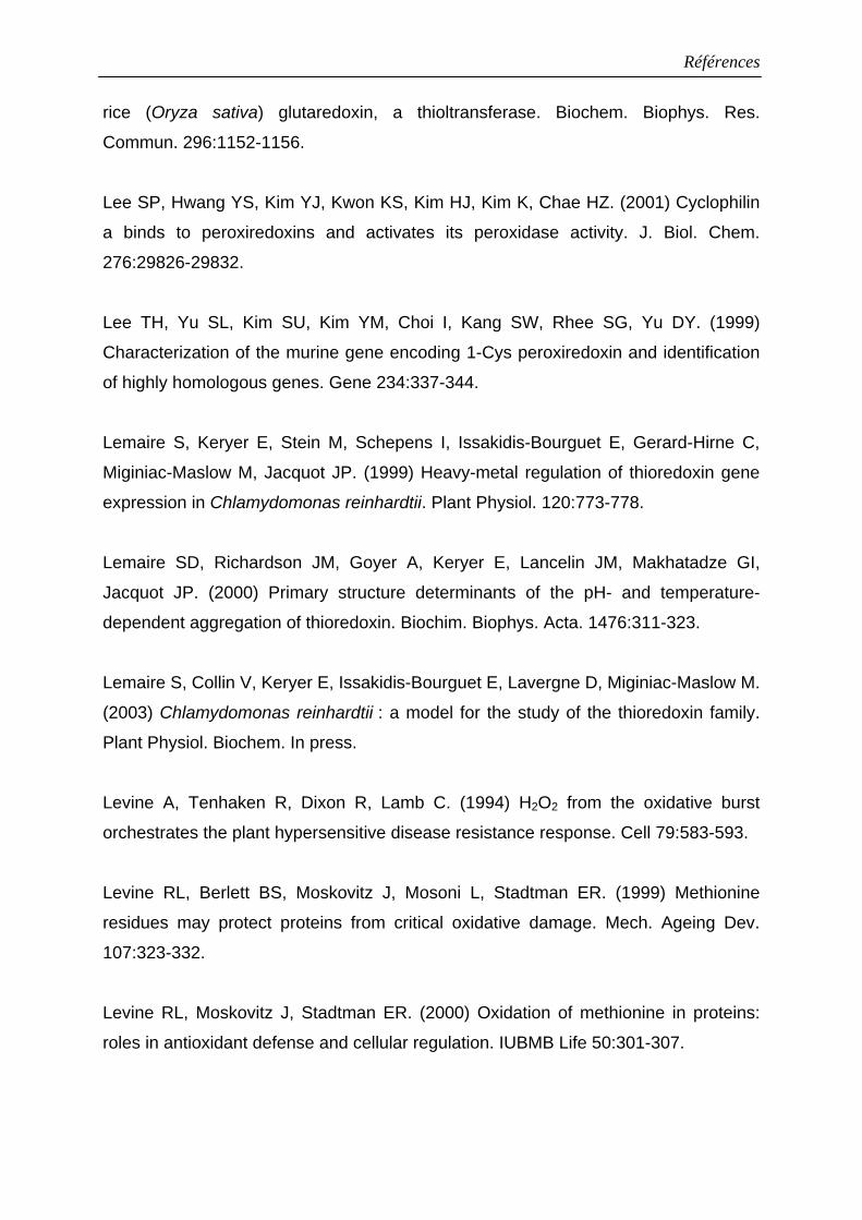
Références
rice (Oryza sativa) glutaredoxin, a thioltransferase. Biochem. Biophys. Res.
Commun. 296:1152-1156.
Lee SP, Hwang YS, Kim YJ, Kwon KS, Kim HJ, Kim K, Chae HZ. (2001) Cyclophilin
a binds to peroxiredoxins and activates its peroxidase activity. J. Biol. Chem.
276:29826-29832.
Lee TH, Yu SL, Kim SU, Kim YM, Choi I, Kang SW, Rhee SG, Yu DY. (1999)
Characterization of the murine gene encoding 1-Cys peroxiredoxin and identification
of highly homologous genes. Gene 234:337-344.
Lemaire S, Keryer E, Stein M, Schepens I, Issakidis-Bourguet E, Gerard-Hirne C,
Miginiac-Maslow M, Jacquot JP. (1999) Heavy-metal regulation of thioredoxin gene
expression in Chlamydomonas reinhardtii. Plant Physiol. 120:773-778.
Lemaire SD, Richardson JM, Goyer A, Keryer E, Lancelin JM, Makhatadze GI,
Jacquot JP. (2000) Primary structure determinants of the pH- and temperature-
dependent aggregation of thioredoxin. Biochim. Biophys. Acta. 1476:311-323.
Lemaire S, Collin V, Keryer E, Issakidis-Bourguet E, Lavergne D, Miginiac-Maslow M.
(2003) Chlamydomonas reinhardtii : a model for the study of the thioredoxin family.
Plant Physiol. Biochem. In press.
Levine A, Tenhaken R, Dixon R, Lamb C. (1994) H2O2 from the oxidative burst
orchestrates the plant hypersensitive disease resistance response. Cell 79:583-593.
Levine RL, Berlett BS, Moskovitz J, Mosoni L, Stadtman ER. (1999) Methionine
residues may protect proteins from critical oxidative damage. Mech. Ageing Dev.
107:323-332.
Levine RL, Moskovitz J, Stadtman ER. (2000) Oxidation of methionine in proteins:
roles in antioxidant defense and cellular regulation. IUBMB Life 50:301-307.
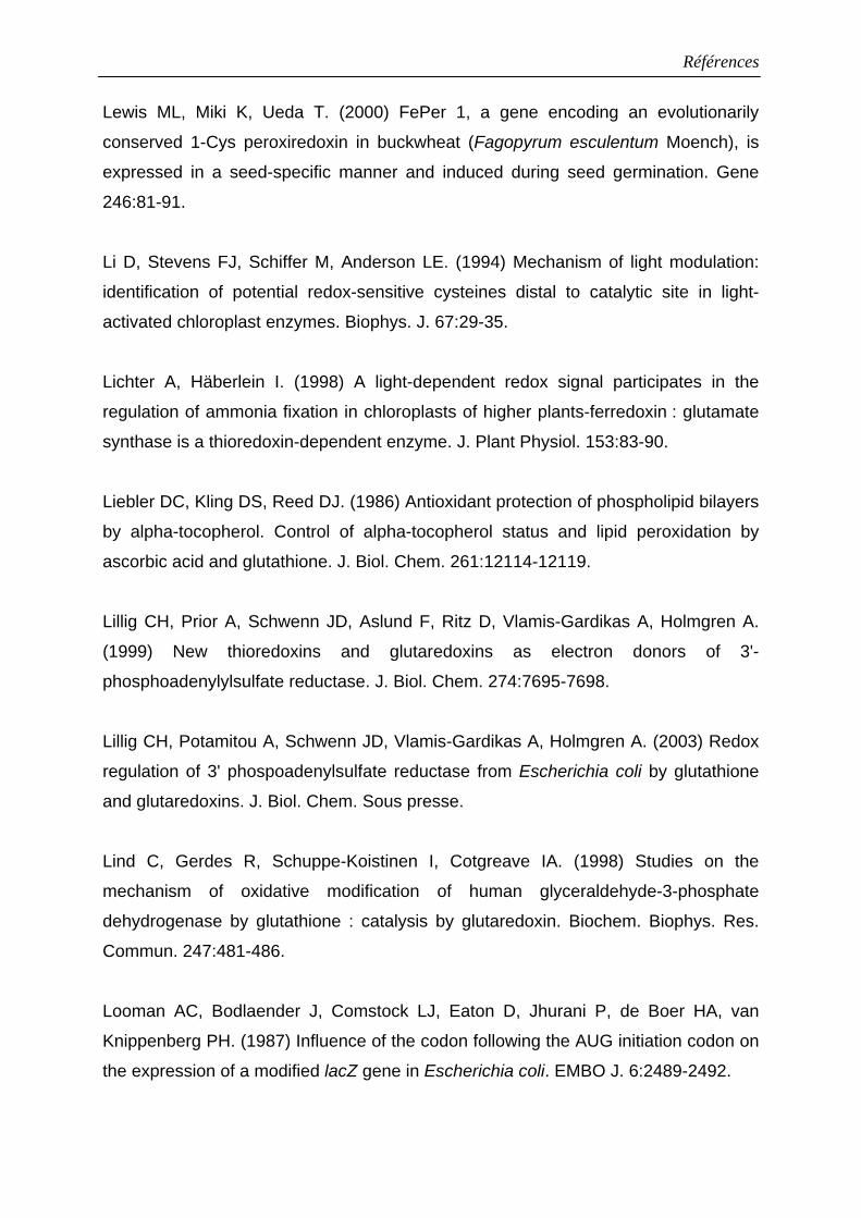
Références
Lewis ML, Miki K, Ueda T. (2000) FePer 1, a gene encoding an evolutionarily
conserved 1-Cys peroxiredoxin in buckwheat (Fagopyrum esculentum Moench), is
expressed in a seed-specific manner and induced during seed germination. Gene
246:81-91.
Li D, Stevens FJ, Schiffer M, Anderson LE. (1994) Mechanism of light modulation:
identification of potential redox-sensitive cysteines distal to catalytic site in light-
activated chloroplast enzymes. Biophys. J. 67:29-35.
Lichter A, Häberlein I. (1998) A light-dependent redox signal participates in the
regulation of ammonia fixation in chloroplasts of higher plants-ferredoxin : glutamate
synthase is a thioredoxin-dependent enzyme. J. Plant Physiol. 153:83-90.
Liebler DC, Kling DS, Reed DJ. (1986) Antioxidant protection of phospholipid bilayers
by alpha-tocopherol. Control of alpha-tocopherol status and lipid peroxidation by
ascorbic acid and glutathione. J. Biol. Chem. 261:12114-12119.
Lillig CH, Prior A, Schwenn JD, Aslund F, Ritz D, Vlamis-Gardikas A, Holmgren A.
(1999) New thioredoxins and glutaredoxins as electron donors of 3'-
phosphoadenylylsulfate reductase. J. Biol. Chem. 274:7695-7698.
Lillig CH, Potamitou A, Schwenn JD, Vlamis-Gardikas A, Holmgren A. (2003) Redox
regulation of 3' phospoadenylsulfate reductase from Escherichia coli by glutathione
and glutaredoxins. J. Biol. Chem. Sous presse.
Lind C, Gerdes R, Schuppe-Koistinen I, Cotgreave IA. (1998) Studies on the
mechanism of oxidative modification of human glyceraldehyde-3-phosphate
dehydrogenase by glutathione : catalysis by glutaredoxin. Biochem. Biophys. Res.
Commun. 247:481-486.
Looman AC, Bodlaender J, Comstock LJ, Eaton D, Jhurani P, de Boer HA, van
Knippenberg PH. (1987) Influence of the codon following the AUG initiation codon on
the expression of a modified lacZ gene in Escherichia coli. EMBO J. 6:2489-2492.
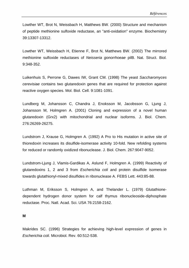
Références
Lowther WT, Brot N, Weissbach H, Matthews BW. (2000) Structure and mechanism
of peptide methionine sulfoxide reductase, an "anti-oxidation" enzyme. Biochemistry
39:13307-13312.
Lowther WT, Weissbach H, Etienne F, Brot N, Matthews BW. (2002) The mirrored
methionine sulfoxide reductases of Neisseria gonorrhoeae pilB. Nat. Struct. Biol.
9:348-352.
Luikenhuis S, Perrone G, Dawes IW, Grant CM. (1998) The yeast Saccharomyces
cerevisiae contains two glutaredoxin genes that are required for protection against
reactive oxygen species. Mol. Biol. Cell. 9:1081-1091.
Lundberg M, Johansson C, Chandra J, Enoksson M, Jacobsson G, Ljung J,
Johansson M, Holmgren A. (2001) Cloning and expression of a novel human
glutaredoxin (Grx2) with mitochondrial and nuclear isoforms. J. Biol. Chem.
276:26269-26275.
Lundstrom J, Krause G, Holmgren A. (1992) A Pro to His mutation in active site of
thioredoxin increases its disulfide-isomerase activity 10-fold. New refolding systems
for reduced or randomly oxidized ribonuclease. J. Biol. Chem. 267:9047-9052.
Lundstrom-Ljung J, Vlamis-Gardikas A, Aslund F, Holmgren A. (1999) Reactivity of
glutaredoxins 1, 2 and 3 from Escherichia coli and protein disulfide isomerase
towards glutathionyl-mixed disulfides in ribonuclease A. FEBS Lett. 443:85-88.
Luthman M, Eriksson S, Holmgren A, and Thelander L. (1979) Glutathione-
dependent hydrogen donor system for calf thymus ribonucleoside-diphosphate
reductase. Proc. Natl. Acad. Sci. USA 76:2158-2162.
M
Makrides SC. (1996) Strategies for achieving high-level expression of genes in
Escherichia coli. Microbiol. Rev. 60:512-538.
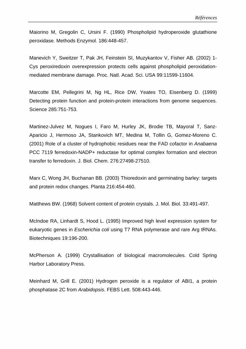
Références
Maiorino M, Gregolin C, Ursini F. (1990) Phospholipid hydroperoxide glutathione
peroxidase. Methods Enzymol. 186:448-457.
Manevich Y, Sweitzer T, Pak JH, Feinstein SI, Muzykantov V, Fisher AB. (2002) 1-
Cys peroxiredoxin overexpression protects cells against phospholipid peroxidation-
mediated membrane damage. Proc. Natl. Acad. Sci. USA 99:11599-11604.
Marcotte EM, Pellegrini M, Ng HL, Rice DW, Yeates TO, Eisenberg D. (1999)
Detecting protein function and protein-protein interactions from genome sequences.
Science 285:751-753.
Martinez-Julvez M, Nogues I, Faro M, Hurley JK, Brodie TB, Mayoral T, Sanz-
Aparicio J, Hermoso JA, Stankovich MT, Medina M, Tollin G, Gomez-Moreno C.
(2001) Role of a cluster of hydrophobic residues near the FAD cofactor in Anabaena
PCC 7119 ferredoxin-NADP+ reductase for optimal complex formation and electron
transfer to ferredoxin. J. Biol. Chem. 276:27498-27510.
Marx C, Wong JH, Buchanan BB. (2003) Thioredoxin and germinating barley: targets
and protein redox changes. Planta 216:454-460.
Matthews BW. (1968) Solvent content of protein crystals. J. Mol. Biol. 33:491-497.
McIndoe RA, Linhardt S, Hood L. (1995) Improved high level expression system for
eukaryotic genes in Escherichia coli using T7 RNA polymerase and rare Arg tRNAs.
Biotechniques 19:196-200.
McPherson A. (1999) Crystallisation of biological macromolecules. Cold Spring
Harbor Laboratory Press.
Meinhard M, Grill E. (2001) Hydrogen peroxide is a regulator of ABI1, a protein
phosphatase 2C from Arabidopsis. FEBS Lett. 508:443-446.
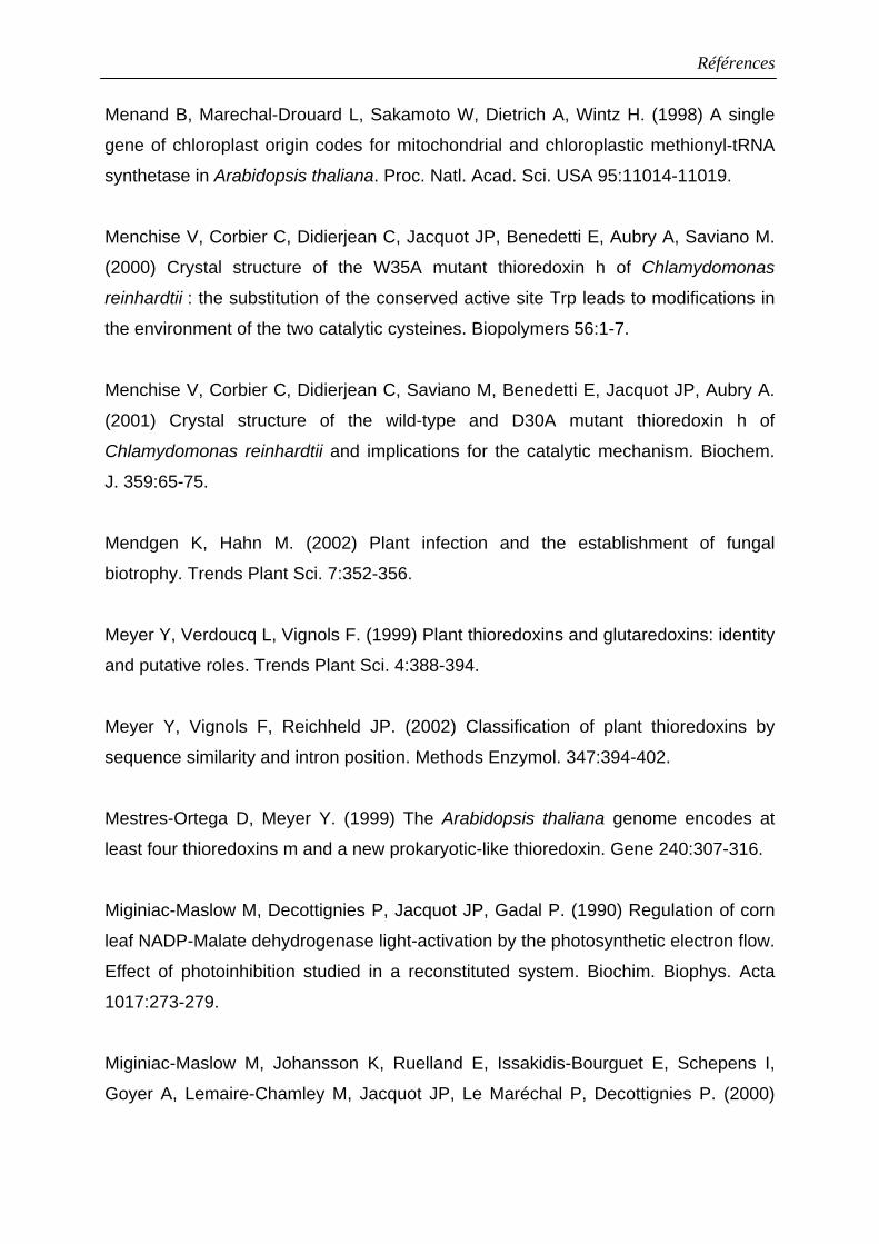
Références
Menand B, Marechal-Drouard L, Sakamoto W, Dietrich A, Wintz H. (1998) A single
gene of chloroplast origin codes for mitochondrial and chloroplastic methionyl-tRNA
synthetase in Arabidopsis thaliana. Proc. Natl. Acad. Sci. USA 95:11014-11019.
Menchise V, Corbier C, Didierjean C, Jacquot JP, Benedetti E, Aubry A, Saviano M.
(2000) Crystal structure of the W35A mutant thioredoxin h of Chlamydomonas
reinhardtii : the substitution of the conserved active site Trp leads to modifications in
the environment of the two catalytic cysteines. Biopolymers 56:1-7.
Menchise V, Corbier C, Didierjean C, Saviano M, Benedetti E, Jacquot JP, Aubry A.
(2001) Crystal structure of the wild-type and D30A mutant thioredoxin h of
Chlamydomonas reinhardtii and implications for the catalytic mechanism. Biochem.
J. 359:65-75.
Mendgen K, Hahn M. (2002) Plant infection and the establishment of fungal
biotrophy. Trends Plant Sci. 7:352-356.
Meyer Y, Verdoucq L, Vignols F. (1999) Plant thioredoxins and glutaredoxins: identity
and putative roles. Trends Plant Sci. 4:388-394.
Meyer Y, Vignols F, Reichheld JP. (2002) Classification of plant thioredoxins by
sequence similarity and intron position. Methods Enzymol. 347:394-402.
Mestres-Ortega D, Meyer Y. (1999) The Arabidopsis thaliana genome encodes at
least four thioredoxins m and a new prokaryotic-like thioredoxin. Gene 240:307-316.
Miginiac-Maslow M, Decottignies P, Jacquot JP, Gadal P. (1990) Regulation of corn
leaf NADP-Malate dehydrogenase light-activation by the photosynthetic electron flow.
Effect of photoinhibition studied in a reconstituted system. Biochim. Biophys. Acta
1017:273-279.
Miginiac-Maslow M, Johansson K, Ruelland E, Issakidis-Bourguet E, Schepens I,
Goyer A, Lemaire-Chamley M, Jacquot JP, Le Maréchal P, Decottignies P. (2000)
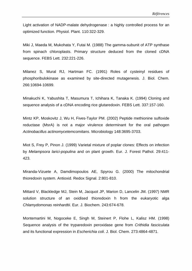
Références
Light activation of NADP-malate dehydrogenase : a highly controlled process for an
optimized function. Physiol. Plant. 110:322-329.
Miki J, Maeda M, Mukohata Y, Futai M. (1988) The gamma-subunit of ATP synthase
from spinach chloroplasts. Primary structure deduced from the cloned cDNA
sequence. FEBS Lett. 232:221-226.
Milanez S, Mural RJ, Hartman FC. (1991) Roles of cysteinyl residues of
phosphoribulokinase as examined by site-directed mutagenesis. J. Biol. Chem.
266:10694-10699.
Minakuchi K, Yabushita T, Masumura T, Ichihara K, Tanaka K. (1994) Cloning and
sequence analysis of a cDNA encoding rice glutaredoxin. FEBS Lett. 337:157-160.
Mintz KP, Moskovitz J, Wu H, Fives-Taylor PM. (2002) Peptide methionine sulfoxide
reductase (MsrA) is not a major virulence determinant for the oral pathogen
Actinobacillus actinomycetemcomitans. Microbiology 148:3695-3703.
Miot S, Frey P, Pinon J. (1999) Varietal mixture of poplar clones: Effects on infection
by Melampsora larici-populina and on plant growth. Eur. J. Forest Pathol. 29:411-
423.
Miranda-Vizuete A, Damdimopoulos AE, Spyrou G. (2000) The mitochondrial
thioredoxin system. Antioxid. Redox Signal. 2:801-810.
Mittard V, Blackledge MJ, Stein M, Jacquot JP, Marion D, Lancelin JM. (1997) NMR
solution structure of an oxidised thioredoxin h from the eukaryotic alga
Chlamydomonas reinhardtii. Eur. J. Biochem. 243:674-678.
Montemartini M, Nogoceke E, Singh M, Steinert P, Flohe L, Kalisz HM. (1998)
Sequence analysis of the tryparedoxin peroxidase gene from Crithidia fasciculata
and its functional expression in Escherichia coli. J. Biol. Chem. 273:4864-4871.
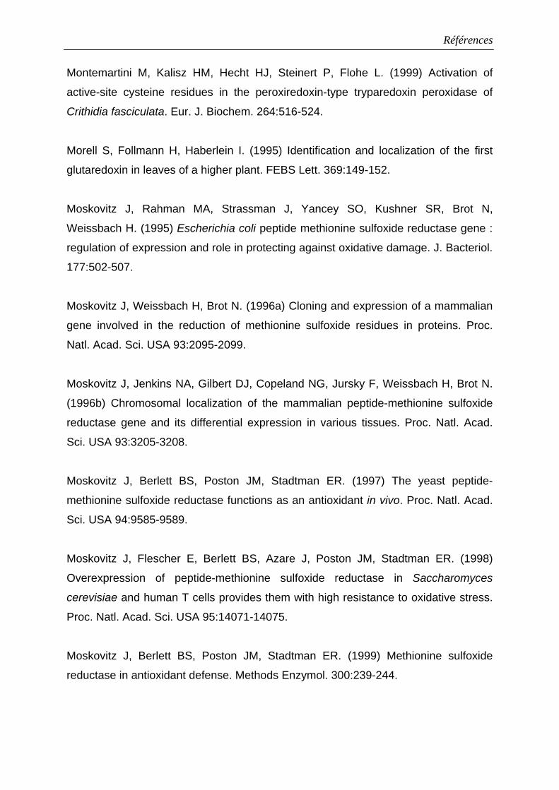
Références
Montemartini M, Kalisz HM, Hecht HJ, Steinert P, Flohe L. (1999) Activation of
active-site cysteine residues in the peroxiredoxin-type tryparedoxin peroxidase of
Crithidia fasciculata. Eur. J. Biochem. 264:516-524.
Morell S, Follmann H, Haberlein I. (1995) Identification and localization of the first
glutaredoxin in leaves of a higher plant. FEBS Lett. 369:149-152.
Moskovitz J, Rahman MA, Strassman J, Yancey SO, Kushner SR, Brot N,
Weissbach H. (1995) Escherichia coli peptide methionine sulfoxide reductase gene :
regulation of expression and role in protecting against oxidative damage. J. Bacteriol.
177:502-507.
Moskovitz J, Weissbach H, Brot N. (1996a) Cloning and expression of a mammalian
gene involved in the reduction of methionine sulfoxide residues in proteins. Proc.
Natl. Acad. Sci. USA 93:2095-2099.
Moskovitz J, Jenkins NA, Gilbert DJ, Copeland NG, Jursky F, Weissbach H, Brot N.
(1996b) Chromosomal localization of the mammalian peptide-methionine sulfoxide
reductase gene and its differential expression in various tissues. Proc. Natl. Acad.
Sci. USA 93:3205-3208.
Moskovitz J, Berlett BS, Poston JM, Stadtman ER. (1997) The yeast peptide-
methionine sulfoxide reductase functions as an antioxidant in vivo. Proc. Natl. Acad.
Sci. USA 94:9585-9589.
Moskovitz J, Flescher E, Berlett BS, Azare J, Poston JM, Stadtman ER. (1998)
Overexpression of peptide-methionine sulfoxide reductase in Saccharomyces
cerevisiae and human T cells provides them with high resistance to oxidative stress.
Proc. Natl. Acad. Sci. USA 95:14071-14075.
Moskovitz J, Berlett BS, Poston JM, Stadtman ER. (1999) Methionine sulfoxide
reductase in antioxidant defense. Methods Enzymol. 300:239-244.
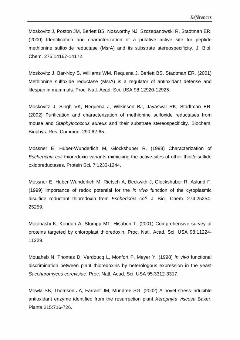
Références
Moskovitz J, Poston JM, Berlett BS, Nosworthy NJ, Szczepanowski R, Stadtman ER.
(2000) Identification and characterization of a putative active site for peptide
methionine sulfoxide reductase (MsrA) and its substrate stereospecificity. J. Biol.
Chem. 275:14167-14172.
Moskovitz J, Bar-Noy S, Williams WM, Requena J, Berlett BS, Stadtman ER. (2001)
Methionine sulfoxide reductase (MsrA) is a regulator of antioxidant defense and
lifespan in mammals. Proc. Natl. Acad. Sci. USA 98:12920-12925.
Moskovitz J, Singh VK, Requena J, Wilkinson BJ, Jayaswal RK, Stadtman ER.
(2002) Purification and characterization of methionine sulfoxide reductases from
mouse and Staphylococcus aureus and their substrate stereospecificity. Biochem.
Biophys. Res. Commun. 290:62-65.
Mossner E, Huber-Wunderlich M, Glockshuber R. (1998) Characterization of
Escherichia coli thioredoxin variants mimicking the active-sites of other thiol/disulfide
oxidoreductases. Protein Sci. 7:1233-1244.
Mossner E, Huber-Wunderlich M, Rietsch A, Beckwith J, Glockshuber R, Aslund F.
(1999) Importance of redox potential for the in vivo function of the cytoplasmic
disulfide reductant thioredoxin from Escherichia coli. J. Biol. Chem. 274:25254-
25259.
Motohashi K, Kondoh A, Stumpp MT, Hisabori T. (2001) Comprehensive survey of
proteins targeted by chloroplast thioredoxin. Proc. Natl. Acad. Sci. USA 98:11224-
11229.
Mouaheb N, Thomas D, Verdoucq L, Monfort P, Meyer Y. (1998) In vivo functional
discrimination between plant thioredoxins by heterologous expression in the yeast
Saccharomyces cerevisiae. Proc. Natl. Acad. Sci. USA 95:3312-3317.
Mowla SB, Thomson JA, Farrant JM, Mundree SG. (2002) A novel stress-inducible
antioxidant enzyme identified from the resurrection plant Xerophyta viscosa Baker.
Planta 215:716-726.
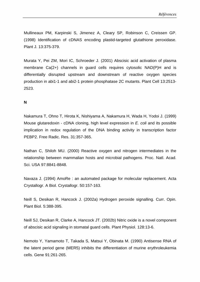
Références
Mullineaux PM, Karpinski S, Jimenez A, Cleary SP, Robinson C, Creissen GP.
(1998) Identification of cDNAS encoding plastid-targeted glutathione peroxidase.
Plant J. 13:375-379.
Murata Y, Pei ZM, Mori IC, Schroeder J. (2001) Abscisic acid activation of plasma
membrane Ca(2+) channels in guard cells requires cytosolic NAD(P)H and is
differentially disrupted upstream and downstream of reactive oxygen species
production in abi1-1 and abi2-1 protein phosphatase 2C mutants. Plant Cell 13:2513-
2523.
N
Nakamura T, Ohno T, Hirota K, Nishiyama A, Nakamura H, Wada H, Yodoi J. (1999)
Mouse glutaredoxin - cDNA cloning, high level expression in E. coli and its possible
implication in redox regulation of the DNA binding activity in transcription factor
PEBP2. Free Radic. Res. 31:357-365.
Nathan C, Shiloh MU. (2000) Reactive oxygen and nitrogen intermediates in the
relationship between mammalian hosts and microbial pathogens. Proc. Natl. Acad.
Sci. USA 97:8841-8848.
Navaza J. (1994) AmoRe : an automated package for molecular replacement. Acta
Crystallogr. A Biol. Crystallogr. 50:157-163.
Neill S, Desikan R, Hancock J. (2002a) Hydrogen peroxide signalling. Curr. Opin.
Plant Biol. 5:388-395.
Neill SJ, Desikan R, Clarke A, Hancock JT. (2002b) Nitric oxide is a novel component
of abscisic acid signaling in stomatal guard cells. Plant Physiol. 128:13-6.
Nemoto Y, Yamamoto T, Takada S, Matsui Y, Obinata M. (1990) Antisense RNA of
the latent period gene (MER5) inhibits the differentiation of murine erythroleukemia
cells. Gene 91:261-265.
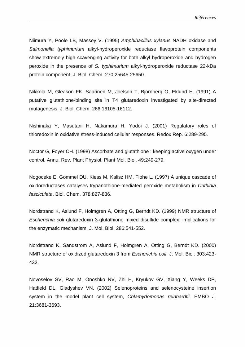
Références
Niimura Y, Poole LB, Massey V. (1995) Amphibacillus xylanus NADH oxidase and
Salmonella typhimurium alkyl-hydroperoxide reductase flavoprotein components
show extremely high scavenging activity for both alkyl hydroperoxide and hydrogen
peroxide in the presence of S. typhimurium alkyl-hydroperoxide reductase 22-kDa
protein component. J. Biol. Chem. 270:25645-25650.
Nikkola M, Gleason FK, Saarinen M, Joelson T, Bjornberg O, Eklund H. (1991) A
putative glutathione-binding site in T4 glutaredoxin investigated by site-directed
mutagenesis. J. Biol. Chem. 266:16105-16112.
Nishinaka Y, Masutani H, Nakamura H, Yodoi J. (2001) Regulatory roles of
thioredoxin in oxidative stress-induced cellular responses. Redox Rep. 6:289-295.
Noctor G, Foyer CH. (1998) Ascorbate and glutathione : keeping active oxygen under
control. Annu. Rev. Plant Physiol. Plant Mol. Biol. 49:249-279.
Nogoceke E, Gommel DU, Kiess M, Kalisz HM, Flohe L. (1997) A unique cascade of
oxidoreductases catalyses trypanothione-mediated peroxide metabolism in Crithidia
fasciculata. Biol. Chem. 378:827-836.
Nordstrand K, Aslund F, Holmgren A, Otting G, Berndt KD. (1999) NMR structure of
Escherichia coli glutaredoxin 3-glutathione mixed disulfide complex: implications for
the enzymatic mechanism. J. Mol. Biol. 286:541-552.
Nordstrand K, Sandstrom A, Aslund F, Holmgren A, Otting G, Berndt KD. (2000)
NMR structure of oxidized glutaredoxin 3 from Escherichia coli. J. Mol. Biol. 303:423-
432.
Novoselov SV, Rao M, Onoshko NV, Zhi H, Kryukov GV, Xiang Y, Weeks DP,
Hatfield DL, Gladyshev VN. (2002) Selenoproteins and selenocysteine insertion
system in the model plant cell system, Chlamydomonas reinhardtii. EMBO J.
21:3681-3693.
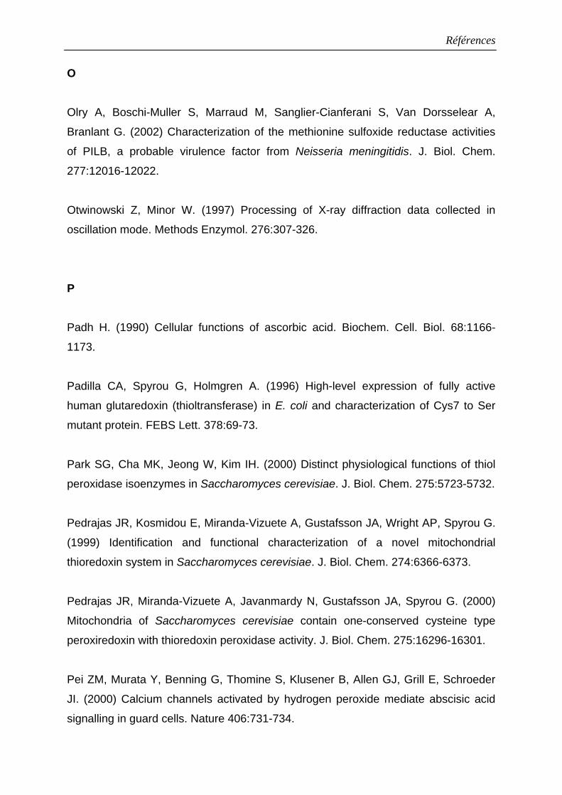
Références
O
Olry A, Boschi-Muller S, Marraud M, Sanglier-Cianferani S, Van Dorsselear A,
Branlant G. (2002) Characterization of the methionine sulfoxide reductase activities
of PILB, a probable virulence factor from Neisseria meningitidis. J. Biol. Chem.
277:12016-12022.
Otwinowski Z, Minor W. (1997) Processing of X-ray diffraction data collected in
oscillation mode. Methods Enzymol. 276:307-326.
P
Padh H. (1990) Cellular functions of ascorbic acid. Biochem. Cell. Biol. 68:1166-
1173.
Padilla CA, Spyrou G, Holmgren A. (1996) High-level expression of fully active
human glutaredoxin (thioltransferase) in E. coli and characterization of Cys7 to Ser
mutant protein. FEBS Lett. 378:69-73.
Park SG, Cha MK, Jeong W, Kim IH. (2000) Distinct physiological functions of thiol
peroxidase isoenzymes in Saccharomyces cerevisiae. J. Biol. Chem. 275:5723-5732.
Pedrajas JR, Kosmidou E, Miranda-Vizuete A, Gustafsson JA, Wright AP, Spyrou G.
(1999) Identification and functional characterization of a novel mitochondrial
thioredoxin system in Saccharomyces cerevisiae. J. Biol. Chem. 274:6366-6373.
Pedrajas JR, Miranda-Vizuete A, Javanmardy N, Gustafsson JA, Spyrou G. (2000)
Mitochondria of Saccharomyces cerevisiae contain one-conserved cysteine type
peroxiredoxin with thioredoxin peroxidase activity. J. Biol. Chem. 275:16296-16301.
Pei ZM, Murata Y, Benning G, Thomine S, Klusener B, Allen GJ, Grill E, Schroeder
JI. (2000) Calcium channels activated by hydrogen peroxide mediate abscisic acid
signalling in guard cells. Nature 406:731-734.
Références
Peshenko IV, Shichi H. (2001) Oxidation of active center cysteine of bovine 1-Cys
peroxiredoxin to the cysteine sulfenic acid form by peroxide and peroxynitrite. Free
Radic. Biol. Med. 31:292-303.
Petropoulos I, Mary J, Perichon M, Friguet B. (2001) Rat peptide methionine
sulphoxide reductase: cloning of the cDNA, and down-regulation of gene expression
and enzyme activity during aging. Biochem. J. 355:819-825.
Poole ES, Brown CM, Tate WP. (1995) The identity of the base following the stop
codon determines the efficiency of in vivo translational termination in Escherichia coli.
EMBO J. 14:151-158.
Poole LB. (1996) Flavin-dependent alkyl hydroperoxide reductase from Salmonella
typhimurium. 2. Cystine disulfides involved in catalysis of peroxide reduction.
Biochemistry 35:65-75.
Potamitou A, Holmgren A, Vlamis-Gardikas A. (2002) Protein levels of Escherichia
coli thioredoxins and glutaredoxins and their relation to null mutants, growth phase,
and function. J. Biol. Chem. 277:18561-18567.
Powis G, Mustacich D, Coon A. (2000) The role of the redox protein thioredoxin in
cell growth and cancer. Free Radic. Biol. Med. 29:312-322.
Prieto-Alamo MJ, Jurado J, Gallardo-Madueno R, Monje-Casas F, Holmgren A,
Pueyo C. (2000) Transcriptional regulation of glutaredoxin and thioredoxin pathways
and related enzymes in response to oxidative stress. J. Biol. Chem. 275:13398-
13405.
Prinz WA, Aslund F, Holmgren A, Beckwith J. (1997) The role of the thioredoxin and
glutaredoxin pathways in reducing protein disulfide bonds in the Escherichia coli
cytoplasm. J. Biol. Chem. 272:15661-15667.
Références
Prosperi MT, Ferbus D, Rouillard D, Goubin G. (1998) The pag gene product, a
physiological inhibitor of c-abl tyrosine kinase, is overexpressed in cells entering S
phase and by contact with agents inducing oxidative stress. FEBS Lett. 423:39-44.
Prouzet-Mauleon V, Monribot-Espagne C, Boucherie H, Lagniel G, Lopez S, Labarre
J, Garin J, Lauquin GJ. (2002) Identification in Saccharomyces cerevisiae of a new
stable variant of alkyl hydroperoxide reductase 1 (Ahp1) induced by oxidative stress.
J. Biol. Chem. 277:4823-4830.
Q Qin J, Yang Y, Velyvis A, Gronenborn A. (2000) Molecular views of redox regulation:
three-dimensional structures of redox regulatory proteins and protein complexes.
Antioxid. Redox Signal. 2:827-840.
R
Rabilloud T, Heller M, Gasnier F, Luche S, Rey C, Aebersold R, Benahmed M,
Louisot P, Lunardi J. (2002) Proteomics analysis of cellular response to oxidative
stress. Evidence for in vivo overoxidation of peroxiredoxins at their active site. J. Biol.
Chem. 277:19396-19401.
Rahlfs S, Fischer M, Becker K. (2001) Plasmodium falciparum possesses a classical
glutaredoxin and a second, glutaredoxin-like protein with a PICOT homology domain.
J. Biol. Chem. 276:37133-37140.
Raines CA, Lloyd JC, Longstaff M, Bradley D, Dyer T. (1988) Chloroplast fructose-
1,6-bisphosphatase: the product of a mosaic gene. Nucleic Acids Res. 16:7931-
7942.
Reichheld JP, Mestres-Ortega D, Laloi C, Meyer Y. (2002) The multigenic family of
thioredoxin h in Arabidopsis thaliana: specific expression and stress response. Plant
Physiol. Biochem. 40:685-690.
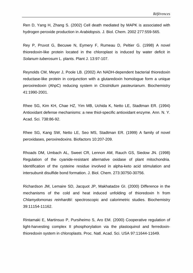
Références
Ren D, Yang H, Zhang S. (2002) Cell death mediated by MAPK is associated with
hydrogen peroxide production in Arabidopsis. J. Biol. Chem. 2002 277:559-565.
Rey P, Pruvot G, Becuwe N, Eymery F, Rumeau D, Peltier G. (1998) A novel
thioredoxin-like protein located in the chloroplast is induced by water deficit in
Solanum tuberosum L. plants. Plant J. 13:97-107.
Reynolds CM, Meyer J, Poole LB. (2002) An NADH-dependent bacterial thioredoxin
reductase-like protein in conjunction with a glutaredoxin homologue form a unique
peroxiredoxin (AhpC) reducing system in Clostridium pasteurianum. Biochemistry
41:1990-2001.
Rhee SG, Kim KH, Chae HZ, Yim MB, Uchida K, Netto LE, Stadtman ER. (1994)
Antioxidant defense mechanisms: a new thiol-specific antioxidant enzyme. Ann. N. Y.
Acad. Sci. 738:86-92.
Rhee SG, Kang SW, Netto LE, Seo MS, Stadtman ER. (1999) A family of novel
peroxidases, peroxiredoxins. Biofactors 10:207-209.
Rhoads DM, Umbach AL, Sweet CR, Lennon AM, Rauch GS, Siedow JN. (1998)
Regulation of the cyanide-resistant alternative oxidase of plant mitochondria.
Identification of the cysteine residue involved in alpha-keto acid stimulation and
intersubunit disulfide bond formation. J. Biol. Chem. 273:30750-30756.
Richardson JM, Lemaire SD, Jacquot JP, Makhatadze GI. (2000) Difference in the
mechanisms of the cold and heat induced unfolding of thioredoxin h from
Chlamydomonas reinhardtii: spectroscopic and calorimetric studies. Biochemistry
39:11154-11162.
Rintamaki E, Martinsuo P, Pursiheimo S, Aro EM. (2000) Cooperative regulation of
light-harvesting complex II phosphorylation via the plastoquinol and ferredoxin-
thioredoxin system in chloroplasts. Proc. Natl. Acad. Sci. USA 97:11644-11649.
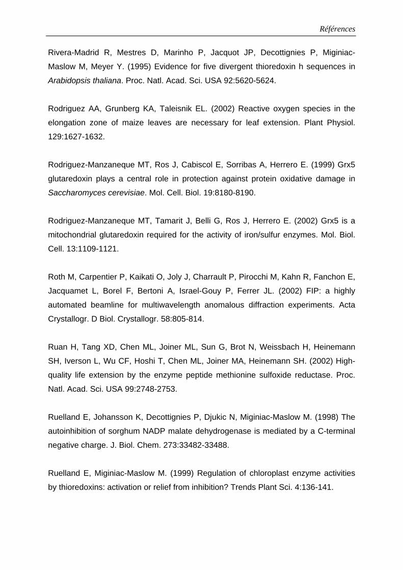
Références
Rivera-Madrid R, Mestres D, Marinho P, Jacquot JP, Decottignies P, Miginiac-
Maslow M, Meyer Y. (1995) Evidence for five divergent thioredoxin h sequences in
Arabidopsis thaliana. Proc. Natl. Acad. Sci. USA 92:5620-5624.
Rodriguez AA, Grunberg KA, Taleisnik EL. (2002) Reactive oxygen species in the
elongation zone of maize leaves are necessary for leaf extension. Plant Physiol.
129:1627-1632.
Rodriguez-Manzaneque MT, Ros J, Cabiscol E, Sorribas A, Herrero E. (1999) Grx5
glutaredoxin plays a central role in protection against protein oxidative damage in
Saccharomyces cerevisiae. Mol. Cell. Biol. 19:8180-8190.
Rodriguez-Manzaneque MT, Tamarit J, Belli G, Ros J, Herrero E. (2002) Grx5 is a
mitochondrial glutaredoxin required for the activity of iron/sulfur enzymes. Mol. Biol.
Cell. 13:1109-1121.
Roth M, Carpentier P, Kaikati O, Joly J, Charrault P, Pirocchi M, Kahn R, Fanchon E,
Jacquamet L, Borel F, Bertoni A, Israel-Gouy P, Ferrer JL. (2002) FIP: a highly
automated beamline for multiwavelength anomalous diffraction experiments. Acta
Crystallogr. D Biol. Crystallogr. 58:805-814.
Ruan H, Tang XD, Chen ML, Joiner ML, Sun G, Brot N, Weissbach H, Heinemann
SH, Iverson L, Wu CF, Hoshi T, Chen ML, Joiner MA, Heinemann SH. (2002) High-
quality life extension by the enzyme peptide methionine sulfoxide reductase. Proc.
Natl. Acad. Sci. USA 99:2748-2753.
Ruelland E, Johansson K, Decottignies P, Djukic N, Miginiac-Maslow M. (1998) The
autoinhibition of sorghum NADP malate dehydrogenase is mediated by a C-terminal
negative charge. J. Biol. Chem. 273:33482-33488.
Ruelland E, Miginiac-Maslow M. (1999) Regulation of chloroplast enzyme activities
by thioredoxins: activation or relief from inhibition? Trends Plant Sci. 4:136-141.
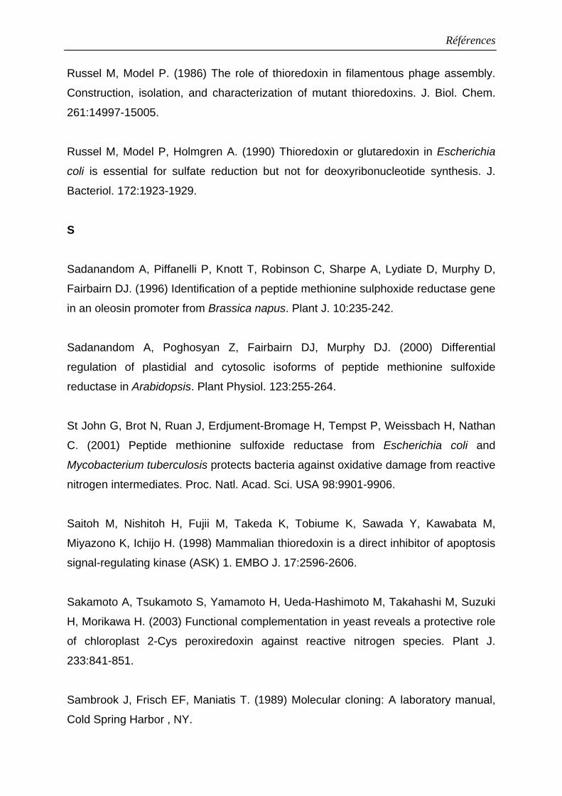
Références
Russel M, Model P. (1986) The role of thioredoxin in filamentous phage assembly.
Construction, isolation, and characterization of mutant thioredoxins. J. Biol. Chem.
261:14997-15005.
Russel M, Model P, Holmgren A. (1990) Thioredoxin or glutaredoxin in Escherichia
coli is essential for sulfate reduction but not for deoxyribonucleotide synthesis. J.
Bacteriol. 172:1923-1929.
S
Sadanandom A, Piffanelli P, Knott T, Robinson C, Sharpe A, Lydiate D, Murphy D,
Fairbairn DJ. (1996) Identification of a peptide methionine sulphoxide reductase gene
in an oleosin promoter from Brassica napus. Plant J. 10:235-242.
Sadanandom A, Poghosyan Z, Fairbairn DJ, Murphy DJ. (2000) Differential
regulation of plastidial and cytosolic isoforms of peptide methionine sulfoxide
reductase in Arabidopsis. Plant Physiol. 123:255-264.
St John G, Brot N, Ruan J, Erdjument-Bromage H, Tempst P, Weissbach H, Nathan
C. (2001) Peptide methionine sulfoxide reductase from Escherichia coli and
Mycobacterium tuberculosis protects bacteria against oxidative damage from reactive
nitrogen intermediates. Proc. Natl. Acad. Sci. USA 98:9901-9906.
Saitoh M, Nishitoh H, Fujii M, Takeda K, Tobiume K, Sawada Y, Kawabata M,
Miyazono K, Ichijo H. (1998) Mammalian thioredoxin is a direct inhibitor of apoptosis
signal-regulating kinase (ASK) 1. EMBO J. 17:2596-2606.
Sakamoto A, Tsukamoto S, Yamamoto H, Ueda-Hashimoto M, Takahashi M, Suzuki
H, Morikawa H. (2003) Functional complementation in yeast reveals a protective role
of chloroplast 2-Cys peroxiredoxin against reactive nitrogen species. Plant J.
233:841-851.
Sambrook J, Frisch EF, Maniatis T. (1989) Molecular cloning: A laboratory manual,
Cold Spring Harbor , NY.
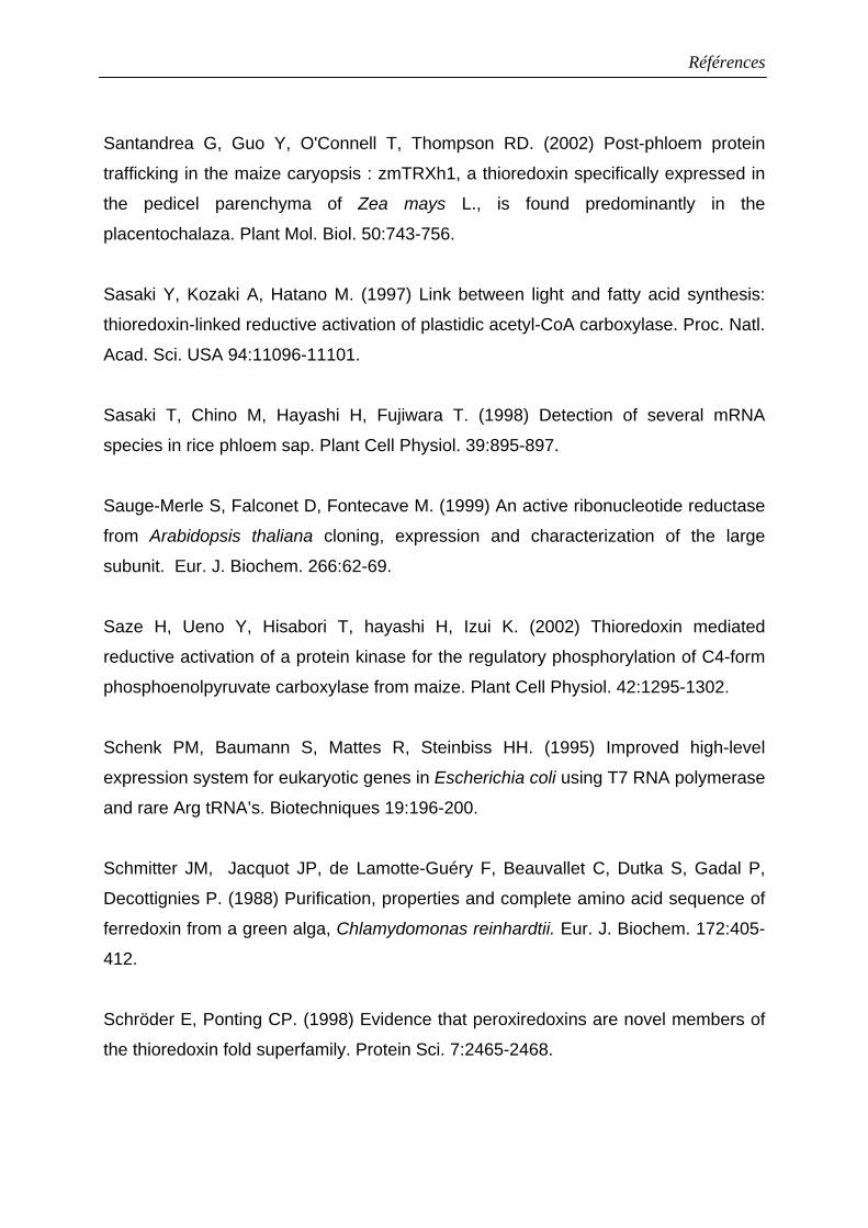
Références
Santandrea G, Guo Y, O'Connell T, Thompson RD. (2002) Post-phloem protein
trafficking in the maize caryopsis : zmTRXh1, a thioredoxin specifically expressed in
the pedicel parenchyma of Zea mays L., is found predominantly in the
placentochalaza. Plant Mol. Biol. 50:743-756.
Sasaki Y, Kozaki A, Hatano M. (1997) Link between light and fatty acid synthesis:
thioredoxin-linked reductive activation of plastidic acetyl-CoA carboxylase. Proc. Natl.
Acad. Sci. USA 94:11096-11101.
Sasaki T, Chino M, Hayashi H, Fujiwara T. (1998) Detection of several mRNA
species in rice phloem sap. Plant Cell Physiol. 39:895-897.
Sauge-Merle S, Falconet D, Fontecave M. (1999) An active ribonucleotide reductase
from Arabidopsis thaliana cloning, expression and characterization of the large
subunit. Eur. J. Biochem. 266:62-69.
Saze H, Ueno Y, Hisabori T, hayashi H, Izui K. (2002) Thioredoxin mediated
reductive activation of a protein kinase for the regulatory phosphorylation of C4-form
phosphoenolpyruvate carboxylase from maize. Plant Cell Physiol. 42:1295-1302.
Schenk PM, Baumann S, Mattes R, Steinbiss HH. (1995) Improved high-level
expression system for eukaryotic genes in Escherichia coli using T7 RNA polymerase
and rare Arg tRNA’s. Biotechniques 19:196-200.
Schmitter JM, Jacquot JP, de Lamotte-Guéry F, Beauvallet C, Dutka S, Gadal P,
Decottignies P. (1988) Purification, properties and complete amino acid sequence of
ferredoxin from a green alga, Chlamydomonas reinhardtii. Eur. J. Biochem. 172:405-
412.
Schröder E, Ponting CP. (1998) Evidence that peroxiredoxins are novel members of
the thioredoxin fold superfamily. Protein Sci. 7:2465-2468.
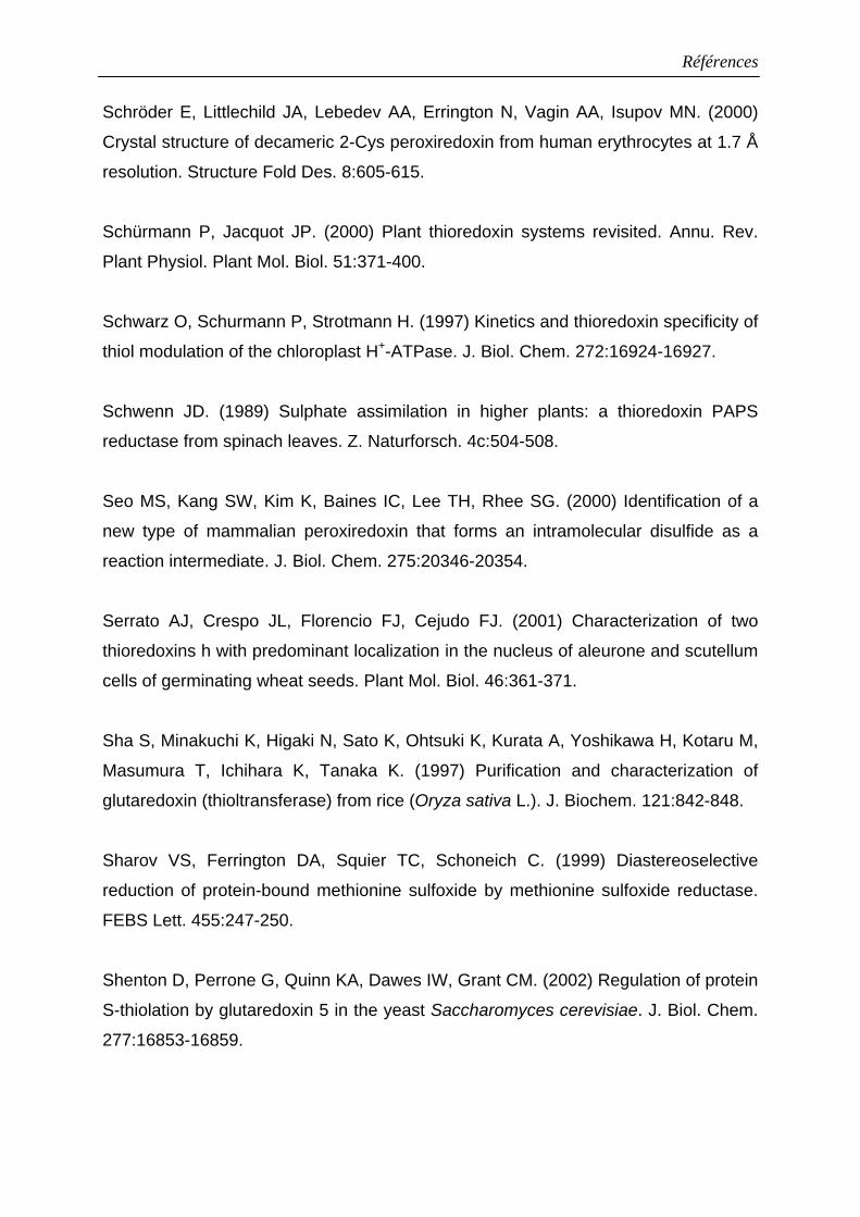
Références
Schröder E, Littlechild JA, Lebedev AA, Errington N, Vagin AA, Isupov MN. (2000)
Crystal structure of decameric 2-Cys peroxiredoxin from human erythrocytes at 1.7 Å
resolution. Structure Fold Des. 8:605-615.
Schürmann P, Jacquot JP. (2000) Plant thioredoxin systems revisited. Annu. Rev.
Plant Physiol. Plant Mol. Biol. 51:371-400.
Schwarz O, Schurmann P, Strotmann H. (1997) Kinetics and thioredoxin specificity of
thiol modulation of the chloroplast H+-ATPase. J. Biol. Chem. 272:16924-16927.
Schwenn JD. (1989) Sulphate assimilation in higher plants: a thioredoxin PAPS
reductase from spinach leaves. Z. Naturforsch. 4c:504-508.
Seo MS, Kang SW, Kim K, Baines IC, Lee TH, Rhee SG. (2000) Identification of a
new type of mammalian peroxiredoxin that forms an intramolecular disulfide as a
reaction intermediate. J. Biol. Chem. 275:20346-20354.
Serrato AJ, Crespo JL, Florencio FJ, Cejudo FJ. (2001) Characterization of two
thioredoxins h with predominant localization in the nucleus of aleurone and scutellum
cells of germinating wheat seeds. Plant Mol. Biol. 46:361-371.
Sha S, Minakuchi K, Higaki N, Sato K, Ohtsuki K, Kurata A, Yoshikawa H, Kotaru M,
Masumura T, Ichihara K, Tanaka K. (1997) Purification and characterization of
glutaredoxin (thioltransferase) from rice (Oryza sativa L.). J. Biochem. 121:842-848.
Sharov VS, Ferrington DA, Squier TC, Schoneich C. (1999) Diastereoselective
reduction of protein-bound methionine sulfoxide by methionine sulfoxide reductase.
FEBS Lett. 455:247-250.
Shenton D, Perrone G, Quinn KA, Dawes IW, Grant CM. (2002) Regulation of protein
S-thiolation by glutaredoxin 5 in the yeast Saccharomyces cerevisiae. J. Biol. Chem.
277:16853-16859.
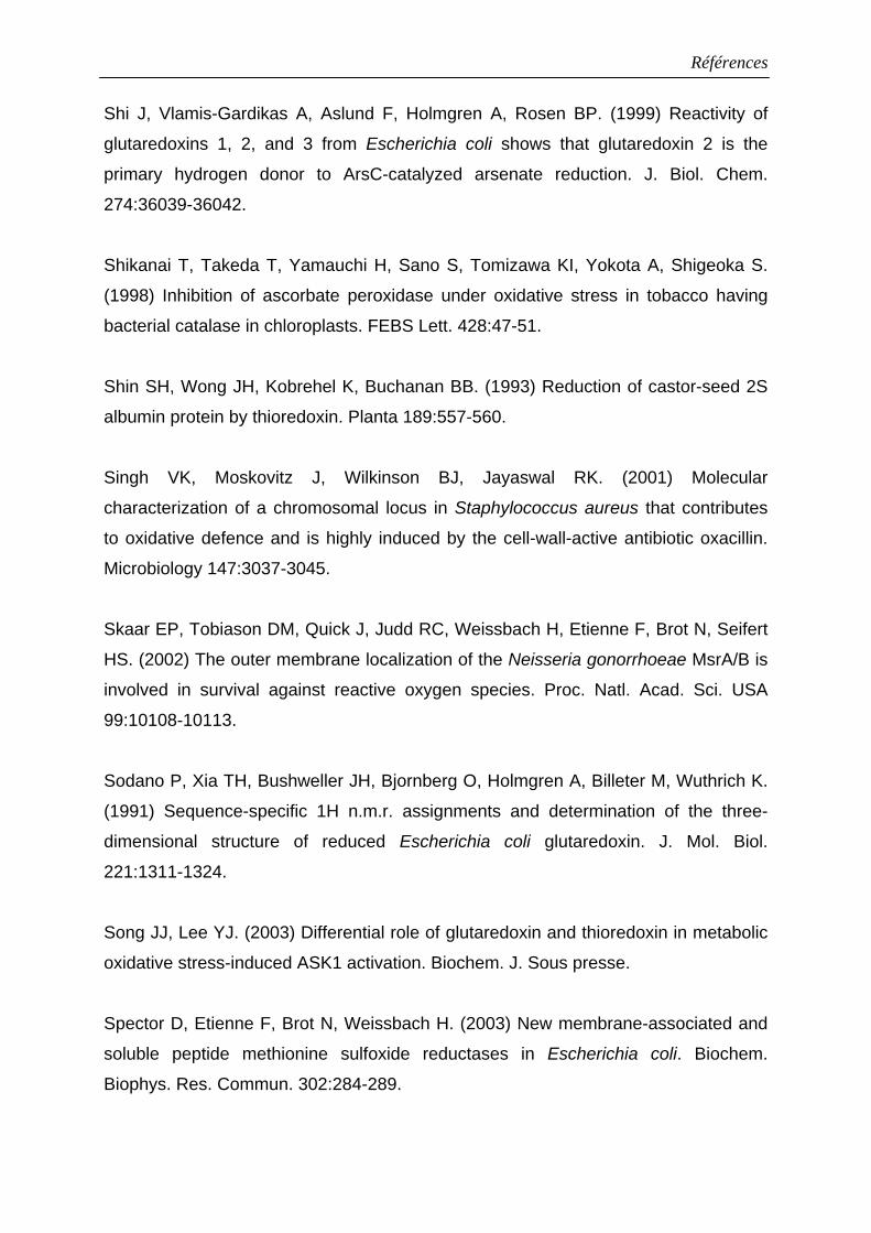
Références
Shi J, Vlamis-Gardikas A, Aslund F, Holmgren A, Rosen BP. (1999) Reactivity of
glutaredoxins 1, 2, and 3 from Escherichia coli shows that glutaredoxin 2 is the
primary hydrogen donor to ArsC-catalyzed arsenate reduction. J. Biol. Chem.
274:36039-36042.
Shikanai T, Takeda T, Yamauchi H, Sano S, Tomizawa KI, Yokota A, Shigeoka S.
(1998) Inhibition of ascorbate peroxidase under oxidative stress in tobacco having
bacterial catalase in chloroplasts. FEBS Lett. 428:47-51.
Shin SH, Wong JH, Kobrehel K, Buchanan BB. (1993) Reduction of castor-seed 2S
albumin protein by thioredoxin. Planta 189:557-560.
Singh VK, Moskovitz J, Wilkinson BJ, Jayaswal RK. (2001) Molecular
characterization of a chromosomal locus in Staphylococcus aureus that contributes
to oxidative defence and is highly induced by the cell-wall-active antibiotic oxacillin.
Microbiology 147:3037-3045.
Skaar EP, Tobiason DM, Quick J, Judd RC, Weissbach H, Etienne F, Brot N, Seifert
HS. (2002) The outer membrane localization of the Neisseria gonorrhoeae MsrA/B is
involved in survival against reactive oxygen species. Proc. Natl. Acad. Sci. USA
99:10108-10113.
Sodano P, Xia TH, Bushweller JH, Bjornberg O, Holmgren A, Billeter M, Wuthrich K.
(1991) Sequence-specific 1H n.m.r. assignments and determination of the three-
dimensional structure of reduced Escherichia coli glutaredoxin. J. Mol. Biol.
221:1311-1324.
Song JJ, Lee YJ. (2003) Differential role of glutaredoxin and thioredoxin in metabolic
oxidative stress-induced ASK1 activation. Biochem. J. Sous presse.
Spector D, Etienne F, Brot N, Weissbach H. (2003) New membrane-associated and
soluble peptide methionine sulfoxide reductases in Escherichia coli. Biochem.
Biophys. Res. Commun. 302:284-289.
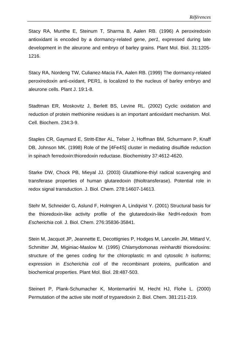
Références
Stacy RA, Munthe E, Steinum T, Sharma B, Aalen RB. (1996) A peroxiredoxin
antioxidant is encoded by a dormancy-related gene, per1, expressed during late
development in the aleurone and embryo of barley grains. Plant Mol. Biol. 31:1205-
1216.
Stacy RA, Nordeng TW, Culianez-Macia FA, Aalen RB. (1999) The dormancy-related
peroxiredoxin anti-oxidant, PER1, is localized to the nucleus of barley embryo and
aleurone cells. Plant J. 19:1-8.
Stadtman ER, Moskovitz J, Berlett BS, Levine RL. (2002) Cyclic oxidation and
reduction of protein methionine residues is an important antioxidant mechanism. Mol.
Cell. Biochem. 234:3-9.
Staples CR, Gaymard E, Stritt-Etter AL, Telser J, Hoffman BM, Schurmann P, Knaff
DB, Johnson MK. (1998) Role of the [4Fe4S] cluster in mediating disulfide reduction
in spinach ferredoxin:thioredoxin reductase. Biochemistry 37:4612-4620.
Starke DW, Chock PB, Mieyal JJ. (2003) Glutathione-thiyl radical scavenging and
transferase properties of human glutaredoxin (thioltransferase). Potential role in
redox signal transduction. J. Biol. Chem. 278:14607-14613.
Stehr M, Schneider G, Aslund F, Holmgren A, Lindqvist Y. (2001) Structural basis for
the thioredoxin-like activity profile of the glutaredoxin-like NrdH-redoxin from
Escherichia coli. J. Biol. Chem. 276:35836-35841.
Stein M, Jacquot JP, Jeannette E, Decottignies P, Hodges M, Lancelin JM, Mittard V,
Schmitter JM, Miginiac-Maslow M. (1995) Chlamydomonas reinhardtii thioredoxins:
structure of the genes coding for the chloroplastic m and cytosolic h isoforms;
expression in Escherichia coli of the recombinant proteins, purification and
biochemical properties. Plant Mol. Biol. 28:487-503.
Steinert P, Plank-Schumacher K, Montemartini M, Hecht HJ, Flohe L. (2000)
Permutation of the active site motif of tryparedoxin 2. Biol. Chem. 381:211-219.
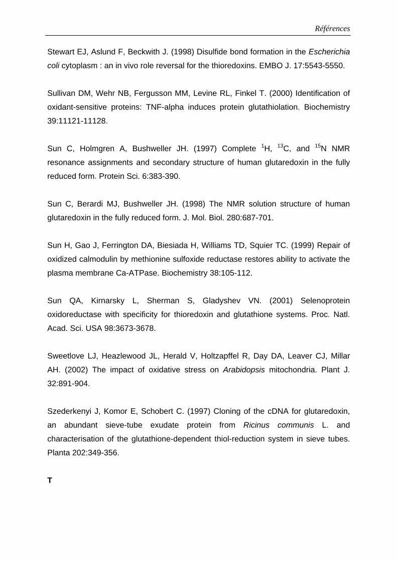
Références
Stewart EJ, Aslund F, Beckwith J. (1998) Disulfide bond formation in the Escherichia
coli cytoplasm : an in vivo role reversal for the thioredoxins. EMBO J. 17:5543-5550.
Sullivan DM, Wehr NB, Fergusson MM, Levine RL, Finkel T. (2000) Identification of
oxidant-sensitive proteins: TNF-alpha induces protein glutathiolation. Biochemistry
39:11121-11128.
Sun C, Holmgren A, Bushweller JH. (1997) Complete 1H, 13C, and 15N NMR
resonance assignments and secondary structure of human glutaredoxin in the fully
reduced form. Protein Sci. 6:383-390.
Sun C, Berardi MJ, Bushweller JH. (1998) The NMR solution structure of human
glutaredoxin in the fully reduced form. J. Mol. Biol. 280:687-701.
Sun H, Gao J, Ferrington DA, Biesiada H, Williams TD, Squier TC. (1999) Repair of
oxidized calmodulin by methionine sulfoxide reductase restores ability to activate the
plasma membrane Ca-ATPase. Biochemistry 38:105-112.
Sun QA, Kirnarsky L, Sherman S, Gladyshev VN. (2001) Selenoprotein
oxidoreductase with specificity for thioredoxin and glutathione systems. Proc. Natl.
Acad. Sci. USA 98:3673-3678.
Sweetlove LJ, Heazlewood JL, Herald V, Holtzapffel R, Day DA, Leaver CJ, Millar
AH. (2002) The impact of oxidative stress on Arabidopsis mitochondria. Plant J.
32:891-904.
Szederkenyi J, Komor E, Schobert C. (1997) Cloning of the cDNA for glutaredoxin,
an abundant sieve-tube exudate protein from Ricinus communis L. and
characterisation of the glutathione-dependent thiol-reduction system in sieve tubes.
Planta 202:349-356.
T
Références
Taha MK, Dupuy B, Saurin W, So M, Marchal C. (1991) Control of pilus expression in
Neisseria gonorrhoeae as an original system in the family of two-component
regulators. Mol. Microbiol. 5:137-148.
Tamburro A, Allocati N, Masulli M, Rotilio D, Di Ilio C, Favaloro B. (2001) Bacterial
peptide methionine sulphoxide reductase: co-induction with glutathione S-transferase
during chemical stress conditions. Biochem. J. 360:675-681.
Tete-Favier F, Cobessi D, Boschi-Muller S, Azza S, Branlant G, Aubry A. (2000)
Crystal structure of the Escherichia coli peptide methionine sulphoxide reductase at
1.9 Å resolution. Structure Fold Des. 8:1167-1178.
Tiwari BS, Belenghi B, Levine A. (2002) Oxidative stress increased respiration and
generation of reactive oxygen species, resulting in ATP depletion, opening of
mitochondrial permeability transition, and programmed cell death. Plant Physiol.
128:1271-1281.
Trebitsh T, Meiri E, Ostersetzer O, Adam Z, Danon A. (2001) The protein disulfide
isomerase-like RB60 is partitioned between stroma and thylakoids in
Chlamydomonas reinhardtii chloroplasts. J. Biol. Chem. 276:4564-4569.
Tron AE, Bertoncini CW, Chan RL, Gonzalez DH. (2002) Redox regulation of plant
homeodomain transcription factors. J. Biol. Chem. 277:34800-34807.
Trotter EW, Grant CM. (2003) Non-reciprocal regulation of the redox state of the
glutathione-glutaredoxin and thioredoxin systems. EMBO Rep. 4:184-188.
Tsukihara T, Fukuyama K, Mizushima M, Harioka T, Kusunoki M, Katsube Y, Hase T,
Matsubara H. (1990) Structure of the [2Fe-2S] ferredoxin I from the blue-green alga
Aphanothece sacrum at 2.2 Å resolution. J. Mol. Biol. 216:399-410.
V
Références
Van Camp W, Van Montagu M, Inze D. (1998) H2O2 and NO : redox signal in disease
resistance. Trends Plant Sci. 3:330-334.
Vanlerberghe GC, McIntosh L, Yip JY. (1998) Molecular localization of a redox-
modulated process regulating plant mitochondrial electron transport. Plant Cell
10:1551-1560.
Vassiliev IR, Jung YS, Yang F, Golbeck JH. (1998) PsaC subunit of photosystem I is
oriented with iron-sulfur cluster F(B) as the immediate electron donor to ferredoxin
and flavodoxin. Biophys. J. 74:2029-2035.
Verdoucq L, Vignols F, Jacquot JP, Chartier Y, Meyer Y. (1999) In vivo
characterization of a thioredoxin h target protein defines a new peroxiredoxin family.
J. Biol. Chem. 274:19714-19722.
Vergauwen B, Pauwels F, Jacquemotte F, Meyer TE, Cusanovich MA, Bartsch RG,
Van Beeumen JJ. (2001) Characterization of glutathione amide reductase from
Chromatium gracile. Identification of a novel thiol peroxidase (Prx/Grx) fueled by
glutathione amide redox cycling. J. Biol. Chem. 276:20890-20897.
Vergauwen B, Pauwels F, Vaneechoutte M, Van Beeumen JJ. (2003) Exogenous
glutathione completes the defense against oxidative stress in Haemophilus
influenzae. J. Bacteriol. 185:1572-1581.
Vignols F, Mouaheb N, Thomas D, Meyer Y. (2003) Redox Control of Hsp70-Co-
chaperone Interaction Revealed by Expression of a Thioredoxin-like Arabidopsis
Protein. J. Biol. Chem. 278:4516-4523.
Villeret V, Huang S, Zhang Y, Xue Y, Lipscomb WN. (1995) Crystal structure of
spinach chloroplast fructose-1,6-bisphosphatase at 2.8 Å resolution. Biochemistry
34:4299-4306.
Références
Vlamis-Gardikas A, Aslund F, Spyrou G, Bergman T, Holmgren A. (1997) Cloning,
overexpression, and characterization of glutaredoxin 2, an atypical glutaredoxin from
Escherichia coli. J. Biol. Chem. 272:11236-11243.
Vlamis-Gardikas A, Holmgren A. (2002) Thioredoxin and glutaredoxin isoforms.
Methods Enzymol. 347:286-296.
Vlamis-Gardikas A, Potamitou A, Zarivach R, Hochman A, Holmgren A. (2002)
Characterization of Escherichia coli null mutants for glutaredoxin 2. J. Biol. Chem.
277:10861-10868.
Vogt W. (1995) Oxidation of methionyl residues in proteins : tools, targets, and
reversal. Free Radic. Biol. Med. 18:93-105.
Von Schaewen A, Langenkamper G, Graeve K, Wenderoth I, Scheibe R. (1995)
Molecular characterization of the plastidic glucose-6-phosphate dehydrogenase from
potato in comparison to its cytosolic counterpart. Plant Physiol. 109:1327-1335.
W
Wang PF, Veine DM, Ahn SH, Williams CH. (1996) A stable mixed disulfide between
thioredoxin reductase and its substrate, thioredoxin : preparation and
characterization. Biochemistry 35:4812-4819.
Wang J, Boja ES, Tan W, Tekle E, Fales HM, English S, Mieyal JJ, Chock PB. (2001)
Reversible glutathionylation regulates actin polymerization in A431 cells. J. Biol.
Chem. 276:47763-47766.
Wangensteen OS, Chueca A, Hirasawa M, Sahrawy M, Knaff DB, Lopez Gorge J.
(2001) Binding features of chloroplast fructose-1,6-bisphosphatase-thioredoxin
interaction. Biochim. Biophys. Acta 154:156-166.
Watson JA, Rumsby MG, Wolowacz RG. (1999) Phage display identifies thioredoxin
and superoxide dismutase as novel protein kinase C-interacting proteins: thioredoxin
Références
inhibits protein kinase C-mediated phosphorylation of histone. Biochem. J. 343:301-
305.
Weeks CM, Miller R. (1999) Optimizing Shake-and-Bake for proteins. Acta
Crystallogr. D Biol. Crystallogr. 55:492-500.
Wells WW, Xu DP, Yang YF, Rocque PA. (1990) Mammalian thioltransferase
(glutaredoxin) and protein disulfide isomerase have dehydroascorbate reductase
activity. J. Biol. Chem. 265:15361-15364.
Wells WW, Yang Y, Deits TL, Gan ZR. (1993) Thioltransferases. Adv. Enzymol.
Relat. Areas Mol. Biol. 66:149-201.
Wenderoth I, Scheibe R, von Schaewen A. (1997) Identification of the cysteine
residues involved in redox modification of plant plastidic glucose-6-phosphate
dehydrogenase. J. Biol. Chem. 272:26985-26990.
Wetterauer B, Véron M, Miginiac-Maslow, M, Decottignies P, Jacquot JP. (1992)
Biochemical characterization of thioredoxin 1 from Dictyostelium discoideum. Eur. J.
Biochem. 209:643-649.
Wieles B, Van Noort J, Drijfhout JW, Offringa R, Holmgren A, Ottenhoff TH. (1995)
Purification and functional analysis of the Mycobacterium leprae
thioredoxin/thioredoxin reductase hybrid protein. J. Biol. Chem. 270:25604-25606.
Winter J, Klappa P, Freedman RB, Lilie H, Rudolph R. (2002) Catalytic activity and
chaperone function of human protein-disulfide isomerase are required for the efficient
refolding of proinsulin. J. Biol. Chem. 277:310-317.
Wizemann TM, Moskovitz J, Pearce BJ, Cundell D, Arvidson CG, So M, Weissbach
H, Brot N, Masure HR. (1996) Peptide methionine sulfoxide reductase contributes to
the maintenance of adhesins in three major pathogens. Proc. Natl. Acad. Sci. USA
93:7985-7990.
Références
Wolff SP. (1994) Ferrous ion oxidation of ferric ion indicator xylenol orange for
measurement of hydroperoxides. Methods Enzymol. 233:182-189.
Wong CM, Zhou Y, Ng RW, Kung Hf HF, Jin DY. (2002) Cooperation of yeast
peroxiredoxins Tsa1p and Tsa2p in the cellular defense against oxidative and
nitrosative stress. J. Biol. Chem. 277:5385-5394.
Woo HA, Chae HZ, Hwang SC, Yang KS, Kang SW, Kim K, Rhee SG. (2003)
Reversing the inactivation of peroxiredoxins caused by cysteine sulfinic acid
formation. Science 300:653-656.
Wood ZA, Schroder E, Robin Harris J, Poole LB. (2003a) Structure, mechanism and
regulation of peroxiredoxins. Trends Biochem. Sci. 28:32-40.
Wood ZA, Poole LB, Karplus PA. (2003b) Peroxiredoxin evolution and the regulation
of hydrogen peroxide signaling. Science. 300:650-653.
Wu C, Colleoni C, Myers AM, James MG. (2002) Enzymatic properties and regulation
of ZPU1, the maize pullulanase-type starch debranching enzyme. Arch. Biochem.
Biophys. 406:21-32.
X
Xia TH, Bushweller JH, Sodano P, Billeter M, Bjornberg O, Holmgren A, Wuthrich K.
(1992) NMR structure of oxidized Escherichia coli glutaredoxin: comparison with
reduced E. coli glutaredoxin and functionally related proteins. Protein Sci. 1:310-321.
Xia B, Vlamis-Gardikas A, Holmgren A, Wright PE, Dyson HJ. (2001) Solution
structure of Escherichia coli glutaredoxin-2 shows similarity to mammalian
glutathione-S-transferases. J. Mol. Biol. 310:907-918.
Y
Références
Yamamoto T, Matsui Y, Natori S, Obinata M. (1989) Cloning of a housekeeping-type
gene (MER5) preferentially expressed in murine erythroleukemia cells. Gene 80:337-
343.
Yamamoto Y. (2000) Fate of lipid hydroperoxides in blood plasma. Free Radic. Res.
33:795-800.
Yamashita H, Avraham S, Jiang S, London R, Van Veldhoven PP, Subramani S,
Rogers RA, Avraham H. (1999) Characterization of human and murine PMP20
peroxisomal proteins that exhibit antioxidant activity in vitro. J. Biol. Chem.
274:29897-29904.
Yang Y, Jao SC, Nanduri S, Starke DW, Mieyal JJ, Qin J. (1998) Reactivity of the
human thioltransferase (glutaredoxin) C7S, C25S, C78S, C82S mutant and NMR
solution structure of its glutathionyl mixed disulfide intermediate reflect catalytic
specificity. Biochemistry 37:17145-17156.
Yang T, Poovaiah BW. (2002a) Hydrogen peroxide homeostasis: activation of plant
catalase by calcium/calmodulin. Proc. Natl. Acad. Sci. USA 99:4097-4102.
Yang KS, Kang SW, Woo HA, Hwang SC, Chae HZ, Kim K, Rhee SG. (2002b)
Inactivation of human peroxiredoxin I during catalysis as the result of the oxidation of
the catalytic site cysteine to cysteine-sulfinic acid. J. Biol. Chem. 277:38029-38036.
Yano H, Wong JH, Lee YM, Cho MJ, Buchanan BB. (2001) A strategy for the
identification of proteins targeted by thioredoxin. Proc. Natl. Acad. Sci. USA 98:4794-
4799.
Yao Y, Yin D, Jas GS, Kuczer K, Williams TD, Schoneich C, Squier TC. (1996)
Oxidative modification of a carboxyl-terminal vicinal methionine in calmodulin by
hydrogen peroxide inhibits calmodulin-dependent activation of the plasma membrane
Ca-ATPase. Biochemistry 35:2767-2787.
Références
Yohn CB, Cohen A, Danon A, Mayfield SP. (1998) A poly(A) binding protein functions
in the chloroplast as a message-specific translation factor. Proc. Natl. Acad. Sci. USA
95: 2238-2243.
Z
Zhang P, Liu B, Kang SW, Seo MS, Rhee SG, Obeid LM. (1997) Thioredoxin
peroxidase is a novel inhibitor of apoptosis with a mechanism distinct from that of
Bcl-2. J. Biol. Chem. 272:30615-30618.
Zhang N, Portis AR. (1999) Mechanism of light regulation of Rubisco: a specific role
for the larger Rubisco activase isoform involving reductive activation by thioredoxin-f.
Proc. Natl. Acad. Sci. USA 96:9438-9443.
Zhang X, Miao YC, An GY, Zhou Y, Shangguan ZP, Gao JF, Song CP. (2001a) K+
channels inhibited by hydrogen peroxide mediate abscisic acid signaling in Vicia
guard cells. Cell Res. 11:195-202.
Zhang X, Zhang L, Dong F, Gao J, Galbraith DW, Song CP. (2001b) Hydrogen
peroxide is involved in abscisic acid-induced stomatal closure in Vicia faba. Plant
Physiol. 126:1438-1448.
Zheng M, Aslund F, Storz G. (1998) Activation of the OxyR transcription factor by
reversible disulfide bond formation. Science 279:1718-1721.
Zhou Y, Kok KH, Chun AC, Wong CM, Wu HW, Lin MC, Fung PC, Kung H, Jin DY.
(2000) Mouse peroxiredoxin V is a thioredoxin peroxidase that inhibits p53-induced
apoptosis. Biochem. Biophys. Res. Commun. 268:921-927.
Résumé
Résumé : Le métabolisme cellulaire basal ainsi que les stress biotiques ou abiotiques génèrent des espèces réactives oxygénées (ROS) qui endommagent les macromolécules, protéines et lipides notamment. En maintenant l’équilibre oxydo-réducteur, les systèmes thiorédoxines et glutarédoxines permettent de lutter contre les ROS. Plusieurs variants moléculaires de ces protéines ont été caractérisés en utilisant une stratégie qui fait appel au clonage de séquences nucléotidiques, à l'expression de protéines recombinantes dans Escherichia coli et à la mutagenèse dirigée. Deux thiorédoxines et une glutarédoxine, toutes bicystéiniques, ont été ainsi caractérisées et leur mécanisme catalytique détaillé. De plus, la structure tridimensionnelle de la glutarédoxine de peuplier a pu être résolue par cristallographie. Notre intérêt s'est également porté sur deux types d'enzymes cibles des thiorédoxines et/ou des glutarédoxines, des peroxyrédoxines qui sont des peroxydases fonctionnant sans groupe prosthétique mais avec des cystéines réactives, et des méthionine sulfoxyde réductases de type A, qui réduisent l’isomère S des méthionine sulfoxydes en méthionine. Chez les plantes en général, et le peuplier en particulier, toutes ces protéines sont représentées par un grand nombre d’isoformes dans différents compartiments cellulaires. La caractérisation structurale et fonctionnelle de plusieurs isoformes de peroxyrédoxines et de méthionine sulfoxyde réductases de type A a permis d'identifier les acides aminés impliqués dans l’activité de ces protéines ainsi que ceux responsables des interactions protéine-protéine. Le résultat le plus original de ces travaux a été de montrer pour la première fois que les glutarédoxines peuvent servir de donneur d'électrons alternatifs et préférentiels pour une classe particulière de peroxyrédoxines. Outre la caractérisation moléculaire de ces systèmes, un travail d’expression et de localisation dans les différents organes de la plante a été entrepris. Mots clés : glutarédoxines, hydroperoxydes, méthionine sulfoxyde réductases, peroxyrédoxines, peuplier, protéines, stress oxydant, thiorédoxines Title : Functional and structural aspects of poplar thioredoxin and glutaredoxin systems and of their target enzymes, peroxiredoxins and type A methionine sulfoxide reductases. Abstract : Basal cellular metabolism as well as biotic and abiotic stress conditions lead to the production of reactive oxygen species (ROS) which are extremely damaging to macromolecules including proteins and lipids. By maintaining the redox equilibrium, the thioredoxin and glutaredoxin systems help fight the damages generated by the ROS. Several molecular variants of these proteins have been characterized using strategies that combine nucleotidic sequence isolation, expression and mutagenesis of recombinant proteins in Escherichia coli. Two thioredoxins and one glutaredoxin, all bicysteinic, have been chracterized and their catalytic mechanism detailed. Moreover, the 3D structure of poplar glutaredoxin has been solved by X-ray crystallography. This work has also focused on two thioredoxin/ glutaredoxin target enzymes, peroxiredoxins which are peroxydases devoid of prosthetic group and type A methionine sulfoxide reductases, which reduce S methionine sulfoxide into methionine. In plants in general and especially in poplar, there are several isoforms of these enzymes distributed in several subcellular compartments. The structural and functional characterization of several peroxiredoxin and type A methionine sulfoxide reductases has led to the identification of the active site amino acids as well as those necessary for the protein/protein interactions. The most original result is that glutaredoxin can serve as an alternative and preferential electron donor for a certain class of peroxiredoxins. In addition to the molecular characterization of these components, expression studies and localization experiments have been undertaken. Discipline : Biochimie et Physiologie Végétale Addresse du laboratoire : UMR 1136 INRA-UHP Université Henri Poincaré, Nancy I, Faculté des sciences, Entrée 1B 54500 Vandoeuvre