arthroplasty with use of a tibial planing device improving tibial component coronal alignment during...
TRANSCRIPT

The PDF of the article you requested follows this cover page.
This is an enhanced PDF from The Journal of Bone and Joint Surgery
89:381-387, 2007. doi:10.2106/JBJS.F.00204 J. Bone Joint Surg. Am.Shantanu Patil, Darryl D. D'Lima, James M. Fait and Clifford W. Colwell, Jr.
Arthroplasty with Use of a Tibial Planing DeviceImproving Tibial Component Coronal Alignment During Total Knee
This information is current as of February 2, 2007
Reprints and Permissions
Permissions] link. and click on the [Reprints andjbjs.orgarticle, or locate the article citation on
to use material from thisorder reprints or request permissionClick here to
Publisher Information
www.jbjs.org20 Pickering Street, Needham, MA 02492-3157The Journal of Bone and Joint Surgery
on February 2, 2007 www.ejbjs.orgDownloaded from

COPYRIGHT © 2007 BY THE JOURNAL OF BONE AND JOINT SURGERY, INCORPORATED
381
Improving Tibial Component Coronal Alignment During Total Knee Arthroplasty
with Use of a Tibial Planing DeviceBy Shantanu Patil, MD, Darryl D. D’Lima, MD, James M. Fait, MD, and Clifford W. Colwell Jr., MD
Investigation performed at the Shiley Center for Orthopaedic Research and Education at Scripps Clinic, La Jolla, California
Background: The outcomes of knee arthroplasty have been shown to be affected by component alignment. In-tramedullary and extramedullary alignment instrumentation are fairly effective for achieving the desired mean tibialcomponent coronal alignment. However, there are outliers representing >3° of varus or valgus alignment with respectto the anatomic tibial shaft axis. We measured the efficacy of a custom tibial planing device for reducing the outliersin tibial alignment.
Methods: We designed a tibial planing tool in an effort to improve tibial alignment. In one cohort (100 knees), weused traditional intramedullary alignment instrumentation to make the tibial bone cut. In a second cohort (120knees), we used intramedullary alignment instrumentation to make the cut and also used a custom tool to check thecut and to correct an inexact cut. Tibial tray alignment relative to the long axis of the tibial shaft was measured in thecoronal and sagittal planes on postoperative radiographs. The target coronal alignment was 90° with respect to thetibial shaft axis (with <90° denoting varus alignment). A total of 100 anteroposterior radiographs and sixty-five lateralradiographs were analyzed for the group that was treated with traditional instrumentation alone, and a total of 120anteroposterior radiographs and fifty-five lateral radiographs were analyzed for the group that was treated with use ofthe custom tibial planing device.
Results: The mean coronal alignment of the tibial component was 89.5° ± 2.1° in the group that was treated withtraditional instrumentation alone and 89.6° ± 1.4° in the group that was treated with use of the custom planing de-vice. Although the mean coronal alignment was not significantly different, the number of outliers was substantially re-duced when the custom planing device was used. All 120 components that had been aligned with use of the customplaning device were within 3° of the target coronal alignment, compared with only eighty-seven of the 100 compo-nents that had been implanted with use of traditional intramedullary alignment alone (p = 0.05).
Conclusions: The use of a simple, inexpensive tibial planing device reduced the number of outliers due to tibial traymalalignment. Tibial varus has been associated with a higher risk of failure. Improving the accuracy of tibial compo-nent alignment may reduce the potential for poor clinical outcomes.
Level of Evidence: Therapeutic Level III. See Instructions to Authors for a complete description of levels of evidence.
ver the past few decades, the surgical instrumenta-tion and implant designs used for total knee arthro-plasty have undergone a series of improvements. The
clinical outcome of total knee arthroplasty depends on manyfactors, including patient selection, prosthetic design, soft-tissue balancing, alignment of the lower limb, and restorationof the joint line. Malalignment of the prosthesis can compro-mise the clinical outcome1,2. Malalignment of the prosthesismay lead to deleterious stresses on implanted components and
to increased wear rates3,4, which are reflected in poor short-termand long-term results and in higher failure rates1,5-7.
Surgical preparation of the proximal part of the tibia istypically done with use of an oscillating saw guided by a cut-ting block, which is in turn positioned with the help ofintramedullary or extramedullary alignment instruments. Er-rors in surgical preparation for the bone cuts can occur as de-viation of the surface from the desired alignment or asdeviation from a flat plane. A maximum roughness of cut sur-
O
Disclosure: The authors did not receive any outside funding or grants in support of their research for or preparation of this work. Neither they nor a member of their immediate families received payments or other benefits or a commitment or agreement to provide such benefits from a commercial entity. No commercial entity paid or directed, or agreed to pay or direct, any benefits to any research fund, foundation, division, center, clinical prac-tice, or other charitable or nonprofit organization with which the authors, or a member of their immediate families, are affiliated or associated.
Patil.fm Page 381 Wednesday, January 10, 2007 1:43 PM
on February 2, 2007 www.ejbjs.orgDownloaded from
382
THE JOU R N A L OF BO N E & JO I N T SU RG ER Y · JB JS .ORG
VOLU M E 89-A · NU M B ER 2 · FE B R UA R Y 2007IM PROV I N G TI B I AL CO M PON E N T CORONAL AL I G N M E N T DU R I N G TKA W IT H US E OF A TI B I A L PL A N IN G DE V I CE
faces (measured as the distance of individual points on thesurface from the midplane of the cut surface) of as much as1.71 mm has been reported8. A mean deviation of 0.5° (maxi-mum, 1.8°) in the frontal plane and of 1° (maximum, 4.1°) inthe coronal plane have been recorded between the position ofthe cutting block and the resulting bone cut9. The quality ofbone can play an important role in the accuracy of cutting.Areas of sclerotic bone and the relatively harder posterior cor-tex can flex the blade, resulting in a curved cut surface. Fur-thermore, a bone cut that more closely approximates theundersurface of the tray may have the potential for better fixa-tion and therefore may enhance bone ingrowth in patientswho are managed with cementless components. This advan-tage may have less value in those who are managed with ce-mented components.
To address concerns regarding deviations in alignmentand the “flatness” of the tibial cut, we designed a simple tibialplaning tool. This device was developed in an effort to im-prove cut alignment and flatness intraoperatively. Therefore,the purpose of the present study was to evaluate the efficacy ofthis device in achieving these goals.
Materials and Methodsfter institutional review board approval had been ob-tained, we evaluated two methods for guiding the tibial
bone cut and aligning the tibial component in a series of totalknee arthroplasties that were performed by one surgeon(C.W.C. Jr.) from 1983 to 2001. From 1988 to 1995 intramed-ullary alignment alone was used in 212 knees, and from 1996to 2001 intramedullary alignment with a custom tibial plan-ing tool was used in 120 knees. All patients were evaluatedthree months postoperatively with use of weight-bearing 1.3-m (51-in) anteroposterior and lateral radiographs. Knee
alignment data for groups treated with extramedullary andintramedullary tibial alignment cutting systems have been re-ported previously10. For the purposes of the present study,the radiographs of 100 knees were randomly selected fromthe original cohort of 212 knees that had undergone tradi-tional intramedullary alignment. The group that was treatedwith intramedullary alignment with a custom tibial planingtool comprised 120 knees. All anteroposterior radiographswere analyzed. Lateral radiographs were considered to besatisfactory if the knee was well centered without substantiallimb rotation and if the entire tibia and the superior part ofthe talar dome could be visualized. The alignment of thefemoral pegs on the lateral radiograph was used to deter-mine limb rotation. We assumed that complete overlap be-tween the two pegs denoted no limb rotation relative to theradiographic plate. Partial overlap was considered accept-able. However, if the two pegs could be seen individually, theradiograph was rejected. A total of 100 anteroposterior ra-diographs and sixty-five lateral radiographs were analyzedfor the group that was treated with traditional intramedul-lary alignment instrumentation alone, and a total of 120 an-teroposterior radiographs and fifty-five lateral radiographswere analyzed for the group that was treated with intramed-ullary alignment with the custom tibial planing tool.
The PFC knee design (DePuy, Johnson and Johnson,Warsaw, Indiana) was implanted in the group that was treatedwith traditional intramedullary alignment instrumentation.The PFC Sigma femoral component (DePuy, Johnson andJohnson) was implanted in the group that was treated withintramedullary alignment with the custom tibial planing tool.The major change in the femoral design was in the coronalconformity between the articulating surfaces of the femoralcondyles and polyethylene insert and in the trochlear groove.
A
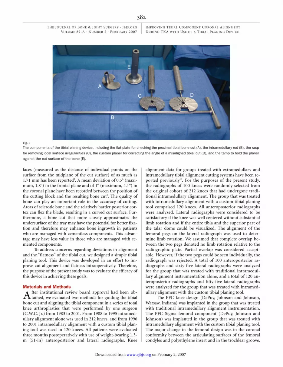
Fig. 1
The components of the tibial planing device, including the flat plate for checking the proximal tibial bone cut (A), the intramedullary rod (B), the rasp
for removing local surface irregularities (C), the custom planer for correcting the angle of a misaligned tibial cut (D), and the tamp to hold the planer
against the cut surface of the bone (E).
Patil.fm Page 382 Wednesday, January 10, 2007 1:43 PM
on February 2, 2007 www.ejbjs.orgDownloaded from

383
THE JOU R N A L OF BO N E & JO I N T SU RG ER Y · JB JS .ORG
VOLU M E 89-A · NU M B ER 2 · FE B R UA R Y 2007IM PROV I N G TI B I AL CO M PON E N T CORONAL AL I G N M E N T DU R I N G TKA W IT H US E OF A TI B I A L PL A N IN G DE V I CE
However, the same PFC tibial tray design was implanted inboth groups.
A medial parapatellar vastus medialis obliquus-splittingapproach was used in all patients. This approach was not“minimally invasive,” and the patella was everted. A measuredresection approach was used to make the bone cuts. The fem-oral cuts were made before the tibial cut. The instrumentationwas set to achieve a target distal femoral cut of 6° of anatomic
valgus, a target posterior femoral cut of 3° of external rotationrelative to the posterior condyles, and a target tibial cut of 90°relative to the anatomic shaft axis of the tibia in the coronalplane and of 85° in the sagittal plane (for 5° of posterior slope).All components were cemented.
Standard surgical instrumentation for intramedullaryalignment was used to make the proximal tibial cut in bothgroups. In the first group, only traditional instrumentationwas used. In the second group, in addition to standard instru-mentation, a custom planing tool was used to check thesurface of the tibia and to make adjustments to any imperfec-tions in the tibial cut. The accuracy of the tibial cut in bothplanes was then checked by sliding a flat plate attached to atrunnion over the intramedullary guide (Figs. 1 and 2-A). De-viation from the desired 90° coronal and 85° sagittal align-
Fig. 2-C
Fig. 2-C The custom planer is threaded on the intramedullary rod. The handle is moved back and forth through an arc while the cutting surface of
the planer is held against the tibial bone cut to realign the cut and to remove any imperfections. Fig. 2-D The tibial cut is now correctly aligned.
Fig. 2-D
Fig. 2-A
Figs. 2-A through 2-D Intraoperative photographs illustrating the sequence of steps involved in the surgical technique. Fig. 2-A The tibial cut ap-
pears to be good when measured with the flat plate alone. Fig. 2-B When the flat plate is threaded over the intramedullary rod, the difference in an-
gulation is more readily seen.
Fig. 2-B
Patil.fm Page 383 Wednesday, January 10, 2007 1:43 PM
on February 2, 2007 www.ejbjs.orgDownloaded from

384
THE JOU R N A L OF BO N E & JO I N T SU RG ER Y · JB JS .ORG
VOLU M E 89-A · NU M B ER 2 · FE B R UA R Y 2007IM PROV I N G TI B I AL CO M PON E N T CORONAL AL I G N M E N T DU R I N G TKA W IT H US E OF A TI B I A L PL A N IN G DE V I CE
ment could be visualized as a gap between the flat plate andthe cut tibial surface (Fig. 2-B). If a gap was detected, the flatplate was removed and the custom planing tool was then sliddown the intramedullary guide and was used to change theangle of the tibial cut (Fig. 2-C). The flat plate was used againto test the alignment of the bone cut. The planing tool wasused primarily to remove any residual imperfections in thetibial cut and not for major correction. In addition to the cus-tom planing tool, a rasp was used to remove surface irregulari-ties and to improve the flatness of the cut. The process wasrepeated, if necessary, until no gap was visible between the flatplate and the tibial bone cut (Fig. 2-D). The rest of the surgicalprocedure, including trial insert selection and cementing ofthe prosthesis, was similar for both groups.
On the anteroposterior radiograph, the axis of the tibialshaft was defined by a line joining the center of the proximaltibial cut and the center of the body of the talus. A second linewas drawn parallel to the undersurface of the tibial component.The medial angle formed between these two lines was measuredto yield the coronal alignment of the tibial component. A coro-nal alignment angle was measured against the target alignmentof 90°. An angle of <90° was classified as varus alignment of thecomponent, whereas an angle of >90° was classified as valgusalignment. Similarly, on the lateral radiograph, the axis of thetibial shaft was defined by a line joining the center of the bodyof the talus and the center of the proximal tibial cut in the lateral
view. The posterior angle formed by the undersurface of the tib-ial component and the tibial axis was measured. The sagittalalignment was compared against the target angle of 85° (the tar-get implantation had a 5° posterior slope). All measurementswere made manually on radiographs by two independent ob-servers (S.P. and C.W.C. Jr.). The mean of two measurementswas recorded for each radiograph. In 93% (409) of the 440 ra-diographs that were measured, there was a disagreement of ≤1°,in 6% (twenty-six) there was a disagreement of 2°, and in 1%(five) there was a disagreement of 3°.
The Student t test was used to test for significant differ-ences between the groups with regard to mean alignment. Thelevel of significance was set at p ≤ 0.05. A sample size of 100was estimated to be sufficient to detect a difference in meanalignment (and standard deviation) of 1° ± 2° between groupswith a power of >90% (alpha = 0.05). The F test was used todetect differences in variance between the groups. The Fisherexact test was used to test for significant differences betweenthe groups with regard to the distribution of cases that werewithin 1°, 2°, or 3° of the target alignment.
Resultshe mean coronal alignment of the tibial component was89.5° ± 2.1° in the group that was treated with traditional
instrumentation alone and 89.6° ± 1.4° in the group that wastreated with use of the custom tibial planing tool (p = 0.52).
T
Fig. 3
Histogram illustrating the distribution of coronal tibial component alignment for the group that was treated with traditional intramedullary alignment
instrumentation only (IM) and the group that was treated with use of the custom planing tool (IMP). Note the reduced variation in alignment for the
latter group.
Patil.fm Page 384 Wednesday, January 10, 2007 1:43 PM
on February 2, 2007 www.ejbjs.orgDownloaded from

385
THE JOU R N A L OF BO N E & JO I N T SU RG ER Y · JB JS .ORG
VOLU M E 89-A · NU M B ER 2 · FE B R UA R Y 2007IM PROV I N G TI B I AL CO M PON E N T CORONAL AL I G N M E N T DU R I N G TKA W IT H US E OF A TI B I A L PL A N IN G DE V I CE
However, the variance in the data was significantly reduced inthe group that was treated with use of the custom planing tool(p < 0.001). The frequency distribution of coronal alignmentfor the two groups is shown in Figure 3. Significantly moretibial components were within 1°, 2°, and 3° of target align-ment in the group that was treated with use of the custom tib-ial planing tool than in the group that was treated withtraditional intramedullary alignment instrumentation alone(p < 0.001, p < 0.001, and p = 0.05, respectively; Fisher exacttest). The proportion of tibial components that were within 3°of the target coronal alignment was 100% in the group thatwas treated with use of the custom tibial planing tool, com-pared with 87% in the group that was treated with traditionalintramedullary alignment instrumentation alone (p = 0.05).In both groups, the tendency was for more components to beimplanted in varus than in valgus.
The mean sagittal alignment was 86.4° ± 3.2° in the groupthat was treated with traditional intramedullary alignment in-strumentation alone and 84.3° ± 1.6° in the group that wastreated with use of the custom tibial planing tool (p < 0.001). Aswas the case for coronal alignment, the variance was significantlylower in the group that was treated with use of the custom plan-ing tool (p < 0.001). Significantly more tibial components werewithin 1°, 2°, and 3° of the target sagittal alignment in the groupthat was treated with use of the custom tibial planing tool than inthe group that was treated with traditional instrumentationalone (p = 0.01, p = 0.05, and p < 0.001, respectively; Fisher ex-act test). The proportion of components that were within 3° ofthe target alignment was 98% in the group that was treated withuse of the custom tibial planing tool, compared with 75% for thegroup that was treated with traditional instrumentation only(p < 0.001). The mean operative times for both groups werevery similar: 118.5 ± 38.6 minutes for the group that wastreated with traditional instrumentation alone and 118.7 ±36.9 minutes for the group that was treated with use of thecustom tibial planing tool (p = 0.97).
Discussionn the present study, the mean coronal alignment was simi-lar between the group that was treated with traditional in-
strumentation alone and the group that was treated with useof the custom tibial planing tool. However, the deviation fromtarget coronal alignment (90° between the tibial tray and thelong axis of tibial shaft) was substantially reduced in the groupthat was treated with use of the custom tibial planing tool asevidenced by the reduced variation in the data. In fact, all ofthe tibial components in the group that was treated with useof the custom tibial planing tool were within 3° of target align-ment, compared with only 87% of the components in thegroup that was treated with traditional instrumentation alone.Errors in the tibial cut may be introduced as a result of flex inthe saw blades, unstable cutting blocks, play in the cutting jig,or imprecise surgical technique9,11,12. For example, a mean dif-ference between cutting block alignment and final bone cutalignment of 0.5° in the coronal plane was recorded with useof computer navigation9. The difference was attributed to the
cumulative cutting error associated with the instrumentation.On the basis of our results, these errors can effectively be re-duced with the use of a tool that detects and smoothes out im-perfections in the tibial cut.
The accuracy of extramedullary as compared with in-tramedullary alignment for guiding the proximal tibial bone cuthas been evaluated before. Several studies have demonstratedno significant difference between these two methods10,13,14. Otherstudies, including one prospective, randomized trial, have dem-onstrated that intramedullary alignment was more accuratethan extramedullary alignment15-18. On the other hand, intra-medullary alignment may be less accurate than extramedullaryalignment in the presence of tibial deformities or a widemedullary canal17,19,20. Direct comparisons with previous re-ports should be made with caution. However, the results of in-tramedullary alignment alone in the present study were similarto those that have been reported for intramedullary or ex-tramedullary alignment, whereas the use of the custom planingdevice significantly reduced the variance of coronal tibial align-ment (p < 0.001). In the present study, all of the tibial compo-nents in the group that was treated with use of the customplaning device were within 3° of target alignment in the coronalplane, which compared favorably with the findings in publishedreports on intramedullary and extramedullary alignment10,14,18,21.
Sagittal plane alignment may be an equally importantfactor in determining prosthetic survival. A number of lateralradiographs could not be measured because of limb rotation;therefore, no definitive conclusions can be made regarding thereduction of outliers in the sagittal plane. Overall, the accu-racy of sagittal alignment was less than that of coronal align-ment. On the basis of the lateral radiographs that could beanalyzed, the accuracy of alignment was substantially greaterin the group that was treated with use of the custom tibialplaning tool, with 98% of the components in that group beingplaced within 3° of target alignment, compared with 77% ofthose in the group that was treated with traditional instru-mentation alone.
Computer-assisted surgical devices, image-guided instru-ments, and active robotic surgery have been proposed to reducealignment errors and to increase the accuracy and precision ofthe femoral and tibial cuts22-24. Several studies have demon-strated that computer-assisted surgical navigation increases theaccuracy of component alignment relative to conventional sur-gical instrumentation and reduces outliers in alignment25-30. Thereduction in outliers with the custom planing device was com-parable with that reported for computer-assisted navigation25-30.
Malalignment of the tibial component alters the distri-bution of tibial loading and can increase shear forces at thetibiofemoral interface, resulting in increased wear. Severalstudies have demonstrated that varus tibiofemoral malalign-ment results in increased wear and a high failure rate4,31-35. Athreshold of 3° of varus malalignment has been reported tosignificantly increase the risk of medial bone collapse (hazardratio = 17.2, p < 0.0001)36. In the present study, although themean tibial alignment was similar in both groups, 100% of thetibial trays in the group that was treated with use of the cus-
I
Patil.fm Page 385 Wednesday, January 10, 2007 1:43 PM
on February 2, 2007 www.ejbjs.orgDownloaded from
386
THE JOU R N A L OF BO N E & JO I N T SU RG ER Y · JB JS .ORG
VOLU M E 89-A · NU M B ER 2 · FE B R UA R Y 2007IM PROV I N G TI B I AL CO M PON E N T CORONAL AL I G N M E N T DU R I N G TKA W IT H US E OF A TI B I A L PL A N IN G DE V I CE
tom tibial planing tool were within 3° of neutral alignmentwhereas only 87% of the trays in the group that was treatedwith traditional instrumentation alone were in this range.This finding suggests that the planing device was effective forreducing clinically relevant magnitudes of malalignment, thusreducing the potential risk of implant failure.
The present study was a comparison of sequential co-horts. A different femoral component design was used in eachcohort. However, the tibial tray instrumentation for in-tramedullary alignment and the cutting blocks used were sim-ilar in both cohorts. The accuracy of measurement of tibialcomponent alignment on radiographs has been shown to besensitive to limb rotation37. Radiographs in which the implantappeared to be rotated were therefore not included in theanalysis. A large number of the lateral radiographs were ex-cluded for this reason. Therefore, firm conclusions cannot bemade regarding the efficacy of this device in reducing outliersin sagittal alignment. This limitation of radiographic analysisis present in the vast majority of the studies of componentalignment cited above. However, a recent study validated simi-lar radiographic measurements, with a mean difference ofonly 0.9° between radiographic measurement and computer-ized tomographic scanograms21.
Currently, computer-navigation systems are expensive,involve a considerable learning curve, and usually add to theoverall operative time, all of which generate resistance to theiruniversal acceptance. Developments are under way to reducethe cost and improve the efficiency of these navigation sys-tems. Computer-navigation systems also have the advantageof improving femoral and overall limb alignment in additionto tibial alignment. However, a simple, inexpensive tool forimproving the accuracy of tibial coronal alignment can matchthe performance reported by most computer-assisted surgicalinstrumentation.
Shantanu Patil, MDDarryl D. D’Lima, MDJames M. Fait, MDClifford W. Colwell Jr., MDShiley Center for Orthopaedic Research and Education at Scripps Clinic, 11025 North Torrey Pines Road, Suite 140, La Jolla, CA 92037. E-mail ad-dress for C.W. Colwell: [email protected]
doi:10.2106/JBJS.F.00204
References
1. Mayor MB, McNamara JL, Surprenant VA, Jensen RE. Analysis of the failure of 122 polyethylene inserts from uncemented tibial knee components. Clin Orthop Relat Res. 1991;273:232-42.
2. Hood RW, Wright TM, Burstein AH. Retrieval analysis of total knee prostheses: a method and its application to 48 total condylar prostheses. J Biomed Mater Res. 1983;17:829-42.
3. D’Lima DD, Chen PC, Colwell CW Jr. Polyethylene contact stresses, articular congruity and knee alignment. Clin Orthop Relat Res. 2001;392:232-8.
4. D’Lima DD, Hermida JC, Chen PC, Colwell CW Jr. Polyethylene wear and varia-tions in knee kinematics. Clin Orthop Relat Res. 2001;392:124-30.
5. Jeffery RS, Morris RW, Denham RA. Coronal alignment after total knee re-placement. J Bone Joint Surg Br. 1991;73:709-14.
6. Rand JA, Coventry MB. Ten-year evaluation of geometric total knee arthro-plasty. Clin Orthop Relat Res. 1988;232:168-73.
7. Ritter MA, Faris PM, Keating EM, Meding JB. Postoperative alignment of total knee replacement. Its effect on survival. Clin Orthop Relat Res. 1994;299:153-6.
8. Toksvig-Larsen S, Ryd L. Surface characteristics following tibial preparation during total knee arthroplasty. J Arthroplasty. 1994;9:63-6.
9. Kozinn SC, Scott R. Unicondylar knee arthroplasty. J Bone Joint Surg Am. 1989;71:145-50.
10. Teter KE, Bregman D, Colwell CW Jr. Accuracy of intramedullary versus ex-tramedullary tibial alignment cutting systems in total knee arthroplasty. Clin Orthop Relat Res. 1995;321:106-10.
11. Otani T, Whiteside LA, White SE. Cutting errors in preparation of femoral components in total knee arthroplasty. J Arthroplasty. 1993;8:503-10.
12. Plaskos C, Hodgson AJ, Inkpen K, McGraw RW. Bone cutting errors in total knee arthroplasty. J Arthroplasty. 2002;17:698-705.
13. Ishii Y, Ohmori G, Bechtold JE, Gustilo RB. Extramedullary versus intra-medullary alignment guides in total knee arthroplasty. Clin Orthop Relat Res. 1995;318:167-75.
14. Yang SH, Liu TK. Intramedullary versus extramedullary tibial alignment guides in total knee arthroplasty. J Formos Med Assoc. 1998;97:564-8.
15. Brys DA, Lombardi AV Jr, Mallory TH, and Vaughn BK. A comparison of in-tramedullary and extramedullary alignment systems for tibial component place-ment in total knee arthroplasty. Clin Orthop Relat Res. 1991;263:175-9.
16. Mahaluxmivala J, Bankes MJ, Nicolai P, Aldam CH, Allen PW. The effect of surgeon experience on component positioning in 673 Press Fit Condylar posterior cruciate-sacrificing total knee arthroplasties. J Arthroplasty. 2001;16:635-40.
17. Maestro A, Harwin SF, Sandoval MG, Vaquero DH, Murcia A. Influence of intramedullary versus extramedullary alignment guides on final total knee arthroplasty component position: a radiographic analysis. J Arthroplasty. 1998;13:552-8.
18. Reed MR, Bliss W, Sher JL, Emmerson KP, Jones SM, Partington PF. Ex-tramedullary or intramedullary tibial alignment guides: a randomised, prospec-tive trial of radiological alignment. J Bone Joint Surg Br. 2002;84:858-60.
19. Simmons ED Jr, Sullivan JA, Rackemann S, Scott RD. The accuracy of tibial intramedullary alignment devices in total knee arthroplasty. J Arthro-plasty. 1991;6:45-50.
20. Bono JV, Roger DJ, Laskin RS, Peterson MG, Paulsen CA. Tibial intramedul-lary alignment in total knee arthroplasty. Am J Knee Surg. 1995;8:7-12.
21. Jeffcote B, Shakespeare D. Varus/valgus alignment of the tibial component in total knee arthroplasty. Knee. 2003;10:243-7.
22. Matsen FA 3rd, Garbini JL, Sidles JA, Pratt B, Baumgarten D, Kaiura R. Ro-botic assistance in orthopaedic surgery. A proof of principle using distal femoral arthroplasty. Clin Orthop Relat Res. 1993;296:178-86.
23. Delp SL, Stulberg SD, Davies B, Picard F, Leitner F. Computer assisted knee replacement. Clin Orthop Relat Res. 1998;354:49-56.
24. Kienzle TC, Stulberg SD, Peshkin M, Quaid A, Lea J, Goswami A, Wu CH. A computer-assisted total knee replacement surgical system using a calibrated robot. In: Taylor RH, Lavallee S, Burdea GC, Mosges R, editors. Computer-integrated surgery: technology and clinical applications. Cambridge, MA: The MIT Press; 1996. p 409-16.
25. Nabeyama R, Matsuda S, Miura H, Mawatari T, Kawano T, Iwamoto Y. The accuracy of image-guided knee replacement based on computed tomography. J Bone Joint Surg Br. 2004;86:366-71.
26. Sparmann M, Wolke B, Czupalla H, Banzer D, Zink A. Positioning of total knee arthroplasty with and without navigation support. A prospective, ran-domised study. J Bone Joint Surg Br. 2003;85:830-5.
27. Jenny JY, Boeri C. Computer-assisted implantation of total knee prostheses: a case-control comparative study with classical instrumentation. Comput Aided Surg. 2001;6:217-20.
Patil.fm Page 386 Wednesday, January 10, 2007 1:43 PM
on February 2, 2007 www.ejbjs.orgDownloaded from

387
THE JOU R N A L OF BO N E & JO I N T SU RG ER Y · JB JS .ORG
VOLU M E 89-A · NU M B ER 2 · FE B R UA R Y 2007IM PROV I N G TI B I AL CO M PON E N T CORONAL AL I G N M E N T DU R I N G TKA W IT H US E OF A TI B I A L PL A N IN G DE V I CE
28. Matsumoto T, Tsumura N, Kurosaka M, Muratsu H, Kuroda R, Ishimoto K, Tsujimoto K, Shiba R, Yoshiya S. Prosthetic alignment and sizing in computer-assisted total knee arthroplasty. Int Orthop. 2004;28:282-5.
29. Haaker RG, Stockheim M, Kamp M, Proff G, Breitenfelder J, Ottersbach A. Computer-assisted navigation increases precision of component placement in total knee arthroplasty. Clin Orthop Relat Res. 2005;433:152-9.
30. Stulberg SD. How accurate is current TKR instrumentation? Clin Orthop Relat Res. 2003;416:177-84.
31. Windsor RE, Scuderi GR, Moran MC, Insall JN. Mechanisms of failure of the femoral and tibial components in total knee arthroplasty. Clin Orthop Relat Res. 1989;248:15-20.
32. Feng EL, Stulberg SD, Wixson RL. Progressive subluxation and polyethylene wear in total knee replacements with flat articular surfaces. Clin Orthop Relat Res. 1994;299:60-71.
33. Hernigou P, Deschamps G. Alignment influences wear in the knee after medial unicompartmental arthroplasty. Clin Orthop Relat Res. 2004;423:161-5.
34. Matsuda S, Miura H, Nagamine R, Urabe K, Harimaya K, Matsunobu T, Iwa-moto Y. Changes in knee alignment after total knee arthroplasty. J Arthroplasty. 1999;14:566-70.
35. Engh GA, Koralewicz LM, Pereles TR. Clinical results of modular polyethylene insert exchange with retention of total knee arthroplasty components. J Bone Joint Surg Am. 2000;82:516-23.
36. Berend ME, Ritter MA, Meding JB, Faris PM, Keating EM, Redelman R, Faris GW, Davis KE. Tibial component failure mechanisms in total knee arthroplasty. Clin Orthop Relat Res. 2004;428:26-34.
37. Elloy MA, Manning MP, Johnson R. Accuracy of intramedullary alignment in total knee replacement. J Biomed Eng. 1992;14:363-70.
Patil.fm Page 387 Wednesday, January 10, 2007 1:43 PM
on February 2, 2007 www.ejbjs.orgDownloaded from