architecture and microstructure of cortical bone in reconstructed canine mandibles after bone...
TRANSCRIPT
ORIGINAL RESEARCH
Architecture and Microstructure of Cortical Bonein Reconstructed Canine Mandibles After Bone TransportDistraction Osteogenesis
Uriel Zapata • Emily K. Halvachs • Paul C. Dechow •
Mohammed E. Elsalanty • Lynne A. Opperman
Received: 27 April 2011 / Accepted: 5 August 2011 / Published online: 17 September 2011
� Springer Science+Business Media, LLC 2011
Abstract Reconstruction of the canine mandible using
bone transport distraction osteogenesis has been shown to
be a suitable method for correcting segmental bone defects
produced by cancer, gunshots, and trauma. Although the
mechanical quality of the new regenerate cortical bone
seems to be related to the mineralization process, several
questions regarding the microstructural patterns of the new
bony tissue remain unanswered. The purpose of this study
was to quantify any microstructural differences that may
exist between the regenerate and control cortical bone. Five
adult American foxhound dogs underwent unilateral bone
transport distraction of the mandible to repair bone defects
of 30–35 mm. Animals were killed 12 weeks after the
beginning of the consolidation period. Fourteen cylindrical
cortical samples were extracted from the superior, medial,
and inferior aspects of the lingual and buccal plates of the
reconstructed aspect of the mandible, and 21 specimens
were collected similarly from the contralateral aspect of the
mandible. Specimens were evaluated using histomorpho-
metric and micro-computed tomographic techniques to
compare their microstructure. Except for differences in
haversian canal area, histomorphometric analyses sug-
gested no statistical differences in microstructure between
regenerate and control cortical bone. Morphological eval-
uation suggested a consistent level of anisotropy, possibly
related to the distraction vector. After 12 weeks’ consoli-
dation, bone created during bone transport distraction
osteogenesis was comparable to native bone in micro-
structure, architecture, and mechanical properties. It is
proposed that, after enough time, the properties of the
regenerate bone will be identical to that of native bone.
Keywords Micro-CT �Histomorphometry �Bone healing �Bone strain � Distraction osteogenesis � Biomechanics
Introduction
The canine mandible is derived from the first visceral, or
mandibular, arch; and through intramembranous ossifica-
tion the dentary bone is formed [1]. This bone fuses with
the condylar cartilage to create the body and ramus of the
mandible. Before about 5 weeks of age, the rate of bone
deposition is very rapid but then slows down; and by
7 months the mandible reaches a stable size, though bone
remodeling continues [1]. Facial growth occurs in an
imbalanced fashion, yet still maintains a functional equi-
librium [2], mainly through deposition on the posterior
Opperman and Elsalanty are co-owners of the patent on the device
and co-owners of Craniotech ACR Devices, LLC. All other authors
have stated no conflict of interest.
U. Zapata
Division of Basic Medical Sciences, Mercer University,
School of Medicine, Macon, GA, USA
U. Zapata
Mechanical Engineering Department, Eafit University, Medellın,
Colombia
E. K. Halvachs
Department of Sociology and Anthropology, University of Texas
at Arlington, Arlington, TX, USA
P. C. Dechow � L. A. Opperman (&)
Department of Biomedical Sciences, Texas A&M Health
Science Center, Baylor College of Dentistry, 3302 Gaston
Avenue, Dallas, TX 75246, USA
e-mail: [email protected]
M. E. Elsalanty
College of Dental Medicine, Georgia Health Science University,
Augusta, GA, USA
123
Calcif Tissue Int (2011) 89:379–388
DOI 10.1007/s00223-011-9529-6
aspect of both the body and ramus, while being resorbed
from the anterior surfaces [1]. Generally, the intramem-
branous formation of the craniofacial bones defines a par-
ticular orientation of the osteons within the mandible,
which is associated with bone function, fracture repair, and
mandibular treatments [3].
Using the principles of fracture repair for the treatment
of deformities and defects of the mandible to restore its
function, a new technique has gained momentum as a
useful surgical procedure. The purpose of this procedure is
not only to correct craniofacial genetic deformities, espe-
cially in cases of asymmetrical growth, but also to repair
defects produced by cancer, gunshots, and trauma. Its first
application was distraction osteogenesis (DO), a popular
procedure used to generate new bony tissue between two
edges by applying gradual tensile force. The procedure was
first applied to the mandible by Snyder et al. [4] using a
canine model, then more recently to correct human facial
deformities [5]. A variation of the procedure is bone
transport distraction osteogenesis (BTDO), in which a
segment of bone is moved between two bony edges while
regenerating a bone to bridge the gap (Fig. 1). BTDO was
first applied in dog mandibles by Costantino et al. [6] and
later in human patients [7]. Since BTDO has become
widely used, its clinical results have been impressive not
only in experimental animals but also in humans [8–11].
However, serious questions remain regarding how differ-
ences in biomechanical characteristics, architecture of
haversian systems, and microstructure of the regenerated
bony tissue when compared with the original bone would
affect the restoration of mandibular function.
Although the biomechanical characteristics of the new
bony tissue generated after DO have been addressed in
different studies [12–15], only one previous study has
focused on the mechanical properties of the new bone
formed after BTDO [16], which showed a direct relationship
between mechanical properties of cortical bone and its
microstructure, similar to Currey [17].
Panikarovskii et al. [18] first examined the histology of
regenerate tissue formed through mandibular distraction,
finding a relationship between the vector of distraction and
the orientation of primary osteons. Subsequently, several
experimental studies looked at the microstructure of the
regenerated cortical bone after DO of the mandible. Most
of them did this using histological evaluation [19–23] as
well as a combination of both micro-computed tomography
(l-CT) and histological techniques [14, 24] to identify
quantitative and qualitative microstructural characteristics
of the newly formed tissue. However, no studies have
performed either histological or l-CT evaluation of bone
tissues obtained after BTDO of the mandible. The objective
of this work was to test the hypothesis that both micro-
structure and haversian structures are different in canine
mandibular control bone compared to regenerated cortical
bone obtained by BTDO. This difference might be a con-
sequence of the differences in strain patterns during func-
tional loads in the former and the tension strain during
BTDO in the latter. Both histomorphometric and l-CT
techniques were used in this study.
Materials and Methods
Specimen Collection
In preparation for mandibular bone transport, a unilateral
defect of 30–35 mm was created in the mandibles of five
adult male American foxhound dogs. The jaw was then
reconstructed with a novel bone transport reconstruction
device (BTRP; Craniotech ACR Devices, Dallas, TX)
composed of a reconstruction plate and an intraoral transport
unit attached to a 10-mm bony disc (Fig. 1) [10]. The device
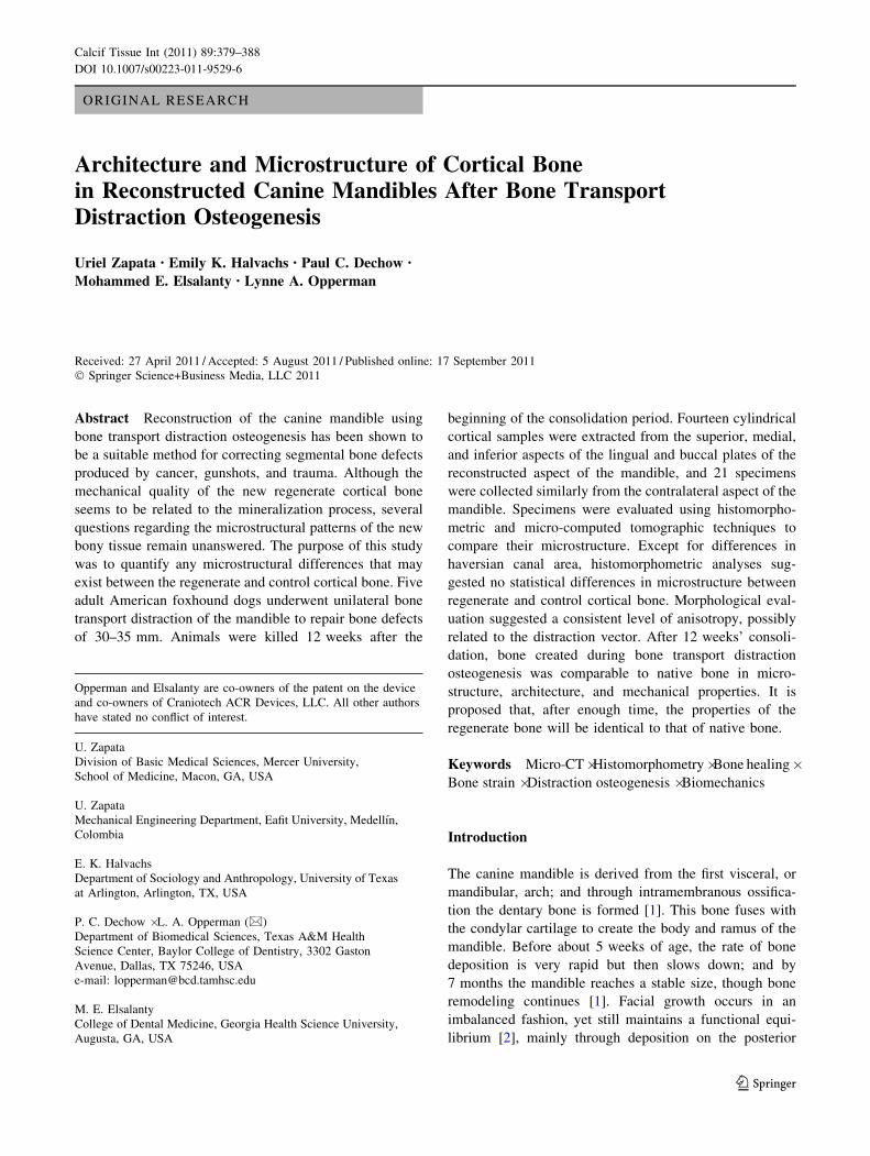
Fig. 1 a The mandibular bone transport (MBT) device in situ is
positioned on the buccal side of the mandible. The transport segment
is detached from the distal segment of bone, with the transport unit
attached (arrow). The cable that protruded through the skin after
wound closure activated the transport unit (arrowhead). b The MBT
device bridges the mandibular bony defect between the anterior edge
(AE) and the docking site (DS). The transport disc (TD) was advanced
from AE to DS, where the process is completed by compression
stress. The new regenerate bone is clearly visible between AE and TD
(asterisks), and it appears parallel to the direction of distraction
380 U. Zapata et al.: Microstructure of Cortical Bone
123
was activated 1 mm/day for 4–5 weeks, and then the new
bone was allowed to consolidate for 6 weeks after the dis-
traction was complete. After consolidation, dental implants
were placed on both the experimental and control sides. The
animals were allowed to heal for 6 more weeks before being
killed and their mandibles dissected. Mandibles were stored
at -20�C prior to removal of bone specimens to ensure that
the bone’s elastic properties were preserved [25]. A total of
35 cylindrical cortical bone specimens (average 5.93 ±
0.06 mm in diameter and 2.72 ± 0.24 mm in thickness)
were removed from the lingual and buccal surfaces of the
basal and alveolar regions of the mandible, from both the
regenerate and control mandibular surfaces. Before removal,
each sample was marked with a pencil line parallel to the
base of the mandible to act as a visual marker of the sample’s
anatomical orientation. Twenty-one cylindrical cortical
specimens were taken from the control side and 14 from the
regenerate side using a low-speed dental drill continuously
cooled with water during drilling. All sample specimens
were stored in a 50:50 solution of 95% ethanol and isotonic
saline at room temperature (19�C) in order to maintain the
elastic properties of the bone [26]. Since only the properties
of cortical bone can be assessed by ultrasound, all visible
trabecular bone was carefully removed down to the cortical
surface to avoid measuring trabecular spaces instead of
cortical porosity. Cortical bone apparent density (mg/cm3)
was calculated from the sample weight using an analytical
balance (PM460; Mettler-Toledo International, Columbus,
OH) and differential volume in water, based on Archimedes’
principle of buoyancy [26]. In addition, elastic mechanical
properties were obtained for all specimens using an ultra-
sound test technique [16]. The direction of maximum
strength was obtained with respect to the original reference
line, and it was marked on the specimen by two notches that
were clearly observed during three-dimensional l-CT
reconstructions (Fig. 2). After the mechanical properties
were obtained for all specimens, the discs were scanned
using l-CT and subsequently embedded and sectioned for
histomorphometric analysis.
l-CT Analysis
l-CT images were captured using a Scanco desktop cone-
beam l-CT 35 (Scanco Medical, Bassersdorf, Switzerland).
To prepare the specimens for scanning, five 12.3-mm tubes
were filled with a 50:50 solution of 95% ethanol and isotonic
saline solution. The number of specimens placed in each tube
varied from four to eight, with all samples in a single tube
belonging to the same individual. The pencil mark parallel to
the base of the mandible made before extraction was used to
orient the specimens within the tubes. Before being scanned,
specimens were viewed in scout view to verify their positions
as well as to set the volume of interest to avoid extraneous
scanning. Specimens were scanned at high resolution
(2,048 9 2,048 pixels) with a voxel size of 6 lm and a beam
intensity of 70 KvP. An 800-ms integration time was used in
order to reduce noise. After scanning, slices were contoured
to specify a second volume of interest, which was the area to
be analyzed for cortical porosity (Fig. 2). A logarithm typi-
cally used to characterize trabecular porosity was applied to
cortical bone in order to measure cortical porosity. This made
it possible to identify osteon orientation (Fig. 3). Thresholds
were set to distinguish between gray-scale values for cortical
bone and pore space. A lower threshold of -1,000 and an
upper threshold of 307 were used, as well as a Gauss sigma of
1.5 and a Gauss support of 2. The relative percentage bone
volume (bone volume/total volume, Bv/Tv) and the degree
of anisotropy (length of longest H-vector/shortest H-vector)
were measured. The Bv/Tv is, in this case, a representation of
the percentage of both haversian canals and voids within the
cortical specimens. In addition, the degree of anisotropy is a
representation of the structural conditions with the cylin-
drical specimen. A value close to 1.0 represents an isotropic
characteristic, whereas values higher than 1.0 correspond to
anisotropic conditions. The main advantage of l-CT evalu-
ation is that the specimens remain intact for the following
histomorphometric assessment.
Histomorphometric Evaluation
Cylindrical specimens were split into two symmetrical
parts through the notches used as a reference for the ori-
entation of maximum strength and then fixed in 10%
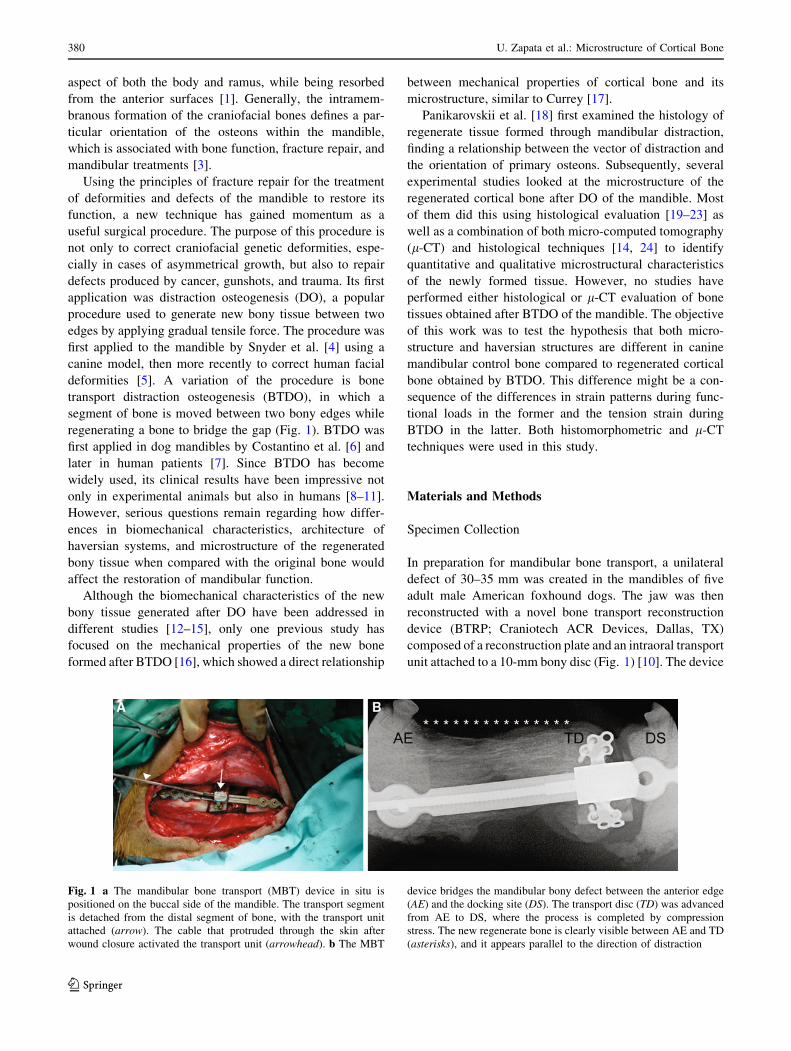
Fig. 2 Cylindrical regenerate cortical bone specimen reconstructed
using l-CT. A marked continuous reference line matching with two
notches represents the direction of maximum strength
U. Zapata et al.: Microstructure of Cortical Bone 381
123
buffered formalin for 7–10 days. At the end of fixation,
specimens were washed well in deionized water and then
dehydrated, soaked in acetone for 2–3 days, and embedded
in methyl methacrylate. Before being placed in the methyl
methacrylate, four specimens at a time were put in a frame
and covered in jade dental stone (Whip-Mix; Pearson
Dental, Houston, TX) in order to embed multiple samples
at once at the same orientation. Sections were cut using an
Isomet low-speed precision sectioning saw (Buehler, Lake
Bluff, IL) with a diamond blade.
Digital photographic registers of each specimen were
recorded using a Nikon (Tokyo, Japan) Coolpix 4500 camera
mounted on a Reichart-Jung (Nussloch, Germany) Microstar
IV microscope via a microscope mount included with the
camera. Pictures were taken in a panoramic sequence from
superior to inferior then merged using Adobe Photoshop
software (Adobe Systems, San Jose, CA) to create a com-
posite image. These images were then transferred to a lab
computer for analysis, and regions of interest were created
within every specimen using BIOQUANT Osteo II software
(Image Analysis, Nashville, TN), with areas ranging
0.622–0.638 mm2. Only bony structures within this area were
quantified. Haversian canal area, osteon area, and osteon
number were measured and calculated using the histomor-
phometric software. Volkmann canals were counted manu-
ally and then recounted for accuracy. When an osteon was on
the perimeter of the region, it was only measured if the
haversian canal was on or inside the border. If the haversian
canal was positioned outside of the borders of the region of
interest, the osteon was not measured. Volkmann canals
visibly associated with only one osteon and those acting as a
connection between two osteons were counted as a single
canal. Those Volkmann canals connecting three or more
osteons were counted as separate canals for each osteon.
Finally, the osteon number was divided by the area of interest
in order to gain the osteon density per square millimeter.
Statistical Analysis
All analyses to test for differences between regenerated and
control cortical bone among various regions of the man-
dibles and between subjects were done using SPSS statis-
tical software (SPSS, Inc., Chicago, IL). One-way ANOVA
was performed to test for histomorphological differences
among the five subjects. In addition, independent sample
tests were performed between 21 regenerate and 14 control
specimens. Because cortical bone specimens were taken
from consistent positions on the mandible at both the
regenerated and control sites, t-tests were used to test for
histomorphological differences between anatomical posi-
tions of the mandible (facial and lingual) by comparing five
pairs of specimens from the regenerate bone obtained from
three subjects against 10 pairs of control specimens
obtained from five subjects and between relative positions
of the mandible (alveolar and basal) by comparing six pairs
of specimens from the regenerate tissue obtained from four
subjects and nine pairs from the control bony tissue
obtained from all subjects [16]. The statistical normality of
the four variables considered in this study was tested using
the Kolmogorov-Smirnov test, resulting in a positive nor-
mality for both osteon area (P = 0.095) and osteon number
(P = 0.20), whereas the Volkmann canal number was
slightly closer to the condition of normality (P = 0.041).
Although haversian canal measurements did not reach the
condition of normality (P \ 0.05), it was assumed that
ANOVA was robust enough to accommodate departures
from this normality. In addition, a Levenes’ test of
homogeneity of variances was performed on the four
variables; homogeneity was satisfied for osteon area
(P = 0.077), osteon number (P = 0.350), and Volkmann
canal number (P = 0.492) but not for haversian area
(P = 0.023). No statistical evaluation was performed on
the qualitative results obtained from the l-CT process.
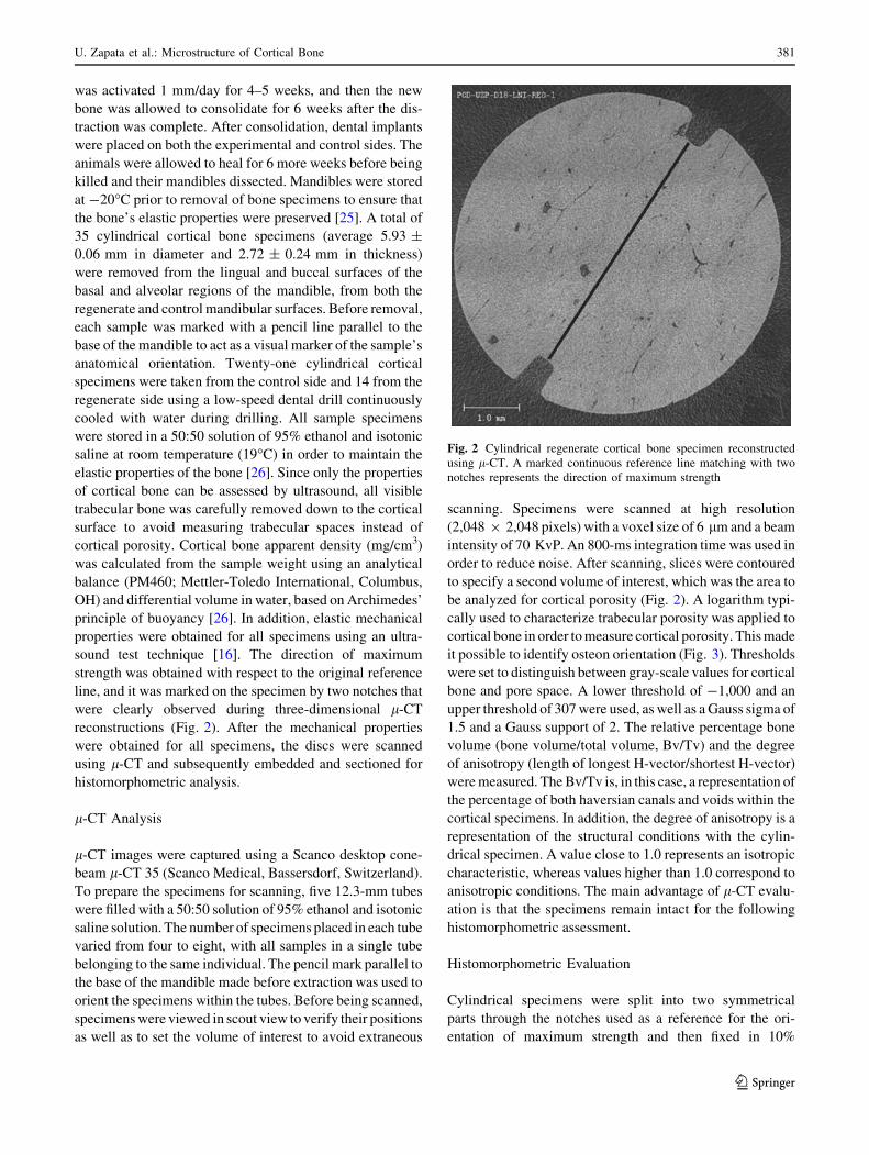
Fig. 3 l-CT scans of cortical bone specimens on the control (a) and regenerate (b) sides. White, bone; gray, haversian spaces. U-shaped divots
indicate the direction of maximal strength. Note that the haversian systems are orientated in the direction of maximal stiffness in both tissues
382 U. Zapata et al.: Microstructure of Cortical Bone
123
Results
Histological Examination
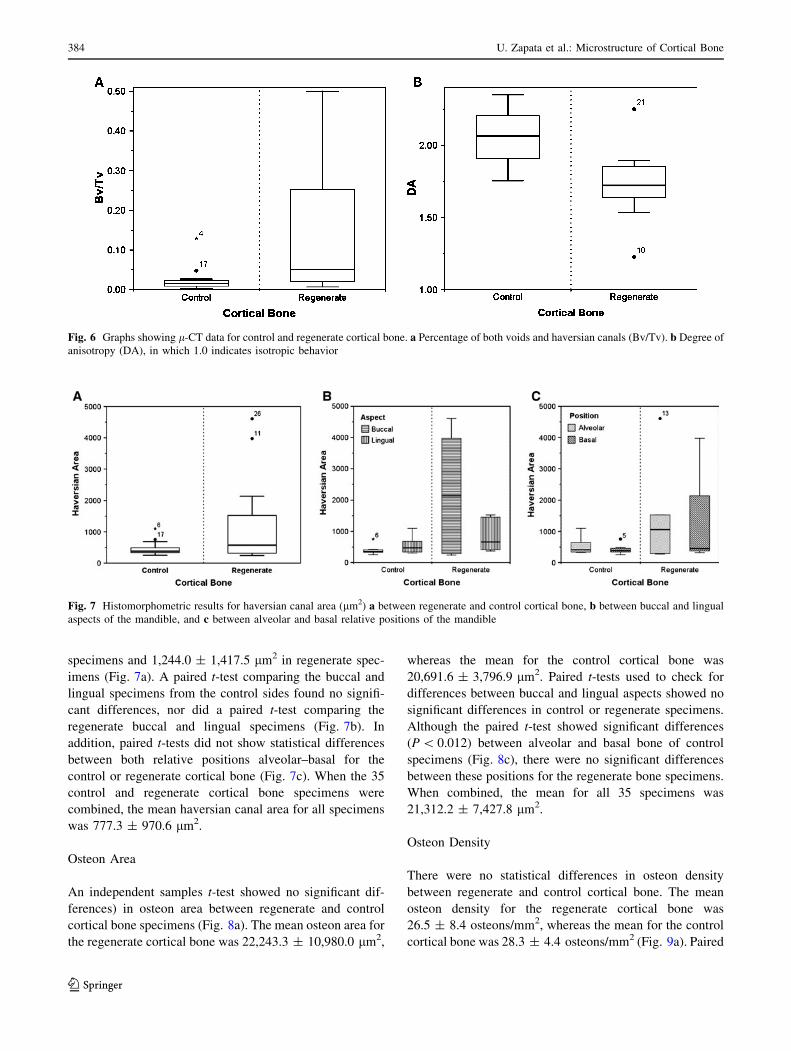
Photomicrographic observations suggested differences in
the microstructure not only between the regenerate and
control cortical bone (Figs. 4, 5) but also between buccal
and lingual positions in the regenerate cortical bone (results
not shown). In addition, it appears that the amount and size
of the osteons were larger in the control than in the
regenerated cortical bone, with fewer interstitial lamellae.
In contrast to control bone, regenerate bone had smaller
osteons with larger haversian canals and voids; under-
mineralized interstitial lamellae and increased numbers of
Volkmann canals were also noted.
l-CT Analysis
Supporting the histological evaluation, l-CT images showed
that the lingual aspect of the regenerate cortical bone evinces
more mature osteons of greater diameter than the buccal
aspect (Fig. 3a, b). Whereas the lingual aspect of the regen-
erate more closely resembles the control, the buccal aspect of
the regenerate contained fewer osteons but many osteocytes,
usually associated with woven bone. However, we observed
that the average osteon orientation in both control and
regenerated cortical bone tended to follow the orientation of
maximum strength (defined by the pair of notches), which
was directly related to the mandibular basal plane for the
control cortical bone (Fig. 3a, b) and the direction of the
distraction vector for the regenerate cortical bone.
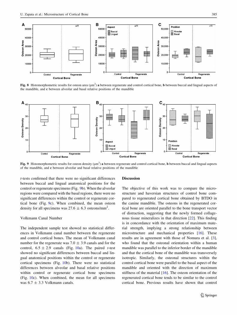
There was a statistical difference in the percentage of
voids and haversian canals (Bv/Tv) between regenerate and
control cortical bone (F = 37.02, P \ 0.02). This was
higher in the regenerate (14.9%) than in the control (2.2%)
cortical bone (Fig. 6a). No statistical differences in Bv/Tv
were present when comparing anatomical positions buc-
cal–lingual or when comparing relative position basal–
alveolar. There was a significant difference in the degree of
anisotropy (DA) between regenerate and control specimens
(F = 0.06, P \ 0.05), which was slightly lower in the
regenerate (1.72 ± 0.23) than in the control (2.05 ± 0.18)
cortical bone (Fig. 6b). The pair test showed no significant
differences in DA between anatomical positions buccal–
lingual or between relative position basal–alveolar.
Haversian Canal Area
Independent sample t-tests showed no significant differ-
ences in haversian canal area between the regenerate and
control cortical bone (P = 0.062, F = 22.368). The mean
haversian canal area was 466.2 ± 203.0 lm2 in control
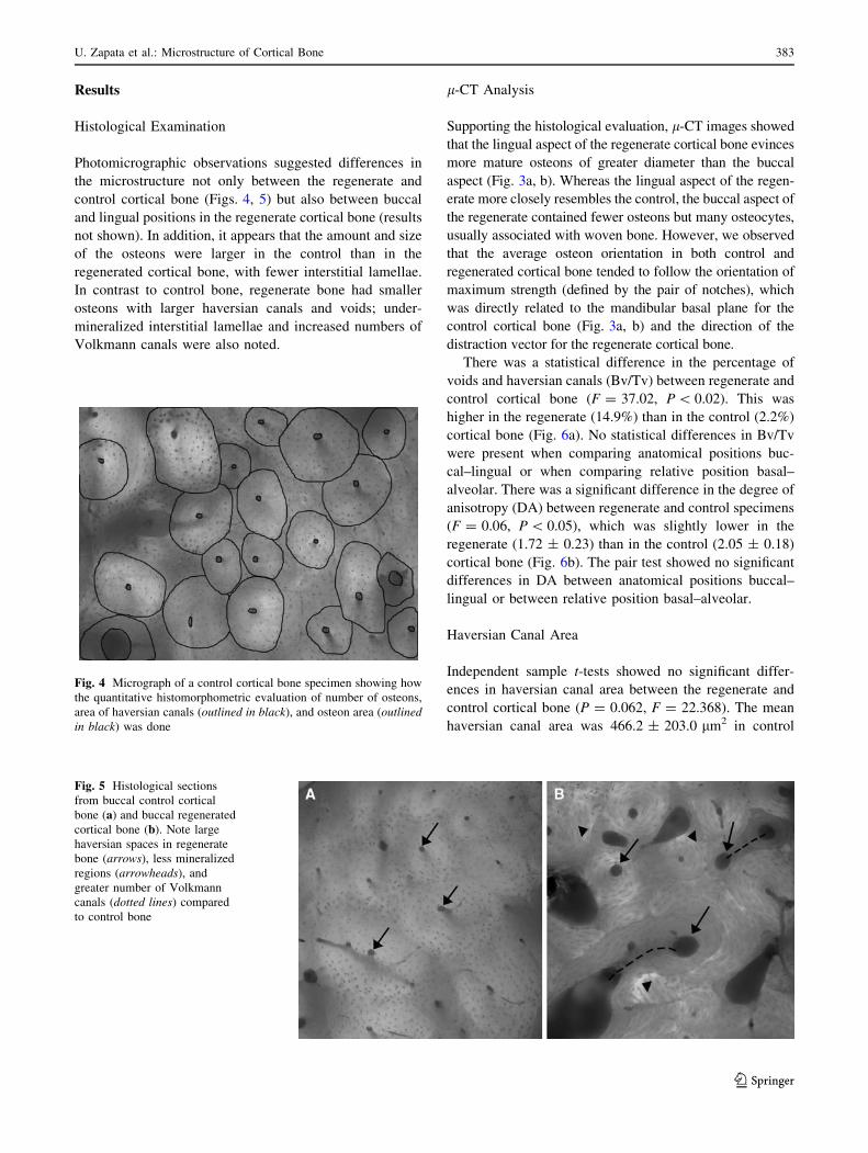
Fig. 4 Micrograph of a control cortical bone specimen showing how
the quantitative histomorphometric evaluation of number of osteons,
area of haversian canals (outlined in black), and osteon area (outlinedin black) was done
Fig. 5 Histological sections
from buccal control cortical
bone (a) and buccal regenerated
cortical bone (b). Note large
haversian spaces in regenerate
bone (arrows), less mineralized
regions (arrowheads), and
greater number of Volkmann
canals (dotted lines) compared
to control bone
U. Zapata et al.: Microstructure of Cortical Bone 383
123
specimens and 1,244.0 ± 1,417.5 lm2 in regenerate spec-
imens (Fig. 7a). A paired t-test comparing the buccal and
lingual specimens from the control sides found no signifi-
cant differences, nor did a paired t-test comparing the
regenerate buccal and lingual specimens (Fig. 7b). In
addition, paired t-tests did not show statistical differences
between both relative positions alveolar–basal for the
control or regenerate cortical bone (Fig. 7c). When the 35
control and regenerate cortical bone specimens were
combined, the mean haversian canal area for all specimens
was 777.3 ± 970.6 lm2.
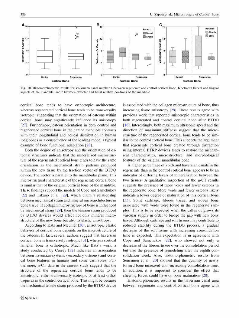
Osteon Area
An independent samples t-test showed no significant dif-
ferences) in osteon area between regenerate and control
cortical bone specimens (Fig. 8a). The mean osteon area for
the regenerate cortical bone was 22,243.3 ± 10,980.0 lm2,
whereas the mean for the control cortical bone was
20,691.6 ± 3,796.9 lm2. Paired t-tests used to check for
differences between buccal and lingual aspects showed no
significant differences in control or regenerate specimens.
Although the paired t-test showed significant differences
(P \ 0.012) between alveolar and basal bone of control
specimens (Fig. 8c), there were no significant differences
between these positions for the regenerate bone specimens.
When combined, the mean for all 35 specimens was
21,312.2 ± 7,427.8 lm2.
Osteon Density
There were no statistical differences in osteon density
between regenerate and control cortical bone. The mean
osteon density for the regenerate cortical bone was
26.5 ± 8.4 osteons/mm2, whereas the mean for the control
cortical bone was 28.3 ± 4.4 osteons/mm2 (Fig. 9a). Paired
Fig. 6 Graphs showing l-CT data for control and regenerate cortical bone. a Percentage of both voids and haversian canals (Bv/Tv). b Degree of
anisotropy (DA), in which 1.0 indicates isotropic behavior
Fig. 7 Histomorphometric results for haversian canal area (lm2) a between regenerate and control cortical bone, b between buccal and lingual
aspects of the mandible, and c between alveolar and basal relative positions of the mandible
384 U. Zapata et al.: Microstructure of Cortical Bone
123
t-tests confirmed that there were no significant differences
between buccal and lingual anatomical positions for the
control or regenerate specimens (Fig. 9b). When the alveolar
regions were compared with the basal regions, there were no
significant differences within the control or regenerate cor-
tical bone (Fig. 9c). When combined, the mean osteon
density for all specimens was 27.6 ± 6.3 osteons/mm2.
Volkmann Canal Number
The independent sample test showed no statistical differ-
ences in Volkmann canal number between the regenerate
and control cortical bones. The mean of Volkmann canal
number for the regenerate was 7.0 ± 3.9 canals and for the
control, 6.5 ± 2.9 canals (Fig. 10a). The paired t-test
showed no significant differences between buccal and lin-
gual anatomical positions within the control or regenerate
cortical specimens (Fig. 10b). There were no statistical
differences between alveolar and basal relative positions
within control or regenerate cortical bone specimens
(Fig. 10c). When combined, the mean for all specimens
was 6.7 ± 3.3 Volkmann canals.
Discussion
The objective of this work was to compare the micro-
structure and haversian structures of control bone com-
pared to regenerated cortical bone obtained by BTDO in
the canine mandible. The osteons in the regenerated cor-
tical bone are oriented parallel to the bone transport vector
of distraction, suggesting that the newly formed collage-
nous tissue mineralizes in that direction [22]. This finding
is in concordance with the orientation of maximum mate-
rial strength, implying a strong relationship between
microstructure and mechanical properties [16]. These
results are in agreement with those of Nomura et al. [3],
who found that the osteonal orientation within a human
mandible was parallel to the inferior border of the mandible
and that the cortical bone of the mandible was transversely
isotropic. Similarly, the osteonal structures within the
control cortical bone were parallel to the basal aspect of the
mandible and oriented with the direction of maximum
stiffness of the material [16]. The osteon orientation of the
regenerated cortical bone tends to be similar to the control
cortical bone. Previous results have shown that control
Fig. 8 Histomorphometric results for osteon area (lm2) a between regenerate and control cortical bone, b between buccal and lingual aspects of
the mandible, and c between alveolar and basal relative positions of the mandible
Fig. 9 Histomorphometric results for osteon density (lm2) a between regenerate and control cortical bone, b between buccal and lingual aspects
of the mandible, and c between alveolar and basal relative positions of the mandible
U. Zapata et al.: Microstructure of Cortical Bone 385
123
cortical bone tends to have orthotropic architecture,
whereas regenerated cortical bone tends to be transversally
isotropic, suggesting that the orientation of osteons within
cortical bone may significantly influence its anisotropy
[27]. Furthermore, osteon orientation in both control and
regenerated cortical bone in the canine mandible contrasts
with their longitudinal and helical distribution in human
long bones as a consequence of the loading mode, a typical
example of bone functional adaptation [28].
Both the degree of anisotropy and the orientation of os-
teonal structures indicate that the mineralized microstruc-
ture of the regenerated cortical bone tends to have the same
orientation as the mechanical strain patterns produced
within the new tissue by the traction vector of the BTDO
device. The vector is parallel to the mandibular plane. This
microstructural characteristic of the regenerate cortical bone
is similar that of the original cortical bone of the mandible.
These findings support the models of Cope and Samchukov
[22] and Takano et al. [29], which claim a relationship
between mechanical strain and mineral microarchitecture in
bone tissue. If collagen microstructure of bone is influenced
by mechanical strain [29], then the tension strain produced
by BTDO devices would affect not only mineral micro-
structure of the new bone but also its elastic anisotropy.
According to Katz and Meunier [30], anisotropic elastic
behavior of cortical bone depends on the microstructure of
the osteons. In fact, several authors suggest that haversian
cortical bone is transversely isotropic [31], whereas cortical
lamellar bone is orthotropic. Much like Katz’s work, a
study conducted by Currey [32] indicates an association
between haversian systems (secondary osteons) and corti-
cal bone features in humans and some carnivores. Fur-
thermore, l-CT data in the current study suggest that the
structure of the regenerate cortical bone tends to be
anisotropic, either transversally isotropic or at least ortho-
tropic as in the control cortical bone. This might be because
the mechanical tensile strain produced by the BTDO device
is associated with the collagen microstructure of bone, thus
increasing tissue anisotropy [29]. These results agree with
previous work that reported anisotropic characteristics in
both regenerated and control cortical bone after BTDO
[16]. Interestingly, both maximum ultrasonic speed and the
direction of maximum stiffness suggest that the micro-
structure of the regenerated cortical bone tends to be sim-
ilar to the control cortical bone. This supports the argument
that regenerate cortical bone created through distraction
using internal BTRP devices tends to restore the mechan-
ical characteristics, microstructure, and morphological
features of the original mandibular bone.
A higher percentage of voids and haversian canals in the
regenerate than in the control cortical bone appears to be an
indicator of differing levels of mineralization between the
two tissues. A qualitative inspection of the l-CT results
suggests the presence of more voids and fewer osteons in
the regenerate bone. More voids and fewer osteons likely
indicate a lower degree of maturation of this cortical bone
[33]. Some cartilage, fibrous tissue, and woven bone
associated with voids were found in the regenerate sam-
ples. This is to be expected when the callus outgrows its
vascular supply in order to bridge the gap with new bony
tissue. Although cartilage and soft tissues may contribute to
reduced stability during the BTDO process, a gradual
decrease of the soft tissue with increasing consolidation
time is expected. This expectation is in agreement with
Cope and Samchukov [22], who showed not only a
decrease of the fibrous tissue over the consolidation period
but also the presence of remodeling after the eighth con-
solidation week. Also, histomorphometric results from
Sencimen et al. [20] showed that the quantity of newly
formed bone increased with increasing consolidation time.
In addition, it is important to consider the effect that
chewing forces could have on bone maturation [20].
Histomorphometric results in the haversian canal area
between regenerate and control cortical bone agree with
Fig. 10 Histomorphometric results for Volkmann canal number a between regenerate and control cortical bone, b between buccal and lingual
aspects of the mandible, and c between alveolar and basal relative positions of the mandible
386 U. Zapata et al.: Microstructure of Cortical Bone
123
those of Sencimen et al. [20], who did not observe histo-
logical differences between the newly formed bone and
native bone. The larger haversian canals seen in the
regenerate compared to control cortical bone suggest that
mineralization of the regenerate is still in process. In
addition, the difference in osteon area between basal and
alveolar positions in the control cortical bone, which is not
present in the regenerate specimens, suggests different
biomechanical tissue responses to different physiological
conditions. This difference could be associated with
bending of the bone in the part of the mandible that is not
supported by the BTRP device. Although we did not find
any statistical differences between the buccal and lingual
regions of the regenerate and control cortical bone, Mulder
et al. [33] reported differences in both architecture and
degree of mineralization between buccal and lingual plates
of the pig mandible, characterized by a more compact
structure and higher mineralization in the lingual region.
Their findings agree with previous work that reported dif-
ferences in mechanical properties between buccal and
lingual positions of the canine mandible in the regenerate
cortical bone, which were assumed to be associated with
the presence of the device on one of the sides of the
mandible [16]. The device was placed on the buccal side of
the regenerate, so these tests were performed to determine
if the device had any effect on the microstructure of the
bone. Although these biomechanical differences can be
associated with the fact that the maturation of the cortical
bone may alter the mechanical properties at the micro-
structural level [34], it is also possibly associated with
differences in the bone formation process.
There are limitations to the present study. For example,
the animals were drawn from a large sample of dogs used
to study different consolidation periods of BTDO; thus,
five animals were considered in our study. This reduction
in the number of animals limited the statistical power,
although the number of cortical bone specimens used for
comparisons is appropriate. Specimens used as control
bone are obtained from the same animal by removing them
from the side contralateral to the device. Thus, increased
physiological loads on the control side during the 17 weeks
of the distraction process could affect control bone prop-
erties. These options were chosen to reduce the number of
animals required for the study. In spite of this approach,
and in contrast to the hypothesis, there were no differences
in microstructure between regenerate and control cortical
bone, except for the difference in density that suggests an
incomplete mineralization of the new tissue. Given a
longer consolidation period, it is probable that the regen-
erate cortical bone would eventually become comparable to
the native bone. Finally, these results appear to support the
argument that BTDO is able to successfully generate new
cortical bone similar to the original bone. In addition,
BTDO restored the mechanical characteristics, micro-
structure, and morphological features of the original cor-
tical bone in the mandible, which facilitate the restitution
of mandibular function.
Conclusion
The combined use of histology to characterize the maturity
of the new cortical bone when compared with control bone
and l-CT to compare the anisotropic differences between
regenerated and control tissues was helpful in understanding
how the strain patterns generated in the mandible by BTDO
affect microstructure, architecture, and mechanical proper-
ties of cortical bone. There is an obvious orientation of the
basic haversian system parallel to the vector of distraction in
the regenerate cortical bone, and the same pattern is present
in the control cortical bone but with their orientation parallel
to the base of the mandible. This similarity suggests no
significant differences in microstructure between the new
regenerate and control cortical bone, except for the level of
mineralization reached at this specific consolidation period.
Furthermore, it is important to note the effect of the vector of
distraction on the patterns of microstructure associated with
the regenerate tissue.
Acknowledgements This study was supported by NIH/NIDCR
Grants R42 DE015437-03 and R43 DE017259-05 and a fellowship
from Departamento Administrativo de Ciencia, Tecnologıa e Inno-
vacion COLCIENCIAS, Bogota, Colombia. The authors thank Ms. Jo
Taylor, from Baylor College of Dentistry, for providing advice, help,
and guidance regarding the histological process. The BTRP device
patent is assigned to a company (Craniotech ACR Devices, LLC)
owned by L. A. O. and M. E. E.
References
1. Hennet PR, Harvey CE (1992) Craniofacial development and
growth in the dog. J Vet Dent 9:11–18
2. Ilizarov GA (1989) The tension-stress effect on the genesis and
growth of tissues. Part I. The influence of stability of fixation and
soft-tissue preservation. Clin Orthop Relat Res 238:249–281
3. Nomura T, Gold E, Powers MP, Shingaki S, Katz JL (2003)
Micromechanics/structure relationships in the human mandible.
Dent Mater 19:167–173
4. Snyder CC, Levine GA, Swanson HM, Browne EZ Jr (1973)
Mandibular lengthening by gradual distraction. Preliminary
report. Plast Reconstr Surg 51:506–508
5. McCarthy JG, Schreiber J, Karp N, Thorne CH, Grayson BH
(1992) Lengthening the human mandible by gradual distraction.
Plast Reconstr Surg 89:1–8 discussion 9–10
6. Costantino PD, Shybut G, Friedman CD, Pelzer HJ, Masini M,
Shindo ML, Sisson GA Sr (1990) Segmental mandibular regen-
eration by distraction osteogenesis. An experimental study. Arch
Otolaryngol Head Neck Surg 116:535–545
7. Costantino PD, Johnson CS, Friedman CD, Sisson GA Sr (1995)
Bone regeneration within a human segmental mandible defect: a
U. Zapata et al.: Microstructure of Cortical Bone 387
123
preliminary report. Am J Otolaryngol Head Neck Med Surg
16:56–65
8. Spagnoli D (2008) Mandible reconstruction with transport dis-
traction osteogenesis. Atlas Oral Maxillofac Surg Clin North Am
16:287–307
9. Zhou LB, Shang HT, Hu M, Li DC, Sigare S, Chen BL, Liu YP,
Zhao JL (2008) Reconstruction of curved mandibular angle
defects using a new internal transport distraction device: an
experiment in goats. Br J Oral Maxillofac Surg 46:445–448
10. Elsalanty ME, Zakhary I, Akeel S, Benson B, Mulone T, Triplett
GR, Opperman LA (2009) Reconstruction of canine mandibular
bone defects using a bone transport reconstruction plate. Ann
Plast Surg 63:441–448
11. Zhang RZ, Zhang L, Deng Y, Zhang QL, Zhen EM, Yu B (2009)
Reconstruction of mandibular symphyseal defects by an internal
trifocal distractor: an experiment in dogs. Br J Oral Maxillofac
Surg 47:205–209
12. Li J, Hu J, Wang D, Tang Z, Gao Z (2002) Biomechanical
properties of regenerated bone by mandibular distraction osteo-
genesis. Chin J Traumatol 5:67–70
13. Perrott DH, Rahn B, Wahl D, Linke B, Thurmuller P, Troulis M,
Glowacki J, Kaban LB (2003) Development of a mechanical
testing system for a mandibular distraction wound. Int J Oral
Maxillofac Surg 32:523–527
14. Gomez DF, Sant’Anna EF, Leven RM, Ostric SA, Figueroa AA,
Royston TJ, Sumner DR, Polley JW (2005) Microstructural and
strength evaluation of regenerate tissue during the consolidation
period after vertical mandibular ramus distraction. J Craniofac
Surg 16:805–811
15. Kunz C, Adolphs N, Buscher P, Hammer B, Rahn B (2006)
Mineralization and mechanical properties of the canine mandible
distraction wound following acute molding. Int J Oral Maxillofac
Surg 35:822–827
16. Zapata U, Opperman LA, Kontogiorgos E, Elsalanty M, Dechow
PC (2011) Biomechanical characteristics of regenerated cortical
bone in the canine mandible. J Tissue Eng Regen Med 5:551–559
17. Currey J (2009) Measurement of the mechanical properties of
bone: a recent history. Clin Orthop Relat Res 467:1948–1954
18. Panikarovskii VV, Grigor’ian AS, Kaganovich SI, Osipian EM,
Antipova ZP (1982) Characteristics of mandibular reparative
osteogenesis under compression-distraction osteosynthesis (an
experimental study). Stomatologiia (Mosk) 61:21–25 (in Russian)
19. Girod A, Roger T, Breton P, Bouletreau P (2005) Experimental
study of mineralization in mandibular bone distraction with
irradiation during the consolidation phase. J Craniomaxillofac
Surg 33:386–394
20. Sencimen M, Aydintug YS, Ortakoglu K, Karslioglu Y, Gunhan
O, Gunaydin Y (2007) Histomorphometrical analysis of new
bone obtained by distraction osteogenesis and osteogenesis by
periosteal distraction in rabbits. Int J Oral Maxillofac Surg
36:235–242
21. Zimmermann CE, Thurmuller P, Troulis MJ, Perrott DH, Rahn B,
Kaban LB (2005) Histology of the porcine mandibular distraction
wound. Int J Oral Maxillofac Surg 34:411–419
22. Cope JB, Samchukov ML (2000) Regenerate bone formation and
remodeling during mandibular osteodistraction. Angle Orthod
70:99–111
23. Duran I, Malkoc S, Iseri H, Tunali M, Tosun M, Kucukkolbasi H
(2006) Microscopic evaluation of mandibular symphyseal dis-
traction osteogenesis. Angle Orthod 76:369–374
24. Fang TD, Nacamuli RP, Song HM, Fong KD, Warren SM, Salim
A, Carano RAD, Filvaroff EH, Longaker MT (2004) Creation and
characterization of a mouse model of mandibular distraction
osteogenesis. Bone 34:1004–1012
25. Zioupus PSC, An YH (eds) (2000) Factors affecting mechanical
properties of bone. CRC Press, Boca Raton
26. Ashman RB, Cowin SC, Van Buskirk WC, Rice JC (1984) A
continuous wave technique for the measurement of the elastic
properties of cortical bone. J Biomech 17:349–361
27. Hara T, Takizawa M, Sato T, Ide Y (1998) Mechanical properties
of buccal compact bone of the mandibular ramus in human adults
and children: relationship of the elastic modulus to the direction
of the osteon and the porosity ratio. Bull Tokyo Dent Coll
39:47–55
28. Petrtyl M, Hert J, Fiala P (1996) Spatial organization of the
haversian bone in man. J Biomech 29:161–169
29. Takano Y, Turner CH, Owan I, Martin RB, Lau ST, Forwood
MR, Burr DB (1999) Elastic anisotropy and collagen orientation
of osteonal bone are dependent on the mechanical strain distri-
bution. J Orthop Res 17:59–66
30. Katz JL, Meunier A (1987) The elastic anisotropy of bone.
J Biomech 20:1063–1070
31. Reilly DT, Burstein AH (1975) The elastic and ultimate proper-
ties of compact bone tissue. J Biomech 8:393–405
32. Currey JD (2003) The many adaptations of bone. J Biomech
36:1487–1495
33. Mulder L, Van Groningen LB, Potgieser YA, Koolstra JH, Van
Eijden TMGJ (2006) Regional differences in architecture and
mineralization of developing mandibular bone. Anat Rec A
Discov Mol Cell Evol Biol 288:954–961
34. Rho JY, Kuhn-Spearing L, Zioupos P (1998) Mechanical prop-
erties and the hierarchical structure of bone. Med Eng Phys
20:92–102
388 U. Zapata et al.: Microstructure of Cortical Bone
123