apsa45th - american pediatric surgical association
TRANSCRIPT

1
www.eapsa.org
APSA45THA N N U A L M E E T I N G
www.eapsa.org
May 29 – June 1, 2014JW Marriott Desert Ridge Resort & SpaPhoenix, Arizona, USA
Final Program


3
www.eapsa.org
Welcome Reception
Committee Rooms
Poster Displays
Education Session/Poster Presentations II Simulation Courses
General Session/Poster Presentations I President’s Banquet
Pre-Meeting Ultrasound Workshop/ Benjy Brooks Meeting
Exhibits
Pediatric Surgery Training Program Director’s Meeting
Speaker Ready Room
APSA
Reg
istra
tion/
Inte
rnet
Caf
é/Tw
itter
Fal
l

2
AMERICAN PEDIATRIC SURGICAL ASSOCIATION

3
www.eapsa.org
American Pediatric Surgical Association Mission
To ensure optimal pediatric surgical care of patients and their families, to promote excellence in the field, and to
foster a vibrant and viable community of pediatric surgeons.
We do this by:• Developing and advocating for standards of care for infants and children
and influencing public policy around the surgical care of children• Encouraging discovery, innovation and improvement of care• Providing rich venues for the dissemination of up-to-date knowledge• Offering high quality continuing education to members• Creating identity and community among pediatric surgeons• Promoting a supportive health care environment for patients, staff and
surgeons and to making certain that it is sustained by economic health
American Pediatric Surgical AssociationAdministrative Offices
111 Deer Lake Road, Suite 100Deerfield, IL 60015 USA
Telephone: +1-847-480-9576Fax: +1-847-480-9282
E-mail: [email protected]

4
AMERICAN PEDIATRIC SURGICAL ASSOCIATION
APSA HEADQUARTERS
Lee Ann Clark Kismet SaglamExecutive Director Education [email protected] [email protected]
Marina Petrulla Gregory ZagarAdministrative Director [email protected] [email protected]
Liz Freyn Djenan SaricConference Director Order Processing [email protected] [email protected]
Christopher Viglione John WaxmanConference Registration and Managing PartnerExhibits Manager [email protected]@eapsa.org
Meagan ComerfordMarketing Communications [email protected]
Go to the APSA website at www.eapsa.org and:• Join the discussions on the All-Member Group• Update your profile• Connect with APSA colleagues

5
www.eapsa.org
TABLE OF CONTENTS
GovernanceBoard of Governors 2014-2015 . . . . . . . . . . . . . . . . 9Past Presidents . . . . . . . . . . . . . . . . . . . . . . . . . . . . 10Past Officers. . . . . . . . . . . . . . . . . . . . . . . . . . . . . . . 15Representatives . . . . . . . . . . . . . . . . . . . . . . . . . . . . 17Committees . . . . . . . . . . . . . . . . . . . . . . . . . . . . . . . 18Pediatric Surgery Training Program Directors . . . . . 24
APSA FoundationBoard of Directors . . . . . . . . . . . . . . . . . . . . . . . . . . 33Grant Recipients. . . . . . . . . . . . . . . . . . . . . . . . . . . . 34Contributors . . . . . . . . . . . . . . . . . . . . . . . . . . . . . . . 37
MembershipAward Recipients . . . . . . . . . . . . . . . . . . . . . . . . . . . 43New Members . . . . . . . . . . . . . . . . . . . . . . . . . . . . . 48Pledge for New Members. . . . . . . . . . . . . . . . . . . . . 49In Memoriam . . . . . . . . . . . . . . . . . . . . . . . . . . . . . . 50Founding Members . . . . . . . . . . . . . . . . . . . . . . . . . 50Charter Members . . . . . . . . . . . . . . . . . . . . . . . . . . . 50
Schedule & ProgramSchedule at a Glance . . . . . . . . . . . . . . . . . . . . . . . . 55Ancillary Meeting Schedule . . . . . . . . . . . . . . . . . . . 59Educational Overview, Learning Objectives andAccreditation Statement. . . . . . . . . . . . . . . . . . . . . . 63Disclosures. . . . . . . . . . . . . . . . . . . . . . . . . . . . . . . . 64Invited Speakers. . . . . . . . . . . . . . . . . . . . . . . . . . . . 66Past Meeting Lectures . . . . . . . . . . . . . . . . . . . . . . . 73Program in Detail . . . . . . . . . . . . . . . . . . . . . . . . . . . 82Poster Presentations . . . . . . . . . . . . . . . . . . . . . . . . 87Podium Presentations . . . . . . . . . . . . . . . . . . . . . . . 93Innovation Abstracts . . . . . . . . . . . . . . . . . . . . . . . . 104Video Abstracts . . . . . . . . . . . . . . . . . . . . . . . . . . . . 105Full Abstracts . . . . . . . . . . . . . . . . . . . . . . . . . . . . . . 109 Exhibits and SupportExhibitors and Supporters . . . . . . . . . . . . . . . . . . . . 246CME Opportunities. . . . . . . . . . . . . . . . . . . . . . . . . . 248Future Meetings . . . . . . . . . . . . . . . . . . . . . . . . . . . . 249
GO
VERN
ANC
EAPSA FO
UN
DATIO
NM
EMBER
SHIP
SCH
EDU
LE & PRO
GR
AMEXH
IBITS & SUPPO
RT


7
www.eapsa.org
GOVERNANCE

AMERICAN PEDIATRIC SURGICAL ASSOCIATION

9
www.eapsa.org
BOARD OF GOVERNORS 2014–2015
Thomas M. KrummelImmediate Past President 2014-2015+1-650-498-4292 [email protected]
Michael D. KleinPresident 2014-2015+1-313-745-5840 [email protected]
Mary E. FallatPresident-Elect [email protected]
Mary L. BrandtSecretary 2012-2015+1-832-822-3135 [email protected]
Daniel von AllmenTreasurer 2014-2017+1-513-636-7365daniel.vonallmen@ cchmc.org
Erik D. SkarsgardGovernor 2012-2015+1-604-875-3744 [email protected]
Marleta ReynoldsGovernor [email protected]
David J. SchmelingGovernor [email protected]
GO
VERN
ANC
E

10
AMERICAN PEDIATRIC SURGICAL ASSOCIATION
APSA THANKS DEPARTING BOARD MEMBERS
PAST PRESIDENTS
Charles J. StolarTreasurer2011-2014+1-805-563-6560 [email protected]
Keith T. OldhamImmediate Past President2013-2014+1-414-266-6557 [email protected]
Kevin P. LallyGovernor [email protected]
Robert E. Gross1970-1971
C. Everett Koop1971-1972
H. William Clatworthy, Jr.1972-1973
Orvar Swenson1973-1974
Harvey E. Beardmore1974-1975
Thomas M. Holder1975-1976

11
www.eapsa.org
PAST PRESIDENTS (CONT.)Alexander H. Bill1976-1977
E. Thomas Boles, Jr.1977-1978
Morton M. Woolley1978-1979
Robert G. Allen1979-1980
Thomas V. Santulli1980-1981
William B. Kiesewetter1981
W. Hardy Hendren1981-1983
Lester W. Martin1983-1984
Judson G. Randolph1984-1985
Dale G. Johnson1985-1986
GO
VERN
ANC
E

12
AMERICAN PEDIATRIC SURGICAL ASSOCIATION
PAST PRESIDENTS (CONT.)
J. Alex Haller, Jr.1986-1987
Robert J. Izant, Jr.1987-1988
James A. O’Neill, Jr.1988-1989
Eric W. Fonkalsrud1989-1990
Robert M. Filler1990-1991
Alfred A. deLorimier1991-1992
Dick G. Ellis1992-1993
Raymond A. Amoury1993-1994
Jay L. Grosfeld1994-1995
Arvin I. Philippart1995-1996

13
www.eapsa.org
PAST PRESIDENTS (CONT.)
Keith W. Ashcraft1996-1997
H. Biemann Othersen, Jr.1997-1998
Marc I. Rowe1998-1999
Kathryn D. Anderson1999-2000
David Tapper2000-2001
Arnold G. Coran2001-2002
R. Peter Altman2002-2003
Bradley M. Rodgers2003-2004
Robert J. Touloukian2004-2005
M. Judah Folkman2005-2006
GO
VERN
ANC
E

14
AMERICAN PEDIATRIC SURGICAL ASSOCIATION
PAST PRESIDENTS (CONT.)
Patricia K. Donahoe2006-2007
Moritz M. Ziegler2007-2008
Michael R. Harrison2008-2009
Keith T. Oldham2012-2013
Thomas M. Krummel2013-2014
Keith E. Georgeson2009-2010
Marshall Z. Schwartz2010-2011
Robert C. Shamberger2011-2012

15
www.eapsa.org
PAST OFFICERSSecretaryThomas M. Holder . . . . . . . . . . . . . . . . . . . . . . . . . . . 1970–1973Dale G. Johnson. . . . . . . . . . . . . . . . . . . . . . . . . . . . . 1973–1976James A. O’Neill, Jr . . . . . . . . . . . . . . . . . . . . . . . . . . 1976–1979Robert J. Touloukian. . . . . . . . . . . . . . . . . . . . . . . . . . 1979–1982Anthony Shaw . . . . . . . . . . . . . . . . . . . . . . . . . . . . . . 1982–1985Raymond A. Amoury . . . . . . . . . . . . . . . . . . . . . . . . . 1985–1988Kathryn D. Anderson . . . . . . . . . . . . . . . . . . . . . . . . . 1988–1991Keith W. Ashcraft . . . . . . . . . . . . . . . . . . . . . . . . . . . . 1991–1994Howard C. Filston . . . . . . . . . . . . . . . . . . . . . . . . . . . 1994–1997Keith T. Oldham . . . . . . . . . . . . . . . . . . . . . . . . . . . . . 1997–2000Robert M. Arensman . . . . . . . . . . . . . . . . . . . . . . . . . 2000–2003Donna A. Caniano . . . . . . . . . . . . . . . . . . . . . . . . . . . 2003–2006Ronald B. Hirschl . . . . . . . . . . . . . . . . . . . . . . . . . . . . 2006–2009Diana L. Farmer . . . . . . . . . . . . . . . . . . . . . . . . . . . . . 2009–2012
TreasurerAlfred A. deLorimier . . . . . . . . . . . . . . . . . . . . . . . . . . 1970–1972Lucian L. Leape . . . . . . . . . . . . . . . . . . . . . . . . . . . . . 1972–1975Robert G. Allen . . . . . . . . . . . . . . . . . . . . . . . . . . . . . . 1975–1978Dick G. Ellis . . . . . . . . . . . . . . . . . . . . . . . . . . . . . . . . 1978–1981J. Alex Haller, Jr . . . . . . . . . . . . . . . . . . . . . . . . . . . . . 1981–1984Dick G. Ellis . . . . . . . . . . . . . . . . . . . . . . . . . . . . . . . . 1984–1987William P. Tunell . . . . . . . . . . . . . . . . . . . . . . . . . . . . . 1987–1990Bradley M. Rodgers . . . . . . . . . . . . . . . . . . . . . . . . . . 1990–1993Donald R. Cooney. . . . . . . . . . . . . . . . . . . . . . . . . . . . 1993–1996Robert M. Arensman . . . . . . . . . . . . . . . . . . . . . . . . . 1996–1999Moritz M. Ziegler . . . . . . . . . . . . . . . . . . . . . . . . . . . . 1999–2002Michael D. Klein . . . . . . . . . . . . . . . . . . . . . . . . . . . . . 2002–2005Neil J. Sherman . . . . . . . . . . . . . . . . . . . . . . . . . . . . . 2005–2008Dennis P. Lund . . . . . . . . . . . . . . . . . . . . . . . . . . . . . . 2008–2011Charles J. Stolar . . . . . . . . . . . . . . . . . . . . . . . . . . . . . 2011-2014
GovernorFederico A. Arcari. . . . . . . . . . . . . . . . . . . . . . . . . . . . 1970–1971Robert J. Izant . . . . . . . . . . . . . . . . . . . . . . . . . . . . . . 1970–1972Tague C. Chisholm . . . . . . . . . . . . . . . . . . . . . . . . . . 1971–1973Robert G. Allen . . . . . . . . . . . . . . . . . . . . . . . . . . . . . . 1972–1974Morton M. Woolley . . . . . . . . . . . . . . . . . . . . . . . . . . . 1973–1975Marc I. Rowe . . . . . . . . . . . . . . . . . . . . . . . . . . . . . . . 1974–1976George W. Holcomb, Jr . . . . . . . . . . . . . . . . . . . . . . . 1975–1977Eric W. Fonkalsrud . . . . . . . . . . . . . . . . . . . . . . . . . . 1976–1978Dale G. Johnson . . . . . . . . . . . . . . . . . . . . . . . . . . . . 1977–1979Lester W. Martin . . . . . . . . . . . . . . . . . . . . . . . . . . . . . 1978–1980
GO
VERN
ANC
E
16
AMERICAN PEDIATRIC SURGICAL ASSOCIATION
PAST OFFICERS (CONT.)Bernard J. Spencer . . . . . . . . . . . . . . . . . . . . . . . . . . 1979–1981Harry C. Bishop . . . . . . . . . . . . . . . . . . . . . . . . . . . . . 1980–1982Judson G. Randolph . . . . . . . . . . . . . . . . . . . . . . . . . 1981–1983Robert M. Filler . . . . . . . . . . . . . . . . . . . . . . . . . . . . . . 1981–1984Keith W. Ashcraft . . . . . . . . . . . . . . . . . . . . . . . . . . . . 1982–1985Alfred A. deLorimier . . . . . . . . . . . . . . . . . . . . . . . . . . 1983–1986Jay L. Grosfeld . . . . . . . . . . . . . . . . . . . . . . . . . . . . . . 1984–1987Robert T. Soper . . . . . . . . . . . . . . . . . . . . . . . . . . . . . 1985–1988H. Biemann Othersen, Jr . . . . . . . . . . . . . . . . . . . . . . 1986–1989Robert J. Touloukian . . . . . . . . . . . . . . . . . . . . . . . . . 1987–1990Arvin I. Philippart . . . . . . . . . . . . . . . . . . . . . . . . . . . . 1988–1991Albert W. Dibbins . . . . . . . . . . . . . . . . . . . . . . . . . . . . 1989–1992Patricia K. Donahoe . . . . . . . . . . . . . . . . . . . . . . . . . . 1990–1993Arnold G. Coran . . . . . . . . . . . . . . . . . . . . . . . . . . . . . 1991–1994Moritz M. Ziegler . . . . . . . . . . . . . . . . . . . . . . . . . . . . 1992–1995David Tapper . . . . . . . . . . . . . . . . . . . . . . . . . . . . . . . 1993–1996Eugene S. Wiener. . . . . . . . . . . . . . . . . . . . . . . . . . . . 1994–1997Samuel H. Kim . . . . . . . . . . . . . . . . . . . . . . . . . . . . . . 1995–1998R. Peter Altman . . . . . . . . . . . . . . . . . . . . . . . . . . . . . 1996–1999Michael D. Klein . . . . . . . . . . . . . . . . . . . . . . . . . . . . . 1997–2000Richard G. Azizkhan . . . . . . . . . . . . . . . . . . . . . . . . . . 1998–2001Thomas M. Krummel . . . . . . . . . . . . . . . . . . . . . . . . . 1999–2002Keith E. Georgeson . . . . . . . . . . . . . . . . . . . . . . . . . . 2000–2003Marshall Z. Schwartz . . . . . . . . . . . . . . . . . . . . . . . . . 2001–2004John Noseworthy . . . . . . . . . . . . . . . . . . . . . . . . . . . . 2002–2005George W. Holcomb, III . . . . . . . . . . . . . . . . . . . . . . . 2003–2006Kurt D. Newman . . . . . . . . . . . . . . . . . . . . . . . . . . . . . 2004–2007Thomas F. Tracy . . . . . . . . . . . . . . . . . . . . . . . . . . . . . 2005–2008Robert C. Shamberger . . . . . . . . . . . . . . . . . . . . . . . . 2006–2009Mary E. Fallat . . . . . . . . . . . . . . . . . . . . . . . . . . . . . . . 2007–2010Henri R. Ford . . . . . . . . . . . . . . . . . . . . . . . . . . . . . . . 2008–2011Fredrick J. Rescorla . . . . . . . . . . . . . . . . . . . . . . . . . . 2009–2012Brad W. Warner . . . . . . . . . . . . . . . . . . . . . . . . . . . . . 2010–2013Kevin P. Lally. . . . . . . . . . . . . . . . . . . . . . . . . . . . . . . . 2011-2014

17
www.eapsa.org
APSA members volunteer and hold positions within many professional organizations worldwide, and we commend their dedication to advancing the field of pediatric surgery. The list below consists of those representatives who have been elected, nominated or otherwise appointed by the APSA Board of Governors. We appreciate their time serving as official APSA representatives.
Alliance for Childhood CancerAnthony D. Sandler
American Academy of Orthopaedic Surgeons Writing Panel of the Appropriate Use Criteria for Pediatric Supracondylar Humerus FracturesFizan Abdullah
Review Panel of the Appropriate Use Criteria for Pediatric Supracondylar Humerus FracturesSara K. Rasmussen
American Board of Surgery Ronald B. Hirschl
Pediatric Surgery Board of the American Board of SurgeryRonald B. HirschlJohn H.T. Waldhausen
American College of Radiology Appropriateness Panel on Pediatric ImagingHenry E. Rice
American College of Surgeons Advisory Council for Pediatric Surgery Specialty Society Representative Dennis P. Lund Young Surgeon Representative Jacqueline M. SaitoBoard of Governors Brad W. WarnerCommission on CancerElizabeth A. Beierle
American Medical AssociationRelative Value Update CommitteeMustafa H. KabeerSamuel D. Smith
Trauma Center Association of AmericaPediatric CommitteeMichael L. Nance
APSA REPRESENTATIVES
GO
VERN
ANC
E

18
AMERICAN PEDIATRIC SURGICAL ASSOCIATION
APSA COMMITTEES 2013-2014AuditDennis P. Lund, Chair 2013-2015
[email protected] J. Aiken 2013-2016Michael J. Allshouse 2013-2016Gail E. Besner 2013-2016Peter W. Dillon 2013-2016Stephen C. Raynor 2013-2016
BylawsMichael D. Klein, Chair 2013-2014
[email protected] S. Azarow 2013-2014Mike K. Chen 2013-2015Roshni A. Dasgupta 2013-2014John W. DiFiore 2013-2015Frederick M. Karrer 2013-2014Jacob C. Langer 2013-2015Christopher R. Newton 2013-2014
CancerAndrea A. Hayes-Jordan, Chair
2013-2015 [email protected]
Max R. Langham, Jr., Vice Chair 2013-2015 [email protected]
Jennifer H. Aldrink 2011-2014Jae-O Bae 2012-2015Roshni A. Dasgupta 2012-2015Andrew M. Davidoff 2011-2014Gerald Gollin 2009-2014Ankush Gosain 2013-2016Angela V. Kadenhe-Chiweshe
2013-2016Dave R. Lal 2009-2014Mary Beth Madonna 2011-2014Michael P. LaQuaglia, Ex Officio
Childhood ObesityCarroll M. Harmon, Chair 2010-2014
[email protected] L. Zitsman, Vice Chair
2010-2014 [email protected]
Mary L. Brandt 2013-2016Matias Bruzoni 2013-2016Daniel DeUgarte 2011-2014J. Craig Egan 2012-2015Mark J. Holterman 2011-2014Thomas H. Inge 2010-2015Sarah A. Jones 2012-2015Bradley C. Linden 2011-2014Marc P. Michalsky 2010-2016Faisal G. Qureshi 2013-2016Mark L. Wulkan 2011-2014
EducationKurt F. Heiss, Chair 2012-2015
[email protected] J. Arca, Vice Chair
2012-2015 [email protected]
Georges Azzie 2012-2015Joanne E. Baerg 2012-2015Elizabeth A. Beierle 2013-2016John C. Bleacher 2012-2015Guy F. Brisseau 2013-2016Clinton M. Cavett 2012-2015A. Alfred Chahine 2012-2015J. Craig Egan 2012-2015Scott A. Engum 2011-2014Kenneth W. Gow 2011-2014Romeo C. Ignacio, Jr. 2013-2016Joseph A. Iocono 2011-2014Michael B. Ishitani 2011-2014Aviva L. Katz 2012-2015Patricia Lange 2013-2016Steven L. Lee 2013-2016Kenneth W. Liechty 2012-2015Grace Mak 2012-2015Eugene D. McGahren, III 2011-2014John H.T. Waldhausen 2009-2014David M. Powell, Ex Officio Brian Kenney, Friend of CommitteeGretchen Jackson, Liaison from
Informatics and Telemedicine Committee

19
www.eapsa.org
APSA COMMITTEES 2013-2014CME SubcommitteeDavid M.Powell, Chair 2010-2015
[email protected] A. Dillon, 2013-2016George W. Holcomb, 2010-2016Michael B. Ishitani, 2011-2014Stephen J. Shochat, 2013-2016KuoJen Tsao, 2013-2016John H.T. Waldhausen, 2010-2014
Patient/Family SubcommitteeClinton M. Cavett, Co-Chair,
2012-2015 [email protected]
Patricia Lange, Co-Chair, 2013-2016 [email protected]
Joanne E. Baerg, 2012-2015Gretchen Jackson, Liaison from
Informatics and Telemedicine Committee
PSSAP SubcommitteeJohn H.T. Waldhausen, Chair, 2009-
2014 [email protected]
Marjorie J. Arca Carroll M. Harmon Kurt F. Heiss Craig W. Lillehei Eugene D. McGahren David M. PowellCharles L. Snyder
Simulation SubcommitteeScott A. Engum, Chair, 2011-2014
[email protected] Azzie, 2012-2015Joanne E. Baerg, 2012-2015Katherine A. Barsness, 2013-2016Joseph A. Iocono, 2011-2014Patricia Lange, 2011-2014Grace Mak, 2012-2015Eugene D. McGahren, 2011-2014Andreas H. Meier, 2011-2014
Todd A. Ponsky, 2013-2016David M. Powell, 2011-2016
Students/ResidentsJoseph A. Iocono, Chair, 2012-2014
[email protected] J. Arca, 2012-2015J. Craig Egan, 2012-2015Kenneth W. Gow, 2013-2017Kurt F. Heiss, 2012-2015Alan P. Ladd, Friend of CommitteeCraig W. Lillehei, Friend of CommitteeAndreas H. Meier, Friend of CommitteeWolfgang Stehr, Friend of Committee
Ethics and AdvocacyAviva L. Katz, Chair, 2013-2015
[email protected] J. Schmeling, Vice Chair,
2013-2015 [email protected]
Charles E. Bagwell, 2012-2015Elizabeth A. Beierle, 2010-2016Joy Collins, 2011-2014Annie H. Fecteau, 2012-2015Ala A. Frey, 2011-2014Stephanie A. Kapfer, 2012-2015Deborah S. Loeff, 2013-2016Abigail E. Martin, 2012-2015Claudia M. Mueller, 2013-2016Erin E. Rowell, 2013-2016John R. Wesley, 2010-2016
Fetal Diagnosis and Treatment
Oluyinka O. Olutoye, Chair, 2011-2014 [email protected]
Alan W. Flake, Vice Chair, 2011-2014 [email protected]
Terry L. Buchmiller, 2011-2014Casey M. Calkins, 2012-2015Diana L. Farmer, 2012-2015Brad A. Feltis, 2010-2016Shinjiro Hirose, 2011-2014
GO
VERN
ANC
E

20
AMERICAN PEDIATRIC SURGICAL ASSOCIATION
APSA COMMITTEES 2013-2014Timothy C. Lee, 2012-2015Foong-Yen Lim, 2012-2015Christopher S. Muratore, 2011-2014George B. Mychaliska, 2013-2016Karl G. Sylvester, 2013-2016Amy J. Wagner, 2012-2015Abdalla E. Zarroug, 2012-2015
Industry Advisory Task ForceCarroll M. Harmon, Chair, 2012-2014
[email protected] A. Barsness, Vice Chair,
2011-2014 [email protected]
Karen A. Diefenbach, 2013-2016James D. Geiger, 2013-2016Timothy D. Kane, 2013-2016Marc P. Michalsky, 2013-2016Todd A. Ponsky, 2013-2016Steven S. Rothenberg, 2010-2016
Informatics and TelemedicineGretchen Jackson, Chair, 2012-2014
[email protected] S. Walsh, Vice Chair,
2012-2014 [email protected]
Philip L. Glick, 2011-2014Ronald B. Hirschl, 2012-2015Benedict C. Nwomeh, 2010-2016Todd A. Ponsky, 2010-2016Steven S. Rothenberg, 2013-2016Edward P. Tagge, 2012-2015Mark L. Wulkan, 2011-2014Matthew T. Harting, 2013-2016,
ConsultantJeffrey S. Upperman, Ex Officio
International RelationsRobert A. Cusick, Chair, 2010-2014
[email protected] Jay L. Grosfeld, Vice Chair,
2011-2014 [email protected]
Georges Azzie, 2012-2015
Stephen W. Bickler, 2012-2015Matias Bruzoni, 2013-2016Marilyn W. Butler, 2012-2015Diana L. Farmer, 2012-2015Gerald M. Haase, 2010-2014Ai-Xuan L. Holterman, 2013-2016Sanjay Krishnaswami, 2009-2015Marc A. Levitt, 2009-2015Donald E. Meier, 2012-2015Benedict C. Nwomeh, 2012-2015Keith T. Oldham, 2012-2015David H. Rothstein, 2011-2014Doruk E. Ozgediz, 2012-2015,
Consultant
Membership and CredentialsMichael A. Skinner, Chair, 2013-2015
[email protected] Applebaum, Vice Chair,
2013-2015 [email protected]
Gail E. Besner, 2012-2015Adela Casas-Melley, 2012-2015Robert A. Cowles, 2010-2016Bryan J. Dicken, 2012-2015Julie R. Fuchs, 2012-2015Loretto A. Glynn, 2012-2015Stephen S. Kim, 2012-2015Richard H. Pearl, 2010-2016Mark L. Silen, 2011-2014
New TechnologyChristopher K. Breuer, Chair,
2013-2015 christopher.breuer@ nationwidechildrens.org
Erik D. Skarsgard, Vice Chair, 2013-2015 [email protected]
Sean S. Barnett, 2013-2016Robert Cina, 2011-2014Sanjeev Dutta, 2013-2016Timothy J. Fairbanks, 2012-2015Marcus Jarboe, 2012-2015Tamir H. Keshen, 2012-2015Steven Teich, 2013-2016

21
www.eapsa.org
APSA COMMITTEES 2013-2014Russell K. Woo, 2013-2016Abdalla E. Zarroug, 2011-2014James K. Wall, 2013-2016, Consultant
NominatingJessica J. Kandel, Chair, 2013-2014
[email protected] A. Gaines, 2013-2014Andrea A. Hayes-Jordan, 2013-2014Don K. Nakayama, 2013-2014Keith T. Oldham, 2013-2016Marshall Z. Schwartz, 2011-2014Robert C. Shamberger, 2012-2015Daniel H. Teitelbaum, 2013-2014
Outcomes and Clinical TrialsSaleem Islam, Chair, 2013-2015
[email protected] D. Downard, Vice Chair,
2013-2015 [email protected]
Mary T. Austin, 2012-2015Martin L. Blakely, 2011-2014Casey M. Calkins, 2010-2014Li Ern Chen, 2012-2015Roshni A. Dasgupta, 2012-2015Katherine J. Deans, 2013-2016Adam B. Goldin, 2010-2015Kathleen Graziano, 2012-2015Monica E. Lopez, 2012-2015Milissa A. McKee, 2013-2016Pramod S. Puligandla, 2012-2015Elizabeth J. Renaud, 2012-2015Jacqueline M. Saito, 2011-2014Shawn D. St. Peter, 2009-2014Fizan Abdullah, 2013-2015,
Ex OfficioCharles D. Vinocur, 2012-2014Holly L. Hedrick, 2012-2015, LiaisonJames E. Stein, 2012-2015, Liaison
Eblast/Literature ReviewSubcommitteeMartin L. Blakely, Chair, 2011-2014
[email protected] E. Lopez, 2012-2015
IT/Website SubcommitteePramod S. Puligandla, Chair,
2012–2015 [email protected]
Survey SubcommitteeAdam B. Goldin, Chair, 2011-2014
[email protected] Ern Chen, 2012-2015Elizabeth J. Renaud, 2013-2014
PracticeJ. Duncan Phillips, Chair, 2012-2014
[email protected] M. Notrica, 2012-2014
[email protected] Abramson, 2012-2015Anthony Chin, 2012-2015John J. Doski, 2012-2015Nicholas F. Fiore, Jr., 2012-2015James C. Gilbert, 2010-2016Philip L. Glick, 2009-2014Randall M. Holland, 2011-2014Mustafa H. Kabeer, 2009-2014Tamir H. Keshen, 2012-2015Stephen G. Kimmel, 2013-2016Marc S. Lessin, 2012-2015Kevin P. Moriarty, 2011-2014Don K. Nakayama, 2010-2016Nam Nguyen, 2011-2014Ann O’Connor, 2012-2015Ellen M. Reynolds, 2011-2014David J. Schmeling, 2013-2016Andrew M. Schulman, 2013-2016Donald B. Shaul, 2012-2015Dennis W. Vane, 2010-2016
GO
VERN
ANC
E

22
AMERICAN PEDIATRIC SURGICAL ASSOCIATION
APSA COMMITTEES 2013-2014 (CONT.)ProgramPeter F. Ehrlich, Chair, 2012-2015
[email protected] J. Ostlie, Vice Chair
2012-2015 [email protected]
Terry L. Buchmiller, 2010-2016Casey M. Calkins, 2012-2015J. Ted Gerstle, 2011-2014Allan M. Goldstein, 2012-2015Kenneth W. Gow, 2012-2015Andre V. Hebra, 2012-2015Romeo C. Ignacio, 2011-2014Sundeep G. Keswani, 2013-2016Steven L. Lee, 2012-2015Tippi C. MacKenzie, 2012-2015George B. Mychaliska, 2013-2016Wallace W. Neblett, 2011-2014Peter F. Nichol, 2013-2016Todd A. Ponsky, 2011-2014David A. Rodeberg, 2011-2014Eric R. Scaife, 2012-2015Anthony Stallion, 2012-2015Adam M. Vogel, 2013-2016
PublicationsAnne C. Fischer, Chair 2013-2015
[email protected] Miniati, Vice Chair, 2013-2015
[email protected] A. Cowles, 2012-2015Charles S. Cox, 2011-2014John Densmore, 2012-2015Patrick Dillon, 2013-2016Thomas E. Hamilton, 2013-2016Andrea A. Hayes-Jordan, 2011-2014Michael A. Helmrath, 2012-2015Ai-Xuan L. Holterman, 2012-2015Gretchen Jackson, 2011-2014Eugene S. Kim, 2011-2014David A. Partrick, 2012-2015Aimen F. Shaaban, 2012-2015Peter F. Ehrlich, 2012-2015, Ex Officio
Surgical Critical Care Brian Kenney, Chair
brian.kenney@ nationwidechildrens.org
Marjorie J. ArcaKelly M. AustinDavid W. BlissAnthony ChinSamir K. GadepalliRaquel GonzalezChad E. HamnerRonald B. HirschlDavid JuangPeter C. MinneciDaniel J. OstliePramod S. PuligandlaFaisal G. QureshiChristopher B. WeldonJill M. Zalieckas
Surgical Quality and SafetyCharles D. Vinocur, Chair, 2012-2014
[email protected] J. Rangel, Vice Chair,
2012-2014 [email protected]
Fizan Abdullah, 2013-2016David W. Bliss, 2010-2016Jennifer L. Bruny, 2012-2015Jeannie Y. Chun, 2012-2015Robert Cina, 2011-2014Adam Goldin, 2013-2016Kurt F. Heiss, 2012-2015Brian Kenney, 2009-2014Allen L. Milewicz, 2012-2015R. Lawrence Moss, 2007-2016Jose M. Prince, 2012-2015Saad A. Saad, 2010-2016James E. Stein, 2012-2015Joseph J. Tepas, 2009-2014KuoJen Tsao, 2012-2015Saleem Islam, 2010-2016, Ex Officio

23
www.eapsa.org
APSA COMMITTEES 2013-2014 (CONT.)TraumaRichard A. Falcone, Chair, 2013-2015
[email protected] P. Mooney, Vice Chair,
2013-2015, david.mooney@ childrens.harvard.edu
Randall S. Burd, 2011-2014Anthony DeRoss, 2011-2014Mauricio A. Escobar, 2012-2015James W. Eubanks, III, 2013-2016Barbara A. Gaines, 2012-2015David L. Gibbs, 2013-2016David M. Gourlay, 2012-2015Joseph A. Iocono, 2011-2014Martin S. Keller, 2013-2016Stephen E. Morrow, 2011-2014Bindi Naik-Mathuria, 2012-2015Mitchell R. Price, 2013-2016Elizabeth J. Renaud, 2011-2014Eric R. Scaife, 2012-2015F. Dylan Stewart, 2011-2014Christian J. Streck, Jr., 2011-2014Adam M. Vogel, 2013-2016
WorkforceJames D. Geiger, Chair, 2013-2015
[email protected] C. Guzzetta, Vice Chair,
2011-2015 [email protected]
William T. Adamson, 2013-2016Brian F. Gilchrist, 2013-2016George W. Holcomb, III, 2011-2014Jessica J. Kandel, 2009-2014Aviva L. Katz, 2011-2014Dennis P. Lund, 2011-2014Daniel A. Saltzman, 2010-2016Donald B. Shaul, 2012-2015Wolfgang Stehr, 2012-2015Richard G. Weiss, 2012-2015
GO
VERN
ANC
E

24
AMERICAN PEDIATRIC SURGICAL ASSOCIATION
PEDIATRIC SURGERY TRAINING PROGRAM DIRECTORS
AlabamaMike K. Chen, MDUniversity of Alabama Medical CenterUniversity of Alabama at Birmingham1600 7th Ave., South, ACC300Birmingham, AL 35233+1-205-939-9751 [email protected]
ArkansasSamuel D. Smith, MDUniversity of Arkansas for Medical
SciencesArkansas Children’s Hospital800 Marshall St.Little Rock, AR 72202+1-501-364-2943 [email protected]
CaliforniaGerald Gollin, MDLoma Linda University and Children’s
Hospital Division of Pediatric Surgery
11175 Campus St., CP 21111Loma Linda, CA 92354+1-909-558-4619 [email protected]
Jeffrey S. Upperman, MDChildren’s Hospital of Los Angeles4650 Sunset Blvd.MS #100Los Angeles, CA 90027+1-323-361-7078 [email protected]
Craig T. Albanese, MD, MBA Stanford University Medical CenterLucile Packard Children’s Hospital780 Welch Road, Suite 206Stanford, CA 94305-5733+1-650-724-3664 [email protected]
ColoradoFrederick M. Karrer, MDUniversity of Colorado Children’s
HospitalDepartment of Pediatric Surgery13123 E. 16th Ave., B-323Aurora, CO [email protected]
ConnecticutChristine M. Finck, MDUniversity of ConnecticutConnecticut Children’s Medical
Center282 Washington St.Hartford, CT 06106+1-860-610-4227 [email protected]
Robert A. Cowles, MDYale UniversityDept of SurgerySection of Pediatric SurgeryP.O. Box 208062New Haven, CT 06520+1-203-785-2701 [email protected]
District of Columbia Timothy D. Kane, MD George Washington UniversityChildren’s National Medical Center111 Michigan Ave., N.W. Washington, DC 20010+1-202-476-2151 [email protected]
FloridaCathy A. Burnweit, MDMiami Children’s Hospital3200 SW 60th Court, Suite 201Miami, FL 33155+1-305-662-8320 [email protected]

25
www.eapsa.org
Saleem Islam, MDUniversity of Florida1600 SW Archer Road, MSB N6-10P.O. Box 100119Gainesville, FL [email protected]
Paul D. Danielson, MDJohn’s Hopkins MedicineAll Children’s Hospital601 5th St. S, Dept 70-6600, 3rd FlrSt. Petersburg, FL [email protected]
GeorgiaMatthew S. Clifton, MDEmory University1975 Century Blvd.Atlanta, GA [email protected]
IllinoisMarleta Reynolds, MDLurie Children’s Hospital of Chicago225 E. Chicago Ave.Chicago, IL [email protected]
Grace Mak, MD University of Chicago Comer
Children’s Hospital5839 S. Maryland Ave.Suite A-426, MC 4062Chicago, IL [email protected]
IndianaFrederick J. Rescorla, MDIndiana University School of MedicineJW Riley Hospital for Children702 Barnhill Dr., Suite 2500Indianapolis, IN 46202+1-317-274-4681 [email protected] KentuckyCynthia D. Downard, MDLouisville-Kosair Children’s Hospital315 E Broadway, Ste. 565Louisville, KY [email protected]
MarylandFizan Abdullah, MD, PhDThe Johns Hopkins HospitalBloomberg Children’s CenterRoom 73371800 Orleans St.Baltimore, MD 21287-0005+1-410-955-1983 [email protected]
MassachusettsCraig W. Lillehei, MDHarvard Medical School Children’s
Hospital Department of Surgery300 Longwood Ave.Boston, MA 02115+1-617-355-3039 [email protected]
MichiganScott E. Langenburg, MDWayne State University/Detroit
Medical CenterChildren’s Hospital of Michigan3901 Beaubien Blvd.Detroit, MI [email protected]
PEDIATRIC SURGERY TRAINING PROGRAM DIRECTORS (CONT.)
GO
VERN
ANC
E

26
AMERICAN PEDIATRIC SURGICAL ASSOCIATION
Daniel H. Teitelbaum, MDUniversity of MichiganUniversity of Michigan Medical
Center1500 E. Medical Center Dr. F3970Mott Children’s HospitalAnn Arbor, MI 48109-0245+1-734-764-6482 [email protected]
MississippiChristopherJ. Blewett, MDUniversity of Mississippi Medical
Center2500 N. State St.Jackson, MS 39216+1-601-984-5050 [email protected]
MissouriDennis W. Vane, MD, MBASaint Louis University/ Cardinal
Glennon Children’s Medical Center1465 South Grand Blvd.Saint Louis, MO 63104+1-314-577-5629 [email protected]
Shawn D. St. Peter, MDUniversity of Missouri at Kansas CityChildren’s Mercy Hospital2401 Gillham RoadKansas City, MO [email protected]
Patrick A. Dillon, MDWashington University/B-JH/SLCH
ConsortiumSt. Louis Children’s HospitalOne Children’s Place, Suite 5S60St. Louis, MO 63110+1-314-454-6022 [email protected]
NebraskaRobert A. Cusick, MDUniversity of Nebraska Children’s
Hospital and Medical Center8200 Dodge St.Omaha, NE 68114+1-402-955-7400 [email protected] New YorkAndrew R. Hong, MDCohen Children’s Medical Center269-01 76th Ave.New Hyde Park, NY 11040+1-718-470-3636 [email protected]
Steven Stylianos, MDMorgan Stanley Children’s Hospital
of NY3959 Broadway, 204NNew York, NY 10032+1-212-342-8586 [email protected]
Carroll M. Harmon, MDSUNY at BuffaloWomen’s and Children’s Hospital of
Buffalo219 Bryant St.Buffalo, NY 14222+1-716-878-7802 [email protected]
OhioDaniel von Allmen, MDCincinnati Children’s HospitalMedical Center3333 Burnet Ave.Cincinnati, OH [email protected]
PEDIATRIC SURGERY TRAINING PROGRAM DIRECTORS (CONT.)

27
www.eapsa.org
Gail E. Besner, MDNationwide Children’s Hospital700 Children’s Dr.ED-379Columbus, OH 43205+1-614-722-3914 [email protected] OklahomaP. Cameron Mantor, MDOklahoma Univ Medical Center1200 Everett Dr.Ste. 2320Oklahoma City, OK 73104+1-405- [email protected]
OregonSanjay Krishnaswami, MDOregon Health and Sciences
University3181 SW Sam Jackson Park RoadPortland, OR 97239+1-503-460-0065 [email protected]
PennsylvaniaBarbara A. Gaines, MDUniversity of Pittsburgh Medical Ctr.
Children’s Hospital of PittsburghDepartment of Pediatric General &
Thoracic SurgeryOne Children’s Hospital Drive4401 Penn Ave.Pittsburgh, PA 15224+1-412-692-8288 [email protected]
Thane A. Blinman, MDChildren’s Hospital of Philadelphia34th Street and Civic Center
Blvd.Philadelphia, PA 19104+1-215-590-4510 [email protected] Rajeev Prasad, MDSt. Christopher’s Hospital for
Children Erie Ave. at Front St. Philadelphia, PA 19134+1-215-427-4956 [email protected]
Rhode IslandThomas F. Tracy, Jr., MDBrown University Hasbro Children’s
Hospital593 Eddy St., Room 147Providence, RI [email protected]
TennesseeMax R. Langham, Jr., MDUniversity of Tennessee
Div of Pediatric Surgery Health Science Center
51 N. Dunlap, Suite 220Memphis, TN [email protected]
Dai H. Chung, MDVanderbilt University Medical CenterMonroe Carell Jr. Children’s Hospital
at Vanderbilt2200 Children’s Way, Suite 7100Nashville, TN 37232+1-615-936-1050 [email protected]
PEDIATRIC SURGERY TRAINING PROGRAM DIRECTORS (CONT.)
GO
VERN
ANC
E

28
AMERICAN PEDIATRIC SURGICAL ASSOCIATION
PEDIATRIC SURGERY TRAINING PROGRAM DIRECTORS (CONT.)
TexasKevin P. Lally, MDUniversity of Texas6431 Fannin St.Suite 5258Houston, TX 77030+1-713-500-7300 [email protected] Jed G. Nuchtern, MDBaylor College of MedicineTexas Children’s Hospital Clinical
Care CenterSuite 6506621 Fannin, MC-CC650Houston, TX 77030-2399+1-832-822-3135 [email protected]
Michael A. Skinner, MD Univ of Texas Southwestern Med Ctr
Division of Pediatric Surgery 1935 Motor St.Dallas, TX [email protected]
UtahDouglas C. Barnhart, MDUniversity of Utah Primary Children’s
Medical Center100 N. Mario Capecchi, Ste. 2600Salt Lake City, UT 84113-1103+1-801- 662-2950 [email protected]
WashingtonJohn H.T. Waldhausen, MD Children’s Hospital and Regional
Medical Center4800 Sand Point Way, N.E.
Pediatric Surgery, T-0111Seattle, WA 98105+1-206-987-1847 john.waldhausen@
seattlechildrens.org
WisconsinThomas T. Sato, MDChildren’s Hospital of Wisconsin999 N. 92nd St.Suite 320Milwaukee, WI 53226+1-414-266-6558 [email protected]
CANADA AlbertaAndrew L. Wong, MDUniversity of CalgaryAlberta Children’s Hospital2888 Shaganappi TrailCalgary, AB T5B [email protected]
British ColumbiaSonia A. Butterworth, MD University of British Columbia B.C.
Children’s HospitalPediatric General Surgery4480 Oak StreetVancouver, BC V6H 3V4+1-604-875-2667 [email protected]

29
www.eapsa.org
PEDIATRIC SURGERY TRAINING PROGRAM DIRECTORS (CONT.)
Nova ScotiaRodrigo L.P. Romao, MDDalhousie University Department
of SurgeryIWK Health Centre Children’s Site5950-5980 University AvenueHalifax, NS B3J [email protected]
OntarioAgostino Pierro, MDUniversity of TorontoThe Hospital for Sick ChildrenRoom 1526 Hill Wing1526-555 University AveToronto, ON M5G 1X8+1-416-813-7340 [email protected]
Kyle Cowan, MDUniversity of OttawaGeneral Surgery ProgramChildren’s Hospital of Eastern Ontario401 Smyth RoadOttawa, ON K1H 8L1+1-613-737-7600 x 2675 [email protected]
Helene Flageole, MDMcMaster Children’s Hospital1200 Main Street, W. #4E8Hamilton, ON L8N 3Z5+1-905-521-2100 x75244 [email protected]
QuebecPramod S. Puligandla, MDMcGill University, Department of
Pediatric SurgeryMontreal Children’s Hospital2300 Tupper StreetMontreal, QC H3H [email protected]
Michel Lallier, MDUniversite de Montreal, Departement
de ChirurgieHopital Sainte-Justine3175 Cote Ste-CatherineMontreal, QC H3T 1C5+1-514-345-4915 [email protected]
GO
VERN
ANC
E


31
www.eapsa.org
APSAFOUNDATION

AMERICAN PEDIATRIC SURGICAL ASSOCIATION

33
www.eapsa.org
APSA FOUNDATION HISTORYThe APSA Foundation is a 501(c)(3) tax-exempt charitable corporation. Its intent is to foster support for scientific investigation in the field of children’s surgery by providing an Annual Enrichment Grant to qualified applicants.
Since its inception, the APSA Foundation has provided more than $385,000 in grant support for our young pediatric surgeon-scientists (see list below). The return on investment has been extraordinary! A formal grant application process with stringent peer review has been established as the method for selecting each grant recipient. The stipend for each grant has gradually increased and in the past five years has reached $25,000 per grant. In 2013, a total of three grants were awarded.
Most of the recipients have used their Enrichment Grants from the APSA Foundation as a springboard from which to acquire significant external funding from the National Institutes of Health (NIH) and other sources.
The Foundation was established in the state of Florida in 1993, thanks to a group of APSA members led by Dr. Albert H. Wilkinson, Jr., and included former presidents Kathryn D. Anderson, James A. O’Neill, Jr., the late Alfred A. de Lorimier, APSA President Dick G. Ellis, APSA Treasurer Bradley M. Rodgers and APSA Secretary Keith W. Ashcraft.
Dr. Jay L. Grosfeld was installed as the Chairman of the Board of Directors serving indefinitely at the discretion of the Board.
APSA FOUNDATION BOARD OF DIRECTORS
ChairmanJay L. GrosfeldIndianapolis, IN
SecretaryMary L. BrandtHouston, TX
Treasurer Charles J. Stolar Santa Barbara, CA
MembersGail E. Besner, Columbus, OH Jacob C. Langer, Toronto, ON CanadaKeith T. Oldham, Milwaukee, WIMarshall Z. Schwartz, Philadelphia, PA Robert C. Shamberger, Boston, MA
Foundation AgentDaniel K. Robie, Jacksonville, FL
Ex Officio MemberThomas M. Krummel, Stanford, CA
To make a donation to the APSA Foundation visit their page on the APSA website
or contact APSA staff at [email protected] www.eapsa.org
APSA FOU
ND
ATION

34
AMERICAN PEDIATRIC SURGICAL ASSOCIATION
APSA FOUNDATION GRANT RECIPIENTS Your tax-exempt contributions to APSAF have energized young and deserving pediatric surgeons to become some of the leading surgeon-scientists of the future.
2013Ankush Gosain, MDSplenic Neurovascular Units in Hirschsprung’s Associated Enterocolitis David M. Gourlay, MDIAP Prevents Intestinal Inflammation in the Newborn Intestine Shawn D. Larson, MDInflammasome Activation is Critical for Neonatal Emergency Myelopoiesis and Expansion of Hematopoietic Stem Cells for Inflammation
2012Harold N. Lovvorn, III, MDInduced Pluripotent Stem Cells for the Study of Wilms’ Tumorigenesis
KuoJen Tsao, MDErrors and Adverse Events in the Setting of the Neonatal Surgery Performed in the NICU
2011Shaun M. Kunisaki, MDMesenchymal Stem Cell Regulation of Fetal Lung Development in Diaphragmatic Hernia
Peter F. Nichol, MDUsing a Genetic Model of Duodenal Atresia to Understand RegenerativeMechanisms within the Intestine
2010Cynthia D. Downard, MDControl of Intestinal Microcirculation in NEC
Cassandra M. Kelleher, MDExtracellular Components Critical to Alveolarization: Contributions of Elastin

35
www.eapsa.org
APSA FOUNDATION GRANT RECIPIENTS (CONT.)2009Tippi C. MacKenzie, MDMaternal Immune Response in Utero Hematopoietic Stem Cell Transplantation
Kelly A. Miller, MDThe Pathogenic Role of Enteric Glia in Hirschsprung’s Enterocolitis
2008Douglas N. Miniati, MDRole of Notch4 Signaling in Aberrant Pulmonary Vascular Development
2007Alan M. Goldstein, MDRole of Sonic Hedgehog in Enteric Nervous System Development
2006James C.Y. Dunn, MDEnteric Nervous System Regeneration for Hirschsprung’s Disease
2005Elizabeth A. Beierle, MDFocal Adhesion Kinase and Vascular Endothelial Growth Factor Receptor-3 inHuman Neuroblastoma
Kerilyn K. Nobuhara, MDIntestinal Dysmotility in Fetal Repair of Gastroschisis
2004Karl G. Sylvester, MDLiver Regeneration and Stem Cell Regulation via the WNT Signaling Pathway
Christopher K. Breuer, MDDo Tissue Engineered Venous Conduits Grow? Investigating the GrowthPotential of Tissue Engineered Venous Conduits in a Juvenile Lamb Model
2003Peter F. Ehrlich, MDInjury Prevention through Brief Intervention: A Novel Approach to PediatricInjury Prevention
APSA FOU
ND
ATION

36
AMERICAN PEDIATRIC SURGICAL ASSOCIATION
APSA FOUNDATION GRANT RECIPIENTS (CONT.)2002Mary Beth Madonna, MDGrowth Factor Receptor Signaling and its Relationship to Cell Proliferation andDifferentiation in a Neuroblastoma Cell Line
2001Anthony Stallion, MDIntestinal Ischemia Reperfusion Injury Contributes to the Initiation of theSystemic Inflammatory Response Syndrome
2000Edward M. Barksdale, Jr., MDThe Therapy of Neuroblastoma-Induced Disorders of Dendropoiesis of Dendritic Cell Development
1999Steven Stylianos, MDEvidence-Based Guidelines for Resource Utilization in Pediatric Spleen/Liver Injury
1998Gail E. Besner, MDHeparin-Binding EGF-Like Growth Factor (HBEGF) and Intestinal IschemiaReperfusion Injury
1997Charles N. Paidas, MD Septation of the Cloaca
1996Michael G. Caty, MD Preservation of Intestinal Mucosal Structure and Function with Intraluminal Oxygenated Perfluorocarbon

37
www.eapsa.org
APSA FOUNDATION CONTRIBUTORS
The American Pediatric Surgical Association Foundation thanks the following individuals who have contributed to the Foundation.
(Donations received as of April 25, 2014.)
WILLIAM E. LADDSOCIETY Bleacher, John C.Engum, Scott A.Grosfeld, Jay L.Hays, Daniel M.Klein, Michael D.Knowles, Joan Krummel, Thomas M.Noseworthy, Barbara
in honor of the late John Noseworthy
Noseworthy, John Oldham, Keith T.Raffensperger, John Schmeling, David J.Wesley, John R.West, Karen
ROBERT E. GROSSBENEFACTORS — $5,000Altman, R. Peter Anderson, Glen F.Ashcraft, Keith W.Billmire, Deborah F.Breaux, Charles W.Caniano, Donna Anne Coran, Arnold G.Donahoe, Patricia K.Dunn, James C. Y.Farmer, Diana L.Filston, Howard C.Fonkalsrud, Eric W.Ford, Edward G.Ford, Henri R.Gilchrist, Brian F.Harrison, Michael R.Hendren, W. Hardy Holcomb, George W.Ladd, Alan P.Lankau, Charles LaQuaglia, Michael P.Lund, Dennis P.Meier, Donald E.Minifee, Paul K.Powell, Randall W.Puranik, Subhash R.Rescorla, Frederick J.Rodgers, Bradley M.Rouse, Thomas M.Sandler, Anthony D.Scherer, L. R. (Tres)
Schwartz, Marshall Z.Shamberger, Robert C.Shaul, Donald B.Sherman, Neil J.Smith, C.D. Stylianos, Steven Taylor, Lesli Ann Toyama, William M.Tracy, Thomas F.Tunell, William P.Vane, Dennis W.von Allmen, Daniel Weinberger, Malvin Ziegler, Moritz M.
GOLD SUPPORTER LEVELBENEFACTORS — $1,000Acton, Robert D.Adzick, N. Scott Alaish, Samuel M.Anderson, Kathryn D.Andrews, David A.Arensman, Robert M.Asch, Morris J.Azizkhan, Richard G.Baldwin, Charles E.Barlow, Barbara AnnBarnhart, Douglas C.Bass, Kathryn D.Beaver, Bonnie L.Beierle, Elizabeth A.Besner, Gail E.Bliss, David P.Borger, James A.Bower, Richard J.Brand, Theodore Brennom, William S.Burnweit, Cathy A.Campbell, John R.Carr, Michael G.Cavett, Clinton M.Chahine, A. Alfred Chen, Li Ern Christian, Jeffrey S.Coln, C. Dale Cosentino, Catherine Coughlin, John P.Curci, Michael R.Curran, Thomas J.Cusick, Robert A.de Lorimier, Alfred A.
Dibbins, Albert W.Dillon, Peter W.Dimler, Michael Dolgin, Stephen E.Doody, Daniel P.Doolin, Edward J.Dorman, George W.Doski, John J.Downey, Earl C.Drucker, David E. M.Ehrlich, Peter F.Ein, Sigmund H.Ellis, Dick G.Emmens, Robert W.Fallat, Mary E.Feins, Neil Fidler, Christopher D. Filler, Robert M.Fiore, Nicholas F.Fishman, Steven J.Folkman, M. Judah Gandhi, Rajinder P.Gauderer, Michael W.L.Gingalewski, Cynthia A.Ginsburg, Howard B.Glick, Philip L.Goodwin, Charles Goretsky, Michael J.Groff, Diller B.Groner, Jonathan I.Guttman, Frank M.Guzzetta, Philip C.Hardin, William D.Harris, Burton H.Hartman, Gary E.Healey, Patrick Hicks, Barry A.Hicks, Leon MiltonHill, J. Laurance Hirschl, Ronald B.Hoelzer, Dennis J. Holder, Thomas M.Holterman, Ai-Xuan L.Hong, Andrew R.Hulka, Frieda MarieJan, Dominique M. Johnson, Dale G.Jolley, Stephen G.Kapfer, Stephanie A.Kays, David W.Kessler, Edmund Kim, Peter C. W.
APSA FOU
ND
ATION

38
AMERICAN PEDIATRIC SURGICAL ASSOCIATION
Kim, Samuel H.King, Denis R.Klotz, Donald Laberge, Jean-Martin Lally, Kevin P.Lam, Vinh T.Langenburg, Scott E.Langer, Jacob C.Latchaw, Laurie A.Ledbetter, Daniel J.Letton, Robert W.Lewis, Fawn Lewis, J. Eugene Leys, Charles M.Lillehei, Craig W.Lobe, Thom E.Loeff, Deborah S.Long, Julie A.Luks, Francois I.Lynch, James M.Maddox, John R.Madonna, Mary Beth Magnuson, David K.Marr, Clifford C.Martin, Abigail E.McGahren, Eugene D.McGill, Leigh C.Meagher, David P.Morgan, William W.Moriarty, Kevin P.Moss, R. Lawrence Muenchow, Sharon K.Murphy, J. Patrick Neblett, Wallace W.Newman, Barry M.O’Neill, James A.Ortiz-Justiniano, Victor N.Ostlie, Daniel J.Othersen, H. Biemann Pearl, Richard H.Pendse, Prabhakar D. Pinch, Lewis W.Pittinger, Timothy P.Powell, David M.Priebe, Cedric J.Radhakrishnan, Jayant Randolph, Judson Ratner, Michael H.Raynor, Stephen C.Reyes, Hernan M.Reynolds, Marleta Rice, Henry E.Ricketts, Richard R.Rothenberg, Steven S.Rowe, Marc I.Ryan, Daniel P.Ryckman, Frederick C.Sawin, Robert
Saxton, Mark L.Scaife, Eric R.Schullinger, John N.Silen, Mark L.Skinner, Michael A.Slim, Michel S.Snyder, Charles L.Soldes, Oliver S.Stolar, Charles J.Superina, Riccardo A.Talbert, James L.Teitelbaum, Daniel H.Templeton, John M.Tepas, Joseph J.Touloukian, Robert J.Turner, Charles S.Upperman, Jeffrey S.Vaughan, W. Glaze Vegunta, Ravindra K.Velcek, Francisca T.Wagner, Charles W.Waldhausen, John H. T.Warner, Brad W.Weber, Thomas R.Weinberg, Gerard Wesson, David E.Whalen, Thomas V.Wiener, Eugene S.Wilkinson, Albert H.Wulkan, Mark L.
DONORSBrown, Rebeccah L.Browne, Allen F.Buchmiller, Terry L.Buckner, Donald M.Bufo, Anthony J.Buntain, William Burrington, John D.Butler, Marilyn W.Cain, Walter S.Campbell, David P.Campbell, Timothy J.Canty, Timothy G.Cartwright, Karen C.Casas-Melley, Adela Cass, Darrell L.Caty, Michael G.Chaet, Mark S.Chang, Chris C. N.Chang, Jack HTChen, Mike K.Chen, Steve C.Chiu, Priscilla Chung, Dai H.Chwals, Walter J.Cigarroa, Francisco G.Cobb, Mason
Cohn, Eric Collins, David L.Colombani, Paul M.Connors, Robert H.Cooke, Ronald W.Cooney, Donald R.Cooper, Arthur Coran, Charles Coryllos, Elizabeth Courtney, Richard A.Cradock, Thomas V.Croitoru, Daniel P.Crombleholme, Timothy M.Crow, John P.Crowe, C. Peter Cywes, Robert D’Angio, Giulio J.Dahman, Bassam M.David, Joseph S.DeCou, James M.DeRoss, Anthony DiFiore, John W.Dokler, Maryanne L.Downard, Cynthia D.DuBois, Jeffrey J.Dudgeon, David L.Eichelberger, Martin R.El-Shafie, Mohamed Emil, Sherif G. S.Escobar, Mauricio A.Fagelman, Kerry M.Falcone, Richard A.Falla, Anita Fecteau, Annie H.Feltis, Brad A.Figueroa Otero, Ivan Finck, Christine M.Fischer, Anne C.Flageole, Helene Flake, Alan W.Foglia, Robert P.Foster, George L.Fowler, Carol L.Frantz, Frazier W.Friedman, David L.Frykman, Philip K.Fullerton, Monte W.Garcia, Victor Garza, Jennifer Geiger, James D.Geissler, Grant H.German, John C.Ghory, Mary Jo Gibbs, David L.Gibson, Peter Gittes, George K.Glasser, James G.Glynn, Loretto A.
APSA FOUNDATION CONTRIBUTORS (CONT.)

39
www.eapsa.org
Goldstein, Allan M.Gosche, John R.Graham, D. David Greenfeld, Jonathan IanGreenholz, Stephen K.Grisoni, Enrique R.Haase, Gerald M.Hall, Dale G.Haller, Jacob AlexanderHamilton, Thomas E.Hansbrough, Faith Harmel, Richard P.Harmon, Carroll M.Harrison, Marvin W.Hartin, Charles W.Haynes, Jeffrey H.Heaton, Todd E.Hebra, Andre VonHechtman, Daniel H.Heiss, Kurt F.Henderson, Bruce M.Henderson, Janette A.Henry, Marion C.Hight, Donald W.Hirsh, Michael P.Hitch, David C.Hixson, S Douglas Hodin, Earl Holgersen, Leif Hollabaugh, Robert S.Holland, Randall M.Hollands, Celeste Holterman, Mark J.Hopkins, James William Horton, John Howard, Michael R.Huang, Yuan-Chao Hutchins, Carol Idowu, Olajire Iocono, Joseph A.Ishitani, Michael B.Izant, Robert J.Jacir, Nabil Jackson, Richard Jaksic, Tom Jegathesan, Subramania Johnson, Frank R.Jona, Juda Z.Jones, Sarah A.Jones, Stephanie A.Kanchanapoom, Visut Karp, Melvyn P.Karrer, Frederick M.Katz, Aviva L.Kavianian, Ali Kelly, Robert E.Kennedy, Alfred P.Kennedy, Richard
Kenney, Brian Kim, Hyun Hahk Kitano, Yoshihiro Klein, Gerald J.Klein, Robert L.Kling, Karen MKokoska, Evan R.Konefal, Stanley H.Koop, C. Everett Kosloske, Ann M.Krasna, Irwin H.Kugaczewski, Jane T.Kunisaki, Shaun M.Kurkchubasche, Arlet G.Lafer, Dennis J.Langham, Max R.Lanning, David A.Larson, Shawn D.Lawrence, John P.Lazar, Eric L.Lee, Steven L.Lemon, David G.Levitt, Marc A.Levy, Marc S.Liebert, Peter S.Lister, Julius Loe, William LoSasso, Barry E.Lovvorn, Harold N.Luck, Susan R.Lynch, James P.Lynn, Hugh B.Mackie, George G.Mallory, Baird Malo, Leslie Manktelow, Anne Manning, Peter B.Martin, Lester W.Martinez-Frontanilla, Luis
Alberto McBride, Whitney J.McGowan, George Meller, Janet L.Menchaca, John Meyers, Rebecka L.Middlesworth, William Miller, David Miller, James P.Miniati, Doug Mirza, Medo Mooney, David P.Moore-Olufemi, Stacey Morden, Robert S.Morgan, Ross A.Morton, Duncan Moulton, Steven L.Musemeche, Catherine A.Nagaraj, Hirikati S.
Nahmad, Michel H.Nanagas, Victor N.Ndiforchu, Fombe Nechter, Jed Newman, Kurt D.Nguyen, Luong T.Nicolette, Linda A.Nikaidoh, Hisashi Noble, H. George S. Nuss, Donald Oiticica, Claudio Olsen, Margaret M.Olutoye, Oluyinka O.Paidas, Charles N.Palder, Steven Parker, Paul M.Pegoli, Walter Pena, Alberto Pettitt, Barbara J.Philippart, Arvin I.Pietsch, John B.Pippus, Kenneth G.Pohlson, Elizabeth C.Pranikoff, Thomas Prasad, Rajeev Price, Mitchell R.Puligandla, Pramod S.Pulito, Andrew R.Ramenofsky, Max L.Rangel, Shawn J.Ranne, Richard D.Ratner, Irving A.Reddy, P. Prithvi Rettig, Arthur Riehle, Kimberly J.Ringer, Jayme Roback, Stacy Robertson, Frank M.Robie, Daniel K.Rosser, Samuel B.Rowe, George A.Saad, Saad A.Sachs, Barry F.Saenz, Nicholas C.Safford, Shawn D.Saites, Constantine G. Saltzman, Daniel A.SanFilippo, J. Anthony Santos, Mary C.Sato, Thomas T.Sauvage, Lester R.Schaller, Robert T.Schindel, David T.Schlatter, Marc G.Schlechter, Robert D.Schnitzer, Jay J.Schuster, Samuel R.Seashore, John H.
APSA FOUNDATION CONTRIBUTORS (CONT.)

40
AMERICAN PEDIATRIC SURGICAL ASSOCIATION
Seider, Erica Shafer, Alan D.Shaker, Issam J.Shaw, Anthony Shilyansky, Joel Shim, Walton K. T.Shochat, Stephen J.Shrock, Peter Sieber, William K.Sigalet, David L.Signer, Richard D.Skarsgard, Erik D.Smith, E. Ide Smith, Melvin D.Smith, Samuel D.Sneider, Erica Snyder, Howard M.Sola, Juan E.Sonnino, Roberta E.Stafford, Perry Worthington
Stallion, Anthony Statter, Mindy B.Stehr, Wolfgang Steichen, Felicien M.Stevenson, Richard J.Stone, Marshall M.Stovroff, Mark C.Stringel, Gustavo L.Swank, Ralph L.Tagge, Edward P.Tamura, Douglas Y.Teich, Steven Telander, Robert L.Ternberg, Jessie L.Thayer, Kristine J.Thompson, W. Raleigh Torres, Ascension M. Towne, Barbara H.Trump, David S.Uceda, Jorge E.
Uitvlugt, Neal D.Upp, James RobertVacanti, Joseph P.Valda, Victor Wahoff, David C.Walburgh, C. Eric Walker, Andrew B.Webb, Howard WarnerWeiss, Richard G.Weissberg, Alan Weitzman, Jordan White, John J.Wilson, Jay Mark Wolf, Stephen A.Wong, Andrew L.Woolley, Morton M.Wrenn, Earle L.Yamataka, Atsuyuki Yedlin, Steven Zeller, Kristen A.
APSA FOUNDATION CONTRIBUTORS (CONT.)

41
www.eapsa.org
MEMBERSHIP

AMERICAN PEDIATRIC SURGICAL ASSOCIATION

43
www.eapsa.org
AWARD RECIPIENTSAPSA Distinguished Service Award RecipientsStephen L. Gans Marc I. Rowe Thomas M. Holder Lucian L. LeapeHarvey E. BeardmoreW. Hardy HendrenJay L. Grosfeld
ACS /APSA Executive Leadership Program in HealthPolicy and ManagementMax R. Langham, Jr. - 2014Steven Teich - 2013Peter W. Dillon - 2012Patrick V. Bailey - 2011Aviva L. Katz - 2010Dennis P. Lund - 2009George W. Holcomb, III - 2008
APSA /Association of Pediatric Surgeons Training Program DirectorsM. Judah Folkman Memorial Award Recipients
Best Podium Presentation2013Basic ScienceEric D. Girard, MDAmniotic Fluid Stem Cells in a Bioengineered Scaffold: a New Frontier in Patient Specific Therapy for Premature Lung Disease
Clinical ScienceRyan P. Cauley, MDHigher Costs Charges and Resource Utilization do not Affect Survival in Congenital Diaphragmatic Hernia
2012Amar Nijagal, MDFetal Intervention Triggers the Activation of Paternal Antigen-Specific Maternal T Cells
MEM
BERSH
IP

44
AMERICAN PEDIATRIC SURGICAL ASSOCIATION
AWARD RECIPIENTS (CONT.)2011Amar Nijagal, MDThe Maternal Adaptive Immune Response Against Paternal Antigens IncitesFetal Demise After Fetal Intervention
2010Mehul V. Raval, MDPediatric ACS NSQIP: Feasibility of a Novel Prospective Assessment of Surgical Outcomes — a Phase I Report
2009Eric Jelin, MDEffects of Notch4 on Lung Vascular Remodeling
2008Emily T. Durkin, MDThe Ontogeny of Human Fetal NK Cell Allorecognition: A Potential Barrier to in Utero Transplantation
Best Poster Presentation2012Eric J. Stanelle, MDPediatric Synovial Sarcoma: Prognostic Factors, Management of PulmonaryMetastasis, and Survival Outcomes
2011Barrie S. Rich, MDPredictors of Survival in Childhood and Adolescent Cutaneous Melanoma
2010Allison L. Speer, MDTissue-Engineered Esophagus is a Versatile in Vivo Mouse Model with Intact Architecture
2009Laura A. Boomer, MDCholangiocyte Apoptosis During Lamprey Metamorphosis
2008Henry L. Chang, MDIn Vivo Metastatic/Invasion Assay to Identify Cancer Stem Cells and their Markers

45
www.eapsa.org
AWARD RECIPIENTS (CONT.)APSA Posters of Distinction
Basic Science2013Leo Andrew O. Benedict, MDSpinal Cord Expression of Virally Derived Mullerian Inhibiting Substance Extends Life and Promotes Survival of Motor Neurons in Transgenic SOD1 Mutant Mice
2012Syamal D. Bhattacharya, MDTemporal Relationships Between Positive Urine Culture and Onset of Necrotizing Enterocolitis
2011R. Dawn Fevurly, MDNovel Zebrafish Model Reveals Critical Role for MAPK in Lymphangiogenesis
2010Hayden W. Stagg, MDMatrix Metalloproteinase-9 Induces Hyperpermeability Following TraumaticBurn Injury
2009Francois I. Luks, MDReflectance Spectrometry for Realtime Hemoglobin Determination of Placental Vessels During Endoscopic Laser Surgery for TTTS
Clinical Science2013Deidre C. Kelleher, MDImpact of a Checklist on ATLS Task Performance During Pediatric Trauma Resuscitation
2012Alejandro Garcia, MDThe Role of Notch Inhibition in a Novel Hepatoblastoma Orthotopic Model
2011Jesse R. Gutnick, MDCirculating Thyrotropin Receptor MRNA for Evalution of Thyroid Nodules andSurveillance of Thyroid Cancer
MEM
BERSH
IP

46
AMERICAN PEDIATRIC SURGICAL ASSOCIATION
AWARD RECIPIENTS (CONT.) 2010Diana L. Diesen, MDTemporal Association Between Blood Transfusion and NecrotizingEnterocolitis in Premature Infants
2009Henry L. Chang, MDMullerian Inhibiting Substance Inhibits Migration of Epithelial Cancer Cell Lines
The Sheikh Zayed Institute Award for Innovation in Pediatric Surgery This award, in the amount of $10,000, is presented for Best Innovation abstract. The award is supported by a generous grant from the Sheikh Zayed Institute for Pediatric Surgical Innovation at Children’s National Medical Center, Washington, DC. The winning presentation is selected by a special committee.
2013Veronica F. Sullins, MDA Novel Biodegradable Device for Intestinal Lengthening
2012Sabina Siddiqui, MDDevelopment of an Isolation Bed for Patients Undergoing MIBG Treatment forNeuroblastoma
2011Maridelle B. Millendez, MDEvaluation of Intestinal Viability Using 3-CCD (Charge Coupled Device) inChildren Undergoing Appendectomy
Travel FellowshipThe Travel Fellowship, supported by APSA and the APSA Foundation, is an annual award for young surgeons from a resource-poor area outside the United States and Canada to attend and experience the educational and networking opportunities of the APSA Annual Meeting. The winner attends and presents at the APSA Annual Meeting. The 2014 Travel Fellowship is supported by a generous grant from Sidra Medical and Research Center.
2014John K.M. Nyagetuba, MB, ChBBethany Kids at Kijabe Hospital Nairobi, KenyaPaediatric Surgery in Kenya: Challenges and Solutions

47
www.eapsa.org
Tran Anh Quynh, MD, PhDNational Hospital of Pediatrics Hanoi, VietnamThe Development of Vietnam Pediatric Surgery
2013Omolara Williams, MDLagos State University College of Medicine and Lagos State University Teaching Hospital, Ikeja, Lagos, NigeriaPracticing in a Resource Constrained Environment: Stumbling Blocks andStepping Stones
MEM
BERSH
IP

48
AMERICAN PEDIATRIC SURGICAL ASSOCIATION
NEW MEMBERS 2013-2014
The APSA Board of Governors and MembershipCongratulates Our Newest Members
Regular MembersAlkhoury, FuadAprahamian, Charles J.Badillo, Andrea T.Bernabe, Kathryn Q.Chang, ShirongCrowley, Helena M.Draus, John M.Fisher, Jason C.Gadepalli, Samir K.Gayer, Christopher P.Grabowski, Julia E.
Hunter, Catherine J.Iqbal, Corey W.Jones, Stephanie A.Kim, Daniel S.Lasko, David S.Lo, Andrea Yan-SinNewman, EricaPerez, Eduardo A.Piper, Hannah G.Rasmussen, Sara K.Reyna, Troy M.
Ricca, Robert L.Russell, Robert T.Schmelzer, Thomas M.Scholz, StefanSegura, Bradley J.Shanti, Christina M.Spilde, TroyStephenson, Jacob T.Taylor, Janice A.Uffman, John K.Vasudevan, Sanjeev A.
Associate MembersPierro, Agostino
Candidate Members(Currently Enrolled as Pediatric Surgery Fellows)Baertschiger, Reto M.Brady, Ann-ChristinaBrahmamdam, PavanChao, Stephanie D.Chokshi, Nkunj K.Dominguez, Kathleen M.Emami, Claudia N. Fraser, Jason D. Hamilton, Nicholas A.Hsieh, Helen
Jensen, Aaron R.Klinkner, Denise B.Kulungowski, Ann M.Le, Hau D.Lemoine, CarolineLoux, Tara J.Marwan, AhmedMirensky, Tamar L.Roach, Jonathan P.Rossini, Connie J.
Sanchez-Glanville, Carlos F.
Schlager, AvrahamShelton, Julia S.Thakkar, Rajan K.Timmons, Jennifer A.Weil, Brent T.Yu, Peter T.
Resident Members (Currently Enrolled as General Surgery Residents)Alemayehu, HannaBehr, Christopher A.Bobanga, Iuliana D.Brown, Erin G.Calabro, Kristen A.Cromeens, Barrett P.Duggan, Eileen M.Farach, Sandra M.Gaffar, Iljana M.Glarner, Carly E.Hernandez, Amy A.
Keller, Benjamin A.Knod, Jennifer L.LaRiviere, Cabrini A.Laird, Christopher T.Lim, Irene Isabel P.Lindholm, Erika B.Malkan, Alpin D.Monteagudo, JulieMoore, Sarah A.Pandian, T. K.Parrish, Dan W.
Podany, Abigail B.Putnam, Luke R.Russell, Katie W.Streit, Stephanie M.Stroud, Andrea M.Talbot, Lindsay J.Tashiro, JunSullins, Veronica F.Waters, Alicia M.

49
www.eapsa.org
NEW MEMBERS 2013-2014 (CONT.)
As president of the American Pediatric Surgical Association, it is my pleasure to welcome you into regular membership and to stress the obligations that you assume by such membership.
The American Pediatric Surgical Association was founded on April 15,1970, by 200 surgeons drawn together to encourage specialization in the field of pediatric surgery; to make available the benefits to be derived from the services of qualified pediatric surgeons; to promote and maintain the quality of education in pediatric surgery through meetings, lectures and the distribution of printed materials; to raise the standards of the specialty by fostering and encouraging research and scientific progress in pediatric surgery and by establishing standards of excellence in the surgical care of infants, children and teenagers; and to provide a forum for the dissemination of information with regard to pediatric surgery.
The association expects its new members to support the objectives and obligations of the association as set forth in the Articles of Incorporation and to reflect the values expressed in the Principles of Medical Ethics as stated in the Preamble to the Bylaws. The members are also expected to support the association through active participation in its meetings. We look forward to your contributions in advancing its proud traditions.
If you pledge to exemplify the high ethical and professional standards of the American Pediatric Surgical Association in your practice of surgery, and if you will participate actively in future meetings, please respond by stating “I will.” Since you have indicated your intent to become an active and worthy member and since you have been duly elected, I now declare you to be a regular member of the American Pediatric Surgical Association.
I now call upon the current members and guests of the American Pediatric Surgical Association to rise and join me in welcoming our new colleagues.
Pledge for New Members of the American Pediatric Surgical AssociationThis pledge will be read before the New Member Induction Ceremony.
MEM
BERSH
IP

50
AMERICAN PEDIATRIC SURGICAL ASSOCIATION
Besserman, Abraham M. . . . . 2013Noseworthy, John. . . . . . . . . . 2013Wilkinson, Albert H., Jr. . . . . . 2013
IN MEMORIAM
FOUNDING MEMBERS
CHARTER MEMBERS
Fred Arcari, Royal Oak, MIE. Thomas Boles, Columbus, OHJohn R. Campbell, Portland, ORAlfred A. de Lorimier, Geyserville, CAFrank G. DeLuca, Barrington, RIRobert M. Filler, Toronto, ON, CanadaEric W. Fonkalsrud, Santa Monica, CAEdward A. Free, Prescott, AZ Dale G. Johnson, Rutledge, TN
Peter K. Kottmeier, Salt Lake City, UTLucian L. Leape, Boston, MAJulius Lister, Framingham, MAJohn Raffensperger, Sanibel, FLMark I. Rowe, Sanibel, FLWilliam K. Sieber, Yerona, PARobert T. Soper, Iowa City, IAJames A. Talbert, Gainesville, FLEdward S. Tank, Portland, OR
Raymond A. Amoury, Kansas City, MOH. Paulsen Armstrong, Baton Rouge, LAA. Robert Beck, New York, NYJerrold M. Becker, New Hyde Park, NYClifford R. Boeckman, Salem, SCScott J. Boley, Bronx, NYWilliam E. Bomar, Gray Court, SCJohn D. Burrington, Colorado Springs,
COJohn L. Cahill, Indian Wells, CAWalter S. Cain, Birmingham, ALGordon S. Cameron, Dunas, ON,
CanadaDaniel T. Cloud, Phoenix, AZDavid L. Collins, San Diego, CAElizabeth Coryllos, Mineola, NYC. Peter Crowe, Tucson, AZJoseph S. David, Eagle, IDJean G. DesJardins, Saint-Laurent,
QC, CanadaPieter A. deVries, Larkspur, CAGeorge W. Dorman, Prescott, AZJacques C. Ducharme, Mont Royal,
QC, CanadaDick G. Ellis, Fort Worth, TXJohn H. Fisher, Marshfield, MAEric W. Fonkalsrud, Santa Monica, CA
Eugene Garrow, Jersey City, NJMarvin Glicklich, Fox Point, WILeonard Graivier, Dallas, TXJacob A. Haller, Glencoe, MDDaniel M. Hays, Riverside, CABruce M. Henderson, Corpus Christi,
TXW. Hardy Hendren, Duxbury, MAJack H. Hertzler, Franklin, MIGeorge W. Holcomb, Nashville, TXThomas M. Holder, Prairie Village, KSJames W. Hopkins, Windsor
Heights, IAGeorge A. Hyde, Horare, Avondale,
ZimbabwePatrick F. Jewell, Lincoln, CAFrank R. Johnson, Woodstock, ILKenneth Kenigsberg, Glen Cove, NYWilliam N. Kincannon, Santa Barbara,
CAMurray R. Kliman, Vancouver, BC,
CanadaCharles H. Klippel, Paxton, MAIrwin H. Krasna, Forest Hills, NYDennis J. Lafer, Jacksonville, FLJ. Eugene Lewis, St. Louis, MOPeter S. Liebert, White Plains, NY

51
www.eapsa.orgM
EMBER
SHIP
CHARTER MEMBERS (CONT.)Hugh B. Lynn, Winchester, VAEnrique Marquez, San Juan, PRLester W. Martin, Bellbrook, OHR. W. Paul Mellish, Dhahran,
Saudi ArabiaAscher L. Mestel, Brooklyn, NYRichard C. Miller, Jackson, MSDavid R. Murphy, Kingston, ON
CanadaH. Biemann Othersen, Charleston, SCCedric J. Priebe, Stony Brook, NYThomas C. Putnam, Rockland, MEJudson Randolph, Nashville, TNLester R. Sauvage, Seattle, WALouise Schnaufer, Philadelphia, PAJohn N. Schullinger, Woodstock, VTLloyd Schultz, Omaha, NESamuel R. Schuster, Westboro, MAAlan D. Shafer, Dayton, OHBarry Shandling, Toronto, ON,
CanadaAnthony Shaw, Pasadena, CAWalton K.T. Shim, Honolulu, HILaurence A. Somers, Lafayette Hill,
PA
Bernard J. Spencer, Sanibel Island, FLRowena Spencer, New Orleans, LANicholas M. Stahl, Charlestown, RIFelicien M. Steichen, Mamaroneck,
NYH. Harlan Stone, Glenville, NCKamthorn Sukarochana, Pittsburgh, PAOrvar Swenson, Charleston, SCJessie L. Ternberg, St. Louis, MORobert J. Touloukian, New Haven, CTDavid S. Trump, Grants Pass, ORKenneth R. Tyson, Burnet, TXArie D. Verhagen, Hamilton, OHVollrad J. Von Berg, Hot Springs, ARTheodore P. Votteler, Dallas, TXH. Warner Webb, Jacksonville, FLJohn J. White, Seattle, WAAlbert H. Wilkinson, Jacksonville, FLMorton M. Woolley, Rancho Mirage,
CAEarle L. Wrenn, Greensboro, NC


53
www.eapsa.org
SCHEDULE & PROGRAM

AMERICAN PEDIATRIC SURGICAL ASSOCIATION

55
www.eapsa.org
SCHEDULE-AT-A-GLANCEWednesday, May 287:00 a.m. – 1:00 p.m. APSA Board of Governors Meeting President’s
Boardroom1:00 p.m. – 6:00 p.m. Ultrasound Course Grand Sonoran I-J
Pre-registration Required1:00 p.m. – 7:00 p.m. Pediatric Surgery Training Program Directors Meeting Grand Saguaro North2:00 p.m. – 6:00 p.m. Registration Grand Sonoran Foyer2:00 p.m. – 6:00 p.m. Internet Cafe/Twitter Fall Open Grand Sonoran Foyer2:00 p.m. – 6:00 p.m. Speaker Ready Room Open Grand Sonoran
Salon H6:30 p.m. – 10:00 p.m. Publications Committee Meeting Chairman’s
Boardroom
Thursday, May 29EDUCATION DAY6:00 a.m. – 8:00 a.m. Committee Meetings See Page 59 for Ancillary Meeting Schedule6:30 a.m. – 5:00 p.m. Registration Open Grand Sonoran Foyer6:30 a.m. – 5:00 p.m. Speaker Ready Room Open Grand Sonoran
Salon H6:30 a.m. – 5:00 p.m. Internet Cafe/Twitter Fall Open Grand Sonoran Foyer7:15 a.m. – 7:45 a.m. Continental Breakfast Grand Sonoran Foyer7:45 a.m. – 8:00 a.m. President’s Welcome Grand Sonoran
Salons F/G8:00 a.m. – 10:00 a.m. Companion Hospitality Room Open for twenty6, Lower
Registered Companions Lobby, Level 18:00 a.m. – 11:00 a.m. Education Session I Grand Sonoran
Surgical Critical Care Salons F/G11:00 a.m. – 11:15 a.m. Refreshment Break Grand Sonoran Foyer 11:15 a.m. – 12:15 p.m. International Guest Lecture Grand Sonoran
Professor Jacques Marescaux Salons F/G Next Step in Minimally Invasive Surgery: Hybrid Image-Guided Surgery
11:15 a.m. – 3:00 p.m. Exhibitors Set Up Grand Saguaro E/W and Foyer
12:15 p.m. – 12:30 p.m. Box Lunch Pick Up Grand Sonoran Foyer12:30 p.m. – 1:30 p.m. Outcomes and Clinical Trials Committee Grand Sonoran
Systematic Reviews on Management of Salons F/G Asymptomatic Malrotation and Congenital Diaphragmatic Hernia
1:30 p.m. – 2:00 p.m APSA Foundation Scholars Grand Sonoran Ankush Gosain, MD Salons F/G Splenic Neurovascular Units in Hirschsprung’s Associated Enterocolitis David M. Gourlay, MD IAP Prevents Intestinal Inflammation in the Newborn Intestine Shawn D. Larson, MD Inflammasome Activation is Critical for Neonatal Emergency Myelopoiesis and Expansion of Hematopoietic Stem Cells for Inflammation
SCH
EDU
LE & PRO
GR
AM

56
AMERICAN PEDIATRIC SURGICAL ASSOCIATION
SCHEDULE-AT-A-GLANCE (CONT.)2:00 p.m. – 4:00 p.m. Education Session II Grand Sonoran
Surgeon Wellness: Achieving and Salon E Maintaining Surgeon Wellness
2:00 p.m. – 4:00 p.m. Education Session III Grand Sonoran Innovation: Where is Salons F/G “the Puck Going” in the Advancement of Our Field?
3:00 p.m. – 5:30 p.m. Exhibits Open Grand Saguaro E/W and Foyer
4:00 p.m. – 5:00 p.m. Wine and Cheese Reception Grand Saguaro E/W with Exhibitors and Foyer 4:30 p.m. – 6:15 p.m. Concurrent Poster Sessions Poster Session I Grand Sonoran Clinical, Fetal and Trauma Surgery Salons F/G Poster Session II Grand Sonoran
Basic Science, Quality Improvement, Salon E Critical Care and Oncology
7:00 p.m. – 9:00 p.m. Welcome Reception Ballroom Lawn
Friday, May 306:00 a.m. – 7:30 a.m. Committee Meetings See Page 59 for Ancillary Meeting Schedule6:15 a.m. – 7:30 a.m. APSA Foundation Board Meeting Desert Conference
Suite 36:30 a.m. – 10:00 a.m. Poster Set Up Grand Sonoran
Salons A-D6:30 a.m. – 1:00 p.m. Registration Open Grand Sonoran Foyer6:30 a.m. – 1:00 p.m. Internet Café/Twitter Fall Open Grand Sonoran Foyer6:30 a.m. – 1:00 p.m. Exhibits Open Grand Saguaro E/W
and Foyer6:30 a.m. – 2:00 p.m. Speaker Ready Room Open Grand Sonoran
Salon H6:45 a.m. – 7:30 a.m. Continental Breakfast Grand Saguaro E/W
and Foyer7:30 a.m. – 9:00 a.m. Scientific Session I Grand Sonoran
Basic Science, Fetal and Salons F/G Developmental Biology
8:00 a.m. – 10:00 a.m. Companion Hospitality Room Open for twenty6, Lower Registered Companions Lobby, Level 1
9:00 a.m. – 10:00 a.m. Robert E. Gross Lecture Grand Sonoran Diana L. Farmer, MD Salons F/G Standing on the Shoulders of Giants: From Singapore to Stem Cell Therapy
10:00 a.m. – 10:30 a.m. Refreshment Break Grand Saguaro E/W and Foyer
10:00 a.m. – 1:00 p.m. Posters Open for Viewing Grand Sonoran Salons A-D
10:30 a.m. – 11:00 a.m. Travel Fellow Presentations Grand Sonoran John K.M. Nyagetuba, MB, ChB Salons F/G

57
www.eapsa.org
SCHEDULE-AT-A-GLANCE (CONT.)Pediatric Surgery in Kenya: Challenges and Solutions Tran Anh Quynh, MD, PhD The Development of Vietnam Pediatric Surgery
11:00 a.m. – 12:15 p.m. Scientific Session II Grand Sonoran Oncology and General Surgery Salons F/G
12:15 p.m. – 1:15 p.m. Jay and Margie Grosfeld Lecture Grand Sonoran Gail E. Besner, MD Salons F/G A Pain in the NEC: Research Challenges and Opportunities
1:15 p.m. – 2:45 p.m. Benjy Brooks Meeting and Luncheon Grand Sonoran I/J Pre-Registration Required
1:15 p.m. Leisure Time1:30 p.m. – 9:00 p.m. Committee Meetings See Page 59 for Ancillary Meeting Schedule2:00 p.m. – 4:00 p.m. APSMA Session Grand Sonoran
ICD-10 Coding: Introduction, Salons F/G Documentation and Financial Impact of ICD-10 CME not offered
2:00 p.m. – 5:00 p.m. Simulation Courses Grand Sonoran Pre-Registration Required Salon E • Endosurgical Simulation Course
CME not offered • High-fidelity Endosurgical Simulation Course CME not offered
5:00 p.m. – 6:30 p.m. Journal of Pediatric Surgery Reception Desert Conference By Invitation Suites 3-56:00 p.m. – 6:30 p.m. Rehearsal for New Member Grand Sonoran Induction Ceremony Salons F/G By Invitation 6:30 p.m. – 7:30 p.m. New Member Reception President’s Suite By Invitation
Saturday, May 316:30 a.m. – 8:00 a.m. Member Business Grand Sonoran Meeting with Breakfast Salons F/G Members Only 6:30 a.m. – 11:30 a.m. Exhibits Open Grand Saguaro E/W
and Foyer6:30 a.m. – 3:30 p.m. Registration Open Grand Sonoran Foyer6:30 a.m. – 3:30 p.m. Speaker Ready Room Open Grand Sonoran
Salon H6:30 a.m. – 3:30 p.m. Internet Café/Twitter Fall Open Grand Sonoran Foyer6:30 a.m. – 3:30 p.m. Posters Open for Viewing Grand Sonoran
Salons A-D7:00 a.m. – 8:00 a.m. Continental Breakfast for Non-members Grand Saguaro E/W
and Foyer8:00 a.m. – 9:00 a.m. Journal of Pediatric Surgery Lecture Grand Sonoran
Eric A. Rose, MD Salons F/G Understanding Translational Research
SCH
EDU
LE & PRO
GR
AM

58
AMERICAN PEDIATRIC SURGICAL ASSOCIATION
SCHEDULE-AT-A-GLANCE (CONT.)8:00 a.m. – 10:00 a.m. Companion Hospitality Room Open twenty6, Lower
for Registered Companions Lobby, Level 19:00 a.m. – 10:30 a.m. Scientific Session III Grand Sonoran
Surgical Quality: Are We Doing What Salons F/G We Think We Are?
10:30 a.m. – 11:00 a.m. Refreshment Break Grand Saguaro E/W and Foyer
11:00 a.m. – Noon Scientific Session IV Grand Sonoran Trauma and Critical Care Salons F/G
11:30 a.m. Exhibit Dismantle Grand Saguaro E/W and Foyer
Noon – 12:15 p.m. Introduction of New Members Grand Sonoran Salons F/G
12:15 p.m. – 1:15 p.m. Presidential Address Grand Sonoran Thomas M. Krummel, MD Salons F/G Try Again. Fail Again...Fail Better.
1:15 p.m. – 1:30 p.m. Box Lunch Pick Up Grand Sonoran Foyer1:30 p.m. – 2:30 p.m. Innovation Session Grand Sonoran
Salons F/G2:30 p.m. – 3:30 p.m. Video Session Grand Sonoran
Salons F/G3:30 p.m. – 5:30 p.m. Poster Dismantle Grand Sonoran
Salons A-D3:30 p.m. – 6:30 p.m. Leisure Time 6:30 p.m. – 7:15 p.m. President’s Reception Grand Sonoran Foyer7:15 p.m. – 11:00 p.m. President’s Banquet Grand Sonoran
Salons F/G
Sunday, June 16:00 a.m. – 8:00 a.m. Committee Meetings See Page 59 for Ancillary Meeting Schedule7:00 a.m. – 8:00 a.m. Continental Breakfast Grand Sonoran Foyer7:00 a.m. – 10:30 a.m. Speaker Ready Room Open Grand Sonoran
Salon H7:00 a.m. – 11:30 a.m. Registration Open Grand Sonoran Foyer7:00 a.m. – 11:30 a.m. Internet Café/Twitter Fall Open Grand Sonoran Foyer8:00 a.m. – 9:15 a.m. Scientific Session V Grand Sonoran
Miscellaneous Surgery Salons F/G9:15 a.m. – 10:15 a.m. COG Surgeon Update Grand Sonoran
New Concepts for Neuroblastoma Salons F/G Surgery, Spinal Tumors and Understanding GIST Tumors
10:15 a.m. – 10:30 a.m. Refreshment Break Grand Sonoran Foyer10:30 a.m. – Noon Pediatric Surgery Case Debates and Grand Sonoran
Controversies Salons F/GNoon Annual Meeting Concludes

59
www.eapsa.org ANCILLARY MEETINGS BY GROUP
Committee Date/Time Room
APSA Board of Governors Board Meeting
Wednesday, May 28, 7:00 a.m. – 1:00 p.m. President’s Boardroom
APSA Foundation Board Meeting
Friday, May 30, 6:15 a.m. – 7:30 a.m. Desert Conference Suite 3
APSMA Friday, May 30, 1:00 p.m. – 2:00 p.m. Desert Conference Suite 3
APSTPD – Association of Pediatric Surgery Training Program Directors
Wednesday, May 28, 1:00 p.m. – 7:00 p.m. Grand Saguaro North
BCM Reunion – Baylor College of Medicine
Friday, May 30, 5:30 p.m. – 7:00 p.m. Desert Conference Suite 7
Cancer Committee Sunday, June 1, 7:00 a.m. – 8:00 a.m. Desert Conference Suite 3
Childhood Obesity Committee
Thursday, May 29, 6:00 a.m. – 7:00 a.m. Desert Conference Suite 8
CME Subcommittee Friday, May 30, 6:00 a.m. – 7:30 a.m. Desert Conference Suite 8
DHREAMS Saturday, May 31, 3:30 p.m. – 5:00 p.m. Desert Conference Suite 3
Education Committee Friday, May 30, 6:00 a.m. – 7:30 a.m. Desert Conference Suite 8
Ethics and Advocacy Committee
Thursday, May 29, 6:00 a.m. – 7:00 a.m. Desert Conference Suite 6
Fetal Diagnosis & Treatment Committee
Friday, May 30, 6:00 a.m. – 7:30 a.m. Desert Conference Suite 5
HDRC – Hirschsprung Disease Research Collaborative
Thursday, May 29, 7:00 a.m. – 8:00 a.m. President’s Boardroom
Industry Advisory Committee Thursday, May 29, 7:00 a.m. – 8:00 a.m. Desert Conference Suite 8
Informatics and Telemedicine Committee
Thursday, May 29, 6:00 a.m. – 7:00 a.m. Desert Conference Suite 3
International Relations Committee
Friday, May 30, 6:00 a.m. – 7:30 a.m. Desert Conference Suite 6
JPS ReceptionBy invitation
Friday, May 30, 5:00 p.m. – 6:30 p.m. Desert Conference Suites 3-5
SCH
EDU
LE & PRO
GR
AM

60
AMERICAN PEDIATRIC SURGICAL ASSOCIATION
ANCILLARY MEETINGS BY GROUP
Committee Date/Time Room
Membership & Credentials Committee
Thursday, May 29, 7:00 a.m. – 8:00 a.m. Desert Conference Suite 3
NAT Subcommittee Friday, May 30, 2:30 p.m. – 4:30 p.m. Chairman’s Boardroom
NEST – Necrotizing Enterocolitis Surgery Trial
Friday, May 30, 1:30 p.m. – 3:00 p.m. Desert Conference Suite 8
New Technology Committee Friday, May 30, 6:00 a.m. – 7:30 a.m. Desert Conference Suite 4
Outcomes & Clinical Trials Committee
Thursday, May 29, 6:00 a.m. – 7:00 a.m. Desert Conference Suite 5
PAACS Friday, May 30, 1:30 p.m. – 2:30 p.m. Desert Conference Suite 7
Patient/Family Subcommittee Friday, May 30, 6:00 a.m. – 7:30 a.m. Desert Conference Suite 8
PedSRC – Pediatric Surgery Research Collaborative
Friday, May 30, 3:00 p.m. – 5:00 p.m. Desert Conference Suite 6
Practice Committee Thursday, May 29, 7:00 a.m. – 8:00 a.m. Desert Conference Suite 4
Program Committee Thursday, May 29, 7:00 a.m. – 8:00 a.m. Desert Conference Suite 7
Publications Committee Wednesday, May 28, 6:30 p.m. – 10:00 p.m. Chairman's Boardroom
Simulation Subcommittee Friday, May 30, 6:00 a.m. – 7:30 a.m. Desert Conference Suite 8
Student/Resident Subcommittee
Thursday, May 29, 7:00 a.m. – 8:00 a.m. Chairman’s Boardroom
Surgical Quality & Safety Committee
Thursday, May 29, 7:00 a.m. – 8:00 a.m. Desert Conference Suite 5
Surgical Critical Care Committee
Friday, May 30, 6:00 a.m. – 7:30 a.m. Desert Conference Suite 7
SURPris Saturday, May 31, 4:00 p.m. – 5:00 p.m. Desert Conference Suite 4
Trauma Committee Thursday, May 29, 6:00 a.m. – 7:00 a.m. Desert Conference Suite 4
W. Hardy Hendren Education Foundation for Pediatric Surgery and Urology
Friday, May 30, 1:30 – 2:30 p.m. Desert Conference Suite 5
Workforce Committee Thursday, May 29, 7:00 a.m. – 8:00 a.m. Desert Conference Suite 6

61
www.eapsa.org
ANCILLARY MEETINGS BY DAY
Committee Time Room
Wednesday, May 28
APSA Board of Governors Board Meeting
7:00 a.m. – 1:00 p.m. President’s Boardroom
Association of Pediatric Surgery Training Program Directors (APSTD)
1:00 p.m. – 7:30 p.m. Grand Saguaro North
Publications Committee 6:30 p.m. – 10:00 p.m. Chairman's Boardroom
Thursday, May 29
Childhood Obesity Committee 6:00 a.m. – 7:00 a.m. Desert Conference Suite 8
Ethics and Advocacy Committee
6:00 a.m. – 7:00 a.m. Desert Conference Suite 6
HDRC – Hirschsprung Disease Research Collaborative
7:00 a.m. – 8:00 a.m. President’s Boardroom
Industry Advisory Committee 7:00 a.m. – 8:00 a.m. Desert Conference Suite 8
Informatics and Telemedicine Committee
6:00 a.m. – 7:00 a.m. Desert Conference Suite 3
Membership & Credentials Committee
7:00 a.m. – 8:00 a.m. Desert Conference Suite 3
Outcomes & Clinical Trials Committee
6:00 a.m. – 7:00 a.m. Desert Conference Suite 5
Practice Committee 7:00 a.m. – 8:00 a.m. Desert Conference Suite 4
Program Committee 7:00 a.m. – 8:00 a.m. Desert Conference Suite 7
Student/Resident Subcommittee
7:00 a.m. – 8:00 a.m. Chairman’s Boardroom
Surgical Quality & Safety Committee
7:00 a.m. – 8:00 a.m. Desert Conference Suite 5
Trauma Committee 6:00 a.m. – 7:00 a.m. Desert Conference Suite 4
Workforce Committee 7:00 a.m. – 8:00 a.m. Desert Conference Suite 6
Friday, May 30
APSA Foundation Board Meeting
6:15 a.m. – 7:30 a.m. Desert Conference Suite 3
APSMA 1:00 p.m. – 2:00 p.m. Desert Conference Suite 3
BCM Reunion – Baylor College of Medicine
5:30 p.m. – 7:00 p.m. Desert Conference Suite 7
CME Subcommittee 6:00 a.m. – 7:30 a.m. Desert Conference Suite 8
Education Committee 6:00 a.m. – 7:30 a.m. Desert Conference Suite 8
SCH
EDU
LE & PRO
GR
AM

62
AMERICAN PEDIATRIC SURGICAL ASSOCIATION
ANCILLARY MEETINGS BY DAY
Committee Time Room
Friday, May 30 (cont.)
Fetal Diagnosis & Treatment Committee
6:00 a.m. – 7:30 a.m. Desert Conference Suite 5
International Relations Committee
6:00 a.m. – 7:30 a.m. Desert Conference Suite 6
W. Hardy Hendren Foundation for Pediatric Surgery and Urology
1:30 p.m. – 2:30 p.m. Desert Conference Suite 5
NAT Subcommittee 2:30 p.m. – 4:30 p.m. Chairman’s Boardroom
JPS Reception By invitation
5:00 p.m. – 6:30 p.m. Desert Conference Suites 3-5
NEST – Necrotizing Enterocolitis Surgery Trial
1:30 p.m. - 3:00 p.m. Desert Conference Suite 8
New Technology Committee 6:00 a.m. – 7:30 a.m. Desert Conference Suite 4
PAACS 1:30 p.m. – 2:30 p.m. Desert Conference Suite 7
Patient/Family Subcommittee 6:00 a.m. – 7:30 .am. Desert Conference Suite 8
PedSRC – Pediatric Surgery Research Collaborative
3:00 p.m. - 5:00 p.m. Desert Conference Suite 6
Simulation Subcommittee 6:00 a.m. – 7:30 a.m. Desert Conference Suite 8
Surgical Critical Care Committee
6:00 a.m. – 7:30 a.m. Desert Conference Suite 7
Saturday, May 31
DHREAMS 3:30 p.m. – 5:00 p.m. Desert Conference Suite 3
SURPris 4:00 p.m. – 5:00 p.m. Desert Conference Suite 4
Sunday, June 1
Cancer Committee 7:00 a.m. – 8:00 a.m. Desert Conference Suite 3

63
www.eapsa.org
EDUCATIONAL OVERVIEWThe APSA Annual Meeting is designed to provide comprehensive continuing education in the field of pediatric surgery. APSA strives to bring together the world’s leading pediatric surgery authorities to present and discuss the most recent clinical and research efforts. Education Day will feature sessions on surgical critical care, surgeon wellness, innovation, systematic reviews, as well as two poster sessions focusing on clinical and basic science. There will be five scientific sessions with abstract presentations, a video session, an innovation session, a COG update session and case debates and controversies. As is customary, there will be four invited lecturers, the presidential address and three APSA Foundation scholar presentations.
The APSA Annual Meeting covers the breadth of pediatric surgery and is intended to acquaint attendees with the latest research findings, updates on evidence-based care guidelines, innovations in quality improvement and clinical discoveries and trends that influence the day-to-day practice of pediatric surgery. Specific sessions relating to educating members on new developments in medical technology have been added to supplement the traditional sessions on clinical practice and basic science research chosen by the Program and Education Committees. The scientific sessions consist of basic research and practical clinical presentations. The poster sessions are intended to provide young investigators an opportunity to share preliminary clinical research, basic science work and novel ideas.
Accreditation StatementThe American Pediatric Surgical Association is accredited by the Accredi- tation Council for Continuing Medical Education (ACCME) to provide continuing medical education for physicians. This live CME activity has been planned and implemented in accordance with the Essential Areas and Policies of the Accreditation Council for Continuing Medical Education (ACCME).
CME Credit for Session ParticipationAPSA designates this live activity for a maximum of 23.75 AMA PRA Category 1 Credits™. Physicians should claim only the credit commensurate with the extent of their participation in the activity.
MOC Credit for Session ParticipationAttendees earn maintenance of certification (MOC) CME credits for session attendance. As with general meeting CME credits, attendees will be able to claim their credits online via the APSA website after the meeting.
SCH
EDU
LE & PRO
GR
AM

64
AMERICAN PEDIATRIC SURGICAL ASSOCIATION
DISCLOSURESDisclaimer: THESE MATERIALS AND ALL OTHER MATERIALS PROVIDED IN CONJUNCTION WITH CME ACTIVITIES ARE INTENDED SOLELY FOR PURPOSES OF SUPPLEMENTING CME PROGRAMS FOR QUALIFIED HEALTH CARE PROFESSIONALS. ANYONE USING THE MATERIALS ASSUMES FULL RESPONSIBILITY AND ALL RISK FOR THEIR APPROPRIATE USE. APSA MAKES NO WARRANTIES OR REPRESENTATIONS WHATSOEVER REGARDING THE ACCURACY, COMPLETENESS, CURRENTNESS, NONINFRINGEMENT, MERCHANTABILITY OR FITNESS FOR A PARTICULAR PURPOSE OF THE MATERIALS. IN NO EVENT WILL APSA BE LIABLE TO ANYONE FOR ANY DECISION MADE OR ACTION TAKEN IN RELIANCE ON THE MATERIALS. IN NO EVENT SHOULD THE INFORMATION IN THE MATERIALS BE USED AS A SUBSTITUTE FOR PROFESSIONAL CARE.
Policy on Faculty DisclosureIt is the policy of the ACCME and APSA that the planning committee and faculty disclose and resolve real or apparent conflicts of interest relating to the content of the educational activity, and also disclose discussions of unlabeled/unapproved uses of drugs or devices during their presentations.
FacultyIn the case of faculty presentations the following faculty members have disclosed a financial relationship with an industry partner. The relationship was proven not to have an impact on the science presented at this annual meeting. All other faculty indicated that they have no financial relationships to disclose
Jacques Marescaux Karl Storz, COVIDIEN, Intuitive Surgical, SIEMENSEric A. Rose Salaries: SIGA Technologies, MacAndrews &
Forbes Holdings, Inc; Ownership Interest: SIGA Technologies, Circulite
Thomas M. Krummel Ownership Interest: California Water Services, Cantimer, EarLens, eMed, Intervene, Miret Surgical, Procept Biorobotics, PuraCath, Relign, Vantage Surgical, Visible productions, Wadsworth Medical, Wing-Tec, Zipline Medical
Michael R. Harrison Grant/research support: Magnets in Me, Magnamosis Inc, Magnap, Inc., My Pectus, Inc.; Ownership interest (i.e. stockholder): Magnets in Me, Magnamosis, Inc., Magnap, Inc., My Pectus, Inc.

65
www.eapsa.org
DISCLOSURESCommitteesDisclosure forms were provided to and signed by all APSA 2013-2014 committee members. These committee members have reported the following financial relationships and it has been determined that no conflict of interest exists with any of these relationships. All other committee members indicated that they have no financial relationships to disclose.
Sean J. Barnett Salary: Kaleidescope Animations Inc.; Consultant: Surgical Innovations Groups PLC
Sanjeev Dutta Consultant: Stryker; Johnson & Johnson; Commercial Interest/stock holder: NuRep
George W. Holcomb, III Commercial Interest/minor stock holder: Just Right Surgical
SCH
EDU
LE & PRO
GR
AM

66
AMERICAN PEDIATRIC SURGICAL ASSOCIATION
Thursday, May 29, 11:15 a.m. – 12:15 p.m.Professor Jacques MarescauxHead of the Department of Digestive and Endocrine Surgery, New Hôpital Civil, Hepatic and Digestive Pole - Department of Digestive and Endocrine SurgeryUniversity Hospital of Strasbourg, Strasbourg, France
Next Step in Minimally Invasive Surgery: Hybrid Image-Guided Surgery
Professor Jacques Marescaux is Professor of Surgery, CEO of the Image-Guided Minimally Invasive Surgical Institute (IHU Strasbourg) and President and Founder of IRCAD (1994) a uniquely structured institute advancing the field of surgery into the information era. In addition he founded the European Institute for Telesurgery (EITS) as a training facility to disseminate the groundbreaking work at IRCAD. Over the last 19 years this center has gained international acclaim by training more than 38,000 surgeons from 124 countries, and mirror IRCAD institutes have been created in Taiwan and Brazil.
In 2000, the IRCAD-EITS implemented a virtual online university, WeBSurg, resulting from the need to maintain the link between the training center and the surgeons. WeBSurg encapsulates high-quality technology with high-speed multimedia communication systems to broadcast pre-recorded surgical interventions; the website is available in five different languages (French, English, Spanish, Japanese and Chinese).
In 2001, Marescaux performed the first transcontinental laparoscopic surgery on a patient in Strasbourg, France, while being in New York; this is known as “Operation Lindbergh.” On April 2, 2007, he is believed to be the first in the world to operate a person without leaving a scar.
Marescaux and his team published up to 3,200 national and international articles and communications. He has received a number of honorary degrees and honorary fellowships. He was made Chevalier de la Légion d’honneur (1999), Officier dans l’Ordre national du Mérite (2007) and Officier de la Légion d’honneur (2012).
Marescaux was invited to deliver more than 380 speeches in many European, American, Japanese and Chinese universities or schools of medicine, including the “Address to Diplomates” he gave at the Royal College of Surgeons in London, the Nobel Lecture he gave in Stockholm, and the Fogarty Lecture he gave at Stanford University.
INTERNATIONAL GUEST LECTURE

67
www.eapsa.org
ROBERT E. GROSS LECTURE Friday, May 30, 9:00 a.m. – 10:00 a.m.
Diana L. Farmer, MDPearl Stamps Stewart Professor and Chair Department of Surgery, University of California, Davis School of Medicine Surgeon-in-Chief, University of California, Davis Children’s Hospital
Sacramento, CA USA
Standing on the Shoulders of Giants: From Singapore to Stem Cell Therapy
Dr. Diana L. Farmer, an internationally renowned fetal and neonatal surgeon, is chair of the Department of Surgery at UC Davis Health System. Previously at UC San Francisco, she was chief of pediatric surgery, vice chair of surgery, surgeon-in-chief of the UCSF Benioff Children’s Hospital and a professor of surgery, pediatrics and obstetrics, gynecology and reproductive sciences.
Farmer is known for her skilled surgical treatment of congenital anomalies and for her expertise in cancer, airway and intestinal surgeries in newborns. She is principal investigator of several National Institutes of Health clinical trials on the effectiveness and safety of spina bifida treatments before birth, and she is researching a novel stem cell therapy for repairing damaged neural tissue in spina bifida patients. She is a worldwide innovator in treating complex birth defects and diseases in very young children.
A recognized leader in pediatric surgery, Farmer has authored more than 100 peer-reviewed research articles, including studies of neuroblastoma, Chiari II malformation, twin-to-twin transfusion syndrome, fetal trauma and necrotizing enterocolitis.
After receiving a bachelor’s degree in marine and molecular biology at Wellesley College, Farmer completed her medical degree and internship at the University of Washington in Seattle. She completed her general surgery residency training at UC San Francisco and pediatric surgical training at Children’s Hospital of Michigan. In 2010, Farmer was inducted as a fellow into the Royal College of Surgeons of England, becoming only the second woman surgeon from the United States to receive this prestigious honor. In 2011, she was elected to membership in the Institute of Medicine of the National Academies, one of the highest honors in medicine.
Farmer consistently has been recognized for her teaching and service by UC San Francisco, including the 2009 Golden-Headed Cane Award (the highest faculty teaching award), She is included in the 2010 edition of Who’s Who Among American Women and the 2011 edition of Best Doctors in America. She has appeared on the Lifetime television network and the “Oprah Winfrey Show” to discuss surgical innovations.
SCH
EDU
LE & PRO
GR
AM

68
AMERICAN PEDIATRIC SURGICAL ASSOCIATION
JAY & MARGIE GROSFELD LECTURE Friday, May 30, 12:15 p.m. – 1:15 p.m.
Gail E. Besner, MDH. William Clatworthy, Jr. Professor of Surgery, and Chief of the Department of Pediatric Surgery at Nationwide Children’s Hospital and the Ohio State University College of MedicineColumbus, OH USA
A Pain in the NEC: Research Challenges and Opportunities
Dr. Gail Besner is the H. William Clatworthy, Jr. Professor of Surgery and Pediatrics at Nationwide Children’s Hospital and the Ohio State University College of Medicine. Her laboratory work focuses on novel strategies to protect the intestines from necrotizing enterocolitis and other forms of injury. Much of the work centers around a growth factor known as heparin-binding EGF-like growth factor (HB-EGF) which Besner first identified. More recently her laboratory has focused on both mesenchymal stem cell and neural stem cell transplantation to protect the intestines and the enteric nervous system from injury.
Recently she was appointed to the National Advisory General Medical Sciences Council by HHS Secretary Sebelius. This council of 17 of America’s most distinguished and accomplished scientists advises the Secretary and NIH Director Collins and other key government leaders on our national research agenda.

69
www.eapsa.org
JOURNAL OF PEDIATRIC SURGERY LECTURE Saturday, May 31, 8:00 – 9:00 a.m.
Eric A. Rose, MDMount Sinai Medical Center, Professor Health Evidence & Policy, Medicine Surgery and CT Surgery; SIGA Technologies, CEO & Chairman of the Board of Directors; MacAndrews & Forbes Holdings Inc., Executive VP for Life SciencesNew York, NY USA
Understanding Translational Research
Dr. Eric Rose, is an academic physician and entrepreneur with interests in drug discovery, biodefense, clinical evaluative research and health policy. From 1994 through 2007, he served as Surgeon in Chief at New York-Presbyterian Hospital/Columbia and Chairman of the Department of Surgery at the Columbia University College of Physicians and Surgeons, where he held a distinguished professorship. Rose grew one of the nation’s premier departments of surgery while managing, investigating and developing complex medical technologies ranging from heart transplantation and novel approaches to Alzheimer’s disease to bioterrorism.
Since 2007, he is the Executive Vice President for Life Sciences at MacAndrews & Forbes and CEO of Siga Technologies, Inc., a developer of anti-viral drug directed at potential agents of bioterror. From 2001 through 2007, he served on the National Biodefense Scientific Board which advises the HHS Secretary on biodefense, influenza and emerging diseases. From 2008 through 2012, he chaired the Department of Health Evidence & Policy at the Mount Sinai School of Medicine, which he now serves as co-chair and Professor.
Rose has authored or co-authored more than 300 scientific publications and has received more than $25 million in NIH support for his research. He pioneered heart transplantation in children, performing the first successful pediatric heart transplant in 1984, and has investigated many alternatives to heart transplantation, including cross-species transplantation and man-made heart pumps. Siga has received more than $100 million in federal research support since he joined the company, developing anti-viral drugs for smallpox, dengue and Lassa fever. In 2011, Siga was awarded a $433 million contract to provide 2 million courses of its novel oral smallpox anti-viral drug to the Strategic National Stockpile to protect the civilian population in the event of a smallpox outbreak, a recognized material threat to US national security. More than 1 million courses of the drug have been delivered to the SNS, with completion of deliveries expected by the 4th quarter of 2014. He received both his undergraduate and medical degrees from Columbia University.
SCH
EDU
LE & PRO
GR
AM

70
AMERICAN PEDIATRIC SURGICAL ASSOCIATION
PRESIDENTIAL ADDRESS Saturday, May 31, 12:15 p.m. – 1:15 p.m.
Thomas M. Krummel, MDEmile Holman Professor and Chair, Department of SurgeryStanford University School of MedicineSusan B. Ford Surgeon-in-ChiefLucile Packard Children’s Hospital at Stanford Co-Director, Biodesign Program at Stanford
Palo Alto, CA USA
Try Again. Fail Again...Fail Better.
Dr. Thomas M. Krummel has served in leadership positions in the American College of Surgeons, the American Surgical Association, the American Board of Surgery, the American Board of Pediatric Surgery and is currently President of the American Pediatric Surgical Association. He has mentored over 200 students, residents and post docs.
Throughout his career, Krummel has been a pioneer and an innovator. • While just a surgical resident, he formed what was then the world’s second
ECMO team. The success of that team served as a major impetus to wide spread adoption.
• He helped “jump start” the study of the cellular and biochemical mechanisms of scarless repair in the fetus; his work has been funded by the NIH for more than 18 years. He is the recipient of more than $3M in research funding over his career.
Over the last 15 years he has been a pioneer in the application of information technology to simulation-based surgical training and surgical robotics. Along with Dr. Kenneth Salisbury, Professor of Surgery and Computer Science, Krummel is the recipient of one of the first NIH Phased Innovation R21/R33 programs to develop collaborative simulation-based surgical training systems. For his work in this arena he was awarded two Smithsonian Information Technology Innovators Awards. • For the past 12 years he has partnered with Dr. Paul Yock to direct the
Biodesign Innovation Program. This Program is designed to teach the invention and implementation of new surgical technologies through interdisciplinary education at the emerging frontiers of engineering and the biomedical sciences (http://innovation.stanford.edu).
• Krummel has lectured throughout the world and is author or co-author of more than 300 publications, chapters and books. He has served as a frequent consultant to the medical device industry.
• He serves on the following company Boards: California Water Service Company – Board of Directors; Cantimer, Inc. – SAB; eMed – SAB; InterVene, Inc – SAB; Miret Surgical, Inc. – SAB; Procept Biorobotics – Board of Directors; Relign, Inc. – SAB; Vantage Surgical – SAB; Visible Productions – SAB; WingTec, Inc. – SAB; and Zipline Medical, Inc. – SAB.

71
www.eapsa.org
APSA 2013 FOUNDATION SCHOLARSThursday, May 29, 1:30 – 2:00 p.m. Ankush Gosain, III, MD
Assistant Professor University of Wisconsin, American Family Children’s HospitalMadison, WI USA
Splenic Neurovascular Units in Hirschsprung’s-Associated Enterocolitis
David A. Gourlay, MDAssociate Professor of SurgeryMedical College of WisconsinMilwaukee, WI USA
IAP Prevents Intestinal Inflammation in the Newborn Intestine
Shawn D. Larson, MDAssistant Professor of Surgery, Division of Pediatric SurgeryUniversity of Florida College of MedicineGainesville, FL USA
Inflammasome Activation is Critical for Neonatal Emergency Myelopoiesis and Expansion of Hematopoietic Stem Cells for Inflammation
SCH
EDU
LE & PRO
GR
AM

72
AMERICAN PEDIATRIC SURGICAL ASSOCIATION
APSA 2014 TRAVEL FELLOWSFriday, May 30, 10:30 a.m. – 11:00 a.m. John K.M. Nyagetuba, MB, ChB
AIC Kijabe Hospital, Nairobi, Kenya
Pediatric Surgery in Kenya: Challenges and Solutions
Dr. John Nyagetuba received a MB, ChB and Mmed from University of Nairobi, Kenya. He is completing his pediatric
surgery residency at AIC Kijabe Hospital in partnership with BethanyKids of Kijabe Hospital. His goals are to practice as a pediatric surgeon while doing research and mentoring the next generation of surgeons.
Tran Anh Quynh, MD, PhDNational Hospital of Pediatrics, Hanoi, Vietnam
The Development of Vietnam Pediatric Surgery
Dr. Tran Quynh received his diploma in general doctoring from Thai Nguyen Medical University of Vietnam, and in general
surgery from the Hanoi Medical University of Vietnam. He received his Master of Science in Medicine and a PhD from the Military Academy of Medicine.
Dr. Quynh is a surgeon at the National Hospital of Pediatrics in Hanoi, Vietnam. He is especially interested in developing and perfecting the area of laparoscopic surgery in the treatment of childhood illnesses such as Hirschsprung’s disease, imperforate anus and duodenal obstruction.

73
www.eapsa.org
APSA PAST MEETING LECTURESJournal of Pediatric Surgery Lectures
2013David B. Hoyt, MDThe American College of Surgeons Model for Quality Improvement
2012Brad W. Warner, MD Adaptation: Paradigm for an Academic Career and the Gut
2011Professor Lewis SpitzThe History of Paediatric Surgery in the United Kingdom and the National Health Service
2010Robert H. Bartlett, MDECMO: Gross, Beethoven, Krummel and Georgeson
2008Thomas M. Krummel, MDInventing Our Future: Training the Next Generation of Surgeon Innovators
2007Alan W. Flake, MDStem Cell Biology and Pediatric Surgery – Deciphering the Venn Diagram
2006Pedro Rosselló, MDThe Unfinished Business of American Healthcare
2005Alberto Peña, MDLuck and Serendipity, the History of a Surgical Technique
2004R. Scott Jones, MDThe American College of Surgeons Initiatives for Safety and Quality Improvement 2003Patricia K. Donahoe, MDSustained Inquiry and Perseverance in the Clinic and at the Bench
SCH
EDU
LE & PRO
GR
AM

74
AMERICAN PEDIATRIC SURGICAL ASSOCIATION
APSA PAST MEETING LECTURES (CONT.)2002Michael R. Harrison, MDFetal Surgery: Trials, Tribulations and Territory
2001Joseph P. Vacanti, MDThe History and Current Status of Tissue Engineering
Robert E. Gross Lectures
2013Jorge D. Reyes, MDIntestinal Transplantation: an Unexpected Journey
2012Daniel M. Green, MDThe Evolution of Treatment of Wilms’ Tumor
2011Judson G. Randolph, MDNotes on the Early Development of Pediatric Surgery in the United States
2010John D. Birkmeyer, MD Measuring and Improvement the Quality of Pediatric Surgery
2009Stanley B. Prusiner, MD Designer Prions and a Quest for Therapy
2008Michael W.L. Gauderer, MDCreativity and the Surgeon 2007Francisco G. Cigarroa, MDLeading an Academic Health Center in the 21st Century: A Pediatric Surgeon’s Perspective
2006Diana Bianchi, MDFetomaternal Cell Trafficking: A Story that Begins with Prenatal Diagnosis and May End with Stem Cell Therapy

75
www.eapsa.org
APSA PAST MEETING LECTURES (CONT.)2005W. Hardy Hendren, MDLooking Back 50 Years
2004Giulio (Dan) D’Angio, MDThe Role of the Surgeon in the Past, Present and Future of Pediatric Oncology
2003Lucien Leape, MDSafe Health Care — Are We Up to It?
2002Harold Shapiro, PhDThe Ethical Dimensions of Scientific Progress
2001M. Judah Folkman, MDAngiogenesis-Dependent Diseases
2000J. Bruce Beckwith, MD Pediatric Renal Tumors at the New Millennium: Myths, Misunderstandings, Controversies and Opportunities 1999Samuel A . Wells, Jr., MD(Title not available)
1998Richard M. Satava, MDMedicine in the 21st Century
1997Douglas W. Wilmore, MD Will Organ Growth Replace Transplantation? Lessons from Patients with Short Bowel Syndrome
1996Robert H. Bartlett, MDSurgery, Science and Respiratory Failure
1995David A. Williams, MDThe Role of Interleukin-II on the Pathophysiology of the Small Intestine
SCH
EDU
LE & PRO
GR
AM

76
AMERICAN PEDIATRIC SURGICAL ASSOCIATION
APSA PAST MEETING LECTURES (CONT.)1994W. French Anderson, PhDHuman Gene Therapy
1993M. Judah Folkman, MDClinical Applications of Angiogenesis Research
1992Warren Zapol, MDInhaled Nitric Oxide: A Selective Vaso-Dilator
1991Joel Cooper, MDHistory and Current Status of Lung Transplantation
1990Richard Simmons, MDRole of the Gut Flora in Surgery
Jay & Margie Grosfeld Lectures
2013Jessica J. Kandel, MDSerendipity, Translational Research, High Quality Care, and the Children’s Hospital
2012M. James Kaufman, PhDHeath Care Reform – The Impact on Children
2011Anthony Atala, MD Regenerative Medicine: New Approaches to Healthcare
2010Christopher K. Breuer, MDThe Development and Translation of the Tissue Engineered Vascular Grafts
2009Michael T. Longaker, MD, MBA Regenerative Medicine: A Surgeon’s Perspective

77
www.eapsa.org
APSA PAST MEETING LECTURES (CONT.)2008Frederick J. Rescorla, MDWhat’s New in Pediatric Surgery
International Guest Lectures
2013Agostino Pierro, MDAcross the Ocean: Perspectives for Clinical Care, Training and Research
2012Benno M. Ure, MDEnthusiasm, Evidence and Ethics: the Triple E of Minimally Invasive Pediatric Surgery
2011Professor Takeshi Miyano, MDA Brief History of Pediatric Surgery and Healthcare Delivery Systems in Japan 2010Jan Alice Marcel Deprest, MD Prenatal Management of the Fetus with Isolated CDH
2009Marcelo Martinez Ferro, MDNew Approaches to Pectus and Other MIS in Argentina
2008Tadashi Iwanaka, MDTechnical Innovation, Standardization and Skill Qualification of Pediatric Minimally Invasive Surgery in Japan
2007Claire Nihoul-Fékété, MDIs Regionalism of Complex Pediatric Malformations Desirable and Feasible? The Example of Disorders of Sexual Development
2005Prof. Frans W.J. Hazebroek, MD, PhDIs Continuation of Life Support Always the Best Option for the Surgical Neonate?
SCH
EDU
LE & PRO
GR
AM

78
AMERICAN PEDIATRIC SURGICAL ASSOCIATION
APSA PAST MEETING LECTURES (CONT.)2004David A. Lloyd, MDTomorrow’s Surgeons: Who Cares for the Patient?
2003Claire Nihoul-Fékété, MD Modern Surgical Management of Congenital Hyperinsulinemic Hypoglycemia
2002Takeshi Miyano, MDBiliary Tree: A Gardener’s 30-Year Experience 2001Pedro Rosselló, MDOne Nation, with Liberty and Justice…and Healthcare for All
2000Leela Kapila, MDAre These the Children of a Lesser God?
1999Bernardo Ochoa, MDPediatric Surgery in Latin America
1998Sidney Cywes, MDSome of the Little Things We Do — Something Old, Something New
1997Justin Kelly, MDBladder Exstrophy — Problems and Solutions
1996Prem Puri, MDVariant Hirschsprug’s Disease
1995Sir Lewis Spitz, MD, PhD Esophageal Atresia — Past, Present and Future 1994Sean J. Corkery, MChIn Pursuit of the Testis

79
www.eapsa.org
APSA PAST MEETING LECTURES (CONT.)1993Edward M. Kiely, MDThe Surgical Challenge of Neuroblastoma
1992Yann Revillon, MDIntestinal Transplantation in France
1991Shemuel Nissan, MDThe History of Surgery and Medicine in the Holy Land from the 19th Century
1990Jan C. Molenaar, MDCongenital Diaphragmatic Hernia — What Defect?
SCH
EDU
LE & PRO
GR
AM


81
www.eapsa.org
APSA 45TH ANNUAL MEETINGPROGRAM IN DETAIL

82
AMERICAN PEDIATRIC SURGICAL ASSOCIATION
PROGRAM IN DETAILWednesday, May 28, 2014
1:00 p.m. – 6:00 p.m. Ultrasound Course Grand Sonoran I-J Pre-registration Required Thursday, May 29, 2014
8:00 a.m. – 11:00 a.m. Education Session I Grand Sonoran Salons F/G
Surgical Critical CareModerator:Brian Kenney, MD
Ventilatory SupportMarjorie J. Arca, MD
ECMO UpdateRonald B. Hirschl, MD
Learning ObjectivesAt the conclusion of this session, participants will be able to:• Identify the current indications for use of extracorporeal support in the
setting of cardiorespiratory failure• Recognize the advent of new devices which are being applied to
extracorporeal support and how they have changed the complexity and challenges associated with the technique
• Recognize cannulation options and associated advantages, disadvantages and potential complications of each
Pulmonary HypertensionPramod S. Puligandla, MD, MSc
Learning ObjectivesAt the conclusion of this session, participants will be able to:• Describe the basic pathophysiologic principles of pulmonary hypertension
and its clinical implications• Explain the latest developments in local and regional modalities of pain
management as well as complementary and alternative pain therapies• Develop a better understanding of the specific management strategies for
infants and children with acute and chronic pulmonary hypertension

83
www.eapsa.org
PROGRAM IN DETAIL
Intensive Support Monitoring in the ICUDavid W. Bliss, MD
Learning ObjectivesAt the conclusion of this session, participants will be able to:• Apply existing technologies for monitoring children in the ICU in
recognizable clinical scenarios• Explain the latest developments in local and regional modalities of pain
management as well as complementary and alternative pain therapies• Implement emerging monitoring technologies such as near infrared
spectroscopy and pulse waveform analysis and their implications for ICU care
Head InjuryDavid Juang, MD
Learning ObjectivesAt the conclusion of this session, participants will be able to:• Identify the key issues in management of children with severe TBI • Apply the current guidelines and updates in the management of
pediatric TBI
Surgical NeonateFaisal G. Qureshi, MD
Learning ObjectivesAt the conclusion of this session, participants will be able to:• Apply optimal neonatal nutrition strategies in varying clinical scenarios• Explain the latest developments in local and regional modalities of pain
management as well as complementary and alternative pain therapies• Assess perioperative problems in the neonate and the implications of
non-surgical comorbidities on surgical outcome• Determine appropriate surgical decisions for necrotizing enterocolitis and
the implications for long-term outcome
11:15 a.m. – 12:15 p.m. International Guest Lecture Grand Sonoran Salons F/G Professor Jacques MarescauxNext Step in Minimally Invasive Surgery: Hybrid Image-Guided Surgery
Thursday, May 29, 2014 (cont.)
SCH
EDU
LE & PRO
GR
AM

84
AMERICAN PEDIATRIC SURGICAL ASSOCIATION
PROGRAM IN DETAIL
Learning ObjectivesAt the conclusion of this session, participants will be able to:• Understand the imaging technology leading to personalized anatomical
surgery• Understand the role of laparoscopic, endoscopic and interventional
radiology, all together leading to a new hybrid speciality• Understand the future of robotics
12:15 p.m. – 12:30 p.m. Box Lunch Pick Up Grand Sonoran Foyer12:30 p.m. – 1:30 p.m. Outcomes and Clinical Trials Grand Sonoran Committee Systematic Reviews Salons F/G
Systematic Reviews on Management of Asymptomatic Malrotation and Congenital Diaphragmatic HerniaModerators:Saleem Islam, MD; Cynthia D. Downard, MD
Ladds Procedure for Asymptomatic MalrotationKathleen Graziano, MD; Katherine J. Deans, MD
Learning ObjectivesAt the conclusion of this session, participants will be able to:• Select the best tests to study malrotation• Decide which anomaly requires surgical evaluation• Identify the pros and cons of the different surgical approaches (MIS vs. Open)
Management of the Newborn with Congenital Diaphragmatic HerniaSaleem Islam, MD; Holly L. Hedrick, MD
Learning ObjectivesAt the conclusion of this session, participants will be able to:• Define best practices in ventilation of the newborn CDH• Assess the role of vasoactive drugs in managing cardiac issues and
pulmonary HTN• Determine what is the best time to repair the defect – before, during
or after ECMO?
Thursday, May 29, 2014 (cont.)

85
www.eapsa.org
PROGRAM IN DETAIL
1:30 p.m. – 2:00 p.m. APSA Foundation Scholars Grand Sonoran Salons F/G
Splenic Neurovascular Units in Hirschsprung’s Associated EnterocolitisAnkush Gosain, MDUniversity of Wisconsin, Madison, WI, USA
IAP Prevents Intestinal Inflammation in the Newborn IntestineDavid M. Gourlay, MDChildren’s Hospital of Wisconsin, Milwaukee, WI, USA
Inflammasome Activation is Critical for Neonatal Emergency Myelopoiesis and Expansion of Hematopoietic Stem Cells for InflammationShawn D. Larson, MDUniversity of Florida, Gainesville, FL, USA
2:00 p.m. – 4:00 p.m. Education Session II Grand Sonoran Salon E
Surgeon Wellness: Achieving and Maintaining Surgeon WellnessModerator:Aviva L. Katz, MD
Ergonomics and Surgeon WellnessMary L. Hilfiker, MD
Learning ObjectivesAt the conclusion of this session, participants will be able to:• Explain the role of ergonomics in the operating room environment• Identify good habits/body usage early in a surgical career• Describe methods to help older surgeons work more comfortably and safely
Work Hours and Impact on Surgeon WellnessAviva L. Katz, MD
Learning ObjectivesAt the conclusion of this session, participants will be able to:• Restate the history behind the institution of work hour reforms• Describe the impact of acute sleep deprivation, especially the impact on
cognitive skills• Explain the impact of chronic sleep deprivation, especially the impact on
mental and physical health
Thursday, May 29, 2014 (cont.)
SCH
EDU
LE & PRO
GR
AM

86
AMERICAN PEDIATRIC SURGICAL ASSOCIATION
PROGRAM IN DETAIL
Work-life BalanceJohn R. Wesley, MD
Learning ObjectivesAt the conclusion of this session, participants will be able to:• Describe the need for a well thought-out balance between a surgical career
and a healthy personal and family life• Explain the impact of a stressful and demanding surgical career on fertility,
both male and female, and delayed child-bearing in the female• Recognize the importance of developing interests outside of surgery in
planning for a rewarding retirement
Second Victim SyndromeLouis M. Marmon, MD
Learning ObjectivesAt the conclusion of this session, participants will be able to:• Explain the concept of the surgeon as a “second victim” of poor patient outcomes• Identify how surgeons are affected by medical errors and their patients’
adverse outcomes• Design a support system to assist surgeons in coping with the emotional
and psychological impact of medical errors and adverse patient outcomes
Stress and the Surgeon: Mind-Full-Mess or MindfulnessEdward P. Tagge, MD
Learning ObjectivesAt the conclusion of this session, participants will be able to:• Explain the mechanism and physiologic impact of stress• Assess the impact of stress on both the physician and the patient• Describe potential management strategies, including exercise and mindfulness
2:00 p.m. – 4:00 p.m. Education Session III Grand Sonoran Salons F/G
Innovation: Where is “the Puck Going” in the Advancement of Our Field?Moderator:Thomas M. Krummel, MD
Learning Objectives:At the conclusion of this session, participants will be able to:• Understand that there are many options to move the field of pediatric
surgery forward outside of the basic laboratory research paradigm• Gain a deeper appreciation for what ‘big data’ can and can’t do• Learn basic concepts of LEAN management in hospital and practice management
Thursday, May 29, 2014 (cont.)

87
www.eapsa.org
PROGRAM IN DETAIL
Surgical Procedure InnovationBradley M. Rodgers, MD
Device DevelopmentMichael R. Harrison, MD
Education and TrainingDavid M. Powell, MD
Informatics/Big DataGretchen Purcell Jackson, MD
Process/Quality ImprovementCraig T. Albanese, MD
4:30 p.m. – 6:15 p.m. Concurrent Sessions
Poster Session I: Grand Sonoran Salons F/GClinical Fetal and Trauma SurgeryModerators:Terry L. Buchmiller, MD; Adam M. Vogel, MD
Learning ObjectivesAt the conclusion of this session, participants will be able to:• Discuss surgical options for Myasthenia Gravis• Explain the difference between adult and pediatric trauma centers• Determine long-term risk factors for children with intestinal failure
P1 THYMECTOMY FOR MYASTHENIA GRAVIS IN CHILDREN: A COMPARISON OF OPEN AND THORACOSCOPIC APPROACHES
Seth D. Goldstein, MD, Nicholas Culbertson, BS, Deiadra Garrettt, MD, Kimberly McIltrot, DNP, Michelle Felix, CRNP, Jose H. Salazar, MD, Kyle Van Arendonk, MD, PhD, Fizan Abdullah, MD, PhD, Thomas Crawford, MD, Paul Colombani, MD, MBA.
Johns Hopkins Hospital, Baltimore, MD, USA.
P2 PEDIATRIC SURGICAL COMPLICATIONS OF MAJOR GENITOURINARY RECONSTRUCTION IN THE EXSTROPHY-EPISPADIAS COMPLEX
Dylan Stewart, MD, Brian M. Inouye, MD, Ali Tourchi, MD, Seth D. Goldstein, MD, Eric Z. Massanyi, MD, Heather N. Di Carlo, MD, Adam J. Kern, MD, Bhavik B. Shah, MD, Nima Baradaran, MD, John P. Gearhart, MD.
Johns Hopkins Hospital, Baltimore, MD, USA.
Thursday, May 29, 2014 (cont.)
SCH
EDU
LE & PRO
GR
AM

88
AMERICAN PEDIATRIC SURGICAL ASSOCIATION
PROGRAM IN DETAIL
P3 IDENTIFYING STRATEGIES TO DECREASE INFECTIOUS COMPLICATIONS OF GASTROSCHISIS REPAIR
Rachel K. Lemke, Kenneth S. Azarow, MD, Andrea Green, MD, Meera Varman, MD, Shahab F. Abdessalam, MD, Stephen C. Raynor, MD, Robert A. Cusick, MD.
University of Nebraska Medical Center and Children’s Hospital, Omaha, NE, USA.
P4 METABOLIC BONE DISEASE IN PEDIATRIC INTESTINAL FAILURE PATIENTS: PREVALENCE AND RISK FACTORS
Faraz A. Khan, MD, Sigrid Bairdain, MD, MPH, Jeremy G. Fisher, MD, Eric Sparks, MD, David Zurakowski, PhD, Biren P. Modi, MD, Tom Jaksic, MD, PhD.
Boston Children’s Hospital, Boston, MA, USA.
P5 VACTERL ASSOCIATIONS IN CHILDREN WITH TRACHEO-ESOPHAGEAL FISTULA/ESOPHAGEAL ATRESIA AND ANORECTAL MALFORMATIONS
Timothy B. Lautz, MD1, Ankur Mandelia, MBBS1, Jayant Radhakrishnan, MD2. 1Lurie Children’s Hospital of Chicago, Chicago, IL, USA, 2University of Illinois
Chicago, Chicago, IL, USA.
P6 CLINICAL FEATURES AND OCCURRENCE OF HIRSCHSPRUNG’S DISEASE IN THE PREMATURE NEWBORN
Earl C. Downey, MD, Elizabeth Hughes, Angelica Putnam, MD, Henry Baskin, MD, Michael D. Rollins, MD.
University of Utah, Salt Lake City, UT, USA.
P7 DOES THORACOSCOPIC REPAIR OF CONGENITAL DIAPHRAGMATIC HERNIA AND ESOPHAGEAL ATRESIA CAUSE NEURODEVELOPMENTAL DELAY? FOLLOWUP OF A RANDOMISED CONTROLLED TRIAL
Nurain Z. Sim1, Mark Bishay1, Angela Huertas2, Joe Brierley3, Kate MK Cross3, Edward M. Kiely3, Joe I. Curry3, Paolo De Coppi1, Agostino Pierro4, Simon Eaton1.
1UCL Institute of Child Health, London, United Kingdom, 2University College London Hospitals, London, United Kingdom, 3Great Ormond Street Hospital, London, United Kingdom, 4Hospital for Sick Children, Toronto, ON, Canada.
Thursday, May 29, 2014 (cont.) POSTER SESSIONS

89
www.eapsa.org
PROGRAM IN DETAIL
P8 THREE-YEAR CONSECUTIVE PROSPECTIVE STUDY CHARACTERIZING THE NORMAL ADAPTIVE RESPONSE AND FACTORS LEADING TO ENTERAL AUTONOMY IN NEONATAL SURGICAL PATIENTS ON TPN
Kavita Deonarine, MD, Misty Troutt, MS, Samuel Kocoshis, MD, Conrad Cole, MD, Michael A. Helmrath, MD.
Cincinnati’s Children Medical Center, Cincinnati, OH, USA.
P9 CONGENITAL HEART DISEASE AND NECROTIZING ENTEROCOLITIS IN VERY LOW BIRTH WEIGHT NEONATES: A PROSPECTIVE COHORT ANALYSIS
Jeremy G. Fisher, MD1, Sigrid Bairdain, MD, MPH2, Eric Sparks, MD, MPH1, Faraz Khan, MD, MPH1, Jeremy Archer, MD3, Michael Kenny, MS4, Biren P. Modi, MD1, Scott Yeager, MD5, Jeffrey Horbar, MD4, Tom Jaksic MD, PhD1.
1Department of Surgery, Center for Advanced Intestinal Rehabilitation, Boston Children’s Hospital, Boston, MA USA, 2Department of Surgery, Boston Children’s Hospital, Boston, MA, USA, 3Congenital Heart Center, University of Florida, Gainesville, FL and Billings Clinic, Billings, MT, 4Vermont Oxford Network, Burlington, VT, USA, 5Division of Pediatric Cardiology, University of Vermont, Burlington, VT, USA.
P10 THE ROLE OF SURGERY FOR CHILDREN WITH PERIANAL CROHN’S DISEASE IN THE ERA OF ANTI-TNF THERAPY
Natashia M. Seemann, MD1, Abdul Elkadri, MD2, Thomas D. Walters, MBBS, MSc, FRACP2, Jacob C. Langer, MD, FRCSC, FACS3.
1Department of General Surgery University of Toronto, Toronto, ON Canada, 2Department of Gastroenterology, The Hospital for Sick Children, Toronto, ON Canada, 3Department of General and Thoracic Surgery, The Hospital for Sick Children, Toronto, ON, Canada.
P11 PROCEDURAL MANAGEMENT OF CHOLELITHIASIS IN INFANTS UNDER ONE YEAR OF AGE
Cerine Jeanty, MD, S. Christopher Derderian, MD, Jesse Courtier, MD, Shinjiro Hirose MD.
University of California, San Francisco, San Francisco, CA, USA.
P12 CT-RELATED RADIATION EXPOSURE IN CHILDREN TRANSFERRED TO A LEVEL 1 PEDIATRIC TRAUMA CENTER
Adam S. Brinkman, MD1, Kara G. Gill, MD2, Carly M. Glarner, MD1, Jocelyn Burke, MD1, Andrew P. Rogers, MD1, Mary J. Anderson, RN3, Charles M. Leys, MD1, Daniel J. Ostlie, MD1, Ankush Gosain, MD, PhD1.
1University of Wisconsin Department of Surgery Division of Pediatric Surgery, Madison, WI, USA, 2University of Wisconsin Department of Radiology, Division of Pediatric Radiology, Madison, WI, USA, 3University of Wisconsin American Family Children’s Hospital Pediatric Trauma Program, Madison, WI, USA.
Thursday, May 29, 2014 (cont.) POSTER SESSIONS
SCH
EDU
LE & PRO
GR
AM

90
AMERICAN PEDIATRIC SURGICAL ASSOCIATION
PROGRAM IN DETAIL
P13 THE EPIDEMIOLOGY AND TEMPORAL TRENDS IN TREATMENT AND OUTCOMES FOR NEUROBLASTOMA: AN ANALYSIS OF THE NCDB
Brian C. Gulack, MD, Brian R. Englum, MD, Asvin M. Ganapathi, MD, Paul J. Speicher, MD, Timothy A. Driscoll, MD, Susan G. Kreissman, MD, Henry E. Rice, MD.
Duke University, Durham, NC, USA.
P14 PRENATAL SILDENAFIL AND DEXAMETHASONE RESPECTIVELY IMPROVE OXYGENATION AND VENTILATION IN THE NITROFEN MODEL OF CONGENITAL DIAPHRAGMATIC HERNIA (CDH)
Carmen Mesas Burgos, Pablo Laje, Huimin Jia, Erik Pearson, Marcus G. Davey, Alan W. Flake, William H. Peranteau.
The Children’s Hospital of Philadelphia, Philadelphia, PA, USA.
P15 A COMPARISON OF PEDIATRIC AND ADULT TRAUMA CENTERS IN THE TREATMENT OF 15-17 YEAR-OLD PATIENTS
James M. DeCou, MD1, Jeremy C. Bushman, MD2, Erik Akopian3, Derek Axibal3, David J. Hobbs, MD2, Alan T. Davis, PhD2, Todd A. Nickoles, RN1.
1Helen DeVos Children’s Hospital, Grand Rapids, MI, USA, 2GRMEP and MSU Dept of Surgery, Grand Rapids, MI, USA, 3Michigan State University, Grand Rapids, MI, USA.
4:30 p.m. – 6:15 p.m. Poster Session II Grand Sonoran Salon E
Basic Science, Quality Improvement, Critical Care and OncologyModerators:Steven L. Lee, MD; Wallace W. Neblett, III, MD
Learning ObjectivesAt the conclusion of this session, participants will be able to:• Identify basic science research as a basis for advancing pediatric
surgical therapy• Utilize laboratory tests in acute appendicitis• Identify airway foreign bodies in children
P16 A NOVEL SMALL BOWEL MODEL OF INFLAMMATORY BOWEL DISEASE Jennifer L. Knod, MD, Mary Dusing, Kelly Crawford, Artur Chernoguz, MD,
Jason S. Frischer, MD. Cincinnati Children’s Hospital Medical Center, Cincinnati, OH, USA.
Thursday, May 29, 2014 (cont.) POSTER SESSIONS

91
www.eapsa.org
PROGRAM IN DETAIL
P17 TISSUE INHIBITOR OF MATRIX METALLOPROTEINASES-2 INHIBITS THERMAL INJURY INDUCED HYPERPERMEABILITY IN MICROVASCULAR ENDOTHELIAL CELLS
Katie Wiggins-Dohlvik, MD, Min S. Han, BA, Hayden W. Stagg, MD, Dhriti Mukhopadhyay, MD, Lena Perger, MD, Himakarnika Alluri, MS, Matthew L. Davis, MD, Binu Tharakan, PhD.
Scott and White Memorial Hospital, Temple, TX, USA.
P18 ECMO IN POST-TERM INFANTS: IS BIGGER ALWAYS BETTER? Elizabeth M. Pontarelli, MD, Philippe Friedlich, MD, MSEpi, MBA, James E.
Stein, MD, MS. Children’s Hospital Los Angeles, Los Angeles, CA, USA.
P19 DIAGNOSTIC AND CLINICAL UTILITY OF COMMON LABORATORY TESTS IN CHILDREN WITH COMPLICATED APPENDICITIS: DO THEY HELP GUIDE MANAGEMENT?
Feroze Sidhwa, MD, MPH, Christina Feng, MD, Seema Anandalwar, BS, Shawn Rangel, MD, MSCE.
Children’s Hospital Boston, Boston, MA, USA.
P20 DEFINING PREDICTORS OF ENTERAL AUTONOMY IN PEDIATRIC SHORT BOWEL SYNDROME AFTER 12 MONTHS OF PARENTERAL NUTRITION
Farokh R. Demehri, MD, Lauren Stephens, BS, Brady West, MA, Ann Mehringer, MS, Meghan A. Arnold, MD, Pamela Brown, MD, Daniel H. Teitelbaum, MD.
University of Michigan, Ann Arbor, MI, USA.
P21 NUTRITION IN NEONATAL AND PEDIATRIC EXTRACORPOREAL LIFE SUPPORT: A SURVEY OF CURRENT PRACTICE
Thomas J. Desmarais, BS1, Yan Yan, MD, PhD2, Martin S. Keller, MD2, Adam M. Vogel, MD2.
1Geisel School of Medicine at Dartmouth, Hanover, NH, USA, 2Washington University School of Medicine in Saint Louis, St Louis, MO, USA.
P22 COLLAGEN DEPOSITION IS CRUCIAL TO THE DEVELOPMENT OF PULMONARY HYPERTENSION IN SLIT3 KNOCKOUT MODEL OF CONGENITAL DIAPHRAGMATIC HERNIA
Michael R. Phillips, MD, Scott Moore, MD, Sean E. McLean, MD. University of North Carolina School of Medicine, Chapel Hill, NC, USA.
P23 THE ESOPHAGEAL ANASTOMOTIC STRICTURE INDEX (EASI) AS A MANAGEMENT TOOL AFTER ESOPHAGEAL ATRESIA REPAIR
Linda Yi-Chan Sun, Jean-Martin Laberge, MD, Yasmine Yousef, Robert Baird. McGill University, Montreal, QC, Canada.
Thursday, May 29, 2014 (cont.) POSTER SESSIONS
SCH
EDU
LE & PRO
GR
AM

92
AMERICAN PEDIATRIC SURGICAL ASSOCIATION
PROGRAM IN DETAIL
P24 AIRWAY FOREIGN BODIES IN PEDIATRIC PATIENTS: IN-HOSPITAL MORTALITY AND CORRELATION OF ANATOMIC LOCATION OF FOREIGN BODY WITH OUTCOME
Kevin N. Johnson1, David Notrica, MD2. 1Mayo Clinic Arizona, Phoenix, AZ, USA, 2Phoenix Children’s Hospital,
Phoenix, AZ, USA.
P25 HEALTH DISPARITIES IMPACT OUTCOME IN CHILDREN WITH CANCER Mary T. Austin, MD, MPH1, Hoang Nguyen, PhD1, Jan M. Eberth, PhD2,
Andras Heczey, MD3, Dennis P. Hughes, MD, PhD1, Kevin P. Lally, MD, MS4, Linda S. Elting, DrPh1.
1The University of Texas MD Anderson Cancer Center, Houston, TX, USA, 2University of South Carolina, Columbia, SC, USA, 3Baylor College of Medicine, Houston, TX, USA, 4The University of Texas Medical School at Houston, Houston, TX, USA.
P26 DEFINING THE ROLE OF PROTEIN KINASE A AND APOPTOSIS IN NECROTIZING ENTEROCOLITIS
Catherine J. Hunter MD, Douglas Wood, BS. Ann & Robert H. Lurie Children’s Hospital of Chicago, Chicago, IL, USA.
P27 HYPOXIA ENHANCES AN ALTERNATIVE DNA REPAIR PATHWAY IN NEUROBLASTOMA CELLS
Lindsey Gasto, Daniela Bashllari, BS, Fujia Lu, PhD, Anthony Opipari, MD, PhD, Valerie Castle, Erika Newman.
The University of Michigan, Ann Arbor, MI, USA.
P28 DIFFERENTIAL EXPRESSION OF AN ALTERNATIVE END-JOINING PATHWAY PROVIDES A NOVEL THERAPEUTIC STRATEGY IN HIGH-RISK NEUROBLASTOMA
Fujia Lu, PhD, Daniela Bashllari, BS, Anthony Opipari, MD, PhD, Valerie Castle, MD, Erika Newman MD.
The University of Michigan, Ann Arbor, MI, USA.
P29 PROGNOSTIC FACTORS IN FIBROLAMELLAR HEPATOCELLULAR CARCINOMA IN YOUNG PEOPLE
David G. Darcy, MD, Marcus M. Malek, MD, Michael P. La Quaglia, MD. Memorial Sloan-Kettering Cancer Center, New York, NY, USA.
P30 RAPID SCREENING FOR PIK3CA MUTATIONS IN LYMPHATIC MALFORMATIONS
Alexander Osborn, MD, PhD, Peter Dickie, PhD, Anita Gupta, MD, Denise Adams, MD, Belinda Hsi Dickie, MD, PhD.
Cincinnati Children’s Hospital, Cincinnati, OH, USA.
Thursday, May 29, 2014 (cont.) POSTER SESSIONS

93
www.eapsa.org
PROGRAM IN DETAIL
7:30 a.m. – 9:00 a.m. Scientific Session I Grand Sonoran Salons F/G
Basic Science, Fetal and Developmental BiologyModerators:George B. Mychaliska, MD; Thomas M. Krummel, MD
Learning ObjectivesAt the conclusion of this session, participants will be able to:• Describe the role of pulmonary hypoplasia in congenital lung disease• Explain how amniotic mesenchymal stem cells helps in experimental spina
bifia treatment• Indicate how propranolol interacts with lymphatic malformations
1 THE MACROPHAGE INHIBITOR CNI-1493 PREVENTS LUNG METASTASES IN EWING’S SARCOMA
Anthony J. Hesketh, MD, MS1, Christopher A. Behr, MD1, Morris Edelman, MD2, Richard D. Glick, MD2, Yousef Al-Abed, PhD1, Marc Symons, PhD1, Bettie M. Steinberg, PhD1, Samuel Z. Soffer, MD2.
1Feinstein Institute for Medical Research, Manhasset, NY, USA, 2Steven and Alexandra Cohen Children’s Medical Center of New York, New Hyde Park, NY, USA.
2 OVEREXPRESSION OF HB-EGF PROMOTES SURVIVAL AND DIFFERENTIATION OF ENTERIC NEURAL STEM CELLS AFTER TRANSPLANTATION IN MOUSE DYSGANGLIONIC COLON
Yu Zhou, MD, PhD, Gail E. Besner, MD. Nationwide Children’s Hospital, Columbus, OH, USA.
3 VCAM-1 EXPRESSION IN A MURINE OSTEOSARCOMA MODEL ENHANCES METASTASIS TO DRAINING LYMPH NODE AND LUNG
Iuliana D. Bobanga1, Francesca Scrimieri, PhD2, David J. Corn3 Jaclyn A. Bjelac, MD2, Saada Eid2, Alex Y. Huang, MD, PhD2.
1University Hospital Case Medical Center/CWRU SOM, Cleveland, OH, USA, 2Case Western Reserve University School of Medicine, Cleveland, OH, USA, 3Case Western Reserve University Biomedical Engineering, Cleveland, OH, USA.
4 HIRSCHSPRUNG-ASSOCIATED ENTEROCOLITIS CHANGES COMPOSITION OF INTESTINAL MICROBIOTA IN CHILDREN WITH HIRSCHSPRUNG’S DISEASE
Philip K. Frykman, MD, PhD, MBA1, Tomas Wester, MD, PhD2, Agneta Nordenskjold, MD, PhD3, Akemi Kawaguchi, MD4, Thomas T. Hui, MD5, Anna L. Granstrom, MD2, Zhi Cheng, MD1, Vince Funari, PhD6, for the HAEC Collaborative Research Group1.
1Division of Pediatric Surgery, Departments of Surgery and Biomedical
Friday, May 30, 2014 SCIENTIFIC SESSION I
SCH
EDU
LE & PRO
GR
AM

94
AMERICAN PEDIATRIC SURGICAL ASSOCIATION
PROGRAM IN DETAIL
Sciences, Cedars-Sinai Medical Center, Los Angeles, CA, USA, 2Department of Pediatric Surgery, Astrid Lindgren’s Children’s Hospital, Karolinska University Hospital, Stockholm, Sweden, 3Department of Women’s and Children’s Health and Center of Molecular Medicine-CMM, Karolinska Institute, Stockholm, Sweden, 4Division of Pediatric Surgery, Children’s Hospital Los Angeles, Los Angeles, CA, USA, 5Division of Pediatric Surgery, Children’s Hospital of Oakland, Oakland, CA, USA, 6Genomics Core Laboratory, Medical Genetics Institute, Cedars- Sinai Medical Center, Los Angeles, CA, USA.
5 FECAL TRANSPLANT CAN RESCUE FROM CONFIRMED DISSEMINATED GUT-DERIVED SEPSIS DUE TO RESISTANT AND VIRULENT HUMAN PATHOGENS: POSSIBLE ROLE IN LATE ONSET NEONATAL SEPSIS
Jennifer R. DeFazio, Sangman Kim, Irma Fleming, Bana Jabri, Olga Zaborina, John C. Alverdy.
University of Chicago, Chicago, IL, USA.
6 PROPRANOLOL AS A NOVEL THERAPY FOR LYMPHATIC MALFORMATIONS
Connie H. Keung MD1, Julie Monteagudo, MD1, Peter Liou, MD1, Chris K. Kitajewski, BA1, Maia Reiley, MS1, John Paul Andrews, BA1, June K. Wu, MD1, Carrie J. Shawber, PhD1, Jessica J. Kandel, MD2.
1Columbia University Medical Center, New York, NY, SA 2Comer Children’s Hospital, The University of Chicago Medicine & Biological Sciences, Chicago, IL, USA.
7 PARTIAL OR COMPLETE COVERAGE OF EXPERIMENTAL SPINA BIFIDA BY SIMPLE INTRA-AMNIOTIC INJECTION OF CONCENTRATED AMNIOTIC MESENCHYMAL STEM CELLS
Beatrice Dionigi, MD, Azra Ahmed, BS, Joseph Brazzo, III, BS, John Patrick Connors, BS, David Zurakowski, PhD, Dario O. Fauza, MD, PhD.
Boston Children’s Hospital, Boston, MA, USA.
8 ARE ALL PULMONARY HYPOPLASIA THE SAME? A COMPARISON OF PULMONARY OUTCOMES IN NEONATES WITH OMPHALOCELE, CONGENITAL LUNG MALFORMATION AND CONGENITAL DIAPHRAGMATIC HERNIA
Adesola C. Akinkuotu, MD, Fariha Sheikh, MD, Iriving J. Zamora, MD, Darrell L. Cass, MD, Timothy C. Lee, MD, Christopher I. Cassady, MD, Amy R. Mehollin-Ray, MD, Jennifer L. Williams, MD, Rodrigo Ruano, MD, PhD, Stephen E. Welty, MD, Oluyinka O. Olutoye, MD, PhD.
Baylor College of Medicine, Houston, TX, USA.
Friday, May 30, 2014 (cont.) SCIENTIFIC SESSION I

95
www.eapsa.org
PROGRAM IN DETAIL
9 IS ALL HYDROPS THE SAME? A COMPARISON BETWEEN HYDROPIC PATIENTS WITH DIFFERENT UNDERLYING DISEASE PROCESSES
S. Christopher Derderian, MD, Cerine Jeanty, MD, Lily Cheng, MD, Roberta L. Keller, MD, Anita Moon-Grady, MD, Jody A. Farrell, MSN, PNP, Shinjiro Hirose, MD, Juan M. Gonzalez, MD, Tippi C. MacKenzie.
University of California, San Francisco, San Francisco, CA, USA.
10 MATERNAL PRE-ECLAMPSIA INCREASES RISK OF NEONATAL NECROTIZING ENTEROCOLITIS
Dhriti Mukhopadhyay, MD1, Luka Komidar, PhD2, Katie Wiggins-Dohlvik, MD1, Madhava R. Beeram, MD1, Lena Z. Perger, MD3.
1Scott & White Hospital/Texas A&M University, Temple, TX, USA, 2University of Ljubljana, Ljubljana, Slovenia, 3McLane Children’s Hospital/Texas A&M University, Temple, TX, USA.
9:00 a.m. – 10:00 a.m. Robert E. Gross Lecture Grand Sonoran Salons F/G
Diana L. Farmer, MDStanding on the Shoulders of Giants: From Singapore to Stem Cell Therapy
Learning ObjectivesAt the conclusion of this session, participants will be able to:• Understand ongoing research to develop stem cell-based therapies for
birth defects• Identify key milestones in the history of regenerative medicine• Describe key strategies to successfully navigate a career as a
surgeon-scientist
10:30 a.m. – 11:00 a.m. Travel Fellows Presentations Grand Sonoran Salons F/G
John K.M. Nyagetuba, MB, ChB Pediatric Surgery in Kenya: Challenges and Solutions
Tran Anh Quynh, MD, PhDThe Development of Vietnam Pediatric Surgery
11:00 a.m. – 12:15 p.m. Scientific Session II Grand Sonoran Salons F/G
Oncology and General SurgeryModerators:Marleta Reynolds, MD, Sundeep G. Keswani, MD
Friday, May 30, 2014 (cont.) SCIENTIFIC SESSION I
SCH
EDU
LE & PRO
GR
AM

96
AMERICAN PEDIATRIC SURGICAL ASSOCIATION
PROGRAM IN DETAIL
Learning Objectives:At the conclusion of this session, participants will be able to:• Identify complications of antiangiogenic therapy• Discuss the concept of surgical quality in cancer• Compare vertical sleeve gastrectomy as compared in gastric bands in
bariatric surgery
11 PNEUMOTHORAX AS A COMPLICATION OF COMBINATION ANTI-ANGIOGENIC CHEMOTHERAPY IN CHILDREN AND YOUNG ADULTS WITH REFRACTORY SOLID TUMORS
Rodrigo B. Interiano, MD, Beth McCarville, MD, Jianrong Wu, PhD, Andrew M. Davidoff, MD, Fariba Navid, MD, John Sandoval, MD.
St. Jude Children’s Research Hospital, Memphis, TN, USA.
12 UTILITY OF SENTINEL LYMPH NODE BIOPSY VERSUS PET CT IN DIAGNOSIS OF LYMPH NODE METASTASIS IN PEDIATRIC SOFTTISSUE SARCOMA — A PROSPECTIVE ANALYSIS
Nathalie L. Kremer, MD, Lars Wagner, MD, Michael Gelfand, MD, Greg M. Tiao, MD, Daniel von Allmen, MD, Brian Turpin, MD, Rajaram Nagarajan, MD, Hong Yin MD, Roshni A. Dasgupta, MD, MPH.
Cincinnati Children’s Hospital Medical Center, Cincinnati, OH, USA.
13 COG RENAL TUMOR STUDY: SURGICAL PROTOCOL VIOLATIONS AS A SURROGATEMARKER FOR PEDIATRIC SURGERY CANCER QUALITY
Peter F. Ehrlich1, Tom Hamilton2, Ken Gow3, Doug Barnhart4, Roshni Das Gupta5, Mike Chen6, Jessica Kandel7, Richard Glick8, Ferd Ferrer9.
1University of Michigan, Ann Arbor, MI, USA, 2Boston Childrens Hopsital, Boston, MA, USA, 3University of Washington, Seattle, WA, USA, 4Primary Childrens Hospital, Salt Lake City, UT, USA, 5Cincinnati, OH, USA, 6University of Alabama Birmingham, Birmingham, AL, USA, 7University of Chicago, Chicago, IL, USA, 8Cohen Childrens Hospital, Long Island, NY, USA, 9Connecticut Childrens Hopsital, Hartford, CT, USA.
14 PULMANARY RESECTION IN CHILDREN AND ADOLESCENTS WITH OSTEOSARCOMA — IS IT STILL HELPFUL WHEN METASTATIC DISEASE IS NOT LIMITED TO THE LUNGS?
Austen Slade, BS1, Carla L. Warneke, MS2, Dennis P. Hughes, MD, PhD2, Pamela A. Lally, MD1, Kevin P. Lally, MD, MS1, Andrea Hayes-Jordan, MD2, Mary T. Austin, MD, MPH2.
1The University of Texas Medical School at Houston, Houston, TX, USA, 2The University of Texas MD Anderson Cancer Center, Houston, TX, USA.
Friday, May 30, 2014 (cont.) SCIENTIFIC SESSION II

97
www.eapsa.org
PROGRAM IN DETAIL
15 COMPLETE RESECTION OF HIGH-RISK NEUROBLASTOMA WITH METASTATIC DISEASE: A PROPENSITY ANALYSIS
Brian R. Englum, Paul J. Speicher, MD, Asvin M. Ganapathi, MD, Anthony W. Castleberry, MD, Timothy A. Driscoll, MD, Susan G. Kreissman, MD, Henry E. Rice, MD.
Duke University Medical Center, Durham, NC, USA.
16 MYCN DEREGULATION MODELS TUMOR INITIATION IN HUMAN NEURAL CREST STEM CELLS
Daniela Bashllari, BS, Fujia Lu, PhD, Elizabeth Lawlor, MD, PhD, Anthony Opipari, MD, PhD, Valerie Castle, MD, Erika Newman, MD.
The University of Michigan, Ann Arbor, MI, USA.
17 LAPAROSCOPIC VERTICAL SLEEVE GASTRECTROMY SIGNIFICANTLY IMPROVES SHORT-TERM WEIGHT LOSS AS COMPARED TO LAPAROSCOPIC GASTRIC BAND PLACEMENT IN MORBIDLY OBESE PEDIATRIC PATIENTS
Felipe E. Pedroso, MD, Jeffrey Gander, MD, Pilyung Stephen Oh, MD, Jeffrey L. Zitsman, MD.
New York Presbyterian Hospital-Columbia University Medical Center, Dept. of Surgery, Division of Pediatric Surgery, New York, NY, USA.
18 MINIMAL VS MAXIMAL ESOPHAGEAL DISSECTION AND MOBILIZATION DURING LAPAROSCOPIC FUNDOPLICATION: LONG-TERM FOLLOW-UP FROM A PROSPECTIVE RANDOMIZED TRIAL
Amita A. Desai, MD, Hanna Alemayehu, MD, George W. Holcomb, III, MD, Shawn D. St. Peter, MD.
Children’s Mercy Hospital, Kansas City, MO, USA.
12:15 p.m. – 1:15 p.m. Jay & Margie Grosfeld Lecture Grand Sonoran Salons F/G
Gail E. Besner, MDA Pain in the NEC: Research Challenges and Opportunities
Learning ObjectivesAt the conclusion of this session, participants will be able to:• Understand how growth factors and stem cells may be used in the future
for patients with necrotizing enterocolitis• Understand the challenges inherent in developing therapeutic strategies for
necrotizing enterocolitis• Understand future opportunities for treating patients with necrotizing
enterocolitis
Friday, May 30, 2014 (cont.) SCIENTIFIC SESSION II
SCH
EDU
LE & PRO
GR
AM

98
AMERICAN PEDIATRIC SURGICAL ASSOCIATION
PROGRAM IN DETAIL
2:00 p.m. – 4:00 p.m. ICD-10 Coding and Billing Grand Sonoran Salons F/G
John F. Burns, CPC, CPC-I, CEMC, CPMA®, AHIMAIntroduction, Documentation and Financial Impact of ICD-10
2:00 p.m. – 5:00 p.m. Simulation Courses Grand Sonoran Pre-registration Required Salon E CME Not Offered
Endosurgical Simulation Course A hands-on course designed to teach beginning and advanced endosurgical techniques to learners of all levels. Limited to 45 participants.
High-fidelity Endosurgical Simulation CourseA hands-on course designed for advanced learners, ideally those who are already beginning or are about to begin implementing advanced minimally invasive surgical techniques for congenital anomalies of neonates and infants. The course focuses on thoracoscopic esophageal atresia/tracheoesophageal fistula repair; thoracoscopic diaphragmatic hernia repair; thoracoscopic lobectomy; and laparoscopic duodenal atresia repair. Limited to 16 participants.
Learning ObjectivesAt the conclusion of this session, the participants will be able to:• Practice basic and advanced suturing skills• Practice how to perform complex procedures laparoscopically
(SILS, lobectomy, sleeve gastrectomy, TEF, diaphragmatic hernia repair, gastrostomy placement)
• Learn complication avoidance and recovery
Friday, May 30, 2014 (cont.)

99
www.eapsa.org
PROGRAM IN DETAIL
8:00 a.m. – 9:00 a.m. Journal of Pediatric Grand Sonoran Surgery Lecture Salons F/G Eric A. Rose, MDUnderstanding Translational Research
Learning ObjectivesAt the conclusion of this session, participants will be able to:• Define transitional research• Understand the classical, the alternative pathway, and the components of
translational research• Understand the concept of interventional biology and the role of surgeon
scientists
9:00 a.m. – 10:30 a.m. Scientific Session III Grand Sonoran Salons F/G
Surgical Quality: Are We Doing What We Think We Are?Moderators:Allan M. Goldstein, MD; Charles J. Stolar, MD
Learning Objectives:At the conclusion of this session, participants will be able to:• Compare the evidence for home oral antibiotics compared to intravenous
antibiotics for perforated appendicitis• Assess the timing of Ladds procedure in patients with critical heart disease• Discuss surgical wound classification
19 SURGICAL WOUND MISCLASSIFICATION: A MULTICENTER EVALUATION
Shauna M. Levy, MD1, Kevin P. Lally, MD, MS1, Martin L. Blakely, MD, MS2, Casey M. Calkins, MD3, Sid Dassinger, MD4, Eileen Duggan, MD2, Eunice Y. Huang, MD, MS5, Akemi L. Kawaguchi, MD6, Monica E. Lopez, MD7, Robert T. Russell, MD, MPH8, Shawn D. St. Peter, MD9, Christian J. Streck, MD10, Adam M. Vogel, MD11, KuoJen Tsao, MD1, for the Pediatric Surgical Research Collaborative1.
1Children’s Memorial Hermann Hospital, University of Texas Health Science Center at Houston, Houston, TX, USA, 2Vanderbilt Children’s Hospital, Vanderbilt University Medical Center, Nashville, TN, USA, 3Children’s Hospital of Wisconsin, Medical College of Wisconsin, Milwaukee, WI, USA, 4Arkansas Children’s Hospital, University of Arkansas for Medical Sciences, Little Rock, AR, USA, 5Le Bonheur Children’s Hospital, The University of Tennessee Health Science Center, Memphis, TN, USA, 6Children’s Hospital Los Angeles, Keck Medical Center of USC, Los Angeles, CA, USA, 7Texas Children’s Hospital, Baylor College of Medicine, Houston, TX, USA, 8Children’s of Alabama,
Saturday, May 31, 2014
SCH
EDU
LE & PRO
GR
AM

100
AMERICAN PEDIATRIC SURGICAL ASSOCIATION
PROGRAM IN DETAIL
University of Alabama Birmingham School of Medicine, Birmingham, AL, USA, 9Children’s Mercy Hospital, University of Missouri-Kansas City School of Medicine, Kansas City, MO, USA, 10MUSC Children’s Hospital, Medical University of South Carolina, Charleston, SC, USA, 11St. Louis Children’s Hospital, Washington University School of Medicine, St. Louis, MO, USA.
20 RECENT TRENDS IN THE OPERATIVE EXPERIENCE OF JUNIOR PEDIATRIC SURGEONS: A STUDY OF APSA APPLICANT CASE LOGS
Christopher A. Behr, MD1, Anthony J. Hesketh, MD, MS1, Meredith Akerman, MS1, Stephen E. Dolgin, MD2, Robert A. Cowles MD3.
1The Feinstein Institute for Medical Research, Manhasset, NY, USA, 2Steven and Alexandra Cohen Children’s Medical Center, New Hyde Park, NY, USA, 3Yale School of Medicine, New Haven, CT, USA.
21 EARLY VERSUS DELAYED REPAIR FOR NEONATAL INGUINAL HERNIA Jason P. Sulkowski, MD1, Jennifer N. Cooper, PhD1, Eileen M. Duggan, MD2,
Ozlem Balci, MD3, Seema Anandalwar4, Martin L. Blakely, MD, MS2, Kurt Heiss, MD3, Shawn J. Rangel, MD, MSCE4, Peter C. Minneci, MD, MHSc1, Katherine J. Deans, MD, MHSc1.
1Nationwide Children’s Hospital Columbus OH, USA, 2Monroe Carell Jr Children’s Hospital, Nashville, TN, USA, 3Children’s Hospital of Atlanta, Atlanta, GA, USA, Children’s Hospital Boston, Boston, MA, USA.
22 TIMING OF LADD’S PROCEDURE IN PATIENTS WITH CRITICAL CONGENITAL HEART DISEASE
Jason P. Sulkowski MD1, Jennifer N. Cooper, PhD1, Eileen M. Duggan, MD2, Ozlem Balci, MD3, Seema Anandalwar4, Martin L. Blakely, MD, MS2, Kurt Heiss, MD3, Shawn J. Rangel, MD, MSCE4, Katherine J. Deans, MD, MHSc1, Peter C. Minneci, MD, MHSc1.
1Nationwide Children’s Hospital, Columbus, OH, USA, 2Monroe Carell Jr Children’s Hospital, Nashville, TN, USA, 3Children’s Hospital of Atlanta, Atlanta, GA, USA, 4Children’s Hospital Boston, Boston, MA, USA.
23 PRIORITIZING QUALITY IMPROVEMENT IN PEDIATRIC GENERAL SURGERY: INSIGHT FROM THE PEDIATRIC NATIONAL SURGICAL QUALITY IMPROVEMENT PROJECT
Anne M. Stey, MD, MSc1, R. Lawrence Moss, MD2, Bruce Lee Hall, MD, PhD, MBA3, Charles Vinocur, MD4, Kari Kraemer, PhD3, Mark E. Cohen, PhD3, Clifford Y. Ko, MD, MS, MSHS3, Shawn J. Rangel, MD, MSCE5.
1University of California, Los Angeles, Los Angeles, CA, USA, 2Nationwide Children’s Hospital, Columbus, OH, USA, 3American College of Surgeons National Surgery Quality Improvement Program, Chicago, IL, USA, 4Nemours /Alfred I duPont Hospital for Children, Wilmigton, DE, USA, 5Boston Children’s Hospital, Boston, MA, USA.
Saturday, May 31, 2014 (cont.) SCIENTIFIC SESSION III

101
www.eapsa.org
PROGRAM IN DETAIL
24 CURRENT TRENDS IN THE SURGICAL TREATMENT OF PEDIATRIC OVARIAN TORSION: WE CAN DO BETTER
Danielle M. Austin1, Owen Kahn1, Melissa C. McCann2, Trudy J. Lerer2, Kyle Lee2, Shefali Thaker2, Katherine W. Herbst2, Christine M. Rader1, Brendan T. Campbell1.
1Connecticut Children’s Medical Center and University of Connecticut School of Medicine, Hartford, CT, USA, 2Connecticut Children’s Medical Center, Hartford, CT, USA.
25 DECREASED MORTALITY FROM PARENTERAL NUTRITION-ASSOCIATED LIVER DISEASE IN PAIR-MATCHED INFANTS TREATED WITH PARENTERAL FISH OIL
Prathima Nandivada, MD, Kathleen M. Gura, PharmD, Mark Puder, MD, PhD. Boston Children’s Hospital, Boston, MA, USA.
26 A DYNAMIC DISCHARGE PROTOCOL PROVIDES EFFICIENT CARE FOR UNCOMPLICATED APPENDICITIS
David E. Skarda, MD1, Kathy Schall, MD1, Doug Barnhart, MD1, Michael Rollins, MD1, Rebecka Meyers, MD1, Elizabeth Soukup, MD1, Seth Andrews, MBA2, Molly McFadden3, Jared A. Olson, PharmD2, Eric Scaife, MD1.
1Division of Pediatric Surgery, Primary Children’s Hospital, Salt Lake City, UT, USA, 2Intermountain Healthcare, Primary Children’s Hospital, Salt Lake City, UT, USA, 3University of Utah School of Medicine, Division of Epidemiology, Salt Lake City, UT, USA.
27 HOME INTRAVENOUS VERSUS ORAL ANTIBIOTICS FOLLOWING APPENDECTOMY FOR PERFORATED APPENDICITIS, A RANDOMIZED CONTROLLED TRIAL
David A. Klima, MD, Blair A. Wormer, MD, Paul D. Colavita, MD, Chukwuma N. Eruchalu, Amanda L. Walters, MS, Graham H. Cosper, MD, B Todd Heniford, MD, Andrew M. Schulman, MD.
Carolinas Medical Center, Charlotte, NC, USA.
28 REDUCING CT SCANS FOR APPENDICITIS BY INCREASING THE DIAGNOSTIC ACCURACY OF ULTRASONOGRAPHY
Jason W. Nielsen, MD1, Laura Boomer, MD1, Kelli Kurtovic, BS1, Ryan Mallory, BA2, Eric Lee, MD1, Brent Adler, MD1, Greg Bates, MD1, Jennifer Cooper, PhD1, Brian Kenney, MD1.
1Nationwide Children’s Hospital, Columbus, OH, USA, 2Ohio State University College of Medicine, Columbus, OH, USA.
29 A RISK-STRATIFIED COMPARISON OF FASCIAL VERSUS FLAP CLOSURE TECHNIQUES ON EARLY OUTCOMES OF INFANTS WITH GASTROSCHISIS
Claudia N. Emami, MD, MPH, Fouad Youssef, MD, Robert J. Baird, MD, CM, MSc, Pramod Puligandla, MD, MSc.
Montreal Children’s Hospital, Montreal, QC, Canada.
Saturday, May 31, 2014 (cont.) SCIENTIFIC SESSION III
SCH
EDU
LE & PRO
GR
AM

102
AMERICAN PEDIATRIC SURGICAL ASSOCIATION
PROGRAM IN DETAIL
11:00 a.m. – Noon Scientific Session IV Grand Sonoran Salons F/G
Trauma and Critical CareModerators:Anthony Stallion, MD; Kevin P. Lally, MD
Learning ObjectivesAt the conclusion of this session, participants will be able to:• Discuss blood transfusion components flowing major trauma in children• Explain the efficacy of perflubron induced lung growth in congenital
diaphragmatic hernia• Assess nutritional concerns in children with congenital diagrammatic hernia
30 SAFETY AND EFFICACY OF PERFLUBRONINDUCED LUNG GROWTH IN NEONATES WITH CONGENITAL DIAPHRAGMATIC HERNIA: RESULTS OF A PROSPECTIVE, RANDOMIZED TRIAL
George B. Mychaliska, MD, Benjamin S. Bryner, MD, Ronald Dechert, DrPH, MSc, RRT, Jeannie Kreutzman, RN, MSN, CPNP, Michael Becker, RRT, Ronald B. Hirschl, MD.
University of Michigan, Ann Arbor, MI, USA.
31 CAROTID REPAIR POST-ECMO: PATENCY RATES AND DEVELOPMENTAL OUTCOMES
Eileen M. Duggan, MD, Amy Zhai, MD, Harish Krishnamoorthi, Melissa E. Danko, MD, Jamie Tice, BSN, Igor V. Voskresensky MD, Daphne Hardison BSN, John B. Pietsch, MD, Harold N. Lovvorn III, MD.
Vanderbilt University Medical Center, Nashville, TN, USA.
32 NUTRITIONAL STATE OF SURVIVORS OF CONGENITAL DIAPHRAGMATIC HERNIA — PREDICTORS OF GROWTH IN THE FIRST 12 MONTHS
Sigrid Bairdain, MD, MPH, Jeremy Fisher, MD, Faraz A. Khan, MD, Ryan P. Cauley, MD, MPH, Katelyn Ariagno, RD, David Zurakowski, PhD, Jill Zalieckas, MD, MPH, Tom Jaksic, MD, PhD, Jay M. Wilson, MD, Nilesh Mehta, MD.
Boston Children’s Hospital, Boston, MA, USA.
33 MANAGING MODERATELY INJURED PEDIATRIC PATIENTS WITHOUT IMMEDIATE SURGEON PRESENCE: 10 YEARS LATER
Laura A. Boomer, MD, Jason Nielsen, MD, Wendi Lowell, Kathy Haley, Carla Coffey, Kathy Nuss, MD, Benedict C. Nwomeh, MD, MPH, Jonathan I. Groner, MD.
Nationwide Children’s Hospital and the Ohio State University College of Medicine, Columbus, OH, USA.
Saturday, May 31, 2014 (cont.) SCIENTIFIC SESSION IV

103
www.eapsa.org
PROGRAM IN DETAIL
34 THE EFFECT OF BALANCED BLOOD COMPONENT RESUSCITATION AND CRYSTALLOID ADMINISTRATION IN PEDIATRIC TRAUMA PATIENTS REQUIRING TRANSFUSION IN IRAQ AND AFGHANISTAN
Mary J. Edwards, MD, Michael B. Lustik, MS, Margaret Clark, MD. Tripler Army Medical Center, TAMC, HI, USA.
35 PEDIATRIC EMERGENCY DEPARTMENT THORACOTOMY: A LARGE CASE SERIES REVIEW FROM A LEVEL 1 TRAUMA CENTER
Casey J. Allen, MD, Evan J. Valle, MD, Chad Thorson, MD, MSPH, Anthony R. Hogan, MD, Eduardo A. Perez, MD, Holly Neville, MD, Tanya Zakrison, MD, MPH, Juan E. Sola, MD.
University of Miami Miller School of Medicine, Miami, FL, USA.
Noon – 12:15 p.m. Introduction of New Members Grand Sonoran Salons F/G
12:15 p.m. – 1:15 p.m. Presidential Address Grand Sonoran Salons F/G
Thomas M. Krummel, MD Try Again. Fail Again...Fail Better.
Learning ObjectivesAt the conclusion of this session, participants will be able to:• Recall that failures throughout the early days of pediatric surgery have
enabled and reformed our current success• Use the lessons of failure from endurance athletics, medtech start ups and
surgery to better understand failure• Rethink our current culture of risk/failure aversion
1:30 p.m. – 2:30 p.m. Innovation Session Grand Sonoran Salons F/G
Abstracts on New and Innovative Techniques and ProceduresModerators:Erik D. Skarsgard, MD; Todd A. Ponsky, MD
Learning ObjectivesAt the conclusion of this session, participants will be able to:• Discuss the artificial placenta• Assess the use of a novel tool for noninvasive treatment of pyloric stenosis• Evaluate the use of a mobile tool to help with remote patient care
Saturday, May 31, 2014 (cont.) SCIENTIFIC SESSION IV
SCH
EDU
LE & PRO
GR
AM

104
AMERICAN PEDIATRIC SURGICAL ASSOCIATION
PROGRAM IN DETAIL
i1 AN EXTRACORPOREAL ARTIFICIAL PLACENTA SUPPORTS EXTREMELY PREMATURE LAMBS FOR ONE WEEK
Benjamin S. Bryner, MD, Brian W. Gray, MD, Elena Perkins, Hayley Hoffman, BS, Gabe Owens, MD, PhD, John Barks, MD, Alvaro Rojas-Pena, MD, Robert H. Bartlett, MD, George B. Mychaliska, MD.
University of Michigan, Ann Arbor, MI, USA.
i2 MYPECTUS: A NOVEL MOBILE HEALTH SYSTEM FOR REMOTE ASSESSMENT OF TREATMENT
Brittany Harrison, BA1, Lily Stern, BS1, Philip Chung, MS1, Mozziyar Etemadi, MS, PhD1, Dillon Kwiat, BS1, Michael R. Harrison, MD1, Marcelo Martinez Ferro, MD2.
1University of California, San Francisco, San Francisco, CA, USA, 2Hospital Privado de Niños Fundación Hospitalaria, Buenos Aires, Argentina.
i3 A NOVEL ENDOLUMINAL CATHETER TOOL SHOWS POTENTIAL FOR NON-INVASIVE TREATMENT OF INFANTILE HYPERTROPHIC PYLORIC STENOSIS (IHPS)
Carolyn Cochenour, BSc, Tim Kane, MD, Peter C. Kim, MD, PhD, Axel Krieger.
Children’s National Health System, Washington, DC, USA.
i4 GENERATION OF AN ARTIFICIAL INTESTINE AND VALIDATION IN DOGS: A PROOF-OF-CONCEPT STUDY
Shahab Shaffiey, MD1, Hongpeng Jia, MD1, Tim Keane, BS2, Misty Good, MD1, Chhinder Sodhi, PhD1, Tom Prindle, BS1, Cait Costello, PhD3, John March, PhD3, Deborah Nagle, MD4, Stephen Badylak, DVM PhD MD2, David J. Hackam, MD, PhD1.
1Children’s Hospital of Pittsburgh, Pittsburgh, PA, USA, 2University of Pittsburgh, Pittsburgh, PA, USA, 3Cornell University Ithaca, NY, USA, 4Beth Israel Deaconess Medical Center, Boston, MA, USA.
i5 ENDOBRONCHIAL OCCLUSION WITH ONE-WAY ENDOBRONCHIAL VALVES: A NOVEL TECHNIQUE FOR PERSISTENT AIR LEAKS IN CHILDREN
Michael F. Reed, MD, Abigail B. Podany, MD, Dorothy V. Rocourt MD, Christopher R. Gilbert, DO, Mary C. Santos, MD, Robert E. Cilley, MD, Peter W. Dillon, MD, Jennifer W. Toth, MD.
Pennsylvania State Hershey Medical Center, Hershey, PA, USA.
i6 SACRAL NERVE STIMULATOR FOR DYSFUNCTIONAL ELIMINATION SYNDROME IN CHILDREN
Jason P. Sulkowski, MD, Kristine M. Nacion, MPH, Peter C. Minneci, MD, MHSc, Hayat M. Mousa, MD, Seth A. Alpert, MD, Steven Teich, MD.
Nationwide Children’s Hospital, Columbus, OH,USA.
Saturday, May 31, 2014 (cont.) INNOVATION SESSION

105
www.eapsa.org
PROGRAM IN DETAIL
2:30 p.m. – 3:30 p.m. Video Session Grand Sonoran Salons F/G
Moderators:Casey M. Calkins, MD; Mary L. Brandt, MD
Learning ObjectivesAt the conclusion of this session, participants will be able to:• Assess the role of ultrasound in rectal procedures• Perform duodenal web repair using a natural orifice• Employ surgery for congenital sternal cleft repair
V1 THORACOSCOPIC REPAIR OF ESOPHAGEAL ATRESIA WITH DISTAL TRACHEOESOPHAGEAL FISTULA AND A PROXIMAL TYPE-H TRACHEOESOPHAGEAL FISTULA
Zachary J. Kastenberg, MD, James K. Wall, MD, Matias Bruzoni. Stanford University, Palo Alto, CA, USA.
V2 HYDROCOLPOS DRAINAGE IN CLOACA Andrea Bischoff, MD, Belinda Dickie, MD, Jason Frischer, MD, Marc A.
Levitt, MD, Alberto Peña, MD. Cincinnati Children’s Hospital, Cincinnati, OH, USA.
V3 CONGENITAL STERNAL CLEFT REPAIR Cathy A. Burnweit, MD1, Jun Tashiro, MD, MPH2. 1Miami Children’s Hospital, Miami, FL, USA, 2University of Miami Miller School
of Medicine, Miami, FL, USA.
V4 RESECTION OF DUODENAL WEB USING HYBRID NATURAL ORIFICE TRANSLUMINAL ENDOSCOPIC SURGERY (NOTES)
Maria Carmen Mora, MD1, Kevin P. Moriarty, MD2, Michael V. Tirabassi, MD2, Gregory T. Banever, MD2.
1Baystate Medical Center, Springfield, MA, USA, 2Baystate Children’s Hospital, Springfield, MA, USA.
V5 ENDOSONOGRAPHY IN PERIRECTAL PROCEDURES Arun Thenappan, Daniel Teitelbaum, Marcus Jarboe University of Michigan C.S. Mott Children’s Hospital, Ann Arbor, MI, USA.
V6 ULTRASOUND-GUIDED LATERAL APPROACH TO INTERNAL JUGULAR CATHTER PLACEMENT
Marcus Jarboe MD, K Elizabeth Speck, MD. University of Michigan, Ann Arbor, MI, USA..
Saturday, May 31, 2014 (cont.) VIDEO SESSION
SCH
EDU
LE & PRO
GR
AM

106
AMERICAN PEDIATRIC SURGICAL ASSOCIATION
PROGRAM IN DETAIL
8:00 a.m. – 9:15 a.m. Scientific Session V Grand Sonoran Salons F/G
Miscellaneous SurgeryModerators:Kenneth W. Gow, MD; Michael D. Klein, MD
Learning ObjectivesAt the conclusion of this session, participants will be able to:• Describe how an electronic medical record can be leveraged for surgical
quality improvement• Identify the role of diaphragmatic pacers in pulmonary disease• Explain the management of recurrent intussusceptions
36 MYPOD: AN EMR-BASED TOOL THAT FACILITATES QUALITY IMPROVEMENT AND MAINTENANCE OF CERTIFICATION
Loren Berman, MD, Brian J. Duffy, MD, Charles D. Vinocur, MD. A.I. duPont Hospital for Children, Wilmington, IL, USA.
37 THORACOSCOPIC PLACEMENT OF DIAPHRAGMATIC PACERS FOR ONDINE’S CURSE
Kristina J. Nicholson, BS1, Lauren B. Nosanov, BA1, Kanika A. Bowen, MD2, Iris A. Perez, MD2, Thomas G. Keens, MD2, Cathy E. Shin, MD2.
1University of Southern California Keck School of Medicine Los Angeles, CA, USA, 2Children’s Hospital Los Angeles, Los Angeles, CA, USA.
38 MANAGEMENT OF RECURRENT INTUSSUSCEPTION IN THE AIR CONTRAST ENEMA ERA: A REVIEW OF 716 PATIENTS
Jeremy G. Fisher, MD, Eric A. Sparks, MD, Christopher GB Turner, MD, Justin D. Klein, MD, Elliot Pennington, MD, Faraz Khan, MD, David Zurakowski, PhD, Dario O. Fauza, MD, PhD, Biren P. Modi, MD.
Department of Surgery, Boston Children’s Hospital, Boston, MA, USA.
39 COMBINED LAPAROSCOPIC/FLUOROSCOPIC PRIMARY GASTROJEJUNOSTOMY BUTTON TUBE PLACEMENT: DESCRIPTION OF TECHNIQUE AND REVIEW OF INITIAL CLINICAL EXPERIENCE
Mariya Skube, MD, Elizabeth Berdan, MD, Robert D. Acton, MD, Daniel A. Saltzman, MD, PhD, Bradley J. Segura, MD, Donavon J. Hess, MD, PhD.
University of Minnesota, Minneapolis, MN, USA.
40 H-TYPE TRACHEOESOPHAGEAL FISTULAS: A MULTICENTER REVIEW OF OUTCOMES IN A RARE DISEASE
Sara C. Fallon, MD1, Shawn D. St. Peter, MD2, Jacob C. Langer, MD3, Kuojen Tsao, MD4, Caroline Kellagher, BA4, Dave R. Lal, MD5, Jill S. Whitehouse, MD5, Diana L. Diesen, MD6, Michael D. Rollins, MD7, Elizabeth M. Pontarelli, MD8,
Sunday, June 1, 2014 SCIENTIFIC SESSION V

107
www.eapsa.org
PROGRAM IN DETAIL
Jeffrey S. Upperman, MD8, Charles M. Leys, MD9, Mark L. Wulcan, MD10, Sarah J. Hill, MD10, Martin L. Blakely, MD11, Corey W. Iqbal, MD12, Timothy D. Kane, MD13, David E. Wesson, MD1.
1Division of Pediatric Surgery, Michael E. DeBakey Department of Surgery, Baylor College of Medicine, Houston, TX, USA, 2Department of Surgery, Children’s Mercy Hospital and Clinics, Kansas City, MO, USA, 3Division of Pediatric Surgery, The Hospital for Sick Children, Toronto, ON, Canada, 4Departments of Pediatric Surgery and Surgery and the Center for Surgical Trials and Evidence-based Practice at the University of Texas Medical School at Houston, Houston, TX, USA, 5Department of Surgery, Division of Pediatric Surgery, Medical College of Wisconsin and Children’s Hospital of Wisconsin, Milwaukee, WI, USA, 6Children’s Medical Center Dallas, UT Southwestern Medical Center, Dallas, TX, USA, 7Division of Pediatric Surgery, Primary Children’s Medical Center, University of Utah, Salt Lake City, UT, USA, 8Department of Pediatric Surgery, Children’s Hospital Los Angeles, Los Angeles, CA, USA, 9Department of Surgery, Indiana University Medical Center, Indianapolis, IN, USA, 10Emory University School of Medicine, Atlanta, GA, USA, 11Department of Surgery, Vanderbilt University Medical Center, Nashville, TN, USA, 12Division of Pediatric Surgery, University of California, San Francisco, San Francisco, CA, USA, 13Department of Surgery, The George Washington University School of Medicine & Health Sciences, Washington, DC, USA.
41 PATIENTS’ SELF-REPORTED OUTCOME AFTER UNDERGOING ELECTIVE LAPAROSCOPIC APPENDECTOMY FOR THE TREATMENT OF CHRONIC RIGHT LOWER QUADRANT PAIN
Jose S. Lozada, MD1, A. Daniel Guerron, MD1, Oliver Soldes, MD2, Lori Mahajan, MD1, Federico G. Seifarth, MD1.
1Cleveland Clinic, Cleveland, OH, USA, 2Akron Children’s Hospital, Cleveland, OH, USA.
42 INFANT GASTROSTOMY TUBE OUTCOMES BASED ON TUBE CHARACTERISTICS
Naomi-Liza Denning, Danielle N. Leranth, PA-C, John C. Densmore, MD. Medical College of Wisconsin, Wauwatosa, WI, USA.
9:15 a.m. – 10:15 a.m. COG Surgeon Update Grand Sonoran Salons F/G
New Concepts for Neuroblastoma Surgery, Spinal Tumors and Understanding GIST TumorsModerators:Michael P. LaQuaglia, MD; Andrea A. Hayes-Jordan, MD
Sunday, June 1, 2014 (cont.) SCIENTIFIC SESSION V
SCH
EDU
LE & PRO
GR
AM

108
AMERICAN PEDIATRIC SURGICAL ASSOCIATION
PROGRAM IN DETAIL
Learning objectivesAt the conclusion of this session, participants will be able to:• Describe the clinical presentation and management of GIST tumors• Cite evidence demonstrating that complete resection improves survival for
Stage IV Neuroblastoma• Identify the key imaging and technical considerations when operation on
tumors near the spinal cord or brachial plexus
Getting the GIST of Gastrointestinal Stromal TumorsMichael P. LaQuaglia, MD
Neuroblastoma Update — Complete Resection Really Does Matter — The Key EvidenceDaniel von Allmen, MD
Imaging and Technical Concerns for Tumors Around the Spinal CordEugene S. Kim, MD
10:30 a.m. – Noon Pediatric Surgery Case Debates Grand Sonoran and Controversies Salons F/G
Moderator:Carroll M. Harmon, MD; Todd A. Ponsky, MD
Learning ObjectiveParticipants in this session will debate treatment options for difficult pediatric surgical cases.
Noon Annual Meeting Concludes
Sunday, June 1, 2014 (cont.) COG SURGEON UPDATE

109
www.eapsa.org
POSTER SESSION IPoster Session IClinical, Fetal and Trauma SurgeryThursday, May 29, 4:30 p.m. – 6:15 p.m.
P1THYMECTOMY FOR MYASTHENIA GRAVIS IN CHILDREN: A COMPARISON OF OPEN AND THORACOSCOPIC APPROACHESSeth D. Goldstein, MD, Nicholas Culbertson, BS, Deiadra Garrettt, MD, Kimberly McIltrot, DNP, Michelle Felix, CRNP, Jose H. Salazar, MD, Kyle Van Arendonk, MD, PhD, Fizan Abdullah, MD, PhD, Thomas Crawford, MD, Paul Colombani, MD, MBA.Johns Hopkins Hospital, Baltimore, MD, USA.
Purpose:Thymectomy is an accepted component of treatment for juvenile myasthenia gravis (MG), but optimal timing and surgical approach have not been determined. Though small series have reported the feasibility of thoracoscopic resection, some studies have suggested that minimally invasive methods are suboptimal due to incomplete clearance of thymic tissue. Herein, we report the largest series of thymectomies for pediatric myasthenia gravis in the literature to date.
Methods:A prospectively recorded database of 28 patients undergoing thymectomy for MG between 1993 and 2013 in a tertiary referral hospital was reviewed. Twelve patients who underwent thoracoscopic thymectomy were compared to 16 patients who underwent open thymectomy via median sternotomy. Outcomes were assessed in consultation with the treating pediatric neurologist and graded according to the Myasthenia Gravis Foundation of America (MGFA) research recommendations.
Results:Compared to open surgeries, thoracoscopic resections were performed on patients earlier in disease progression and resulted in shorter inpatient hospital stays (1.8 vs. 8.0 days, p=0.045) with fewer perioperative complications (8% vs. 50%, p=0.039). Both groups experienced a decrease in disease severity with the pooled mean quantitative MG score falling from 9.8 to 2.0 after surgery (p<0.0001). At 18 months after thymectomy, quantitative MG scores were similar between the thoracoscopic and open groups (1.3 and 2.4, respectively, p=0.24). Overall median follow-up was 30 months.
Conclusions:This analysis suggests that thoracoscopic thymectomy is efficacious for myasthenia gravis and can be considered early in the sequence of potential treatment options. Minimally invasive resection does not appear inferior to median sternotomy in terms of disease control, while decreases in morbidity
SCH
EDU
LE & PRO
GR
AM

110
AMERICAN PEDIATRIC SURGICAL ASSOCIATION
POSTER SESSION I (CONT.)and hospital length of stay make it an attractive option in centers with sufficient medical and surgical experience.
NOTES:

111
www.eapsa.org
POSTER SESSION I (CONT.)P2PEDIATRIC SURGICAL COMPLICATIONS OF MAJOR GENITOURINARY RECONSTRUCTION IN THE EXSTROPHY-EPISPADIAS COMPLEXDylan Stewart, MD, Brian M. Inouye, MD, Ali Tourchi, MD, Seth D. Goldstein, MD, Eric Z. Massanyi, MD, Heather N. Di Carlo, MD, Adam J. Kern, MD, Bhavik B. Shah, MD, Nima Baradaran, MD, John P. Gearhart, MD.Johns Hopkins Hospital, Baltimore, MD, USA.
Purpose:Urinary continence is the goal of reconstruction for children with anomalies in the exstrophy-epispadias complex (EEC). Patients may require a continent urinary diversion (CUD) if they are a poor candidate for bladder neck reconstruction or are receiving an augmentation cystoplasty (AC) or neobladder (NB). This study was designed to identify the incidence of surgical complications among various bowel segments typically used for CUD.
Methods:A prospectively kept database of 1046 patients with EEC at a tertiary referral center from 1980 to 2012 was reviewed for major genitourinary reconstruction. Patient demographics, surgical indications, perioperative complications, and outcomes were recorded
Results:Among all EEC patients reviewed, 134 underwent CUD (81 male, 53 female). Concomitant AC was performed in 106 patients and NB in 11. Median follow up time after initial diversion was 5 years (range: 6 months – 20 years). The most common bowel segments used for these procedures were appendix and tapered ileum. Ninety-eight percent are currently continent of urine via their stoma. The most common surgical complications after CUD were small bowel obstruction (5%), post- operative ileus (4%), and intraabdominal abscess (3%). There was a significantly increased risk in the occurrence of pelvic or abdominal abscess when colon was used as a conduit compared to all other bowel segments (OR = 16.7, 95% CI: 1.16 – 239) and following NB creation compared to AC (OR = 39.4, 95% CI: 3.66 – 423).
Conclusions:We report the largest series to date examining CUD in the exstrophy population. The increased risk of abdominal and pelvic abscesses in patients who receive a colon CUD and undergo NB instead of AC indicates that while surgical complications following major genitourinary reconstruction are rare, they are possible. Practitioners must be wary of potential complications that are best managed by a multi -disciplinary team approach.
NOTES:
SCH
EDU
LE & PRO
GR
AM

112
AMERICAN PEDIATRIC SURGICAL ASSOCIATION
POSTER SESSION I (CONT.)P3IDENTIFYING STRATEGIES TO DECREASE INFECTIOUS COMPLICATIONS OF GASTROSCHISIS REPAIRRachel K. Lemke, Kenneth S. Azarow, MD, Andrea Green, MD, Meera Varman, MD, Shahab F. Abdessalam, MD, Stephen C. Raynor, MD, Robert A. Cusick, MD. University of Nebraska Medical Center and Children’s Hospital - Omaha, NE, Omaha, NE, USA.
Purpose:We describe the infectious complications of gastroschisis in order to identify modifiable factors to decrease these complications.
Methods: Data were collected from a prospective database and chart review of 155 gastroschisis patients born from August 2001 to June 2013 was completed. Complicated gastroschisis patients, defined as intestinal atresia, necrotic bowel, or perforation, were excluded, leaving 129 patients for review. Patient demographics, surgical details, postoperative infections and complications, and length of stay were reviewed. We used CDC definitions of infectious complications. Comparisons were conducted using the Mann-Whitney and Fisher’s Exact tests.
Results:The average gestational age of patients was 35.97 weeks. Silos were used in 46% of patients (n=59) for an average of 7.4 days. Thirty-one patients (24%) acquired an infection within the first 60 days of life, the most prevalent infections being sepsis (n=11), surgical site infection (n=11), and central line associated blood stream infection (n=8). Patients who developed infection were born earlier in gestation (P=0.02), weighed less (P=0.01), required silos more often (P=0.01), and received a sutured repair (P=0.05). At 30 days of life, 69% of silo patients were infection free as opposed to 97% of primary closure patients. The length of stay of patients who acquired an infection was longer than for those without infection (P=0.01).
Conclusions: Infectious complications following gastroschisis repair are common and are associated with a significant increase in length of hospital stay. Factors increasing risk of infection include use of a silo, preterm delivery, low birth weight, and sutured repair. Strategies to avoid complications in gastroschisis patients include avoidance of silo usage when appropriate, consideration of a sutureless closure, and avoidance of preterm deliveries.

113
www.eapsa.org
POSTER SESSION I (CONT.)Infection vs. No InfectionInfection (n=31) No Infection (n=98) P= Odds Ratio
IUGR 23% (7) 11% (11) 0.14 2.33
Delivery Method 65% (20) Cesarean 73% (72) Cesarean 0.34 1.52
Birth weight 2271 ± 539.6 g 2591 ± 467.4 g 0.01
Gestational Age 35.2 ± 1.85 weeks 36.2 ± 1.78 weeks 0.02
To Term 23% (7) Yes 42% (41) Yes 0.06 2.47
Method of Closure 74% (23) Silo 37% (36) Silo 0.01 4.95
Sutureless Repair 0% (0) Yes 29% (21) Yes 0.05 16.67
Length of Stay 70.8 ± 48.3 days 37.5 ± 19.9 days 0.01
NOTES:
SCH
EDU
LE & PRO
GR
AM

114
AMERICAN PEDIATRIC SURGICAL ASSOCIATION
POSTER SESSION I (CONT.)P4METABOLIC BONE DISEASE IN PEDIATRIC INTESTINAL FAILURE PATIENTS: PREVALENCE AND RISK FACTORSFaraz A. Khan, M.D., Sigrid Bairdain, M.D., M.P.H, Jeremy G. Fisher, M.D, Eric Sparks, M.D., David Zurakowski, PhD., Biren P. Modi, M.D., Tom Jaksic, M.D., Ph.D.Boston Children’s Hospital, Boston, MA, USA.
Purpose:Patients with intestinal failure (IF) are known to have impaired absorption of nutrients required for growth and maintenance of skeletal mass. Prevalence of low bone mineral density (BMD) and its risk factors are largely unknown in pediatric IF patients. This study examines the prevalence of low BMD and fractures and evaluates potential risk factors impacting these clinical outcomes.
Methods:Following IRB approval, patients with IF managed in a multidisciplinary intestinal rehabilitation program were reviewed. DEXA scans, laboratory, clinical and nutritional intake variables were recorded. Low BMD was defined by a Z-score of < -2.0. 12 candidate variables were evaluated. Continuous variables were analyzed using the Student’s t - test and categorical variables were compared using the Fisher’s exact test.
Results:65 patients underwent routine DEXA scans. Median age at the time of scan was 8.3 years (range 4-26 years). Median duration of PN was 1,243 days (range 60-5760 days) and 26 (40%) were on PN at the time of the scan. 17 (26%) had low BMD and 16 (25%) had history of at least one fracture. Lower weight for age z-score was associated with an increased likelihood of BMD z-scores < -2.0 (odds ratio 2.5, 95% CI: 1.2-5.1, p<0.01). Older age at diagnosis leading to IF was associated with an increased rate of fractures (p<0.05). The other evaluated variables were not related to rate of low BMD or fracture.
Conclusions:The incidence of low bone mineral density in children with intestinal failure was 26% and 25% had at least one fracture. Prospective longitudinal studies are warranted to further define risk factors for osteoporosis and to elucidate possible preventative strategies.
NOTES:

115
www.eapsa.org
POSTER SESSION I (CONT.)P5VACTERL ASSOCIATIONS IN CHILDREN WITH TRACHEO-ESOPHAGEAL FISTULA/ESOPHAGEAL ATRESIA AND ANORECTAL MALFORMATIONSTimothy B. Lautz, M.D.1, Ankur Mandelia, MBBS, MCh1, Jayant Radhakrishnan, M.D. 2.1Lurie Children’s Hospital of Chicago, Chicago, IL, USA, 2University of Illinois, Chicago, Chicago, IL, USA.
Purpose:The aim of this study was to determine the frequency of VACTERL associations among children who underwent surgery for tracheo-esophageal fistula /esophageal atresia (TEF -EA) and anorectal malformation (ARM).
Methods:We identified all children who underwent surgery for TEF-EA and/or ARM at hospitals participating in the Pediatric Health Information System (PHIS) database from 2004-2012. PHIS is an administrative database of free -standing children’s hospitals managed by the Child Health Corporation of America that contains patient-level care data from 43 hospitals. The complete records of patients in this cohort were cross-referenced across all encounters for diagnoses of vertebral, cardiac, renal and limb anomalies.
Results:2689 children underwent repair of TEF-EA. Associated VACTERL diagnoses included vertebral anomaly in 686 (25.5%), ARM in 312 (11.6%), congenital heart disease in 1588 (59.1%), renal disease in 587 (21.8%) and limb defect in 192 (7.1%). 899 (33.4%) had 3 or more anomalies and met criteria for a VACTERL diagnosis. 4962 children underwent repair of ARM. Associated VACTERL diagnoses included vertebral anomaly in 1562 (31.5%), congenital heart disease in 2007 (40.4%), TEF-EA in 348 (7.0%), renal disease in 1723 (34.7%) and limb defect in 359 (7.2%). 1795 (36.2%) had 3 or more anomalies and met criteria for a VACTERL diagnosis. Noteworthy findings occurring in >5% of patients included ASD in 46.6% and 33.7%, VSD in 21.2% and 13.6%, renal agenesis in 4.8% and 9.4%, obstructive uropathy in 9.4% and 15.2%, and vesicoureteral reflux in 7.9% and 16.0% of TEF-EA and ARM, respectively.
Conclusion:Although first described 40 years ago, prior data on the frequency of VACTERL associations has been extremely limited. This comprehensive analysis of the frequency of these relatively common associations in children with TEF and ARM may help guide diagnostic workup and improve prenatal consultations for parents of these children.
NOTES:
SCH
EDU
LE & PRO
GR
AM

116
AMERICAN PEDIATRIC SURGICAL ASSOCIATION
POSTER SESSION I (CONT.)P6CLINICAL FEATURES AND OCCURRENCE OF HIRSCHSPRUNG’S DISEASE IN THE PREMATURE NEWBORNEarl C. Downey, MD , Elizabeth Hughes, Angelica Putnam, MD, Henry Baskin, MD, Michael D. Rollins, MD. University of Utah, Salt Lake City, UT, USA.
Purpose: The clinical features and occurrence of Hirschsprung’s disease (HD) in premature babies (PHD) are not reported. We have sought in this study to describe these characteristics of PHD in a stable referral population.
Methods:Patients with HD 1970-2011 treated at a tertiary care children’s hospital were identified. PHD with biopsy confirmed HD and gestational age < 37 weeks were selected for review. Clinical features of PHD were assessed to include prenatal and birth parameters, clinical signs, and radiologic and pathologic data. Operative procedures and early outcomes were also examined. Demographic data of PHD were examined by first identifying the birthplace of each patient. For data consistency, only patients from the state with the majority of patients were selected for demographic observations. Using data for 1997-2011, the occurrence of PHD was calculated.
Results: 404 patients with HD from 1970-2011 were treated. 27 (6.7%) patients were premature. Mean birth weight in PHD was 2196 grams with a mean gestational age of 34 weeks (range 29-36). Associated anomalies were present in 15 (56%): 8 chromosomal and 7 non-chromosomal. Median time to diagnosis was 49.7 days (range 5-240 days). Most common presenting symptoms were abdominal distension and bilious emesis. Clinical presentation, radiographic findings, transition zone, and enterocolitis were similar to reported observations for term infants. The incidence of HD for term births was 0.023% and was 0.027% for premature births. The average occurrence of PHD among those with HD per year was 9.7% (0 -18%).
Conclusions: The occurrence and clinical features of Hirschsprung’s disease in premature infants is similar to those reported for term infants. Diagnosis of Hirschsprung’s disease is often delayed in premature newborns and these patients frequently have associated anomalies.
NOTES:

117
www.eapsa.org
POSTER SESSION I (CONT.)P7DOES THORACOSCOPIC REPAIR OF CONGENITAL DIAPHRAGMATIC HERNIA AND ESOPHAGEAL ATRESIA CAUSE NEURODEVELOPMENTAL DELAY? FOLLOW-UP OF A RANDOMISED CONTROLLED TRIALNurain Z. Sim1, Mark Bishay1, Angela Huertas 2, Joe Brierley 3, Kate MK Cross 3, Edward M. Kiely3, Joe I. Curry3, Paolo De Coppi1, Agostino Pierro4, Simon Eaton 1.1UCL Institute of Child Health, London, United Kingdom, 2University College London Hospitals, London, United Kingdom, 3Great Ormond Street Hospital, London, United Kingdom, 4Hospital for Sick Children, Toronto, ON, Canada.
Purpose:Thoracoscopic repair of congenital diaphragmatic hernia (CDH) and esophageal atresia with tracheo-esophageal fistula (EA/TEF) may cause hypercapnia and acidosis. Our aim was to determine neurodevelopmental outcome in patients randomised to either open (laparotomy for CDH; thoracotomy for EA/TOF) or minimally invasive surgery (MIS; thoracoscopy).
Methods:Following IRB approval, patients who had been enrolled (aged <1 month) in a pilot randomised controlled trial of MIS vs. open repair of CDH or EA-TEF were invited to attend a neurodevelopmental follow-up. Bayley III testing was performed by a trained observer blinded to surgical approach and intra -operative blood gases. Data are reported as median (range) and analysed by linear regression.
Results:14 of the original cohort of 20 randomised patients attended follow-up at 33 (29-36) months. Their composite motor, language, and cognitive scores are shown (Figure); 2 language scores could not be done (English not first language). A score of 100 is the normal population mean. Two CDH patients had developmental delay (1MIS patient classified as ‘extremely low’ motor score, 1 open patient classified as ‘borderline’ language score). Both had experienced prolonged intra -operative acidosis and hypercapnia. However, other patients who also experienced intraoperative hypercapnia and acidosis had scores within the normal range, so there was no correlation between degree or duration of acidosis and hypercapnia with motor, language or cognitive scores. Overall, the scores were not significantly different between the MIS and open group, suggesting that there was not a marked systematic detrimental effect of MIS.
Conclusions: There is no marked systematic adverse effect of thoracoscopy on 30 month neurovelopmental outcome. However, the single patient with marked motor delay was operated thoracoscopically and experienced acidosis and
SCH
EDU
LE & PRO
GR
AM

118
AMERICAN PEDIATRIC SURGICAL ASSOCIATION
POSTER SESSION I (CONT.)hypercapnia. The ‘low average’ scores for both thoracoscopic and open surgery highlight the importance of neurodevelopmental follow-up in neonatal surgery.
NOTES:

119
www.eapsa.org
POSTER SESSION I (CONT.)P8THREE YEAR CONSECUTIVE PROSPECTIVE STUDY CHARACTERIZING THE NORMAL ADAPTIVE RESPONSE AND FACTORS LEADING TO ENTERAL AUTONOMY IN NEONATAL SURGICAL PATIENTS ON TPN.Kavita Deonarine, M.D., Misty Troutt, M.S., Samuel Kocoshis, M.D., Conrad Cole, M.D., Michael A. Helmrath, M.D.Cincinnati’s Children Medical Center, Cincinnati, OH, USA.
Purpose:Characterize the timing to full enteral autonomy of all neonatal surgical patients with intestinal failure due to atresia, gastroschisis, medical and surgical NEC, and intestinal perforation. Determine the influence of factors contributing to weaning off Parenteral Nutrition (PN); Percent of small intestine remaining, proximal or distal bowel loss, gestational age, operative vs medical NEC, intestinal perforation (IP), presence of stoma and secondary surgical procedures.
Methods:IRB approved prospective study of all neonatal surgical patients at risk for intestinal failure defined as requiring PN > 1 week (n=108) from 2010- July 2013. Intestinal loss was measured, location documented in all cases and the percent bowel remaining calculated based on published methods. Percent of enteral and parenteral nutrition and growth was document during inpatient and outpatient management. Statistics were one way ANOVA (p < .05 significant).
Results:Excluded from the study were 2 patients receiving organ transplants (gastroschisis, atresia), one remains listed (complex gastroschisis) and 3 deaths (NEC). Follow up was 100%. 91% were weaned off PN; of those remaining, 42.3% were dependent for >1yr. Graph demonstrates time to wean TPN by diagnosis or length. Important significant observations include: 1.) Loss of bowel >50% significantly increases TPN time 2.) Medical NEC is similar to SIP and is not effected by secondary surgical procedures (for take down stoma, strictures) compared to primary resection at time of NEC 3.) Compex gastroschisis (with Atresia or NEC) is a significant risk factor compared to gastroschisis and isolated atresias.
Conclusion: Understanding the adaptive history is heterogeneous based on disease. This data is informative for future prospective studies looking at this process.
SCH
EDU
LE & PRO
GR
AM

120
AMERICAN PEDIATRIC SURGICAL ASSOCIATION
POSTER SESSION I (CONT.)
NOTES:

121
www.eapsa.org
POSTER SESSION I (CONT.)P9CONGENITAL HEART DISEASE AND NECROTIZING ENTEROCOLITIS IN VERY LOW BIRTH WEIGHT NEONATES: A PROSPECTIVE COHORT ANALYSISJeremy G. Fisher, MD1, Sigrid Bairdain, MD, MPH2, Eric Sparks, MD, MPH1, Faraz Khan, MD, MPH1, Jeremy Archer, MD3, Michael Kenny, MS4, Biren P. Modi, MD1, Scott Yeager, MD5, Jeffrey Horbar, MD4, Tom Jaksic, MD, PhD1.1Department of Surgery, Center for Advanced Intestinal Rehabilitation, Boston Children’s Hospital, Boston, MA, USA, 2Department of Surgery, Boston Children’s Hospital, Boston, MA, USA, 3Congenital Heart Center, University of Florida, Gainesville, FL and Billings Clinic, Billings, MT, Gainesville, FL, USA, 4Vermont Oxford, Network, Burlington, VT, USA, 5Division of Pediatric Cardiology, University of Vermont, Burlington, VT, USA.
Purpose:Abnormal splanchnic blood flow has been implicated as an etiology of necrotizing enterocolitis (NEC) and infants with congenital heart disease (CHD) appear to be at increased risk for NEC. Predicting mortality in these complex patients is a challenge for healthcare providers. The aims of this study were to: (1) quantify the incidence and mortality of NEC among very low birth weight (VLBW) neonates with CHD, and (2) identify specific CHD diagnoses at the highest risk for developing NEC.
Methods:Data were prospectively collected on 257,794 VLBW neonates born between January 2006 and December 2011 admitted to 941 participating U.S centers. Entries were coded for specific CHD and reviewed for veracity by pediatric cardiologists. Survival was defined as alive in-hospital at one year or discharge from hospital.
Results:Of eligible VLBW neonates, 1,931 had CHD. Among CHD patients, 253 (13%) developed NEC. There were 20,989 without CHD who developed NEC (incidence 9%, P versus CHD <0.0001). Mortality for neonates with CHD and no NEC was 34%, compared with 55% for those with CHD and NEC (P<0.0001). Both groups of CHD patients had higher mortality than those with NEC without CHD, 28% (P<0.0001). While NEC mortality overall improves with increasing birth weight, mortality for NEC and CHD together does not. Among CHD diagnoses, infants with complete atrioventricular (AV) canal had the highest risk for developing NEC (OR=1.68, 95%CI 1.10 - 2.56).
Conclusions:The incidence of NEC is significantly higher in VLBW neonates when CHD is present (13 vs. 9%). The mortality of CHD and NEC together is 55%, substantially higher than each disease alone. Infants with AV canal appear to be at highest risk for developing NEC. In addition to providing benchmark
SCH
EDU
LE & PRO
GR
AM

122
AMERICAN PEDIATRIC SURGICAL ASSOCIATION
POSTER SESSION I (CONT.)incidence and mortality data, these findings may have utility in the further study of the pathophysiology of NEC.
NOTES:

123
www.eapsa.org
POSTER SESSION I (CONT.)P10THE ROLE OF SURGERY FOR CHILDREN WITH PERIANAL CROHN’S DISEASE IN THE ERA OF ANTI -TNF THERAPYNatashia M. Seemann, MD1, Abdul Elkadri, MD2, Thomas D. Walters, MBBS, MSc, FRACP2, Jacob C. Langer, MD, FRCSC, FACS3.1Department of General Surgery, University of Toronto, Toronto, ON, Canada, 2Department of Gastroenterology, The Hospital for Sick Children, Toronto, ON, Canada, 3Department of General and Thoracic Surgery, The Hospital for Sick Children, Toronto, ON, Canada.
Purpose:Anti-TNF therapy has been used increasingly in recent years for patients with perianal Crohn’s disease, but pediatric data are lacking. The purpose of this study was to describe the spectrum of disease and role of surgery in this population.
Methods:Retrospective chart review of all children having at least one surgical intervention for perianal Crohn’s disease over 10 years at a major pediatric hospital.
Results:57 patients aged 0.5 to 17 (median 13) years were identified (63% male). Mean age at diagnosis of Crohn’s disease was 11.4 years, and perianal disease was 11.7 years. 26% had perianal disease preceding Crohn’s diagnosis. Perianal disease consisted of skin tags (49%), superficial fistulae (49%), deep fistulae (37%), superficial abscesses (68%), deep abscesses (9%), skin breakdown (19%) and anal strictures (7%). 84% received anti-TNF therapy, with 27% of these subsequently treated with a second anti-TNF medication. 56% responded positively to anti-TNF medication, 33% transiently and 10% had no response. Minor surgical procedures, commonly done during anti-TNF therapy, included abscess drainage (67%) and seton placement (33%). Major surgical procedures, done almost exclusively after anti-TNF failure, included defunctioning ileostomy (23%), subtotal colectomy (9%), and total proctocolectomy with anus excision and plastic surgeon-assisted flap or graft reconstruction (9%). Follow -up ranged from 7-160 (median 54) months. 46% of the children had significant psychological issues related to their perianal disease with at least 65% of those improving post-surgery.
Conclusions:Pediatric perianal Crohn’s disease ranges in severity. Minor surgical intervention provides consistent drainage before and during anti-TNF therapy, while major surgery plays a role in medically refractory disease. Perianal Crohn’s disease has a significant impact on psychological wellness and quality of life in children. Appropriate surgical intervention remains an important part of the treatment paradigm.
NOTES:
SCH
EDU
LE & PRO
GR
AM

124
AMERICAN PEDIATRIC SURGICAL ASSOCIATION
POSTER SESSION I (CONT.)P11PROCEDURAL MANAGEMENT OF CHOLELITHIASIS IN INFANTS UNDER ONE YEAR OF AGECerine Jeanty, MD , S. Christopher Derderian, MD, Jesse Courtier, MD, Shinjiro Hirose, MD. University of California, San Francisco, San Francisco, CA, USA.
Purpose:Gallstones in infants under one year of age are rare and management is poorly defined. We examined risk factors, complications, and outcomes at our institution.
Methods:We retrospectively reviewed infants with cholelithiasis diagnosed on ultrasound between 1997 and 2013. Patients were divided into conservatively and procedurally-managed groups. Symptoms, risk factors, imaging, and laboratory values were compared between groups using student’s T-test and Chi-square analysis, with p<0.05 considered statistically significant.
Results:Fifty patients were evaluated for cholelithiasis. Risk factors for development of gallstones included sepsis or antibiotics (72%), blood products (50%), parenteral nutrition (46%), diuretics (42%), cardiac surgery (30%), and phototherapy (16%). Ten (20%) patients had no risk factors, while 52% had 3 or more. Twelve (24%) patients underwent procedures at an average age of 4.6 months (range 0.2-8.3), including open (4) or laparoscopic (6) cholecystectomy, intraoperative cholangiogram (5), and ERCP alone (2) or in combination with cholecystectomy (2). Compared to conservative management, procedurally-managed patients more commonly had symptoms (11 vs 92%, p<0.005) including jaundice (9), emesis (5), acholic stools (3), and a right upper quadrant mass (1) as well as complications of cholelithiasis, including choledocholithiasis (6), cholecystitis (3), pancreatitis (1), cholangitis (1), and common bile duct perforation (1). Procedurally -managed patients more frequently had choledocholithiasis and biliary ductal dilation on imaging, though there were no differences in WBC count or total bilirubin. Of the 38 patients managed conservatively, 19 had follow-up ultrasounds with gallstone resolution in 21%. Patients with persistent cholelithiasis more commonly had risk factors for gallstone formation (93 vs 50%, p=0.04). One patient required cholecystectomy for postprandial abdominal pain which subsequently resolved, while the remainder of conservatively -managed patients remained asymptomatic.

125
www.eapsa.org
POSTER SESSION I (CONT.)
Conclusions:Infantile cholelithiasis has a variable course ranging from spontaneous resolution to choledocholithiasis or cholecystitis. Our findings support conservative management for less severe disease and cholecystectomy for infants with complicated cholelithiasis.
NOTES:
SCH
EDU
LE & PRO
GR
AM

126
AMERICAN PEDIATRIC SURGICAL ASSOCIATION
POSTER SESSION I (CONT.)P12CT -RELATED RADIATION EXPOSURE IN CHILDREN TRANSFERRED TO A LEVEL 1 PEDIATRIC TRAUMA CENTERAdam S. Brinkman, MD 1, Kara G. Gill, MD2, Carly M. Glarner, MD1, Jocelyn Burke, MD1, Andrew P. Rogers, MD1, Mary J. Anderson, RN 3, Charles M. Leys, MD1, Daniel J. Ostlie, MD1, Ankush Gosain, MD, PhD1.1University of Wisconsin Department of Surgery, Division of Pediatric Surgery, Madison, WI, USA, 2University of Wisconsin Department of Radiology, Division of Pediatric Radiology, Madison, WI, USA, 3University of Wisconsin American Family Childrens’s Hospital, Pediatric Trauma Program, Madison, WI, USA.
Purpose:Pediatric trauma patients presenting to Referring Facilities (RF) often undergo computed tomography scans (CT) to identify injuries before transfer to a Level 1 Pediatric Trauma Center (PTC). The purpose of our study was to evaluate RF compliance with the American College of Radiology (ACR) guidelines to minimize ionizing radiation exposure in pediatric trauma patients and to determine the frequency of additional or repeat CT imaging after transfer.
Methods:After IRB approval, retrospective review of all pediatric trauma admissions from January 2010-December 2011 at our American College of Surgeons-verified PTC was performed. Patient demographics and means of arrival were analyzed. Patients who underwent CT were grouped by means of arrival: those that were transferred from a RF versus those that presented primarily to the PTC. Compliance with ACR guidelines and need for additional or repeat CT scans were assessed for both groups.
Results: 697 children (<18yo) were identified with a mean age of 9.5 years. 321 (46%) patients presented primarily to the PTC. 376 (54%) were transferred from a RF, of which 90 (24%) patients underwent CT prior to transfer. CT radiation dosing information was available for 79/90 patients (88%); 61/79 (77%) received doses of radiation above ACR-recommended levels. After transfer, 9% (8/90) of children imaged at a RF required additional/repeat CT scans. In comparison, 98% (314/321) of patients who presented primarily to the PTC and underwent CT received appropriate pediatric radiation dosing. Mean radiation dose at PTC was approximately half of that at RF (p<0.01).

127
www.eapsa.org
POSTER SESSION I (CONT.)Conclusions:Pediatric trauma patients transferred from RF often undergo CT scanning with higher than recommended radiation doses, potentially placing them at higher carcinogenic risk. Fortunately, few RF patients required additional CT scans after PTC transfer. Finally, compliance with ACR radiation dose limit guidelines is better achieved at a PTC compared to a RF.
NOTES:
SCH
EDU
LE & PRO
GR
AM

128
AMERICAN PEDIATRIC SURGICAL ASSOCIATION
POSTER SESSION I (CONT.)P13THE EPIDEMIOLOGY AND TEMPORAL TRENDS IN TREATMENT AND OUTCOMES FOR NEUROBLASTOMA: AN ANALYSIS OF THE NCDBBrian C. Gulack, MD , Brian R. Englum, MD, Asvin M. Ganapathi, MD, Paul J. Speicher, MD, Timothy A. Driscoll, MD, Susan G. Kreissman, MD, Henry E. Rice, MD. Duke University, Durham, NC, USA.
Purpose:Neuroblastoma (NB) is a rare and complex tumor with a complicated management due to wide ranges of tumor stages and activity. The purpose of this study is to provide epidemiologic, treatment, and outcome data for pediatric neuroblastoma using a large national dataset.
Methods:We performed a retrospective review of the National Cancer Data Base (NCDB) of all patients under 18 years of age with a diagnosis of neuroblastoma from 1998-2011. Data collected included demographics, tumor specific characteristics, treatment regimens, and outcomes. Analysis was performed using Pearson’s chi-squared and Cochran -Armitage trend tests.
Results:The NCDB dataset included 6,137 patients with NB. 5,209 (88.1%) patients were less than 6 years old. The most frequent tumor locations were the adrenal/peri- adrenal region (62.8%), mediastinum (3.2%), and the kidney (2.2%). Poorly differentiated tumors were present in 65.4% of patients. Most patients (73.7%) underwent at least one type of surgical intervention ranging from biopsy (23.6%) to complete resection (38.1%). Among those undergoing surgery, 1,272 children (39.8%) received neoadjuvant chemotherapy, and 1,015 children (23.3%) received adjuvant radiation. 5-year survival was 69%, and higher in children who received surgery (73%) compared to children treated without surgery (56%, p = <0.001). Temporal changes in treatment strategies demonstrated recent increases in the use of neoadjuvant chemotherapy (p<0.001) and adjuvant radiation (p<0.001). 5-year survival has increased to over 70% in the mid -2000’s and appears to be improving over time (p<0.001; Figure).
Conclusion:This study confirms findings from previous neuroblastoma research, with a wide range of tumor characteristics, treatment regimens, and clinical outcomes. More aggressive treatment strategies have seen increasing utilization over the past decade, and long-term survival also appears to be improving. Use of large national datasets offers a rich source of clinical data to better understand the risk factors, natural history, and treatment options for this complicated disease.

129
www.eapsa.org
POSTER SESSION I (CONT.)
NOTES:
SCH
EDU
LE & PRO
GR
AM

130
AMERICAN PEDIATRIC SURGICAL ASSOCIATION
POSTER SESSION I (CONT.)P14PRENATAL SILDENAFIL AND DEXAMETHASONE RESPECTIVELY IMPROVE OXYGENATION AND VENTILATION IN THE NITROFEN MODEL OF CONGENITAL DIAPHRAGMATIC HERNIA (CDH)Carmen Mesas Burgos, Pablo Laje, Huimin Jia, Erik Pearson, Marcus G. Davey, Alan W. Flake, William H. Peranteau. The Children’s Hospital of Philadelphia, Philadelphia, PA, USA.
Purpose:To evaluate the effect of prenatal pharmacologic therapies on postnatal lung function in the nitrofen model of CDH.
Methods:Pregnant Sprague -Dawley rats were randomized into 4 groups: control, nitrofen, nitrofen+antenatal dexamethasone, and nitrofen+antenatal sildenafil. Nitrofen (100mg) dissolved in 1 ml olive oil was orally administered to dams on embryonic day (E) 9.5; control animals received olive oil only. Dexamethasone (DM) (0.25 mg/kg) was administered intraperitoneally on E18.5 and E19.5. Sildenafil (100 mg/kg/d) was administered daily from E11.5 to E20.5 by subcutaneous injection. Laparotomy/hysterotomy were performed on E21.5 and fetuses were intubated (24G catheter) while on placental support. After ligation of the umbilical cord, intubated pups were ventilated (FiO2 0.6, 120 breaths/min, I: E ratio 1:2, PEEP 1 cm H2O) while maintaining the peak inspiratory pressure (PIP) constant at 20mmH 20. After 30 minutes, a blood sample was obtained to assess gas exchange.
Results:Prenatal treatment with dexamethasone increased stroke volume while reducing blood pCO2 levels; no improvement in oxygenation was observed. Prenatal treatment with sildenafil improved oxygenation as evidenced by higher pO2 and oxygen saturation levels compared to those of untreated CDH pups.
Conclusion: Prenatal treatment with dexamethasone increases lung compliance and stroke volume in CDH rat pups leading to improved gas exchange. While the mechanisms by which prenatal sildenafil improve oxygenation in CDH rat pups is unknown, we speculate that changes in pulmonary vascular structure, including a reduction in pulmonary artery hypermuscularization, may be responsible. Further studies using combined pharmacologic therapies and translation of these therapies into large animal models are required to assess potential optimization of prenatal management of infants with CDH.

131
www.eapsa.org
POSTER SESSION I (CONT.)
StrokeVolume pH pCO2 pO2
% Saturation
control(n=14-stroke volume; n=5-blood gas)
170.5±15.3 7.5±0.04 31.44±2.8 182.2±1.8 100
nitrofen non-CDH(n=19-stroke volume; n=9-blood gas)
155.9±6 7.28±0.02 47.8±4.7 105±8.8 96.2±1
CDH(n=12-stroke volume; n=5-blood gas)
144.1±6.1 7.1±0.09 85.72±18.5 57±8.5 73±1.8
CDH+sildenafil(n=5-stroke volume; n=5-blood gas)
144±6.1 7.1±0.01 77.6±4.1 105.7±25 93.5±2.8
CDH+dexamethasone (n=5-stroke volume; n=4-blood gas)
157.9±8 7.5±0.05 32.8±5.8 32.5±4.55 70±5.65
NOTES:
SCH
EDU
LE & PRO
GR
AM

132
AMERICAN PEDIATRIC SURGICAL ASSOCIATION
POSTER SESSION I (CONT.)P15A COMPARISON OF PEDIATRIC AND ADULT TRAUMA CENTERS IN THE TREATMENT OF 15-17 YEAR OLD PATIENTSJames M. DeCou, MD1, Jeremy C. Bushman, MD2, Erik Akopian3, Derek Axibal3, David J. Hobbs, MD2, Alan T. Davis, PhD. 2, Todd A. Nickoles, RN 1. 1Helen DeVos Children’s Hospital, Grand Rapids, MI, USA, 2GRMEP and MSU Dept of Surgery, Grand Rapids, MI, USA, 3
Michigan State University, Grand Rapids, MI, USA.
Purpose:The age at which a trauma patient goes from pediatric to adult is not well defined, but usually ranges from 14-19 years. Our aims are to evaluate the outcomes of older adolescent patients at pediatric trauma centers (PTC) and adult trauma centers (ATC), and to evaluate actual and optimal age cutoffs for pediatric trauma.
Methods:After IRB approval, we reviewed 2011 National Trauma Data Bank (NTDB) data on 15-17 year-olds, comparing PTC and ATC patients. Demographics, mechanisms, injury severity scores (ISS), and outcomes were statistically compared, including logistic regression analysis of mortality. Also, pediatric and adult trauma surgeons (PTS, ATS) were surveyed regarding actual age cutoffs used for pediatric trauma and their opinions on the optimal age cutoff.
Results:Of 29,645 15-17 year old patients with evaluable NTDB data, 85.8% were treated at ATC and 14.2% at PTC. ATC patients had higher ISS (9.0 vs. 6.7), more penetrating injuries (14.2% vs. 10.0%), longer lengths of stay (3.7 vs. 3.1 days), and higher mortality rates (2.1% vs. 0.5%) (all p<0.001). Controlling for ISS, transfer status, penetrating mechanism, age, and gender, patients treated at an ATC were 1.7 times more likely to die (p<0.05). The survey response rate was 38.5% (495/1287). The actual age cutoff for pediatric trauma was reported as 12.1 years by ATS and 15.9 years by PTS (p<0.001). Optimal age cutoffs, as selected by the survey respondents, were 14.4 years for ATS and 15.9 years for PTS (p<0.001).
Conclusions:Most injured 15-17 year-olds are treated at ATC and are more severely injured. However, controlling for ISS and other variables, mortality appears to be decreased for older adolescent trauma patients when treated at pediatric hospitals. Survey results suggest that both PTS and ATS currently treat older adolescents, and that both groups are interested in caring for these patients.
NOTES:

133
www.eapsa.org
POSTER SESSION II Poster Session IIBasic Science, Quality Improvement, Critical Care and OncologyThursday, May 29, 4:30 p.m. – 6:15 p.m.
P16A NOVEL SMALL BOWEL MODEL OF INFLAMMATORY BOWEL DISEASEJennifer L. Knod, MD , Mary Dusing, Kelly Crawford, Artur Chernoguz, MD, Jason S. Frischer, MD. Cincinnati Children’s Hospital Medical Center, Cincinnati, OH, USA.
Purpose:We propose an unreported, murine model of small bowel inflammation that mirrors that of human inflammatory bowel disease.
Methods:Ileitis was induced in GM-CSF KO and wild type (C57BL/6) mice (n=5-20/group) with piroxicam or regular chow for 14 or 28 days. Intestine was analyzed by H&E and immunohistochemistry (Meca -32) then scored for inflammation and microvessel density (MVD). Bacterial metagenomic stool analysis was performed on C57BL/6 mice in clean and conventional housing (n=10-14/group). Results expressed as mean±SEM were analyzed for significance (P=0.05) by Student’s t -test.
Results:Ileal inflammation with piroxicam administration was significantly elevated in C57BL/6 compared to GM-CSF KO groups (day 14, P=0.001; day 28, P=0.003), while both strains were similar at baseline (day 14, P=0.544; day 28, P=0.3). At necropsy, both strains exhibited intestinal adhesions and mucosal ulcerations. The MVD increased after piroxicam administration in all groups, but C57BL/6 mice exhibited a greater gain in MVD over the GM-CSF KO by 10% at day 14 and 26% at day 28. Bacterial flora in C57BL/6 clean compared to conventionally housed mice demonstrated increased Proteobacteria at days 14 and 28 (P=0.001, P=0.003 respectively) and decreased Firmicutes at days 14 and 28 (P<0.001, P=0.003 respectively).
SCH
EDU
LE & PRO
GR
AM

134
AMERICAN PEDIATRIC SURGICAL ASSOCIATION
POSTER SESSION II (CONT.)
Inflammation Score
Microvessel Density(µm2)
C57BL/6 C57BL/6GM-CSF KO P
value C57BL/6 GM-CSF KO% change after piroxicam
Day 14 NoPiroxicam 4.62±0.47 3.83±0.50 0.544 10,883±760 15,018±,673
52% C57BL/6 vs.
Day 14WithPiroxicam 11.21±1.07 5.78±0.48 *0.001 16,546±1,375 21,422±2,055 42%
GM-CSF KO
Day 28 NoPiroxicam 5.67±0.75 4.19±0.44 0.3 8,815±517 14,672±1,449
64% C57BL/6 vs.
Day 28WithPiroxicam 10.25±1.10 5.60±0.50 *0.003 14,455±1,284 20,312±1,225 38%
GM-CSF KO
Stool Proteobacteria(%)
Stool Firmicutes(%)
C57BL/6Clean
C57BL/6Clean
C57BL/6Conventional
Pvalue
C57BL/6Clean
C57BL/6Conventional P value
Day 14 0.81±0.04 13.95±2.74 *0.001 58.74±6.55 25.08±3.62 *<0.001
Day 28 2.20±0.41 14.60±3.31 *0.003 47.70±2.50 32.32±2.86 *0.003
Table:Ileal inflammation & microvessel density in GM-CSF KO & C57BL/6 mice with stool dysbacteriosis

135
www.eapsa.org
POSTER SESSION II (CONT.)Conclusion:Following piroxicam administration, ileitis and percent increase in angiogenesis are more pronounced in wild type compared to the known model of small bowel inflammation (GM-CSF knockout). The dysbacteriosis exhibited in conventionally housed C57BL/6 mice shifted from Firmicutes to Proteobacteria. These characteristics parallel human inflammatory bowel disease data. We describe a novel model of experimental ileitis, who’s immunocompetent and inducible features produce a tool primed to evaluate etiology and potential therapies of inflammatory bowel disease.
NOTES:
SCH
EDU
LE & PRO
GR
AM

136
AMERICAN PEDIATRIC SURGICAL ASSOCIATION
POSTER SESSION II (CONT.)P17TISSUE INHIBITOR OF MATRIX METALLOPROTEINASES-2 INHIBITS THERMAL INJURY INDUCED HYPERPERMEABILITY IN MICROVASCULAR ENDOTHELIAL CELLSKatie Wiggins-Dohlvik, MD , Min S. Han, BA, Hayden W. Stagg, MD, Dhriti Mukhopadhyay, MD, Lena Perger, MD, Himakarnika Alluri, MS, Matthew L. Davis, MD, Binu Tharakan, PhD.Scott and White Memorial Hospital, Temple, TX, USA.
Purpose:Burns induce intense systemic inflammatory reactions and microvascular hyperpermeability. The mechanisms that regulate this process are unclear but it is known that the breakdown of endothelial cell adherens junctions is integral. We hypothesized that matrix metalloproteinases (MMPs) are important in burn induced pathophysiology and that burn induced junctional damage and hyperpermeability could be attenuated with the use of tissue inhibitors of metalloproteinase -2 (TIMP-2), an endogenous regulator of MMPs.
Methods:After IACUC approval, serum was collected from Sprague Dawley rats in either sham or burn groups (30% total body surface area). Burn serum is known to increase permeability and was used to treat rat lung microvascular endothelial cells (RLMEC) grown as monolayers. Wells were divided into four groups (n=six) and treated as follows:1. Sham serum2. Burn serum3. TIMP -2 plus sham serum4. TIMP -2 plus burn serumFITC albumin flux across the Transwell was obtained and permeability was measured. RLMEC cells were grown on chamber slides and divided into the experimental groups listed above. Immunofluorescence staining of adherens junction proteins ß-catenin and VE-Cadherin, and staining of F -actin was performed. Images were obtained. Statistical analysis was conducted using Student’s t -test (p< 0.05).
Results:Monolayer permeability was increased with burn serum when compared with sham (p=0.001). However, this increase in permeability was attenuated with TIMP -2 treatment (p=0.002). Immunofluorescence staining showed damage of rat lung microvascular endothelial cell adherens junctions and actin stress fiber formation with exposure to burn serum and restoration of integrity with TIMP -2 treatment (Figure 1).

137
www.eapsa.org
POSTER SESSION II (CONT.)Conclusion:We conclude Tissue Inhibitor of Metalloproteinases-2 attenuates thermal injury induced microvascular hyperpermeability and decreases damage to endothelial adherens junctions. These data highlight the role of matrix metalloproteinases in the pathophysiology of burn induced microvascular hyperpermeability and pave the way for better understanding and treatment of this process.
NOTES:
SCH
EDU
LE & PRO
GR
AM

138
AMERICAN PEDIATRIC SURGICAL ASSOCIATION
POSTER SESSION II (CONT.)P18ECMO IN POST-TERM INFANTS: IS BIGGER ALWAYS BETTER?Elizabeth M. Pontarelli, MD , Philippe Friedlich, MD, MSEpi, MBA, James E. Stein, MD, MS. Children’s Hospital Los Angeles, Los Angeles, CA, USA.
Purpose:Several studies have reported that infants born after 39 weeks have better outcomes on Extracorporeal Membrane Oxygenation (ECMO). However, survival investigations have shown an increased morbidity and mortality in babies born after 41 weeks. We aimed to evaluate ECMO use in post-term infants and compared their outcomes to full -term infants.
Methods:We reviewed neonatal ECMO runs from the Extra-corporeal Life Support Organization registry between 1990 and 2011. We stratified infants based on their gestational age (GA) at birth. First ECMO run data from patients with PPHN, CDH, and Meconium Aspiration (MA) were used.
Results:9,998 patients met inclusion criteria. Gender distributions were similar among groups, but racial distribution showed a decrease in Caucasian infants and an increase in Black and Hispanic infants as gestational age increased. Post-term infants were more likely to be delivered by emergency C-section. Primary diagnosis differed among groups with higher rate of MA, and lower rate of CDH with increased GA. The type of ventilation was similar among groups. Overall mortality decreased with increasing GA, however, when stratified by diagnosis, infants with meconium aspiration had the same mortality regardless of GA. Overall, the median number of complications per ECMO run decreased with GA, except in infants with MA who had similar complication rates throughout. Specific complication types showed no difference except for neurologic complications which increased with GA.
Conclusions:While post-term infants have an overall lower mortality rate on ECMO compared full -term infants, this benefit is not present in patients treated for meconium aspiration. Post-term infants with MA also have higher complications rates on ECMO, especially neurologic complications. This is consistent with investigations showing a higher rate of spontaneous neurologic problems in infants born after 41 weeks. Understanding this patient population will help improve treatment decisions and family counseling of post-term babies undergoing ECMO.

139
www.eapsa.org
POSTER SESSION II (CONT.)
NOTES:
SCH
EDU
LE & PRO
GR
AM

140
AMERICAN PEDIATRIC SURGICAL ASSOCIATION
POSTER SESSION II (CONT.)P19DIAGNOSTIC AND CLINICAL UTILITY OF COMMON LABORATORY TESTS IN CHILDREN WITH COMPLICATED APPENDICITIS: DO THEY HELP GUIDE MANAGEMENT?Feroze Sidhwa, MD, MPH, Christina Feng, MD, Seema Anandalwar, BS, Shawn Rangel, MD, MSCE. Children’s Hospital Boston, Boston, MA, USA.
Purpose:Postadmission fever, dysuria, and diarrhea are common in patients with complicated appendicitis, although the diagnostic utility of laboratory tests in evaluating these findings is unknown. We sought to characterize the diagnostic yield of “symptom-initiated” laboratory tests, and the utility of “trending” WBC counts in predicting the presence of actionable intraabdominal infection.
Methods:We reviewed all cases of complicated appendicitis at one Children’s Hospital from 1/1/2009 -6/12/2013 for postadmission blood cultures, urinalyses, urine cultures, and Clostridium difficile tests. Admission and postadmission WBC counts were compared. Sensitivity and specificity for predicting need for intervention (abscess drainage or extended antibiotics) were calculated based on whether the postadmission WBC counts normalized (< 10.3/µL), remained elevated (> 10.3/µL), or increased above the admission WBC count.
Results:433 patients had 330 symptom-initiated tests, of which 0 were positive (0/125 urinalyses, 0/71 urine cultures, 0/76 blood cultures, 0/58 C. difficile tests). 233 (53.8%) patients had a trending WBC count, of which 46 (19.7%) of these patients received a subsequent intervention. Of those requiring an intervention, the WBC count normalized in 9 (19.6%), remained elevated in 28 (75.7%), and increased in 17 (37%). Of 187 patients not requiring an intervention, the WBC count normalized in 76 (40.6%), remained elevated in 95 (58.3%), and increased in 30 (16%). Sensitivity and specificity for predicting the need for intervention was 19.6% and 59.4% for a normalized WBC count, 75.7% and 41.7% for a persistently elevated WBC count, and 37% and 84% for an increased WBC count.
Conclusion:Laboratory tests obtained in response to fever, dysuria, and diarrhea are rarely abnormal, and the practice of trending WBC counts is neither sensitive nor specific enough to predict the need for intervention for intraabdominal infections. Clinical guidelines to reduce low-yield laboratory testing may reduce unnecessary venipuncture and urethral catheterization.

141
www.eapsa.org
POSTER SESSION II (CONT.)
NOTES:
SCH
EDU
LE & PRO
GR
AM

142
AMERICAN PEDIATRIC SURGICAL ASSOCIATION
POSTER SESSION II (CONT.)P20DEFINING PREDICTORS OF ENTERAL AUTONOMY IN PEDIATRIC SHORT BOWEL SYNDROME AFTER 12 MONTHS OF PARENTERAL NUTRITIONFarokh R. Demehri, MD, Lauren Stephens, BS, Brady West, MA, Ann Mehringer, MS, Meghan A. Arnold, MD, Pamela Brown, MD, Daniel H. Teitelbaum, MD. University of Michigan, Ann Arbor, MI, USA.
Objective: To determine predictors of achieving enteral autonomy in pediatric short bowel syndrome (SBS) patients at diagnosis and post-1 year of parenteral nutrition(PN).
Background:Pediatric SBS studies demonstrate that most successfully wean off PN within the first year. Little prognostic information is currently available for those remaining on PN.
Methods:Retrospective single -institution analysis of 171 pediatric SBS patients. Inclusion criteria included SBS onset (defined =50% small bowel (SB) length loss or =60 days of PN) before 6 weeks of age. Patients with intestinal failure without SBS were excluded (e.g., dysmotility and malabsorption). Multivariate Cox proportional hazards analysis was conducted, with subgroup analysis of patients requiring PN for =1 year (n=59). Primary outcome was successful wean off PN. P<0.05 was considered significant.
Results:Over a mean follow-up of 4.1±4.8 years, 64.3% of children weaned from PN. Mortality was 15.2%, and 2.9% required intestinal transplant. Ileocecal valve (ICV) presence predicted weaning (hazard ratio [HR]=2.82, p<0.001). Children with <10% expected SB length were less likely to wean (HR=0.156, p=0.002). Overall, 32.7% achieved enteral autonomy within 1 year. Of those on PN beyond 1, 2, 3, and 4 years, the rates of eventual weaning were 50.8%, 41.9%, 42.1%, and 37.5%, respectively. For children >1 year post-SBS diagnosis, ICV no longer predicted weaning (p=0.153). Successful weaning predictors in this group included: diagnoses of intestinal atresia (HR=4.26, p=0.011) and necrotizing enterocolitis (NEC, HR=2.84, p=0.025). SB length <10% of expected continued to predict failure to wean (HR 0.121, p=0.010).
Conclusions:Children with SBS are more likely to achieve enteral autonomy with an ICV and >10% expected SB length. For children on PN >1 year, ability to wean

143
www.eapsa.org
POSTER SESSION II (CONT.)continues, though at a lower rate. In this group, having <10% SB length continues to predict failure to wean while children with NEC or atresia are more likely to achieve enteral autonomy.
NOTES:
SCH
EDU
LE & PRO
GR
AM

144
AMERICAN PEDIATRIC SURGICAL ASSOCIATION
POSTER SESSION II (CONT.)P21NUTRITION IN NEONATAL AND PEDIATRIC EXTRACORPOREAL LIFE SUPPORT: A SURVEY OF CURRENT PRACTICEThomas J. Desmarais, BS1, Yan Yan, MD, PhD2, Martin S. Keller, MD2, Adam M. Vogel, MD 2. 1Geisel School of Medicine at Dartmouth, Hanover, NH, USA, 2Washington University School of Medicine in Saint Louis, St Louis, MO, USA.
Purpose:Although there are clear benefits of enteral nutrition (EN) in critical care, several barriers may exist for providing EN during extracorporeal life support (ECLS) in neonates and children.
Methods:A survey was sent to ECLS directors and coordinators from the Extracorporeal Life Support Organization who provide care to neonatal or pediatric patients. The questionnaire addressed the use of EN and factors that may influence the implementation of EN while on ECLS such as mode, diagnoses, vasopressor support, and pharmacologic paralysis.
Results:Surveys were sent to 521 individuals from 187 institutions. 122 responses (23.4%) were received representing 96 unique institutions (51.3%). 78 institutions were represented by one response, 15 had two, and 3 had three. Of the 122 responses, only 115 provided neonatal or pediatric ECLS and 84.2% reported utilizing EN during ECLS, while 15.8% reported that they did not. During venoarterial ECLS, 55% reported providing EN ‘often’ or ‘always’, while 71% did the same during venovenous ECLS. EN was reported as given ‘often’ or ‘always’ by 24% with increased vasopressor support, 53% with “stable” vasopressor support, and 60% with weaning of vasopressor support. Diagnoses favored by respondents for providing EN include respiratory distress syndrome, pneumonia, asthma, trauma/post-operative, pulmonary hemorrhage, and infectious cardiomyopathy. As compared to ECLS mode and use of pharmacologic paralysis, vasopressor requirement and underlying diagnosis were considered the primary or secondary determinant of whether to provide EN 81% and 72% of the time. Finally, 38% of respondents reported no established protocol for providing EN.
Conclusion: EN support is common among neonatal and pediatric patients receiving ECLS. Mode, vasopressor status and underlying diagnosis play an important role in the decision to provide enteral support. Additional research is needed to evaluate these preferences on the delivery of EN and its impact on outcome in this critically ill patient population.
NOTES:

145
www.eapsa.org
POSTER SESSION II (CONT.)P22COLLAGEN DEPOSITION IS CRUCIAL TO THE DEVELOPMENT OF PULMONARY HYPERTENSION IN SLIT3 KNOCKOUT MODEL OF CONGENITAL DIAPHRAGMATIC HERNIAMichael R. Phillips, MD , Scott Moore, MD, Sean E. McLean, MD. University of North Carolina School of Medicine, Chapel Hill, NC, USA.
Purpose:The mechanisms and timing of the development of pulmonary arterial hypertension (PAH) in congenital diaphragmatic hernia (CDH) are not understood. We hypothesize that increased perivascular collagen leads to PAH in CDH. Alterations in vessel collagen content may be crucial to the development of PAH in CDH.
Methods:Slit3 wild type (WT) and knockout (KO) mice were harvested at 2 and 12 weeks of age. Right ventricular (RV) pressures were measured by cardiac puncture. Lung tissues were stained with Masson’s Trichrome for histologic examination of vascular wall thickness (VWT) and perivascular collagen diameter (CD) and collagen thickness (CT). Statistical analysis was performed with significance set to p<0.05.
Results:At 2 weeks WT and KO mice with CDH had similar RV pressures. Adult Slit3 KO mice showed significantly elevated RV pressures. In adult KO mice there was significantly increased arterial VWT (8.4 vs 4.9um), venous VWT (6.5 vs 2.9um), and CD around arteries (163.7 vs 107.3um) and veins (235.5 vs 93.7um). CT was also significantly increased. However, in tissue analysis of 2 week mice there was significantly increased venous VWT (8.0um vs 2.2um) and no difference in arterial VWT (4.2 vs 3.7um). Additionally, the CD in 2 week mice was similar for arteries and veins, while CT was increased in veins but not arteries.
Conclusion:The Slit3 KO mouse model for CDH develops PAH. We demonstrate that the development of PAH occurs between 2 and 12 weeks of life (period of murine alveologenesis) with increasing vessel wall remodeling and vessel wall collagen content. We show that vessel remodeling occurs first in pulmonary veins. We conclude that collagen and vessel remodeling in the period of alveologenesis may play an important role in the development of PAH. Preventing excessive collagen deposition in the vessel wall may be a therapeutic target for the treatment of PAH in CDH.
SCH
EDU
LE & PRO
GR
AM

146
AMERICAN PEDIATRIC SURGICAL ASSOCIATION
POSTER SESSION II (CONT.)
NOTES:

147
www.eapsa.org
POSTER SESSION II (CONT.)P23THE ESOPHAGEAL ANASTOMOTIC STRICTURE INDEX (EASI) AS A MANAGEMENT TOOL AFTER ESOPHAGEAL ATRESIA REPAIRLinda Yi-Chan Sun, Jean-Martin Laberge, MD, Yasmine Yousef, Robert Baird. McGill University, Montreal, QC, Canada.
Purpose:Anastomotic stricture is the most common complication following esophageal atresia (EA) repair. An Esophageal Anastomotic Stricture Index (EASI) derived from an early post-operative esophagram may identify patients at high risk of stricture formation, although it is unclear whether it should be based on the upper or lower esophageal pouch.
Methods:Digitized images of early post-operative esophagrams from 2005-2013 were assessed at a single institution, which are routinely performed 5-10 days after anastomosis. Demographics and outcomes, including dilations were prospectively collected as part of a multidisciplinary clinic. Upper and lower esophageal pouch ratios were generated using the smallest stricture diameter divided by the maximal respective pouch diameter, the EASI was calculated from the average of the antero-posterior and lateral projection (figure). Score performances were evaluated with area under the receiver operator curves (AUC) and the Fischer’s exact test for single and multiple (>3) dilatations after testing multiple cut-off points. Intra-rater and Inter-rater agreement in measuring the EASI was evaluated using intraclass correlation coefficients (ICC) of blinded serial measurements.
Results:Forty-five patients had esophagrams suitable for analysis. Twenty-eight (62%) patients required dilatation and 19 received > 3 dilatations (42%). EASI scores ranged from 0.17 -0.70; the EASI based on the lower pouch measurements outperformed the upper pouch EASI, see table. All patients with a lower EASI of < 0.30 (n=8) required multiple esophageal dilatations. Serial blinded measurements yielded near perfect agreement in EASI of the lower pouch.
Summary of performance of upper and lower Esophageal Atresia Stricture Index (EASI) Parameter
Upper EASI
Lower EASI
Area Under ROC curve for requiring 1 dilatation 0.56 0.66
Area Under ROC curve for requiring >3 dilatation 0.67 0.65
Discriminates patient requiring multiple dilatation at cut-off of <0.30 63%*
100%* *p=0.006
InterClass Coefficient of inter-rater agreement 0.87 0.91
InterClass Coefficient of intra-rater agreement 0.70 0.88
SCH
EDU
LE & PRO
GR
AM

148
AMERICAN PEDIATRIC SURGICAL ASSOCIATION
POSTER SESSION II (CONT.)Conclusions:The EASI using the lower esophageal pouch is a simple, reproducible tool in predicting the development and severity of anastomotic stricture after esophageal atresia repair and can direct postoperative surveillance and caregiver counseling.
NOTES:

149
www.eapsa.org
POSTER SESSION II (CONT.)P24AIRWAY FOREIGN BODIES IN PEDIATRIC PATIENTS: IN-HOSPITAL MORTALITY AND CORRELATION OF ANATOMIC LOCATION OF FOREIGN BODY WITH OUTCOMEKevin N. Johnson 1, David Notrica, MD2. 1Mayo Clinic, Arizona, Phoenix, AZ, USA, 2Phoenix Children’s Hospital, Phoenix, AZ, USA.
Background:Airway foreign bodies (FB) are a common medical emergency in the pediatric population. While deaths are not uncommon, the in-hospital mortality rates and correlation with anatomic location of the airway foreign body have not been previously reported.
Methods:The KID database was reviewed for 2003, 2006, and 2009 for patients with a discharge diagnosis of airway foreign body using ICD-9 codes (933.1, 934.x).
Results:8,653 patients, ages 0-17, were found to have an airway FB. Patients admitted for airway FB were more likely to be male (57%, p<0.01). Mechanical ventilation was required 19.3% during the same hospitalization, and the overall mortality rate was 2.5%. Mean age of patients varied by location of the airway FB (laryngeal 2.2 years, tracheal 3.2 years, mainstem bronchus 4.6 years, lower respiratory tract 5.5 years (p<0.01). Use of mechanical ventilation was dependent on the location of the airway FB (see table), hospital transfer status (OR 2.64, p<0.01), as well as weekend admission (OR 1.43, p<0.01). Univariate analysis demonstrated differences in hospital mortality based on anatomic location (see table, p<0.01), requirement for mechanical ventilation during hospitalization (OR 27.0, p<0.01), and transfer from another hospital (OR 2.57, p<0.01).
Conclusions:The in-hospital mortality rate for airway foreign bodies is 2.5%. The anatomic location of airway FB in pediatric patients is related to age, and affects the frequency of complications, including respiratory failure and mortality. Other factors associated with complications included transfer of care from another hospital, and hospital admission on a weekend also affect outcomes.
SCH
EDU
LE & PRO
GR
AM

150
AMERICAN PEDIATRIC SURGICAL ASSOCIATION
POSTER SESSION II (CONT.)
Outcomes based on location of airway foreign body
Mechanical Ventilation Percentage (n) Odds
Ratio P-value
- by location Laryngeal FB 14.1% (611/4317) - Reference Value
Tracheal FB 33.0% (169/512) 2.98 <0.001
Bronchial FB 21.9% (409/1863) 1.70 <0.001
Lower Airway FB 27.4% (261/951) 2.29 <0.001
In-hospital mortality
- by location Laryngeal FB 1.9% (82/4314) - Reference Value
Tracheal FB 2.7% (14/512) 1.45 0.20
Bronchial FB 2.8% (53/1861) 1.51 0.02
Lower Airway FB 3.6% (35/951) 1.97 0.001
NOTES:

151
www.eapsa.org
POSTER SESSION II (CONT.)P25HEALTH DISPARITIES IMPACT OUTCOME IN CHILDREN WITH CANCERMary T. Austin, MD, MPH1, Hoang Nguyen, PhD1, Jan M. Eberth, PhD2, Andras Heczey, MD3, Dennis P. Hughes, MD, PhD1, Kevin P. Lally, MD, MS4, Linda S. Elting, DrPh 1. 1The University of Texas MD Anderson Cancer Center, Houston, TX, USA, 2University of South Carolina, Columbia, SC, USA, 3Baylor College of Medicine, Houston, TX, USA, 4The University of Texas Medical School at Houston, Houston, TX, USA.
Purpose:To identify barriers to health care in children with solid tumors and examine their impact on disease presentation and outcome.
Methods:We examined all children (age < 18 years) diagnosed with a non-CNS solid tumor malignancy and enrolled in the Texas Cancer Registry between 1995 and 2009 (n=4,603). Geocoded information was used to calculate the driving distance between a patient’s home address and the nearest pediatric cancer treatment center. Socioeconomic status (SES) was determined using the Agency for Healthcare Research and Quality (AHRQ) formula and 2007-2011 US Census block group data. Logistic regression was used to determine factors associated with advanced-stage disease. Life table methods and Cox regression were used to estimate survival probability and hazard ratios. Statistical significance was defined as p<0.05 and results are reported as odds ratios or hazard ratios with 95% confidence intervals (CI).
Results:Children with advanced-stage non-CNS solid tumor malignancies were more likely to be male, < 10 years old, and Hispanic or non-Hispanic Black (all p<0.05). The adjusted odds ratios of presenting with advanced-stage disease were higher in children age 1-10 years compared to children > 10 years (OR 1.55, 95%CI 1.36,1.76), and in Hispanics (OR 1.01, 95%CI 1.01,1.36), and non-Hispanic Blacks (OR 1.32, 95%CI 1.05,1.62) compared to non-Hispanic Whites. Distance to treatment and SES did not impact stage of disease at presentation. However, Hispanic and non-Hispanic Blacks and patients in the lowest SES quartile had the worst 1- and 5-year survival probability (all p<0.05). The adjusted overall survival differed by age, race, and stage, but not SES or distance to the nearest treatment facility.
Conclusions:Geographic and socioeconomic disparities fail to explain significant racial/ethnic differences in disease presentation and survival for children with non-CNS solid tumors. Future studies should be directed at identifying and addressing modifiable health disparities in pediatric cancer.
NOTES:
SCH
EDU
LE & PRO
GR
AM

152
AMERICAN PEDIATRIC SURGICAL ASSOCIATION
POSTER SESSION II (CONT.)P26DEFINING THE ROLE OF PROTEIN KINASE A AND APOPTOSIS IN NECROTIZING ENTEROCOLITISCatherine J. Hunter, M.D., Douglas Wood, BS. Ann & Robert H. Lurie Children’s Hospital of Chicago, Chicago, IL, USA.
Purpose:Necrotizing enterocolitis (NEC) is a deadly intestinal disease that typically affects premature infants. Cronobacter sakazakii (CS) has been associated with outbreaks of NEC. We hypothesized that protein kinase A (PKA) mediated signaling may contribute to epithelial cell death in NEC.
Methods:After IRB approval human intestinal segments were obtained from infants undergoing bowel resection for NEC (September 2012-September 2013), NEC strictures, or ostomy closures (controls). Samples were collected in the operating room and preserved in formalin and cryomoulds. Protein was extracted for western blot analysis with antibodies for markers of apoptosis and PKA. Rat intestinal epithelial cells (IEC-6) were grown to 90% confluence and exposed to CS in vitro. PKA inhibitors (KT -5720 & SC-3010) were added at doses of 0.1uM, 1uM and 10uM prior to CS infection. IEC-6 cell apoptosis was assayed by western blot analysis of caspases, and by TUNEL staining using the ApoTag red kit. Differences were analysed with students T-test where appropriate.
Results:Activated protein kinase A was identified by Western blot analysis in human specimens with NEC (n=4), as compared with controls (n=6) p<0.005. Additionally increased caspase 3 activation was seen in patients with NEC as compared with controls. CS induces capase 3 activation after 4 hours of co-culture in IEC-6 cells (P<0.005). PKA is present in IEC-6 and appears to be activated by 4 hours of infection with CS. The addition of a PKA inhibitor prior to IEC-6 infection with CS prevents CS-induced apoptosis (Figure 1) p<0.005.
Conclusion: We conclude that PKA mediated signaling may play an important role in CS-induced intestinal epithelial apoptosis. The prospect of PKA inhibitors presents an interesting potential therapeutic line of investigation.

153
www.eapsa.org
POSTER SESSION II (CONT.)
NOTES:
SCH
EDU
LE & PRO
GR
AM

154
AMERICAN PEDIATRIC SURGICAL ASSOCIATION
POSTER SESSION II (CONT.)P27HYPOXIA ENHANCES AN ALTERNATIVE DNA REPAIR PATHWAY IN NEUROBLASTOMA CELLSLindsey Gaston, Daniela Bashllari, BS, Fujia Lu, PhD, Anthony Opipari, MD, PhD, Valerie Castle, Erika Newman, MD. The University of Michigan, Ann Arbor, MI, USA.
Introduction:Neuroblastoma (NB) is an embryonic cancer of neural crest origin. Our overarching hypothesis is that error-prone alternative nonhomologous end-joining (alt-NHEJ) mediated by DNA Ligase III (Lig III) is a novel mechanism of pathogenicity in NB. Lig III is highly expressed in both undifferentiated human neural crest stem cells and in tumorigenic MYCN -amplified NB cell lines. Hypoxia has been shown to dedifferentiate NB cells to an aggressive, neural-crest like state, and clinical studies have associated neuroblastic tumor induction with poor fetal oxygenation. We further hypothesized that hypoxia induces Lig III expression in NB cells.
Methods:S -type NB cells (SHEP and SHEP-MYCN) were cultured for 1) 72 hrs at normal oxygen tension (22% O2), 2) 72 hrs at 5% oxygen tension, and 3) 72 hrs at 5% oxygen followed by 24 h reperfusion. Cells were fixed and stained for Lig III and HIF-1a using immunofluorescence. Fluorescent intensity was quantified in randomly selected regions of interest (n=6) using Image J software. Mean fluorescent intensity was reported in arbitrary fluorescence units (afu) and analyzed with ANOVA.
Results:SHEP cells expressed low levels of Lig III (0.345±0.058 afu) that increased nearly 6-fold following hypoxia (2.03±0.058 afu) and returned to baseline with reperfusion (0.603±0.082 afu; p<.0001). SHEP-MYCN cells expressed higher levels of Lig III at normal oxygen tension compared to SHEP cells (2.08±0.505 afu) that rose over 2.5- fold following hypoxia (5.37±1.32 afu) and decreased with reperfusion (2.12±0.424 afu; p<.0001). In all cells, HIF-1a was induced with hypoxia and returned to normoxic levels following reperfusion, though expression was higher in SHEP-MYCN cells in all conditions.
Conclusions: DNA Ligase III is induced under hypoxic conditions in both SHEP and more tumorigenic SHEP-MYCN NB cells. Future work characterizing the role of hypoxia in genetic instability via an erroneous DNA repair pathway may yield insight into NB tumorigenesis.
NOTES:

155
www.eapsa.org
POSTER SESSION II (CONT.)P28DIFFERENTIAL EXPRESSION OF AN ALTERNATIVE END-JOINING PATHWAY PROVIDES A NOVEL THERAPEUTIC STRATEGY IN HIGH-RISK NEUROBLASTOMAFujia Lu, PhD, Daniela Bashllari, BS, Anthony Opipari, MD, PhD, Valerie Castle, MD, Erika Newman, MD. The University of Michigan, Ann Arbor, MI, USA.
Purpose:Neuroblastoma (NB) is an embryonic cancer of neural crest lineage and acquires genomic aberrations that correlate with clinical outcomes. Double-strand breaks (DSB) are the most serious threat to genome integrity. The nonhomologous end-joining (NHEJ) pathway protects chromosomal integrity when DSB arise. Recent evidence has suggested that in contrast to canonical NHEJ, an alternative NHEJ (aNHEJ) pathway is functional and more prone to deletions and translocations. We recently found that high-risk NB cell survival is reliant upon an aNHEJ mechanism that is error-prone. We hypothesized that the major aNHEJ mediators PARP1, DNA Ligase III (Lig III) and DNA Ligase I (Lig I), are novel therapeutic targets in high-risk NB.
Methods:Neuroblastic (N-type) and Schwanninan (S-type) NB cells and fibroblasts were cultured. Lysates were analyzed for protein expression of PARP1, Lig III, and Lig I. Plasmid end-joining assays were used to determine end-joining efficiency and fidelity. We examined the effects of aNHEJ inhibition on cellular proliferation and apoptosis using DNA Ligase III siRNA knockdown, DNA Ligase III/I inhibitors, and PARP1 inhibitors. Correlative gene expression data was obtained from Affymetrix micro-array analysis of 88 NB tumor samples.
Results:N-type cells uniformly express higher levels of PARP1, Lig III, and Lig I (DNA ligase III, ca. 4-fold elevation) and are highest in cells with MYCN overexpression. NB cells display increased end- joining activity that is efficient but error-prone. aNHEJ inhibition results dose -dependent accumulation of DSB and apoptosis in high-risk NB cells. High expression of PARP1, Lig III, and Lig I is associated with poor overall survival probability (p<0.01).
Conclusion: aNHEJ is a cell survival mechanism that inversely correlates with overall survival probability in NB patients. aNHEJ inhibition leads to DSB accumulation and induces apoptosis in high-risk NB phenotypes. These data support further translational studies examining the use of PARP1 and aNHEJ ligase inhibitors for therapy-resistant NB tumors.
SCH
EDU
LE & PRO
GR
AM

156
AMERICAN PEDIATRIC SURGICAL ASSOCIATION
POSTER SESSION II (CONT.)
NOTES:

157
www.eapsa.org
POSTER SESSION II (CONT.)P29PROGNOSTIC FACTORS IN FIBROLAMELLAR HEPATOCELLULAR CARCINOMA IN YOUNG PEOPLEDavid G. Darcy, MD, Marcus M. Malek, MD, Michael P. La Quaglia, MD. Memorial Sloan -Kettering Cancer Center, New York, NY, USA.
Purpose:Fibrolamellar hepatocellular carcinoma (FLC) is a rare hepatoma that arises in adolescent patients without cirrhosis. There are few reports focusing on this entity, and reported complete (R0) resection rates range from 20-42% in pediatric cooperative group studies. We reviewed our experience to document the rate of R0 resection and evaluate the impact of resection, nodal status, and PRETEXT stage on survival.
Methods:With IRB approval, we retrospectively reviewed the medical records of 25 consecutive pediatric patients with FLC treated at our institution from 1981 to 2011. Variables analyzed for association with overall survival included: PRETEXT stage, nodal involvement, and R0 resection status. Standard statistical analysis was done.
Results:The median age at diagnosis was 17.1 years (range, 11.6 -20.5 years), and the median follow-up interval was 2.74 years (range, 5 months-9.5 years). PRETEXT stage distribution was 5 (28%) for stage 1, 10 (56%) for stage 2, 2 (11%) for stage 3, and 2 (11%) for stage 4. AJCC stage distribution was 5 (20%) stage I, 1 (4%) stage II, 1 (4%) stage III, and 18 (72%) stage IV. Seventeen patients (68%) had N1 disease and 7 (28%) had parenchymal metastases at diagnosis. R0 resection was achieved in17 (80.9%) of 21 patients who underwent resection with intent to cure. The overall 5-year survival was 42.6% (95% CI 20-65.2%). Survival was positively correlated with R0 resection (p=0.003), and negatively associated with positive regional lymph nodes (p=0.044) and increasing PRETEXT stage (p < 0.001).
Conclusions:In pediatric patients with FLC, lower PRETEXT stage was a useful predictor of long-term survival, positive lymph nodes were a poor prognosticator, and an R0 resection was significantly associated with prolonged survival. These data support aggressive attempts at R0 resection and regional lymphadenectomy in patients with fibrolamellar hepatocellular carcinoma.
NOTES:
SCH
EDU
LE & PRO
GR
AM

158
AMERICAN PEDIATRIC SURGICAL ASSOCIATION
POSTER SESSION II (CONT.)P30RAPID SCREENING FOR PIK3CA MUTATIONS IN LYMPHATIC MALFORMATIONSAlexander Osborn, MD PhD, Peter Dickie, PhD, Anita Gupta, MD, Denise Adams, MD, Belinda Hsi Dickie, MD PhD. Cincinnati Children’s Hospital, Cincinnati, OH, USA.
Purpose:It has been shown that the PI3 kinase (PI3K) pathway is abnormally activated in some lymphatic anomalies (LA) in children. We have isolated lymphatic endothelial cells (LEC) from lesions, followed by genomic sequencing, and confirmed the presence of common PIK3CA activating mutations in some cells. However, the number and type of lymphatic anomalies with such mutations is unknown. Amplification refractory mutation system (ARMS) PCR is a rapid and cost efficient means of detecting PIK3CA mutations in cancer. Its applicability in the genetic analysis of LAs was explored to potentially guide future management regimens.
Methods:Lymphatic anomalies (lymphatic malformations (LM) and combined malformations with a lymphatic component (Capillary Lymphatic Venous (CLVM), Venolymphatic (VLM)) from the Vascular Tissue Repository were obtained - 21 tissue samples, 10 cultured LEC lines obtained from LAs. Known mutations of PI3K (E542K, E545K, E545A, H1047L, and H1047R) were screened for using ARMS PCR and correlated with direct sequencing of the PIK3CA locus.
Results:7 of 10 cell lines, and 13 of 21 tissue samples were positive for activating PIK3CA mutations by ARMS PCR: 4/31 (13%) E542K, 7/31 (23%) E545K, 1/31 (3%) E545A, 1/31 (3%) H1047L, and 7/31 (23%) H1047R. PIK3CA mutations were found in LMs (13/19) and CLVM (7/8) but not VLM (0/4). The technique requires approximately 25 mg of tissue and can be completed in less than 24 hours. As little as 0.5% mutant DNA in 20 ng can be reliably detected.
Conclusions:ARMS PCR provides a rapid and economical means of detecting the presence of PIK3CA mutations in LAs (LM and CLVM). Stratification of patients with respect to PIK3CA mutations may help direct treatment for LAs. Our results suggest that this technique, when used as a first screening step, will immediately stratify at least half of all patients. Screening for more mutations in the future will likely capture more patients.
NOTES:

159
www.eapsa.org
SCIENTIFIC SESSION I Scientific Session IBasic Science, Fetal and Development BiologyFriday, May 30, 7:30 a.m. – 9:00 a.m.1THE MACROPHAGE INHIBITOR CNI-1493 PREVENTS LUNG METASTASES IN EWING’S SARCOMAAnthony J. Hesketh, MD, MS1, Christopher A. Behr, MD1, Morris Edelman, MD2, Richard D. Glick, MD2, Yousef Al-Abed, PhD1, Marc Symons, PhD1, Bettie M. Steinberg, PhD1, Samuel Z. Soffer, MD2. 1Feinstein Institute for Medical Research, Manhasset, NY, USA, 2Steven and Alexandra Cohen Children’s Medical Center of New York, New Hyde Park, NY, USA.
Purpose:Metastatic Ewing’s Sarcoma (ES) carries a poor prognosis. Recent evidence demonstrates that tumor -associated macrophages in ES are associated with more advanced disease. While some macrophage phenotypes (M1) exhibit anti-tumor activity, distinct phenotypes (M2) may contribute to malignant progression and metastasis. We hypothesized that inhibition of tumor -associated macrophages with the macrophage inhibitor CNI-1493 may decrease metastatic burden in ES.
Methods:In vivo, human ES cells (SK-NEP1) were surgically implanted into the left kidney of athymic mice. Mice were treated for 7 weeks with CNI-1493 or vehicle. Lung parenchyma was assessed for metastases utilizing both H&E and immunohistochemistry for the ES-specific CD99 and endothelial-specific CD31 markers. In vitro, primary macrophages isolated from human blood were polarized to M1 or M2 phenotypes by exposure to GM-CSF or M-CSF and activated with IFN-? And LPS or IL-4. Cells were co-cultured with polarized macrophages and underwent proliferation and invasion assays in the presence or absence of CNI-1493.
Results:In vivo, overall metastatic tumor burden was significantly decreased in CNI-1493-treated mice (p<0.05). Primary tumor size was not affected in CNI-1493- treated mice versus control (5.7±3.3g vs. 7.0±5.2g, p=0.54). Immunohistochemistry of lung tissue revealed micrometastases confined to the vasculature with CNI-1493 treatment, compared to larger metastases with extensive parenchymal invasion in controls. In vitro, ES cellular invasion was significantly enhanced in the presence of M2 (p<0.01), but not M1 macrophages and this effect was strongly decreased by CNI-1493 treatment (p<0.01). ES cellular proliferation was inhibited in the presence of M1 (p<0.01), but not M2 macrophages and was unaffected by CNI-1493 treatment.
SCH
EDU
LE & PRO
GR
AM

160
AMERICAN PEDIATRIC SURGICAL ASSOCIATION
SCIENTIFIC SESSION I (CONT.)Conclusion:Macrophages expressing the pro-tumor M2 phenotype induce ES cellular invasion. Treatment with the macrophage inhibitor CNI-1493 decreases ES invasion in vitro and in vivo, resulting in less invasive metastatic tumors. CNI-1493 may be useful as a novel adjunct in the treatment or prevention of metastatic ES.
NOTES:

161
www.eapsa.org
SCIENTIFIC SESSION I (CONT.)2OVEREXPRESSION OF HB -EGF PROMOTES SURVIVAL AND DIFFERENTIATION OF ENTERIC NEURAL STEM CELLS AFTER TRANSPLANTATION IN MOUSE DYSGANGLIONIC COLONYu Zhou, MD, PhD, Gail E. Besner, MD. Nationwide Children’s Hospital, Columbus, OH, USA.
Purpose:Neural stem cell (NSC) transplantation can replace defective or missing neurons in bowel afflicted with intestinal innervation disorders including Hirschsprung’s Disease and hypoganglionosis, leading to improved intestinal motility. We have previously shown that heparin binding-EGF like growth factor (HB-EGF) knockout (KO) mice have defects in neural colonization of the distal gut, and decreased intestinal motility. In addition, HB -EGF promotes the survival and differentiation of NSC. The goals of this study were to investigate whether the over-expression of HB -EGF in NSCs could promote the survival, engraftment, and differentiation of transplanted NSC in hypoganglionic bowel.
Methods:GFP -labeled embryonic intestinal NSCs were isolated and transfected with a plasmid encoding the human HB -EGF gene or control vector. HB -EGF KO mice received an intestinal intramuscular injection of 20,000 HB -EGF-overexpressing NSCs in three sites of the proximal colon. After 1 and 2 weeks, grafted cells were visualized and characterized by whole mount immunohistochemistry for GFP, neuronal and glial cell markers. Colonic motility was assessed using the bead propulsion test and quantified by colonic bead expulsion time.
Results:Transplanted HB -EGF-overexpressing NSCs had reduced apoptosis compared to transplanted control vector-NSCs 1 week post-transplantation. Overexpression of HB -EGF in transplanted NSCs enhanced their differentiation into PGP9.5-positive, GFAP -positive, and neuronal nitric oxide synthase-positive neurons. One week after transplantation, colonic motility was significantly improved in KO mice transplanted with HB -EGF-overexpressing NSCs, as demonstrated by significantly decreased colonic bead expulsion time (7.4 ± 1.2 minutes for HB -EGF-overexpressing NSCs; 8.7 ± 1.1 minutes for control vector-NSCs; 11.7 ± 0.9 minutes for normal saline injected group; p<0.05).
Conclusions:These results demonstrate that transplantation of HB -EGF-overexpressing NSCs enhances the survival and neurogenesis of grafted cells in the defective ENS. Transplantation of NSCs genetically modified to overexpress HB
SCH
EDU
LE & PRO
GR
AM

162
AMERICAN PEDIATRIC SURGICAL ASSOCIATION
SCIENTIFIC SESSION I (CONT.)-EGF may represent a potential novel future therapy for the treatment of Hirschsprung’s disease.
NOTES:

163
www.eapsa.org
SCIENTIFIC SESSION I (CONT.)3VCAM -1 EXPRESSION IN A MURINE OSTEOSARCOMA MODEL ENHANCES METASTASIS TO DRAINING LYMPH NODE AND LUNGIuliana D. Bobanga1, Francesca Scrimieri, PhD2, David J. Corn3, Jaclyn A. Bjelac, MD2, Saada Eid 2, Alex Y. Huang, MD, PhD2. 1University Hospital Case Medical Center/CWRU SOM, Cleveland, OH, USA, 2Case Western Reserve University School of Medicine, Cleveland, OH, USA, 3Case Western Reserve University Biomedical Engineering, Cleveland, OH, USA.
Purpose:Vascular cell adhesion molecule -1 (VCAM-1) overexpression has recently been shown to provide survival advantages to metastatic cancer cells. K7M2 is an immune -resistant murine osteosarcoma cell line in which we observed a previously under -appreciated overexpression of VCAM -1 compared to the parental line K7. We hypothesize that VCAM -1 expression affects tumor -surrounding immune cells interactions, promoting immune tolerance and enhancing tumor metastasis.
Methods:To evaluate in vivo how VCAM -1 affects tumor -surrounding immune cells interactions, we injected intratibially 106 firefly luciferase-expressing K7 or K7M2 cells in immunocompetent mice (3 mice per group). Bioluminescent (BLI) images were taken at several time points to follow tumor dissemination and growth. Inguinal and popliteal draining lymph nodes (DLNs) were collected and analyzed by flow cytometry for changes in immune cell infiltration and activation and by real-time quantitative PCR (qPCR) for the presence of MuLV gp70, a widely expressed murine tumor biomarker.
Results:VCAM -1 overexpression increases tumor growth kinetics and metastatic potential in immune -competent mice as demonstrated by qPCR and bioluminescence. Molecular evidence of metastasis was found in DLN as early as day 4 post-injection and in lung by day 14 in the mice injected with VCAM -1 overexpressing line, whereas mice injected with primary line showed only a small tibial tumor. A larger tumor burden is seen by bioluminescence on day 28 in the lungs of mice injected with K7M2 cells when compared to the K7 group (p=0.02) (Figure 1). Moreover, flow cytometry of the DLNs on day 4 post-injection showed a trend towards increased CD4+CD44+ effector memory T cells in mice injected with K7M2 compared to K7 and to control non-injected mice.
SCH
EDU
LE & PRO
GR
AM

164
AMERICAN PEDIATRIC SURGICAL ASSOCIATION
SCIENTIFIC SESSION I (CONT.)Conclusion:K7M2 murine osteosarcoma cell line that overexpresses VCAM -1 bypasses the immune system and metastasizes sooner than the parental K7 line to the DLN and lungs. VCAM -1 inhibition is a potential therapeutic strategy in osteosarcomas with VCAM -1 overexpression.
NOTES:

165
www.eapsa.org
SCIENTIFIC SESSION I (CONT.)4HIRSCHSPRUNG-ASSOCIATED ENTEROCOLITIS CHANGES COMPOSITION OF INTESTINAL MICROBIOTA IN CHILDREN WITH HIRSCHSPRUNG DISEASEPhilip K. Frykman, M.D., Ph.D., M.B.A.1, Tomas Wester, M.D., Ph.D. 2, Agneta Nordenskjold, M.D., Ph.D. 3, Akemi Kawaguchi, M.D. 4, Thomas T. Hui, M.D. 5, Anna L. Granstrom, M.D. 2, Zhi Cheng, M.D. 1, Vince Funari, Ph.D. 6, for the HAEC Collaborative Research Group 1. 1Division of Pediatric Surgery, Departments of Surgery and Biomedical Sciences, Cedars-Sinai Medical Center, Los Angeles, CA, USA, 2Department of Pediatric Surgery, Astrid Lindgren’s Children’s Hospital, Karolinska University Hospital, Stockholm, Sweden, 3Department of Women’s and Children’s Health and Center of Molecular Medicine-CMM, Karolinska Institute, Stockholm, Sweden, 4Division of Pediatric Surgery, Children’s Hospital Los Angeles, Los Angeles, CA, USA, 5Division of Pediatric Surgery, Children’s Hospital of Oakland, Oakland, CA, USA, 6Genomics Core Laboratory, Medical Genetics Institute, Cedars-Sinai Medical Center, Los Angeles, CA, USA.
Hypothesis:Children with Hirschsprung disease (HD) who develop Hirschsprung -associated enterocolitis (HAEC) have pathogenic gut microbiota compared with HD patients who have not developed HAEC.
Methods:An IRB-approved international multicenter study enrolled n=18 consecutive HD patients ages 3 months to 7.6 years. Each subject’s medical record was reviewed and one parent interviewed for detailed history of feeding, antibiotic and probiotic use, and enterocolitis symptoms. HAEC status was determined according to Pastor et al. and patients with active HAEC were excluded. Fresh feces was collected, DNA isolated and 16S libraries constructed and sequenced using semiconductor sequencing. Approximately 18,000 sequences were generated per patient, and Qiime pipeline was used to assess the diversity and prevalence of bacterial signatures.
Results:Nine patients met criteria for having had at least one episode of HAEC. The HAEC and the HD groups were similar in age, length of aganglionosis, feeding type and probiotics received. Three HAEC subjects received antibiotics within 2 months of stool collection compared with one in the HD group (Table). Fecal microbiota composition of the HAEC group showed changes in diversity with increased Bacteriodetes and Proteobacteria, and a reduction in Firmicutes and Verrucomicrobia at the phyla-level compared with the HD group (Figure). When detailed compositional analysis was performed at the genus-level, we found a striking 54-fold reduction in Lactobacillus (a beneficial strain), a 22-fold increase in Clostridium and a remarkable 360-fold increase in Campylobacter (both pathogenic strains) in the HAEC group.
SCH
EDU
LE & PRO
GR
AM

166
AMERICAN PEDIATRIC SURGICAL ASSOCIATION
SCIENTIFIC SESSION I (CONT.)Conclusion:The HAEC group demonstrated reduced beneficial, and increased pathogenic bacterial strains. Changes in bacterial diversity of the HAEC group revealed increased Bacteriodetes and reduced Firmicutes support dysbiosis in children with HAEC.
Patient Characteristics
HD HAEC
n 9 9
Median age (years) 2.3 3.5
Rectosigmoid transition zone (n) 8 8
Breast milk 5 5
Formula 7 7
Probiotics 4 3
Antibiotics (<2 months of stool collection) 1 3
Trisomy 21 1 2
Post op complications (<30 days) 0 3
NOTES:

167
www.eapsa.org
SCIENTIFIC SESSION I (CONT.)5FECAL TRANSPLANT CAN RESCUE FROM CONFIRMED DISSEMINATED GUT- DERIVED SEPSIS DUE TO RESISTANT AND VIRULENT HUMAN PATHOGENS: POSSIBLE ROLE IN LATE ONSET NEONATAL SEPSISJennifer R. DeFazio, Sangman Kim, Irma Fleming, Bana Jabri, Olga Zaborina, John C. Alverdy. University of Chicago, Chicago, IL, USA.
Purpose:Late-onset neonatal sepsis due to resistant, virulent gut pathogens is emerging as an important cause of mortality. New data suggests that restoration of normal gut flora can effectively treat sepsis. We tested the hypothesis that a model of lethal gut -derived sepsis using multi -drug resistant (MDR) human pathogens would recapitulate neonatal illness, and would allow us to evaluate the potential role for fecal transplant in its treatment.
Methods:Mice underwent 30% hepatectomy to induce surgical stress and intestinal inoculation with an intact human pathogen community consisting of C. albicans, S. marcescens (MDR), E. faecalis, and K. oxytoca (MDR) isolated from a critically ill patient. Sepsis developed by 24h along with pathogen dissemination to lung, liver, spleen and blood. Septic mice were randomized to receive a fecal transplant using either homogenized (FT) or autoclaved (AC) feces from untreated healthy mice given as enemas on POD 1 and 2. To test the virulence potential of the cecal contents from the two groups, contents were lysed and inoculated onto cultured mouse bone marrow -derived dendritic cells (BMDCs).
Results:FT treatment significantly reduced mortality in septic mice as compared to control AC mice (13% versus 80%, N=15/group, p<0.001). IL-6 and IL-10 release from BMDCs were significantly increased in FT mice compared to AC mice (P<0.05). Finally, BMDC assays performed on all healthy-appearing mice surviving to POD7 demonstrated that cecal contents from AC -treated mice remained proinflammatory while cecal contents from FT expressed low baseline levels of cytokines.
Conclusions:Our model recapitulates gut -derived sepsis with pathogens that parallel those detected in critically ill neonates. Our data suggests that transplantation of normal mouse feces into the gut can rescue gut -derived sepsis and prevent mortality. This effect may be due, in part, to the re-establishment of the probiotic effect of normal gut flora on local and systemic immune function.
NOTES:
SCH
EDU
LE & PRO
GR
AM

168
AMERICAN PEDIATRIC SURGICAL ASSOCIATION
SCIENTIFIC SESSION I (CONT.)
6PROPRANOLOL AS A NOVEL THERAPY FOR LYMPHATIC MALFORMATIONSConnie H. Keung, MD1, Julie Monteagudo, MD1, Peter Liou, MD1, Chris K. Kitajewski, BA 1, Maia Reiley, MS1, John Paul Andrews, BA 1, June K. Wu, MD1, Carrie J. Shawber*, PhD1, Jessica J. Kandel*, MD2. 1Columbia University Medical Center, New York, NY, USA, 2Comer Children’s Hospital, the University of Chicago Medicine & Biological Sciences, Chicago, IL, USA.
Purpose:Lymphatic malformations (LM) are associated with significant morbidities for which there are currently no consistently effective treatments. Development of novel therapies has been limited by lack of biologic understanding of LMs. We characterized a novel human LM progenitor cell (LMPC) population that recapitulates LMs in a mouse model. Propranolol has been proven to be an effective therapy for infantile hemangiomas, another vascular anomaly with a progenitor cell origin. We hypothesized that propranolol would alter development of experimental LMs.
Methods:De-identified patient samples (IRB-AAAA7338) of LMs and uninvolved tissues (N=8) were immunostained for beta1/2/3-adrenergic receptors and podoplanin, a lymphatic endothelial marker. Propranolol effects on LM-derived LMPCs and LM endothelial cells (LMECs) on in vitro proliferation were assessed (WST-8 cell-counting kit). Cytotoxicity was determined by fluorescence-based digital image microscopy. Next, NCR nude mice were randomized to treatment/control arms (N=6 each) and pretreated with propranolol (40mg/kg/day) or vehicle for 5 days prior to implantation of 1.8 x10⁶ LMPCs in each cohort. Propranolol administration was continued throughout the experiment. Implants were removed at 5 weeks for analysis.
Results:Immunohistochemistry demonstrated an increase in all three beta -adrenergic receptors in LM tissues. A significant decrease in LMEC proliferation was observed at 25uM propranolol, with LMPC proliferation significantly decreased at 50uM. Propranolol-induced cytotoxicity was found at 10-3M (p <0.05) for both LMECs and LMPCs. In vivo, implants from propranolol -treated mice exhibited 3.2-fold decrease in lymphatic channel area and a 2-fold increase in adipose area as compared to controls.
Conclusion:Human LMs contain a progenitor cells that can recapitulate LMs in an in vivo model. Propranolol dose -dependently represses proliferation and induces cytotoxicity in LMPCs in vitro, and decreases lymphatic channel area in vivo. Our data suggests that beta -adrenergic blockade may prove useful in therapy

169
www.eapsa.org
SCIENTIFIC SESSION I (CONT.)of human LMs.
NOTES:
SCH
EDU
LE & PRO
GR
AM

170
AMERICAN PEDIATRIC SURGICAL ASSOCIATION
SCIENTIFIC SESSION I (CONT.)
7PARTIAL OR COMPLETE COVERAGE OF EXPERIMENTAL SPINA BIFIDA BY SIMPLE INTRA-AMNIOTIC INJECTION OF CONCENTRATED AMNIOTIC MESENCHYMAL STEM CELLSBeatrice Dionigi, MD , Azra Ahmed, BS, Joseph Brazzo III, BS, John Patrick Connors, BS, David Zurakowski, PhD, Dario O. Fauza, MD, PhD. Boston Children’s Hospital, Boston, MA, USA.
Purpose:Surgical prenatal coverage of spina bifida is associated with significant complications, most notably preterm labor. Amniotic mesenchymal stem cells (aMSCs) can enhance fetal wound healing. We hypothesized that simple intra -amniotic delivery of large amounts of aMSCs may elicit some degree of repair of the spina bifida defect.
Methods:Following IACUC approval, 24 time-dated pregnant Sprague -Dawley dams exposed to retinoic acid for the induction of fetal neural tube defects were divided in three groups. Group I had no further manipulations. Groups II and III received volume -matched intra -amniotic injections of either saline (group II) or a suspension of 2x10⁶ cells/mL of aMSCs (group III) blindly in all fetuses (n=202) on gestational day 17 (term=21-22 days). Infused aMSCs consisted of syngeneic Lewis rat cells with identity confirmed by flow cytometry for CD29, CD44, CD45, CD73, and CD90 expressions, labeled with green fluorescent protein (GFP; 77-89% positivity by FACS analysis). Animals were killed before term. Statistical comparisons were by generalized estimating equations, ANOVA, the Wald test, and Bonferroni comparisons, as appropriate (P<0.05).
Results:A total of 165 fetuses were viable at euthanasia. Among fetuses with spina bifida (96/165; 58%), there were no significant differences in the overall dimensions of the discernible defect across the groups (P=0.19). However, there was a statistically significant increase in the proportion of fetuses with variable degrees of coverage (some complete) of the defect by a thin, rudimentary skin, confirmed histologically, in group III (P<0.001), with no differences between groups I and II (P=0.98). Donor aMSCs were identified in 83% (33/40) of the fetuses in that group via immunohistochemistry for GFP.
Conclusions:Amniotic mesenchymal stem cells can induce partial or complete coverage of experimental spina bifida after concentrated intra -amniotic injection. Intra-amniotic administration of amniotic mesenchymal stem cells may become a practical option in the prenatal management of spina bifida.
NOTES:

171
www.eapsa.org
SCIENTIFIC SESSION I (CONT.)
8ARE ALL PULMONARY HYPOPLASIA THE SAME? A COMPARISON OF PULMONARY OUTCOMES IN NEONATES WITH OMPHALOCELE, CONGENITAL LUNG MALFORMATION AND CONGENITAL DIAPHRAGMATIC HERNIAAdesola C. Akinkuotu, MD, Fariha Sheikh, MD, Iriving J. Zamora, MD, Darrell L. Cass, MD, Timothy C. Lee, MD, Christopher I. Cassady, MD, Amy R. Mehollin- Ray, MD, Jennifer L. Williams, MD, Rodrigo Ruano, MD PhD, Stephen E. Welty, MD, Oluyinka O. Olutoye, MD PhD. Baylor College of Medicine, Houston, TX, USA.
Omphaloceles (OM), congenital lung malformations (CLM) and congenital diaphragmatic hernias (CDH) may be associated with pulmonary hypoplasia and pulmonary morbidity. We hypothesize that given equivalent lung volumes, pulmonary morbidity will be comparable regardless of the etiology of the pulmonary hypoplasia.
Methods:We reviewed all fetuses diagnosed with CDH, OM and CLM at a comprehensive fetal center between January 2000 and June 2013. Observed -to-expect total fetal lung volumes (O/E-TFLV) were calculated using fetal MRI measurements. For fetuses with CLM, O/E-TFLV was based on normal lung tissue. Patient characteristics and outcomes were compared between groups. A sub -analysis of patients with ratios between 40-60%, the most inclusive range for all anomalies, was performed.
Results:Of the 306 patients, 179 had CDH, 25 had OM and 102 had CLM. Fetuses with CDH had the smallest mean O/E- TFLV (CDH: 34.2 ± 15.2% vs. OM: 56.3 ± 23.9% vs. CLM: 88.9 ± 31.1%; p<0.001). Fifty-six patients had O/E-TFLV between 40 and 60% (Table 1). The duration of intubation, need for ECMO and supplemental oxygen at 30 day of life and 6-month mortality were similar among groups. Significantly more patients with CDH had pulmonary hypertension than those with OM or CLM. The median length of stay was three times as long in CDH and OM compared to CLM which may reflect non-pulmonary co-morbidities.
Conclusions:Fetuses with equivalent lung volumes (O/E-TFLV between 40 and 60%) had similar pulmonary outcomes regardless of diagnosis. There was a higher incidence of pulmonary hypertension in CDH patients. MRI-derived lung volumes may useful in predicting pulmonary outcomes in fetuses regardless of the etiology of the pulmonary hypoplasia.
SCH
EDU
LE & PRO
GR
AM

172
AMERICAN PEDIATRIC SURGICAL ASSOCIATION
SCIENTIFIC SESSION I (CONT.)
Table 1: Characteristics and outcomes of OM, CLM and CDH patients with O/E-TFLV between 40-60%
Variable OM n=13
CLM n=16
CDH n=27 p-value
GA at Birth, wks mean ± SD 36.3 ± 4.5 36.3 ± 3.5 37.5 ± 2.1 0.388
O/E-TFLV,% mean ± SD 49.3 ± 5.9 50.3 ± 5.8 47.9 ± 5.0 0.384
Length of Intubation, days median (range) 6.5 (2-92) 7.5 (1-20) 9.0 (2-64) 0.327
Length of hospital stay, days median, (range) 30.5 (10-185) 8.0(1-61) 29.0 (4-189) 0.084
Pulmonary HTN, n (%) 0/12 (0%)* 0/16 (0%)Ŧ 11/27 (40.7%) 0.001
ECMO, n (%) 0/13 (0%) 0/16 (0%) 1/27 (3.7%) 0.579
Supplemental Oxygen at 30 DOL n (%)
2/12 (16.7%) 1/16 (6.3%) 7/27 (25.9%) 0.267
Mortality at 6 months n(%) 1/12 (2.2%) 0/14 (0%) 0/20 (0%) 0.235
OM= Omphalocele; CLM= Congenital lung malformation; CDH= Congenital Diaphragmatic Hernia; GA= Gestational Age; O/E TFLV= Ratio of Observed to Expected Total Fetal Lung Volume; HTN = hypertension; ECMO= Extra Corporeal Membrane Oxygenation; DOL = days of life * Significant at the p< 0.05 lever for OM vs. CDH; Ŧ Significant at the p< 0.05 lever for CLM vs. CDH
NOTES:

173
www.eapsa.org
SCIENTIFIC SESSION I (CONT.)9THE MANY FACES OF HYDROPSS. Christopher Derderian, MD, Cerine Jeanty, MD, Shannon R Fleck BS, Lily S Cheng, MD, Shabnam Peyvandi, MD, Anita J Moon-Grady, MD, Jody Farrell, MSN, PNP, Shinjiro Hirose, MD, Juan Gonzalez, MD, Roberta L Keller, MD, and Tippi C MacKenzie, MD.University of California, San Francisco, San Francisco, CA, USA.
Purpose:Fetal hydrops develops from a myriad of disease processes and predicting survival is challenging. We reviewed our experience with hydropic fetuses to understand relevant anatomic and physiologic parameters in survival.
Methods:We reviewed pre - and postnatal records of hydropic fetuses evaluated between 1996-2013 (Table). In twin-to-twin transfusion syndrome, the hydropic recipient was included.
Results:Fetuses with sacrococcygeal teratomas or anemia had a higher cardiac output state compared to primary hydrothorax, congenital high airway obstructive syndrome, and congenital pulmonary airway malformation, indicated by a higher combined ventricular output index (p=0.005) and cardiothoracic ratio (p<0.0001). Despite differences in cardiac function, there were no differences in survival between fetuses with high cardiac output states and ones with low/normal cardiac output states (11/23 (48%) vs. 49/73 (67%), p=0.26). The physiologic distribution of fluid was variable and not significantly different between survivors and non-survivors. Interestingly, resolution of hydrops was not predictive of survival to birth (survival following resolution was 24/27 (89%) vs. 48/71 (68%) without resolution, p=0.18). The incidence of spontaneous preterm labor was 39/133 (29%) and of mirror syndrome was 15/133 (11%).
Conclusions:Hydrops is the anatomic end-point of different physiologic processes and patient outcome is more dependent on the individual disease rather than the anatomic and physiologic manifestations of hydrops. The fact that survival is equal with and without resolution of hydrops suggests that resolution should not be the goal of fetal intervention. These patients remain at high risk of pregnancy complications including preterm labor and mirror syndrome. We propose that amniotic fluid or serum biomarkers in addition to detailed echocardiographic and sonographic evaluation will be a better predictor of survival.
SCH
EDU
LE & PRO
GR
AM

174
AMERICAN PEDIATRIC SURGICAL ASSOCIATION
SCIENTIFIC SESSION I (CONT.)
Table 1: Anatomic and physiologic characteristics and outcomes in hydropic patients.
sadfgsd
Ascite / Pleural Effusion / Skin Edema
Combined Ventricular Output Index
CardiothoracicRatio
FetalTreatment
HydropsResolution
Survival to Birth
Sacrococcygeal Teratoma(12)
4 (33%) / 4 (33%) /12 (100%)
540±145 0.47±0.04 4 (44%) 0 (0%) 2 (22%)
Anemia (15)15 (100%) / 4 (27%) / 10 (67%)
526±73 0.47±0.04 10 (71%) 5 (36%) 9 (64%)
Primary Hydrothorax (24)
20 (83%) / 24 (100%) / 20 (83%)
358±265 0.22±0.01 19 (90%) 7 (33%) 15 (71%)
Congenital PulmonaryAirway Malformation (55)
53 (96%) / 30 (55%) / 43 (78%)
311±120 0.17±0.01 41 (85%) 12 (25%) 31 (65%)
Congenital High AirwayObstruction Syndrome (8)
8 (100%) / 1 (13%) /8 (100%)
290±57 0.16±0.00 3 (75%) 1 (25%) 3 (75%)
Congenital DiaphragmaticHernia (12)
9 (75%) / 11 (92%) /8 (67%)
NA NA 4 (40%) 1 (10%) 7 (70%)
Twin-to-Twin TransfusionSyndrome (38)
35 (92%) / 17 (47%)/ 35 (92%)
NA NA 18 (67%) 2 (7.4%) 12 (44%)
NOTES:

175
www.eapsa.org
SCIENTIFIC SESSION I (CONT.)10MATERNAL PRE -ECLAMPSIA INCREASES RISK OF NEONATAL NECROTIZING ENTEROCOLITISDhriti Mukhopadhyay, MD 1, Luka Komidar, PhD2, Katie Wiggins -Dohlvik, MD1, Madhava R. Beeram, MD1, Lena Z. Perger, MD3. 1Scott & White Hospital/Texas A&M University, Temple, TX, USA, 2University of Ljubljana, Ljubljana, Slovenia, 3McLane Children’s Hospital/Texas A&M University, Temple, TX, USA.
Purpose:Similar immunologic mechanisms underlie development of pre -eclampsia (PE) and necrotizing enterocolitis (NEC). Though PE is a known risk factor for prematurity and intrauterine growth restriction (IUGR), our hypothesis was that maternal PE independently increases NEC risk in newborns.
Methods:Database of all births at our institution from 2008 to 2011 (n=9,993) was retrospectively reviewed after IRB approval. Multiple mother and baby variables including maternal age, race, parity, mode of delivery, gestational age (GA), birth weight, sex, and APGARs were included in analysis. Babies born to PE mothers (n=1368) were compared to the control group of non-pre -eclamptic mothers (n=8625). Babies with birth weights <10th percentile for GA were classified as small-for- gestational-age (SGA). Statistics were performed using Pearson’s chi-squared test.
Results:Incidence of NEC was 1.5% in the control group (n=126) and 4.6% in the PE group (n=63) (P<0.001). A higher percentage of PE babies were preterm < 37 weeks GA (14.5% in control vs. 41.4% in PE, p<0.001) and SGA (6.3% in control vs. 10.2% in PE, p<0.001). Within the preterm population, 9.0% of control babies and 10.8% of PE babies developed NEC (p=0.25). Effect of PE was dramatic in the SGA group, where 1.5% of controls developed NEC compared to 13.6% of PE babies (p<0.001).
Conclusion:We conclude that pre -eclampsia is an independent risk factor for development of necrotizing enterocolitis in babies with intrauterine growth restriction given the observed tenfold odds increase. Abnormal placentation results in the pro-inflammatory condition that characterizes maternal pre -eclampsia and often leads to fetal hypoxia, which manifests as growth restriction. This combination of maternal systemic inflammatory response and fetal hypoxia can prime the fetal immune system in favor of pro-inflammatory phenotype and reduced immune regulation in utero. Affected newborns are thus predisposed to development of systemic inflammatory diseases such as NEC at birth.
NOTES:
SCH
EDU
LE & PRO
GR
AM

176
AMERICAN PEDIATRIC SURGICAL ASSOCIATION
SCIENTIFIC SESSION II Scientific Session IIOncology and General SurgeryFriday, May 30, 11:30 a.m. – 12:15 p.m.
11PNEUMOTHORAX AS A COMPLICATION OF COMBINATION ANTI -ANGIOGENIC CHEMOTHERAPY IN CHILDREN AND YOUNG ADULTS WITH REFRACTORY SOLID TUMORSRodrigo B. Interiano, MD , Beth McCarville, MD, Jianrong Wu, PhD, Andrew M. Davidoff, MD, Fariba Navid, MD, John Sandoval, MD. St. Jude Children’s Research Hospital, Memphis, TN, USA.
Purpose:Antiangiogenic agents show significant anti-tumor activity against various tumors. In a pediatric phase I study evaluating the combination of sorafenib, bevacizumab and low-dose cyclophosphamide in children with solid tumors, we noted pneumothorax as a complication. The purpose of this study was to identify patient characteristics and risk factors, and establish treatment recommendations for the development of pneumothorax in children receiving antiangiogenic therapy.
Methods:We reviewed the demographics, clinical course, and radiographic data for patients (n=44) with solid tumors treated with sorafenib, bevacizumab and cyclophosphamide. We analyzed factors related to the development and management of pneumothorax.
Results:Thirty three of 44 patients had pulmonary lesions. Eleven (33%) of these patients developed pneumothorax likely related to study therapy. Of these eleven patients, six were male, 8 were white and the median age was 14.7 years (range, 1.08 - 24 years). Histologies associated with pneumothorax included rhabdoid tumor, synovial sarcoma, osteosarcoma, Ewing sarcoma, Wilms tumor and renal cell carcinoma. Seven patients had prior chest surgery. All 11 patients had pulmonary lesions at the time of study entry. Nine of the 11 who developed pneumothoraces had cavitary lesions in response to study therapy (p<0.001). Median time to development of pneumothorax was 5.7 weeks (range, 2.4 - 31.6) from the start of therapy. Four (36%) patients were symptomatic and five (45%) required intervention. One patient died from complications related to pneumothorax.
Conclusions:Pneumothorax is a potentially life-threatening complication of anti-angiogenic therapy in children with solid tumors. Development of cavitary pulmonary

177
www.eapsa.org
SCIENTIFIC SESSION II (CONT.)lesions in response to therapy represents a risk factor for pneumothorax. Antiangiogenic chemotherapy may cause central necrosis of lesions leading to pneumothorax.
Characteristics of patients with pulmonary lesions with or without pneumothorax
All Patients Patients with Pneumothorax
Patients without Pneumothorax P-value
No. of Patients 44 11 33
Age (years)
Median (range) 12.5 (1.08-24.5) 14.7 (1.08-24.5) 11.6 (1.19-22.4) 0.136
Pulmonary lesions at study entry
Yes 33 11 22 0.041
No 11 0 11 0.041
Cavitary Pulmonary Lesion in Response to Therapy
Yes 12 9 3 0.0002
No 21 2 19 0.0002
NOTES:
SCH
EDU
LE & PRO
GR
AM

178
AMERICAN PEDIATRIC SURGICAL ASSOCIATION
SCIENTIFIC SESSION II (CONT.)12UTILITY OF SENTINEL LYMPH NODE BIOPSY VERSUS PET CT IN DIAGNOSIS OF LYMPH NODE METASTASIS IN PEDIATRIC SOFT TISSUE SARCOMA — A PROSPECTIVE ANALYSISNathalie L. Kremer, MD , Lars Wagner, MD, Michael Gelfand, MD, Greg M. Tiao, MD, Daniel von Allmen, MD, Brian Turpin, MD, Rajaram Nagarajan, MD, Hong Yin, MD, Roshni A. Dasgupta, MD MPH. Cincinnati Children’s Hospital Medical Center, Cincinnati, OH, USA.
Purpose:Diagnosis of lymphatic spread is essential as it is the strongest predictor of overall failure in soft tissue sarcoma and occurs in 15-42% of patients. It is unclear whether positron emission tomography (PET)-CT scan or sentinel lymph node biopsy (SLN) is the best means of diagnosis of lymph node metastases. The purpose of the study was to prospectively compare the utility of SLN mapping with lymphoscintigraphy vs. PET-CT and cross-sectional imaging in the diagnosis of lymph node metastasis in pediatric soft tissue sarcoma patients.
Methods:23 patients with soft tissue sarcoma were enrolled in a prospective IRB approved protocol from 2008 to 2013. All patients underwent PET-CT scans and CT -lymphoscintigraphy before SLN dissection
Results:All patients underwent SLN biopsy with a median of 2 lymph nodes excised with a mean lymph node size of 1.7 ± 0.64cm. There were no postoperative complications. 5/ 23 (21.7%) of biopsies were positive for tumor and changed therapy in all patients including larger radiation fields and chemotherapy regimens. Size of lymph node was not predictive (P=0.1). 4/5 (80%) positive lymph node biopsies were not identified on MRI or PET-CT. 11/23 (48%) had FDG uptake in the regional lymph node basin on PET-CT, however only one (9%) had a pathologically proven nodal involvement, the SUV -intensity was not predictive.
Conclusion:SLN biopsy identified positive nodal disease that was not diagnosed on cross-sectional imaging or PET-CT scan. SLN biopsy appears to have better efficacy than PET-CT in detecting lymph node metastases. SLN can be done safely with minimal post- operative complications and should be considered in staging of all patients with soft tissue sarcoma.

179
www.eapsa.org
SCIENTIFIC SESSION II (CONT.)
Patient characteristics and histopathology
Total number of patients enrolled 23
Mean age (range) 14 years +/- 9.1 (14 months - 32 years)
Male : Female 1.3:1
Primary tumor
15 of extremities 6 of trunk 1 nasal cavity 1 mandible
Histopathology
5 Synovial Sarcoma 4 Rhabdomyosarcoma 1 Clear Cell Sarcoma 1 Spindle Cell Sarcoma 4 Other Sarcomas
Average survival up to current date (range) 24 months (2 months - 53 months)
Deaths 4
NOTES:
SCH
EDU
LE & PRO
GR
AM

180
AMERICAN PEDIATRIC SURGICAL ASSOCIATION
SCIENTIFIC SESSION II (CONT.)13COG RENAL TUMOR STUDY: SURGICAL PROTOCOL VIOLATIONS AS A SURROGATE MARKER FOR PEDIATRIC SURGERY CANCER QUALITYPeter F. Ehrlich1, Tom Hamilton2, Ken Gow3, Doug Barnhart4, Roshni Das Gupta5, Mike Chen 6, Jessica Kandel7, Richard Glick8, Ferd Ferrer9. 1University of Michigan, Ann Arbor, MI, USA, 2Boston Childrens Hospital, Boston, MA, USA, 3University of Washington, Seattle, WA, USA, 4Primary Childrens Hospital, Salt Lake City, UT, USA, 5Cincinnati, Cincinnati, OH, USA, 6University of Alabama Birmingham, Birmingham, AL, USA, 7University of Chicago, Chicago, IL, USA, 8Cohen Childrens Hospital, Long Island, NY, USA, 9Connecticut Childrens Hospital, Hartford, CT, USA.
Purpose:Optimization of care includes implementation of best practice guidelines that are designed to improve outcomes. However, there is a lack of benchmarks to assess Pediatricsurgical quality especially in cancer care. National guidelines exist for the surgical management of renal tumors in children. The purpose of this study was to evaluate the institutional rate of surgical protocol violations (SPV).
Methods:Renal tumor study AREN03B2 open in 2006 and as of March 31, 2013, there were 3536 eligible patients. The surgical review forms for unilateral disease were reviewed for SPVs. The frequency, type, number of violations institutional prevalence, and quartiles are presented
Results:Of the 3536 cases there were a total of 505 SPVs, for an overall incidence of 14.28%. The types of SPVs included a lack of lymph node sampling in 365 (73.0%)), avoidable spill in 61 (12.0%), biopsy immediately before nephrectomy in 89 (17.6%), an incorrect abdominal incision 32 (6.3%), and unnecessary resection of organs in 17 (3.3%). The SPVs occurred in 163/215 institutions (75.8%). For each center, the mean SPVs reported was 3.10 ± 2.38. The incidence of protocol violation per institution ranged from (0 to 67%). Centers with =1 case/year had 12.01± 2% incidence of SPV, those with 2 to 3 cases/year 17.1 ± 1.2% and those with =4 cases had 13.8 ± 1%.
Conclusions:Despite several decades of education, a majority of institutions continue to have protocol violations in the surgical care of renal tumor patients. These violations potentially results in additional exposure to chemotherapy or radiation therapy.

181
www.eapsa.org
SCIENTIFIC SESSION II (CONT.)
NOTES:
SCH
EDU
LE & PRO
GR
AM

182
AMERICAN PEDIATRIC SURGICAL ASSOCIATION
SCIENTIFIC SESSION II (CONT.)14PULMONARY RESECTION IN CHILDREN AND ADOLESCENTS WITH OSTEOSARCOMA — IS IT STILL HELPFUL WHEN METASTATIC DISEASE IS NOT LIMITED TO THE LUNGSAusten Slade, BS1, Carla L. Warneke, MS2, Dennis P. Hughes, MD, PhD2, Pamela A. Lally, MD1, Kevin P. Lally, MD, MS1, Andrea Hayes-Jordan, MD2, Mary T. Austin, MD, MPH2. 1The University of Texas Medical School at Houston, Houston, TX, USA, 2The University of Texas MD Anderson Cancer Center, Houston, TX, USA.
Purpose:To evaluate the impact of concurrent extra-pulmonary metastatic disease on overall (OS) and event-free survival (EFS) for pediatric osteosarcoma patients undergoing pulmonary metastatectomy.
Methods:We retrospectively reviewed all patients < 21 years old who received pulmonary metastatectomy for osteosarcoma at our institution between January 1, 2001 and December 31, 2011 (n=76). We compared OS and EFS between patients with metastases limited to the lungs (Group A, n=58) to those with extra-pulmonary metastases (Group B, n=18) at the time of first pulmonary metastatectomy. Kaplan-Meier survival curves were compared using log -rank test. Cox proportional hazards regression analysis was used to determine independent predictors of survival. Statisticalsignificance is defined as P<0.05.
Results:The median age at diagnosis was 14.4 years (range 4-20 years). The estimated median OS and EFS from first pulmonary metastatectomy were 2.0 years (95% CI 1.5, 2.8 years) and 5.5 months (95% CI 3.0, 8.1 months), respectively. Fifty-five patients (72%) died due to progression of metastatic disease. Median OS was significantly greater for Group A (2.6 years, 95% CI 1.9, 3.8) compared to Group B patients (0.9 years, 95% CI 0.6, 1.4) (log rank p<0.0001). Median EFS was significantly greater for Group A (7.9 months, 95% CI 5.0, 10.7) compared to Group B patients (1.6 months, 95% CI 0.8, 2.7) (log rank p<0.0001). There were no long-term survivors in Group B. Independent predictors of death included extra-pulmonary metastatic disease at the time of first thoracotomy (HR 3.1, 95%CI 1.7,5.7), bilateral pulmonary metastases (HR 3.0, 95% CI 1.7,5.3), and > 4 nodules resected at first thoracotomy (HR 2.8, 95% CI 1.7,4.9) (all p<0.001).
Conclusions:Osteosarcoma patients with concurrent extra-pulmonary metastatic disease at the time of pulmonary metastatectomy have significantly worse survival compared to those with disease limited to the lungs. Pulmonary metastatectomy should only be considered in a select group of osteosarcoma patients.
NOTES:

183
www.eapsa.org
SCIENTIFIC SESSION II (CONT.)15COMPLETE RESECTION OF HIGH-RISK NEUROBLASTOMA WITH METASTATIC DISEASE: A PROPENSITY ANALYSISBrian R. Englum , Paul J. Speicher, MD, Asvin M. Ganapathi, MD, Anthony W. Castleberry, MD, Timothy A. Driscoll, MD, Susan G. Kreissman, MD, Henry E. Rice, MD. Duke University Medical Center, Durham, NC, USA.
Purpose:With mortality near 50% and existing studies reporting conflicting results, surgical therapy for high-risk neuroblastoma (NB) remains an important and controversial subject. Using the National Cancer Data Base (NCDB), we evaluated the benefit of complete surgical resection for this disease.
Methods:The NCDB collects data from more than 80% of cancers in the United States. Children with high-risk NB, defined as metastatic disease at the time of diagnosis, from 2004-2006 were analyzed. Patients were divided into two cohorts, complete primary tumor resection (CR) and less than CR (<CR). Log-Rank Test and Kaplan-Meier survival curves before and after propensity analysis with inverse probability weighting were used to assess overall survival (OS).
Results:Of more than 6,000 patients with neuroblastoma in NCDB, 566 met inclusion criteria, with 135 (23.9%) undergoing CR. Children with CR were older (Age<1: 15.6% vs. 25.5%; p=0.025), more likely to have periadrenal tumors (97.0% vs. 79.6%; p<0.001), and showed a trend towards larger tumors (Table). 5-year mortality was 38.6% and 49.6% (p=0.055) for CR and <CR, respectively. CR patients trended toward improved OS (p=0.092), and after propensity analysis adjustment, the improvement in OS was significant (p=0.008; Figure).
Conclusions:In the largest study of surgical treatment for high-risk NB to date, we demonstrate the value of CR for this disease and establish the importance of adjusted analysis in comparative effectiveness studies of NB. While these results strongly support continued use of aggressive surgical resection, further studies using large, NB - specific datasets are needed to guide surgical decisions.
SCH
EDU
LE & PRO
GR
AM

184
AMERICAN PEDIATRIC SURGICAL ASSOCIATION
SCIENTIFIC SESSION II (CONT.)
Table. Patient and tumor characteristics in neuroblastoma with metastatic disease by complete resect
Variable Overall Less than CR CR P-value
N 566 431 (76.1%) 135 (23.9%)
Age less than 1 yr 1 yr 2-5 yrs > 5 yrs
- 131 (23.1%) 90 (15.9%) 277 (48.9%) 68 (12%)
- 110 (25.5%) 63 (14.6%) 202 (46.9%) 56 (13%)
- 21 (15.6%) 27 (20%) 75 (55.6%) 12 (8.9%)
0.025
Tumor size < 3 cm 3-5.9 cm 6-9.9 cm ≥10 cm
- 31 (8.2%) 96 (25.4%) 136 (36%) 115 (30.4%)
- 25 (9.1%) 77 (28.1%) 90 (32.8%) 82 (29.9%)
- 6 (5.8%) 19 (18.3%) 46 (44.2%) 33 (31.7%)
0.082
Location of primary Periadrenal Other
- 474 (83.7%) 92 (16.3%)
- 343 (79.6%) 88 (20.4%)
- 131 (97%) 4 (3%)
<0.001
1-year mortality 77 (14.5%) 63 (15.7%) 14 (10.8%) 0.212
5-year mortality 213 (46.8%) 169 (49.6%) 44 (38.6%) 0.055
NOTES:

185
www.eapsa.org
SCIENTIFIC SESSION II (CONT.)16MYCN DEREGULATION MODELS TUMOR INITIATION IN HUMAN NEURAL CREST STEM CELLSDaniela Bashllari, BS, Fujia Lu, PhD, Elizabeth Lawlor, MD, PhD, Anthony Opipari, MD, PhD, Valerie Castle, MD, Erika Newman, MD.The University of Michigan, Ann Arbor, MI, USA.
Introduction:Neuroblastoma (NB) is an embryonic cancer of neural crest lineage. The transcription factor MYCN is an important regulator of progenitor cell proliferation and differentiation. Current understanding of MYCN tumorigenicity derives from studies in the transgenic mouse where MYCN overexpression in neuroectodermal cells leads to NB. To better understand how MYCN contributes to tumorigenesis, we hypothesized that MYCN deregulation during early human neural crest stem cell (NCSC) differentiation is a critical component of tumor initiation.
Methods:Human embryonic stem cells (hESC) were grown in NCSC differentiation culture on a feeder layer of mouse embryonic fibroblasts. Using FACS and immunocytochemistry, we generated p75 and HNK1+ human neural crest neurospheres. Sorted cells were transduced with an inducible MYCN cDNA construct. We then tested the consequences of manipulating the expression MYCN on NCSC proliferation, migration, motility, and differentiation using multiple molecular assays. All experiments were repeated independently three times and results were analyzed with ANOVA.
Results:NCSC-MYCN+ cells had growth factor independent proliferation that was twice that of NCSC as measured by trypan blue exclusion. NCSC-MYCN+ cells plated on a transwell membrane displayed significantly faster migration rates at 6, 12, and 24hrs. Cell motility as measured by a gap-closure assay showed that NCSC-MYCN+ cells moved more quickly to close the gap than did NCSC. Time -lapse analysis showed that NCSC-MYCN+ appeared to move in a cohesive sheet with complete gap closure by 8 hrs. NCSC-MYCN+ cells remained undifferentiated while control NCSC underwent spontaneous differentiation with cell elongation, neuronal projections and loss of p75 by day 14.
SCH
EDU
LE & PRO
GR
AM

186
AMERICAN PEDIATRIC SURGICAL ASSOCIATION
SCIENTIFIC SESSION II (CONT.)Conclusion:Our results demonstrate progress towards establishing the 1st human model of neuroblastoma development using hESC and further implicate MYCN in early NCSC tumor initiation. These data suggest that dysregulation of a single gene in normal NCSC is sufficient to cause phenotypic changes typical of neuroblastoma pathogenesis.
NOTES:

187
www.eapsa.org
SCIENTIFIC SESSION II (CONT.)17LAPAROSCOPIC VERTICAL SLEEVE GASTRECTROMY SIGNIFICANTLY IMPROVES SHORT TERM WEIGHT LOSS AS COMPARED TO LAPAROSCOPIC GASTRIC BAND PLACEMENT IN MORBIDLY OBESE PEDIATRIC PATIENTS.Felipe E. Pedroso, M.D., Jeffrey Gander, M.D., Pilyung Stephen Oh, M.D., Jeffrey L. Zitsman, M.D. New York Presbyterian Hospital -Columbia University Medical Center, Dept of Surgery, Division of Pediatric Surgery, New York, NY, USA.
Purpose:The incidence of morbidly obese adolescents has significantly increased, and bariatric surgery has shown to be an effective weight loss treatment in this population. We compared the degree of weight loss exhibited by laparoscopic gastric band (LAGB) placement to laparoscopic vertical sleeve gastrectomy (VSG).
Methods:A single institution, retrospective evaluation of a prospectively collected database of LAGB and VSG procedures between the years 2006-2013 was performed.
Results:A total of 152 morbidly obese patients underwent bariatric surgery at our institution between2006-2013. 128 patients underwent LAGB and 24 underwent VSG. There were no significant differences between VSG and LAGB groups on day of surgery for age (16.0 ± 1.2 vs. 16.9 ± 1.2, P=0.70), gender (P=0.99), preoperative weight (kg) (130.9 ± 18.9 vs. 135.6 ± 27.3, P>0.05), or BMI (47.5 ± 7.9 vs. 48.1 ± 8.2, P>0.05). Patients who underwent VSG vs. LAGB displayed significantly increased percent weight loss at 6 (21.0 ± 6.1 vs. 9.3 ± 6.0, P<0.0001), 12 (32.4 ± 8.9 vs. 12.1 ± 8.3, P<0.0001) and 18 (34.6 ± 9.8 vs. 14.5 ± 9.8, P<0.0001) months as well as significant percent BMI reduction at 6 (21.0 ± 7.5 vs. 9.3 ± 6.1, P<0.0001), 12 (34.1 ± 8.9 vs. 12.6 ± 8.9, P<0.0001), and 18 (35.4 ± 10.4 vs. 14.8 ± 9.8 P<0.001) months. At 12 months, no VSG patients had regained their preoperative weight, while 4.7% (6/128) of LAGB patients had an average 3.6% weight gain. LAGB patients also had an increased complication rate.
Conclusion: We have shown, for the first time, an overall improvement in short term weight loss with VSG as compared to LAGB. However, given our short follow up and small cohort of VSG patients, further studies with longer follow up and greater patients will be required to confirm these findings.
SCH
EDU
LE & PRO
GR
AM

188
AMERICAN PEDIATRIC SURGICAL ASSOCIATION
SCIENTIFIC SESSION II (CONT.)
NOTES:

189
www.eapsa.org
SCIENTIFIC SESSION II (CONT.)18MINIMAL VS MAXIMAL ESOPHAGEAL DISSECTION AND MOBILIZATION DURING LAPAROSCOPIC FUNDOPLICATION: LONG-TERM FOLLOW-UP FROM A PROSPERCTIVE RANDOMIZED TRIALAmita A. Desai, MD , Hanna Alemayehu, MD, George W. Holcomb III, Shawn D. St. Peter, MD. Children’s Mercy Hospital, Kansas City, MO, USA.
Purpose:We have previously conducted a prospective randomized trial (PRT) from February 2006 to May 2008 comparing circumferential phrenoesophageal dissection and esophageal mobilization (MAX) to minimal dissection/mobilization (MIN) which demonstrated that MIN dissection/mobilization decreased post-operative wrap herniation and the need for re-do operation. In this study, we provide long-term follow-up of the patients from our center who participated in the PRT.
Methods:Parents of patients in the PRT were queried regarding symptoms, medication use, post-operative complications, and additional procedures. Medical records were reviewed. Student’s t -test was used for continuous variables, and Fisher’s exact was used for discrete variables using Chi-square with Yates correction where appropriate.
Results:There were 58 MAX and 51 MIN patients from our center presumably alive, of which we were able to contact 39 MAX and 37 MIN patients at a mean follow-up of 6.6 years, and mean age of 8.3 yrs. Between early and late follow-up, a rise in the incidence of image proven herniation was noted in both groups (18% to 31% MAX vs 1.6% to 9.8% MIN). However, a 3 fold reduction in the MIN group remained (P=0.01). Re -operation rate at follow-up was 29% in MAX group compared to 6% in MIN group (P=0.01). Time to diagnosis of hernia was significantly longer in the MIN group (30±24 vs 15±10 months, P=0.04). There was a trend toward a longer time to reoperation in the MIN group (32±22 vs 15±11 months, P=0.06). Esophageal dilation was required in 50% of those who underwent re-do fundoplication compared to 2.1% that did not (p<0.001). Of the patients contacted, there was no significant difference between MIN and MAX group in reports of reflux symptoms or antacid use.
Conclusion:Long -term follow-up continues to demonstrate a higher risk of wrap herniation with maximal esophageal dissection during laparoscopic fundoplication translates into more re-do operations.
NOTES:
SCH
EDU
LE & PRO
GR
AM

190
AMERICAN PEDIATRIC SURGICAL ASSOCIATION
SCIENTIFIC SESSION III Scientific Session IIISurgical Quality: Are We Doing What We Think We Are?Saturday, May 31, 9:00 a.m. – 10:30 a.m.
19SURGICAL WOUND MISCLASSIFICATION: A MULTICENTER EVALUATIONShauna M. Levy, MD1, Kevin P. Lally, MD, MS1, Martin L. Blakely, MD, MS2, Casey M. Calkins, MD3, Sid Dassinger, MD4, Eileen Duggan, MD2, Eunice Y. Huang, MD, MS5, Akemi L. Kawaguchi, MD6, Monica E. Lopez, MD7, Robert T. Russell, MD, MPH8, Shawn D. St. Peter, MD9, Christian J. Streck, MD10, Adam M. Vogel, MD11, KuoJen Tsao, MD1, for the Pediatric Surgical Research Collaborative1. 1Children’s Memorial Hermann Hospital, University of Texas Health Science Center at Houston, Houston, TX, USA, 2Vanderbilt Children’s Hospital, Vanderbilt University Medical Center, Nashville, TN, USA, 3Children’s Hospital of Wisconsin, Medical College of Wisconsin, Milwaukee, WI, USA, 4Arkansas Children’s Hospital, University of Arkansas for Medical Sciences, Little Rock, AR, USA, 5Le Bonheur Children’s Hospital, The University of Tennessee Health Science Center, Memphis, TN, USA, 6Children’s Hospital Los Angeles, Keck Medical Center of USC, Los Angeles, CA, USA, 7Texas Children’s Hospital, Baylor College of Medicine, Houston, TX, USA, 8Children’s of Alabama, University of Alabama Birmingham School of Medicine, Birmingham, AL, USA, 9Children’s Mercy Hospital, University of Missouri - Kansas City School of Medicine, Kansas City, MO, USA, 10MUSC Children’s Hospital, Medical University of South Carolina, Charleston, SC, USA, 11St. Louis Children’s Hospital, Washington University School of Medicine, St. Louis, MO, USA.
Purpose:Hospitals, quality collaborative and third party payers use surgical wound class (SWC) to stratify surgical site infection (SSI) risk. The validity of SWC is questionable and mainly evaluated in single institution studies. We hypothesized that the official SWC assignment in the electronic medical record (EMR) is often inaccurate and that the inter-hospital accuracy varies.
Methods:The accuracy of SWC from the EMR was assessed for eight operations at 11 children’s hospitals. After IRB approval, a maximum of 25 consecutive cases without concomitant procedures for each pre -selected operation during 2011 were selected for review. The NSQIP-SWC algorithm was used by surgeon reviewers to determine the SWC from the operative note for each case. Concordance between SWC from the EMR and surgeon reviewer was evaluated. Discordance was quantified by evaluating the number of classes between each SWC method. ?2 analysis was utilized.
Results:2034 cases were included. Overall, SWC in the EMR was concordant with SWC determined by study surgeon in 1149 (56%) cases, with significant differences across institutions (median 58% concordance, range 47-66%;

191
www.eapsa.org
SCIENTIFIC SESSION III (CONT.)p<0.05). Inguinal hernia repair had the highest median concordance (92%); appendectomy the lowest (12%). EMR and reviewer SWC differed by up to three classes for appendectomy and incision and drainage procedures.
Conclusions:SSI risk-stratification by SWC appears unreliable due to the inaccuracy of SWC and the wide variability of this measure. SWC should not be used for SSI risk stratification unless its accuracy and inter -institutional variability is improved. The scope/magnitude was determined by a surgical research collaborative addressing a clinical question by allowing increased sample size and study efficiency, resulting in generalizability
OperationMedian (Range) InstitutionalConcordance
ConcordantCases
1 ClassDiscordance
2 ClassDiscordance
3 ClassDiscordance
Appendectomy (N=275) 12% (0-28%) 31 (11%) 147 (53%) 79 (29%) 18 (7%)
Fundoplication (N=238) 46% (40-84%) 127 (53%) 110 (46%) 1 (0%) 0
Gastric Tube (N=275) 56% (32-100%) 176 (64%) 94 (34%) 5 (2%) 0
Incision and Drainage(N=217)
59% (12-100%) 114 (53%) 41 (19%) 44 (20%) 18 (8%)
Inguinal Hernia Repair(N=275)
92% (64-100%) 239 (87%) 26 (9%) 10 (4%) 0
LaparoscopicCholecystectomy (N=275)
40% (8-68%) 101 (37%) 141 (51%) 33 (12%) 0
Pyloromyotomy (N=272) 83% (32-100%) 202 (74%) 69 (25%) 1 (0%) 0
Stoma Takedown (N=207) 83% (0-100%) 159 (77%) 46 (22%) 2 (1%) 0
Total (N=2034) 58% (47-66%) 1149 (56%) 46 (22%) 175 (9%) 36 (2%)
NOTES:
SCH
EDU
LE & PRO
GR
AM

192
AMERICAN PEDIATRIC SURGICAL ASSOCIATION
SCIENTIFIC SESSION III (CONT.)20RECENT TRENDS IN THE OPERATIVE EXPERIENCE OF JUNIOR PEDIATRIC SURGEONS: A STUDY OF APSA APPLICANT CASE LOGSChristopher A. Behr, M.D.1, Anthony J. Hesketh, M.D., M.S.1, Meredith Akerman, M.S.1, Stephen E. Dolgin, M.D. 2, Robert A. Cowles, M.D. 3. 1The Feinstein Institute for Medical Research, Manhasset, NY, USA, 2Steven and Alexandra Cohen Children’s Medical Center, New Hyde Park, NY, USA, 3Yale School of Medicine, New Haven, CT, USA.
Purpose:Pediatric surgical education and workforce have changed significantly in the past decade. To document trends in the operative experiences of junior pediatric surgeons, we examined case logs submitted by applicants for membership to APSA.
Methods:Case logs for 172 APSA membership applicants from 2006-2013 were reviewed. One hundred forty seven (85%) subjects were in practice < 5 years. Total case volume, case categories, and specific common and index operations were analyzed. Negative binomial regression was used to assess significant associations between the number of cases and the application year, presence of a pediatric surgery training program, region of the country, and years since completion of fellowship. Significance was considered at p<0.05.
Results:Overall case numbers decreased since 2006/2007, and similar trends were seen in specific cases/categories (Table 1). The number of newborn cases did not change. Significant variations in operative experience were identified depending upon region, presence of pediatric surgery training program, and years since fellowship completion. Median reported value for several index cases was = 4 per year, including: gastroschisis/omphalocele (4.0/year), anorectal malformations (3.0/year), malrotation (3.0/year), orchidopexy (3.0/year), Hirschsprung’s disease (2.0/year), CDH (2.0/year), intestinal atresia (2.0/year), esophageal atresia (1.1/year), and chest wall deformity (0.9/year). The median for the following cases was zero: choledochal cyst, portoenterostomy, neuroblastoma and sacrococcygeal teratoma.
Conclusions:These data describing the experience of young pediatric surgeons supplement recent observations regarding pediatric surgery fellows and general surgery residents. The minimal exposure of young surgeons to particularly rare conditions appears to be one of a number of unresolved problems. This information will be useful in developing future workforce proposals.

193
www.eapsa.org
SCIENTIFIC SESSION III (CONT.)
Table 1 - Select cases as reported by 2006-2013 APSA new member applicants (median cases/year)
2006-2007 (n=12)
2008 (n=17)
2009 (n=29)
2010 (n=46)
2011 (n=34)
2012-2013 (n=34)
All cases (p<0.05) 584.1 397.3 416.5 404.4 372.0 398.7
All antireflux cases (p<0.01) 12.7 9.0 7.0 5.2 6.2 5.5
Antireflux operation, open (p<0.0001) 5.3 1.0 0.0 1.0 0.0 0.9
Pyloromyotomy (p<0.001) 21.1 11.0 6.7 8.9 8.6 7.2
Omphalocele / gastroschisis (p<0.01) 5.0 5.0 5.0 5.0 3.5 3.8
Inguinal herniorrhaphy/ hydrocelectomy (p<0.0001)
56.3 46.9 38.9 32.0 25.5 27.5
Orchidopexy (p<0.05) 7.0 2.0 4.0 3.0 2.4 2.1
Repair deformity of chest wall (p<0.01) 1.1 1.0 1.9 0.3 0.0 0.9
Other abdominal (p<0.01) 99.4 100.7 97.3 80.5 66.0 77.8
NOTES:
SCH
EDU
LE & PRO
GR
AM

194
AMERICAN PEDIATRIC SURGICAL ASSOCIATION
SCIENTIFIC SESSION III (CONT.)21EARLY VERSUS DELAYED REPAIR FOR NEONATAL INGUINAL HERNIAJason P. Sulkowski, MD 1, Jennifer N. Cooper, PhD1, Eileen M. Duggan, MD2, Ozlem Balci, MD3, Seema Anandalwar4, Martin L. Blakely, MD, MS2, Kurt Heiss, MD3, Shawn J. Rangel, MD, MSCE4, Peter C. Minneci, MD, MHSc1, Katherine J. Deans, MD, MHSc1. 1Nationwide Children’s Hospital, Columbus, OH, USA, 2Monroe Carell Jr Children’s Hospital, Nashville, TN, USA, 3Children’s Hospital of Atlanta, Atlanta, GA, USA, 4Children’s Hospital Boston, Boston, MA, USA.
Purpose:Management of inguinal hernias (IH) diagnosed in hospitalized neonates remains controversial with both early and delayed repair having unique risks and benefits. Our objective was to characterize practice variability and compare rates of serious adverse events (SAE), including recurrence and incarceration.
Methods:Patients admitted to neonatal intensive care units with a diagnosis of IH at 25 children’s hospitals participating in the Pediatric Health Information System from 1999-2011 were identified. Exclusion criteria included IH repair after 1 year of age or having another concurrent surgical procedure. Repair during initial hospitalization was considered “early” and at a subsequent encounter was considered “delayed”. Inter-hospital variability in the proportion of delayed repairs was determined using an adjusted logistic mixed effects model. Differences in rates of SAEs, including IH-related emergency department visits; urgent/emergent repair; post-operative mechanical Ventilation; vasopressor support; need for reoperation; and in-hospital death were determined within 1 year of IH diagnosis.
Results:Of the 2,030 patients identified, 32.9% underwent delayed IH repair. The proportion of patients with delayed repair varied widely across hospitals (p<0.0001, Figure). Compared to the early group, more patients in the delayed group were male, had a congenital anomaly, received mechanical ventilation, parenteral nutrition, and blood transfusions prior to IH repair (all p<0.01). The overall SAE rate was similar between groups (41% vs. 40%, p=0.29); however, more patients in the early group underwent reoperation for hernia within 1 year after initial repair (5.9% vs. 3.7%, p=0.04). In the delayed group, 8.2% had a diagnosis of incarceration at repair.
Conclusions:Wide variability in the timing of neonatal hernia repair exists across hospitals. Early repair has an increased rate of reoperation, while delayed repair has a non-trivial rate of incarceration. Confirmatory chart validation and matched analyses are planned. A multi -institutional randomized trial may be necessary to determine optimal timing of repair.

195
www.eapsa.org
SCIENTIFIC SESSION III (CONT.)
NOTES:
SCH
EDU
LE & PRO
GR
AM

196
AMERICAN PEDIATRIC SURGICAL ASSOCIATION
SCIENTIFIC SESSION III (CONT.)22TIMING OF LADD’S PROCEDURE IN PATIENTS WITH CRITICAL CONGENITAL HEART DISEASEJason P. Sulkowski, MD 1, Jennifer N. Cooper, PhD1, Eileen M. Duggan, MD2, Ozlem Balci, MD3, Seema Anandalwar4, Martin L. Blakely, MD, MS2, Kurt Heiss, MD3, Shawn J. Rangel, MD, MSCE4, Katherine J. Deans, MD, MHSc1, Peter C. Minneci, MD, MHSc1. 1Nationwide Children’s Hospital, Columbus, OH, USA, 2Monroe Carell Jr Children’s Hospital, Nashville, TN, USA, 3Children’s Hospital of Atlanta, Atlanta, GA, USA, 4Children’s Hospital Boston, Boston, MA, USA.
Purpose:The optimal timing of Ladd’s procedure for malrotation without volvulus in patients with critical congenital heart disease (CHD) is unknown. Our objective was to characterize practice variability and determine if timing of Ladd’s procedure in this population affects rates of volvulus, intestinal loss, or death.
Methods:Patients with critical CHD (cardiac surgical procedure by 1 year of age) and malrotation diagnosed during their initial admission at 34 of the hospitals contributing to the Pediatric Health Information System between 2004-2009 were included for analysis. Ladd’s procedures performed during the first admission were classified as “early” and those performed at a subsequent admission were classified as “delayed”. Inter-hospital variability in the timing of Ladd’s procedure was determined using an adjusted logistic mixed effects model. Post-operative outcomes were compared between the two groups. Multi-institutional chart review validation was performed.
Results:Of the 324 patients identified, 85.2% underwent an early Ladd’s procedure. Chart validation confirmed 100% accuracy of diagnosis and treatment group assignment. There was significant variability in the proportion of delayed Ladd’s procedures performed among hospitals (p<0.0001). Baseline characteristics including the severity of CHD and rates prematurity and non-cardiac anomalies were similar between the groups. Post-operative outcomes are shown in the table. In the delayed group, 13 patients underwent their Ladd’s procedure during an urgent or emergent admission, but none had volvulus or underwent intestinal resection. Mortality and readmission rates were higher in the early group.

197
www.eapsa.org
SCIENTIFIC SESSION III (CONT.)Conclusions:
Outcomes Following Ladd’s Procedure in Patients with Critical Congenital Heart Disease
Variable Total Cohort (N=324)
Early Ladd’s (N=276)
Delayed Ladd’s (N=48)
Early vs. Delayed p-value
Volvulus at subsequent admission within 1 year after malrotation diagnosis
4 (1.2) 4 (1.4) 0 (0) 1.0
Intestinal resection at subsequent admission after malrotation diagnosis to 1 year after Ladd’s
15 (4.6) 15 (5.4) 0 (0) 0.14
Died at Ladd’s admission 32 (9.9) 31 (11.2) 1 (2.1) 0.06
Died in-hospital within 1 year of malrotation diagnosis 43 (13.3) 41 (14.9) 2 (4.2) 0.04
Readmitted within 30 days 71 (21.9) 66 (23.9) 5 (10.4) 0.04
Readmitted within 1 year 228 (70.4) 201 (72.8) 27 (56.3) 0.02
NOTES:
SCH
EDU
LE & PRO
GR
AM

198
AMERICAN PEDIATRIC SURGICAL ASSOCIATION
SCIENTIFIC SESSION III (CONT.)23PRIORITIZING QUALITY IMPROVEMENT IN PEDIATRIC GENERAL SURGERY: INSIGHT FROM THE PEDIATRIC NATIONAL SURGICAL QUALITY IMPROVEMENT PROJECTAnne M. Stey, MD, MSc1, R. Lawrence Moss, M.D. 2, Bruce Lee Hall, MD PhD MBA 3, Charles Vinocur, MD4, Kari Kraemer, PhD3, Mark E. Cohen, PhD3, Clifford Y. Ko, MD MS MSHS3, Shawn J. Rangel, MD, MSCE5. 1University of California Los Angeles, los angeles, CA, USA, 2Nationwide Children’s Hospital, Columbus, OH, USA, 3American College of Surgeons National Surgery Quality Improvement Program, Chicago, IL, USA, 4Nemours /Alfred I duPont Hospital for Children, Wilmigton, DE, USA, 5Boston Children’s Hospital, Boston, MA, USA.
Introduction:Pediatric surgical procedures which account for the greatest relative burden of morbidity and mortality have not been characterized and may provide high-priority targets for quality improvement efforts. The goal of this study was to identify these high-burden procedures using standardized, prospectively collected outcomes data from the National Surgical Quality Improvement Program-Pediatric (NSQIP-P).
Methods:We analyzed 30-day morbidity and mortality for children undergoing general surgical procedures captured in 50 NSQIP-P hospitals during 2011-2012. A procedure’s morbidity event burden was the number of morbidities detected within that procedure’s volume divided by the total number of morbidities from all procedures. Mortality event burden was calculated for each procedure using the same approach.
Results:During the two year period, 35,339 children undergoing 309 procedures were captured by NSQIP-P. The cumulative event burden was 2,760 morbidities and 153 mortalities. Thirteen procedures (4.2%) accounted for 51% of morbidity events, with the greatest relative burden attributable to laparoscopic appendectomy (14.9% of all morbidity events), small bowel resection with ostomy (4.9%), laparoscopic gastrostomy (4.1%), and small bowel resection with anastomosis (3.8%) (Figure). Six procedures (1.9%) accounted for 50% of all deaths, with the greatest burden attributable to small bowel resection with ostomy (13.7% of all deaths), small bowel resection with anastomosis (5.9%), and congenital diaphragmatic hernia repair (5.2%). By organ system, gastrointestinal procedures contributed the greatest relative burden of morbidity (77% of all morbidity events) and mortality (50% of all deaths).

199
www.eapsa.org
SCIENTIFIC SESSION III (CONT.)Conclusion:A relatively small number of procedures account for the majority of morbidity and mortality in the NSQIP-P cohort. These findings provide a data -driven framework for the prioritization of quality improvement efforts around high-risk surgical procedures.
NOTES: SCH
EDU
LE & PRO
GR
AM

200
AMERICAN PEDIATRIC SURGICAL ASSOCIATION
SCIENTIFIC SESSION III (CONT.)24CURRENT TRENDS IN THE SURGICAL TREATMENT OF PEDIATRIC OVARIAN TORSION: WE CAN DO BETTERDanielle M. Austin 1, Owen Kahn1, Melissa C. McCann2, Trudy J. Lerer2, Kyle Lee2, Shefali Thaker 2, Katherine W. Herbst2, Christine M. Rader 1, Brendan T. Campbell1. 1Connecticut Children’s Medical Center and University of Connecticut School of Medicine, Hartford, CT, USA, 2Connecticut Children’s Medical Center, Hartford, CT, USA.
Introduction: Current literature strongly recommends ovarian preservation for pediatric patients with ovarian torsion. The purpose of this study was to evaluate national trends in the surgical management of pediatric ovarian torsion and to compare outcomes between pediatric surgeons (PED) and gynecologists (GYN).
Methods: We queried Pediatric Health Information System (PHIS) data from 44 children’s hospitals from January 2007 through December 2011. Patient inclusion criteria was age <18 years, female gender, diagnosis code for ovarian torsion, and procedure code for an ovarian surgical procedure. Patients with malignant disease were excluded. Outcomes were compared between pediatric general surgeons and gynecologists. All cases readmitted within one year of their procedure were reviewed. Data analysis was performed using Statistical Package for Social Scientists (SPSS).
Results:One thousand one hundred and fifty-one patients that underwent surgical treatment for ovarian torsion and benign ovarian disease were identified. Mean age was 10.7 ± 4.1 years with a bimodal distribution peaking at 0 and 12 years. The majority of patients were treated using an open approach (n = 868, 75%). More than one- third (n = 398, 34.6%) of patients underwent oophorectomy. Concurrent oophoropexy (2.6%) and incidental appendectomy (0.2%) were infrequently performed. Pediatric surgeons performed the majority of procedures (81%) and were more apt to use a laparoscopic approach (PED 27% vs. GYN 17%, p< .05). Gynecologists were more likely to conserve ovarian tissue (PED 38% vs. GYN 27%, p< .01), and less likely to administer antibiotics for this clean procedure (PED 61% vs. GYN 29%, p< .001). The overall reoperation rate was 5.1% and did not differ significantly by subspecialty (PED 4.4% vs. GYN 7.8%).
Conclusions:These data demonstrate that there is a significant opportunity for both pediatric surgeons and gynecologists to improve ovarian salvage rates and to reduce unnecessary antibiotic utilization for children with ovarian torsion.
NOTES:

201
www.eapsa.org
SCIENTIFIC SESSION III (CONT.)25DECREASED MORTALITY FROM PARENTERAL NUTRITION-ASSOCIATED LIVER DISEASE IN PAIR -MATCHED INFANTS TREATED WITH PARENTERAL FISH OILPrathima Nandivada, MD, Kathleen M. Gura, PharmD, Mark Puder, MD, PhD. Boston Children’s Hospital, Boston, MA, USA.
Purpose:Studies demonstrating the benefits of parenteral fish-oil (FO) therapy for the treatment of parenteral nutrition-associated liver disease (PNALD) have been criticized for the lack of well-matched controls. The purpose of this study is to determine if mortality is decreased in infants treated with parenteral FO compared to infants treated with parenteral soybean oil (SO), when matched for established risk factors in infants with intestinal failure and PNALD.
Methods:178 patients treated with parenteral FO for PNALD from 2004 to 2012 at a tertiary-care center were identified. PNALD was defined by at least two consecutive direct bilirubin levels greater than 2mg/dL. Infants were matched for direct bilirubin level at initiation of FO, age at initiation of FO, and gestational age. Study endpoints included mortality from liver failure, resolution of cholestasis (defined as a direct bilirubin less than 2 mg/dL), and time to resolution of cholestasis. Statistical analysis was performed using McNemar’s test and the student’s t -test.
Results:31 pairs of infants were successfully matched. No deaths occurred in the patients treated with parenteral FO, while 19.4% of infants treated with parenteral SO died (p<0.001). Resolution of cholestasis was achieved in 97% of infants treated with parenteral FO and 52% of infants treated with parenteral SO (p=0.001). Of the 15 pairs of infants that had resolution of cholestasis, cholestasis resolved more rapidly in the parenteral FO group than in the parenteral SO group (45.8 days versus 139.4 days, p<0.001).
Conclusions:We conclude that parenteral FO therapy for infants with PNALD is associated with decreased mortality, increased rates of resolution of cholestasis, and a decreased time to resolution of cholestasis. These data suggest that the previously reported improvements in morbidity and mortality with FO therapy are likely due to the therapy itself and not a result of poorly matched controls.
NOTES:
SCH
EDU
LE & PRO
GR
AM

202
AMERICAN PEDIATRIC SURGICAL ASSOCIATION
SCIENTIFIC SESSION III (CONT.)26A DYNAMIC DISCHARGE PROTOCOL PROVIDES EFFICIENT CARE FOR UNCOMPLICATED APPENDICITISDavid E. Skarda, MD1, Kathy Schall, MD 1, Doug Barnhart, MD1, Michael Rollins, MD1, Rebecka Meyers, MD1, Elizabeth Soukup, MD1, Seth Andrews, MBA 2, Molly McFadden 3, Jared A. Olson, PharmD2, Eric Scaife, MD1. 1Division of Pediatric Surgery, Primary Children’s Hospital, Salt Lake City, UT, USA, 2Intermountain Healthcare, Primary Children’s Hospital, Salt Lake City, UT, USA, 3University of Utah School of Medicine, Division of Epidemiology, Salt Lake City, UT, USA
Purpose:Postoperative management of uncomplicated appendicitis is highly variable and often includes an overnight stay in the hospital. We implemented a dynamic criteria -based discharge protocol designed to facilitate timely discharge for patients with uncomplicated appendicitis. We evaluated the effect of our protocol on length of stay (LOS), and total cost of care.
Methods:After obtaining IRB approval, we implemented a dynamic criteria -based bedside nurse evaluation discharge protocol at our tertiary care children’s hospital. Postoperatively, the bedside nurse evaluated patient’s oral intake, pain control with oral medications and parent comfort with discharge. When all discharge criteria were met, the bedside nurse contacted the surgical team for discharge orders. We collected data on all patients with uncomplicated appendicitis at our institution following protocol implementation (May 1, 2012 to May 1, 2013) and compared them to a control group.
Results: 561 patients were treated for uncomplicated appendicitis (273 pre -, 288 post protocol). Following implementation of our protocol the percentage of patients with uncomplicated appendicitis discharged between the hours of 6:00 pm and 12:00 am increased from 11% to 27% (p < 0.001) and there was an overall reduction in LOS (including pre - and postoperative time) from 38 (95% CI 35-40) to 24 (95% CI 22-27) hours (p < 0.001). Total cost of care per patient also decreased from $5622 (95% CI $5416-$5836) pre -protocol to $4563 (95% CI $4399-$4732) following implementation (p < 0.001). There was no change in hospital readmission rate (3.1% pre -, 2.0% post protocol) or postoperative abscess rate (1.3% pre -, 0.9% post protocol).
Conclusion:Criteria-based bedside nurse evaluation led to decreased LOS and total cost of care in patients with uncomplicated appendicitis. Reducing variability in postoperative care did not adversely affect clinical outcomes.
NOTES:

203
www.eapsa.org
SCIENTIFIC SESSION III (CONT.)27HOME INTRAVENOUS VERSUS ORAL ANTIBIOTICS FOLLOWING APPENDECTOMY FOR PERFORATED APPENDICITIS, A RANDOMIZED CONTROLLED TRIALDavid A. Klima, MD, Blair A. Wormer, MD, Paul D. Colavita, MD, Chukwuma N. Eruchalu, Amanda L. Walters, MS, Graham H. Cosper, MD, B Todd Heniford, MD, Andrew M. Schulman, MD. Carolinas Medical Center, Charlotte, NC, USA.
Purpose:To compare the effect of home intravenous versus oral antibiotic therapy on complication rates and resource utilization following appendectomy for perforated appendicitis.
Methods:IRB approved randomized controlled trial including patients aged 4-17 with surgically treated perforated appendicitis from 1/2011 - 9/2013. Perforation was defined as a hole in the appendix or intra -abdominal fecalith at time of operation, and further divided into three grades: I-contained perforation, II-localized contamination to right gutter/pelvis, III-diffuse contamination extending to left gutter/upper quadrants. One hundred thirty-seven consecutive patients underwent appendectomy for perforated appendicitis. All received IV ertapenem while inpatients. Postoperatively, 80(58%) were consented and randomized by perforation grade to complete a 10 day course of antibiotic therapy at home with either IV ertapenem via PICC line, or oral amoxicillin/clavulanate. Criteria for discharge included >24hrs without fever (>101.5º), tolerating diet, and pain control with oral medications. Thirty day postoperative complication rates including abscess, readmission, and wound infection, as well as hospital charges, were compared.
Results:Forty -three (54%) patients were randomized to the IV group and 37(46%) to the oral group. IV patients were slightly older (12.4±3.5yrs v. 10.2±3.6yrs;p<0.01) with higher BMI(21±6 v. 18±4;p<0.05) than oral patients; however there were no differences in gender, race, comorbidities, temperature at admission(101.5º±1.6º v. 101.5º±1.5º;p>0.05), WBC count at admission(17.9±6.6 v. 16.8±4.8;p>0.05), or perforation grade (I-20.9% v. 24.3%, II-32.6% v. 35.1%, III-46.5% v. 40.5%;all p>0.05). When comparing IV to oral, there was no difference in operative approach(Laparoscopic-95.4% v. 91.7%, Open-2.3% v. 8.3%, Converted-2.3% v. 0;all p>0.05), length of stay(4.3±1.5days v. 4.2±1.9days;p>0.05), postoperative abscess rate(11.9% v. 8.3%;p>0.05), wound infection(0 v. 5.6%;p>0.05), or readmission rate(14.3% v. 16.7%;p>0.05). Total hospitalization charges were significantly higher for IV versus oral therapy ($42,049±$8,404 v. $34,029±$8,064; p<0.0001).
SCH
EDU
LE & PRO
GR
AM

204
AMERICAN PEDIATRIC SURGICAL ASSOCIATION
SCIENTIFIC SESSION III (CONT.)Conclusions:Oral antibiotics were as effective at preventing post-operative abscess and readmission, and incurred fewer hospital charges than intravenous antibiotics for completion of therapy following appendectomy for perforated appendicitis.
NOTES:

205
www.eapsa.org
SCIENTIFIC SESSION III (CONT.)28REDUCINGCT SCANS FOR APPENDICITIS BY INCREASING THE DIAGNOSTIC ACCURACY OF ULTRASONOGRAPHYJason W. Nielsen, MD1, Laura Boomer, MD 1, Kelli Kurtovic, BS1, Ryan Mallory, BA 2, Eric Lee, MD1, Brent Adler, MD1, Greg Bates, MD1, Jennifer Cooper, PhD1, Brian Kenney, MD1. 1Nationwide Children’s Hospital, Columbus, OH, USA, 2Ohio State University College of Medicine, Columbus, OH, USA.
Purpose:CT scans for appendicitis lead to increased cost and radiation exposure, whereas ultrasounds are often inconclusive and fail to visualize the appendix. In order to reduce the use of CT scans, we implemented a standardized ultrasound report based on validated signs of appendicitis to improve its diagnostic utility.
Methods:As part of a quality improvement effort, we introduced a standardized ultrasound report in September 2012. Patients were definitively classified into four categories: 1. Normal appendix; 2. Appendix not fully visualized without secondary signs; 3. Appendix not fully visualized with secondary signs; 4. Appendicitis. Category 1 and 2 reports were considered negative for appendicitis; Category 3 and 4 reports were considered positive. Outcomes for patients undergoing ultrasound or CT scan for appendicitis between 9/1/2012 -7/7/2013 (Period B, n=1307) were compared to the 3 months prior to the standardized report (Period A, n=278). Data were analyzed using chi-square or Fisher’s exact tests with a significance level of p<0.05.
Results:In Period A, 76 of 278 (27.3%) patients had appendicitis versus 264 of 1307 (20.2%) in Period B. Patients per category in Period B were: Category 1 n=494, Category 2 n=550, Category 3 n=46, Category 4 n=214, and inconclusive n=3. Inconclusive exams decreased from 47.8% to 0.23% (p<0.001). Ultrasound sensitivity improved from 68.4% to 92.4% (p<0.001); specificity did not significantly change (97.0% to 98.3%, p=0.26). Negative predictive values of Category 1 and 2 reports were 96.7% and 99.6%. CT utilization for appendicitis decreased from 43% in Period A to 21.7% in the second half of Period B (p<0.001).
Conclusions:Implementation of a standardized ultrasound report with definitive category selection based on validated secondary signs of appendicitis nearly eliminated inconclusive exams, improved diagnostic accuracy even when the appendix was not fully visualized, and resulted in decreased use of CT scans.
SCH
EDU
LE & PRO
GR
AM

206
AMERICAN PEDIATRIC SURGICAL ASSOCIATION
SCIENTIFIC SESSION III (CONT.)
NOTES:

207
www.eapsa.org
SCIENTIFIC SESSION III (CONT.)29A RISK -STRATIFIED COMPARISON OF FASCIAL VERSUS FLAP CLOSURE TECHNIQUES ON EARLY OUTCOMES OF INFANTS WITH GASTROSCHISISClaudia N. Emami, MD MPH, Fouad Youssef, MD, Robert J. Baird, MD CM MSc, Pramod Puligandla, MD MSc. Montreal Children’s Hospital, Montreal, QC, Canada.
Background:While fascial closure is the traditional surgical management of gastroschisis (GS), flap closure (skin or umbilical cord) has gained popularity. We evaluated the early outcomes and complications of the fascial and flap techniques after stratifying patients by disease severity.
Methods:A national, population -based gastroschisis data registry was analyzed from 2005-2011. We compared fascial to flap closures and stratified patients into low or high-risk groups using the validated Gastroschisis Prognostic Score (GPS), which evaluates bowel injury based on the severity/presence of bowel matting, atresia, necrosis and perforation at birth. Demographic and outcome data, including length of stay (LOS), complications and markers of resource utilization, were analyzed using Fisher’s exact and Student’s t -tests for categorical and continuous variables, respectively (p<0.05 significant).
Results:Of 701 eligible GS cases, 76 were excluded due to missing closure method data. There were 435 fascial closures (343[78.8%] low-risk, 92 high-risk) and 128 flap closures (111[86.7%] low-risk, 17 high-risk; p=0.06). Demographics and birthweight did not differ between groups. In patients with low GPS scores (Table 1), flap closure demonstrated significant differences in resource utilization and failure of closure, with no differences in overall complication rates or wound infections. Analysis of high- risk patients revealed no statistically significant differences in outcomes, although a clinically important reduction in LOS was observed with flap closure (73.6 vs. 95.6 days; p=0.44).
Conclusion:Flap closure was not associated with an increase in patient morbidity and seemed suitable as a definitive closure method for GS patients irrespective of disease severity. Furthermore, flap closure reduced several markers of resource utilization in patients with low-risk disease.
SCH
EDU
LE & PRO
GR
AM

208
AMERICAN PEDIATRIC SURGICAL ASSOCIATION
SCIENTIFIC SESSION III (CONT.)
Fascial Closure (n=342)
Flap Closure (n=111)
Use of GA 232 (67.6%) 31 (27.9%) P<0.0001
Location of Closure (OR v NICU) 310:32 34:72 P<0.0001
Closure success 302 (87.8%) 107 (96.4%) P<0.01
Length of stay 42.69 41.95 P=0.879
Complication rate (overall) 148 (43.1%) 43 (38.7%) P=0.50
Wound Infection 38 (11.1%) 6 (5.4%) P=0.096
Compartment syndrome 10 (2.9%) 1 (0.9%) P=0.307
Days NPO 14.4 14.2 P=0.879
Days TPN 33.3 34.4 P=0.213
NOTES:

209
www.eapsa.org
SCIENTIFIC SESSION IV Scientific Session IVTrauma and Critical CareSaturday, May 31, 11:00 a.m. – Noon
30SAFETY AND EFFICACY OF PERFLUBRON-INDUCED LUNG GROWTH IN NEONATES WITH CONGENITAL DIAPHRAGMATIC HERNIA: RESULTS OF A PROSPECTIVE, RANDOMIZED TRIALGeorge B. Mychaliska, MD, Benjamin S. Bryner, MD, Ronald Dechert, DrPH, MSc, RRT, Jeannie Kreutzman, RN, MSN, CPNP, Michael Becker, RRT, Ronald B. Hirschl, MD. University of Michigan, Ann Arbor, MI, USA.
Purpose:In perflubron-induced lung growth (PILG) therapy, mechanical transduction is used to stimulate lung growth. This study examined the safety and efficacy of PILG in neonates with congenital diaphragmatic hernia (CDH) requiring extracorporeal life support (ECLS).
Methods:Infants with left-sided CDH requiring ECLS were eligible. Exclusion criteria included active air-leak, intracranial hemorrhage and major congenital anomalies. In the experimental arm, perflubron was instilled endotracheally and continuous positive airway pressure of 8mmHg was applied without ventilation. Chest x-rays were used to quantify lung size (standardized against L1 vertebral body area). Midway through the study period our group’s practice shifted toward “early repair” (CDH repair during the first 1-2 days of ECLS).
Results:Sixteen infants were enrolled with eight randomized to each arm. In the conventional-ventilation arm, ECLS run was 11.1 ± 3.7 days and six survived to discharge (75%). In the perflubron arm, ECLS run length was 17.8 ± 3.1 days, and four survived (50%). No adverse events related to perflubron occurred. Within the experimental group, four patients had “late repair” (15-19 days of life [DOL]) and four had early repair (2 -3 DOL) (figure). When compared to the “late repair” group, the “early repair” patients had similar total lung growth, but accelerated growth and shorter ECLS runs (table). Despite a more than doubling of left-sided lung area (126±33% increase), all four nonsurvivors succumbed with suprasystemic pulmonary hypertension.
Conclusions:PILG is safe in neonates with CDH requiring ECLS. A doubling of lung size was seen on average, which was accelerated with early CDH repair. Interestingly, despite amelioration of pulmonary hypoplasia, pulmonary hypertension persisted and was a major contributor to mortality.
SCH
EDU
LE & PRO
GR
AM

210
AMERICAN PEDIATRIC SURGICAL ASSOCIATION
SCIENTIFIC SESSION IV (CONT.)
Outcomes and lung growth parameters for eight infants receiving perflubron in a trial of PILG.
“Late repair” group “Early repair” group
Years of Birth 2006-2008 2009-2010
Days of Life at CDH Repair (range) 15-19 2-3
ECLS Run Length (days) 21.5 ± 4.8 14.0 ± 5.3
Duration of Perflubron Instillation (days) 13.8 ± 5.1 10.3 ± 5.1
Survival 2/4 (50%) 2/4 (50%)
Left Lung Growth (% increase) 128 ± 65% 136 ± 43%
Right Lung Growth (% increase) 66 ± 63% 66 ± 23%
Mean % Left Lung Growth Per Day of Perflubron Instillation 6.0 ± 1.0% 16.2 ± 16.0%
Mean % Right Lung Growth Per Day of Perflubron Instillation 1.1 ± 1.6% 3.7 ± 3.9%
NOTES:

211
www.eapsa.org
SCIENTIFIC SESSION IV (CONT.)31CAROTID REPAIR POST-ECMO: PATENCY RATES AND DEVELOPMENTAL OUTCOMESEileen M. Duggan, MD , Amy Zhai, MD, Harish Krishnamoorthi, Melissa E. Danko, MD, Jamie Tice, BSN, Igor V. Voskresensky, MD, Daphne Hardison, BSN, John B. Pietsch, MD, Harold N. Lovvorn III, MD. Vanderbilt University Medical Center, Nashville, TN, USA.
Purpose:Infants who are placed on peripheral jugular/carotid veno-arterial extracorporeal membrane oxygenation (VA-ECMO) undergo either carotid repair or ligation at time of decannulation. Study aim: to evaluate patency rates after carotid repair and compare developmental outcomes between the repaired and the ligated groups.
Methods:After IRB approval, a retrospective cohort study of our institutional ECMO Registry was completed. The cohort was comprised of all neonates without congenital heart disease (CHD) who had VA-ECMO between 1989 and 2012. Carotid patency was determined by review of imaging reports. Post-ECMO developmental testing was completed using the Developmental Assessment of Young Children (DAYC) test. Continuous variables were compared using the student’s t -test or Wilcoxon rank-sum test. Categorical variables were compared using the chi-square test.
Results:Of 818 total children registered in our ECMO database, 124 neonates without CHD were placed on VA-ECMO via the common carotid. Of the 100 patients decannulated, 53 had repair, and 47 had ligation. The table shows comparisons between these two groups. Patients who had ligation had longer cannulation times and hospitalizations. 83% of carotids repaired and imaged (n=36) remained patent at last study (mean - 542 days). Twenty-three patients had DAYC testing documented (see table) - these patients had similar baseline and hospitalization characteristics. DAYC scores did not differ between repaired and ligated patients in any category.
Conclusions:At this single institution, long-term patency is excellent after carotid artery repair post-ECMO with no increase in stroke rate. It should be considered for all children if vessel condition permits. Developmental outcomes appear similar for patients having either carotid repair or ligation, although the numbers and follow-up time are limited.
SCH
EDU
LE & PRO
GR
AM

212
AMERICAN PEDIATRIC SURGICAL ASSOCIATION
SCIENTIFIC SESSION IV (CONT.)
Comparisons between neonates receiving carotid repair and ligation post-ECMO
Variable Repaired (n=53) Ligated (n=47) P-value
EGA (wks) 38.5 37.6 0.06
Cannulation time (days) 5.2 8.1 <0.0001
Length of hospitalization (days) 37 50 0.04
IVH 26% 34% 0.39
Stroke 4.7% 2.3% 0.54
DAYC scores Repaired (n=8) Ligated (n=15) P-value
cognitive 100 103 0.63
physical development 86 87 0.88
communication 97 102 0.39
NOTES:

213
www.eapsa.org
SCIENTIFIC SESSION IV (CONT.)32NUTRITIONAL STATE OF SURVIVORS OF CONGENITAL DIAPHRAGMATIC HERNIA - PREDICTORS OF GROWTH IN THE FIRST 12 MONTHSSigrid Bairdain, MD, MPH, Jeremy Fisher, MD, Faraz A. Khan, MD, Ryan P. Cauley, MD, MPH, Katelyn Ariagno, RD, David Zurakowski, PhD, Jill Zalieckas, MD, MPH, Tom Jaksic, MD, PhD, Jay M. Wilson, MD, Nilesh Mehta, MD. Boston Children’s Hospital, Boston, MA, USA.
Purpose:Protein energy malnutrition is prevalent among survivors of congenital diaphragmatic hernia (CDH) and may be associated with delayed growth and development. We aimed to examine factors that impact growth over the 12-months following intensive care unit (ICU) discharge.
Methods:Following IRB approval, patients with CDH treated in the ICU from 2000-2010 with follow-up of at least 12 months in a multidisciplinary program were reviewed. Nutritional intake, anthropometry, and clinical ICU variables were recorded. Multivariate regression determined variables that predicted a significant increase in Weight for Age Z-score [(WAZ) = 1] over 12 months. Optimal provision of specific nutritional variables was determined via a receiver operating characteristic (ROC) curve.
Results:110 infants with CDH (88% left-sided) were analyzed. 51% required patch repair, while 24% required ECMO. Mean (SD) birth weight (BW) was 3.1kg (0.6). WAZ at ICU discharge was -1.4(1.6), but improved to -0.5(1.2) at 12-months [mean (SD) change in WAZ was 1.0(1.5)]. BW<3kg (OR=3.9, 95% CI 1.5-9.8, p=0.003), protein intake = 2.3 g/kg/day (OR=4.1, 95% CI 1.7-10.3, p=0.002), and inhaled nitric oxide (iNO) (OR=2.7, 95% CI 1.1-7.2, p=0.050) were independent predictors of increase in WAZ = 1 over 12 months. A minimum daily protein intake of 2.3g/kg/day was associated with improved nutritional status (area under ROC curve 0.692; 95% CI 0.586-0.800, p<0.001).
Conclusions:The nutritional status of CDH survivors followed in a multidisciplinary clinic improved significantly between discharge and 12-month follow up. Higher BW was a significant predictor of larger WAZ at 12 months, but infants with BW <3kg demonstrated a faster rate of growth over this period. Aggressive treatment of pulmonary hypertension with iNO appears to have a positive impact on nutritional status. Protein provision should exceed =2.3g/kg/day to ensure optimal growth in this cohort.
NOTES:
SCH
EDU
LE & PRO
GR
AM

214
AMERICAN PEDIATRIC SURGICAL ASSOCIATION
SCIENTIFIC SESSION IV (CONT.)33MANAGING MODERATELY INJURED PEDIATRIC PATIENTS WITHOUT IMMEDIATE SURGEON PRESENCE: 10 YEARS LATERLaura A. Boomer, MD , Jason Nielsen, MD, Wendi Lowell, Kathy Haley, Carla Coffey, Kathy Nuss, MD, Benedict C. Nwomeh, MD, MPH, Jonathan I. Groner, MD. Nationwide Children’s Hospital and the Ohio State University College of Medicine, Columbus, OH, USA.
Purpose:Beginning in 2003, the pediatric emergency medicine (PEM) physician replaced the surgeon as the team leader for all second tier or “level II” trauma resuscitations at a busy pediatric trauma center. The initial experience with this paradigm change was reported in 2006 and 2007. The purpose of this study was to review our experience and outcomes 10 years after implementing this practice change.
Methods:Trauma registry data for all level II activations were extracted for the three years (2000-2003) prior to policy change (period 1, n=1011) and compared to the 10 years afterward (2003-2013; period 2, n=4736). Data included demographics, length of stay (LOS), and injury severity score (ISS), and the following outcomes: readmissions, complications, delays in diagnosis and mortality. Differences between the cohorts were analyzed using? 2 or Fischer’s Exact Test where appropriate, with significance at p<.05.
Results:Mean ISS scores for admitted patients during period 1 (8.2) and period 2 (7.7) were similar. During period 1, 918 of 1011 (91%) patients were admitted compared to 2674 of 4726 (56.6%, p<.001) in period 2. During the final 18 months of period 1, 336 of 627 (53.6%) patients underwent abdominal CT versus 1892 of 4736 (39.9%, p<.001) in period 2. Additionally during this 18 month period, the median ED LOS was 135 minutes versus 177 minutes in period 2. There were no missed abdominal injuries identified in either cohort. Four patients died during period 1 compared to 2 during period 2 (p=0.011). The readmission rate during period 2 was low at 0.7%.
Conclusions:We conclude that level II trauma resuscitations can be safely coordinated without immediate surgeon presence. In addition, although ED LOS increased when PEM physicians replaced surgeons, admission, CT scan, and mortality rates decreased significantly without an increase in missed injuries.
NOTES:

215
www.eapsa.org
SCIENTIFIC SESSION IV (CONT.)34THE EFFECT OF BALANCED BLOOD COMPONENT RESUSCITATION AND CRYSTALLOID ADMINISTRATION IN PEDIATRIC TRAUMA PATIENTS REQUIRING TRANSFUSION IN IRAQ AND AFGHANISTANMary J. Edwards, M.D.1, Michael B. Lustik, M.S.2, Margaret Clark, M.D.1, Kevin M. Creamer, MD3, David W. Tuggle, MD4. 1Department of Surgery, Tripler Army Medical Center, Honolulu HI, 2Department of Clinical Investigation, Tripler Army Medical Center, Honolulu HI, USA, 3Hospitalist Division, Children’s National Medical Center, Washington, DC, USA, 4Department of Trauma, UT Southwestern, Dell Children’s Medical Center, Austin, TX, USA.
Purpose:Evidence suggests that component balanced (1:1 PRBC:FFP or whole blood) resuscitation, and avoidance of crystalloids in traumatically injured adults requiring massive transfusion results in improved clinical outcomes. Such evidence for pediatric patients is lacking.
Methods:The Joint Trauma System Registry provides data for the 1311 injured children aged 14 years or younger admitted to U.S. military hospitals and requiring transfusion in Iraq and Afghanistan from 2002-2012. Logistic regression was used to determine risk factors for high volume (>= 40 ml/kg) or massive (>= 70 ml/kg) transfusions. The effect of crystalloid volume and balanced component resuscitation in the first 24 hours was assessed.
Results:907 patients met criteria for analysis. 174 children received high volume transfusions of either whole blood or packed red blood cells in the first 24 hours of admission. 52 received massive transfusions. Mortality was higher (29%) in patients receiving massive transfusions and high volume transfusions (19%) as opposed to others (10%). Younger age, penetrating injury, severe abdominal and extremity injury and increased ISS were associated with the need for a large volume transfusion. Increased ISS was associated with the need for a massive transfusion. Increased crystalloid administration was an independently associated with increased ICU and hospital stay in children requiring large volume and massive transfusions. Increased crystalloid use was associated with higher mortality among all patients requiring a transfusion. Balanced component resuscitation did not improve mortality, hospital stay, and ventilator or ICU days.
Conclusions:This retrospective review suggests that for injured children requiring large volume or massive transfusions, heavy reliance on crystalloid for resuscitation has an adverse effect on outcomes. There was no evidence of improved outcome with balanced component resuscitation in this cohort.
SCH
EDU
LE & PRO
GR
AM
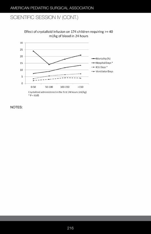
216
AMERICAN PEDIATRIC SURGICAL ASSOCIATION
SCIENTIFIC SESSION IV (CONT.)
NOTES:

217
www.eapsa.org
SCIENTIFIC SESSION IV (CONT.)35PEDIATRIC EMERGENCY DEPARTMENT THORACOTOMY: A LARGE CASE SERIES REVIEW FROM A LEVEL 1 TRAUMA CENTERCasey J. Allen, MD , Evan J. Valle, MD, Chad Thorson, MD, MSPH, Anthony R. Hogan, MD, Eduardo A. Perez, MD, Holly Neville, MD, Tanya Zakrison, MD, MPH, Juan E. Sola, MD. University of Miami Miller School of Medicine, Miami, FL, USA.
Purpose:There are established guidelines for performing an emergency department thoracotomy (EDT) in the adult trauma patient. However, since it is rarely utilized in the pediatric patient, it is difficult to identify trends and factors associated with survival. We reviewed our pediatric experience at a level 1 trauma center and report the largest analysis over the past 25 years.
Methods:All pediatric patients (age = 18) who received an EDT from 1991-2012 were reviewed. Demographics analyzed included age, sex, mechanism of injury, and ISS. We reviewed patient records for the presence of vital signs (VS) or signs of life (SOL) in the field, time without VS in the field, presence of VS or SOL upon arrival, location of injuries, return of spontaneous circulation (ROSC), and factors associated with survival.
Results:A total of 61 patients were identified with a median age of 16 years and an average ISS of 46. Most patients were male (90%) and sustained a penetrating injury (88%).The most common injuries were gunshot wound to the chest (79%) and abdomen (38%). In the field, 46% had initial VS and 67% had SOL. Upon arrival, 29% had VS and 56% had SOL. After EDT, 23 patients (38%) had return of spontaneous circulation (ROSC). Of these, 21 expired (OR 16, ED 4, ICU 1). Both survivors (15y, 16y) sustained penetrating injury and had VS upon arrival.
Conclusion:EDT survivors sustained penetrating injuries and had VS upon arrival. There were no survivors in blunt trauma patients regardless of the presence of VS and SOL on arrival. Pediatric trauma patients who arrive without VS and SOL do not warrant EDT. The over-performance of EDT is likely due to the lack of known survival factors and perhaps an overly aggressive approach when faced with a potential pediatric mortality.
SCH
EDU
LE & PRO
GR
AM

218
AMERICAN PEDIATRIC SURGICAL ASSOCIATION
SCIENTIFIC SESSION IV (CONT.)
NOTES:

219
www.eapsa.org
INNOVATION SESSIONInnovation Session Saturday, May 31, 1:30 p.m. – 2:30 p.m.i1AN EXTRACORPOREAL ARTIFICIAL PLACENTA SUPPORTS EXTREMELY PREMATURE LAMBS FOR ONE WEEKBenjamin S. Bryner, MD, Brian W. Gray, MD, Elena Perkins, Hayley Hoffman, BS, Gabe Owens, MD, PhD, John Barks, MD, Alvaro Rojas -Pena, MD, Robert H. Bartlett, MD, George B. Mychaliska, MD. University of Michigan, Ann Arbor, MI, USA.
Purpose:The treatment of prematurity remains an unsolved problem. We developed an extracorporeal artificial placenta (AP) that simulates the intrauterine environment, provides gas exchange and maintains fetal circulation while avoiding mechanical ventilation (MV). We compared the AP with MV in an extremely premature lamb model.
Methods:Extremely premature lambs (110 -120 days; term=145 days) were used. AP lambs (n=9) were cannulated for VV-ECLS (jugular drainage, reinfusion via umbilical vein) on native placental support. An endotracheal tube was placed and filled with amniotic fluid. The cord was divided and VV-ECLS was initiated. MV lambs (n=8) were intubated, ventilated, given exogenous surfactant, and transitioned to high-frequency oscillatory ventilation (HFOV). Two near-term lambs (128 -130 days) were used to validate our MV protocol. All lambs received fluids, parenteral nutrition, prophylactic antibiotics, and methylprednisolone. Hemodynamics, blood gases, hemoglobin, electrolytes, ventilator parameters, and circuit flows were measured. Echocardiography was used to assess patency of the ductus arteriosus.
Results:Four premature lambs survived for one week on the AP (see figure); another lamb survived 6 days. The mean pO2 was 38.4±10.8 mmHg; mean pCO2 was 42.3±10.7 mmHg; mean arterial pressure had a mean of 47.9±9.8 mmHg, and circuit flow averaged 87.4±17.9 cc/min/kg body weight. The ductus arteriosus remained patent in three of these lambs. Four AP lambs died earlier from various complications. The MV lambs survived 2-8 hours. Each of these lambs experienced a transient improvement with surfactant, but developed progressive hypercarbia and hypoxia despite high airway pressures and HFOV. Two near-term lambs were ventilated for 41-49 hours.
Conclusions: Extremely premature lambs can be supported for one week with the artificial placenta, but not with mechanical ventilation. The AP provided long-term
SCH
EDU
LE & PRO
GR
AM

220
AMERICAN PEDIATRIC SURGICAL ASSOCIATION
INNOVATION SESSION (CONT.)hemodynamic stability and good gas exchange. Further studies will assess prolonged control of fetal circulation and organ maturation during application of the AP.
NOTES

221
www.eapsa.org
INNOVATION SESSION (CONT.)i2MYPECTUS: A NOVEL MOBILE HEALTH SYSTEM FOR REMOTE ASSESSMENT OF TREATMENTBrittany Harrison, BA 1, Lily Stern, BS1, Philip Chung, MS1, Mozziyar Etemadi, MS, PhD1, Dillon Kwiat, BS1, Michael R. Harrison, MD1, Marcelo Martinez Ferro, MD2. 1University of California, San Francisco, SAN FRANCISCO, CA, USA, 2Hospital Privado de Niños, Fundación Hospitalaria, Buenos Aires, Argentina.
Purpose:Mobile technology provides the opportunity to verify and incentivize patient compliance. We developed a novel assessment system that senses temperature and pressure, transmits this data to clinicians, and engages patients. The purpose of this pilot study was to test our MyPectus system in patients with Pectus Carinatum (PC) undergoing non-invasive dynamic compression, for which the outcome depends on patient compliance.
Methods:We developed a system which includes (1) MyPod (My Pectus Onboard Device) inserted in the brace, (2) smartphone application for patients, and (3) web -based dashboard for clinicians. MyPod (which is the size of a smartphone) contains temperature and pressure sensors, and an onboard data -logger. Every 20 minutes, data wirelessly downloads via Bluetooth 4.0 from MyPod to patients’ smartphones and is stored in the cloud server for clinical analysis. With IRB approval and informed consent, we studied eight patients ages 10 - 16 with moderately severe PC deformities who underwent bracing with FMF DynamicCompression System® at Fundación Hospitalaria Children´s Hospital in Buenos Aires, Argentina. We measured patients’ baseline pressure. Over four weeks, patients wirelessly synced MyPod data with a MyPectus iOS application. They received points for increased brace compliance. Clinicians accessed data from the cloud in real-time. Efficacy was determined after one month by comparing objective online data and patient-reported online surveys.
Results:In all eight patients, MyPod recorded temperature and pressure data, wirelessly sent it to smartphone applications, and saved it in the cloud. The data correlated with patient-reported usage. All patients had positive feedback about receiving points for compliance.
Conclusion:This first-in-human study demonstrates that the MyPectus System continuously updates relevant clinical data, transmits it to clinicians, and engages patients. We are developing a modular, integrated, mobile patient-monitoring system to incentivize patients and allow for remote management in
SCH
EDU
LE & PRO
GR
AM

222
AMERICAN PEDIATRIC SURGICAL ASSOCIATION
INNOVATION SESSION (CONT.)various clinical scenarios.
NOTES:

223
www.eapsa.org
INNOVATION SESSION (CONT.)i3A NOVEL ENDOLUMINAL CATHETER TOOL SHOWS POTENTIAL FOR NON-INVASIVE TREATMENT OF INFANTILE HYPERTROPHIC PYLORIC STENOSIS (IHPS)Carolyn Cochenour, BSc, Tim Kane, MD, Peter C. Kim, MD PhD, Axel Krieger. Children’s National Health System, Washington, DC, USA.
Background:Laparoscopic and open pyloromyotomy offer definitive treatment for pyloric stenosis but are associated with complications including perforation, incomplete myotomy, and infection. Effective pyloromyotomy enlarges the pyloric channel at most 30% circumferentially. We hypothesized that a similar outcome can be accomplished effectively with an endoluminal balloon tool.
Methods:A new endopyloric catheter-based balloon dilation tool was designed and evaluated for proof-of -concept in cadaveric models. Efficacy of dilatation following 3 cycles of inflation and limits of pyloric over-insufflation for safety were measured in rabbit cadaveric pyloric and cervix tissues. Insertion force was measured using test models and compared to catheter deflection force in preparation for planned testing in preclinical models, using nonparametric analysis (p<0.01).
Results:Effective dilatation was achieved (increase in normalized circumference (C), 2pR) of 63% in the rabbit pylorus (n=4, ?C=7.97±2.71mm) and 112% in the rabbit cervices (n=4, ?C=6.64 ± 0.42mm) resulting in a proportional increase in flow based on a normalized cross sectional lumen area (A) of 181% in rabbit pylorus (? A=17.68±7.61mm 2) and 354% in rabbit cervices (?A=10.02 ± 1.98mm2) (p=0.001). Histological examination demonstrated intact mucosa in all specimens (p<0.01). Balloon inflation up to three times the diameter of the pre -dilated pyloric lumen was determined to be safe with no tearing of the samples. Using a simulated anatomical environment, we determined the insertion force through the hypertrophic pylorus lumen is less than 100g. The prototype catheters (n=5) kinked at 364.4±35.1g, indicating adequate safety margin.
Conclusion:Our novel balloon device for endoluminal dilatation can achieve an effective and safe increase of the pyloric channel lumen in a cadaver model. Our data indicate that non-invasive endoluminal dilation can significantly increase the flow by 2-3 folds. This novel endopyloric tool has potential for less invasive surgical treatment of IHPS. Preclinical in-vivo testing of this non-invasive endoscopic approach and technique will be conducted.
NOTES:
SCH
EDU
LE & PRO
GR
AM
224
AMERICAN PEDIATRIC SURGICAL ASSOCIATION
INNOVATION SESSION (CONT.)i4GENERATION OF AN ARTIFICIAL INTESTINE AND VALIDATION IN DOGS: A PROOF-OF -CONCEPT STUDYShahab Shaffiey, MD 1, Hongpeng Jia, MD1, Tim Keane, BS2, Misty Good, MD1, Chhinder Sodhi, PhD1, Tom Prindle, BS1, Cait Costello, PhD3, John March, PhD3, Deborah Nagle, MD4, Stephen Badylak, DVM., PhD, MD2, David J. Hackam, MD, PhD1. 1Children’s Hospital of Pittsburgh, Pittsburgh, PA, USA, 2University of Pittsburgh, Pittsburgh, PA, USA, 3Cornell University, Ithaca, NY, USA, 4Beth Israel Deaconess Medical Center, Boston, MA, USA.
Purpose:Short bowel syndrome remains a major pediatric problem for which intestinal transplantation represents definitive care but is fraught with complications. Investigators have turned to the development of tissue -engineered approaches to enhance absorptive capacity. We now sought to define the ability of intestinal stem cell precursors to form intact intestinal units on a novel scaffold, and then developed a large animal model to evaluate its regenerative properties in vivo.
Methods:Intestinal stem cells were isolated from intestine resected from human neonates with NEC (n=5), and mice (6 weeks, n=10) expressing the stem cell reporter gene Lgr5, and were cultured in a collagen -coated scaffold derived from acellular porcine intestinal submucosa. To mimic the natural environment of the intestine, murine intestinal myofibroblasts and microbiome components were added. Whole mount confocal microscopy and mRNA expression were used to evaluate epithelial differentiation (MUC2, lys, e-cadherin, chromagranin), and recapitulation of a native intestinal architecture. We developed a canine model in which we performed rectal mucosectomy then implanted the novel scaffold into the mucosal defect (n=4), then evaluated the scaffold biweekly via colonoscopy and biopsy to determine neo-mucosal growth.
Results:Isolated intestinal stem cells from both mice and humans formed donor-derived structures resembling villi and crypts within the collagen lined scaffold, and to differentiate into goblet, enteroendocrine, paneth and enterocytes based on immunofluorescence and RT -PCR. The addition of the microbiome and intestinal myofibroblasts induced increased differentiation into goblet and paneth cells (p=0.03). Strikingly, histologic analysis of implanted scaffold into dogs revealed a newly formed mucosal layer by six weeks with intact large intestinal crypt and villi structures on the scaffold.

225
www.eapsa.org
INNOVATION SESSION (CONT.)Conclusions:We now demonstrate the feasibility of developing an artificial intestine from the intestinal stem cells of mice and humans, providing proof-of -concept validation for our scaffold using a large animal model in dogs.
NOTES:
SCH
EDU
LE & PRO
GR
AM

226
AMERICAN PEDIATRIC SURGICAL ASSOCIATION
INNOVATION SESSION (CONT.)i5ENDOBRONCHIAL OCCLUSION WITH ONE-WAY ENDOBRONCHIAL VALVES: A NOVEL TECHNIQUE FOR PERSISTENT AIR LEAKS IN CHILDRENMichael F. Reed, MD, Abigail B. Podany, MD, Dorothy V. Rocourt, MD, Christopher R. Gilbert, DO, Mary C. Santos, MD, Robert E. Cilley, MD, Peter W. Dillon, MD, Jennifer W. Toth, MD. Pennsylvania State Hershey Medical Center, Hershey, PA, USA.
Purpose:In the pediatric population, persistent air leaks can result from pulmonary infection or barotrauma. Management strategies include surgical intervention, prolonged pleural drainage, ventilator manipulation, and even ECMO. We hypothesized that endobronchial valve placement would be an effective minimally invasive intervention for prolonged air leaks in children.
Methods:Endobronchial valve placement was approved by the Institutional Review Board (IRB) under a Humanitarian Device Exemption, and review of children undergoing the procedure was also approved by the IRB. Children with prolonged air leaks refractory to conventional management strategies were evaluated by a multidisciplinary team (pediatric surgery, interventional pulmonology, and thoracic surgery) for endobronchial valve placement. Airway management and bronchoscopic techniques were adapted to accommodate the pediatric population. Flexible bronchoscopy was performed under general anesthesia. Airways leading to the air leaks were isolated with balloon occlusion. Retrievable one-way endobronchial valves were deployed via catheter (figure).
Results:Four children (16 months to 16 years) had prolonged air leaks following necrotizing pneumonia (2), lobectomy (1), and pneumatocele (1). All had pleural drains. The number of valves placed per patient ranged from 1-4. Average time to air leak resolution was 12 days (range 0-39). Average duration to chest tube removal was 25 days (range 7-39). All four children had complete resolution of their air leaks. All were discharged from the hospital. Three of four have had uneventful valve removal. None required additional surgical interventions.
Conclusion:Endobronchial valve placement for prolonged air leaks due to a variety of etiologies is safe and effective in children for resolving fistulae, achieving early pleural drain removal and shortening length of stay. This novel technique has particular utility in the pediatric population to avoid the morbidity of

227
www.eapsa.org
INNOVATION SESSION (CONT.)thoracotomy and preserve lung volume and function.
NOTES: SCH
EDU
LE & PRO
GR
AM

228
AMERICAN PEDIATRIC SURGICAL ASSOCIATION
INNOVATION SESSION (CONT.)i6SACRAL NERVE STIMULATOR FOR DYSFUNCTIONAL ELIMINATION SYNDROME IN CHILDRENJason P. Sulkowski, MD, Kristine M. Nacion, MPH, Peter C. Minneci, MD, MHSc, Hayat M. Mousa, MD, Seth A. Alpert, MD, Steven Teich, MD. Nationwide Children’s Hospital, Columbus, OH, USA.
Purpose:Neuromodulation with a percutaneously implanted sacral nerve stimulator (SNS) has been used to treat adult patients with dysfunctional elimination syndrome (DES), a constellation of symptoms associated with both gastrointestinal (GI) and genitourinary dysfunction. This series of patients represents our early experience with this new technique in pediatric patients.
Methods:Between May 2012 and September 2013, 17 patients had a SNS placed. A temporary SNS was placed initially to determine clinical efficacy, followed by permanent SNS placement 2-3 weeks later. Retrospective chart review was performed to collect baseline clinical characteristics. Patients completed the Fecal Incontinence Qualify of Life Scale, the Fecal Incontinence Severity Scale, the PedsQL Gastrointestinal Symptom Scale (PedsQL), and a validated voiding survey to obtain a pre -operative baseline and after surgery to determine post-operative outcomes.
Results:Within our population, the average age was 11.5 ± 3.6 years, eight patients were female, and seven had primarily GI symptoms while nine had both fecal and urinary dysfunction. Since SNS placement, five patients had decreased doses of/or ceased using laxatives, cecostomy flushes, or both. Three patients were weaned off anticholinergics for urinary dysfunction. In the ten patients with complete pre - and post-operative data, all three GI measures improved with significant improvement in the overall score on the Fecal Incontinence Severity Scale (p=0.02); and abdominal discomfort reported on the PedsQL (p=0.02). Of the five patients reporting urinary symptoms, three demonstrated marked improvement, two of whom had complete resolution. After implantation, stimulators were repositioned in two patients for reported discomfort, and a UTI was reported in another patient. Average follow-up has been 8.4 ± 6 months.
Conclusions:This series demonstrates improvements in both fecal and urinary function after sacral nerve stimulator placement in pediatric patients with dysfunctional elimination syndrome. Sacral nerve stimulation represents a promising therapy for this challenging clinical condition.
NOTES:

229
www.eapsa.org
VIDEO SESSION Video Session Saturday, May 31, 2:30 p.m. – 3:30 p.m.V1THORACOSCOPIC REPAIR OF ESOPHAGEAL ATRESIA WITH DISTAL TRACHEOESOPHAGEAL FISTULA AND A PROXIMAL TYPE-H TRACHEOESOPHAGEAL FISTULAZachary J. Kastenberg, MD, James K. Wall, MD, Matias Bruzoni. Stanford University, Palo Alto, CA, USA.
Purpose:To report a combined approach using thoracoscopy and flexible bronchoscopy to identify and repair a rare tracheoesophageal fistula (TEF) anatomic variant. This variant had both a distal tracheoesophageal fistula and a proximal tracheoesophageal fistula in an H-type configuration located high in the thoracic inlet.
Methods:A 2,045-gram, ex-35 week female with a history of CHARGE syndrome in mild respiratory distress underwent thoracoscopy for what was preoperatively believed to be a Gross type C tracheoesophageal fistula. After ligation of the distal fistula, ventilation remained challenging and intraoperative flexible bronchoscopy through the endotracheal tube revealed a proximal fistula. The proximal fistula was in an H-type configuration high in the thoracic inlet. The video describes the surgical technique used to repair both fistulae and the esophageal atresia thoracoscopically.
Results:The operative time was 125 minutes and there were no intraoperative complications. A postoperative contrast study at 10 days and 3 months revealed an intact esophageal repair without a leak or stricture. A postoperative rigid bronchoscopy at 10 days showed no airway abnormalities.
Conclusions:The thoracoscopic repair of this rare tracheoesophageal fistulae variant with H-type anatomy between the proximal pouch and trachea located high in the thoracic inlet is feasible in low birth weight infants. Intraoperative flexible bronchoscopy is a valuable tool in determining a second fistula and assisting with the repair.
NOTES:
SCH
EDU
LE & PRO
GR
AM

230
AMERICAN PEDIATRIC SURGICAL ASSOCIATION
V2HYDROCOLPOS DRAINAGE IN CLOACAAndrea Bischoff, M.D., Belinda Dickie, M.D., Jason Frischer, M.D, Marc A. Levitt, M.D., Alberto Peña, MD. Cincinnati Children’s Hospital, Cincinnati, OH, USA.
Purpose:30% of the patients with cloaca have a hydrocolpos that may compress the trigone, producing uretero -vesico obstruction, megaureters, and hydronephrosis. In addition, the hydrocolpos may become infected and can perforate, with serious consequences for the patient. Ideally, the hydrocolpos must be drained at the time of colostomy opening.
Methods:A video was recorded highlighting the important technical details of hydrocolpos drainage in two cloaca patients that had previously underwent a colostomy opening and were left with an undrained hydrocolpos. In one patient, a vesicostomy was also previously performed in an attempt to drain the hydrocolpos, which in retrospect was unnecessary.
Results:With an infra-umbilical midline laparotomy or with a left lower quadrant oblique incision used for the colostomy opening, the hydrocolpos can be found behind the bladder. When opening the posterior vaginal wall at the dome, special emphasis should be placed on identification and protection of the uterus. When two hemivaginas are present a window can be created within the vaginal septum to allow for a single tube to drain both hemivaginas. The draining tube should remain in place until the time of the definitive cloacal reconstruction.
Conclusion:Perineal intermittent catheterization of the common channel, single percutaneous aspiration of the hydrocolpos content, and vesicostomies are inadequate and contra-indicated methods of hydrocolpos drainage. During colostomy opening the hydrocolpos should be properly drained with a transabdominal indwelling tube that should remain in place until the time of the cloacal repair.
NOTES:
VIDEO SESSION (CONT.)

231
www.eapsa.org
VIDEO SESSION (CONT.)V3CONGENITAL STERNAL CLEFT REPAIRCathy A. Burnweit, MD1, Jun Tashiro, MD MPH2. 1Miami Children’s Hospital, Miami, FL, USA, 2University of Miami Miller School of Medicine, Miami, FL, USA.
Abstract:Congenital sternal cleft is a rare congenital chest wall deformity. Three types exist: complete, inferior, and superior. The complete and inferior variants are often associated with intra -cardiac or intra -abdominal anomalies. The superior type is usually found in isolation. Regardless of the type, the defect leaves the heart and great vessels unprotected and can lead to loss of thoracic domain from organ herniation if left untreated. Repair in the neonatal period is recommended, as the sternum is unossified and the chest compliant, allowing for easier manipulation and approximation. This presentation depicts the primary closure of a superior cleft in a two-month old child. The key steps of the operation include excision of the abnormal overlying skin, dissection of the sternal bars, tapering of the lower cartilage, and approximation of the sternal bars. Ensuring hemodynamic stability and avoiding compression of the thoracic structures are critical for this procedure. The postoperative course was uneventful, and the child recovered well. She developed normally over two years of follow-up.
NOTES:
SCH
EDU
LE & PRO
GR
AM

232
AMERICAN PEDIATRIC SURGICAL ASSOCIATION
VIDEO SESSION (CONT.)V4RESECTION OF DUODENAL WEB USING HYBRID NATURAL ORIFICE TRANSLUMINAL ENDOSCOPIC SURGERY (NOTES)Maria Carmen Mora, MD 1, Kevin P. Moriarty, MD2, Michael V. Tirabassi, MD2, Gregory T. Banever, MD2. 1Baystate Medical Center, Springfield, MA, USA, 2Baystate Children’s Hospital, Springfield, MA, USA.
Purpose:We present a case of a hybrid NOTES (natural orifice transluminal endoscopic surgery) for resection of a duodenal web. The operation performed was a transgastric duodenal web resection through an existing gastrostomy site.
Methods:The patient is a 2-year-old -male with Down syndrome and congenital heart disease. From birth the patient had persistent emesis and failure to thrive for which he underwent a laparoscopic gastrostomy tube placement at 3-weeks of age. At 29-months of age with continued failure to thrive he underwent further work-up including a repeat upper GI. This was suspicious for a duodenal web near the ampulla, later confirmed on EGD. We therefore performed an incisionless resection of the duodenal web via the existing gastrostomy site. Initially the plan was to use the endoscope for visualization and the gastrostomy site for instrumentation; however, the endoscope visualization was inadequate. The gastrostomy site was dilated and an extra small wound protector was placed with a sterile glove over it allowing insufflation and access via the fingers for the laparoscope and 3mm instruments. A 70-degree laparoscope was used for visualization. The opening of the web was cannulated using a Fogarthy catheter prolapsing the web towards the stomach. A 3mm hook cautery and then the LigaSure were used to incise and excise the anteriolateral aspect of the duodenal web. Intraoperative CXR ruled out free air. A 1cm 14-French Mickey button was placed at the completion of the procedure. The length of the operation was 100 minutes.
Results: The patient did well postoperatively and was discharged home on POD 4. At his four -month postoperative visit he was at the 7th percentile on the growth curve compared to 4th percentile preoperatively.
Conclusions:Hybrid NOTES for resection of duodenal web via an existing gastrostomy site provides a new minimally invasive approach for treatment of this anomaly.
NOTES:

233
www.eapsa.org
VIDEO SESSION (CONT.)V5ENDOSONOGRAPHY IN PERIRECTAL PROCEDURESArun Thenappan, Daniel Teitelbaum, Marcus Jarboe. University of Michigan C.S. Mott Children’s Hospital, Ann Arbor, MI, USA.
Purpose:Perirectal procedures in the pediatric population are often difficult to perform and prone to complications secondary to the underdeveloped tissues of children and the difficulty clearly identifying the anatomy purely by visualization or palpation. Endosonography may be an extremely beneficial adjunct in the efficient and effective performance of several of these procedures.
Methods:We demonstrate operative footage and ultrasound images utilized in the care of patients treated at the University of Michigan.
Results:Here we demonstrate the use of ultrasound in three common perirectal procedures: injection of Clostridium botulinum toxin or BoTox for internal sphincter achalasia or in Hirschsprung’s disease who are suffering from recurrent enterocolitis, sclerotherapy for rectal prolapse, and seton placement in complicated Crohn’s perirectal fistulas.
Conclusions:We demonstrate that ultrasound is a simple and useful adjunct in the performance of such perirectal procedures.
NOTES:
SCH
EDU
LE & PRO
GR
AM

234
AMERICAN PEDIATRIC SURGICAL ASSOCIATION
VIDEO SESSION (CONT.)V6ULTRASOUND-GUIDED LATERAL APPROACH TO INTERNAL JUGULAR CATHTER PLACEMENTMarcus Jarboe, MD, K Elizabeth Speck, MD. University of Michigan, Ann Arbor, MI, USA.
Purpose:The purpose of this video is to demonstrate the ultrasound-guided lateral approach to placing a central venous catheter and show its advantages related to the standard ultrasound-guided approach.
Methods:The approach to the internal jugular vein is started adjacent to the clavicle, just lateral to the sternocleidomastoid muscle on the the right side. The ultrasound probe is placed in a transverse fashion cephalad and adjacent to the clavicle. The needle trajectory is in-line with the probe.
Results:The lateral approach enables clear and simultaneous visualization of the entire needle and key anatomic structures such as the edge of the lung, the internal jugular vein, and the carotid artery. Second, the approach allows a gentle curve on the catheter when tunneling, avoiding kinks and avoiding tendency of catheter movement in the tunnel pocket when the neck moves. Third, in cases of internal jugular occlusion, the lateral approach makes it possible to access the brachiocephalic vein.
Conclusions:The ultrasound-guided lateral approach to central venous catheter placement shows all important structures simultaneously, provides better tunneling and has more options as far as access to more central vessels.
NOTES:

235
www.eapsa.org
SCIENTIFIC SESSION V Scientific Session VMiscellaneous SurgerySunday, June 1, 8:00 a.m. – 9:15 am36MYPOD: AN EMR-BASED TOOL THAT FACILITATES QUALITY IMPROVEMENT AND MAINTENANCE OF CERTIFICATIONLoren Berman, MD, Brian J. Duffy, MD, Charles D. Vinocur, MD. A.I. duPont Hospital for Children, Wilmington, IL, USA.
Background:Maintenance of Certification was designed to regularly assess physician competencies including operative case volume and outcomes. Current national programs do not adequately track clinical performance, including surgical occurrences. This information, if collected consistently and systematically, can be used to facilitate quality improvement efforts.
Methods:We developed an EMR-based program called MyPOD (My Personal Outcome Data) to track surgical outcomes at our institution. Cases are automatically downloaded to a surgeon’s peer-protected site from EPIC Op-Time, an operating room management system, two weeks after the procedure. Downloaded information includes demographic data, procedure(s), ASA, and wound class. The surgeon records a case summary including any occurrences, deaths, or systems issues as defined by the NSQIP-Pediatric program (FORM 1A). Data can be directly uploaded to the ACS case log system for maintenance of certification purposes. All occurrences are reviewed at the divisional M&M and scored on FORM 1B. After divisional review, the cases are forwarded to the director of surgical outcomes and quality for a final review. This enables trending of systems issues and clinical problems within and across divisions and facilitates the process of gathering further information or follow-up about a case.
Results:Over the first 18 months, 4736 cases were performed by 18 surgeons. There were 101 occurrences, and multiple systems issues were identified and acted upon in order to prevent future similar occurrences. We plan to roll out MyPOD to all divisions and develop the ability to automatically refer a case to another division(s) where issues were identified in order to achieve loop closure.
Conclusion:The MyPOD program provides the basis for comparative data that is essential in evaluation of performance and facilitates quality improvement in surgery. This and similar EMR-driven tools are becoming essential components of the MOC process.
SCH
EDU
LE & PRO
GR
AM

236
AMERICAN PEDIATRIC SURGICAL ASSOCIATION
Form 1A
Form 1B
NOTES:
SCIENTIFIC SESSION V (CONT.)

237
www.eapsa.org
SCIENTIFIC SESSION V (CONT.)37THORACOSCOPIC PLACEMENT OF PHRENIC NERVE PACERS FOR ONDINE’S CURSEKristina J. Nicholson, BS1, Lauren B. Nosanov, BA 1, Kanika A. Bowen, MD2, Iris A. Perez, MD2, Thomas G. Keens, MD2, Cathy E. Shin, MD2. 1University of Southern California Keck School of Medicine, Los Angeles, CA, USA, 2Children’s Hospital Los Angeles, Los Angeles, CA, USA.
Purpose:Congenital central hypoventilation syndrome, or Ondine’s curse, is a rare disorder affecting central respiratory drive. Patients with this disorder fail to ventilate adequately, particularly while sleeping, and require lifelong ventilatory support. Thoracoscopic placement of diaphragmatic pacers can reduce or eliminate mechanical ventilation requirements. As this operation is performed at only a few centers, little is known about complications and long-term outcomes.
Methods:Following IRB approval (CCI-13-000-50), a single -center retrospective review was performed for patients undergoing operative management of congenital central hypoventilation syndrome between 2000-2012. Data abstracted from the medical record included operation duration, ventilation method, number of trochars required, post-operative and pacing outcomes.
Results:Eighteen patients undergoing thoracoscopic placement of diaphragmatic pacers for congenital central hypoventilation syndrome were identified during the study period. One patient underwent both a primary operation and a replacement during this period, and one patient only received a replacement. Mean surgical time was 3.2 +/ - 0.8 hours. In all cases except one, three trochars were utilized for each hemithorax, and no cases were converted to open procedures. All patients had bilateral chest tubes placed post-operatively except one who had only unilateral chest tube placement. Five patients (27.8%) experienced post-operative complications, including atelectasis and pneumothorax. The mean ICU stay was 4.3 +/ - 2.0 days, and the mean hospital stay 5.7 + -/1.5 days. Twelve patients (66.7%) achieved their daily goal pacing times within the follow-up period.
Conclusions:Thoracoscopic placement of diaphragmatic pacers is a safe and effective treatment modality for congenital central hypoventilation syndrome. Observed complications were temporary, and the majority of patients were able to achieve pacing goals and reduce or eliminate mechanical ventilation requirements. Few require re- operation. This case series represents significant single -center experience with an uncommonly performed procedure.
NOTES:
SCH
EDU
LE & PRO
GR
AM

238
AMERICAN PEDIATRIC SURGICAL ASSOCIATION
SCIENTIFIC SESSION V (CONT.)38MANAGEMENT OF RECURRENT INTUSSUSCEPTION IN THE AIR CONTRAST ENEMA ERA: A REVIEW OF 716 PATIENTSJeremy G. Fisher, MD, Eric A. Sparks, MD, Christopher GB Turner, MD, Justin D. Klein, MD, Elliot Pennington, MD, Faraz Khan, MD, David Zurakowski, PhD, Dario O. Fauza, MD, PhD, Biren P. Modi, MD. Department of Surgery, Boston Children’s Hospital, Boston, MA, USA.
Purpose:Air contrast enema (ACE) is standard treatment for primary ileocolic intussusception. Management of recurrences is less clear. This study describes patterns of management for recurrent intussusception and aims to delineate its appropriate therapy by quantifying the relationship between recurrence and need for bowel resection, pathologic lead points (PLPs), and complication rates.
Methods:Following IRB approval, patients with intussusception at one institution from 1997 to 2013 were reviewed. Management of ultrasound-proven recurrences, ACE outcomes, operations, resections, and PLPs were noted. Student’s t -test and Fisher’s exact test were used for univariate analysis and risk factors for resection and PLP were evaluated by multivariate logistic regression (P<0.05).
Results:Of 716 cases of intussusception, 666 were ileocecal, with 29 PLPs: 18 Meckel’s diverticuli, 5 polyps, 4 lymphomas, 2 duplication cysts. Initial episodes underwent ACE (636; 95.5%), operation (22; 3.3%), or resolved spontaneously (8; 1.2%). Among those undergoing ACE initially, >1 recurrence was seen in 95 (15%). Successful initial ACE had a negative predictive value of 96% for resection and 99% for PLP (i.e. after successful ACE, 4% had resections, 1% PLP). Conversely, after failed initial ACE, 40% of patients required resection and 23% had PLPs (P<0.0002 for both). Number of recurrences was not predictive of PLP (P=0.25) or resection (P=0.20). Four ACE- related complications occurred (0.5%), three at 1st ACE and one at 3rd ACE.
Conclusions:This large cohort of patients with intussusception treated with ACE demonstrates that unsuccessful ACE in children with ileocolic intussusception is associated with increased risk for requiring bowel resection and for harboring pathologic lead points. Number of recurrences is not related to presence of a lead point, bowel resection rate, or ACE-related complications. These data suggest that ACE should be the treatment of choice in recurrent intussusception and no set number of recurrences should trigger automatic operative intervention.
NOTES:

239
www.eapsa.org
SCIENTIFIC SESSION V (CONT.)39COMBINED LAPAROSCOPIC/FLUOROSCOPIC PRIMARY GASTROJEJUNOSTOMY BUTTON TUBE PLACEMENT: DESCRIPTION OF TECHNIQUE AND REVIEW OF INITIAL CLINICAL EXPERIENCEMariya Skube, MD , Elizabeth Berdan, MD, Robert D. Acton, MD, Daniel A. Saltzman, MD,PhD, Bradley J. Segura, MD, Donavon J. Hess, MD,PhD. University of Minnesota, Minneapolis, MN, USA.
Purpose:We present a technique for primary laparoscopic/fluoroscopic gastrojejunostomy (GJ) button tube placement. This technique was developed to avoid the difficulties of a combined laparoscopic/endoscopic approach and to mitigate difficulties associated with directing the catheter through the pylorus, preventing the catheter from coiling in the stomach, and advancing the catheter into the jejunum. We describe the conduct of the operation and evaluate the initial experience.
Methods:27 patients with 27 procedures performed from 2011 to 2013 at a tertiary care children’s hospital were retrospectively reviewed. All attempted operations were included in the analysis. The stomach was retained to the abdominal wall with through -and-through sutures. A gastrotomy was dilated and the pylorus transited with a 20 French introducer. Under fluoroscopy, a wire was passed through the introducer, and advanced into the jejunum through a catheter. A 20 Fr. Peel-away introducer was placed over the wire and through the pylorus. The GJ button was advanced over the wire and through the peel -away introducer. The retaining sutures were tunneled subcutaneously.
Results:Patients averaged 34 months of age (range 1 mo. – 17 yr.), had a mean weight of 12.2 kg (range 3.8 - 46.7 kg), and a mean ASA score of 2.8 (range 2 – 3). Indications for all operations were GERD with failure to thrive, or GERD with aspiration. All operations initiated for laparoscopic/fluoroscopic GJ tube placement were successful. Operative time averaged 40 minutes (range 16 – 71 min). Estimated blood loss averaged 2 mL (range 1 – 25 mL). There were no operative complications. In a mean follow-up period of 16 months (range 4 – 29 mo.), no complications were noted.
Conclusions:This technique mitigates many of the difficulties of primary GJ button placement, and can be performed with a high level of success and reasonable operative times with minimal morbidity.
NOTES:
SCH
EDU
LE & PRO
GR
AM

240
AMERICAN PEDIATRIC SURGICAL ASSOCIATION
SCIENTIFIC SESSION V (CONT.)40H-TYPE TRACHEOESOPHAGEAL FISTULAS: A MULTICENTER REVIEW OF OUTCOMES IN A RARE DISEASESara C. Fallon, MD1, Shawn D. St. Peter, MD2, Jacob C. Langer, MD3, Kuojen Tsao, MD4, Caroline Kellagher, BA 4, Dave R. Lal, MD5, Jill S. Whitehouse, MD5, Diana L. Diesen, MD6, Michael D. Rollins, MD7, Elizabeth M. Pontarelli, MD8, Jeffrey S. Upperman, MD8, Charles M. Leys, MD9, Mark L. Wulcan, MD10, Sarah J. Hill, MD10, Martin L. Blakely, MD11, Corey W. Iqbal, MD12, Timothy D. Kane, MD13, David E. Wesson, MD1. 1Division of Pediatric Surgery, Michael E. DeBakey Department of Surgery, Baylor College of Medicine, Houston, TX, USA, 2Department of Surgery, Children’s Mercy Hospital and Clinics, Kansas City, MO, USA, 3Division of Pediatric Surgery, The Hospital for Sick Children, Toronto, ON, Canada, 4Departments of Pediatric Surgery and Surgery and the Center for Surgical Trials and Evidence-based Practice at the University of Texas Medical School at Houston, Houston, TX, USA, 5Department of Surgery, Division of Pediatric Surgery, Medical College of Wisconsin and Children’s Hospital of Wisconsin, Milwaukee, WI, USA, 6Children’s Medical Center Dallas, UT Southwestern Medical Center,, Dallas, TX, USA, 7Division of Pediatric Surgery, Primary Children’s Medical Center, University of Utah, Salt Lake City, UT, USA, 8Department of Pediatric Surgery, Children’s Hospital Los Angeles, Los Angeles, CA, USA, 9Department of Surgery, Indiana University Medical Center, Indianapolis, IN, USA, 10Emory University School of Medicine, Atlanta, GA, USA, 11Department of Surgery, Vanderbilt University Medical Center, Nashville, TN, USA, 12Division of Pediatric Surgery, University of California at San Francisco, San Francisco, CA, USA, 13Department of Surgery, The George Washington University School of Medicine & Health Sciences, Washington, DC, USA.
Purpose:The H-type tracheoesophageal fistula (TEF), without esophageal atresia (EA), is a rare anomaly with its own unique management issues. Outcomes after surgical treatment are largely unknown due to small compilations of patients. We conducted a large, multicenter review to characterize this population and assess the operative outcomes.
Methods:A multicenter retrospective review of all H-type TEF patients treated from 2002-2012 was performed after IRB approval at each institution. Variables collected were patient demographics, operative technique, hospital course, and long -term outcomes. Descriptive analyses were performed.
Results:Twelve centers identified 81 patients with H-type TEF. The overall survival was 95% (Table). Most patients were repaired via the cervical approach (95%). Esophageal leak occurred in 5 of 62 who had a contrast study. Two (2%) developed a recurrent TEF. A total of 23% (19/81) failed extubation after repair, 19% had vocal cord abnormalities on laryngoscopy, and 7% required a tracheostomy (2 patients with tracheomalacia, 1 patient with

241
www.eapsa.org
SCIENTIFIC SESSION V (CONT.)bilateral cord paresis +tracheomalacia, 1 patient with unilateral vocal cord paralysis+tracheomalacia, and 1 patient with an aberrant compressive right mainstem bronchus). One patient with choanal atresia/CHARGE had a tracheostomy pre -operatively.
Conclusions:Patient Demographics and Operative Outcomes
Patient Demographics n=81 Operation/
Hospital Course n=81 Long-term Outcomes n=81
Gender (M/F) 63%/37%Cervical/ Thoracotomy/ Thoracoscopy
77 (95%) 3 (4%) 1 (1%)
Laryngoscopy performed
25 (31%)
EGA 37.2(+2.8) Age at surgery (median, days)
16 (2-1427)
If performed, evidence of vocal cord dysfunction
15 (19%)
Birth weight (mean, g)
2585 (+711) Drain placed 20
(25%)Delayed stenosis
4 (5%)
Median POD swallow study 7 (2-34) GERD 28
(35%)
Leak on swallow study
5/62 (8%) Recurrence 2
(2%)
Time to start feeds (median, days)
4 (0-15) Tracheostomy 6 (7%)
Time to full feeds (median, days) 9 (1-57) Survival 77
(95%)
In-hospital complications
Wound infxn (2), sepsis/line infection (2), aspiration pna (1), medical nec (2), GI bleed/ carotid injury (1)
Length of follow up (median)
803 (7 days-5412 days)
This study suggests repair of H-type TEF is associated with a substantial risk of breathing difficulties and vocal cord dysfunction. These data argue for review of surgical technique and routine laryngoscopy to evaluate vocal cord function in patients who demonstrate breathing difficulties after repair.
NOTES:
SCH
EDU
LE & PRO
GR
AM

242
AMERICAN PEDIATRIC SURGICAL ASSOCIATION
SCIENTIFIC SESSION V (CONT.)41PATIENTS’ SELF-REPORTED OUTCOME AFTER UNDERGOING ELECTIVE LAPAROSCOPIC APPENDECTOMY FOR THE TREATMENT OF CHRONIC RIGHT LOWER QUADRANT PAINJose S. Lozada, MD1, A. Daniel Guerron, MD1, Oliver Soldes, MD2, Lori Mahajan, MD1, Federico G. Seifarth, MD1. 1Cleveland Clinic, Cleveland, OH, USA, 2Akron Children’s Hospital, Cleveland, OH, USA.
Purpose:To describe the patients’ self-reported outcome of elective laparoscopic appendectomy as a treatment for chronic right lower quadrant abdominal pain.
Methods:We conducted a retrospective analysis at our institution of patients who underwent elective appendectomies for chronic right lower quadrant abdominal pain in the last 40 months. Patients who suffered from unclear RLQ abdominal pain >12 months were included. All subjects underwent extensive gastroenterological workup with negative results; including negative CT scans, upper and lower endoscopies, ultrasounds, laboratory work and HIDA scans. Once patients were identified, we then contacted the families, who were asked to rate, on a 5 point likert scale the change in post-operative pain where 1 was much worse, 2 was somewhat worse, 3 was no change, 4 was somewhat resolved, and 5 was completely resolved.
Results:We identified 28 patients: 10 males, 18 females, ages 4-17y (mean 13.4y). All 28 patients reported resolution of pain during their visit 4 weeks after surgery. Sixteen of the 28 surgical specimens revealed inflammatory changes. We were able to follow-up with 22 of the 28 patients by telephone. Post-operative phone follow-up time ranged from 9-30 months. On the 5-point scale, of the patients whom we were able to contact, 19/22 reported complete resolution of pain, 2/22 reported somewhat improved pain, and 1/22 reported no change. No patients reported a negative effect after the surgery.
Conclusions:Elective laparoscopic appendectomy is a valuable diagnostic and therapeutic tool. The procedure is associated with low morbidity and a short post-operative hospitalization. Given the potentially invasive, expensive and long diagnostic trail experienced by patients with unclear abdominal pain, we recommend early consideration of laparoscopic appendectomy in the care of patients with unclear chronic right lower abdominal pain.
NOTES:

243
www.eapsa.org
SCIENTIFIC SESSION V (CONT.)42INFANT GASTROSTOMY TUBE OUTCOMES BASED ON TUBE CHARACTERISTICSNaomi -Liza Denning, Danielle N. Leranth, PA -C, John C. Densmore, MD. Medical College of Wisconsin, Wauwatosa, WI, USA.
Purpose:Although gastrostomy complication rates have been studied, a paucity of reports correlate tube characteristics and method of placement to patient outcome. This retrospective review analyzes these effects in infants receiving gastrostomy tubes.
Methods:Charts of 314 infant gastrostomies from 2004-2010 were reviewed. Tube characteristics were long vs. button, silicon vs. latex, and balloon vs. mushroom. Method of placement was categorized as PEG, laparoscopic Georgeson (LG), laparoscopic pexy (LP), and Stamm gastrostomy (SG). Two -year outcomes were tracked (leakage, granulation, prolapse, cellulitis, wound infection, hernia, dehiscence, small bowel obstruction, peritonitis, dislodgement < and > 6 weeks, migration, misplacement, persistent fistula, reoperation). Mantel -Haenszel chi-square (trend) test and Poisson regression with scale deviance were used for analysis.
Results:314 patients experienced 481 complications. Individual complications were not correlated with tube characteristics. Analyzing total complication rates revealed that balloon tubes had a higher mean complication rate (1.75 vs. 1.36, p = 0.04). Buttons trended a higher rate of overall complication (1.82 vs. 1.43, p = 0.06). Individual complications occurring > 20 times were analyzed by tube characteristics and surgical technique. Balloon tipped tubes had a higher rate of fistula requiring closure (23% vs. 13%, p = 0.025). Long tubes and latex tubes had a higher rate of cellulitis (13% vs. 5.9%, p = 0.029 and 18.2% vs. 9.3%, p = 0.011). Surgical technique was correlated with likelihood of tract leakage (LP 29.3%, SG 22.1%, PEG 13.4%, LG 9.5%, p = .047). Long tubes, mushroom tubes, and PEG tubes had lowest incidence of tube dislodgement < 6 weeks (all p < 0.01).
Conclusion:Gastrostomy tubes incur significant morbidity. Tube characteristics did not correlate with individual complication rates. However, among tubes with complications, differences in tube characteristics and method of placement were noted. Increasing the observations in this study will likely illuminate differences in outcome.
NOTES:
SCH
EDU
LE & PRO
GR
AM

244
AMERICAN PEDIATRIC SURGICAL ASSOCIATION

245
www.eapsa.org
EXHIBITS& SUPPORT

246
AMERICAN PEDIATRIC SURGICAL ASSOCIATION
THANK YOU!
Animal Technologies, Inc.Applied Medical
Applied Medical Technology, Inc. (AMT)BK Medical/Analogic Ultrasound
JustRight SurgicalKarl Storz Endoscopy-America, Inc.
Kimberly-ClarkMicroCutter
BK Medical/Analogic UltrasoundHill-Rom
Philips HealthcareSidra Medical and Research Center
Zonare Medical Systems
Educational Grants
Sponsorship
Simulation Courses Supporters
Ultrasound Course Supporters

247
www.eapsa.org
APSA THANKS THE FOLLOWING EXHIBITORS FOR THEIR CONTRIBUTIONS TO THE
45TH ANNUAL MEETING
Traditional ExhibitorsAmerican College of SurgeonsAmerican Pediatric Surgical Association
Foundation (APSAF)American Pediatric Surgical Nurses
Association (APSNA)Animal TechnologiesApplied MedicalApplied Medical Technology, Inc. (AMT)BK Medical/Analogic UltrasoundBlank Children’s HospitalCarolinas HealthCare SystemChildren’s Mercy HospitalCRC PressELSEVIER, Inc.Enterprise Medical ServicesHCA KidsHuntsville Health SystemKaiser Permanente/Mid-Atlantic
Permanente Medical GroupLocumTenens.com
Lumitex MDLutheran Health NetworkMediflex Surgical ProductsMegadyneNorthwestern University & Lurie Children’s
Hospital of Chicago Laparoscopic Duodenal Atresia and Thoracoscopic Diaphragmatic Hernia Simulation
Pediatric Search PartnersSidra Medical and Research CenterSpecialty Surgical Products, Inc.StrykerRespitek (Pty) Ltd.Thompson Surgical InstrumentsWeatherby HealthcareW. Hardy Hendren Education Foundation for
Pediatric Surgery and UrologyZonare Medical Systems
EXHIBITS & SU
PPORT
TM
Gold Exhibitor
Silver Exhibitors
(as of 5/1/2014)

AMERICAN PEDIATRIC SURGICAL ASSOCIATION
248
EARN VALUABLE PEDIATRIC SURGERY CMES
➢ NEW! ExPERT (Exam-based Pediatric surgery Educational Reference Tool)Try the NEW APSA ExPERT CME platform to earn MOC CME credits,
as an information resource, as a real-time, point-of-care knowledge reference or all of the above.
The new APSA ExPERT continuing medical education program is an innovative platform designed to help practicing pediatric surgeons incorporate the available medical literature into the care of their patients. It addresses maintenance of certification (MOC) needs and the scope of the core competencies as defined by the American Board of Surgery (ABS).
ExPERT can be used to earn MOC CME credits, as an information resource, as a real-time, point-of-care knowledge reference or all of the above. See below for a list of ExPERT Courses. The ExPERT platform is optimized for use on your desktop or laptop computer, tablet or smartphone — you can even pause a course started on one device and finish it later on another.
Individual Token(s) Subscription Subscription
APSA Members – Regular, Associate, International, Senior, Honorary
$8 USD for a bundle of 8 tokens $195 USD for unlimited access including the Spaced Learning Program (SLP)
APSA Members - Candidate, Resident
$8 USD for a bundle of 8 tokens
Non-Members $12 USD for a bundle of 8 tokens
Visit the APSA website for more information and start earning more pediatric surgery CMEs.
www.eapsa.org


A M E R I C A N P E D I A T R I C S U R G I C A L A S S O C I A T I O N
w w w . e a p s a . o r g
American Pediatric Surgical AssociationFuture Meetings
46th Annual MeetingApril 30 – May 3, 2015
Harbor Beach Marriott Resort & SpaFort Lauderdale, FL
APSA 47th Annual Meeting and Joint Meeting with IPSOMay 14–18, 2016
Manchester Grand Hyatt San DiegoSan Diego, CA
APSA 48th Annual MeetingMay 4–7, 2017
The Westin Diplomat Resort & SpaHollywood, FL
APSA 49th Annual MeetingMay 3–6, 2018
JW Marriott Desert Springs Resort & SpaPalm Desert, CA