application of x-ray fluorescence analytical techniques in phytoremediation and plant biology...
TRANSCRIPT
Spectrochimica Acta Part B 63 (2008) 1240–1247
Contents lists available at ScienceDirect
Spectrochimica Acta Part B
j ourna l homepage: www.e lsev ie r.com/ locate /sab
Application of X-ray fluorescence analytical techniques in phytoremediationand plant biology studies
Marijan Nečemer a,⁎, Peter Kump a, Janez Ščančar a, Radojko Jaćimović a, Jurij Simčič a, Primož Pelicon a,Miloš Budnar a, Zvonka Jeran a, Paula Pongrac b, Marjana Regvar b, Katarina Vogel-Mikuš b
a Jožef Stefan Institute, Jamova 39, 1000 Ljubljana, Sloveniab Department of Biology, Biotechnical Faculty, University of Ljubljana, Večna pot 111, SI-1000 Ljubljana, Slovenia
⁎ Corresponding author. Tel.: +386 1 477 3687; fax: +E-mail address: [email protected] (M. Nečemer
0584-8547/$ – see front matter © 2008 Elsevier B.V. Aldoi:10.1016/j.sab.2008.07.006
a b s t r a c t
a r t i c l e i n f oArticle history:
Phytoremediation is an em Received 11 February 2008Accepted 31 July 2008Available online 12 August 2008Keywords:CadmiumLeadZincPhytoremediationXRF
erging technology that employs the use of higher plants for the clean-up ofcontaminated environments. Progress in the field is however handicapped by limited knowledge of thebiological processes involved in plant metal uptake, translocation, tolerance and plant–microbe–soilinteractions; therefore a better understanding of the basic biological mechanisms involved in plant/microbe/soil/contaminant interactions would allow further optimization of phytoremediation technologies. In view ofthe needs of global environmental protection, it is important that in phytoremediation and plant biologystudies the analytical procedures for elemental determination in plant tissues and soil should be fast andcheap, with simple sample preparation, and of adequate accuracy and reproducibility. The aim of this studywas therefore to present the main characteristics, sample preparation protocols and applications of X-rayfluorescence-based analytical techniques (energy dispersive X-ray fluorescence spectrometry—EDXRF, totalreflection X-ray fluorescence spectrometry—TXRF and micro-proton induced X-ray emission—micro-PIXE).Element concentrations in plant leaves from metal polluted and non-polluted sites, as well as standardreference materials, were analyzed by the mentioned techniques, and additionally by instrumental neutronactivation analysis (INAA) and atomic absorption spectrometry (AAS). The results were compared andcritically evaluated in order to assess the performance and capability of X-ray fluorescence-based techniquesin phytoremediation and plant biology studies. It is the EDXRF, which is recommended as suitable to be usedin the analyses of a large number of samples, because it is multi-elemental, requires only simple preparationof sample material, and it is analytically comparable to the most frequently used instrumental chemicaltechniques. The TXRF is compatible to FAAS in sample preparation, but relative to AAS it is fast, sensitive andmulti-elemental. The micro-PIXE technique requires rather expensive instrumentation, but offers multi-elemental analysis on the tissue and cellular level.
© 2008 Elsevier B.V. All rights reserved.
1. Introduction
Biospheric pollution bymetals has accelerated dramatically duringthe last few decades, mainly due to mining, smelting and manufactur-ing, and the treatment of agricultural soils with agrochemicals and soilsludges. Problems associated with soil and water contamination,human and animal welfare, health and destruction, and disruption ofnatural ecosystems arewell documented [1]. Because of their capacityto act as efficient interceptors and accumulators of pollutants, plantspecies are widely employed as passive monitors in areas contami-nated by metals [2]. In addition, in the past decade extensive researchhas been conducted to investigate possibilities for the use of suitableplant species and their associated microbes in phytoremediation ofmetal contaminated sites [3]. The concept of using plants to clean up
386 1 251 93 85.).
l rights reserved.
the environment, however, is not new. About 300 years ago, plantswere proposed for use in the treatment of wastewater, and at the endof the 19th century Thlaspi caerulescens and Viola calaminaria werethe first species documented to accumulate high levels of metals intheir leaves [4–6]. The term “metal hyperaccumulating plant” wasused by Brooks [7] for the first time to describe plants with an extremeaccumulation capacity, containing more than 1000 μg/g (0.1%) of Ni indry leaves, which is an order of magnitude higher than in “normal”plants growing on contaminated soils, and today more that 440 plantspecies are known to hyperaccumulate more than 10,000 μg/g of Znand Mn, 1000 μg/g of Al, As, Se, Ni, Co, Cr, Cu and Pb, and more than100 μg/g of Cd [8,9]. The idea of using metal hyperaccumulating plantsto extract metals from contaminated soils was reintroduced anddeveloped by Utsunamyia [10] and Chaney [11], and the first trial onZn and Cd phytoextraction was conducted by Baker et al. [12]. Besidesphytoextraction, phytostabilization was introduced as another phy-toremediation technology to prevent soil erosion and metal leaching
1241M. Nečemer et al. / Spectrochimica Acta Part B 63 (2008) 1240–1247
in environments that were too contaminated for the use ofphytoextraction techniques (as reviewed in Barceló and Poschenrieder[13] and Ernst [14]).
In the last decade phytoremediation has gained popularity withgovernment agencies and industry worldwide, as a cost-effective non-invasive alternative or complementary technology to engineering-based remediation methods, such as soil excavation, soil washing orburning, or pump and treat systems [15]. Because plant biologicalprocesses are ultimately solar driven, phytoremediation is on averagetenfold cheaper than engineering-based remediation methods. Toincrease the efficiency of phytoremediation technologies, it isimportant to learn more about the specific plant physiologicalprocesses involved, including plant metal uptake, translocation andtolerance (compartmentation, complexation) mechanisms, plant–microbe interactions and other rhizosphere processes [6].
In relation to global environmental protection and lowering ofcosts by saving energy, it is of significance that in the environmentalmonitoring and quality control processes involved in phytoremedia-tion and plant biology studies the analytical procedures for elementaldetermination in plant tissues and soil are fast and cheap, with simplesample preparation, and of adequate accuracy and reproducibility.Only in a such case can the requirements for the analysis of a largenumber of samples be met [16]. For this purpose a wide range ofdiverse instrumental techniques are available [17]. FAAS (flame atomicabsorption spectrometry), ETAAS (electro-thermal atomic absorptionspectrometry), ICP-AES (inductively coupled plasma atomic emissionspectrometry) and ICP-MS (inductively coupled plasma mass spectro-metry) are rather popular and frequently used techniques forelemental determination in these kinds of samples [18,19]. However,these techniques imply a prior total destruction of the matrix bymineral acids, which may lead to problems of contamination by thereactants employed or disturbances of the measured concentration byelement losses due to incomplete solubilization and/or evaporation[16,18]. Moreover, the methods of matrix destruction strongly dependon the chemical composition of the sample and on the element to bedetermined [20]. In view of the above mentioned problems, notforgetting the need for comprehensive, time consuming, andexpensive sample preparation, the use of methods such as energydispersive X-ray fluorescence spectrometry (EDXRF) for direct andmulti-elemental analysis of plant and soil samples has increased overthe last few years [21–25]. These EDXRF techniques are characterizedby a simple sample preparation, mainly sample homogenization, andfast and multi-elemental analysis over a large concentration rangefrom % to mg kg−1, which makes the procedure fast and cheap andtherefore suitable for application to a large number of samples. Themain objective of this work was to present the typical characteristicsof EDXRF-based analytical techniques compared to the most popularand often used instrumental analytical techniques such as FAAS, ICP-AES and NAA which can be applied as a quantitative tool inphytoremediation studies, including plant biology research, environ-mental monitoring and quality control.
Within the framework of current phytoremediation studies in ourlaboratories, the capabilities of several analytical techniques werecompared by the analysis of a set of samples consisting of dryhomogenized plant materials and soil. The following techniques, i.e.standard energy dispersive X-ray fluorescence (EDXRF), total reflec-tion X-ray fluorescence (TXRF), proton-induced X-ray emission (PIXE)with micro beam (micro-PIXE), atomic absorption spectroscopy (AAS)and instrumental neutron activation analysis (INAA) were applied tothe same sets of samples. These consisted of leaves of pennycress(Thlaspi praecoxWulf.) grown at a Pb, Zn and Cd polluted site, leaves ofthe walnut tree (Juglans regia L.) grown at a non-polluted site, as wellas NIST SRM 1573a (tomato leaves), by which the accuracy of theanalytical procedures was checked. Themain focus of the analysis wasthe metals Zn, Cd and Pb, which are usually present at higherconcentrations in the metal hyperaccumulator T. praecox, but only in
natural background concentrations in walnut, which was used as thereference plant material. Sample preparation and the analysisprocedure for each of the above mentioned analytical techniques aredescribed and a comparison of analytical and other parameters suchas uncertainty, accuracy, limits of detection (LOD), cost of analysis persample, instrumental cost etc. is critically evaluated.
2. Experimental
2.1. Sampling and general sample pretreatment
The leaves of pennycress (T. praecox) were collected at highlypolluted area in Žerjav, Slovenia (46°28′49.9″N, 14°51′59.4″E) [26,27],while the leaves of a walnut tree were collected at a non-polluted siteat Zaplana near Vrhnika (45°58′25.0″N, 14°14′25.0″E) [25,26]. Typicalconcentration ranges of Cd, Zn, and Pb in soil at a polluted site were70–200 μg/g, 1500–4000 μg/g, and 30,000–70,000 μg/g, respectively,compared to non-polluted site ranging between 2–4 μg/g for Cd, 100–300 μg/g for Zn, and 60–120 μg/g for Pb. The collected fresh samples ofleaves were frozen in liquid nitrogen and freeze-dried by lyophiliza-tion. The dried samples were pulverized and homogenized by a Fritschpulverizer using a tungsten carbide mortar. The samples were thendivided into five parts, stored in plastic containers, and used in theanalyses by EDXRF, TXRF, AAS, micro-PIXE and INAA.
2.2. Sample preparation, measurements and analysis
2.2.1. EDXRFFrom 0.5 to 1.0 g of powdered sample material pellets were
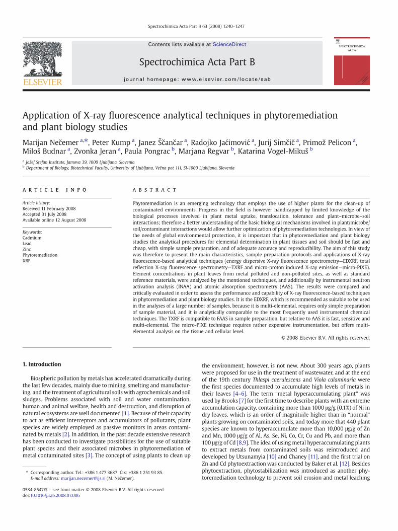
prepared using a pellet die and hydraulic press. As primary excitationsources, the annular radioisotope excitation sources of Fe-55 (10 mCi),Cd-109 (25 mCi) or Am-241 (20 mCi) from Isotope ProductsLaboratories U.S.A. were used. The emitted fluorescence radiationwas measured by an energy dispersive X-ray spectrometer composedof a Si(Li) detector (Canberra), a spectroscopy amplifier (CanberraM2024), ADC (Canberra M8075) and PC based MCA (S-100, Canberra).The spectrometer was equipped with a vacuum chamber. The energyresolution of the spectrometer was 175 eV at 5.9 keV. A typicalfluorescence spectrum of a T. praecox leaves sample, measured by theabove mentioned system, is shown in Fig. 1.
The analysis of complex X-ray spectra was performed by the AXIL[28] spectral analysis program. The evaluated uncertainty of thisprocedure included the statistical uncertainty of measured intensitiesand the uncertainty of the mathematical fitting procedure. The overalluncertainty of spectral measurement and analysis was in most casesbetter than 1%.
Quantification was then performed utilizing QAES (QuantitativeAnalysis of Environmental Samples ) software developed in ourlaboratory [29]. The estimated uncertainty of the analysis was around5% to 10%, LODs for Zn, Cd and Pb were from 5 to 10 μg/g. Rather hightotal estimated uncertainty is mainly due to contributions of matrixcorrection and geometry calibration procedures, which include errorsof tabulated fundamental parameters, and also contributions ofspectrum acquisition and analysis. The uncertainty due to inhomo-geneity of the sample was not included. The price of the instrumentwas around 25,000 EUR. Considering the cost per sample, its multi-elemental capability, simple sample preparation, and the minimumnumber of steps in the measurement and quantification procedure, itwas undoubtedly the cheapest and the simplest analysis among thementioned analytical techniques. The application of this techniquealso requires personnel with a minimum of manual skill for samplepreparation.
2.2.2. TXRFA CEMMARS 5 microwave oven (Matthews, NC, USA) was used for
the digestion of plant samples. Approximately 0.35 g of lyophilized
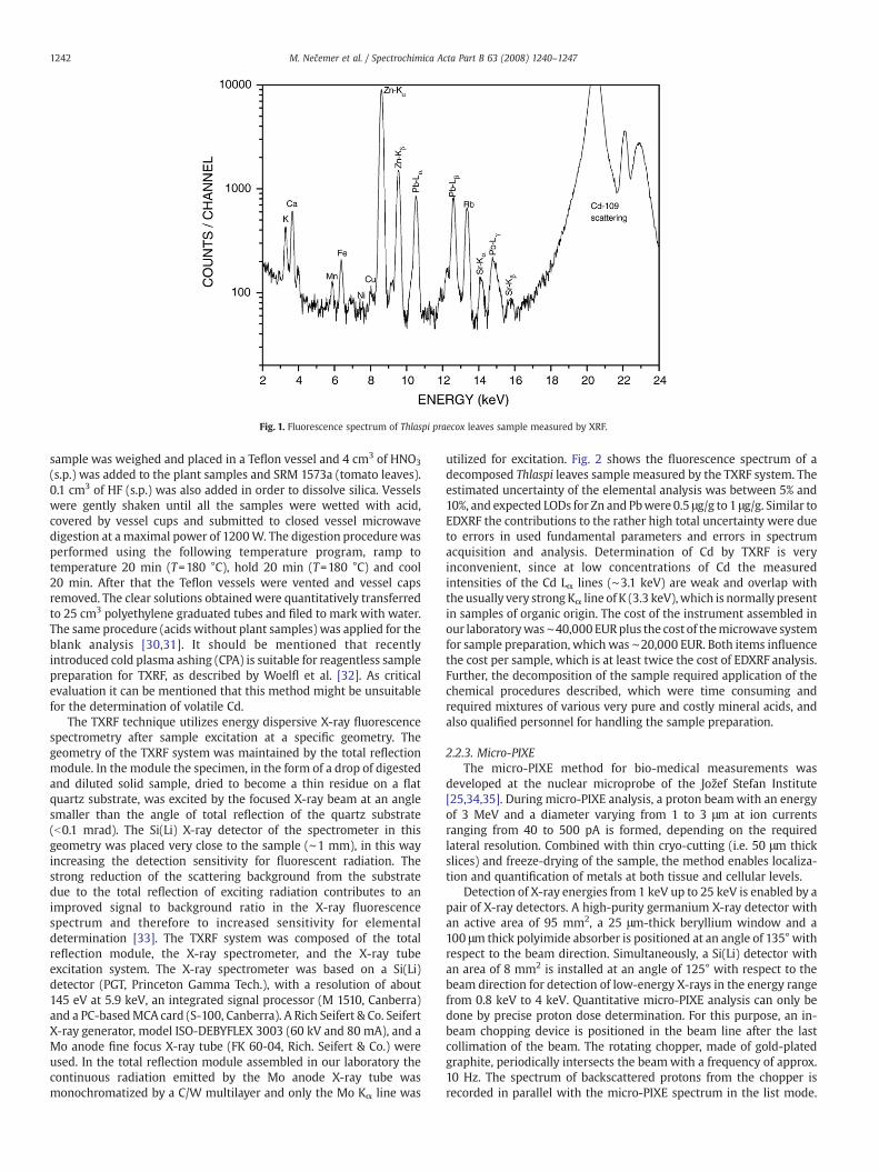
Fig. 1. Fluorescence spectrum of Thlaspi praecox leaves sample measured by XRF.
1242 M. Nečemer et al. / Spectrochimica Acta Part B 63 (2008) 1240–1247
sample was weighed and placed in a Teflon vessel and 4 cm3 of HNO3
(s.p.) was added to the plant samples and SRM 1573a (tomato leaves).0.1 cm3 of HF (s.p.) was also added in order to dissolve silica. Vesselswere gently shaken until all the samples were wetted with acid,covered by vessel cups and submitted to closed vessel microwavedigestion at a maximal power of 1200W. The digestion procedurewasperformed using the following temperature program, ramp totemperature 20 min (T=180 °C), hold 20 min (T=180 °C) and cool20 min. After that the Teflon vessels were vented and vessel capsremoved. The clear solutions obtained were quantitatively transferredto 25 cm3 polyethylene graduated tubes and filed to mark with water.The same procedure (acids without plant samples) was applied for theblank analysis [30,31]. It should be mentioned that recentlyintroduced cold plasma ashing (CPA) is suitable for reagentless samplepreparation for TXRF, as described by Woelfl et al. [32]. As criticalevaluation it can be mentioned that this method might be unsuitablefor the determination of volatile Cd.
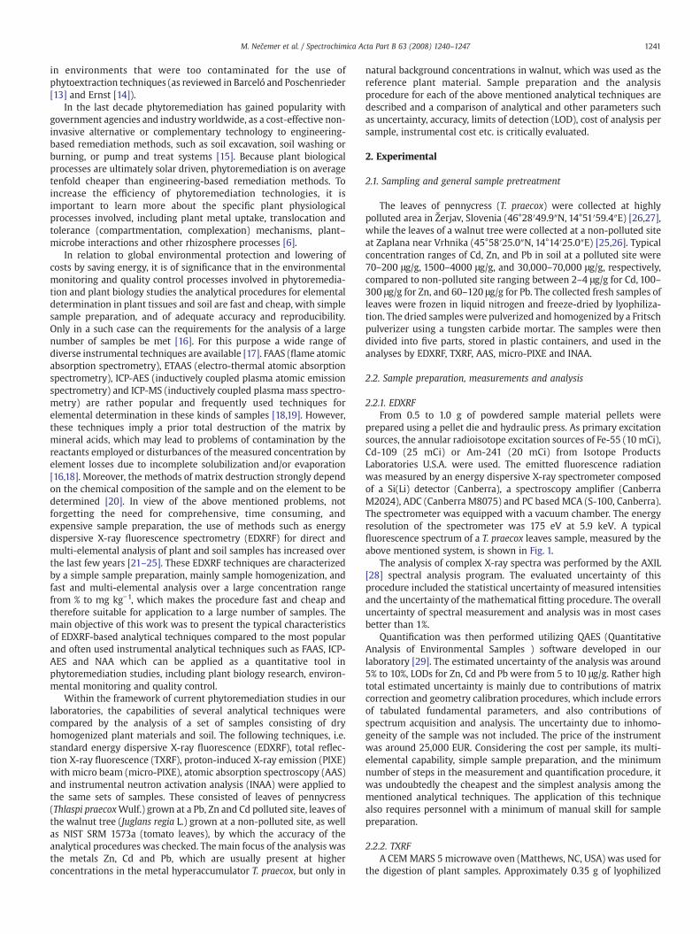
The TXRF technique utilizes energy dispersive X-ray fluorescencespectrometry after sample excitation at a specific geometry. Thegeometry of the TXRF system was maintained by the total reflectionmodule. In the module the specimen, in the form of a drop of digestedand diluted solid sample, dried to become a thin residue on a flatquartz substrate, was excited by the focused X-ray beam at an anglesmaller than the angle of total reflection of the quartz substrate(b0.1 mrad). The Si(Li) X-ray detector of the spectrometer in thisgeometry was placed very close to the sample (∼1 mm), in this wayincreasing the detection sensitivity for fluorescent radiation. Thestrong reduction of the scattering background from the substratedue to the total reflection of exciting radiation contributes to animproved signal to background ratio in the X-ray fluorescencespectrum and therefore to increased sensitivity for elementaldetermination [33]. The TXRF system was composed of the totalreflection module, the X-ray spectrometer, and the X-ray tubeexcitation system. The X-ray spectrometer was based on a Si(Li)detector (PGT, Princeton Gamma Tech.), with a resolution of about145 eV at 5.9 keV, an integrated signal processor (M 1510, Canberra)and a PC-basedMCA card (S-100, Canberra). A Rich Seifert & Co. SeifertX-ray generator, model ISO-DEBYFLEX 3003 (60 kV and 80 mA), and aMo anode fine focus X-ray tube (FK 60-04, Rich. Seifert & Co.) wereused. In the total reflection module assembled in our laboratory thecontinuous radiation emitted by the Mo anode X-ray tube wasmonochromatized by a C/W multilayer and only the Mo Kα line was
utilized for excitation. Fig. 2 shows the fluorescence spectrum of adecomposed Thlaspi leaves sample measured by the TXRF system. Theestimated uncertainty of the elemental analysis was between 5% and10%, and expected LODs for Zn and Pbwere 0.5 μg/g to 1 μg/g. Similar toEDXRF the contributions to the rather high total uncertainty were dueto errors in used fundamental parameters and errors in spectrumacquisition and analysis. Determination of Cd by TXRF is veryinconvenient, since at low concentrations of Cd the measuredintensities of the Cd Lα lines (∼3.1 keV) are weak and overlap withthe usually very strongKα line of K (3.3 keV),which is normally presentin samples of organic origin. The cost of the instrument assembled inour laboratorywas∼40,000 EURplus the cost of themicrowave systemfor sample preparation, which was ∼20,000 EUR. Both items influencethe cost per sample, which is at least twice the cost of EDXRF analysis.Further, the decomposition of the sample required application of thechemical procedures described, which were time consuming andrequired mixtures of various very pure and costly mineral acids, andalso qualified personnel for handling the sample preparation.
2.2.3. Micro-PIXEThe micro-PIXE method for bio-medical measurements was
developed at the nuclear microprobe of the Jožef Stefan Institute[25,34,35]. During micro-PIXE analysis, a proton beamwith an energyof 3 MeV and a diameter varying from 1 to 3 μm at ion currentsranging from 40 to 500 pA is formed, depending on the requiredlateral resolution. Combined with thin cryo-cutting (i.e. 50 μm thickslices) and freeze-drying of the sample, the method enables localiza-tion and quantification of metals at both tissue and cellular levels.
Detection of X-ray energies from 1 keV up to 25 keV is enabled by apair of X-ray detectors. A high-purity germanium X-ray detector withan active area of 95 mm2, a 25 μm-thick beryllium window and a100 μm thick polyimide absorber is positioned at an angle of 135° withrespect to the beam direction. Simultaneously, a Si(Li) detector withan area of 8 mm2 is installed at an angle of 125° with respect to thebeam direction for detection of low-energy X-rays in the energy rangefrom 0.8 keV to 4 keV. Quantitative micro-PIXE analysis can only bedone by precise proton dose determination. For this purpose, an in-beam chopping device is positioned in the beam line after the lastcollimation of the beam. The rotating chopper, made of gold-platedgraphite, periodically intersects the beamwith a frequency of approx.10 Hz. The spectrum of backscattered protons from the chopper isrecorded in parallel with the micro-PIXE spectrum in the list mode.
Fig. 2. Spectrum of decomposed Thlaspi praecox leaves sample measured by TXRF.
1243M. Nečemer et al. / Spectrochimica Acta Part B 63 (2008) 1240–1247
The high-energy part of the spectrum consists of an isolated peakoriginating from protons scattered on the Au layer and is used todetermine the proton flux during the measurements. To preventcharging of insulated samples, a hot filament is used inside thevacuum chamber as a source of low-energy electrons, whichneutralize the charged surfaces.
Assuming a cellulose matrix and thick target formalism, themeasured spectra were analyzed using the GUPIXWIN program [36].To improve the definition of efficiency, especially at energies belowthe K absorption edge of Si and Ge for the Si(Li) and Ge detectors,respectively, additional efficiency-energy dependence was introducedin GUPIXWIN, based on calibration measurements on monoelementalsamples and compounds.
The accuracy of the micro-PIXE method was verified by theanalysis of the multi-elemental standard reference materials NISTSRM 1107 (Naval Brass B, alloy) and NIST SRM 620 (Soda-Lime Flat
Fig. 3. Spectrum of Thlaspi praecox le
Glass), as well as NIST SRM 1573a (tomato leaves, homogenizedpowder, analyzed in the form of a pressurized pellet).
Analyses described here were made on bulk palletized material,therefore the thick target PIXE approach was used. A PIXE spectrum ofThlaspi leaves measured by Ge detector is shown in Fig. 3. The Caelemental maps obtained during the pellet analysis provide informa-tion on the powder particulate size used for pellet production (Fig. 5).The total uncertainty of quantitative results includes a number ofcontributions such as the uncertainty of proton dose measurement,estimation of the sample thickness and the respective proton range,and also uncertainties typical for all other XRF techniques.
Micro-PIXE techniques require an accelerator facility and thereforethe cost per sample was much higher than in the case of EDXRF andTXRF. But it is necessary to keep inmind that the information obtainedabout the distribution of elements on even the cellular scale isextremely valuable.
aves sample measured by PIXE.
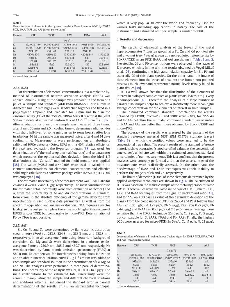
Table 1Concentrations of elements in the hyperaccumulator Thlaspi praecox Wulf. by EDXRF,PIXE, INAA, TXRF and AAS (mean±uncertainty)
Element XRF TXRF PIXE INAA AAS
(μg g−1)
K 16,700±1780 14,300±2240 19,130±1842 17,018±599 14,126±706Ca 15,800±1270 14,400±2240 16,946±1335 15,440±618 15,138±757Fe 223±22 257±40 251±15 260±10 n.d.Zn 4270±330 4190±65 4530±280 4224±148 4156±208Pb 418±33 394±62 509±31 n.d. 369±19Rb 101±8 109±17 112±9 100±4 n.d.Sr 12.4±1.2 15±2 12.6±2.2 b20 12.3±0.6Cd 1250±73 n.d. 1425±108 1381±49 1221±61Br 8.92±1.04 9.8±2.0 6.3±2.3 7.90±0.28 n.d.
n.d.—not determined.
Table 2Concentrations of elements in walnut leaves (Juglans regia) by EDXRF, PIXE, INAA, TXRFand AAS (mean±uncertainty)
Element XRF TXRF PIXE INAA AAS
(μg g−1)
K 5150±680 4770±747 6195±598 4970±175 4590±230Ca 23,700±1900 22,200±3460 25,675±2022 23,791±891 23,286±1164Fe 165±18 149±23 155±10 139±6 n.d.Zn 60±6 55±9 62±4 56±2 45±2Pb 9.1±1.8 4.1±0.9 ≤7.9 n.d. ≤12Rb 5.6±1.1 6.9±1.2 5.7±4.1 5.4±0.2 n.d.Sr 36±3 44±7 36±6 37.3±2.2 30.0±1.5Cd ≤3 n.d. ≤138 ≤0.3 ≤1Br 30±3 n.d. 31±4 29±1 n.d.
n.d.—not determined.
1244 M. Nečemer et al. / Spectrochimica Acta Part B 63 (2008) 1240–1247
2.2.4. INAAFor determination of elemental concentrations in a sample the k0-
method of instrumental neutron activation analysis (INAA) wasapplied. About 200 mg of the sample was prepared in the form of apellet. A sample and standard (Al-0.1%Au IRMM-530 disc 6 mm indiameter and 0.2 mm high) were sandwiched together and fixed in apolyethylene ampoule and irradiated for 3 min and 16 h in thecarousel facility (CF) of the 250 kW TRIGA Mark II reactor at the JožefStefan Institute at a thermal neutron flux of 1.1 ·1012 n cm−2 s−1 [37].After irradiation for 3 min, the sample was measured three times:after 5 min, 30 min and 2.5 h cooling time to determine radionuclideswith short half-lives (of some minutes up to some hours). After longirradiation (16 h) the sample was measured twice: after 4 and 14 dayscooling time. Measurements were performed on an absolutelycalibrated HPGe detector (Ortec, USA) with a 40% relative efficiency.For peak area evaluation, the HyperLab program [38] was used. Fordetermination of f (thermal to epithermal flux ratio) and α (parameterwhich measures the epithermal flux deviation from the ideal 1/Edistribution), the “Cd-ratio” method for multi-monitor was applied[38]. The values f=28.8 and α=−0.005 were used to calculate theelement concentrations. For elemental concentrations and effectivesolid angle calculations a software package called KAYZERO/SOLCOI®was employed [38].
The estimated uncertainty of themeasurement was 3–5%. LODs forZn and Cd were 0.2 and 3 μg/g, respectively. The main contributions tothe estimated total uncertainty were from evaluation of factors f andα, from the uncertainty of Al and Au concentrations in standard,from errors in the absolute efficiency of the gamma detector, fromuncertainties in used nuclear data parameters, as well as from thespectrum acquisition and analysis evaluation. INAA requires a reactorfacility, so the cost per sample is therefore much higher than in case ofEDXRF and/or TXRF, but comparable to micro-PIXE. Determination ofPb by INAA is not possible.
2.2.5. AASZn, Cu, Pb and Cd were determined by flame atomic absorption
spectrometry (FAAS) at 213.8, 324.8 nm, 283.3 nm, and 228.8 nm,respectively, in an air-acetylene flame using deuterium backgroundcorrection. Ca, Mg and Sr were determined in a nitrous oxide-acetylene flame at 239.9 nm, 285.2 and 460.7 nm, respectively. Nawas determined by flame atomic emission spectrometry (FAES) at589.0 nm. To compensate for interferences arising from ionizationand to obtain linear calibration curves, 2 g l−1 cesium was added toeach sample and standard solution in the determination of Ca, Mg, Srand Na. The analyses were performed in three parallel determina-tions. The uncertainty of the analysis was 5%, LODs 0.5 to 5 μg/g. Themain contributions to the estimated total uncertainty were theerrors in manipulating the sample and different chemical solutionsand additions which all influenced the standard error in paralleldeterminations of the results. This is an instrumental technique,
which is very popular all over the world and frequently used forvarious tasks including applications in botany. The cost of theinstrument and estimated cost per sample is similar to TXRF.
3. Results and discussion
The results of elemental analysis of the leaves of the metalhyperaccumulator T. praecox grown at a Pb, Zn and Cd polluted siteand a walnut tree (J. regia) grown at a non-polluted reference site byEDXRF, TXRF, micro-PIXE, INAA, and AAS are shown in Tables 1 and 2.Elevated Zn, Cd and Pb concentrations were observed in the leaves ofT. praecox, which is in line with the results obtained by Vogel-Mikušet al. [26], confirming the high accumulation capacity for Zn, Pb andespecially Cd of this plant species. On the other hand, the intake ofthese elements into the leaves of a walnut tree from a non-pollutedarea was much lower and represented normal levels usually found inplant tissues [39].
It is a well known fact that the distribution of the elements ofinterest in biological samples such as plants (roots, leaves, etc.) is veryinhomogeneous [40]. Therefore the analysis of a large number ofparallel sub-samples helps to achieve a statistically more meaningfulaverage concentration for the elements of interest in such samples.
The estimated combined standard uncertainties for resultsobtained by EDXRF, micro-PIXE and TXRF were ∼10%, for NAA 4%and for AAS 5%. Thus the estimated combined standard uncertaintiesof INAA and AAS are better than those obtained by EDXRF, TXRF andmicro-PIXE.
The accuracy of the results was assessed by the analysis of thestandard reference material NIST SRM C1573a (tomato leaves)(Table 3) in which the certified values were assumed to be theconventional true values. The present results of the standard referencematerials show accuracies (stated certified values as the conventionaltrue values), which are well within the estimated combined standarduncertainties of ourmeasurements. This fact confirms that the presentanalyses were correctly performed and that the uncertainties of themeasurements were realistically assessed. On the other hand thedisadvantage of INAA and TXRF techniques was their inability toperform the analysis of Pb and Cd, respectively.
The limits of detection (LODs) of some elements determined by theapplied analytical techniques are shown in Fig. 4. The calculation ofLODswas based on the realistic sample of themetal hyperaccumulatorThlaspi. These values were evaluated in the case of EDXRF, micro-PIXE,TXRF and INAA techniques from the signal to background ratio [41],and for AAS on a 3σ basis (a value of three standard deviations of theblank). From the comparison of LODs for Zn, Cd and Pb it follows thatAAS (Zn 0.35 μg/g, Cd 1.25 μg/g, Pb 5 μg/g), TXRF (Zn 0.27 μg/g, Pb0.44 μg/g) and INAA (Zn 0.25 μg/g Cd 2.5 μg/g) are on average moresensitive than the EDXRF technique (Zn 6 μg/g, Cd 2 μg/g, Pb 5 μg/g),but comparable for Cd (AAS, INAA) and Pb (AAS). Finally, the highestLODs were assessed for micro-PIXE (Zn 5 μg/g, Cd 17 μg/g, Pb 21 μg/g),
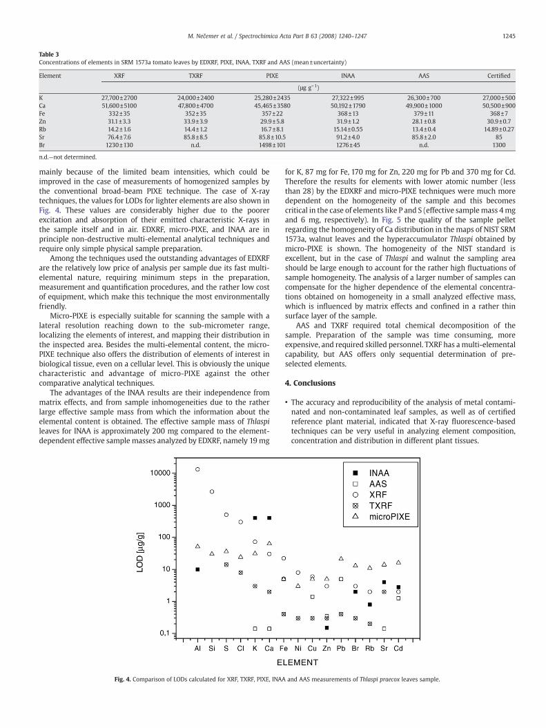
Table 3Concentrations of elements in SRM 1573a tomato leaves by EDXRF, PIXE, INAA, TXRF and AAS (mean±uncertainty)
Element XRF TXRF PIXE INAA AAS Certified
(μg g−1)
K 27,700±2700 24,000±2400 25,280±2435 27,322±995 26,300±700 27,000±500Ca 51,600±5100 47,800±4700 45,465±3580 50,192±1790 49,900±1000 50,500±900Fe 332±35 352±35 357±22 368±13 379±11 368±7Zn 31.1±3.3 33.9±3.9 29.9±5.8 31.9±1.2 28.1±0.8 30.9±0.7Rb 14.2±1.6 14.4±1.2 16.7±8.1 15.14±0.55 13.4±0.4 14.89±0.27Sr 76.4±7.6 85.8±8.5 85.8±10.5 91.2±4.0 85.8±2.0 85Br 1230±130 n.d. 1498±101 1276±45 n.d. 1300
n.d.—not determined.
1245M. Nečemer et al. / Spectrochimica Acta Part B 63 (2008) 1240–1247
mainly because of the limited beam intensities, which could beimproved in the case of measurements of homogenized samples bythe conventional broad-beam PIXE technique. The case of X-raytechniques, the values for LODs for lighter elements are also shown inFig. 4. These values are considerably higher due to the poorerexcitation and absorption of their emitted characteristic X-rays inthe sample itself and in air. EDXRF, micro-PIXE, and INAA are inprinciple non-destructive multi-elemental analytical techniques andrequire only simple physical sample preparation.
Among the techniques used the outstanding advantages of EDXRFare the relatively low price of analysis per sample due its fast multi-elemental nature, requiring minimum steps in the preparation,measurement and quantification procedures, and the rather low costof equipment, which make this technique the most environmentallyfriendly.
Micro-PIXE is especially suitable for scanning the sample with alateral resolution reaching down to the sub-micrometer range,localizing the elements of interest, and mapping their distribution inthe inspected area. Besides the multi-elemental content, the micro-PIXE technique also offers the distribution of elements of interest inbiological tissue, even on a cellular level. This is obviously the uniquecharacteristic and advantage of micro-PIXE against the othercomparative analytical techniques.
The advantages of the INAA results are their independence frommatrix effects, and from sample inhomogeneities due to the ratherlarge effective sample mass from which the information about theelemental content is obtained. The effective sample mass of Thlaspileaves for INAA is approximately 200 mg compared to the element-dependent effective sample masses analyzed by EDXRF, namely 19 mg
Fig. 4. Comparison of LODs calculated for XRF, TXRF, PIXE, INAA
for K, 87 mg for Fe, 170 mg for Zn, 220 mg for Pb and 370 mg for Cd.Therefore the results for elements with lower atomic number (lessthan 28) by the EDXRF and micro-PIXE techniques were much moredependent on the homogeneity of the sample and this becomescritical in the case of elements like P and S (effective samplemass 4mgand 6 mg, respectively). In Fig. 5 the quality of the sample pelletregarding the homogeneity of Ca distribution in the maps of NIST SRM1573a, walnut leaves and the hyperaccumulator Thlaspi obtained bymicro-PIXE is shown. The homogeneity of the NIST standard isexcellent, but in the case of Thlaspi and walnut the sampling areashould be large enough to account for the rather high fluctuations ofsample homogeneity. The analysis of a larger number of samples cancompensate for the higher dependence of the elemental concentra-tions obtained on homogeneity in a small analyzed effective mass,which is influenced by matrix effects and confined in a rather thinsurface layer of the sample.
AAS and TXRF required total chemical decomposition of thesample. Preparation of the sample was time consuming, moreexpensive, and required skilled personnel. TXRF has amulti-elementalcapability, but AAS offers only sequential determination of pre-selected elements.
4. Conclusions
• The accuracy and reproducibility of the analysis of metal contami-nated and non-contaminated leaf samples, as well as of certifiedreference plant material, indicated that X-ray fluorescence-basedtechniques can be very useful in analyzing element composition,concentration and distribution in different plant tissues.
and AAS measurements of Thlaspi praecox leaves sample.
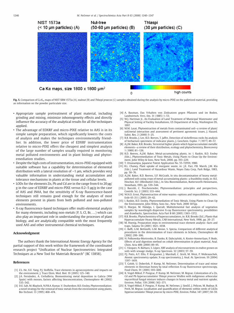
Fig. 5. Comparison of Ca Kα maps of NIST SRM 1573a (A), walnut (B) and Thlaspi praecox (C) samples obtained during the analysis by micro-PIXE on the palletized material, providingan information on the powder particulate size.
1246 M. Nečemer et al. / Spectrochimica Acta Part B 63 (2008) 1240–1247
• Appropriate sample pretreatment of plant material, includinggrinding and mixing, minimize inhomogeneity effects and directlyinfluence the accuracy of the analytical results for all the techniquesapplied.
• The advantage of EDXRF and micro-PIXE relative to AAS is in itssimple sample preparation, which significantly lowers the costsof analysis and makes the techniques environmentally friend-lier. In addition, the lower price of EDXRF instrumentationrelative to micro-PIXE offers the cheapest and simplest analysisof the large number of samples usually required in monitoringmetal polluted environments and in plant biology and phytor-emediation studies.
• Despite the high costs of instrumentation,micro-PIXE equippedwithsuitable software has a capability for determination of elementaldistribution with a lateral resolution of ∼1 μm, which provides veryvaluable information in understanding metal accumulation andtolerance mechanisms in plants at the tissue and cellular levels.
• LODs for the elements Zn, Pb and Cd are in the range from 6 to 20 μg/g in the case of EDXRF and micro-PIXE versus 0.2–5 μg/g in the caseof AAS and INAA, but the sensitivity of X-ray fluorescence-basedtechniques still remains good enough for the analyses of mostelements present in plants from both polluted and non-pollutedenvironments.
• X-ray fluorescence-based techniques offer multi-elemental analysisfor many elements, including non-metals (P, S, Cl, Br, …) which canalso play an important role in understanding the processes of plantbiology, and are analytically compatible with the most frequentlyused AAS and other instrumental chemical techniques.
Acknowledgment
The authors thank the International Atomic Energy Agency for thepartial support of this work within the framework of the coordinatedresearch project “Unification of Nuclear Spectrometries: IntegratedTechniques as a New Tool for Materials Research” (RC 13858).
References
[1] Z.L. He, X.E. Yang, P.J. Stoffella, Trace elements in agroecosystems and impacts onthe environment, J. Trace Elem. Med. Biol. 19 (2005) 125–140.
[2] J.A. Fernández, A. Cerballeria, Biomonitoring metal deposition in Galicia (NWSpain) with mosses, factors affecting bioconcentration, Chemosphere 46 (2002)535–542.
[3] D.E. Salt, M. Blaylock, N.P.B.A. Kumar, V. Dushenkov, B.D. Ensley, Phytoremediation:a novel strategy for the removal of toxicmetals from the environment using plants,Bio-Technol. 13 (1995) 468–474.
[4] A. Bauman, Das Vrhalten von Zinksatzen gegen Pflanzen und im Boden,Landwirtsch. Vers.-Stn. 31 (1885) 1–53.
[5] W.J. Hartman Jr., An Evaluation of Land Treatment of Municipal Wastewater andPhysical Setting of Facility Installations, US Department of Army, Washington DS,1997.
[6] M.M. Lasat, Phytoextraction of metals from contaminated soil—a review of plant/soil/metal interaction and assessment of pertinent agronomic issues, J. Hazard.Subst. Res. 2 (2000) 5–25.
[7] R.R. Brooks, J. Lee, R.D. Reeves, T. Jaffre, Detection of nickelferous rocks by analysisof herbarium specimens of indicator plants, J. Geochem. Explor. 7 (1977) 49–57.
[8] A.J.M. Baker, R.R. Brooks, Terrestrial higher plants which hyperaccumulate metallicelements—a review of their distribution, ecology and phytochemistry, Biorecovery1 (1989) 81–126.
[9] R.D. Reeves, A.J.M. Baker, Metal-accumulating plants, in: I. Raskin, B.D. Ensley(Eds.), Phytoremediation of Toxic Metals, Using Plants to Clean Up the Environ-ment, John Wiley & Sons, New York, 2000, pp. 193–229.
[10] T. Utsunamyia, Japanese Patent Application No. 55-72959, 1980.[11] R.L. Chaney, Plant uptake of inorganic waste, in: J.E. Parr, P.B. Marsh, J.M. Kla
(Eds.), Land Treatment of Hazardous Waste, Noyes Data Corp, Park Ridge, 1983,pp. 50–76.
[12] A.J.M. Baker, R.D. Reeves, S.P. McGrath, In situ decontamination of heavy metal-polluted soils using crops of metal-accumulating plants—a feasibility study, in: R.E.Hinchee, R.F. Olfenbuttel (Eds.), In Situ Bioremediation, Butterworth-Heinemann,Stoneham, 1991, pp. 539–544.
[13] J. Barceló, C. Poschenrieder, Phytoremediation: principles and perspectives,Contrib. Sci. 2 (2003) 333–444.
[14] W.H.O. Ernst, Phytoextraction of mine wastes—options and impossibilities, Chem.Erde 65 (S1) (2005) 29–42.
[15] I. Raskin, B.D. Ensley, Phytoremediation of Toxic Metals, Using Plants to Clean Upthe Environment, John Wiley, Sons Inc., New York, 2000 307pp.
[16] E. Margui, M. Hidalgo, I. Queralt, Multielemental fast analysis of vegetationsamples by wavelength dispersive X-ray fluorescence spectrometry, possibilitiesand drawbacks, Spectrochim. Acta Part B 60 (2005) 1363–1372.
[17] R.R. Brooks, Phytochemistry of hyperaccumulators, in: R.R. Brooks (Ed.), Plants thatHyperaccumulate Heavy Metals, CAB International, New York, 1998, pp. 20–27.
[18] M. Hoenig, Preparation steps in environmental trace element analysis—facts andtraps, Talanta 54 (2001) 1021–1038.
[19] C. Baffi, G.M. Bettinelli, G.M. Beone, S. Spezia, Comparison of different analyticalprocedures in the determination of trace elements in lichens, Chemosphere 48(2002) 299–306.
[20] H. Polkowska-Mortrenko, B. Danko, R. Dybczyński, A. Koster-Ammerlaan, P. Bode,Effects of acid digestion method on cobalt determination in plant material, Anal.Chim. Acta 408 (2000) 89–95.
[21] C. Vázquez, N. Bárbaro, S. López, XRF analysis of micronutrients in endive grown onsoils with sewage sludge, X-ray Spectrom. 32 (2003) 57–59.
[22] P.J. Potts, A.T. Ellis, P. Kregsamer, J. Marshall, C. Streli, M. West, P. Wobrauschek,Atomic spectrometry update, X-ray spectrometry, J. Anal. At. Spectrom. 19 (2004)1397–1419.
[23] T. Golob, U. Doberšek, P. Kump, M. Nečemer, Determination of trace and minorelements in Slovenian honey by total reflection X-ray fluorescence spectroscopy,Food Chem. 91 (2005) 593–600.
[24] K. Vogel-Mikuš, P. Pongrac, P. Kump, M. Nečemer, M. Regvar, Colonization of a Zn,Cd and Pb hyperaccumulator Thlaspi praecox Wulfen with indigenous arbuscularmycorrhizal fungal mixture induces changes in heavy metal and nutrient uptake,Environ. Pollut. 139 (2006) 362–371.
[25] K. Vogel-Mikuš, P. Pongrac, P. Kump, M. Nečemer, J. Simčič, J. Pelicon, M. Budnar, B.Povh, M. Regvar, Localisation and quantification of elements within seeds of Cd/Znhyperaccumulator Thlaspi praecox by micro-PIXE, Environ. Pollut. 147 (2007) 50–59.
1247M. Nečemer et al. / Spectrochimica Acta Part B 63 (2008) 1240–1247
[26] K. Vogel-Mikuš, D. Drobne, M. Regvar, Zn, Cd and Pb accumulation and arbuscularmycorrhizal colonization of pennycress Thlaspi praecox Wulf. Brassicaceae fromthe vicinity of a lead mine and smelter in Slovenia, Environ. Pollut. 133 (2005)233–242.
[27] M. Regvar, K. Vogel-Mikuš, N. Kugonič, B. Turk, F. Batič, Vegetational andmycorrhizal successions at a metal polluted site—indications for the direction ofphytostabilisation, Environ. Poll. 144 (2006) 976–984.
[28] P.J.M. Van Espen, K.H.A. Janssens, Spectrum evaluation, in: R.E. Van Grieken, A.A.Markowicz (Eds.), Handbook of X-ray Spectroscopy: Methods and Techniques,Marcel Dekker, New York, 1993, p. 222.
[29] P. Kump, M. Nečemer, B. Smodiš, R. Jačimović, Multielement analysis of rubbersamples by X-ray fluorescence, Appl. Spectrosc. 50 (1996) 1373–1377.
[30] R. Milačič, B. Kralj, Determination of Zn, Cu, Cd, Pb, Ni in some Slovenian foodstuffs,European Food Research and Technology, Z. Lebensm.-Unters. Forsch. 217 (2003)211–214.
[31] T. Zuliani, B. Kralj, V. Stibilj, R. Milačič, Minerals and trace elements in foodcommonly consumed in Slovenia, Ital. J. Food Sci. 17 (2005) 155–166.
[32] S. Woelfl, M. Mages, F. Encina, Cold plasma ashing improves the trace elementdetection of single Daphnia specimens by total reflection X-ray fluorescencespectrometry, Spectrochim. Acta Part B 58 (2003) 2157–2168.
[33] P. Kump, M. Nečemer, J. Šnajder, Determination of trace elements in bee honey,pollen and tissue by total reflection and radioisotope X-ray fluorescence spectro-metry, Spectrochim. Acta Part B 51 (1996) 499–507.
[34] J. Simčič, P. Pelicon, M. Budnar, Ž. Šmit, The performance of the Ljubljana ionmicroprobe, Nucl. Instrum. Methods Part B 190 (2002) 283–286.
[35] P. Pelicon, J. Simčič, M. Jakšić, Z. Medunić, F. Naab, F.D. McDaniel, Sphericalchamber—effective solution for multipurpose nuclear microprobe, Nucl. Instrum.Methods Part B 231 (2005) 53–59.
[36] J.L. Campbell, T.L. Hopman, J.A. Maxwell, Z. Nejedly, The Guelph PIXE softwarepackage III: alternative proton database, Nucl. Instrum. Methods Part B 170 (2000)193–204.
[37] R. Jaćimović, Evaluation of the use of the TRIGA Mark II reactor for the k0-methodof activation analysis, Ph. D. Thesis (in Slovene), University of Ljubljana, 2003.
[38] R. Jaćimović, B. Smodiš, T. Bučar, P. Stegnar, k0-NAA quality assessment by analysisof different certified reference materials using the KAYZERO/SOLCOI software,J. Radioanal. Nucl. Chem. 257 (2003) 659–663.
[39] D.C. Adriano, Trace Elements in Terrestrial Environments: Biochemistry, Bioavail-ability and Risk of Metals, second ed., Springer-Verlag, New York, 2001, p. 867.
[40] S. Scheloske, T. Schneider, BIOPIXE: a new PIXE-data software package to analysequantitative elemental distributions of inhomogeneous samples, Nucl. Instrum.Methods Part B 189 (2002) 148–152.
[41] P. Kump, Some considerations on the definition of the limit of detection in X-rayfluorescence spectrometry, Spectrochim. Acta Part B 52 (1997) 405–408.