apoptosis induced by prolonged exposure to odorants in cultured cells from rat olfactory epithelium
TRANSCRIPT
B R A I N R E S E A R C H 1 1 0 3 ( 2 0 0 6 ) 1 1 4 – 1 2 2
ava i l ab l e a t www.sc i enced i r ec t . com
www.e l sev i e r. com/ loca te /b ra in res
Research Report
Apoptosis induced by prolonged exposure to odorants incultured cells from rat olfactory epithelium
Sebastian Brauchia,b,1, Christian Ceaa,1, Jorge G. Fariasc,Juan Bacigalupod,e, Juan G. Reyesa,⁎aInstituto de Quimica, Pontificia Universidad Catolica de Valparaiso, Casilla 4059, Valparaiso, ChilebUniversidad Austral de Chile, Valdivia, ChilecInstituto de Estudios de la Salud, Universidad Arturo Prat, Iquique, ChiledDepartmento de Biologia, Facultad de Ciencias, Universidad de Chile, Santiago, ChileeCentro de Investigacion de Dinamica Celular y Biotechnologia, Universidad de Chile, Santiago, Chile
A R T I C L E I N F O
⁎ Corresponding author. Fax: +56 32 273422.E-mail address: [email protected] (J.G. Reyes).
1 These authors contributed equally to this
0006-8993/$ – see front matter © 2006 Elsevidoi:10.1016/j.brainres.2006.05.072
A B S T R A C T
Article history:Accepted 12 May 2006Available online 11 July 2006
Multicellular organisms undergo programmed cell death (PCD) as a mechanism for tissueremodeling during development and tissue renewal throughout adult life. Overdose of someneuronal receptor agonists like glutamate can trigger a PCD process termed excitotoxicity inneurons of the central nervous system. Calcium has an important role in PCD processes,especially in excitotoxicity. Since the normal turnover of olfactory receptor neurons (ORNs)relies, at least in part, on an apoptoticmechanismandodor transduction inORNs involves anincrease in intracellular Ca2+ concentration ([Ca2+]i), we investigated the possibility that long-term exposures to odorants could trigger an excitotoxic process in olfactory epithelial cells(EC). We used single-cell [Ca2+]i determinations and fluorescence microscopy techniques tostudy the effects of sustained odorant exposures in olfactory EC in primary culture. Inductionof PCD was evaluated successively by three independent criteria: (1) measurements of DNAfragmentation, (2) translocation of phosphatidylserine to the external leaflet of the plasmamembrane, and (3) caspase-3 activation. Our results support the notion of an odorant-induced PCD in olfactory EC. This odorant-induced PCD was prevented by LY83583, anodorant response inhibitor, suggesting that ORNs are the main epithelial cell populationundergoing odorant-induced PCD.
© 2006 Elsevier B.V. All rights reserved.
Keywords:ApoptosisOlfactory epitheliumOlfactory neuronSignal transductionCalcium
1. Introduction
Considerable progress has been made on elucidating themechanisms of olfactory transduction over the past decade.Thus, several models have been proposed to explain howodors are detected (Dionne and Dubin, 1994; Morales andBacigalupo, 1996; Madrid et al., 2005; Schild and Restrepo,
work.
er B.V. All rights reserved
1998). In the best-established model, binding of an odorant toits G-protein-coupled odorant receptor triggers a cyclic AMPcascade (Schild and Restrepo, 1998). Cyclic AMP opens a cyclicnucleotide-gated cation channel (CNGC; Nakamura and Gold,1987). This event increases intracellular Ca2+ concentration([Ca2+]i) (Restrepo et al., 1993; Leinders-Zufall et al., 1998),which in turn gates a calcium-dependent chloride channel
.
115B R A I N R E S E A R C H 1 1 0 3 ( 2 0 0 6 ) 1 1 4 – 1 2 2
(Kleene and Gesteland, 1991; Lowe and Gold, 1993; Kurahashiand Yau, 1993). Both conductances are responsible for adepolarizing (excitatory) receptor potential, leading to anincrease in action potential discharge frequency. Calciumhas also been shown to mediate odor inhibition by openingCa2+-dependent K+ channels, causing a hyperpolarizing re-ceptor potential that leads to a reduction in firing rate (Moraleset al., 1994; Sanhueza et al., 2000; Madrid et al., 2005).
As part of its normal turnover, ORN population density iscontrolled by the balance between neurogenesis and apopto-sis. Apoptosis occurs in mature ORNs under normal condi-tions (see Cowan and Roskams, 2002; Suzuki, 2004 for reviews),but the molecular entities that trigger this normal turnoverapoptotic process have not been identified. Sensory depriva-tion by naris occlusion (Meisami, 1976) causes a reduction inthe turn over rate of ORNs, without changing the total numberof mature ORNs in the epithelium (Farbman et al., 1988). Thus,the delay observed in normal apoptotic process was attributedto a protective effect from airborn odorants.
Because a rise in [Ca2+]i is essential for the odorant response(see Schild and Restrepo, 1998), we reasoned that a prolongedexposure to high levels of odorants may be equivalent to asustained presence of an excitotoxic agent, which can lead toprogrammed cell death (PCD) in other cell types throughprocesses that involve an increase in intracellular Ca2+ (Saidet al., 1996; Mattson, 2000). In this work, we show, for the firsttime that sustained over-exposition to odorants causesapoptosis in a large fraction of cultured olfactory epithelialcells (EC), including olfactory receptor neurons (ORNs). TheCNGC blocker LY83583 prevented the rise in [Ca2+]i and theodorant induction of apoptosis, presumably because it imped-ed an abnormally sustained high intracellular Ca2+ level byabolishing the Ca2+ influx through its channel, stronglysuggesting that ORNs underwent an excitotoxicity-like PCDin culture.
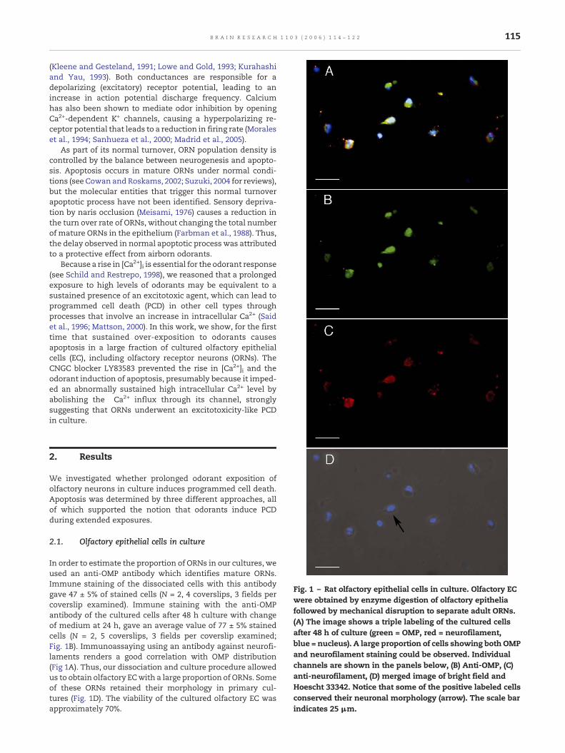
Fig. 1 – Rat olfactory epithelial cells in culture. Olfactory ECwere obtained by enzyme digestion of olfactory epitheliafollowed by mechanical disruption to separate adult ORNs.(A) The image shows a triple labeling of the cultured cellsafter 48 h of culture (green = OMP, red = neurofilament,blue = nucleus). A large proportion of cells showing both OMPand neurofilament staining could be observed. Individualchannels are shown in the panels below, (B) Anti-OMP, (C)anti-neurofilament, (D) merged image of bright field andHoescht 33342. Notice that some of the positive labeled cellsconserved their neuronal morphology (arrow). The scale barindicates 25 μm.
2. Results
We investigated whether prolonged odorant exposition ofolfactory neurons in culture induces programmed cell death.Apoptosis was determined by three different approaches, allof which supported the notion that odorants induce PCDduring extended exposures.
2.1. Olfactory epithelial cells in culture
In order to estimate the proportion of ORNs in our cultures, weused an anti-OMP antibody which identifies mature ORNs.Immune staining of the dissociated cells with this antibodygave 47 ± 5% of stained cells (N = 2, 4 coverslips, 3 fields percoverslip examined). Immune staining with the anti-OMPantibody of the cultured cells after 48 h culture with changeof medium at 24 h, gave an average value of 77 ± 5% stainedcells (N = 2, 5 coverslips, 3 fields per coverslip examined;Fig. 1B). Immunoassaying using an antibody against neurofi-laments renders a good correlation with OMP distribution(Fig 1A). Thus, our dissociation and culture procedure allowedus to obtain olfactory ECwith a large proportion of ORNs. Someof these ORNs retained their morphology in primary cul-tures (Fig. 1D). The viability of the cultured olfactory EC wasapproximately 70%.

Fig. 2 – Fractional fluorescence change of Fluo-3 AM loadedORNs in primary culture upon odor stimulation. (A)Responses of an ORN to 200 s stimuli of 150 μM isobutyrate(Isob). (B) At the indicated times, the cell was sequentiallyexposed to 150 μM isobutyrate (200 s), 150 μMisobutyrate +10μM LY83583, and back to isobutyrate. (C) Thecells were incubated for 5 min in the presence of 10 μMLY83583, inhibitor of the odorant transduction cascade, andthen theywere sequentially exposed to isobutyrate and ethylvanillin (EV, 150μMeach). The ability of Fluo-3 loaded cells torespond with [Ca2+]i changes was tested with 5 μMionomycin.
Fig. 3 – Single-cell peak fractional fluorescence change ofFluo-3 AM loaded ORNs in primary culture upon odorstimulation. LY83583 inhibits the [Ca2+]i response toisobutyrate (Isob) or EV. Preincubation with 10 μM LY83583significantly diminished the [Ca2+]i increase induced by150 μM Isob or EV (**P < 0.01, *P < 0.05; ANOVA-Bonferronitest, N is the number of cell preparations tested).
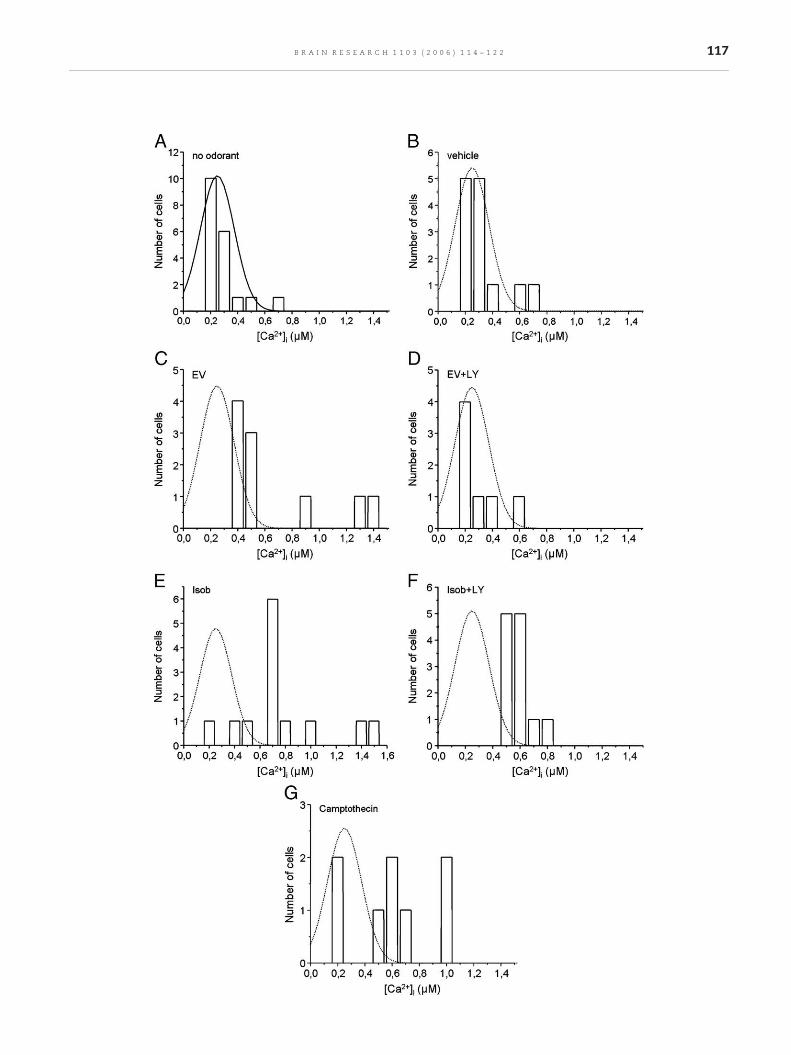
Fig. 4 – Single-cell [Ca2+]i measurements on Fluo-3 loaded olfactoaddition (control), (B) vehicle, (C) ethyl vanillin (EV), (D) LY83583camptothecin (positive control). Continuous line in panel A indicawere drawn for comparison purposes, and correspond to a Gaussthe plotted data), but similar position and width parameters as t
116 B R A I N R E S E A R C H 1 1 0 3 ( 2 0 0 6 ) 1 1 4 – 1 2 2
2.2. [Ca2+]i responses to odorants in ORNs inhibited byLY83583
The [Ca2+]i response of ORNs to odorants was studied in orderto determine, first, the functionality of these cells (selected bytheir morphological characteristics), second, whether theodorant concentration used was adequate to generate asubstantial raise in [Ca2+]i, and third, whether LY83583, ablockerof the transductionchannelCNGC,effectively inhibitedthe odorant-induced Ca2+ response. A specific ORNwas able torespond repeatedly and reproducibly to an odorant (Fig. 2A).Some cells responded to different odorants (Fig. 2C). We wereunable to determine the early time course of the [Ca2+]iresponse due to limitations in the time resolution of ourimage acquisition system. We observed that the total timeperiod that [Ca2+]i stayed high varied from 100 to 300 s in thedifferent cells tested. The size of the response (peak value, ΔF/Fo * 100) varied from18 to 49% indifferent cells. IncubationwithLY83583 changed the responseamplitudesignificantly (Figs. 2Band C and 3). Fig. 2C also shows that ionomycin was able toinduce a prominent [Ca2+]i raise even in the presence ofLY83583 and after repeated exposures to the odorants,indicating that the fluorescence probe was clearly sensitiveto changes in [Ca2+]i and that the magnitude of the [Ca2+]iresponse was not limited by probe sensing capabilities.Statistical analysis of the magnitudes of [Ca2+]i responses forEV and isobutyrate showed a significant difference (P < 0.01,ANOVA followed by Bonferroni t test) in the presence andabsence of LY83583 (Fig. 3).
2.3. [Ca2+]i in ORNs under sustained odorant exposure
An elevation of [Ca2+]i appears to be a key factor in neuronalexcitotoxic PCD induction (Mattson, 2000). Using fluorescence
ry EC continuously exposed to odorants for 3 h. (A) No odorant+ ethyl vanillin, (E) isobutyrate (Isob), (F) LY83583 + Isob, (G)tes a Gauss fitting to the data. Dotted lines in all other panelsian curve with variable amplitude (arbitrarily set according tohe control Gaussian curve.
117B R A I N R E S E A R C H 1 1 0 3 ( 2 0 0 6 ) 1 1 4 – 1 2 2

Fig. 5 – DNA fragmentation analysis. The effects of odorantson DNA strand breakage were evaluated using an ISOL assayafter 3 h incubation under the experimental conditionsshown in the figure (odorant-free, EV, EV + LY38538, andIsob). Results shown in the graphs are presented as thepercentage of positive labeled cells in the culture.Kruskall–Wallis–Dunn statistical analyses are shown(*P < 0.05 vs. control, **P < 0.05 vs. EV).
Fig. 6 – Apoptosis revealed by the presence ofphosphatidylserine (PS) in the plasma membrane externalleaflet. Effects of odorants (150μM) and 10μMLY83583 on PStranslocation to the external leaflet of the plasmamembranewas estimated by the Annexin V assay. The data areexpressed as the relative apoptotic index of each treatmentwith respect to the control without odorant.Kruskall–Wallis–Dunn statistical analyses are shown(*P < 0.05).
118 B R A I N R E S E A R C H 1 1 0 3 ( 2 0 0 6 ) 1 1 4 – 1 2 2
microscopy and Fluo-3 AM as a [Ca2+]i probe, we studiedwhether a continuous exposure to an odorant was capable ofinducing a sustained rise in [Ca2+]i. The values of [Ca2+]i incultures subjected to diverse experimental conditions for 3 hare plotted in Fig. 4: (A) no odorant addition, (B) vehicle, (C)ethyl vanillin (EV), (D) LY83583 + ethyl vanillin, (E) isobuty-rate (Isob), (F) LY83583 + isobutyrate, (G) camptothecin(positive control). As seen in Fig. 4, 94% of control cellspresented [Ca2+]i values below 400 nM. In the presence of EVsome cells presented [Ca2+]i as high as 1.3 μM. LY83583prevented this [Ca2+]i rise in EV treated cells, showing afrequency profile very similar to the control values. Iso-butyrate was also able to induce an increase in [Ca2+]i, with>75% of the cells showing [Ca2+]i values larger than 400 nM.In contrast to EV, LY83583 partially prevented the rise in[Ca2+]i in olfactory EC treated with isobutyrate. Cells treatedwith camptothecin, a topoisomerase inhibitor, showedincreased cell death and release of cells from the cultureplates. The remaining cells presented relatively high [Ca2+]i(Fig. 4G).
2.4. Odorant-induced apoptosis in olfactory epithelial cellsrevealed by DNA fragmentation
Because the different procedures to estimate apoptosis inolfactory EC were not compatible with the simultaneousdetermination of ORNs identity, we expressed our apoptosismeasurements as percentages of total olfactory EC. Theseolfactory EC consisted of at least 77% ORNs (see above).
Continuous exposure to ethyl vanillin or isobutyrate for 3 hinduced apoptotic DNA strand breakages, of 66 ± 8 and 74 ± 5%in the presence of ethyl vanillin and isobutyrate, respectively.Considering spontaneous apoptosis of 10% of the cells, theodorant-induced apoptosis was approximately 56 and 64% forethyl vanillin and isobutyrate, respectively. LY83583 reducedthe odorant-induced apoptosis to approximately 24% of thecells (Fig. 5).
2.5. Odorant-induced apoptosis revealed byphophatidylserine presence in the external plasmamembrane leaflet
DNA strand breakage, estimated by the ISOL assay, suggestedthat an odor-induced apoptotic process was taking place inolfactory EC. In order to confirm this observation, we indepen-dently used an Annexin-V assay, which detects exposure of PSon the external leaflet of the plasmamembrane. High annexin-V reactivity was found in all odorant-exposed cultures (Fig. 6).Because of the variability in background annexin V values (3–30%) between different cell preparations, we have expressedthe effects of the odorants as the relative changeswith respectto the control in each preparation. When the cells wereincubated with the odorants isobutyrate or ethylvanillin inthe presence of the olfactory response inhibitor LY83583, thelevels of apoptosis were not significantly different (P > 0.05;Kruskall–Wallis–Dunntest) fromthoseunder control condition(in the absence of odorant; Fig. 6). The effect of LY83583 ofabolishing odorant-induced annexin-V exposure to the exter-nal leaflet of the plasmamembrane strongly suggests that theapoptotic process observed in olfactory EC was triggered by areceptor-mediated odorant-induced mechanism.
2.6. Odorant-induced apoptosis revealed by caspase-3activation
Activation of a caspase cascade is a clear and definitiveindication that a cell has entered a PCD process. Specifically,caspase-3 activation (an executor caspase) can be consideredas a no-return step in the apoptotic process. Because caspaseactivation is a definitive apoptotic index,we performed amoreexhaustive study of this parameter, including controls withthe different agents with which the cells were challenged. Thepercentage of cells exhibiting caspase-3 activation was

Fig. 7 – Apoptosis revealed by caspase-3 activation. The datawere obtained after 3 h of exposure to the differentconditions. Odorant, camptothecin, LY83583, and caspaseinhibitor concentrations were (in μM) 150, 0.15, 10, and 2,respectively. The bars show the control (C) in the absence ofodorant, LY83583, vehicle addition (veh), the reaction in thepresence of caspase-3 inhibitor (CIn), LY83583 + veh,camptothecin (cpt), LY 83583 + camptothecin; ethyl vainillin(EV), LY83583 + EV, isobutyrate (Isob), LY83583 + Isob.Kruskall–Wallis–Dunn statistical analyses are shown(*P < 0.05 vs. control, **P < 0.05 vs. EV, ***P < 0.05 vs. Isob).
119B R A I N R E S E A R C H 1 1 0 3 ( 2 0 0 6 ) 1 1 4 – 1 2 2
significantly larger in odorant-exposed than in the controlcultures (P < 0.05, Kruskall–Wallis–Dunn test) (Fig. 7). LY83583prevented odorant-induced apoptosis in all odorant-exposedcell cultures. No significant differences between odorantsupplemented with LY83583 treated cultures and controlcultures were observed. LY83583 by itself had no effect oncaspase-3 activation. Furthermore, LY83583 had no effect oncaspase-3 activation induced by camptothecin, a well knowninducer of apoptosis. These results confirmed that PCD wasinduced by odorants in olfactory EC, as suggested by DNAfragmentation and Annexin-V assays.
3. Discussion
The olfactory epithelium is a remarkable tissue, where acontinuous neuronal cell renewal takes place throughout thelife span of an individual (Mackay-Sim, 2003). The cellularresponse to odorants and the molecular mechanisms under-lying it have been explored in a number of recent studies (e.g.,Schild and Restrepo, 1998; Gibson and Garbers, 2000). Howev-er, less attention has been given to the association of odorantsand cell death and renewal in this neuroepithelium. PCD hasbeen described in the olfactory epithelium, especially as aconsequence of the removal of the olfactory bulb (Nakagawaet al., 1996; Michel et al., 1997) or due to normal turn over(Cowan and Roskams, 2002; Suzuki, 2004). The relationshipbetween cellular turnover and environmental odorant expo-sure has been previously explored (Maruniak et al., 1989;Farbman et al., 1988; Hinds et al., 1984). Although it seemsclear that ORNs can die by the action of external noxious
stimuli, the role of extended odorant exposure on inducingPCD in ORNs and its possible relationship with excitotoxicityhad not been previously investigated.
We were able to obtain a cultured cell preparation with77 ± 5% ofmature ORN cells, as judged by their reactivity to theanti-OMP antibody;∼40% of the OMP-positive cells retain theirneuronal characteristics. It has been described that 12–33% ofORNs responded to a specific odorant (Sato et al., 1994;Duchamp-Viret et al., 1999; Gomez et al., 2000). However, inour experiments approximately 40% (range 32–45%) of thecells responded to a pure odorant, an observation that waslikely due to the relatively high odorant concentration used inour protocols. We found that prolonged exposure to odorantstriggered apoptosis in 57–83% of the cultured olfactory EC.Because we used three independent criteria to estimate PCDinduction in these cells, we think that our results showunequivocally the existence of an apoptotic mechanisminduced by odorants in olfactory EC. The fact that the fractionof apoptotic cells (approximately 80%) was much larger thanthe fraction of non-ORNs (approximately 25%) in culturestrongly suggests that apoptosis was induced in an importantfraction of ORNs. In fact, these numbers are in agreement withthe idea that ORNs are the main apoptotic target of prolongedodorant exposure. This idea was confirmed both by the abilityof LY83583 to significantly diminish the apoptotic action of theodorants, and the fact that apoptosis induced by camptothe-cin, an inhibitor of mammalian topoisomerase I (Hsiang et al.,1985), was not affected by LY83583. The effects of LY83583strongly suggest that the odorant-induced PCD we observed ismediated by the transduction mechanism of the sensoryneurons. Thus, our in vitro data provide good evidence thatolfactory EC can be induced to undergo apoptosis by prolongedand continuous odorant exposure. Given the fact that LY83583can decrease the odorant-, but not the campotheticin-inducedapoptosis, our results can only be explained if an importantfraction of the apoptotic cells are ORNs.
It has been shown that [Ca2+]i higher than 350 nM cantrigger a PCD process in neurons (Collins et al., 2001; Ferri andKroemer, 2001). Hence, the existence of a population of cellsthat presented an odorant-dependent high [Ca2+]i changeamong the cells exposed to the odorants suggests that anincrease in [Ca2+]i was involved in the odorant-induced PCDobserved in our studies. This odorant-induced [Ca2+]i increasewas inhibited by LY83583, also hinting that the odorantinduced PCD and odorant induced increase in[Ca2+]I werelinked. We attempted to test the hypothesis that [Ca2+]iincreases were responsible for the induction of apoptosis byloading the cells with the Ca2+ chelator BAPTA (using BAPTA-AM), but failed due to the cytotoxic effects of this compoundon olfactory EC.
In vivo, Watt et al. (2004) have shown that ORNs over-expressing an octanal receptor-GFP protein are protected fromdeath by intermittent exposure to octanal. Instead, Carr et al.(2001) have shown that exposure to continuous high odorantconcentrations induced death in both ORNs and supportingcells. Our data strongly indicate that ORNs undergo anexcitotoxic-like apoptotic death in vitro. We think that ourresults can be taken as a basis to understand the apparentlymore complex and sometimes contradictory results obtainedin vivo, that most likely are reflecting differences betweenintermittent and continuous odorant exposure as well as thecomplexity of cell–cell interactions in the olfactory epithelium.

120 B R A I N R E S E A R C H 1 1 0 3 ( 2 0 0 6 ) 1 1 4 – 1 2 2
Furthermore, it has been observed that following long-termodorant exposures (days), several days are needed for in vivorecovery of odor-sensitivity. This observation has been inter-preted as odor-induced adaptation, a phenomenon defined asa decrease in odor sensitivity following repetitive or continu-ous exposure to the same odorant (Dalton, 2000, Yee andWysocki, 2001). Our finding that ORNs undergo apoptosisduring prolonged odor exposures offers an alternative expla-nation for this phenomenon in termsofdeathand replacementof functional ORNs, particularly considering that the recoverytime of odor sensitivity in vivo is similar to the time thatgerminal epithelial basal cells take to proliferate and differen-tiate into a new (naive) population of cells after damage(Dalton, 2000; Herzog and Otto, 1999).
4. Experimental procedures
4.1. Cell preparation and culture
Nasal olfactory mucosa was obtained from two or three adultmale Wistar rats after cervical dislocation and decapitation,according to the guidelines of the Animal Ethics Committee ofthe University of Chile, Santiago, Chile. The epithelia weredissected and placed in Krebs–Henseleit buffer (KH; inmM: 144NaCl, 4.2KCl, 1.6MgCl2, 1.6KH2PO4, 10HEPES, 300mosM,pH7.4)supplemented with 1.8 mg/ml glucose. A cell preparationsuitable for giving a high yield of ORNs was developed. Inbrief, the epithelial tissue was placed in a Hanks balancedsolution with dispase II (4.8 U/ml, Sigma-Aldrich, USA). Thetissue after the enzyme digestion was mechanically disruptedto separate adult ORNs. Large pieces of tissue were removedwith tweezers and the cell suspensionwas pipetted repeatedlywith a fire polished Pasteur pipette. The cell suspension waspelleted at 340 × g for 5 min and the resulting pellet wassuspended in Dulbecco's modified Eagle's medium (DMEM)supplemented with 10% bovine fetal serum, penicillin (100 U/ml) and streptomycin (0.1 mg/ml). The cells were cultured on12 mm cover slips pre-coated with pegotine (a bioadhesive,BiosChile, Santiago, Chile), at 2–2.5million cells per cover,withthree cover slips perwell for 48hat 33 °C in 95%air/5%CO2. Thispreparation gave a high percentage of OMP positive cells (77%,see Fig. 1 and Results).
4.2. Immunocytochemistry
The proportion of mature ORNs was estimated using apolyclonal anti-olfactory marker protein (OMP) goat-anti-ratantibody (Wako Chemicals USA Inc., Richmond, USA). TheOMP is present only in mature ORNs (Farbman and Margolis,1980). After cell culture, the cells were fixed in 5% paraformal-dehyde for 15min at 4 °C. The fixative waswashed out and -onspecific sites were blocked with 3% BSA for 3 h. Subsequently,the ORNs were incubated with the primary antibody in thepresence of BSA (3%) at 4 °C for 1 h. After washing them withPBS, the cells were incubated with a secondary antibody in thepresence of 3% BSA at 4 °C for 30 min. Fluorescence wasanalyzed using a Zeiss Axiovert 200 M epifluorescence micro-scope (Oberkochen, Germany) (excitation wavelength 340–380 nm, emission wavelength >410 nm) and a 40× objectivelens. Primary antibodies used were polyclonal goat anti-ratOMP (1:10,000) and monoclonal mouse anti-rat neurofilament
200 (1:500; Sigma-Aldrich, USA). Secondary antibodies usedwere rabbit anti-goat IgG (1:500) and rabbit anti-mouse IgG(1:200) coupled with AlexaFluor-488 and AlexaFluor-594, re-spectively (Molecular Probes, USA). The nucleus was counter-stained using Hoescht 33342 (H33342, Molecular Probes, USA).Controls were performed omitting the primary antibody andtreating the sample with pre-immune goat serum.
4.3. Apoptosis induction
Cultured cells were exposed to odorants for 3 h. We utilizedthe odorants isobutyrate (2-methylpropanoic acid and ethyl-vanillin (3-ethoxy-4-hydroxybenzaldehyde) (Sigma ChemicalCo., St Louis, MO, USA), a putrid and floral odorant, respec-tively, at final concentrations of 150 μM. This value waschosen in order to expose the olfactory EC to a high odorantconcentration, but within the concentration range commonlyused in physiological studies (e.g., Ma and Shepherd, 2000;Zufall and Leinders-Zufall, 2000). The odorant stock solutionswere prepared in ethanol (EtOH) (ethyl vanillin) or KH buffer(isobutyrate) and added to the culture medium at a final EtOHconcentration of 0.4%. Several controls were designed forcaspase measurements: (1) no addition, (2) vehicle addition(EtOH 0.4%), and (3) positive control (camptothecin, 0.15 μM,Sigma Chemical Co., St Louis, MO, USA) (Hsiang et al., 1985).The odorant response inhibitor LY83583 (10 μM) was used inorder to test the specificity of odorant PCD induction. Thiscompound effectively inhibits the cyclic nucleotide-gatedchannel (Kd = ∼1.4 μM), as well as guanylyl cyclase III(Leinders-Zufall and Zufall, 1995), two essential componentsof the odorant transduction cascade.
4.4. Determination of cellular DNA fragmentation
In order to detect the typeofDNA fragmentation that is specificin apoptosis, we utilized the In Situ Oligo Ligation method(ApoTag ISOL, Q-BIOgene, UK). The ISOLmethod is based uponthe specificity of the enzyme T4 DNA ligase (Weiss et al., 1968);in this method, synthetic nucleotides labeled with biotinselectively react with the specific types of genomic DNA ends(blunt and short single base ends) characteristics of apoptoticcells. This reaction is followed by addition of a streptavidin–peroxidase conjugate that binds tightly to the biotin on theoligonucleotide. Lastly, the addition of DAB (diaminobenzi-dine), a chromogenic peroxidase substrate, causes a brownprecipitate that in this work was visualized using bright fieldmicroscopy. The ISOL reaction leads to a significantly smallernumber of false-positive results in comparison to terminaldeoxynucleotidyl transferase-mediated deoxyuridine triphos-phate nick-end labeling (TUNEL) and in situ end labeling (ISEL)(Lesauskaite et al., 2004). DNA fragmentation estimated byTUNEL assay has been utilized previously as an apoptoticcriterion in olfactory epithelium (Michel et al., 1997). In theseexperiments, we utilized two cell preparations, 2 plates(coverslips) per preparation and selected at least 3 fields perplate, counting 50–87 cells/field. This procedure was repeatedfor every condition tested.
4.5. Annexin-V labeling assay
An early indicator of apoptosis is the translocation ofphosphatidylserine (PS), from the internal to the external
121B R A I N R E S E A R C H 1 1 0 3 ( 2 0 0 6 ) 1 1 4 – 1 2 2
leaflet of the plasma membrane. In order to detect phospha-tidylserine exposition to the outer leaflet,we used anAnnexin-V-FLUOS kit for apoptosis detection (Boehringer-Mannheim,Werk Penzberg, Germany). After carefully washing out theculture medium, the cells were incubated with annexin-Vsolution andpropidium iodide for 15min at room temperature.After this incubation, the fluorescent cellswere analyzedusinga NIKONDiaphotmicroscopewith the appropriate set of filtersfor fluorescein and propidium iodide. This method allowed usto differentiate apoptotic from lysed cells. Apoptotic cellspresented green membranes, and lysed cells presented greenmembranes and red nuclei (Fadok et al., 1992). Non-apoptoticcells did not fluoresce. Because lysed cells could havepreviously undergone apoptosis, the annexin V assay gives aminimum estimate of apoptosis. In these experiments, weutilized 3 cell preparations, 3 plates (coverslips) per prepara-tion and selected at least 3 fields per plate, counting 28–47cells/field, a procedure that was repeated for every condition.
4.6. Determination of caspase-3 activation
A protease (caspase) cascade is one of the essential aspects ofPCD. Activation of caspase-3 implies an irreversible commit-ment to cell death in the PCD process (e.g., Adrain and Martin,2001). We estimated caspase-3 activation using a specificsubstrate (PhiPhilux™ G2D2) from Calbiochem (CA, USA). Inthis assay, a seven residue long peptide is linked to a fluo-rescent probe that yields an intense red fluorescence signalwhen it is cleaved by the enzyme (Komoriya et al., 2000). After60 min of incubation in culture conditions with the caspase-3substrate, the cells were carefully washed and their fluores-cence was analyzed using a NIKON Diaphot epifluorescencemicroscope (excitation wavelength 510–550 nm, emissionwavelength >570 nm). In some measurements, a caspase-3inhibitor (Z-DEVD-FMK, Calbiochem, USA) was used to esti-mate the specificity of the caspase-3 reaction. In theseexperiments, we utilized 3 cell preparations, 3 plates (cover-slips) per preparation and selected at least 3 fields per plate,counting at least 43–310 cells/field. Such a procedure wasrepeated for every condition.
4.7. Dynamic changes of [Ca2+]i induced by odorants inolfactory EC
In order to correlate the effects of the odorants on apoptosisand the changes in [Ca2+]i, itwas important to characterize firstif the isolated cells [Ca2+]i responded to the addition ofodorants. Fluo-3 AM (Molecular Probes, Eugene, OR) was usedto analyze the acute response of [Ca2+]i in ORNs to 150 μModorant addition, and the effect of LY83583 on this response.Freshly isolated cells were incubatedwith Fluo-3 AM (5 μM) for30 min at 33 °C. The probe was washed out from the dish andfluorescence images were acquired with a cooled CCD camera(SpectraSource, Los Angeles, USA) (excitation wavelength 440–480, emission wavelength > 510) in an inverted Olympus IX-70fluorescence microscope (40× objective lens). For the LY83583assays, the cells were incubated with 10 μM LY83583 between30 and 120 s prior to odorant addition. The measurements arepresented as the percentage of Fluo-3 fluorescence changes(ΔF/Fo), where ΔF corresponds to the difference between thetime-dependent fluorescence after odorant addition and thebasal (Fo) average value 1 min before odorant addition. In
single-cell [Ca2+]i measurements, photo bleaching of thefluorescent probe was reduced by using a 25% transmittanceneutral density filter and, when present, it was corrected usinga first order exponential decay function. The measurementswere performed in three different cell preparations and 10–15cells were tested per condition.
4.8. [Ca2+]i measurements in cells chronically exposed toodorants
In order to analyze the steady state levels of [Ca2+]i in cellsexposed to the odorants for a prolonged time, we conductedsingle-cell [Ca2+]i measurements. After being exposed to theodorants for 3 h, the cells were incubated with 5 μM Fluo-3 AMfor 30min. Followingwash-out, calibration of the Fluo-3 signalwas performed as described by Kao et al. (1989). In brief,addition of ionomycin (5 μM)was followed byMn2+ (2mM) andended with digitonin to permeabilize the cells. These exper-imental conditions allowed us to estimate Fmax, Fmin, andFbackground, as described by Kao et al. (1989). In these experi-ments, we utilized two cell preparations, 2 coverslips perpreparation and selected at least 3 fields per plate, deter-mining [Ca2+]i in 7–19 cells/coverslip. This procedure was re-peated for every condition.
4.9. Data acquisition and analysis
Calcium imaging was accomplished using an Olympus IX-70epifluorescence inverted microscope with a cooled 16 bit CCDcamera model MCD-220 (SpectraSource Instruments, CA,USA), attached to the videoport of the microscope. Imageswere digitized with SpectraSource software and analyzedwithScion Image for Windows (Scion Corporation, USA). Pictureswere acquired by an Axiocam photocamera (Zeiss, Oberko-chen, Germany), and processed with Adobe Photoshop (AdobeSystems Incorporated, USA). Data analysis was performedwith Microcal Origin (Microcal Software Inc., MA, USA).GraphPAD InStat version 1.1 (GraphPAD software, USA) wasused for statistical analysis. This analysis included ANOVAand Bonferroni test. Kruskall–Wallis non-parametric testfollowed by Dunn's multiple comparison test was applied tothe data in order to determine differences in apoptosis. Eachcell preparation was derived from at least two rats.
Acknowledgments
We are indebted to Alan Mackay-Sim for critical reading of anearly version of the manuscript and to Javier Díaz for help onpreparing the figures. We also thank Dr. Ricardo Moreno forproviding us with some of the reagents. Supported byFONDECYT 1990938 (JB and JR), FONDECYT 1050124, andMIDEPLAN ICM P99-031-F (JB).
R E F E R E N C E S
Adrain, C., Martin, S.J., 2001. The mitochondrial apoptosome: akiller unleashed by the cytochrome seas. Trends Biochem. Sci.26, 390–397.
Carr, V.M., Menco, B.P., Yankova, M.P., Morimoto, R.I., Farbman, A.I., 2001. Odorants as cell-type specific activators of a heat shock
122 B R A I N R E S E A R C H 1 1 0 3 ( 2 0 0 6 ) 1 1 4 – 1 2 2
response in the rat olfactory mucosa. J. Comp. Neurol. 432,425–439.
Collins, T.J., Lipp, P., Berridge, M.J., Bootman, M.D., 2001.Mitochondrial Ca2+ uptake depends on the spatial andtemporal profile of cytosolic Ca2+ signals. J. Biol. Chem. 276,26411–26420.
Cowan, A.J., Roskams, C.M., 2002. Apoptosis in the mature anddeveloping olfactory neuroepithelium. Microsc. Res. Tech. 58,204–215.
Dalton, P., 2000. Psychophysical and behavioral characteristics ofolfactory adaptation. Chem. Senses 25, 487–492.
Dionne, V., Dubin, A.E., 1994. Transduction diversity in olfaction.J. Exp. Biol. 194, 1–21.
Duchamp-Viret, P., Chaput, M.A., Duchamp, A., 1999. Odorresponse properties of rat olfactory receptor neurons. Science284, 2171–2174.
Fadok, V.A., Voelker, D.R., Campbell, P.A., Cohen, J.J., Bratton, D.L.,Henson, P.M., 1992. Exposure of phosphatidylserine on thesurface of apoptotic lymphocytes triggers specific recognitionand removal by macrophages. J. Immunol. 148, 2207–2216.
Farbman, A.I., Margolis, F.I., 1980. Olfactory marker protein duringontogeny: immunohistochemical localization. Dev. Biol. 74,205–215.
Farbman, A.I., Brunjes, P.C., Rentfro, L., Michas, J., Ritz, S., 1988.The effect of unilateral naris occlusion on cell dynamics in thedeveloping rat olfactory epithelium. J. Neurosci. 8, 3290–3295.
Ferri, K.F., Kroemer, G., 2001. Organelle-specific initiation of celldeath pathways. Nat. Cell Biol. 3, E255–E263.
Gibson, A.D., Garbers, D.L., 2000. Guanylyl cyclases as a family ofputative odorant receptors. Annu. Rev. Neurosci. 23, 417–439.
Gomez, G., Rawson, N.E., Cowart, B., Lowry, L.D., Pribitkin, E.A.,Restrepo, D., 2000. Modulation of odor-induced increases in[Ca2+]i by inhibitors of protein kinases A and C in rat andhuman olfactory receptor neurons. Neuroscience 98, 181–189.
Herzog, C., Otto, T., 1999. Regeneration of olfactory receptorneurons following chemical lesion: time course andenhancement with growth factor administration. Brain Res.849, 155–161.
Hinds, J.W., Hinds, P.L., McNelly, N.A., 1984. An autoradiographicstudy of the mouse olfactory epithelium: evidence forlong-lived receptors. Anat. Rec. 210, 375–383.
Hsiang, Y.H., Hertzberg, R., Hecht, S., Liu, L.F., 1985. Camptothecininduces protein-linked DNA breaks via mammalian DNAtopoisomerase I. J. Biol. Chem. 260, 14873–14878.
Kao, J.P.Y., Harootunian, A.T., Tsien, R.Y., 1989. Photochemicallygenerated cytosolic calcium pulses and their detection byFluo-3. J. Biol. Chem. 264, 8179–8184.
Kleene, S.J., Gesteland, R.C., 1991. Transmembrane currents in frogolfactory cilia. J. Membr. Biol. 120, 75–81.
Komoriya, A., Packard, B.Z., Brown, M.J., Wu, M.L., Henkart, P.A.,2000. Assessment of caspase activities in intact apoptoticthymocytes using cell permeable fluorogenic caspasesubstrates. J. Exp. Med. 191, 1819–1828.
Kurahashi, T., Yau, K.-W., 1993. Co-existence of cationic andchloride components in odorant-induced current of vertebrateolfactory receptor cells. Nature 363, 71–74.
Leinders-Zufall, T., Zufall, F., 1995. Block of cyclic nucleotide-gatedchannels in salamander olfactory receptor neurons by theguanylyl cyclase inhibitor LY83583. J. Neurophysiol. 74,2759–2762.
Leinders-Zufall, T., Greer, C.A., Shepherd, G.M., Zufall, F., 1998.Visualizing odor detection in olfactory cilia by calciumimaging. Ann. N. Y. Acad. Sci. 855, 205–207.
Lesauskaite, V., Epistolato, M.C., Ivanoviene, L., Tanganelli, P.,2004. Apoptosis of cardiomyocytes in explanted andtransplanted hearts. Comparison of results from in situTUNEL, ISEL, and ISOL reactions. Am. J. Clin. Pathol. 121,108–116.
Lowe, G., Gold, G.H., 1993. Nonlinear amplification by
calcium-dependent chloride channels in olfactory receptorcells. Nature 366, 283–286.
Ma, M., Shepherd, G.M., 2000. Functional mosaic organization ofmouse olfactory receptor neurons. Proc. Natl. Acad. Sci. U. S. A.97, 12869–12874.
Mackay-Sim, A., 2003. Neurogenesis in the adult olfactoryepithelium, In: Doty, R.L. (Ed.), Handbook of Olfaction andGustation, 2nd ed. Marcel Decker Inc., pp. 93–113. chapt. 5.
Madrid, R., Delgado, R., Bacigalupo, J., 2005. Cyclic AMP cascademediates the inhibitory odor response of isolated toadolfactory receptor neurons. J. Neurophysiol. 94, 1781–1788.
Maruniak, J.A., Lin, P.J., Henegar, J.R., 1989. Effects of unilateralnaris closure on the olfactory epithelia of adult mice. Brain Res.490, 212–218.
Mattson, M.P., 2000. Apoptosis in neurodegenerative disorders.Nat. Rev., Mol. Cell Biol. 1, 120–129.
Meisami, E., 1976. Effects of olfactory deprivation on postnatalgrowth of the rat olfactory bulb utilizing a new method forproduction of neonatal unilateral anosmia. Brain Res. 107,437–444.
Michel, D., Moyse, E., Trembleau, A., Jourdan, F., Brun, G., 1997.Clusterin/apoJ expression is associated with neuronalapoptosis in the olfactory mucosa of the adult mouse. J. CellSci. 110, 1635–1645.
Morales, B., Bacigalupo, J., 1996. Chemical reception in vertebrateolfaction: evidence for multiple transduction pathways. Biol.Res. 29, 333–341.
Morales, B., Ugarte, G., Labarca, P., Bacigalupo, J., 1994. InhibitoryK+ current activated by odorant in toad olfactory neurons. Proc.R. Soc. London, Ser. B Biol. Sci. Biol. Sci. 257, 235–242.
Nakagawa, T., Aiba, T., Shiotani, H., Tomiyama, K., Nakai, Y., 1996.Apoptosis in the normal olfactory epithelium of the adultguinea pig. Eur. Arch. Otorhinolaryngol. 253, 371–373.
Nakamura, T., Gold, G.H., 1987. A cyclic nucleotide-gatedconductance in olfactory receptor cilia. Nature 325, 442–444.
Restrepo, D., Okada, Y., Teeter, J.H., Lowry, L.D., Cowart, B.,Brand, J.G., 1993. Human olfactory neurons respond to odorstimuli with an increase in cytoplasmic Ca2+. Biophys. J. 64,1961–1966.
Said, S.I., Berisha, I., Pakbaz, H., 1996. Excitotoxicity in the lung:N-methyl-D-aspartate-induced, nitric oxide-dependent,pulmonary edema is attenuated by vasoactive intestinalpeptide and by inhibitors of poly(ADP-ribose) polymerase. Proc.Natl. Acad. Sci. U. S. A. 93, 4688–4692.
Sanhueza, M., Schmachtenberg, O., Bacigalupo, J., 2000. Excitation,inhibition, and suppression by odors in isolated toad and ratolfactory receptor neurons. Am. J. Physiol.: Cell Physiol. 279,C31–C39.
Sato, T., Hirono, J., Tonoike, M., Takebayashi, M., 1994. Tuningspecificities to aliphatic odorants in mouse olfactory receptorneurons and their local distribution. J. Neurophysiol. 72,2980–2989.
Schild, D., Restrepo, D., 1998. Transduction mechanisms invertebrate olfactory receptor cells. Physiol. Rev. 78, 429–466.
Suzuki, Y., 2004. Fine structural aspects of apoptosis in theolfactory epithelium. J. Neurocytol. 33, 693–702.
Watt, C., Sacarno, H., Lee, Z., Reusch, J.E., Trinh, K., Storm, D.R.,2004. Odorant stimulation enhances survival of olfactorysensory neurons via MAPK and CREB. Neuron 41, 955–967.
Weiss, B., Jacquemin-Sablon, A., Live, T., Fareed, G., Richardson, C.,1968. Enzymatic breakage and joining of deoxyribonucleic acid:VI. Further purification and properties of polynucletoide ligasefrom Escherichia coli infected with bacteriophage T4. J. Biol.Chem. 243, 4543–4555.
Yee, K., Wysocki, C., 2001. Odorant exposure increases olfactorysensitivity: olfactory epithelium is implicated. Physiol. Behav.72, 705–711.
Zufall, F., Leinders-Zufall, T., 2000. The cellular and molecularbasis of odor adaptation. Chem. Senses 25, 473–481.