antioxidant and antibacterial activities of some natural
TRANSCRIPT
CELLULOSE CHEMISTRY AND TECHNOLOGY
Cellulose Chem. Technol., 47 (5-6), 387-399 (2013)
ANTIOXIDANT AND ANTIBACTERIAL ACTIVITIES OF SOME NATURAL
POLYPHENOLS
IOANA IGNAT,
* DANA G. RADU,
* IRINA VOLF,
** ANDREEA I. PAG
*,*** and
VALENTIN I. POPA**
*“Aurel Vlaicu” University of Arad, Research, Development & Innovation in Technical and Natural
Sciences Institute, 2, E. Drăgoi Str., 310330, Arad, Romania **
“Gheorghe Asachi” Technical University, Faculty of Chemical Engineering and Environmental
Protection, 71 A, Mangeron Blvd., 700050 Iasi, Romania ***
University of Agricultural Sciences and Veterinary Medicine, Cluj-Napoca, 3-5, Mănăştur Str., 400372
Cluj-Napoca, Romania
Received March 6, 2013
The present study provides information on separation and identification of natural bioactive compounds from different
vegetal materials with potential applications as antibacterials and plant growth modulators. Natural extracts were
obtained by alcoholic and water extraction from spruce bark, grape seeds, Crataegus monogyna (hawthorn) and
Asclepias syriaca (milkweed).
HPLC-DAD analysis revealed that gallic acid (12.54 mg/100 g) and catechin (85.52 mg/100 g) were the most abundant
compounds in hawthorn and grape seed extracts, vanillic acid (71.9 mg/100 g) was found in high concentrations in
spruce bark wood, while milkweed extracts were characterized by the presence of hydroxycinnamic acids and
flavonoids. The DPPH assay indicated that the highest radical scavenging activity was registered by grape seeds (EC50
= 45.75 µg) and hawthorn (EC50 = 17.8 µg) alcoholic extracts. The same extracts exhibited antibacterial properties on
Staphylococcus aureus, while the samples obtained from spruce bark and milkweed extracts inhibited the development
of Escherichia coli and Pseudomonas aeruginosa species. Furthermore, the toxicity of the natural extracts was
evaluated by their application in germination tests of Phaseolus vulgaris. The obtained results demonstrated that a high
concentration of polyphenols has an inhibitory effect on plant development, while lower concentrations determine a
considerable stimulation of plantlet elongation.
Keywords: polyphenols, extraction, characterization, HPLC, radical scavenging, antibacterial activity, seed germination
INTRODUCTION
Polyphenolic compounds are a wide spread
group of secondary metabolites found in all plants
and represent the most desirable phytochemicals
due to their valuable properties and potential use
as additives in food industry, cosmetics, medicine,
and agriculture.1,2
In plants, polyphenols play an important role
in various defense reactions to protect them
against abiotic stresses like UV light or biotic
stresses, such as predators and pathogen attacks,
being also involved in plant growth and
development cycle, as they have been shown to
regulate the transport of polar auxins.3 The
stimulatory or inhibitory effect on plant
development depends on the concentration of
polyphenolic compounds and plant species.
Different activities of polyphenolic products can
be similar to those of growth hormones (auxins,
cytokines).4,5
Polyphenols have been demonstrated to act as
antioxidants and free radical scavengers. The
interest in the physiological role of bioactive
compounds present in plants has increased
dramatically over the last decade, particularly in
relation to human health. The antioxidant effects
of polyphenols are considered to be derived from
the scavenging action of their structures on
oxygen containing free radicals. The in vitro
antioxidant effectiveness of polyphenols is
essentially the result of the ease with which an H
IOANA IGNAT et al.
388
atom from aromatic hydroxyl is donated to a free
radical and of the ability of the aromatic structure
to support an impaired electron due to
delocalization of the π-electron system. In this
study, for characterizing the antioxidant activity
of naturally occurring phenolic compounds, a
method using 1,1-diphenyl-2-picrylhydrazyl
(DPPH) as a reactive free radical was used. Now
this method is recognized as giving the
opportunity to search for radical scavenging
ability depending on the electronic structure of
polyphenols.
The pharmacological effects exerted by
polyphenols on the human body are thought to be
strongly related with their high antioxidant
capacity,6,7,8
which may be responsible for the
anti-allergic, anti-inflammatory, anticancer,
antihypertensive, and antimicrobial
properties.9,10,11
Infectious diseases caused by bacteria resistant
to multiple drugs have become one of the most
serious problems nowadays. Although the use of
antibiotics has greatly reduced the incidence of
infectious diseases, it has also led to the
appearance of drug-resistant bacteria.12
Various
plant phenolics, including phenolic acids,
flavonoids and tannins known to be synthesized
by plants in response to microbial infection have
been shown to possess a broad spectrum of
antibacterial effects against a wide array of
microorganisms and a number of effective drugs
have been created against them.13,14
Polyphenols not only exhibit independent
antibacterial effects, but also can suppress the
antibiotic resistance of pathogen microorganisms
or act synergistically in combination with
conventional antimicrobial agents like
antibiotics.10,12,13
Moreover, herbal preparations
are comparatively cheaper, have lesser side
effects and can supplement other medical systems
for the treatment of diseases caused by pathogen
microorganisms.
Our special interest is in the polyphenols
extracted from spruce bark and grape seeds,
which constitute abundant and inexpensive
residual sources resulting annually from vineyards
and pulp and paper companies. In addition, the
potential use of spruce bark and grape seed
extracts as dietary supplements and alternative
medical products15,16
justify our interest in these
materials. The other two vegetal materials used in
this study are Crataegus monogyna and Asclepias
syriaca, which are known for their complex
chemical composition, health benefits and
potential use in different fields.17,18,19
This paper presents our recent results,
revealing that several types of phenolics extracted
from different raw materials are characterized by
multifunctional activities, having a significant
antioxidant capacity, being effective against
Gram-positive and Gram-negative pathogen
bacteria and also influencing the growth of plants.
EXPERIMENTAL Plant material
Aerial parts of Asclepias syriaca were collected
during the blossoming stage, from an Agricultural
Research Station, where the plant was cultivated as an
energy crop (1998), as a part of a research program of
“Gheorghe Asachi” Technical University of Iasi,
Natural and Synthetic Polymer Department. Crataegus
monogyna fruits were collected from Cucuteni, Iasi
County, spruce wood bark was provided by a
Romanian pulp and paper company and Merlot grape
seeds were obtained from Panciu, Vrancea vineyard.
All the raw materials were air-dried, ground in an
electric mill (RETSH GRINDOMIX, GM 200) and
reduced to a fine powder of 0.5 mm.
Extraction procedures
The extraction of phenolic compounds was carried
out on the defatted (hexane extraction) samples, using
either 80% ethanol, or water as solvents. The alcoholic
extractions were carried out in a Soxhlet apparatus,
during 8 hours at 70 °C. The extracts were then
concentrated to 50 mL in a rotary evaporator at 40 °C.
These concentrated extracts were tested for
antimicrobial activity.
The fractionation of the concentrated extracts was
achieved using successive liquid-liquid extractions
with ethyl acetate. The organic phases were evaporated
to dryness, diluted in methanol and followed by HPLC
determination.
The aqueous extraction was performed on 20 g of
dried material using 125 mL of distilled water at 70 °C
for 45 min. The extraction was repeated three times
and the extracts were cumulated and concentrated
under vacuum. The fractionation by liquid-liquid
extraction was also carried out on the concentrated
extracts prior to HPLC analysis.
Spectrophotometric determinations
The total phenolic content of the plant extracts was
determined using FCR (Folin-Ciocalteu’s Reagent)
using a previously developed method.7 Total phenolic
content is expressed as the number of equivalents of
gallic acid (GAE).
The total content of tannins was determined using a
method based on the precipitation of tannins with
casein. The tannins content was determined using the
FC method and expressed as the difference between
Polyphenols
389
the initial content of polyphenols and the content after
the precipitation with casein.20
The contents of flavonoids and flavonols were
determined by the aluminum chloride method, using
rutin as a reference compound, according to a
previously developed modification of the method.20,21
The results were expressed as mg rutin equivalents
(RE). The anthocyanins content was determined using
the pH differential method.22
HPLC analysis
A previously developed reversed-phase high-
performance liquid chromatographic technique was
used to identify and quantify the phenolic compounds.7
Chromatographic analyses were carried out on a
Dionex UltiMate 3000, liquid chromatography system,
equipped with a photodiode array detector. Eluent (A)
was 1% aqueous acetic acid, and eluent (B) 1% acetic
acid in methanol, the flow rate was kept constant
throughout the analysis, at 1.2 mL/min. Injections were
accomplished with a 5 µL fixed loop. The column was
an Agilent Zorbax, C18 RP (4.6 x 150 mm, particle
size: 5 µm) and the temperature was maintained at 30
°C. For phenolic acids the elution programme used was
the following: the percentage of solvent B was
increased linearly from 10 to 40% in 40 min and then
decreased in one minute to 10% and maintained for 10
min. For quantification, standards for external
calibration were used. Chromatograms were monitored
at 280 and 320 nm.
For identifying and quantifying the flavonoids, a
gradient method was applied. The flow rate was of 1
mL/min and the elution conditions were as follows:
30% B 0-10 min, 30-80% B 20-25 min, 80% B 25-29
min, 80-30% B 29-30 min, 30% B 31-32 min. The
standards used for external calibration were rutin,
quercetin, apigenin and kaempferol and the
chromatograms were recorded at 254 nm, 360 nm and
365 nm.
For all the standard polyphenols and flavonoids, the
regression coefficient was higher than 0.997 and the
limits of detection were 0.1 mg/L for gallic acid, 1.4
mg/L for catechine and rutin, 0.6 mg/L for vanillic
acid, syringic acid, ferulic acid, sinapic acid, 0.3 mg/L
for p-coumaric acid, 1.2 mg/L for kaempferol and
apigenin and 0.8 mg/L for quercetin.
Radical scavenging activity
The radical scavenging activity of the natural
extracts was evaluated by the reduction of the DPPH
(2,2-diphenyl-1-picrylhydrazyl) radical. The
antioxidant activity of the extracts was expressed as
EC50, equivalent amount of an extract that inhibits 50%
of the radical. The colorimetric assay was performed
according to a modified method.23
For the DPPH assay the concentrated extracts
(alcoholic and aqueous) were freeze-dried to avoid
solvents interferences. Thereafter, different dosages of
a 0.5 mg/mL methanolic solution of either water or
ethanol freeze-dried extracts (25, 50, 100, 200, 300,
400, 500 µL each) were added in screw capped glass
vials containing 2 mL of the DPPH.
All tubes were adjusted to 3.1 mL with MeOH.
After a reaction time of 30 min, the absorbance was
measured at 517 nm.
The inhibition percentage of the free radical DPPH
(%I) was calculated according to the following
equation:
100%0
0x
A
AAI
−
=
where A0 = absorbance of DPPH, A = absorbance of
DPPH + extracts.
Methanolic solutions of ascorbic acid, gallic acid,
vanillic acid and catechin were tested as reference
antioxidants. The different quantities of the extracts
tested, expressed in milligrams, were plotted on a dose-
inhibition curve.
Antibacterial activity testing
Before the analysis, bacterial strains were cultured
on tryptic soy agar (TSA) pH 7.3, under aerobic
conditions for 24 h, at 37 °C, except for the fungal
strain Candida parapsilosis cultivated on Sabouraud
2% (m/v) – glucose agar for 48 h, at 37 °C. These
microbial cultures were used as inocula in order to test
the antibacterial activity of some crude herbal extracts.
The antibacterial activity of the extracts was
estimated using the disk agar diffusion method,
according to the Kirby-Bauer test,24
one of the most
widely spread and frequently used methods, also
recommended by the European Pharmacopoeia.25
Its
advantages, compared to the dilution tests, consist in
simplicity and facility to use, the direct control of the
bacterial culture purity, easiness to read and interpret
the results.14,26
Briefly, inocula of bacterial cells and yeast-like
fungal blastospores were suspended in sterile
physiological saline solutions, in sterile tubes, and
homogenized on Vortex, until the density of the test
suspension matched the turbidity standard, which was
equivalent to a concentration of 1.5×10⁸/mL
(McFarland Standard, GrantBio, UK). Petri dishes (d =
9 cm) with Müller-Hinton agar (pH 7.3) for bacterial
species, or Sabouraud-agar (pH 6.5) for yeast-like
fungi, were inoculated with 1 mL of microbial
suspension spread to ensure complete coverage. The
plates were left for 5 min before excess fluid was
removed using a sterile pipette. Then, the inoculated
Petri dishes were dried at room temperature for a
maximum of 20 min. Inoculation was carried out using
a Bioquell ATC 1200 N-MK2 biological safety cabinet
(UK).
Sterile paper discs impregnated with 20 µL crude
herbal extracts were aseptically applied to the surface
of each of the inoculated plates in a central position,
using a sterile forceps, gently pressing to ensure even
contact with the medium surface. Control discs
IOANA IGNAT et al.
390
impregnated with antibacterial substances and blank
discs (impregnated with solvent) were also placed into
the inoculated Petri dishes using a disc dispenser.
Gentamicin, ofloxacin, amikacin, kanamycin,
cefuroxim, erythromycin were used as reference
antibacterial, and fluconazole and nystatin as reference
antifungal substances. The inoculated plates were
incubated for 20 h, at 37 °C for bacterial species, and
for 48 h, at 37 °C for C. parapsilosis. The inhibition
zones were expressed in mm, as the diameters of clear
zones around the discs. The results of antimicrobial
activity were expressed as the mean value of three
independent analyses.
Seed germination tests
Germination tests were carried out in Petri dishes,
each one containing 5 bean seeds pre-disinfected with
1% NaOCl, for 15 min and 10 mL tested sample
solution.
Three different concentrations of polyphenolic
extracts (ethanolic and aqueous) expressed as TFC
(0.05 mg GAE/L, 0.1 mg GAE/L and 0.2 mg GAE/L)
were tested. The reference was tap water. Petri dishes
(5/each sample) were kept at dark in a thermostatic
chamber (25 °C) for seven days. The experiment was
carried out in triplicates and the average values of the
parameters were reported.
Pot experiments
Pot tests were carried out under greenhouse
conditions using sandy soil. Bean seeds (Phaseolus
vulgaris) were sown directly into pots after applying
10 mL of the tested solution. Three different
concentrations of natural extracts were tested (0.05 g
GAE/L. 0.1 g GAE/L, 0.2 g GAE/L). Each treatment
was replicated in five pots, and three uniform plants
were allowed to grow in each pot, at uniform spacing.
For ten days, the cultivated soils were wetted daily
with 15 mL of the tested extracts. After 10 days from
the beginning of the experiments, the bean plantlets
were separated into roots, stems and primary leaves
and measured to evidence the different effects of the
natural extracts on the growth and development of the
plants. Biometric measurements of plantlet elongation
and quantitative determination of biomass fresh weight
(FW) were determined.
RESULTS AND DISCUSSION It is known that the biological properties of
natural compounds depend on their composition
and concentrations. Therefore, to establish the
range of applications of polyphenols, it is
necessary to isolate and characterize them from
the viewpoints of compositional, structural and
biological activities.
Characterization of polyphenolic extracts
Spectrophotometric determinations The concentrations of different classes of
phenolic compounds existing in the extracts
separated from different raw materials were
determined spectrophotometrically using
colorimetric methods based on complex
formation, oxidation and pH decreasing. The
content of the extracted polyphenols (Table 1)
varied depending on the plant material and the
extraction agent used. Thus, the highest content of
polyphenols was acquired for the grape seed and
Crataegus monogyna alcoholic extracts (13.68,
respectively, 57.22 mg GAE/g). The same
samples were also characterized by higher
amounts of flavonoids and flavonols, compared to
the spruce bark and Asclepias syriaca extracts.
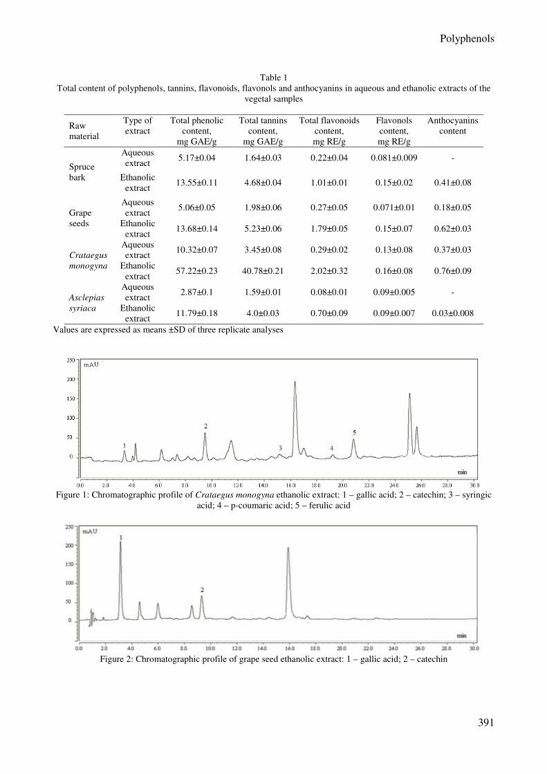
HPLC analysis The major compounds identified in almost all
the samples in different ranges of concentration
were gallic acid and catechin. Hence, the highest
content of these polyphenols was registered in the
case of grape seed and Crataegus monogyna
extracts. As expected, the concentrations in the
alcoholic samples were significantly higher,
compared to the aqueous ones. The concentrations
calculated based on the calibration curves of the
standards are reported in Table 2 and the
chromatographic profiles of ethanolic extracts are
presented in Figures 1, 2 and 3.
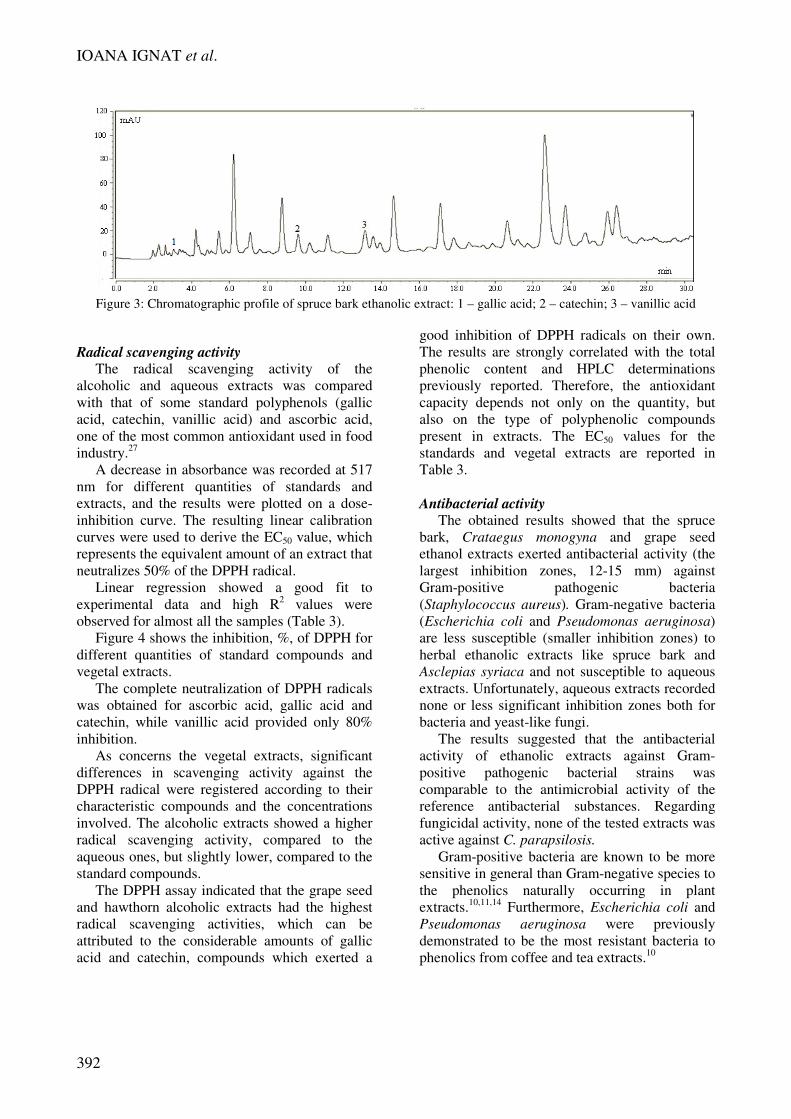
In the spruce bark alcoholic extract, besides
the presence of the gallic acid and catechin,
vanillic acid was found in high concentration
(71.9 mg/g), while the other four phenolic acids
(gallic, vanillic, syringic and p-coumaric acids)
were identified in Asclepias syriaca extracts.
Concerning the four flavonoids surveyed in the
natural extracts, the results showed that quercitin
was present in all the vegetal materials in different
ranges of concentration, starting from 0.14±0.05
mg/100 g in Asclepias syriaca extract and up to
2.38±0.1 mg/100 g in the grape seed extract. On
the other hand, relatively high amounts of rutin
were found in Crataegus monogyna (30.32±0.9
mg/100 g) and Asclepias syriaca (2.25±0.21)
samples.
Polyphenols
391
Table 1
Total content of polyphenols, tannins, flavonoids, flavonols and anthocyanins in aqueous and ethanolic extracts of the
vegetal samples
Raw
material
Type of
extract
Total phenolic
content,
mg GAE/g
Total tannins
content,
mg GAE/g
Total flavonoids
content,
mg RE/g
Flavonols
content,
mg RE/g
Anthocyanins
content
Spruce
bark
Aqueous
extract 5.17±0.04 1.64±0.03 0.22±0.04 0.081±0.009 -
Ethanolic
extract 13.55±0.11 4.68±0.04 1.01±0.01 0.15±0.02 0.41±0.08
Grape
seeds
Aqueous
extract 5.06±0.05 1.98±0.06 0.27±0.05 0.071±0.01 0.18±0.05
Ethanolic
extract 13.68±0.14 5.23±0.06 1.79±0.05 0.15±0.07 0.62±0.03
Crataegus
monogyna
Aqueous
extract 10.32±0.07 3.45±0.08 0.29±0.02 0.13±0.08 0.37±0.03
Ethanolic
extract 57.22±0.23 40.78±0.21 2.02±0.32 0.16±0.08 0.76±0.09
Asclepias
syriaca
Aqueous
extract 2.87±0.1 1.59±0.01 0.08±0.01 0.09±0.005 -
Ethanolic
extract 11.79±0.18 4.0±0.03 0.70±0.09 0.09±0.007 0.03±0.008
Values are expressed as means ±SD of three replicate analyses
Figure 1: Chromatographic profile of Crataegus monogyna ethanolic extract: 1 – gallic acid; 2 – catechin; 3 – syringic
acid; 4 – p-coumaric acid; 5 – ferulic acid
Figure 2: Chromatographic profile of grape seed ethanolic extract: 1 – gallic acid; 2 – catechin
IOANA IGNAT et al.
392
Figure 3: Chromatographic profile of spruce bark ethanolic extract: 1 – gallic acid; 2 – catechin; 3 – vanillic acid
Radical scavenging activity
The radical scavenging activity of the
alcoholic and aqueous extracts was compared
with that of some standard polyphenols (gallic
acid, catechin, vanillic acid) and ascorbic acid,
one of the most common antioxidant used in food
industry.27
A decrease in absorbance was recorded at 517
nm for different quantities of standards and
extracts, and the results were plotted on a dose-
inhibition curve. The resulting linear calibration
curves were used to derive the EC50 value, which
represents the equivalent amount of an extract that
neutralizes 50% of the DPPH radical.
Linear regression showed a good fit to
experimental data and high R2 values were
observed for almost all the samples (Table 3).
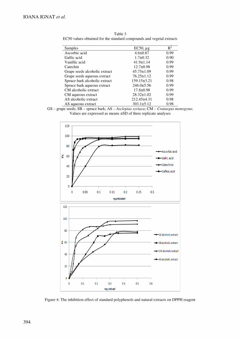
Figure 4 shows the inhibition, %, of DPPH for
different quantities of standard compounds and
vegetal extracts.
The complete neutralization of DPPH radicals
was obtained for ascorbic acid, gallic acid and
catechin, while vanillic acid provided only 80%
inhibition.
As concerns the vegetal extracts, significant
differences in scavenging activity against the
DPPH radical were registered according to their
characteristic compounds and the concentrations
involved. The alcoholic extracts showed a higher
radical scavenging activity, compared to the
aqueous ones, but slightly lower, compared to the
standard compounds.
The DPPH assay indicated that the grape seed
and hawthorn alcoholic extracts had the highest
radical scavenging activities, which can be
attributed to the considerable amounts of gallic
acid and catechin, compounds which exerted a
good inhibition of DPPH radicals on their own.
The results are strongly correlated with the total
phenolic content and HPLC determinations
previously reported. Therefore, the antioxidant
capacity depends not only on the quantity, but
also on the type of polyphenolic compounds
present in extracts. The EC50 values for the
standards and vegetal extracts are reported in
Table 3.
Antibacterial activity
The obtained results showed that the spruce
bark, Crataegus monogyna and grape seed
ethanol extracts exerted antibacterial activity (the
largest inhibition zones, 12-15 mm) against
Gram-positive pathogenic bacteria
(Staphylococcus aureus). Gram-negative bacteria
(Escherichia coli and Pseudomonas aeruginosa)
are less susceptible (smaller inhibition zones) to
herbal ethanolic extracts like spruce bark and
Asclepias syriaca and not susceptible to aqueous
extracts. Unfortunately, aqueous extracts recorded
none or less significant inhibition zones both for
bacteria and yeast-like fungi.
The results suggested that the antibacterial
activity of ethanolic extracts against Gram-
positive pathogenic bacterial strains was
comparable to the antimicrobial activity of the
reference antibacterial substances. Regarding
fungicidal activity, none of the tested extracts was
active against C. parapsilosis.
Gram-positive bacteria are known to be more
sensitive in general than Gram-negative species to
the phenolics naturally occurring in plant
extracts.10,11,14
Furthermore, Escherichia coli and
Pseudomonas aeruginosa were previously
demonstrated to be the most resistant bacteria to
phenolics from coffee and tea extracts.10
Polyphenols
393
Table 2
Concentration of phenolic compounds (mg/100 g dried plant) in the investigated samples
Raw
material
Type of
extract
Gallic
acid
Catechin
Vanillic
acid
Syringic
acid
p-coumaric
acid
Ferulic
acid
Sinapic
acid
Rutin
Quercetin
Kaempferol
Apigenin
Grape
seeds
Aqueous
extract 6.12±0.2 44.36±0.1 - - - - -
Ethanolic
extract 12.54±0.8 63.60±1.7 - - - - - 2.38±0.1
Crataegus
monogyna
Aqueous
extract - 23.42±0.9 - 2.14±0.09 - - -
Ethanolic
extract 10.98±0.7 89.52±2.1 - 2.95±1.1 3.59±0.4 2.25±1.4 - 30.32±0.9 0.64±0.02
Spruce
bark
Aqueous
extract - 31±1.9 39.4±0.2 - - - -
Ethanolic
extract 10.2±0.3 71.9±2.7 71.9±0.8 - - - - 1.39±0.08
Asclepias
syriaca
Aqueous
extract - - 0.87±0.1 0.98±0.09 0.11±0.1 - -
Ethanolic
extract 0.65±0.4 - 2.94±1.1 1.94±0.9 0.40±0.09 - - 2.25±0.21 0.14±0.05 0.17±0.02
Values are expressed as means ±SD of three replicate analyses
IOANA IGNAT et al.
394
Table 3
EC50 values obtained for the standard compounds and vegetal extracts
Samples EC50, µg R2
Ascorbic acid 4.6±0.87 0.99
Gallic acid 1.7±0.32 0.90
Vanillic acid 41.9±1.14 0.99
Catechin 12.7±0.98 0.99
Grape seeds alcoholic extract 45.75±1.09 0.99
Grape seeds aqueous extract 76.25±1.12 0.99
Spruce bark alcoholic extract 159.15±3.21 0.98
Spruce bark aqueous extract 246.0±5.56 0.99
CM alcoholic extract 17.8±0.98 0.99
CM aqueous extract 28.32±1.02 0.99
AS alcoholic extract 212.45±4.31 0.98
AS aqueous extract 303.1±5.12 0.98
GS – grape seeds; SB – spruce bark; AS – Asclepias syriaca; CM – Crataegus monogyna;
Values are expressed as means ±SD of three replicate analyses
Figure 4: The inhibition effect of standard polyphenols and natural extracts on DPPH reagent
Polyphenols
395
Table 4
Antimicrobial activity of the herbal extracts compared to antibacterial and antifungal reference substances
Inhibition zones (mm)
Reference microbial
strains
Staphylococcus
aureus
Pseudomonas
aeruginosa
Escherichia
coli
Candida
parapsilosis
Asclepias syriaca
aqueous extract - 6 6 -
Asclepias syriaca
ethanolic extract - 12 6 -
Spruce bark aqueous
extract - - - -
Spruce bark ethanolic
extract 15 10 10 -
Crataegus monogyna
aqueous extract - - - -
Crataegus monogyna
ethanolic extract 15 - - -
Grape seed aqueous
extract - - - -
Grape seed ethanolic
extract 12 - - -
Gentamicin 17 19 ND ND
Ofloxacin 21 19 22 ND
Amikacin ND 21 21 ND
Kanamycin 18 - 18 ND
Cefuroxime 26 - 17 ND
Erythromycin 20 10 11 ND
Fluconazole ND ND ND -
Nystatin ND ND ND 25
ND – not determined
Due to the lipophilic nature of phenolics, they
fail to diffuse across the outer membrane, this fact
offering a reasonable explanation for their
generally reduced activity towards Gram-negative
bacteria.
Correlating the antibacterial activity with the
antioxidant capacity and the characteristic
compounds from each sample, one can affirm that
ethanolic extracts from Crataegus monogyna,
spruce bark and grape seeds, having high amounts
of catechin and gallic acid, were the most efficient
samples.
As expected, all ethanolic extracts having
higher content of total phenolic, compared to
aqueous extracts, exerted statistically significant
antibacterial activity (inhibition zones between
10-15 mm).
Milkweed extracts characterized by the
presence of hydroxycinnamic acids and
flavonoids exhibited antibacterial effects on
Pseudomonas aeruginosa and Escherichia coli
and the spruce bark alcoholic extracts showed to
be more effective with relatively large inhibition
zones on S. aureus, P. aeruginosa and E. coli.
Generally, flavonoids are effective compounds
on pathogenic bacteria, but in our case due to the
reduced concentrations in vegetal extracts, no
significant correlation was observed.
Germination tests and pot experiments The polyphenols are known not only as potent
antioxidants, but also as cell regulators. In this
process, cell-cell communication and signal
transduction is very important and some
polyphenols can be considered to be essential for
plant life. Thus, the polyphenols, as a function of
their structure, can be involved in dividing plant
cells, polar transportation of auxins, and
enhancing the protection system against
ultraviolet rays. At the same time, polyphenols
exhibit antibiotic and phytoalexin activities,
protecting plants from diseases, or acting directly
on the genes required for infection. Therefore, the
vegetal systems could play an important role in
the study of the biological properties of
IOANA IGNAT et al.
396
polyphenols, using tests of seed germination and
plant cultivation.
In this study, the aqueous and alcoholic
extracts obtained were applied in different
concentrations in seed germination tests and plant
growth and development experiments, using
Phaseolus vulgaris seeds.
Biotests, unlike instrumental (chemical)
methods, allow simple and inexpensive estimation
of toxicity and stimulatory or inhibitory effects of
natural bioactive compounds. These experiments
are intended to evaluate the potential use of
vegetal extracts as amendments in plant growth
and by testing different concentrations of
polyphenols, we can also obtain valuable
information on the toxicity of the samples.
Considering these aspects, the selected plant
for these tests was Phaseolus vulgaris, as it is one
of the plant species recommended by the US FDA
(Food and Drugs Administration) for
phytotoxicity tests.28
To evaluate the plant response in the presence
of phenolic compounds, germination percentage,
biometric measurements (shoot and rootlet
elongation) and fresh biomass quantity were
determined.
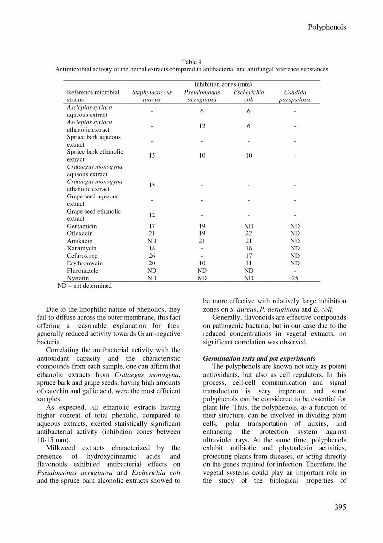
The influence of milkweed and grape seed
alcoholic extracts on plantlet elongation and fresh
biomass accumulation are presented in Figures 5
and 6.
Our experiments revealed that the effect of
natural polyphenols is strongly related to the
concentration used. In the case of Asclepias
syriaca, for all the concentrations applied, a
generally stimulatory effect was obtained for
shoot and rootlet elongation, excepting the
concentration of 0.2 g/L, which had an inhibitory
effect on shoots’ length. The most significant
results were registered for the concentration of
0.05 g/L, where the stimulatory effect on rootlets
was more than 100%, compared with the control.
For all the concentrations tested, a significant
inhibitory effect (50% compared with the control
in the case of 0.2 g/L) was registered for the shoot
and rootlet fresh biomass accumulation.
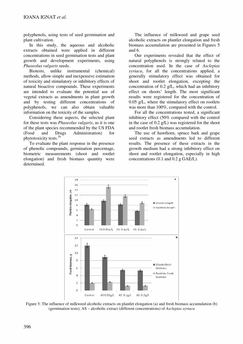
The use of hawthorn, spruce bark and grape
seed extracts as amendments led to different
results. The presence of these extracts in the
growth medium had a strong inhibitory effect on
shoot and rootlet elongation, especially in high
concentrations (0.1 and 0.2 g GAE/L).
Figure 5: The influence of milkweed alcoholic extracts on plantlet elongation (a) and fresh biomass accumulation (b)
(germination tests); AS – alcoholic extract (different concentrations) of Asclepias syriaca
Polyphenols
397
Figure 6: The influence of grape seed alcoholic extracts on plantlet elongation (a) and fresh biomass accumulation (b)
(germination tests); GS – alcoholic extract (different concentrations) of grape seeds
IOANA IGNAT et al.
398
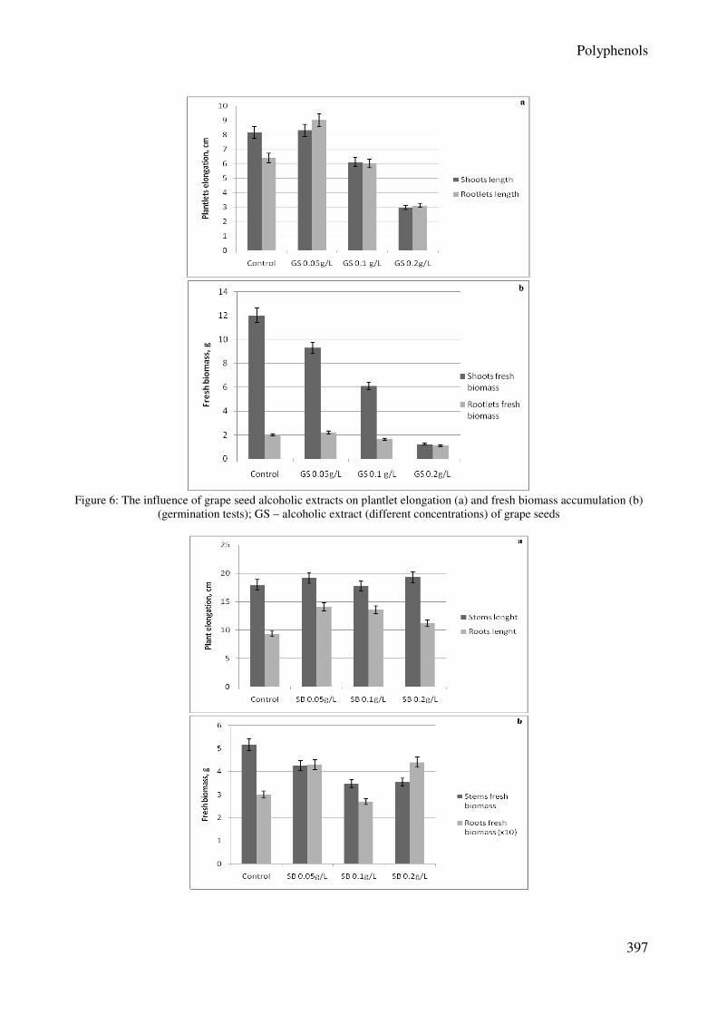
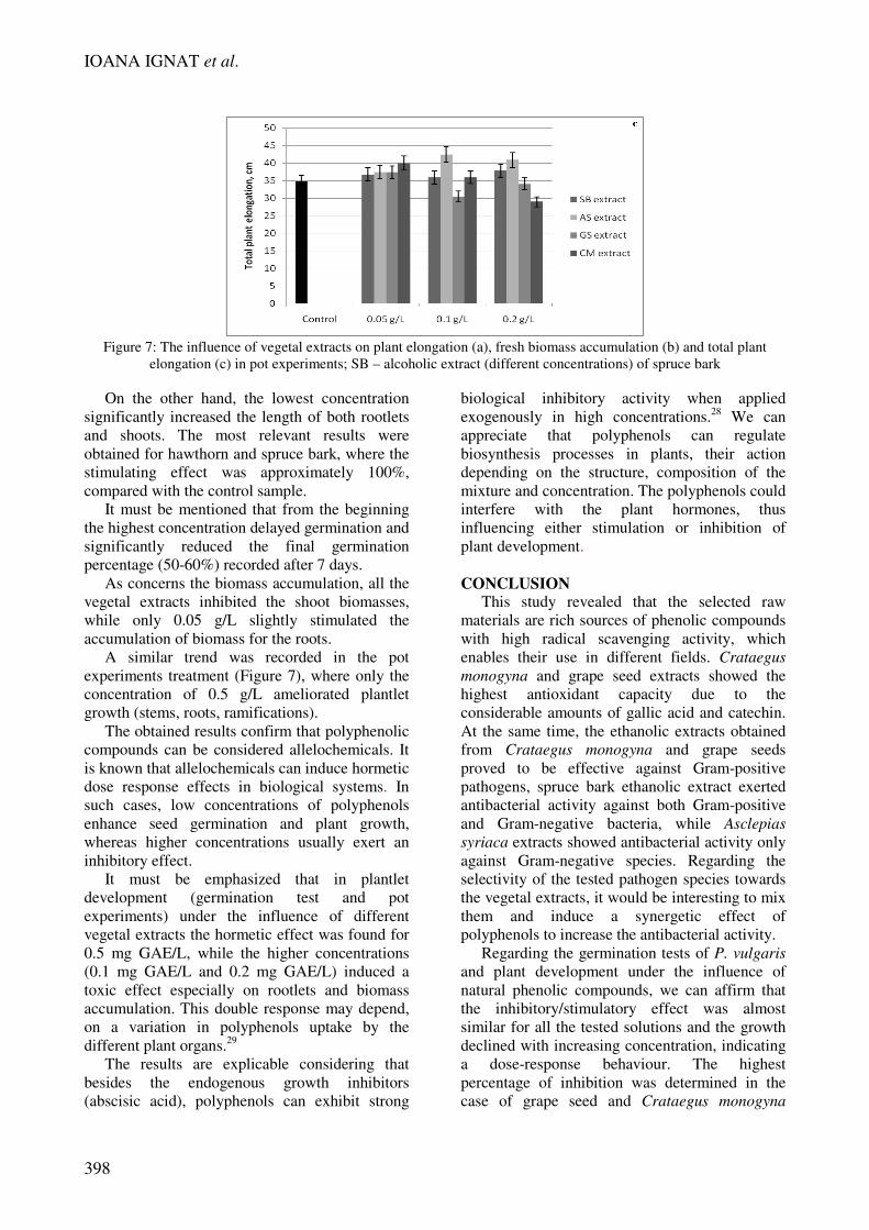
Figure 7: The influence of vegetal extracts on plant elongation (a), fresh biomass accumulation (b) and total plant
elongation (c) in pot experiments; SB – alcoholic extract (different concentrations) of spruce bark
On the other hand, the lowest concentration
significantly increased the length of both rootlets
and shoots. The most relevant results were
obtained for hawthorn and spruce bark, where the
stimulating effect was approximately 100%,
compared with the control sample.
It must be mentioned that from the beginning
the highest concentration delayed germination and
significantly reduced the final germination
percentage (50-60%) recorded after 7 days.
As concerns the biomass accumulation, all the
vegetal extracts inhibited the shoot biomasses,
while only 0.05 g/L slightly stimulated the
accumulation of biomass for the roots.
A similar trend was recorded in the pot
experiments treatment (Figure 7), where only the
concentration of 0.5 g/L ameliorated plantlet
growth (stems, roots, ramifications).
The obtained results confirm that polyphenolic
compounds can be considered allelochemicals. It
is known that allelochemicals can induce hormetic
dose response effects in biological systems. In
such cases, low concentrations of polyphenols
enhance seed germination and plant growth,
whereas higher concentrations usually exert an
inhibitory effect.
It must be emphasized that in plantlet
development (germination test and pot
experiments) under the influence of different
vegetal extracts the hormetic effect was found for
0.5 mg GAE/L, while the higher concentrations
(0.1 mg GAE/L and 0.2 mg GAE/L) induced a
toxic effect especially on rootlets and biomass
accumulation. This double response may depend,
on a variation in polyphenols uptake by the
different plant organs.29
The results are explicable considering that
besides the endogenous growth inhibitors
(abscisic acid), polyphenols can exhibit strong
biological inhibitory activity when applied
exogenously in high concentrations.28
We can
appreciate that polyphenols can regulate
biosynthesis processes in plants, their action
depending on the structure, composition of the
mixture and concentration. The polyphenols could
interfere with the plant hormones, thus
influencing either stimulation or inhibition of
plant development.
CONCLUSION This study revealed that the selected raw
materials are rich sources of phenolic compounds
with high radical scavenging activity, which
enables their use in different fields. Crataegus
monogyna and grape seed extracts showed the
highest antioxidant capacity due to the
considerable amounts of gallic acid and catechin.
At the same time, the ethanolic extracts obtained
from Crataegus monogyna and grape seeds
proved to be effective against Gram-positive
pathogens, spruce bark ethanolic extract exerted
antibacterial activity against both Gram-positive
and Gram-negative bacteria, while Asclepias
syriaca extracts showed antibacterial activity only
against Gram-negative species. Regarding the
selectivity of the tested pathogen species towards
the vegetal extracts, it would be interesting to mix
them and induce a synergetic effect of
polyphenols to increase the antibacterial activity.
Regarding the germination tests of P. vulgaris
and plant development under the influence of
natural phenolic compounds, we can affirm that
the inhibitory/stimulatory effect was almost
similar for all the tested solutions and the growth
declined with increasing concentration, indicating
a dose-response behaviour. The highest
percentage of inhibition was determined in the
case of grape seed and Crataegus monogyna
Polyphenols
399
extracts (0.2 mg GAE/L), while a significant
stimulatory effect was observed for all the
extracts at the minimum concentration (0.5 mg
GAE/L).
An interpretation of the mechanisms of
different effects of the natural extracts and of the
individual compounds existing in their
composition is now being undertaken. We are
seeking for information from studies (using
different experimental models) of molecular
biology of genomics and proteomics in order to
enhance our comprehension of the role of
polyphenols at the molecular level.
ACKNOWLEDGEMENTS: This paper was
realized with the support of BASTEURES “Bast
Plants - Renewable Strategic Resources for
European Economy” (2010-2013); co-funded by
European Union and Romanian Government
through the European Regional Development
Fund, Sectorial Operational Programme “Increase
of Economic Competitiveness. Investing for your
future”.
REFERENCES 1 I. Ignat, I. Volf, V. I. Popa, Food Chem., 126, 1821
(2011). 2
I. Volf, I. Mamaliga, V. I. Popa, Cellulose Chem.
Technol., 40, 211 (2006). 3 N. Rispail, R. Nash, K. J. Webb, in “Lotus japonicus
Handbook”, edited by A. J. Márquez , Springer,
Netherlands, 2005, pp 341-348. 4
A. Stingu, I. Volf, V. I. Popa, I. Gostin, Ind. Crop.
Prod., 35(1), 53 (2012). 5 A. Balas, V. I. Popa, BioResources, 2, 363 (2007).
6 I. Ignat, A. Stîngu, I. Volf, V. I. Popa, Cellulose
Chem. Technol., 45(3-4), 205 (2011). 7
A.R. Hainal, I.Ignat, I.Volf, V.I. Popa, Cellulose
Chem. Technol., 45(3-4), 211 (2011). 8 A. J. Parr, G. P. Bolwell, J. Sci. Food Agric., 80, 985
(2000). 9 V. Dragović-Uzelac, I. Elez Garofulić, M. Jukić, M.
Penić, M. Dent, Food Technol. Biotech., 50(3), 377
(2012). 10
G. J. E. Nychas, in “New Methods of Food
Preservation”, edited by G. W. Gould, Aspen
Publishers Inc., USA, 1995, pp 58-80. 11
D. Condrat, M. R. Szabo, D. Radu, A. X. Lupea,
Oxid. Commun., 32, 924 (2009). 12
T. Hatano, M. Tsugawa, M. Kusuda, S. Taniguchi,
T. Yoshida et al., Phytochemistry, 69, 3111 (2008). 13
T. Hatano, M. Kusuda, K. Inada, T. Ogawa, S.
Shiota et al., Phytochemistry, 66, 2047 (2005). 14
M. R. Szabo, D. Radu, S. Gavrilas, D. Chambre, C.
Iditoiu, Int. J. Food Prop., 13, 535 (2010).
15 A. Moure, J. M. Cruz, D. Franco, J. M. Domingue,
J. Sineiro, et al., Food Chem., 72, 145 (2001). 16
C. S. Ku, S. P. Mun, Wood Sci. Technol., 41, 235
(2007). 17
T. Froehlicher, T. Hennebelle, F. Martin-Nizard, P.
Cleenewerck, J. L. Hilbert et al., Food Chem., 115,
897 (2009). 18
U. Svedstrom, H. Vuorela, R. Kostiainen, I. Laakso,
R. Hiltunen, J. Chromatogr. A, 1112, 103 (2006). 19
Z. Zhang, Q. Chang, M. Zhu, Y. Huang, W. K. K.
Hoa, Z. Y. Chena, J. Nutr. Biochem., 12, 144 (2001). 20
El-Sayed Saleh Abdel-Hameed, Food Chem., 114,
1271 (2009). 21
R. J. Grubesic, J. Vukovic, D. Kremer, S. Vladimir-
Knezevic, J. Pharmaceut. Biomed., 39, 837 (2005). 22
P. Ribereau-Gayon, PhD Thesis, University of
Bordeaux, France, 1959. 23
L. Almela, B. Sanchez-Munoz, J. A. Fernandez-
Lopez, M. J. Roca, V. Rabe, J. Chromatogr. A, 1120,
221 (2006). 24
A. W. Bauer, W. M. M. Kirby, J. C. Sherris, M.
Turck, Am. J. Clin. Pathol., 45, 493 (1966). 25
European Pharmacopoeia. Microbiological assay of
antibiotics: Diffusion method, 2005, pp. 188-191. 26
G. Kronvall, J. Clin. Microbiol., 16, 784 (1982). 27
A. Rajaei, M. Barzegar, A. M. Mobarez, M. A.
Sahari, Z. H. Esfahani, Food Chem. Toxicol., 48, 107
(2010). 28
A. Jităreanu, G. Tătărîngă, A. M. Zbancioc, U.
Stănescu, Not. Bot. Horti. Agrobo., 39(2), 130 (2011). 29
L. Migliore, S. Cozzolino, M. Fiori, Chemosphere,
52, 1233 (2003).