in vitro antioxidant and thrombolytic activities of medicinal plants
TRANSCRIPT
78 | P a g e International Standard Serial Number (ISSN): 2319-8141
Full Text Available On www.ijupbs.com
International Journal of Universal Pharmacy and Bio Sciences 3(1): January-February 2014
INTERNATIONAL JOURNAL OF UNIVERSAL
PHARMACY AND BIO SCIENCES IMPACT FACTOR 1.89***
ICV 5.13***
Pharmaceutical Sciences RESEARCH ARTICLE……!!!
IN VITRO ANTIOXIDANT AND THROMBOLYTIC ACTIVITIES OF
MEDICINAL PLANTS
Swati R. Dhande*, Priya P. Dongare, Aakruti A. Kaikini,
Kalpana A. Patil, Dr. Vilasrao J. Kadam
Bharati Vidypeeth‟s College of Pharmacy, Sector-8, C.B.D. Belapur, Navi Mumbai-400086,
Maharashtra, India.
KEYWORDS:
Antioxidant, Bambusa
bambos, Pogostemon
patchouli, Swertia chirata,
thrombolytic.
For Correspondence:
Swati R. Dhande*
Address: Bharati
Vidypeeth‟s College of
Pharmacy, Sector-8,
C.B.D. Belapur, Navi
Mumbai-400086,
Maharashtra, India.
Email:
bvppharmacology@gmai
l.com
ABSTRACT Thrombosis is one of the disorders caused by oxidative injury. A
causative relationship exists between thrombus formation in the vessel
and oxidative stress. Oxidants influence the balance of coagulation
system towards platelet aggregation and thrombus formation.
Therapeutic approaches towards antithrombotic therapy by means of
antioxidants are promising in both experimental and clinical designs.
Thus the present study covers in vitro evaluation of antioxidant and
thrombolytic activities of three medicinal plants in order to provide an
edge for investigation of in vivo thrombolytic activity of respective
plants. Over production of lipid peroxides is the result of oxidant
induced injury that may cause disturbance of cell homeostasis. Level
of MDA formation is directly proportional to and is the marker
indicator of extent of lipid peroxidation. Inhibition of pro-oxidant
(Ferrous sulphate and Sodium nitroprusside) induced MDA formation
after addition of HEPP, MEBB and EESC was found to be
comparable to Gallic acid (standard). Thrombolytic activity was
determined using in vitro clot lysis assay method. The crude extracts
were found to have significant (p<0.01) thrombolytic activity at
varying doses with maximum effect at the dose of 1000µg/ml.
Streptokinase was used as standard.
79 | P a g e International Standard Serial Number (ISSN): 2319-8141
Full Text Available On www.ijupbs.com
INTRODUCTION:
Oxidative stress is now recognized to be associated with more than 200 diseases, as well as with the normal
aging process.[1-3]
Diseases like Coronary heart disease and hyperlipidemia are associated with the
increased intracellular generation of reactive oxygen species leading to tissue injury with a variety of
pathological processes like ischemia, inflammation, atherosclerosis, and thrombosis. Recent evidence
indicates that reactive oxygen species (ROS) play an important role in the control of both blood
coagulation and thrombosis.[4, 5]
Redox mechanisms control platelet function [6]
and platelet activation is
accompanied by a burst of oxygen consumption and ROS generation. Hence compounds, which can
scavenge the excess of free radicals formed or inhibit their production or protect membranes from
peroxidation, are of wide therapeutic value in disorders like ischemic stroke and cardiovascular
diseases.[7,8]
Pogostemon patchouli, Bambusa bambos and Swertia chirata are the Asian plants used for the treatment of
various diseases by different tribes. A great deal of research has been carried out on these plants with
regards to their usefulness in traditional medicine.Several antioxidant therapies have shown
neuroprotection in experimental models of brain ischemia.These antioxidants pertain to major classes that
include inhibitors of free radical production, free radical scavengers, and boosters of free radical
degradation. However, only few of these agents have been assessed in combination with thrombolytic
therapy.[9, 10]
Present study is a scientific approach to evaluate the antioxidant and thrombolytic properties
of these Asian plants.
Pogostemon patchouli (Lamiaceae) commonly known as „Patchouli‟, is one of the Chinese herbal
medicines. In Asian countries, such as Japan and China, this herb has been used traditionally as an
energizer, tonic, febrifuge, antiseptic and insecticide since ancient time. The plant is also of economic
importance due to patchouli essential oil. Patchouli oil is obtained mainly from distillation of leaves and
found to posses variety of pharmacological activities including radical-scavenging[11]
, analgesic and anti-
inflammatory,[12-15]
antiemetic, antiallergic,[16]
immunomodulatory[17]
antimicrobial actions,[18]
anti-
IFV[19]
and antimutagenic.[20]
Bambusa bambos Druce. (Graminae) is a species of clumping bamboo commonly known as “Indian thorny
bamboo” in English and “Vanshi” in Sanskrit. It mainly occurs throughout India, Srilanka, Malaya, Peru
and Myanmar. According to Ayurveda text, the plant is claimed to be medhoghna (removing or destroying
excessive fat). Charakha prescribed decoction of leaves or seeds in treatment of excessive fat. Fruit and
seeds act on medhadhatu and are useful in fat metabolism and obesity. The other traditional uses of the
80 | P a g e International Standard Serial Number (ISSN): 2319-8141
Full Text Available On www.ijupbs.com
plant are as emmenogouge, anti-inflammatory, astringent, anti-spasmodic, tonic and to check cattle in
diarrhoea.[21]
The herb Swertia chirata commonly known as „Chirata‟ is well reputed for its multifarious therapeutic
values since the era of „Atharvaveda‟.The plant Swertia chirata (gentianaceae)aboriginal to Himalayas in
India, Nepal and Bhutan, have been used for millennia, to cure variety of ailments and diseases. The chief
bioactive constituents of plant are xanthones, flavanoids, iridoids, secoiridoids glycosides, which plays
momentous role in its biological activities like antidiabetic, anti-inflammatory, hepatoprotective,
antioxidant, antipyretic, antimalarial, analgesic, anticarcinogenic, antibacterial, antiviral, gastroprotective,
antileishmanial, anthelmentic etc.[22]
Materials and methods
Collection of Plant material
The leaves of Pogostemon patchouli and Bambusa bamboswere collected from V.G. Vase Kelkar College
and Keshavshrusshti, Mumbai, India respectively and were taxonomically identified and authenticated by
Dr. H.M. Pandit, Department of Botany, Guru Nanak Khalsa College, Mumbai. The voucher specimen of
both Pogostemonpatchouli and Bambusa bambos (accession no: pd/150912, ak/170912 respectively) were
deposited at the Herbarium unit for future reference.
Dried aerial parts of Swertia chirata were purchased from Yucca Enterprises, Wadala, Mumbai, in the
month of July. The aerial parts were authenticated by Dr.Vinayak Pandit, Department of Botany, Piramal
Life Sciences, Goregaon, Mumbai and a voucher specimen (accession number:PHL/6524) was deposited
for further reference.
Preparation of plant extracts
Shade dried leaves of Pogostemon patchouli were grind to coarse powder. Powder was subjected to
extraction using n-hexane solvent for 23 hr in soxhlet apparatus. Same way, coarse powder obtained from
dried leaves of Bambusa bambos was extracted with methanol solvent using soxhlet apparatus for 72 hr.
Dried powder obtained from Swertia chirata was subjected to cold maceration with 12% ethanol for 48 hr
with continuous stirring. Later all the extracts were concentrated using rotary evaporator under reduced
pressure and low temperature and then preserved in desiccator.
Chemicals
Thiobarbituric acid (TBA), Sodium nitroprusside (SNP), Ferroussulphate (FeSO4), malondehyde
bis(dimethyl acetal) (MDA) were obtained from SD fine chemicals, Mumbai. All solutions were prepared
81 | P a g e International Standard Serial Number (ISSN): 2319-8141
Full Text Available On www.ijupbs.com
just prior to use. Sodium dodecyl sulphate (SDS) and acetic acid were of LR grade. Lyophilized
Streptokinase (15, 00,000 I.U) was obtained as a gift sample from DongkookPharma. Co. Ltd.
Animals
Female Sprague Dawley rats, weighing 180-200g were procured from Haffkine Institute, Mumbai.
Animals were housed in environmentally controlled room (temperature 23-27 C̊, 50-70% humidity with 12
hr light/dark cycle). Animals were provided with food and water ad libitium.Animals of control group were
used for ex vivo TBARS assay. All experiments in this study were carried out with the prior approval of the
Institutional Animal Ethics Committee (IAEC/PR/2012/03) strictly adhering to the guidelines of
Committee for the Purpose of Control and Supervision of Experiments on Animals (CPCSEA) constituted
by the Animal Welfare Division of Government of India.
Evaluation of in vitro antioxidant activity
Production of TBARS from animal tissue
Thiobarbituric acid reactive acid substance formation was estimated using the modified method of Ohkawa
et al. (1979). Rats were sacrificed by carbon dioxide overdose. Livers were immediately excised, washed
with ice cold saline and placed on ice bath. Livers were homogenized in ice-cold 100 mm Tris buffer pH
7.4 to obtain 10% liver tissue homogenate. The homogenates were centrifuged for 10 min at 1000g to yield
a pellet that was discarded and supernatant was separated. The supernatant was used for the assay. The
homogenates (100 µl) were incubated with or without 50 µl of the pro-oxidants (FeSO4 and sodium
nitroprusside) and 50 µl of the plant extracts together with an appropriate volume of distilled water to give
a total volume of 300 µl at 37ºC for 1 hr. The colour reaction was carried out by addition of 200, 250, 500
µl each of 8.1% SDS, acetic acid pH 3.4 and 0.6 %TBA, respectively. Final volume was made to 4 ml with
distilled water. The mixtures were incubated at 95-100ºC for 1 hr. Absorbance was measured at 532 nm.
Serial dilutions of 0.03 mM standard MDA were prepared and the absorbance was read at 532nm after
cooling. Observations for basal control (to determine normal level of MDA in rat liver tissue homogenate),
FeSO4 and SNP control (to determine maximum level of MDA formed by respective pro-oxidants) values
of MDA were also recorded. MDA content of test samples was calculated by extrapolation method using
standard graph. The results were expressed in µmol/g liver wt.[1]
In Vitro Thrombolytic activity:
Phosphate buffered saline (PBS) (5 ml) was added to the commercially available lyophilized streptokinase
vial (15, 00,000 I.U.) and mixed properly. 0.5 ml blood was withdrawn from rats by retro orbital plexus.
Blood was collected in pre weighed sterile microcentrifuge tube (0.5ml/tube) and incubated at 37°C for 45
82 | P a g e International Standard Serial Number (ISSN): 2319-8141
Full Text Available On www.ijupbs.com
min. Observations/readings were taken in triplicate. After clot formation, serum was completely removed
(aspirated out without disturbing the clot formed) and each tube having clot was again weighed to
determine the clot weight (clot weight = weight of clot containing tube – weight of tube alone). To each
microcentrifuge tube containing pre-weighed clot, 100 μl of n-hexane extract of Pogostemon patchouli
(HEPP), methanolic extract of Bambosa bambus (MEBB) and ethanolic extract of Swertia chirata (EESC)
(0.01-1 mg/ml) were added separately. 100 μl of Streptokinase was used as positive control and 100 μl of
sterile distilled water was used as negative non-thrombolytic control. All tubes were then incubated at 37ºC
for 90 min and observed for clot lysis. After incubation, fluid released was removed and tubes were again
weighed to observe difference in weight after clot disruption. Difference obtained in weight taken before
and after clot lysis was expressed as percentage of clot lysis and calculated using the following formula.[23,
7]
% 𝑐𝑙𝑜𝑡𝑙𝑦𝑠𝑖𝑠 =weight of clot after lysis
weight of clot before lysis× 100
Statistical analysis
The values were expressed as mean ± SEM. Statistical analysis and comparison between the groups for was
performed by one way analysis of variance (ANOVA) followed by Tukey‟s test. Results showed
significant (p<0.01) thrombolytic activity.
Result and discussion
Evaluation of in vitro antioxidant activity
There was a significant increase in the formation of TBARS in SNP and FeSO4-induced oxidative stress as
compared to the basal. Results were compared with Gallic acid. At the highest concentration (1000µg/ml),
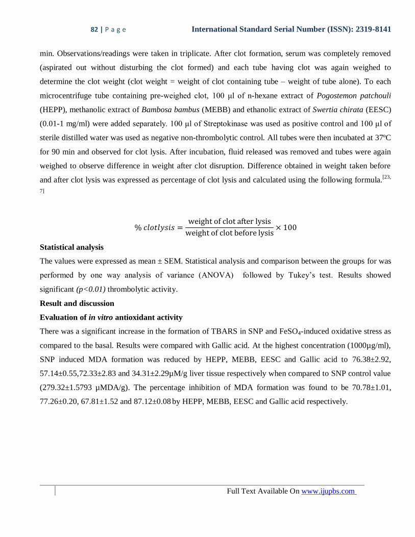
SNP induced MDA formation was reduced by HEPP, MEBB, EESC and Gallic acid to 76.38±2.92,
57.14±0.55,72.33±2.83 and 34.31±2.29µM/g liver tissue respectively when compared to SNP control value
(279.32±1.5793 µMDA/g). The percentage inhibition of MDA formation was found to be 70.78±1.01,
77.26±0.20, 67.81±1.52 and 87.12±0.08 by HEPP, MEBB, EESC and Gallic acid respectively.
83 | P a g e International Standard Serial Number (ISSN): 2319-8141
Full Text Available On www.ijupbs.com
Figure 1: Comparison of Effect of HEPP, MEBB, EESC, andGallic acid on SNP induced elevation of MDA
levels in rat liver homogenate. Mean basal value and SNP control values were 26.86±0.06 and 279.32±1.5793
µMDA/g of tissue respectively.
0 200 400 600 800 10000
20
40
60
80HEPP
MEBB
EESC
Gallic acid
Concentration (g/ml)
%In
hib
itio
n o
f F
eS
O4
in
du
ced
form
ati
on
of
MD
A
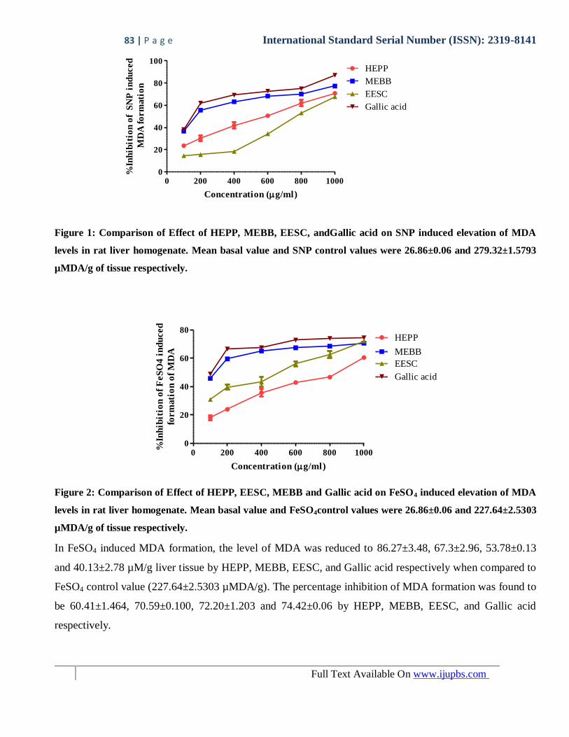
Figure 2: Comparison of Effect of HEPP, EESC, MEBB and Gallic acid on FeSO4 induced elevation of MDA
levels in rat liver homogenate. Mean basal value and FeSO4control values were 26.86±0.06 and 227.64±2.5303
µMDA/g of tissue respectively.
In FeSO4 induced MDA formation, the level of MDA was reduced to 86.27±3.48, 67.3±2.96, 53.78±0.13
and 40.13±2.78 µM/g liver tissue by HEPP, MEBB, EESC, and Gallic acid respectively when compared to
FeSO4 control value (227.64±2.5303 µMDA/g). The percentage inhibition of MDA formation was found to
be 60.41±1.464, 70.59±0.100, 72.20±1.203 and 74.42±0.06 by HEPP, MEBB, EESC, and Gallic acid
respectively.
0 200 400 600 800 10000
20
40
60
80
100
MEBB
HEPP
EESC
Gallic acid
Concentration (g/ml)
%In
hib
itio
n o
f S
NP
in
du
ced
MD
A f
orm
ati
on
84 | P a g e International Standard Serial Number (ISSN): 2319-8141
Full Text Available On www.ijupbs.com
Increase in the formation of TBARS in FeSO4 induced oxidative stress suggests cellular damage due to
overload of iron. Iron overload results in the formation of lipid peroxidation products.[24]
The possible
mechanism of iron toxicity includes free radical mediated peroxidative reactions, which are readily
catalyzed by iron. The protections offered by these plants extracts suggest that they may be useful in the
treatment of disease resulting from free radical induced damage.
SNP induced cellular toxicity is associated with overproduction of reactive nitrogen species (RNS) which
are as reactive as ROS producing damage to cellular contents. The results demonstrate the ability of all the
tested plant extracts to protect the cell against oxidative stress induced by various pro-oxidants and
suggests their use in the treatment of various diseases which may be linked with their antioxidant activity.
In Vitro Thrombolytic activity assay
100 200 400 600 800 10000
20
40
60
80HEPP
MEBB
EESC
plant extract concentrations (g/ml)
Clo
t ly
sis
(%)
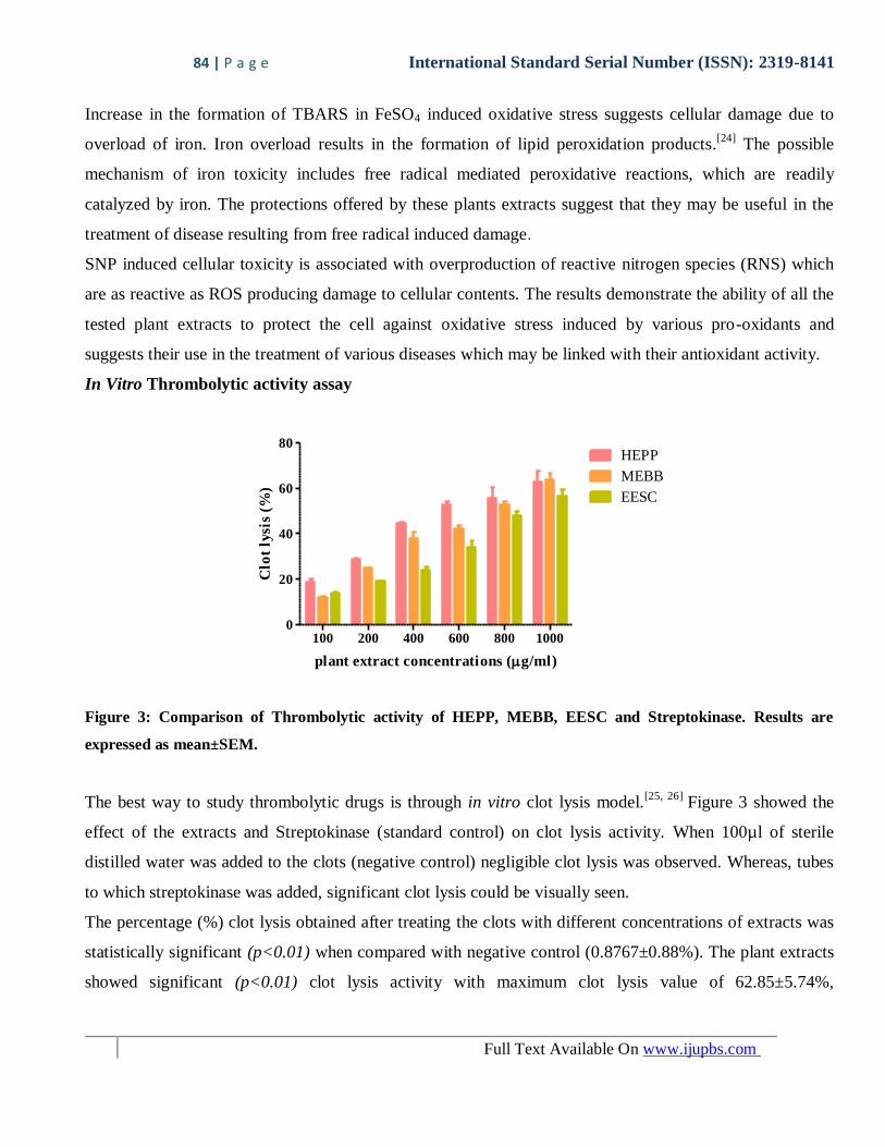
Figure 3: Comparison of Thrombolytic activity of HEPP, MEBB, EESC and Streptokinase. Results are
expressed as mean±SEM.
The best way to study thrombolytic drugs is through in vitro clot lysis model.[25, 26]
Figure 3 showed the
effect of the extracts and Streptokinase (standard control) on clot lysis activity. When 100µl of sterile
distilled water was added to the clots (negative control) negligible clot lysis was observed. Whereas, tubes
to which streptokinase was added, significant clot lysis could be visually seen.
The percentage (%) clot lysis obtained after treating the clots with different concentrations of extracts was
statistically significant (p<0.01) when compared with negative control (0.8767±0.88%). The plant extracts
showed significant (p<0.01) clot lysis activity with maximum clot lysis value of 62.85±5.74%,
85 | P a g e International Standard Serial Number (ISSN): 2319-8141
Full Text Available On www.ijupbs.com
63.69±2.98% and 56.58±3.42% by HEPP, MEBB and EESC respectively, at concentration of 1000 µg/ml.
Percentage (%) clot lysis value for Streptokinase (standard control) was 77.13±2.12%.
Conclusion
In conclusion, the extracts of Swertia chirata, Bambusa bambos and Pogostemon patchouli showed
considerable antioxidant and significant thrombolytic activity in vitro. The mechanism of action of these
three plants is still unknown. This is only a preliminary study and to make a final comment the extracts
should be thoroughly investigated phytochemically and pharmacologically to exploit their medicinal and
pharmaceutical potentialities. Thus, these extracts may be incorporated in herbal preparations as
thrombolytic agents for the improvement of patients in clinical practice.
References:
1. Olalye MT, Rocha JBT, (2007), Commonly used tropical medicinal plants exhibit distinct in vitro
antioxidant activities against hepatotoxins in rat liver. ExpToxicol Pathol.58, 433–8.
2. Ghasanfari G, Minaie B, Yasa N, Leilu AN, Azadeh M, Shekoufeh N, et al., (2006), Biochemical and
histopathological evidences for beneficial effects of SaturejaKhuzestanicaJamzad essential oil on the
mouse model of inflammatory bowel diseases. ToxicolMech Method. 16, 365–72.
3. Sanvicens N, Violeta G, Angel M, Thomas GC, (2006),The radical scarvenger CR-6 proteccts sh-sy5y
neuroblastoma cells from oxidative stress induced apoptosis effects on survival pathways. J
Neurochem.98, 735–42.
4. Chakrabarti S and Freedman JE, (2010), Review: Nutriceuticals as Antithrombotic Agents.
Cardiovascular Therapeutics. 28, 227-35.
5. Gorlach A, (2005), Redox regulation of the coagulation cascade. Antioxid Redox Signal. 7,1398–404.
6. Essex DW. (2009), Redox control of platelet function. Antioxid Redox Signa.l11,1191–225.
7. Chowdhury N.S, Alam MB. Haque ASMT, Zahan R, MazumderMEH and Haque ME, (2011), In vitro
Free Radical Scavenging and Thrombolytic Activities of Bangladeshi Aquatic Plant
AponogetonundulatusRoxb. Glob Pharmacol. 5 (1), 27-32.
8. Diaz, M.N., F. Balz, A.V. Joseph and F.K. John, (1997), Antioxidants and atherosclerotic heart disease.
New Eng. J. Med, 337, 408-16.
9. Amaro S, Chamarrow A, (2011),Translational Stroke Research of the Combination of Thrombolysis
and Antioxidant Therapy. Stroke. 42, 1495-99.
10. Margaill I, Plotkine M, Lerouet D, (2005), Antioxidant strategies in the treatment of stroke. Free
RadicBiol Med. 39, 429–43.
86 | P a g e International Standard Serial Number (ISSN): 2319-8141
Full Text Available On www.ijupbs.com
11. Kim HW, Cho SJ, Kim BY, Cho SI, Kim YK,(2010), Pogostemon cablin as ROS scavenger in oxidant-
induced cell death of human neuroglioma cells. Evidence Based Complementary Alternative
Medicine.7(2), 239–47.
12. Li YC, Xian YF, Ip SP, Su ZR, Su JY, He JJ. et al.,(2011), Anti-inflammatory activity of patchouli
alcohol isolated from PogostemonisHerba in animal models. Fitoterapia. 82(8), 1295-301.
13. Xian YF, Suo J, Huang XD, Hou SZ, Chen JN, Ye MR, et al.,(2007), A pharmacological study on anti-
inflammatory effects of refined Huodan recipe. Chinese Journal of Experimental Traditional Medical
Formulae. 13, 54-6.
14. Zhao SC, Jia Q, Liao FL. (2007), The anti-inflammatory and analgesic pharmacological study of
Patchouli extract. Chin Tradit Plant Med. 29, 285–7.
15. Lu TC, Liao JC, Huang TH, Lin YC, Liu CY, Chiu YJ, et al.(2011), Analgesic and anti Inflammatory
activities of the methanol extract from Pogostemon cablin. Evidence. Based Complementary
Alternative Medicine.1-9
16. Suo J, Xian YF, Huang XD, Hou SZ, Chen JN, Ye MR, et al., (2007), A pharmacological study on the
anti-allergy effects of refined Houdan recipe. Chinese Journal of Experimental Traditional Medical
Formulae. 13:47–9.
17. Qi SS, Hu LP, Chen WN, Sun HB, Ma XD,(2009), Immunological regulation effects of essential oil in
leaves of Cablin Patchouli herbal on mice. Chin Arch Tradit Chin Med.27,774–6.
18. Liu XR, Fan R, Zhang YY, Zhu MJ, (2009), Study on antimicrobial activities of extracts from
Pogestemoncablin (Blanco) Benth. Food Science and Technology. 24, 220–7.
19. Kawamura Y, Kiyohara H, Nagai T, Hiramoto T, Yamada H, (2010), Anti-influenza virus active
sesquiterpene from leaves of Pogostemon cablin. Annual Meetingof the Pharmaceutical Society of
Japan. 130(2), 17-9.
20. Miyazawa M, Okuno Y, Nakamura S, Kosaka H, (2000),Antimutagenic Activity of Flavonoids from
Pogostemon cablin. Journal of Agricultural and Food Chemistry.48(3), 642-7.
21. Kaikini AA, Dhande SR, Kadam VJ, (2013), Overview of Indian medicinal Tree: Bambusa bambos
(Druce), Int. Res. J. Pharm. 4(8), 53-56.
22. Patil K, Dhande S, Kadam V, (2013), Thearautic Swertia chirata: An Overview. RJPP. 5(4), 199-207.
23. Bhuiya AM et al.,(2013), In vitro thrombolytic and antioxidant activity Study of Abromaaugusta
(Ulatkambal). The experiment. 14(2), 888-93.
87 | P a g e International Standard Serial Number (ISSN): 2319-8141
Full Text Available On www.ijupbs.com
24. Houglum K, Filip M, Witztum JL, Chojkier M. (1990), Malondialdehyde and 4-hydroxynonenal
protein adducts in plasma and liver of rats with iron overload. J Clin Invest. 86, 1991–8.
25. Prasad S, Kashyap RS, Deopujari JY, Purohit HJ, Taori GM and Daginawala HF, (2006),
Development of an in vitro model to study clot lysis activity of thrombolytic drugs. Thrombosis
journal, 4(14), 1-4.
26. Strief TW, (2006), In vitro simulation of therapeutic thrombolysis with microtiter plate clot-lysis assay.
ClinAppl Thrombosis/ Hemostatis.12, 21-32.