antioxidant and antimicrobial activities of chemically ... - mdpi
TRANSCRIPT
Molecules 2022, 27, 1136. https://doi.org/10.3390/molecules27031136 www.mdpi.com/journal/molecules
Article
Antioxidant and Antimicrobial Activities of
Chemically-Characterized Essential Oil from
Artemisia aragonensis Lam. Against Drug-Resistant Microbes
Khalid Chebbac 1,*, Hazem K. Ghneim 2, Abdelfattah El Moussaoui 3, Mohammed Bourhia 4, Azeddin El Barnossi 3,
Zineb Benziane Ouaritini 5, Ahmad Mohammad Salamatullah 6,*, Abdulhakeem Alzahrani 6,
Mourad A. M. Aboul-Soud 2,*, John P. Giesy 7,8,9,10 and Raja Guemmouh 1
1 Laboratory of Biotechnology Conservation and Valorisation of Natural Resources,
Faculty of Sciences Dhar El Mahraz, Sidi Mohammed Ben Abdallah University, Fez 30000, Morocco;
[email protected] 2 Department of Clinical Laboratory Sciences, College of Applied Medical Sciences, King Saud University,
P.O. Box 10219, Riyadh 11433, Saudi Arabia; [email protected] 3 Laboratory of Biotechnology, Environment, Agri-Food and Health, Faculty of Sciences Dhar El Mahraz,
Sidi Mohammed Ben Abdellah University, Fez 30000, Morocco;
[email protected] (A.E.M.); [email protected] (A.E.B.) 4 Laboratory of Chemistry, Biochemistry, Nutrition, and Environment, Faculty of Medicine and Pharmacy,
University Hassan II, Casablanca 20000, Morocco; [email protected] 5 Laboratory of Natural Substances, Pharmacology, Environment, Modeling, Health and Quality of Life,
Faculty of Sciences, Sidi Mohamed Ben Abdellah University, Fez 30000, Morocco;
[email protected] 6 Department of Food Science & Nutrition, College of Food and Agricultural Sciences, King Saud University,
P.O. Box 2460, Riyadh 11451, Saudi Arabia; [email protected] 7 Toxicology Centre, University of Saskatchewan, Saskatoon, SK S7N 5B3, Canada; [email protected] 8 Department of Veterinary Biomedical Sciences, University of Saskatchewan,
Saskatoon, SK S7N 5B3, Canada 9 Department of Integrative Biology, Center for Integrative Toxicology, Michigan State University,
East Lansing, MI 48824, USA 10 Department of Environmental Science, Baylor University, Waco, TX 76706, USA
* Correspondence: [email protected] (K.C.); [email protected] (A.M.S.);
[email protected] (M.A.M.A.-S.)
Abstract: This study investigated the chemical composition, antioxidant and antimicrobial activity
of essential oil extracted from Artemisia aragonensis Lam. (EOA). Hydrodistillation was employed to
extract EOA. Gas chromatography with flame ionization detection (GC-FID) and gas chromatog-
raphy-mass spectrometry analyses (GC-MS) were used to determine the phytochemical composi-
tion of EOA. Antioxidant potential was examined in vitro by use of three tests: 2.2-diphenyl-1-pic-
rilhidrazil (DPPH), ferric reducing activity power (FRAP) and total antioxidant capacity assay
(TAC). Agar diffusion and microdilution bioassays were used to assess antimicrobial activity.
GC/MS and GC-FID detected 34 constituents in the studied EOA. The major component was Cam-
phor (24.97%) followed by Borneol (13.20%), 1,8 Cineol (10.88%), and Artemisia alcohol (10.20%).
EOA exhibited significant antioxidant activity as measured by DPPH and FRAP assays, with IC50
and EC50 values of 0.034 ± 0.004 and 0.118 ± 0.008 mg/mL, respectively. EOA exhibited total antiox-
idant capacity of 7.299 ± 1.774 mg EAA/g. EOA exhibited potent antibacterial activity as judged by
the low minimum inhibitory concentration (MIC) values against selected clinically-important path-
ogenic bacteria. MIC values of 6.568 ± 1.033, 5.971 ± 1.033, 7.164 ± 0.0 and 5.375 ± 0.0 μg/mL were
observed against S. aureus, B. subtills, E. coli 97 and E. coli 57, respectively. EOA displayed significant
antifungal activity against four strains of fungi: F. oxysporum, C. albicans, A. flavus and A. niger with
values of 21.50 ± 0.43, 5.31 ± 0.10, 21.50 ± 0.46 and 5.30 ± 0.036 μg/mL, respectively. The results of
the current study highlight the importance of EOA as an alternative source of natural antioxidant
and antibacterial drugs to combat antibiotic-resistant microbes and free radicals implicated in the
inflammatory responses accompanying microbial infection.
Citation: Chebbac, K.;
Ghneim, H.K.; El Moussaoui, A.;
Bourhia, M.; El Barnossi, A.;
Benziane Ouaritini, Z.;
Salamatullah, A.M.; Alzahrani, A.;
Aboul-Soud, M.A.M.; Giesy, J.P.;
et al. Antioxidant and Antimicrobial
Activities of
Chemically-Characterized Essential
Oil from Artemisia aragonensis Lam.
Against Drug-Resistant Microbes.
Molecules 2022, 27, 1136. https://
doi.org/10.3390/molecules27031136
Academic Editor:
Carmen Formisano
Received: 17 January 2022
Accepted: 4 February 2022
Published: 8 February 2022
Publisher’s Note: MDPI stays
neutral with regard to jurisdictional
claims in published maps and
institutional affiliations.
Copyright: © 2022 by the authors.
Licensee MDPI, Basel, Switzerland.
This article is an open access article
distributed under the terms and
conditions of the Creative Commons
Attribution (CC BY) license
(https://creativecommons.org/license
s/by/4.0/).

Molecules 2022, 27, 1136 2 of 16
Keywords: essential oils; phytochemical analysis; antioxidant; antibacterial; antimicrobial resistance
1. Introduction
Plants constitute a natural reservoir of substances with antioxidant potential [1]. The
use and development of natural antioxidants are highly appreciated due to their role in
the protection of human cells from damage caused by free radicals [2,3]. Natural antioxi-
dants, rather than synthetic antioxidants, appear to be preferred by food industry users
for preventing oxidative deterioration of foodstuffs caused by free radicals. It has been
previously reported that the use of synthesized antioxidants such as tertbutyl hydroqui-
none (TBHQ), butylated hydroxytoluene, and butylated hydroxyanisole is no longer ad-
vised because of their carcinogenic potential [4]. BHA and BHT are also involved in liver
damage along with other adverse health effects. Indeed, TBHQ is currently banned by
some European countries and Japan [5].
Antimicrobial resistance (AMR) is a phenomenon whereby microorganisms develop
a variety of strategies to combat medications designed to kill them, resulting in microbes
that are resistant to treatment protocols [6]. Globally, growing attention is being allocated
by scientists to AMR since it has evolved into a widespread and serious problem affecting
the entire healthcare system. In addition, the World Health Organization has pointed out
that AMR is the most significant concern in 2019 and has classified it among the top 10
global public health threats to humanity. The overuse of antibiotics in human medicine,
animal husbandry, hygiene, and the food industry can contribute to the rise of AMR [7,8].
Fatalities attributed to AMR are alarmingly increasing and it is being projected to claim
10 million annually by the year 2050. Significant global economic losses are also expected
to reach a cumulative $100 trillion if more efficient and novel therapeutic alternatives are
not developed soon to contain the rapidly-evolving causative microbial agents [9]. The list
of microbes that are becoming resistant to all known antibiotics is expanding, under the
currently limited and insufficient repertoire of new treatments, necessitating the develop-
ment of novel classes of drugs to avoid serious public health problems.
The microorganisms examined in this study are classified among the drug-resistant
pathogens namely Escherichia coli and Staphylococcus aureus. As previously documented,
these species are multidrug-resistant [10,11]. In addition, Candida albicans, which was also
evaluated in this study, is known to be a drug-resistant pathogen. Candida albicans re-
sistance is being widely recognized as one of the greatest expanding health burdens, ow-
ing to the widespread use of various drugs, particularly oral azoles to control this strain
[12]. However, none of the currently available traditional antimycotic medications fit all
of the criteria in terms of patient toxicity, ease of administration and minimal risk of re-
sistance development.
Recently, alternative therapeutic solutions based on the exploitation of natural re-
sources have been thoroughly researched [13,14]. In this context, the chemical constituents
of the genus Artemisia have been the subject of numerous previous reports, which showed
that this genus possesses several potentially-bioactive classes of compounds including fla-
vonoids, polyphenols, tannins, sesquiterpene lactones and essential oil (EO) [14]. EOs
from the genus Artemisia were reported to possess multiple biological and pharmacologi-
cal effects including antimicrobial [15,16]. EOs are complex combinations of chemical mol-
ecules from various chemical families, such as aldehydes, alcohols, esters, phenols, ethers
and ketones terpenes, among others. Terpenes, terpenoids, and other aromatic and ali-
phatic components with low molecular weights make up the majority of EOs [10,11].
The current study investigated the chemical composition of EOA along with its anti-
oxidant and antibacterial potential against drug-resistant pathogenic microorganisms.

Molecules 2022, 27, 1136 3 of 16
2. Materials and Methods
2.1. Plant Material Selection and Identification
In April 2021, A. aragonensis was collected from the southern slopes of Jbel Bou-
Naceur in Morocco (latitude 33.59885133, longitude −3.74447934, and altitude: 1350 m).
Botanical identification was conducted by a botanist under reference AHA001T7621.
Thereafter, leaves were dried at room temperature for 11 days before being extracted by
use of Clevenger equipment to obtain EO.
2.2. Extraction of Essential Oil
In the current study, hydrodistillation was used to extract EOA. Briefly, 100 g of the
dried aerial parts of A. aragonensis were soaked in 600 mL of distilled water and boiled for
2 h using a Clevenger-type apparatus. The obtained essential oil was kept at 4 °C and its
yield (%) was calculated on the basis of the dry weight of the plant material.
2.3. Essential Oil Chemical Identification
The EOA was characterized by GC-ULTRA apparatus equipped with VB-5 column
(length: 30.00 m, internal diameter: 0.250 mm, film thickness: 0.250 μm). Operational con-
ditions were set as follows: carrier gas (helium), injection temperature (220 °C), injection
volume (1μL), flow rate (1.4 mL/min), temperature-programmed gas chromatography (40
to 180 °C at 4 °C/min, followed by 20 min at 300 °C). The temperature of the interface was
300 °C with the following conditions: type of ionization EI (70 eV) and temperature of the
ionization source (200 °C). The identification of the phytochemical components of EOA
was carried out by determining their retention indices relative to a homologous series of
n-alkanes and by comparing their registered mass spectra with those reported in refer-
enced databases (NIST MS Library v.2.00) (NIST MS Library v.2.00) [17].
2.4. Antioxidant Activity
2.4.1. Radical Scavenging Activity Test
DPPH assay was carried out according to Chebbac’s protocols [18]. To achieve this,
100 μL of EOA, at different concentrations, prepared with methanol (1.0, 0.25, 0.125,
0.0625, 0.0312, 0.0156, 0.0078, 0.0064 and 0.0019 mg/mL), were used for the testing pur-
poses. The anti-free radical effect was measured by mixing 100 μL of each concentration
previously prepared (EOA, Quercetin, Ascorbic acid and BHT) with 750 μL of DPPH
(0.004%), while methanol was included as a negative control. Next, incubation was con-
ducted in the dark for 30 min at room temperature prior to recording absorbance values
at 517 nm by use of a spectrophotometer and the DPPH trapping capacity was represented
as percent inhibition (Equation (1)):
PI (%) = (A0 − A/A0) × 100 (1)
where PI represents the proportion of inhibition, A0 represents the negative control
(methanol), and A represents the combined absorbance of DPPH and samples. All anal-
yses were performed three times, and the findings were presented as means with standard
deviations. The IC50 was calculated graphically by use of linear regression.
2.4.2. Total Antioxidant Capacity Test (TAC)
One milliliter of a solution containing sulfuric acid, sodium phosphate, and ammo-
nium molybdate was combined with 25 μL of each EOA concentration. The solution was
then incubated for 91 min at 96 °C. The absorbance was then recorded at 695 nm against
the blank with 25 μL of methanol [19]. TAC per gram of EO was expressed in milligrams
of ascorbic acid equivalent (mg EAA/g). The experiment was conducted in triplicates and
the obtained results were represented as means with standard deviations.

Molecules 2022, 27, 1136 4 of 16
2.4.3. Reducing Power Test (FRAP)
This test was carried out using the method proposed by Bourhia et al. [20]. In meth-
anol, 500 μL of phosphate buffer solution and potassium ferricyanide were combined with
100 μL of varied doses of EOA (0.1, 0.2, 0.4, 0.8, 1.6 mg/mL). Following a 21 min incubation
period, 500 μL of a 10% aqueous TCA solution, 500 μL of distilled water and 100 μL of
0.1% FeCl3 were added to the reaction medium. The absorbance was subsequently meas-
ured against a reagent blank containing no sample. The results were expressed as a 50%
effective concentration (EC50).
2.5. Antimicrobial Activity
Antifungal activity of EOA was tested against four fungal species, including Candida
albicans ATCC 10231, Aspergillus niger, Aspergillus flavus, and Fusarium oxysporum, as well
as four bacterial strains, including Escherichia coli (ATB: 57/B6N), Escherichia coli (ATB:
97/BGM), Staphylococcus aureus, and Bacillus subtills, which were kindly provided by Has-
san II University Hospital Center of Fez, Morocco.
2.5.1. Disk Diffusion Method
In the present study, the disk diffusion method was used to evaluate antifungal and
antibacterial activity of EOA [21]. For this purpose, bacteria were grown in Petri plates
having nutrient broth medium (NB), whereas fungal strains were grown in Petri dishes
possessing a malt extract agar (MEA) medium. From fresh bacteria culture, a few colonies
were aseptically seeded in 0.9% sodium chloride (NaCl) at a density of 0.5 McFarland (107
to 108 CFU/mL), whilst the yeast suspension was determined to be approximately 106
CFU/mL. After being soaked in 10 μL of EOA, 6 mm diameter disks were placed on the
surface of petri dishes. [15,22,23]. Next, the inoculated Petri dishes were incubated in the
dark at 30 °C and 37 °C for the fungal and bacterial species, respectively. The inhibition
rate, expressed in percentages, was calculated 24 and 48 h post-incubation for bacteria and
C. albicans, respectively, and 7 days post-inoculation for A. niger, A. flavus and F. oxysporum
[18]. The growth inhibition zones were determined in mm.
2.5.2. Determination of the Minimum Inhibitory Concentration (MIC)
The microdilution method, which was originally published in Balouir’s earlier work
[24], was undertaken to determine the MIC of EOA against bacterial and fungal strains.
In this context, the MIC was calculated by direct observation of growth in the wells using
the colorimetric method (TTC 0.20 percent (w/v)) after an incubation period of 24 h for
bacteria at 37 °C, 48 h for yeast and seven days for fungi at 30 °C.
2.6. Statistical Analysis
The obtained findings in this research work were expressed as means with standard
deviations of triplicate tests. Normality was checked by use of the Shapiro–Wilks test and
the assumption of homogeneity of variance was evaluated using Levene’s test. The non-
parametric Tukey’s statistical test was employed as a post-hoc test for multiple compari-
sons. When p < 0.05, a statistically significant difference was considered.
3. Results and Discussion
3.1. Essential Oil Yield
The yield of EOA was 1.18%, which was reasonable compared with EOs extracted
from plants that have been industrially exploited as a natural source of EOs. In this con-
text, species among genus Artemisia were used for comparison purposes including Arte-
misia frigida (1.5%) and Artemisia cana (1.3%). By contrast, species of A. absinthium, A. dra-
cunculus, A. biennis, A. ludoviciana and A. longifolia were found to produce lower EO yield
than A. aragonensis [25]. The observed difference can be explained by the environmental
Molecules 2022, 27, 1136 5 of 16
and edaphic factors, extraction technique, drying, harvesting period and cultural practices
that influence both quality and quantity of compounds in plants [26].
3.2. Chemical Composition Identification of the Essential Oil
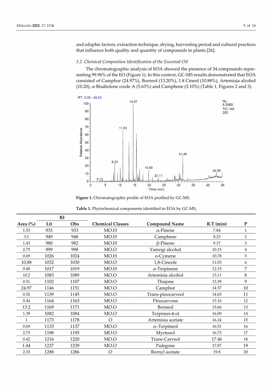
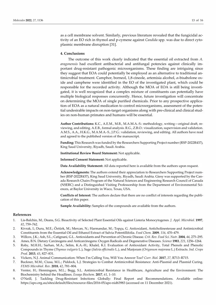
The chromatographic analysis of EOA showed the presence of 34 compounds repre-
senting 99.96% of the EO (Figure 1). In this context, GC-MS results demonstrated that EOA
consisted of Camphor (24.97%), Borneol (13.20%), 1.8 Cineol (10.88%), Artemisia alcohol
(10.20), α-Bisabolone oxide A (5.63%) and Camphene (3.10%) (Table 1, Figures 2 and 3).
Figure 1. Chromatographic profile of EOA profiled by GC-MS.
Table 1. Phytochemical components identified in EOA by GC-MS.
RI
Area (%) Lit Obs Chemical Classes Compound Name R.T (min) P
1.53 933 933 MO.H α-Pinene 7.84 1
3.1 949 948 MO.H Camphene 8.23 2
1.43 980 982 MO.H β-Pinene 9.17 3
2.75 999 998 MO.O Yamogi alcohol 10.15 4
0.69 1026 1024 MO.H o-Cymene 10.78 5
10.88 1032 1030 MO.O 1,8-Cineole 11.03 6
0.48 1017 1019 MO.H α-Terpinene 12.15 7
10.2 1083 1089 MO.O Artemisia alcohol 13.11 8
0.51 1102 1107 MO.O Thujone 13.39 9
24.97 1146 1151 MO.O Camphor 14.57 10
0.52 1139 1145 MO.O Trans-pinocarveol 14.65 11
0.44 1164 1163 MO.O Pinocarvone 15.16 12
13.2 1169 1171 MO.O Borneol 15.66 13
1.39 1082 1084 MO.O Terpinen-4-ol 16.09 14
1 1173 1178 O Artemisia acetate 16.24 15
0.69 1133 1137 MO.O α–Terpineol 16.51 16
2.73 1198 1195 MO.O Myrtenol 16.73 17
0.42 1216 1220 MO.O Trans-Carveol 17.46 18
1.44 1237 1239 MO.O Pulegone 17.87 19
2.33 1288 1286 O Bornyl acetate 19.8 20

Molecules 2022, 27, 1136 6 of 16
0.83 1326 1327 O Myrtenyl acetate 21.05 21
0.75 1376 1372 SQ.H α-Copaene 23.11 22
0.71 1485 1480 SQ.H Germacrene D 26.12 23
1.26 1578 1579 SQ.O Spathulenol 28.66 24
1.26 1586 1583 SQ.O Caryophyllene oxide 28.77 25
0.5 1624 1625 SQ.O Isospathulenol 30.12 26
2.2 1632 1633 SQ.O γ-Eudesmo 30.26 27
0.51 1640 1642 SQ.O Cadinol 30.5 28
1.3 1650 1652 SQ.O β-Eudesmo 30.64 29
0.45 1658 1657 SQ.O Bisabolol oxyde B 30.93 30
5.63 1685 1688 SQ.O Bisabolone oxide A 31.45 31
0.56 1749 1751 SQ.O α-Bisabolol oxide A 33.14 32
1.33 2800 2804 O Octacosane 40.32 33
1.63 2500 2503 ST.H Pentacosane 42.99 34 Chemical classes
7.23 Monoterpene hydrocarbons (MO.H)
70.14 Oxygenated monoterpenes (MO.O)
1.46 Sesquiterpene hydrocarbons (SQ.H)
13.67 Oxygenated sesquiterpenes (SQ.O)
1.63 Sesterpene (ST.H)
5.49 Other compounds (O)
99.62 Total identification
P: Peak; R.T: Retention time; Obs: Observed; Lit: Literature; R.I: Retention index; MO.H: Monoter-
pene hydrocarbons; MO.O: Oxygenated monoterpenes; SQ.H: Sesquiterpene hydrocarbons; SQ.O:
Oxygenated sesquiterpenes; ST.H: Sesterpene; O: Other compounds.
Figure 2. Molecular structure of some major phytochemicals identified in EOA.
The detected components were classified according to functional categories and the
results revealed that oxygenated monoterpenes (70.14%) were the most abundant in EOA.
These findings matched those reported in previous studies [16], which reported the rich-

Molecules 2022, 27, 1136 7 of 16
ness of EOA native to Spain, in Camphor (15%), Cineol (13.3%), Borneol (4.8%) and Cam-
phene (1.9%). The chemical composition, particularly amounts of monoterpene alcohols,
found in EOA in our study is similar to those reported in Israeli species. However, the chem-
ical composition of EOA was different when compared to Algeria and Tunisia cultivars in
terms of chemical content, notably monoterpene alcohols [23,24]. For a more detailed com-
parison, the chemical content of EOs extracted from the aerial parts of four Artemisia spe-
cies, A. cana, A. frigida, A. longifolia and A. ludoviciana, growing in Canada was higher in 1,8-
cineole (21.5–27.6%), davanone (11.50%) and camphor (15.9–37.3%). EO of A. absinthium was
found to be rich in myrcene (10.80%), trans-sabinyl acetate (26.40%) and trans-thujone
(10.1%). A. biennis contained more (E)-β-farnesene (40%), (Z)-β-ocimene (34.7%), acetylenes
(11.00%) (Z)- and (E)-en-yn-dicycloethers. Phenylpropanoids (16.2%) and methyl eugenol
were the primary components of the EO from A. dracunculus (35.8%) [27,28].
Several studies indicated that A. aragonensis was characteristically distinguished by
the presence of potentially bioactive compounds including chrysantenone and davanone
[26,29,30], which are absent in our plant that was collected from the southern slope of Jbel
Bou-Naceur. Therefore, this difference in chemical composition can be an indicator of the
difference in the ecosystem diversity where species grow. Moreover, our results showed
that EOA contained artemisia alcohol and artemisia acetate, which can be used as a dis-
tinctive indicator of A. aragonensis from the southern slope of Jbel Bou-Naceur of the
folded Middle Atlas of Morocco.
3.3. Antioxidant Activities
3.3.1. Test DPPH
The ability of EOA to scavenge the DPPH free radical was used to assess its anti-
radical activity (Figure 3A). Figure 3B shows the obtained results of tests measuring the
percentage of DPPH inhibition as well as the IC50 values. In this respect, the results indi-
cated that the EOA was capable of inhibiting the DPPH free radicals with an IC50 value of
0.034 ± 0.004 mg/mL, whilst other tested synthetic antioxidants such as BHT, Ascorbic
Acid, and Quercetin showed IC50 values of 0.0203 ± 0.005, 0.0124 ± 0.001 and 0.0342 ± 0.002
mg/mL, respectively. Furthermore, when compared to other EOs from Artemisia species
tested by the same bioassays such as A. absinthium, A. biennis, A. cana, A. longifolia, A. dra-
cunculus, A. frigida and A. ludoviciana, the EOA exhibited the strongest radical-scavenging
activity [31]. Therefore, it can be concluded that EOA under investigation in the current
study possesses a significant antioxidant capacity that is superior to EOs extracted from
the other species in the genus Artemisia.
(A) (B)
Figure 3. (A) Anti-radical activity of EOA and controls (BHT, Ascorbic Acid and Qercetin) by use of
DPPH assay. (B) IC50 values of anti-radical activity of EOA and controls (BHT, Ascorbic Acid and
Qercetin).

Molecules 2022, 27, 1136 8 of 16
The EOA was higher in oxygenated monoterpenes (Table 1), which can act as radical
scavengers. Our results are in agreement with other reports that documented that the high
antioxidant effects of EOs are attributed to their richness in oxygenated monoterpenes
and/or sesquiterpenes [32]. Terpenes are a type of natural chemical derived from plants;
they are generated by the condensation of isoprene units (C5H8) and are divided into
monoterpenes (C10) and sesquiterpenes (C15). Terpenoids are oxygen-containing com-
pounds of these plants [33]. Moreover, several studies have documented that EOs pos-
sessing minimal phenolic components may exhibit antioxidant properties [34]. Further-
more, camphor, which is a major constituent in our essential oil (24.97%), has been re-
ported to have a potent antioxidant activity [35]. Typically, the chemical characterization
of several EOs has indicated the existence of only 2–3 primary components at relatively
high concentrations (20–70%) relative to other constituents contained in minute quantities
[36].
The lipid peroxidation process, which is characterized by a radical chain reaction,
produces peroxyl radicals as intermediates. Antioxidants, including terpenes break the
chain and react with the lipid peroxyl radicals, and thereby interrupt this process. By re-
acting with another radical, the antioxidant radical can be eliminated, resulting in a stable
product [37]. The presence of antioxidants in terpenoids causes the reduction of the Fe3+
or ferricyanide complex to its ferrous form [38]. Polyphenols with hydroxyl groups pos-
sessed powerful antioxidant activity, which was due to their ability to release more atoms
to stabilize free radicals [39]. Generally, antiradical activity depends on the number, posi-
tion and nature of substituents on the B and C rings (hydroxylated, methoxylated, glyco-
sylated groups) and the degree of polymerization [40].
3.3.2. Total Antioxidant Capacity
In the present work, the total antioxidant capacity (TAC) content of EOA was ob-
tained by use of an ascorbic acid calibration curve. The obtained results revealed that the
examined EOA had considerable antioxidant potency equivalent to 7.299 ± 1.774 EAA/g
EOA. Earlier studies identified a strong link between the chemical content of EOs and
their antioxidant activity, particularly when molecules possess hydroxyl functionalities
[41]. In this context, it was reported that EOs that are rich in oxygenated monoterpenes
and phenolic compounds possess high antioxidant potency, which conforms to our pre-
sent findings revealing the richness of EOA in oxygenated monoterpenes [42].
TAC exhibited by EOA is likely to be attributed to some chemical constituents that
were identified by GC-MS (Figures 1 and 2), particularly pulegone [43]. It is well docu-
mented in the literature that the antioxidant effect of EOs prepared from aromatic plants
are mostly due to active molecules, particularly monoterpenes, ketones menthone, and
isomenthone [44–46]. As a result, EOs with a higher terpene content have been reported
to possess powerful antioxidant potential [35,36]. Minor compounds in EOs are more
likely than major chemicals to play a significant role in the observed antioxidant [47].
3.3.3. Ferric Reducing Antioxidant Power Assay
The findings of the FRAP test (Figure 4A,B) revealed that the EOA resulted in a dose-
dependent reduction in antioxidant power with an EC50 value of 0.118 ± 0.008 mg/mL.
EOA had a powerful antioxidant effect when compared to the EC50 obtained with the pos-
itive control quercetin (EC50 = 0.032 ± 0.004 mg/mL) and ascorbic acid (EC50 = 0.124 ± 0.011
mg/mL). The reducing power of the EOA extracted from the studied plant was probably
due to the presence of chemically bioactive compounds such as Camphor, Borneol, 1.8
Cineol, Artemisia alcohol, α-Bisabolone oxide and camphene, which can serve as electron
donors to scavenge free radicals [48]. Therefore, it can be confirmed that the EOA under
investigation has a very significant antioxidant capacity.

Molecules 2022, 27, 1136 9 of 16
(A) (B)
Figure 4. (A) Ferric reducing antioxidant power of EOA and controls (BHT, Ascorbic Acid and Qer-
cetin). (B) IC50 values of Ferric reducing antioxidant power of EOA and controls (BHT, Ascorbic
Acid and Qercetin).
It has been suggested that the antioxidant effect of EOs is mostly attributed to their
chemical constituents possessing hydroxyl functionalities [49]. As a result, EOs higher in
terpene content have more antioxidant power, which is in agreement with our GC-MS
results, indicating the richness of EOA in terpenes. Moreover, results of TAC were in ac-
cordance with the literature [50], where it was reported that EOs from species among Ge-
nus Artemisia possessed antioxidant power, particularly Artemisia annua L., Artemisia ju-
daica L. and Artemisia vulgaris L.
3.4. Antibacterial and Antifungal Activity of EOA
3.4.1. Antibacterial Activity of Essential EOA
The results of the antibacterial effect of EOA, including the inhibition diameter and
minimum inhibitory concentration (MIC) are summarized in Tables 2 and 3 and Figure 5.
EOA showed significant antibacterial effects against all bacterial strains tested, whether
Gram-negative (E. coli ATB: 57, E. coli ATB: 97) or Gram-positive (B. Subtilis and S. aureus)
(p > 0.05). Although these bacteria are known to be highly virulent and pathogenic, they
were found to be sensitive to EOA.
The observed antibacterial effect exhibited by EOA was significant compared to the
positive antibiotic controls Streptomycin and Ampicillin that have been shown to be gen-
erally ineffective against most of the tested strains, with neither inhibition zones nor bac-
tericidal effect being observed (p < 0.05). The results presented here clearly document the
development of antibacterial resistance by bacterial strains, which is in agreement with
previous studies [17,51]. Additionally, our findings were consistent with those reported
elsewhere [50], which showed that the EO extracted from Withania frutescens L. possessed
significant antibacterial effects against S. aureus, E. coli 57 and E. coli 97.
Our findings were consistent with previous results [51,52], which showed that the
EOA from Tunisia had high efficacy against E. coli (11.30 mm) and B. cereus (23 mm), as
well as P. aeruginosa PAA1. Moreover, previous work showed significant activity of the
genus Artemisia against S. aureus SASMA1 (17.70 mm). The antibacterial activity of EOA
could be due to Borneol, 1,8 cineol and Artemisia alcohol identified by GC-MS [52]. Our
results were in agreement with a previous study that attributed the antibacterial power of
EOA to the presence of a significant amount of Camphor [52], which can confirm that the
oxygenated monoterpenes possess antibacterial power against several bacteria [53,54].
The mechanism of action (MOA) of Bornoel, Artemisia alcohol and 1,8-cineole is most
likely due to their ability to form hydrogen bonds, which determines their activity to-
wards Gram positive bacteria [55]. Essential oils rich in terpenes including camphor can
penetrate cell walls and the cytoplasmic membrane, inducing polysaccharide structure,

Molecules 2022, 27, 1136 10 of 16
fatty acid, and phospholipid permeability disorders [56]. Since camphor is the most pre-
dominant component in EOA (24.97%; Table 1), it could be responsible for the observed
antimicrobial effect of EOA. The molecular interaction of the functional groups of the com-
ponents of EOA with the bacteria’s wall, which creates deep lesions, could explain EOA’s
antibacterial effectiveness. It’s also possible that this activity is the consequence of a syn-
ergistic effect of various components of EOA [57].
Table 2. Inhibition zones induced by EOA and controls (Streptomycin and Ampicillin) vs. bacterial
strains (mm).
Compound Gram-Negative Bacteria Gram-Positive Bacteria
E. coli (ATB:57) E. coli (ATB:97) S. aureus B. subtilis
Essential oil 13.00 ± 0.00 a 13.67 ± 1.15 a 14.67 ± 0.58 a 13.33 ± 0.58 a
Streptomycin _ _ 9.11 ± 0.43 _
Ampicillin _ _ _ _
Row values with the same letter (a) did not differ significantly (means ± SD, n = 3, one-way
ANOVA; Tukey’s test, p ≤ 0.05).
Table 3. Minimum inhibitory concentration induced by EOA and controls (Streptomycin and Am-
picillin) vs. bacterial strains (μg/mL).
Compound Gram-Negative Bacteria Gram-Positive Bacteria
E. coli (ATB:57) E. coli (ATB:97) S. aureus B. subtilis
EOA 5.375 ± 0.00 a 5.971 ± 1.033 a 6.568 ± 1.033 a 7.164 ± 0.0 a
Streptomycin 0.25 ± 0.00 a 0.5 ± 0.00 b 0.062 ± 0.00 c _
Ampicillin _
_ _ _
Row values with the same letters (a, b or c) did not differ significantly (means ± SD, n = 3, one-way
ANOVA; Tukey’s test, p ≤ 0.05).
E. coli 57
(Negative control)
E. coli 97
(Negative control)
S. aureus
(Negative control)
E. coli (ATB:57)
(Treated with EOA)
E. coli (ATB:97)
(Treated with EOA)
S. aureus
(Treated with EOA)
Figure 5. Photographs displaying the effects of EOA on the tested bacteria.

Molecules 2022, 27, 1136 11 of 16
The antibacterial capabilities of EOA documented in the current study can be ex-
plained by the lipophilic nature of the oil, which allows it to easily infiltrate bacterial cells
and kill them. In this sense, it has been claimed that hydrocarbons make EOs preferentially
lodge in biological membranes leading to the disruption of membrane permeability and
eventually triggering rapid death of microorganisms [46,47]. Phytochemicals (Camphor,
Borneol, 1,8-cineole, Artemisia alcohol, -Bisabolone oxide, and Camphene) in the oil can
function in synergy more than individually since previous studies have demonstrated that
the antibacterial activity of EOs was shown to be greater than its individually examined
constituents [58–60]. In order to have an antibacterial effect, antimicrobial agents must
reach and interact with target microorganism-specific sites. In bacteria, the drug–target
interaction is commonly disrupted by a variety of resistance mechanisms, resulting in in-
effectiveness of antimicrobial drugs and ultimately aiding the development of bacterial
strains that are resistant to the examined agents [61,62]. However, EOs can easily perme-
ate cell walls and cytoplasmic membranes due to their lipophilic nature, which leads to
bacterial death by disrupting polysaccharide structure, fatty acids and phospholipids [63].
EOA has essentially the same efficacy against Gram-positive and Gram-negative bacteria,
according to our results, and therefore, it has great potential as a powerful broad-spec-
trum weapon to control pathogenic and multidrug-resistant strains.
3.4.2. Antifungal Activity of the Essential Oil
In vitro testing of EO derived from A. aragonensis against A. niger, A. flavus, F. ox-
sporum and C. albicans revealed promising antifungal activity with inhibition zones of
68.51 ± 1.06; 71.72 ± 0.52, 46.50 ± 1.01 and 40.00 ± 1.00 mm, respectively. Additionally, the
MIC values observed for EOA against A. niger, A. flavus and F. oxysporum were 21.50 ±
0.43, 5.31 ± 0.10 and 21.50 ± 0.46 μg/mL, respectively (Table 4 and Figure 6). EOA was
more effective towards A. flavus (MIC = 21.50 ± 0.46 μg/mL) and C. albicans (MIC = 5.31 ±
0.10 μg/mL) when compared to A. niger (MIC = 5.30 ± 0.036 μg/mL) and F. oxysporum (MIC
= 21.50 ± 0.43 μg/mL) (p < 0.05). Regardless of the dose used for testing, EOA significantly
inhibited fungal growth compared to the control antifungal pharmaceutical drug Flucon-
azole (p < 0.05). These findings agreed with those reported in earlier works [51], which
showed that the EO from Withania frutescens L. possess antifungal effects against C. albi-
cans, with a MIC value of 4 μg/mL.
Several studies have been devoted to the control of A. niger, A. flavus, Fusarium ox-
ysporum and C. albicans by the use of natural products including the study by El Barnossi
et al. [10], which demonstrated that the Bacillus sp Gn-A11-18 had antifungal activity
against C. albicans and A. niger. Bulgasem’s study also reported that cell-free supernatant
produced by Lactobacillus plantarum isolated from vegetables has strong antifungal activ-
ity against C. albicans [31].
Results of the present work indicated significant antifungal activity of EOA against
Aspergillus flavus, which is classified as a saprophyte in soils worldwide. This fungal strain
has been reported to inflect a serious burden loss on cash crops including peanuts, corn
and cottonseed during both pre-and post-harvest conditions [45–47]. A. flavus fungus has
also been associated with human and animal diseases, either through invasive growth
causing aspergillosis or through consumption of contaminated food causing aflatoxicosis,
which is often fatal in immunocompromised humans [48].

Molecules 2022, 27, 1136 12 of 16
Table 4. Evaluation of the antifungal activity of EOA and Fluconazole by use of inhibition zone and
minimum inhibitory concentration (MIC).
Inhibition Diameter (mm) Minimum Inhibitory Concentration (µg/mL)
Fungal strains EOA Fluconazole EOA Fluconazole
A. niger 68.51 ± 1.06 a 36.12 ± 3.70 b 21.50 ± 0.43 c 2.01 ± 0.01 d
A. flavus 71.72 ± 0.52 a 29.41 ± 5.07 b 5.31 ± 0.10 c 1.21 ± 0.01 d
F. oxysporum 46.50 ± 1.01 a 39.52 ± 2.16 a 21.50 ± 0.46 a 1.82 ± 0.01 d
C. albicans 40.00 ± 1.0 a 33.08 ± 4.17 a 5.30 ± 0.036 c 3.12 ± 0.20 d
Row values with the same letters (a, b, c or d) did not differ significantly (means ± SD, n = 3, one-
way ANOVA; Tukey’s test, p ≤ 0.05).
F. oxysporum
(Negative control)
C. albicans
(Negative control)
F. oxysporum
(Treated with EOA)
C. albicans
(Treated with EOA)
Figure 6. Photographs displaying the effects of EOA on the tested fungi.
The antimicrobial MOA of EOs is multifaceted, and it is determined by their chemical
makeup and quantities of the prominent single compounds present. Numerous studies
have revealed insightful data on the MOA of the observed antifungal activity exhibited
by EOs. The MOAs of the observed EO-mediated antifungal and antibacterial effects are
analogous to one another. A large number of studies have found that the phytochemicals
present in EOs disrupt cell membranes and alter a variety of other cellular functions, in-
cluding production of energy [63]. Reduced membrane potentials, interruption of proton
pumps and ATP exhaustion may all contribute to the observed antimicrobial activity [64].
The coagulation of cell content, potassium ion efflux, cytoplasm leakage, and finally cell
apoptosis or necrosis, which leads to cell death, are all biochemical hallmarks of the noted
antimicrobial activity of EOs. In this context, it was reported that EOs rich in thymol and
p-cymene easily permeate fungal cells causing membrane damage [65]. The fungicidal
effect of natural agents is attributed to direct damage to the cell membrane rather than
metabolic impairment, eventually leading to the execution of fungal death [66]. This cyto-
toxic effect can be linked to monoterpenes present in EOs, as it might potentially operate
Molecules 2022, 27, 1136 13 of 16
as a cell membrane solvent. Similarly, previous literature revealed that the fungicidal ac-
tivity of an EO rich in thymol and p-cymene against Candida spp. was due to direct cyto-
plasmic membrane disruption [31].
4. Conclusions
The outcome of this work clearly indicated that the essential oil extracted from A.
aragonensis had excellent antibacterial and antifungal potencies against clinically im-
portant drug-resistant pathogenic microorganisms. These finding are intriguing since
they suggest that EOA could potentially be employed as an alternative to traditional an-
timicrobial treatment. Camphor, borneol, 1,8-cineole, artemisia alcohol, α-bisabolone ox-
ide and camphene were identified in the EO of the investigated plant, which could be
responsible for the recorded activity. Although the MOA of EOA is still being investi-
gated, it is well recognized that a complex mixture of constituents can potentially have
multiple biological responses concurrently. Hence, future investigation will concentrate
on determining the MOA of single purified chemicals. Prior to any prospective applica-
tion of EOA as a natural medication to control microorganisms, assessment of the poten-
tial undesirable impacts on non-target organisms along with pre-clinical and clinical stud-
ies on non-human primates and humans will be essential.
Author Contributions: K.C., A.E.M., M.B., M.A.M.A.-S.: methodology, writing—original draft, re-
viewing, and editing. A.E.B., formal analysis. R.G., Z.B.O.: visualization, supervision and validation.
A.M.S., A.A., H.K.G., M.A.M.A.-S., J.P.G.: validation, reviewing, and editing. All authors have read
and agreed to the published version of the manuscript.
Funding: This Research was funded by the Researchers Supporting Project number (RSP-2022R437),
King Saud University, Riyadh, Saudi Arabia.
Institutional Review Board Statement: Not applicable.
Informed Consent Statement: Not applicable.
Data Availability Statement: All data reported here is available from the authors upon request.
Acknowledgments: The authors extend their appreciation to Researchers Supporting Project num-
ber (RSP-2022R437), King Saud University, Riyadh, Saudi Arabia. Giesy was supported by the Can-
ada Research Chairs Program of the Natural Sciences and Engineering Research Council of Canada
(NSERC) and a Distinguished Visiting Professorship from the Department of Environmental Sci-
ences, at Baylor University in Waco, Texas, USA.
Conflicts of Interest: The authors declare that there are no conflict of interests regarding the publi-
cation of this paper.
Sample Availability: Samples of the compounds are available from the authors.
References
1. Lis-Balchin, M.; Deans, S.G. Bioactivity of Selected Plant Essential Oils against Listeria Monocytogenes. J. Appl. Microbiol. 1997,
82, 759–762.
2. Kivrak, I.; Duru, M.E.; Öztürk, M.; Mercan, N.; Harmandar, M.; Topçu, G. Antioxidant, Anticholinesterase and Antimicrobial
Constituents from the Essential Oil and Ethanol Extract of Salvia Potentillifolia. Food Chem. 2009, 116, 470–479.
3. Willcox, J.K.; Ash, S.L.; Catignani, G.L. Antioxidants and Prevention of Chronic Disease. Crit. Rev. Food Sci. Nutr. 2004, 44, 275–295.
4. Ames, B.N. Dietary Carcinogens and Anticarcinogens: Oxygen Radicals and Degenerative Diseases. Science 1983, 221, 1256–1264.
5. Roby, M.H.H.; Sarhan, M.A.; Selim, K.A.-H.; Khalel, K.I. Evaluation of Antioxidant Activity, Total Phenols and Phenolic
Compounds in Thyme (Thymus vulgaris L.), Sage (Salvia officinalis L.), and Marjoram (Origanum majorana L.) Extracts. Ind. Crops
Prod. 2013, 43, 827–831.
6. Vickers, N.J. Animal Communication: When I’m Calling You, Will You Answer Too? Curr. Biol. 2017, 27, R713–R715.
7. Buckner, M.M.; Ciusa, M.L.; Piddock, L.J. Strategies to Combat Antimicrobial Resistance: Anti-Plasmid and Plasmid Curing.
FEMS Microbiol. Rev. 2018, 42, 781–804.
8. Venter, H.; Henningsen, M.L.; Begg, S.L. Antimicrobial Resistance in Healthcare, Agriculture and the Environment: The
Biochemistry behind the Headlines. Essays Biochem. 2017, 61, 1–10.
9. O’Neill, J. Tackling Drug-Resistant Infections Globally: Final Report and Recommendations. Available online:
https://apo.org.au/sites/default/files/resource-files/2016-05/apo-nid63983 (accessed on 11 December 2021).

Molecules 2022, 27, 1136 14 of 16
10. Amin, S.M.; Hassan, H.M.; El Gendy, A.E.-N.G.; El-Beih, A.A.; Mohamed, T.A.; Elshamy, A.I.; Bader, A.; Shams, K.A.;
Mohammed, R.; Hegazy, M.-E.F. Comparative Chemical Study and Antimicrobial Activity of Essential Oils of Three Artemisia
Species from Egypt and Saudi Arabia. Flavour Fragr. J. 2019, 34, 450–459.
11. Charchari, S.; Dahoun, A.; Bachi, F.; Benslimani, A. Antimicrobial Activity in Vitro of Essential Oils of Artemisia Herba-Alba
Asso and Artemisia judaica L. from Algeria. Riv. Ital. EPPOS 1996, 18.
12. Zeragui, B.; Hachem, K.; Halla, N.; Kahloula, K. Essential Oil from Artemisia Judaica L.(Ssp. Sahariensis) Flowers as a Natural
Cosmetic Preservative: Chemical Composition, and Antioxidant and Antibacterial Activities. J. Essent. Oil Bear. Plants 2019, 22,
685–694.
13. Lopes-Lutz, D.; Alviano, D.S.; Alviano, C.S.; Kolodziejczyk, P.P. Screening of Chemical Composition, Antimicrobial and
Antioxidant Activities of Artemisia Essential Oils. Phytochemistry 2008, 69, 1732–1738.
14. Ahmed, H.M. Ethnomedicinal, Phytochemical and Pharmacological Investigations of Perilla frutescens (L.) Britt. Molecules 2019,
24, 102.
15. El Barnossi, A.; Moussaid, F.; Housseini, A.I. Antifungal Activity of Bacillussp. Gn-A11-18isolated from Decomposing Solid
Green Household Waste in Water and Soil against Candida Albicans and Aspergillus Niger. E3S Web Conf. 2020, 150, 02003.
16. Feuerstein, I.; Danin, A.; Segal, R. Constitution of the Essential Oil from an Artemisia Herba-Alba Population of Spain.
Phytochemistry 1988, 27, 433–434.
17. Adams, R.P. Identification of Essential Oil Components by Gas Chromatography/Mass Spectrometry; Allured Publishing Corporation:
Carol Stream, IL, USA, 2007; Volume 456.
18. Chebbac, K.; Moussaoui, A.E.; Bourhia, M.; Salamatullah, A.M.; Alzahrani, A.; Guemmouh, R. Chemical Analysis and
Antioxidant and Antimicrobial Activity of Essential Oils from Artemisia negrei L. against Drug-Resistant Microbes. Evid.-Based
Complement. Altern. Med. 2021, 2021, 5902851. https://doi.org/10.1155/2021/5902851.
19. Bourhia, M.; Laasri, F.E.; Aourik, H.; Boukhris, A.; Ullah, R.; Bari, A.; Ali, S.S.; El Mzibri, M.; Benbacer, L.; Gmouh, S. Antioxidant
and Antiproliferative Activities of Bioactive Compounds Contained in Rosmarinus Officinalis Used in the Mediterranean Diet.
Evid.-Based Complement. Altern. Med. 2019, 2019, 7623830. https://doi.org/10.1155/2019/7623830.
20. Bourhia, M.; Elmoussaoui, A.; Kadiri, M.; Chedadi, M.; Agour, A.; Sfaira, M.; Bari, A. Promising Antioxidant and Anticorrosion
Activities of Mild Steel in 1.0 M Hydrochloric Acid Solution by Withania frutescens L. Essential Oil. Front. Chem. 2021.
https://doi.org/10.3389/fchem.2021.739273.
21. Balouiri, M.; Bouhdid, S.; Harki, E.; Sadiki, M.; Ouedrhiri, W.; Ibnsouda, S.K. Antifungal Activity of Bacillus Spp. Isolated from
Calotropis Procera AIT. Rhizosphere against Candida Albicans. Asian J. Pham. Clin. Res. 2015, 8, 213–217.
22. Ahmed, H.M.; Al-Zubaidy, A.M.A. Exploring Natural Essential Oil Components and Antibacterial Activity of Solvent Extracts
from Twelve Perilla Frutescens L. Genotypes. Arab. J. Chem. 2020, 13, 7390–7402.
23. Elegbede, J.A.; Lateef, A.; Azeez, M.A.; Asafa, T.B.; Yekeen, T.A.; Oladipo, I.C.; Hakeem, A.S.; Beukes, L.S.; Gueguim-Kana, E.B.
Silver-Gold Alloy Nanoparticles Biofabricated by Fungal Xylanases Exhibited Potent Biomedical and Catalytic Activities.
Biotechnol. Prog. 2019, 35, e2829.
24. Balouiri, M.; Sadiki, M.; Ibnsouda, S.K. Methods for in Vitro Evaluating Antimicrobial Activity: A Review. J. Pharm. Anal. 2016,
6, 71–79.
25. Bencheqroun, H.K.; Ghanmi, M.; Satrani, B.; Aafi, A.; Chaouch, A. Activité Antimicrobienne Des Huiles Essentielles d’Artemisia
Mesatlantica, Plante Endémique Du Maroc Antimicrobial Activity of the Essential Oil of an Endemic Plant in Morocco Artemisia
Mesatlantica. Bull. Société R. Sci. Liège 2012, 81, 4–21.
26. Bourhia, M.; Bouothmany, K.; Bakrim, H.; Hadrach, S.; Salamatullah, A.M.; Alzahrani, A.; Khalil Alyahya, H.; Albadr, N.A.;
Gmouh, S.; Laglaoui, A. Chemical Profiling, Antioxidant, Antiproliferative, and Antibacterial Potentials of Chemically
Characterized Extract of Citrullus colocynthis L. Seeds. Separations 2021, 8, 114.
27. Cui, S.; Zhang, S.; Ge, S.; Xiong, L.; Sun, Q. Green Preparation and Characterization of Size-Controlled Nanocrystalline Cellulose
via Ultrasonic-Assisted Enzymatic Hydrolysis. Ind. Crops Prod. 2016, 83, 346–352.
28. El Abdali, Y.; Agour, A.; Allali, A.; Bourhia, M.; El Moussaoui, A.; Eloutassi, N.; Salamatullah, A.M.; Alzahrani, A.; Ouahmane,
L.; Aboul-Soud, M.A.M.; et al. Lavandula dentata L.: Phytochemical Analysis, Antioxidant, Antifungal and Insecticidal Activities
of Its Essential Oil. Plants 2022, 11, 311. https://doi.org/10.3390/plants11030311.
29. Ghanmi, M.; Satrani, B.; Aafi, A.; Isamili, M.R.; Houti, H.; Monfalouti, H.; Benchakroun, K.H.; Aberchane, M.; Harki, L.; Boukir,
A. Effect of Harvest Period on Yield, Chemical Composition and Bioactiviy Sagebrush’s (Artemisia Herba-Alba) Essential Oils
in Guercif (Eastern Region of Morocco). Phytotherapie 2010, 8, 295.
30. Paolini, J.; Ouariachi, E.; Bouyanzer, A.; Hammouti, B.; Desjobert, J.-M.; Costa, J.; Muselli, A. Chemical Variability of Artemisia
Herba-Alba Asso Essential Oils from East Morocco. Chem. Pap. 2010, 64. https://doi.org/10.2478/s11696-010-0051-5.
31. Aimad, A.; Sanae, R.; Anas, F.; Abdelfattah, E.M.; Bourhia, M.; Mohammad Salamatullah, A.; Alzahrani, A.; Alyahya, H.K.;
Albadr, N.A.; Abdelkrim, A. Chemical Characterization and Antioxidant, Antimicrobial, and Insecticidal Properties of Essential
Oil from Mentha pulegium L. Evid.-Based Complement. Altern. Med. 2021, 2021, 1108133. https://doi.org/10.1155/2021/1108133.
32. Abdelfattah, E.M.; Aimad,A.; Bourhia, M.; Chebbac, K.;Salamatullah, A.M.; Soufan, W.;Nafidi, H.-A.; Aboul-Soud,
M.A.M.;Ouahmane, L.; Bari, A. Insecticidal and Antifungal Activities of Chemically-Characterized Essential Oils from the
Leaves of Withania frutescens L. Life 2022, 12, 88. https://doi.org/10.3390/life12010088.
33. Bakkali, F.; Averbeck, S.; Averbeck, D.; Idaomar, M. Biological Effects of Essential Oils—A Review. Food Chem. Toxicol. 2008, 46,
446–475.

Molecules 2022, 27, 1136 15 of 16
34. Chun, S.-S.; Vattem, D.A.; Lin, Y.-T.; Shetty, K. Phenolic Antioxidants from Clonal Oregano (Origanum Vulgare) with
Antimicrobial Activity against Helicobacter Pylori. Process Biochem. 2005, 40, 809–816. https://doi.org/10.1016/j.procbio.2004.02.018.
35. Svoboda, K.P.; Hampson, J.B. Bioactivity of Essential Oils of Selected Temperate Aromatic Plants: Antibacterial, Antioxidant,
Antiinflammatory and Other Related Pharmacological Activities. Plant Biol. Dep. SAC Auchincruive 1999, 16, 1–7.
36. Pandey, A.K.; Singh, P.; Tripathi, N.N. Chemistry and Bioactivities of Essential Oils of Some Ocimum Species: An Overview.
Asian Pac. J. Trop. Biomed. 2014, 4, 682–694.
37. Graßmann, J. Terpenoids as Plant Antioxidants. Vitam. Horm. 2005, 72, 505–535.
38. Zheng, W.; Wang, S.Y. Antioxidant Activity and Phenolic Compounds in Selected Herbs. J. Agric. Food Chem. 2001, 49, 5165–5170.
39. De Pinedo, A.T.; Peñalver, P.; Morales, J.C. Synthesis and Evaluation of New Phenolic-Based Antioxidants: Structure–Activity
Relationship. Food Chem. 2007, 103, 55–61.
40. Popovici, C.; Saykova, I.; Tylkowski, B. Evaluation de l’activité Antioxydant Des Composés Phénoliques Par La Réactivité Avec
Le Radical Libre DPPH. Rev. Génie Ind. 2009, 4, 25–39.
41. Durazzo, A. Study Approach of Antioxidant Properties in Foods: Update and Considerations. Foods 2017, 6, 17.
42. Bouhdid, S.; Skali, S.N.; Idaomar, M.; Zhiri, A.; Baudoux, D.; Amensour, M.; Abrini, J. Antibacterial and Antioxidant Activities
of Origanum Compactum Essential Oil. Afr. J. Biotechnol. 2008, 7, 1563–1570.
43. Chalchat, J.-C.; Gorunovic, M.S.; Maksimovic, Z.A.; Petrovic, S.D. Essential Oil of Wild Growing Mentha pulegium L. from
Yugoslavia. J. Essent. Oil Res. 2000, 12, 598–600.
44. Yadegarinia, D.; Gachkar, L.; Rezaei, M.B.; Taghizadeh, M.; Astaneh, S.A.; Rasooli, I. Biochemical Activities of Iranian Mentha
piperita L. and Myrtus communis L. Essential Oils. Phytochemistry 2006, 67, 1249–1255.
45. Fayed, S.A. Antioxidant and Anticancer Activities of Citrus reticulate (Petitgrain Mandarin) and Pelargonium graveolens
(Geranium) Essential Oils. Res. J. Agric. Biol. Sci. 2009, 5, 740–747.
46. Mukazayire, M.-J.; Tomani, J.C.; Stévigny, C.; Chalchat, J.C.; Conforti, F.; Menichini, F.; Duez, P. Essential Oils of Four Rwandese
Hepatoprotective Herbs: Gas Chromatography–Mass Spectrometry Analysis and Antioxidant Activities. Food Chem. 2011, 129,
753–760.
47. Carvalho, M.F.N.; Leite, S.; Costa, J.P.; Galvão, A.M.; Leitão, J.H. Ag (I) Camphor Complexes: Antimicrobial Activity by Design.
J. Inorg. Biochem. 2019, 199, 110791.
48. Amrati, F.E.-Z.; Bourhia, M.; Saghrouchni, H.; Slighoua, M.; Grafov, A.; Ullah, R.; Ezzeldin, E.; Mostafa, G.A.; Bari, A.;
Ibenmoussa, S. Caralluma europaea (Guss.) NE Br.: Anti-Inflammatory, Antifungal, and Antibacterial Activities against
Nosocomial Antibiotic-Resistant Microbes of Chemically Characterized Fractions. Molecules 2021, 26, 636.
49. Burits, M.; Asres, K.; Bucar, F. The Antioxidant Activity of the Essential Oils of Artemisia afra, Artemisia abyssinica and Juniperus
procera. Phytother. Res. 2001, 15, 103–108.
50. EL Moussaoui, A.; Bourhia, M.; Jawhari, F.Z.; Salamatullah, A.M.; Ullah, R.; Bari, A.; Majid Mahmood, H.; Sohaib, M.; Serhii,
B.; Rozhenko, A. Chemical Profiling, Antioxidant, and Antimicrobial Activity against Drug-Resistant Microbes of Essential Oil
from Withania frutescens L. Appl. Sci. 2021, 11, 5168.
51. Zouari, S.; Zouari, N.; Fakhfakh, N.; Bougatef, A.; Ayadi, M.A.; Neffati, M. Chemical Composition and Biological Activities of
a New Essential Oil Chemotype of Tunisian Artemisia Herba Alba Asso. J. Med. Plants Res. 2010, 4, 871–880.
52. Oussalah, M.; Caillet, S.; Saucier, L.; Lacroix, M. Inhibitory Effects of Selected Plant Essential Oils on the Growth of Four
Pathogenic Bacteria: E. coli O157: H7, Salmonella typhimurium, Staphylococcus aureus and Listeria monocytogenes. Food Control 2007,
18, 414–420.
53. Dorman, H.-D.; Deans, S.G. Antimicrobial Agents from Plants: Antibacterial Activity of Plant Volatile Oils. J. Appl. Microbiol.
2000, 88, 308–316.
54. Faleiro, M.L.; Miguel, M.G.; Ladeiro, F.; Venancio, F.; Tavares, R.; Brito, J.C.; Figueiredo, A.C.; Barroso, J.G.; Pedro, L.G.
Antimicrobial Activity of Essential Oils Isolated from Portuguese Endemic Species of Thymus. Lett. Appl. Microbiol. 2003, 36,
35–40.
55. Gafter-Gvili, A.; Vidal, L.; Goldberg, E.; Leibovici, L.; Paul, M. Treatment of Invasive Candidal Infections: Systematic Review
and Meta-Analysis. Mayo Clin. Proc. 2008, 83, 1011–1021.
56. Jawhari, F.Z.; Moussaoui, A.E.; Bourhia, M.; Imtara, H.; Saghrouchni, H.; Ammor, K.; Ouassou, H.; Elamine, Y.; Ullah, R.;
Ezzeldin, E. Anacyclus pyrethrum Var. pyrethrum (L.) and Anacyclus pyrethrum Var. depressus (Ball) Maire: Correlation between
Total Phenolic and Flavonoid Contents with Antioxidant and Antimicrobial Activities of Chemically Characterized Extracts.
Plants 2021, 10, 149.
57. Atkins, K.E.; Travis, J.M.J. Local Adaptation and the Evolution of Species’ Ranges under Climate Change. J. Theor. Biol. 2010,
266, 449–457.
58. Srivastava, J.P.; Lambert, J.; Vietmeyer, N. Medicinal Plants: An Expanding Role in Development; The World Bank: Washington,
DC, USA, 1996.
59. Yu, Z.; Tang, J.; Khare, T.; Kumar, V. The Alarming Antimicrobial Resistance in ESKAPEE Pathogens: Can Essential Oils Come
to the Rescue? Fitoterapia 2020, 140, 104433.
60. Cosentino, S.; Tuberoso, C.I.G.; Pisano, B.; Satta, M.L.; Mascia, V.; Arzedi, E.; Palmas, F. In-Vitro Antimicrobial Activity and
Chemical Composition of Sardinian Thymus Essential Oils. Lett. Appl. Microbiol. 1999, 29, 130–135.
61. Butler, M.S.; Blaskovich, M.A.; Cooper, M.A. Antibiotics in the Clinical Pipeline at the End of 2015. J. Antibiot. 2017, 70, 3–24.
62. Ellepola, A.N.B.; Samaranayake, L.P. Oral Candidal Infections and Antimycotics. Crit. Rev. Oral Biol. Med. 2000, 11, 172–198.

Molecules 2022, 27, 1136 16 of 16
63. Swamy, M.K.; Akhtar, M.S.; Sinniah, U.R. Antimicrobial Properties of Plant Essential Oils against Human Pathogens and Their Mode
of Action: An Updated Review. Evid.-Based Complement. Altern. Med. 2016, 2016, 3012462. https://doi.org/10.1155/2016/3012462.
64. Turina, A.d.V.; Nolan, M.V.; Zygadlo, J.A.; Perillo, M.A. Natural Terpenes: Self-Assembly and Membrane Partitioning. Biophys.
Chem. 2006, 122, 101–113.
65. Pina-Vaz, C.; Gonçalves Rodrigues, A.; Sansonetty, F.; Martinez-De-Oliveira, J.; Fonseca, A.F.; Mårdh, P.-A. Antifungal Activity
of Local Anesthetics against Candida Species. Infect. Dis. Obstet. Gynecol. 2000, 8, 124–137.
66. Pina-Vaz, C.; Sansonetty, F.; Rodrigues, A.G.; Martinez-De-Oliveira, J.; Fonseca, A.F.; Mårdh, P.-A. Antifungal Activity of
Ibuprofen Alone and in Combination with Fluconazole against Candida Species. J. Med. Microbiol. 2000, 49, 831–840.