amoebic forms of blastocystis spp.-evidence for a pathogenic role
TRANSCRIPT
Rajamanikam and Govind Parasites & Vectors 2013, 6:295http://www.parasitesandvectors.com/content/6/1/295
RESEARCH Open Access
Amoebic forms of Blastocystis spp. - evidencefor a pathogenic roleArutchelvan Rajamanikam and Suresh Kumar Govind*
Abstract
Background: Blastocystis spp. are one of the most prevalent parasites isolated from patients suffering from diarrhea,flatulence, constipation and vomiting. It’s pathogenicity and pathophysiology remains controversial to date. Proteaseactivity and amoebic forms have been reported previously in symptomatic isolates but there has been no conclusiveevidence provided to correlate the protease activity and any specific life cycle stage of the parasite thus far.
Methods: Symptomatic isolates with amoebic form were tested for protease activity and compared withsymptomatic and asymptomatic isolates without amoebic form for 10 days culture period.
Results: The present study demonstrates an elevated protease activity in cultures having a higher percentage ofamoebic forms seen in symptomatic isolates. The growth curve demonstrated a significantly (p < 0.05) higheraverage number of parasite counts in asymptomatic compared to symptomatic isolates. Symptomatic isolatesshowed amoebic forms with percentages ranging from 5% to 17%. Elevated protease activity was demonstrated inisolates that had higher percentages of amoebic forms with intense bands at higher molecular weight proteases(60 – 100 kDa). As days of culture proceeded, the protease quantification also showed a steady increase.
Conclusion: This study elucidates a correlation between protease activity and percentage of amoebic forms.The finding implies that these forms could play a role in exacerbation of intestinal symptoms during Blastocystisspp. infection.
Keywords: Blastocystis spp, Protease activity, Amoebic form, Gastrointestinal
BackgroundBlastocystis spp., a controversial anaerobic parasite, hasincreasingly gained a reputation for being implicated incausing flatulence, diarrhea, constipation, vomiting andskin rash [1-3]. The mechanism of pathogenicity still re-mains unclear. The prevalence in humans is reported to bebetween 0.5% [4] to 60% [5] and 40-60% in animals [2,6].Genotypic variability has been reported to play an in-
fluential role in the pathogenicity of Blastocystis [7]. Thisis evidenced by studies showing Blastocystis subtype 3being the most common subtype isolated from patientswith various gastrointestinal (GI) disorders. Despite afew prevalence studies that have been carried out in dif-ferent countries, which showed subtype 3 followed bysubtype 1 to be the highest [7-10], its pathogenic rolehas not been clearly defined. The question of whether
* Correspondence: [email protected] of Parasitology, University of Malaya, Kuala Lumpur 50603,Malaysia
© 2013 Rajamanikam and Govind; licensee Biothe Creative Commons Attribution License (htdistribution, and reproduction in any mediumDomain Dedication waiver (http://creativecomarticle, unless otherwise stated.
there are other factors influencing the parasite in itsability to be opportunistic in immunocompetent and im-munocompromised patients [2,11] has not yet beenclearly answered.There are also reports that demonstrate the lack of
correlation between the symptomatic and asymptomaticgroup in terms of genotype distribution [12-14] andwhether this is related to factors such as age of host andgenetics is still uncertain [7]. Amoebic forms, one of thelife cycle stages of the parasite, have been reported to bepresent in higher percentages in in vitro cultures ofsymptomatic isolates compared to asymptomatic isolates[15,16]. Previously, amoebic forms were also observed incolonoscopic lavage and patients with acute diarrhealsyndrome [17,18].Proteases from Blastocystis spp. have been proven
to be one of the important candidates that contributeto the pathogenicity of this protozoan parasite [19,20].Proteases from Blastocystis spp. have been shown to
Med Central Ltd. This is an Open Access article distributed under the terms oftp://creativecommons.org/licenses/by/2.0), which permits unrestricted use,, provided the original work is properly cited. The Creative Commons Publicmons.org/publicdomain/zero/1.0/) applies to the data made available in this
Rajamanikam and Govind Parasites & Vectors 2013, 6:295 Page 2 of 9http://www.parasitesandvectors.com/content/6/1/295
be able to degrade immunoglobin A (Ig A) giving riseto the parasite’s ability to have greater virulence andcolonization through the elicitation and alteration of im-munological response as well as the disruption of barrierfunction [21-23].To date there have been no studies to correlate the
presence of amoebic forms in in vitro culture and prote-ase activity of Blastocystis spp. The present study at-tempts to elucidate if there is a correlation between thepercentage of amoebic forms in cultures isolated fromasymptomatic and symptomatic patients to proteaseactivity.
MethodsSource of Blastocystis spp.Parasites were obtained from random stool sample collec-tion in a survey carried out in a particular rural area inSelangor, Malaysia. A total of 5 isolates from symptomatic(S1-S5) and asymptomatic (A1-A5) patients were continu-ously cultured in Jones’ medium. The symptomatic isolatesoriginated from patients showing symptoms such as flatu-lence, abdominal pain, diarrhea and constipation. This
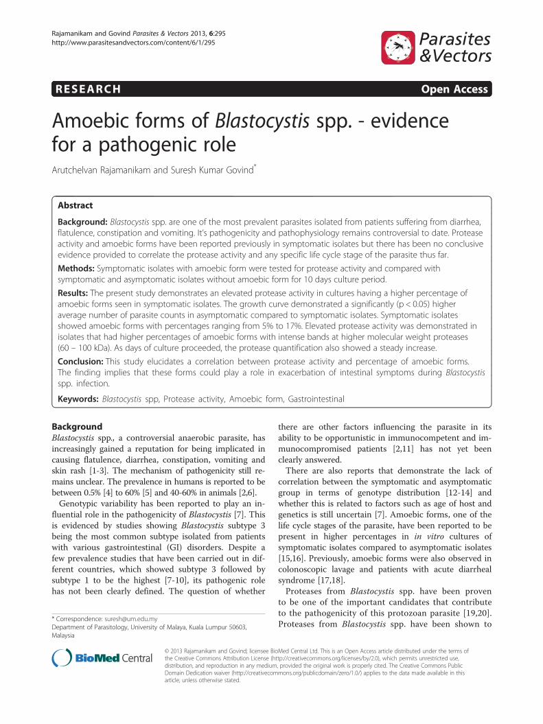
Figure 1 Growth profile of Blastocystis spp. isolated from symptomatnumber of cells is exhibited.
information was obtained using a questionnaire. Sampleswith Blastocystis spp. were selected through direct fecalscreening and cyst concentration technique to select thesamples with only Blastocystis spp. as the sole symptomcausative agent.
Culture and purification of Blastocystis spp.All isolates were inoculated in Jones’ medium sup-plemented with 10% horse serum as described previouslyby Suresh et al. [24]. Cultures were incubated at a con-stant temperature of 37°C before sub-culturing into freshmedium once every 3 days. Subsequently, the cells werepurified from bacterial-contaminated culture using densitygradient centrifugation. The cells were pooled into onetube and washed twice with phosphate buffered saline(PBS) for 5 minutes at 500 g. Five milliliters of the cell sus-pension was then layered carefully onto 6 ml of Ficoll-Paque without agitation. It was then spun for 20 mins at700 g. Blastocystis spp. cells with minimal bacterial con-taminants found above the thick layer of yellowish whiteclump was then gently isolated and washed with PBS. Thepellet was stored at -20°C until further use.
ic (A) and asymptomatic (B) patients. A comparison on the average
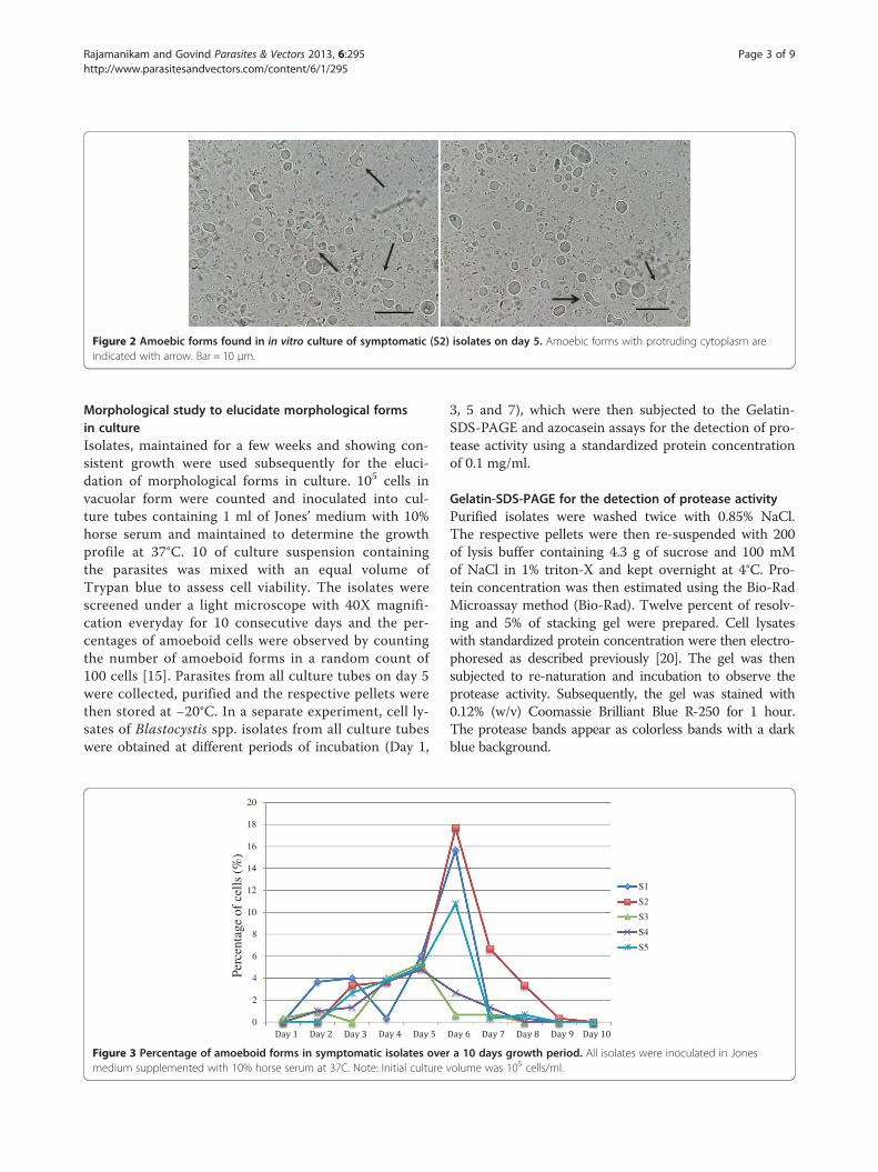
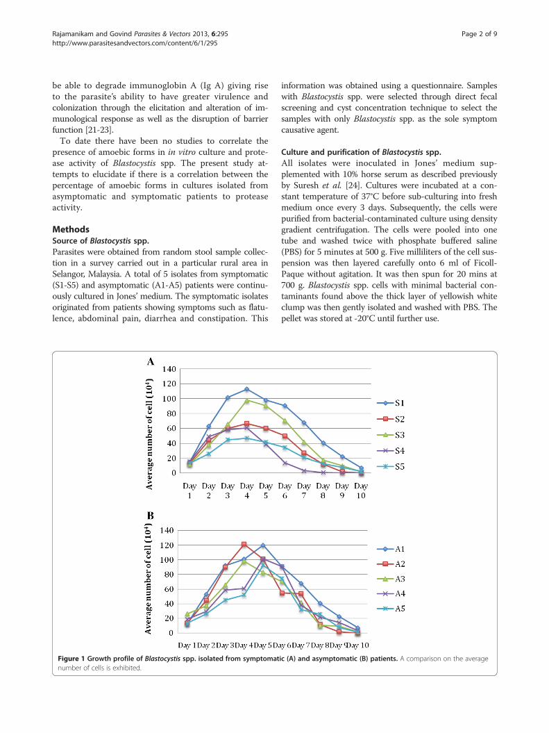
Figure 2 Amoebic forms found in in vitro culture of symptomatic (S2) isolates on day 5. Amoebic forms with protruding cytoplasm areindicated with arrow. Bar = 10 μm.
Rajamanikam and Govind Parasites & Vectors 2013, 6:295 Page 3 of 9http://www.parasitesandvectors.com/content/6/1/295
Morphological study to elucidate morphological formsin cultureIsolates, maintained for a few weeks and showing con-sistent growth were used subsequently for the eluci-dation of morphological forms in culture. 105 cells invacuolar form were counted and inoculated into cul-ture tubes containing 1 ml of Jones’ medium with 10%horse serum and maintained to determine the growthprofile at 37°C. 10 of culture suspension containingthe parasites was mixed with an equal volume ofTrypan blue to assess cell viability. The isolates werescreened under a light microscope with 40X magnifi-cation everyday for 10 consecutive days and the per-centages of amoeboid cells were observed by countingthe number of amoeboid forms in a random count of100 cells [15]. Parasites from all culture tubes on day 5were collected, purified and the respective pellets werethen stored at −20°C. In a separate experiment, cell ly-sates of Blastocystis spp. isolates from all culture tubeswere obtained at different periods of incubation (Day 1,
0
2
4
6
8
10
12
14
16
18
20
Perc
enta
ge o
f ce
lls (
%)
Figure 3 Percentage of amoeboid forms in symptomatic isolates overmedium supplemented with 10% horse serum at 37C. Note: Initial culture
3, 5 and 7), which were then subjected to the Gelatin-SDS-PAGE and azocasein assays for the detection of pro-tease activity using a standardized protein concentrationof 0.1 mg/ml.
Gelatin-SDS-PAGE for the detection of protease activityPurified isolates were washed twice with 0.85% NaCl.The respective pellets were then re-suspended with 200of lysis buffer containing 4.3 g of sucrose and 100 mMof NaCl in 1% triton-X and kept overnight at 4°C. Pro-tein concentration was then estimated using the Bio-RadMicroassay method (Bio-Rad). Twelve percent of resolv-ing and 5% of stacking gel were prepared. Cell lysateswith standardized protein concentration were then electro-phoresed as described previously [20]. The gel was thensubjected to re-naturation and incubation to observe theprotease activity. Subsequently, the gel was stained with0.12% (w/v) Coomassie Brilliant Blue R-250 for 1 hour.The protease bands appear as colorless bands with a darkblue background.
S1
S2
S3
S4
S5
a 10 days growth period. All isolates were inoculated in Jonesvolume was 105 cells/ml.
S1 S2 S3 S4 S5 A1 A2 A3 A4 A5M
60 kDa –
100 kDa –75 kDa –
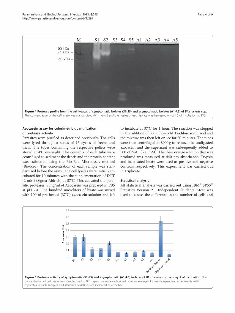
Figure 4 Protease profile from the cell lysates of symptomatic isolates (S1-S5) and asymptomatic isolates (A1-A5) of Blastocystis spp.The concentration of the cell lysate was standardized (0.1 mg/ml) and the lysates of each isolate was harvested on day 5 of incubation at 37C.
Rajamanikam and Govind Parasites & Vectors 2013, 6:295 Page 4 of 9http://www.parasitesandvectors.com/content/6/1/295
Azocasein assay for colorimetric quantificationof protease activityParasites were purified as described previously. The cellswere lysed through a series of 15 cycles of freeze andthaw. The tubes containing the respective pellets werestored at 4°C overnight. The contents of each tube werecentrifuged to sediment the debris and the protein contentwas estimated using the Bio-Rad Microassay method(Bio-Rad). The concentration of each sample was stan-dardized before the assay. The cell lysates were initially in-cubated for 10 minutes with the supplementation of DTT(2 mM) (Sigma-Aldrich) at 37°C. This activated the para-sitic proteases. 5 mg/ml of Azocasein was prepared in PBSat pH 7.4. One hundred microliters of lysate was mixedwith 100 of pre-heated (37°C) azocasein solution and left
0
0.1
0.2
0.3
0.4
0.5
0.6
0.7
Azo
case
in U
nit
Figure 5 Protease activity of symptomatic (S1-S5) and asymptomaticconcentration of cell lysate was standardized to 0.1 mg/ml. Values are obtatriplicates in each samples and standard deviations are indicated as error b
to incubate at 37°C for 1 hour. The reaction was stoppedby the addition of 300 of ice-cold Trichloroacetic acid andthe mixture was then left on ice for 30 minutes. The tubeswere then centrifuged at 8000 g to remove the undigestedazocasein and the supernant was subsequently added to500 of NaCl (500 mM). The clear orange solution that wasproduced was measured at 440 nm absorbance. Trypsinand inactivated lysate were used as positive and negativecontrols respectively. This experiment was carried outin triplicate.
Statistical analysisAll statistical analysis was carried out using IBM© SPSS©
Statistics Version 21. Independent Students t-test wasused to assess the difference in the number of cells and
(A1-A5) isolates of Blastocystis spp. on day 5 of incubation. Theined from an average of three independent experiments withars.
Rajamanikam and Govind Parasites & Vectors 2013, 6:295 Page 5 of 9http://www.parasitesandvectors.com/content/6/1/295
protease activity between symptomatic isolates withamoebic forms, symptomatic isolates without amoebicforms and asymptomatic isolates. Pearson Correlation Testwas used to assess the correlation between protease activityand percentage of amoebic forms. A value of p < 0.05 isconsidered statistically significant.
75 kDa –
M D1
M D1
M D1
60 kDa –
100 kDa –
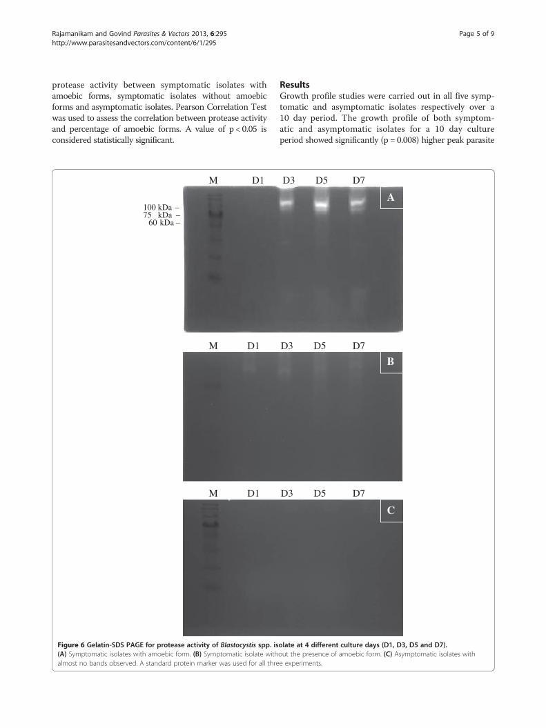
Figure 6 Gelatin-SDS PAGE for protease activity of Blastocystis spp. is(A) Symptomatic isolates with amoebic form. (B) Symptomatic isolate withalmost no bands observed. A standard protein marker was used for all thre
ResultsGrowth profile studies were carried out in all five symp-tomatic and asymptomatic isolates respectively over a10 day period. The growth profile of both symptom-atic and asymptomatic isolates for a 10 day cultureperiod showed significantly (p = 0.008) higher peak parasite
B
D3 D5 D7
D3 D5 D7
D3 D5 D7
A
C
olate at 4 different culture days (D1, D3, D5 and D7).out the presence of amoebic form. (C) Asymptomatic isolates withe experiments.
Rajamanikam and Govind Parasites & Vectors 2013, 6:295 Page 6 of 9http://www.parasitesandvectors.com/content/6/1/295
counts in asymptomatic isolates on day 5 to day 7(Figure 1). All symptomatic isolates (S1 to S5) showedamoebic forms over the 10 day culture period with a sizerange between 2.9 – 8.0 μm (Figure 2). Amoebic formswere first observed on day 3 with cells showing multipleextended irregular shaped cytoplasm and a prominent nu-cleus. These forms persisted until day 7 (S1, S3 and S5) today 8 (S2 and S4) with the percentage ranging from 5% to17% (Figure 3). Higher percentages of amoebic form wereobserved in S2 (18%) followed by S1 and S5, which was16% and 11% respectively. S3 and S4 showed the leastnumber of amoebic forms. Asymptomatic isolates showedno amoebic forms in culture but had a higher parasitecount with forms being mostly vacuolar followed by granu-lar (data not shown).Protease activity using the azocasein assay and gelatin-
SDS-PAGE demonstrated higher activity in symptomaticisolates with the highest observed in S2 followed by S1,S5, S4 and S3. Asymptomatic isolates demonstrated aconsistently lower activity of protease in all five isolates.Elevated protease activity was demonstrated in isolates
that exhibited higher percentages of amoebic forms withintense bands (Figure 4). The variation in the intensity ofthe band was observed only with higher molecular weightproteases (60 – 100 kDa). Protease banding patterns incurrent protease zymography showed a similar outcome asin the azocasein assay (Figure 5). The bands were more in-tense in S1, and S2 whereas the bands in S3, S4 and S5showed less intense bands. However, only high molecularweight protease bands were observed to correlate with thepercentage of amoebic forms seen in culture. Lower mo-lecular weight protease did not show much correlation andan intense band was seen only in isolate S4 at 28–17 kDa.As days of culture preceded the protease quantifica-
tion (Figure 6) also showed a steady increase in the
0
0.1
0.2
0.3
0.4
0.5
0.6
Day 1 Day 3 Day 5 Day 7 PosCo
Azo
case
in U
nit
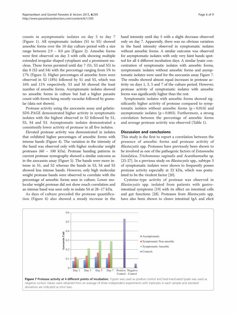
Figure 7 Protease activity at 4 different points of incubation. Trypsin wnegative control. Values were obtained from an average of three independdeviations are indicated as error bars.
band intensity until day 5 with a slight decrease observedonly on day 7. Apparently, there was no obvious variationin the band intensity observed in symptomatic isolateswithout amoebic forms. A similar outcome was observedfor asymptomatic isolates with only very faint bands spot-ted for all 4 different incubation days. A similar lysate con-centration of symptomatic isolates with amoebic forms,symptomatic isolates without amoebic forms and asymp-tomatic isolates were used for the azocasein assay Figure 7.The results showed almost equal increases in protease ac-tivity on days 1, 3, 5 and 7 of the culture period. However,protease activity of symptomatic isolates with amoebicforms was significantly higher than the rest.Symptomatic isolates with amoebic forms showed sig-
nificantly higher activity of protease compared to symp-tomatic isolates without amoebic forms (p = 0.014) andasymptomatic isolates (p = 0.003). Furthermore, a strongcorrelation between the percentage of amoebic formsand average protease activity was observed (Table 1).
Discussion and conclusionsThis study is the first to report a correlation between thepresence of amoebic forms and protease activity ofBlastocystis spp. Proteases have previously been shown tobe involved as one of the pathogenic factors of Entamoebahistolytica, Trichomonas vaginalis and Acanthamoeba sp.[25-27]. In a previous study on Blastocystis spp., subtype 3of symptomatic isolates were shown to frequently possesprotease activity especially at 32 kDa, which was postu-lated to be the virulent factor [20].Cysteine-type activity of protease was observed in
Blastocystis spp. isolated from patients with gastro-intestinal symptoms [19] with its effect on intestinal cellsand gut functions [28]. Proteases from Blastocystis spp.have also been shown to cleave intestinal IgA and elicit
itiventrol
NegativeControl
Asymptomatic
Symptomatic Non-amoebic
Symptomatic Amoebic
Controls
as used as positive control and heat-inactivated lysate was used asent experiments with triplicates in each sample and standard
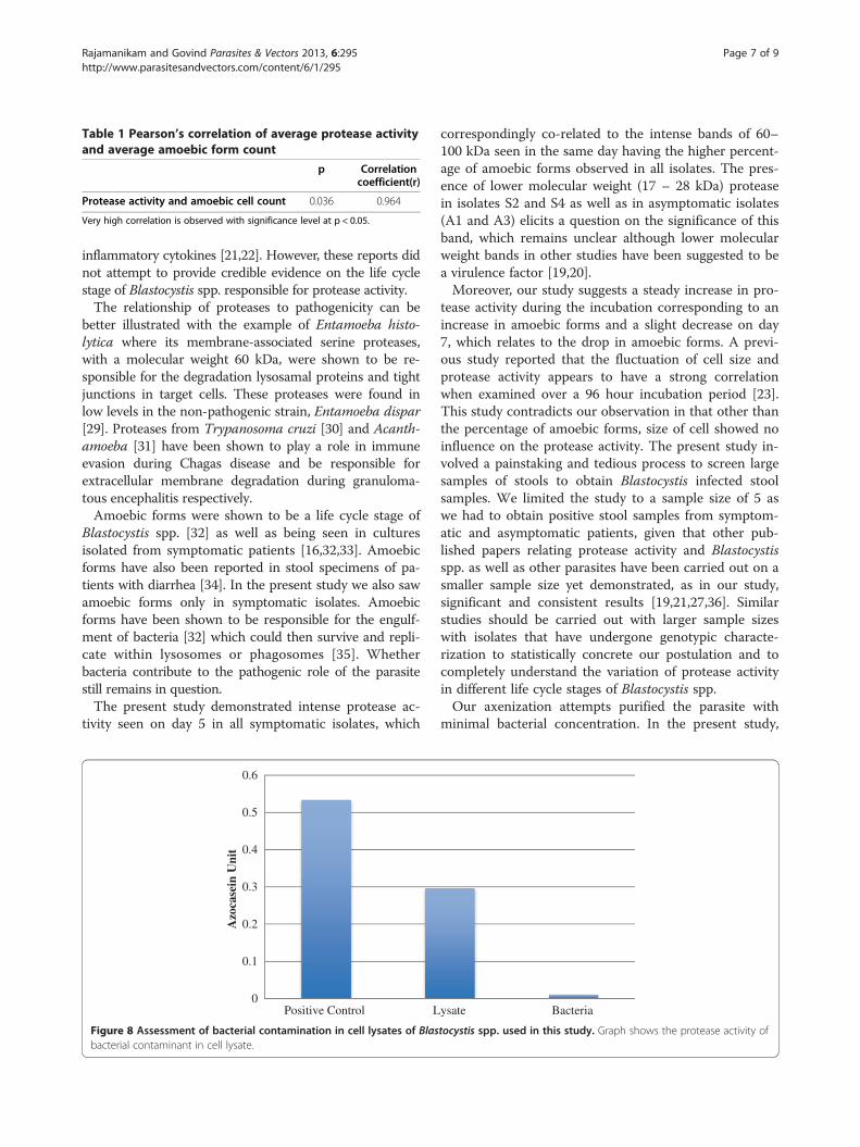
Table 1 Pearson’s correlation of average protease activityand average amoebic form count
p Correlationcoefficient(r)
Protease activity and amoebic cell count 0.036 0.964
Very high correlation is observed with significance level at p < 0.05.
Rajamanikam and Govind Parasites & Vectors 2013, 6:295 Page 7 of 9http://www.parasitesandvectors.com/content/6/1/295
inflammatory cytokines [21,22]. However, these reports didnot attempt to provide credible evidence on the life cyclestage of Blastocystis spp. responsible for protease activity.The relationship of proteases to pathogenicity can be
better illustrated with the example of Entamoeba histo-lytica where its membrane-associated serine proteases,with a molecular weight 60 kDa, were shown to be re-sponsible for the degradation lysosamal proteins and tightjunctions in target cells. These proteases were found inlow levels in the non-pathogenic strain, Entamoeba dispar[29]. Proteases from Trypanosoma cruzi [30] and Acanth-amoeba [31] have been shown to play a role in immuneevasion during Chagas disease and be responsible forextracellular membrane degradation during granuloma-tous encephalitis respectively.Amoebic forms were shown to be a life cycle stage of
Blastocystis spp. [32] as well as being seen in culturesisolated from symptomatic patients [16,32,33]. Amoebicforms have also been reported in stool specimens of pa-tients with diarrhea [34]. In the present study we also sawamoebic forms only in symptomatic isolates. Amoebicforms have been shown to be responsible for the engulf-ment of bacteria [32] which could then survive and repli-cate within lysosomes or phagosomes [35]. Whetherbacteria contribute to the pathogenic role of the parasitestill remains in question.The present study demonstrated intense protease ac-
tivity seen on day 5 in all symptomatic isolates, which
0
0.1
0.2
0.3
0.4
0.5
0.6
Positive Control L
Azo
case
in U
nit
Figure 8 Assessment of bacterial contamination in cell lysates of Blasbacterial contaminant in cell lysate.
correspondingly co-related to the intense bands of 60–100 kDa seen in the same day having the higher percent-age of amoebic forms observed in all isolates. The pres-ence of lower molecular weight (17 – 28 kDa) proteasein isolates S2 and S4 as well as in asymptomatic isolates(A1 and A3) elicits a question on the significance of thisband, which remains unclear although lower molecularweight bands in other studies have been suggested to bea virulence factor [19,20].Moreover, our study suggests a steady increase in pro-
tease activity during the incubation corresponding to anincrease in amoebic forms and a slight decrease on day7, which relates to the drop in amoebic forms. A previ-ous study reported that the fluctuation of cell size andprotease activity appears to have a strong correlationwhen examined over a 96 hour incubation period [23].This study contradicts our observation in that other thanthe percentage of amoebic forms, size of cell showed noinfluence on the protease activity. The present study in-volved a painstaking and tedious process to screen largesamples of stools to obtain Blastocystis infected stoolsamples. We limited the study to a sample size of 5 aswe had to obtain positive stool samples from symptom-atic and asymptomatic patients, given that other pub-lished papers relating protease activity and Blastocystisspp. as well as other parasites have been carried out on asmaller sample size yet demonstrated, as in our study,significant and consistent results [19,21,27,36]. Similarstudies should be carried out with larger sample sizeswith isolates that have undergone genotypic characte-rization to statistically concrete our postulation and tocompletely understand the variation of protease activityin different life cycle stages of Blastocystis spp.Our axenization attempts purified the parasite with
minimal bacterial concentration. In the present study,
ysate Bacteria
tocystis spp. used in this study. Graph shows the protease activity of
Rajamanikam and Govind Parasites & Vectors 2013, 6:295 Page 8 of 9http://www.parasitesandvectors.com/content/6/1/295
lysates were extracted from purified xenic culture iso-lates. This was thought to mimic an environment similarto the natural intestinal microflora [15]. We have alsoshown that cell lysates with bacterial proteins had min-imal effect since the bacterial protein demonstrated min-imal protease activity (Figure 8).The peak parasite count (parasite count between day
5 – 7) was used as one of the parameters in a previ-ous study to differentiate the phenotypic characteristicsof Blastocystis spp. It has been reported that asymptomaticisolates grew faster and the average total number of cellsreached up to 7 times more than symptomatic isolate. Thegeneration time was twice as high in asymptomatic iso-lates [37]. The present study concurred with these findingsin that the asymptomatic isolates showed higher parasitepeak counts than symptomatic ones (Figure 2). It is there-fore evident that protease activity did not arise despite thehigher parasite count consisting of mostly vacuolar andgranular forms seen in asymptomatic isolates but from thelarger percentage of amoebic forms seen in symptomaticisolates.The amoebic form of Blastocystis spp. has been postu-
lated previously to posses a sticky surface, which en-hances the adherence to the intestinal epithelial celllining [16]. This adherence could facilitate the release ofproteases from amoebic forms to degrade extracellularmembrane similar to that as seen in Entamoeba histolytica[38]. Higher occurrence of Blastocystis spp. has been seenin cancer patients undergoing chemotherapy [39]. Exacer-bation of symptoms in such situations could be attributedto vacuolar stages reverting to amoebic forms, which inturn could be causing the related symptoms due to the re-lease of proteases.In conclusion, this is the first study to provide evi-
dence co-relating amoebic forms of Blastocystis spp. andprotease activity, which suggests a strong pathophysio-logical role that amoebic forms can play in exacerbatingsymptoms in patients infected with Blastocystis spp.
Ethical approvalThis study was approved by the Medical Ethics Commit-tee of the University Malaya Medical Centre (UMMC)(Kuala Lumpur, Malaysia) according to the Declarationof Helsinki approved this study.
Competing interestBoth authors declared that they have no competing interest.
Authors’ contributionAR and SKG were involved in the intellectual planning of the experiment;SKG designed the study; AR carried out the experimental work; AR and SKGanalysed the results and wrote the paper. Both authors read and approvedthe final manuscript.
AcknowledgementsWe would like to extend our gratitude and heartfelt thanks to our lab colloguesand all the staff in the Department of Parasitology, University of Malaya.
FundingThis study was entirely supported by Postgraduate Research Grant fromUniversity of Malaya (PG108-2012B) and High Impact Research Grant (HIR)(UM.C/625/1/HIR/031).
Received: 3 October 2013 Accepted: 3 October 2013Published: 11 October 2013
References1. Hussainqadri SM, Al-Okaili GA, Al-Dayel F: Clinical significance of
Blastocystis hominis. J Clin Microbiol 1989, 27:2407–2409.2. Tan KSW: New insights on classification, identification, and clinical
relevance of Blastocystis spp. Clin Microbiol Rev 2008, 21:639–665.3. Stenzel DJ, Boreham PFL: Blastocystis hominis revisited. Clin Microbiol Rev
1996, 9:563–584.4. Horiki N, Maruyama M, Fujita Y, Yonekura T, Minato S, Kaneda Y:
Epidemiologic survey of Blastocystis hominis infection in Japan. Am JTrop Med Hyg 1997, 56:370–374.
5. Pegelow K, Gross R, Pietrzik K, Lukito W, Richards A, Fryauff DJ:Parasitological and nutritional situation of school children in theSukaraja district, West Java. Indonesia. Southeast Asian J Trop Med PublicHealth 1997, 28:173–190.
6. Tan TC, Tan PC, Sharma R, Sugnaseelan S, Suresh KG: Genetic diversityof caprine Blastocystis from Peninsular Malaysia. Parasitol Res 2012,112:85–89.
7. Souppart L, Sanciu G, Cian A, Wawrzyniak I, Delbac F, Capron M, Dei-Cas E,Boorom K, Delhaes L, Viscogliosi E: Molecular epidemiology of humanBlastocystis isolates in France. Parasitol Res 2009, 105:413–421.
8. Dogruman-Al F, Dagci H, Yoshikawa H, Kurt Ö, Demirel M: A possible linkbetween subtype 2 and asymptomatic infections of Blastocystis hominis.Parasitol Res 2008, 103:685–689.
9. Li LH, Zhang XP, Lv S, Zhang L, Yoshikawa H, Wu Z, Steinmann P, Utzinger J,Tong XM, Chen SH, Zhou XN: Cross-sectional surveys and subtypeclassification of human Blastocystis isolates from four epidemiologicalsettings in China. Parasitol Res 2007, 102:83–90.
10. Kaneda Y, Horiki N, Cheng X-J, Fujita Y, Maruyama M, Tachibana H:Ribodemes of Blastocystis hominis isolated in japan. Am J Trop Med Hyg2001, 65:393–396.
11. Moosavi A, Haghighi A, Mojarad EN, Zayeri F, Alebouyeh M, Khazan H,Kazemi B, Zali MR: Genetic variability of Blastocystis sp. isolated fromsymptomatic and asymptomatic individuals in Iran. Parasitol Res 2012,111:2311–2315.
12. Böhm-Gloning B, Knobloch J, Walderich B: Five subgroups of Blastocystishominis from symptomatic and asymptomatic patients revealed byrestriction site analysis of PCR-amplified 16S-like rDNA. Trop Med IntHealth 1997, 2:771–778.
13. Yoshikawa H, Abe N, Wu Z: PCR-based identification of zoonotic isolatesof Blastocystis from mammals and birds. Microbiology 2004, 150:1147–1151.
14. Özyurta M, Kurtb Ö, Mølbakc K, Nielsend HV, Haznedaroglua T, Stensvold CR:Molecular epidemiology of Blastocystis infections in Turkey. Parasitol Int 2008,57:300–306.
15. Tan TC, Suresh KG: Predominance of amoeboid forms of Blastocystishominis in isolates from symptomatic patients. Parasitol Res 2006,98:189–193.
16. Tan TC, Suresh KG: Amoeboid form of Blastocystis hominis—a detailedultrastructural insight. Parasitol Res 2006, 99:737–742.
17. Stenzel DJ, Boreham PFL, McDougall R: Ultrastructure of Blastocystishominis in human stool samples. Int J Parasitol 1991, 21:807–812.
18. Lanuza M, Carbajal J, Villar J, Borrás R: Description of an improved methodfor Blastocystis hominis culture and axenization. Parasitol Res 1997,83:60–63.
19. Sio SWS, Puthia MK, Lee ASY, Lu J, Tan KSW: Protease activity ofBlastocystis hominis. Parasitol Res 2006, 99:126–130.
20. Abdel-Hameed DM, Hassanin OM: Proteaese activity of Blastocystis hominissubtype3 in symptomatic and asymptomatic patients. Parasitol Res 2011,109:321–327.
21. Puthia MK, Lu J, Tan KSW: Blastocystis ratti contains cysteine proteasesthat mediate interleukin-8 response from human intestinal epithelialcells in an NF-kB-dependent manner. Eukaryot Cell 2008, 7:435–443.
22. Puthia MK, Vaithilingam A, Lu J, Tan KSW: Degradation of human secretoryimmunoglobulin a by Blastocystis. Parasitol Res 2005, 97:386–389.
Rajamanikam and Govind Parasites & Vectors 2013, 6:295 Page 9 of 9http://www.parasitesandvectors.com/content/6/1/295
23. Mirza H, Tan KSW: Blastocystis exhibits inter- and intra-subtype variationin cystiene protease. Parasitol Res 2009, 104:355–361.
24. Suresh K, Ng GC, Ho LC, Yap EH, Singh M: Differentiation of the variousstages of Blastocystis hominis by acridine orange staining. Int J Parasitol1994, 24:605–606.
25. Que X, Reed SL: Cysteine proteinases and the pathogenesis of amebiasis.Clin Microbiol Rev 2000, 13:196–206.
26. Yadav M, Dubey ML, Gupta I, Bhatti G, Malla N: Cysteine proteinase 30 inclinical isolates of T. vaginalis from symptomatic and asymptomaticinfected women. Exp Parasitol 2007, 116:399–406.
27. Khan NA, Jarroll EL, Panjwani N, Cao Z, Paget TA: Proteases as markers fordifferentiation of pathogenic and nonpathogenic species ofacanthamoeba. J Clin Microbiol 2000, 38:2858–2861.
28. Wawrzyniak I, Texier C, Poirier P, Viscogliosi E, Tan KSW, Delbac F, Alaoui HE:Characterization of two cysteine proteases secreted by Blastocystis ST7, ahuman intestinal parasite. Parasitol Int 2012, 61:437–442.
29. Barrios-Ceballos MP, Martínez-Gallardo NA, Anaya-Velázquez F, Mirelman D,Padilla-Vaca F: A novel protease from Entamoeba histolytica homologousto members of the family S28 of serine proteases. Exp Parasitol 2005,110:270–275.
30. Doyle PS, Zhou YM, Hsieh I, Greenbaum CD, McKerrow JH, Engel JC:The trypanosoma cruzi protease cruzain mediates immune evasion.PLoS Pathog 2011, 7:1–11.
31. Sissons J, Alsam S, Goldsworthy G, Lightfoot M, Jarroll EL, Khan NA:Identification and properties of proteases from an Acanthamoeba isolatecapable of producing granulomatous encephalitis. BMC Microbiol 2006,6:42–50.
32. Singh M, Suresh K, Ho LC, Ng GC, Yap EH: Elucidation of the life cycle ofthe intestinal protozoan Blastocystis hominis. Parasitol Res 1995, 81:446–450.
33. He N, Zhang Y, Hong M, Cong M: Morphological and ultrastructuralobservation of Blastocystis hominis. Zhongguo Ji Sheng Chong Xue Yu JiSheng Chong Bing Za Zhi 2001, 19:169–172.
34. Zhang X, Zhang S, Qiao J, Wu X, Zhao L, Liu Y, Fan X: Ultrastructuralinsights into morphology and reproductive mode of Blastocystis hominis.Parasitol Res 2012, 110:1165–1172.
35. Scanlan PD: Blastocystis: past pitfalls and future perspectives. TrendsParasitol 2012, 28:327–334.
36. Carvalho TB, David EB, Coradi ST, Guimarães S: Protease activity inextracellular products secreted in vitro by trophozoites of Giardiaduodenalis. Parasitol Res 2008, 104:185–190.
37. Tan TC, Suresh KG, Smith HV: Phenotypic and genotypic characterisationof Blastocystis hominis isolates implicates subtype 3 as a subtype withpathogenic potential. Parasitol Res 2008, 104:85–93.
38. Schulte W, Scholze H: Action of the major protease from Entamoebahistolytica on proteins of the extracellular matrix. J Protozool 1989,36:538–543.
39. Samudi C, Kumar S, Anitab ZB, Kuppusamy UR: Infections of Blastocystishominis and microsporidia in cancer patients: are they opportunistic?Trans R Soc Trop Med Hyg 2012, 106:267–269.
doi:10.1186/1756-3305-6-295Cite this article as: Rajamanikam and Govind: Amoebic forms ofBlastocystis spp. - evidence for a pathogenic role. Parasites & Vectors2013 6:295.
Submit your next manuscript to BioMed Centraland take full advantage of:
• Convenient online submission
• Thorough peer review
• No space constraints or color figure charges
• Immediate publication on acceptance
• Inclusion in PubMed, CAS, Scopus and Google Scholar
• Research which is freely available for redistribution
Submit your manuscript at www.biomedcentral.com/submit