adar-mediated rna.pdf - idus
TRANSCRIPT
ARTICLE
ADAR-mediated RNA editing of DNA:RNA hybridsis required for DNA double strand break repairSonia Jimeno1,2,6✉, Rosario Prados-Carvajal1,2,6, María Jesús Fernández-Ávila 2, Sonia Silva 1,2,
Domenico Alessandro Silvestris3, Martín Endara-Coll4, Guillermo Rodríguez-Real1,2, Judit Domingo-Prim 4,5,
Fernando Mejías-Navarro 1,2, Amador Romero-Franco1,2, Silvia Jimeno-González 1,2, Sonia Barroso 1,2,
Valeriana Cesarini3, Andrés Aguilera 1,2, Angela Gallo3, Neus Visa 4 & Pablo Huertas 1,2✉
The maintenance of genomic stability requires the coordination of multiple cellular tasks
upon the appearance of DNA lesions. RNA editing, the post-transcriptional sequence
alteration of RNA, has a profound effect on cell homeostasis, but its implication in the
response to DNA damage was not previously explored. Here we show that, in response to
DNA breaks, an overall change of the Adenosine-to-Inosine RNA editing is observed, a
phenomenon we call the RNA Editing DAmage Response (REDAR). REDAR relies on the
checkpoint kinase ATR and the recombination factor CtIP. Moreover, depletion of the RNA
editing enzyme ADAR2 renders cells hypersensitive to genotoxic agents, increases genomic
instability and hampers homologous recombination by impairing DNA resection. Such a role
of ADAR2 in DNA repair goes beyond the recoding of specific transcripts, but depends on
ADAR2 editing DNA:RNA hybrids to ease their dissolution.
https://doi.org/10.1038/s41467-021-25790-2 OPEN
1 Departamento de Genética, Universidad de Sevilla, Sevilla 41080, Spain. 2 Centro Andaluz de Biología Molecular y Medicina Regenerativa-CABIMER,Universidad de Sevilla-CSIC-Universidad Pablo de Olavide, Sevilla 41092, Spain. 3 RNA Editing Lab, Oncohaematology Department, IRCCS OspedalePediatrico “Bambino Gesù”, Viale San Paolo 15, 00146 Rome, Italy. 4 Department of Molecular Biosciences, The Wenner-Gren Institute, Stockholm University,10691 Stockholm, Sweden. 5Present address: Moirai Biodesign SL, Parc Científic de Barcelona, 08028 Barcelona, Spain. 6These authors contributed equally:Sonia Jimeno, Rosario Prados-Carvajal. ✉email: [email protected]; [email protected]
NATURE COMMUNICATIONS | (2021) 12:5512 | https://doi.org/10.1038/s41467-021-25790-2 |www.nature.com/naturecommunications 1
1234
5678
90():,;
Cells are continuously challenged by DNA damage. Amongall kinds of insults that a DNA molecule has to deal with,double-strand breaks (DSBs) are the most dangerous.
Indeed, just one unrepaired DSB is enough to either kill orterminally arrest cells. For these reasons when DSBs are formed, acomplex cellular response—the DNA damage response (DDR)—is triggered in order to ensure the proper repair of such a threat togenomic integrity1.
There are several pathways that can be used in order to repair aDSB and the choice between them is highly regulated. A eukar-yotic cell can repair a DSB either by the simple re-ligation of theDNA ends (a process known as Non-Homologous End-Joining,NHEJ)2 or by a homology-driven repair event. There are differentroutes among the repair pathways that use homologous regionsfor repair, all of which are grouped in a process called homo-logous recombination (HR)3. All HR events share a first bio-chemical step called DNA resection, which is the key to decide thepathway that will be eventually used to repair the DSB4,5. Thisprocess consists of the nucleolytic degradation of the DNA endsof the break that produces tails of 3' ended single-stranded DNA(ssDNA), that are rapidly protected by the RPA protein complex.
In recent years, the importance of RNA and RNA-relatedfactors in DNA repair has become clear6–9. Indeed, many RNA-related proteins have been shown to be targets of the DNAdamage-induced post-translational modifications10–12. Also,direct roles of specific RNA-related factors in DNA repair havebeen recently reported (for a review see9). Moreover, the RNAmolecule itself seems to impact DNA repair. Several labs haveshown the formation of DNA:RNA hybrids around DSBs indifferent eukaryotes, either dependent on previoustranscription13,14 or upon de novo transcription of the brokenchromatin15,16. The relevance of such RNA molecules is stillunder debate, with both pro- and anti-repair effects ascribed tothem9.
An important co-transcriptional RNA modification that, sofar, has not been extensively studied in its putative relationshipwith DNA repair and the response to DNA damage is RNAediting. This process alters RNA sequences by the action ofspecific deaminases that convert one base into another. Everymammalian transcript can be subjected to RNA editing17–19.RNA editing can be classified into several categories20, includingadenosine-to-inosine (A-to-I) deamination, which is accom-plished by a family of RNA-specific adenosine deaminasesknown as ADARs18,19. This family is formed by ADAR1,ADAR2 (also known as ADARB1), and ADAR3; however, onlyADAR1 and ADAR2 have been shown to present catalyticactivity. A-to-I deamination is the most abundant form of RNA-editing in mammals and defects in this process are associatedwith human diseases, such as disorders of the central nervoussystem21 or pediatric astrocytomas22. Only limited informationhas been published regarding the connection of A-to-I editingand DNA damage, albeit at least the mRNA of NEIL1, has beenshown to be re-coded by ADAR1 to alter its enzymaticproperties23. Moreover, A-to-I editing has been proposed to beinvolved in the pathogenesis of cancer24,25.
Here, we show that the general pattern of ADAR2-mediated A-to-I editing changes upon DSB formation. Such changes dependon the DDR, specifically the ATR kinase and the resection proteinCtIP. As a consequence, ADAR2 is required for the maintenanceof genomic integrity and, specifically for DNA end resection andHR. Strikingly, mRNAs from either resection-related orrecombination-related genes are not affected by ADAR2. Instead,ADAR2 role in resection is related to its ability to edit DNA:RNAhybrids. Not only do such structures increase when ADAR2 isdepleted, but this protein physically and functionally interactswith the BRCA1-SETX complex for this role.
ResultsRNA editing changes after DNA damage. As previously men-tioned, crosstalk between RNA metabolism and DNA repair hasbeen extensively documented9, but a connection between DNArepair and RNA editing has not been extensively analyzed. Thus,we wanted to study whether the appearance of DNA damage hadany effect on RNA editing. In order to explore this possibility, weused a previously published reporter system (RNAG) that mea-sures levels of RNA editing using the accumulation of thefluorescent proteins GFP and RFP26. This system bears both theRFP and GFP ORFs in a single transcript, with a stop codonbetween them (Fig. 1A). So, cells bearing such reporter expressRFP constitutively, but GFP is only produced if an RNA editingevent changes the A of the stop codon to an I (Fig. 1A)26.Therefore, the number of red cells that are also green indicates theefficiency of RNA editing. As a control to discard other effectsnon-related to the editing on this system, we used the RNWGcontrol reporter, in which the stop codon is pre-edited, so all cellsbearing the construct fluoresce, indeed, in red and green26. InU2OS cells stably transfected with the reporter, we observed thatDNA damage induced by ionizing radiation increased GFPexpression by 50% specifically in the RNAG reporter and not inthe RNWG control, in agreement with a DNA damage stimula-tion of RNA A-to-I editing in this system (Fig. 1B). Similar resultswere obtained when using the DNA damage-inducing drugcamptothecin (Fig. 1C), where we could observe a dose-dependent effect on RNA editing stimulation. One possibility isthat DNA damage induces the accumulation of the A-to-I editingmachinery, namely ADAR1 and ADAR2 enzymes, thus increas-ing this process. However, neither of these proteins was upre-gulated, but were slightly downregulated, upon exposure to IR(Supplementary Fig. 1A). Then, in order to confirm this was acanonical induction of RNA editing, we depleted the A-to-Iediting machinery. To choose which member of the ADAR familyto downregulate, we revisited the data we obtained in a previousgenome-wide screening for factors that unbalance the choicebetween DSB repair pathways27. Interestingly, both ADAR1 andADAR2, but not the catalytically inactive ADAR3, skewed DSBrepair towards end-joining (Supplementary Figure 1B), and thiswas not due to changes in the cell cycle (Supplementary Fig. 1C).However, the effect was more prominent and clearer uponADAR2 depletion. Indeed, downregulation of ADAR2 severelycompromised both the basal and the DNA damage-inducedexpression of the GFP in the RNAG (Fig. 1D; for ADAR2depletion efficiency see Supplementary Fig. 2A), but, as expected,not in the RNWG control reporter (Supplementary Fig. 2B).
To better understand this phenomenon, we decided to look forDDR factors that affected the DNA damage-induced RNAediting. Recently, we have found that CtIP, a core DNA endresection factor that is also required for ATR activation, playsadditional roles in DNA damage-induced RNA splicing28.Interestingly, we could see that CtIP downregulation specificallyeliminated the DNA damage-dependent induction of RNAediting without affecting the basal levels (Fig. 1D). Again, CtIPdepletion did not alter GFP levels in the control RNWG system(Supplementary Fig. 2B). We could also complement this effectwith the expression of siRNA-resistant flag-tagged CtIP in CtIPdepleted cells, to the same extent as the control cells transfectedwith a non-targeting siRNA, even though overexpression ofFLAG-CtIP on its own reduced the intensity of this DNAdamage-induced phenotype (Supplementary Fig. 2C).
The general response to DNA damage is mainly controlled bythe activation of two related apical kinases, ATM and ATR1.Thus, we also tested if any of them was required for the inductionof RNA editing upon irradiation. Interestingly, ATM inhibitiondid not affect DNA damage-induced RNA editing, while ATR
ARTICLE NATURE COMMUNICATIONS | https://doi.org/10.1038/s41467-021-25790-2
2 NATURE COMMUNICATIONS | (2021) 12:5512 | https://doi.org/10.1038/s41467-021-25790-2 | www.nature.com/naturecommunications

inhibition decreased the DNA damage-induced editing increase(Fig. 1E). This agrees with the notion that ATR and CtIP act onthe same branch of the DNA damage checkpoint in response toDSBs29. The lack of response with the ATM inhibitor could beexplained by a compensation by another member of the PIKKfamily, most likely DNA-PK. Along those lines, the ATR effectcould also be affected by this phenomenon. Thus, we repeated the
experiment with ATM, ATR, and DNA-PK inhibitors in differentcombinations (Fig. 1F). As shown, inhibition of ATR suppressedthe DNA damage-induction of RNA editing, regardless of thepresence of the inhibitors of ATM or DNA-PK. Interestingly,chemical inhibition of DNA-PK showed a limited increase in thebasal levels of RNA editing, but importantly the exposure to DNAdamage still provoked a hyperactivation of the process. Notably,
A B
C
RNA Editing
GA
UNLS GFPRFP
GIU
NLS GFPRFP
*
RNWG
RNAG0.0
0.5
1.0
1.5
2.0
% R
ed c
ells
pos
itive
for G
FP
nor
mal
ized
to u
ntre
ated
cel
ls
-IR +IR
RNWG
RNAG0.5
1.0
1.5
2.0
0,001 mM0,005 mM0,05 mM0,1 mM
0 mM
% R
ed c
ells
pos
itive
for G
FP
nor
mal
ized
to D
MS
O
siNT
siCtIP
siADAR2
0
5
10
15
20
25
% R
ed c
ells
pos
itive
for G
FP
******
*
-IR +IR
*
D
- IR
+IR
0
5
10
15
% R
ed c
ells
pos
it ive
for G
FP
**
***
* *
DMSO
ATRi
ATMi
E
DMSO
ATRi
ATMi
DNA-PKi
ATRi + A
TMi
ATRi + D
NA-PKi
ATMi +
DNA-P
Ki0
5
10
15
20
25
*
*
**
% R
ed c
ells
pos
itive
for
GF
P
F
-IR +IR
Fig. 1 DNA damage increases RNA editing. A Scheme of the RNAG editing system. A bi-cistronic mRNA containing the RFP and GFP sequences isproduced. The presence of a stop codon impedes the expression of the GFP ORF, except when the adenine is edited to inosine. The presence of asecondary structure containing such stop codon allows its recognition and deamination of the adenine by ADAR proteins. B DNA damage-induced RNAediting. The plot shows the percentage of cells bearing the RNAG reporter or the constitutively edited RNWG system that express both the RFP (red cells)and GFP (green cells). Cells were either irradiated (+IR; 10 Gy; black bars) or mock-treated (–IR; white bars) and incubated for 12 h. The percentage ofgreen cells over 10.000 red cells were analyzed on BD FACSAriaTM using FACSDiva v5.0.3 software. For each reporter, the ratio of green and red cellswas normalized with the untreated conditions. Statistical significance was determined with a two-tailed paired Student’s t-test. C Same as B, but cells weretreated with the indicated concentration of camptothecin (CPT). D Cells bearing the RNAG reporter were transfected with the indicated siRNAs andirradiated or not, and the percentage of red cells that were also green is plotted. Statistical significance was determined with a two-way ANOVA. E Same asD, but cells were pretreated for 2 h with 10 μM of inhibitors of ATM (ATMi), ATR (ATRi), or DMSO as control, previous to the irradiation. Cells werecollected to check for editing levels 10 h after irradiation. The inhibitors were kept for the duration of the experiment. Statistical significance wasdetermined with a two-way ANOVA. F Same as E, but, cells were also treated with DNA–PK inhibitor (DNA–PKi), as well as the double combinations of theATM, ATR, and DNA–PK inhibitors, as indicated. Statistical significance was determined with a two-way ANOVA. The average and the standard deviationof the medians of four (panels B and F) or three (panels C–E) independent experiments are shown. Each individual replica is marked with a colored symbol.One, two, or three asterisks represent p < 0.05, p < 0.01, or p < 0.001, respectively. Actual p-values can be found in the Source data file. Only biologicalrelevant comparisons are shown.
NATURE COMMUNICATIONS | https://doi.org/10.1038/s41467-021-25790-2 ARTICLE
NATURE COMMUNICATIONS | (2021) 12:5512 | https://doi.org/10.1038/s41467-021-25790-2 |www.nature.com/naturecommunications 3
concomitant inhibition of both DNA-PK and ATM abolished theinduction of RNA editing caused by irradiation. Thus, it seemsthat those two kinases could have an overlapping role in thisphenomenon.
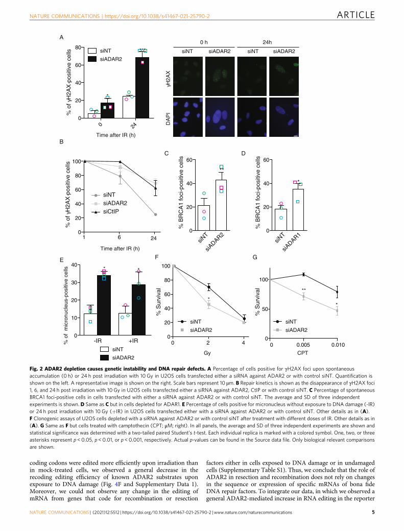
ADAR2 depletion causes genomic instability and DNA damagesensitivity. To understand the consequences of reduced A-to-IRNA editing for genomic stability, we decided to globally reducesuch RNA modifications. Based on our previous data withADAR2 (Fig. 1D and Supplementary Fig. 1B), we decided to usethe downregulation of this protein as a tool to reduce A-to-I RNAediting. Strikingly, and in agreement with a role in maintaininggenomic stability, the depletion of ADAR2 impaired DSB repair,measured as the presence of γH2AX foci 24 h after irradiation.Spontaneous DNA damage accumulated in the absence of anyexogenous genotoxic agent in ADAR2-depleted cells (Fig. 2A).Confirming a DSB repair impairment, the disappearance ofγH2AX foci upon exposure to ionizing radiation was delayed(Fig. 2B). Indeed, repair levels of DSBs at 6 and 24 h after irra-diation in ADAR2-depleted cells were similar to those observedafter downregulation of the critical repair factor CtIP (Fig. 2B).Interestingly, and in agreement with an increased burden ofspontaneous DNA damage, in the absence of ADAR2, weobserved a significant increase of BRCA1 foci in cells unchal-lenged with any genotoxic agent (Fig. 2C). A similar effect wasobserved upon ADAR1 downregulation (Fig. 2D). Furthermore,micronuclei accumulated at high levels in ADAR2-depleted cells,regardless of the exposure to an external source of DNA damage(Fig. 2E). Finally, and confirming the role of ADAR2 in DNArepair and the maintenance of genomic stability, its depletionrendered cells hypersensitive to DSBs-inducing agents such asionizing radiation or camptothecin (Fig. 2F, G).
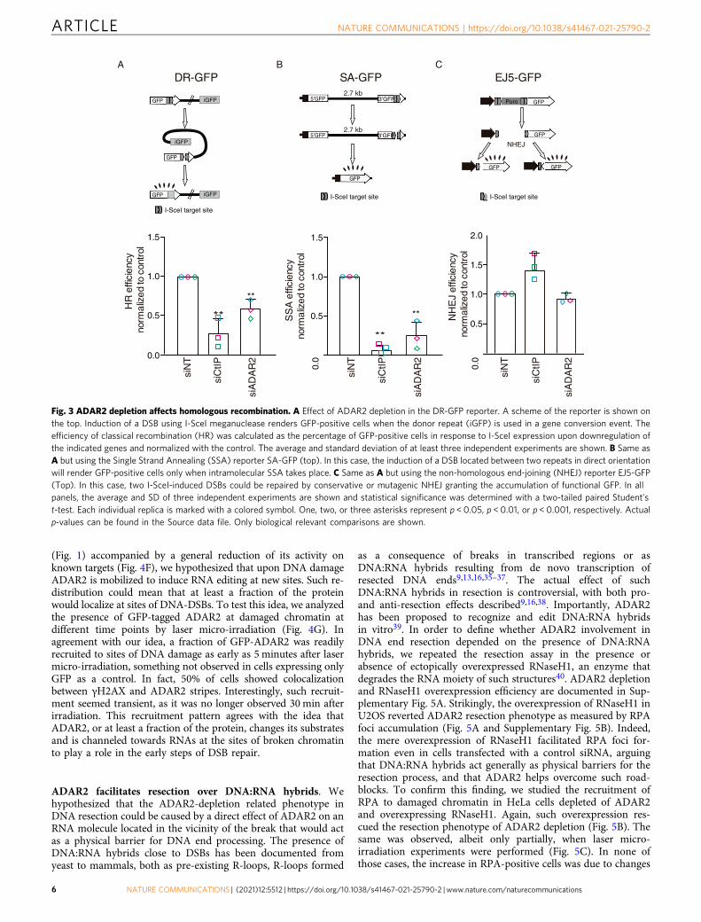
ADAR2 depletion affects DNA repair pathway choice. Next, wedecided to test a possible requirement of A-to-I RNA editing forDSB repair. As mentioned, ADAR2 depletion skewed the balancebetween HR and NHEJ towards the latter (see ref. 27and Sup-plementary Fig. 1B), suggesting that recombination might becompromised. Indeed, both RAD51-dependent gene conversion(GC) and RAD51-independent Single Strand Annealing (SSA),two types of homology-dependent repair (HDR), were reduced incells downregulated for ADAR2 (Fig. 3A, B). In stark contrast,there was no impact on NHEJ efficiency (Fig. 3C), arguing thatADAR2 was particularly required for HR. The cell cycle is amajor regulator of DSB repair pathway choice, as HR is limited inG1. However, the observed HR defect was not caused by anaccumulation of G1 cells (Supplementary Fig. 1C). Thus, weconclude that ADAR2 facilitates repair by HR.
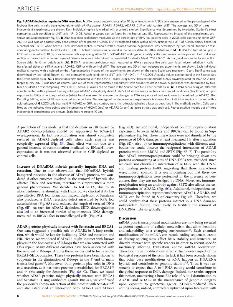
DNA resection requires ADAR2 editing activity. Due to itseffect in recombination, we hypothesized that ADAR2 might havea role in the common, early steps of the homology-dependentrepair pathways, namely in DNA end resection. To test this idea,we first studied RPA foci formation upon ionizing radiation inADAR2-depleted cells. RPA is an ssDNA binding complex thataccumulates at DNA breaks as a direct consequence of DNA endresection4,5,30. Thus, the percentage of RPA foci-positive cells isthe standard readout of resection in mammalian cells. Depletionof ADAR2 in U2OS cells caused a significant defect in resection,though less pronounced than that observed with the down-regulation of the key resection factor CtIP (Fig. 4A and Supple-mentary Fig. 3A). For representative images of the experiment seeSupplementary Fig. 3A. The same resection impairment was alsoobserved upon depletion of ADAR2 in the HeLa cells (Supple-mentary Fig. 3B). Strikingly, similar results were observed upon
depletion of ADAR1, but not the catalytically dead member of theADAR family, ADAR3, thus suggesting that active RNA editing isrequired for DNA end resection (Fig. 4A and SupplementaryFig. 3A).
To confirm that the observed phenotype in DNA resection wastruly due to the reduction of ADAR2 levels and not to an indirectoff-target effect, we studied RPA foci formation in cells bearingsiRNA-resistant, GFP-tagged variants of ADAR2. Indeed, theresection impairment caused by depletion of ADAR2 was rescuedby wild-type GFP-ADAR2 (Fig. 4B and Supplementary Fig. 3C).Importantly, this rescue was not observed with the expression of acatalytically dead version of the protein (GFP-ADAR2E-A)(Fig. 4B), arguing that ADAR2 deaminase activity was requiredfor processive resection. Moreover, we could reproduce theresection defect in U118 cells31, a glioblastoma cell line that isdefective for ADAR2 expression, when compared with the samecells complemented with a wild-type copy of the gene, but not acatalytically dead mutant (Fig. 4C).
To validate these observations, we analyzed recruitment ofRPA to DSBs by other means. U2OS cells depleted for ADAR2, orCtIP as a control, were laser micro-irradiated and immunostainedwith antibodies against RPA and γH2AX to identify the irradiatedareas. The percentage of γH2AX-positive stripes that were co-stained by RPA was determined (Fig. 4D). In agreement with ourprevious results, depletion of ADAR2, or CtIP, significantlydiminished the presence of RPA at the irradiated areas. Finally, inorder to analyze in more detail the resection defect related toADAR2 depletion and to investigate whether only resectioninitiation was impaired or if resection processivity was alsocompromised, we used SMART, a high-resolution technique thatmeasures resection in individual DNA fibres32,33. As seen inFig. 4E, the length of ssDNA fibres formed during the resectionprocess was reduced upon ADAR2 depletion. Again, similarresults were observed upon ADAR1 downregulation (Supple-mentary Fig. 3D), reinforcing the connection between A-to-Iediting and DNA end resection.
Altogether, these results confirm that RNA editing by ADARproteins facilitates DNA end resection at DSBs.
The role of ADAR2 on resection does not rely on the recodingof mRNAs that encode resection factors
Next, we wondered how this RNA editing activity might beneeded for DNA end processing. We studied the recruitment DSBrepair factors, such as 53BP1, BRCA1, and CtIP, to DNA damagefoci shortly after DNA damage induction upon ADAR2depletion. Notably, neither of them was affected in cells exposedto ionizing radiation or laser micro-irradiation (SupplementaryFig. 4A–C). Then, we wondered if ADAR2 was specifically editingmRNAs that code for resection factors. To analyze this possibility,we exposed the ADAR2-defective cell line U118 to ionizingradiation or mock treatment, isolated total RNA, and sequencedit. U118 cells complemented with either wild-type ADAR2 or acatalytically dead mutant were also processed in parallel. Thelevels of ADAR2 mRNA in the different samples are shown inSupplementary Fig. 4D. As expected, U118 cells complementedwith wild-type ADAR2 showed a higher efficiency in editing thecoding codons of known ADAR2 targets, expressed asthe weighted average over all known recoding sites, known asthe REI34. Instead, non-complemented U118 cells showed littlerecoding editing, regardless of whether or not exposed to ionizingradiation (Fig. 4F). Equally expected, the expression of acatalytically dead enzyme also showed almost no recoding ofmRNAs (Fig. 4F), despite the fact that such variant was expressedalmost 3 times more than the wild-type ADAR2 (SupplementaryFig. 4D). Thus, only the expression of catalytically active enzymeled to the expected ADAR2-dependent recoding due to editing ofspecific codons (Fig. 4F). Interestingly, although some specific
ARTICLE NATURE COMMUNICATIONS | https://doi.org/10.1038/s41467-021-25790-2
4 NATURE COMMUNICATIONS | (2021) 12:5512 | https://doi.org/10.1038/s41467-021-25790-2 | www.nature.com/naturecommunications
coding codons were edited more efficiently upon irradiation thanin mock-treated cells, we observed a general decrease in therecoding editing efficiency of known ADAR2 substrates uponexposure to DNA damage (Fig. 4F and Supplementary Data 1).Moreover, we could not observe any change in the editing ofmRNA from genes that code for recombination or resection
factors either in cells exposed to DNA damage or in undamagedcells (Supplementary Table S1). Thus, we conclude that the role ofADAR2 in resection and recombination does not rely on changesin the sequence or expression of specific mRNAs of bona fideDNA repair factors. To integrate our data, in which we observed ageneral ADAR2-mediated increase in RNA editing in the reporter
A
0 240
20
40
60
80siNT
siADAR2
Time after IR (h)
*
***
γ H2A
X
siADAR2siNT
DA
PI
0 h 24h
siADAR2siNT
% o
f γH
2AX
pos
itive
cel
ls-
1 6 240
20
40
60
80
100
% o
f γH
2AX
pos
itive
cel
ls- siNT
siADAR2siCtIP
B
Time after IR (h)
0
20
40
60
**
siADAR2
siNT
% B
RC
A1
foci
-pos
itive
cel
ls
C
0
10
20
30
40
siNT
siADAR2
% o
f m
icro
nucl
eus-
posi
tive
cells **
-IR +IR
E
siNT
siADAR2
0 0.005 0.0100
50
100
CPT
**
*
0 2 40
20
40
60
80
100
Gy
*
% S
urvi
val
% S
urvi
val
siNT
siADAR2
F G
0
20
40
60
*
siADAR1
siNT
% B
RC
A1
foci
-pos
itive
cel
ls
D
Fig. 2 ADAR2 depletion causes genetic instability and DNA repair defects. A Percentage of cells positive for γH2AX foci upon spontaneousaccumulation (0 h) or 24 h post irradiation with 10 Gy in U2OS cells transfected either a siRNA against ADAR2 or with control siNT. Quantification isshown on the left. A representative image is shown on the right. Scale bars represent 10 µm. B Repair kinetics is shown as the disappearance of γH2AX foci1, 6, and 24 h post irradiation with 10 Gy in U2OS cells transfected either a siRNA against ADAR2, CtIP or with control siNT. C Percentage of spontaneousBRCA1 foci-positive cells in cells transfected with either a siRNA against ADAR2 or with control siNT. The average and SD of three independentexperiments is shown. D Same as C but in cells depleted for ADAR1. E Percentage of cells positive for micronucleus without exposure to DNA damage (-IR)or 24 h post irradiation with 10 Gy (+IR) in U2OS cells transfected either with a siRNA against ADAR2 or with control siNT. Other details as in (A).F Clonogenic assays of U2OS cells depleted with a siRNA against ADAR2 or with control siNT after treatment with different doses of IR. Other details as in(A). G Same as F but cells treated with camptothecin (CPT; μM; right). In all panels, the average and SD of three independent experiments are shown andstatistical significance was determined with a two-tailed paired Student’s t-test. Each individual replica is marked with a colored symbol. One, two, or threeasterisks represent p < 0.05, p < 0.01, or p < 0.001, respectively. Actual p-values can be found in the Source data file. Only biological relevant comparisonsare shown.
NATURE COMMUNICATIONS | https://doi.org/10.1038/s41467-021-25790-2 ARTICLE
NATURE COMMUNICATIONS | (2021) 12:5512 | https://doi.org/10.1038/s41467-021-25790-2 |www.nature.com/naturecommunications 5
(Fig. 1) accompanied by a general reduction of its activity onknown targets (Fig. 4F), we hypothesized that upon DNA damageADAR2 is mobilized to induce RNA editing at new sites. Such re-distribution could mean that at least a fraction of the proteinwould localize at sites of DNA-DSBs. To test this idea, we analyzedthe presence of GFP-tagged ADAR2 at damaged chromatin atdifferent time points by laser micro-irradiation (Fig. 4G). Inagreement with our idea, a fraction of GFP-ADAR2 was readilyrecruited to sites of DNA damage as early as 5 minutes after lasermicro-irradiation, something not observed in cells expressing onlyGFP as a control. In fact, 50% of cells showed colocalizationbetween γH2AX and ADAR2 stripes. Interestingly, such recruit-ment seemed transient, as it was no longer observed 30min afterirradiation. This recruitment pattern agrees with the idea thatADAR2, or at least a fraction of the protein, changes its substratesand is channeled towards RNAs at the sites of broken chromatinto play a role in the early steps of DSB repair.
ADAR2 facilitates resection over DNA:RNA hybrids. Wehypothesized that the ADAR2-depletion related phenotype inDNA resection could be caused by a direct effect of ADAR2 on anRNA molecule located in the vicinity of the break that would actas a physical barrier for DNA end processing. The presence ofDNA:RNA hybrids close to DSBs has been documented fromyeast to mammals, both as pre-existing R-loops, R-loops formed
as a consequence of breaks in transcribed regions or asDNA:RNA hybrids resulting from de novo transcription ofresected DNA ends9,13,16,35–37. The actual effect of suchDNA:RNA hybrids in resection is controversial, with both pro-and anti-resection effects described9,16,38. Importantly, ADAR2has been proposed to recognize and edit DNA:RNA hybridsin vitro39. In order to define whether ADAR2 involvement inDNA end resection depended on the presence of DNA:RNAhybrids, we repeated the resection assay in the presence orabsence of ectopically overexpressed RNaseH1, an enzyme thatdegrades the RNA moiety of such structures40. ADAR2 depletionand RNaseH1 overexpression efficiency are documented in Sup-plementary Fig. 5A. Strikingly, the overexpression of RNaseH1 inU2OS reverted ADAR2 resection phenotype as measured by RPAfoci accumulation (Fig. 5A and Supplementary Fig. 5B). Indeed,the mere overexpression of RNaseH1 facilitated RPA foci for-mation even in cells transfected with a control siRNA, arguingthat DNA:RNA hybrids act generally as physical barriers for theresection process, and that ADAR2 helps overcome such road-blocks. To confirm this finding, we studied the recruitment ofRPA to damaged chromatin in HeLa cells depleted of ADAR2and overexpressing RNaseH1. Again, such overexpression res-cued the resection phenotype of ADAR2 depletion (Fig. 5B). Thesame was observed, albeit only partially, when laser micro-irradiation experiments were performed (Fig. 5C). In none ofthose cases, the increase in RPA-positive cells was due to changes
A B
0.0
0.5
1.0
1.5
DR-GFP
H
R e
ffici
ency
no
rmal
ized
to c
ontr
ol
**
**
siN
T
siC
tIP
siA
DA
R2
0.0
0.5
1.0
1.5
2.0
EJ5-GFP
siN
T
siC
tIP
siA
DA
R2
**
0.0
0.5
1.0
1.5
SA-GFP
**
siN
T
siC
tIP
siA
DA
R2
S
SA
effi
cien
cy
norm
aliz
ed to
con
trol
NH
EJ
e ffic
ienc
y no
rma l
ized
to c
ontr
ol
GFP iGFP
GFP
iGFP
GFP iGFP
I-SceI target site
2.7 kb
2.7 kb
5’GFP 3’GFP
5’GFP 3’GFP
GFP
I-SceI target site
GFPPuro
GFP
GFP GFP
NHEJ
I-SceI target site
C
Fig. 3 ADAR2 depletion affects homologous recombination. A Effect of ADAR2 depletion in the DR-GFP reporter. A scheme of the reporter is shown onthe top. Induction of a DSB using I-SceI meganuclease renders GFP-positive cells when the donor repeat (iGFP) is used in a gene conversion event. Theefficiency of classical recombination (HR) was calculated as the percentage of GFP-positive cells in response to I-SceI expression upon downregulation ofthe indicated genes and normalized with the control. The average and standard deviation of at least three independent experiments are shown. B Same asA but using the Single Strand Annealing (SSA) reporter SA-GFP (top). In this case, the induction of a DSB located between two repeats in direct orientationwill render GFP-positive cells only when intramolecular SSA takes place. C Same as A but using the non-homologous end-joining (NHEJ) reporter EJ5-GFP(Top). In this case, two I-SceI-induced DSBs could be repaired by conservative or mutagenic NHEJ granting the accumulation of functional GFP. In allpanels, the average and SD of three independent experiments are shown and statistical significance was determined with a two-tailed paired Student’st-test. Each individual replica is marked with a colored symbol. One, two, or three asterisks represent p < 0.05, p < 0.01, or p < 0.001, respectively. Actualp-values can be found in the Source data file. Only biological relevant comparisons are shown.
ARTICLE NATURE COMMUNICATIONS | https://doi.org/10.1038/s41467-021-25790-2
6 NATURE COMMUNICATIONS | (2021) 12:5512 | https://doi.org/10.1038/s41467-021-25790-2 | www.nature.com/naturecommunications
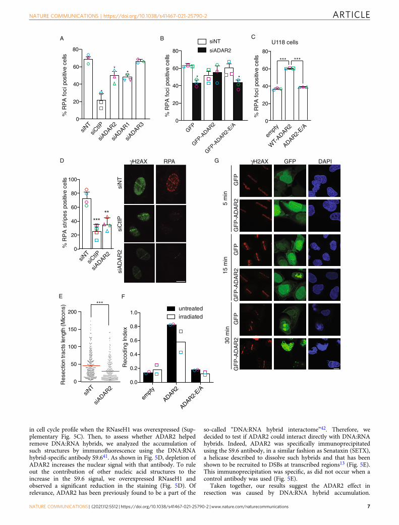
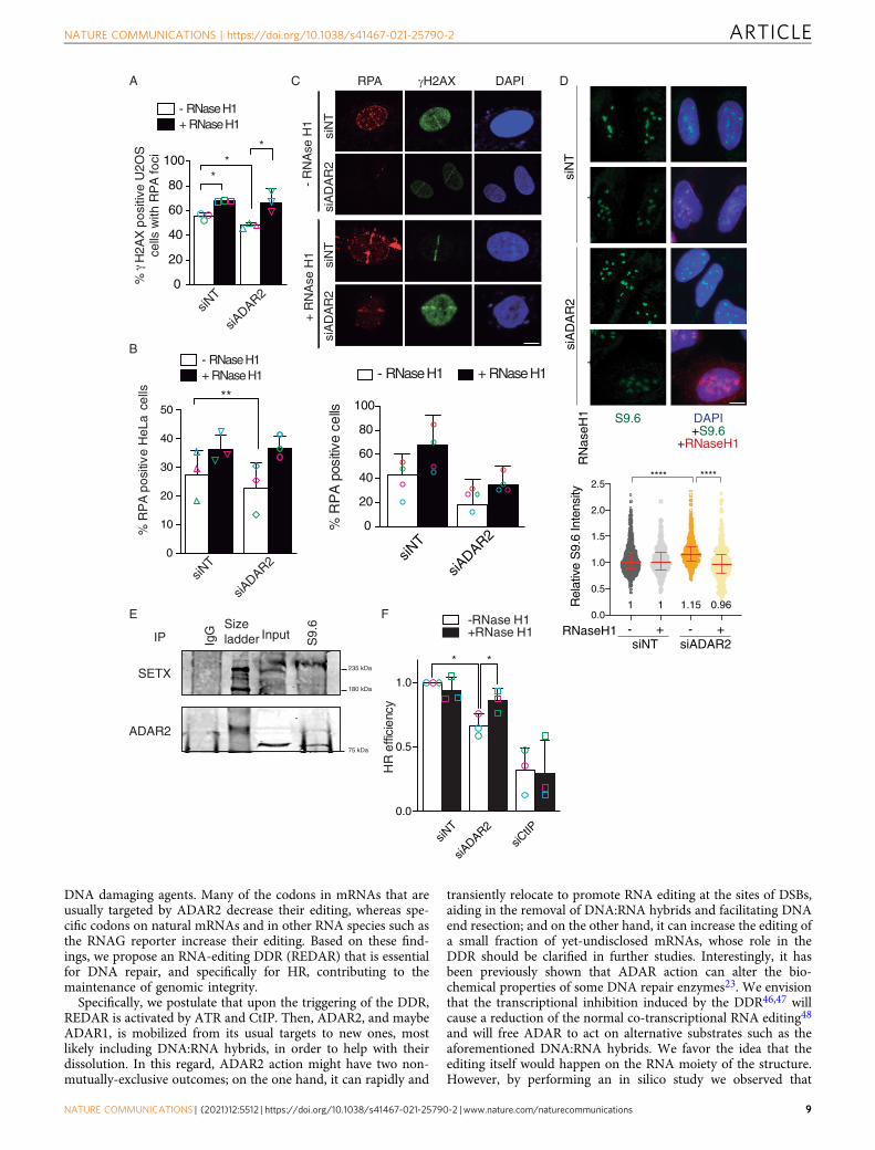
in cell cycle profile when the RNaseH1 was overexpressed (Sup-plementary Fig. 5C). Then, to assess whether ADAR2 helpedremove DNA:RNA hybrids, we analyzed the accumulation ofsuch structures by immunofluorescence using the DNA:RNAhybrid-specific antibody S9.641. As shown in Fig. 5D, depletion ofADAR2 increases the nuclear signal with that antibody. To ruleout the contribution of other nucleic acid structures to theincrease in the S9.6 signal, we overexpressed RNaseH1 andobserved a significant reduction in the staining (Fig. 5D). Ofrelevance, ADAR2 has been previously found to be a part of the
so-called “DNA:RNA hybrid interactome”42. Therefore, wedecided to test if ADAR2 could interact directly with DNA:RNAhybrids. Indeed, ADAR2 was specifically immunoprecipitatedusing the S9.6 antibody, in a similar fashion as Senataxin (SETX),a helicase described to dissolve such hybrids and that has beenshown to be recruited to DSBs at transcribed regions13 (Fig. 5E).This immunoprecipitation was specific, as did not occur when acontrol antibody was used (Fig. 5E).
Taken together, our results suggest the ADAR2 effect inresection was caused by DNA:RNA hybrid accumulation.
0
20
40
60
80
siADAR2
siNT
siCtIP
%R
PA
foci
pos
itive
cells
0
20
40
60
80
siNT
siADAR2
*
GFP-ADAR2
GFP-ADAR2-
E/A
%R
PA
foci
pos
itive
cells
0
20
40
60
80
100
%R
PA
str
ipes
pos
itive
cells
*****
A
GFP
B
0
20
40
60
80
*** ***
U118 cells
%R
PA
foci
pos
itive
cells
WT-
ADAR2
ADAR2-E/A
empt
y
C
siN
Tsi
CtIP
γH2AX RPAD
siA
DA
R2
*
*
*
***
0
50
100
150
200
Res
ectio
n tr
acts
leng
th (M
icon
s)
E
siADAR2
siNT
siADAR2
siNT
siCtIP
empt
y
ADAR2
ADAR2-E/A
0.0
0.2
0.4
0.6
0.8
1.0
Rec
odin
g In
dex
untreatedirradiated
F
*
siADAR1
siADAR3
G γH2AX GFP DAPI
GF
PG
FP
-AD
AR
2G
FP
GF
P-A
DA
R2
GF
PG
FP
-AD
AR
2
5 m
in15
min
30 m
in
NATURE COMMUNICATIONS | https://doi.org/10.1038/s41467-021-25790-2 ARTICLE
NATURE COMMUNICATIONS | (2021) 12:5512 | https://doi.org/10.1038/s41467-021-25790-2 |www.nature.com/naturecommunications 7
A prediction of this model is that the decrease in HR caused byADAR2 downregulation should be suppressed by RNaseH1overexpression. In fact, recombination was almost completelyrestored in ADAR2-depleted cells when such enzyme wasectopically expressed (Fig. 5F). Such effect was not due to ageneral increase of recombination mediated by RNaseH1 over-expression, as it was not observed either in CtIP depleted orcontrol cells.
Increase of DNA:RNA hybrids generally impairs DNA endresection. Due to our observation that DNA:RNA hybridshampered resection in the absence of ADAR proteins, we won-dered if other enzymes involved in the removal of hybrids alsoaffected DNA end resection, therefore this represented a moregeneral phenomenon. We decided to test SETX, due to itsaforementioned relationship with DSBs. So, we checked if its lossalso affected RPA foci formation. Indeed, depletion of Senataxinalso produced a DNA resection defect measured by RPA fociaccumulation (Fig. 6A) and reduced the length of resected DNA(Fig. 6B). As seen for ADAR2 depletion, SETX downregulationalso led to an increased burden of spontaneous DNA damage,measured as BRCA1 foci in unchallenged cells (Fig. 6C).
ADAR proteins physically interact with Senataxin and BRCA1.Our data suggested a possible role of ADAR2 in R-loop resolu-tion, which would be key for facilitating DNA end resection andHR. Hence, we wondered if ADAR2 might interact with knownplayers in the homeostasis of R-loops that are also connected withDSB repair. Many different enzymes have been associated withthe removal of R-loops. Among them, we decided to focus on theBRCA1–SETX complex. These two proteins have been shown tocooperate in the elimination of R-loops in the 3' end of manytranscribed genes43. Moreover, both proteins play roles in DNAend resection, as it has been previously established for BRCA133
and in this study for Senataxin (Fig. 6A–C). Thus, we testedwhether ADAR proteins might physically interact with BRCA1and Senataxin. Using antibodies against BRCA1, we confirmedthe previously shown interaction of this protein with Senataxin43
and also established an interaction with ADAR1 and ADAR2
(Fig. 6D). An additional, independent co-immunoprecipitationexperiment between ADAR2 and BRCA1 can be found in Sup-plementary Fig. 6A. These interactions were not stimulated by thepresence of DNA damage, at least measured 1 h after irradiation(Fig. 6D). Also, by co-immunoprecipitations with different anti-bodies we could observe the reciprocal interaction of ADARproteins with both BRCA1 and SETX (Fig. 6E–F). The possibilitythat ADAR immunoprecipitation could be bringing down anyproteins accumulating at sites of DNA DSBs was excluded, sincewe could not observe an interaction of ADAR2 with the DNA-end binding protein Ku80, suggesting that those interactionswere, indeed, specific. It is worth pointing out that these co-immunoprecipitations were performed in the presence of ben-zonase, thus they are not bridged by DNA. Moreover, immuno-precipitation using an antibody against SETX also allows the co-precipitation of ADAR2 (Fig. 6G). Additional, independent co-immunoprecipitation experiments between ADAR1, ADAR2, andSETX can be found in Supplementary Fig. 6B. Therefore, wecould confirm that these proteins interact in a DNA damage-independent fashion, most likely to facilitate the removal ofDNA:RNA hybrids globally.
DiscussionmRNA post-transcriptional modifications are now being revealedas potent regulators of cellular metabolism that allow flexibilityand adaptability to a changing environment44. Such chemicalmodifications of the mRNA can recode coding sequences, createor destroy splicing sites, affect RNA stability and structure, ordirectly interact with specific readers in order to recruit specificmachinery affecting translation and/or mRNA localization.Therefore, those modifications affect virtually every aspect of thebiological response of the cells. In fact, it has been recently shownthat other base modifications of RNA happen at DNA:RNAhybrids and contribute to genomic stability45. Thus, it was rea-sonable to expect that A-to-I RNA editing would contribute tothe global response to DNA damage. Indeed, our results supportthis notion, uncovering a bona fide role of A-to-I deamination byADAR1 and ADAR2 in the maintenance of genomic integrityupon exposure to genotoxic agents. ADAR2-mediated RNAediting seems, indeed, completely upturned upon treatment with
Fig. 4 ADAR depletion impairs in DNA resection. A DNA resection proficiency after 10 Gy of irradiation in U2OS cells measured as the percentage of RPAfoci-positive cells in cells transfected either with siRNAs against ADAR1, ADAR2, ADAR3, CtIP or with control siNT. The average and SD of threeindependent experiments are shown. Each individual replica is marked with a colored symbol. Significance was determined by two-tailed Student’s t-testcomparing each condition to siNT cells. *P < 0.05. Actual p-values can be found in the Source data file. Representative images of the experiments areshown on Supplementary Fig. 3A. B DNA resection proficiency measured as the percentage of RPA foci-positive cells in U2OS cells expressing either GFP-ADAR2 wild type or a catalytically dead version of the protein (ADAR2 E/A) transfected either with a siRNA against the 3'UTR of ADAR2 (black boxes) ora control siNT-UTR (white boxes). Each individual replica is marked with a colored symbol. Significance was determined by two-tailed Student’s t-testcomparing each condition to siNT cells. *P < 0.05. Actual p-values can be found in the Source data file. Other details as in (A). C RPA foci formation upon inU118 cells treated with 10 Gy of radiation in cells expressing either GFP, GFP-ADAR2 wild type or a catalytically dead version of the protein. Each individualreplica is marked with a colored symbol. Significance was determined by two-tailed Student’s t-test. ***P < 0.001. Actual p-values can be found in theSource data file. Other details as in (A). D DNA resection proficiency was measured as RPA stripes-positive cells upon laser microirradiation in cellstransfected either an siRNA against ADAR2, CtIP or with control siNT. The average and SD of four independent experiments are shown. Each individualreplica is marked with a colored symbol. Representative images of the experiments are shown on the right. Scale bars represent 10 µm. Significance wasdetermined by two-tailed Student’s t-test comparing each condition to siNT cells. **P < 0.01; ***P < 0.001. Actual p-values can be found in the Source datafile. Other details as in (A). E Resection length measured with the SMART assay using DNA fibers extracted from U2OS downregulated for ADAR2. A non-target siRNA (siNT) was used as control. One out of three representative experiment with similar results is shown. Significance was determined by two-tailed Student’s t-test comparing. ***P < 0.001. Actual p-values can be found in the Source data file. Other details as in (A). F RNA sequencing of U118 cellscomplemented with a plasmid bearing wild type ADAR2, catalytically dead ADAR2 E/A or the empty vectors in untreated conditions (black bars) or uponexposure to 10 Gy of ionizing radiation (white bars) was used to analyze the changes in RNA sequence of codons known to be edited by ADAR2. TheRecoding Editing Index (REI) was reported as percentage. The average of two independent experiments is shown. Each individual replica is marked with acolored symbol. G U2OS cells bearing GFP-ADAR2 or GFP, as a control, were micro-irradiated using a laser as described in the methods section. Cells werefixed at the indicated time points and the presence of γH2AX (red) or ADAR2 (green) at lasers stripes was analysed. Representative images out of threeindependent experiments are shown. Scale bars represent 10 µm.
ARTICLE NATURE COMMUNICATIONS | https://doi.org/10.1038/s41467-021-25790-2
8 NATURE COMMUNICATIONS | (2021) 12:5512 | https://doi.org/10.1038/s41467-021-25790-2 | www.nature.com/naturecommunications
DNA damaging agents. Many of the codons in mRNAs that areusually targeted by ADAR2 decrease their editing, whereas spe-cific codons on natural mRNAs and in other RNA species such asthe RNAG reporter increase their editing. Based on these find-ings, we propose an RNA-editing DDR (REDAR) that is essentialfor DNA repair, and specifically for HR, contributing to themaintenance of genomic integrity.
Specifically, we postulate that upon the triggering of the DDR,REDAR is activated by ATR and CtIP. Then, ADAR2, and maybeADAR1, is mobilized from its usual targets to new ones, mostlikely including DNA:RNA hybrids, in order to help with theirdissolution. In this regard, ADAR2 action might have two non-mutually-exclusive outcomes; on the one hand, it can rapidly and
transiently relocate to promote RNA editing at the sites of DSBs,aiding in the removal of DNA:RNA hybrids and facilitating DNAend resection; and on the other hand, it can increase the editing ofa small fraction of yet-undisclosed mRNAs, whose role in theDDR should be clarified in further studies. Interestingly, it hasbeen previously shown that ADAR action can alter the bio-chemical properties of some DNA repair enzymes23. We envisionthat the transcriptional inhibition induced by the DDR46,47 willcause a reduction of the normal co-transcriptional RNA editing48
and will free ADAR to act on alternative substrates such as theaforementioned DNA:RNA hybrids. We favor the idea that theediting itself would happen on the RNA moiety of the structure.However, by performing an in silico study we observed that
A
B
0
10
20
30
40
50**
%R
PA
posi
tive
HeL
ace
ll s
DAPI�H2AXRPA
siA
DA
R2
siN
Tsi
AD
AR
2si
NT
- R
NA
se H
1+
RN
Ase
H1
C
siNT
siADAR2
0
20
40
60
80
100
%R
PA
posi
tive
cells
- RNase H1 + RNase H1
0
20
40
60
80
100
%H
2AX
posi
tive
U2O
Sce
lls w
ithR
PA
foci
*
**
- RNase H1 + RNase H1
siADAR2
siNT
siADAR2
siNT
- RNase H1 + RNase H1
S9.6
RN
aseH
1 DAPI+S9.6
+RNaseH1
-
siN
Tsi
AD
AR
2
+
-
+
Rel
ativ
e S
9.6
Inte
nsity
**** ****
RNaseH1 - + - +siNT siADAR2
0.0
0.5
1.0
1.5
2.0
2.5
1 1 1.15 0.96
D
ADAR2
SETX
Input S9.
6
IgGIP
E
siNT
siADAR2
siCtIP
0.0
0.5
1.0
HR
effic
ienc
y
-RNase H1+RNase H1
**
FSizeladder
235 kDa
180 kDa
75 kDa
NATURE COMMUNICATIONS | https://doi.org/10.1038/s41467-021-25790-2 ARTICLE
NATURE COMMUNICATIONS | (2021) 12:5512 | https://doi.org/10.1038/s41467-021-25790-2 |www.nature.com/naturecommunications 9
cancer cells that overexpress ADAR2 are statistically more likelyto accumulate this kind of mutations (Supplementary Fig. 7).Thus, it is also possible that the editing occurs in the DNA strand,as suggested in vitro39, albeit how frequently and whether thishappens in the context of DNA repair is still unclear. Editing ofthe DNA strand would greatly increase the mutagenesis asso-ciated with HR, something that might be deleterious for the cells,but has been observed due to the action of C-to-U deaminases incancer49. Interestingly, the accumulation of N6-methyladenosinemodification on the RNA at DNA:RNA hybrids has also beenshown to ease the dissolution of such structures45. As A-to-Iediting is negatively influenced by m6A modifications50,51, thecrosstalk between those two RNA post-transcriptional modifica-tions during DNA repair and in response to DNA damage will beworth exploring further.
The mechanism by which ADAR2 is relocated in response toDNA damage is still far from clear. On the one hand, a passivemodel is possible, in which ADAR2 naturally recognizes andbinds to DNA:RNA hybrids. It has been shown that DNAdamage induces the formation of such structures and, in thisscenario, the accumulation of hybrids would sequester ADAR2,reducing its availability to edit its usual targets. Indeed, usingGFP-tagged versions of ADAR2 we could observe a re-localization to laser micro-irradiated stripes. However, suchaccumulation is very transient, and in a completely differenttimeframe of the changes in recoding we have also shown. So, thisrecruitment cannot fully explain the changes in editing weobserved globally, arguing for the coexistence of both a global,genome-wide effect, and a site-specific role. It is even possiblethat, in this context, during REDAR, ADAR2 would not actspecifically at DNA:RNA hybrids that are close to DSBs, butrather relocated to hybrids spread through the genome as a broadresponse to DNA damage. In this scenario, the accumulation atlaser lines we observed would rely simply on the already knownaccumulation of hybrids at break sites. Indeed, the SETX–BRCA1complex has been proposed to be important and recruited mainlyto transcription termination sites, regardless of the presence ofDNA damage43, thus again arguing with for putative role thatdoes not require specific recruitment to DSBs. Alternatively, there
could be an active, recruitment of ADAR2 to hybrids that sitspecifically in the proximity of DSBs. The physical interactionwith BRCA1, a well-established, bona fide member of the DDR,which is recruited to DSBs52 and Senataxin, that is also localizedto damaged chromatin13, might favor this model. This idea is alsosupported by the fact that A-to-I editing at new sites is upregu-lated upon REDAR activation in an ATR-dependent manner,arguing for a DNA damage-dependent recruitment of ADAR2 tothose new target sites. Interestingly, the ATR checkpoint ismainly active in S and G2, as it requires ssDNA for its activationthat is produced by DNA end resection. This would explain whyCtIP downregulation also decreases the DNA damage-inducedincrease of RNA editing in the RNAG system, simply reflectingthe role of CtIP in the ATR activation upon formation of DSBs.
CtIP is linked to RNA metabolism in multiple ways. It affectsRNA editing, as shown here, but also interacts with multiple RNAbinding proteins53 that in turn are required for proper resection.Moreover, we have recently shown that CtIP controls the splicingof specific factors, in many cases facilitating the accumulation ofspecific alternative splicing forms upon exposure to DNAdamage54. Additionally, CtIP not only interacts with BRCA1,which also affects RNA splicing, but also CtIP deficiency has beenshown to promote the accumulation of DNA:RNA hybrids atsites of highly expressed genes14. Paradoxically, CtIP depletionreduces DNA:RNA hybrid accumulation dependent on de novotranscription of dilncRNA (damage-induced long non-codingRNAs) starting at DSBs36. Hence, CtIP depletion seems toincrease R-loops that are produced as a consequence of theprevious transcription and appears to decrease de novo produc-tion of diRNAs (DSB-induced small RNA), thus reducing theDNA:RNA hybrids formed after DNA damage. The data suggestthat DNA resection, and specifically CtIP and BRCA1, are inclose relationship with the general metabolism of RNAs, a reci-procal connection that is worth to continue exploring.
To integrate all our observations, we present a model in whichADAR2 (and most likely ADAR1), BRCA1, and SETX facilitatethe removal of hybrids genome-wide even in the absence of DNAdamage, but in a way that is stimulated by the DDR duringREDAR. When exposed to a source of DSBs, the levels of
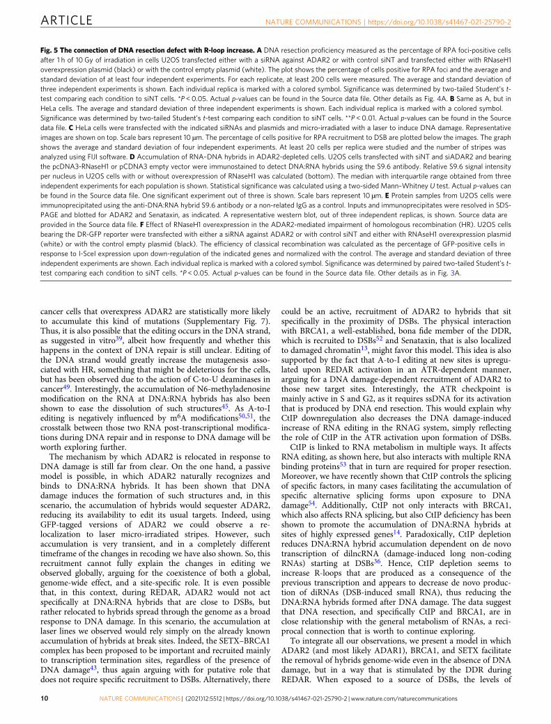
Fig. 5 The connection of DNA resection defect with R-loop increase. A DNA resection proficiency measured as the percentage of RPA foci-positive cellsafter 1 h of 10 Gy of irradiation in cells U2OS transfected either with a siRNA against ADAR2 or with control siNT and transfected either with RNaseH1overexpression plasmid (black) or with the control empty plasmid (white). The plot shows the percentage of cells positive for RPA foci and the average andstandard deviation of at least four independent experiments. For each replicate, at least 200 cells were measured. The average and standard deviation ofthree independent experiments is shown. Each individual replica is marked with a colored symbol. Significance was determined by two-tailed Student’s t-test comparing each condition to siNT cells. *P < 0.05. Actual p-values can be found in the Source data file. Other details as Fig. 4A. B Same as A, but inHeLa cells. The average and standard deviation of three independent experiments is shown. Each individual replica is marked with a colored symbol.Significance was determined by two-tailed Student’s t-test comparing each condition to siNT cells. **P < 0.01. Actual p-values can be found in the Sourcedata file. C HeLa cells were transfected with the indicated siRNAs and plasmids and micro-irradiated with a laser to induce DNA damage. Representativeimages are shown on top. Scale bars represent 10 µm. The percentage of cells positive for RPA recruitment to DSB are plotted below the images. The graphshows the average and standard deviation of four independent experiments. At least 20 cells per replica were studied and the number of stripes wasanalyzed using FIJI software. D Accumulation of RNA–DNA hybrids in ADAR2-depleted cells. U2OS cells transfected with siNT and siADAR2 and bearingthe pcDNA3-RNaseH1 or pCDNA3 empty vector were immunostained to detect DNA:RNA hybrids using the S9.6 antibody. Relative S9.6 signal intensityper nucleus in U2OS cells with or without overexpression of RNaseH1 was calculated (bottom). The median with interquartile range obtained from threeindependent experiments for each population is shown. Statistical significance was calculated using a two-sided Mann–Whitney U test. Actual p-values canbe found in the Source data file. One significant experiment out of three is shown. Scale bars represent 10 µm. E Protein samples from U2OS cells wereimmunoprecipitated using the anti-DNA:RNA hybrid S9.6 antibody or a non-related IgG as a control. Inputs and immunoprecipitates were resolved in SDS-PAGE and blotted for ADAR2 and Senataxin, as indicated. A representative western blot, out of three independent replicas, is shown. Source data areprovided in the Source data file. F Effect of RNaseH1 overexpression in the ADAR2-mediated impairment of homologous recombination (HR). U2OS cellsbearing the DR-GFP reporter were transfected with either a siRNA against ADAR2 or with control siNT and either with RNAseH1 overexpression plasmid(white) or with the control empty plasmid (black). The efficiency of classical recombination was calculated as the percentage of GFP-positive cells inresponse to I-SceI expression upon down-regulation of the indicated genes and normalized with the control. The average and standard deviation of threeindependent experiments are shown. Each individual replica is marked with a colored symbol. Significance was determined by paired two-tailed Student’s t-test comparing each condition to siNT cells. *P < 0.05. Actual p-values can be found in the Source data file. Other details as in Fig. 3A.
ARTICLE NATURE COMMUNICATIONS | https://doi.org/10.1038/s41467-021-25790-2
10 NATURE COMMUNICATIONS | (2021) 12:5512 | https://doi.org/10.1038/s41467-021-25790-2 | www.nature.com/naturecommunications
DNA:RNA hybrids and R-loops increase, as previously describedby other authors13–16, and many of them specifically accumulateat break sites. Although some authors argue that these structuresmight favor the repair process38, others favor a view in whichthey act as roadblocks for repair (for a review see ref. 37). Thiscontradiction might simply stem from a differential effect ofDNA:RNA hybrids depending on the timing of repair, i.e. veryearly events might need them but later they have to be eliminated,
or the position of the hybrids, as discussed elsewhere9,55. Ourdata agree with the notion that at least some of them representroadblocks for the progression of the repair machinery that has tobe removed prior to repair (Fig. 6G-1). Such blocking effect ofR-loops is well established for other DNA transactions such astranscription and replication37,56. We propose that, upon irra-diation, a CtIP- and ATR-mediated global induction of A-to-Iediting at DNA:RNA hybrids facilitate their removal both
0
10
20
30
40
50
% B
RC
A1
foci
-pos
itive
cel
ls
**
0
20
40
60
80
100
RP
A fo
ci-p
ositi
ve c
ells
*
siNT siSETX0
50
100
150
Res
ectio
n le
ngth
(Mic
rons
)
siNT siSETX
A B C
siNT siSETX
SETX IgGD
E
SETX
ADAR2
BRCA1
G
ADAR2
BRCA1
ADAR2 IgG
SETX
ADAR2
Input
Input
Input BR
CA
1
IgG
-IR
IP
Input BR
CA
1
IgG
+IR
IP
IP
IP
BRCA1
SETX ADAR2
~ ~ ~
SETX
1
2
3
4
5
BRCA1
ADAR2
ATR
CtIP
H
KU80
ADAR1
F
ADAR1
SETX
AD
AR
1
Input
IP
AD
AR
2
IgG
180 kDa
180 kDa
100 kDa
75 kDa
180 kDa
100 kDa
75 kDa
180 kDa
75 kDa
180 kDa
245 kDa
100 kDa
245 kDa
100 kDa
75 kDa
NATURE COMMUNICATIONS | https://doi.org/10.1038/s41467-021-25790-2 ARTICLE
NATURE COMMUNICATIONS | (2021) 12:5512 | https://doi.org/10.1038/s41467-021-25790-2 |www.nature.com/naturecommunications 11
genome-wide and locally, at sites of DNA breaks, hence permit-ting the DNA end resection machinery to overcome the physicalbarrier represented by pre-existing or recently formed hybridsclose to the DNA breaks (Fig. 6G–2). Mechanistically, we suggestthat ADAR-mediated editing of DNA:RNA hybrids, an activitythat has been confirmed in vitro39,57, might alter the sequence ofthe RNA strand creating ribo–inosine (rI) and thedeoxiribose–thymine (dT) mismatches (Fig. 6G–3). The appear-ance of those mismatches will loosen up the interaction betweenthe RNA and the DNA strand, facilitating the unwinding of thestructure by the helicase activity of SETX (Fig. 6G–3,4), allowingthe resection machinery to go through (Fig. 6G–4,5). Strikingly,ADAR proteins were first discovered in Xenopus as havingdevelopmentally regulated dsRNA unwinding activity inoocytes58, and later shown to rely on the modification of A–to–Iin the RNA substrate, which modified the base-pairing propertiesand facilitated the melting of that RNA:RNA double-strandedstructures59. Alternatively, rather than physically loosening theinteraction between the DNA and RNA, A-to-I modification ofthe RNA might help the recruitment of proteins involved inR-loop removal or even the recruitment of bona fide resectionfactors. We propose that this alteration of the RNA moiety of thehybrids will happen at all R-loops scattered across the genome,even in the absence of DNA damage. But particularly at DSBs,ADAR2 will contribute to the processivity of resection overDNA:RNA hybrids, regardless of if these structures preceded orformed as a consequence of the break in its vicinity. Our modelproposes a role of ADAR in DNA end resection and recombi-nation that acts independently but not exclusively, of the recodingor regulation of expression of yet-undisclosed mRNAs. Interest-ingly, recently it has been shown that ADAR1 editing activity isrequired to eliminate R-loops at telomeric repeats in order topreserve genomic integrity57. In this case, ADAR1 has beenproposed to edit mismatched hybrids formed by TERRAs andnon-canonical telomeric repeats in order to facilitate its removalby RNaseH2, a phenomenon that is specific in non-ALT cancercell lines. Thus, albeit with slightly different mechanisms, thisconfirms that RNA editing affects genomic integrity by regulatingDNA:RNA hybrid elimination.
Strikingly, ADAR deficiency, and generally unbalance the levelsof editing of the RNA, have been associated with the development
of many different tumors24,25,34,60. Specific editing events havealready been associated with glioblastoma22,61. However, in thelight of this new role for ADAR2 in DNA repair and the DDR, itmight be important to extend the connection of this protein withcancer and establish if it is linked to a particular genomicinstability signature.
MethodsCell lines and growth conditions. All cell lines were grown in DMEM (Sigma-Aldrich) supplemented with 10% fetal bovine serum (Sigma-Aldrich), 2 mML-glutamine (Sigma-Aldrich), 100 units ml−1 penicillin, and 100 μg ml−1 strep-tomycin (Sigma-Aldrich). U2OS and U118-derived cell lines stably expressing GFPor GFP–ADAR2 plasmids31 were grown in a standard medium supplemented with0.5 mg ml−1 G418 (Gibco, Invitrogen). Cells expressing RNWG and RNAG weregrown in standard medium supplemented with 0.5 mg ml−1 G418 (Gibco,Invitrogen).
siRNAs, plasmids and transfections. siRNA duplexes were obtained from Sigma-Aldrich or Dharmacon (Supplementary Table 1) and were transfected usingRNAiMax Lipofectamine Reagent Mix (Life Technologies), according to themanufacturer’s instructions. RNWG and RNAG was a gift from Dr. Jantsch’s lab26.The GFP-ADAR2 and GFP-ADAR2 mutant (GFP-ADAR2-E/A-) plasmids werepreviously described31. RNaseH1 overexpression was achieved with thepCDNA3–RNAseH1 vector62 and pCDNA3 (Invitrogen) was used as a control.Plasmid transfection of U2OS cells was carried out using FuGENE 6 TransfectionReagent (Promega) according to the manufacturer’s protocol, with the exception ofpCDNA3–RNaseH1 and pCDNA3 plasmids that were transfected using Lipo-fectamine 3000 (Invitrogen) according to the manufacturer’s instructions.
HR and NHEJ analysis. U2OS cells bearing a single copy integration of thereporters DR-GFP (Gene conversion)63, SA-GFP (SSA)64, or EJ5-GFP (NHEJ)64
were used to analyze the different DSB repair pathways. In all cases, 50,000 cellswere plated in 6-well plates in duplicate. One day after seeding, cells were trans-fected with the indicated siRNA and the medium was replaced with a fresh one24 h later. The next day, each duplicate culture was infected with lentiviral particlescontaining I-SceI–BFP expression construct at MOI 10 using 8 µg/ml polybrene in1.5 ml of DMEM. Then, cells were left to grow for an additional 24 h beforechanging the medium for fresh DMEM, 48 h after siRNA transfection, cells werewashed with PBS, trypsinized, neutralized with DMEM, centrifuged for 5 min at700 g, fixed with 4% paraformaldehyde for 20 min, and collected by centrifugation.Then, cell pellets were washed once with PBS before resuspension in 150 µl of PBS.Samples were analyzed with a BD FACSAria with the BD FACSDiva Softwarev5.0.3. Four different parameters were considered: side scatter (SSC), forwardscatter (FSC), blue fluorescence (407 nm violet laser BP, Filter 450/40), greenfluorescence (488 nm blue laser BP Filter 530/30). Finally, the number of green cellsfrom at least 10,000 events positives for blue fluorescence (infected with the I-SceI–BFP construct) was scored. The average of both duplicates was calculated for
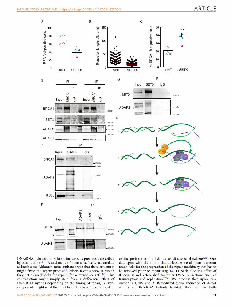
Fig. 6 DNA:RNA hybrid stabilization impairs resection. A Percentage of RPA foci-positive cells in cells transfected either with a siRNA against SETX orwith control siNT. The average and standard deviation of four independent experiments is shown. Each individual replica is marked with a colored symbol.Significance was determined by paired two-tailed Student’s t-test comparing each condition to siNT cells.*P < 0.05. Actual p-values can be found in theSource data file. Other details are as in Fig. 4A. B DNA resection proficiency is measured as the length of resected DNA with SMART in cells transfectedeither with a siRNA against SETX or with control siNT. Other details as in Fig. 4E. C Percentage of BRCA1 foci-positive cells in cells transfected either with asiRNA against SETX or with control siNT. The average and standard deviation of four independent experiments are shown. Each individual replica is markedwith a colored symbol. Significance was determined by paired two-tailed Student’s t-test comparing each condition to siNT cells. **P < 0.01. Actual p-valuescan be found in the Source data file. Other details as in Fig. 1C. D Protein samples from U2OS cells were immunoprecipitated using an anti-BRCA1 antibodyor a non-related IgG as a control, in cells irradiated (+IR; right) or not (-IR; left). Inputs and immunoprecipitates (IP) were resolved in SDS-PAGE andblotted for BRCA1, ADAR1, ADAR2, and Senataxin, as indicated. A representative western blot, out of four independent replicas, is shown. Source data areprovided in the Source data file. E Protein samples from U2OS cells were immunoprecipitated using an anti-ADAR2 antibody or a non-related IgG as acontrol, in non-irradiated cells. Inputs and immunoprecipitates were resolved in SDS-PAGE and blotted for BRCA1, ADAR2, SETX and Ku80 as indicated. Arepresentative western blot, out of three independent experiments, is shown. Source data are provided in the Source data file. F Protein samples fromU2OS cells were immunoprecipitated using an anti-Senataxin antibody or a non-related IgG as a control, in non-irradiated cells. Inputs andimmunoprecipitates were resolved in SDS-PAGE and blotted for Senataxin and ADAR2, as indicated. A representative western blot, out of three, is shown.Source data are provided in the Source data file. G Protein samples from U2OS cells were immunoprecipitated using an anti-SETX antibody or a non-relatedIgG as a control, in non-irradiated cells. Inputs and immunoprecipitates were resolved in SDS-PAGE and blotted for ADAR2 and SETX as indicated. Arepresentative western blot, out of three independent experiments, is shown. H A schematic representation of how ADAR2 might help resection.1 DNA:RNA hybrids might appear close to DSBs, either because they were already formed there or specifically formed upon DNA damage. Those hybridswill block resection progression. 2 The formation of some ssDNA by CtIP will activate the ATR branch of the checkpoint, that in turn will stimulate theactivity of ADAR2 at DNA:RNA hybrids, including those close to DNA breaks. 3 ADAR2 activity will create mismatches in the DNA:RNA pairing (red tilde),facilitating the dissolution of those structures by SETX–BRCA1. 4–5 Once the DNA:RNA hybrids are eliminated, resection can proceed unimpeded.
ARTICLE NATURE COMMUNICATIONS | https://doi.org/10.1038/s41467-021-25790-2
12 NATURE COMMUNICATIONS | (2021) 12:5512 | https://doi.org/10.1038/s41467-021-25790-2 | www.nature.com/naturecommunications
each sample of every experiment. To facilitate the comparison between experi-ments, this ratio was normalized with siRNA control. At least four completelyindependent experiments were carried out for each condition and the average andstandard deviation are represented.
RNA editing assay in vivo. The cells were seeded in 60 mm plates and transfectedwith siRNAs 24 h later. The cells were irradiated with 10 Gy or treated with theindicated doses of CPT and incubated during 12 h before harvesting. In theexperiments performed with protein inhibitors, the medium was exchanged 2 hbefore irradiation with fresh DMEM containing 10 μM ATMi (KU55933), 5 μMATRi (ETP46464), 10 μM DNA–PKi (NU7441), or DMSO as control. After that,the medium was replaced, DSBs were induced with the indicated DNA damageagent, and cells were incubated for 10 h. Then, cells were harvested with trypsin,spun down at 500 g for 5 min and washed with PBS. The cells were fixed with 4%paraformaldehyde for 15 min at 4 °C in the dark and later rinsed and resuspendedin 150 μl of PBS. Red and green fluorescence was measured on BD FACSAriaTMusing FACSDiva v5.0.3 software as indicated in the above section.
Clonogenic cell survival assays. To study cell survival after DNA damage, clo-nogenic assays were carried out seeding cells in 6-well plates at two differentconcentrations in triplicates. DSBs were produced by IR or by acute treatment withtopoisomerase inhibitor camptothecin (CPT; Sigma). For IR, 250 and 500 trans-fected cells were seeded per well and, for drug treatments, 500 and 1,000 cells perwell. The following day, cells were exposed to DNA damaging agents: 2 Gy, 4 Gy ormock treated or incubated for 1 h with 0.01, 0.05, or 0.1 μM CPT or vehicle(DMSO) as control. After two washes with PBS, a fresh medium was added andcells were incubated at 37 °C for 7–14 days to allow colony formation. Afterward,cells were stained and visualized in the solution of 0.5% Crystal Violet (Merck) and20% ethanol (Merck). Once the colonies were stained, this solution was removedand plates were washed with water. The surviving percentage at each dose wascalculated by dividing the average number of visible colonies in treated versuscontrol (untreated or vehicle-treated) dishes.
SDS-PAGE and western blot analysis. Protein extracts were prepared in 2×Laemmli buffer (4% SDS, 20% glycerol, 125 mM Tris–HCl, pH 6.8) and passed tentimes through a 0.5 mm needle–mounted syringe to reduce viscosity. Proteins wereresolved by SDS–PAGE and transferred to low fluorescence PVDF membranes(Immobilon-FL, Millipore). Membranes were blocked with Odyssey BlockingBuffer (LI-COR) and blotted with the appropriate primary antibody and infrareddyed secondary antibodies (LI-COR) (Supplementary Tables 2, 3). Antibodies wereprepared in blocking buffer supplemented with 0.1% Tween-20. Membranes wereair-dried in the dark and scanned in an Odyssey Infrared Imaging System (LI-COR), and images were analyzed with Image Studio software (LI-COR).
Immunoprecipitation. U2OS cells or U2OS cells containing GFP or GFP-CtIPwere harvested in lysis buffer (50 mM Tris–HCl, pH 7.4, 100 mM NaCl, 1 mMEDTA, 0.2 % Triton X-100, 1X protease inhibitors (Roche), 1X phosphataseinhibitor cocktail 1 (Sigma)) and incubated for 30 min on ice with Benzonase(90 U/ml), with the exception of the S9.6 IP in which samples were sonicated for15 min. Protein extract (1 mg) was incubated at 4 °C with 10 μl of anti-BRCA1,anti-SETX, anti-ADAR2, or S9.6 antibody (Supplementary Table 2) or with anequivalent amount of IgG (Mouse or Rabbit) as the negative control. Afterward,extracts were incubated with magnetic protein A Dynabeads (Novex) overnight.Beads were then washed three times with lysis buffer, and the precipitate was elutedin 50 μl of Laemmli buffer 2x.
Immunofluorescence and microscopy. For RPA, γH2AX, and BRCA1 focivisualization, U2OS cells knocked down for different proteins were seeded oncoverslips. The cells were treated with 10 Gy ionizing irradiation and incubated for1 h. Then, coverslips were washed once with PBS followed by treatment with Preextraction Buffer (25 mM Tris–HCl, pH 7.5, 50 mM NaCl, 1 mM EDTA, 3 mMMgCl2, 300 mM sucrose, and 0.2% Triton X-100) for 5 min on ice. Cells were fixedwith 4% paraformaldehyde (w/v) in PBS for 20 min. For 53BP1 foci, cells growingon coverslips were treated for 10 min on ice with methanol and 30 s with acetone.In all cases, following two washes with PBS, cells were blocked for 1 h with 5% FBSin PBS, co-stained with the appropriate primary antibodies (SupplementaryTable 2) in blocking solution overnight at 4 °C or for 2 h at room temperature,washed again with PBS and then co-immunostained with the appropriate sec-ondary antibodies (Supplementary Table 3) in blocking buffer. After washing withPBS and dried with ethanol 70% and 100% washes, coverslips were mounted intoglass slides using Vectashield mounting medium with DAPI (Vector Laboratories).Images were acquired and analyzed using a Leica fluorescence microscope. Theanalysis of 53BP1 number foci formation was performed automatically usingMetaMorph software.
SMART (Single-Molecule Analysis of Resection Tracks). SMART was per-formed as described32. Briefly, cells were grown in the presence of 10 μM BrdU for<24 h. Cultures were then irradiated (10 Gy) and harvested after 1 h. Cells were
embedded in low-melting agarose (Bio-Rad), followed by DNA extraction. DNAfibers were stretched on silanized coverslips, and immunofluorescence was carriedout to detect BrdU (Supplementary Table 2, 3). Samples were observed under aNikon NI-E microscope, and images were taken and processed with the NISELEMENTS Nikon Software. For each experiment, at least 200 DNA fibers wereanalyzed, and the length of the fibers was measured with Adobe Photoshop CS4.
Cell cycle analysis. Cells were fixed with cold 70% ethanol overnight, incubatedwith 250 μg ml−1 RNase A (Sigma) and 10 μg ml−1 propidium iodide (Fluka) at37 °C for 30 min and analyzed with a FACSCalibur (BD). Cell cycle distributiondata were further analyzed using ModFit LT 3.0 software (Verity SoftwareHouse Inc).
UV laser micro-irradiation. Cells were micro-irradiated using a wide-field Ang-ström’s microscope (Leica) equipped with a Micropoint pulsed dye laser of 365 nm(Photonic Instruments, Inc.). The cells were seeded in 25 mm coverslips and cul-tured overnight in the presence of 10 μM BrdU before laser micro-irradiation.About 40–50 cells were micro-irradiated with one laser stripe per cell. For RPAstudy, cells were pre-permeabilized with CSK Buffer (10 mM PIPES, 300 mMsucrose, 100 mM NaCl, 3 mM MgCl2, 1 mM EGTA) for 10 min. Then, cells werefixed for 10 min with 3.6% formaldehyde, permeabilized with 0.1% Triton X-100for 15 min, and blocked for 30 min with 5% bovine serum albumin (BSA) inphosphate-buffered saline (PBS). Antibodies (Supplementary Tables S3, S4) werediluted in 1% BSA in PBST (PBS containing 0.01% Tween-20) and incubated for1 h. Coverslips were mounted using Vectashield mounting medium with DAPI(Vector Labs) and the slides were visualized in an LSM780 confocal microscope(Carl Zeiss) with an optical thickness of 0.9 μm. Quantitative analyses of thenumber of cells with foci or stripes were carried out in random areas using FIJIsoftware. The number of stripes was quantified in 20–30 cells per preparation.
RNA isolation, RNA sequencing, and in silico analysis. RNA extracts wereobtained from cells using the RNeasy Mini kit (QIAGEN, 74104) according to themanufacturer’s instructions. The RNA was purified with a standard phenol:-chloroform extraction followed by ethanol precipitation.
RNA concentration was quantified by measuring 260 nm absorbance using aNanoDrop ND-1000 spectrophotometer, and the quality of the sample waschecked by running a 1% agarose gel and by RNA 6000 Nano assay on a 2100Bioanalyzer (Agilent Technologies).
RNA-Seq data (76 bp strand-oriented reads generated from Illumina platform)were first processed for adapters trimming and low-quality reads filtering. Then,cleaned reads were mapped against reference human genome (hg19),transcriptome and dpSNP by HISAT2 v.2.0.165, and only uniquely andconcordantly mapped reads have been used for subsequent analyses. RNA editinganalysis was performed using a specific Python tool, REDItools66, with defaultparameters for the detection of the RNA editing sites collected in the REDIportaldatabase67. Recoding Editing Index was calculated as previously shown34.
DNA:RNA hybrid detection. S9.6 (hybridoma cell line HB-8730) and RNaseH1(15606-1-AP; Proteintech) immunofluorescence (IF) was performed 48 h aftersiRNA transfection. Cells were fixed with ice-cold methanol, blocked and subse-quently incubated with the primary and secondary antibodies. Nuclei were stainedwith DAPI. The S9.6 signal in nucleoli was subtracted from the integrated nuclearS9.6 signal to perform the analysis. Immunofluorescence images were acquiredusing a Leica DM6000 wide-field microscope equipped with a DFC390 camera atx63 magnification using the LAS AF software (Leica). Microscopy data analysis wasperformed using the Metamorph v7.5.1.0 software (Molecular Probes).
ICGC data retrieval and analysis. Mutations sets were retrieved from the Inter-national Cancer Genome Consortium (ICGC) Data Portal MALY-DE and CLLE-ES datasets. ADAR2 expression levels from each donor were obtained using theUCSC Xena web tool. Open-access somatic mutations information from eachmutation set was obtained by comparing each mutation set with the latest release ofthe Aggregated Somatic Mutation VCF file by the ICGC using custom pythonscripts. The percentage of A to G and T to C mutations was calculated as thequotient between the number of A to G mutations and T to C mutations to thetotal number of mutations from each donor set.
Statistical analysis. Unless specifically specified, statistical significance wasdetermined with a Student’s t-test using PRISM software (Graphpad Software Inc.).Statistically significant differences were labeled with one, two, or three asterisks ifp < 0.05, p < 0.01, or p < 0.001, respectively.
NATURE COMMUNICATIONS | https://doi.org/10.1038/s41467-021-25790-2 ARTICLE
NATURE COMMUNICATIONS | (2021) 12:5512 | https://doi.org/10.1038/s41467-021-25790-2 |www.nature.com/naturecommunications 13
Reporting summary. Further information on research design is available in the NatureResearch Reporting Summary linked to this article.
Data availabilityThe data supporting the findings of this study are available from the correspondingauthors upon reasonable request. RNA-sequencing data have been deposited SequenceRead Archive (SRA) database and are accessible through the SRA accession numberPRJNA747125. Source data for the figures and supplementary figures are provided as aSource data file. Source data are provided with this paper.
Received: 9 May 2020; Accepted: 23 August 2021;
References1. Jackson, S. P. & Bartek, J. The DNA-damage response in human biology and
disease. Nature 461, 1071–1078 (2009).2. Davis, A. J. A. & Chen, D. D. J. DNA double strand break repair via non-
homologous end-joining. Transl. Cancer Res. 2, 130–143 (2013).3. Jasin, M. & Rothstein, R. Repair of strand breaks by homologous
recombination. Cold Spring Harb. Perspect. Biol. 5, https://pubmed.ncbi.nlm.nih.gov/24097900 (2013).
4. Symington, L. S. et al. End Resection at Double-Strand Breaks: Mechanism andRegulation End Resection at Double-Strand Breaks: Mechanism andRegulation. https://doi.org/10.1101/cshperspect.a016436 (2014).
5. Cejka, P. DNA end resection: nucleases team up with the right partners toinitiate homologous recombination. J. Biol. Chem. 290, 22931–22938 (2015).
6. Boucas, J. et al. Posttranscriptional regulation of gene expression-addinganother layer of complexity to the DNA damage response. Front. Genet. 3, 159(2012).
7. Kai, M. Roles of RNA-binding proteins in DNA damage response. Int. J. Mol.Sci. 17, 1–9 (2016).
8. Naro, C., Bielli, P., Pagliarini, V. & Sette, C. The interplay between DNAdamage response and RNA processing: The unexpected role of splicing factorsas gatekeepers of genome stability. Front. Genet. 6, 1–10 (2015).
9. Jimeno, S., Prados-Carvajal, R. & Huertas, P. The role of RNA and RNA-related proteins in the regulation of DNA double strand break repair pathwaychoice. DNA Repair (Amst). https://doi.org/10.1016/j.dnarep.2019.102662(2019).
10. Jungmichel, S. & Blasius, M. Rapid and transient protein acetylation changesin response to DNA damage. Cell Cycle 12, 1993–1994 (2013).
11. Matsuoka, S. et al. ATM and ATR substrate analysis reveals extensive proteinnetworks responsive to DNA damage. Science 316, 1160–1166 (2007).
12. Smolka, M. B., Albuquerque, C. P., Chen, S.-H. & Zhou, H. Proteome-wideidentification of in vivo targets of DNA damage checkpoint kinases. Proc. NatlAcad. Sci. 104, 10364–10369 (2007).
13. Cohen, S. et al. Senataxin resolves RNA:DNA hybrids forming at DNAdouble-strand breaks to prevent translocations. Nat. Commun. https://pubmed.ncbi.nlm.nih.gov/29416069/ (2018).
14. Makharashvili, N. et al. Sae2/CtIP prevents R-loop accumulation in eukaryoticcells. Elife 7, 1–33 (2018).
15. Francia, S. et al. Site-specific DICER and DROSHA RNA products control theDNA-damage response. Nature 488, 231–235 (2012).
16. Prim, J. D. et al. EXOSC10 is required for RPA assembly and controlled DNAend resection at DNA double-strand breaks. Nat. Commun. 10, 2135 (2019).
17. Laurencikiene, J., Kallman, A. M., Fong, N., Bentley, D. L. & Ohman, M. RNAediting and alternative splicing: the importance of co-transcriptionalcoordination. EMBO Rep. 7, 303–307 (2006).
18. Gallo, A., Vukic, D., Michalík, D., O’Connell, M. A. & Keegan, L. P. ADARRNA editing in human disease; more to it than meets the I. Hum. Genet. 136,1265–1278 (2017).
19. Nishikura, K. A-to-I editing of coding and non-coding RNAs by ADARs. Nat.Rev. Mol. Cell Biol. 17, 83–96 (2016).
20. Maas, S. & Rich, A. Changing genetic information through RNA editing.Bioessays 22, 790–802 (2000).
21. Maas, S., Kawahara, Y., Tamburro, K. M. & Nishikura, K. A-to-I RNA editingand human disease. RNA Biol. 3, 1–9 (2006).
22. Cenci, C. et al. Down-regulation of RNA editing in pediatric astrocytomas:ADAR2 editing activity inhibits cell migration and proliferation. J. Biol. Chem.283, 7251–7260 (2008).
23. Yeo, J., Goodman, R. A., Schirle, N. T., David, S. S. & Beal, P. A. RNA editingchanges the lesion specificity for the DNA repair enzyme NEIL1. Proc. NatlAcad. Sci. U. S. A. 107, 20715–20719 (2010).
24. Paz, N. et al. Altered adenosine-to-inosine RNA editing in human cancer.Genome Res. 17, 1586–1595 (2007).
25. Han, L. et al. The Genomic landscape and clinical relevance of A-to-I RNAediting in human cancers. Cancer Cell 28, 515–528 (2015).
26. Garncarz, W., Tariq, A., Handl, C., Pusch, O. & Jantsch, M. F. A highthroughput screen to identify enhancers of ADAR-mediated RNA-editing.RNA Biol. 10, 192–204 (2013).
27. López-Saavedra, A. et al. A genome-wide screening uncovers the role ofCCAR2 as an antagonist of DNA end resection. Nat. Commun. http://pubmed.ncbi.nlm.nih.gov/27503537 (2016).
28. Carvajal, R. P., Real, G. R., Gutierrez-Pozo, G. G. & Huertas, P. CtIP-mediatedalternative mRNA splicing finetunes the DNA damage response. RNA 27,303–323 (2021).
29. Peterson, S. E. et al. Activation of DSB processing requires phosphorylation ofCtIP by ATR. Mol. Cell 49, 657–667 (2013).
30. Huertas, P. DNA resection in eukaryotes: deciding how to fix the break. Nat.Struct. Mol. Biol. 17, 11–16 (2010).
31. Tomaselli, S. et al. Modulation of microRNA editing, expression and processingby ADAR2 deaminase in glioblastoma. Genome Biol. 16, 1–19 (2015).
32. Huertas, P. & Cruz-Garcia, A. Single molecule analysis of resectiontracks.Genome Instability https://link.springer.com/book/10.1007/978-1-4939-7306-4 (2018).
33. García, A. C., Saavedra, A. L., & Huertas, P. BRCA1 accelerates CtIP-mediatedDNA-end resection. Cell Rep. 9, 451–459 (2014).
34. Silvestris, D. A. et al. Dynamic inosinome profiles reveal novel patientstratification and gender-specific differences in glioblastoma. Genome Biol. 20,1–18 (2019).
35. Costantino, L. & Koshland, D. Genome-wide map of R-Loop-induced damagereveals how a subset of R-Loops contributes to genomic instability. Mol. Cell71, 487–497 (2018).
36. D’Alessandro, G. et al. BRCA2 controls DNA:RNA hybrid level at DSBs bymediating RNase H2 recruitment. Nat. Commun. 9, https://www.nature.com/articles/s41467-018-07799-2 (2018).
37. Muse, T. G., & Aguilera, A. R Loops: from physiological to pathological roles.Cell 179, 604–618 (2019).
38. Ohle, C. et al. Transient RNA-DNA hybrids are required for efficient double-strand break repair. Cell 167, 1001–1013 (2016).
39. Zheng, Y., Lorenzo, C. & Beal, P. A. DNA editing in DNA/RNA hybrids byadenosine deaminases that act on RNA. Nucleic Acids Res. 45, 3369–3377 (2017).
40. Cerritelli, S. M. & Crouch, R. J. Ribonuclease H: the enzymes in eukaryotes.FEBS J. 276, 1494–1505 (2009).
41. García-Rubio, M., Barroso, S. I. & Aguilera, A. Detection of DNA-RNA hybridsin vivo. In Genome Instabilitys (eds. Muzi-Falconi, M. & Brown, G. W.)347–361 (Springer, 2018).
42. Cristini, A., Groh, M., Kristiansen, M. S. & Gromak, N. RNA/DNA hybridinteractome identifies DXH9 as a molecular player in transcriptionaltermination and R-Loop-associated DNA damage. Cell Rep. 23, 1891–1905(2018).
43. Hatchi, E. et al. BRCA1 recruitment to transcriptional pause sites is requiredfor R-loop-driven DNA damage repair. Mol. Cell 57, 636–647 (2015).
44. Jordan Ontiveros, R., Stoute, J. & Liu, K. F. The chemical diversity of RNAmodifications. Biochem. J. 476, 1227–1245 (2019).
45. Abakir, A. et al. N6-methyladenosine regulates the stability of RNA:DNAhybrids in human cells. Nat. Genet. https://doi.org/10.1038/s41588-019-0549-x(2019).
46. Shanbhag, N. M., Metcalf, I. U. R., Bolivar, C. B., Janicki, S. M. & Greenberg, R. A.ATM-dependent chromatin changes silence transcription in cis to DNA double-strand breaks. Cell 141, 970–981 (2010).
47. Massip, L., Caron, P., Iacovoni, J. S., Trouche, D. & Legube, G. Decipheringthe chromatin landscape induced around DNA double strand breaks. CellCycle 9, 3035–3044 (2010).
48. Bentley, D. L. Coupling mRNA processing with transcription in time andspace. Nature reviews. Genetics 15, 163–175 (2014).
49. Liu, X. & Meng, F.-L. in Chromosome Translocation (ed. Zhang, Y.) 49–64(Springer, 2018).
50. Xiang, J. F. et al. N6-Methyladenosines modulate A-to-I RNA editing. Mol.Cell 69, 126–135 (2018) .
51. Tassinari, V. et al. ADAR1 is a new target of METTL3 and plays a pro-oncogenic role in glioblastoma by an editing-independent mechanism.Genome Biol. 22, 1–22 (2021).
52. Huen, M. S. Y., Sy, S. M. H. & Chen, J. BRCA1 and its toolbox for themaintenance of genome integrity. Nat. Rev. Mol. Cell Biol. 11, 138–148 (2010).
53. Carvajal, R. P., Saavedra, A. L., García, C. C., Jimeno, S. & Huertas, P. Multipleroles of the splicing complex SF3B in DNA end resection and homologousrecombination. DNA Repair (Amst.) 66–67, 11–23 (2018).
54. Carvajal, R. P., Real, G. R., Pozo, G. G. & Huertas, P. CtIP -mediatedalternative mRNA splicing finetunes the DNA damage response. bioRxivhttps://doi.org/10.1101/849547 (2019).
55. Domingo-Prim, J., Bonath, F. & Visa, N. RNA at DNA double-strand breaks:the challenge of dealing with DNA:RNA hybrids. BioEssays 42, 1–7 (2020).
ARTICLE NATURE COMMUNICATIONS | https://doi.org/10.1038/s41467-021-25790-2
14 NATURE COMMUNICATIONS | (2021) 12:5512 | https://doi.org/10.1038/s41467-021-25790-2 | www.nature.com/naturecommunications
56. Aguilera, A. & García-Muse, T. R Loops: from transcription byproducts tothreats to genome stability. Mol. Cell 46, 115–124 (2012).
57. Shiromoto, Y., Sakurai, M., Minakuchi, M., Ariyoshi, K. & Nishikura, K.ADAR1 RNA editing enzyme regulates R-loop formation and genomestability at telomeres in cancer cells. Nat. Commun. 12, 1654 (2021).
58. Bass, B. L. & Weintraub, H. A developmentally regulated activity that unwindsRNA duplexes. Cell 48, 607–613 (1987).
59. Bass, B. L. & Weintraub, H. An unwinding activity that covalently modifies itsdouble-stranded RNA substrate. Cell 55, 1089–1098 (1988).
60. Tomaselli, S. et al. ADAR2 editing activity in newly diagnosed versus relapsedpediatric high-grade astrocytomas. BMC Cancer 13, 255 (2013).
61. Galeano, F. et al. ADAR2-editing activity inhibits glioblastoma growththrough the modulation of the CDC14B/Skp2/p21/p27 axis. Oncogene 32,998–1009 (2013).
62. Ten Asbroek, A. L. M. A., Van Groenigen, M., Nooij, M. & Baas, F. Theinvolvement of human ribonucleases H1 and H2 in the variation of responseof cells to antisense phosphorothioate oligonucleotides. Eur. J. Biochem. 269,583–592 (2002).
63. Pierce, A. J., Johnson, R. D., Thompson, L. H. & Jasin, M. XRCC3 promoteshomology-directed repair of DNA damage in mammalian cells service XRCC3promotes homology-directed repair of DNA damage in mammalian cells.Genes Dev. 2633–2638 (1999).
64. Bennardo, N., Cheng, A., Huang, N. & Stark, J. M. Alternative-NHEJ is amechanistically distinct pathway of mammalian chromosome break repair.PLoS Genet. https://journals.plos.org/plosgenetics/article?id=10.1371/journal.pgen.1000110 (2008).
65. Kim, D., Paggi, J. M., Park, C., Bennett, C. & Salzberg, S. L. Graph-basedgenome alignment and genotyping with HISAT2 and HISAT-genotype. Nat.Biotechnol. 37, 907–915 (2019).
66. Picardi, E. & Pesole, G. REDItools: high-throughput RNA editing detectionmade easy. J. Bioinform. 29, 1813–1814 (2013).
67. Picardi, E., D’Erchia, A. M., Giudice, C. L. & Pesole, G. REDIportal: acomprehensive database of A-to-I RNA editing events in humans. NucleicAcids Res. 45, D750–D757 (2017).
AcknowledgementsThis work was funded by an R+D+ I grant from the Agencia Estatal de InvestigaciónPID2019-104195GB-I00/ AEI/10.13039/501100011033 (P.H.), R+D+ I grants from theSpanish Ministry of Economy and Competitiveness (SAF2016-74855-P to P.H. andBFU2016-75058-P to A.A.), the European Research Council (ERC2014 AdG669898TARLOOP to A.A.), the Associazione Italiana Ricerca sul Cancro-AIRC (AIRC IG 22080to A.G.), The Swedish Research Council (grants 2015-04553 to N.V.), The SwedishCancer Society (grant CAN 2016/460 to N.V.) and the European Union (FEDER). AR is,and FM-N and RP-C were, funded with FPU fellowships from the Spanish Ministry ofEducation and S.S. by a Juan de la Cierva contract from the Spanish Ministry of Economyand Competitiveness. J.D.P. and M.E-C. were supported by the Department of MolecularBiosciences, The Wenner-Gren Institute at Stockholm University. J.D-P. and R.P-C. wererecipients of short-term EMBO fellowships (STF- 7513-2018 and STF-7764-2018). Themutational results shown here are in whole or part based upon the data generated by theTCGA Research Network: https://www.cancer.gov/tcga. All the bioinformatic analyseswere performed using custom scripts that were run on the High performance computing
cluster provided by the Centro Informatico Cientifico de Andalucia (CICA). CABIMERis supported by the regional government of Andalucia (Junta de Andalucía).
Author contributionsS.S. and S.B. performed the analysis of DNA:RNA hybrid accumulation upon ADARdepletion at AA laboratory. V.C. created the ADAR2 cells used in the paper, and DASanalyzed the RNA seq data to calculate the recoding index, both at the AG laboratory.M.E.-C. and J.D.-P. performed the laser microirradiation experiments at the NVlaboratory. A.R.-F. performed the in silico analysis of DNA mutations associated toADAR levels in tumor samples. MF-A analysed first the resection defect of ADARdepleted cells. G.R.-R. performed RNA editing experiments in the presence of an inhi-bitor and tested the effect of RNase H overexpression in the DR-GFP upon ADARdepletion. F.M.-N. analyzed resection by SMART upon ADAR depletion. S.J.-G. colla-borates with the interpretation of some genome-wide experiments. The rest of theexperiments were performed by SJ and RP-C. S.J. and P.H. directed the work andcoordinate the people involved. P.H. wrote the manuscript.
Competing interestsThe authors declare no competing interests.
Additional informationSupplementary information The online version contains supplementary materialavailable at https://doi.org/10.1038/s41467-021-25790-2.
Correspondence and requests for materials should be addressed to Sonia Jimeno orPablo Huertas.
Peer review information Nature Communications thanks the anonymous reviewer(s) fortheir contribution to the peer review of this work.
Reprints and permission information is available at http://www.nature.com/reprints
Publisher’s note Springer Nature remains neutral with regard to jurisdictional claims inpublished maps and institutional affiliations.
Open Access This article is licensed under a Creative CommonsAttribution 4.0 International License, which permits use, sharing,
adaptation, distribution and reproduction in any medium or format, as long as you giveappropriate credit to the original author(s) and the source, provide a link to the CreativeCommons license, and indicate if changes were made. The images or other third partymaterial in this article are included in the article’s Creative Commons license, unlessindicated otherwise in a credit line to the material. If material is not included in thearticle’s Creative Commons license and your intended use is not permitted by statutoryregulation or exceeds the permitted use, you will need to obtain permission directly fromthe copyright holder. To view a copy of this license, visit http://creativecommons.org/licenses/by/4.0/.
© The Author(s) 2021
NATURE COMMUNICATIONS | https://doi.org/10.1038/s41467-021-25790-2 ARTICLE
NATURE COMMUNICATIONS | (2021) 12:5512 | https://doi.org/10.1038/s41467-021-25790-2 |www.nature.com/naturecommunications 15