adam (mdma) and eve (mdea) misuse: an immunohistochemical study on three fatal cases
TRANSCRIPT

Forensic Science International104 (1999) 65–74
www.elsevier.com/ locate / forsciint
Adam (MDMA) and Eve (MDEA) misuse: animmunohistochemical study on three fatal cases
a,b , b c*Vittorio Fineschi , Fabio Centini , Elena Mazzeo ,bEmanuela Turillazzi
aUnit of Legal Medicine, University of Bari, Piazza G. Cesare, I-70124 Bari, ItalybDepartment of Forensic Sciences, University of Siena, Policlinico ‘Le Scotte’, Viale Bracci, I-53100
Siena, ItalycInstitute of Legal Medicine, University of Perugia, Via del Giochetto, I-06100 Perugia, Italy
Received 11 May 1999; received in revised form 9 July 1999; accepted 12 July 1999
Abstract
Three fatal cases of MDMA/MDEA misuse have been examined. These referred to white malesbetween 19 and 20 years of age, in which post-mortem toxicology showed the presence of MDMA(in one case), MDEA (in one case) and both (in one case). The clinical data were analysed and thehistopathological findings were studied following immunohistochemical investigations. A com-plete immunohistochemical study has made it possible to demonstrate rhabdomyolysis andmyoglobinuria with alterations of the organs typical of a DIC. Clinical, histopathological andtoxicological data suggest that severe or fatal complications following ecstasy ingestion could berelated to idiosyncratic response. 1999 Elsevier Science Ireland Ltd. All rights reserved.
Keywords: Ecstasy; Postmortem pathology; Immunohistochemistry
1. Introduction
MDMA, or ‘Ecstasy’, and 3,4-methylenedioxyethylamphetamine (MDEA, or ‘Eve’)have emerged as popular recreational drugs of abuse over the last decade. In WesternEurope, due to the erroneous belief that these are relatively safe hallucinogens, the use
*Corresponding author. Corresponding address: Department of Forensic Sciences, University of Siena,Policlinico ‘Le Scotte’, Viale Bracci, 53100 Siena, Italy. Tel.: 139-057-726-3209; fax: 139-057-742-474.
E-mail address: [email protected] (V. Fineschi)
0379-0738/99/$ – see front matter 1999 Elsevier Science Ireland Ltd. All rights reserved.PI I : S0379-0738( 99 )00095-X
66 V. Fineschi et al. / Forensic Science International 104 (1999) 65 –74
of such substances as mood enhancers has steadily increased, especially in discotheques,during the 1990s [1]. Recreational use of these drugs has spread in Italy in the last fewyears [2].
Pharmacological studies indicate that these substances produce a mixture of centralstimulant and psychedelic effects, many of which appear to be mediated by brainmonoamines, particularly serotonin and dopamine [3].
Recent well-publicised reports of deaths resulting from MDMA/MDEA abuse at‘rave’ parties have led to an increased understanding of the pathology of their misuse.Toxic effects and the occasional death following ring substituted amphetamine misusehave been reported but postmortem data are lacking [4] previous to a recent reviewwhich illustrates seven fatal cases with a detailed examination of the post-mortemfindings [5]. Additionally, while deaths due to MDMA/MDEA have been reported, noneof these reports have on focused on immunohistochemical investigation. Three cases ofdeath following the ingestion of these substances have been here examined to betterdefine the histopathological findings related to MDMA/MDEA intoxication. One casehas been reported previously [2]. In all cases we were able to carry out a completeimmunohistochemical studies on autopsy specimens.
2. Methods
2.1. Case 1
B.F., 19 years of age, was seen ingesting numerous tablets of Ecstasy in a discothequethe night of June 15th, 1996, for the entire duration of the party until early morning. OnJune 16th, in the late morning, he began to experience respiratory difficulty, uncoordi-nated movements, generalised hypertonia and hyperpyrexia (40.68C). He was trans-ported to the local hospital where the following hematochemical values were registeredat 18:00 h: PT 50%; PTT 52.70; TT 240; AT III 102%; fibrinogen 172 mg%; WBC
3 3 315 900/mm ; RBC 5 410 000/mm ; PLT 191 000/mm ; HB 15.2 g/dl; HCT 46%; BP100/50 mmHg; cardiac rate 165/min.
The patient was given artificial ventilation; an arterial blood gas analysis at 19:33 hrevealed: pH 7.326; pCO 37 mmHg; pO 89,8 mmHg; HCO 18.8 mmol / l; SBC 19.42 2 3
mmol / l; So 96.3%.2
He was diagnosed disseminated intravascular coagulation (DIC) and received therapywith heparin bolus 2000 IU (25 IU/kg) and heparin infusion at 7 IU/kg/h (12 500 IUfor 24 h) together with the administration of three bags of plasma.
At 23:30 h he suffered a severe loss of blood from the oral cavity and injectionwounds: BP 60/30 mmHg, PR 40/min., PT 21%, PTT 227.90, TT 199.40, AT III 75%,
3 3fibrinogen 2 mg%, WBC 17 000/mm , RBC 3 230 000/mm , HB 9.1 g/dl, HCT 28%,3PLT 74 000/mm , uraemia 58.3 mg/dl, creatinine level 3.83 mg/dl, K 5.37 mEq/ l, Na
141 mEq/ l, CPK 7395, CPK MB 50, AST 222, ALT 112.At 01:50 h he suffered a cardiac arrest unresponsive to cardiopulmonary resuscitation.

V. Fineschi et al. / Forensic Science International 104 (1999) 65 –74 67
2.2. Case 2
C.C., 20 years of age, went with some friends to a discotheque, where he remained forsome hours and where he was seen to ingest numerous tablets of Ecstasy.
When he returned home around 02:00 h on December 26th, he told his mother that hefelt feverish. The armpit temperature was established at 408C and he immediately wentto bed. At 12:00 h he was found dead, his pillow soaked with blood.
2.3. Case 3
On May 19th,1996 at 15:00 h, R.L., 19 years of age, was found unconscious near adiscotheque. After being carried to the Intensive Care Unit of the local hospital, thefollowing clinical and laboratory data were established: PT 52%; PTT 55,40; TT 290;
3 3 3fibrinogen 150 mg%; WBC 11 200/mm ; RBC 5 220 000/mm ; PLT 180 000/mm ;HB 14g/dl; HCT 43%; BP 90/50 mmHg; PR 170/min; T 40.58C.
The clinical course progressively worsened and at 15:00 h on May 20th diffusedsubcutaneous petechiae appeared; the patient sustained convulsions and treatment withdopamine was started because of progressive hypotension: BP 60/40 mmHg; PR
360/min; PT 20%; PTT 2100; TT 1950; fibrinogen 5 mg%; WBC 15 000/mm ; RBC3 33 200 000/mm ; HB 9.8 g/dl; HCT 30%; PLT 90 000/mm ; uraemia 74 mg/dl;
creatinine level 5.05 mg/dl; K 5.6 mEq/ l; Na 138 mEq/ l; CPK 8200; CPK MB 40;AST 110; ALT 90; T 40.98C.
At 19:00 h on May 20th he was pronounced dead.Amphetamines were detected in the urine of the subjects by immunoenzymatic
screening. Toxicological analyses by solid–liquid extraction and gas chromatography–mass spectrometry analysis were therefore carried out to identify and quantify theindividual substances present in the biological fluids and organs. Table 1 shows theconcentrations of MDMA, MDEA and MDA (metabolite of MDMA) in current cases.
Table 1Toxicological data
Case 1 Case 2 Case 3
MDMA MDA MDMA MDA MDEA MDEA(ml /ml or g) (ml /ml or g) (ml /ml or g) (ml /ml or g) (ml /ml or g) (ml /ml or g)
Urine 31.00 0.85 263.13 5.25 183.73 16.1Blood 7.15 0.25 0.18 0.12 1.59 DetectedLiver 5.10 13.23 0.17 10.68 0.42Kidney 8.70 0.97 9.81 1.36 8.04 0.98Lung 6.75 10.70 8.03 0.24Brain 7.10 12.79 8.43 0.21Spleen 5.00 9.17 7.05 0.56Bile 2.50 27.34 21.93 1.37

68 V. Fineschi et al. / Forensic Science International 104 (1999) 65 –74
2.4. Immunohistochemical staining
Immunohistochemical investigation of the kidney and the muscle structures wasperformed utilising polyclonal anti-myoglobin antibodies (Dako, Germany) [6,7]. Lungswere examined utilising polyclonal anti-fibrinogen antibodies (Calbiochem, USA). Liverstructures were examined utilising polyclonal anti-fibrinogen antibodies (Calbiochem,USA), anti-FDP-D (AGC, USA), and anti-FDP-E antibodies (ICN Biomedicals, USA)[8]. Sections were counterstained, dehydrated, coverslipped and observed in a LeitzAristoplan optical microscope.
2.5. Other tests
A routine microscopic histopathological study was performed by using formalin-fixedparaffin embedded tissue sectioned at 4 mm and stained with haematoxylin–eosin,periodic acid-Schiff and phosphotungstic acid–haematoxylin (PTAH).
3. Results
3.1. Pathological findings
The morphological data together with the autopsy findings revealed diffused subser-ous petechiae, polyvisceral stasis and the following histo-pathological alterations:
3.1.1. BrainMassive oedema and signs of neuronal hypoxia were present in all cases; in one case
perivascular ring haemorrhages, especially in the cortical zone, were observed.
3.1.2. HeartCoagulative myocytolysis was present in two cases. We observed plurifocal foci of
myocells with hypercontraction of the whole myocell and myofibrillar rhexis withanomalous deeply eosinophilic cross-bands formed by hypercontracted sarcomeres.More advanced stages of coagulative myocytolysis (alveolar or healing patterns) and oldmyocardial fibrosis were absent or minimal. In one case areas of subendocardialhaemorrhage were also noticed.
3.1.3. LungAll cases revealed subpleural and intra-alveolar haemorrhage with severe oedema and,
in two cases, microthrombotic formations inside lung capillaries.
3.1.4. LiverIn two cases there was evidence of microvescicular steatosis and in one case clear
centrilobular necrosis around the central veins. Liver cells in the central zones revealedcoagulation necrosis with precipitation of fibrin in the whole area affected by necrosis.There were occasionally fibrillar or fine granular fibrin thrombi and Kupffer cells
V. Fineschi et al. / Forensic Science International 104 (1999) 65 –74 69
ingesting fibrin in sinusoids around the area of necrosis. The PTAH staining method wasrevealed to be suitable for organized fibrin molecules, but not for fibrin molecules duringthe process of either polymerization or degradation. In contrast an indirect peroxidaseantibody method using anti-fibrinogen, anti-FDP-D and anti-FDP-E antibodies wassuitable for the latter, but not for the former [8]. Post-mortem virology was notperformed in these cases.
3.1.5. KidneyFibrin thrombi in the renal glomeruli were observed in two cases. All glomeruli
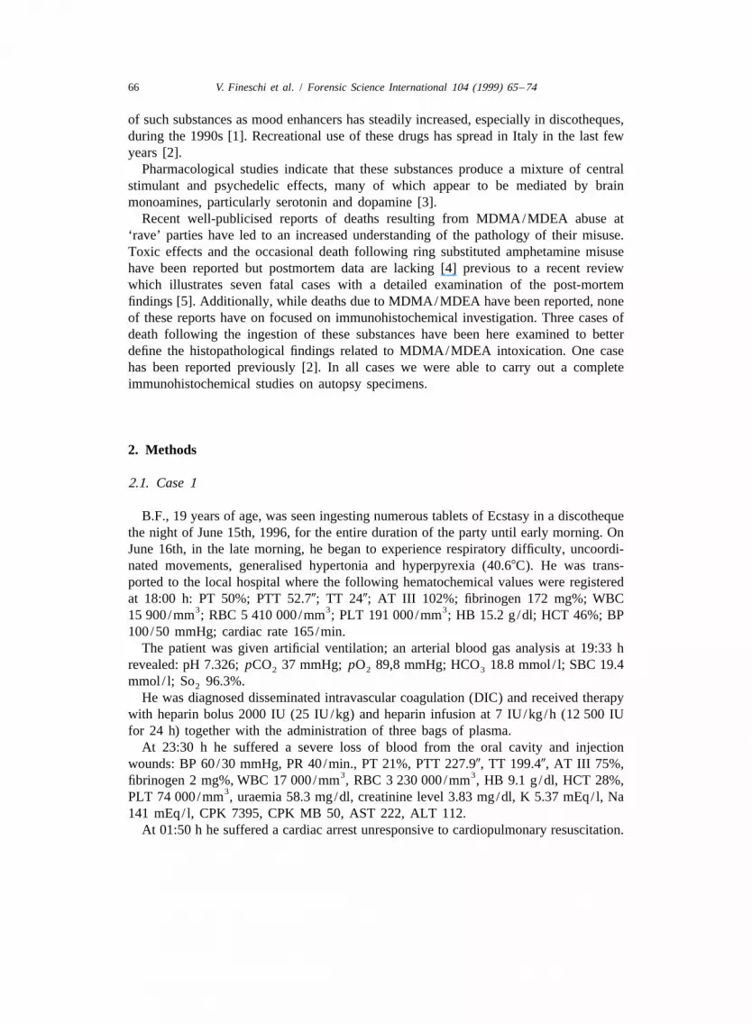
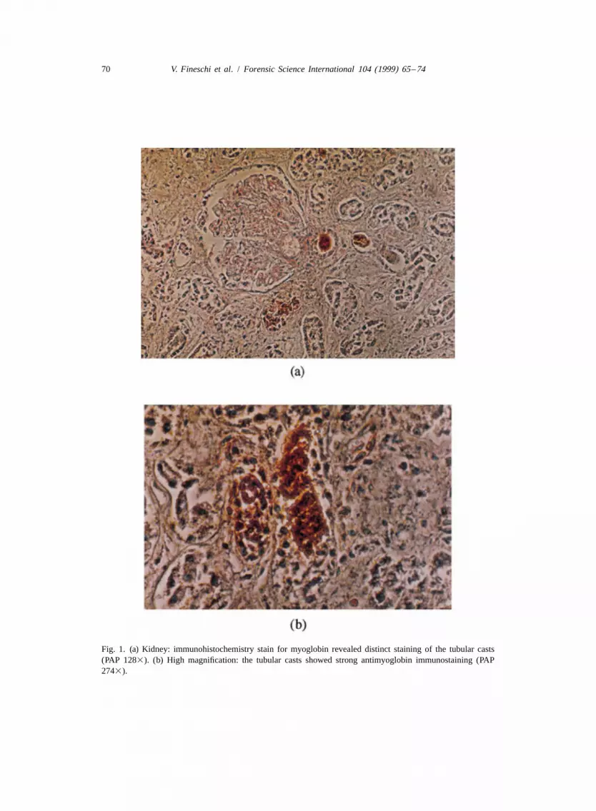
thrombi were already organised and were stained prominently by PTAH. In one caseacute tubular necrosis was observed. H&E examination revealed numerous reddish-brown granular and amorphous casts within the renal tubules. In all the cases thepresence of tubular casts was studied with immunohistochemical technique, thusdemonstrating their myoglobinic nature (Figs. 1 and 2).
3.1.6. MuscleIn two cases hypercontracted fibers with an absence of the striations and cystic
cavities adjacent to zones of fiber ruptures were present (Fig. 3a). The immuno-histochemical investigation for myoglobin performed according to Fechner and co-workers [7,9] showed accumulation of the protein in the breakage areas of the fibresespecially on the surface of the contraction caps (Fig. 3b).
4. Discussion
There are substantial similarities in the clinical and histopathological findings of thefatal cases herein presented. Two deaths were caused by single intoxication, respective-ly, from MDMA and MDEA and the third from both MDMA and MDEA. No otherdrugs were detected.
In the present study the pathological data are in accordance with a well studied anddocumented clinical entity, i.e. hyperthermia and disseminated intravascular coagulation[10–12]; however the macro and microscopic findings have not been definitivelyclassified or defined yet.
After the onset of deaths related to MDMA ingestion [13], reports have been compiledabout fatal arrhythmia caused by the ingestion of MDMA [14] or cases of hyperthermiafollowed by DIC [15–19]. Clinical proofs of hyperthermia, rhabdomyolysis and DIC arealso evident in deaths caused by MDEA intoxication with findings of subseroushaemorrhage and severe polyvisceral stasis [20–24]. Similar pathological findings aredescribed in the cases of death due to combined intoxication of MDMA and MDEA[2,25,26].
A report describes acute myocardial infarction associated with amphetamine abuse[27]. Potential explanations include coronary vasospasm, excessive catecholaminedischarge resulting in ischemic myocardial necrosis, and catecholamine mediated plateletaggregation with subsequent thrombus formation. The syndrome closely resembles acutemyocardial infarction due to cocaine abuse [3].
70 V. Fineschi et al. / Forensic Science International 104 (1999) 65 –74
Fig. 1. (a) Kidney: immunohistochemistry stain for myoglobin revealed distinct staining of the tubular casts(PAP 1283). (b) High magnification: the tubular casts showed strong antimyoglobin immunostaining (PAP2743).

V. Fineschi et al. / Forensic Science International 104 (1999) 65 –74 71
Fig. 2. Myoglobin in renal tubules (immunofluorescence polyclonal antibody against myoglobin, 2743).
A comparison of our findings with presently available related literature reveals slightdifferences in histopathological findings, particularly of the heart, liver and kidneys ascompared to those described in previous reports. The types of myocardial necrosis foundin our cases were coagulative myocytolysis. No histological signs of infarct necrosiswere detected. Coagulative myocytolysis, even if confined to few myocells, can beinterpreted as a histological sign of adrenergic overdrive [28]. Coagulative myocytolyticchanges may be related to the type and length of survival, particularly in subjectspredisposed to cardiac adrenergic response. It remains to be established if coagulativemyocytolysis is due to unspecific agonal stimuli or drug action. In other words, anyattempt to evaluate its meaning requires the recognition of older stages, the de-termination of survival time, and the exclusion of reanimative procedures.
In two cases, the study of muscle samples revealed hypercontracted fibers withdisruption of the cell architecture, typical pathological changes observed in deaths due tomalignant hyperpyrexia [29]. Malignant hyperthermia is a rare, inherited abnormalsusceptibility to certain drugs, mostly inhalational anaesthetics [30].
It is possible that individual susceptibility to the adverse effects of the amphetaminederivatives exists, with deficient demethylation of MDMA by debrisoquine hydroylase(CYP2D6), being shown in certain individuals. The absence of CYP2D6, a member ofthe cytochrome P450 superfamily of enzymes, in 5–9% of whites, may be another factorexplaining apparently idiosyncratic or severe responses to the drug [31].
Rhabdomyolysis and myoglobinuria are often described [10,32]. The presence ofmyoglobin in the kidney has been reported in a histopathological study only [2]. In a

72 V. Fineschi et al. / Forensic Science International 104 (1999) 65 –74
Fig. 3. (a) Muscle: hypercontracted fibres and cystic cavities adjacent to zones of fiber ruptures were present(Phaco 1283). (b) Muscle: immunohistochemical stain for myoglobin showed positive reaction (arrows) in thebreakage areas of the fibres. Depletion of myoglobin in lighter fibres (PAP 633).
recent review, myoglobin was not detected in the only two cases in which the kidneyswere examined [5]. We were able to demonstrate the presence of myoglobin in theproximal tubules in all the three cases here examined. Two cases presented mi-
V. Fineschi et al. / Forensic Science International 104 (1999) 65 –74 73
crothrombosis of the pulmonary and renal microcirculation related to DIC. Finally, intwo cases has been possible to demonstrate a hepatic microvescicular steatosis andaspects of centrilobular necrosis related to DIC in one case.
The genesis of the hepatic damage caused by MDMA/MDEA still remains un-explained and does not seem to be related to the dose or frequency of drug ingestion.Idiosyncratic reactions or individual susceptibility are likely pathogenic causes of thehepatic pictures described [33,34]. The quality of Ecstasy tablets should also beconsidered due to the established and documented presence of toxic contaminants [5].
The increase in cases of toxicity due to MDMA and drugs sold as ‘Ecstasy’ deservesto be publicised for various reasons. First, it is not possible to establish the cause ofsevere or fatal complications following the ingestion of Ecstasy with the data availableat present. Individuals who experience such an adverse reaction have often used the drugpreviously without problems [10]. Chemical and toxicological analysis of post-mortembiological material shows variable concentrations of amphetamines, suggesting hyper-sensitivity in the cases of low doses. Individual susceptibility to ring substitutedamphetamines may be related to its metabolism in the liver [35] and can explain severeor fatal responses to the drug [31]. Second, clinicians should be aware of the pattern oftoxicity so as to be able to perform correct diagnoses and treatments. Finally, from adiagnostic standpoint, in all ring-substituted amphetamines-related deaths, a completeimmunohistochemical study should be performed especially on muscle [36] and kidney[37] samples.
References
[1] R.W. Byard, J. Gilbert, R. James, R.J. Lokan, Amphetamine derivative fatalities in South Australia. Is‘Ecstasy’ the culprit?, Am. J. Forensic Med. Pathol. 19 (1998) 261–265.
[2] V. Fineschi, A. Masti, Fatal poisoning by MDMA (ecstasy) and MDEA: a case report, Int. J. Leg. Med. 5(1996) 272–275.
[3] F.D. Kolodgie, A. Burke, J. Narula, F.G. Mullick, R. Virmani, Vascular effects of substance abuse, in:S.B. Karch (Ed.), Drug Abuse Handbook, CRC Press, Boca Raton, 1998, pp. 113–129.
[4] J.M. White, F. Bochner, R.J. Irvine, The agony of ‘ecstasy’, Med. J. Aust. 166 (1997) 117–118.[5] C.M. Milroy, J.C. Clark, A.R.W. Forrest, Pathology of deaths associated with ‘ecstasy’ and ‘eve’ misuse,
J. Clin. Pathol. 49 (1996) 149–153.[6] S.M. Hsu, A. Werner, P.A. Goyle Griffiths, L. Raine, Demonstration of myoglobin in formalin-fixed renal
sections by immunoperoxidase technique, Am. J. Clin. Pathol. 77 (1982) 316–319.[7] G. Fechner, R. Hauser, M.A. Sepulchre, B. Brinkmann, Immunohistochemical investigations to
demonstrate vital direct traumatic damage of skeletal muscle, Int. J. Leg. Med. 104 (1991) 215–219.[8] Y. Esaki, K. Hirokawa, T. Fukazawa, T. Matsuda, Immunohistochemical study on the liver in autopsy
cases with disseminated intravascular coagulation (DIC) with reference to clinicopathological analysis,Virchow’s Arch. 404 (1984) 229–241.
[9] G. Fechner, T. Bajanowsky, B. Brinkmann, Immunohistochemical alterations after muscle trauma, Int. J.Leg. Med. 105 (1993) 203–207.
[10] A.R. Bodenham, A. Mollick, New dimensions in toxicology: hypertermic syndrome following amphet-amine derivative, Intens. Care Med. 22 (1996) 622–624.
[11] I.H. Fahal, D.F. Sallomi, M. Yaqoob, G.M. Bell, Acute renal failure after ecstasy, Br. Med. J. 305 (1992)29.
[12] G.R. Screaton, M. Singer, H.S. Cairns, A. Thrasher, M. Sarner, S.L. Cohen, Hyperpyrexia andrhabdomyolisis after MDMA (ecstasy) abuse, Lancet 339 (1992) 677–678.

74 V. Fineschi et al. / Forensic Science International 104 (1999) 65 –74
[13] G.P. Dowling, E.T. McDonough, R.O. Bost, Eve and Ecstasy. A report of five deaths associated with theuse of MDEA and MDMA, J. Am. Med. Assoc. 257 (1987) 1615–1617.
[14] R.V. Suarez, R. Riemersma, ‘Ecstasy’ and sudden cardiac death, Am. J. Forensic Med. Pathol. 4 (1988)339–341.
[15] J.A. Henry, K.J. Jeffreys, S. Dawling, Toxicity and death from 3,4-ethylenedioxymetamphetamine(ecstasy), Lancet 340 (1992) 384–387.
[16] I.S. Chadwick, A. Linsley, A.J. Freemont, B. Doran, Ecstasy, 3,4-methylenedioxymetamphetamine(MDMA), a fatality associated with coagulopathy and hyperthermia, J. R. Soc. Med. 84 (1991) 371.
[17] N.T.A. Campkin, U.M. Davies, Another death from ecstasy, J. R. Soc. Med. 85 (1992) 61.[18] K.J. Dar, M.E. McBrien, MDMA induced hyperthermia: report of a fatality and review of current
therapy, Intens. Care Med. 22 (1996) 995–996.[19] P.D. Mueller, W.S. Korey, Death by ‘Ecstasy’: the serotonin syndrome?, Ann. Emerg. Med. 32 (1998)
377–380.[20] S. Iwersen, A. Schmoldt, Two very different fatal cases associated with the use of methyl-
enedioxyethylamphetamine (MDEA): Eve as deadly as Adam, Clin. Toxicol. 34 (1996) 241–243.[21] C. Lora-Tamayo, T. Tena, A. Rodriguez, Amphetamine derivative related deaths, Forensic Sci. Int. 85
(1997) 149–157.[22] J. Arimany, J. Medallo, A. Pujol, A. Vingut, J.C. Borondo, J. L Valverde, Intentional overdose and death
with 3,4-methylenedioxyethamphetamine (MDEA; Eve), Am. J. Forensic Med. Pathol. 19 (1998)148–151.
[23] A.M. Tsatskis, M.N. Michalodimitrakis, A.N. Patsalis, MDEA related death in Crete: a case report andliterature review, Vet. Hum. Toxicol. 39 (1997) 241–244.
[24] W. Weinmann, M. Bohnert, Lethal monointoxication by overdosage of MDEA, Forensic Sci. Int. 91(1998) 91–101.
[25] A.R.W. Forrest, J.H. Galloway, I.D. Marsh, G.A. Strachan, J.C. Clark, A fatal overdose with 3,4,-methylenedioxyamphetamine derivatives, Forensic Sci. Int. 64 (1994) 57–59.
[26] D.E. Cox, K.R. Williams, Adam or Eve? A toxicological conundrum, Forensic Sci. Int. 77 (1996)101–108.
[27] T.T. Beshour, Acute myocardial infarction resulting from amphetamine abuse: a spasm–thrombusinterplay?, Am. Heart J. 128 (1994) 1237–1239.
[28] V. Fineschi, M. Di Paolo, C.W. Wetli, G. Baroldi, Myocardial necrosis and cocaine. A quantitativemorphologic study in 26 cocaine associated death, Int. J. Leg. Med. 4 (1997) 193–198.
[29] D.G.F. Harriman, The pathology of malignant hyperpyrexia, in: J. Walton, F.I. Mastalgia (Eds.), SkeletalMuscle Pathology, Churchill Livingstone, Edinburgh, 1982, pp. 575–591.
[30] N.C. Mambo, M.D. Silver, P.R. Mc Laughlin, V.F. Huckell, P.M. Mc Ewan, B.A. Britt, J.E. March,Malignant hyperthermia susceptibility, Hum. Pathol. 11 (1980) 381–388.
[31] G.T. Tucker, M.S. Lennard, S.W. Ellis, H.F. Woods, A.K. Cho, L.Y. Lin, A. Hiratsuyka, D.A. Schmitz,I.Y. Chu, The demethylation of methylenedioxymethamphetamine (‘ecstasy’) by debrisoquine hydroxy-lase (CYP2D6), Biochem. Pharmacol. 47 (1994) 1151–1156.
[32] A.J. Larner, Complications of ‘ecstasy’ misuse, Lancet 340 (1992) 726.[33] A.J. Ellis, J.A. Wendon, B. Portman, R. Williams, Acute liver damage and ecstasy ingestion, Gut 38
(1996) 454–458.[34] H. Fidler, A. Dhillon, D. Gertner, A. Burroughs, Chronic ecstasy (3,4-methylenedioxymetamphetamine)
abuse: a recurrent and unpredictable cause of severe acute hepatitis, J. Hepatol. 25 (1996) 563–566.[35] G.N. Rutty, C.M. Milroy, The pathology of the ring-substituted amphetamine analogue 3,4-methyl-
enedioxymethylamphetamine (MDMA, ‘Ecstasy’), J. Pathol. 181 (1997) 255–256.¨ ¨[36] P. Saukko, E. Lignitz, T. Sarkioja, W. Keil, Traumatische, ischamische und toxische Rhabdomyolyse, Z.
Rechtsmed. 102 (1989) 117–126.[37] R.A. Grossman, R.W. Hamilton, B.M. Morse, A.S. Penn, M. Goldberg, Nontraumatic rhabdomyolysis and
acute renal failure, N. Engl. J. Med. 291 (1974) 807–811.