molecular basis of the selective binding of mdma enantiomers
TRANSCRIPT
1
Molecular basis of the selective binding of MDMA enantiomers to the
alpha4beta2 nicotinic receptor subtype: synthesis, pharmacological evaluation
and mechanistic studies
Salomé Llabrés,1,& Sara García-Ratés,2,& Edgar Cristóbal-Lecina3 Antoni Riera,3 José
Ignacio Borrell,4 Jorge Camarasa, 2 David Pubill,2 F. Javier Luque,1 Elena Escubedo2,*
1 Department of Physical Chemistry and Institut of Biomedicine (IBUB), Faculty of Pharmacy, Campus
Avda. Prat de la Riba 171, Santa Coloma de
Gramenet, E-08921 Barcelona, Spain
2 Department of Pharmacology and Therapeutic Chemistry and Institut of Biomedicine (IBUB), Faculty
of Pharmacy, University of Barcelona, Nucli Univ. Pedralbes, E-08028 Barcelona
3 Institute for Research in Biomedicine (IRB Barcelona), Baldiri i Reixac, 10, 08028 Barcelona, Spain
4 IQS School of Engineering, Universitat Ramon Llull, Via Augusta 390, E-08017 Barcelona, Spain
& These authors have contributed equally to this work.
* Corresponding author: E-mail: eescubedo @ ub.edu; Phone: +34 934024531
P-p
rint –
Ava
ilabl
e in
http
://w
ww
.rece
rcat
.cat
2
Keywords
Alpha4Beta2 nicotinic receptor, MDMA, enantioselective binding, receptor up-regulation, molecular
modelling, stereoselective synthesis
Abbreviations
AP: alternative pose; BP: best pose; CNS: central nervous system; DA: dopamine; MD: molecular
dynamics; MDMA: N 3,4-methylenedioxymethamphetamine; nAChR: nicotinic acetylcholine receptor;
QM/MM: quantum mechanical/molecular mechanical; SIE: Solvated Interaction Energy.
P-p
rint –
Ava
ilabl
e in
http
://w
ww
.rece
rcat
.cat
3
Abstract
T nicotinic acetylcholine receptor (nAChR) is a molecular target of 3,4-
methylenedioxymethamphetamine (MDMA), a synthetic drug also known as ecstasy, and it modulates the
MDMA-mediated reinforcing properties. However, the
subtype still remains unknown. Since the two enantiomers exhibit different pharmacological profiles and
stereoselective metabolism, the aim of this study is to assess a possible difference in the interaction of the
MDMA enantiomers with this nAChR subtype. To this end, we report a novel simple, yet highly efficient
enantioselective synthesis of the MDMA enantiomers, in which the key step is the diastereoselective
reduction of imides derived from optically pure tert-butylsulfinamide. The enantioselective binding to the
receptor is examined using [3H]epibatidine in a radioligand assay. Even though the two enantiomers
induced a concentration-dependent binding displacement, (S)-MDMA has an inhibition constant 13-fold
higher than (R)-MDMA, which shows not significantly different from unity, implying
a competitive interaction. Furthermore, when NGF-differentiated PC12 cells were pretreated with the
compounds, a significant increase in binding of [3H]epibatidine was found for (R)-MDMA, indicating up-
regulation of heteromeric nAChR in the cell surface. Finally, docking and molecular dynamics studies
have been used to identify the binding mode of the two enantiomers, which provides a structural basis to
justify the differences in affinity from the differential interactions played by the substituents at the
stereogenic center of MDMA. The results provide a basis to explore the distinct psychostimulant profiles
nAChR subtype.
P-p
rint –
Ava
ilabl
e in
http
://w
ww
.rece
rcat
.cat
4
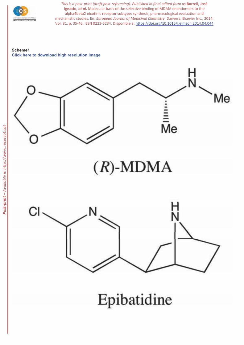
3,4-Methylenedioxymethamphetamine (MDMA; Scheme 1), also known as ecstasy, is a synthetic drug
widely abused in the United States and Europe, where it is taken in a recreational context due to its
stimulant and hallucinogenic properties. Amphetamines produce a dose-dependent increase of locomotor
activity in rodents [1], which reflects an increase of dopamine (DA) transmission in the nucleus
accumbens [2]. Activation of nicotinic acetylcholine receptors (nAChRs) is a common event in the
pathway used by several addictive drugs to stimulate the mesolimbic DA system, which is a relevant
component of the brain stimulation reward pathway [3,4]. Thus, in rats systemic nicotine or alcohol
administration elevates extracellular DA levels in the nucleus accumbens, an effect that requires
stimulation of nAChRs in this area as well as in the ventral tegmental one, in which the mesolimbic
dopaminergic cell bodies are located [5 7].
is involved in regulating the nicotine dependence [8], is
strongly associated with DA release in the nucleus accumbens [9] and with drug-seeking behaviour
[10,11]. Therefore, it is generally assumed that the nAChR subtype plays a major role in reinforcing
the effects of nicotine and has been recognized as a major target in addictive pathologies. This is reflected
in the hypothesis that the main addictive drugs with psychostimulant properties share the capacity to
interact with receptors [12,13] In this regard, the nAChR subtype can also explain the
mechanism of other reinforcing substances. A particular feature of nAChR is that chronic exposure to
nicotinic ligands induces a higher level of epibatidine (Scheme 1) -containing nAChRs,
leading to a functional up-regulation [14 16] that should enhance the addictive effects of these
compounds [8]. This phenomenon is independent of de novo protein synthesis. Vallejo et al. [17]
-subtype receptors exist in two interconvertible states, with high and with low
af inities for nicotine, respectively, and that chronic exposure to a nAChR ligand stabilizes a larger fraction of
receptors in the high-af inity state at the membrane surface. Other authors [18,19] suggest a chaperone-
maturation enhancing effect of nicotine on immature receptors inhibiting its degradation. This is not related to the
ef icacy of the ligand in activating the receptor but to its af inity.
P-p
rint –
Ava
ilabl
e in
http
://w
ww
.rece
rcat
.cat
5
We have demonstrated that nAChRs are a pharmacological target for MDMA and mediate some actions
of this drug of abuse [20,21] including analgesia or locomotor activity [22], tumor necrosis factor alpha
suppression [23] and neurotoxicity [24 26]. Recently, Ciudad-Roberts et al. [27] established that the
nAChR subtype modulates MDMA-mediated reinforcing properties. Previous studies [20,28] had
also shown that MDMA has affinity for and induced their up-regulation in PC12 cells
following a similar mechanism than that of nicotine. Lately it was reported that rat exposure to nicotine or
MDMA each induced significant increases in [3H]epibatidine binding (about 30 and 35%, respectively)
with respect to saline-treated rats, and that this effect was significantly potentiated (up to 72%) when the
two drugs were associated [21] . Consequently, knowledge of the interaction of MDMA with nAChRs is
valuable to understand the role of this receptor in both nicotine and psychostimulant addictions.
(Scheme 1). Even though it is
consumed as a racemate, the two enantiomers exhibit different pharmacological profiles and
stereoselective metabolism [29,30]. Thus, in vitro models have revealed that (S)-MDMA is more active
than (R)-MDMA on the central nervous system (CNS) [31 34]. On the other hand, MDMA is known to
undergo extensive hepatic metabolism, leading to the formation of highly redox-active metabolites that
have been implicated in MDMA-induced hepato-, neuro-, nephron- and cardiotoxicity [35,36]. However,
these effects are sensitive to the enantiomeric form of MDMA. For instance, (R)-MDMA primarily
contributes to the depletion of the hepatic glutathione induced by the racemic mixture [37]. Moreover,
studies in animal models revealed that (S)-MDMA rather than (R)-MDMA contributes to the serotonergic
injury and astroglial and microglial activation associated with MDMA consumption [38,39]. Finally, rac-
MDMA exerts simultaneous effects, reducing L-DOPA-induced dyskinesia and extending its
antiparkinsonian benefits (ON-time) by 5-HT2A antagonism and serotonin transporter selective inhibition,
which arise from its R and S enantiomers, respectively [40].
P-p
rint –
Ava
ilabl
e in
http
://w
ww
.rece
rcat
.cat
6
The aim of this study is to characterize for the first time the interaction of the two enantiomeric forms of
MDMA upon binding to subtype, and to explore the molecular basis of the selective
binding to this receptor. To this end, we have examined a novel and efficient strategy for the synthesis of
both enantiomers of MDMA based on the diastereomeric reduction of the imides derived from optically
pure tert-butylsulfinamide. Afterwards, we have evaluated the experimental affinity of both enantiomers
for the nAChR using radioligand binding assays. Attention has also been paid to the differential
-regulation. Finally, molecular modelling
and computational techniques have been used to study the interaction with the nAChR subtype and
to identify the molecular determinants responsible of the different affinity of the enantiomeric forms of
MDMA.
2. Results and discussion
2.1. Synthesis of MDMA enantiomers
Several enantioselective syntheses of amphetamines have been reported [41 45]. The first stereoselective
synthesis of MDMA was based on the hydrogenation of imines derived from 1-phenylethylamine as a
chiral auxiliary [41]. The same quiral auxilliary was used by Pizarro et al. in the preparation of
enantiomerically enriched HMMA (4-hydroxy-3-methoxymethamphetamine) and HHMA (3,4-
dihydroxymethamphetamine) [42]. An enzymatic methodology was reported for the preparation of chiral
cyanohydrins that were transformed into 3,4-methylenedioxyamphetamines [43]. Wagner et al. reported a
stereoespecific synthesis of amphetamines, although it was not applied to MDMA [44]. Recently, Huot et
al. published another synthesis of MDMA enantiomers by enantiospecific ring opening of aziridines with
a Grignard reagent, even though the experimental details have not been described [40]. These synthetic
approaches are not practical since they involve either low diastereoselectivities, costly preparation of the
starting ketones or lack of experimental procedures. Therefore, we have developed a new and efficient
synthesis of both enantiomers of MDMA based on the diastereomeric reduction of the imides derived
from optically pure tert-butylsulfinamide.
P-p
rint –
Ava
ilabl
e in
http
://w
ww
.rece
rcat
.cat
7
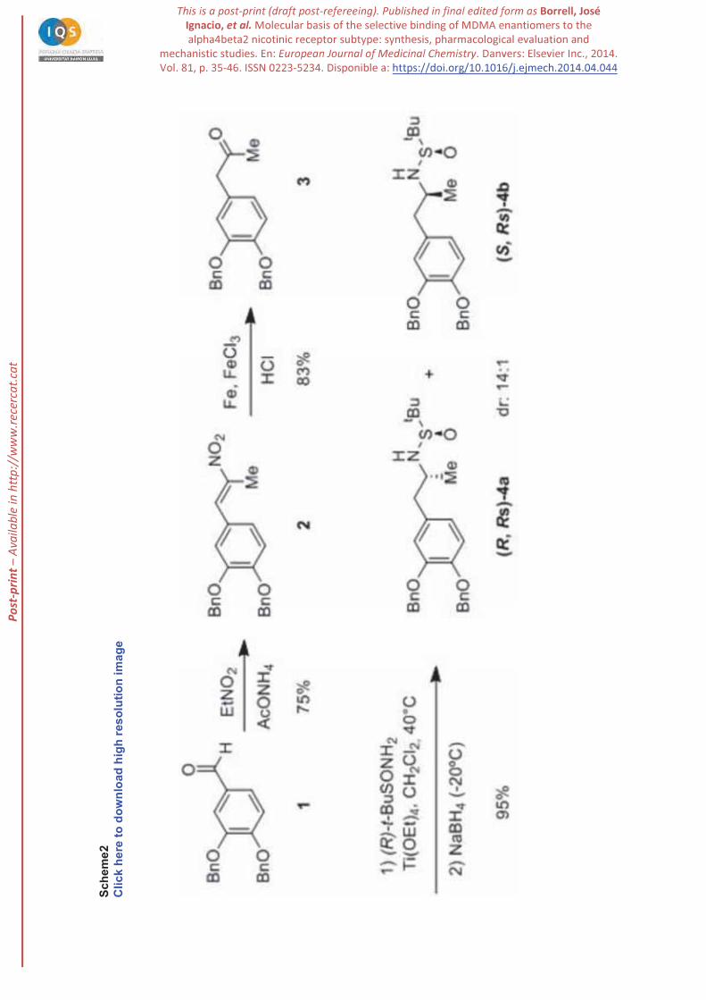
Our synthesis started from commercially available 3,4-dibenzyloxybenzaldehyde (1), which was
transformed into the methyl ketone 3 by condensation with nitroethane followed by reduction with
Fe/HCl (Scheme 2). The corresponding imine, formed by treatment of ketone 3 with (R)-(+)-tert-
butylsulfinamide and Ti(OEt)4 was reduced in situ with NaBH4 at room temperature to afford the two
diastereomeric sulfinamides 4 in excellent yield but with moderate (3:1) diastereomeric ratio. Gratifyingly,
the diastereoselectivity rose to 14:1 by lowering the temperature to -20ºC in the reduction step. The major
isomer was easily purified by crystallization from hexane affording diastereomerically pure (R,RS)-4a as
a white solid (Scheme 2).
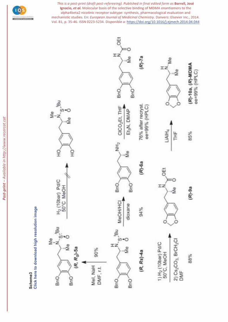
Methylation of sulfonamide (R,RS)-4a was carried out uneventfully with NaH and MeI in DMF at room
temperature to give (R,RS)-5a in 90% yield. However, we were unable to cleave the benzyloxy groups by
catalytic hydrogenation (Pd/C) even at high hydrogen pressures (50 bar) and/or temperatures (50 ºC). We
hypothesized that sulphur byproducts derived from sulfinimides hampered the reaction poisoning the
catalyst (Scheme 3). Alternatively, derivatization of (R,RS)-4a as ethyl carbamate was unsuccessful since
this sulfonamide is a very weak nucleophile. Therefore, we envisaged the hydrolysis of (R,RS)-4a to the
primary amine. Reductive cleavage of N-S bond was easily performed with 4M MeOH/HCl in dioxane
affording amine (R)-6a in 94% yield. Formation of the carbamate of the primary amine (R)-7a was
carried out with ClCO2Et, Et3N in DMF. This compound was highly crystalline and allowed to confirm
its high optical purity (99%ee by chiral HPLC). Cleavage of the dibenzyloxy group in (R)-7a could be
performed by hydrogenolysis and the resulting diphenol was cyclized with bromochloromethane and
cesium carbonate as described by Pizarro et al. [42]. Finally, reduction with lithium aluminum hydride in
THF yielded the desired compound (R)-10a (MDMA) in 85% yield, which was isolated as a
hydrochloride. The enantiomeric purity of the final product was checked by chiral HPLC of the
corresponding N-Boc derivative, being 99% ee, as expected. Since both enantiomers of tert-
butylsulfinamide are commercially available, the same sequence starting from (S)-( )-tert-
butylsulfinamide afforded the (S)-MDMA enantiomer also in 99%ee.
P-p
rint –
Ava
ilabl
e in
http
://w
ww
.rece
rcat
.cat
8
2.2. Experimental binding affinity of MDMA enantiomers
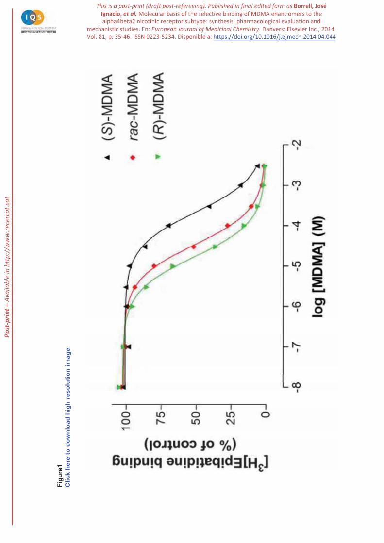
When [3H]epibatidine was used as a radioligand to label nAChR subtype, all the compounds
induced a concentration-dependent binding displacement, with IC50 values in the micromolar range (see
Figure 1 and Table 1), indicating that they can directly interact with the nicotinic receptor. The affinity of
(R)-MDMA for the [3H]epibatidine binding site was higher than (S)-MDMA, as noted in the 13-fold ratio
between the inhibition constants (Ki) determined for both enantiomers, respectively (see Table 1). The Ki
value of (R)-MDMA is in the submicromolar range (Ki = 0.63), which thus compares with the low
micromolar concentrations found in the brain after administration of this drug [45,46] . In fact, the
binding constant for this heteromeric receptor is lower than the Ki value determined for the serotonin
) [40].
The Hill coefficients resulting from the analysis of competition data of (R)-MDMA or rac-MDMA versus
[3H]epibatidine were not significantly different from unity, pointing to a competitive displacement. In
S)-MDMA was significantly less than unity, suggesting a more
complex interaction.
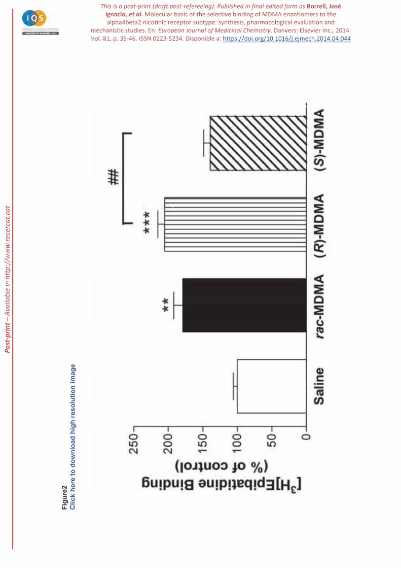
2.3. Up-regulation of heteromeric nAChR by MDMA enantiomers
nAChR up-regulation. When NGF-differentiated PC12 cells were pretreated with (R)-MDMA, (S)-
[3H]epibatidine was found for (R)-MDMA and rac-MDMA, indicating up-regulation of heteromeric
nAChR. It is known that this up-regulation is independent of de novo protein synthesis [47] but it is
consistent with a higher ratio of the high-affinity component of [3H]epibatidine binding [17], as suggested
by previous studies on chronic exposure to nicotine [28] . This effect was significantly higher for (R)-
P-p
rint –
Ava
ilabl
e in
http
://w
ww
.rece
rcat
.cat
9
MDMA than for the racemic mixture (see Figure2). (S)-MDMA did not significantly modify the nAChR
population. These results support the direct interaction with nAChR reported above and corroborates the
higher affinity of (R)-MDMA compared with (S)-MDMA.
2.4. Binding mode of MDMA
Previous studies have indicated that the nicotinic binding site is located at the centre of the extracellular
domain of the receptor and lies at the interface between the
principal (P) and complementary (C) components, respectively [48 50]. Several residue stretches referred
to as loops A, B and C in the subunit, and D, E and F in the
following the symbols "
and the numbering refers to the Conserved residues at the binding site are the
aromatic amino acids Tyr128 (loop A), Trp184 and Tyr186 (loop B), Tyr225 and Tyr232 (loop C)
and Trp81 (loop D). Residues Val135, Phe143 and Leu145 shape the top of the binding site. The
bridged cysteine residues in loop C ( Cys227 and Cys228 -subunit.
The binding mode of epibatidine, (R)-MDMA and (S)-MDMA was examined by means of docking
calculations with MOE (Chemical Computing Group). The predicted pose of epibatidine nicely
reproduced the binding mode observed in the X-ray structure (PDB entries 2BYQ [51] and 3SQ6 [52]),
thus giving confidence to the docking protocol. In particular, the binding mode reproduced the cation-pi
interaction with the indole ring of Trp184 (corresponding to Trp147 in 2BYQ) and the hydrogen bond
with the carbonyl unit of this residue (see Figure S1 in Supporting Information). It is worth noting that
these interactions are crucial for modulating the affinity of nicotine toward brain and muscle receptor
subtypes [53]. Recent studies have also highlighted the relevance of the hydrogen bond with the backbone
carbonyl of Trp184 for the binding of ligands to nAChRs [54].
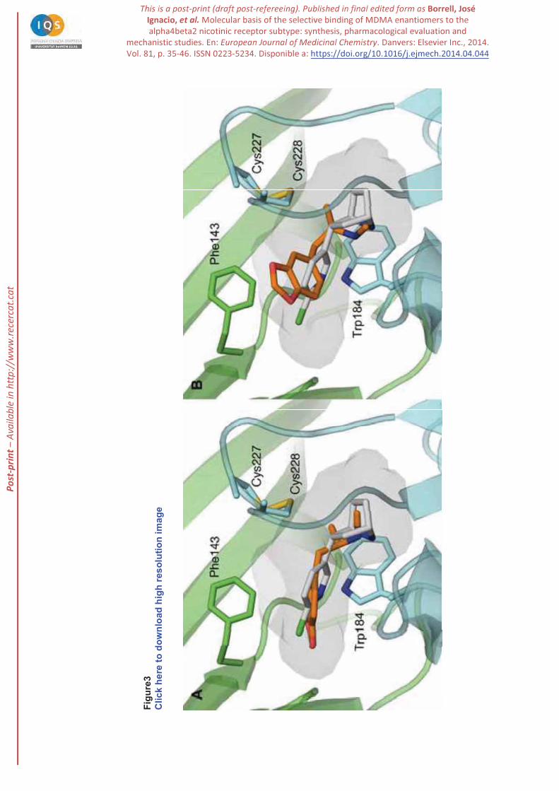
For (R)-MDMA the best pose (denoted BP hereafter; MOE score: -6.7 kcal/mol) obtained in docking
calculations nicely matches the structure of epibatidine, as noted in the superposition of the protonated
P-p
rint –
Ava
ilabl
e in
http
://w
ww
.rece
rcat
.cat
10
secondary nitrogen atoms and the overlap between the chloropiperidine and methylenedioxybenzene
moieties of these compounds (see Figure 3A). The analysis of the results also showed an alternative pose
(designated AP; MOE score: -4.9 kcal/mol) that retains the interaction of the secondary amine with
Trp184, but the methylenedioxybenzene ring adopts a different orientation and fills the region located
between the disulfide bridge formed by Cys227- Cys228 and Phe143 (Figure 3B).
Docking of (S)-MDMA led to similar binding modes as those described for (R)-MDMA (see Figure S2 in
Supporting Information). The scores of the two poses (MOE scores of -6.4 and -5.1 kcal/mol for BP and
AP solutions, respectively) compare well with those found for (R)-MDMA, thus reflecting the similar
arrangement of the protonated nitrogen atom and the methylenedioxybenzene unit in the binding site for
the equivalent poses of both (R)- and (S)-MDMA.
2.5. Structural basis of the enantioselective binding.
MD simulations were run to examine the structural integrity of the two binding modes (BP, AP) found for
(R)- and (S)-MDMA. For (R)-MDMA, the simulation started from the BP binding mode (Figure 3A;
designated MD-BP) remained stable along the whole trajectory, as noted in the lack of significant
alterations in the positional root-mean square-deviation for the residues that shape the binding site
(RMSD ~ 1.9 Å). In contrast, when the AP binding mode (Figure 3B; denoted MD-AP) was used as the
starting structure, a fast rearrangement was observed for the ligand, which adopted the BP binding mode
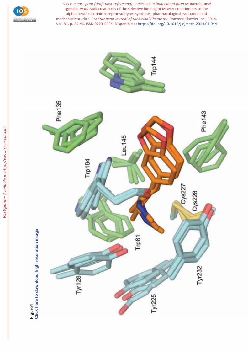
for the rest of the trajectory. The ligand is closely packed in the binding site, forming interactions that are
preserved along the whole trajectory (Figure 4). Thus, the secondary amine nitrogen is located at around
3.3 Å from the indole ring of Trp184 and maintains the hydrogen bond with the carbonyl group (average
distance of 2.9 Å). In addition, the amine nitrogen forms a hydrogen bond with the hydroxyl group of
Tyr128, which in turn is hydrogen-bonded to the carbonyl oxygen of Ser183. The
methylenedioxybenzene moiety fills a hydrophobic cavity formed by Thr185, Phe338, Val135,
Phe143, Trp144 and Leu145. The methyl group in the ethylamine chain faces the six-membered rings
P-p
rint –
Ava
ilabl
e in
http
://w
ww
.rece
rcat
.cat
11
of (average distance of 3.7 Å), and the phenol ring of Tyr128 (at 4.0 Å). The
ethylene chain forms van der Waals contacts with the disulfide bridge (at around 3.5 Å). Finally, the N-
bound methyl interacts with the aromatic ring of Tyr225 and Tyr232 (average distance of 3.9 and 3.5
Å, respectively). Overall, the consistency of the binding modes found in the two independent trajectories
supports the binding mode proposed for (R)-MDMA.
The trajectories run for the two binding modes of (S)-MDMA did not converge to a common binding
mode, and the ligand retained the distinct orientation of the methylenedioxybenzene unit. In agreement
with docking results, SIE calculations favors the BP binding mode by 0.7 kcal/mol, which reflects the
enhanced van der Waals component due to the deeper insertion of the methylenedioxybenzene unit in the
binding site (Table S1). Noteworthy, a common feature in the two simulations is the insertion of a water
molecule that fills the region between the secondary amine nitrogen and the indole rings of Trp184 and
(Figure S3). The entrance of the water molecule takes place through the passage shaped by
Cys227 and Cys228 in loop C, which is more flexible in the
presence of (S)-MDMA (Figure S4).
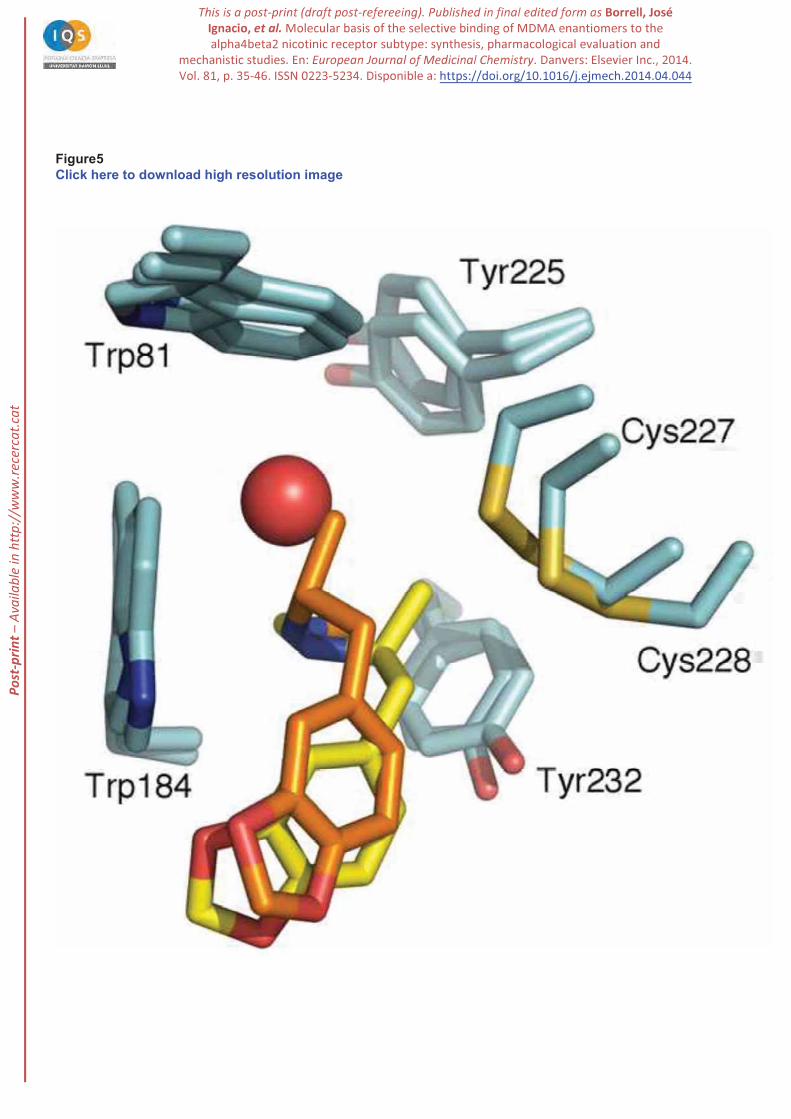
The origin of the different behaviour found for the two enantiomeric species can be attributed to the
specific interactions formed by the methyl group bound to the stereogenic center in MDMA. Thus, for
(R)-MDMA the methyl group is tightly packed against
filled by the water molecule in the complex with (S)-MDMA, whereas the methyl group fills a pocket
formed by the disulfide bridge and residues Tyr225 and Tyr232 (Figure 5). Overall, these findings
suggest that the specific orientation of the methyl group in (S)-MDMA makes the loop C to adopt a less
stable fold onto the ligand in the binding pocket, which would exhibit larger fluctuations compared to the
binding of (R)-MDMA -
conformation to the structural features of the ligand bound to the receptor [51], the largest flexibility
observed for (S)-MDMA might thus be related to the weaker binding compared to (R)-MDMA.
P-p
rint –
Ava
ilabl
e in
http
://w
ww
.rece
rcat
.cat
12
2.6. Relative affinities between MDMA enantiomers
To calibrate the reliability of the BP binding mode for both (R)- and (S)-MDMA, the relative affinity of
the two enantiomers was estimated from QM/MM calculations using suitable thermodynamic cycles
(Figure S5). The QM/MM interaction energy between ligand and receptor was found to favour the
binding of (R)-MDMA by 3.2 kcal/mol with regard to the monohydrated S enantiomer. This contribution
mainly comes from the van der Waals term, which favours the binding of the R enantiomer by 4.6
kcal/mol, suggesting a tighter packing of (R)-MDMA in the binding pocket. For the S enantiomer,
however, the QM/MM interaction energy must be corrected by the contribution due to the formation of
the (S)-MDMA-water complex, which was estimated to be -11.4 kcal/mol (at the MP2 level with
inclusion of the basis set superposition correction). This contribution, nevertheless, must be corrected by
the entropic change due to formation of the monohydrated (S)-MDMA, which is estimated to be +8.0
kcal/mol at 298 K, and by the contribution due to the vaporization of the water molecule from liquid,
which amounts to +2.1 kcal/mol. By taking into account these corrections, the formation of the (S)-
MDMA-water complex is estimated to be -1.3 kcal/mol. Accordingly, the binding of (R)-MDMA is
predicted to be favored by 1.9 kcal/mol, which compares well with the free energy difference determined
from the inhibition constants reported in Table 1 (estimated to be -1.5 kcal/mol).
4. Conclusions
Since the two enantiomers of MDMA exhibit different pharmacological profiles and stereoselective
metabolism, it is necessary to discern the different mechanism associated with the enantioselective
activity of the two enantiomeric forms of this drug, specifically regarding to its interaction with the
nicotinic acetylcholine receptor. To this end, this study reports a new and efficient synthesis of both
enantiomers of MDMA based on the diastereomeric reduction of the imides derived from optically pure
tert-butylsulfinamide, which provides a simple, yet practical way of obtaining the two enantiomers of
MDMA. The experimental data point out that the nicotinic receptor has larger binding affinity for
P-p
rint –
Ava
ilabl
e in
http
://w
ww
.rece
rcat
.cat
13
(R)-MDMA than for (S)-MDMA, as noted by the 13-fold ratio between the inhibitions constants
determined in radiolabeled assays. The enantioselective binding of (R)-MDMA is consistent with the
enhanced up-regulation of the nAChR subtype determined in PC12 cultured cells. Therefore, these
results indicate that (R)-MDMA is the main effector of the effects mediated by this receptor subtype. The
molecular modelling studies point out that the two enantiomers adopt a similar arrangement in the
binding pocket of the receptor, even though there are significant differences regarding the interactions of
the methyl group bound to the stereogenic center. As a result, the loop C exhibits larger fluctuations in the
presence of (S)-MDMA, which facilitates the access of water molecules that mediate the binding of the
ligand, whereas a tighter interaction is found for (R)-MDMA. Overall, this provides a structural basis to
pursue the development of novel compounds useful to understand the reinforcing properties of MDMA.
5. Experimental
5.1. Chemical synthesis
(E)-1,2-Dibenzyloxy-4-(2-nitroprop-1-en-1-yl)benzene (2). Into a 250 mL round bottomed flask, 3,4-
dibenzyloxybenzaldehyde 1 (7.94 g, 24.95 mmol) and ammonium acetate (1.92 g, 24.95 mmol) were
dissolved in nitroethane (120 mL) and the mixture heated at reflux for 17h. Nitroethane was then removed
under reduced pressure. The crude was dissolved in ethyl acetate (25 mL) at 40ºC, cooled to r.t. and
precipitated with hexane. The solid was filtered and the procedure repeated with the filtrate. Compound 2
(6.49 g, 75% yield) was obtained as a yellow solid.
1H-NMR (400 MHz, CDCl3): = 2.30 (s, 3H, CH3), 5.21 (s, 2H, CH2), 5.23 (s, 2H, CH2), 6.96-6.98 (m,
1H, CH), 6.99 (s, 1H, CH), 7.00 (m, 1H, CH), 7.29-7.49 (m, 10H, CH), 7.97 (s, 1H, CH). 13C-NMR (100
MHz, CDCl3): = 14.1 (CH3), 71.1 (CH2), 71.6 (CH2), 114.4 (CH), 117.0 (CH), 125.0 (CH), 127.27
(CH), 127.33 (CH), 128.18 (CH), 128.22 (CH), 128.79 (CH), 128.81 (CH), 133.8 (CH), 136.7 (C), 136.9
P-p
rint –
Ava
ilabl
e in
http
://w
ww
.rece
rcat
.cat
14
(C), 146.2 (C), 148.7 (C), 150.9 (C=O) ppm. IR max: 2923, 2857, 1512, 1308, 1264 cm-1. HRMS
(ESI): calc. for C23H22O4N+ ([M+H]+): 376.1543; found: 376.1547.
1-(3,4-bis(benzyloxy)phenyl)propan-2-one (3). In a 3 necked round bottomed flask, nitro compound 2
(1.24 g, 3.31 mmol), Fe powder (1.11 g, 19.9 mmol, 6 equiv) and FeCl3 (0.12 mg, 0.76 mmol) were
suspended in ethanol (15 mL). To this mixture water (30 mL) and conc HCl (2 mL) were added and the
solution was heated at reflux during 5h. The reaction was cooled down and the pH adjusted to 8 with 40%
NH4OH solution, filtered through celite and extracted with EtOAc. The organic phase was washed with
water, dried with MgSO4 and evaporated to give a brown crude, which was purified by column
chromatography (SiO2 / hexanes/ethyl acetate) to afford 3 (0.95 g, 83% yield) as brown oil.
1H-NMR (400 MHz, CDCl3): = 2.07 (s, 3H, CH3), 3.57 (s, 2H, CH2), 5.14 (s, 4H, CH2), 6.72 (dd, J=8,2
Hz, 1H, CH), 6.78 (d, J=2.0 Hz, 1H, CH), 6.90 (d, J=8.0 Hz, 1H, CH), 7.27-7.39 (m, 6H, CH), 7.41-7.44
(m, 2H, CH). 13C-NMR (100 MHz, CDCl3): = 29.2 (CH3), 50.6 (CH2), 71.43 (CH2), 71.49 (CH2), 115.4
(CH), 116.5 (CH), 122.6 (CH), 127.4 (CH), 127.5 (CH), 127.6 (CH), 127.90 (CH), 127.93 (CH), 128.6
(CH), 137.3 (C), 137.4 (C), 148.3 (C), 149.1 (C), 206.8 (C=O) ppm. IR max: 1708, 1512, 1262,
1137 cm-1 HRMS (ESI): calc. for C23H23O3+ ([M+H]+): 347.1642; found: 347.1651.
(R,RS)-N-[1-(3,4-Dibenzyloxyphenyl)propan-2-yl]-tert-butylsulfinamide, (R,RS)-4a. In a 2 necked
250mL round bottomed flask was dissolved (R)-(+)-tert-butylsulfinamide (0.49 g, 4.0 mmol, 2 equiv) in
anhydrous THF (11 mL). To this solution was added Ti(OEt)4 (4.2mL, 20.2 mmol, 10equiv) and 3 (0.7 g,
2.0 mmol) in anhydrous THF (13 mL). The brown solution was heated to reflux and monitored by TLC.
After 5h the reaction was allowed to cool to room temperature. The reaction was then cooled down to -
20ºC, NaBH4 (76 mg, 2.0 mmol, 1 equiv) was added and the reaction was stirred 3h at -20ºC and
overnight at room temperature. The solution was then filtered through celite and the solvent evaporated
under reduced pressure. The crude showed a 14:1 diastereomeric ratio by NMR. Column chromatography
(SiO2, hexane/ethyl acetate) afforded (R,RS)-4a (0.80, 95% yield) as a white solid.
P-p
rint –
Ava
ilabl
e in
http
://w
ww
.rece
rcat
.cat
15
M.p.: 91.3- 92.6ºC [ ]D -53.6 (c 0.7, MeOH). 1H-NMR (400 MHz, CDCl3): =1.12 (d, J=6.3 Hz, 3H,
CH3), 1.15 (s, 9H, CH3), 2.72 (qd, J=6.6, 13.5 Hz, 2H, CH2), 3.19 (d, J=4.5 Hz, 1H, NH), 3.58 (m, 1H,
CH), 5.14 (m, 4H, CH2), 6.71 (dd, J=1.9, 8.1 Hz, 1H, CH), 6.82 (d, J= 1.9 Hz, 1H, CH), 6.87 (d, J=8.1
Hz, 1H, CH), 7.27-7.39 (m, 6H, CH), 7.41-7.46 (m, 4H, CH) ppm. 13C-NMR (100 MHz, CDCl3): =
20.7 (CH3), 22.7 (CH3), 44.2 (CH2), 51.5 (CH), 55.5 (C), 71.48 (CH2), 71.59 (CH2), 115.5 (CH), 116.7
(CH), 112.7 (CH), 127.46 (CH), 127.53 (CH), 127.88 (CH), 127.91(CH), 128.58 (CH), 128.59 (CH),
130.92 (C), 137.4 (C), 137.5 (C), 147.9 (C), 149.1 (C) ppm. IR max: 3025, 3033, 2969, 1508,
1271cm-1. HRMS (ESI): Calc. for C27H33NO3SNa+ ([M+Na]+): 474.2073; found: 474.2071. Calc for
C54H67N2O6S2+ ([2M+H]+): 903.4435; found: 903.4424. EA. Calc. for C27H33NO3S: C, 71.81: H, 7.37; N,
3.10; S, 7.10. found: C, 71.83; H, 7.45; N, 3.36, S, 7.02.
(S,SS)-N-[1-(3,4-Dibenzyloxyphenyl)propan-2-yl]-tert-butylsulfinamide, (S,SS)-4a. The procedure
described for (R,RS)-4a, starting from 3 (2.9 g, 8.36 mmol), Ti(OEt)4 (17.4 mL, 83.7 mmol, 10 equiv) and
(S)-(-)-tert-butylsulfinamide: (2.0 g, 16.8 mmol, 2 equiv), afforded 3.4 g (90% yield) of S,SS)-4a. M.p.
90.1-90.4 ºC. [ ]D -51.0 (c 0.7, MeOH). The spectroscopic data were identical to (R,RS)-4a.
(R,RS)-N-[1-(3,4-dibenzyloxyphenyl)propan-2-yl]-N-methyl-tert-butylsulfinamide, (R,RS)-5a. In a 10
mL round bottomed flask, NaH (4 mg, 0.17 mmol, 2 equiv) was suspended in anhydrous DMF (1.5 mL).
A solution of 4a (34 mg, 0.075 mmol) in anhydrous DMF (3.5 mL) was added and the reaction stirred 20
min at room temperature. MeI (28μL, 0.45 mmol, 6 equiv) was added via syringe and the reaction was
monitored by TLC. When the starting material was consumed, water (10 mL) and EtOAc (10 mL) were
added. Phases were separated and the organic phase was dried (MgSO4) and evaporated to give a yellow
oil. Column chromatography (SiO2, hexanes/ethyl acetate) afforded (R,RS)-5a (33 mg, 99% yield) as a
yellow oil.
1H-NMR (400 MHz, CDCl3): = 1.06 (d, J=6.7 Hz, 3H, CH3), 1.14 (s, 9H, CH3), 2.56 (s, 3H, CH3),
2.57 (m, 1H, CH2), 2.88 (dd, J=4.8, 13.3Hz, 1H, CH2), 3.38 (m, 1H, CH), 5.13 (s, 2H, CH2), 5.16 (s, 2H,
CH2), 6.67 (dd, J=2.0, 8.1 Hz, 1H, CH), 6.74 (d, J=2.0 Hz, 1H, CH), 6.86 (d, J=8.2 Hz, 1H, CH) 7.28-
P-p
rint –
Ava
ilabl
e in
http
://w
ww
.rece
rcat
.cat
16
7.39 (m, 6H, CH), 7.41-7.46 (m, 4H, CH) ppm. 13C-NMR (100 MHz, CDCl3): =17.6 (CH3), 23.7 (CH3),
26.5 (CH3), 41.1 (CH2), 58.2 (CH), 60.7 (C), 71.6 (CH2), 115.4 (CH), 116.8 (CH), 122.4 (CH), 127.48
(CH), 127.54 (CH), 127.9 (CH), 128.6(CH), 132.5 (C), 137.5 (C), 137.6 (C), 147.8 (C), 149.0 (C) ppm.
IR max: 3436, 2923, 2872, 1508, 1264 cm-1 HRMS (ESI): calc. for C28H36NO3S+ ([M+H]+):
466.2410; found: 466.2419.
(R)-1-(3,4-Bis(benzyloxy)phenyl)propan-2-amine, (R)-6a. In a 250 mL round bottomed flask, a
solution of 4a (1.73 g, 3.82 mmol) in MeOH (15 mL) was placed. 4M HCl in dioxane (15 mL) was added
dropwise at room temperature. The reaction was stirred overnight. The solution was extracted with 0.1 M
HCl (20 mLx5). The aquous phase was treated with 1M NaOH (20 mL) and extracted with EtOAc (15
mLx5). The organic phases where dried (MgSO4) and evaporated to afford (R)-6a (1.25 g, 94% yield) as
a colorless oil.
[ ]D = - 4.7 (c 0.7, MeOH). 1H-NMR (400 MHz, MeOD): = 1.1 (m, 3H, CH3), 2.70 (dd, J=7.8, 13.7Hz,
1H, CH2), 2.85 (dd, J=6.2, 13.6 Hz, 1H, CH2), 3.43 (m, 1H, CH), 5.12 (s, 2H, CH2), 5.14 (s, 2H, CH2),
6.78 (dd, J=4.0, 8.1 Hz, 1H, CH), 6.91 (d, J= 1.6 Hz, 1H, CH), 7.01 (d, J=8.2 Hz, 1H, CH), 7.26-7.37 (m,
6H, CH), 7.41-7.46 (m, 4H, CH) ppm. 13C-NMR (100 MHz, MeOD): = 18.3 (CH3), 41.3 (CH2), 50.3
(CH), 72.4 (CH2), 72.5 (CH2), 116.8 (CH), 117.7 (CH), 123.5 (CH), 128.67 (CH), 128.74 (CH), 128.92
(CH), 128.95 (CH), 129.45 (CH), 129.47(CH), 130.6 (C), 138.69 (C), 138.74 (C), 149.7 (C), 150.4 (C)
ppm. IR (film): max: 3045, 2923, 1508, 1258 cm-1. HRMS (ESI): Calc. for C23H26O2N+ ([M+H]+):
348.1958; found: 348.1957. Calc. for C23H25O2NNa+ ([M+Na]+): 370.1778; found: 370.1778.
(S)-1-(3,4-Bis(benzyloxy)phenyl)propan-2-amine, (S)-6a. The procedure described for (R)-6a starting
from (S)-4a (1.52g, 3.36 mmol) and using 4M HCl in dioxane (15 mL) afforded 0.97 g (83% yield) of
(S)-6a. [ ]D = +3.1 (c 0.7, MeOH). The spectroscopic data were identical to (R)-6a.
(R)-Ethyl [1-(3,4-dibenzyloxyphenyl)propan-2-yl]carbamate, (R)-7a. In a 100 mL round bottomed
flask 6a (1.25 g, 3.58 mmol), 4-(dimethylamino)pyridine (DMAP) (4.38 mg, 0.04mmol, 0.01 equiv) and
triethylamine (2 mL, 14.33mmol, 4 equiv) were solved in THF (25mL). The stirred solution was cooled
P-p
rint –
Ava
ilabl
e in
http
://w
ww
.rece
rcat
.cat
17
to 0º C and a solution of ethyl chloroformate (0.7 mL, 7.17 mmol, 2 equiv) was slowly added. The
mixture was stirred 6h at room temperature. The residue was dissolved in ether (60mL) and washed with
water (20mL). The aqueous phase was extracted with diethyl ether (2x25mL) and the combined organic
phases were washed with water (10 mL), 1N HCl (20 mL) and brine (10 mL), dried (MgSO4) and
evaporated to obtain a white solid. Crystallization from hot heptane (20 mL) was afforded (R)-7a (1.14 g,
76% yield) as a white solid. The enantiomeric purity was 99% ee by HPLC.
M.p. 102.9-104.6. [ ]D = -7.2 (c = 0.7, MeOH). 1H-NMR (400 MHz, CDCl3): = 1.03 (d, J=6.6 Hz, 3H,
CH3), 1.22 (t, J=7.1Hz, 3H, CH3), 2.57 (dd, J=7.2, 13.5 Hz, 1H, CH2), 2.73 (dd, J=5.0, 13.2Hz, 1H, CH2),
3.88 (bs, 1H, NH), 4.09 (m, 2H, CH2), 4.43 (bs, 1H, CH), 5.13 (s, 2H, CH2), 5.14 (s, 2H, CH2), 6.68 (dd,
J= 1.9, 8.1Hz, 1H, CH), 6.77 (s, 1H, CH), 6.86 (d, J=8.1 Hz, 1H, CH), 7.27-7.39 (m, 6H, CH), 7.41-7.46
(m, 4H, CH) ppm.13C-NMR (100 MHz, CDCl3): =14.6 (CH3), 20.0 (CH3), 42.2 (CH2), 47.7 (CH), 60.5
(CH2), 71.2 (CH2), 71.3 (CH2), 115.0 (CH), 116.5 (CH), 122.4 (CH), 127.2 (CH), 127.3 (CH), 127.6 (CH),
127.7 (CH), 128.4 (CH), 131.3 (C), 137.2 (C), 137.4 (C), 147.6 (C), 148.6 (C), 155.8 (C=O). IR (film):
max: 3340, 2971, 1682, 1537, 1513 cm-1. HRMS (ESI): calc. for C26H30O4N+ ([M+H]+): 420.21693;
found: 420.21723. Calc. for C26H29O4NNa+ ([M+Na]+): 442.19888; found: 442.19913. Analysis. Calc. for
C26H29NO4: C, 74.84; H, 6.97; N, 3.34; Found: C, 74.71; H, 7.02; N, 3.75. HPLC: CHIRALCEL AS,
80% heptane- (S) = 14.8 min, t(R) = 17.0 min. Optical purity 99% ee
(S)-Ethyl [1-(3,4-dibenzyloxyphenyl)propan-2-yl]carbamate, (S)-7a. The procedure described for (R)-
7a starting from (S)-6a (0.97 g, 2.78 mmol) and using DMAP (3.4 mg, 0.03 mmol, 0.01 equiv), EtN3 (1.6 mL,
11.1 mmol, 4 equiv) and ethyl chloroformate (0.53 mL, 5.57 mmol, 2 equiv) afforded 1.02 g (88% yield) of
(S)-7a.
M.p. 103.4-104.5 ºC. [ ]D = +5.5 (c = 0.7, MeOH). The spectroscopic data were identical to (R)-7a.
HPLC: CHIRALCEL AS, 80% heptane- (S) = 14.8 min, t(R) = 17.0
min. Optical purity 99% ee
P-p
rint –
Ava
ilabl
e in
http
://w
ww
.rece
rcat
.cat
18
(R)-Ethyl [1-(3,4-dihydroxyphenyl)propan-2-yl]carbamate, (R)-8a. To a solution 7a (1.08 g, 2.58
mmol) in methanol (37 mL) was added 10wt % Pd/C (0.03 g, 0.1 equiv). The suspension was
hydrogenated at 10 bar of H2 and 65ºC during 15h. The solution was filtered through celite and
evaporated to yield (R)-8a (615 mg, 99% of yield) as a brown oil.
[ ]D = -14.9 (c = 0.2, MeOH). 1H-NMR (400 MHz, MeOD): = 1.06 (d, J=6.3Hz, 3H, CH3), 1.20 (t,
J=7.0Hz, 3H, CH3), 2.46 (m, 1H, CH2), 2.65 (m, 1H, CH2), 3.74 (m, 1H, CH), 4.02 (m, 2H, CH2), 6.50
(m, 1H, CH), 6.63 (m, 1H, CH), 6.66 (m, 1H, CH) ppm. 13C-NMR (100 MHz, MeOD ): = 15.0 (CH3),
20.4 (CH3), 43.6 (CH2), 49.7 (CH), 61.5 (CH2), 116.1 (CH), 117.4 (CH), 121.7 (CH), 131.8 (C), 144.8
(C), 146.0 (CH2), 158.5 (C=O) ppm. IR (film): max: 3339, 2977, 2929, 1689, 1063 cm-1. HRMS (ESI):
Calc. for C12H18O4N+ ([M+H]+): 240.1230; found: 240.1230. Calc. for C12H17O4NNa+ ([M+Na]+):
262.1050; found: 262.1049.
(S)-Ethyl [1-(3,4-dihydroxyphenyl)propan-2-yl]carbamate, (S)-8a. The procedure described for (R)-
8a starting from (S)-7a (730 mg, 1.74 mmol) and using Pd/C (18.5 mg, 0.1 equiv) afforded 415 mg (99%
yield) of (S)-8a.
[ ]D = +15.0 (c = 0.2, MeOH). The spectroscopic data were identical to (R)-8a.
(R)-Ethyl 1-(benzo[d][1,3]dioxol-5-yl)propan-2-ylcarbamate, (R)-9a. To a solution of (R)-8a (668 mg,
2.79 mmol) and CsCO3 (2.95 g, 8.37 mmol, 3 equiv) in DMF (20 mL) was added BrCH2Cl (0.3 mL, 4.47
mmol, 1.6 equiv) and the mixture was stirred for 2h at room temperature. The solution was filtered
through celite and evaporated. The residue was then dissolved in ethyl acetate (200 mL) and washed with
water (2x25mL) and brine (25mL). The organic layer was dried (MgSO4) filtered and the solvent was
evaporated to afford (R)-9a (0.69 g, 89% yield) as a white solid.
[ ]D = -11.0 (c = 0.2, MeOH). 1H-NMR (400 MHz, CDCl3): = 1.10 (d, J = 6.6Hz, 3H, CH3), 1.23 (t, J =
7.1Hz, 3H, CH3), 2.60 (dd, J = 7.2,13.5Hz, 1H, CH2), 2.75 (m, 1H, CH2), 3.90 (m, 1H, CH), 4.09 (m, 2H,
CH2), 4.47 (bs, 1H, NH), 5.93 (m, 2H, CH2), 6.62 (dd, J=1.6, 7.9Hz, 1H, CH), 6.67 (d, J = 1.5Hz, 1H,
CH), 6.74 (d, J = 8.0Hz, 1H, CH) ppm. 13C-NMR (100 MHz, CDCl3): = 14.8 (CH3), 20.3 (CH3),
P-p
rint –
Ava
ilabl
e in
http
://w
ww
.rece
rcat
.cat
19
42.7(CH2), 48.1 (CH), 60.8 (CH2), 101.0 (CH2), 108.3 (CH), 109.9 (CH), 122.5 (CH), 131.9 (C), 146.3
(C), 147.7 (C), 156.0 (C=O) ppm. IR (film): max: 3327, 2968, 2923, 1700, 1245 cm-1. HRMS (ESI):
Calc. for C13H18O4N+ ([M+H]+): 252.12303; found: 252.12332. Calc. for C13H17O4NNa+ ([M+Na]+):
274.1050; found: 274.1053.
(S)-Ethyl 1-(benzo[d][1,3]dioxol-5-yl)propan-2-ylcarbamate, (S)-9a. The procedure described for (R)-
9a starting from (S)-8a (458 mg, 1.91mmol) and using Cs2CO3 (2.03 g, 5.75 mmol, 3 equiv) in DMF (12 mL).
and BrCH2Cl: (0.2 mL, 3.06 mmol, 1.6 equiv) afforded 460 mg (95% yield) of (S)-9a.
[ ]D = +11.4 (c = 0.2, MeOH). The spectroscopic data were identical to (R)-9a.
(R)-1-(benzo[d][1,3]dioxol-5-yl)-N-methylpropan-2-amine, (R)-10a, (R)-MDMA. A solution of (R)-9a
(106 mg, 0.42 mmol) in anh THF (5 mL) was added dropwise to a suspension of LiAlH4 (0.048 g, 1.26
mmol, 3 equiv) in anh THF (3 mL). The mixture was refluxed with stirring for 4h and then allowed to
cool to room temperature. The excess LiAlH4 was destroyed by slow addition of water at 0ºC. Ethyl
acetate was added, extracted with NH4Cl (5 mL) and NaCl (5 mL). The organic layers were dried over
anhydrous MgSO4, filtered and the solvent was removed under reduced pressure affording (R)-10a (70
mg, 0.36 mmol) in 85% of yield as a colourless oil. This compound was then dissolved in anhydrous
diethyl ether (4 mL) and 2M HCl in diethyl ether was added dropwise, yielding the corresponding
hydrochloride HCl (R)-10a as a white solid.
[ ]D = -12.4 (c = 0.6, H2O). 1H-NMR (400 MHz, D2O): = 1.29 (d, J= 6.6Hz, 3H, CH3), 2.71 (s, 3H,
CH3), 2.86 (dd, J= 7.6, 13.8Hz, 1H, CH2), 2.99 (dd, J= 6.5, 14.0 Hz, 1H, CH2), 3.50 (m, 1H, CH), 5.99 (s,
2H, CH2), 6.80 (dd, J= 1.7, 8.0 Hz, 1H, CH), 6.86 (d, J= 1.3 Hz, 1H, CH), 6.91 (d, J= 7.9 Hz, 1H, CH)
ppm. 13C-NMR (100 MHz, D2O): =14.7 (CH3), 29.9 (CH3), 38.4 (CH2), 56.4 (CH2), 101.0 (CH2), 108.6
(CH), 109.6 (CH), 122.7 (CH), 129.4 (C), 146.2 (C), 147.4 (C). IR (film): max: 2955, 2731, 2469, 1482,
1264.8 cm-1. HRMS (ESI): Calc. for C11H16O2N+ ([M+H]+): 194.1175; found: 194.1177. HPLC (on the
N-Boc-derivative): (S) = 21.4 min,
t(R) = 23.9 min. Optical purity: 99% ee.
P-p
rint –
Ava
ilabl
e in
http
://w
ww
.rece
rcat
.cat
20
(S)-1-(benzo[d][1,3]dioxol-5-yl)-N-methylpropan-2-amine, (S)-10a, (S)-MDMA. The procedure
described for (R)-10a starting from (S)-9a (67 mg, 0.27 mmol) and using LiAlH4 (0.030 g, 0.79 mmol)
afforded 41 mg (79% yield) of HCl·(S)-10a.
[ ]D = +14.2 (c = 0.6, H2O). The spectroscopic data were identical to HCl·(R)-10a.
5.2. PC12 cell cultures
The culture was routinely plated in 92 mm dishes (Nunc) coated with collagen and maintained in
Dulbecco's modified Eagle's medium (DMEM) supplemented with heat-inactivated 5% fetal bovine
streptomycin. Cells were cultured to semi-confluency in a humidified 5%CO2 atmosphere at 37 °C and
medium was changed every 2 3 days. For splitting, cells were dislodged from the dish using a pipette
with medium, with a portion of these replated onto new culture dishes. Cells were used between passages
12 and 18.
To ensure their proper differentiation, cells were mechanically dislodged and seeded (200×103 cells per
well) onto collagen-coated 24-well plates (Nunc) in medium containing 50 ng/ml nerve growth factor
(NGF, Upstate Biotechnology, Lake Placid, NY), 1% horse serum, 10 mM HEPES and 2% glutamine in
DMEM. Under these conditions, the cells developed a neuronal phenotype with neurite outgrowth that
was already apparent 24 h after seeding. As the expression of nAChR varies during the differentiation
period, we always used the cells at day 4 of differentiation [55].
5.3. Brain membrane preparations
Binding assays were performed using cortical rat brain tissue. Experimental protocols regarding the use of
animals in this study were approved by the Animal Ethics Committee of the University of Barcelona
under the supervision of the Autonomous Government of Catalonia, and in accordance with guidelines of
P-p
rint –
Ava
ilabl
e in
http
://w
ww
.rece
rcat
.cat
21
the European Communities Council (86/609/ECC). Efforts were made to minimize suffering and reduce
the number of animals used.
Male Sprague-Dawley rats were killed by cervical dislocation (under isoflurane anesthesia). Immediately
after sacrifice, they were decapitated and the brains rapidly removed from the skull. The cerebellum was
quickly dissected out and discarded and the rest of brain frozen on dry ice and stored at -80ºC until later
use. When required, brains were thawed, pooled and homogenized at 4 ºC in 10 volumes of buffer
consisting of 5 mM Tris-HCl, 320 mM sucrose and protease inhibi
PMSF and 1 mM sodium orthovanadate), pH 7.4 using a Polytron homogenizer. The homogenates were
centrifuged at 15,000 x g for 30 min at 4ºC. The pellets were resuspended in fresh buffer and incubated at
37ºC for 10 min to remove endogenous neurotransmitters. The protein samples were subsequently re-
centrifuged and washed two additional times. The final pellets (crude membrane preparations) were
resuspended in 50 mM Tris-HCl buffer plus protease inhibitors and stored at -80 ºC until later use in
radioligand binding experiments. Protein concentration was determined using the Bio-Rad Protein
Reagent (Bio-
5.4. [3H]Epibatidine binding
Competition [3H]epibatidine binding experiments were carried out using the membrane preparations
described above. They were performed in glass tubes containing 1 nM [3H]epibatidine (55.5 Ci/mmol),
the competing drugs ((R) , (S) and rac MDMA) at increasing concentrations and 200 μg of brain
membranes. The incubation buffer consisted of 50 mM Tris-HCl plus protease inhibitors and incubation
was carried out for 3 h at 25 ºC. Non-specific binding was determined in the presence of 300 μM nicotine.
Binding was terminated by rapid filtration under vacuum.
IC50
regression analysis. The inhibition constants (Ki) were calculated from the Cheng-Prusoff equation
P-p
rint –
Ava
ilabl
e in
http
://w
ww
.rece
rcat
.cat
22
Ki=IC50/(1+(L/Kd) [56], where L (1 nM) is the total radioligand concentration and Kd is the dissociation
constant of the radioligand in rat brain (estimated to be 50 pM [57]).
To measure variations on the surface receptors in PC 12 cells, cells were pretreated for 24h with either
culture medium (control) or 100 μM (R)-, (S)- or (R,S)-MDMA. Then, a protocol similar to that described
for [3H]epibatidine was used with the difference that cells were incubated with 2 nM [3H]epibatidine
(55.5 Ci/mmol, Perkin Elmer, Boston, MA) for 90 min at 37 °C and non-specific binding was determined
through vacuum. The wells were washed 3 times with ice-cold buffer and the filter was finally washed
with 4 ml of it.
In all binding experiments, the radioactivity retained on the filters was counted by liquid scintillation
spectrometry. Specific binding was defined as the difference between the radioactivity measured in the
absence (total binding) and in the presence (non-specific binding) of an excess of unlabeled ligand.
Different determinations were performed in duplicates or triplicates for each experiment with every
experiment repeated at least three times with similar results.
5.5. Molecular modeling studies: System setup and docking
To address the binding of MDMA to the 4 2 receptor, the homology model of the extracellular domains
of the neuronal rat ( 4)2( 2)3 nAChR subtype constructed by Le Novère et al. was used (PDB ID: 1OLE)
[49]. The 3D model was refined with the software MOE 2008.10. The stereochemical properties were
- the backbone torsion angles
tend to cluster in favourable regions (only 2.1% of residues were found in generously allowed regions or
disallowed ones), thus supporting the structural model of the receptor. The few residues that were found
in certain disallowed regions were supervised and manually corrected. Disulfide bonds were created
between residues Cys167-Cys177 and Cys227-Cys228 of subunit 4 and between Cys154-Cys168 of
subunit 2.
P-p
rint –
Ava
ilabl
e in
http
://w
ww
.rece
rcat
.cat
23
Docking studies were performed for the agonist epibatidine (exo-(+)-1R,2R,4S) and for the two
enantiomeric forms of MDMA, which were modelled in their protonated form. Ligands were minimized
using the force field MMFF94x. Docking was carried out with Molecular Operating Environment (MOE)
2008.10. (Chemical Computing Group). For all cases 30 runs per ligand were performed. Epibatidine was
used as reference to obtain the best docking parameters. The binding site was defined as the box centered
in the aromatic moieties of the binding pocket and extended up to around 20 x 22 x 27 Å. To explore for
alternative binding sites, a blind docking was also carried out. To this end, the box included all the
protein.
5.6. Molecular modeling studies: Molecular dynamics simulations
The structural integrity of the binding mode was further explored by means of MD simulations performed
for the complexes between (R)-MDMA and (S)-MDMA with the receptor. Due to the huge size of the
whole receptor, simulations were performed for a simplified model consisting of the dimeric model
containing adjacent 4 and 2 subunits, as derived from the whole 3D model of the pentameric receptor,
thus containing the binding site at the dimer interface.
Simulations were run using the PMEMD module of the Amber software package [58]. The parm99SB
parameters [59] were used for the protein and the gaff force field [60] was used to assign parameters to
the ligands. The charge distribution of the ligands was refined using RESP charges [61] fitted to the
B3LYP/6-31G(d) electrostatic potential obtained with Gaussian03 [62]. The system was neutralized and
immersed in an octahedral box (12 Å) of TIP3P water molecules [63]. Each simulation system thus
contained the protein-ligand complex, Na+ cations and around 18000 water molecules, leading to around
78500 atoms.
Starting from the minimized docking positions, a series of 40 ns MD simulations were run for each
protein-ligand complex. For each simulated system, a geometry optimization was conducted on two steps.
First, the water molecules and counterions were refined though 3000 steps of steepest descent algorithm
P-p
rint –
Ava
ilabl
e in
http
://w
ww
.rece
rcat
.cat
24
followed by 1000 steps of conjugate gradient. Last, the whole system was optimized using 3000 steps of
steepest descent and 1000 steps of conjugate gradient. Thermalization of the system was performed in the
NVT ensemble during five steps of 50 ps, using a time step of 1fs and increasing the temperature from 50
to 298 K. Concomitantly, the inhibitor and the residues that define the binding site were restrained during
thermalization using a variable restraint force. Thus, a force constant of 25 kcal mol-1 Å-2 was used in the
first stage of warming up, and was subsequently decreased by increments of 5 kcal mol-1 Å-2 in the next
stages. Following the last stage of thermalization, and prior to the production runs, a short of MD
simulation of 0.5 ns in the NPT ensemble was conducted in order to allow the system to achieve a stable
density value at 1 atm. Then a 40 ns trajectory was run using SHAKE to all those bonds involving
hydrogen atoms, allowing for a timestep of 2 fs, in conjunction with periodic boundary conditions at
constant volume and temperature (298 K). Constant temperature was achieved by using the Langevin
thermostat whit a collision frequency of 3 ps-1, particle mesh Ewald was used to deal with long range
electrostatic interactions, and a cutoff of 9 Å was applied for nonbonded interactions. It is worth noting
that through the complete simulation a restraint force of 5 kcal mol-1 Å-2 was applied to the residues
farther than 12 Å from MDMA, which involve interfaces with other subunits or with transmembrane
region.
Binding free energies were calculated with the SIETRAJ [64,65] scoring function, which is based in the
SIE approach and uses parameters that have been fitted to reproduce binding free energies of a data set of
99 protein-ligand complexes. 500 snapshots sampled during stable parts of the trajectories were examined
and treated accordingly with the standard SIE protocol.
5.7. QM/MM calculations
In order to estimate the relative affinity between MDMA enantiomers, the thermodynamic cycles shown
in Figure 8 were used. For (R)-MDMA, the binding affinity (�Gb,aq) can be determined from Eq. 1 (see
Figure 8A).
P-p
rint –
Ava
ilabl
e in
http
://w
ww
.rece
rcat
.cat
25
�Gb,aq ��Gb,gas ��Gsol (P R)��Gsol (P)��Gsol (R) (1)
where �Gb(P R)gas stands for the interaction free energy in the gas phase, and the other terms account
for the solvation of the receptor (P), ligand (R) and protein-ligand complex (P.R).
With regard to (S)-MDMA, since the interaction is mediated by a water molecule, the binding affinity
was decomposed in two steps: (i) the formation of the complex between (S)-MDMA and a water
molecule (left cycle in Figure 8B), and (ii) the binding of the monohydrated (S)-MDMA to the receptor
(right cycle in Figure 8B). Let us note that the formation of the complex between (S)-MDMA in aqueous
solution and a water molecule in liquid water does not involve any change in free energy (i.e.,
�Gb(S H2O)aq �0). Accordingly, the binding free energy can be determined from Eq, 2.
�Gb,aq ��Gb(P S H2O)aq ��Gb(S H2O)gas ��Gb(P S H2O)gas �
�Gsol (P S H2O)��Gsol(P)��Gsol (S)��Gsol (H2O)(2)
The relative affinity between enantiomers can be estimated as noted in Eq (3), which benefits from the
cancellation of most of the terms included in Eqs. (1) and (2).
��Gb,aq ��Gb (S H2O)gas ��Gb (P S H2O)gas ��Gsol (P S H2O)��Gsol (H2O)��Gb (P R)gas ��Gsol (P R)
(3)
The free energy change due to the formation of the monohydrated (S)-MDMA.H2O in the gas phase (
�Gb(S H2O)gas ) was determined from quantum mechanical (QM) calculations at the MP2/6-31+G(d)
level, including the basis set superposition error correction [66], and the thermal and entropic
contributions estimated by using the harmonic oscillator-rigid rotor model. The contribution due to the
formation of the ligand-protein complex (�Gb(P S H2O)gas; �Gb(P R H2O)gas) was determined from
hybrid quantum mechanical/molecular mechanical (QM/MM) calculations. To this end, the ligand was
treated at the QM level and all the residues located at less than 15 Å of the ligand were treated classically.
Accordingly, the electrostatic term accounts for the QM interaction (determined at the MP2/6-31+G(d)
level) of the ligand with the set of point charges of the residues included in the MM region. The van der
P-p
rint –
Ava
ilabl
e in
http
://w
ww
.rece
rcat
.cat
26
Waals term was determined using the 6-12 expression as implemented in AMBER. Calculations were
performed for a set of 25 snapshots taken evenly along the last 10 ns of the trajectories and averaging the
electrostatic and van der Waals components of the individual snapshots. The contribution due to
thesolvation of the complexes (�Gsol (P S H2O); �Gsol (P R)) was assumed to cancel. This assumption is
motivated by i) the fact that QM/MM calculations were performed using a common set of residues within
the sphere (15 Å) that embodies the binding site for the two enantiomers of MDMA, ii) the close
structural resemblance of the MM subsystems in the simulations performed for the two enantiomers,
indicating the lack of relevant structural rearrangements, and iii) the burial of the water molecule bound to
(S)-MDMA in the binding cavity. It must be noted, however, that the reliability of this assumption was
further supported by MM/PBSA computations performed for the subsystems used in QM/MM
calculations. Finally, the solvation free energy of the water molecule was corrected to take into account
the contributions due to the changes in standard states in the process of transferring a water molecule
from the gas phase (at 1 atm and 298 K) to bulk water [67].
Acknowledgements
This study was supported by grants from the Plan Nacional sobre Drogas (2008/003 and 2010/005), the
Spanish Ministerio de Ciencia e Innovación (SAF2010-15948, CTQ2011-23620, SAF2011-27642) and
the Generalitat de Catalunya (SGR977, 2009SGR00901, 2009SGR249). SL and EC-L thank the
Generalitat de Catalunya and the Ministerio de Ciencia e Innovación for doctoral fellowship. FJL
acknowledges the support from ICREA and the Xarxa de Recerca en Química Teòrica I Computacional
(XQRTC). Computational facilities from the Center for Scientific and Academic Services of Catalonia
(CESCA) are acknowledged.
Conflict of interest
P-p
rint –
Ava
ilabl
e in
http
://w
ww
.rece
rcat
.cat

27
Authors certify that there is no conflict of interest with any financial organization regarding the material
discussed in the manuscript.
Individual contribution to the paper:
AR and EC-L designed and performed the synthesis of the MDMA enantiomers. JC, DP and SG-R
planned and achieved the pharmacological study, and JIB and SL performed molecular modelling studies.
FJL and EE designed the study and wrote the first draft of the manuscript. All authors contributed to and
have approved the final manuscript.
References
[1] J. Izawa, K. Yamanashi, T. Asakura, Y. Misu, Y. Goshima, Differential effects of methamphetamine and cocaine on behavior and extracellular levels of dopamine and 3,4-dihydroxyphenylalanine in the nucleus accumbens of conscious rats, Eur. J. Pharmacol. 549 (2006) 84 90.
[2] T. Ljungberg, U. Ungerstedt, A rapid and simple behavioural screening method for simultaneous assessment of limbic and striatal blocking effects of neuroleptic drugs, Pharmacol. Biochem. Behav. 23 (1985) 479 485.
[3] O. Blomqvist, J.A. Engel, H. Nissbrandt, B. Söderpalm, The mesolimbic dopamine-activating properties of ethanol are antagonized by mecamylamine, Eur. J. Pharmacol. 249 (1993) 207 213.
[4] B. Söderpalm, M. Ericson, P. Olausson, O. Blomqvist, J.A. Engel, Nicotinic mechanisms involved in the dopamine activating and reinforcing properties of ethanol, Behav. Brain Res. 113 (2000) 85 96.
[5] O. Blomqvist, M. Ericson, J.A. Engel, B. Söderpalm, Accumbal dopamine overflow after ethanol: localization of the antagonizing effect of mecamylamine, Eur. J. Pharmacol. 334 (1997) 149 156.
[6] Y. Tizabi, R.L. Copeland Jr, V.A. Louis, R.E. Taylor, Effects of combined systemic alcohol and central nicotine administration into ventral tegmental area on dopamine release in the nucleus accumbens, Alcohol. Clin. Exp. Res. 26 (2002) 394 399.
[7] M. Ericson, A. Molander, E. Löf, J.A. Engel, B. Söderpalm, Ethanol elevates accumbal dopamine levels via indirect activation of ventral tegmental nicotinic acetylcholine receptors, Eur. J. Pharmacol. 467 (2003) 85 93.
[8] A.P. Govind, P. Vezina, W.N. Green, Nicotine-induced upregulation of nicotinic receptors: underlying mechanisms and relevance to nicotine addiction, Biochem. Pharmacol. 78 (2009) 756765.
[9] N. Champtiaux, C. Gotti, M. Cordero-Erausquin, D.J. David, C. Przybylski, C. Léna, et al., Subunit composition of functional nicotinic receptors in dopaminergic neurons investigated with knock-out mice, J. Neurosci. Off. J. Soc. Neurosci. 23 (2003) 7820 7829.
[10] D.J.K. Balfour, The neuronal pathways mediating the behavioral and addictive properties of nicotine, Handb. Exp. Pharmacol. (2009) 209 233.
[11] B. Le Foll, S.I. Chefer, A.S. Kimes, D. Shumway, E.A. Stein, A.G. Mukhin, et al., Baseline expression of alpha4beta2* nicotinic acetylcholine receptors predicts motivation to self-administer nicotine, Biol. Psychiatry. 65 (2009) 714 716.
P-p
rint –
Ava
ilabl
e in
http
://w
ww
.rece
rcat
.cat
28
[12] A. Metaxas, H. Keyworth, J. Yoo, Y. Chen, I. Kitchen, A. Bailey, The stereotypy-inducing and OCD-acetylcholine receptors, Br. J. Pharmacol. 167 (2012) 450 464.
[13] A.R. Tapper, S.L. McKinney, R. Nashmi, J. Schwarz, P. Deshpande, C. Labarca, et al., Nicotine activation of alpha4* receptors: sufficient for reward, tolerance, and sensitization, Science. 306 (2004) 1029 1032.
[14] B. Buisson, D. Bertrand, Nicotine addiction: the possible role of functional upregulation, Trends Pharmacol. Sci. 23 (2002) 130 136.
[15] M.J. Marks, J.B. Burch, A.C. Collins, Effects of chronic nicotine infusion on tolerance development and nicotinic receptors, J. Pharmacol. Exp. Ther. 226 (1983) 817 825.
[16] C.M. Flores, S.W. Rogers, L.A. Pabreza, B.B. Wolfe, K.J. Kellar, A subtype of nicotinic cholinergic receptor in rat brain is composed of alpha 4 and beta 2 subunits and is up-regulated by chronic nicotine treatment, Mol. Pharmacol. 41 (1992) 31 37.
[17] Y.F. Vallejo, B. Buisson, D. Bertrand, W.N. Green, Chronic nicotine exposure upregulates nicotinic receptors by a novel mechanism, J. Neurosci. Off. J. Soc. Neurosci. 25 (2005) 5563 5572.
[18] J. Sallette, S. Pons, A. Devillers-Thiery, M. Soudant, L. Prado de Carvalho, J.-P. Changeux, et al., Nicotine upregulates its own receptors through enhanced intracellular maturation, Neuron. 46 (2005) 595 607.
[19] R. Srinivasan, R. Pantoja, F.J. Moss, E.D.W. Mackey, C.D. Son, J. Miwa, et al., Nicotine up-regulates alpha4beta2 nicotinic receptors and ER exit sites via stoichiometry-dependent chaperoning, J. Gen. Physiol. 137 (2011) 59 79.
[20] S. Garcia-Ratés, J. Camarasa, A.I. Sánchez-García, L. Gandía, E. Escubedo, D. Pubill, The effects of 3,4-methylenedioxymethamphetamine (MDMA) on nicotinic receptors: intracellular calcium increase, calpain/caspase 3 activation, and functional upregulation, Toxicol. Appl. Pharmacol. 244 (2010) 344 353.
[21] D. Pubill, S. Garcia-Ratés, J. Camarasa, E. Escubedo, 3,4-Methylenedioxy-methamphetamine induces in vivo regional up-regulation of central nicotinic receptors in rats and potentiates the regulatory effects of nicotine on these receptors, Neurotoxicology. 35 (2013) 41 49.
[22] J. Camarasa, S.G. Ratés, D. Pubill, E. Escubedo, The involvement of nicotinic receptor subtypes in the locomotor activity and analgesia induced by methamphetamine in mice, Behav. Pharmacol. 20 (2009) 623 630.
[23] J. Camarasa, C. Ros, D. Pubill, E. Escubedo, Tumour necrosis factor alpha suppression by MDMA is mediated by peripheral heteromeric nicotinic receptors, Immunopharmacol. Immunotoxicol. 32 (2010) 265 271.
[24] C. Chipana, J. Camarasa, D. Pubill, E. Escubedo, Memantine prevents MDMA-induced neurotoxicity, Neurotoxicology. 29 (2008) 179 183.
[25] C. Chipana, I. Torres, J. Camarasa, D. Pubill, E. Escubedo, Memantine protects against amphetamine derivatives-induced neurotoxic damage in rodents, Neuropharmacology. 54 (2008) 1254 1263.
[26] E. Escubedo, J. Camarasa, C. Chipana, S. García-Ratés, D. Pubill, Involvement of nicotinic receptors in methamphetamine- and MDMA-induced neurotoxicity: pharmacological implications, Int. Rev. Neurobiol. 88 (2009) 121 166.
[27] A. Ciudad-Roberts, J. Camarasa, D. Pubill, E. Escubedo, Heteromeric nicotinic receptors are involved in the sensitization and addictive properties of MDMA in mice, Prog. Neuropsychopharmacol. Biol. Psychiatry. 44 (2013) 201 209.
[28] S. Garcia-Ratés, J. Camarasa, E. Escubedo, D. Pubill, Methamphetamine and 3,4-methylenedioxymethamphetamine interact with central nicotinic receptors and induce their up-regulation, Toxicol. Appl. Pharmacol. 223 (2007) 195 205.
P-p
rint –
Ava
ilabl
e in
http
://w
ww
.rece
rcat
.cat
29
[29] K. Matsushima, T. Nagai, S. Kamiyama, Optical isomer analysis of 3,4-methylene-dioxyamphetamine analogues and their stereoselective disposition in rats, J. Anal. Toxicol. 22 (1998) 33 39.
[30] N. Pizarro, J. Ortuño, M. Farré, C. Hernández-López, M. Pujadas, A. Llebaria, et al., Determination of MDMA and its metabolites in blood and urine by gas chromatography-mass spectrometry and analysis of enantiomers by capillary electrophoresis, J. Anal. Toxicol. 26 (2002) 157 165.
[31] T.D. Steele, D.E. Nichols, G.K. Yim, Stereochemical effects of 3,4-methylenedioxymethamphetamine (MDMA) and related amphetamine derivatives on inhibition of uptake of [3H]monoamines into synaptosomes from different regions of rat brain, Biochem. Pharmacol. 36 (1987) 2297 2303.
[32] M.P. Johnson, A.J. Hoffman, D.E. Nichols, Effects of the enantiomers of MDA, MDMA and related analogues on [3H]serotonin and [3H]dopamine release from superfused rat brain slices, Eur. J. Pharmacol. 132 (1986) 269 276.
[33] E. Acquas, A. Pisanu, S. Spiga, A. Plumitallo, G. Zernig, G. Di Chiara, Differential effects ofintravenous R,S-(+/-)-3,4-methylenedioxymethamphetamine (MDMA, Ecstasy) and its S(+)- and R(-)-enantiomers on dopamine transmission and extracellular signal regulated kinase phosphorylation (pERK) in the rat nucleus accumbens shell and core, J. Neurochem. 102 (2007) 121 132.
[34] R. Young, R.A. Glennon, MDMA (N-methyl-3,4-methylenedioxyamphetamine) and its stereoisomers: Similarities and differences in behavioral effects in an automated activity apparatus in mice, Pharmacol. Biochem. Behav. 88 (2008) 318 331.
[35] -methyl-3,4-methylenedioxyamphetamine-induced hepatotoxicity in rats: oxidative stress after acute and chronic administration, Vojnosanit. Pregl. Mil.-Med. Pharm. Rev. 61 (2004) 125 131.
[36] S.K. Shenouda, K.C. Lord, E. McIlwain, P.A. Lucchesi, K.J. Varner, Ecstasy produces left ventricular dysfunction and oxidative stress in rats, Cardiovasc. Res. 79 (2008) 662 670.
[37] T.C. Lourenço, G.C. Bósio, N.M. Cassiano, Q.B. Cass, R.L.M. Moreau, Chiral separation of 3,4-methylenedioxymethamphetamine (MDMA) enantiomers using batch chromatography with peak shaving recycling and its effects on oxidative stress status in rat liver, J. Pharm. Biomed. Anal. 73 (2013) 13 17.
[38] C.J. Schmidt, Neurotoxicity of the psychedelic amphetamine, methylenedioxymethamphetamine, J. Pharmacol. Exp. Ther. 240 (1987) 1 7.
[39] L. Frau, N. Simola, A. Plumitallo, M. Morelli, Microglial and astroglial activation by 3,4-methylenedioxymethamphetamine (MDMA) in mice depends on S(+) enantiomer and is associated with an increase in body temperature and motility, J. Neurochem. 124 (2013) 69 78.
[40] P. Huot, T.H. Johnston, K.D. Lewis, J.B. Koprich, M.G. Reyes, S.H. Fox, et al., Characterization of 3,4-methylenedioxymethamphetamine (MDMA) enantiomers in vitro and in the MPTP-lesioned primate: R-MDMA reduces severity of dyskinesia, whereas S-MDMA extends duration of ON-time, J. Neurosci. Off. J. Soc. Neurosci. 31 (2011) 7190 7198.
[41] D.E. Nichols, A.J. Hoffman, R.A. Oberlender, P. Jacob, A.T. Shulgin, Derivatives of 1-(1,3-benzodioxol-5-yl)-2-butanamine: representatives of a novel therapeutic class, J. Med. Chem. 29 (1986) 2009 2015.
[42] N. Pizarro, R. de la Torre, M. Farré, J. Segura, A. Llebaria, J. Joglar, Synthesis and capillary electrophoretic analysis of enantiomerically enriched reference standards of MDMA and its main metabolites, Bioorg. Med. Chem. 10 (2002) 1085 1092.
[43] F. Effenberger, J. Jäger, Stereoselective Synthesis of (S)-3,4-Methylenedioxyamphetamines from (R)-Cyanohydrins, Chem. - Eur. J. 3 (1997) 1370 1374.
[44] J.M. Wagner, C.J. McElhinny Jr., A.H. Lewin, F.I. Carroll, Stereospecific synthesis of amphetamines, Tetrahedron Asymmetry. 14 (2003) 2119 2125.
P-p
rint –
Ava
ilabl
e in
http
://w
ww
.rece
rcat
.cat
30
[45] R. García-Repetto, E. Moreno, T. Soriano, C. Jurado, M.P. Giménez, M. Menéndez, Tissueconcentrations of MDMA and its metabolite MDA in three fatal cases of overdose, Forensic Sci. Int.135 (2003) 110 114.
[46] -MDMA are increasedafter stress, Psychopharmacology (Berl.). 173 (2004) 278 286.
[47]Acetylcholine Receptor Function, J. Neurosci. 21 (2001) 1819 1829.
[48] K. Brejc, W.J. van Dijk, R.V. Klaassen, M. Schuurmans, J. van Der Oost, A.B. Smit, et al.,Crystal structure of an ACh-binding protein reveals the ligand-binding domain of nicotinic receptors,Nature. 411 (2001) 269 276.
[49] N. Le Novère, P.-J. Corringer, J.-P. Changeux, The diversity of subunit composition in nAChRs:evolutionary origins, physiologic and pharmacologic consequences, J. Neurobiol. 53 (2002) 447 456.
[50] W.H. Bisson, L. Scapozza, G. Westera, L. Mu, P.A. Schubiger, Ligand selectivity for theacetylcholine binding site of the rat alpha4beta2 and alpha3beta4 nicotinic subtypes investigated bymolecular docking, J. Med. Chem. 48 (2005) 5123 5130.
[51] S.B. Hansen, G. Sulzenbacher, T. Huxford, P. Marchot, P. Taylor, Y. Bourne, Structures ofAplysia AChBP complexes with nicotinic agonists and antagonists reveal distinctive bindinginterfaces and conformations, EMBO J. 24 (2005) 3635 3646.
[52] S.-X. Li, S. Huang, N. Bren, K. Noridomi, C.D. Dellisanti, S.M. Sine, et al., Ligand-binding-nicotinic receptor chimera and its complex with agonist, Nat. Neurosci. 14 (2011)
1253 1259.[53] X. Xiu, N.L. Puskar, J.A.P. Shanata, H.A. Lester, D.A. Dougherty, Nicotine binding to brain
receptors requires a strong cation-pi interaction, Nature. 458 (2009) 534 537.[54] D.C. Kombo, T.A. Hauser, V.P. Grinevich, M.S. Melvin, J.-P. Strachan, S.S. Sidach, et al.,
Pharmacological properties and predicted binding mode of arylmethylene quinuclidine-like
(2013) 1450 1455.[55] T. Takahashi, H. Yamashita, S. Nakamura, H. Ishiguro, T. Nagatsu, H. Kawakami, Effects of
nerve growth factor and nicotine on the expression of nicotinic acetylcholine receptor subunits inPC12 cells, Neurosci. Res. 35 (1999) 175 181.
[56] Y.C. Cheng, W.H. Prusoff, Mouse ascites sarcoma 180 thymidylate kinase. General properties,kinetic analysis, and inhibition studies, Biochemistry (Mosc.). 12 (1973) 2612 2619.
[57] Y. Xiao, K.J. Kellar, The comparative pharmacology and up-regulation of rat neuronal nicotinicreceptor subtype binding sites stably expressed in transfected mammalian cells, J. Pharmacol. Exp.Ther. 310 (2004) 98 107.
[58] D.A. Case, T.A. Darden, T.E. Cheatham III, C.L. Simmerling, J. Wang, R.E. Duke, et al.,AMBER 9, Univ. Calif. San Franc. (2006).
[59] V. Hornak, R. Abel, A. Okur, B. Strockbine, A. Roitberg, C. Simmerling, Comparison of multipleAmber force fields and development of improved protein backbone parameters, Proteins. 65 (2006)712 725.
[60] J. Wang, R.M. Wolf, J.W. Caldwell, P.A. Kollman, D.A. Case, Development and testing of ageneral amber force field, J. Comput. Chem. 25 (2004) 1157 1174.
[61] C.I. Bayly, P. Cieplak, W. Cornell, P.A. Kollman, A well-behaved electrostatic potential basedmethod using charge restraints for deriving atomic charges: the RESP model, J. Phys. Chem. 97(1993) 10269 10280.
[62] M.J. Frisch, G.W. Trucks, H.B. Schlegel, G.E. Scuseria, M.A. Robb, J.R. Cheeseman, et al.,Gaussian 03, Revision B. 01. Pittsburgh, PA, USA: Gaussian, Inc, 2003.
[63] W.L. Jorgensen, J. Chandrasekhar, J.D. Madura, R.W. Impey, M.L. Klein, Comparison of simplepotential functions for simulating liquid water, J. Chem. Phys. 79 (1983) 926.
P-p
rint –
Ava
ilabl
e in
http
://w
ww
.rece
rcat
.cat
31
[64] Q. Cui, T. Sulea, J.D. Schrag, C. Munger, M.-N. Hung, M. Naïm, et al., Molecular dynamics-solvated interaction energy studies of protein-protein interactions: the MP1-p14 scaffolding complex,J. Mol. Biol. 379 (2008) 787 802.
[65] M. Naïm, S. Bhat, K.N. Rankin, S. Dennis, S.F. Chowdhury, I. Siddiqi, et al., Solvated interactionenergy (SIE) for scoring protein-ligand binding affinities. 1. Exploring the parameter space, J. Chem.Inf. Model. 47 (2007) 122 133.
[66] S.F. Boys, F. Bernardi, The calculation of small molecular interactions by the differences ofseparate total energies. Some procedures with reduced errors, Mol. Phys. 19 (1970) 553 566.
[67] V.S. Bryantsev, M.S. Diallo, W.A. Goddard III, Calculation of Solvation Free Energies ofCharged Solutes Using Mixed Cluster/Continuum Models, J. Phys. Chem. B. 112 (2008) 9709 9719.
Legends to Schemes and Figures
Scheme 1. Molecular structure of epibatidine and (R)-MDMA.
Scheme 2. Diastereoselective synthesis of sulfinamides 4.
Scheme 3. Synthesis of (R)-MDMA from diastereomerically pure sulfinamide 4a.
Figure 1. Representative competition curves showing the inhibition of [3H]epibatidine binding by (R)-,
(S)- and (R,S)-MDMA in membranes from rat brain. Membranes were incubated for 2 h at 4°C with 2 nM
[3H]epibatidine in the absence or the presence of increasing concentrations of MDMA. Inhibition curves
were calculated using the nonlinear least squares method and adjusted to a one-site model. Data represent
means of duplicates and the experiments were performed three times with similar results.
Figure 2. [3H]-Epibatidine binding sites after treatment with (R)-, (S)- and rac- ). PC 12
cells were incubated with amphetamine derivatives over 24 h and radioligand binding was performed on
intact cells in culture. Data represent the means ± SEM of three separate experiments carried out in
triplicates (**p<0.01, ***p<0.001 vs. control cells; ##p<0.01, vs. (R)-MDMA).
Figure 3. (A) Best pose (BP) of (R)-MDMA in the binding site of the dimeric 3D model of the 4 2
receptor. (B) Representation of the alternative pose (AP) characterized by the distinct arrangement of the
methylenedioxybenzene ring in the binding pocket. The backbone of the two chains in the dimer is shown
P-p
rint –
Ava
ilabl
e in
http
://w
ww
.rece
rcat
.cat
32
in blue ( subunit) and green ( subunit). Trp184, Cys227 and Cys228 are shown as blue sticks, and
Phe143 as green sticks (R)-MDMA is shown as orange sticks, and epibatidine as white sticks.
Figure 4. Representation of (R)-MDMA in the binding site of the dimeric 3D model of the 4 2 receptor
as obtained from MD-BP simulations. Selected residues pertaining to the snapshots collected at 20, 30
and 40 ns of the MD trajectory are shown as blue ( subunit) and green ( subunit) sticks, and the ligand
is shown as orange sticks.
Figure 5. Representation of the closest contacts between the methyl group attached to the ethylamine
chain of (R)- and (S)-MDMA in the receptor binding site. Selected residues pertaining to the snapshot
collected at the end of the MD-BP trajectories are shown as blue and green sticks. (R)- and (S)-MDMA
are shown as orange and yellow sticks, respectively, and the water molecule that mediates the interaction
of the amine nitrogen in (S)-MDMA is shown as a red sphere.
P-p
rint –
Ava
ilabl
e in
http
://w
ww
.rece
rcat
.cat
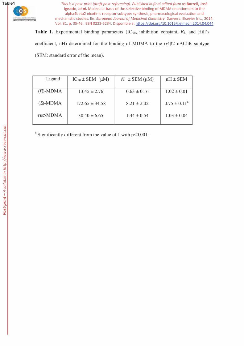
Table 1. Experimental binding parameters (IC50, inhibition constant, Ki
coefficient, nH) determined for the binding of MDMA to the 4 2 nAChR subtype
(SEM: standard error of the mean).
a Significantly different from the value of 1 with p<0.001.
Ligand IC50 SEM ( M) Ki SEM ( M) nH SEM
(R)-MDMA 13.45 2.76 0.63 0.16 1.02 0.01
(S)-MDMA 172.65 34.58 8.21 2.02 0.75 0.11a
rac-MDMA 30.40 6.65 1.44 0.54 1.03 0.04
P-p
rint –
Ava
ilabl
e in
http
://w
ww
.rece
rcat
.cat
P-p
rint –
Ava
ilabl
e in
http
://w
ww
.rece
rcat
.cat
P-p
rint –
Ava
ilabl
e in
http
://w
ww
.rece
rcat
.cat
P-p
rint –
Ava
ilabl
e in
http
://w
ww
.rece
rcat
.cat
P-p
rint –
Ava
ilabl
e in
http
://w
ww
.rece
rcat
.cat
P-p
rint –
Ava
ilabl
e in
http
://w
ww
.rece
rcat
.cat
P-p
rint –
Ava
ilabl
e in
http
://w
ww
.rece
rcat
.cat
P-p
rint –
Ava
ilabl
e in
http
://w
ww
.rece
rcat
.cat
P-p
rint –
Ava
ilabl
e in
http
://w
ww
.rece
rcat
.cat
P-p
rint –
Ava
ilabl
e in
http
://w
ww
.rece
rcat
.cat

P-p
rint –
Ava
ilabl
e in
http
://w
ww
.rece
rcat
.cat