abstracts of the 33rd european symposium on calicified
TRANSCRIPT

Abstracts of the 33rd
European Symposium on Calicified Tissues
These abstracts are published exactly as received from the submitting authors. The opinions and views expressed arethose of the authors and have not been verified by the Publisher or by the European Calcified Tissue Society, who acceptno responsibility for the statements made or the accuracy of the data presented.
Calcif Tissue Int (2006) 78(Suppl 1):S17–S167
DOI: 10.1007/s00223-006-6003-y
I001
ROLE OF MITOCHONDRIAL DNA IN AGEINGH.T. Jacobs*1,2,3, A.T. Rovio1, A. Wredenberg4, K. Minkkinen1, I. Pyykko2,J.N. Spelbrink1 , N.-G. Larsson4, A. Trifunovic41Institute of Medical Technology, University of Tampere, Finland; 2TampereUniversity Hospital, Tampere, Finland; 3Institute of Biomedical and Life Sci-ences, University of Glasgow, Scotland, UK; 4Dept of Laboratory Medicine,Novum, Karolinska Institute, Huddinge, Sweden
The mitochondrial theory of ageing proposes that the lifetime accumulationof mutations in mitochondrial DNA leads to the progressive loss of cellularviability, via some combination of bioenergy insufficiency, enhanced productionof reactive oxygen species (ROS) and apoptosis. It has also been suggested thatthis could entrain a vicious cycle of ROS damage and mutagenesis. Mitochon-drial DNA mutations do indeed accumulate with age, albeit to a very low level.Debate rages as to whether this could have any physiological significance, even ifmitotic segregation is substantial. However, in stem cells, where mtDNA copynumber may be relatively low, and the number of rounds of cell division high,segregation of mutants could result in eventual loss of precisely those cells thatprovide the body with the repair capacity needed to stave off functional declineand eventual death. Until recently, our ability to evaluate the role of mtDNAmutations in ageing was based purely on correlative data. However, the intro-duction of germline mutations into the nuclear genome which affect the mtDNAreplication apparatus has allowed us to manipulate the mitochondrial genomeexperimentally, and provide a more formal test of the hypothesis. By cripplingthe proofreading exonuclease activity of the mitochondrial DNA polymerase(DNA polymerase gamma, POLG) we were able to induce the progressiveaccumulation of mtDNA point mutations to high levels. In cultured human cellsthis results in a selective crisis, once a critical mutation load is crossed, mani-festing after some 60–90 cell divisions. When such a mutator was introduced intothe mouse, a similar accumulation of mtDNA mutations in all tissues was found,associated with a phenotype of premature ageing, including many featuresreminiscent of human ageing: loss of mineral bone density and kyphosis,megalocytic anemia, loss of muscle mass, adipose tissue and hair, loss of fertilityin both sexes and early death. The accumulation of mtDNA mutations in Polgmutator mice appears to be approximately linear with age, refuting the idea of amutagenic vicious cycle, and instead consistent with the idea of a selective crisisbrought about progressively by a constant rate of mutagenesis and the effects ofmitotic segregation. A recent analysis of mtDNA from hair roots of young andaged humans supports this interpretation.
I002
GENDER SPECIFIC PATTERNS OF AGE-RELATED BONE
LOSSS. Khosla*11Endocrine Research Unit, Mayo Clinic College of Medicine, Rochester, UnitedStates
Based largely on studies using dual energy x-ray absorptiometry (DXA), it hasgenerally been believed that, in both sexes, bonemass increases during adolescenceand early adulthood, reaches a peak in the third to fourth decades, and then beginsto decline.Moreover, while there has been some evidence for bone loss prior to themenopause in women, conventional wisdom held that young women and menusually do not lose significant amounts of bone in the absence of secondary factors,such as glucocorticoid use. Finally, due to inherent limitations of DXA, changesseparately in trabecular versus cortical bone or the structural basis for age-relatedbone loss could not be assessed. Over the past several years, our group has beenconducting cross-sectional and, more recently, longitudinal assessments of chan-ges in bone mass and structure in a population-based cohort of women and menspanning the age range of 20 to 97 years (n = 688 subjects). All subjects hadbasline quantitative computed tomography (QCT) scans of the lumbar spine (L1–L3) and femur, as well as conventional and high resolution peripheral QCT scans,with follow up scans being performed at 3 and 6 years. Analysis of the spine andfemur scans required the development of new image analysis software (Proc. SPEE5369:74–88, 2004). Data from this cohort has revealed important gender differ-ences in bone mass and structure in young adulthood, as well as similarities anddifferences in bone losswith aging. Perhaps themost surprisingfindinghasbeen thedemonstration, using both cross-sectional (at all sites) and longitudinal (at theperipheral sites, with the central sites currently being analyzed) data that whilecortical bone mass remains relatively stable until mid-life in women and in men,trabecular bone mass begins to decrease in young adult life in both sexes, with anapparent accelaration in women around the time of menopause. Since sex steroidlevels are, by definition, ‘‘normal’’ in young adult life, these findings will likelyrequire a reevaluation of current concepts of the role of hormonal and othervariables leading to loss of trabecular bone throughout life. By contrast, changes incortical bone do seem to follow existing paradigms of sex steroid deficiency inwomen after the menopause and with aging in men as significant determinants ofage-related bone loss in both sexes.
Supported by a grant (AR027065) from the National Institutes of Health.
I003
BONE MASS ACQUISITION AND FRACTURE RISK
IN CHILDHOOD: GENDER DIFFERENCESS. L. Ferrari*11Service of Bone Diseases, WHO Collaborating Center for Osteoporosis Pre-vention, Geneva University Hospital, Geneva, Switzerland
Bone mineral density (BMD) is lower in daughters of women with spinefractures compared to their age-matched peers, illustrating the contribution ofpeak bone mass, and heredity therein, to the risk of osteoporotic fractures.Accordingly, gender-related differences of peak bone mass likely contribute to thenearly two fold difference in fracture risk between aging men and women. Asecond peak in the incidence of fractures occurs during childhood. Contrarily toosteoporotic fractures, childhood fractures are more common in males than fe-males. These fractures occur mostly during the years of peak height velocity(PHV), i.e., 1–2 years earlier in girls than boys. Girls with a history of fracturehave a lower gain in bone mineral content (BMC) and decreased peak bone massat sites with a large trabecular bone content (spine, trochanter and distal radius),indicating that childhood fractures may be markers of a persistent bone fragility.Although areal BMD is similar in pre-pubertal boys and girls, important quan-titative and qualitative differences in bone mass and structure appear betweenboys and girls during pubertal growth, leading to higher aBMD and BMC inmales than females by the completion of puberty. Widening of the bone andthickening of the cortex are the hallmarks of bone mass growth, whereas volu-metric BMD shows little changes. Although there is apparently no sex differencein cortical thickness, the latter increases primarily by periosteal bone formation inmales, whereas endocortical apposition is more prominent in females. Transgenicmouse models have started to elucidate the role of systemic and local factors, suchas gonadal steroids and IGF-1, in bone modeling and remodeling during growth.Genetic association studies in humans have found that variations in several genes,including estrogen receptor alpha (ESR1) and LDL-receptor related protein 5(LRP5), may contribute to gender-specific differences in aBMD and bone size inchildren. On another side, epidemiological and intervention studies in childrenand adolescents have shown that nutrients, particularly calcium, and/or physicalexercise, may also influence aBMD gain differently in males and females. Inconclusions, sexual dimorphism in bone mineral mass and structure that appearsduring growth under the influence of modifiable and fixed factors contributes tothe pathophysiology of fractures, both in children and the elderly.
I004
INFLAMMATORY DISEASES IN CHILDHOOD AND
THEIR IMPACT ON BONEF. De Benedetti*11Direzione Scientifica, IRCCS Ospedale Pediatrico Bambino Gesu, Roma, Italy
In children, chronic inflammatory diseases, such as juvenile idiopathicarthritis (JIA) or Crohn�s disease, as well diseases characterized by severerecurrent infections, such as cystic fibrosis, have a major impact on the growingskeletal system. This includes decrease in bone mass and defective growth. Thesefeatures may appear during the disease course or, in some instances, may alreadybe noted at presentation. These children may also reach a suboptimal bone masspeak, which is a well-known risk factor for developing osteoporosis later in life.
A vast body of evidence points to inflammatory cytokines as mediators ofthe effects of chronic inflammation on bone. However the great majority ofstudies provides data on adult bone. In growing subjects, bone formation mustexceed bone resorption, allowing gain in bone mass and skeletal growth. Growthplate ossification and epiphyseal ossification are necessary to ensure growth.Balanced bone resorption is essential for mineralized cartilage and woven bonereplacement. However, the mechanisms behind this different regulation have notyet been clarified.
We will review the available evidence on the abnormalities of bones, ossifi-cation and growth plates in children with chronic inflammation. In this contextwe will discuss the role of cytokines on the growing skeleton, focusing particu-larly on the role of chronic over-production of interleukin-6 (IL–6) in the settingof the yet to be clarified unique regulation of bone homeostasis in growingindividuals.
The identification of the mediators involved and of the cellular and molec-ular mechanisms involved in these manifestations of chronic inflammation inchildren will provide the rational for targeted approaches that will affect not onlythe quality of life of the affected children, but also their future lives as adults.
I005
PRIMARY BONE DISEASES IN CHILDRENN. J. Bishop*11Academic Unit of Child Health, University of Sheffield, Sheffield, UnitedKingdom
S18 Abstracts
Primary bone diseases in children are legion. The disorders fall into groupsaffecting skeletal pattern, bone size and gross morphology, bone mass and bonemineralisation. In many instances there are clearly defined genetic origins for thedisorder. However, specific medical treatments have been targeted largely intotwo areas; disorders where low bone mass is a major feature, using techniquesand approaches adapted from adult practice in osteoporosis; and disorders ofbone mineralisation, where specific approaches have developed from the study ofpaediatric populations. This talk summarises current knowledge and practice inthese areas in terms of drug and cell-based interventions and discusses likelyfuture approaches.
I006
THE SPECTRUM OF KIDNEY DISEASES AND BONE IN
CHILDRENC. B. Langman*11Kidney Diseases, Feinberg School of Medicine, Northwestern University &Children’s Memorial Hospital, Chicago, United States
The biology of bone is tied directly to normal kidney functions for iontransport, hormone synthesis, and mineral integration, and perturbations ofthose functions leads to disordered bone in children. Children have two simul-taneous, mostly, unrelated, processes occurring in bone: modeling (a dual-sur-face process) and re-modeling (single surface process, as in adults). Childhoodbone diseases due to specific or generalized dysfunction of the kidney can bedivided based on how the disease presents clinically, and how it affects each ofthese processes uniquely. Thus, Rickets, Growth Failure, Osteitis Fibrosa, orFractures are common ways in which kidney-bone disease presents in patients.Kidney-bone disease may also be discussed by whether global kidney function,or more properly, glomerular filtration, is normal or reduced. Bone disease thatresults from chronic kidney disease with reduced glomerular function is termed‘‘renal osteodystrophy’’. Osteodystrophy encompasses a wide spectrum of bonepathology, ranging from reduced or absent turnover, through normal remod-eling parameters, to advanced secondary hyperparathyroidism. Recently, it hasbeen appreciated that renal osteodystrophy leads to disordered vascular biology,with resultant pathologic arterial calcifications. A schema for classifying renalosteodystrophy in its totality will be presented. Successful kidney transplantationis the goal for renal replacement therapy in children and adolescents, withmaintenance dialysis reserved for those children who cannot receive a kidneyallograft. However, kidney transplantation has brought with it the clinicalexpression of additional bone diseases, and these too will be discussed.
I007
ENDOCRINE REGULATION OF C. ELEGANS
DEVELOPMENT AND AGINGA. Antebi*11Huffington Center on Aging, Dept. of Molecular and Cellular Biology, BaylorCollege of Medicine, Houston, United States
In response to dietary and environmental cues, the C. elegans orphan nu-clear receptor DAF-12 regulates larval developmental timing, arrest at the dauerdiapause (a long lived stress resistant stage), and life span. DAF-12 is mosthomologous to vertebrate vitamin D, liver-X, and pregnane receptors. Molec-ular genetic studies suggest that DAF-12 transcriptional complexes, regulated bya steroid-like hormone, can specify short-lived or long-lived modes. For regu-lation of the dauer diapause, environmental cues are integrated by insulin/IGF-Iand TGF-beta pathways, which then control production of the DAF-12 ligand.In the presence of ligand, DAF-12 specifies reproductive growth, while in itsabsence, DAF-12 specifies arrest at the dauer diapause. Under reproductivegrowth conditions, it subsequently works in developmental timing circuits withincells to advance stage specific larval programs, by regulating downstream tran-scription factors and microRNAs. Thus, DAF-12 integrates information aboutthe environment and thereby controls the pace of development and aging. Thesestudies may illuminate how steroid-like signaling can work in the context oflarger endocrine networks to retard or advance organismal developmental ageand aging.
I008
THE ROLE OF CELLULAR SENESCENCE IN WERNER’S
SYNDROME: TOWARDS THERAPEUTIC INTERVENTION IN
HUMAN PREMATURE AGINGD. Kipling*1, T. Davis1, D. M. Baird1, M. F. Haughton1, C. J. Jones11Pathology, School of Medicine, Cardiff University, Cardiff, United Kingdom
Werner�s Syndrome (WS) is one of the most dramatic of the human pre-mature ageing (progeroid) syndromes, and is studied as a model system fornormal human ageing. Individuals with WS show the accelerated ageing ofmany, but not all, tissues. WS individuals have a markedly elevated frequency ofmany of the diseases of old age, including atherosclerosis, type II diabetes,ulceration, osteoporosis, and also show soft tissue calcification.
The premature ageing is mainly restricted to those tissues that containdivision-competent cells. The premature ageing in vivo is mirrored by acceler-ated cell ageing in vitro. Dermal fibroblasts from WS individuals show theshortest replicative lifespan of cells from any human genetic syndrome, leadingmany to speculate that the disease is caused by premature replicative senescence.Because many fibroblasts use the gradual erosion of chromosomal telomeres tocount cell divisions and trigger replicative senescence, we have addressed whe-ther fibroblasts from WS individuals also use telomere erosion to triggersenescence, and if so whether telomere erosion is faster in WS individuals. Ourfindings indicate that preventing telomere erosion by forced expression of telo-merase is sufficient for WS cells to bypass senescence, but at the single-cell levelthey do not show evidence of accelerated telomere erosion. We therefore pos-tulated that WS cells senescence faster because of the existence of a second,parallel process of telomere-independent senescence that synergises with thenormal process of telomere-driven senescence.
To address the mechanistic basis of this second senescence pathway we haveinvestigated the role of the stress signalling p38MAPK pathway using SB203580,a cytokine-suppressive anti-inflammatory drug that targets p38 activity.SB203580 treatment reverts the aged morphology of young WS fibroblasts tothat seen in young normal fibroblasts. In addition, SB203580 increases the lifespan and growth rate of WS fibroblasts to within the normal range. These datasuggest that the abbreviated replicative life span of WS cells is due to a stress-induced, p38-mediated growth arrest that is independent of telomere erosion.With some p38 inhibitors already in clinical trials, our data suggest a potentialroute to drug intervention in a premature aging syndrome.
I009
THE AGING OSTEOBLASTM. Kassem*11Department of Endocrinology and Metabolism, University Hospital of Odense,Odense, Denmark
One of the main pathophysiological mechanism underlying age-relatedbone loss is decreased osteoblast-mediated bone formation. However, the cel-lular mechanisms responsible for impaired osteoblast functions are poorlyunderstood. Based on understanding of the cellular events of bone formationphase of bone remodelling, several investigators have examined potentialmechanisms causing age-related osteoblast failure using in vitro osteoblastculture systems. These studies can be grouped according to the workinghypotheses tested into several categories including: decreased osteogenic cellpopulation size due stem cell atrophy or due to impaired stem cell differenti-ation leading to decreased number of differentiated osteoblastic cells, decreasedosteoblastic bone formation capacity due to either impaired responsiveness tosystemic hormones and growth factors signals or changes in bone microenvi-ronment composition. These specific manifestations of the aged osteoblasts areinteracting with general mechanisms underlying homeostatic failure of the agedorganism. In this regard, recent studies of premature aging syndromes inmutant mouse models and in humans led to the identification of specific ge-netic pathways responsible for generalized organismic aging including aging ofthe osteoblasts. Thus, the current understanding of osteoblast aging provides awealth of information regarding new possibilities to prevent and treat the age-related bone loss.
I010
BREAST AND BONE: RELATIONSHIP BETWEEN BREAST
CANCER AND ITS TREATMENT AND OSTEOPOROSISS. Cummings*11Prevention Sciences Group, University of California, San Francisco, UnitedStates
Bone loss, fracture risk, and the risk of estrogen sensitive (ER+) breastcancer are all influenced by estrogen. The role of estradiol is strongest and mostevident at exteremely low serum levels of estradiol. These biological relationshipsmean that women with osteoporosis have a somewhat lower risk of breastcancer. Treatment or prevention of breast cancer with agents that block estrogenproduction (aromatase inhibitors) cause rapid bone loss and, probably, a sub-stantial increase in fracture risk. The effect of SERMs is more biologicallycomplex, leading to a reduction in ER+ cancer and vertebral fracture risk.Markers of bone turnover that are sensitive to very low levels of estradiol mightbe indicators of breast cancer risk.
Abstracts S19
I011
GENE SIGNATURES AND BONE METASTASEST. A. Guise*11Dept Int Med, Div Endocrinology and Metabolism, University of Virginia,Seville, United States
Certain tumors have a propensity to metastasize to bone to cause bonedestruction or new bone formation. These osteolytic and osteoblastic metastasescause significant morbidity and represent one of the most devastating attributesof cancer. The mechanisms responsible for this tropism to bone have been un-clear. MDA-MB-231 human breast cancer cells with specific metastatic tropismtowards bone were isolated and compared to the parental population. The highlybone metastatic populations and the parental cell line have similar growth ratesas subcutaneous tumors or in tissue culture. Bioinformatic analysis of the gen-ome-wide expression profiles showed that the parental cell population as well asits highly metastatic derivatives display a previously identified poor-prognosisgene-expression signature. In addition to this signature, populations with bonetropism displayed a specific gene expression signature. Most of these genes fulfillnon cell-autonomous functions including homing (CXCR4), collagenolysis(MMP-1), angiogenesis (CTGF) and osteoclastogenesis (IL-11). Thus, it appearsthat the ability of tumor cells to influence their environment plays a central rolein metastasis. Endogenous or exogenous overexpression of combinations of bonemetastasis genes causes a significant increase in the incidence of osteolytic bonelesions. The pro-metastatic cytokine TGFb, stored in bone matrix and activatedby osteoclastic bone resorption, is a potent activator of two of the bonemetastasis genes, CTGF and IL-11. Thus, bone-derived TGFb may mediateosteolytic bone metastasis by enhancing the expression of these pro-metastaticgenes, in addition to those already known to mediate osteolytic bone destruc-tion, such as PTHrP. Overexpression of a dominant-negative TGFb( type IIreceptor cells delayed the formation of bone metastases whereas expression of anactivated type I receptor accelerated it. Natural variants overexpressing differentcombinations of these genes are present in the parental MDA-MB-231 cellpopulation. These results indicate that breast cancer metastasis to bone is acomplex multigenic process driven by tissue-specific gene sets that act superim-posed on the genetic events that originally lead to tumor formation. Thecooperation of specific gene products that subserve a set of concrete cellularfunctions is required for successful colonization of the bone.
I012
IMPACT OF CYTOKINE BLOCKADE ON JOINT AND BONE
DESTRUCTIONP. Emery*11Academic Unit of Musculoskeletal Disease, University of Leeds, Leeds, UnitedKingdom
Rheumatoid arthritis (RA) is a systemic inflammatory disease associatedwith rapid functional decline and disability. Skeletal manifestations includesystemic osteoporosis, periarticular osteopenia, bone erosion, and progressivejoint destruction. Irreversible joint destruction occurs early in the disease pro-cess, thus elevating the importance of early diagnosis and intervention. Inaddition, low bone mass is common in patients with RA, predisposing them tofracture.
Current management consists of early suppression of joint inflammationwith disease-modifying antirheumatic drugs (DMARDs), corticosteroids, andanticytokine therapy. While methotrexate (MTX) remains the gold-standard,first-line treatment choice, many patients require combination therapy withother DMARDs in order to achieve an adequate response. The recently intro-duced tumor necrosis factor-alpha (TNF-a) antagonists offer an important ad-vance in treatment, but their use is limited by cost.
Bone loss in the first years of disease is predominately determined bydisease activity and can be inhibited by agents that suppress inflammation aswell as helped by anti-resorptive agents. TNF blocking agents have a rapidstablisation of systemic bone loss, but periarticular bone loss in early diseasecontinues.
TNF blocking agents have been studied for their impact on bone, andproduce rapid stabilisation of systemic bone loss. However, in very early diseasethe periarticular bone loss persists. In the inflammatory spondyloarthropathiesthere is immediate system benefit from blockade of TNF. Thus the mechanism ofsystemic and periarticular bone loss seems different response in a quantitativelydifferent way to different methods of reducing osteoclast activation.
Research supports the osteoclast as the effector cell for bone damage in RA.Consequently, bisphosphonates—the most potent inhibitors of osteoclasticactivity, with proven impact on BMD—would seem to offer benefits in RA. Thishypothesis has been tested in several clinical studies, with inconclusive results.Zoledronic acid (ZA) is a third-generation bisphosphonate with greater bio-availability and specificity for osteoclasts than current bisphosphonates wastested in a 6-month proof-of-concept trial with positive results. This opens thepossibility that an ideal therapeutic approach may involve both blockade withanti TNF and an initial use of bisphonate.
I013
MECHANISMS OF OSTEOCLAST ACTIVATION BY RANKL
AND CO-STIMULATORY LIGANDS: NOVEL ROLE OF NFAT
IN BONE HOMEOSTASISH. Takayanagi*11Cell Signaling, Graduate School, Tokyo Medical and Dental University,Bunkyo-ku, Japan
Nuclear factor of activated T cells (NFAT) family transcription factors havebeen identified in the immune system, but we showed that NFATc1 is selectivelyinduced byRANKLduring osteoclast differentiation (1). Using an in vitro culturesystem of embryonic stem cells, we suggested that NFATc1 is essential for osteo-clast differentiation, but there has been no in vivo evidence for the essential role.Our recent study provides genetic evidence that NFATc1 is essential for osteoclastdifferentiation in vivo by adoptive transfer ofNFATc1)/) hematopoietic stem cellsto osteoclast-deficient Fos)/) mice and by Fos)/) blastocyst complementation,thus avoiding the embryonic lethality ofNFATc1)/)mice (2).We also showed thatselective autoregulation of the NFATc1 gene by NFAT through its promoterregion is crucial for the essential function of NFATc1 in cell lineage commitment(2). Furthermore, the mechanism of NFATc1 induction by RANKL will be dis-cussed in the context of costimulatory signals mediated by ITAM (3). It is alsonotable that NFATc1 directly regulates a number of osteoclast-specific genes, butcooperative partners of NFAT vary depending on the target gene (4).
Calcineurin inhibitors FK506 and cyclosporin A inhibit NFAT activity andinduce strong immunosuppression. We found that FK506 administration in-duces the reduction of bone mass despite a blockade of osteoclast differentiation.This reduction is caused by severe impairment of bone formation, suggestingthat NFAT transcription factors also play an important role in the transcrip-tional program of osteoblasts. Overexpression of NFATc1 stimulates Osterix-dependent activation of the type I collagen promoter, but not Runx2-dependentactivation of the osteocalcin promoter. NFAT and Osterix form a complexbinding to DNA, and this interaction is important for the transcriptional activityof Osterix. Thus, NFAT and Osterix cooperatively control osteoblastic boneformation (5). These results establish NFAT transcription factors as criticalregulators of bone metabolism controling both osteoclasts and osteoblasts.
References1) Takayanagi H. et al. Dev Cell 3, 889–901(2002)2) Asagiri M. et al. J Exp Med, 202, 1261–1269 (2005)3) Koga T. et al. Nature 428,758–763 (2004)4) Kim Y. et al. J Biol Chem, 280, 32905–32913 (2005)5) Koga T. et al. Nat Med 11, 880–885, (2005)
I014
LOCAL THERAPIES FOR THE ENHANCEMENT OF
FRACTURE HEALINGT. Einhorn*11Boston University Medical Center, Boston University, Boston, MA, UnitedStates
Local strategies for the repair and regeneration of bone can be divided intofour categories: use of osteogenic materials (autologous bone, allogeneic bone,autologous bone marrow, and autologous blood concentrates), use of osteo-conductive materials (calcium phosphates, calcium sulfate, calcium phosphate/collagen composites, and demineralized bone matrix), use of tissue repair factors(recombinant peptide signaling molecules such as FGF, PDGF, VEGF, IGF, aswell as thrombin peptides and prostaglandin agonists) and use of osteoinductivefactors (bone morphogenetic proteins and other members of the TGF-ƒoƒnsu-perfamily). The best evidence for efficacy in patients is seen with the use ofautologous bone, autologous bone marrow, bone morphogenetic proteins(BMPs), and combinations of BMP with allogeneic bone. Autologous bone re-mains the standard treatment for enhancing skeletal repair, though its avail-ability and ability to bridge large segmental defects is limited. Studies on the useof autologous bone marrow show a strong association between the number andconcentration of osteoprogenitor cells in the implanted graft and the clinicalefficacy to enhance fracture healing. Reports on the combination of osteopro-genitor cells with calcium-phosphate implants have suggested the cells bothproduce new bone as well as secrete chemotactic and osteoinductive factors intothe surrounding tissues. BMP-2 and BMP-7 (OP-1) have demonstrated efficacyin randomized controlled clinical trials for the treatment of open fractures andnonunions respectively. Use of both of these BMPs in the spine has also beendemonstrated to enhance spinal fusion. Limited animal and clinical studiessuggest combinations of allogeneic bone with BMPs are effective at healingfractures at risk. There is some clinical evidence for efficacy of FGF-2 andIGF(via growth hormone) as well. Future strategies to enhance healing willinvolve optimizing the effects of BMPs, developing new recombinant moleculeswith demonstrated clinical efficacy, optimizing delivery systems and developingintraoperative methods for the concentration of autologous bone marrow cells.Gene therapy and the development of inexpensive synthetic small peptides thatinduce bone hold promise for the future.
S20 Abstracts
I015
THERAPEUTIC MANIPULATION OF THE ANABOLIC AND
CATABOLIC RESPONSES IN FRACTURE REPAIRD. G. Little*11Orthopaedic Research and Biotechnology, The Childrens Hospital atWestmead, Westmead, Australia
Bone repair is a fast-moving field in which many biotechnology initiativesare starting to make an impact. While orthopaedic surgeons have traditionallyrelied on anabolic treatments such as bone grafting to supplement bone repair,anti-catabolic therapies are also available.
We have hypothesised that the outcome in bone repair is determined by themagnitude and interaction of the anabolic and catabolic responses. It is logicalthat if an endogenous anabolic response is underway, the effect of this can beaugmented by control of catabolism. Similarly, anabolic and anti-catabolictherapy could be synergistic if timed appropriately.
In distraction osteogenesis, the anabolic and catabolic responses are bothhigh. Increased catabolism leads to osteopenia and the risk of fracture afterdevice removal. Interestingly, Hamdy et al. failed to show any improvement in arabbit distraction model with the addition of the anabolic stimulus of OP-1(BMP-7). In stark contrast, inhibition of catabolism with IV zoledronic acid(ZA) led to up to 89% increases in strength.
In fracture repair, anabolic therapies (PTH, BMP) have shown increases instrength in animal models of up to 60%. Single dose ZA produces increases of upto 51% by the alternate mechanism of interfering with catabolism andunmasking the underlying anabolic effort. Either strategy may prove viable infracture repair.
In non-union the underlying anabolic effort is assumed to be insufficient tolead to bone union. Clinical trials of OP-1 showed a 75% success rate in non-union. Our pre-clinical work in a gap healing model shows that OP-1 and ZA aresynergistic, with increases in net callus formation of 86% and increases instrength of 107% over OP-1 alone. Manipulation of both sides of the anabolic/catabolic equation may allow more control than either alone.
Current and emerging therapies allow dissociation of the anabolic and cat-abolic responses in bone repair, such that a more optimal balance can be dictatedfor a given situation. Translation of these concepts could provide a frameworkfor the use of new agents in bone repair.
I016
BONE MORPHOGENETIC PROTEINSS. Vukicevic*11Department of Anatomy, School of Medicine, Zagreb, Croatia
Considerable clinical experience with bone morphogenetic proteins in thetreatment of fractures, especially long bone nonunions, indicates a high efficacy,safety and superiority to allogenous and autogenous bone grafting. In vitro andin vivo evidence show that right dose, timing, carriers, routes of administrationand indications all influence bone regeneration and repair independently of theBMP family member used. Partially overlapping expression patterns of BMPsduring development of the skeleton and late in the post-natal life serve tomodulate strength of BMP signaling rather than create discreet fields of ligandswith intrinsically different signaling properties. For example, BMP-7 can sub-stitute for loss of BMP-4 and BMP-6 during in vitro and in vivo bone formationboth locally and systemically demonstrating that a BMP can functionally sub-stitute for another BMP in an autocrine/paracrine manner, or mediate a re-sponse to an endocrine action on osteoblasts. Local and systemic administrationof BMPs accelerate the rate of fracture repair and increase the bone volume ofboth axial and peripheral skeleton via a mechanism of increasing bone formationand suppressing bone resorption. 17b estradiol decreases expression of BMP-2and BMP-4 in bone, but initiates synthesis of BMP-6, an endocrine signal forosteoblasts which is then released to circulation and increases the number ofcirculating osteoblast like cells. Microarray analysis following administration ofa recombinant BMP to bone results in increased expression of extra cellularmatrix, cell cycle, growth factor and transcription factor related genes, includingprocollagen type IX, IGF-1, EGF, interleukins 12 and 17. Rather than a BMPreceptor/ligand expression tissue specificity circulating BMPs are reflectingvarious skeletal diseases and the severity of injury.
I017
SYSTEMS BIOLOGY: ROBUSTNESS THROUGH ADAPTION
AND REGENERATIONH. Westerhoff*11Department of Molecular and Cellular Physiology, Free University Faculty ofEarth and Life Sciences, Amsterdam, Netherlands, and Manchester Centre forIntegrative Systems Biology, UK
Not all aspects of the functioning of a macromolecule in a living cell aredetermined by that molecule itself: much comes from interactions between thecomponents of biological systems. Together the interactions constitute networksthat tend to be so extensive that their analysis is complicated for the unaidedhuman mind.
Systems Biology attempts to help the human mind to understand how andwhat functional properties arise from the networks in living organisms. It in-volves quantitative experimentation, as well as the use of computers to analyzethese and to understand their implications. Systems Biology also aims at eluci-dating some general principles of the regulation and control of cellular processes,which may then be relevant if those processes are deregulated such as in quite afew diseases.
One set of principles pertains to the regulation and control of signal trans-duction, which we elaborated experimentally for the MAP kinase pathway. Onthe assumption that a disease may be caused by a defaulting signal transductionpathway, we wondered how many factors might be involved in the control ofsuch a pathway. For the pathway leading from EGF to ERK-PP we found thatquite a few factors controlled this considerably, but many more did not. Thisunderpins the phenomenon that many diseases are multifactorial.
We then looked more closely at the role that protein kinases and proteinphosphatases may play in such control. I shall show that those roles can be quiteconsiderable, and depending on what feature of the signal is considered, preciselyor more subtly opposite to each other.
The development of the �omics approaches has led to numerous reportsabout changes in the transcriptome of organisms engaged in a regulatory tran-sition. More recently, considerable efforts lead to reports of changes in themetabolome as well. Knowing that there are changes at the transcriptome level,one might wonder how useful it should be to monitor changes at the level ofmetabolism; everything seems to be regulated at the transcription level already:why expect more regulation at the metabolic level?
We recently developed and applied a method that enables one to resolve thisparadox. It measures experimentally how much of the regulation of a cellularprocess proceeds through gene-expression and how much through metabolism.We found a relative but certainly not complete dominance of metabolic reg-ulation.
Faults in this diversity of regulation may again be responsible for what weobserve as multifactorial disease. I shall show how Systems Biology attempts todeal with this in terms of network based drug design.
I018
UNDERSTANDING DIABETES PATHOGENESIS: THE NEED
FOR SYSTEMS BIOLOGYP. De Meyts*11Receptor Systems Biology Laboratory, Hagedorn Research Institute, NielsSteensens Vej 6, DK-2820 Gentofte, Denmark
The two major forms of diabetes mellitus, type 1 and type 2 diabetes, are bothcomplex diseases of poorly understood pathogenesis. The poor understanding ofmolecular mechanisms underlying pathogenesis results in a penury of effectivedrug therapies. Type 1 diabetes results from progressive beta cell destruction byincompletely defined autoimmune processes. Insulin treatment is an absoluterequirement to prevent death from ketoacidosis. Type 2 diabetes is a complexdisease which is reaching epidemic proportions due a combination of lifestylefactors that favor obesity, and genetic and environmental factors. The pathogen-esis of type 2 diabetes involves alterations in insulin production and secretion bythe pancreatic beta cells, as well as disturbances in the target cells sensitivity toinsulin action. Reductionist approaches have failed to unravel the complex inter-actions between transcriptional networks that regulate beta cell development anddifferentiated function, and the signal transduction mechanisms in both beta cellsand insulin target tissues, the combined disturbance of which results in impairedmetabolic homeostasis. A systems biology approach is clearly warranted.
I019
DYNAMIC PATHWAY MODELING OF THE JAK-STAT AND
SMAD SIGNALING CASCADESU. Klingmuller*11Boveri Group Systems Biology of Signal Transduction, German CancerResearch Center, Heidelberg, Germany
Growth and differentiation of cells are regulated by the coordinated acti-vation of signaling pathways. The components of many signaling cascades havebeen identified, yet it is largely unknown how information is processed and howdecisions are taken. To elucidate this it is important to analyze timing, extentand duration of signal activation. We are using a systems biology approach toexamine the dynamic behavior of signaling pathways that are altered in cancersand predict targets for intervention. To generate novel biological information bya systems biology approach it is important that high quality quantitative data isused for mathematical modeling and that model predictions are experimentally
Abstracts S21
verified. Establishing data-based mathematical models for the JAK-STAT andSMAD signaling pathways has enabled us to identify systems properties of thepathways and predict steps suitable for effective perturbation.
I020
CELLULAR REGULATION OF MINERALISATIONT. Kirsch*11Orthopaedics, University of Maryland School of Medicine, Baltimore, UnitedStates
Physiological mineralization is restricted to bones, teeth and growth platecartilage. Articular cartilage, cardiovascular tissues and kidney are prone toundergo pathological mineralization. Mineralization in these tissues leads tomorbidity and mortality. Recent findings have suggested that the mechanismsregulating physiological mineralization may be similar to those regulatingpathological mineralization. Mineralization-competent cells release specializedmembrane-enclosed matrix vesicles, which contain all the necessary componentsto initiate the mineralization process. The first mineral phase forms inside thesevesicles in a protected microenvironment. Annexins and type III Na+/phosphateco-transporters (Pit-1, Pit-2) mediate Ca2+ and phosphate (Pi) influx into thevesicles leading to the formation of the first mineral phase inside the vesiclelumen. Once the intravesicular mineral has reached a certain size it ruptures themembrane and grows out into the extracellular matrix. Blocking annexin func-tion led to inhibition of mineralization. Matrix vesicles also contain alkalinephosphatase (TNAP) and plasma cell membrane glycoprotein-1 (PC-1) attachedto the outer membrane surface. PC-1 generates pyrophosphate (PPi), an inhib-itor of mineralization, whereas TNAP hydrolyzes PPi and other phospho com-pounds resulting in controlled mineralization inside and outside the vesicles.Retinoic acid (RA) triggers terminal differentiation and mineralization events ingrowth plate chondrocytes by upregulating mineralization-related genes,including annexins and TNAP, by allowing annexin channel formation in matrixvesicle and plasma membranes of growth plate chondrocytes, and by the releaseof matrix vesicles. RA also upregulates the expression of the progressive anky-losis gene (ank). ANK is trans-membrane protein that transports intracellularPPi to the extracellular milieu. Pi generated by TNAP-mediated hydrolysis of PPi
is then being transported back into growth plate chondrocytes by Pit-1 and Pit-2,and acts as a signaling molecule leading to further upregulation of mineraliza-tion-related genes. In conclusion, annexins, ANK, and TNAP play major rolesin the regulation of mineralization and inhibiting the function of one of thesecomponents leads to reduced mineralization. Therefore, these proteins, whichare also highly expressed in tissues undergoing pathological mineralization, maybe novel therapeutical targets to prevent pathological mineralization.
I021
GENETICS AND DISORDERS OF MINERALISATIONM. A. Brown*11Musculoskeletal Genetics Program, Centre for Immunology and CancerResearch, Brisbane, Australia
Inorganic pyrophosphate (PPi) PPi is a natural inhibitor of tissue miner-alisation, the deposition of calcium hydroxyapatite crystals. Excess extracellularPPi (ePPi) leads to formation of calcium pyrophosphate dihydrate (CPPD)crystals as well as inhibition of calcium hydroxyapatite crystal formation, andconversely, reduced ePPi levels favour calcium hydroxyapatite formation, lead-ing to ectopic mineralisation.
CPPD chondrocalcinosis is present in over 50% of joints in patientsundergoing total hip replacement and is thought to accelerate the rate of jointdegeneration in osteoarthritic cartilage. Genetic studies of this condition havedemonstrated that different mutations in genes influencing PPi transport andmetabolism are causally associated with the development of the condition.Functional studies of those variants suggest that these mutations may alsoinfluence chondrocyte maturation/differentiation, and cause CPPD depositionthrough influences on PPi levels and other mechanisms.
Determining the functional significance of the genetic variants associated withthis disease is of crucial importance in the process of developing targeted therapiesfor chondrocalcinosis. These early studies suggest that whilst controlling extra-cellular PPi is likely to be beneficial for many cases of CPPD chondrocalcinosis, itmay exacerbate hydroxyapatite disease, and not benefit those where the geneticvariants involved affected chondrocyte differentiation/maturation.
I022
EMERGING TARGETS FORNEWDRUGS IN BONEDISEASESR. Baron*11Orthopaedics and Cell Biology, Yale University School of Medicine, NewHaven, United States
Recent advances in the understanding of the molecular mechanisms thatregulate the biology of bone cells and bone remodeling have lead to innovative
approaches to discover new chemical entities to treat osteoporosis and otherskeletal diseases. Drug discovery in bone diseases has followed 3 approaches: 1)Pathophysiological, attempting to correct the mechanisms that lead to alteredbone remodeling, 2) Anti-resorptives, targeting the osteoclast lineage and 3)Anabolics, targeting the osteoblast lineage. In the pathophysiology of osteopo-rosis, the main discovery efforts are aimed at identifying new and better selectivemodulators of steroid receptors targeting the Ers, the AR or the Vitamin DReceptor. In all instances the main issue is not effcicacy on bone, which is usuallyachieved, but safety, avoiding effects on reproductive organs, the cardiovascularsystem and/or hypercalcemia in the case of VitD3 analogs. In the field of anti-resorptives, and beyond bisphosphonates and calcitonin, several recent findingshave lead to programs in drug discovery. The main current targets are : thevitronectin receptor, cathepsin K, c-Src, the vacuolar proton pump, and thepathway of RANK Ligand and its receptor RANK. In addition, the mevalonatepathway has been demonstrated to be the target for bisphosphonates, leading toa new drug discovery approach. The therapies of the future will also involvebone anabolics. First and foremost, the anabolic potential of PTH given inter-mittently has been firmly established in human studies, validating the concept ofanabolics. An alternative to the use of PTH is the regulation of its secretion bytargeting the calcium receptors in the parathyroid gland, antagonising thebinding of Ca and thereby increasing PTH levels (calcilytics). But the mostimportant finding in the last few years in anabolics has been the identification ofLRP5 and the Wnt pathway as major regulators of bone mass in mice and inhumans. Several targets involved in Wnt signaling are currently being exploredfor drug discovery: blocking the activity of endogenous antagonists of thepathway (Sclerostin, Dkk1, sFRPs), agonists of the LRP5 or LRP6 receptorsand inhibitors of signaling events downstream of the LRP5/6 activation by Wnts(GSK3B, LiCl) are all being explored as possible anabolic agents.
I023
NEW APPLICATIONS FOR OLD DRUGSL. Mosekilde*11Dept. of Endocrinology and Metabolism C, Aarhus University Hospital,Aarhus, Denmark
Several old drugs have side effects that affect bone metabolism or risk of fallsand may thereby influence fracture risk. This review focuses on drugs with po-tential preventive effects on fracture risk.
Thiazide diuretics (TD) reduce renal calcium excretion and bone turnoverand increase BMD. In a metaanalysis hip fracture risk is reduced by 18%. In anation wide case-control study we found that current TD-use reduced risk of anyfracture by 10% and risk of forearm fracture by 17%. High accumulated TD-dose reduced hip fracture risk by 19%. In contrast loop diuretics increasedfracture risk by 51%.
Statins (ST) shows antiresorptive and anabolic effects in in-vitro and animalstudies. Most human intervention studies have failed to demonstrate an effect ofST on bone turnover or BMD. ST treatment decreases fracture risk in some butnot all epidemiological studies. In a case-control study we found that ST reducedrisk of any fracture by 13% and risk of hip fractures by 43% in a dose dependentway. Use of pravastatin and non-statin lipid lowering drugs did not decreasefracture risk.
Beta2-adrenergic receptors blockers (BB) have been identified on humanosteoblastic and osteoclastic cells. In humans, there are only few and conflictingdata on the effects of beta-blockers on bone metabolism and fracture risk. In aprevious clinical study we found a three-fold increased fracture risk, whereasboth British and Australian register studies have shown decreased risk. In ournation wide case-control study we found a 9% reduction in any fracture risk aswell as in hip fracture risk.
Lithium (Li) inhibits bone resorption and interacts with Wnt signalingsuggesting anabolic properties. It also interacts with the CaSR and stimulatesPTH secretion. Clinical studies have indicated variable effects on BMD but norisk of osteoporosis. In a case-control study we found that Li-use reduced risk ofany fracture by 26–33% depending on accumulated dose. Colles fractures werereduced by 43% and spine fractures by 68% at higher doses.
Most of these effects have been examined in pharmaco-epidemiologicalstudies that may be subjected to selection bias, healthy drug user effects and theinfluence of various confounders. Only few drugs have been evaluated in RCTs.Further clinical studies are needed to explore the clinical consequences in termsof fractures of the use of these ‘‘old’’ drugs.
I024
EXPERIMENTAL CONTROL OF MECHANICALLY
ADAPTIVE BONE (RE)MODELLING IN VIVOL. E. Lanyon*11Veterinary Basic Sciences, Royal Veterinary College, London, United Kingdom
Bones are organs that maintain their structure, adapt their shape, or re-arrange their internal architecture, according to the loads they experience. Thiscapability implies that the cells responsible for controlling modelling andremodelling assess the suitability of the existing structure in relation to its
S22 Abstracts

loading and if this is inappropriate in some way regulate the (re)modellingnecessary to restore it. The controlling stimulus, and the set point that the cellsuse as a reference for structural suitability, are assumed to be the direct orindirect effects of load-induced strain.
Since the technology does not yet exist to maintain whole bones alive inculture for the time necessary for measurable (re)modelling to take place bones�adaptability to mechanical loading has to be assessed in vivo. Exercise and disusestudies have the disadvantage that the stimulus to the bones is mixed and un-quantified in terms of strain. Direct loading experiments in animals have proveduseful in determining that the osteo-regulatory effects of a bone�s loading regi-men are preferentially derived from a subset of the total loading experiencedominated by short, rest interrupted, periods of dynamic loading involving highstrains, high strain rates and unusual strain distributions.
The animal models available for studying loading-related adaptation havebeen progressively refined from those involving surgical intervention and loadingvia implants in large animals, to applying loads to limb bones from externaldevices through the intact skin in rats and mice. In the rat and mouse this hasproved successful for cortical bone and in the mouse tibia for both cortical andcancellous bone.
Typically experiments using direct, non-invasive loading in rodents allowsquantification of the remodelling response to defined strain-related stimulationover a number of weeks. Using genetic strains of mouse allows determination ofthe involvement of one or more factors (such as the estrogen receptor or LRP5)in this short/medium term (re)modelling response. The more difficult determi-nation of the nature of the ‘‘mechanostat�s’’ set-point, and the mechanisms bywhich differences in strain waveform and distribution are transduced into longterm, coherent, control of bone (re)modelling to achieve and maintain structuralsuitability remain almost uninvestigated.
I025
INFLAMMATORY MEDIATORS AND MECHANICAL
LOADING OF THE JOINTB. Fermor*11Surgery, Division of Orthopaedics, Duke University Medical Center, Durham,United States
Mechanical loading is essential for the homeostasis of bone, articularcartilage and meniscus. In order to further understand the mechanisms ofmechanotransduction in these tissues, in vitro systems have carefully beendeveloped in order that the findings from these studies are relevant to the invivo situation. Physiological and hyperphysiological regimens of mechanicalloading have been found to be associated with significantly increased pro-duction of inflammatory mediators such as nitric oxide and prostaglandins inbone, articular cartilage and meniscus. Nitric oxide production is regulated bythe enzyme nitric oxide synthase (NOS), which has 3 isoforms (NOS1, NOS2and NOS3). The mechanically induced nitric oxide can react with reactiveoxygen species to form other derivatives such as peroxynitrite or nitrosothiols.The formation of peroxynitrite leads to protein nitration whereas formationof nitrosothiols may cause nitrosylation of proteins. The nitric oxide deriva-tive that is formed can be partially controlled by the ratio of nitric oxide tooxygen, which in turn may affect the biological responses to mechanicallyinduced nitric oxide. The oxygen tension of the joint can alter with disease,but it is always significantly lower than ambient oxygen tension (20%). Inavascular tissues such as articular cartilage the oxygen tension might be aslow as 1% oxygen. Selective inhibition of the nitric oxide synthase pathway,using pharmacologic agents, can lead to complex interactions with the cyclo-oxygenase pathway and hence alter the magnitude of prostaglandins that areformed. Furthermore inhibition of the cyclo-oxygenase pathway can regulatethe nitric oxide synthase pathway. Mechanically induced production ofinflammatory mediators is affected by oxygen tension, which may influencethe subsequent biological responses observed.
I026
FLUID FLOW EFFECTS ON OSTEOCYTES AND
OSTEOBLASTSH. J. Donahue*1, R. C. Riddle1, A. F. Taylor1, D. S. Shingle1, D. C. Genetos11Musculoskeletal Sciences, Pennsylvania State University, Hershey, UnitedStates
The mechanism by which bone cells respond to their biophysical environ-ment is not known. We propose that biophysical signals, such as fluid flow,stimulate osteoblast proliferation and differentiation via a mechanism involvingmobilization of cytosolic Ca2+, activation of gap junctional intercellular com-munication (GJIC) and release of ATP through gap junction (GJ) hemichannels.We utilized a novel co-culture flow apparatus to examine whether osteocytic cellsexposed to mechanical signals communicate proliferation, differentiation andosteogenic signals to osteoblasts. Osteocytic MLO-Y4 and osteoblastic hFOB1.19 cells were cultured on opposite sides of perforated membranes enabling us
to apply fluid shear to MLO-Y4 cells while they are in direct contact with hFOB1.19 cells that are not themselves exposed to flow induced shear stress. Dyetransfer analysis with calcein-AM showed that MLO-Y4 cells are coupled via GJto hFOB 1.19 cells cultured on the opposite side of the membrane. GJIC betweenMLO-Y4 and hFOB 1.19 cells was completely blocked by the application aGAto the culture system. MLO-Y4 cells were exposed to flow sufficient to induce ashear stress of 5 dynes/cm2 for 1 hour and post incubated for 2 hours at 370C. Ahighly significant increase in alkaline phosphatase activity was detected in thehFOB 1.19 cells in contact with flowed MLO-Y4, compared to un-flowed controlco-cultures (P ( 0.01). When hFOB1.19 were exposed either to direct flow or toconditioned media from flowed MLO-Y4 they did not display increased alkalinephosphatase activity. We also demonstrated that MLO-Y4 cells exposed to fluidflow display activated GJ hemichannels. Interestingly, fluid flow did not activatehemichannels in osteoblastic MC3T3-E1 cells. Fluid flow activated hemichannelactivity and ATP release were significantly attenuated in cells transfected withCx43 siRNA suggesting that flow activated hemichannels composed of Cx43mediate ATP release. We also found that flow induced a rapid, flow rate-dependent increase in [Ca2+]i, that triggered a 116% increase in proliferation ofhuman mesenchymal stem cells (hMSC) and resulted in increases in steady statelevels of osteocalcin, type 1 collagen and osteopontin mRNA. Exposure to ATPalso resulted in an increase in hMSC proliferation and apyrase inhibited flowinduced proliferation. These results suggest that fluid flow stimulates hMSCproliferation and differentiation, perhaps through release of ATP.
I027
GENETICS AND DEVELOPMENT OF CRANIOFACIAL
DISORDERSA. O. M. Wilkie*11Weatherall Institute of Molecular Medicine, University of Oxford, Oxford,United Kingdom
Malformations of craniofacial structures are best understood in terms ofkey developmental processes. Failure of forebrain division causes holopros-encephaly; disruption of the branchial arches results in Treacher Collinssyndrome and facial microsomia; cleft lip and palate arise from disturbance inthe normal fusion of facial processes and palatal shelves; and craniosynostosisis caused by premature fusion of the cranial sutures. The genetic investigationof these disorders has identified many key developmental genes regulatingthese processes; many of these genes encode transcription factors or signaltransduction molecules. These genetic signposts provide a route into thedissection of the pathways in specific developmental processes: some keyfindings from the study of cranial suture biogenesis will be illustrated. Cra-niofacial syndromes frequently exhibit phenotypic pleiotropy. For example,associated limb malformations are common and, unexpectedly, specificmutations appear to confer a selective advantage in the testis, giving rise toelevated mutation rates. This reflects the re-deployment of shared molecularpathways in different cellular contexts.
I028
GENETICS OF TOOTH DEVELOPMENTP. T. Sharpe1
1Craniofacial Development, Kings College London, London, United KingdomTeeth occupy a unique position in vertebrate evolution as a result of changes
in dentition being a major driving force behind adaptation to new feeding nichesand the fact that they are so well preserved in the fossil record. The relative easewith which tooth development can be studied in mice has led to an increasinglydetailed understanding of the molecules and pathways that control tooth shapeand dental patterns.
The Barx1 homeobox gene is expressed in the jaw primordial ectomesenc-hyme cells from which molar teeth will develop and participates in a homeoboxodontogenic code that specifies tooth shape. Misexpression of this gene in pre-sumptive incisor mesenchyme results in the transformation of incisor toothshape into molars. Barx1 is also expressed in the mesenchyme of the developingstomach and both misexpression and knock-out of Barx1 during development ofthe digestive system indicates a role in controlling stomach morphogenesis.Barx1 thus provides an intriguing morphogenetic link between feeding anddigestion.
The NF-?B pathway has a very specific role in regulating cusp formationduring molar development. Manipulation of the level of NF-?B activity in toothbuds can produce teeth with different numbers of cusps. This pathway musttherefore have been important in the evolution of different tooth types via actingto interpret the positional information provided by homeoproteins such asBarx1.
The Shh signalling pathway plays a key role in determining tooth position byregulating dental placode cell proliferation. Genetic manipulation of the Shhpathway can result in the development of extra teeth in the mouse diastema.These ectopic teeth develop with crown shapes characteristic of their relative
Abstracts S23

positions in the dentition. The mouse thus retains the genetic information(homeobox code) to make tooth shapes it lost over 30 million years ago.
I029
SURGERY OF CRANIOFACIAL MALFORMATIONS AND
DEFECTSH. P. Howaldt*11Department of Oral and Maxillofacial Surgery, Universitatsklinikum Gießen,Gießen, Germany
Craniofacial malformations in its particular sense comprises all deformities ofthe scull and facial bones which are mainly caused by premature fusion of cra-niofacial sutures or other malformations mainly of bone tissue. It is not longerthan 40 years ago that all these deformities could not be treated surgically at all.
Through the pioneering work of a French surgeon named Paul Tessier it waspossible to correct abnormal head-shapes caused by premature fusions of cra-nial-sutures.
Paul Tessier was the first to discover that bone of the cranial mould can beremoved, remodelled and replaced in the same operative setting and he achievedcomplete re-ossification of these re-transplanted bone fragments. This is onlypossible if the operation is performed in the first year of life. Since 1970 and thefollowing decades many improvements have been achieved to re-model cranialdeformities in small children. Some of these cranio-facial procedures will bedescribed presenting few clinical cases and the surgical and medical principalsmaking these corrections possible will be explained.
Remaining cranial defects in elder children or in adult patients reveal adifferent problem and can not be corrected with the operative techniques of PaulTessier and his successors. In adults large cranial defects can be closed withindividually moulded titanium implants which are generated through CT-data ofthe patient. This promising technique is not possible in children who still havesome residual growth potential of their skull. On the other hand in these casesconventional bone crafts of the outer table of the scull of the remaining parts cannot easily be achieved because harvesting these crafts in small children is not veryeasy.
The use of stem cells, derived from fat-tissue of the patient presents in newand promising approach to reconstruct defects of the cranial mould. This newtechnique will be described with two clinical cases and it hopefully leads to somefruitful discussion on the forthcoming meeting in Prague.
I030
THE BIOLOGICAL BASIS FOR DISORDERS OF BONE
MATRIXP. G. Robey*11CSDB, NIDCR, NIH, DHHS, Bethesda, United States
With the advent of techniques for isolation of proteins intimately associatedwith mineralized matrix in an intact form, establishment of cell culture systemsthat faithfully represent initial stages of bone formation, and sensitive techniquesfor identifying mRNA and protein in bone, the major structural proteins of bonematrix have been identified. Depending on the stage of development and site, 70–90% of bone is composed of a carbonate-rich hydroxyapatite. The remainingorganic matrix is notable for its high content of a type I collagen-rich scaffold(�90%), which is thought to orient non-collagenous proteins that serve tocontrol deposition of mineral, and control crystal habit. While it was initiallythought that bone matrix proteins would be unique, it is now recognized thatmost, if not all, are expressed in non-skeletal tissues, albeit at different levels anddifferent times during development. The non-collagenous constituents of boneinclude proteins made endogenously by bone-forming cells and proteins ad-sorbed from the circulation due to their affinity to apatite, and are comprised ofdiverse classes of molecules including glycosaminoglycan-containing compo-nents (small leucine-rich proteoglycans), glycoproteins (osteonectin, the SIB-LING family and other RGD-containing proteins), and gla-containing proteins(osteocalcin). These constituents play important roles not only in the structuralorganization of bone, but also in modulating a variety of other activities such ascell-matrix interactions, growth factor action, and initiation of intracellularsignaling pathways via cell surface receptors. Derangements in bone matrix canresult in several ways: 1) deficiency, 2) gene mutation of bone matrix proteinsthemselves, or 3) cellular defects that affect the ability of osteogenic cells tosynthesize and deposit matrix appropriately. Mice deficient in a particular matrixprotein often fail to display an overt skeletal phenotype, unless functionallychallenged, perhaps due to functional redundancies. However, systematic elim-ination of related molecules has unveiled some of their potential functions inskeletal homeostasis. Finally, changes in the stoichiometry of matrix compo-nents as a result of abnormal cellular activity due to gene mutation or aging canhave profound effects on the material properties of mineralized matrix andprovide further insight into their contributions to the integrity of bone. (Sup-ported by in part by DIR, NIDCR; IRP, NIH)
I031
NEW BIOCHEMICAL MARKERS OF BONE AND CARTILAGEP. Garnero*11Molecular Markers, Synarc, Lyon, France
Alterations of bone and cartilage turnover in osteoporosis (OP), osteoar-thritis (OA) and rheumatoid arthritis (RA) can be detected by systemic bio-chemical markers (BM). Improvement in tissue and biological process specificityof BM has been achieved.
The discovery that cathepsin K (Cat K) releases the type I collagen te-lopeptide fragments NTX-I /CTX-I has provided a mechanistic rational for thesensitivity of these BM in OP. An assay for serum Cat K has been developed andincreased levels were found in patients with RA. Whether Cat K will prove to beuseful in OP and OA remains to be investigated.
For cartilage, BM of type II collagen (CII) metabolism - the most abundantprotein of the tissue- have been developed. Using an ELISA for the N-pro-peptide of CIIA (PIIANP), we found increased CII synthesis in early but de-creased values in late knee OA, indicating a deficit of cartilage repair withadvanced OA. Proteolytic fragments of the helicoidal (Helix-II, Coll2-1) and C-telopeptide (CTX-II) domains of CII have been identified and increased levelswere found in OA and RA before radiological evidence of joint damage. Thecombined measurement of these BMs is more effective that one BM alone topredict progression of joint damage, probably because they reflect differentmetabolic processes (synthesis vs degradation) or distinct enzymatic pathways ofcartilage degradation (Helix-II and CTX-II). Emerging cartilage BM includeproteolytic neoepitopes of aggrecan –the major proteoglycan of cartilage- al-though clinical data are lacking.
Bone and cartilage proteins undergo enzymatic and non enzymatic post-translational modifications such as advanced glycation end products (AGE) andisomerization which accumulate with aging. We found that these posttransla-tional modifications contributed to mechanical competence of bone indepen-dently of BMD and that AGEs can regulate osteoclastic activity in vitro. Inpostmenopausal women, the ratio of the native to isomerized CTX-I is signifi-cantly associated with increased fracture risk independently of BMD. Thedevelopment of BM based on posttranslational modifications of proteins may beuseful to assess changes in matrix properties an important determinant of tissueintegrity.
The panel of bone and cartilage BM is likely to expand with the optimizationof proteomic-based technologies. An optimal combination of BM will be usefulfor identifying patients at increased risk for disease progression and to monitortreatment efficacy.
I032
CAN WE AFFORD TO TREAT OSTEOPOROSIS?D. J. Torgerson*11York Trials Unit, Dept of Health Sciences, University of York, York, UnitedKingdom
Due to an increasingly aged population the social burden of fracturesassociated with osteoporosis will increase. An ideal treatment would be inex-pensive with few side-effects and easily implemented to the whole population. Noeffective therapy fulfills all of these criteria. There are effective treatments andthese include: bisphosphonates, hormone replacement therapy (HRT), para-thyroid hormone (PTH) strontium ranelate. Other widely used treatments suchas vitamin D, calcium supplements and hip protection have little evidence tosupport their use outside of some small population groups. The drawbacks ofsome of the effective treatments include: cost (e.g., PTH) and unacceptable side-effects (e.g., HRT), which tend to mitigate against their use on a populationbasis. In the UK the National Institute of Clinical Effectiveness (NICE) hassuggested that bisphosphonates can be used cost effectively in a secondaryprevention category. It is suggested that treatment can be used without a bonemineral density (BMD) measurement: therapy can be introduced on the basis ofrisk clinical factors alone. On the other hand it is widely believed that bis-phosphonate therapy may be ineffective among patients who do not have adiagnosis of osteoporosis using BMD measurements. This belief is partly basedupon a large RCT of risedronate, which seemed to indicate that whilst there wasa statistically significant benefit of the bisphosphonate on hip fractures amongwomen with low BMD this effect was no longer significant among women whowere targetted with treatment on the basis of clinical risk factors. However, thissupposed treatment by BMD interaction is not statistically significant, therefore,the evidence does suggest that bisphosphonates are beneficial for fracture pre-vention whatever the underlying BMD. However, even if bisphosphonates, orany of the effective treatments, do prevent fractures irrespective of underlyingBMD they are simply too expensive to be used as a population preventivemeasure. Current treatments all need targetting either by BMD or clinical riskfactors or a combination of both to be cost effective. We can prevent fractures ina cost effective manner through the use of careful targetting of therapies. Thiswill reduce the individual suffering of some patients at high risk of fracture.Unfortunately, given the current cost and effectiveness of existing treatments wewill not be able to prevent the majority of osteoporotic fractures.
S24 Abstracts

I033
HEALTH ECONOMICS - CLINICIAN’S RESPONSEC. Cooper*11MRC Epidemiology Resource Centre, University of Southampton,Southampton, United Kingdom
Osteoporosis constitutes a major public health problem through itsassociation with age related fractures. These fractures typically occur at thehip, spine and distal forearm. It has been estimated from incidence ratesderived in North America that the lifetime risk of a hip fracture in caucasianwomen is 17.5%, with a comparable risk in men of 6%. Hip fractures lead toan overall reduction in survival of around 15% and the majority of excessdeaths occur within the first six months following the fracture. They are alsoassociated with considerable morbidity: they invariably necessitate hospital-ization and the average length of hospital stay is around 30 days. Althoughall vertebral deformities do not come to clinical attention, the lifetime risk ofclinically diagnosed vertebral fractures is around 15% in caucasian women.There is now convincing longitudinal evidence that a reduction in bonedensity is an important determinant of fracture risk. The determinants ofbone density can be categorised into those influencing the peak which isattainable during growth and consolidation; and the subsequent rate of boneloss. Preventive strategies against osteoporotic fracture can be targeted at thegeneral population and at those at the greatest risk. Thus, modification ofphysical activity and dietary calcium/vitamin D nutrition in the elderly andduring mid-life, should complement high risk approaches entailing appropri-ate measurement of bone mineral density and targeting of antiresorptive andformation stimulating drugs. In addition, the recently developed WHOalgorithm for evaluation of 10-year absolute risk of fracture provides a meanswhereby various antiresorptive and formulation-stimulating therapies can betargeted most cost-effectively to those at the greatest risk. These data willprovide the basis for translation into coherent public health strategies aimingto prevent osteoporosis both in individuals and in the general population.
OC001
SERUM TESTOSTERONE IS ASSOCIATED WITH FRACTURE
RISK IN ELDERLY MEN: THE DUBBO OSTEOPOROSIS
EPIDEMIOLOGY STUDYC. Meier1, T. V. Nguyen2, M. Jimenez3, J. R. Center2, D. J. Handelsman3,M. J. Seibel*4, J. A. Eisman21Clinic for Endocrinology and Diabetes, University Hospital, Basel, Switzer-land, 2Bone and Mineral Research Program, Garvan Institute of MedicalResearch, St. Vincent’s Hospital and UNSW, 3Department of Andrology,4Bone Research Program, ANZAC Research Institute, University NSW,Sydney, Australia
The effect of gonadal hormones on the skeletal health of elderly men iscontroversial, and data on the influence of sex hormones on fracture risk arelimited. The present study examined the relationship between serum testos-terone and fracture risk in a prospective cohort of men aged 60 years andolder.
This case-cohort study was part of the on-going Dubbo Osteoporosis Epi-demiology Study, in which 609 men (aged 72.6 ± 5.7 yrs; mean ± SD) hadbeen followed for a median of 4.0 (range, 1–11.5) years. Anthropometric data,calcium intake, smoking habits, lumbar spine and femoral neck BMD, serumlevels of total testosterone (TT) and SHBG were measured at baseline. Freetestosterone was calculated as previously described (Vermeulen et al., JCEM1999). The incidence of low-trauma fracture was ascertained by X-ray recordand confirmed by personal interview. The association between testosterone andfracture risk was assessed by Cox�s proportional hazards model with adjustmentfor potential confounders.
Results: During the follow-up period, 107 men had sustained 163 frac-tures (any site). At baseline and compared to controls, cases were older (p <0.001), had lower body weight (p < 0.001) and height (p = 0.03), lowerBMD (p < 0.001), and lower calcium intake (p = 0.02). Serum TT levelswere lower (p = 0.04) and SHBG levels (p = 0.006) were higher in men withfractures than in men without fracture during follow-up. TT was correlatedwith age (r = -0.14, p = 0.001), weight (r = )0.12, p = 0.003) and SHBG(r = 0.14, p < 0.001). In multivariate analysis including age, weight, height,calcium intake, SHBG and BMD, serum TT was significantly associated withincident fracture risk (hazard ratio per SD change [HR] 1.5, 1.3–1.8). Age(HR 1.6, 1.3–2.0), calcium intake (HR 1.4, 1.4-1.4), BMD (1.5, 1.2–2.0) andSHBG (HR 1.3, 1.1–1.7) were also independently associated with fracture riskin this model. Calculated free testosterone was lower in fracture cases(p = 0.001), and also associated with fracture risk after adjustment for co-variables (HR 1.5, 1.3–1.8).
Conclusion: In elderly men, low serum testosterone levels are associated withincreased risk of any fracture, independent of established risk factors such asBMD, body weight and age.
OC002
EFFECT OF ANASTROZOLE ON BONE MINERAL DENSITY:
5-YEAR RESULTS OF THE ‘ARIMIDEX’ (ANASTROZOLE),
TAMOXIFEN, ALONE OR IN COMBINATION (ATAC) TRIALR. Eastell*1, R. A. Hannon1, J. Cuzick2, G. Clack3, J. Adams41Academic Unit of Bone Metabolism, University of Sheffield, Sheffield, 2Sta-tistics Unit, Cancer Research UK, London, 3Oncology Department, Astrazen-eca, Macclesfield, 4Radiology Department, University of Manchester,Manchester, United Kingdom
The ATAC trial is a randomized, double-blind trial of anastrozole (1 mg/day, A), tamoxifen (20 mg/day, T) or the combination (C) in 9366 postmeno-pausal women with early breast cancer (adjuvant therapy). At the most recentanalysis (median follow-up 68 months for disease-free survival) treatment with Aresulted in a superior efficacy and tolerability profile compared with T; however,the odds ratio for all fractures (A vs T) was 1.49 (1.25 to 1.77, 95% CI) (ATACTrialists� Group. Lancet 2005, 365: 60–62). Here we report the updated bonemineral density (BMD) results of the ATAC bone sub-protocol in a subset of308 women from the ATAC trial and 46 unrandomized control patients withbreast cancer receiving no hormone therapy. The control group was included forobservational reasons, and did not form part of the statistical analysis; thecombination group was discontinued as the efficacy on breast cancer recurrencewas similar to tamoxifen alone. At 5 years, an ANCOVA of change in BMDfrom baseline indicated an 8.1% and 7.4% reduction in LS and TH BMDrespectively for patients treated with A relative to T (p < 0.001). With A, forpatients with data at baseline, 2 and 5 years, the rate of bone loss slowed betweenyears 2 to 5 compared to baseline to 2 years for LS, but not TH. With T therapy,the rate of change was more negative between years 2 to 5 compared to baselineto 2 years for LS and TH (Table). No patients with normal BMD at baselinebecame osteoporotic at 5 years. By 5 years, 24 A and 35 T patients had with-drawn from the sub-protocol, mainly for reasons unrelated to bone.
Although at 5 years there was still a significant difference between anas-trozole and tamoxifen in terms of BMD loss, no patients with normal BMD atbaseline became osteoporotic at 5 years. There was a slowing down in the rate ofbone loss at the spine in the anastrozole treated group. The impact of A on boneneeds to be balanced against the overall efficacy and tolerability benefits ob-served in the main ATAC trial.
OC003
AN MTHFR GENETIC VARIANT INTERACTS WITH
DIETARY B2 INTAKE TO DETERMINE FRACTURE RISKN. Yazdanpanah1, A. G. Uitterlinden1, J. Lindemans2, H. A. P. Pols1,J. B. J. Van meurs*11Internal Medicine, 2Clinical Chemistry, ErasmusMC, Rotterdam, Netherlands
Homocysteine (Hcy) level is a risk factor for osteoporotic fractures. TheMTHFR C677T polymorphism is an important determinant of Hcy levels whilevitamin B2 and B11 can modulate Hcy level in 677 TT homozygotes. We
Abstracts S25

investigated whether B2 and B11 dietary intake affected the relationship betweenthe MTHFR C677T polymorphism and fracture risk, BMD and bone loss.
Methods We analysed genotype, BMD and dietary food record in 5131individuals aged 55 years and over. During 8.2 years follow up, 723 fractureswere recorded.
Results We observed no association between MTHFR genotype and BMD,rate of bone loss or fracture risk in the total population. However, in the lowestquartile of B2 intake, women with the TT genotype had a 1.8 (95%CI:1.1–2.9,p = 0.01) higher risk for any osteoporotic fracture compared to the CC-ho-mozygotes. TT-homozygotes had a 2.7 times higher risk (p = 0.004) for fragilityfractures in the lowest B2 intake quartile, while we observed in the highest B2quartile a strong trend towards a protective effect on fragility fracture(RR = 0.3, p = 0.07) (Fig., p = 0.00008 for interaction). Similarly, we ob-served that only in TT women, B2 intake was significantly correlated with boneloss (p = 0.04, p-interaction = 0.05). No significant influence of B11 intake onthe associations was observed.
Conclusion While the MTHFR 677T variant had no overall effect on frac-ture risk, it strongly interacts with dietary vitamin B2 intake to determinefracture risk in this cohort of elderly Caucasian women. This study highlights theimportance of gene-environment interaction in osteoporosis.
OC004
MULTILOCUS ANALYSIS: A NOVEL APPROACH IN GENET-
ICS OF OSTEOPOROSISF. Rivadeneira*1, J. J. Houwing-Duistermaat2, J. B. Van Meurs1, M. C. Zilli-kens1, H. A. P. Pols1, A. G. Uitterlinden11Internal Medicine, Erasmus MC, Rotterdam, 2Medical Statistics, Leiden Uni-versity Medical Center, Leiden, Netherlands
AIM: Several polymorphisms have been found associated with fracture riskbut no extensive modeling has been undertaken to analyze combined effects. Weperformed a simultaneous evaluation of 16 polymorphisms in eight candidategenes previously associated with fracture risk.
METHODS: This study is part of a large prospective population-basedcohort of elderly Caucasian individuals with available genotypes (n = 6500).We included ESR1 Xbai PvuII, ESR2 intron2-T>C 3¢UTR-C>T, IGF1 (CA)npromoter, COL1A1 Sp1, LRP5 A1330V V667M, LRP6 I1062V, SOST
10565insGGA A75707G, VDR Cdx2 FokI U-A311C U-G464T U-A2978Tvariants. For the multilocus modeling we used the FAMHAP program whichapplies an expectation-maximization algorithm allowing inference and contrastof (haplotype) allele frequencies in fractured and non-fractured individuals, withadjustment for multiple testing (p-adj).
RESULTS: Of all the interactions tested, the risk of vertebral fracture wasassociated to ESR1*ESR2 (p = 0.002; p-adj = 0.01) in women, and toESR1*VDR (p = 0.03; p-adj = 0.05) in men; while the risk of fragility (hip,pelvis and proximal humerus) fracture was associated to ESR2*IGF1*CO-
L1A1*SOST (p = 0.0001; p-adj = 0.002) in women, and toESR1*LRP5*LRP6 (p = 0.004; p-adj= 0.02) in men. When we combined inone outcome both vertebral and fragility fractures in men and women, ESR1 wasthe only gene which remained significantly associated (p = 0.003; p-adj = 0.04).
CONCLUSION: In line with our previous associations, the significantmultilocus interactions we identified were gender- and site-specific reinforcingthe complex character of osteoporosis. Replication will be pursued using alter-native analytical tools and warranted in other populations. Multilocus modelslike ours will allow the evaluation of additional genetic factors and will beevaluated in relation to their additive predictive value over existing non-geneticrisk factors for osteoporotic fracture.
OC005
MATERNAL VITAMIN D DEFICIENCY IS ASSOCIATED
WITH REDUCED FETAL GROWTHJ. D. Wark*1, R. Morley2, J. Carlin3, J. Pascoe41Department of Medicine, 2Clin Epidemiology and Biostatistics, 3Clinical Epi-demiology and Biostatistics, The University of Melbourne, Melbourne, 4Clinicaland Biomedical Sciences, Barwon Health, Geelong, Australia
Maternal vitamin D deficiency has been linked with reduced offspring birthsize, an indicator of fetal growth. However, vitamin D supplementation trials inpregnancy have yielded inconsistent results. Therefore we conducted a pro-spective observational study of 475 healthy Australian women with singlepregnancies, measuring maternal circulating 25–hydroxyvitamin D (25-OHD)and parathyroid hormone (PTH) levels, and offspring size at birth. Maternalblood was collected before 16 weeks (w) and at 28–32 w gestation. The mainhypothesis was that offspring of mothers with 25-OHD levels <28 nmol/L at 28-32 w would be smaller at birth than offspring whose mothers had higher 25-OHD levels. Data from 374 eligible pregnancies were evaluated. 25-OHD was<28 nmol/L in 27 women (7.2 %) at 28-32 w. After adjusting for potential
confounders (including gestation length), and for log PTH, log serum calciumand log serum albumin, infants in this group differed as follows from infants ofmothers with higher 25-OHD levels: knee-heel length )3.2 mm (95% CI )5.9,)0.6), mid-upper arm and calf circumference )0.2 mm ()0.6, 0.1) and )0.3 ()0.7,0.06) respectively. The knee-heel difference was greater unadjusted for gestationlength. Maternal 25-OHD at 28–32 w was weakly positively associated withgestation length (doubling the 25-OHD equated to 0.3 w increase in gesta-tion),which may partly explain the association between 25-OHD and birth size.Maternal PTH at 28–32 w was negatively associated with 25-OHD. Afteradjustment, log2 PTH was related positively to infant knee-heel length, birthweight, and mid-upper arm and calf skinfold thickness. These associations wereindependent of 25-OHD. Low maternal 25-OHD in late pregnancy is associatedwith reduced intra-uterine long bone growth. The long term consequences forlinear growth require follow-up. The observed strong positive relationship be-tween maternal PTH in late pregnancy and infant size warrants further inves-tigation.
OC006
DIRECT IDENTIFICATION, PROSPECTIVE ISOLATION, AND
PHENOTYPE OF IN VIVO SELF-RENEWING, MULTIPOTENT
SKELETAL STEM CELLSB. Sacchetti*1, A. Funari2, S. Di cesare3, S. Michienzi1, S. Piersanti4, I. Saggio4,M. Riminucci2, P. Bianco11Department of experimental medicine and pathology, University of Rome LaSapienza, rome, 2Department of experimental medicine, University of L’Aquila,L’Aquila, 3, S.Raffaele Scientific Park of Rome, 4Department of Genetics andMolecular biology, University of Rome La Sapienza, Rome, Italy
To track human SSCs over the entire cycle of explantation, in vitro growth,and in vivo transplantation, we analyzed by FACS the expression of severalproposed markers of ‘‘mesenchymal stem cells’’ in cultures of a) bone marrow(BM) stromal cells, b) periosteal cells, c) skin fibroblasts, d) muscle-derivedprogenitors, e) fibrous dysplastic marrow; f) trabecular bone cells, all grownunder identical conditions. CD105, CD49a, ALP, CD63, and CD140b did notdistinguish among the different cell strains. CD146/MCAM, in contrast, dis-tinguished BMSC (hi/bright) from all other strains (lo/dim). Upon in vivotransplantation, only BMSC generated a complete ossicle (bone and hemato-poiesis) in the subcutaneous (s.c.) tissue of SCID/bg mice; periosteal anad bonecells generated bone but not marrow; skin and muscle fibroblasts generatedfibrous tissue only. Using a sequential magnetic separation (CD45-)/FACS(CD146+) sorting, we isolated a fraction in which all CFU-F were found, and�80% of them recovered. All clones (36/36) generated by plating unseparatedBM cell suspensions at clonal density generated a uniform (>90%) progeny ofCD146hi cells. Homogeneous multi-clonal (12–17 CFU-F) strains of CD146+cells generated in culture were tranplanted along with an osteoconductive carrierin the s.c. tissue of SCID/bg mice. In the human BM in situ, CD146 expressionwas restricted to adventitial reticular cells, residing at the abluminal side ofsinusoids. In the heterotopic ossicles generated by transplanted CD146+ cells, atimed developmental sequence progressed through the generation of CD146-bone and mesenchymal cells of human origin (4wk), the development of sinu-soids (6-7wk), and the appearence of murine hematopoiesis (7-8wk). In theheterotopic sinusoids, endothelial cells were of murine origin, and human su-bendothelial, CD146+ adventitial cells developed over 4–8 wk post-transplant.As hematopoiesis appeared, these cells assumed a stromal/myelosupportiveattitude. Human stromal cells could be released from heterotopic ossicles and re-established in culture. Hence, CD146+ adventitial reticular cells explanted fromthe human BM are the CFU-F, generate a CD146+ progeny in culture, andupon transplantation give rise to CD146- mesenchymal, adipocytic and bonecells, and to a specific subset of CD146+ subendothelial cells. These data di-rectly identify SSCs as the abluminal subendothelial cells of BM sinusoids anddemonstrate for the first time their self-renewal in vivo.
OC007
FUNCTIONS OF C-FOS PHOSPHORYLATION IN BONE
DEVELOPMENT AND TUMORIGENESISL. Bakiri*1, M. O. Reschke1, M. Idarraga1, K. Polzer2, V. Komnenovic1,G. Schett2, E. F. Wagner11IMP, Research Institute of Molecular Pathology, 2Internal Medicine III,Division of Rheumatology, University of Vienna, Vienna, Austria
The proto-oncogene c-Fos, a member of the AP-1 family of transcriptionfactors, is essential for normal skeletogenesis, since mice lacking c-Fos developsevere osteopetrosis due to a block in osteoclast differentiation (1). In osteoclastprogenitors stimulated with M-CSF, c-Fos is phosphorylated by ERK and theERK-dependent kinase RSK2 on serines 362 and 374 which increases its sta-bility. Mice overexpressing c-Fos develop osteosarcomas due to osteoblasttransformation (2). Recent studies have shown that the RSK2 kinase is essential
S26 Abstracts
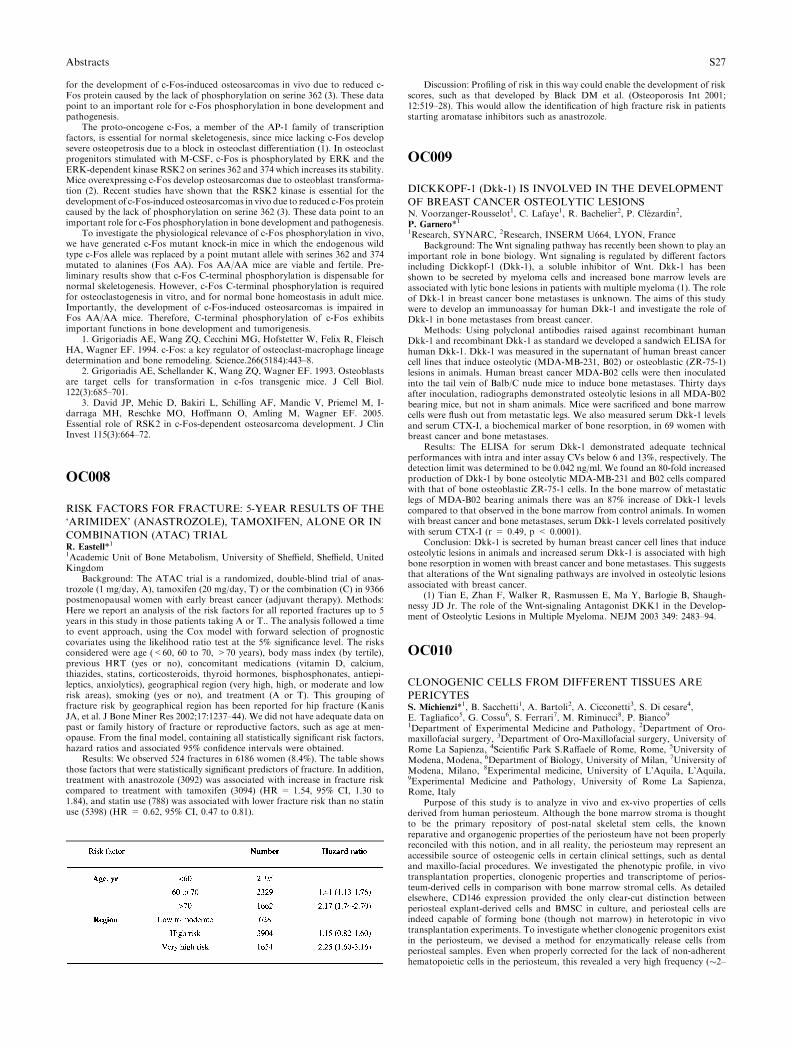
for the development of c-Fos-induced osteosarcomas in vivo due to reduced c-Fos protein caused by the lack of phosphorylation on serine 362 (3). These datapoint to an important role for c-Fos phosphorylation in bone development andpathogenesis.
The proto-oncogene c-Fos, a member of the AP-1 family of transcriptionfactors, is essential for normal skeletogenesis, since mice lacking c-Fos developsevere osteopetrosis due to a block in osteoclast differentiation (1). In osteoclastprogenitors stimulated with M-CSF, c-Fos is phosphorylated by ERK and theERK-dependent kinase RSK2 on serines 362 and 374 which increases its stability.Mice overexpressing c-Fos develop osteosarcomas due to osteoblast transforma-tion (2). Recent studies have shown that the RSK2 kinase is essential for thedevelopment of c-Fos-induced osteosarcomas in vivo due to reduced c-Fos proteincaused by the lack of phosphorylation on serine 362 (3). These data point to animportant role for c-Fos phosphorylation in bone development and pathogenesis.
To investigate the physiological relevance of c-Fos phosphorylation in vivo,we have generated c-Fos mutant knock-in mice in which the endogenous wildtype c-Fos allele was replaced by a point mutant allele with serines 362 and 374mutated to alanines (Fos AA). Fos AA/AA mice are viable and fertile. Pre-liminary results show that c-Fos C-terminal phosphorylation is dispensable fornormal skeletogenesis. However, c-Fos C-terminal phosphorylation is requiredfor osteoclastogenesis in vitro, and for normal bone homeostasis in adult mice.Importantly, the development of c-Fos-induced osteosarcomas is impaired inFos AA/AA mice. Therefore, C-terminal phosphorylation of c-Fos exhibitsimportant functions in bone development and tumorigenesis.
1. Grigoriadis AE, Wang ZQ, Cecchini MG, Hofstetter W, Felix R, FleischHA, Wagner EF. 1994. c-Fos: a key regulator of osteoclast-macrophage lineagedetermination and bone remodeling. Science.266(5184):443–8.
2. Grigoriadis AE, Schellander K, Wang ZQ, Wagner EF. 1993. Osteoblastsare target cells for transformation in c-fos transgenic mice. J Cell Biol.122(3):685–701.
3. David JP, Mehic D, Bakiri L, Schilling AF, Mandic V, Priemel M, I-darraga MH, Reschke MO, Hoffmann O, Amling M, Wagner EF. 2005.Essential role of RSK2 in c-Fos-dependent osteosarcoma development. J ClinInvest 115(3):664–72.
OC008
RISK FACTORS FOR FRACTURE: 5-YEAR RESULTS OF THE
‘ARIMIDEX’ (ANASTROZOLE), TAMOXIFEN, ALONE OR IN
COMBINATION (ATAC) TRIALR. Eastell*11Academic Unit of Bone Metabolism, University of Sheffield, Sheffield, UnitedKingdom
Background: The ATAC trial is a randomized, double-blind trial of anas-trozole (1 mg/day, A), tamoxifen (20 mg/day, T) or the combination (C) in 9366postmenopausal women with early breast cancer (adjuvant therapy). Methods:Here we report an analysis of the risk factors for all reported fractures up to 5years in this study in those patients taking A or T.. The analysis followed a timeto event approach, using the Cox model with forward selection of prognosticcovariates using the likelihood ratio test at the 5% significance level. The risksconsidered were age (<60, 60 to 70, >70 years), body mass index (by tertile),previous HRT (yes or no), concomitant medications (vitamin D, calcium,thiazides, statins, corticosteroids, thyroid hormones, bisphosphonates, antiepi-leptics, anxiolytics), geographical region (very high, high, or moderate and lowrisk areas), smoking (yes or no), and treatment (A or T). This grouping offracture risk by geographical region has been reported for hip fracture (KanisJA, et al. J Bone Miner Res 2002;17:1237–44). We did not have adequate data onpast or family history of fracture or reproductive factors, such as age at men-opause. From the final model, containing all statistically significant risk factors,hazard ratios and associated 95% confidence intervals were obtained.
Results: We observed 524 fractures in 6186 women (8.4%). The table showsthose factors that were statistically significant predictors of fracture. In addition,treatment with anastrozole (3092) was associated with increase in fracture riskcompared to treatment with tamoxifen (3094) (HR = 1.54, 95% CI, 1.30 to1.84), and statin use (788) was associated with lower fracture risk than no statinuse (5398) (HR = 0.62, 95% CI, 0.47 to 0.81).
Discussion: Profiling of risk in this way could enable the development of riskscores, such as that developed by Black DM et al. (Osteoporosis Int 2001;12:519–28). This would allow the identification of high fracture risk in patientsstarting aromatase inhibitors such as anastrozole.
OC009
DICKKOPF-1 (Dkk-1) IS INVOLVED IN THE DEVELOPMENT
OF BREAST CANCER OSTEOLYTIC LESIONSN. Voorzanger-Rousselot1, C. Lafaye1, R. Bachelier2, P. Clezardin2,P. Garnero*11Research, SYNARC, 2Research, INSERM U664, LYON, France
Background: The Wnt signaling pathway has recently been shown to play animportant role in bone biology. Wnt signaling is regulated by different factorsincluding Dickkopf-1 (Dkk-1), a soluble inhibitor of Wnt. Dkk-1 has beenshown to be secreted by myeloma cells and increased bone marrow levels areassociated with lytic bone lesions in patients with multiple myeloma (1). The roleof Dkk-1 in breast cancer bone metastases is unknown. The aims of this studywere to develop an immunoassay for human Dkk-1 and investigate the role ofDkk-1 in bone metastases from breast cancer.
Methods: Using polyclonal antibodies raised against recombinant humanDkk-1 and recombinant Dkk-1 as standard we developed a sandwich ELISA forhuman Dkk-1. Dkk-1 was measured in the supernatant of human breast cancercell lines that induce osteolytic (MDA-MB-231, B02) or osteoblastic (ZR-75-1)lesions in animals. Human breast cancer MDA-B02 cells were then inoculatedinto the tail vein of Balb/C nude mice to induce bone metastases. Thirty daysafter inoculation, radiographs demonstrated osteolytic lesions in all MDA-B02bearing mice, but not in sham animals. Mice were sacrificed and bone marrowcells were flush out from metastatic legs. We also measured serum Dkk-1 levelsand serum CTX-I, a biochemical marker of bone resorption, in 69 women withbreast cancer and bone metastases.
Results: The ELISA for serum Dkk-1 demonstrated adequate technicalperformances with intra and inter assay CVs below 6 and 13%, respectively. Thedetection limit was determined to be 0.042 ng/ml. We found an 80-fold increasedproduction of Dkk-1 by bone osteolytic MDA-MB-231 and B02 cells comparedwith that of bone osteoblastic ZR-75-1 cells. In the bone marrow of metastaticlegs of MDA-B02 bearing animals there was an 87% increase of Dkk-1 levelscompared to that observed in the bone marrow from control animals. In womenwith breast cancer and bone metastases, serum Dkk-1 levels correlated positivelywith serum CTX-I (r = 0.49, p < 0.0001).
Conclusion: Dkk-1 is secreted by human breast cancer cell lines that induceosteolytic lesions in animals and increased serum Dkk-1 is associated with highbone resorption in women with breast cancer and bone metastases. This suggeststhat alterations of the Wnt signaling pathways are involved in osteolytic lesionsassociated with breast cancer.
(1) Tian E, Zhan F, Walker R, Rasmussen E, Ma Y, Barlogie B, Shaugh-nessy JD Jr. The role of the Wnt-signaling Antagonist DKK1 in the Develop-ment of Osteolytic Lesions in Multiple Myeloma. NEJM 2003 349: 2483–94.
OC010
CLONOGENIC CELLS FROM DIFFERENT TISSUES ARE
PERICYTESS. Michienzi*1, B. Sacchetti1, A. Bartoli2, A. Cicconetti3, S. Di cesare4,E. Tagliafico5, G. Cossu6, S. Ferrari7, M. Riminucci8, P. Bianco91Department of Experimental Medicine and Pathology, 2Department of Oro-maxillofacial surgery, 3Department of Oro-Maxillofacial surgery, University ofRome La Sapienza, 4Scientific Park S.Raffaele of Rome, Rome, 5University ofModena, Modena, 6Department of Biology, University of Milan, 7University ofModena, Milano, 8Experimental medicine, University of L’Aquila, L’Aquila,9Experimental Medicine and Pathology, University of Rome La Sapienza,Rome, Italy
Purpose of this study is to analyze in vivo and ex-vivo properties of cellsderived from human periosteum. Although the bone marrow stroma is thoughtto be the primary repository of post-natal skeletal stem cells, the knownreparative and organogenic properties of the periosteum have not been properlyreconciled with this notion, and in all reality, the periosteum may represent anaccessibile source of osteogenic cells in certain clinical settings, such as dentaland maxillo-facial procedures. We investigated the phenotypic profile, in vivotransplantation properties, clonogenic properties and transcriptome of perios-teum-derived cells in comparison with bone marrow stromal cells. As detailedelsewhere, CD146 expression provided the only clear-cut distinction betweenperiosteal explant-derived cells and BMSC in culture, and periosteal cells areindeed capable of forming bone (though not marrow) in heterotopic in vivotransplantation experiments. To investigate whether clonogenic progenitors existin the periosteum, we devised a method for enzymatically release cells fromperiosteal samples. Even when properly corrected for the lack of non-adherenthematopoietic cells in the periosteum, this revealed a very high frequency (�2–
Abstracts S27
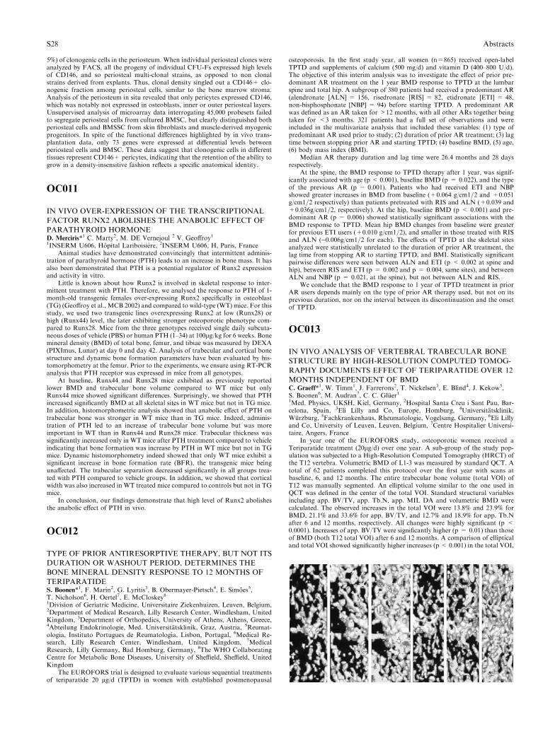
5%) of clonogenic cells in the periosteum. When individual periosteal clones wereanalyzed by FACS, all the progeny of individual CFU-Fs expressed high levelsof CD146, and so periosteal multi-clonal strains, as opposed to non clonalstrains derived from explants. Thus, clonal density singled out a CD146+ clo-nogenic fraction among periosteal cells, similar to the bone marrow stroma.Analysis of the periosteum in situ revealed that only pericytes expressed CD146,which was notably not expressed in osteoblasts, inner or outer periosteal layers.Unsupervised analysis of microarray data interrogating 45,000 probesets failedto segregate periosteal cells from cultured BMSC, but clearly distinguished bothperiosteal cells and BMSSC from skin fibroblasts and muscle-derived myogenicprogenitors. In spite of the functional differences highlighted by in vivo trans-plantation data, only 73 genes were expressed at differential levels betweenperiosteal cells and BMSC. These data suggest that clonogenic cells in differenttissues represent CD146+ pericytes, indicating that the retention of the ability togrow in a density-insensitive fashion reflects a specific anatomical identity.
OC011
IN VIVO OVER-EXPRESSION OF THE TRANSCRIPTIONAL
FACTOR RUNX2 ABOLISHES THE ANABOLIC EFFECT OF
PARATHYROID HORMONED. Merciris*1 C. Marty2, M. DE Vernejoul 2 V. Geoffroy11INSERM U606, Hopital Lariboisiere, 2INSERM U606, H, Paris, France
Animal studies have demonstrated convincingly that intermittent adminis-tration of parathyroid hormone (PTH) leads to an increase in bone mass. It hasalso been demonstrated that PTH is a potential regulator of Runx2 expressionand activity in vitro.
Little is known about how Runx2 is involved in skeletal response to inter-mittent treatment with PTH. Therefore, we analysed the response to PTH of 1-month-old transgenic females over-expressing Runx2 specifically in osteoblast(TG) (Geoffroy et al., MCB 2002) and compared to wild-type (WT) mice. For thisstudy, we used two transgenic lines overexpressing Runx2 at low (Runx28) orhigh (Runx44) level, the later exhibiting stronger osteoporotic phenotype com-pared to Runx28. Mice from the three genotypes received single daily subcuta-neous doses of vehicle (PBS) or human PTH (1–34) at 100lg/kg for 6 weeks. Bonemineral density (BMD) of total bone, femur, and tibiae was measured by DEXA(PIXImus, Lunar) at day 0 and day 42. Analysis of trabecular and cortical bonestructure and dynamic bone formation parameters have been evaluated by his-tomorphometry at the femur. Prior to the experiments, we ensure using RT-PCRanalysis that PTH receptor was expressed in mice from all genotypes.
At baseline, Runx44 and Runx28 mice exhibited as previously reportedlower BMD and trabecular bone volume compared to WT mice but onlyRunx44 mice showed significant differences. Surprisingly, we showed that PTHincreased significantly BMD at all skeletal sites in WT mice but not in TG mice.In addition, histomorphometric analysis showed that anabolic effect of PTH ontrabecular bone was stronger in WT mice than in TG mice. Indeed, adminis-tration of PTH led to an increase of trabecular bone volume but was moreimportant in WT than in Runx44 and Runx28 mice. Trabecular thickness wassignificantly increased only in WT mice after PTH treatment compared to vehicleindicating that bone formation was increase by PTH in WT mice but not in TGmice. Dynamic histomorphometry indeed showed that only WT mice exhibit asignificant increase in bone formation rate (BFR), the transgenic mice beingunaffected. The trabecular separation decreased significantly in all groups trea-ted with PTH compared to vehicle groups. In addition, we showed that corticalwidth was also increased in WT treated mice compared to controls but not in TGmice.
In conclusion, our findings demonstrate that high level of Runx2 abolishesthe anabolic effect of PTH in vivo.
OC012
TYPE OF PRIOR ANTIRESORPTIVE THERAPY, BUT NOT ITS
DURATION OR WASHOUT PERIOD, DETERMINES THE
BONE MINERAL DENSITY RESPONSE TO 12 MONTHS OF
TERIPARATIDES. Boonen*1, F. Marın2, G. Lyritis3, B. Obermayer-Pietsch4, E. Simoes5,T. Nicholson6, H. Oertel7, E. McCloskey81Division of Geriatric Medicine, Universitaire Ziekenhuizen, Leuven, Belgium,2Department of Medical Research, Lilly Research Center, Windlesham, UnitedKingdom, 3Department of Orthopedics, University of Athens, Athens, Greece,4Abteilung Endokrinologie, Med. Universitatsklinik, Graz, Austria, 5Reumat-ologia, Instituto Portugues de Reumatologia, Lisbon, Portugal, 6Medical Re-search, Lilly Research Center, Windlesham, United Kingdom, 7MedicalResearch, Lilly Germany, Bad Homburg, Germany, 8The WHO CollaboratingCentre for Metabolic Bone Diseases, University of Sheffield, Sheffield, UnitedKingdom
The EUROFORS trial is designed to evaluate various sequential treatmentsof teriparatide 20 lg/d (TPTD) in women with established postmenopausal
osteoporosis. In the first study year, all women (n=865) received open-labelTPTD and supplements of calcium (500 mg/d) and vitamin D (400–800 U/d).The objective of this interim analysis was to investigate the effect of prior pre-dominant AR treatment on the 1 year BMD response to TPTD at the lumbarspine and total hip. A subgroup of 380 patients had received a predominant AR(alendronate [ALN] = 156, risedronate [RIS] = 82, etidronate [ETI] = 48,non-bisphosphonate [NBP] = 94) before starting TPTD. A predominant ARwas defined as an AR taken for >12 months, with all other ARs together beingtaken for <3 months. 321 patients had a full set of observations and wereincluded in the multivariate analysis that included these variables: (1) type ofpredominant AR used prior to study; (2) duration of prior AR treatment; (3) lagtime between stopping prior AR and starting TPTD; (4) baseline BMD, (5) age,(6) body mass index (BMI).
Median AR therapy duration and lag time were 26.4 months and 28 daysrespectively.
At the spine, the BMD response to TPTD therapy after 1 year, was signif-icantly associated with age (p < 0.001), baseline BMD (p = 0.022), and the typeof the previous AR (p = 0.001). Patients who had received ETI and NBPshowed greater increases in BMD from baseline (+0.064 g/cm1=2 and +0.051g/cm1=2 respectively) than patients pretreated with RIS and ALN (+0.039 and+0.036g/cm1=2, respectively). At the hip, baseline BMD (p < 0.001) and pre-dominant AR (p = 0.006) showed statistically significant associations with theBMD response to TPTD. Mean hip BMD changes from baseline were greaterfor previous ETI users (+0.010 g/cm1=2), and smaller in those treated with RISand ALN ()0.006g/cm1=2 for each). The effects of TPTD at the skeletal sitesanalyzed were statistically unrelated to the duration of prior AR treatment, thelag time from stopping AR to starting TPTD, and BMI. Statistically significantpairwise differences were seen between ALN and ETI (p < 0.002 at spine andhip), between RIS and ETI (p = 0.002 and p = 0.004, same sites), and betweenALN and NBP (p = 0.021, at the spine), but not between ALN and RIS.
We conclude that the BMD response to 1 year of TPTD treatment in priorAR users depends mainly on the type of prior AR therapy used, but not on itsprevious duration, nor on the interval between its discontinuation and the onsetof TPTD.
OC013
IN VIVO ANALYSIS OF VERTEBRAL TRABECULAR BONE
STRUCTURE BY HIGH-RESOLUTION COMPUTED TOMOG-
RAPHY DOCUMENTS EFFECT OF TERIPARATIDE OVER 12
MONTHS INDEPENDENT OF BMDC. Graeff*1, W. Timm1, J. Farrerons2, T. Nickelsen3, E. Blind4, J. Kekow5,S. Boonen6, M. Audran7, C. C. Gluer11Med. Physics, UKSH, Kiel, Germany, 2Hospital Santa Creu i Sant Pau, Bar-celona, Spain, 3Eli Lilly and Co, Europe, Homburg, 4Universitatsklinik,Wurzburg, 5Fachkrankenhaus, Rheumatologie, Vogelsang, Germany, 6Eli Lillyand Co, University of Leuven, Leuven, Belgium, 7Centre Hospitalier Universi-taire, Angers, France
In year one of the EUROFORS study, osteoporotic women received aTeriparatide treatment (20lg/d) over one year. A sub-group of the study pop-ulation was subjected to a High-Resolution Computed Tomography (HRCT) ofthe T12 vertebra. Volumetric BMD of L1-3 was measured by standard QCT. Atotal of 62 patients completed this protocol over the first year with scans atbaseline, 6, and 12 months. The entire trabecular bone volume (total VOI) ofT12 was manually segmented. An elliptical volume similar to the one used inQCT was defined in the center of the total VOI. Standard structural variablesincluding app. BV/TV, app. Tb.N, app. MIL DA and volumetric BMD werecalculated. The observed increases in the total VOI were 13.8% and 23.9% forBMD, 21.1% and 33.6% for app. BV/TV, and 12.7% and 18.9% for app. Tb.Nafter 6 and 12 months, respectively. All changes were highly significant (p <0.0001). Increases of app. BV/TV were significantly higher (p = 0.01) than thoseof BMD (both T12 total VOI) after 6 and 12 months. A comparison of ellipticaland total VOI showed significantly higher increases (p < 0.001) in the total VOI,
S28 Abstracts
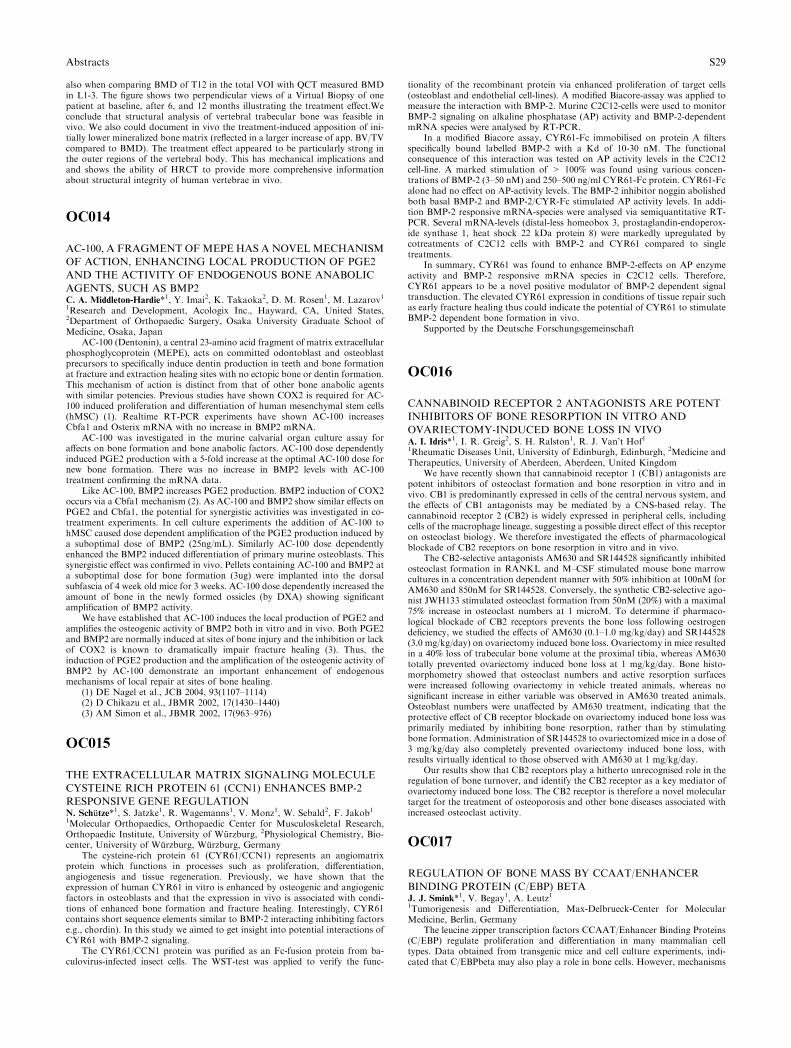
also when comparing BMD of T12 in the total VOI with QCT measured BMDin L1-3. The figure shows two perpendicular views of a Virtual Biopsy of onepatient at baseline, after 6, and 12 months illustrating the treatment effect.Weconclude that structural analysis of vertebral trabecular bone was feasible invivo. We also could document in vivo the treatment-induced apposition of ini-tially lower mineralized bone matrix (reflected in a larger increase of app. BV/TVcompared to BMD). The treatment effect appeared to be particularly strong inthe outer regions of the vertebral body. This has mechanical implications andand shows the ability of HRCT to provide more comprehensive informationabout structural integrity of human vertebrae in vivo.
OC014
AC-100, A FRAGMENT OFMEPE HAS A NOVELMECHANISM
OF ACTION, ENHANCING LOCAL PRODUCTION OF PGE2
AND THE ACTIVITY OF ENDOGENOUS BONE ANABOLIC
AGENTS, SUCH AS BMP2C. A. Middleton-Hardie*1, Y. Imai2, K. Takaoka2, D. M. Rosen1, M. Lazarov11Research and Development, Acologix Inc., Hayward, CA, United States,2Department of Orthopaedic Surgery, Osaka University Graduate School ofMedicine, Osaka, Japan
AC-100 (Dentonin), a central 23-amino acid fragment of matrix extracellularphosphoglycoprotein (MEPE), acts on committed odontoblast and osteoblastprecursors to specifically induce dentin production in teeth and bone formationat fracture and extraction healing sites with no ectopic bone or dentin formation.This mechanism of action is distinct from that of other bone anabolic agentswith similar potencies. Previous studies have shown COX2 is required for AC-100 induced proliferation and differentiation of human mesenchymal stem cells(hMSC) (1). Realtime RT-PCR experiments have shown AC-100 increasesCbfa1 and Osterix mRNA with no increase in BMP2 mRNA.
AC-100 was investigated in the murine calvarial organ culture assay foraffects on bone formation and bone anabolic factors. AC-100 dose dependentlyinduced PGE2 production with a 5-fold increase at the optimal AC-100 dose fornew bone formation. There was no increase in BMP2 levels with AC-100treatment confirming the mRNA data.
Like AC-100, BMP2 increases PGE2 production. BMP2 induction of COX2occurs via a Cbfa1 mechanism (2). As AC-100 and BMP2 show similar effects onPGE2 and Cbfa1, the potential for synergistic activities was investigated in co-treatment experiments. In cell culture experiments the addition of AC-100 tohMSC caused dose dependent amplification of the PGE2 production induced bya suboptimal dose of BMP2 (25ng/mL). Similarly AC-100 dose dependentlyenhanced the BMP2 induced differentiation of primary murine osteoblasts. Thissynergistic effect was confirmed in vivo. Pellets containing AC-100 and BMP2 ata suboptimal dose for bone formation (3ug) were implanted into the dorsalsubfascia of 4 week old mice for 3 weeks. AC-100 dose dependently increased theamount of bone in the newly formed ossicles (by DXA) showing significantamplification of BMP2 activity.
We have established that AC-100 induces the local production of PGE2 andamplifies the osteogenic activity of BMP2 both in vitro and in vivo. Both PGE2and BMP2 are normally induced at sites of bone injury and the inhibition or lackof COX2 is known to dramatically impair fracture healing (3). Thus, theinduction of PGE2 production and the amplification of the osteogenic activity ofBMP2 by AC-100 demonstrate an important enhancement of endogenousmechanisms of local repair at sites of bone healing.
(1) DE Nagel et al., JCB 2004, 93(1107–1114)(2) D Chikazu et al., JBMR 2002, 17(1430–1440)(3) AM Simon et al., JBMR 2002, 17(963–976)
OC015
THE EXTRACELLULAR MATRIX SIGNALING MOLECULE
CYSTEINE RICH PROTEIN 61 (CCN1) ENHANCES BMP-2
RESPONSIVE GENE REGULATIONN. Schutze*1, S. Jatzke1, R. Wagemanns1, V. Monz1, W. Sebald2, F. Jakob11Molecular Orthopaedics, Orthopaedic Center for Musculoskeletal Research,Orthopaedic Institute, University of Wurzburg, 2Physiological Chemistry, Bio-center, University of Wurzburg, Wurzburg, Germany
The cysteine-rich protein 61 (CYR61/CCN1) represents an angiomatrixprotein which functions in processes such as proliferation, differentiation,angiogenesis and tissue regeneration. Previously, we have shown that theexpression of human CYR61 in vitro is enhanced by osteogenic and angiogenicfactors in osteoblasts and that the expression in vivo is associated with condi-tions of enhanced bone formation and fracture healing. Interestingly, CYR61contains short sequence elements similar to BMP-2 interacting inhibiting factorse.g., chordin). In this study we aimed to get insight into potential interactions ofCYR61 with BMP-2 signaling.
The CYR61/CCN1 protein was purified as an Fc-fusion protein from ba-culovirus-infected insect cells. The WST-test was applied to verify the func-
tionality of the recombinant protein via enhanced proliferation of target cells(osteoblast and endothelial cell-lines). A modified Biacore-assay was applied tomeasure the interaction with BMP-2. Murine C2C12-cells were used to monitorBMP-2 signaling on alkaline phosphatase (AP) activity and BMP-2-dependentmRNA species were analysed by RT-PCR.
In a modified Biacore assay, CYR61-Fc immobilised on protein A filtersspecifically bound labelled BMP-2 with a Kd of 10-30 nM. The functionalconsequence of this interaction was tested on AP activity levels in the C2C12cell-line. A marked stimulation of > 100% was found using various concen-trations of BMP-2 (3–50 nM) and 250–500 ng/ml CYR61-Fc protein. CYR61-Fcalone had no effect on AP-activity levels. The BMP-2 inhibitor noggin abolishedboth basal BMP-2 and BMP-2/CYR-Fc stimulated AP activity levels. In addi-tion BMP-2 responsive mRNA-species were analysed via semiquantitative RT-PCR. Several mRNA-levels (distal-less homeobox 3, prostaglandin-endoperox-ide synthase 1, heat shock 22 kDa protein 8) were markedly upregulated bycotreatments of C2C12 cells with BMP-2 and CYR61 compared to singletreatments.
In summary, CYR61 was found to enhance BMP-2-effects on AP enzymeactivity and BMP-2 responsive mRNA species in C2C12 cells. Therefore,CYR61 appears to be a novel positive modulator of BMP-2 dependent signaltransduction. The elevated CYR61 expression in conditions of tissue repair suchas early fracture healing thus could indicate the potential of CYR61 to stimulateBMP-2 dependent bone formation in vivo.
Supported by the Deutsche Forschungsgemeinschaft
OC016
CANNABINOID RECEPTOR 2 ANTAGONISTS ARE POTENT
INHIBITORS OF BONE RESORPTION IN VITRO AND
OVARIECTOMY-INDUCED BONE LOSS IN VIVOA. I. Idris*1, I. R. Greig2, S. H. Ralston1, R. J. Van’t Hof11Rheumatic Diseases Unit, University of Edinburgh, Edinburgh, 2Medicine andTherapeutics, University of Aberdeen, Aberdeen, United Kingdom
We have recently shown that cannabinoid receptor 1 (CB1) antagonists arepotent inhibitors of osteoclast formation and bone resorption in vitro and invivo. CB1 is predominantly expressed in cells of the central nervous system, andthe effects of CB1 antagonists may be mediated by a CNS-based relay. Thecannabinoid receptor 2 (CB2) is widely expressed in peripheral cells, includingcells of the macrophage lineage, suggesting a possible direct effect of this receptoron osteoclast biology. We therefore investigated the effects of pharmacologicalblockade of CB2 receptors on bone resorption in vitro and in vivo.
The CB2-selective antagonists AM630 and SR144528 significantly inhibitedosteoclast formation in RANKL and M–CSF stimulated mouse bone marrowcultures in a concentration dependent manner with 50% inhibition at 100nM forAM630 and 850nM for SR144528. Conversely, the synthetic CB2-selective ago-nist JWH133 stimulated osteoclast formation from 50nM (20%) with a maximal75% increase in osteoclast numbers at 1 microM. To determine if pharmaco-logical blockade of CB2 receptors prevents the bone loss following oestrogendeficiency, we studied the effects of AM630 (0.1–1.0 mg/kg/day) and SR144528(3.0 mg/kg/day) on ovariectomy induced bone loss. Ovariectomy in mice resultedin a 40% loss of trabecular bone volume at the proximal tibia, whereas AM630totally prevented ovariectomy induced bone loss at 1 mg/kg/day. Bone histo-morphometry showed that osteoclast numbers and active resorption surfaceswere increased following ovariectomy in vehicle treated animals, whereas nosignificant increase in either variable was observed in AM630 treated animals.Osteoblast numbers were unaffected by AM630 treatment, indicating that theprotective effect of CB receptor blockade on ovariectomy induced bone loss wasprimarily mediated by inhibiting bone resorption, rather than by stimulatingbone formation. Administration of SR144528 to ovariectomized mice in a dose of3 mg/kg/day also completely prevented ovariectomy induced bone loss, withresults virtually identical to those observed with AM630 at 1 mg/kg/day.
Our results show that CB2 receptors play a hitherto unrecognised role in theregulation of bone turnover, and identify the CB2 receptor as a key mediator ofovariectomy induced bone loss. The CB2 receptor is therefore a novel moleculartarget for the treatment of osteoporosis and other bone diseases associated withincreased osteoclast activity.
OC017
REGULATION OF BONE MASS BY CCAAT/ENHANCER
BINDING PROTEIN (C/EBP) BETAJ. J. Smink*1, V. Begay1, A. Leutz11Tumorigenesis and Differentiation, Max-Delbrueck-Center for MolecularMedicine, Berlin, Germany
The leucine zipper transcription factors CCAAT/Enhancer Binding Proteins(C/EBP) regulate proliferation and differentiation in many mammalian celltypes. Data obtained from transgenic mice and cell culture experiments, indi-cated that C/EBPbeta may also play a role in bone cells. However, mechanisms
Abstracts S29
and precise functions have remained unclear. Here, we show that C/EBPbetamRNA is expressed in growth plate chondrocytes, osteoblasts and osteoclasts intibiae of 8 week old mice. C/EBPbeta knock-out mice (1) showed a significantreduction both in body and tibia length. Histomorphometric analyses of thetibiae showed a 1.6-fold decrease in bone volume in C/EBPbeta-deficient mice(n = 9), caused by a significant reduction in trabecular thickness and number.
Although osteoclast surface to bone surface was similar in wild-type and C/EBPbeta-deficient mice, the osteoclast number was reduced in the latter. Thissuggested the presence of larger osteoclasts in C/EBPbeta-deficient mice. Ex vivoosteoclast culture confirmed the formation of larger osteoclasts from bonemarrow cells of C/EBPbeta-deficient mice. These cultured osteoclasts werefunctional, as determined by in vitro resorption activity. Strikingly, functionalmulti-nucleated osteoclasts formed even in the absence of RANK-L, suggestingthat C/EBPbeta is a negative regulator of RANK-L signaling. Co-cultureexperiments with osteoblasts derived from either wild-type or C/EBPbeta-defi-cient mice showed that the observed osteoclast phenotype is cell-autonomous.Furthermore, C/EBPbeta-deficient osteoblasts inhibited osteoclast formation,indicating an additional role for the osteoblasts in the osteopenic phenotype ofthe C/EBPbeta-deficient mice.
Taken together, our data show that C/EBPbeta regulates bone mass byaffecting both osteoclasts and osteoblasts. These data suggest that C/EBPbetacould be a novel candidate involved in bone diseases such as osteoporosis.
1. Generously provided by E. Sterneck (E. Sterneck, L. Tessarollo and P.F.Johnson. 1997. An essential role for C/EBPbeta in female reproduction. GenesDev 11:2153–62)
J.J. Smink is a recipient of a Marie Curie fellowship
OC018
OVEREXPRESSION OF THE INHIBITOR OF
METALLOPROTEINASES TIMP-1 RESCUES THE STRONG
BONE LOSS INDUCED BY RUNX2 OVEREXPRESSION IN
MICEV. Geoffroy*1, C. Prouillet1, C. Marty1, M. De Vernejoul11INSERM U606, Hopital Lariboisiere, Paris, France
Osteoblastic matrix metalloproteinases (MMPs) have been reported to berequired for normal bone resorption. Several lines of evidence suggest thatosteoblastic MMPs could take part of the increased bone turnover observed inmice after overexpression of Runx2 (Runx2 mice). The goal of our study was todefine using transgenic approach to define whether inhibition of osteoblasticmatrix metalloproteinases (MMPs) in vivo can moderate the hyper resorptionobserved in these mice.
Thus, we examined the rescue of the severe osteopenic phenotype in Runx2mice by overexpressing the MMP inhibitor TIMP-1 (tissue inhibitor of metal-loproteinase 1) specifically in osteoblasts. One-, four- and eight-month-old fe-males with the different genotype (WT, Runx2, TIMP-1 and TIMP-1/Runx2)have been generated. Analysis of the BMD (Bone Mineral Density) has beenmeasured by DXA (Piximus, Lunar) at the total body, the femur and the caudalvertebrae and by pQCT (Stratech) at the tibia for all mice. In addition, analysisof bone architecture as well as the determination of the bone formation andresorption parameters have been performed in 4-month-old female at the fem-oral metaphysis.
Our results indicated that the TIMP-1 overexpression in osteoporotic miceoverexpressing Runx2 has no effect on BMD in one-month-old females. Butin 4- and 8-month-old females, a partial rescue of the bone loss observed inthe Runx2 mice was observed in TIMP-1/Runx2 mice at all sites. In addition,evaluation of BMD by pQCT indicated that the increase of BMD observedby DXA in TIMP-1/Runx2 mice compared to Runx2 mice was due to a effectof the TIMP-1 on both cortical and trabecular BMD. Analysis of bonearchitecture in four-month-old TIMP-1/Runx2 females moreover indicatedthat a decrease of the resorption activity (decreased trabecular separation)and an increase of osteoformation (increased trabecular thickness) comparedto Runx2 mice. Interestingly, osteoclastic sufaces were not different in bothgenotypes indicating that only osteoclasts activity was reduced. In addition,we also reported a partial rescue of the cortical bone loss (increased corticalthickness and outer bone diameter) in TIMP-1/Runx2 mice compared toRunx2 mice.
In conclusion, our results indicate that increased matrix metalloproteinaseshave a significant role in the bone loss observed in the runx2 mice.
OC019
THE PLEKHM1 PROTEIN IS INVOLVED IN OSTEOPETROSIS
IN RAT AND HUMAN AND APPEARS TO PLAY A ROLE IN
VESICULAR TRANSPORT IN OSTEOCLASTSL. Van Wesenbeeck*1, P. R. Odgren2, F. P. Coxon3, A. Frattini4, P. Moens5,B. Perdu1, C. A. MacKay2, E. Van Hul1, J. Timmermans6, R. Jacobs5,B. Peruzzi7, M. J. Rogers3, M. H. Helfrich3, A. Teti7, A. Villa4, W. Van hul8
1Department of Medical Genetics, University of Antwerp, Wilrijk, Belgium,2Department of Cell Biology, University of Massachusetts Medical School,Worcester, United States, 3Bone Research Group, Institute of Medical Sciences,University of Aberdeen, Aberdeen, United Kingdom, 4Instituto di TecnologieBiomediche Avanzate, Consiglio Nazionale delle Ricerche, Segrate, Italy,5Pediatric Orthopaedics, Catholic University of Leuven, Leuven, 6Laboratory ofCell Biology and Histology, University of Antwerp, Wilrijk, Belgium,7Department of Experimental Medicine, University of L’Aquila, L’Aquila, Italy,8Department of Medical Genetics, University and University Hospital of An-twerp, Wilrijk, Belgium
The osteopetroses are a genetically heterogeneous group of bone disorderscharacterized by a reduction in bone resorption resulting in an overall increase inskeletal mass. The incisors absent (ia) rat displays a generalized skeletal sclerosisand a delay in tooth eruption due to numerous dysfunctional osteoclasts. Weidentified a deletion of 1 nucleotide in the plekhm1 gene in ia rats, resulting in theincorporation of unrelated amino acids and a premature stopcodon. We alsoperformed mutation analysis of the entire coding sequence of the plekhm1 genein patients diagnosed with various forms of osteopetrosis and identified a splicesite mutation in the human plekhm1 gene in one family diagnosed with theintermediate type of osteopetrosis. We compared the morphology and the boneresorption capacity of osteoclasts (derived from M-CSF-dependent peripheralblood monocytes) from a patient in this family with osteoclasts generated fromthe unaffected brother. SEM and confocal microscopy showed that osteoclastsdifferentiated normally from patient PBMCs and were able to polarize. How-ever, unlike osteoclasts from the unaffected sibling, osteoclasts from the patientshowed no evidence of resorptive activity when cultured on dentine discs, andhad a more flattened morphology.
So far, the role of the plekhm1 protein in bone remains unknown. However,the presence of a RUN and Pleckstrin Homology domains suggests that it isinvolved in small GTPase signalling. Rab GTPases play an important role invesicular transport in osteoclasts. Using overexpression studies in HEK293 cells,we found that the plekhm1 protein is partially associated with intracellularvesicles and colocalizes with Rab7, but not Rab5 or Rab6, suggesting that thesevesicles are late-endosomes/lysosomes. Furthermore, inhibiting the prenylationof Rab proteins disrupted the endosomal localization of the plekhm1 protein,supporting its role in small GTPase signaling.
In conclusion, the identification of the plekhm1 gene as a new osteopetrosisgene indicates its crucial role in bone resorption with a putative function invesicular transport in the osteoclast.
OC020
UROKINASE RECEPTOR IS INVOLVED IN BONE REMOD-
ELING BY INFLUENCING OSTEOCLASTS RESORBTIONF. Furlan*1, N. Jorgensen2, J. Jemsen2, E. Mrak3, A. Rubinacci3, P. Verde4,F. Blasi11Molecular Genetics, DIBIT, H. San Raffaele, Milan, Italy, 2Osteoporosis Unit,Dpt of Endocrinology, Copenhagen, Denmark, 3Bone metabolic Unit, H. SanRaffaele, Milan, 4Genetics and Biophysics, CNR, Naples, Italy
Urokinase receptor (uPAR) is actively involved in the regulation of importantcell functions like adhesion and migration and it has been shown to interact withintegrins. It has been previously shown that the major players of bone remodeling,osteoblasts (obs) and osteoclasts (ocs), produce urokinase (uPA) and express itsreceptor. The purpose of this study was to investigate the role of uPAR in boneremodeling.We analyzed the bone phenotype in uPARko femalemice. The size ofko mice was reduced as compared to wt animals, and there was a significantdecrease in tibia length in komice as compared towt. Themost surprising foundingwas that bone mass analysis of uPAR ko by pQCT revealed an increase in bonemass with respect to wt (p< 0.02). The histomorphometric analysis of the tibia inuPAR ko showed a reduction in the bone volume compared to wt (more bonedensity but less bone volume)with a significant reduction in cortical thickness in komice, and a reduction of resorbing surface in Ko bones compare to wt. Moreover,mechanical tests showed a reduction in the capability to sustain a given load inuPAR ko tibias as compared to tibias wt (p < 0.02). Proliferative advantage withnodifference in apoptosis, andahigher susceptibility tomatrixmineralizationwereseen in uPAR ko obs compared to wt. The production of proteins involved in themineralization process, like ALP, was increased only during the first weeks of obsmaturation. After 4 weeks, ALP was similar in both genotypes. RANKL expres-sion on obs surface did not change between two different genotypes at differentstages of differentiation. The expression levels of important proteins involved inobs development and function, like AP-1 transcription factors were overexpressedin mice uPAR ko as compared to wt cells. On the contrary the number of ocsformed in vitro using uPAR ko monocytes was decreased compared to wt.Moreover, we have found that uPAR co-localized with integrin b3 on ocs podo-soms by confocal microscopy. Therefore, we would like to better understand therole of uPAR in ocs function by crossing uPAR ko mice with integrin b3 ko mice.Preliminary results showed that double ko mice are viable, fertile and with normalteeth eruption. Experiments in order to investigate the downstream pathways inocs are under current investigation. Together our preliminary data indicate thatuPAR may play an important role in bone physiology by influencing both,osteoblast and osteoclasts function.
S30 Abstracts
OC021
TWIST CONTROLS CBL-MEDIATED PI3K
UBIQUITINATION AND SIGNALLING IN
OSTEOBLASTSH. Guenou*1, C. Dufour1, K. Kaabeche1, H. Miraoui1, P. J. Marie11Laboratory of Osteoblast Biology and Pathology, INSERM U606, Paris,France
Genetic mutations of Twist, a bHLH transcription factor, induce pre-mature fusion of cranial sutures (craniosynostosis) in the Saethre-Chotzensyndrome (SCS). The mechanisms by which Twist haploinsufficiency altersosteoblasts and bone formation in humans are not fully understood. Werecently showed that the reduced Twist dosage in SCS downregulates fibro-blast growth factor receptor-2 (Fgfr2) expression in Twist mutant osteoblasts.We also reported that the ubiquitin ligase Cbl interacts with Fgfr2 to controlFgfr2 signalling and phenotypic markers in Fgfr2 mutant human osteoblasts,suggesting a role for Cbl in the control of osteoblast differentiation. Cbl isknown to interact with the p85 regulatory subunit of phosphatidyl inositol 3kinase (PI3K), a pathway which has recently emerged as an importantmodulator of osteoblast differentiation. In this study, we investigated the roleof Twist and Cbl in the control of PI3K signalling in human osteoblasts. Wefound that cranial osteoblasts from an SCS patient with a Twist Y103Xmutation inducing deletion of the bHLH domain showed decreased CblmRNA and protein levels compared with wild-type osteoblasts, indicatingthat Twist controls Cbl expression. Consistently, transfection with a Twistexpression vector rescued Cbl levels in Twist mutant osteoblasts. The de-creased Cbl expression in Twist mutant osteoblasts was associated with in-creased total PI3K and phosphorylated PI3K levels whereas PI3K mRNAlevels were unaffected, indicating that the reduced Cbl levels led to accumu-lation and activation of PI3K. We demonstrated that Twist overexpressionwhich increased Cbl levels in Twist mutant osteoblasts led to reduce PI3Klevels to normal levels. Moreover, transfection with Cbl which rescued Cbllevels also corrected PI3K protein levels in Twist mutant osteoblasts. Thesestudies indicate that Twist haploinsufficiency downregulates Cbl expression inosteoblasts, which in turn leads to reduced PI3K ubiquitination and degra-dation, resulting in PI3K accumulation and activation in human calvarialosteoblasts. This provides novel genetic and biochemical evidence for a role ofTwist in the control of Cbl-mediated PI3K signalling in osteoblasts.
OC022
NFAT AND SRF TRANSCRIPTION FACTORS
COOPERATE TO REGULATE KROX20 GENE
EXPRESSION IN BONEM. Frain*1, A. Nordheim2, P. Charnay11INSERM U368, Ecole Normale Superieure, Paris, France, 2Institute of CellBiology, Tuebingen University, Tuebingen, Germany
The Krox20 gene encodes a zinc finger transcription factor that plays a keyrole in regulating bone formation. We have shown that Krox20 is expressed in asubpopulation of growth plate hypertrophic chondrocytes and in differentiatingosteoblasts and that Krox20 conditional knockout mice develop severe osteo-porosis.
To investigate the mechanisms of Krox20 transcriptional regulation inbone, we have used a transgenic mice approach and identified a bone-specificenhancer in the 5’ flanking region of the mouse Krox20 gene which spans 860bp and recapitulates Krox20 expression during bone development. Combiningphylogenetic footprinting analyses and in vitro and in vivo experiments, wehave defined three types of regulatory elements within the enhancer: Krox20binding sites involved in a direct positive autoregulatory loop, Runx2 bindingsites modulating Krox20 expression and a 13 bp A/T rich element essentialfor Krox20 activation in both osteogenic and chondrogenic cells. This keyregulatory element is conserved among vertebrates and contains partiallyoverlapping canonical binding sites for two transcription factors : Srf (SerumResponse Factor) and NFAT (Nuclear Factor of Activated T-cells) known tocontrol several developmental processes. Bandshift and competition assaysusing recombinant protein extracts have confirmed that the bone enhancercontains bona-fide binding sites for Srf and NFAT. Moreover, transfectionexperiments in NIH 3T3 and MC3T3-E1 have demonstrated that NFAT1 isthe only NFAT family member that can transactivate a construct containinga polymer of the A/T rich element. Point mutations that affect specifically thebinding of Srf or NFAT1 in vitro have been introduced within the boneenhancer and tested in transgenic mice. We have found that mutating eitherthe Srf or the NFAT site dramatically reduced bone enhancer activity.Presently, we are generating Srf bone-specific conditional knockout to furtheranalyse its role in the control of Krox20 expression.
In any case, our results indicate that Srf and NFAT are both required forthe initiation of Krox20 expression in osteoblasts and hypertrophic chon-drocytes and thereby, we define a novel function for Srf in regulating skel-etogenesis.
OC023
BIM IS A NOVEL REGULATOR OF APOPTOSIS IN
OSTEOBLASTSB. Espina1, M. Liang1, G. Russell1, P. A. Hulley*11Botnar Research Centre, University of Oxford, Oxford, United Kingdom
Bim (Bcl-2 Interacting Mediator of Cell Death), a proapoptotic Bcl-2 familymember, is upregulated in osteoclasts in response to M-CSF withdrawal andcauses apoptosis. We report that Bim also plays a role in apoptosis of non-haemopoietic lineage osteoblasts. Both osteoblasts and osteocytes undergoapoptosis in vitro in response to detachment, growth factor withdrawal and highdose glucocorticoid (GC) treatment. However, the molecular mechanisms re-main elusive. We have developed models of all 3 apoptosis-inducing conditionsin murine MBA-15.4 osteoblasts. These cells respond to 1uM dexamethasone(Dex) with increased caspase 3 activity, fragmented DNA and morphologicalchanges consistent with apoptotic cell death within 24–48 h of treatment. Wefind dose- and time-dependent upregulation of Bim, with highest levels at 48h for10-6 M Dex (7 fold). This same pattern of expression was observed in humanprimary osteoblasts. Moreover, Bim protein expression correlated with increasedapoptosis (4-5 fold) as measured by acridine orange stain, Nick Translation orCaspase 3&7 activity assays. Serum withdrawal and detachment also increasedexpression of Bim in osteoblasts. This was more rapid (2h after serum starvation)and preceded detection of caspase 3 activity (8h) and apoptosis (8h onwards).Bim levels are reported to be regulated by PKB/Akt and/or ERK-mediatedrepression of the forkhead transcription factors which drive Bim gene expres-sion. These survival kinases also phosphorylate mature Bim protein and target itfor proteasomal destruction. Whereas Bim basal levels were hardly detectable inosteoblastic cells, 0.5 lM MG132 (proteasome inhibitor) strongly increased Bimprotein levels (17 fold). The GC receptor antagonist, RU486, partially sup-pressed Dex-induced Bim, suggesting that it is regulated directly or indirectly bythe GC receptor. Cycloheximide (Chx) alone and Chx plus Actinomycin Deffectively suppressed Dex-induced Bim indicating a requirement for de novoprotein synthesis. We used insulin or 20% FCS to activate the survival kinases,PKB/Akt and ERK, and this strongly repressed the increase in Bim levels in-duced by Dex and also protected the cells from apoptosis. These findings suggestthat Bim is a novel regulator of osteoblast apoptosis and may be a target fortherapeutic approaches.Funded by the Arthritis Research Campaign
OC024
DEFICIENT OSTEOBLAST PROLIFERATION,
DIFFERENTIATION AND FUNCTION INDUCE OSTEOPENIC
PHENOTYPE IN SOX4 HAPLOINSUFFICIENT MICEN. Rucci*1, L. H. Nissen Meyer2, R. Jemtland3, V. T. Gautvik2, M. Pedersen2,R. Paro1, D. Fortunati2, A. Del Fattore1, A. Rufo1, S. Reppe2, D. Pierroz4,S. Ferrari4, F. P. Reinholt5, A. Teti1, K. M. Gautvik61Department of Experimental Medicine, University of L’Aquila, L’Aquila, Italy,2Department of Biochemistry, Institute for Basic Medical Sciences, University ofOslo, 3Endocrine Section, Department of Medicine, National University Hos-pital, Oslo, Norway, 4Department of Rehabilitation and Geriatrics, GenevaUniversity Hospital, Geneva, Switzerland, 5Department of Pathology, NationalUniversity Hospital, 6Department of Biochemistry, Institute for Basic MedicalSciences, University of Oslo and Ullevaal University Hospital, Oslo, Norway
Sox4 is a BMP2 and PTH-regulated transcription factor which is expressedby hypertrophic chondrocytes and osteoblasts. Homozygote Sox4-/- mice die inutero from cardiovascular malformations. Sox4+/- mice appear normal, butdevelop osteopenia later in life as shown by DEXA and mCT analyses comparedto wildtype (WT). The differences are most prominent in trabecular bone(lumbar spine, trochanter femurs and proximal tibiae) but cortical bone is alsoaffected (femoral diaphysis). To explore the underlying cellular mechanisms weperformed primary calvarial cells and observed a remarkably less alkalinephosphatase (ALP) mRNA expression and activity. [3H]-thymidine incorpora-tion tests demonstrated >50 % lower cellular proliferation rate in Sox4+/- vs.WT. Mineralization was also impaired in Sox4+/- osteoblasts relative to WT.Sox4 haploinsufficient osteoblasts exhibited significantly lower transcriptionalexpression of osterix (>50 %), osteocalcin and collagen 1A2 chain, compared toWT cultures, while Runx-2 and osteopontin mRNAs were unaffected. Histo-morphometric analysis showed a trend to a lower Ob.S/BS and unchanged Oc.S/BS in Sox4+/- mice relative to WT. Bone marrow cultures from Sox4+/- mice(incubated for 7 days in the presence of vitamin D3) showed normal osteo-clastogenic efficiency as shown by TRAcP activity and pit index, compared toWT cultures. Furthermore, WT bone marrow cells were not differentiallyinfluenced by conditioned media from Sox4+/- and WT osteoblasts, and theexpression of osteoclastogenic cytokines (e.g. IL-1b IL-6, TNF alpha) bySox4+/- and WT osteoblasts was similar. On the basis of the present results, wetested the effect of Sox4 siRNA on WT osteoblasts. Incubation of WT osteo-blasts for 72 hours with Sox4 siRNA reduced Sox4 mRNA about 80% incomparison to control siRNA. These cells showed similar reduction of ALP,proliferation, osterix, osteocalcin and collagen 1A2 mRNAs, and unremarkable
Abstracts S31

Runx-2 and osteopontin vs. control siRNA-treated cells, overlapping the resultsobserved with the genuine Sox4+/- and WT osteoblasts. Collectively, these re-sults suggest that Sox4 regulates osteoblast proliferation, differentiation andfunction, whereas it plays an insignificant role in osteoclasts. Therefore, weconclude that osteopenia in Sox4 haploinsufficient mice is due to lower boneformation, opening up a new avenue towards the understanding of the molecularmechanisms of osteoporosis. Funded by the EC Osteogene LSH-2002-502941grant.
OC025
MICE LACKING THE EPITHELIAL CALCIUM CHANNEL
TRPV5 HAVE THINNER AND UNDERMINERALISED BONESB. C. J. Van der Eerden*1, N. Fratzl-Zelman2, P. Roschger2, J. G. J. Hoende-rop3, H. Weinans4, H. A. P. Pols1, R. J. M. Bindels3, J. P. T. M. Van Leeuwen11Internal Medicine, Erasmus MC, Rotterdam, Netherlands, 2Ludwig BoltzmannInstitute of Osteology, WGKK and AUVA Trauma Centre Meidling, 4thMedical Department, Hanusch Hospital, Vienna, Austria, 3Cell Physiology,NCMLS, Radboud University Nijmegen Medical Center, Nijmegen, 4Orthope-dics, Erasmus MC, Rotterdam, Netherlands
We recently demonstrated that transient receptor potential channel V5(TRPV5) deficiency (TRPV5-/-) in mice display reduced trabecular and corticalbone thickness compared to wildtype littermates (TRPV5+/+). Moreover,TRPV5-/- mice have severely reduced osteoclastic bone resorption. Knowing thatchanges occur in Ca2+ homeostasis with aging, we further explored the bonephenotype of TRPV5-/- mice during aging.
Femurs were analysed by muCT for several structural parameters, quanti-tative backscattered electron imaging (qBEI) was applied on tibiae to determinebone mineralisation density distribution and calcein labels were analysed forcalculation of mineral apposition rate (MAR) and bone formation rate (BFR).
In the femoral head of the TRPV5-/- mice, trabecular thickness was reducedin the 8-week-old animals and the difference with the TRPV5+/+ mice pro-gressively increased with age. In the diaphysis of the 8-week-old TRPV5-/- mice,cortical thickness was lower compared to TRPV5+/+ mice. During aging of theTRPV5+/+ mice it remained similar, while it decreased with age in the TRPV5-/-
mice. The polar moment of inertia, a proxy for bone strength, was lower inTRPV5-/- versus TRPV5+/+ mice. The other parameters were unaffected byTRPV5 deficiency. The qBEI analyses demonstrated that in the secondaryspongiosa the weighted mean Ca2+ content (CaMean) and the most frequentCa2+ concentration (CaPeak) were reduced in the 8- and 52-week-old TRPV5-/-
mice compared to TRPV5+/+ mice, while there were no changes in minerali-sation heterogeneity (CaWidth). These parameters were not different in corticalbone. Interestingly, both in the spongiosa and in the cortical bone, the amount oflowly mineralised areas (CaLow) that correspond to sites of primary minerali-sation, increased in 52-week-old TRPV5-/- mice versus wildtype. In addition,calcein staining in bone sections from the young TRPV5-/- age group revealedlower MAR and BFR compared to TRPV5+/+ mice. Serum 1,25(OH)2D3 inTRPV5-/- mice was strongly elevated at all ages compared to TRPV5+/+ mice.Bone resorption as measured by deoxypyridinoline was reduced in all TRPV5-/-
age groups.In conclusion, the bone phenotype of TRPV5-/- mice progresses during
aging. More specifically, mice lacking TRPV5 have thinner and underminera-lised bones. This may be explained either by their high serum 1,25(OH)2D3 levelsor by their lower bone resorption rate in vivo that, via coupling, leads to lessbone formation.
OC026
CARDIOTROPHIN-1 REGULATES OSTEOCLAST AND
OSTEOBLAST FUNCTION IN A MANNER DISTINCT FROM
OTHER GP130 CYTOKINESN. A. Sims*1, E. C. Walker1, J. M. W. Quinn1, E. A. Allan1, H. Saleh1,T. Martin11Bone, Joint and Cancer Group, St. Vincent’s Institute, Fitzroy, Australia
The gp130 family of cytokines signal by complexing with the gp130 receptorsubunit. In gp130 null mice there is a dramatic increase in osteoclast andosteoblast generation in vivo, in contrast to the stimulatory effects of gp130cytokines on cultured osteoclasts. Knockout (KO) mice for gp130 cytokineshave distinct bone phenotypes, suggesting distinct signaling pathways inducedby the cytokine-specific receptor complex formed. We examined the skeletons ofcardiotrophin-1 (CT-1) null mice to determine the unique roles of CT-1 in bone.
Compared with wild type (WT), adult CT-1 KO mice demonstrated a sig-nificant increase in trabecular bone volume (+ 229%) and trabecular BMD (+31%). The increase in bone mass was associated with an increased number ofabnormally large osteoclasts; osteoclast surface and number were approximatelydoubled and the amount of bone surface covered by each osteoclast was sig-nificantly elevated. Stimulation of CT-1 KO bone marrow cultures with
RANKL and M-CSF also led to a significant elevation in osteoclast number incomparison with WT marrow. The osteoclasts generated in vitro were alsoabnormally large, suggesting that the osteoclast phenotype was cell-lineageautonomous. These osteoclasts appeared to be functionally impaired, suggestedby the increased bone mass in vivo, and by an increase in cartilage remnantswithin the trabecular bone. Furthermore, while osteoblast surface and numberwere unchanged, bone formation rate was very low, indicating that coupling ofbone formation and resorption remains intact.
CT-1 is therefore unique in its actions in bone and essential for normalosteoclast and osteoblast function, and may provide a novel therapeutic targetfor modifying bone formation or resorption.
OC027
DELETION OF GHRELIN, BUT NOT ITS RECEPTOR GHSR,
LEADS TO TRABECULAR BONE CHANGES IN AGEING
MALE MICEM. Van der Velde*1, B. C. J. Van der Eerden1, P. J. Delhanty1, Y. Sun2, R. G.Smith2, A. J. Van der Lely1, H. A. P. Pols1, J. P. T. M. Van Leeuwen11Internal Medicine, Erasmus MC, Rotterdam, Netherlands, 2Huffington Centeron Aging, Department of Molecular and Cellular Biology, Baylor College ofMedicine, Houston, United States
Ghrelin is a peptide hormone with several functions, including stimulation ofgrowth hormone (GH) release from the pituitary via its receptor GHSR. In-creases in serum GH can enhance bone growth by stimulating insulin-likegrowth factor (IGF-I) production and secretion from the liver and from localtissues, including bone. In a previous study we showed that ghrelin can stimulatethe proliferation of cultured human osteoblastic cells, providing a direct mech-anism through which ghrelin can act on bone.
In this study we examined whether bone morphometry was affected in maleC57Bl/6 mice lacking the Ghrl or Ghsr gene, encoding ghrelin and its receptor.Femurs from both Ghrl-/-and Ghsr-/- mice (15 months old) and their wild typelittermates were analysed by micro-computed tomography. Deletion of the Ghrlgene resulted in a higher connectivity density (+89.4%), indicative of a denserand more interwoven trabecular network, together with a higher trabecular bonevolume fraction (+39.3%) while trabecular thickness was unaltered (+0.4%).This indicates the presence of more trabeculae in Ghrl-/- mice. Structural modelindex was lower ()13.7%) in Ghrl-/- mice, indicating a more plate-like structureof these trabeculae. In contrast to trabecular bone, most cortical parameterswere similar between Ghrl-/- mice and their wild type littermates: cortical volume(+4.5%), cortical thickness (-2.7%), endocortical volume (+4.3%). Deletion ofthe Ghsr gene did not lead to major changes in trabecular or cortical bone:connectivity density (+2.6%), trabecular bone volume fraction ()3.9%), tra-becular thickness ()2.1%), structural model index (+3.1%), cortical volume()0.7%), cortical thickness ()2.4%), endocortical volume ()5.4%).
In conclusion: firstly, deletion of ghrelin increases trabecular bone volume.This may seem counterintuitive, considering ghrelin,s positive role in the GH-IGF-I axis and the bone anabolic action of IGF-I. However, serum IGF-I levelswere not decreased in Ghrl-/- mice. So, another yet undefined mechanism must beresponsible for the observed increase in trabecular bone. Secondly, in contrast toghrelin, deletion of its receptor did not show any significant effect on bonestructure. This indicates that an additional unknown receptor may be involved inghrelin,s action on bone. This is not unlikely considering several other obser-vations suggesting a second receptor for ghrelin (Chen et al. Gastroenterology.2005; 129(1):8–25).
OC028
INHIBITION OF GLUCOCORTICOID SIGNALING IN
MATURE OSTEOBLASTS INHIBITS CRITICAL PARACRINE
SIGNALS BETWEEN OSTEOBLASTS AND THEIR
PRECURSORSH. Zhou1, W. W. Mak1, Y. Zheng1, C. R. Dunstan1, M. J. Seibel*11Bone Research Programme, Anzac Research Institute, Sydney, Australia
Background: Transgenic (tg) expression of 11beta-hydroxysteroid dehy-drogenase type 2 (HSD2), a glucocorticoid (GC) inactivating enzyme, under thecontrol of a 2.3Kb collagen type I promoter (Col2.3-HSD2) abrogates intra-cellular GC signalling in mature osteoblasts (1). We here characterise the phe-notypic and functional properties of osteoblasts overexpressing Col2.3-HSD2.
Methods: Primary osteoblast cultures were generated from the calvaria of 1-day-old tg mice and wild type (WT) littermates. Nodule assays were carried outby culturing cells in the presence of ascorbate and ß-glycerophosphate for 2weeks. RNA was isolated at days 1, 3 and 7 of cultures for gene expressionstudies.
Results: mRNA for HSD2 was detected by RT-PCR in Col2.3-HSD2 tgcultures with increased expression observed from days 3-7 as cells differentiatedinto osteoblasts. No HSD2 expression was observed in WT cultures at any time.
S32 Abstracts

Col2.3-HSD2 cultures developed 50% fewer nodules than WT cultures butgenerated increased numbers of adipocytes, suggesting a shift in lineage com-mitment from osteoblast to adipocyte. Treating cells with 10-7M thiram, aninhibitor for HSD2 activity, rescued Col2.3-HSD2 tg bone nodule formation buthad no effect on WT cultures, confirming that the effects of HSD2 transgeneexpression are mediated its enzyme activity.
RT-PCR and real time RT-PCR revealed that mRNA for Runx2, ALP, andosteocalcin were expressed at low but similar levels in cells from both Col2.3-HSD2 tg and WT at day 1 culture. These mRNA levels remained low after 3days Col2.3-HSD2 tg culture, while they increased concurrently with osteoblastdifferentiation in the WT culture. Conversely, mRNA expression of the adipo-genic transcription factors C/EBP alpha and PPAR gamma was increased at day3 in Col2.3-HSD2 tg cultures but not in WT cultures. These results indicate thatthe lineage shift from osteoblast to adipocyte was regulated at the transcriptionallevel.
Conclusions: Disrupting GC signalling in mature osteoblasts impairs earlyosteoblast differentiation, promotes adipogenic differentiation, and alters releaseof soluble factors that signal to earlier precursors. These findings suggest theexistence of a GC regulated paracrine signal between mature osteoblasts andtheir mesenchymal precursors that influences lineage commitment.
(1) Endocrinology 145:922–9, 2004. We thank Barbara Kream for kindlyproviding the transgenic animals.
OC029
MECHANICAL STRAIN AND THE ESTROGEN
RECEPTOR INTERACT WITH THE WNT PATHWAY IN
OSTEOBLAST-LIKE CELLSV. J. Armstrong*1, M. Muzylak1, S. Allen1, D. B. Ong1, H. L. Jessop1,J. S. Price1, L. E. Lanyon11Veterinary Basic Sciences, The Royal Veterinary College, London, UnitedKingdom
The Wnt pathway�s influence on bone mass has been proposed to derivefrom its role in the regulation of bone architecture by mechanical strain1. Fullexpression of bones� early osteogenic responses to strain involve estrogenreceptor \alpha (ER\alpha)2. Since there is evidence that some genes in mam-mary cells can be regulated by binding of estrogen, ER\alpha and Tcfs to theirpromoter region, we hypothesised an interaction in osteoblasts between thepathways used by strain, estrogen and Wnts. In this study we determined inosteoblast-like cells in vitro the effects of strain on \beta catenin expression andlocalisation and the effects of strain and ER activity on LEF/Tcf activity.
Monolayer cultures of ROS 17/2.8 cells were strained in four-point bendingas previously described3. Western blot analysis showed a significant increase inlevels of the activated (de-phosphorylated) \beta catenin 1 hour following straincompared with static controls. By 6 hours, activated \beta catenin levels haddeclined to control levels. Immunocytochemistry showed that in control culturesboth total and activated \beta catenin localised predominantly at the membrane.1 hour and 3 hours following strain, \beta catenin had translocated to the nu-cleus. By 6 hours the pattern of localisation was similar to controls. 10mM LiCl,an activator of \beta catenin at the level of GSK, also induced trafficking of \betacatenin to the nucleus and significantly increased LEF/Tcf-inducible luciferasereporter activity (TOPflash assay). Strain did not increase basal or LiCl-inducedchanges in LEF/Tcf- activity. Inhibition of ER activity by ICI 182780 signifi-cantly reduced the LiCl-induced LEF/Tcf activity whereas tamoxifen, a partialER antagonist, had no effect.
The increased expression and nuclear translocation of activated \beta cate-nin in response to strain suggests that stimulation of osteoblasts by strain in-volves the Wnt pathway. However, nuclear \beta catenin does not appear tomodulate Wnt pathway activation via LEF/Tcf in these cells suggesting thatstrain induced Wnt activation may target a different promoter site. ICI 182780�smodulation of LEF/Tcf activity in response to LiCl suggests involvement of theER in this reaction. That tamoxifen does not have this effect suggests involve-ment of the AF-1 region of the ER.
1. Sawakami K, et al. (2004) JBMR 19(1):11492. Lee K, et al. (2003) Nature 424:3893. Zaman G, et al. (1997) JBMR 12(5):769–777Funded by BBSRC
OC030
ACTIVATION OF THE ONCOSTATIN M RECEPTOR RESULTS
IN SUBSTANTIALLY MORE EFFECTIVE STIMULATION OF
BONE RESORPTION, RANKL AND IL-6 MRNA EXPRESSION,
STAT3 AND ERK1/2 ACTIVATION, COMPARED TO THE
LEUKEMIA INHIBITORY FACTOR RECEPTORE. Persson*1, U. H. Lerner11Dept. Oral Cell Biology, Umea University, Umea, Sweden
Themembers of the interleukin-6 (IL-6) family are pleiotropic cytokines, ofteninducing overlapping actions. In mouse tissues, murine and human leukemiainhibitory factor (mLIF and hLIF, respectively), and human oncostatin M(hOSM), all exert their effects through the LIF receptor (LIFR), whereas murineOSM (mOSM) signals through the OSM receptor (OSMR). In the present study,the effects of LIF and OSM on bone resorption, mRNA expression of IL-6,RANKL and OPG, as well as IL-6 promoter induction have been investigated.Both mLIF and mOSM concentration-dependently stimulated 45Ca release inmouse calvarial bones, OSM being about 3-4 times more effective than LIF.Furthermore, mLIF and mOSM increased IL-6 and RANKL mRNA expressionin mouse calvarial osteoblasts, with OSM being a substantially more effectivestimulator of both IL-6 and RANKL mRNA expression. In contrast, neithermLIF nor mOSM caused any regulation of OPG mRNA expression. In osteo-blastic MC3T3-E1 cells transfected with a fragment of the IL-6 promoter coupledto a luciferase reporter, human and murine LIF and OSM all stimulated IL-6promoter induction as assessed by luciferase activity. Treatment with mOSM re-sulted in about 10-fold higher luciferase activity compared to mLIF, hLIF, andhOSM. To investigate possible explanations for the differences between mOSMand mLIF, we studied the mRNA expression of LIFR and OSMR, as well asactivation of STAT3 and ERK1/2. Murine OSM increased the OSMR mRNAlevel in calvarial osteoblasts, and the stimulatory effect was specific for OSM sinceincubation with IL-11, IL-6+ soluble IL-6R, PTH or vitamin D3 did not result inany regulation of OSMRmRNA expression. Treatment with mLIF did not resultin any regulation of neither OSMR nor LIFR mRNA levels. Both mLIF andmOSM induced activation of STAT3 in calvarial osteoblasts, mOSM treatmentresulting in a more prominent activation as assessed by Western blot for thephosphorylated form of STAT3. In addition, mOSM clearly enhanced phos-phorylation of ERK1/2, whereas mLIF did not have any effect compared tocontrol cells. In conclusion, both LIF and OSM stimulate bone resorption inmouse calvariae, and stimulation of IL-6 andRANKL expression are downstreameffects of the LIF and OSM receptors. Signaling through the OSMR is substan-tially more effective, with specific OSMR autostimulation and activation of dif-ferent intracellular signaling pathways by LIF and OSM receptors being possibleexplanations.
OC031
NO ASSOCIATION BETWEEN TGF BETA 1 ALLELES AND
BONE DENSITY IN A LARGE POPULATION OF BRITISH
WOMENF. E. A. McGuigan*1, A. Bassiti1, H. M. Macdonald1, R. Farmer1, S. Bear2,A. Stewart1, F. Walsh1, A. Black1, D. M. Reid1, S. H. Ralston31Medicine and Therapeutics, University of Aberdeen Medical School, Aberdeen,2Molecular and Clinical Medicine, University of Edinburgh, 3Molecular Medi-cine Centre, Western General Hospital, Edinburgh, United Kingdom
The gene encoding transforming growth factor- beta 1 (TGFB1) is astrong functional candidate gene for genetic susceptibility to osteoporosis. Thegene product is abundant in bone matrix, and has been implicated in theregulation of bone remodelling, by coupling bone resorption and formation.Moreover, mutations affecting the LAP domain of TGFB1 are known tocause Camurati-Engelman disease, a condition associated with increased bonemass and osteosclerosis affecting the diaphysis of long bones. Several poly-morphisms (SNPs) have been identified in TGFB1 and previous work hassuggested that SNPs in the promoter and exon 1 of TGFB1 regulate bonemineral density (BMD) and circulating protein levels. Most of these studieshave been of limited sample size however and the role that TGFB1 plays as agenetic determinant of BMD in the normal population remains unclear. Inthis study, we investigated the relationship between common SNPs of theTGFB1 gene, and BMD and fractures in a large population based study of3189 women from the North East of Scotland. Genotyping was conducted forSNPs in the promoter (-800G/A; -509C/T), exon 1 (T29C; resulting in aLeu10Pro amino acid change), (G74C; resulting in an Arg25Pro amino acidchange) and exon 5 (C788T; resulting in a Thre263Ile amino acid change).Haplotypes were constructed from genotype data using the PHASE softwareprogram. All genotypes were in Hardy-Weinberg equilibrium and in stronglinkage disequilibrium (D� range 0.58–1.0). We found no significant associa-tion between BMD or bone loss over a 6.3-year follow-up period at thelumbar spine or femoral neck, for any of the individual SNPs. Haplotypeanalysis showed that 4 common haplotypes accounted for more than 95% ofalleles at the locus. These were: (in order G800A-C509T-T29C-G74C-C788T):Hap 9: G-C-T-G-C (53%), Hap 1: G-T-C-G-C (25%), Hap 16: A-C-T-G-C(9.5%), and Hap 7: G-C-T-C-C (7.5%). There was a weak association betweencarriage of haplotype 9 and FN-BMD (p = 0.042), but no association be-tween the other haplotypes and BMD or bone loss. Similar results wereobtained when pre and postmenopausal women were analysed separately.None of the genotypes or haplotypes was associated with incident fracturealthough the number of fractures sustained was low (less than 3%). In con-clusion the present study shows that common allelic variants of the TGFB1gene do not contribute appreciably to the regulation of BMD or to fracturein this population.
Abstracts S33

OC032
SERUM OPG LEVELS ARE CORRELATED POSITIVELY
WITH BONE STRENGTH AND INVERSELY WITH BONE
TURNOVER IN MATURE OPG TRANSGENIC RATSM. S. Ominsky*1, T. Corbin2, M. Stolina1, D. Dwyer1, X. Li1, S. Morony1,K. S. Warmington1, Z. Geng1, M. Grisanti1, H. Tan1, S. Adamu1, F. Asuncion1,B. Bolon1, J. McCabe2, P. J. Kostenuik11Metabolic Disorders, 2Laboratory Animal Research, Amgen Inc., ThousandOaks, United States
RANK ligand (RANKL) is an essential mediator of bone turnover, and itsactivity is inhibited naturally by osteoprotegerin (OPG). The extent to which cir-culating levels of OPG regulate bone mineral density (BMD), resorption, andstrength is not clear. Epidemiology studies in various population cohorts fail toshow correlations between serum OPG levels and BMD. We therefore exploredwhether serum OPG levels were related to BMD, bone resorption, and strength innormal rats and in transgenic (Tg) rats overexpressing soluble OPG. Tg rats(n = 10) and wildtype (WT) littermate controls (n = 5) were sacrificed at 6.5months of age. Bone turnover markers and OPG were measured in serum. Ver-tebral DXA scans were performed, and lumbar vertebrae were collected forstrength testing (L5) and histomorphometry (L3). Serum OPG concentrations inOPG-Tg rats were increased by>500 fold relative toWT control rats (51.8 ± 5.7ng/ml vs 0.10 ± 0.01 ng/ml, respectively). The continuous and lifelong inhibitionofRANKL inTg ratswas associatedwithmarked suppression of bone turnover, asevidenced by reductions in osteoclast number ()99%), osteoblast number ()55%),and thebone resorptionmarker serumTRAP-5B ()81%) (p<0.05 versusWT).Tgrats had significant increases in lumbar BMD (23%) and vertebral maximum load(96%) compared toWT rats (p< 0.05). Endogenous serumOPG levels inWT ratsdid not correlate with BMD or bone turnover markers, perhaps due to the narrowrange of OPG values (96 ± 10 pg/ml). When combined, Tg and WT groups hadsignificant linear regressions between serum OPG, BMD, bone turnover markers,and strength (all reported r2 values, p < 0.02). Serum OPG was inversely corre-lated with bone turnover (serum TRAP-5B, osteoclast and osteoblast number;r2 = 0.62–0.75), and positively correlated with lumbar BMD (r2 = 0.71).Strength testing revealed positive correlations between OPG and maximum load(r2 = 0.71), stiffness (r2 = 0.50), and energy to failure (r2 = 0.44), suggestingthat higher OPG levels resulted in stronger bones in these animals. Lumbar BMDwas positively correlated with maximum load (r2 = 0.63), stiffness (r2 = 0.38),and energy to failure (r2 = 0.50). Continuous suppression of RANKL byOPG intransgenic rats results in low bone turnover, increased BMD, and improved bonestrength. Our data demonstrate that high levels of serum OPG correlate stronglywith increased bone density and strength, despite large reductions in boneresorption and formation indices.
OC033
DIFFERENTIAL GENE EXPRESSION IN THE PAGETIC
OSTEOBLASTD. Naot*1, B. G. Matthews1, U. Bava1, K. E. Callon1, M. Black2, S. Song2,G. D. Gamble1, R. P. Pitto3, I. R. Reid1, J. Cornish11Medicine, 2Statistics, 3Surgery, University of Auckland, Auckland,New Zealand
Paget�s disease is a focal disease of bone characterised by accelerated boneturnover with increased numbers of osteoclasts and osteoblasts. It has tradi-tionally been regarded as primarily an osteoclast disorder, but the tight couplingof the activity of osteoclasts and osteoblasts implies the osteoblast could play akey role in its pathogenesis. Our research focuses on identifying possible changesin the pagetic osteoblast that might give rise to this condition. Bone samples havebeen collected from affected and unaffected sites from pagetic patients as well asfrom non-pagetic control subjects. We have collected samples from 24 pageticand 40 non-pagetic sites, and RNA has been extracted from osteoblasts andbone marrow stromal cells cultured from these bone samples. Gene expressionprofiles of 5 pagetic osteoblast samples and 7 controls was analysed using theAffymetrix microarray HG U133A 2.0. The analysis identified up to 20 genesthat showed substantial differential expression in the pagetic osteoblasts. Thedifferential expressions of some of the candidate genes were validated using realtime PCR to compare the levels of gene expression in the larger collection ofRNA samples. Real time PCR has confirmed the finding of our own and otherswork, that there is significant up-regulation of IL-6 in pagetic osteoblasts. It hasalso demonstrated significant up-regulation of alkaline phosphatase, while bonesialoprotein II and the late osteoblast marker, osteocalcin, were down-regulatedin the pagetic osteoblasts. Other genes that were significantly up-regulated inpagetic samples include keratin 18 and interferon alpha-inducible protein 27.The expression of the transcription factor MAF-B was significantly down reg-ulated in the pagetic samples. Differential gene expression in the group of pairedsamples obtained from pagetic and non pagetic bone regions from individualpatients, indicated that the changes are not due to a secondary response to anti-resorptive treatment, but rather an endogenous change in the pagetic osteoblast,which persists in long-term culture. The data suggest that there are changes in
the expression of both bone matrix proteins and of cytokines involved in com-munication between osteoblasts and osteoclasts, which could give major insightsinto the pathogenesis of Paget�s disease.
Support: Health Research Council of NZ; Paget�s Disease Charitable TrustInc, NZ; The Paget Foundation for Paget�s Disease of Bone and Related Dis-orders, USA
OC034
THE SER37ALA POLYMORPHISM IN THE BONE
MORPHOGENETIC PROTEIN 2 IS ASSOCIATED WITH
FRACTURE RISK IN SCOTTISH WOMENO. M. E. Albagha*1, A. Alhejaily2, A. Qureshi2, D. M. Reid2, S. H. Ralston11Molecular Medicine Centre, University of Edinburgh, Edinburgh, 2Medicineand Therapeutics, University of Aberdeen, Aberdeem, United Kingdom
Genetic factors are important in the pathogenesis of osteoporosis and reg-ulation of bone mass, but most genes responsible remain to be defined. Linkagestudies in families have shown evidence of a quantitative trait locus for regula-tion of bone mass and increased susceptibility to osteoporotic fracture onchromosome 20p12 (Styrkarsdottir et al., PLOS Biol 2003). Bone morphogeneticprotein -2 (BMP2) is the strongest candidate gene located in this region becauseit plays an important role in regulation of osteoblast differentiation and boneformation. A recent report has shown an association between a Ser37Alapolymorphism located in exon 2 of the BMP2 gene and fracture risk in patientsfrom Iceland. In the present study, we investigated the Ser37Ala polymorphismin relation to osteoporotic fracture risk in women from the northeast of Scot-land. We studied 346 patients with hip (n = 250) or vertebral (n = 96) fractureand 152 age-matched controls. Both fracture cases and controls were womenaged between 50-80 years. Genotypes were determined by PCR-direct DNAsequencing using a Megabace automated DNA sequencer. Results showed anover-representation of the Ala variant in the fracture cases (2.6%) compared tothe age-matched controls (0.3%; chi2 =6.3; p = 0.012). Carriers of the Alavariant have increased fracture risk with an odds ratio of 8.1 (95%CI 1.1–60.0;p = 0.04). In conclusion, our results are in agreement with those observed insubjects from Iceland and confirm that the Ser37Ala polymorphism is associatedwith fracture risk in women. However, the exact mechanism by which thispolymorphism affects osteoporotic fracture risk remains to be determined.
OC035
TWO POLYMORPHISMS IN THE GENE ENCODING THE
LOW-DENSITY LIPOPROTEIN RECEPTOR-RELATED
PROTEIN-5 (LRP5) ARE ASSOCIATED WITH PEAK BONE
MASS MAINLY IN NON-SEDENTARY MEN OF THE ODENSE
ANDROGEN STUDYK. Brixen*1, S. Beckers2, A. Peeters2, T. L. Nielsen1, K. Wraae1, E. Piters2,W. Balemans2, L. Bathum3, M. Andersen1, W. V. Hul2, B. Abrahamsen41Endocrinology, Odense University Hospital, Odense, Denmark, 2Departmentof Medical Genetics, University and University Hospital Antwerp, Antwerp,Belgium, 3Clinical Biochemistry, University and University Hospital Antwerp,Odense, 4Internal Medicine, Roskilde County Hospital KoegeRoskilde CountyHospital Koege, Koege, Denmark
Genetic factors play a crucial role in the variation in bone mineral density(BMD). The transmembrane protein LRP5 has been implicated in the adaptiveresponse of bone to mechanical load and mutations in this gene are responsiblefor the monogenic conditions with increased or decreased BMD. Moreover,polymorphisms in this gene have also been reported to affect bone accretion inchildhood, to increase the risk of osteoporosis, and susceptibility to osteoporoticfractures. To evaluate whether two coding polymorphisms (Val667Met,Ala1330Val) in the LRP5 gene might be associated with peak bone mass inyoung men. The Odense Androgen Study is a population-based, prospective,observational study on the inter-relationship between endocrine status, bodycomposition, muscle function, and bone metabolism in young men. It includes783 males aged 20–30 years,randomly selected from the civil registration data-base in Funen County, Denmark. Participants were genotyped using real-timePCR and fluorescence polarisation. Whole body BMD, BMD of the lumbarspine and hip were measured using a Hologic-4500a densitometer. TheAla1330Val polymorphism has a minor allele (C) frequency of 13.5% and for theVal667Met polymorphism the A allele is present in 5.4% already indicating thatthey are not in complete LD (D� = 0.955, R2 = 0.33). Both snps are compatiblewith Hardy-Weinberg equilibrium. In the overall group, for the Val667Met theBMD is significantly lower in the AA genotype (p = 0.027) than in the com-bined GA and GG genotypes after adjusting for body mass index, lean bodymass, smoking, chronic medication and serum 25-hydroxy-vitamin-D. Fur-thermore, in physically active young men (n = 589) an association betweenBMD of the spine was found for both polymorphisms. For Ala1330Val the
S34 Abstracts

BMD is significantly higher in CC genotype (p = 0.02) than in the combined CTand TT genotypes. Using multiple regression analysis after adjusting, a signifi-cant relationship between the number of T alleles and BMD of the spine wasfound in the physically active men (R = )0.21, p < 0.05). For the Val667Metpolymorphism this resulted for the number of A alleles in a p-value of 0.041(R = 0.084), after adjustments p = 0.051 (R = 0.081). Both coding snps of theLRP5-gene are significantly associated with lumbar spine peak bone mass inphysically active men. This gene-environment interaction provides support forLRP5 as a mediator of load-induced bone formation.
OC036
OSTEOPOROSIS IS RELATED TO WHITE CELL TELOMERE
LENGTH AND INCREASED BIOLOGICAL AGINGT. D. Spector*1, A. Valdes1, F. Williams1, A. Aviv21Twin Research Unit, St Thomas Hospital, London, United Kingdom,2Hypertension Research Center, New Jersey Medical School, Newark, UnitedStates
Telomeres capthe ends of chromosomes protecting them from DNA damageduring replication. The length of white cell telomeres decreases with age andcorrelates inversely with mortality and cardiovascular morbidity and is shorterinsmokers and overweight individuals. Telomere shortening and telomeraseactivity have been related to osteoblast senescence. Osteoporosis patients haveincreased mortality and morbidity.
In this study we have investigated for the first time the relationship betweenwhite cell telomere length and bone mineral density (BMD) and clinical osteo-porosis. We studied a cohort of 2458 unselected Caucasian women aged 18–79(mean 48.1) with multiple bone phenotypes (from TwinsUK). Telomere lengthwas measured in duplicate in white cells from extracted DNA using the Southernblot method and blinded to status.
The mean Telomere length was 7.1 (range 5–8.5) which showed a negativecorrelation with age (r-0.4) and an extrapolated erosion rate of 21.3bp/yr. Afteradjusting for age and known confounders (BMI and smoking status) we found astrong positive correlation ( r = -0.14) between serum 25(OH)D and telomerelength (p < 5 · 10–6). We also found that telomere length was positively cor-related with spine BMD (p < 0.026) and hip BMD (p < 0.087) and negativelyassociated with WHO defined osteoporosis of the spine (p < 0.0034) but not ofthe hip. The association of TRF and osteoporosis was not affected after statis-tical adjustment for vitamin D levels.
Age adjusted telomere length was 200 bp lower in those with WHO osteo-porosis (equivalent to 9.5 biological years) and 152 bp shorter among womenwho had reported fractures after the age of 16 than those not reporting anyfractures (p < 0.0008, equivalent to � 7 biological years), although we hadinsufficient numbers of hip and spine fractures for sub-analyses.
Our results suggest that white cell telomere length is a marker of boneageing and women with osteoporosis have telomeres on average 9 years shorterthan expected. Although probably multifactorial, involving genetic and envi-ronmental factors, we hypothesize the association is due in part to the role ofoxidative stress which is known to contribute to telomere shortening and thesignificant influence of vitamin D may be explained by its role as an anti-oxidant.
OC037
APC-MEDIATED BETA-CATENIN DEGRADATION IS
ESSENTIAL FOR CHONDROCYTE FORMATION FROM
MESENCHYMAL PRECURSORSR. L. Miclea*1, E. C. Robanus-Maandag2, C. A. J. Bosch2, T. Kobayashi3,H. M. Kronenberg3, J. Hoogendam1, C. W. G. Lowik4, J. M. Wit1, R. Fodde5,M. Karperien61Pediatrics, 2Human and Clinical Genetics, Leiden University Medical Centre,Leiden, Netherlands, 3Endocrine Unit, Massachusetts General Hospital andHarvard Medical School, Boston, United States, 4Endocrinology and MetabolicDiseases, Leiden University Medical Centre, Leiden, 5Pathology, JosephineNefkens Institute, Erasmus Medical Centre, Rotterdam, 6Pediatrics and Endo-crinology and Metabolic Diseases, Leiden University Medical Centre, Leiden,Netherlands
Chondrocyte-specific beta-catenin deletion and conversely chondrocyte-specific expression of a stabilized oncogenic form of beta-catenin both result inaberrant endochondral ossification, indicating that cellular levels of beta-cateninin chondrocytes have to be tightly controlled. The Adenomatous polyposis coligene (Apc) is an intracellular gatekeeper controlling the degradation and nuclearlocalization of beta-catenin. Whether Apc is involved in the regulation ofchondrogenesis during endochondral bone formation is not known. To addressthis issue we generated conditional knockout mice in which the mouse Apc wasspecifically ablated in chondrocytes using the Cre/loxP system.
Conditional heterozygous and homozygous Apc mutants were monitoredfor the development of an abnormal phenotype. Col2Cre;Apc15lox/+ mice dis-played normal growth with no macroscopic skeletal phenotype. In contrast,Col2Cre;Apc15lox/15lox mutants died perinatally. These mutants showed abnor-mal endochondral bone formation due to an almost complete lack of chon-drocyte formation and there was hardly any joint formation. Endochondralbones contained isolated islands of chondrocytes surrounded by spindle shaped,undifferentiated mesenchymal cells. These islands lacked the typical organizationnormally observed in growth plates and demonstrated a complete loss in syn-chronous differentiation. Microscopical analysis proved that nuclear localizationof beta-catenin, resulting from Apc loss, was not compatible with the formationof chondrocytes depositing a collagen 2 rich extracellular matrix. In vitro dif-ferentiation experiments using a murine mesenchymal progenitor cell line con-firmed that activation of the canonical Wnt/beta-catenin signaling pathwayinhibits chondrogenesis.
In conclusion, Apc-mediated beta-catenin degradation in mesenchymalprecursors and chondrocytes is required for the normal formation and differ-entiation of cartilage in endochondral bone formation.
OC038
THYROID STIMULATING HORMONE (TSH) MAY SERVE
AS A NOVEL BONE ANABOLIC AGENT FOR
POSTMENAPAUSAL OSTEOPOROSISK. T. Sampath*1, P. Simic2, R. Sendak1, N. Draca2, S. Schiavi1, J. McPherson1,Z. Giljevic3, S. Vukicevic21Genzyme Discovery, Genzyme Corporation, Framingham, United States,2Department of Anatomy, School of Medicine, 3Department of Internal Medi-cine, University Clinical Hospital Centre, Zagreb, Croatia
Thyroid stimulating hormone (TSH) affects bone remodeling as demon-strated by reduced bone mass in TSH receptor knockout mice and its directaction on osteoblast and osteoclast progenitors (Abe E, et al. Cell, 115: 130–140,2003). We show here that the systemic administration of TSH restores the lostbone volume in an osteoporosis animal model. Female SD rats were ovariec-tomized (OVX) at 6 months and the TSH therapy was started 7 months fol-lowing OVX. Animals were divided into six groups (12 rats/group): (1) Sham, (2)Ovariectomized (OVX), (3) OVX + TSH (0.01 lg), (4) OVX + TSH (0.1 lg),(5) OVX + TSH (0.3 lg) and TSH was administered i.p., three times per week.The whole body, lumbar spine and hind limbs bone mineral density (BMD) weremeasured at 4 wk intervals in vivo and ex vivo upon sacrifice at wk 20. Resultsshow that TSH increased BMD at all measured sites as compared to OVXcontrol animals. Doses of 0.1 and 0.3 lg/rat of TSH increased the BMD of thehind limbs whereas the dose of 0.01 lg/rat of TSH did not have an effect onBMD. Ex vivo BMD values of excised femur, tibia and lumbar spine confirmedthe in vivo measurements. Compared to OVX, OVX plus TSH increased corticalthickness of femurs by 14.5% as measured by pQCT, and increased BV/TV by163%, trabecular thickness by 21.1% and trabecular number by 125% as deter-mined by lCT and static bone histmorphometric analyses. Subsequent dosingstudies showed that once in a week administration of TSH (3 lg) is sufficient torestore the lost bone mass compared to OVX animals. Dynamic histomorpho-metric analyses of the femur cortical bone indicate that TSH greatly increasedthe mineral apposition rate and bone formation rate. Importantly, low TSHdoses had no effect on serum T3 and T4 values, suggesting that TSH at theselevels may have a direct effect on bone without affecting the thyroid axis. In vitrostudies demonstrate that TSH inhibits RANKL induced-osteoclast formation,osteoclast-mediated resorption, promotes osteoblast differentiation and providesprotection against apoptosis in osteoblast enriched cultures. Futhermore, anal-ysis of serum TSH values in more than 1100 postmenopausal women suggests apositive correlation with BMD in women with established osteoporosis. Theseresults suggest, for the first time, that systemically administered TSH has ananabolic effect on bone e volume in osteoporotic rats, and that endogenous TSHmay serve as a novel bone anabolic agent in postmenopausal osteoporosis.
OC039
TRANSFER AND SILENCING OF A DISEASE GENE
(R201C-GNAS) IN HUMAN SKELETAL STEM CELLS (SSCS)S. Piersanti*1, B. Sacchetti2, A. Funari3, C. Remoli1, S. Michienzi2,M. Riminucci3, I. Saggio1, P. Bianco21Department of Genetics and MolecularBbiology, 2Department of Eexperi-mental Medicine and Pathology, University of Rome La Sapienza, Rome,3Department of Experimental Medicine, University of L’Aquila, L’Aquila, Italy
Although gene therapy in SSCs is a conceivable option for treating geneticbone diseases, important hurdles must be overcome before this approach can beused for clinical applications. Two of these are critical: 1) the design of strategiesfor systemic delivery of SSCs, and 2) the design of specific strategies for genecorrection in SSCs. We have developed protocols for use of lentivectors in SSCs,
Abstracts S35
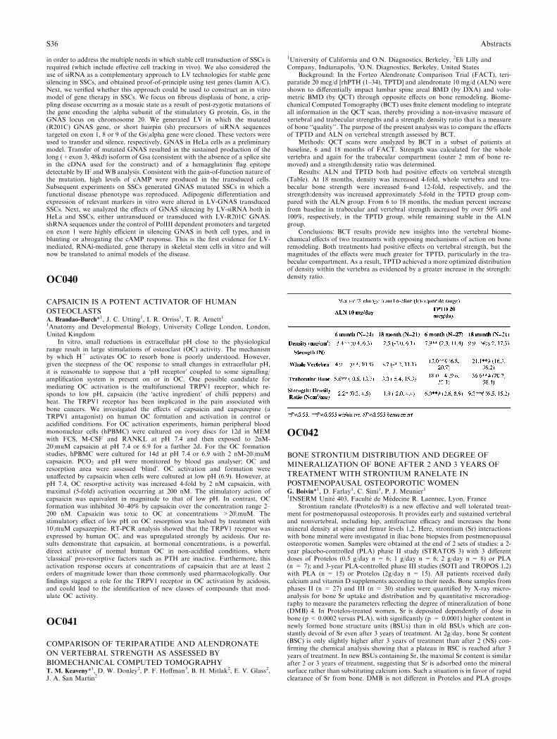
in order to address the multiple needs in which stable cell transduction of SSCs isrequired (which include effective cell tracking in vivo). We also considered theuse of siRNA as a complementary approach to LV technologies for stable genesilencing in SSCs, and obtained proof-of-principle using test genes (lamin A/C).Next, we verified whether this approach could be used to construct an in vitromodel of gene therapy in SSCs. We focus on fibrous displasia of bone, a crip-pling disease occurring as a mosaic state as a result of post-zygotic mutations ofthe gene encoding the \alpha subunit of the stimulatory G protein, Gs, in theGNAS locus on chromosome 20. We generated LV in which the mutated(R201C) GNAS gene, or short hairpin (sh) precursors of siRNA sequencestargeted on exon 1, 8 or 9 of the Gs\alpha gene were cloned. These vectors wereused to transfer and silence, respectively, GNAS in HeLa cells as a preliminarymodel. Transfer of mutated GNAS resulted in the sustained production of thelong (+exon 3, 48kd) isoform of Gsa (consistent with the absence of a splice sitein the cDNA used for the construct) and of a hemagglutinin flag epitopedetectable by IF and WB analysis. Consistent with the gain-of-function nature ofthe mutation, high levels of cAMP were produced in the transduced cells.Subsequent experiments on SSCs generated GNAS mutated SSCs in which afunctional disease phenotype was reproduced. Adipogenic differentiation andexpression of relevant markers in vitro were altered in LV-GNAS transducedSSCs. Next, we analyzed the effects of GNAS silencing by LV-siRNA both inHeLa and SSCs, either untransduced or transduced with LV-R201C GNAS.shRNA sequences under the control of PolIII dependent promoters and targetedon exon 1 were highly efficient in silencing GNAS in both cell types, and inblunting or abrogating the cAMP response. This is the first evidence for LV-mediated, RNAi-mediated, gene therapy in skeletal stem cells in vitro and willnow be translated to animal models of the disease.
OC040
CAPSAICIN IS A POTENT ACTIVATOR OF HUMAN
OSTEOCLASTSA. Brandao-Burch*1, J. C. Utting1, I. R. Orriss1, T. R. Arnett11Anatomy and Developmental Biology, University College London, London,United Kingdom
In vitro, small reductions in extracellular pH close to the physiologicalrange result in large stimulations of osteoclast (OC) activity. The mechanismby which H+ activates OC to resorb bone is poorly understood. However,given the steepness of the OC response to small changes in extracellular pH,it is reasonable to suppose that a �pH receptor� coupled to some signalling/amplification system is present on or in OC. One possible candidate formediating OC activation is the multifunctional TRPV1 receptor, which re-sponds to low pH, capsaicin (the �active ingredient� of chilli peppers) andheat. The TRPV1 receptor has been implicated in the pain associated withbone cancers. We investigated the effects of capsaicin and capsazepine (aTRPV1 antagonist) on human OC formation and activation in control oracidified conditions. For OC activation experiments, human peripheral bloodmononuclear cells (hPBMC) were cultured on ivory discs for 12d in MEMwith FCS, M-CSF and RANKL at pH 7.4 and then exposed to 2nM-20\muM capsaicin at pH 7.4 or 6.9 for a further 2d. For the OC formationstudies, hPBMC were cultured for 14d at pH 7.4 or 6.9 with 2 nM-20\muMcapsaicin. PCO2 and pH were monitored by blood gas analyser; OC andresorption area were assessed �blind�. OC activation and formation wereunaffected by capsaicin when cells were cultured at low pH (6.9). However, atpH 7.4, OC resorptive activity was increased 4-fold by 2 nM capsaicin, withmaximal (5-fold) activation occurring at 200 nM. The stimulatory action ofcapsaicin was equivalent in magnitude to that of low pH. In contrast, OCformation was inhibited 30–40% by capsaicin over the concentration range 2–200 nM. Capsaicin was toxic to OC at concentrations >20\muM. Thestimulatory effect of low pH on OC resorption was halved by treatment with10\muM capsazepine. RT-PCR analysis showed that the TRPV1 receptor wasexpressed by human OC, and was upregulated strongly by acidosis. Our re-sults demonstrate that capsaicin, at hormonal concentrations, is a powerful,direct activator of normal human OC in non-acidified conditions, where�classical� pro-resorptive factors such as PTH are inactive. Furthermore, thisactivation response occurs at concentrations of capsaicin that are at least 2orders of magnitude lower than those commonly used pharmacologically. Ourfindings suggest a role for the TRPV1 receptor in OC activation by acidosis,and could lead to the identification of new classes of compounds that mod-ulate OC activity.
OC041
COMPARISON OF TERIPARATIDE AND ALENDRONATE
ON VERTEBRAL STRENGTH AS ASSESSED BY
BIOMECHANICAL COMPUTED TOMOGRAPHYT. M. Keaveny*1, D. W. Donley2, P. F. Hoffman3, B. H. Mitlak2, E. V. Glass2,J. A. San Martin2
1University of California and O.N. Diagnostics, Berkeley, 2Eli Lilly andCompany, Indianapolis, 3O.N. Diagnostics, Berkeley, United States
Background: In the Forteo Alendronate Comparison Trial (FACT), teri-paratide 20 mcg/d [rhPTH (1–34), TPTD] and alendronate 10 mg/d (ALN) wereshown to differentially impact lumbar spine areal BMD (by DXA) and volu-metric BMD (by QCT) through opposite effects on bone remodeling. Biome-chanical Computed Tomography (BCT) uses finite element modeling to integrateall information in the QCT scan, thereby providing a non-invasive measure ofvertebral and trabecular strengths and a strength: density ratio that is a measureof bone ‘‘quality’’. The purpose of the present analysis was to compare the effectsof TPTD and ALN on vertebral strength assessed by BCT.
Methods: QCT scans were analyzed by BCT in a subset of patients atbaseline, 6 and 18 months of FACT. Strength was calculated for the wholevertebra and again for the trabecular compartment (outer 2 mm of bone re-moved) and a strength:density ratio was determined.
Results: ALN and TPTD both had positive effects on vertebral strength(Table). At 18 months, density was increased 4-fold, whole vertebra and tra-becular bone strength were increased 6-and 12-fold, respectively, and thestrength:density was increased approximately 5-fold in the TPTD group com-pared with the ALN group. From 6 to 18 months, the median percent increasefrom baseline in trabecular and vertebral strength increased by over 50% and100%, respectively, in the TPTD group, while remaining stable in the ALNgroup.
Conclusions: BCT results provide new insights into the vertebral biome-chanical effects of two treatments with opposing mechanisms of action on boneremodeling. Both treatments had positive effects on vertebral strength, but themagnitudes of the effects were much greater for TPTD, particularly in the tra-becular compartment. As a result, TPTD achieved a more optimized distributionof density within the vertebra as evidenced by a greater increase in the strength:density ratio.
OC042
BONE STRONTIUM DISTRIBUTION AND DEGREE OF
MINERALIZATION OF BONE AFTER 2 AND 3 YEARS OF
TREATMENT WITH STRONTIUM RANELATE IN
POSTMENOPAUSAL OSTEOPOROTIC WOMENG. Boivin*1, D. Farlay1, C. Simi1, P. J. Meunier11INSERM Unite 403, Faculte de Medecine R. Laennec, Lyon, France
Strontium ranelate (Protelos�) is a new effective and well tolerated treat-ment for postmenopausal osteoporosis. It provides early and sustained vertebraland nonvertebral, including hip, antifracture efficacy and increases the bonemineral density at spine and femur levels 1,2. Here, strontium (Sr) interactionswith bone mineral were investigated in iliac bone biopsies from postmenopausalosteoporotic women. Samples were obtained at the end of 2 sets of studies: a 2-year placebo-controlled (PLA) phase II study (STRATOS 3) with 3 differentdoses of Protelos (0.5 g/day n = 6; 1 g/day n = 6; 2 g/day n = 8) or PLA(n = 7); and 3-year PLA-controlled phase III studies (SOTI and TROPOS 1,2)with PLA (n = 15) or Protelos (2g/day n = 15). All patients received dailycalcium and vitamin D supplements according to their needs. Bone samples fromphases II (n = 27) and III (n = 30) studies were quantified by X-ray micro-analysis for bone Sr uptake and distribution and by quantitative microradiog-raphy to measure the parameters reflecting the degree of mineralization of bone(DMB) 4. In Protelos-treated women, Sr is deposited dependently of dose inbone (p < 0.0002 versus PLA), with significantly (p = 0.0001) higher content innewly formed bone structure units (BSUs) than in old BSUs which are con-stantly devoid of Sr even after 3 years of treatment. At 2g/day, bone Sr content(BSC) is only slightly higher after 3 years of treatment than after 2 (NS) con-firming the chemical analysis showing that a plateau in BSC is reached after 3years of treatment. In new BSUs containing Sr, the maximal Sr content is similarafter 2 or 3 years of treatment, suggesting that Sr is adsorbed onto the mineralsurface rather than substituting calcium ions. Such a situation is in favor of rapidclearance of Sr from bone. DMB is not different in Protelos and PLA groups
S36 Abstracts
regardless of the dose used in phase II or the duration of the treatment. Het-erogeneity index of mineralization does not significantly change after either 2 or3 years of treatment. To conclude, these data suggest that the increased BMDobserved during Protelos treatment could be due to increased bone mass, as BSCreaches a plateau between 2 and 3 years of treatment and DMB is not modified.
1-Meunier et al. 2004 NEJM 350:459–682-Reginster et al. 2005 JCEM 90:2816–223-Meunier et al. 2002 JCEM 87:2060–64-Farlay et al. 2005 JBMR 20:1569–78
CC001
MUTATIONS IN OSTM1 DEFINE A SEVERE FORM OF
AUTOSOMAL RECESSIVE OSTEOPETROSIS WITH NEURAL
INVOLVEMENTA. Pangrazio*1, F. Rucci1, L. Susani1, L. Poliani2, M. Di Rocco3, A. Megar-bane4, G. Lefranc5, M. Abinun6, A. Villa1, A. Frattini71Human Genome, Istituto Tecnologie Biomediche - CNR, Segrate, 2Departmentof Pathology I, University of Brescia, Brescia, 3Unit of Rare Diseases, GasliniInstitute, Genova, Italy, 4Unite de Genetique Medicale, Campus des SciencesMedicales, Beyrouth, Lebanon, 5Laboratoire d’Immunogenetique Moleculaire,Universite Montpellier II, Montpellier, France, 6Children’s BMT Unit, New-castle General Hospital, 7Unit of Rare Diseases, University of Brescia, New-castle upon Tyne, United Kingdom
Autosomal Recessive Osteopetrosis (ARO) is a severe hereditary bone dis-ease whose cellular basis is in osteoclast, but whose molecular defect is hetero-geneous. Approximately 55% of ARO patients show an abnormality in theTCIRG1 gene coding for the a3 subunit of the vacuolar proton pump that playsa fundamental role in acidifying the osteoclast-bone interface. This acidificationis also hampered by a defect in the ClCN7 gene which is mutated in about 10%of ARO patients. A third gene found mutated in human ARO is the OSMT1 (thehuman homologous of the murine grey lethal gene) which has been described sofar in only two ARO patients.
We report three novel cases with mutation in the Ostm1 gene. These patientswere characterized by severe bone sclerosis, growth failure, anemia, thrompo-citopenia, visual impairment with optic atrophy. The analysis of the Ostm1 geneshowed in two patients, both from Kuwait, a homozygous two nucleotidesdeletion in exon 2, this mutation leads to a frameshift starting at codon 140 andpremature termination.
The third patient showed a single point mutation in exon 1, leading to anonsense mutation (Cys12X). The parents of the three patients were heterozy-gous for this mutation.
The clinical neurological evaluation of the two Kuwaiti patients by CT scanand MRI showed a defect in the white matter, with an aspecific diagnosis ofsevere cerebral atrophy.
With the aim to confirm these data, we analyzed the gl/gl mutant. The gl/glmouse brain showed a diffuse translucent appearance with loss of the normaldemarcation between the white and the grey matter, features consistent withmyelin loss or hypomyelination. Histological and myelin staining analysis evi-denced an atrophy of the corpus callosum with loss of myelin fibers. Moreover,many sections throughout the whole brain showed cortical areas with loss ofnormal lamination constituted of misaligned pleomorphic dysplastic neuronsand consistent with multiple foci of cortical dysplasia.
These findings allowed us to suggest that Ostm1-dependent ARO defines anew subset of patients with severe central nervous system involvement leading toa very poor prognosis. Interestingly, our Ostm1-dependent ARO are reminiscentof those described under the MIM entry ‘‘osteopetrosis associated with neuronalstorage disease’’ (MIM 600329).
The fact that the CNS involvement is also present in the grey lethal mousemutant, suggests that this mouse is a good model to test possible alternativetherapies.
CC002
LIPOSCLEROSING MYXOFIBROUS TUMOR OF BONE. A
MIME OF PROXIMAL FEMUR MONOSTOTIC FIBROUS
DYSPLASIAA. Corsi*1, F. De Maio2, E. Ippolito2, M. Riminucci3, P. Bianco11Department of Experimental Medicine and Pathology, University La Sapienza,2Department of Orthopaedic Surgery, University of Tor Vergata, Rome,3Department of Experimental Medicine, University of L’Aquila, L’Aquila, Italy
A 33-year-old woman presented with pain in left hip and sacral region. X-rays revealed a lytic lesion with perilesional sclerosis wthin the left femur neck. Abone scan showed increased uptake selectively at this site. Physical examinationrevealed a cafe au lait spot on the left thigh. Experienced radiologists andorthopaedics concluded for the diagnosis of monostotic Fibrous Dysplasia (FD).A biopsy confirmed the histological diagnosis of FD. Surgical curettage was
performed and femur neck stabilization was obtained with dynamic hip screw.The patient was well with no recurrence 2 years after surgery. Microscopicexamination of the curetted material and review of the original bioptic materialwas again regarded as diagnostic of FD by different experienced bone pathol-ogists. Mutation analysis was conducted on fresh surgical material, cells isolatedin culture from the surgical material, paraffin blocks of the original bone biopsy,and a punch biopsy of the cafe-au-lait spot. Analysis included codon 201, thecommon site of mutation in FD, and codon 227, involved in activating muta-tions in related disorders. Direct sequencing of the PCR amplification productsand PCR-RFLP failed to disclose any mutation. This led to a thorough re-evaluation of the case, including retrospective analysis of all available radio-graphic material. Radiographs performed for other reasons 6 years before theonset of symptoms failed to show any lesion in the left proximal femur. Takentogether with the consistent failure to detect any GNAS mutation, this findingdispelled true FD, in which lesions are thought never to develop in adulthood.We concluded for the diagnosis of Liposclerosing Myxofibrous Tumor (LMT).This is a benign fibro-osseous bone lesion displaying a complex mixture of his-tologic elements including FD-like features, commonly occurring in the proximalfemur. Curiously, LMT is thought to be related to FD in terms of nature andpathogenesis, and in two published cases point mutation at codon R201 in theGNAS gene was identified. This case highlights a relatively poorly known bonelesion behaving as a close histologic and radiographic mime of FD; provides aparadigm situation in which mutation analysis was superior to histology in termsof diagnostic stringency; and emphasizes the strong relevance and diagnosticvalue of proper anamnestic evaluation of adults with FD-like lesions. The truebiological relationship of LMT to FD remains to be defined.
CC003
FIBRODYSPLASIA OSSIFICANS PROGRESSIVA: REPORT OF
A FAMILYM. Dumic1, T. Matic*1, Z. Bilinovac1, K. Potocki11Department of Pediatrics, University Hospital Rebro, Zagreb, Croatia
Fibrodysplasia ossificans progressiva (FOP) or myositis ossificans pro-gressiva is a rare autosomal dominant disorder characterized by congenitalmalformation of the great toes and intermittent progressive ectopic ossification.The cause of the disease is still not fully understood. Some recent studies suggestthat FOP maps to 4q27-31 region that contains at least one interval in the bonemorphogenetic protein signaling pathway. Other studies have described muta-tions of the noggin (NOG) gene in the 17q21-22 region in FOP patients. Wereport on a female child with FOP and her partially affected father and twinsister.
The girl, in whom soft tissue swellings were already noticed at birth, wasreferred to us at the age of 10.5 years. Clinical examination revealed short stature(height 124 cm; -2.8 sd), bilateral short great toe with hallux valgus, shortthumbs, numerous sites of subcutaneous tender indurations on the trunk and theextremities, and irregularly spaced dentition. X-ray demonstrated bilateral hal-lux valgus with shortening of the first metatarsal, short distal phalanges of thethumbs, multiple calcifications of the soft tissue paravertebral and around thejoints and the trunk, and exostosis-like bony projection of distal fibula. Labo-ratory findings showed partial deficiency of growth hormone (peak GH value:6.8 mIU/L) and primary hypothyroidism of unknown cause (T4: 42.0 nmol/L,T3: 1.6 nmol/L, TSH: 6.150 mIU/L, antithyroid antibodies: negative). Audio-gram didn�t reveal any kind of hearing loss. Clinical and radiological examina-tion of her father and twin sister revealed only a bilateral short great toe withhallux valgus, indicating autosomal dominant mode of inheritance with fullpenetrance only for short great toes with hallux valgus and varying expressionfor heterotopic ossification.
In conclusion, we have reported on FOP patient with partial growth hor-mone deficiency and hypothyroidism. Endocrine abnormalities have not beenpreviously reported in FOP. It should be stressed that FOP patients and mem-bers of the family require a thorough clinical evaluation, which might detectadditional abnormalities requiring medical treatment, with regard to the rec-ommended precautions with invasive procedures.c
CC004
CIANALCET AND HYPOCALCIURIC HYPERCALCEMIA
WITH RECURRENT PANCREATITISP. Filipponi*1, S. Cristallini1, G. Policani1, A. Nicasi11ASL1 Umbertide Hospital, Centre for Osteoporosis and Metabolic Bone Dis-eases, Umbertide, Italy
Introduction. Inactivating mutations in CaSR gene has been reported as thecause of hypocalciuric hypercalcemia (HH) with inappropriate PTH serumlevels. Calcimimetic agents, allosterical activators of CaSR, have been success-fully tested in the treatment of primary, as well of secondary, hyperprathy-roidism. We report the effect of the calcimimetic Cinacalcet on mineral
Abstracts S37
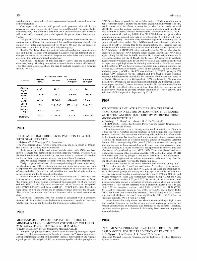
metabolism in a patient affected with hypocalciuric hypercalcemia and recurrentacute pancreatitis.
Case report and methods. A16 year-old male presented with mild hyper-calcemia and acute pancreatitis at Perugia Regional Hospital. The patient had acholecistectomy and initiated a treatment with ursodesossicolic acid, which isstill in use. After a second pancreatitis episode the patient was referred to ourCentre.
The patient�s basal mineral metabolism parameters were assessed over 4months using 4 different determinations. After basal investigations, cinacalcet 30mg/day was started and administered for 15 days. On day 16, the dosage ofcinacalcet was doubled to 30 mg twice daily (60 mg/day).
Results. The Table shows the patient�s mineral metabolism parameters be-fore and during treatment with cinacalcet. Cinacalcet was well tolerated and nochanges were observed in pancreatic enzymes and other laboratory parametersexcept for the indices of mineral metabolism.
Conclusions.The results of this case report shows that the calcimimeticcinacalcet, 30 mg twice daily, normalizes serum calcium in a patient affected withHH. This encouraging case shows the need to evaluate cinacalcet as a potentialtherapy for HH.
P001
DECREASED FRACTURE RISK IN PATIENTS TREATED
WITH ORAL NITRATESL. Rejnmark*1, P. Vestergaard1, L. Mosekilde21The Osteoporosis Clinic, 2Dept of Endocrinology and Metabolism C, Univer-sity Hospital of Aarhus, Aarhus, Denmark
Background: In cellular and animal models, nitric oxide (NO) has beenshown to affects bone remodelling. In postmenopausal women, treatment withthe NO donor isosorbide mononitrate, has been shown to decrease biochemicalmarkers of bone resoprtion and increase markers of bone formation.
Aim: We studied whether treatment with oral nitrates affects fracture risk.Design: a population-based pharmaco-epidemiological case-control study
with fracture in year 2000 as outcome and drug use during the previous five yearsas exposure. We used nation-wide computerized registers to assess individual useof drugs and related these data to individual fracture records and information onsocioeconomic and health-related confounders.
Results: Our study included 124,655 fracture cases and 373,962 age- andgender-matched controls. After adjustment for potential confounders, we foundthat treatment with oral nitrates is associated with a reduced risk of any fracture(OR 0.8; 95%CI, 0.86–0.92) as well as a reduced risk of fractures at the hip (OR0.85; 95%CI, 0.79–0.92) and forearm (OR 0.91; 95%CI; 0.83–1.00). The effectswere similar in men and women and in subjects younger and older the 65 years.Risk of any fracture and hip fracture decreased with increased accumulateddose.
Conclusion: Treatment with oral nitrates is associated with a decreasedfracture risk. Randomised controlled studies are warranted in order to determinewhether oral nitrates can be used in the treatment of osteoporosis.
P002
MECHANISMS OF PYROPHOSPHATE INHIBITION OF
MINERALIZATION IN MC3T3-E1 OSTEOBLAST CULTURESW. N. Addison1, F. Azari1, M. T. Kaartinen1, M. D. McKee*11Faculty of Dentistry, McGill University, Montreal, Canada
Inorganic pyrophosphate (PPi) inhibits mineralization by binding to crystalsurfaces. Its ubiquitous presence is thought to prevent �soft� tissues from miner-alizing, whereas its degradation in bone may facilitate mineralization and regulatecrystal growth. Hydrolysis of PPi by tissue-nonspecific alkaline phosphatase
(TNAP) has been proposed for extracellular matrix (ECM) mineralization inbone. Although much is understood about the crystal-binding properties of PPi,less is known about its effects on osteoblast activity. In this study, we usedMC3T3-E1 osteoblast cultures and enzyme kinetic studies to investigate the ef-fects of PPi on osteoblast-directed mineralization. Mineralization of MC3T3-E1cultures was dose-dependently inhibited by PPi. PPi inhibition was greater whenmineralization was induced with beta-glycerophosphate (ß-GP) than with inor-ganic phosphate (Pi) - the former being a source of organic Pi commonly used inculture mineralization studies. Since ß-GP-induced mineralization requires theaction of TNAP to provide free Pi for mineralization, this suggests that themechanism of PPi inhibition may involve altered TNAP-mediated hydrolysis ofß-GP. Inhibition of MC3T3-E1 culture mineralization by PPi was abolished byaddition of exogenous TNAP. Enzyme kinetic studies showed that TNAP has ahigher affinity for PPi than for ß-GP, and that PPi competitively inhibited the ß-glycerophosphatase activity of TNAP. Furthermore, PPi adsorbed to synthetichydroxyapatite was resistant to TNAP hydrolysis, data consistent with its havingan important physiological role in inhibiting mineralization. Finally, we exam-ined the effect of PPi on the expression of another mineralization inhibitor, thesecreted ECM protein osteopontin (OPN), on the cultures. The mineralization-inhibiting property of OPN, like PPi, was abolished by TNAP hydrolysis. PPiinduced OPN expression via the ERK1/2 and P38 MAPK kinase signalingpathways. Inhibitor studies showed that PPi induction of OPN does not appear tobe Protein Kinase A-, C- or G-dependent. OPN regulation by PPi was alsoinsensitive to Probenecid (an anion-transport inhibitor) and Foscarnet (a phos-phate import inhibitor). These experiments show that PPi prevents mineralizationin MC3T3-E1 osteoblast cultures by at least three different mechanisms thatinclude direct binding to growing crystals, inhibition of TNAP activity, andinduction of OPN expression. Supported by CIHR.
P003
STRONTIUM RANELATE REDUCES NEW VERTEBRAL
FRACTURES IN A SEVERE OSTEOPOROTIC MICE MODEL
WITH SPONTANEOUS FRACTURES BY IMPROVING BONE
MICROARCHITECTUREV. Geoffroy*1, C. Marty1, A. Lalande2, M. C. De Vernejoul11INSERM U606, Hospital Lariboisiere, Paris, 2Groupe Servier, RheumatologyDivision, Courbevoie, France
Strontium ranelate is a novel therapy which has demonstrated its efficacy toreduce the risk of vertebral and hip fractures in post-menopausal osteoporoticwomen. However, the mechanism underlying this anti-fracture effect needsfurther investigations. We therefore used a murine model of severe osteoporosisto go into the mechanism of strontium ranelate efficacy in preventing vertebralfracture. Mice overexpressing Cbfa1/runx2 (Cbfa1) in osteoblastic lineage ex-hibit an increase in bone remodelling with bone resorption exceeding boneformation leading to a severe osteoporosis and spontaneous vertebral fracturesafter 4-week of age (Geoffroy et al, MCB, 2002). These mice (7-week-old) wererandomized to receive either strontium ranelate (1800 mg/kg/d) or vehicle (VEH)for 9 weeks once daily by gavage. At the end of treatment, strontium ranelate -treated mice had a plasmatic strontium concentration in the same range than theone observed in patients receiving the therapeutic dose.
The fractures number at the caudal vertebrae was measured (X-ray; FAX-ITRON) before and after 5 and 9 weeks of dosing. At baseline, fractures number(mean ± SD) was 2.75 ± 1.49 and 3.63±2.00 in the VEH and strontium ra-nelate therapeutic groups respectively (n=8/group). The number of new frac-tures per mice was reduced in strontium ranelate group by 47% and 60% at 5 and9 weeks respectively (VEH, 2.13 vs strontium ranelate, 1.13; p=0.021 and VEH,3.13 vs strontium ranelate, 1.25; p<0.005). At the end of the experiment, bonehistomorphometry showed a significant strontium ranelate effect on trabeculararchitecture at the lumbar vertebrae with a markedly higher BV/TV (VEH,10.5±0.3% vs strontium ranelate, 16.8±3.9%; p<0.005) and Tb.N (VEH,2.71±0.71 vs strontium ranelate, 4.07±0.86; p<0.005), and a lower Tb.Sp(VEH, 356.8±99.7lm vs strontium ranelate, 216.9±62.9lm; p<0.005). Verte-brae cortical thickness increased also significantly (VEH, 60.2±4.0lm vsstrontium ranelate, 70.5±4.7lm; p<0.001).
In conclusion: this study shows that when bone remodelling is high, stron-tium ranelate decreases the number of new vertebral fracture per mice by pre-venting disconnection of trabeculae and thinning of the cortices. Therefore,strontium ranelate prevents fractures in increasing bone mass and improvingmicroarchitecture.
P004
INCREMENTAL PROGNOSTIC VALUES OF RISK FACTORS-
BASED MODEL FOR THE PREDICTION OF FRACTUREN. D. Nguyen*1, J. A. Eisman1, J. R. Center1, T. V. Nguyen11Bone and Mineral Research Program, Garvan Institute of Medical Research,Sydney, Australia
S38 Abstracts
Background: Apart from bone mineral density (BMD), clinical risk factorssuch as age, weight, a history of fall and a prior fracture are individually asso-ciated with fracture risk. The present study sought to assess the incrementalprognostic values of risk factors-based models in the prediction of fracture riskin osteoporotic and non-osteoporotic (i.e., ‘‘normal BMD’’) individuals.
Methods: Participants in the Dubbo Osteoporosis Epidemiology Study wererandomly divided into two groups: a development cohort (n=405 men, 632women) and a validate cohort (n=418 men, 667 women), all aged 60+ years asat 1989. Femoral neck BMD (GE Lunar Corp, WI), body weight, and clinicalhistory were assessed at baseline (1989). Subsequent fracture incidence wasascertained by X-ray report between 1989 and 2004. In the development cohort,three models for predicting fracture were developed: model I included onlyFNBMD; model II included age and weight; and model III utilized age, weight,fall and prior fracture as predictors. The positive predictive value (PPV) andAUC value of the three models were then assessed in the validation cohort.
Results: In the validation cohort, 209 women and 71 men had sustained afracture during the 15-year follow-up period. Risk estimate based on model Icorrectly identified 52% and 45% fractures in women and men, respectively. Riskestimates from models II and III had a slightly higher predictive values thanmodel I; however, the concordance between model I and model II (or model III)was between 50% and 66%. Thus, when risk derived from model II was com-bined with model I, the predictive value increased to 77% in women and 70% inmen, representing an absolute increase of 25% in both sexes. Comparableimprovement was also observed in model III. The AUC values for the threemodels ranged between 0.61 to 0.68 in both sexes.
Conclusion: These data indicate that the PPV of risk factors-based modelwas slightly higher than that of BMD-based model, and that the incorporationof clinical risk factors can significantly increase the prognostic value of fractureprediction over and above of BMD alone.
P005
HIGH RESOLUTIONGENOTYPING OF THE ESR1 GENE AND
ASSOCIATION WITH HEIGHTL. Stolk*1, J. B. J. Van Meurs1, A. Hofman2, H. A. P. Pols1, A. G. Uitterlinden11Internal Medicine, 2Epidemiology and Biostatistics, Erasmus MC, Rotterdam,Netherlands
Background: Sex steroid signaling genes such as Estrogen Receptor alpha(ESR1) are interesting candidates to explain part of the genetic risk for osteo-porosis. Most association studies examining ESR1 analysed the PvuII(rs2234693, -397T/C), XbaI (rs9340799, -351A/G) polymorphisms and the pro-moter TA-VNTR and haplotypes thereof. We previously found associations ofthese variants with BMD, vertebral fracture risk and height in a large cohort ofelderly men and women. However, no functionality of these three polymor-phisms has been reported yet, and linkage disequilibrium in this area has notbeen studied to identify other variants that are in high linkage.
The objective of this study was first to determine the complete LD-block inwhich PvuII, XbaI and the TA-VNTR are located. The second was to performassociation studies with the newly defined haplotypes in relation to height.
Methods: We used data from both the Hap Map project and the Perlegendatabase to determine LD-blocks. The program Haploview was used to deter-mine haplotype tagging (ht) SNPs in the block. Three tagging SNPs (promoterT-9929G, and G-3604A and intron 1 A+2818G) and PvuII & XbaI weregenotyped in 2327 men and 3301 women in our population-based cohort ofsubjects over 55 years of age using Taqman allelic discrimination. Haplotypeswere constructed using the PHASE program, and association with height wasanalysed using ANCOVA.
Results: We identified a block of high LD including PvuII and XbaI poly-morphisms, spanning about 57 kb, starting at 18.000 bp in front of exon 1 to4500 bp downstream of exon 2. Furthermore, we observed the original PvuII-XbaI (risk) haplotype 1 (frequency: 58.8%) to be split up in 4 haplotypes (fre-quencies: 1a 31.6%; 1b 10.7%; 1c 6.6%; 1d 4.4%) when using 3 extra taggingSNPs. Of these 4 haplotypes, 1a was found to be associated with an allele-doseeffect on height (P = 0.001) that remained after adjusting for gender, age andprevalent vertebral fractures.
Conclusion: By increasing genetic resolution in this area of the ESR1 gene,we observed that a newly defined haplotype is the major driving haplotype in theassociation with height that was previously detected by PvuII and XbaI. Thisobservation will enable a more focused analysis of potentially functional SNPs inthis LD-block.
P006
EVIDENCE THAT A POLYMORPHISM IN THE LACTASE
PHLORIZIN HYDROLASE GENE CAUSES DIFFERENCES IN
HEIGHT INDEPENDENT OF CURRENT CALCIUM INTAKEW. N. H. Koek*1, J. B. Van Meurs1, B. C. J. Van der Eerden1,A. G. Uitterlinden1, H. A. P. Pols1, J. P. T. M. Van Leeuwen11Internal Medicine, Erasmus MC, Rotterdam, Netherlands
Background: There is a wide variety of lactose intolerance world wide, with ahigh prevalence in populations with a low lactose intake and a low prevalence incountries with a high calcium intake. The C-variant of a T-13910-C polymor-phism upstream of the lactase phlorizin hydrolase (LPH) gene is known to causelactose intolerance1. We studied the association of this polymorphism with bodysize, bone mineral density (BMD) and fracture risk in an elderly population withhigh calcium intake.
Materials and Methods: The Rotterdam Study population constitute about8000 participants, men and women 55 years and older. Genotyping was per-formed using the Taqman allelic discrimination assay. Statistical analyses wereperformed using the program SPPS 11 and associations were all adjusted for age.
Results: The allele frequency of the 13910-C was 31%. C-homozygotes(lactose intolerant) were modestly but significantly associated with lower calciumintake in both men and women ()13%:p<0.001 and )4.4%:p<0.05). In male aswell as female C-homozygotes height was significantly (p<0.001) lower ()1.2cm). For both height and calcium intake an allele dose effect was observed. Afteradjustment for current calcium intake and vertebral fractures the associationwith height persisted. The effects on height and calcium intake are not explainedby differences in overall energy intake since no difference in energy intake wasfound between the different genotypes for men and women; (p=0.44) and(p=0.55), respectively. No associations with femoral neck and lumbar spineBMD and with fractures (vertebral and non-vertebral) were detected. Interest-ingly, additional expression analyses demonstrated LPH in bone cells.
Conclusion: We demonstrate that a LPH polymorphism leading to lactoseintolerance results in a reduced height. This is independent of current modestlyreduced calcium intake. This suggests larger calcium intake differences andinfluence on longitudinal growth during the growth spurt in the early phase oflife when lactose intolerance is becoming overt and/or a direct influence of LPHon longitudinal growth. Expression analyses supports this latter possibility of adirect effect on the skeleton. Finally, the LPH polymorphism causing lactoseintolerance and consequently reduced calcium intake does not alter the miner-alisation of bone and eventually fracture risk.
1: Enattah NS, et al. Nat Genet 2002;30:233
P007
A SINGLE DOSE OF ZOLEDRONIC ACID 5 MG ACHIEVES
MORE SUSTAINED BIOCHEMICAL REMISSION VERSUS
DAILY 30MG RISEDRONATE IN PATIENTS WITH PAGET�SDISEASEI. Reid*1, P. Miller2, J. Brown3, W. Fraser4, D. Hosking5, J. P. Devogelaer6,C. Moniz7, M. Hooper8 Y. Saidi9, G. Su10, J. Pak10, J. Davis10, T. Fashola9,.J. Krasnow10, K. Zelenakas10, K. Lyles111Faculty of Medicine and Health Sciences, University of Auckland, Auckland,New Zealand, 2Colarado Center for Bone Research, Lakewood, CO, UnitedStates, 3Le Centre hospitalier universitaire de Quebec, Sainte-Foy, Quebec,Canada, 4Royal Liverpool, University Hospital, Liverpool, 5Nottingham, CityHospital, Nottingham, United Kingdom, 6Cliniques Universitaires, St. Luc,Brussels, Belgium, 7Kings College, Hospital, London, United Kingdom,8Repatriation General Hospital, Concord, Australia, 9Novartis Pharma AG,Basel, Switzerland, 10Novartis Pharmaceuticals Corporation, East Hanover,11Duke University and VA Medical Centers, Durham, NC, United States
Background/Aims: Zoledronic acid (ZOL) has been shown to have superiorefficacy and faster time to onset than risedronate (RIS) in Paget�s disease frompooled data of the 6-month core period of two identical, randomized controlledtrials comparing a single ZOL 5mg 15-minute infusion with oral RIS 30mg/dayfor 60 days (1), suggesting the potential for longer and more sustained bio-chemical remission by ZOL. To assess this, the results from the extendedobservation period (EOP) are presented.
Methods: The primary efficacy endpoint of the core study was therapeuticresponse at 6 months, defined as a ‡75% reduction in serum alkaline phospha-tase (ALP) excess or its normalization. Of the 169 ZOL and 125 RIS patientseligible for entry, 152 ZOL and 115 RIS responders entered the EOP and hadALP tested every 6 months. Time to first loss of therapeutic response, time tofirst partial disease relapse (defined as a ‡50% increase from the ALP mea-surement at month 6 and at least 1.25 times the upper normal limit) and com-plete disease relapse (defined as ALP level returned within 20% of baseline ALPvalue) were evaluated.
Results: As of October 21, 2005, 57/115 RIS patients have lost therapeuticresponse compared to only 3/152 ZOL patients (P<.0001). Furthermore, 49/115and 9/115 RIS patients experienced partial and complete disease relapse com-pared to only 3/152 and 0/152 ZOL patients, respectively (P<.0001 and P<.001,respectively).
Within the first 3 days of drug administration, ZOL patients experiencedabout 2-fold higher rate of post-dose symptoms than RIS patients. The symp-toms were mild to moderate in severity and mostly resolved after 4 days of onset.After 3 days of drug administration, side effects rate was similar betweentreatment groups.
Conclusion(s): A single ZOL 5mg infusion shows longer duration of effectrelative to the reduction in ALP than RIS. Treatment efficacy was sustained in
Abstracts S39
the majority of ZOL patients compared to the substantial number of RIS pa-tients who either lost their therapeutic response or experienced partial or com-plete disease relapse. Hence, a single ZOL 5mg dose offers the potential formulti-year biochemical remission in patients with Paget�s disease.
(1) Reid I et al. N Engl JMed 2005; 353:898–90
P008
ANDROGEN RECEPTOR POLYMORPHISM: ASSOCIATION
WITH LEAN MASS AND BONE MASS IN MEN OF AFRICAN
HERITAGEJ. M. Zmuda*1, L. Yerges2, S. P. Moffett2, C. Nestlerode2, J. A. Cauley2, C. H.Bunker2, V. W. Wheeler3, A. L. Patrick3, R. E. Ferrell41Epidemiology and Human Genetics, 2Epidemiology, University of Pittsburgh,Pittsburgh, United States, 3Epidemiology, Tobago Health Studies Office, Scar-borough, Trinidad and Tobago, 4Human Genetics, University of Pittsburgh,Pittsburgh, United States
Men of African ancestry have higher bone and muscle mass than men ofCaucasian ancestry, and these differences have been attributed in part to ethnicdifferences in androgen metabolism. Androgen effects on bone and muscle aremediated through the androgen receptor (AR), a ligand activated transcriptionfactor. The AR gene contains a CAG repeat polymorphism in exon 1 that codesfor a glutamine sequence of variable length. This polymorphism influences ARtranscriptional activity, with shorter CAG repeats conferring enhanced sensi-tivity to androgens in vitro. We have previously shown that Caucasian Americanmen with shorter CAG repeats have higher hip bone mass and body weight thanmen with longer CAG repeats. In the present study, we tested whether this ARpolymorphism is associated with bone mass and body composition in 1,035Afro-Caribbean men aged 40 yrs and older who were participants in an ongoingpopulation-based study on the island of Tobago. Hip BMD and whole bodycomposition were measured with a Hologic QDR 4500 densitometer. Men withlonger AR CAG repeats (highest quartile) had lower total hip BMD comparedto men with shorter repeats (lowest quartile) (1.15 vs 1.10; p=0.018). Men withlonger CAG repeats also had significantly lower lean body mass (p<0.01). Theassociation between the AR polymorphism and hip BMD was attenuated and nolonger statistically significant (p=0.21) after adjusting for the lower lean bodymass of men with longer CAG repeats. Of interest, the frequency of the shorter,more active AR alleles is higher in men of African descent than among Cau-casians. Collectively, these data raise the possibility that a higher prevalence ofshorter androgen receptor alleles among men of African descent may contributein part to their greater bone and muscle mass and lower osteoporotic riskcompared with Caucasian men.
P009
CALCITONIN PROTECTS AGAINST EXPERIMENTIALLY
INDUCED OSTEOARTHRITISB. C. Sondergaard*1, K. Henriksen1, H. Wulf1, S. Oestergaard1, L. B. Tanko1, P.Qvist1, C. Christiansen1, M. A. Karsdal11Pharmacology, Nordic Bioscience, Herlev, Denmark
Purpose: To investigate the potential chondroprotective effects of oral cal-citonin in an experimental model of osteoarthritis (OA), the ovariectomized(OVX) rat model. Secondly, to investigate possible direct effects of calcitonin onarticular chondrocytes by using ex vivo explants cultures.
Methods: Fifty rats aged 7 months were randomised according to weightsinto 5 groups. Animals were either subjected to OVX or sham operation.Sham, OVX, OVX+estrogen, OVX+2mg/kg calcitonin and 50mg/mg 5-CNAC, OVX+50mg/kg 5-CNAC. After OVX, treatment lasted for 9 weeks.Cartilage degradation was both assessed by measurement of collagen type IIdegradation resulting in the release of C-terminal telopeptides of collagen typeII (CTX II ELISA) and histological scoring of articular cartilage erosion inrat knees. Total proteins were extracted from rat joints for biochemicalassessment of protein content, collagen content (hydroxyproline), proteogly-can (GAG), Procollagen type II (PII), total aggrecan, aggrecan neoepitopesand CTX-II measurements. The direct effects of calcitonin on articular car-tilage were firstly investigated in bovine articular cartilage explants. Cartilagedegradation was induced by TNF-alpha [20ng/ml] and oncostatin M (OSM)[10ng/ml] and investigated by release of C-terminal-telopeptides of collagentype II, CTX-II, in the presence of human or salmon calcitonin [0.0001-1muM]. Secondly, intracellular signalling of calcitonin was investigatedthrough induction of cAMP and the P44/42 MAP kinase.
Results: In vivo, ovariectomy resulted in a marked increase in boneresorption, bone formation, and cartilage degradation. Calcitonin abrogatedcartilage degradation measured by both CTX-II (95%, p<0.001) and scoring ofcartilage erosion in the rat knee (P<0.01). Calcitonin completely preventedproteoglycan loss in the rat joints, as measured by GAG content in the proteinextracts. Ex vivo, calcitonin stimulated cAMP activity in articular chondrocytes.In explants cultures, treatment with TNF-alpha + OSM resulted in a 50 fold
induction of CTX-II release with was significantly and dose-dependently reducedby 75% in the presence of 100nM calcitonin.
Conclusion: In vivo, calcitonin almost abrogated cartilage degradation. Exvivo, we demonstrated direct chondroprotective effect of calcitonin on artic-ular cartilage. Thus, calcitonin might both exert direct chondroprotective ef-fect on chondrocytes in addition to the well-established effects on boneremodelling.
P010
EPIGENETICS: A MOLECULAR TARGET IN OSTEOARTHRI-
TIS?H. I. Roach*1, N. Yamada2, K. S. Cheung1, R. O. Oreffo1, F. Bronner31Bone and Joint Research Group, University of Southampton, Southampton,United Kingdom, 2Orthopaedic Dept, Tohoku University, Sendai, Japan,3Health Center, University of Connecticut, Farmington, United States
More than 60% of people over 65 suffer from osteoarthritis (OA). Thecauses are not known nor the reasons why the disease gets progressivelyworse. OA is failure of the articular cartilage matrix due to degradation byproteases. We have shown that OA chondrocytes change from quiescent,slowly turning over cartilage cells, to abnormal, enzyme producing cells,termed ‘‘degradative’’ chondrocytes. We hypothesize that the abnormal geneexpression is due to epigenetic �unsilencing�, i.e., that the proteases are si-lenced by DNA methylation in normal articular chondrocytes, while deme-thylation has taken place in OA. To test the hypothesis, we selected MMP-3,-9, -13 and ADAMTS-4 as typical proteases expressed by degradative, butnot normal, chondrocytes and identified the so-called CpG sites (at which amethyl-group is added to the cytosine) in the promoters. DNA was extractedfrom articular cartilage of 10 control and 16 OA patients and the methylationstatus was determined using methylation-sensitive restriction enzymes followedby PCR. We had expected methylation of all CpGs in controls and completede-methylation in OA. This was not found. Overall, 80% of all CpGs weremethylated in controls vs 51% in OA, suggesting that complete de-methyla-tion of every CpG site in the promoter is not necessary to activate geneexpression. Not all sites were equally susceptible to de-methylation: somewere uniformly methylated in all patients, whereas in others methylation wasalways absent. However, for each enzyme there was one specific CpG, wherethe differences between control and OA patients were statistically significant:at -110 for MMP-13, -36 for MMP-9, -635 for MMP-3 and -753 for AD-AMTS-4. Previous studies have shown that demethylation at a single CpGmay be sufficient for altered gene transcription. Gene �unsilencing� may thusresult from overall demethylation, as well as demethylation at specific sites.The studies provide evidence that the abnormal synthesis of degradative en-zymes by OA chondrocytes may be associated with epigenetic changes inmethylation status. Epigenetic misregulation in combination with geneticpredisposition is said to give rise to the pathology of complex, late-onsetdiseases, such as Alzheimer�s. Since epigenetic changes, although heritable atthe cellular level, are potentially reversible, epigenetic changes could be apotential molecular target for therapeutic intervention, especially early in thedisease.
Funded by Wessex Medical Trust
P011
SKELETAL STEM CELLS AND HEMATOPOIESIS: A STEM
CELL-MAINTAINED STEM CELL NICHEA. Funari*1, B. Sacchetti2, S. Michienzi2, M. Riminucci1, P. Bianco21Department of Experimental Medicine, University of L’Aquila, L’Aquila,2Department of Experimental Medicine and Phatology, University La Sapienza,Rome, Italy
Recent data have ascribed to ‘‘osteoblasts’’ a key role in maintaining theHSC ‘‘niche’’ in the bone marrow and identified angiopoietin-1 as a key regu-lator thereof. By binding to Tie-2 in HSC and in endothelia, ang-1 exerts anantiapoptotic effect and prevents commitment and differentiation of HSC. Morerecent data have localized HSC in the mouse to microvascular walls rather thanto bone surface. We investigated the expression of ang-1 in human CD146+stromal cells in vitro and in vivo and observed that higher levels (mRNA andprotein) of ang-1 are produced by stromal cells in vitro compared to otherosteogenic populations. In vivo, ang-1 immunoreactivity is restricted to stromalcells and excluded from bone surface-associated cells. Based on our observationthat skeletal stem cells (SSCs) are established in the bone marrow as part of theterminal events of sinusoidal angiogenesis, we asked whether factors involved inthose events (TGF-_eta1, FGF-2, VEGFs) would affect SSC behaviour andproperties. Given the very low levels of expression of VEGF receptors in SSCs,we focused on TGF-_eta1 and FGF-2. TGF-_eta1 inhibited, and FGF-2 pro-moted, SSC growth in culture. FGF-2 significantly altered, in a time- and dose-dependent fashion, the expression of an array of surface markers all putatively
S40 Abstracts
involved in stromal cell endothelial cell interaction (CD146, PDGF-R_eta,CD105 and CD49a). Inhibition of CD146 expression by FGF-2 resulted in asharp conversion from hi/bright to lo/dim profile by FACS analysis; corre-sponded to a decrease in protein synthesis as shown by WB, and in mRNA levelsas shown by q-PCR. TGF-_eta1 had opposite effects. Likewise, TGF-_eta1appeared to inhibit, and FGF-2 to promote, ang-1 production in CD146+SSCs. FGF-2 is a known pro-proliferative, pro-angiogenic cytokine.TGF-_eta1is known to be protease-activated specifically at sites of contacts between peri-cytes and endothelial cells in newly formed blood vessels. Both factors regulatecell growth of SSCs in opposite ways and also regulate in opposite ways mole-cules that are thought to be involved in adhesion to endothelium and regulationof HSC. The study highlights the functional nature of a SSC-maintained HSC‘‘niche’’ in the wall of marrow sinusoids. In this view, the growth or quiescencestatus of SSCs regulated by FGF-2 and TGF-_eta1 respectively, directly affectsthe fate of HSC in the ‘‘niche’’, dictating vis a’ vis their quiescence vs prolifer-ation and hematopoietic commitment.
P012
EVIDENCE THAT ACTIVATION OF THE CANONICAL WNT
SIGNALLING PATHWAY MAY HAVE DIRECT EFFECTS ON
OSTEOCLASTS FROM REGENERATING BONET. A. Althnaian*1, M. Muzylak1, J. G. Mount1, H. L. Jessop1, J. S. Price11Veterinary Basic Sciences, The Royal Veterinary College, London, UnitedKingdom
The Wnt signalling pathway is known to control osteoblast lineage specifi-cation and bone formation. It has also recently been shown that _eta catenin, acritical component of the canonical Wnt signalling pathway, inhibits osteopro-togerin expression in osteoblasts and thus inhibits osteoclast differentiationleading to a high bone mass phenotype1. Previously we have shown that the Wntpathway inhibits bone resorption by deer antler osteoclasts and inhibits theirformation2. The aim of this study was to establish whether the Wnt pathway hasdirect effects on antler osteoclast function. Osteoclast differentiation was inducedin micromass cultures of antler chondrocytes as described previously3. Immu-nofluorescent staining with antibodies that recognised either total or activated_eta catenin, showed that in control cultures both forms localise predominantlyin the cytoplasm of osteoclasts. At 1, 3 and 6h after treatment with 5mM LiCl(an activator of _eta catenin via GSK inhibition), translocation of _eta catenin tothe nucleus was observed, suggesting that it may regulate transcription. How-ever, after 24 hours _eta catenin was again localised mainly in the cytoplasm.Treatment with LiCl for 24h had no effect on the number of TRAP+ve mul-tinucleated cells but significantly decreased the number of F-actin rings formed(by 60–70%) and inhibited resorption of dentine (by 30%). Activation of _etacatenin was associated with a dramatic change in osteoclast morphology; cellsbecame more �dome-like� and in z-x confocal sections F-actin was observed toaccumulate in the apical region of the cell. To confirm that Wnt pathway acti-vation by LiCl can occur in cells of the osteoclast lineage, western blot analysisof _eta catenin levels was undertaken in precursors cultured from deer blood.This showed that levels of both forms of _eta catenin increased 1–3h afterincubation with LiCl. To confirm the physiological relevance of these results,activated _eta catenin was localised in antler tissue sections at sites of osteoclastdifferentiation in vivo. In conclusion, this study shows for the first time that _etacatenin can be localised in osteoclasts and that its activation is associated withnuclear translocation and inhibition of resorption. This provides further evi-dence that cells of the osteoclast lineage are a major target of Wnt proteins.
1. Glass DA, et al (2005) Dev Cell 8(5):751–764.2. Althnaian T et al (2004) JBMR19 (6):SU324: S281.3. Faucheux C, et al. (2001). J Exp Biol 204:443–455.
P013
INCREASED BONE FORMATION IN TRANSGENIC MICE
EXPRESSING THE TRANSCRIPTION FACTOR FRA-2:
IDENTIFICATION OF TARGET GENES USING
TRANSCRIPTOME ANALYSISA. Bozec*1, A. Hoebertz1, T. Garcia2, A. Jackson2, R. Baron2, A. F. Schilling3,H. Scheuch1, M. Priemel3, M. Amling3, R. Eferl1, E. F. Wagner11Institute of Molecular Pathology, Wien, Austria, 2Prostrakan, Romainville,France, 3Department of Trauma, University School of Medicine, Hamburg,Germany
Fra-2 is a Fos-related protein of the AP-1 family of transcription factors andit is expressed in several cell types including all bone cells. The role of Fra-2 inbone development is not understood. We generated mice lacking Fra-2 andfound that mutant pups die shortly after birth and display severe osteopenia dueto several cellular defects including a chondrocyte differentiation defect (1). Incontrast, transgenic mice over-expressing Fra-2 exhibit increased bone mass,similarly to what is observed in Fra-1 transgenic mice (2). The increased bonemass in Fra-2 transgenic mice is likely explained by increased osteoblastic bone
formation, although transgenic osteoclasts are also more resorptive in vivo andin vitro.
We set out to identify Fra-1 and Fra-2 target genes in bone formation to gainmolecular insights into the anabolic actions of these proteins. We have usedRNA from Fra-1 and Fra-2 transgenic long bones and analysed it using anAffymetrix GeneChip array. Interestingly, 30% of the down-regulated genes inFra-2 transgenic long bones, of which some are shared with Fra-1 specific genes,correspond to genes involved in immune functions. Among the most upregulatedgenes is the protease Mep1b in both Fra-1 and Fra-2 transgenic long bones.Moreover, some of the changes in gene expression in Fra-2 transgenic long bonescan be attributed to the adipocyte population of the bone marrow, such asCebpb, Cited2, Cri2, Csnk1e, Akt and E2F5. The expression of these candidatetarget genes was confirmed by QPCR in Fra-2 transgenic long bones and itcorrelates with changes observed in Fra-2 knock-out bones. Present analyses areperformed with selected cell populations to better define Fra2 regulated genescausally involved in the complex bone cell phenotypes of the gain and loss offunction mutants.1. Karreth F, Hoebertz A, Scheuch H, Eferl R and Wagner EF. 2004. Devel-opment 131, 5717–5725.2. Jochum W, David JP, Elliott C, Wutz A, Plenk H Jr, Matsuo K and WagnerEF. 2000. Nat Med 6, 1412–1417.
P014
TRABECULAR BONE OF MALE MICE IS NOT RESPONSIVE
TO MECHANICAL LOADINGR. L. de Souza*1, M. Matsuura2, F. Eckstein2, L. E. Lanyon1, C. Chenu1,A. A. Pitsillides11Veterinary Basic Science, Royal Veterinary College, London, United Kingdom,2Institute of Anatomy, Ludwig_Maximillians Universitat, Munich, Germany
It is possible that the decline in bone mass associated with aging andpostmenopausal osteoporosis is the product of a declining adaptive responseto load bearing. It remains unclear, however, if trabecular and corticalcompartments within a single bone exhibit coordinated adaptive responses toloading and whether these responses are similar in males and females. Usingour mouse tibial loading model we therefore examined cortical and trabecularbone responses to load-induced mechanical strains in male and female C57Bl/J6 mice, at different ages. Thus, right tibiae from 10, 12, 22 week and 18month-old male and female mice were loaded for 2 weeks to engender peakstrains of 2000microstrain on the medial midshaft cortex; contra-lateral tibiaeused as controls. All mice received a calcein label on the first and last days ofloading and killed 3 days later. Confocal images of transverse sections from 3diaphyseal sites were analysed to assess cortical new bone formation. QCTscans were used to measure trabecular parameters. We found that loadinginduced significant osteogenic responses in cortical bone of both genders agedup to 22 weeks, but only in males aged 18 months. Trabecular bone volumewas significantly increased by loading in 10 week-old female mice; 12 and 22week-old female mice, however, showed small load-related decreases, andloading did not modify trabecular volume in male mice at any ages analysed.Loading also increased trabecular thickness in young and adult female mice,but failed to modify trabecular thickness in males. Low trabecular bonevolumes in 18-month old mice made measurement of these changes imprac-tical. Our studies indicate that cortical bone responses are similar in youngand adult mice of both genders, but decline in sensitivity only in aged fe-males. This suggests that the importance of maintaining cortical bone�s pre-dominantly mechanical role is apparent in young and adult mice of bothgenders, but declines with age only in females. In contrast, adaptive trabec-ular bone responses to loading are obvious, and changing with age, only infemale mice. This may reflect the distinct �reproductive reserve� and �load-bearing� roles proposed for trabecular bone in females, and the possibilitythat its �reproductive� function is attenuated and its sensitivity to load-bearingdeclines with ageing in females. This is supported by a relative insensitivity ofmale trabecular bone to load bearing, even in younger reproductively activemice.
P015
PTH A MEDIATOR OF BONE ANABOLIC EFFECT OF
PAMIDRONATE IN ADULT RATS FED A LOW PROTEIN
DIETP. Ammann*1, R. Dayer1, R. Rizzoli11Division of Bone Diseases, Department of Rehabilitation and Geriatrics,Geneva, Switzerland
Isocaloric low protein intakes decrease bone mass and bone strength inadult rats. This is associated with increased bone resorption and decreasedbone formation. We previously reported that bisphosphonate administrationnot only prevented bone quality alteration but also significatively improved
Abstracts S41
bone microarchitecture as compared with pretreatment values (JBMR2005,20:1365). In order to better understand the mechanism underlining thispotential anabolic effect of bisphosphonates, we investigated the effects ofpamidronate in 6-month-old female pair-fed a control (15% casein) or anisocaloric low-protein (2.5% casein) diet. Pamidronate (0.6 mg/kg.day) wasgiven subcutaneously during 5 days at the beginning of the study. Bone andserum samples were collected 6 weeks later. Serum PTH, IGF-I and osteo-calcin were measured. Microarchitecture (trabecular bone volume: BV/TV;trabecular thickness: Tb.Th) and bone strength were also measured at thelevel of vertebrae. Values are means±SEM, *p<0.05 vs control, p<0.05 vslow casein as evaluated by ANOVA.The increase in trabecular thickness andbone strength could be the reflection of a positive bone balance. Isocaloriclow protein intake increased PTH secretion despite identical calcium andvitamin D intakes. The administration of pamidronate accentuated secondaryhyperparathyroidism. The increased PTH together with an inhibition of boneresorption by pamidronate could possibly explain the bone anabolic effectobserved in rats fed a low protein diet and treated with a bisphosphonate. Inconclusion, secondary hyperparathyroidism induced by a low protein intakeand the inhibition of bone resorption by pamidronate were associated with ananabolic bone response and an increase in bone strength.
P016
DELIVERY OF COMMITTED HUMAN OSTEOCLAST
PRECURSORS INTO THE BONE MARROW OF NEWBORN
MOUSE MODELS OF AUTOSOMAL RECESSIVE
OSTEOPETROSISA. Cappariello*1, A. C. Berardi1, A. Del Fattore2, B. Peruzzi3, M. Longo2,A. Rufo2, N. Rucci2, A. Ugazio1, G. F. Bottazzo1, A. Teti21Medicina Pediatrica, Ospedale Pediatrico Bambino Gesu, Roma, 2MedicinaSperimentale, Universita De L’Aquila, L’Aquila, Italy, 3Istologia e EmbriologiaMedica, Universita di Roma ‘‘La Sapienza’’, Roma
Autosomal Recessive Osteopetrosis, ARO, is a genetic disease of the bonecaused by defective osteoclast (OC) function, which results in severe abnor-malities such as blindness, deafness, anaemia, hepato- and spleno-megaly,eventually leading to fatal outcome within the first years of life. To date, the onlyavailable cure for ARO is bone marrow transplantation, which provides thebone tissue with functional OCs. However, the appearance of differentiateddonor OCs is a slow process (�4–6 months), during which rapid disease pro-gression continues, with high occurrence of irreversible (neurosensorial) damagesbefore efficacious bone resorption is achieved. It is likely that such outcomecould be prevented if later-stage OC precursors were delivered to the bonemarrow during this latency period. We tested the feasibility of such cell-therapyapproach in immuno-deficient Balb nu/nu mice, and sought evidence of itsefficaciousness in newborn mouse models of ARO, the osteopetrotic oc/oc mice.For the former experiments, human peripheral-blood mononuclear cells(hPBMC) were subjected to the following sequential treatments in vitro: Stem-Cell Factor/IL3/IL6, 1 week, then GM-CSF, 1 week, and M-CSF/RANKL, oneday, which yielded a highly osteoclastogenic precursor population, as demon-strated by in vitro OC-genesis assays. Of note, high OC-genic potential was notlost upon cryo-preservation. Aliquots of 0.3 million cells were injected into theleft ventricle of 4-weeks-old immuno-deficient mice, followed by weekly sacrificeand PCR for human ALU sequences in whole limb long bones. Human DNAwas evidenced after 1, 3, and 4 weeks from inoculation. Based on this result,murine PBMC from WT mice, treated as above, were injected intrahepaticallyinto newborn oc/oc littermates (60 · 103 cells/pup), which were then monitoredfor disease progression and tooth eruption, a clue of phenotype rescue. Althoughmortality was unchanged, episodic early-phase eruption was observed in oneexperiment. Taken together, these data suggest that inoculation of pre-differ-entiated OC precursor in newborn individuals can result in rapid formation offunctional OCs, at least at sites of active resorption. Thus, such approach appearpromising as a tool for early therapy of ARO symptoms before and duringmarrow transplant engraftment, particularly for the many cases of post-nataldiagnosis.
P017
ROLE OF DLK1 (EGF-LIKE HOMEOTIC PROTEIN) IN BONE
MARROW MICROENVIRONMENT: THE IMMUNOMODU-
LATORY EFFECT OF DLK1 ON HUMAN MESENCHYMAL
STEM CELLSB. M. Abdallah*1, P. Boissy2, Q. Tan3, J. Dahlgaard3, G. A. Traustadottir1,J. Delaisse2, M. Kassem1
1Endocrinology and Metabolism dept, Odense University Hospital, Odense C,2Clinical Research Unit and Division of Hematology, Vejle Hospital, Vejle,3Clinical Biochemistry and Genetics, Odense University Hospital, Odense C,Denmark
Dlk1/Pref-1 (delta-like 1/pre-adipocyte factor 1) is a transmembrane proteinbelongs to the EGF-like homeotic protein family whose expression is critical formodulating the differentiation signals of mesenchymal and hematopoietic stemcells in bone marrow compartment. We have previously demonstrated the role ofDlk1 in maintaining the human bone marrrowmesenchymal stem cells (hMSC) inan undifferentiated state (Abdallah BM, et al., JBMR, 19(5):841–852, 2004). Weaimed in this study to identify themolecular mechanisms underlying the biologicaleffect of Dlk1 on hMSC using microarray technology. Highly purified RNA fromboth control hMSC cell line ()ve for Dlk1 expression) and Dlk1 overexpressingcells (hMSC-Dlk1) was subjected to gene chip analysis using the Affymetrix HG-U133A and this was followed by quantitative real-time PCR analysis. 128 geneswere found to be significantly upregulated (with > 2 folds; P<0.001). Interest-ingly, around 22% of these genes were annotated as pro-inflammatory cytokinesincluding several interleukins (such as IL-1a, IL-1b, IL-6) and chemokines (such asIL-8, CXCL2, )3,)6, )11 and CCL20). Also, other immune response relatedfactors involved in complement system, apoptosis and cell adhesion were upreg-ulated. Real-time PCR confirmed the microarray data. In consistent, addition ofpurified biological active soluble form of Dlk1, FA-1 (fetal antigen-1) to normalhMSC increased the same cytokines expression profile in dose dependent manner.We found that Dlk1 provoked the immuno-response action of hMSC to Lipop-olysaccharid LPS (1 and 100 ng/ml) by significantly promoting the expression ofIL-1a, IL-1b, IL-6, IL-8, CXCL6, CCL20, C3 by 2.5, 3, 6, 4.2, 2.8, 5.4, 3 foldsrespectively. In addition, we showed the ability of Dlk1 to markedly transactivatethe nuclear factor-kappaB (NF- kB), an important transcription factor involved inthe regulation of immune responses. Finally, the inhibitory effect of Dlk1 on latestage differentiation markers of both osteoblast and adipocyt differentiation ofhMSC were associated with increased expression of several cytokines that havebeen previously reported as inhibitors for osteogenesis and adipogenesis. Insummary, the immunomodultory effect of Dlk1 on hMSC provide a novelmechanism by which Dlk1 could maintain the hMSC in an undifferentiated statewhile stimulating their cytokines production to provide supportive niche forhematopoiesis in bone marrow.
P018
CHARACTERIZATION OF AGE-DEPENDENT OSTEOPORO-
SIS IN STEM CELL ANTIGEN-1 NULL MICET. S. Khan*1, C. Holmes1, M. D. Grynpas2, W. L. Stanford11Institute of Biomaterials and Biomedical Engineering, University of Toronto,2Samuel Lunenfeld Research Institute, Mount Sinai Hospital, Toronto, Canada
Stem cell antigen-1 (Sca-1), otherwise known as Ly6A, is an 18-kDa glycosylphosphatidylinositol-anchored cell surface protein, which is required directly forthe self-renewal of mesenchymal progenitor cells and indirectly for the regulationof osteoclast differentiation. Sca-1 Knockout (KO) mice undergo normal bonedevelopment but by 12 months of age exhibit dramatically decreased bone masscharacteristic of age-dependent osteoporosis due to clonal exhaustion of boneforming progenitors. The objective of this study was to further characterize thelink between cellular phenotype and the bone structure of Sca-1 KO mice. Toachieve this, we performed both in vitro and in vivo longitudinal analyses of 3, 5,7, 9-month old Sca-1 KO mice and compared them to wild type (WT) agematched controls. Colony-forming unit (CFU)-Fibroblast assays revealed thatthe KO mice exhibit an 83% reduction in the number of total mesenchymalprogenitors compared to the WT controls by 7 months of age onwards; a de-crease mirrored by a similar dramatic reduction in the CFU-Osteoblast andCFU-Adipocyte assays. The KO mice also displayed a cell-autonomous osteo-clast defect which increased with age, demonstrated by 76% and 52% reductionsin the number of multinucleated osteoclasts formed by splenic and bone marrowderived precursors from 9-month-old mice, respectively, compared to WT con-trols. These cellular defects translated into a progressive decrease in whole bodyBMD in Sca-1 KO mice with age as compared to WT controls, paralleled bysignificantly decreased femoral BMD in the KO mice at both 7 and 9 months ofage. This reduced femoral BMD led to inferior mechanical properties as deter-mined by both femoral neck fracture and three point bending tests. In bothcases, bones from KO mice required significantly less energy to fracture com-pared to femurs from WT mice. Although no statistically significant differenceswere observed in the spinal BMD of the two genotypes, microcomputedtomography (microCT) scans of the 4th Lumbar vertebrae revealed inferior
S42 Abstracts
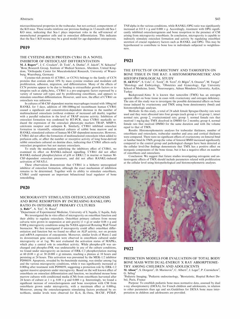
microarchitectural properties in the trabecular, but not cortical, compartment ofthe KO mice. These results reinforce our previous findings in 12-month old Sca-1KO mice, indicating that Sca-1 plays important roles in the self-renewal ofmesenchymal progenitor cells and in osteoclast differentiation. This indicatesthat the Sca-1 KO mouse may be a good model for age-dependent osteoporosis.
P019
THE CYSTEINE-RICH PROTEIN CYR61 IS A NOVEL
INHIBITOR OF OSTEOCLAST DIFFERENTIATIONM. J. Rogers*1, J. C. Crockett1, D. Tosh1, A. Duthie1, F. Jakob2, N. Schuetze21Bone Research Group, Institute of Medical Sciences, Aberdeen, United King-dom, 2Orthopedic Center for Musculoskeletal Research, University of Wuerz-burg, Wuerzburg, Germany
Cysteine-rich protein 61 (CYR61, or CCN1) belongs to the family of CCNproteins that contain about 10% by mass cysteine residues and modulate cellproliferation, adhesion, migration, and differentiation. Many of the effects ofCCN proteins appear to be due to binding to extracellular growth factors or tointegrins such as alphavbeta3. CYR61 is a pro-angiogenic factor expressed by avariety of tumour cell types and by proliferating osteoblasts, and appears tostimulate osteoblast differentiation. However, until now, the effect of CYR61 onosteoclasts was unknown.
In cultures of M-CSF-dependent murine macrophages treated with 100ng/mlRANKL for 5 days, addition of 100-1000ng/ml recombinant human CYR61caused a significant and concentration-dependent decrease in the number ofmultinucleated osteoclasts (without evidence of increased cell death), togetherwith a parallel reduction in the level of TRAP enzyme activity. Inhibition ofosteoclast formation was confirmed by RT-PCR, since CYR61 markedly re-duced the expression of the osteoclast phenotypic markers TRAP, MMP-9,calcitonin receptor and cathepsin K. Similarly, CYR61 inhibited osteoclastformation in vitaminD3 -stimulated cultures of rabbit bone marrow and inRANKL-stimulated cultures of human M-CSF-dependent monocytes. However,CYR61 did not affect the formation of multinucleated osteoclasts when added toprefusion cells, or affect the number or resorptive activity of long bone-derivedrabbit osteoclasts cultured on dentine discs, indicating that CYR61 affects earlyosteoclast progenitors but not mature osteoclasts.
To study the mechanism underlying the inhibitory effect of CYR61, weexamined its effect on RANKL-induced signalling. CYR61 did not affectRANKL-induced phosphorylation of p38 or ERK1/2 in murine or human M-CSF-dependent osteoclast precursors, and did not affect RANKL-inducedactivation of NFATc1.
These observations demonstrate that CYR61 is a hitherto unrecognisedinhibitor of osteoclast formation, although the exact mechanism of inhibitionremains to be determined. Together with its ability to stimulate osteoblasts,CYR61 could represent an important bifunctional local regulator of boneremodelling.
P020
MICROGRAVITY STIMULATES OSTEOCLASTOGENESIS
AND BONE RESORPTION BY INCREASING RANKL/OPG
RATIO IN OSTEOBLAST PRIMARY CULTURESA. Rufo*1, A. Teti1, N. Rucci11Department of Experimental Medicine, University of L’Aquila, L’Aquila, Italy
We investigated the in vitro effect of microgravity on osteoblast function andtheir ability to regulate osteoclasts. Osteoblast primary cultures from mousecalvaria were grown in suspension at unit gravity (1 · g) or under 0.08 · g and0.008 g microgravity conditions using the NASA-approved Rotating Wall Vesselbioreactor. We first investigated if microgravity could affect osteoblast differ-entiation and function but we found no effect on ALP activity, nor on proteinand mRNA expression of osteopontin. Moreover, similar levels of Runx-2 andits downstream gene osteocalcin were observed in osteoblasts cultured undermicrogravity or at 1xg. We next evaluated the activation status of MAPKs,which play a central role in osteoblast activity. While phospho-p38 was un-changed and phospho-JNK was undetectable in any of the culture conditions,we found under microgravity an increase of ERK-1/2 phosphorylation startingat 60 (0.08 · g) or 30 (0.008 · g) minutes, reaching a plateau at 10 hours andpersisting at 24 hours. This activation was prevented by the MEK-1/2 inhibitorPD98059. Apoptosis, revealed by bis-benzimide staining, was similar among 1xgand the various microgravity conditions, while it was significantly increased at0.008xg after treatment with PD98059, suggesting a protection role by ERK-1/2against massive apoptosis under microgravity. Based on the well known effect ofosteoblasts on osteoclast differentiation and function, we incubated mouse bonemarrow cultures with conditioned media (CM) from osteoblasts harvested after24 hours of culture at 1 · g, 0.08 · g and 0.008 · g. Interestingly, we found asignificant increase of osteoclastogenesis and bone resorption with CM fromosteoblasts grown under microgravity, with a maximum effect at 0.008xg.Moreover, among the osteoclastogenesis stimulating factors produced by os-teoblasts, similar levels were observed for IL-6, IL-1beta, M-CSF, PTHrP,
TNFalpha in the various conditions, while RANKL/OPG ratio was significantlyincreased at 0.0 8 · g and 0.008 · g. Accordingly, treatment with OPG signifi-cantly inhibited osteoclastogenesis and bone resoprtion in the presence of CMarising from microgravity osteoblasts. In conclusion, microgravity is capable toindirectly stimulate osteoclast formation and activity by regulating osteoblastsecretion of crucial regulatory factors such as RANKL and OPG. This may behypothesized to contribute to bone loss in individuals subjected to weightless-ness.
P021
THE EFFECTS OF OVARIECTOMY AND TAMOXIFEN ON
BONE TISSUE IN THE RAT: A HISTOMORPHOMETRIC AND
HISTOPATHOLOGICAL STUDYH. AKTUG*1, S. Uslu1, C. Terek2, H. Terzi2, O. Bilgin2, S. Ozsener2, M. Turgut31Histology and Embryology, 2Obstetrics and Gynecology, Ege UniversitySchool of Medicine, Izmir, 3Neurosurgery, Adnan Menderes University, Aydyn,Turkey
Background/Aims: It is known that tamoxifen (TMX) has an estrogenagonist effect on bone tissue in cases with ovariectomy and estrogen deficiency.The aim of this study was to investigate the possible detrimental effects on bonetissue induced by ovariectomy and TMX using bone densitometry (bmd) andhistomorphologic analyses.
Methods: In this study, a total of 24 adult female Sprague-Dawley rats wereused and they were allocated into four groups (each group n=6) group 1, intactnormal rats; group 2, ovariectomized rats; group 3, normal female rats thatreceived 1 mg/kg/day TMX dissolved in DMSO for 2 months; group 4, normalfemale rats that received DMSO for the same duration and with the volumeequal to that of TMX.
Results: Histomorphometric analyses for trabecular thickness, number ofosteoblasts and osteoclasts, trabecular number and area and cortical thicknesswere compared. There were no significant effects of ovariectomy on femoral bmdor lumbar bmd.In TMX group,the value of femoral BMD increased significantlycompared to the control group and pathological changes have been detected atthe cellular level.Our findings demonstrate that TMX has a positive effect oninorganic components of the bone tissue, but it has a negative effect on numberof osteoblasts and osteoclasts.
Conclusion: We suggest that future studies investigating estrogenic and an-tiestrogenic effects of TMX should include parameters related with proliferationat the cellular level using histopathological and histomorphometric analyses.
P022
PREDICTION MODELS FOR EVALUATION OF TOTAL BODY
BONE MASS WITH DUAL-ENERGY X RAY ABSORPTIOME-
TRY AMONG CHILDREN AND ADOLESCENTSM. Alison*1, S. Dorgeret1, D. Marinovic2, C. Alberti3, J. Leger2, P. Czernichow2,G. Sebag11Pediatric Imaging, 2Pediatric endocrinology, 3Biostatistic, Hopital Robert De-bre, Paris, France
Purpose: To establish pediatric bone mass normative data, assessed by dualx-ray absorptiometry (DEXA), for French children and adolescents, in relationto other parameters than age and sex.Guidelines for DEXA bone mass inter-pretation in children and adolescents are provided.
Abstracts S43

Methods: Bone mineral content (BMC), bone area (BA), bone mineraldensity (BMD) and lean tissue mass (LM) were measured with DEXA (GELunar Prodigy), in 366 healthy children and adolescents (180 boys and 186 girls,aged 3 to 22 years), with normal growth. BMC data were analysed and corre-lated with differents variables: weight, height, body area and volume, pubertalstages, bone area and lean tissue mass.
Results: BMC showed strong correlation with BA (r=0.98), weight(r=0.96), body area (r=0.97), and LM (boys r=0.98 and girls r=0.97) in bothsexes, with BMC/LM ratio always higher for girls. Regression models for BMC,adjusted to differents variables, were obtained.
Multivariate analysis demonstrates that best regression equation is based onsex, age, BA, height (r=0.98 for girls) or weight (r=0.99 for boys).
Conclusion: Bone mass DEXA measurement in children, should be assessedin 3 steps: 1) Bone mass adjusted to bone area, 2) Bone mass adjusted to weight,to determine ‘‘suitable’’ weight for measured bone mass and ‘‘expected’’ bonemass compared to ideal weight, 3) Bone mass adjusted to LM, by comparingBMC/LM ratio to height (figure),to differenciate primary low bone mass, lowbone mass secondary to muscle disorder or mixed disorder.
P023
CALCIFICATION OF SHARPEY’S FIBRES AND PERIOSTEUM
WITH AGE IN THE PORCINE MANDIBLEA. I. Al-Qtaitat*1, R. C. Shore2, J. E. Aaron11Faculty of Biological Sciences, 2Leeds Dental Institute, University of Leeds,Leeds, United Kingdom
Sharpey�s fibres are periosteal collagenous interconnections between muscleand bone. Because of their strategic position the possibility arises that they are aneglected factor in age-related skeletal pathology. Using young (1 yr) and older(3 yrs) pig mandible as a major site for their location, Sharpey�s fibres werehistologically observed by means of optical and ultrastructural proceduresincluding laser confocal and polarization microscopy, mineral histochemistry,collagen immunohistochemistry, EDX mineral microanalysis and also chro-mium-staining for �crystallite ghosts� [1]. Calcification of the aged Sharpey�s fi-bres was compared with calcifying turkey leg tendon (another fibrous extensionof the periosteum). Collagen type III staining was a characteristic of Sharpey’sfibres [2] which were birefringent in polarised light and 5–25lm thick. Confocalmicroscopy enabled their mapping in three-dimensions, demonstrating theirextension from the periosteum deep into the cortex and their classificationaccording to their insertion (horizontal, vertical, oblique). The young Sharpey�sfibres were uncalcified, in contrast to the dense surrounding bone matrix. Theircalcification was apparently a feature of age, in the form of discrete particles,about 1 lm in diameter, and linked assemblies of particles looped around thefibre bundles, and sometimes spreading into the adjacent periosteum; a similarparticulate arrangement occurred around the parallel fibre bundles of the turkeyleg tendon. Calcification with aging was confirmed in the electron microscope byraised calcium, phosphorus and chromium peaks in relation to filamentousmineral microspheres [3] and their chromium-stained ‘‘ghosts.’’ In contrast toturkey leg tendon, calcification of Sharpey�s fibres was accompanied by theirfragmentation and the diminution of their collagen III-stained subperiostealdomain [4]. It was concluded that the function of Sharpey’s fibres as avenues formusculoskeletal exchange as well as anchorage may be augmented by modestcalcification, but impaired by excess, contributing to their disconnection byresorption and with implications for skeletal incapacity in the elderly. Refer-ences;
1. Appleton J. Calcif Tissue Res 2, 270–6, 19702. CarterDH, Sloan P, Aaron JE. Histochem Cytochem 39, 599–606, 19913. Aaron JE, Oliver B, Clarke N, Carter DH. Histochem J 31,455–70, 19994. Luther F, Saino H, Carter DH, Aaron JE. Bone 32, 652–9, 2003
P024
IMPLANTATION OF OCTACALCIUM PHOSPHATE
(OCP) ENHANCES LONG BONES REPAIR IN
THE RATSM. R. Arab1, F. Sargolzaei Avval11Anatomy, Zahedan University of Medical Sciences, Zahedan, Iran (IslamicRepublic of)
Background & aim: In several conditions we have face long bone defects.Reconstruction of these defects remains one of the most challenging problemsencountered by orthopedic surgeons. This study was designed to investigate theprocess of bone formation caused by implantation of octacalcium phosphate inrat tibiae.
Methods & materials: synthetic octacalcium phosphate was implanted into a3 mm bony defect was surgically created with a bur on the superior end of righttibiae in twenty five male spague Dawley rats. No OCP particles were implantedin the left tibiae as a control group that was otherwise treated identically. Boneformation was examined histologically on 7th, 10th, 14th, 21st, 28th days afterimplantation.
Results: in the experimental group, on the 7th day after implantation, afew clusters of cartilage cells were observed between the OCP particles nearthe defects margin. By 14th day after implantation, Alcian blue stainingshowed hypertrophic chondrocytes that replaced by new bone. Osteogenesiswas initiated locally between the OCP particles in central position of thedefects on 10th day after implantation. In addition to bone formation locallyaround the OCP particles, more apposition of new bone was observed nearthe defects margin on 14th and 21st days after implantation. At the end ofthe study, the defect was almost completely filled with new bone, which wasin close contact with host bone and implanted OCP was surrounded by newlyformed bone.
In the control group, bone formation was observed only along and near thedefects margin.
Conclusion: these results demonstrate that octacalcium phosphate could beused in the repair of the long bone defects.
Key word: Octacalcium phosphate, Osteogenesis, Tibia, Rat
P025
VITAMIN K ANALOGUES PROMOTE THE
DIFFERENTIATION OF PRIMARY HUMAN
OSTEOBLASTSG. J. Atkins1, K. J. Welldon1, C. Vincent1, D. M. Findlay*11Department of Orthopaedics and Trauma, University of Adelaide, Adelaide,Australia
The vitamin K family members, phylloquinone (vitamin K1) and themenaquinones (vitamin K2) are currently under study for their roles in bonemetabolism and as potential therapeutic agents for skeletal diseases. Currentdata suggest that K2 in particular has a positive role in post-menopausalbone loss, both as a single agent or in combination with anti-resorptives.There is considerable evidence that high plasma vitamin K levels equate withhealthier bone in population based studies, and that under conditions of boneloss in both humans and animal studies, vitamin K is protective. While thereis evidence for direct effects of vitamin K on rodent osteoblasts, there are fewreports of the action of vitamin K in human osteoblasts. Koshihara et al.[1]found that both K1 and K2 increased the number of CFU-F/ALP+ coloniesgenerated from human bone marrow cells and enhanced mineralisation bythese cells. In the present study, we have investigated the effects of phyto-nadione (K1) and menatetrenone (K2) on primary adult human osteoblasts(NHB) derived from trabecular bone. Both analogues induced mineralisationin vitro, with K2 having the more consistent effect on all parameters studied.K2 (10 microM) reproducibly increased apposition of calcium into cellmonolayers approximately two-fold that of controls. This effect correspondedwith phenotypic maturation, as assessed by the expression of tissue non-specific alkaline phosphatase (AP) and the immature osteoblast marker,STRO-1.[2] K2 induced maturation, indicated by eventual loss of AP onmature (STRO-1-) osteoblasts. Only transient effects on STRO-1 expressionwere observed, indicating that the principal effect was on the mature osteo-blast. Consistent with this, both K1 and K2 inhibited osteoblast proliferation,as assessed by labelling parental cells with the fluorescent dye CFSE and flowcytometric analysis. Also consistent with osteoblast maturation, both ana-logues decreased collagen type 1-alpha1 mRNA expression, while K2 but notK1 increased markedly the expression of osteocalcin mRNA. Overall, theresults are consistent with K2 in particular inducing a maturational effect onhuman osteoblasts, increasing the prevalence of a post-osteoblast, pre-osteo-cyte phenotype.
1. Koshihara Y, et al. (2003) J Endocrinol 176:339–3482. Gronthos S, et al. (1999) J Bone Miner Res 14:47–56.
S44 Abstracts
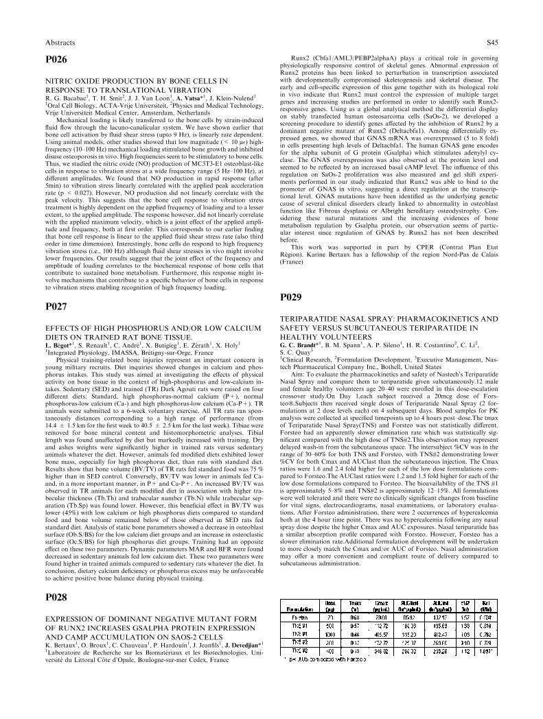
P026
NITRIC OXIDE PRODUCTION BY BONE CELLS IN
RESPONSE TO TRANSLATIONAL VIBRATIONR. G. Bacabac1, T. H. Smit2, J. J. Van Loon1, A. Vatsa*1, J. Klein-Nulend11Oral Cell Biology, ACTA-Vrije Universiteit, 2Physics and Medical Technology,Vrije Universiteit Medical Center, Amsterdam, Netherlands
Mechanical loading is likely transferred to the bone cells by strain-inducedfluid flow through the lacuno-canalicular system. We have shown earlier thatbone cell activation by fluid shear stress (upto 9 Hz), is linearly rate dependent.Using animal models, other studies showed that low magnitude (< 10 l�) high-frequency (10–100 Hz) mechanical loading stimulated bone growth and inhibiteddisuse osteoporosis in vivo. High frequencies seem to be stimulatory to bone cells.Thus, we studied the nitric oxide (NO) production of MC3T3-E1 osteoblast-likecells in response to vibration stress at a wide frequency range (5 Hz–100 Hz), atdifferent amplitudes. We found that NO production in rapid response (after5min) to vibration stress linearly correlated with the applied peak accelerationrate (p < 0.027). However, NO production did not linearly correlate with thepeak velocity. This suggests that the bone cell response to vibration stresstreatment is highly dependent on the applied frequency of loading and to a lesserextent, to the applied amplitude. The response however, did not linearly correlatewith the applied maximum velocity, which is a joint effect of the applied ampli-tude and frequency, both at first order. This corresponds to our earlier findingthat bone cell response is linear to the applied fluid shear stress rate (also thirdorder in time dimension). Interestingly, bone cells do respond to high frequencyvibration stress (i.e., 100 Hz) although fluid shear stresses in vivo might involvelower frequencies. Our results suggest that the joint effect of the frequency andamplitude of loading correlates to the biochemical response of bone cells thatcontribute to sustained bone metabolism. Furthermore, this response might in-volve mechanisms that contribute to a specific behavior of bone cells in responseto vibration stress enabling recognition of high frequency loading.
P027
EFFECTS OF HIGH PHOSPHORUS AND/OR LOW CALCIUM
DIETS ON TRAINED RAT BONE TISSUE.L. Begot*1, S. Renault1, C. Andre1, X. Butigieg1, E. Zerath1, X. Holy11Integrated Physiology, IMASSA, Bretigny-sur-Orge, France
Physical training-related bone injuries represent an important concern inyoung military recruits. Diet inquiries showed changes in calcium and phos-phorus intakes. This study was aimed at investigating the effects of physicalactivity on bone tissue in the context of high-phosphorus and low-calcium in-takes. Sedentary (SED) and trained (TR) Dark Agouti rats were raised on fourdifferent diets: Standard, high phosphorus-normal calcium (P+), normalphosphorus-low calcium (Ca-) and high phosphorus-low calcium (Ca-P+). TRanimals were submitted to a 6-week voluntary exercise. All TR rats ran spon-taneously distances corresponding to a high range of performance (from14.4 ± 1.5 km for the first week to 40.5 ± 2.5 km for the last week). Tibiae wereremoved for bone mineral content and histomorphometric analyses. Tibiallength was found unaffected by diet but markedly increased with training. Dryand ashes weights were significantly higher in trained rats versus sedentaryanimals whatever the diet. However, animals fed modified diets exhibited lowerbone mass, especially for high phosphorus diet, than rats with standard diet.Results show that bone volume (BV/TV) of TR rats fed standard food was 75 %higher than in SED control. Conversely, BV/TV was lower in animals fed Ca-and, in a more important manner, in P+ and Ca-P+. An increased BV/TV wasobserved in TR animals for each modified diet in association with higher tra-becular thickness (Tb.Th) and trabecular number (Tb.N) while trabecular sep-aration (Tb.Sp) was found lower. However, this beneficial effect in BV/TV waslower (45%) with low calcium or high phosphorus diets compared to standardfood and bone volume remained below of those observed in SED rats fedstandard diet. Analysis of static bone parameters showed a decrease in osteoblastsurface (Ob.S/BS) for the low calcium diet groups and an increase in osteoclasticsurface (Oc.S/BS) for high phosphorus diet groups. Training had an oppositeeffect on these two parameters. Dynamic parameters MAR and BFR were founddecreased in sedentary animals fed low calcium diet. These two parameters werefound higher in trained animals compared to sedentary rats whatever the diet. Inconclusion, dietary calcium deficiency or phosphorus excess may be unfavorableto achieve positive bone balance during physical training.
P028
EXPRESSION OF DOMINANT NEGATIVE MUTANT FORM
OF RUNX2 INCREASES GSALPHA PROTEIN EXPRESSION
AND CAMP ACCUMULATION ON SAOS-2 CELLSK. Bertaux1, O. Broux1, C. Chauveau1, P. Hardouin1, J. Jeanfils1, J. Devedjian*11Laboratoire de Recherche sur les Biomateriaux et les Biotechnologies, Uni-versite du Littoral Cote d’Opale, Boulogne-sur-mer Cedex, France
Runx2 (Cbfa1/AML3/PEBP2alphaA) plays a critical role in governingphysiologically responsive control of skeletal genes. Abnormal expression ofRunx2 proteins has been linked to perturbation in transcription associatedwith developmentally compromised skeletogenesis and skeletal disease. Theearly and cell-specific expression of this gene together with its biological rolein vivo indicate that Runx2 must control the expression of multiple targetgenes and increasing studies are performed in order to identify such Runx2-responsive genes. Using as a global analytical method the differential displayon stably transfected human osteosarcoma cells (SaOs-2), we developed ascreening procedure to identify genes affected by the inhibition of Runx2 by adominant negative mutant of Runx2 (Deltacbfa1). Among differentially ex-pressed genes, we showed that GNAS mRNA was overexpressed (5 to 8 fold)in cells presenting high levels of Deltacbfa1. The human GNAS gene encodesfor the alpha subunit of G protein (Gsalpha) which stimulates adenylyl cy-clase. The GNAS overexpression was also observed at the protein level andseemed to be reflected by an increased basal cAMP level. The influence of thisregulation on SaOs-2 proliferation was also measured and gel shift experi-ments performed in our study indicated that Runx2 was able to bind to thepromoter of GNAS in vitro, suggesting a direct regulation at the transcrip-tional level. GNAS mutations have been identified as the underlying geneticcause of several clinical disorders clearly linked to abnormality in osteoblastfunction like Fibrous dysplasia or Albright hereditary osteodystrophy. Con-sidering these natural mutations and the increasing evidences of bonemetabolism regulation by Gsalpha protein, our observation seems of partic-ular interest since regulation of GNAS by Runx2 has not been describedbefore.
This work was supported in part by CPER (Contrat Plan EtatRegion). Karine Bertaux has a fellowship of the region Nord-Pas de Calais(France)
P029
TERIPARATIDE NASAL SPRAY: PHARMACOKINETICS AND
SAFETY VERSUS SUBCUTANEOUS TERIPARATIDE IN
HEALTHY VOLUNTEERSG. C. Brandt*1, B. M. Spann1, A. P. Sileno1, H. R. Costantino2, C. Li2,S. C. Quay31Clinical Research, 2Formulation Development, 3Executive Management, Nas-tech Pharmaceutical Company Inc., Bothell, United States
Aim: To evaluate the pharmacokinetics and safety of Nastech�s TeriparatideNasal Spray and compare them to teriparatide given subcutaneously.12 maleand female healthy volunteers age 20–40 were enrolled in this dose-escalationcrossover study.On Day 1,each subject received a 20mcg dose of Fors-teo�.Subjects then received single doses of Teriparatide Nasal Spray (2 for-mulations at 2 dose levels each) on 4 subsequent days. Blood samples for PKanalysis were collected at specified timepoints up to 4 hours post–dose.The tmaxof Teriparatide Nasal Spray(TNS) and Forsteo was not statistically different.Forsteo had an apparently slower elimination rate which was statistically sig-nificant compared with the high dose of TNS#2.This observation may representdelayed wash-in from the subcutaneous space. The intersubject %CV was in therange of 30–60% for both TNS and Forsteo, with TNS#2 demonstrating lower%CV for both Cmax and AUClast than the subcutaneous injection. The Cmaxratios were 1.6 and 2.4 fold higher for each of the low dose formulations com-pared to Forsteo.The AUClast ratios were 1.2 and 1.5 fold higher for each of thelow dose formulations compared to Forsteo. The bioavailability of the TNS #1is approximately 5–8% and TNS#2 is approximately 12–15%. All formulationswere well tolerated and there were no clinically significant changes from baselinefor vital signs, electrocardiograms, nasal examinations, or laboratory evalua-tions. After Forsteo administration, there were 2 occurrences of hypercalcemiaboth at the 4 hour time point. There was no hypercalcemia following any nasalspray dose despite the higher Cmax and AUC exposures. Nasal teriparatide hasa similar absorption profile compared with Forsteo. However, Forsteo has aslower elimination rate.Additional formulation development will be undertakento more closely match the Cmax and/or AUC of Forsteo. Nasal administrationmay offer a more convenient and compliant route of delivery compared tosubcutaneous administration.
Abstracts S45

P030
OSTEOBLAST APOPTOSIS ASSAY IN CELL CULTURE
SYSTEMY. Chung*1, E. Yu1, S. Yun1, Y. Shen1, J. Kim2, S. Lim3, W. Choi4, H. Choi51Endocrinology and Metabolism, Ajou University School of Medicine, Suwon,2Obstetrics and Gynecology, Seoul National University School of Medicine,3Internal Medicine, Yonsei University College of Medicine, 4Internal Medicine,Hanyang University College of Medicine, 5Obstetrics and Gynecology, InjeUniversity College of Medicine, Seoul, South Korea
Glucocorticoids are widely used as an anti-inflammatory and chemothera-peutic agents. However, prolonged administration of glucocorticoids is one ofthe leading causes of osteoporosis. This study was designed to examine thedexamethasone (DEX)-induced apoptosis in murine osteoblast cell line, MC3T3-E1, C3H/10T1/2, and bone marrow stromal cell, and fibroblast cell line,NIH3T3.
Cell viability was measured by MTT assay from each cell lines treated with10–7M DEX for 48h. Western blot analysis showed expression of caspase-3 afterDEX treatment. To detection of apoptotic cell was performed with DAPIstaining and Annexin-V-FLUOS staining.
Each cell lines were decreased in cell viability after DEX treatment except offibroblast cell line. Only C3H/10T1/2 cell line was highly expressed in cleavedcaspase-3with time-dependentmanner from 24hrs to 72hrs.AfterDEX treatment,all cell lines expressed apoptotic bodies byDAPI staining andAnnexin-V-FLUOSstaining, and C3H/10T1/2 cell line was most prominent among all cell lines.
In this study, we demonstrated that C3H/10T1/2 cell was the most suitablefor in vitro glucocorticoid-induced osteoblast apoptosis assay.
This research was supported by CCRB through the ‘‘GRRC’’ Project ofGyeonggi Provincial Government, Republic of Korea
P031
HUMAN EMBRYONIC STEM CELL-DERIVED CONNECTIVE
TISSUE PROGENITORS FOR TISSUE ENGINEERINGS. Cohen*1, L. Leshansky1, E. Zussman2, J. Itskovitz-Eldor11Stem Cell Center, Bruce Rappaport Faculty of Medicine, 2Department ofMechanical Engineering, Technion-Israel Institute of Technology, Haifa, Israel
Cell-based tissue engineering is an evolving interdisciplinary area that offersnew tools for repairing or replacing damaged or lost tissues. Human embryonicstem cells (hESCs) have been proposed to be the ultimate source for cell-basedapplications as well as an exciting tool for investigating the fundamentals ofhuman development.
Here, we describe the efficient derivation of connective tissue progenitors(CTPs) from hESCs. So termed, we show that these cells have multilineagedevelopmental potential, yet are committed to connective tissue derivatives.
Osteogenic differentiation was achieved by the addition of dexamethasone,ascorbic acid and 2-glycerophosphate to the growth medium, and confirmed bythe detection of osteoblast markers. Bone mineralized matrix production wasdetected both histochemically and by scanning electron microscopy coupled withenergy dispersive spectroscopy (EDS) analysis. Chondrogenic differentiation wasinduced and confirmed both by the intact layer suspension method, resulting in atypical cartilage morphology and by the low-serum TGF-beta3 supplementedpellet cultures, showing cartilage marker up-regulation and matrix production.Additionally, CTPs are shown to be easily expandable and non-tumorigenic, andupon seeding on PCL/PLA nanofiber scaffolds, they extensively excrete extra-cellular matrix and form 3D sheet-like tissues.
We further show the potential of these cells to generate tendon-like struc-tures. Applying the long-term high-density culture technique to these cells, wehave successfully assembled cylinder-shaped constructs, with typical ultrastruc-ture characteristics and biomechanical properties of early tendons.
In view of the limited availability of tissues for transplantation, this work notonly holds great promise for a potentially unlimited source of cells for tissueengineering, but also suggests a great tool for investigating human embryonicdevelopment.
P032
REGULATION OF OSTEOBLAST DIFFERENTIATION BY
THE RHO/RHO KINASE SIGNALLING PATHWAYA. E. Coudert*1, A. Danikas1, A. E. Grigoriadis11Department of Craniofacial Development, King’s College London, London,United Kingdom
The Rho family of GTP binding proteins (Rho, Rac and cdc42) are smallGTPases that cycle between inactive, GDP-bound states, and active GTP-boundstates, and play important roles in cytoskeletal rearrangements, cell migrationand differentiation. We have recently studied the role of the Rho GTPase in
osteoblast differentiation using the bacterial protein toxin, Pasteurella MultocidaToxin (PMT). PMT is a unique toxin and a potent mitogen, which activates theheterotrimeric G-proteins of the Gq/11 and G12/13 families, leading to stimu-lation of Rho, its downstream effector Rho Kinase (ROCK), causing actinrearrangements. PMT also activates phospholipase C, protein kinase C and theRas/MAP kinase pathways. We have previously shown that addition of PMT toprimary mouse calvarial osteoblasts potently inhibits bone nodule formation andosteoblast marker gene expression and this was reversed by addition of Rho andROCK inhibitors. Interestingly, addition of Rho/ROCK inhibitors alone toprimary osteoblasts stimulated bone nodule formation. These results suggestthat the Rho/ROCK pathway is a critical determinant of osteoblast differenti-ation and bone formation.
To investigate further the role of ROCK in osteoblast differentiation, both invitro and ex vivo strategies were chosen. GTP pulldown assays demonstratedthat PMT markedly induced the levels of active GTP-bound Rho in primaryosteoblasts and induced stress fibre formation. Western blot analysis showedthat osteoblasts express both ROCK1 and ROCK2 isoforms. To define the roleof ROCK in osteoblast differentiation, we have generated retroviruses con-taining ROCK1 and ROCK2 siRNAs. RT-PCR and Western blot analysesshowed that the siRNAs selectively reduced ROCK RNA and protein. Thereduced ROCK expression was functionally analysed by cytoskeletal rear-rangements that showed abolition of stress fibre formation in osteoblasts andthese siRNAs are currently being used in differentiation assays of primary os-teoblasts and expression of osteoblast marker genes. In a parallel approach, weare utilizing calvaria explant cultures treated with PMT to assess its effects oncalvarial osteogenesis. Preliminary results have shown that PMT exhibitedmarked mitogenic effects in mesenchymal cells, resulting in an apparent reduc-tion in calvarial osteogenesis. Thus, alteration of endogenous Rho and ROCKlevels in bone will provide further evidence of the key role of the Rho-ROCKpathway in osteoblast differentiation.
P033
DEVELOPMENT OF A BONE GRAFT SUBSTITUTE: OPTIMI-
ZATION OF THE GROWTH OF HUMAN OSTEOBLASTS ON A
3D COLLAGEN SCAFFOLDF. Tan1, F. J. O’Brien1, J. S. Daly*11Anatomy, Royal College of Surgeons in Ireland, Dublin, Ireland
Background: Collagen is a significant constituent of natural extracellularmatrix, scaffolds made from collagen have been widely used and observed topromote cell and tissue attachment and growth [1]. The initial aim of this re-search is to ascertain whether human osteoblasts (hFOB 1.19) are able to attach,grow and remain viable on a collagen scaffold in vitro, as relevant experimentsshowed mice osteoblasts can do so [2]. This study also aims to optimize a real-time AlamarBlue viability assay on the scaffolds.
Methods: (1)The collagen-glycosaminoglycan (CG) scaffold was fabricatedusing a lyophilisation technique [3].(2)Cell scaffold attachment was investigatedby cell seeding, fixation, tissue processing, slicing and H&E staining. Optimal cellseeding density was determined by measuring attached cell number to scaffoldfrom a series of seeding densities. A Dispase/Trypan blue method was used forcell viability counting.(3)An AlamarBlue viability assay was optimized for thehFOB1.19 cells on the scaffold.
Results:(1) Osteoblasts sucessfully attached to the CG scaffold, and pene-trated right to the centre of the scaffold.(2)Results from the optimal cell seedingtrial reveal the highest cell number attached is achieved at 6 · 106 cells/ml.(Datanot shown).(3)The AlamarBlue assay compared favorably to the dispase meth-od, optimization of the method including shaking during the latter stages of thedetection stage which improved results.
Conclusion: Cell-scaffold histological results confirm that hFOB 1.19 cellssuccessfully attach to the CG scaffold. 6 · 10 6 cells/ml is the optimal cell seedingdensity required to allow the maximum number of cells attach. Experimentalresults of AlamarBlue suggest that this assay is efficient at assessing viability in-situ on the CG scaffold. Future studies including long term incubation, and theuse of growth factors to promote development of the osteoblasts are in progress.
References: [1]Yannas IV. Tissue and organ regeneration in adults. NewYork: Springer; 2001. [2]O Brien FJ (et al). The effect of pore size on celladhesion in collagen-GAG scaffolds. Biomaterials 2005. [3]O Brien FJ (et al).Influence of freezing rate on pore structure in freeze-dried collagen-GAG scaf-folds. Biomaterials 2004.
Acknowledgements: RCSI (FT) and a SFI President of Ireland Young Re-searcher Award (FOB)
P034
EFFECTS OF HYALURONAN ON THREE-DIMENSIONAL
MICROARCHITECTURE OF SUBCHONDRAL BONE TISSUES
IN GUINEA PIG PRIMARY OSTEOARTHROSISM. Ding*1, C. C. Danielsen2, I. Hvid3
S46 Abstracts

1Orthopaedic Research Laboratory, Department of Orthopaedics, Odense andAarhus University Hospitals, Odense C, 2Department of Connective TissueBiology, Institute of Anatomy, University of Aarhus, 3Department of Ortho-paedics, Aarhus University Hospital, Aarhus C, Denmark
Introduction: It is not known whether hyaluronan (HA) has any effect onthe underlying subchondral bone tissues. This study was to investigate the effectsof high molecular weight HA (1.5 · 106 Daltons) intra-articular injection onsubchondral bone tissues.
Methods: Fifty-six male guinea pigs (6.5 months of age) were randomlydivided into 5 groups studied in a short-term and a long-term experimentalperiod. All HA groups received intra-articular injection of HA 0.4 mg/kg/weekfor 5 weeks in both knee joints. HA-II received injection for additional 5 weeks;HA-III received no more injection; and the control groups received vehicle. Aftersacrifice, the left tibiae were harvested and micro-CT scanned, followed bymechanical testing and collagen and mineral determination.
Results: The HA-treated groups had almost normal cartilage, whereas thecontrol groups had typical osteoarthrosis (OA)-related cartilage degradation. Inthe short-term study, compared with the control group, HA-injection resulted ina significantly decreased subchondral plate volume fraction and plate thickness.HA-treated cancellous bone had significantly lower bone volume fraction, andtypical rod-like structure. In the long-term study, these latter changes were morepronounced, with an additionally significant decrease in connectivity and bonesurface density. HA groups had greater bone mineral concentration and mineraldensity, lower collagen to mineral ratio, and preserved the mechanical propertiesof cancellous bone. The effects of HA on cartilage and subchondral bone weremaintained when HA treatment was discontinued.
Discussion: Significant positive effects of high molecular weight HA on thearticular cartilage and subchondral bone tissues were seen. HA protects againstOA-related cartilage degradation to almost normal level, and effectively changesthe subchondral bone tissue microarchitecture, collagen and mineral content anddensity without altering the mechanical properties of cancellous bone. The moststriking features are the microarchitectural changes in the subchondral cancellousbone that lead to lower bone density and markedly rod-like structure, and thusreducing cartilage stress during impact loading. Still, the subchondral bone has agreater mineral concentration, and a lower collagen to mineral ratio, and thuspreserves the mechanical properties of cancellous bone. Short-term HA is suffi-cient and early HA is needed for intervention of OA initiation and progression.
P035
PRECONDITIONING EXERCISE AND ANATOMIC SITE
RELATE TO MINERALISATION AND THICKNESS IN
EQUINE THIRD METACARPAL ARTICULAR CALCIFIED
CARTILAGEM. Doube*1, E. C. Firth2, A. Boyde11Dental Institute, Queen Mary, University of London, London, United King-dom, 2Institute of Veterinary, Animal and Biomedical Sciences, Massey Uni-versity, Palmerston North, New Zealand
Condylar fracture of the third metacarpal bone (Mc3) is a common cause ofracetrack fatality in Thoroughbred horses. Fractures originate at the articularcalcified cartilage (ACC) mineralising front (MF) of the distal Mc3 condyle, typ-ically in the parasagittal groove. Abnormal ACC may be observed in this region.Giving preconditioning exercise (PX) to young horses may enable the distal Mc3better to resist the stresses of later training and racing, reducing fracture risk inadults. Twelve Thoroughbred horses were raised at pasture in New Zealand. Sixwere given PX from 10 d old. IV injections of calcein were given 19 and 8 d prior toeuthanasia at 18 months old. Osteochondral specimens were cut from the distalMc3 in the dorsal and palmar oblique frontal planes, fixed in acetone andembedded inPMMA. Surfaceswere finished to a diamondpolish and imaged usingquantitative backscattered electron scanning electron microscopy (qBSE). Mon-tages of 35–45 images were made of the entire mediolateral width (�50mm) of theACC. Specimens were then diamond ultramilled and 7 sites per specimen imagedwith confocal scanning laser microscopy (CSLM) and qBSE: montages were re-peated on the ultramilled surface. CSLM images were registered tomatching qBSEimages using in-house software. ImageJ v1.34swas used tomeasureACC thicknessand total thickness mineralisation density (TTDm; n=9385) on montaged imagesets, and linear accretion rate (LAR) and inter-labelmineralisationdensity (ILDm;n=3051) on registered image pairs. ACC thickness, LAR, TTDm, and ILDmvaried significantly (p<0.001) by site andPX.Thicknesswas greatest on the dorsalsagittal ridge and palmar parasagittal grooves. Grossly increased ACC thicknesswas not accompanied by a proportional increase in LAR. LAR was greatest inregions of thin ACC and high ILDm, which were found at lower-load regions onthe joint edges andpalmar sagittal ridge. Combining globalmedianLAR (0.79lm/d) and thickness (156.2 lm) gave an ACCMF to vascular invasion lag time of 198d. Using maximum regional LAR (3.35 lm/d) and thickness (1500 lm) gives a lagtime of 448 d. Grossly thickened ACC may thus represent delayed vascular inva-sion of cartilage due to events occurring as early as 3months of age, indicating thatthe pathogenesis ofMc3 condylar fracture may begin earlier in life than previouslythought.
Supported by the Veterinary Advisory Committee of the Horserace BettingLevy Board and the Global Equine Research Alliance.
P036
EFFECT OF SILICON ON OSTEOBLAST SURVIVAL AND
ADHESION IN VITROW. C. Duivenvoorden*1, A. Middleton2, A. TanBeeWan2, C. Pretto2, A. Spoo-ner2, S. D. Kinrade21Department of Chemistry and Biology, 2Department of Chemistry, LakeheadUniversity, Thunder Bay, Canada
Silicon may play an important role in bone metabolism. Silicon deficiency inchicken and rats leads to bone defects. Silicon is found in the diet (rice, cereal,beer) and concentrations in plasma are generally around 0.1 mg/L. Siliconconcentrations are high in bone, nails, aorta and tendon, however the mecha-nism whereby silicon increases bone health is unknown.
Silicon from two different sources was used to determine its effect on cellsurvival and adhesion in vitro. To test survival the colony forming efficiency(CFE) assay was carried out using human osteoblast-like Saos-2 cells in thepresence or absence of various concentrations of silicon (0.01–4 mM). Cellproliferation of Saos-2 cells was measured after a 4-day incubation in thepresence or absence of silicon by means of the DNA content using Hoechst33258. The effect of silicon on cell adhesion was determined by seeding cells inthe absence or presence of silicon. One h later, this was followed by a 30-minincubation in crystal violet. The relative protein content of the wells wasdetermined spectrophotometrically and served as a measure of the number ofadhered cells.
Our results show a significant increase in the CFE after treatment with oneof the two forms of silicon but not with the other. This decrease in the number ofcolonies appears only in part to be due to a decrease in cell proliferation, andmay be due to diminished cell survival and adhesion in the presence of ortho-silicic acid. Adhesion of human osteoclast-like GCT, and osteoblast-like Saos-2and hFOB 1.19 cells was severely affected in the presence of high concentrationsof orthosilicic acid, whereas in human neuroblastoma SK-N-SH cells noinhibitory effect was observed. The effect on adhesion was not simply non-specific, but it was not related to the expression of one of the major adhesionmolecules in bone, integrin alphav_eta3. SK-N-SH cells expressed high amountsof integrin alphav_eta3 as determined by flow cytometric analysis, but itsadhesion was not affected by the presence of orthosilicic acid.
The mechanism whereby silicon increases bone health may involve effects oncell adhesion and survival in bone. This research is supported by the NaturalSciences and Engineering Research Council of Canada (to SDK) and CancerCare Ontario (to WCD).
P037
MESENCHYMAL STEM CELLS SEEDED ON BONE GRAFTS
FOR FILLING CRITICAL BONE DEFECTS IN MICEA. Dumas1, M. F. Moreau1, M. F. Basle1, D. Chappard*11INSERM, EMI 0335, Angers, France
Autologous bone grafting is considered the golden standard for regenerationof large bone defects despite of the complications of the harvesting procedure. Itprovides osteoprogenitor cells and bone extracellular matrix containing collagen,hydroxyapatite and osteoinductive growth factors from the patient itself. Tissueengineering of autologous bone by combining osteoprogenitor cells (usuallybone marrow derived mesenchymal stem cells, MSC) with an appropriate scaf-
Abstracts S47
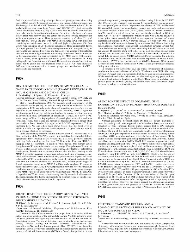
fold, is a potentially interesting technique. Bone xenograft appears an interestingmaterial that exhibits the required mechanical and microarchitectural properties.The bone graft loaded with MSC becomes a ‘‘bioactive bone substitute’’. In thisstudy, MSC were harvested from ‘‘green mice’’ (C57B6TgGFP) that express theGreen Fluorescent Protein (GFP). GFP+ cells can be directly identified andtheir behaviour in the graft can be estimated. Bovin trabecular bone grafts werecleared from bone marrow cells and debris, and delipidated using maceration insodium hydrogenocarbonate. Three groups were studied: graft alone (G), graftwith MSC (G+MSC) and graft with MSCs differenciated in Ob with a mediumcontaining dexamethasone, ascorbic acid and b-glycerophosphate (G+Ob).Grafts were implanted in C57Bl6 mouse calvaria for filling critical-sized defects(N=6 per group). 2 and 8 weeks after transplantation, the osteogenic ability ofthe 3 grafts was examined by X-ray and histology. The number of transplantedcells was evaluated using fluorescent microscopy. Presence of GFP+ osteocytesin the transplantation site was investigated in G+MSC and G+Ob groups.-After 8 weeks, bone apposition on the G group was evidenced on X-rayradiographs but the defect was not healed. The osseointegration of the graft waspartial for G group and was increased when MSCs or Ob were implanted.Differences in ossesintegration between groups and localisation of GFP+grafted cells are presented.
P038
DEVELOPMENTAL REGULATION OF MMP13/COLLAGE-
NASE3 BY TRIIODOTHYRONINE (T3) AND RUNX2/CBFA1 IN
MOUSE OSTEOBLASTIC MC3T3-E1 CELLSE. Durchschlag*1, S. Spitzer1, K. Klaushofer1, F. Varga11Ludwig Boltzmann Institute of Osteology at Hanusch Hospital of WGKK andAUVA Trauma Centre Meidling, Hanusch Hospital-Vienna, Vienna, Austria
Matrix metalloproteinases (MMPs) degrade most components of theextracellular matrix (ECM), as well as many non-ECM molecules. MMP13participates in ECM degradation to allow cell migration and therefore, is a keyregulator for the remodeling of skeletal tissues. Recently, MMP13 regulationwas investigated intensively, as it is also involved in tumour invasion, and maybe important in early development of malignancy. MMP13 is a direct down-stream target of Runx2, a key regulator of growth plate maturation and boneformation. Runx2 itself is used as a marker for breast and prostate cancer whichmetastasise to bone. Another key regulator of MMP13 expression is Triiodo-thyronine (T3). We and others have shown before that the basal levels ofMMP13 mRNA are dependent on the developmental stage of cells and that T3has a positive effect on its expression.
In the present study we show that the inductive effect of T3 is mediated via adirect activation of the MMP13 promoter, and that this effect is regulated devel-opmentally. DNA binding assays revealed that a MMP13 promoter element,exhibiting high homology to functional T3 Responsive Elements (TREs), isoccupied after T3 treatment. In addition, when deleted, this element causesderegulation of T3 responsiveness in reporter assays. Deregulation of T3 respon-siveness is also seen in cells over-expressing Runx2, a key factor for osteoblasticdevelopment. Transfection experiments showed that the basal activity of theMMP13 promoter increases in long term cultures and with progressing osteo-blastic differentiation. On the other hand, early pre-osteoblasts respond to T3withenhanced MMP13 promoter activity, unlike terminally differentiated osteoblasts.Northern blot analysis revealed that Ascorbic Acid, another potent trigger ofRunx2 expression, up-regulates MMP13 mRNA levels. Anyhow, T3 causes anadditional increase ofMMP13mRNAexpression in early pre-osteoblastic cells. Inconclusion, our data suggest that there are at least two separate pathways regu-latingMMP13 promoter activity in developing osteoblastic MC3T3-E1 cells. Oneis dependent on T3 and seems to be necessary in early osteoblastic development.The second is related to Runx2 and seems to be the main regulator in late osteo-blastic development where it overrides T3 regulation.
P039
IDENTIFICATION OF REGULATORY GENES INVOLVED
IN HUMAN BONE AND ECTOPIC GLUCOCORTICOID-IN-
DUCED MINERALIZATIONM. Eijken*1, S. Swagemakers2, M. Koedam1, P. J. Van der Spek2, H. A. P. Pols1,J. P. T. Van Leeuwen11Department of Internal Medicine, 2Department of Bioinformatics, Eras-musMC, Rotterdam, The Netherlands
Glucocorticoids (GCs) are essential for proper human osteoblast differen-tiation and mineralization of the extracellular matrix. Yet little is known aboutthe genes, proteins and regulatory networks involved in this GC-induced dif-ferentiation process. The aim of this study was to identify genes that are spe-cifically induced during GC-induced osteoblast differentiation andmineralization.We used the human pre-osteoblast SV-HFO bone formationmodel that shows a controlled differentiation and mineralization process in thepresence of 100 nM dexamethasone (DEX) in a 3-week time period. At 6 time
points during culture gene-expression was analyzed using Affymetrix HG U133plus 2.0 arrays. GC-specificity was assessed by mineralization-related compar-ative analyses of gene profiles in the presence and absence of DEX. Validation ofGC-controlled genes in the mineralization process was performed by examininga vascular smooth muscle cell (VSMC)-based model of ectopic mineraliza-tion.We identified a set of genes that were specifically regulated by GC-treat-ment. One of the most significantly regulated gene was ZBTB16 (PLZF), atranscription factor recently identified as an important enhancer of humanosteoblast differentiation. Q-PCR showed that ZBTB16 was over 1000-fold up-regulated after GC-treatment followed by an additional 10-fold induction duringmineralization. Regulatory gene-network identification revealed several GC-controlled networks including a network containing ZBTB16 in interaction withthe vitamin D receptor along with other so far non-osteoblast-related genes.ZBTB16 was the first candidate to be validated in the VSMCs mineralizationmodel. Like in osteoblasts, in VSMCs cultures GCs were essential for initiationof mineralization, which coincided with increased expression of Runx2 mRNA.Importantly, ZBTB16 was undetectable in VSMCs, however, GC-treatmentstrongly induced ZBTB16 expression in VSMCs, which progressively increasedduring mineralization.
In conclusion, we showed that GCs are crucial for initiation of mineraliza-tion both in human osteoblasts and VSMCs. We showed that ZBTB16 is a verysensitive GC target gene, which indicates that it acts as an important mediator ofGC-induced mineralization. Moreover, we identified regulatory genes and net-works with yet unknown function in osteoblasts. These powerful analytical toolsprovide genes and networks guiding to identification of novel therapeutic targetsfor bone as well as ectopic calcification.
P040
ALENDRONATE EFFECT IN OPG/RANKL GENE
EXPRESSION. STUDY IN PRIMARY HUMAN OSTEOBLASTS
IN CULTUREA. Enjuanes*1, D. Ozalla1, M. V. Hernandez1, L. alvarez1, J. Riba2,M. J. Martınez de Osaba1, P. Peris1, A. Monegal1, N. Guanabens11Unidad de Patologıa Metabolica osea, 2Servicio de traumatologıa., IDIBAPS-Hospital Clinic, Barcelona, Spain
Nitrogen-containing bisphosphonates (N-BPs) are potent inhibitors ofosteoclast activity and cause suppression of osteoclastic bone resorption andreduction of bone turnover. Recent studies suggest that N-BPs, includingalendronate, may also influence the differentiation, function or survival of os-teoblasts. The aim of this study was to evaluate the effect in vitro of alendronatein OPG/RANKL gene expression in normal human osteoblasts. Primary humanosteoblasts (hOB) were obtained from trabecular bone of four patients (49–64years), who had undergone surgery for hip replacement. Primary normal humanosteoblasts were grown with DMEM/HAM F-12 medium supplemented withascorbic acid (10lg/ml) and FBS (10%). In order to synchronize osteoblasts atconfluency, culture media were replaced with medium containing 100lg/ml ofascorbic acid for 24h. Subsequently, osteoblast cells were incubated for 24, 48 and72 h with alternative concentrations of ALD (10–5,10–7 and 10–9M) and in basalconditions, with FBS (10%) or with 1,25(OH)2D3 (100nM). At the end oftreatments, total RNA was extracted from hOB cells and reverse transcriptionreaction was performed using 1 lg of total RNA. Transcript levels of OPG andRANKL were evaluated by Real-Time PCR. Results were expressed as OPG orRANKL versus beta-actin expression (OPG and RANKL relative gene expres-sion). Statistical analysis was preformed with the SPSS 11.0 software. Alendro-nate does not affect OPG and RANKL gene expression in hOB at 24, 48 and 72h.OPG expression values at 24 hours of culture were higher than those observed at48 and 72 h (p=0.000). However, ALD treatment enhanced RANKL geneexpression at 24, 48 and 72 h in hOB cultures performed with vitamin D. Inconclusion, alendronate does not affect OPG gene expression in primary humanosteoblasts in culture. However, our results suggest that alendronate enhancedRANKL gene expression in the presence of vitamin D. Vitamin D stimulatesRANKL gene expression and does not affect OPG transcript levels in hOB
P041
EFFECTS OF STANDARD HEPARIN AND A
LOW-MOLECULAR-WEIGHT HEPARIN ON ACTIVITY OF
MOUSE OSTEOBLASTS IN VITROJ. Folwarczna*1, L. Sliwinski1, W. Janiec1, H. I. Trzeciak1, A. Karlinska1,P. Kwiecien11Department of Pharmacology, Medical University of Silesia, Sosnowiec, Po-land
Heparin-induced osteoporosis is a rare but potentially serious complicationof therapy with standard heparin and low-molecular-weight heparins. Low-molecular-weight heparins have a number of advantages over standard heparin.It is not clear if they cause lesser osteopenia.
S48 Abstracts
The aim of the present study was to compare the effects of standard heparinand a low-molecular-weight heparin, dalteparin sodium, on the expression ofmRNA, specific for receptor activator of nuclear factor kappaB ligand(RANKL), osteoprotegerin (OPG), macrophage-colony stimulating factor (M-CSF) and interleukin 6 (IL-6), i.e., factors involved in regulation of generationand activity of osteoclasts, in mouse osteoblasts. Also the expression of alpha2procollagen type I chain mRNA was determined. The effects of heparin anddalteparin on the mineralized bone matrix production by osteoblasts werestudied using bone nodule formation assay. Osteoblasts were isolated fromneonate mouse calvariae by sequential digestions with 0.1% collagenase and0.05% trypsin.
Standard heparin (0.1 – 10 IU/ml) or dalteparin (0.1 – 10 anti-Xa IU/ml)were added to the culture media for 48 h (mRNA expression studies) or for 3weeks (bone nodule formation assay).
Reverse transcriptase-polymerase chain reaction (RT-PCR) and densitom-etry were employed for semiquantitative mRNA expression measurements. PCRproducts were separated on a 1.5% agarose gel and visualized with ethidiumbromide. The results were expressed as the ratio of the integrated optical densityof the investigated cDNA amplification product band to the integrated opticaldensity of the glyceraldehyde-3-phosphate dehydrogenase (GAPDH) cDNAamplification product band.
Both heparins tended to decrease M-CSF, OPG and RANKL mRNAexpression (with the exception of heparin at 10 IU/ml, which increased theexpression of RANKL) and did not affect IL-6 mRNA expression. However, theRANKL mRNA/OPG mRNA expression ratio tended to increase, indicatingthe possibility of inducing osteoclastogenesis.
Only dalteparin tended to decrease the expression of alpha2 procollagen typeI chain mRNA. Heparin and dalteparin at the highest concentrations (10 IU/mland 10 anti-Xa IU/ml, respectively) statistically significantly decreased thenumber of bone nodules.
Results of the present study indicate that dalteparin, a low-molecular-weightheparin, has similar potential to induce unfavorable effects in the skeletal systemto that of standard heparin.
P042
EXPRESSION OF PERIOSTIN IN OSTEOBLASTS AND OSTE-
OSARCOMA CELL LINES AND ITS REGULATION BY PTHD. Fortunati*1, A. K. Fjeldheim1, S. Reppe1, M. Nielsen1, F. P. Reinholt2,K. M. Gautvik11Department of Biochemistry, Institute of Basic Medical Sciences, 2Departmentof Pathology, National University Hospital, Oslo, Norway
Background/Aims: Periostin is a disulfide-linked 90 KDa secreted proteinoriginally identified in murine osteoblast-like MCT3T-E1 cells with a stronghomology to the insect adhesion protein fasciclin-1 and is preferentially ex-pressed in periosteum and periodontal ligament. Periostin is also highly ex-pressed in tissues like heart, breast and ovarian cancer, and it can be measured incirculation. The purpose of this project was to extend the studies on expressionpattern of periostin, and test if it is regulated in bone cells in vivo and in vitro.
Methods/Results:Wefirst studied periostin gene expression in bone biopsies inpatients with primary hyperparathyroidism taken during disease and one yearafter successful surgery when bone density and bone remodeling markers hadreturned to normal. Periostin mRNA was two fold upregulated in disease. Tostudy the bone cell target for PTH action, we stimulated primary cultures ofmousecalvarial osteoblasts with human (1-84) 100nM PTH for various lengths of timeand quantitated periostin mRNA levels by Real Time PCR. Periostin mRNAwashighly up-regulated by PTH and showed maximal response after 3h of treatment.Osteoblasts primary cultures were derived from 8–10 day-old mice and grown to80% confluence before treatment. Northern blot analysis and Real Time PCRanalysis demonstrated expression of periostin also in several human osteosarcomacell lines. To elucidate the cellular/organ localization of periostin mRNA, weperformed in situ hybridization (ISH) on paraffin sections of 16.5 ED mouseembryos using digoxigenin-labeled RNA probes. Periostin expression was mostdistinct in the tissue that develops into intervertebral discs and lining of vertebra.Only the skeleton anlagen gave significant positive hybridization signals.
Conclusions: Our data demonstrate for the first time that early periostinexpression is found in vertebra related tissue and that it is regulated by PTH inosteoblasts. Periostin ought to be regarded as an important player in boneformation.
P043
TREATMENT OF OSTEOPENIC RHESUS MONKEYS WITH
PARATHYROID HORMONE 1-84 FOR 16MONTHS IMPROVES
VERTEBRAL TRABECULAR BONE QUANTITY AND
QUALITYJ. Fox*1, I. A. Monreau2, S. Y. Smith2, R. E. Gulberg3, C. H. Turner4,M. K. Newman1
1Nycomed Group, NPS Pharmaceuticals, Salt Lake City, United States, 2Pre-clinical Services, Charles Rivers Laboratories CTBR, Montreal, Canada,3Institute of Technology, Georgia Institute of Thechnology, Atlanta, 4IndianaUniversity, Indiana University, Indianapolis, United States
Densitometry, histomorphometry and biomechanical studies have shownthat daily treatment of ovariectomized (OVX) rhesus monkeys with parathyroidhormone 1–84 (PTH) increased vertebral bone density and significantly im-proved biomechanical competency by stimulating new bone formation. To assesslocal mechanisms of altered function, PTH-induced changes in trabeculararchitecture in thoracic vertebrae were evaluated by micro-computed tomogra-phy (lCT). T10 vertebrae from Sham and OVX controls, and OVX animalstreated with PTH at 5, 10 or 25 lg/kg/day (8 to 10 animals/group) were scanned(VivaCT 40, Scanco). These doses produced plasma PTH exposures that were 2-,4-, and 13-fold greater than with the 100 lg human dose. The results obtainedwere compared with indices of bone strength obtained from compression testingof T10 and T11 vertebral trabecular cores.
Relative to OVX controls, all PTH-treated groups demonstrated increasedbone volume fraction (BV/TV) with associated increases in trabecular number(Tb.N), thickness (Tb.Th) and connectivity (Conn.D) and decreased separation(Tb.Sp). Increasing the dose from 10 to 25 lg/kg/day caused further increases inTb.N and Conn.D and decreases in Tb.Sp, but did not further increase BV/TVor Tb.Th. Despite the similar or improved microarchitectural measures in the 25lg/kg/day group, the 39% improvement of yield stress in T11/T12 cores from the10 lg/kg/day group was reduced to 29% in animals given 25 lg/kg/day. The 35%increase in stiffness in the 10 lg/kg group was also reduced to 21% with thehighest dose. lCT analysis showed that the dose-dependent decrease in thestructural model index (SMI) in the 5 and 10 lg/kg/day groups was partiallyreversed with the highest PTH dose, possibly contributing to the reversal instrength at this dose. A decrease in the SMI is consistent with tunneling ofthickened trabeculae as the mechanism to increase Tb.N, and modification ofarchitecture from a rod-like to more plate-like structure.
In conclusion, the highest dose of PTH in this monkey study uniquely showedthat markedly higher bone turnover rates were associated with increased con-nectivity and a partial reversal of effects on SMI and bone strength. This studydemonstrates that PTH treatment improves biomechanical integrity by alteringboth the quantity and quality of trabecular bone in dose-dependent manner.
P044
REGULATION OF ATP RELEASE FROM HUMAN
OSTEOBLASTS: A CALCIUM DEPENDANT, EXOCYTOTIC
VESICULAR MECHANISMA. Gartland*1, H. E. Burrell1, J. P. Dillon1, J. A. Gallagher11Human Anatomy and Cell BIology, The University of Liverpool, Liverpool,United Kingdom
Extracellular ATP and other nucleotides, acting via multiple P2 receptorsubtypes, have an influential role in the bone microenvironment modulatingmultiple functions including osteoclast formation and resorptive capacity,osteoblast gene expression and proliferation, and apoptosis. Osteoblasts expressreceptors for ATP and they also release ATP into the extracellular environment,although the exact mechanism for this release is not known. The aim of thisstudy was to investigate the ability of several human osteoblastic cell lines, aswell as primary human osteoblasts to release ATP, and to elucidate the regu-lation and mechanism(s) responsible for this release. Cells were plated at anequal density, serum-starved for 2 hours and ATP in the culture medium wasmeasured using the luciferin/luciferase bioluminescence assay. The concentrationof ATP in the bulk phase medium for the osteoblast cell lines SaOS-2, MG63and Te85 was between 0.4 and 5 nM ATP, whilst for primary osteoblasts from 3separate patients it was significantly higher - between 8 and 23 nM ATP.Mechanical stimulation, in the form of medium displacement, increased ATPrelease from the osteosarcoma cells by approximately 10-fold. Ionomycin, aCa2+ ionophore which increases intracellular Ca2+ levels independently ofreceptor stimulation, increased ATP release 16-fold, and treatment with N-eth-ylmaleimide (NEM) totally blocked both constituitive and stimulated release.Given that exocytotic vesicular release is a Ca2+-dependent process, and NEMinhibits N-ethylmaleimide sensitive factor, a protein that regulates membranefusion events necessary for exocytosis, these data provide strong evidence for theinvolvement of an exocytotic vesicular mechanism in controlling ATP releasefrom osteoblasts. Differential ATP release from osteoblasts may be an importantfactor in the local regulation of bone turnover, and inhibition of this releaseprovides a novel target for the treatment of bone disorders.
P045
BONE TISSUE ENGINEERING – WHEN CELLS AND
MATERIALS JUST AIN’T ENOUGHG. Giannicola*1, B. Sacchetti1, A. Corsi1, G. Citro2, S. Michienzi1, A. Funari3,E. Ferrari1, U. Mancini2, F. Postacchini4, G. Cinotti5, M. Riminucci3,P. Bianco1
Abstracts S49

1Department of Experimental Medicine and Pathology, 2Department of ClinicalOrthopedy, University of Rome La Sapienza, Rome, 3Department of Experi-mental medicine, University of L’Aquila, L’Aquila, 4Department of Science ofLocomotor Apparatus, 5Department of Clinical Science, University of Rome LaSapienza, Rome, Italy
We develop a model of posterolateral spinal fusion in the rabbit based onskeletal stem cells (SSCs) which is extreme in four ways: 1)even autologousbone grafting fails in spinal fusion in humans in �30% of cases; 2)the modelfeatures both orthotopic (periapophyseal region) and heterotopic (mid-in-terapophyseal region) bone formation; 3)the space to be filled with bone isgreater than critical; 4) the region is meiopragic in terms of vascularity.Rabbit SSCs were cultured from iliac crest aspirates and combined with acoral-hydroxyapatite material (Pro-Osteon 500RTM). Dexamethasone andascorbic acid greatly promoted cell adhesion to the biomaterial in vitro.Heterotopic transplantation in 8 SCID mice resulted in efficient bone for-mation in all transplants in which biomaterial and cells were used, and in nocontrol transplant (biomaterial alone). Similar constructs were then used fortransplantation of autologous cells in rabbits. 15 rabbits received cell-materialconstructs, 15 rabbits were sham-operated (decortication of transverse ap-ophyses), 15 rabbits received material alone. The animals were housed for 6months and radiographically monitored. At sacrifice, the explanted spine wasanalyzed by conventional and high resolution (Faxitron) radiography, and theoutcome judged, blind of histology results, by two orthopedic surgeons. Re-sults indicated a >90% rate of fusion in animals treated with cell constructsor biomaterial alone, and no fusion in the sham controls. Histology per-formed on the entire explant revealed abundant new bone formation directlyon the scaffold in the cell construct and biomaterial alone groups, but noevidence of bone formation in the midregion of the interapophyseal space,where poorly vascular, dense fibrous tissue was observed. The study showsthat: 1)the cell-biomaterial constructs per se were highly efficient in themouse. The same cells and the same material used in different absolutequantities but identical ratios were not efficient in the direct preclinical model.2) Radiography alone is misleading. 3)If the same study had been conceivedas a phase I-II clinical trial, with imaging as the sole readout, an extraordi-nary success would have been recorder. 4)Once efficient cell and materialpreparations are obtained, additional consideration must be given to specificcircumstances of the pre-clinical and clinical application such as mobility ofthe graft and its component and vascularization of the graft bed.
P046
GENE EXPRESSION OF OSTEOBLAST TRANSCRIPTION
FACTORS RUNX2 AND OSTERIX IN MODELS OF
OSTEOBLAST DIFFERENTIATION AND IMPACT
OF RUNX2 KNOCK DOWNM. Giesen*1, H. Siggelkow1, K. Schafer2, G. Wulf3, M. Hufner1, V. Ritz11Gastroenterology and Endocrinology, 2Cardiology and Pulmonology, 3Hema-tology, University of Gottingen, Gottingen, Germany
Osterix (OSX) and core binding factor alpha1 (cbfa1/RUNX2) are tran-scription factors which are essential for osteoblast differentiation in vivo duringthe process of bone formation. Whether their role in vivo is reflected duringhuman osteoblastic differentiation in vitro as well has not been studied. We useddifferent models of osteoblastic differentiation to reproduce the sequence of geneexpression shown in vivo. By Runx 2 knock down the influence of this factor onbone differentiation in vitro was investigated.Human osteoblasts cultures wereestablished using bone chips from bone material generated during hip or kneereplacement therapy. Cells grown out onto the culture dishes were seeded in 6well plates and transfected once or twice (interval: four days) with siRNAs forknock down of RUNX2, GAPDH and ’scrambled’ siRNA (negative control).Osteoblast differentiation was induced 48 hours past transfection by addition ofdexamethasone (10 nM), vitamin C phosphate (10 lM), vitamin D3 (40 nM),beta-glycerophosphate (10 mM) and BMP (50 ng/ml) to the culture medium.Controls were treated with medium only. After 24 h and 96 h past induction ofdifferentiation RNA was purified and analyzed by a semiquantitative PCR ap-proach (Mimic) for GAPDH, RUNX2, and the presumable RUNX2 targetosteocalcin (OC).
While RUNX2 was detectable in mesenchymal stem cells followingosteoblastic induction, OSX was only faintly detectable. In cells growing outform the bone chips the expression of RUNX2 was followed by OSXexpression which decreased sharply in fully differentiated cells. In contrast,the expression of RUNX2 persists constantly during the entire osteoblasticdifferentiation sequence. Knock down of RUNX2 to 11% 24 h past trans-fection and to 20% after 96 h reduced osteocalcin expression by 30% incontrol cells while there was no effect on OC expression in cells underosteogenic conditions.
In conclusion, the sequence of early osteoblast development can be repro-duced in vitro using human osteoblast models. RUNX2 downregulation inhuman osteoblasts was not sufficient for interfering with osteoblast differentia-tion when grown under osteogenic conditions.
P047
FRIZZLED-RELATED PROTEIN 3 GENE POLYMORPHISM
AND RISK OF OSTEOLYSIS AFTER TOTAL HIP
ARTHROPLASTYA. Gordon*1, L. Southam2, J. Loughlin2, I. Stockley3, R. Eastell1, M. Wilkinson31Academic Unit of Bone Metabolism, University of Sheffield, Sheffield, 2Insti-tute of Musculoskeletal Sciences, Nuffield Orthopaedic Centre, Oxford,3Department of Orthopaedics, The Northern General Hospital, Sheffield, UnitedKingdom
Total hip arthroplasty (THA) wear debris may alter osteoblast functionresulting in negative remodelling balance,osteolysis and aspetic loosening. FRZBencodes for secreted frizzled related protein 3 which can antagonize wingless(wnt) signalling and may play a role in bone formation. Two SNPs at [+6] and[+109] in FRZB may affect its function. The [+109] G-allele has been associatedwith an increased risk of hip osteoarthritis in Caucasian women.
Genomic DNA was extracted from the peripheral blood of 481 Caucasiansubjects at a mean of 12 years following cemented THA for idiopathic osteo-arthritis. The mean age at THA insertion was 62 years. The osteolysis groupconsisted of 214 subjects. The [+6] and [+109] FRZB SNPs were genotyped byPCR-restriction enzyme analysis.
For [+6], the rare T allele was significantly over-represented in controlversus the osteolysis group (\chi2 test for trend, p=0.02, Table). At the [+109]locus, there was a non-significant trend towards over-representation of the G-allele (\chi2 test for trend, p=0.056). The odds ratio for osteolysis associatedwith carriage of the [+6] T-allele versus the [+6] C-allele was 0.58 (0.36 to 0.94),p=0.03 (Rare allele carriage 21.5% vs 16.1% control:loosening). The odds ratiofor osteolysis associated with carriage of the [+109] G-allele versus the [+109]C-allele was 0.66 (0.38 to 1.12), p=0.15. (Rare allele carriage 14.0% vs 11.2%control:loosening). Male sex, age less than 55 years at primary operation, year ofoperation, and THA wear were all significant independent covariate risk factorsfor osteolysis. After adjustment for the effects of covariates using multiplelogistical regression analysis, the odds ratio for osteolysis with carriage of the[+6] T-allele was 0.69 (0.42–1.16) p>0.05.
In this study we found that the FRZB exon 4 [+6] T-allele is less common insubjects with osteolysis after THA versus controls, suggesting that allelic vari-ants of genes associated with bone formation pathways may have a role inmodulating the risk of osteolysis. The loss of significance after correction forother factors suggests an interaction between this allele and other risk factors inosteolysis, and further study in an independent cohort is required to confirm thisassociation.
P048
IRON INHIBITS HYDROXYAPATITE CRYSTAL GROWTH
IN VITROP. Guggenbuhl*1, R. Filmon1, D. Chappard11INSERM, EMI 0335, LHEA, Angers, France
Aims: Genetic hemochromatosis is a known cause of osteoporosis. Recentseries have shown that hypogonadism is not entirely responsible of bone loss [1].Few are known about the pathophysiology of bone loss in this disease. Theproper role of iron remains questionable. In this study, we have investigated therole of iron on the in vitro growth of hydroxyapatite crystals.
Methods: we used a modified polymer, carboxymethylated poly (2-hy-droxyethyl methacrylate) (pHEMA-CM) which mimics the calcification of wo-ven bone and produces calcospherites made of hydroxyapatite crystals. Weincubated polymer disks with body fluid containing iron (Fe3+) at increasingconcentrations (20 lmol/l = I20, 40 lmol/l = I40, 60 lmol/l = I60). Thehydroxyapatite growth in presence of iron was studied by biochemical, scanningelectron microscopy (SEM) and raman microscopy.
Results: when incubated in 1.25X body fluid with iron, there were significantdifferences between iron groups and controls (C) for biochemical analysis. Ironwas detected in the iron groups at a concentration of 5.6 to 7.3 fold of C. Therewas a dramatic decrease of Ca and P and an increase of Ca/P ratio in the irongroups than in controls with respectively 2.9 to 4.3 fold and 2.1 to 3.1 folddecreases in iron groups versus C and a 1.38 fold increase in the Ca/P ratio iniron groups versus C. There was no significant difference among the iron groupsbut a trend to a decrease of Ca with the increase of iron concentration. Thecalcospherite diameter was significantly lower than controls(I20 = 1.28 ± 0.35, I40 = 1.15 ± 0.31, I60 = 1.25 ± 0.41 lm and2.34 ± 0.41 lm in C; p<0.0001). Raman microscopy showed significantly dif-
S50 Abstracts
ferent spectra between iron groups and C with modified peaks for carbonatesand phosphates. SEM identified iron in the hydroxyapatite crystals by back-scattered electron imaging.
Conclusion: in vitro, iron is capable to inhibit bone crystal growth withsignificant changes in Ca and P crystal content, in a non cellular and enzymeindependent system. This could partly explain bone abnormalities in case of ironoverload.
1. Guggenbuhl P, Deugnier Y, Boisdet JF, Rolland Y, Perdriger A, Paw-lotsky Y, Chales G (2005) Bone mineral density in men with genetic hemo-chromatosis and HFE gene mutation. Osteoporos Int (In Press. DOI: 10.1007/s00198-005-1934-0)
Grant from the ‘‘Societe Francaise de Rhumatologie (SFR)’’
P049
EVIDENCE FOR INVOLVEMENT OF A FACTOR X IN THE
PROPAGATION OF THE OSTEOARTHRITIC PHENOTYPEK. Hashimoto*1, N. Yamada1, S. Kokubun1, H. I. Roach21Department of Orthopaedics, Tohoku University Graduate School of Medi-cine, Sendai, Japan, 2Bone and Joint Research Group, University of South-ampton, Southampton, United Kingdom
Osteoarthritis (OA) is characterized by progressive erosion of articularcartilage, which is due to degradation of the articular cartilage matrix. The majorenzymes involved are ADAMTS-4 and -5, and the matrix metalloproteasesMMP-1, -2, -3, -9, -13, which are synthesized by chondrocytes as OA progresses.This abnormal enzyme synthesis is part of a phenotypic change from normal to�degradative� chondrocytes. If this change could be prevented, then diseaseprogression might be slowed.
In early OA, degradative chondrocytes are only present in the superficialzone, but with increasing severity of OA, more and more chondrocytes becomedegradative cells and, in high-grade OA, these cells are also located in the deepzone. We hypothesized the existence of a ‘‘factor X’’, which diffuses from thesuperficial to the deep zone and induces cells to change phenotype and expressthe proteases. We further hypothesize that the factor X is released by chon-drocytes that are already of the degradative phenotype. To test the hypothesis,we co-cultured explants of human superficial zone cartilage (which containsdegradative cells and thus factor X) with explants of deep-zone cartilage (con-taining mostly normal chondrocytes that do not express the proteases) andinvestigated MMP expression by RT-PCR and protein expression by immuno-cytochemistry.
Before culture, MMP-2, -9, or -13 were expressed in the superficial zone, butnot in deep zone cartilage, as expected, and immunostaining of the deep zonewas negative. After 4 weeks with separate culture of superficial zones and deepzones, no MMP-2 or -9 was expressed in deep zone chondrocytes, suggestingthat culture per se did not induce expression of these enzymes. Neither didculture abolish expression in the superficial zone, as confirmed by RT-PCR.However, when superficial zone cartilage was co-cultured with deep zone carti-lage, MMP-2 and -9 expression were induced in deep zone, suggesting that adiffusible factor X, derived from superficial zone degradative chondrocytes, hadinduced normal deep zone chondrocytes to express the enzymes. For MMP-13,culture per se induced a low level of gene expression, but this was considerablyamplified by co-cultures. Immunocytochemistry confirmed the RT-PCR results.These experiments provide strong evidence for the existence of a factor that,when diffusing through the cartilage matrix, has the potential to induce normalnon-enzyme expressing cells to become degradative chondrocytes.
P050
BOTH DIRECT AND COLLAGEN-MEDIATED SIGNALS ARE
REQUIRED FOR ACTIVE VITAMIN D3–ELICITED DIFFER-
ENTIATION OF HUMAN OSTEOBLASTIC CELLSR. Hata*1, Y. Maehata1, S. Takamizawa1, S. Ozawa1, Y. Kato11Department of Biochemistry and Molecular Biology, Kanagawa Dental Col-lege, Yokosuka, Japan
In order to find new molecular target(s) for the treatment of osteoporosis, weinvestigated the mechanisms by which 1alpha,25 (OH)2 vitamin D3 (VD3)stimulates the differentiation of human osteoblasts, which are suggested to bequite different from mouse osteoblasts.
Human osteoblast cell line (MG-63), normal human osteoblasts and humanbone marrow derived mesenchymal stem cells (hBMSC) were cultured in thepresence or absence of VD3 and/or L-ascorbic acid 2-phosphate (Asc 2-P), along-acting vitamin C derivative. Growth of the cells and expression levels ofdifferentiation markers such as type I collagen, alkaline phosphatase (ALP) , aswell as osteoblast related transcription factors, Runx2 and osterix were deter-mined.
The cell growth rate was decreased by the presence of VD3 in the culturemedium. Type I collagen synthesis and alkaline phosphatase (ALP) activity, were
stimulated by the presence of VD3 as well as by that of Asc 2-P. The co-presenceof Asc 2-P and VD3 had a synergistic effect on the collagen synthesis and ALPactivity of the cells. Inhibition of collagen synthesis by the addition of inhibitorsof collagen synthesis to the medium attenuated the stimulative effect of VD3 andAsc 2-P on the ALP activity. Transfection of the cells with siRNA-expressingvectors for COL1A1, the gene for alpha 1 chain of type I collagen, decreased theexpression level of ALP mRNA in addition to that of COL1A1. On the otherhand, ALP activity was significantly increased, and the growth rate was de-creased, when the cells were cultured on type I collagen-coated dishes. VD3 alsoincreased the mRNA levels for Runx2 and osterix, which are transcription factorscritical for osteoblast differentiation. Levels of mRNAs for ALP and COL1A1were increased by the forced expression of osterix in MG-63 cells. Normal humanosteoblasts and hBMSC showed quite similar responses to VD3.
These results taken together indicate that VD3 controls the growth anddifferentiation of human osteoblastic cells by regulating the gene expression ofosteoblast-related transcription factors as well as that of type I collagen, and thatthe co-presence of both signals is essential for VD3 to express full activity towardthe differentiation of human osteoblasts.
P051
HIGHLY SPECIFIC TNFSF14 ELISA DESIGNED BY
BIOINFORMATICS –A NEW TOOL FOR BONE RESEARCHN. Brinskelle-Schmal1, G. Hawa*1, A. Lukas2, J. Marc31Research and Development, Biomedica Gruppe, 2Research and Development,Emergentec Biodevelopment GmbH, Vienna, Austria, 3Faculty of Pharmacy,University of Ljubljana, Ljubljana, Slovenia
Reliability and specificity of modern ELISA systems strongly depend onknowledge about the biochemical role and molecular structure of the moleculewhich should be analysed. Usage of different antibodies with different, or un-known epitope specificity often makes comparison of published results difficultor even leads to misinterpretation of clinical data, because e.g., different frag-ments of the target molecule are detected. Thus thorough knowledge of molec-ular characteristics and the epitope specificity is necessary to develop highlyspecific ELISA systems. We applied a genuine computer program (discoveryBASE) to select proteins with relevance to bone metabolism and the best epi-topes for generation of highly specific antibodies. As a model system, TNFSF14,a member of the tumor necrosis factor superfamily, was chosen, because wespeculate, that, like RANKL, which belongs to the same class of molecules,TNFSF14 might play a role in diseases with elevated bone turnover. DiscoveryBASE was used to calculate suitable epitopes most likely to circulate in serum orplasma (protease fragmentation sites) and showing best surface accessibility aswell as antigenicity. Antibodies against those epitopes were raised in sheep, theiravidity determined and compared against commercially available antibodies.ELISA prototypes were developed and a small study on samples from patientswith osteoporosis, chronical polyarthritis, osteoarthritis, rheumatoid arthritisand with no bone disease performed. The best combination of antisera resultedin an ELISA with significant differentiation between normal (0.59 +/) 0.4 pmol/l), osteoporotic (2.29 +/) 0.81 pmol/l) and rheumatoid patient samples (4.9 +/)2.4 pmol/l). Clinical and specificity data of the assays used are presented.
P052
APOLIPOPROTEIN E DEFICIENCY MICE GIVEN HIGH-FAT
DIET CAUSE MATURATION ARREST IN OSTEOBLASTS,
LOW ENDCORTICAL BONE FORMATION AND OSTEOPE-
NIA.H. Hirasawa*1, S. Tanaka1, A. Sakai1, S. Moriwaki2, S. Niida2, M. Tsutsui3,H. Miyata4, M. Ito5, T. Nakamura11Orthopaedic surgery, University of Occupational and Environmental Health,Kitakyushu, 2Bone and Joint Disease, Research Institute, National Center forGeriatrics and Gerontology, Aichi, 3Pharmacology, 4Laboratory Animal Re-search Center, University of Occupational and Environmental Health, Kit-akyushu, 5Radiology, Nagasaki University Hospital, Nagasaki, Japan
Introduction and Objective: Apolilipoprotein E (ApoE) is the most impor-tant protein, which was reported to regulate osteoblast activity, for lipidmetabolism. The aim of this study is to clarify the role of apolipoprotein E inosteoblasts. Materials and Methods: 5-week-old male apoE knock-out mice andlittermates were given high-fat (groups ApoEHf and WildHf) or standard chaw(groups ApoES andWildS) for 12 weeks. At the age of 17 weeks, bilateral femurs,tibias, humeri and blood samples were harvested. Assessments: Concentration ofserum low density lipoprotein (LDL) and lipid peroxide were measured. Bonehistomorphometry in cortical bone in diaphysis and trabecular bone in distalmetaphysis of right femurs were performed using micro-CT and microscope.Bone marrow adherent cells from left femurs and bilateral humeri were used foralkaline phosphatase positive (ALP+) colony forming units-fibroblastic (CFU-f)assay and mRNA expressions analysis. Apoptotic cells were counted in paraffin
Abstracts S51
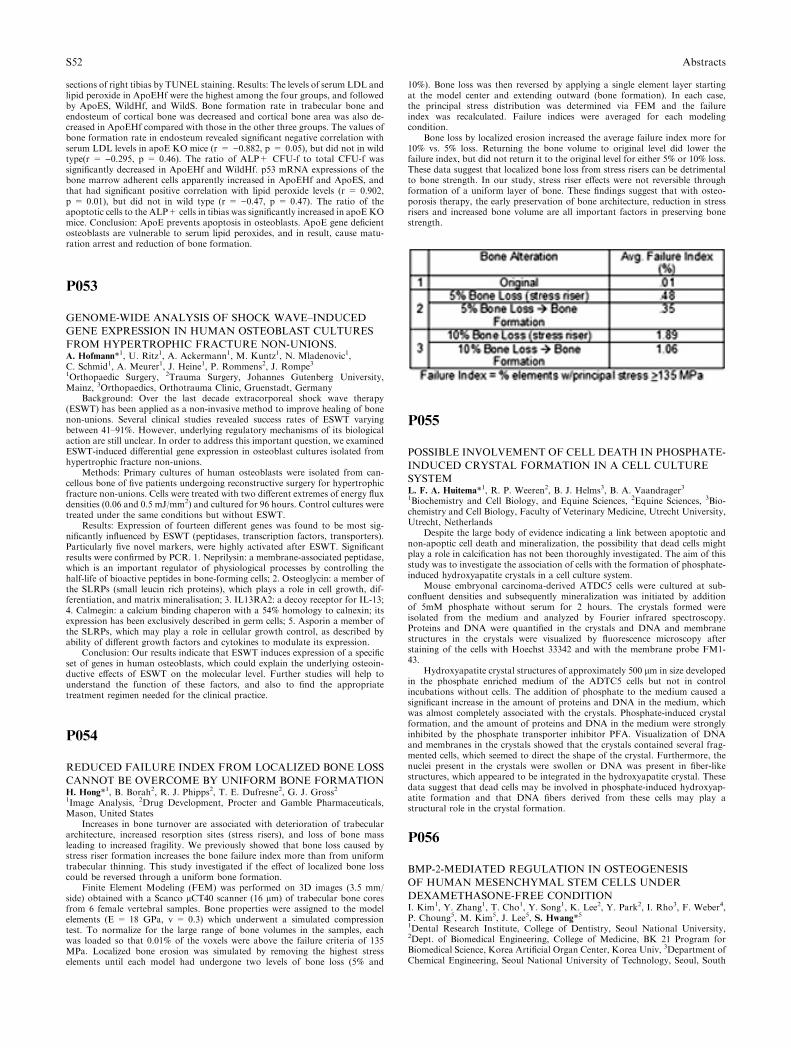
sections of right tibias by TUNEL staining. Results: The levels of serum LDL andlipid peroxide in ApoEHf were the highest among the four groups, and followedby ApoES, WildHf, and WildS. Bone formation rate in trabecular bone andendosteum of cortical bone was decreased and cortical bone area was also de-creased in ApoEHf compared with those in the other three groups. The values ofbone formation rate in endosteum revealed significant negative correlation withserum LDL levels in apoE KO mice (r = )0.882, p = 0.05), but did not in wildtype(r = )0.295, p = 0.46). The ratio of ALP+ CFU-f to total CFU-f wassignificantly decreased in ApoEHf and WildHf. p53 mRNA expressions of thebone marrow adherent cells apparently increased in ApoEHf and ApoES, andthat had significant positive correlation with lipid peroxide levels (r = 0.902,p = 0.01), but did not in wild type (r = )0.47, p = 0.47). The ratio of theapoptotic cells to the ALP+ cells in tibias was significantly increased in apoE KOmice. Conclusion: ApoE prevents apoptosis in osteoblasts. ApoE gene deficientosteoblasts are vulnerable to serum lipid peroxides, and in result, cause matu-ration arrest and reduction of bone formation.
P053
GENOME-WIDE ANALYSIS OF SHOCK WAVE–INDUCED
GENE EXPRESSION IN HUMAN OSTEOBLAST CULTURES
FROM HYPERTROPHIC FRACTURE NON-UNIONS.A. Hofmann*1, U. Ritz1, A. Ackermann1, M. Kuntz1, N. Mladenovic1,C. Schmid1, A. Meurer1, J. Heine1, P. Rommens2, J. Rompe31Orthopaedic Surgery, 2Trauma Surgery, Johannes Gutenberg University,Mainz, 3Orthopaedics, Orthotrauma Clinic, Gruenstadt, Germany
Background: Over the last decade extracorporeal shock wave therapy(ESWT) has been applied as a non-invasive method to improve healing of bonenon-unions. Several clinical studies revealed success rates of ESWT varyingbetween 41–91%. However, underlying regulatory mechanisms of its biologicalaction are still unclear. In order to address this important question, we examinedESWT-induced differential gene expression in osteoblast cultures isolated fromhypertrophic fracture non-unions.
Methods: Primary cultures of human osteoblasts were isolated from can-cellous bone of five patients undergoing reconstructive surgery for hypertrophicfracture non-unions. Cells were treated with two different extremes of energy fluxdensities (0.06 and 0.5 mJ/mm2) and cultured for 96 hours. Control cultures weretreated under the same conditions but without ESWT.
Results: Expression of fourteen different genes was found to be most sig-nificantly influenced by ESWT (peptidases, transcription factors, transporters).Particularly five novel markers, were highly activated after ESWT. Significantresults were confirmed by PCR. 1. Neprilysin: a membrane-associated peptidase,which is an important regulator of physiological processes by controlling thehalf-life of bioactive peptides in bone-forming cells; 2. Osteoglycin: a member ofthe SLRPs (small leucin rich proteins), which plays a role in cell growth, dif-ferentiation, and matrix mineralisation; 3. IL13RA2: a decoy receptor for IL-13;4. Calmegin: a calcium binding chaperon with a 54% homology to calnexin; itsexpression has been exclusively described in germ cells; 5. Asporin a member ofthe SLRPs, which may play a role in cellular growth control, as described byability of different growth factors and cytokines to modulate its expression.
Conclusion: Our results indicate that ESWT induces expression of a specificset of genes in human osteoblasts, which could explain the underlying osteoin-ductive effects of ESWT on the molecular level. Further studies will help tounderstand the function of these factors, and also to find the appropriatetreatment regimen needed for the clinical practice.
P054
REDUCED FAILURE INDEX FROM LOCALIZED BONE LOSS
CANNOT BE OVERCOME BY UNIFORM BONE FORMATIONH. Hong*1, B. Borah2, R. J. Phipps2, T. E. Dufresne2, G. J. Gross21Image Analysis, 2Drug Development, Procter and Gamble Pharmaceuticals,Mason, United States
Increases in bone turnover are associated with deterioration of trabeculararchitecture, increased resorption sites (stress risers), and loss of bone massleading to increased fragility. We previously showed that bone loss caused bystress riser formation increases the bone failure index more than from uniformtrabecular thinning. This study investigated if the effect of localized bone losscould be reversed through a uniform bone formation.
Finite Element Modeling (FEM) was performed on 3D images (3.5 mm/side) obtained with a Scanco lCT40 scanner (16 lm) of trabecular bone coresfrom 6 female vertebral samples. Bone properties were assigned to the modelelements (E = 18 GPa, v = 0.3) which underwent a simulated compressiontest. To normalize for the large range of bone volumes in the samples, eachwas loaded so that 0.01% of the voxels were above the failure criteria of 135MPa. Localized bone erosion was simulated by removing the highest stresselements until each model had undergone two levels of bone loss (5% and
10%). Bone loss was then reversed by applying a single element layer startingat the model center and extending outward (bone formation). In each case,the principal stress distribution was determined via FEM and the failureindex was recalculated. Failure indices were averaged for each modelingcondition.
Bone loss by localized erosion increased the average failure index more for10% vs. 5% loss. Returning the bone volume to original level did lower thefailure index, but did not return it to the original level for either 5% or 10% loss.These data suggest that localized bone loss from stress risers can be detrimentalto bone strength. In our study, stress riser effects were not reversible throughformation of a uniform layer of bone. These findings suggest that with osteo-porosis therapy, the early preservation of bone architecture, reduction in stressrisers and increased bone volume are all important factors in preserving bonestrength.
P055
POSSIBLE INVOLVEMENT OF CELL DEATH IN PHOSPHATE-
INDUCED CRYSTAL FORMATION IN A CELL CULTURE
SYSTEML. F. A. Huitema*1, R. P. Weeren2, B. J. Helms3, B. A. Vaandrager31Biochemistry and Cell Biology, and Equine Sciences, 2Equine Sciences, 3Bio-chemistry and Cell Biology, Faculty of Veterinary Medicine, Utrecht University,Utrecht, Netherlands
Despite the large body of evidence indicating a link between apoptotic andnon-apoptic cell death and mineralization, the possibility that dead cells mightplay a role in calcification has not been thoroughly investigated. The aim of thisstudy was to investigate the association of cells with the formation of phosphate-induced hydroxyapatite crystals in a cell culture system.
Mouse embryonal carcinoma-derived ATDC5 cells were cultured at sub-confluent densities and subsequently mineralization was initiated by additionof 5mM phosphate without serum for 2 hours. The crystals formed wereisolated from the medium and analyzed by Fourier infrared spectroscopy.Proteins and DNA were quantified in the crystals and DNA and membranestructures in the crystals were visualized by fluorescence microscopy afterstaining of the cells with Hoechst 33342 and with the membrane probe FM1-43.
Hydroxyapatite crystal structures of approximately 500 lm in size developedin the phosphate enriched medium of the ADTC5 cells but not in controlincubations without cells. The addition of phosphate to the medium caused asignificant increase in the amount of proteins and DNA in the medium, whichwas almost completely associated with the crystals. Phosphate-induced crystalformation, and the amount of proteins and DNA in the medium were stronglyinhibited by the phosphate transporter inhibitor PFA. Visualization of DNAand membranes in the crystals showed that the crystals contained several frag-mented cells, which seemed to direct the shape of the crystal. Furthermore, thenuclei present in the crystals were swollen or DNA was present in fiber-likestructures, which appeared to be integrated in the hydroxyapatite crystal. Thesedata suggest that dead cells may be involved in phosphate-induced hydroxyap-atite formation and that DNA fibers derived from these cells may play astructural role in the crystal formation.
P056
BMP-2-MEDIATED REGULATION IN OSTEOGENESIS
OF HUMAN MESENCHYMAL STEM CELLS UNDER
DEXAMETHASONE-FREE CONDITIONI. Kim1, Y. Zhang1, T. Cho1, Y. Song1, K. Lee2, Y. Park2, I. Rho3, F. Weber4,P. Choung5, M. Kim5, J. Lee5, S. Hwang*51Dental Research Institute, College of Dentistry, Seoul National University,2Dept. of Biomedical Engineering, College of Medicine, BK 21 Program forBiomedical Science, Korea Artificial Organ Center, Korea Univ, 3Department ofChemical Engineering, Seoul National University of Technology, Seoul, South
S52 Abstracts

Korea, 4Lab. for Osteology, Dept. of Craniomaxillofacial Surgery, UniversityHospital Zuerich, Zuerich, Switzerland, 5Department of Oral and MaxillofacialSurgery, College of Dentistry, Seoul National University, Seoul, South Korea
Osteogenic effect of bone morphogenic protein (BMP)-2 on human mesen-chymal stem cells (hMSCs) is controversal. In this study, we investigated thecapacity of BMP-2 alone as osteogenic inducer and related signaling pathway inhMSCs cultured from bone marrow extract of healthy providers, aged from 18-55 years. All hMSCs of tested ten persons showed significant osteogenic responseto BMP-2 as determined by alkaline phosphatase (ALP) activity. BMP-2 up-regulated other genes associated with osteogenesis such as osteopontin and os-terix as well as IGF-1 and BMP-2. However, in vitro matrix mineralization wasaccelerated only in presence of Dex, and BMP-2 showed very weak effectcompared to Dex. The extracellular secretion of osteocalcin was not increased bythe continuous treatment of BMP-2 and inhibited by Dex at day 21 in EIA assay.BMP-2 did not affect cell proliferation, whereas Dex inhibited proliferation tothe extent of 30% of the control. In immunofluorescence with anti-Cbfa1 anti-body, BMP-2 induced the prompt nuclear translocation and maintained theincreased expression of Cbfa1 with the passage of time, in comparision withcytosolic localized and weak expression of Cbfa1 in control or Dex-treatedcondition. SB203580, inhibitor of p38 kinase, significantly decreased ALPactivity as well as the expression of Cbfa1, especially in BMP-2 treated condi-tion. In conclusion, BMP-2 induced ALP activity and other BMP-regulatedgenes in the osteogenesis of hMSCs whereas it failed the stimulation of miner-alization which was dependent on Dex. Cbfa1 participated in BMP-2 signalingthrough nuclear translocalization with increased expression and p38 kinasemodulated the expression of Cbfa1. Considering on the different function ofBMP-2 and Dex in hMSC, our results may give a guidance for optimizing theefficacy of clinical outcome with BMP-2.
Key words: bone morphogenic protein (BMP)-2, human mesenchymal stemcells (hMSCs), osteogenesis, dexamethasone (Dex), Cbfa1, p38 kinase
References1. Pittenger, M. F., Mackay, A. M., Beck, S. C., Jaiswal, R. K., Douglas, R.,
Mosca, J. D., Moorman, M. A., Simonetti, D. W., Craig, S., and Marshak, D.R. (1999). Science 284, 143–7.
2. Osyczka, A. M., Diefenderfer, D. L., Bhargave, G., and Leboy, P. S.(2004). 176, 109–19.
3. . Shui, C., Spelsberg, T. C., Riggs, B. L., and Khosla, S. (2003). J BoneMiner Res 18, 213–21.
P057
INORGANIC PHOSPHATE STIMULATES TRANSCRIPTION
OF MATRIX GLA PROTEIN GENE IN GROWTH PLATE
CHONDROCYTES THROUGH THE ERK SIGNALING
PATHWAYM. Julien*1, D. Magne2, A. Clochard1, M. Rolli-Derkinderen3, O. Chassande4,C. Cario-Toumaniantz3, Y. Cherel5, P. Weiss6, J. Guicheux61Research Center on Osteoarticular and Dental Tissue Engineering, INSERMEMI9903, Nantes, 2Universite du Littoral, Cote d’Opale, LR2B-Inserm-ERI002, Boulogne-sur-mer, 3Faculte des Sciences, Institut du Thorax, INSERMU533, Nantes, 4Biomateriaux et Reparation Tissulaire, INSERM U443, Bor-deaux, 5Ecole Nationale Veterinaire, Unite d’Anatomie Pathologique, UMR 703INRA ENVN, 6Research center on Osteoarticular and Dental Tissue, INSERMEMI9903, Nantes, France
During endochondral bone formation, extracellular inorganic phosphate(Pi) has been proposed as a key regulator of growth plate chondrocytes differ-entiation, apoptosis and extracellular matrix mineralization. It was also reportedthat Pi stimulates expression of various genes in osteoblasts. Whereas these datasuggest that Pi may act as a signaling molecule on bone-forming cells, itsmechanism of action remains largely unknown. The aims of the present workwere to determine whether Pi may regulate gene expression in growth platechondrocytes and to identify the involved signaling pathways. Murine ATDC5cell line and primary growth plate chondrocytes were used. Pi was used in theform of Na2HPO4/NaH2PO4 (pH 7.2). Messenger RNA analysis was per-formed by microarray analysis, RT-PCR and real-time quantitative PCR.Activation and role of mitogen-activated protein kinase (MAPK) signalingpathways were respectively determined by Western blotting and the use ofspecific inhibitors. Immunohistological detection of extracellular signal-regu-lated kinase 1 and 2 (ERK1/2) was performed in rib organ cultures from new-born mice. In ATDC5 cells, microarray analysis indicated that Pi stimulatedexpression of several chondrocytic markers including matrix Gla protein (MGP),an inhibitor of matrix calcification in arteries and cartilage. The stimulatoryeffect of Pi on MGP mRNA level was confirmed by RT-PCR and real-timequantitative PCR. Investigation of the involved intracellular signaling pathwaysrevealed that Pi activated ERK1/2 in ATDC5 cells and primary growth platechondrocytes, while it did not affect phosphorylation of p38 and JNK. Theactivation of ERK1/2 appeared cell-specific. Indeed, although Pi stimulatedERK1/2 in MC3T3-E1 osteoblasts and ST2 stromal cells, no ERK1/2 phos-phorylation could be detected in L929 fibroblasts or C2C12 myogenic cells.Accordingly, immunohistological detection of ERK1/2 phosphorylation in rib
growth plates revealed a marked signal only in chondrocytes and osteoblasts.Finally, a specific ERK1/2 inhibitor, UO126, blocked Pi-stimulated MGPexpression in ATDC5 cells, indicating that ERK1/2 mediates Pi effects. Thesedata demonstrate for the first time that extracellular Pi regulates MGP expres-sion in growth plate chondrocytes, thereby suggesting a key role for Pi andERK1/2 in the regulation of bone formation.
P058
REGULATION OF MINERALIZATION BY
TRANSGLUTAMINASES IN OSTEOBLAST CULTURESH. F. Al-Jallad1, Y. Nakano1, M. T. Kaartinen*11Faculty of Dentistry, McGill University, Montreal, Canada
Transglutaminases (TGs) are a group of protein-crosslinking enzymes thathave long been implicated in the formation of mineralized tissues. TGs createisopeptide bonds between lysine and glutamine residues in their substrate pro-teins. Crosslinking between protein chains results in the formation of proteinpolymers, and intra-protein crosslinking can induce conformational changes.The TG family consists of 9 genes of which TG2 and FXIIIA are expressed bychondrocytes in mineralizing cartilage. TG2 is also expressed by osteoblasts, andis found in bone, but its role is still unknown. In this study we analyzed theexpression, activity and function of TGs in differentiating osteoblasts. MC3T3-E1 (subclone 14) pre-osteoblast cultures were treated with ascorbic acid andbeta-glycerophosphate to induce cell differentiation, matrix production andmineralization. Expression of TG isoforms was analyzed by RT-PCR. TGactivity was assessed during differentiation by in vitro biochemical assays and byin situ labeling of live cell cultures. We demonstrate that, like chondrocytes,MC3T3-E1/C14 osteoblasts express two TG isoforms-TG2 and FXIIIA.Abundant TG activity was observed during cell differentiation which increasedsignificantly after thrombin treatment, a result confirming the presence ofFXIIIA in the cultures. Ascorbic acid treatment, which stimulates collagensecretion and assembly, also stimulated externalization of TG activity, likelyfrom FXIIIA which was externalized upon this treatment as analyzed by im-munofluoresence microscopy. Inhibition of TG activity in the cultures by cys-tamine resulted in complete abrogation of mineralization attributable todecreased matrix accumulation and an arrested state of osteoblast differentiationas measured by decreased levels of bone sialoprotein, osteocalcin and alkalinephosphatase. TG activity was most critical for mineralization at an early phaseof the differentiation program. In situ substrate labeling experiments duringthese early days showed that the sole TG substrate was fibronectin. Immuno-fluoresence studies showed that TG activity was required for the formation of afibronectin-collagen type I network during the early stages of matrix formationand assembly. This network, in turn, appeared to be essential for further matrixproduction and progression of the osteoblast differentiation program, and ulti-mately for mineralization. Supported by the CIHR.
P059
HARMFUL EFFECT OF FLUORIDE ON FORMATION OF
APATITE CRYSTALSM. Kakei*1, T. Sakae2, M. Yoshikawa311st. Dept. of Oral Anatomy, Meikai Univ. School of Dentistry, Sakado, 2Dept.of Histology, Embryology, Anatomy, Nihon Univ. School of Dentistry, Mat-sudo, 3Dept. of Orthodontics, Meikai Univ. School of Dentistry, Sakado, Japan
There are pros and cons about the effectiveness of fluoride application forhuman health such as prevention of dental caries. Recently, we have proposedthat fluoride ions may inhibit the crystal nucleation process during tooth andbone formation. Based on our assumption, the present study was conducted toprovide conclusive evidence that fluoride causes perforation of the enamelcrystals and to raise awareness of the harmful effects on the bone formation bytaking unnecessary fluoride ions.
In this study, the male rats, weighing approximately 50 gm, were used anddivided into two groups. For experimental group, the water containing up to 1.5ppm of F ions was given. The F-free water was given to the control group. After5–12 weeks had elapsed, the samples of tooth enamel were dissected from theanesthetized rats. Then they were subject to transmission electron microscopy.Also, we analyzed the activity of carbonic anhydrase in the developing enamelmatrix, which is a key enzyme for initiating the nucleation process.
By electron microscopy, crystal perforation of the enamel crystals was foundin the experimental rats. And, biochemical analysis showed that the activity ofcarbonic anhydrase was reduced remarkably after fluoride intake. The resultsstrongly indicate that fluoride ions are hazardous, even at low concentrations.
This work was supported by the Frontier Science Promotion Program atNihon University.
Abstracts S53
P060
ANDROGEN RECEPTOR COORDINATES BOTH OSTEO-
BLASTIC DIFFERENTIATION AND MINERALIZATIONH. Kang*1, C. Huang2, J. Chang2, K. Huang31Center for menopause and reproductive medicine research, Graduate Instituteof Clinical Medical Scie, 2Graduate Institute of Clinical Medical Sciences, ChangGung University, 3Center for menopause and reproductive medicine research,Chang Gung Memorial Hospital, Kaohsiung, Taiwan
Androgens have important effects on the human skeleton in both males andfemales. Hypogonadism in men is associated with increased bone turnover andbone loss, which is reversed after treatment with androgens. Clinical studiessuggested that combined therapy of estrogens plus androgens might enhancebone mineral density and bone mass to a more significant degree than estrogentherapy alone in postmenopausal women. Previous studies demonstrated thatcancellous bone volumes are lower in the 8-week-old androgen receptorknockout (ARKO) mice than in both female and male wt littermates. These dataindicate that AR is required for bone formation and osteoblast differentiation.To determine whether AR play a role in control of osteoblast differentiation andmineralization, we compared differentiation rates of primary calvarial cells iso-lated from wild-type and AR-null 3.5 days fetus in the presence or absence ofDHT treatment by ALP activity assay, calcium deposition and Von Kossa stain.We also generated osteoblastic MC3T3-E1 cells in which over-expression of astably integrated AR and knockdown expression of a stably AR siRNA to studythe role of AR in osteoblastic cell differentiation. Our data showed thatandrogens accelerate cell differentiation of osteoblast cells in a time and dosedependent manner. Western blot analysis showed that the expression level of ARcorrelates with the duration of androgen treatment and degree of mineralizationin osteoblasts. In addition, androgen promotes mineralization of primary calv-arial cells isolated from wild-type mice but not from ARKO mice and AR-siRNA prevented DHT-induced osteoblast differentation. To further study theroles of AR on various differentiation stages of osteoblastic cells, the mRNAexpression of AR, different stage bone marker genes and AR targeted genes fromlong-term wild-type and ARKO osteoblastic cell cultures with or withoutandrogens, were quantified using real time PCR. Our data indicates that the roleof AR on mineralization is dependent on the differentiation stage of the osteo-blasts and the duration of androgen treatment. Together, these findings stronglysuggest that the AR signaling is required for androgenic effects on bonemetabolism, osteoblast differentiation and mineralization.
P061
BONE ULTRASTRUCTURE BY RAMAN IMAGINGM. Kazanci*1, P. Roschger2, E. P. Paschalis2, K. Klaushofer2, P. Fratzl11Biomaterials, Max-Planck Institute of Colloids and Interfaces, Potsdam,Germany, 2Ludwig Boltzmann Institute of Osteology, Hanusch Hospital, Wien,Austria
Bone is a composite material with a hierarchical structure. Its strength de-pends on its structural and material properties. In the present study, Ramanmicrospectroscopic and Imaging analysis was employed to study two osteons inthe cortical bone of different age and mineral content, with a spatial resolution of� 1 lm. Spatial and temporal changes in amount of mineral and organic matrix,as well as the variation in the mineral maturity/crystallinity and carbonatecontent were determined, imaged and plotted. The results showed that theamount of mineral and organic matrix varied in a manner similar to the lamellararrangement in the cortical bone and different age packets in the trabecularbone. The cement lines in the osteonal bone exhibited the highest amount ofmineral but unchanged amount of organic components when compared to othercortical bone areas. The carbonate to phosphate ratio decreased as a function ofdistance from the Haversian canal whereas, other ratios, organic to mineral andorganic (collagen) to organic (non collagenous moieties), remained relativelyconstant. It is demonstrated that the ratios that include Amide I organic com-ponent (mainly collagen) are more sensitive to the orientation. The results of thepresent study demonstrate the versatility of the analytical technique, and provideinsights into the organization of bone tissue at the ultrastructural level.
P062
COMPLEMENTARY INFORMATION ON IN VITRO
CONVERSION OF AMORPHOUS CALCIUM PHOSPHATE
TO HA FROM RAMAN MICROSPECTROSCOPY AND WIDE
ANGLE SCATTERINGM. Kazanci1, E. P. Paschalis*2, K. Klaushofer2, P. Fratzl11Biomaterials, Max-Planck Institute of Colloids and Interfaces, Potsdam, Ger-many, 2Ludwig Boltzmann Institute of Osteology, Hanusch Hospital, Wien,Austria
Mineralized tissues consist mainly of poorly crystalline hydroxyapatite [HA,Ca10(PO4)6(OH)2]. It is important to note that it continues to be chemicallyactive long after it has been initially deposited. A heterogeneous distribution ofmineral crystallites (in terms of size, shape, and maturity) is expected to beencountered in normal bone, depending on remodeling rates. Detailed under-standing of changes in the mineral phase as the HA matures is essential foreventually providing and understanding of how normal bone achieves itsremarkable mechanical performance, how it is altered in disease, as well asmonitor the effects of therapeutic interventions. A model system for investiga-tion of the in vivo maturation of HA is available, namely, the in vitro conversionof amorphous calcium phosphate (ACP) to HA in a supersaturated solution ofcalcium ions with phosphate ions. In the present study, this system was em-ployed to correlate with the changes in chemistry and poorly crystalline HAPcrystal size/shape/habit. The results of the x-ray diffraction as well as the Ramananalyses showed that as the crystallites mature, both in \beta002 and 310directions, and both the FWHH and wavelength at maxima of the Raman peaks,change as a function of reaction extent. Monohydrogen phosphate (surfacephosphate) to phosphate (bulk phosphate) ratio is highly correlated (r2 = 0.82)to crystallinity (\beta002). Moreover, such analyses can be performed in intactbone specimens through Raman microspectroscopic and Imaging analyses witha spatial resolution of 0.6-1 micron by far superior to the one offered by othermicrospectroscopic techniques, thus potentially yielding new important infor-mation on the organization and mineral quality of normal and fragile bone.
P063
ISOLATION AND CHARACTERIZATION OF
MESENCHYMAL STEM CELLS DERIVED FROM
UMBILICAL CORD BLOODJ. Kim*1, M. KIM1, J. Park1, H. Choi21Obstetrics and Gynecology, Seoul National University Hospital, 2Obstetricsand Gynecology, Sanggye Paik Hospital, Seoul, South Korea
Background/Aims: Mesenchymal stem cells (MSCs) have the capability forrenewal and differentiation into various lineage of mesenchymal tissues, andappear to be an attractive tool in the context of tissue engineering and cell-basedtherapy. Umbilical cord blood (UCB) has been a source for hematopoietic stemcells. However, the presence of MSC in the human UCB is not clear.The pur-poses of this study were to isolate and characterize MSCs with osteogenic andadipogenic potential from UCB.
Methods: MSCs were isolated using a density gradient centrifugation andextensive subcultivation from UCB. The proliferation capability, cell cycle,cytochemical markers, and immunopheotype of these UCB derived MSCs weremeasured by colorimetric asay, cytochemical stain, and fluorescence-acivatedcelll sorter. The transcript of osteoblast-specific markers, akaline phospha-tase(AKP), and calicum deposit were analyzed by reverse transcription-poly-merase reaction, cytochemical stain, colorimetric asay, atomic absorptiometry inMSCs cultured for 1-4weeks in osteogenic media, and by MSCs exposed inadipogenic media for weeks were stained by Oilred.
Results: More than 85% of MSCs were in the G0/G1 phase. MSCs werepositive for periodic acid-Schiff, and a-naphthyl acetate esterase activity, butnegative for acid phosphatase, sudan black-B, and AKP activities. MSCs ex-pressed CD13, CD29, CD 44, CD49e, CD51, CD54, CD 90, SH2, SH3, but didnot express CD11b, CD14, CD31, CD34, CD45, CD49d, CD106, CD133,CD144, CD146, CD163, CD166, and Stro-1. When MSCs were cultured inosteogenic media for 1-4weeks, they expressed transcript of ostoblastic specificmarkers: core binding factor a1, AKP, and procollagen type I. During culture of2–4 weeks, AKP activity measured by AKP cytochemical staning and quantifi-cation increased significantly and the deposition of a calcified matrix becameevident by von Kossa staining and calcium quantification. Exposure of MSCs toadipogenic media resulted in morphological change stained by Oil Red O.
Conclusions: UCB contain MSCs with osteogenic and adipogenic differen-tiation potential, and UCB may serve as an potential source of MSCs to beutilized in cell therapy for various diseases.
P064
OSTEOGENESIS VERSUS CHONDROGENESIS BY BMP-2 AND
BMP-7 IN ADIPOSE STEM CELLSM. Knippenberg1, M. N. Helder2, B. Zandieh Doulabi1, A. Vatsa*1,P. I. J. M. Wuisman2, J. Klein-Nulend11Oral Cell Biology, ACTA- Uva and Vrije Universiteit, 2Orthopaedic Surgery,VU University Medical Center, Amsterdam, Netherlands
Background: Bone morphogenetic proteins (BMP) initiate, promote, andmaintain chondrogenesis and osteogenesis. BMP-2 and BMP-7 induce differ-entiation of multipotential mesenchymal cells into both osteochondrogeniclineage cells and osteoblast precursor cells, and are therefore widely used in bonetissue engineering. Adipose tissue has recently been described as a source for
S54 Abstracts
adult mesenchymal stem cells, which can be induced by osteogenic or chon-drogenic factors to differentiate along the osteogenic or chondrogenic pathway.
Objective: To compare the effect of BMP-2 and BMP-7 on osteogenic and/orchondrogenic differentiation of adipose tissue-derived mesenchymal stem cells(AT-MSCs). We hypothesize that BMP-2 induces an osteogenic, and BMP-7 achondrogenic phenotype in AT-MSCs.
Methods: AT-MSCs were obtained from goat adipose tissue, and incubatedwith 10 ng/ml BMP-2 or BMP-7 for 15 min. Expression of osteogenic genesrunx-2 and osteopontin (OPN), and chondrogenic genes aggrecan and biglycanwas studied 4 and 14 days after BMP-treatment.
Results: Fifteen min of BMP-2 stimulated gene expression of the early oste-ogenic transcription factor runx-2, and the bone extracellular matrix protein OPNin AT-MSCs at day 4, whereas BMP-7 did not. Fifteen min of BMP-2 and BMP-7stimulated expression of the extracellular matrix component biglycan at day 4.Fifteenmin ofBMP-7 inhibited gene expression of runx-2,OPN, andbiglycan, andstimulated aggrecan gene expression at day 14, whereas BMP-2 did not.
Conclusion: These data suggest that BMP-2 treatment for only 15 min iscapable of inducing osteogenic differentiation in AT-MSCs, whereas BMP-7may stimulate AT-MSCs towards a more chondrogenic phenotype. Therefore,AT-MSCs triggered with BMP-2 or BMP-7 likely provide a feasible tool forbone and cartilage tissue engineering.
Acknowledgements: This research was supported by the Dutch TechnologyFoundation (STW, grant # VPG.5935).
P065
POLYSACCHARIDES AS NANOCOATINGS OF BONE
IMPLANTSH. Kokkonen*1, J. Ilvesaro2, M. Morra3, H. A. Schols4, M. Nagel5,J. Tuukkanen11Department of Anatomy and Cell Biology, University of Oulu, Oulu, Finland,2Department of Medicine, Division of Hematology-Oncology, University ofAlabama, Birmingham, United States, 3Nobil Bio Ricerche, Villafranca dAsti,Italy, 4Laboratory of Food Chemistry, Wageningen University, Wageningen,Netherlands, 5Department of Biomaterials and Biocompatibility, University ofTechnology of Compiegne, Compiegne, France
The aim of our study is to assess interactions between biomaterial and bonecells. Plant-derived carbohydrate molecules, pectins, were used as implant coatingmaterial. Pectins can be isolated and enzymatically treated to modified hairyregions (MHRs), whose composition, charge, and wettability can be controlled.
The biocompatibility of the implant depends on the attachment and differ-entiation of bone cells growing on the material, which is influenced by theadsorption of proteins in surrounding fluids onto the implant surface depen-dently on the surface properties. Therefore it would be important to developbiocompatible nanocoatings for bone implants. Pectins are potential candidatesfor this application because of the easy controlling of their structure and possibleanti-inflammatory properties.
Osteoclastic and osteoblastic cells were cultured on tissue culture polystyrene(TCPS) dishes coated by covalent linking of different MHRs of apple pectins.Uncoated TCPS or bovine bone slices were used as positive controls. A mixedprimary bone cell population was isolated from rat pup long bones and culturedfor 48 h, after which the cells were stained with tartrate-resistant acid phos-phatase (TRACP). MC3T3-E1 subclone IV preosteoblasts were cultured for 48 hand stained for paxillin, which indicates focal adhesions.
Bone cells prefer MHRB to MHRA. MHRB contains shorter side chainsthan MHRA, and thus provides more aqueous interface with cells promoting celladhesion and spreading, whereas MHRA can resist cell attachment more be-cause of the sterically hindered protein adsorption properties. On MHRA theoverall numbers as well as proportions of TRACP-positive cells were diminishedcompared with MHRB or bone. On MHRB and bone tissue, the amount ofTRACP+ cells were similar, even though the proportion of TRACP+ cells withthree or more nuclei was greater on bone. In addition to bone cell differentiation,MC3T3-E1 cells provided information about cell attachment and spreading:focal adhesions were clearly detected on MHRB and pure TCPS but not onMHRA.
These results demonstrate the possibility to modify pectin nanocoatings inorder to improve bone cell attachment.
EC Project PectiCoat NMP4-CT-2005-517036
P066
EFFECTS OF CHANGES IN MECHANICAL LOADING
ON ENDOCHONDRAL BONE FORMATION IN
HINDLIMB-SUSPENDED RATSS. Kong*1, S. Kim2, H. Kim2
1Brain Korea 21 Project for Medical Science, Orthopaedic Surgery, 2Ortho-paedic Surgery, College of Medicine Yonsei University, Seoul, South Korea
To examine the effects of changes in mechanical loading on endochondralbone formation, a simulated rat model of weightlessness was introduced and thechanges in the growth plate were evaluated.
Unloading condition on the hindlimb of Sprague-Dawley rats was createdby fixing a tail and lifting the hindlimb. Six rats aged 6 weeks old were assignedto each group of unloading, reloading, and control groups of unloading orreloading. Unloading was maintained for three weeks, and then reloading wasapplied for another one week thereafter. Histomorphometry for the assessmentof vertical length of the growth plate, 5-bromo–deoxyuridin(BrdU) immuno-histochemistry for cellular kinetics, and biotin nick end labeling TUNEL assayfor chondrocytes in the growth plate were performed.
The vertical length of the growth plate and the proliferative potential ofchondrocytes were decreased in the unloading group than those of controlgroups. Inter-group differences were more significant in the proliferative andhypertrophic zones. Reloading increased the length of growth plate and prolif-erative potential of chondrocytes as evidenced by increase of the ratio of positiveBrdU stained cells. However, apoptotic changes in the growth plate were notaffected by the alterations of weight bearing.
Alterations in the weight bearing induced changes in the chondrocyticproliferative potential of the growth plate and have no effects on the apoptosisoccurred. This may suggest that deprived weight bearing due to various clinicalsituations hamper normal longitudinal bone growth, and further studiesregarding the factors for reversibility of chontrocytic proliferation upon variablemechanical stresses are needed.
P067
LONG CHAIN POLYUNSATURATED FATTY ACIDS AFFECT
CYTOKINE AND TYROSINE KINASE ACTIVITY INMC3T3-E1
PRE-OSTEOBLAST CELLSM. C. Kruger*1, L. M. Fray1, W. H. Chua1, L. Fremon11Institute of Food, Nutrition and Human Health, Massey University, Palmer-ston North, New Zealand
There is increasing evidence that a diet deficient of certain PUFAs can in-duce bone loss, while dietary supplementation of some PUFAs has been shownto be beneficial for bone. The molecular mechanisms whereby PUFAs affectosteoclastogenesis and bone formation are not fully understood and are cur-rently under investigation.
Nontransformed MC3T3-E1 (subclone 4) mouse calvaria fibroblasts, knownto differentiate into osteoblasts, were grown in alpha-MEM with ribonucleosidesand deoxyribonucleosides supplemented with 10% heat-inactivated foetal calfserum and 25ug/ml gentamicin. Cells were seeded and allowed to establish asmonolayers overnight at 37�C with 5% CO2 before treatment with either 100ng/ml lipopolysaccharide(LPS) or fatty acid in the presence of LPS. Arachidonicacid (AA), eicosapentaenoic acid (EPA), docosahexaenoic acid (DHA) and c-linoleic acid (GLA), after initial dilution in 100% ethanol to 100 mg/ml werediluted in medium and added to cells at a 20ug/ml final concentration. Controlwells contained either medium only, 0.02% ethanol, or LPS only. Cells weretested for the production of IL-6, tyrosine kinase (TyrK) or tyrosine phosphatase(TyrP). Supernatants were collected for the IL-6 assay after 48 hours. Cells forthe tyrosine assays were scraped off the flasks after 2 hours, treated withextraction buffers and supernatants collected after centrifuging at 10,000g. Theeffect of fatty acids on IL-6 production in MC3T3-E1 were assessed by com-paring test samples with the LPS controls using independent samples t-tests. Theeffect of fatty acids on TyrK and TyrP were compared using the Kruskal-Wallisnonparametric test.
AA and GLA raised LPS induced IL-6 secretion significantly while EPA andDHA had no effect. EPA suppressed TyrK activity whilst raising TyrP activity,though not significantly. TyrK activity is associated with osteoclast activity, viaprostaglandin E2 which is associated with increased bone resorption, whilstTyrP is associated with bone maintenance. IL-6 is also associated with boneresorption. By affecting interleukin secretion and the balance between tyrosinekinase and phosphatase activity, PUFAs may modulate the balance betweenbone resorption and formation.
P068
MEMBRANE-ANCHORED NEUTRAL ENDOPEPTIDASE
NEPRILYSIN (NEP) IS UP-REGULATED IN HUMAN
OSTEOBLAST CULTURES AFTER EXTRACORPOREAL
SHOCK WAVE THERAPYM. Kuntz*1, A. Hofmann1, U. Ritz1, A. Ackermann1, N. Mladenovic1,C. Schmid1, A. Meurer1, J. Heine1, P. Rommens2, J. Rompe31Orthopaedic Surgery, 2Trauma Surgery, Johannes Gutenberg University,Mainz, 3Orthopaedics, Orthotrauma Clinic, Gruenstadt, Germany
Abstracts S55
Background: Extracorporeal shock wave therapy (ESWT) has been exten-sively used as a non-invasive method for the treatment of a number of muscu-loskeletal conditions. Previous studies revealed specific ESWT-inducedexpression changes of regulatory factors in osteoblast cultures which induce localcell growth and differentiation. However, it remains unclear whether ESWT-associated pain relief may be due to the local change of the neuropeptideexpression and/or degradation. In the present report we investigated the ESWT-induced expression of NEP which is known to control the half-life of severalgrowth factors and neuropeptides.
Methods: Primary cultures of human osteoblasts were isolated from can-cellous bone of five patients undergoing reconstructive surgery for hypertrophicfracture non-unions. Cells were treated with two different extremes of energy fluxdensities (0.06 and 0.5 mJ/mm2) and cultured for 96 hours. Control cultureswere treated under the same conditions but without ESWT. Global geneexpression profile was performed via Affymetrix U133A-microarray analysis.Expression of NEP was finally confirmed by qPCR.
Results: Expression of NEP was highly up-regulated after ESWT (0.06mJ/mm1=2 )10.5-fold; 0.5mJ/mm1=2 ) 9.1-fold; p < 0.0005). NEP plays animportant role in nervous and peripheral tissues, as it controls several peptide-signaling events at the cell surface. Most important substrates of NEP includethe osteogenic growth peptide (OGP), osteostatin, PTH, calcitonin, as well as theneuropeptides CGRP and Substance P. CGRP is regarded as a primary afferentpeptide with the strongest evidence of a role in pain perception.
Conclusion: ESWT induces the expression of NEP. Further studies will helpto understand the role of NEP in fracture healing and pathophysiology of bonepain.
P069
EXPERIMENTAL BONE CELL MODEL TO STUDY GENE
EXPRESSION IN THE DIFFERENTIATION PATHWAY AFTER
X-IRRADIATIONP. Lau*1, C. E. Hellweg1, C. Baumstark-Khan11German Aerospace Center, Institute of Aerospace Medicine, Cologne,Germany
Osteoblastic differentiation is a complex process including cell proliferation,mineralization and extracellular matrix maturation. The potential of cells todifferentiate into bone-forming cells relies upon molecular regulation and isgoverned by multiple transcription factors, growth regulatory factors and hor-mones as well as exogenous factors like mechanical stress or radiation. The goalof our studies is establishing a cell system, in which complex mechanisms like celldifferentiation triggered by environmental factors, in this regard radiation, canbe studied in detail. Understanding the mechanisms that control the differenti-ation pathway and its regulation, especially in combination with irradiation, willcontribute to clarify osteoblastic and osteocyte function particularly regardingosteogenic bone diseases. Therefore studies were conducted to determine if theosteoblastic differentiation cascade is altered under modified culture conditionsand / or by X-ray irradiation. The bone cell model consists of the osteocyte cellline MLO-Y4, the osteoblast cell line OCT-1 and the subclones 4 and 24 of theosteoblast cell line MC3T3-E1 expressing several differentiation potentials. Thesurvival experiment after X-irradiation revealed no significant differences in X-ray sensitivity for the four cell lines. Bone specific marker gene expression wasmonitored for bone Gla protein (osteocalcin), osteopontin and alkaline phos-phatase during in vitro subculture using standard RT-PCR. Osteoblastic dif-ferentiation was favoured by subculturing the cells in osteogenic medium. Theexpression level of the selected markers was consistent with the expected phe-notype. Real-time reverse transcription PCR (qRT-PCR) was used for quanti-fication of mRNA levels of osteocalcin. The presence of functional protein levelswere investigated by western blot analyses and immunofluorescent techniquesduring bone differentiation and mineralization. To verify the mineralization, theextracellular matrix was analysed histochemically by Alizarin Red S staining.The most intense staining of calcium deposits after culture in osteogenic mediumwas observed in the cell line MC3T3-E1 subclone 4.
P070
STRO-1 EXPRESSION IS ALTERED DURING OSTEOBLASTIC
DIFFERENTIATION OF HUMAN MESENCHYMAL STEM
CELLSS. T. Lehtonen*1, P. Hakso1, P. P. Lehenkari11Department of Surgery, Clinical Research Center, University of Oulu, Oulu,Finland
Human mesenchymal stem cells (hMSCs) derived from bone marrow arecapable in differentiating into osteoblasts and certain other cell types. They arelargely responsible for tissue regeneration and provide attractive source ofmaterial for future cell transplantation therapies. Samples obtained from bone
marrow, however, contain in addition to MSCs also hematopoietic stem cellsand number of other cell types. Therefore, enrichment and characterisation ofstem cells before transplantation would presumably provide even more efficientenhancement of tissue repair.
We have cultured in the laboratory conditions hMSCs derived from bonemarrow and induced in vitro their osteogenesis. Dexamethasone is essentialcomponent for in vitro differentiation of osteoblasts and we hypothesised thatone of its mechanisms is that it activates hMSCs so that osteogenesis is favoured.We analysed the expression of selected cell surface antigens by FACS or MACSanalysis. Our results show that we are able to exclude hematopoietic stem cellswhile culturing hMSCs as shown by CD34 analysis. Stro-1 is considered as aMSC marker and its expression is induced in vitro by adding dexamethasone tothe culture medium. Stro-1 positivity is induced during the first three weeks ofosteogenesis but reduced during late osteogenesis so that after five weeks onlyfew stro-1 positive cells are present. Our results also suggest that CD105 andstro-1 positivity partially co-localise to the same cells but is distinct from CD117positivity. We conclude that dexamethasone affects remarkably the properties ofhMSCs.
P071
RESPONSE OF MESENCHYMAL STEM CELL DERIVED
OSTEOBLASTS TO SEX HORMONES IS INFLUENCED BY
ESTROGEN RECEPTOR GENOTYPEH. Leskela*
1, A. Olkku2, S. Lehtonen1, A. Mahonen2, J. Koivunen3,M. Turpeinen4, J. Uusitalo4, O. Pelkonen4, L. Kangas5, P. Lehenkari11Clinical Research Center, Department of Surgery, University of Oulu, Oulu,2Department of Medical Biochemistry, University of Kuopio, Kuopio,3Department of Anatomy and Cell Biology, 4Department of Pharmacology andToxicology, University of Oulu, Oulu, 5Hormos Medical, Ltd, Turku, Finland
Hormone replacement therapy is effectively used to prevent postmenopausalbone loss, however, variation in response to the therapy is frequently seen amongindividuals. In addition, direct effects of sex steroids on human marrow stromalcells have been reported to be small andvary depending on the donor. The effects ofestradiol and testosterone in human bone marrow derived mesenchymal stem cellcultures from female and male patients of various ages were studied. The differ-entiation of mesenchymal stem cells into osteoblasts and activity of the cells werequantified in vitro by measuring alkaline phosphatase activity and calciumdeposition, respectively. Responses to sex hormones were small and varieddepending on the donor. To explain this individual variation, we hypothesized thatthe estradiol effect is influenced by the estrogen receptor alpha PvuII polymor-phism. Restriction fragment-length polymorphism was represented as P or non-P,with the P signifying the absence of the restriction site. The data shown here,suggests, that higher spontaneous recruitment and activation of osteoblasts frommesenchymal stem cells is associated with the presence of the P allele in females,whereas higher response to sex steroids treatment is associated with the non-Pallele. These results could explain the contradictory effects of estradiol on osteo-blasts in vitro and might also provide new insights to understand the variableresponses to hormone replacement therapy.
P072
THE MATERIAL LITAR USED IN THE COURSE OF
REPLACING BIG BONE TISSUE DEFECTSS. D. Litvinov*1, A. N. Mitroshin2, A. S. Kibitkin21Faculty of Chemistry, Samara State Technical University, Samara, 2Facultyof General Medicine, Penza State Medical Institute, Penza, Russian Federa-tion
It is common knowledge that all the universally adopted methods forsolving the problems for filling the big defects and the cavities of the bonetissue are the methods of the application of the natural and artificial mate-rials.The most widespread method is the method of the application of freebone autotransplants the most important advantages of which are an absolutetissular compatibility and the lack of the reaction ‘‘the host-transplant’’. Thedrawback of the autotransplants are a restricted replaceable volume even inthe case of polyfocal bone taking, difficulties in the course of simulating thedefect, the inevitability of breaching the growth zone in pediatric surgery.Thisproblem can be solved in the course of using the material which during itslocation in recipient’s body can be converted into its own bone tissue. Thematerial LitAr is a hydroxyapatite – collagen composite material differingfrom other similar ones by its minimum biodegradation time which is equalto 15–20 days. The material LitAr consists of calcium hydroxophosphateintroduced into the xenogenic collagen bulk by the special method which hasbeen protected by the RF patent. The material LitAr was used in the courseof treating the 12 patients having different nosologies. The composite materialwas used separated as well as with the use of the autotransplants (6 obser-vations). The treatment of the patients with the help of the method of
S56 Abstracts
replacing the bone tissue defects and with the use of the material LitAr hasmade it possible to achieve filling the big defects by the sound bone tissue.The merit of the material LitAr is the possibility of its use for the purpose ofreplacing the big bone tissue defects without resort to the use of the bonebank. Thanks to the properties of the implant LitAr the sound bone tissueforms for the time periods which are far shorter then it is generally appre-ciated, and this fact, in its turn, reduces the expenditures for the treatmentand improves the life quality of the patients having the illnesses which requirereplacing big bone tissue defects.
P073
MATHEMATICAL DESCRIPTION OF BONE FORMATION
IN RATSV. G. Koveshnikov1, V. I. Luzin*1, O. V. Andrieieva1, D. A. Astrakhantsev11Department of Normal Anatomy, State medical university, Luhansk, Ukraine
Background/aims: Nowadays there are few criteria to describe the changesof bone form in the process of growth. Thus, we set our work to give a math-ematical description of the anterior tibial crest curvature and propose theassessment criteria for its changes.
Methods: For the experiment we used 84 intact rats of 3 age groups with theinitial mass of 70–75 grams, 160–180 grams and 300–320 grams respectively. Theobservation terms were 7, 15, 30 and 90 days. One tibia was placed under ste-reomicroscope in lateral view and using Cartesian system, we defined the pointson the anterior crest on the Y-axis pitching 2.5 mm. The data obtained wereapproximated as the fourth power polynomial. Using MathCad 7.0 for Windowswe calculated the polynomial factors, the maximum curvature radiuses and theirattach points.
Results. The anterior edge of the tibia as a rule possesses two maximumcurvature radiuses - the proximal and the distal. The proximal curvature radiusis attached to the initial point of the curve, is less than 10–2 mm and shows noage changes. The distal radius dynamically changes with aging. In intact animalsits value changed like the following: in growing animals it decreased from4.81 ± 0.32 mm to 3.39 ± 0,08 mm, in adult it also decreased from 3.40 ± 0.11mm to 3.17 ± 0.11 mm and in aged it decreased from 3.29 ± 0.16 mm to2.54 ± 0.10 mm i.e. the value decreased with aging.
The attach point of the distal curvature radius in growing animals was at16.2 ± 17.07 mm from the initial point of the curve, in adults it somewhatdislocated distally – up to 18.03 ± 20.93 mm and in aged animals it dislocated inproximal direction – 12.70 ± 14.67 mm from the initial point of the curve.Probably the proximal position of the curvature radius is preferable for keepingthe structural strength of the bone as during intensive longitudinal growth asduring involution changes.
Conclusions: calculation of the maximum curvature radius and its attachpoint of tibia may serve as a mathematical description criterion of the bonegrowth and formation. These values reflect age changes in bones and may cor-relate with the structural strength of the bone.
P074
EFFECTS OF FGF-2 RELEASE FROM A SYNTHETIC POLY-
MER ON BONE MASS AND MICROARCHITECTUREG. Mabilleau1, E. Aguado1, M. F. Basle1, D. Chappard*11INSERM, EMI 0335, Faculty of medicine, Angers, France
Effects of fibroblast growth factors family members (FGFs) on the con-trol of differentiation genes in osteoblast have been recently recognized [1].Local delivery by a synthetic polymer could represent a good way ofadministration in patients needing bone reconstruction. We have used a co-polymer of poly(2-hydroxyethyl methacrylate-co-vinyl pyrrolidone)-p(HEMA-co-VP)- for the controlled release of FGF-2. Polymer cylinders were im-planted in the distal femoral extremity of 24 female New Zealand rabbits.Animals were randomized in two groups (6 rabbits/group): p(HEMA-co-VP)loaded with vehicle, constituting the control group and p(HEMA-co-VP)loaded with FGF-2 (250 ng/g of polymer), constituting the FGF-2 group.Rabbits were sacrificed 2 and 3 months post-surgery. Femoral ends weredissected and fixed in formalin. Bone samples were examined with a micro-computed tomograph for 3D microarchitecture (BV/TV, Tb.Pf, SMI, Tb.Th,Tb.N, Tb.Sp). Bone samples were embedded in methylmethacrylate andlongitudinal sections were performed. At two months post surgery, animalsfrom the FGF-2 group evidenced significant higher values for BV/TV andTb.N. Significant decreases of Tb.Pf and SMI were evidenced, indicating abetter connected network and a bone remodeling. No significant differencewas noted for Tb.Th and Tb.Sp. FGF-2 group exhibited woven bone atdistance of the implantation site and the quantity was higher than controlgroup (+ 439%). At 3 months post-surgery, control group exhibited higher
values of BV/TV (+ 65.8%) and Tb.N (63%) than control group at 2 months.No significant change was noted for Tb.Pf, SMI and Tb.Sp. Woven bonearea decreased significantly ()49.8%). No noticeable difference was observedbetween control and FGF-2 groups at 3 months post-surgery. FGF-2 groupexhibited a significant decrease of the quantity of woven bone ()93.6%) atthree months post-surgery. Effects of FGF-2 on bone growth appeared rap-idly but the increase in bone mass and bone remodeling seemed to be tran-sitional.
1. Marie PJ (2003) Fibroblast growth factor signaling controlling osteoblastdifferentiation. Gene 316:23–32
P075
DIFFERENTIATION OF HUMAN EMBRYONIC STEM CELLS
INTO OSTEOBLASTSA. Mahmood*1, B. M. Abdallah1, M. Kassem1
1Department of Endocrinology, Odense University Hopital, Odense, Den-mark
Background: Human embryonic stem (hESC) cells are derived from innercell mass (ICM) cells of early mammalian blastocysts. They have the ability todifferentiate in vitro and in vivo into derivatives of all three embryonic germlayers (endoderm, mesoderm, and ectoderm). However, there is no currentstandard protocol for directed differentiation of hESC into mesoderm andosteogenic cells.
Aim & Results: Thus, we first studied the stage specific expression ofseveral CD markers of mesoderm lineage during in vitro differentiation ofhESC into 3D-structures known as embryoid bodies (hEBs). Expression ofCD markers was studied using immune-histochemistry, FACS analysis andquantitative real time PCR analysis over a period of 25 days of culture. Wefound that the undifferentiated markers Oct4 and Nanog were down regulatedduring the hEBs development within the first 10 days while the very earlymesoderm marker Brachyury was up regulated between day 3–6 and declinedrapidly thereafter. On the other hand, most of the mesoderm CD markerssuch as b1-integrin, CD34, CD44, CD166, SCL, GATA-2, VE-Cadherin andvWF started to be expressed at day 10 and their levels continued to increasethroughout the 25 days culture. Interestingly, the hEBs were totally negativefor CD45, CD73, CD105, CD106, and CD117 (c-Kit) over the period of 25days. Based on these results we have cultured hEBs between day 10–20 as amonolayer and induced them to differentiate into osteoblast with 10 mMbeta-glycerophosphate, 100 nM dexamethasone and 50 mM Ascorbic acid.After 3 weeks, the expression of core binding factor/runt-related bindingfactor 2 (Cbfa1/Runx2) and alkaline phosphatase (ALP) were markedly upregulated and the cells formed bone nodules that stained positive for alizarinred.
Conclusion. In conclusion, the in vitro culture of hESC-hEBs revealed tobe a good model to study the developmental stages of the hESC differen-tiation into mesoderm germ layer. Based on the expression of mesenchymalCD surface markers in this culture model, we were able to identify theproper time point for differentiating the hEBs into osteoblast specific celllineage. This method can provide important insights into the relationshipbetween mesoderm commitment of hESC and the differentiation to osteo-genic cells.
P076
DYNAMICALLY APPLIED PRESSURE AFFECTS THE
ARCHITECTURE AND OSTEOGENIC DIFFERENTIATION OF
HUMAN BONE CELLSK. Maniura*1, P. Wick1, S. Ackerknecht1, J. Kaiser1, A. Bruinink11MaTisMed, Empa, St. Gallen, Switzerland
Introduction: Human bone cells (HBC) are known to play an integral part inbone formation. Bone senses and responds to mechanical stimuli. Several in vivostudies showed the anabolic effect of mechanical stimulation on bone formation.Such stimulation summarizes different forces like fluid flow (shear forces),bending, pulsed ultrasound, extracorporeal shock wave stimulation, longitudinalstretch, pressure and tension, as previously described. Each of these treatments isthought to trigger an anabolic effect via a specific mechanism. A number of invitro systems in which a sum of different stimuli is applied have been developedto stimulate in vivo loading environments with the expected anabolic effect. Herewe elucidate the effects of mechanical stimulation by separating the differentforces from each other and to focus on effects of dynamic pressure on HBC withrespect to proliferation, cell architecture as well as differentiation.
Methods: HBC were isolated from bone marrow of patients receiving a totalhip implant. With an apparatus developed in house cells were exposed to dynamicpressure (3, 3 times/d, 10 days; 10 kPa, 20 Hz). Cells were cultivated either inexpansion (\alpha-MEM, 10%FCS, 1% antibioticmix, 10 nMdexamethasone and1ng/mlFGF2)or osteogenicmedium (\alpha-MEM, 10%FCS, 1% antibioticmix,
Abstracts S57
10nM dexamethasone, 50 lM ascorbic acid phosphate, 2mM \beta-glycerophos-phate, 10 nM 1,25 dihydroxyvitamine D3). Cells were fixed after 10 days in cultureand stained for actin, vinculin and nuclei. Bone differentiation was analysed byquantitative RT-PCR using the markers osteocalcin, ALP and col I.
Results: HBC in osteogenic medium, exposed to pressure stimulation have avery similar size and shape as control cells but the cell number within cultureswas increased after 7 days of application of dynamic pressure. Interestingly thedynamic pressure affects the actin filament expression of HBC after 10 days oftreatment, visualised by the actin staining. Also, the differentiation of osteoblastprecursor populations into osteoblasts is enhanced in cells stimulated bydynamically applied pressure as observed by the quantification of osteocalcin,ALP and collagen I mRNA.
Conclusions: Application of dynamic pressure on HBC evokes an effect onproliferation, cellular architecture as well as osteoblast differentiation. If theseobserved effects are specific to HBC and how the increased actin expression islinked to the mechanical stimulation has still to be shown.
P077
MICRO-MECHANICAL PROPERTY OF FRACTURE CALLUS
IN HEALING BONEI. Manjubala*1, D. R. Epari2, G. N. Duda2, P. Fratzl11Department of Biomaterials, Max Planck Institute for Colloids and Interfaces,Potsdam, 2Center for Musculoskeletal Surgery, Charite- UniversitatsmedizinBerlin, Berlin, Germany
In the last few decades, there has been remarkable research focussed onrestoring the structure and function of the damaged bone and joints using newmethods of internal fixation, external fixation and rehabilitation. Bone healingoccurs through a sequence of inflammation, repair and remodelling which re-stores bone to its original state. During the healing process, a fracture callusconsisting of fibrous tissue, cartilage and woven bone forms at the fracture site.Bone healing is known to be influenced by the degree of mechanical stability ofthe fracture fragments. So far, the process of bone healing has been using his-tologic, histomorphometric, radiographic, CT and biomechanical techniques[1,2]. In a previous study, it was shown that the mechanical competence of thehealing callus in a mid-shaft tibial osteotomy in sheep fixed with rigid fixation isgreater compared to semi-rigid fixator [3]. A better understanding of the materialproperties of minieralized callus is essential to understand the process of bonehealing at micron and sub-micron level. The aim of this study is to investigate themechanical properties of mineralized callus over the course of bone healing.Specimens of fracture callus that had been allowed to heal for 2, 3, 6, and 9weeks were embedded in PMMA for analysis. Histological analysis and backscattered electron imaging of the embedded samples was used to identify regionsof mineralised callus adjacent to the periosteal cortex and the fracture gap. Ascanning nanoindentation technique was used to determine the local hardnessand elastic modulus of fracture callus in the region of interest. An array ofthousands of indents with 5000 lN force was made on embedded samples andthe elastic modulus was determined using the Oliver-Pharr method. Preliminaryresults indicate that the indentation modulus Er of the mineralized callus at sixweeks (12–14 GPa) is approximately half of that of the value for mature bone(24–28 GPa). Further work is being carried out to determine the mechanicalproperties of fracture callus over the course of healing at the remaining timepoints.
1. Aro H.T. et al., J. Bone and Jt Surg., 1989;71:1020.2. Schell H et al., J. Orthop. Res., 2005;23:1022.3. Epari D.R. et al., Bone 2005: (In-Press)
P078
DO POLYGLUTAMINE TRACTS IN HUNTINGTIN INFLU-
ENCE GLUTAMATE SIGNALLING IN BONE?D. J. Mason*1, A. Hughes1, B. A. Evans2, A. Rosser11School of Biosciences, 2Department of Child Health, Cardiff University, Car-diff, United Kingdom
There is much evidence that the neuronal cell death associated with Hunt-ingtons disease (HD) is due to excitotoxicity. Expression of the mutant hun-tingtin (htt) protein is associated with increased sensitivity of NMDA receptorsand altered expression of glutamate transporters. Interestingly, some preliminaryreports indicate that bone density is reduced in patients with HD. Since gluta-mate mediated signalling has been implicated in detecting mechanical stimuli inbone, we have investigated whether htt is expressed in bone and whether micecarrying a polyglutamine insertion in htt have alterations in glutamate trans-porter expression.
We investigated the expression of htt in human bone cells. We found that httmRNA is highly expressed in the human osteoblast-like cell lines, SaOS-2 andMG63, as well as in a human osteoprogenitor cell line, HCC1. htt mRNA wasalso detected in human and mouse bone in vivo. Cultured osteoclasts also ex-
pressed htt but this appeared to be at much lower levels than in the osteoblast-like cells. htt mRNA was also expressed by osteocyte-like (MLO-Y4) andchondrocyte-like cells (ATC5).
We are now investigating whether the expression of mutant htt in boneaffects the expression of the glutamate transporter GLAST-1 and its splicevariants. Tibiae and ulnae were dissected from Detloff mice that have 146 polyQinsertion in the Hprt locus. htt mRNA was expressed in both mutant and wtbones (minus bone marrow). Western blots using an antibody raised againstamino acid 181 to 810 of huntingtin revealed that low levels of full length httwere expressed in bone. Interestingly, lower molecular weight species ofapproximately 50Kda and 25Kda were highly expressed in bone. To determinewhether expression of mutant htt influenced expression of GLAST-1, RT-PCRand western blotting were performed. GLAST-1a mRNA were detected in bothmutant and wild type mice. We also detected a 55–65 Kda protein in normal andwild type bones when immunoblotting with an antibody against the N-terminalsequence of GLAST-1 and GLAST-1a.
Further analysis of these bones by microCT will reveal whether disruption ofhtt alters bone structure. If so, we hypothesise that polyglutamine tracts in httmay be associated with disregulation of glutamate signalling in bone, in the sameway that has been reported for the CNS.
P079
DO OSTEOCYTES CULTURED IN 3D TYPE I COLLAGEN
GELS ACTIVATE GLUTAMATE-MEDIATED SIGNALLING
PATHWAYS IN RESPONSE TO COMPRESSIVE LOADING?D. J. Mason*1, B. A. E. Evans2, A. Burleigh1, K. Duggen1, O. Lorraine1,A. Pexa3, A. Deussen3, J. Ham4
1School of Biosciences, 2Department of Child Health, Cardiff University, Car-diff, United Kingdom, 3Institute of Physiology, Carl Gustav Carus, Dresden,Germany, 4Centre for Endocrine and Diabetes Sciences, Cardiff University,Cardiff, United Kingdom
Glutamate receptors and transporters are expressed in osteocytes andmechanically regulated in bone in vivo. Glutamate mediated signalling is wellmatched to detect mechanical stimuli in bone as it can detect fast signals and selfmodify or have a �memory� of previous signalling episodes. Various molecularmechanisms have been attributed to synaptic plasticity associated with memory,eg. glutamate signals may be regulated by interaction of different types of glu-tamate receptor, or via inhibition of these with adenosine receptors. We areculturing MLO-Y4 osteocytes in 3D collagen gels to determine whether com-pressive forces activate adenosine and glutamate mediated signalling, and if so,whether the nature of this response is altered by continuous versus intermittentloads.
We have previously shown that osteocytes express GLAST-1 and GLAST-1a mRNA and others report expression of AMPA ionotropic glutamate recep-tors. RT-PCR has shown that MLO-Y4 cells express all adenosine receptormRNAs (A1,A2a,A2b,A3) and enzymes involved in metabolism of adenosine(CD73, adenosine deaminase). MLO-Y4 cells cultured on type I collagen pro-duce relatively little adenosine (0.1lM/million cells/hr) compared with osteo-blasts (5–7lM/million cells/hr).
MLO-Y4 cells were cultured in type I collagen gels (2mg/ml, 2 million cells/ml, alpha MEM+FBS). The cells retained their dendritic morphology for atleast 72hrs. RNA and protein was extracted from the osteocytes using TriZOL.RT-PCR revealed expression of beta actin, osteocalcin and GLAST-1. GLAST-1 protein expression was also detected by immunohistochemistry and westernblotting.
The effect of compressive loading ofMLO-Y4 osteocytes in type I collagen gelswas investigated. Prior to loading media was changed to deplete nucleosides andglutamic acid. Compressive loads (5% deformation, 3Hz) were applied eitheraccording to an osteogenic, intermittent loading regime (90 cycles, 3 hours rest, 90cycles) or a desensitising regime (180 cycles). Cells and media were analysed at 5min and 1hr after loading to determine release of glutamate, adenosine and ATPand relate this to receptor trafficking, and cell-cell interactions via gap junctions.
This culture system will allow us to investigate whether glutamatergic andadenosine signalling is activated by mechanical loading of osteocytes and self-modifies according to the type of force applied. This may start to provide amolecular basis for strain memory in osteocytes.
P080
EVIDENCE OF SNARE-DEPENDENT GLUTAMATE RELEASE
MECHANISMS IN MEGAKARYOCYTES AND PARACRINE
SIGNALLING THROUGH HETEROTYPIC INTERACTIONS
WITH MESENCHYMAL STEM CELLSC. J. McGrath*1, G. J. Spencer1, P. G. Genever1
S58 Abstracts

1Department of Biology, University of York, York, United KingdomMulticellular signalling networks involving mesenchymal and haematopoi-
etic lineages drive bone remodelling. Evidence is accumulating that megak-aryocytes (MKs) are instrumental in regulating bone marrow stem cell activityand differentiated bone cell function by locally releasing cytokines and growthfactors. Recent attention has focused on neuronal-like glutamate signallingpathways in the bone marrow microenvironment based on observations that arange of bone cell types express functional glutamate receptors and transporters.We hypothesised that MKs also release glutamate and contribute to paracrineglutamatergic signalling events. Using the human MK cell line Meg-01 andprimary mouse MKs, we determined the glutamate release capability of MKcells and its influence on the osteogenic differentiation of human mesenchymalstem cells (MSCs). By RT-PCR, western blot analysis and immunocytochemistrywe demonstrated that MK cells expressed numerous regulatory proteins requiredfor vesicular glutamate release, including core SNARE proteins, VAMP, SNAP-23 (but not SNAP-25) and syntaxin; and specific glutamate-loading vesicleproteins, VGLUT1 and VGLUT2. Active vesicle recycling was observed in MKcells using a fluorescent reporter and an enzyme-linked fluorimetric assay con-firmed that MK cells constitutively released glutamate and that glutamate releaselevels increased significantly following MK differentiation. Transient transfec-tion of Meg-01 cells with tetanus toxin, which cleaves VAMP to disable vesiclerecycling, induced a 30% decrease (P < 0.001) in released glutamate comparedto empty vector controls. In contrast, over-expressing VGLUT1 caused a 41%increase (P < 0.001) in glutamate release activity of Meg-01 cells compared tocontrols. VGLUT1 over-expressing Meg-01 cells (VG1MK) and empty vectorcontrols were parachuted onto MSC monolayers and allowed to form hetero-typic contacts. Over fours days of culture under osteogenic conditions, alkalinephosphatase activities were 26% greater (P < 0.05) in VG1MK-MSC co-culturescompared to MSCs co-cultured with empty vector Meg-01 controls. Thesefindings support our observations that osteogenic differentiation of MSCs issignificantly inhibited by exposure to glutamate receptor antagonists.
These data demonstrate that MK cells regulate glutamate exocytosisthrough specific vesicular proteins and that modified MK release activity issufficient to influence MSC differentiation through heterotypic cell interac-tions.
P081
IDENTIFICATION AND FUNCTIONAL ANALYSIS OF
PROCESSED AMYLOID PRECURSOR PROTEIN
FRAGMENTS IN OSTEOBLASTSJ. McLeod*1, H. D. Lewis2, P. G. Genever11Dept. of Biology, University of York, York, 2Dept. of Molecular and CellularNeuroscience, Merck Sharp and Dohme, Harlow, United Kingdom
The \gamma-secretase complex is an aspartyl protease that targets sub-strates involved in key bone cell signaling events, such as Notch and N-cadherin. Amyloid precursor protein (APP) is a type 1 transmembrane pro-tein, which is also cleaved by \gamma-secretase to release amyloid-\beta(A\beta) peptides into the extracellular space. The excessive accumulation ofA\beta peptides in neuronal tissue can form amyloid plaques, which are be-lieved to contribute to neurodegeneration in Alzheimer’s disease. Cleavage ofAPP by \gamma-secretase also produces an amyloid intracellular domain(AICD) that can bind the nuclear adapter protein Fe65, translocate to thenucleus and complex with Tip60 to form a transcriptionally active AFTcomplex. In this study the expression, cleavage and potential functions ofAPP were examined for the first time in human primary osteoblasts.Expression of \gamma-secretase components APH 1\alpha, APH-1\beta, nic-astrin, PEN-2, presenilin 1 and presenilin 2 was demonstrated in osteoblastsby qRT-PCR and western blot analysis. Using a specific fluorimetric assay,\gamma-secretase activity was confirmed and shown to increase during oste-ogenic differentiation in vitro. By RT-PCR, osteoblasts were found to expressan isoform of APP different to that normally observed in neurons. Sequenceanalysis of the osteoblast APP amplicon identified a Kunitz Inhibitor domain,which biases APP cleavage to favour A\beta production and is not present inthe APP isotype common to neuronal tissue. In western blot analyses ofosteoblasts we identified a novel 55kDa C-terminal APP fragment containingthe A\beta domain, and observed that the repertoire of A\beta-containingAPPs was modified during the later stages of osteogenic differentiation toinclude additional 35 and 25kDa molecular forms. A specific chemilumines-cent immunoassay demonstrated that osteoblasts secreted A\beta40, con-firming APP processing and providing indirect evidence of AICD generation,which was subsequently validated by western blotting. The presence of theAFT complex components Fe65 and Tip60 was also identified by RT-PCRand western blot analysis. Osteoblasts preferentially adhered to A\beta sub-strates compared to controls and congo red staining of rat tibiae identifiedamyloid deposition in osteoid seams, supporting an adhesive role for A\betain bone.
These findings indicate that APP is actively processed in osteoblasts and thatthe resulting fragments may have diverse functions, ranging from adhesion to theregulation of gene transcription in osteoblasts.
P082
MICROTOMOGRAPHY PROVIDES THREE-DIMENSIONAL
IMAGING REPRESENTATION OF BONE INGROWTH IN
CALCIUM PHOSPHATE BIOMATERIALSN. Mino-Farina*1, J. L. Pereira-Espinel1, D. Ayala2, A. Gonzalez-Cantalapie-dra1, M. Ginebra2 ,M. Lopez-Pena1, F. Munoz-Guzon1, D. Tost2, S. Grau21Ciencias Clınicas Veterinarias, Facultad de Veterinaria de Lugo, Lugo,2Llenguatges i Sistemes Informatics, Universitat Politecnica de Catalunya,Barcelona, Spain
Background: New medical input technologies have opened a new track formedical science: Synchrotron X-Ray microtomography complementing the tra-ditional in-vivo and in-vitro experimental methods. In this study we used Syn-chrotron X-Ray microtomography to compare three-dimensional (3D) boneingrowth after implantation of various calcium phosphate bone substitutes in arabbit model. The advantage of using this new method for the study of bi-omaterials was then compared with histomorphometry for bone tissue regen-eration and resorption of the calcium phosphate biomaterials. Two cements,hidroxiapatite deficient in calcium (Ca9 (PO4)5 (HPO4) OH), one dense and theother porous, and one glass, calcium phosphate reinforced with TiO2 werestudied after in vivo implantation.
Methods: 36 New Zealand white rabbits 6 months old were used and thematerials were implanted at the lateral condyle of the femur. So, we were pro-vided with 6 samples fixed in formol of each material in different moments afterimplantation: 1, 4 and 12 weeks. Firstly we obtained the 3D images in the ID17Laboratory of the ESRF and afterwards we prepared the histological cuts withthe aim of evaluating the histological response in the bone assisted by the two-dimensional histomorphometry study. A 2-ways ANOVA was used to determinethe effect of composition on bone tissue regeneration and resorption of thecalcium phosphate biomaterials.
Results: All calcium phosphates tested tested showed osteoconduction. Theimages observed after 3D reconstruction showed progressive bone tissueregeneration and resorption of the calcium phosphate biomaterials along thestudy. This experimentation was consistent with two-dimensional histomor-phometric analysis, wich confirmed its suitability for biomaterials.
This 3D study relates the different types of bone substitution to biomaterialarchitecture. As porosity and interconnection increase, bone ingrowth becomesgreater at the expense of the bone substitute: porous cement > dense cement >glass.
Conclusion: The results show that the Synchrotron X-Ray microtomogra-phy can give similar results than the traditional methods using the same preci-sion with additional advantages such as the capability of choosing the best, oreven several, plane orientations to do the study. There are also the intrinsicadvantages of animal sacrificies reduction and sample preservation.
P083
THE MATURATION OF THE MINERAL PARTICLES IN BONE
TISSUE FROM HEALTHY INDIVIDUALS AND CASES OF
CELIAC AND PAGET’S DISEASEB. M. Misof*1, P. Roschger1, H. Amenitsch2, S. Bernstorff3, K. Klaushofer1,P. Fratzl41Ludwig Boltzmann Institute of Osteology, Hanusch Hospital of WGKK andAUVA Trauma Centre Meidling, Vienna, 2Institute of Biophysics and X-RayStructure Research, Austrian Academy of Science, Graz, Austria, 3SincrotroneTrieste, Area Science Park, Basovizza-Trieste, Italy, 4Dept. of Biomaterials, MaxPlanck Institute of Colloids and Interfaces, Potsdam, Germany
During remodelling of adult bone, mineral particles nucleate in newlyformed osteoid and then mature. Not much is known about this maturationprocess during normal or pathologically altered bone mineralization. How-ever, the structure of the collagen-mineral composite is essential for themechanical competence of the bone material. We have studied the maturationof mineral platelets as a function of the mineral content (volume fraction ofmineral),phi in iliac crest bone samples from healthy adult individuals andpatients with celiac and Paget’s disease. Celiac disease causes a form ofintestinal osteopathy with osteomalacia and reactive hyperparathyroidism bymalabsorption. Pagetic bone is characterized by pathologically overactiveosteoclasts resulting in increased and uncontrolled bone turnover. At the samespecimen positions, phi was measured by quantitative backscattered electronimaging (qBEI) and characteristics of the mineral platelets, were obtainedfrom scanning small angle X-ray scattering (SAXS) using synchrotron radi-ation. In particular, we determined the parameters T and D, which charac-terize the thickness and lateral spacing of the mineral platelets, respectively.As a consequence of the remodelling process, all three parameters, T, D andphi varied considerably across the biopsy depending on the age of the bonepacket at every given position. Typically, we found T and T/D to increasewith phi in healthy bone (p < 0.001). Comparing T and D for same range ofphi-values (0.25<phi<0.3), we revealed statistically significant differences(ANOVA on Ranks, p < 0.001) for celiac (T=3.3 ± 0.6 nm;
Abstracts S59
D = 14.0 ± 2.4 nm, mean ± SD), pagetic (T = 2.6 ± 0.2 nm;D=11.8 ± 0.7 nm) and healthy bone (T=3.0 ± 0.3 nm; D = 12.6 ± 1.1nm). The differences observed for T and D suggest less but thicker mineralplatelets in bone from patients with celiac disease and a higher amount ofthinner platelets in pagetic bone compared to normal at same phi. Thesefindings indicate that in healthy bone the maturation of the mineral particlesis related to the volume fraction of mineral in individual bone packets. Thiscorrelation is altered differently by the two pathologic conditions reflectingdifferent pathophysiological mechanisms.
P084
GENOME-WIDE EXPRESSION PROFILING REVEALS
REDUCED GENE EXPRESSION OF STANNIOCALCIN 1 IN
BONE FRACTURE NON-UNIONS AND ITS ACTIVATION
AFTER SHOCK WAVE THERAPYN. Mladenovic*1, A. Hofmann1, U. Ritz1, A. Ackermann1, M. Kuntz1,C. Schmid1, A. Meurer1, P. Drees1, J. Heine1, P. Rommens2, J. Rompe31Orthopaedic Surgery, 2Trauma Surgery, Johannes Gutenberg University,Mainz, 3Orthopaedics, Orthotrauma Clinic, Gruenstadt, Germany
Background: Over the last decade extracorporeal shock wave therapy(ESWT) has been applied as a non-invasive method to enhance healing offracture non-unions. However, underlying regulatory mechanisms of theinduction of cell growth and differentiation are still unclear. We examined thegenome-wide gene expression in osteoblasts from non-union patients with andwithout ESWT in order to identify differentially expressed genes.
Methods: Primary cultures of human osteoblasts were isolated from can-cellous bone of seven patients undergoing reconstructive surgery for fracturenon-unions and 5 control patients. Cells were treated with two different energyflux densities (0.06 and 0.5 mJ/mm2) using the Sonocure-device (Siemens AG)and cultured for a further period of 96 hours in monolayer cultures. Controlcultures were isolated from patients who did not suffer from any conditionsknown to affect skeletal healing. RNA was isolated and prepared for micro-array-analysis (U133A, Affymetrix). Significant results were confirmed viaqPCR.
Results: Expression of 395 different genes was found to be significantlyinfluenced by ESWT (peptidases, transcription factors, transporters showingat least 2-fold expression, p>0.001). These changes were dose dependent witha maximum activation or suppression level after high energy treatment (0,5mJ/mm2). Novel markers have been identified to be highly up- or down-regulated after ESWT. Particularly expression of stanniocalcin type I washighly activated after ESWT beeng at a very low expression level in non-treated controls.
Conclusion: Stanniocalcin, a glycopeptide expressed among others byosteoblast cells has been previously described to be important in bone devel-opment. Stanniocalcin stimulates bone formation during osteoblast differentia-tion and matrix mineralization. However, its expression in bone tissue undernormal conditions or during fracture healing is not understood completely. Wecould show a dose dependent increase of stanniocalcin expression, which couldexplain the underlying osteoinductive effects of ESWT at molecular level. Fur-ther studies will help to understand the role of stanniocalcin in ESWT-inducedbone healing.
P085
EXPRESSION AND REGULATION OF TENASCINS IN BONE
DEVELOPMENT AND REPAIRB. I. Morrison*1, C. N. Pagel1, T. M. Campbell1, R. Chiquet-Ehrismann2,E. J. Mackie11School of Veterinary Science, University of Melbourne, Parkville, Australia,2Friedrich Miescher Institute for Biomedical Research, Basel, Switzerland
Two members of the tenascin (TN) family of extracellular matrix pro-teins, TN-C and TN-W, have been shown to be expressed in bone. TN-Cbeen shown to have many effects on osteoblasts and chondrocytes, but theskeletons of TN-C)/) mice appear to develop normally. TN-W, discoveredmore recently than TN-C, appeared to be a candidate protein to compensatefor TN-C deficiency. Immunohistochemical staining of embryos of TN-C+/+
and TN-C)/) mice has shown that TN-W is expressed on the periostealsurfaces of endochondral bones and in the matrix of intramembranous bones.At no time point did there appear to be a difference in TN-W distributionbetween TN-C+/+ and TN-C)/) mice. TN-C is known to be up-regulated inareas of wound healing, therefore a mouse model of bone healing, where ahole was drilled through the tibia, was used to examine TN-W expression inresponse to injury. TN-W was seen to be up-regulated in the drill site andadjacent periosteum five days after injury. TN-W expression persisted in thedrill site until 7 days post injury, after which the presence of TN-W retreatedtowards the periosteum. It appears that the cells that produce TN-W in the
drill site are cells derived from the periosteum. Quantitative real time poly-merase chain reaction (qPCR) was used to measure the effect of variousgrowth factors and culture conditions on TN-C and TN-W production inprimary osteoblast cultures. All treatments were performed in triplicate andstatistical analysis was done using the Relative Expression Software Tool(REST). Bone morphogenic protein-2, vitamin-C, and eta-glycerophosphateall caused an increase in TN-W expression while thrombin, fibroblast growthfactor-2, parathyroid hormone, vitamin-C, and eta-glycerophosphate stimu-lated TN-C expression. Compared to levels expressed in bone, cultured pri-mary osteoblasts expressed approximately 1200 fold less TN-W mRNA butthe same levels of TN-C mRNA. The results from our experiments suggestthat TN-W does not compensate for a lack of TN-C in the TN-C)/) mice.We have also shown that TN-W may have a role in repairing bone andidentified various factors that regulate expression of TN-W in osteoblasts.
P086
THE INFLUENCE OF IMPLANT MATERIAL ON
EXTRACELLULAR MATRIX ADSORPTION AND
CONFORMATION IN VITRV. Muhonen*1 C. Fauveaux2, G. Olivera2, J. L. Duval2, A. Danilov3,M. D. Nagel2, J. Tuukkanen11Department of Anatomy and Cell Biology, University of Oulu, Oulu, Finland,2Department of Biomaterials and Biocompatibility, University of Technology ofCompiegne, Compiegne, France, 3Department of Medical Technology, Uni-versity of Oulu, Oulu, Finland
Nitinol (NiTi) is an implant material with a shape memory effect, super-elasticity and high damping properties. Furthermore, it has an elastic moduluscloser to that of cortical bone than any other metal. NiTi has two phasesdepending on the proportion of Ni and Ti in the alloy and the temperature. The‘‘soft’’ phase is called martensite and the metallic hard phase austenite. Whentesting the biocompatibility of NiTi we have demonstrated that bone cellattachment is compromised on martensite. NiTi implants under mechanicalstrain can activate martensite phase and thus cause adverse effects on cells. Inorder to understand this phenomenon we investigated the mechanisms involvedin the implant–extracellular matrix (ECM)–cell interface transferring the phaseeffect on cells. Our hypothesis was that as soon as the implant is inserted into thetissue, the ECM proteins will adsorb on the surfaces and the metal may influencethe conformation of ECM proteins.
The austenite and martensite NiTi surfaces were compared by physicalmeasures and used as substrate for in vitro cell cultures. The alloy discs werecoated with fibronectin-FITC, and MC3T3-E1 subclone IV osteoblastic cellswere grown on the coated metal samples. Cells were counted before seeding, andafter the culture period cell cycle and apoptosis index were defined. Fibronectin(Fn) conformation was studied with ELISA by using antibodies for different Fnepitopes.
The martensitic surface had larger contact angles and smaller surface energythan the austenite NiTi. With or without fibronectin-FITC coating the cellmorphology was flattened and paxillin containing focal adhesions were numer-ously exhibited. Fibronectin-FITC coating was reorganized into fibronectin-fibernetwork, which was closely associated with the cells and also between adjacentosteoblasts. The different stages of cell cycle were similarly present on bothphases. Adherent MC3T3-E1 cells did not show signs of apoptosis neither onaustenite nor on martensite sample type. In poorly adherent cells western blot-ting study revealed caspase 3 activation on austenite. The ELISA results suggestthat N-terminal conformation of Fn plays a role in cell behaviour on NiTi andadsorbed fibronectin improves the number of adherent cells. In conclusion, Fnconformation is not influenced by the crystal conformation of the implantmaterial but differences in the adsorption of ECM proteins may influence thedifferences observed in cell behavior.
P087
THE PERICHONDRAL RING OF LACROIX AS
EXPRESSION OF MEMBRANOUS OSSIFICATION IN
THE ENDOCHONDRAL BONESC. Nyssen-Behets*1, B. Lengele11Experimental Morphology Unit, Universite catholique de Louvain, Brussels,Belgium
Background: The first mineralized, membranous lamina surrounding thecartilaginous anlage of each long bone was identified by Lacroix 60 years ago.Throughout the growth, this calcified perichondral ring is confined to theossification groove of Ranvier, between growth cartilage and perichondrium,where it is supposed to be involved in both chondro- and osteogenesis.Materials and methods: The endochondral bones of 15 human fetuses aged 10to 40 weeks were analysed by microradiography, histology and immunohis-tochemistry with particular emphasis on their metaphyses. Results: In all
S60 Abstracts

endochondral bones, the perichondral ring presents the microradiographicand histological features of chondroid tissue, i.e., a highly and irregularlymineralized matrix containing numerous large confluent lacunae. Further-more, its matrix was shown to contain both types I and II collagen, likechondroid tissue. Its noncollagenous protein content, also analysed byimmunohistochemistry, appeared different from the bone one by higheramounts of bone sialoprotein, osteopontin and osteonectin but almost noosteocalcin. Conclusions: The present study identifies the perichondral ring asconstituted of chondroid tissue, a calcified tissue different from both bone andcartilage, which is known to result from a specific expression of membranousossification in minute growth areas, particularly in the craniofacial skeleton.Our results also complete the biochemical description of the chondroid tissuematrix and emphasizes its morphologic specificity among the skeletal tissues.The chondroid nature of the perichondral ring confers to it particular bio-mechanical properties which are essential in the modeling control of thegrowing skeletal piece.
P088
EFFECTS OF IRRADIATION ON BONE REMODELLING
AROUND DENTAL IMPLANTS IN DOG MANDIBLES:
HISTOLOGICAL STUDYM. Brasseur1, V. Brogniez1, V. Gregoire2, W. D’Hoore3, H. Reychler4,B. Lengele5, C. Nyssen-Behets*51Prosthodontics department, 2Radiation oncology department, 3Epidemiologyunit, 4Oral and maxillofacial surgery department, 5Experimental MorphologyUnit, Universite catholique de Louvain, Brussels, Belgium
Aim : To analyse the effects of radiotherapy on haversian bone remod-elling around mandibular implants in dogs. Materials and methods : Afterbilateral extraction of the mandibular premolars and first two molars, each of11 beagles received 8 dental implants. 4 animals were irradiated 4 weeks afterimplantation, 4 others 8 weeks before implantation, the last 3 were free ofradiotherapy. Cobalttherapy was delivered in 10 daily fractions of 4,3 Gy60Co, which is equivalent to a clinical protocol of 60 Gy. Fluorochromeswere given at implantation and irradiation times. The dogs were killed 6months after implantation. Each hemi-mandible was processed according tobone-specific histological techniques. Osteon formation rate was assessed inultraviolet light microscopy by measuring the surface of Haversian bonedelineated by the fluorescent labels. Statistical differences were searched usingmeans of groups defined by the experimental factor (irradiated versus non-irradiated, irradiated before or after implantation) with t test for independentsamples (degrees of freedom proportional to no. of dogs/group). Results: Newbone was observed around 85 of the 88 implants and confirmed their clinicalstability. No necrosis was visible. Mandibular remodelling was stimulated inboth irradiated groups, as attested by an increased bone porosity andnumerous labeled osteons. Resorption tended to be predominant in the groupirradiated after implantation. However, no quantitative difference in osteonformation efficiency was observed between the 3 groups. Conclusions:Osseointegration was shown to be compatible with bone irradiation owing tothe fact that the bone turnover activities were maintained. As the appositionstage of the remodelling cycle appears crucial to achieve optimal osseointe-gration, its completion should be taken into account in clinical practice,which means respecting a 6-month-period between irradiation and implanta-tion.
P089
HIP GEOMETRY, METABOLIC AND TISSUE PROPERTIES
ARE ASSOCIATED WITH COLIA SP1 GENOTYPESB. M. Obermayer-Pietsch*1, C. M. Bonelli1, D. Walter1, H. Clar2, H. Dobnig1,A. Fahrleitner-Pammer11Internal Medicine, 2Orthopedic Surgery, Medical University Graz, Graz,Austria
Background: Hip geometry has been associated with Collagen Type I AlphaI (COLIA) Sp1 genotypes (Quereshi, Ralston et al., CTI 2001), which maycontribute to an increased fracture risk in carriers of the ‘‘s’’ allele (SC, for ‘‘ss’’or ‘‘Ss’’ genotypes) as compared to ‘‘SS’’ homozygotes independently of bonemineral density (BMD). We investigated whether biochemical and clinicalparameters of collagen properties were related to these differences.
Methods: 319 healthy premenopausal women (33 +/- 7 years) were invitedto a prospective investigation of lumbar and hip BMD, X-rays of the pelvisincluding femoral neck geometry, past medical history including birth dimen-sions, former herniation or organ descensus, fractures, current anthropometricaland joint laxity measurements as well as routine and bone and collagen serumparameters.
Results: 246 datasets were complete for evaluation. Significant differenceswere found at the center-edge angle (SC +5�, p = 0.004), the center-collum-
diaphysis angle (‘‘ss’’ homozygotes +3�, p = 0.036), for N-terminal propeptideof type I procollagen (PINP, SC )27%, p = 0.002), osteoprotegerin (OPG, SC+41%, p = 0.003), and for joint laxity (SC +40%, p = 0.035), scoliosis (SC+16%, p = 0.049), peripheral fractures (SC +13%, p = 0.05) and documentedbirth length (SC )1cm, p = 0.01).
Conclusion: The COLIA Sp1 SNP or a linked locus may not only beassociated with hip geometry, but also with collagen metabolism as well as softand joint tissue properties during lifetime as it is indicated by allele specificdifferences from transcription experiments and biomechanical investigation. Thissuggests an important role of the Collagen I Alpha I gene in tissue properties andneeds further investigation.
P090
WNT SIGNALING PATHWAY PARTICIPATES IN THE REG-
ULATION OF GLUTAMINE SYNTHETASE AND OSTEOCAL-
CIN EXPRESSION IN HUMAN OSTEOBLASTIC CELLSA. Olkku*1 A. Mahonen11Institute of Biomedicine, Department of Medical Biochemistry and MolecularBiology, University of Kuopio, Kuopio, Finland
We have shown that glucocorticoids induce glutamine synthetase (GS)expression in human osteoblastic cells (1). Based on literature, Wnt signalingpathway regulates GS activity in mouse liver (2). In general, Wnt target genesare regulated by a complex cascade involving multiple molecules, e.g., phos-phatidylinositol 3-kinase (PI3K), glycogen synthase kinase-3beta (GSK3beta),and beta-catenin. Activation of the pathway leads to stabilization of beta-catenin and thereby, regulation of the target genes. Although, there are noprevious reports on Wnt signaling and GS in bone, Wnt pathway has beenfound to be functional in bone cells too (3,4).The effects of Wnt signalingmolecules on GS mRNA levels in human osteoblastic MG-63 cells were testedwith real-time quantitative PCR. Dexamethasone induced 9-fold and 26-foldincreases in GS mRNA level at 3 h and 24 h, respectively. Wnt pathwayactivator, Wnt-3A, was found to decrease the Dex-induced mRNA expressionby 35% at 24 h. In addition, GSK3beta inhibitor LiCl, and PI3K inhibitorLY294002, both decreased this Dex-induced GS mRNA expression by 50% at3 h. Furthermore, osteocalcin (OCN), a well characterized osteoblastic mar-ker, has been identified as a Wnt target gene (5). Its expression is highlyinduced in MG-63 cells with active vitamin D3 (1.25D). In our experiments,1.25D induced OCN mRNA expression by 8-fold and 70-fold at 3 h and 24h, respectively. Dex decreased this induction by 50% at 3 h. LiCl, however,totally inhibited the inductive effect of 1.25D on OCN at both timepoints. Inconcordance to GS expression, also Wnt-3A and LY294002 decreased the1.25D-induced OCN expression.
Wnt pathway is a multicomponent signaling cascade that involves interac-tion of several proteins. Based on our findings, at least some signaling moleculesof the Wnt pathway participate in the regulation of glutamine synthetase andosteocalcin expression in human osteoblasts. There could be a modulative cross-talk between the steroid pathway and Wnt cascade, but as it is a complex route,further studies are needed to characterize its exact role in GS and OC regulationand osteoblast function.
1. Olkku A, et al. Bone 34: 320–329, 20042. Cadoret A, et al. Oncogene 21: 8293–8301, 20023. Westendorf JJ, et al. Gene 34: 19–39, 20044. Boland GM, et al. J Cell Biochem. 93: 1210–1230, 20045. Kahler RA & Westendorf JJ. J Biol Chem. 278: 11937–11944, 2003
P091
EXTRACELLULAR NUCLEOTIDES INHIBIT MINERALISA-
TION OF BONE NODULES FORMED BY RAT OSTEOBLASTSI. R. Orriss*1, J. C. Utting1, A. Brandao-Burch1, G. Burnstock1, T. R. Arnett11Anatomy and Developmental Biology, University College London, London,United Kingdom
Accumulating evidence suggests that extracellular nucleotides, signallingthrough P2 receptors, may play an important role in bone biology, modulatingboth osteoblast and osteoclast function. Osteoblasts express multiple P2 receptorsubtypes including P2X2, P2X5, P2X7, P2Y1, P2Y2, P2Y4 and P2Y6 receptors.Primary osteoblast cultures were derived from neonatal rat calvariae by trypsin/collagenase digestion and cultured for up to 21 days in the presence of nucleotideagonists (1–100 \muM ATP, ADP, UTP or UDP); bone nodule formation wasmeasured by image analysis of alizarin red-stained cell layers. Both ATP andUTP (> 1 \muM) inhibited bone nodule formation by up to 95%, whereas ADPand UDP were without effect. Light microscopy revealed that ATP and UTP-treated osteoblasts deposited abundant, fibrous collagenous material with thecharacteristic morphology of bone nodules, but that mineralisation had failed tooccur. Alkaline phosphatase (ALP) activity was decreased by up to 65% incultures treated with 10 \muM ATP or UTP. ATP and UTP, but not ADP and
Abstracts S61
UDP, decreased the expression of ALP, osteocalcin and type I collagen mRNAin primary rat osteoblasts. The potent inhibitory actions of ATP and UTP areconsistent pharmacologically with mediation by the P2Y2 or P2Y4 receptorsubtypes. Reactive blue 2, a P2Y4 receptor antagonist, failed to prevent thenucleotide-induced block of mineralisation, suggesting that activation of P2Y2
receptor may lead to the functional effects of ATP and UTP. In agreement, P2Y2
receptor knockout mice display an increased BMC in the femora (9%) and tibia/fibula (17%). Furthermore, as shown by RT-PCR, immunofluorescence andwestern blotting, expression of the UTP-selective P2Y2 receptor is increasedstrongly in mature osteoblasts at the onset of bone formation. However, severalecto-nucleotidases can generate pyrophosphate (PPi) from extracellular ATPand UTP; since PPi is a potent inhibitor of mineralisation, the possibility re-mains that the actions of ATP and UTP could also be occurring independentlyof P2 receptors. Thus, the data presented here suggest that extracellular nucle-otides could function as local signalling agents that ‘‘switch off’’ bone miner-alisation, acting through the P2Y2 receptor, or possibly also via a directinhibitory effect of PPi on hydroxyapatite crystal formation.
P092
FLEXERCELL OPTIMISATION STUDY USING MC3T3-E1
OSTEOBLAST-LIKE CELLS AND PRIMARY OSTEOBLASTS
WITH NOVEL FULL-FIELD CHARACTERISATION OF
MECHANICAL STIMULATIONC. E. Ott*1, S. Ahrens1, M. Thompson2, U. Kornak1, G. Duda2, S. Mundlos11Institute for Medical Genetics, 2Center for Musculosceletal Surgery, Charite -Universitatsmedizin Berlin, Berlin, Germany
Background: Mechanical stimulation of cells using the Flexercell device hasbecome a well established method. However, no systematic optimisation studyfor all available parameters has been performed and further, no full-fieldmechanical characterisation of the strains to which the cells are exposed isavailable. Such studies are vital to enable results from widely differing stimula-tion protocols to be compared and to confirm the homogeneity of mechanicalstimulus.
Methods: Using an entirely new method we analysed the mechanicalbehaviour of Flexercell membranes with high resolution photography and Vic2DDigital Image Correlation (DIC) software. DIC analysis of images of unstrainedand nominal strains of 1–10% was performed. The systematic optimisation studyvaried strain level, frequency and duration to identify a protocol giving maximalresponse of MC3T3-E1 cells to mechanical loading. Expression of c-Fos andPtgs2 (COX2) was analysed by quantitative PCR relative to GapDH and Actbas endogenous controls at different time points. In addition we performedWestern blot analysis to assess Erk and Akt activity.
Results: In two independent experiments per stimulation protocol weshowed an up to 200-fold up-regulation of c-Fos as well as an up to 50-foldenhanced mRNA-Expression of Ptgs2 compared to non-stimulated wells. Thecellular response was confirmed by western blot analysis showing increased levelsof p-Akt and p-Erk. Additionally, similar results were obtained when stimulatingmouse osteoblasts under the same conditions. DIC strain maps of the Flexercellmembrane confirmed the homogeneity of the biaxial strain field, and showedlinear correspondence with the nominally applied strain values.
Conclusion: The homogeneity of the applied strain field and the linearitywith the nominal value showed that the Flexercell device is an appropriate toolfor the reproducible application of mechanical loading in vitro. By quantitativePCR and western blot analysis it was possible to show strong gene regulationchanging over time. Only several minutes of stretching are enough to get strongresponses of early response genes like c-Fos and Ptgs2.
P093
PROTEASE-ACTIVATED RECEPTOR-INDEPENDENT
INDUCTION OF INTERLEUKIN-6 IN OSTEOBLASTS BY
THROMBIN.C. N. Pagel1, S. Song1, L. Loh1, E. M. Tudor2, R. N. Pike3, E. J. Mackie*11School of Veterinary Science, 2Department of Pharmacology, University ofMelbourne, Parkville, 3Department of Biochemistry and Molecular Biology,Monash University, Clayton, Australia
Thrombin inhibits osteoblast apoptosis induced by serum deprivation orglucocorticoid treatment. The mechanism by which thrombin exerts this effectis independent of any of the thrombin-responsive protease-activated receptors(PAR-1 and -4) expressed by these cells, but dependent upon the synthesisand secretion of a survival factor and thrombin�s proteolytic activity. Thecurrent study was undertaken to investigate the nature of thrombin�s PAR-independent effects by analysing the expression of nine candidate genes inthrombin-stimulated primary mouse osteoblasts. Quantitative PCR analysis ofthe candidate genes demonstrated that thrombin induced tenascin C, fibro-
blast growth factor-2, connective tissue growth factor and interleukin-6 (IL-6)expression in wild type osteoblasts, and IL-6 alone in PAR-1 null primarymouse osteoblasts. Thrombin dose-dependently stimulated release of IL-6 intomedium conditioned by PAR-1 null osteoblasts. Neither a specific PAR-4-activating peptide nor catalytically inactive thrombin induced release of IL-6by PAR-1 null osteoblasts. Selective cyclooxygenase-1 and )2 inhibitorsabrogated thrombin-induced IL-6 release, and prostaglandin E2 (10 ?M)stimulated secretion of IL-6. Pre-treatment of osteoblast cultures with theprotein kinase A inhibitor H89 or the protein kinase C inhibitor calphostin Cabolished the ability of thrombin, but not PGE2, to induce IL-6 expressionby these cells. Whilst IL-6 or IL-6 in combination with its soluble receptorfailed to inhibit serum deprivation-induced apoptosis of osteoblasts, theseobservations indicate that thrombin is able to induce IL-6 through a mech-anism independent of known PARs and dependent upon cyclooxygenase, andprotein kinase A and C activity.
P094
DIFFERENTIAL EFFECTS OF ALENDRONATE AND
RISEDRONATE ON COLL1A1 EXPRESSION IN MOUSE
MC3T3-E1 OSTEOBLASTIC CELLSE. Durchschlag1, E. P. Paschalis*1, R. Zoehrer1, F. Varga1, R. J. Phipps2,K. Klaushofer11Hanusch Hospital of WGKK and AUVA Trauma Centre Meidling, 4th MedDept, Ludwig Boltzmann Institute of Osteology, Vienna, Austria, 2Research andDevelopment, Procter and Gamble Pharmaceuticals, Mason, United States
Antiresorptive agents are the mainstay of treatment of osteoporosis. Theyinclude estrogen, raloxifene, and various bisphosphonates (BPs) the most widelyused of which are alendronate (ALN) and risedronate (RIS). These agentssuppress bone resorption through inhibition of osteoclast activity, and this ac-tion has been well studied. However, little is known about whether these drugsalso have effects on the formation process. In a recent study that monitored geneexpression by DNA microarray analysis in whole rat femurs, type I collagen(coll1a1) expression was not affected by raloxifene but was significantly sup-pressed by estrogen and ALN [1]. In the present experiments, we studied theeffects of ALN and RIS on coll1a1 expression in MC3T3 E1 osteoblast-typecells. All experiments were run in triplicate. Cells were seeded at 20,000/cm2 andcultured in standard medium (alpha-MEM + 5% fetal calf serum + 0.05%ascorbic acid; control) with or without ALN or RIS (both at 1 · 10)6 M) for 7days. There were no differences in either cell viability or cell morphology(determined by light microscopy) in control or BP treated groups throughout theexperiment duration. After 7 days, total RNA was isolated by standard Trizol(Gibco) extraction, and coll1a1 mRNA expression was determined via semi-quantitative RT-PCR. mRNA expression was normalized to that in controlculture (no BP added, mRNA = 1.00). Compared to control culture, ALNsuppressed coll1a1 mRNA expression by 75% (mRNA = 0.253±0.098), whileRIS suppressed coll1a1 mRNA expression by 30 % (mRNA = 0.710±0.093).The results between ALN and RIS groups were significantly different(p = 0.03). The results on the effect of ALN on coll1a1 expression are inagreement with previously published reports[1]. At equal doses, RIS had less of asuppressive effect suggesting possible differences among BPs. Another recentstudy has shown that different BPs also have significantly different bone bindingaffinities and different effects on the physical chemical properties of calciumphosphate mineral[2]. Taken together, these data suggest that certain antire-sorptive agents may have direct effects on the bone formation process. Differ-ences among agents may result in differences in bone matrix properties that aremanifested in bone quality and bone strength.
REFERENCES[1]Helvering LM, et al; Mol Pharmacol. 2005 Nov;68(5):1225–38[2]Nancollas GH, et al; Bone. 2005 Jul 19; [Epub ahead of print]
P095
MECHANICAL AND HISTOMORPHOMETRIC
CHARACTERIZATION OF TRABECULAR BONE OF THE
FEMORAL HEAD OF OSTEOARTHRITIC PATIENTSE. Perilli*1, F. Baruffaldi1, M. Baleani1, R. Fognani1, M. Visentin1, S. Stea1,F. Traina11Laboratorio di Tecnologia Medica, Istituti Ortopedici Rizzoli, Bologna, Italy
Aim: The aim of this study was to investigate the mechanical compressiveproperties and histomorphometric parameters of trabecular bone of femoralheads of osteoarthritic patients.
Materials and Methods: Femoral head samples were obtained fromosteoarthritic patients submitted to hip arthroplasty. From the primarycompressive region of each femoral head a cylindrical bone specimen wasretrieved, with the axis of the cylinder aligned with the principal orientationof the trabeculae. The specimens were scanned by microCT for the histo-
S62 Abstracts

morphometric characterization. The bone volume fraction (BV/TV), trabecu-lar thickness (Tb.Th), trabecular separation (Tb.Sp), trabecular number(Tb.N) and structure model index (SMI) were calculated. Then the sampleswere tested mechanically in uniaxial compression, to determine elastic mod-ulus (E) and ultimate stress (Su). After mechanical testing, the samples weresubmitted to ashing procedure.
Results: A strong linear dependence of ash density on BV/TV (r2=0.93)supported the use of BV/TV as a predictor of the density. The mechanicalproperties were significantly correlated with BV/TV (E vs. BV/TV, r2=0.82; Suvs. BV/TV, r2=0.79). Significant correlations were also found between Su andTb.Th (r2=0.62), Su and Tb.Sp (r2=0.76), Su and Tb.N (r2=0.63), Su and SMI(r2=0.76).
Discussion: These preliminary results show that the compressive propertiesof trabecular bone of the femoral head of arthritic patients, when loaded alongthe main axis of the trabecular structure, are significantly correlated with thehistomorphometric parameters such as BV/TV, Tb.Th, Tb.Sp, Tb.N, SMI. Thebest predictor of the mechanical properties was BV/TV, followed by the otherhistomorphometric parameters. The data collection continues, with the aim toobtain a comparison with trabecular bone samples of normals.
P096
DIGITAL X-RAY RADIOGRAMMETRY (DXR) FOR DETEC-
TION OF PERIARTICULAR DEMINERALIZATION IN EARLY
RHEUMATOID ARTHRITISA. Pfeil*1, J. Bottcher1, A. Petrovitch1, G. Lehmann2, M. L. Schafer1,A. Malich3, W. A. Kaiser1, G. Hein2, G. Wolf21Friedrich-Schiller-University Jena, Institute of Diagnostic and InterventionalRadiology, 2Friedrich-Schiller-University Jena, Clinic of Internal Medicine III,Jena, 3Sued-Harz Klinikum, Department of Radiology, Nordhausen, Germany
Aims: Rheumatoid arthritis (RA) is a systemic inflammatory disease, whichprematurely affected the small joints of the hand leading to destruction of per-iarticular tissue, including juxta-articular bone. Osteoporosis is a major clinicalcomplication in RA and occurs in two forms: periarticular osteoporosis in nearproximity to inflamed joints, which is a typical phenomenon in early RA, andgeneralised osteoporosis affecting the axial and appendicular bones occurringduring the course of rheumatoid disease. Recently receptor activator of nuclearjB ligand (RANKL) and osteoprotegerin (OPG) have been identified as centralregulators of osteoclast recruitment and activation and appear to systemicallyinfluence bone resorption in the juxta-articular bone. Our study evaluates theability of DXR to measure inflammation-related variations of cortical bonepartition in patients with early rheumatoid arthritis compared with Dual X-rayabsorptiometry (DXA).
Methods: 77 patients with early rheumatoid arthritis (disease duration:<1year) underwent a prospective analysis of Bone Mineral Density (BMD) andMetacarpal Index (MCI) by DXR. BMD and MCI were estimated from plainradiographs of the non dominant hand using the Pronosco X-Posure System(Version 2.0, Sectra, Sweden), which digitized images with a scanner and derivedradio-geometrical parameters from the three middle metacarpals. DXA (HologicQDR 4500, Waltham, USA) calculated BMD on lumbar spine and proximalfemur. C-reactive Protein (CRP) and Erythrocyte Sedimentation Rate (ESR)were recorded. The patients were devided into two groups: group 1: CRP>20and ESR>15mm/first hour (n=36), group 2: CRP < 20 and ESR<15mm/firsthour (n=41).
Results: The relative decrease of DXR-BMD between group 1 and 2 was -15.1 % (p < 0.01). For group 1 versus group 2 DXR-MCI showed a significantreduction of )17.5%. The reduction of DXA-BMD revealed only significantresults regarding measurements at the lumbar spine with -7.7%.
Conclusion: DXR can quantify inflammation-related cortical BMD-loss inpatients suffering from early rheumatoid arthritis, whereas Dual X-ray absorp-tiometry only documents minor alterations of bone tissue at the axial mea-surement site.
P097
QUANTIFYING PERIARTICULAR DEMINERALIZATION
USING DIGITAL X-RAY RADIOGRAMMERTY AND
PERIPHERE MULTI-SITE QUANTITATIVE ULTRASOUND IN
RHEUMATOID ARTHRITISA. Pfeil*1, J. Bottcher1, H. J. Mentzel1, G. Lehmann2, M. L. Schafer1,A. Kramer1, A. Petrovitch1, A. Malich3, W. A. Kaiser1, G. Hein2, G. Wolf21Friedrich-Schiller-University Jena, Institute of Diagnostic and InterventionalRadiology, 2Friedrich-Schiller-University Jena, Clinic of Internal Medicine III,Jena, 3Sued-Harz Klinikum, Department of Radiology, Nordhausen, Germany
Aims: The development of secondary osteoporosis in rheumatoid arthritis(RA) is recently well recognized, showing demineralization at axial and in par-
ticular at periarticular peripheral bone sites. The purpose of our study was toevaluate the ability of multi-site Quantitative Ultrasound (QUS) compared toDigital X-ray Radiogrammetry (DXR) for the quantification of cortical boneloss dependent on the severity of rheumatoid arthritis.
Methods: 53 patients with verified RA underwent measurements of QUS(Sunlight multi-site Omnisense 7000, Sunlight Medical Ltd., Tel Aviv, Israel)with estimation of the Speed of Sound (QUS-SOS) at the distal radius and at thephalanx III. Also Bone Mineral Density (DXR-BMD) and Metacarpal Index(DXR-MCI) were estimated on the metacarpals II-IV using DXR-technology(Pronosco X-Posure System� Version 2.0, Pronosco/Sectra ASS, Denmark).Additionally Larsen–Score and Steinbroker Stage were assessed by digitalradiographs.
Results: Regarding the Larsen-Score the DXR-BMD showed a significantdecline from )25.9 % and DXR-MCI also revealed a reduction upto )38.6 %.QUS-SOS (radius) decreased from score 1 to score 5 with )2.6 % (p = n.s.).Regarding QUS-SOS (phalanx) a reduction of –3.9% (p = n.s.) was observed.For Steinbroker Stage DXR-BMD significantly decreased with )27.1 % fromstage 1 to stage 4. In this context the relative reduction of DXR-MCI was -38.6% (p < 0.001). QUS-SOS (radius) showed a decline of )2.7 % (p = n.s.),whereas QUS-SOS (phalanx) documented a relative reduction with -4.0 %(p = n.s.).
Conclusion: Digital X-ray Radiogrammetry reveal a significant reduction ofDXR-BMD as well as DXR-MCI dependent on the severity of RA. Regardingmulti-site QUS (radius and phalanx) no significant severity dependent demin-eralization was verified. Consequently Digital X-ray Radiogrammetry is apromising tool for the diagnosis of periarticular bone loss in RA.
P098
JOINT SPACE NARROWING IN RHEUMATOID ARTHRITIS
DETECTED BY RADIOGRAMMETRY KIT (RK)A. Pfeil*1, J. Bottcher1, G. Lehmann2, B. E. Seidl1, M. L. Schafer1, A. Kramer1,A. Petrovitch1, A. Malich3, W. A. Kaiser1, G. Wolf2, G. Hein21Friedrich-Schiller-University Jena, Institute of Diagnostic and InterventionalRadiology, 2Friedrich-Schiller-University Jena, Clinic of Internal Medicine III,Jena, 3Sued-Harz Klinikum, Department of Radiology, Nordhausen, Germany
Aims: Inflammatory-related joint alteration in rheumatoid arthritis is asso-ciated by cartilage destruction and a consecutive joint space narrowing. Thiscross sectional study presents the Radiogrammetry Kit (RK) as a new diagnostictool for quantification of disease-related joint space narrowing dependent on theseverity of rheumatoid arthritis (RA).
Methods: Two-hundred fifty eight Caucasian patients were enrolled withoutpreselection regarding the grade of RA based on ACR-Criteria. For every pa-tient a radiograph of the non dominant hand was performed. The RK (Version1.3.5; Sectra Pronosco A/S; Denmark) estimated the Joint Space Distance of themetacarpal-phalangeal articulation II –V (JSD-MCP). This RK-technique per-formed a joint space analysis of a finger joint by detection of the joint edgeswithin a rectangular Region of Interest (ROI). The software based on an edgefiltering of the ROI and automatically determined the distance between the twoedges. The distance between the bone edges were given in cm for the metacarpal-phalangeal articulation II –V. Severity of RA were assessed using Larsen Score,Steinbroker Stage and Sharp Score.
Results: For Steinbroker Stage JSD-MCP (mean) shows a significant nar-rowing of -52.9 %. Regarding the various joints a relative decrease between)40.0 % (JSD-MCP V) and )63.2 % (JSD-MCP II) was observed. Regarding theLarsen-Score the Joint space narrowing varied from –25.0 % (JSD-MCP IV) to)44.4 % (JSD-MCP II). A reduction of )35.3 % for JSD-MCP (mean) wasobserved. In both Sharp Scores JSD-MCP (mean) showed an narrowing of –41.2% for the Joint Space Narrowing segment of the score, whereas the SharpErosion Score revealed a lower decline of JSD-MCP (mean) with –29.6 %. Forthe other joints there was a relative reduction between )16.5 % (JSD-MCP IV,Sharp Erosion part) and )52.6 % (JSD-MCP II, Sharp Joint Space Narrowingpart).
Conclusion: The Radiogrammetry Kit could be a promising, widely avail-able diagnostic tool to supplement the different scoring methods of RA withquantitative data; allowing an earlier and improved diagnosis of RA and moreprecision in determining disease progression.
P099
LONGITUDINAL STUDY FOR DETECTION OF JUXTA-
ARTICULAR DEMINERALIZATION AND JOINT SPACE
NARROWING BY DIGITAL X-RAY RADIOGRAMMETRY
(DXR) AND RADIOGRAMMETRY KIT (RK) IN RHEUMA-
TOID ARTHRITISJ. Bottcher*1, A. Pfeil1, M. L. Schafer1, G. Lehmann2, A. Kramer1, A. Malich3,W. A. Kaiser1, G. Hein2, G. Wolf2
Abstracts S63

1Friedrich-Schiller-University, Institute of Diagnostic and Interventional Radi-ology, 2Friedrich-Schiller-University, Clinic of Internal Medicine III, Jena,3Sued-Harz Klinikum, Department of Radiology, Nordhausen, Germany
Aim: This study evaluates Digital X-ray Radiogrammetry (DXR) and Ra-diogrammetry Kit (RK) as new diagnostic tools for quantification of disease-related periarticular osteoporosis and joint space narrowing dependent on thecourse of 6 years in patient suffering form rheumatoid arthritis (RA).
Methods: 258 patients with verified RA (ACR-Criteria) underwent com-puterized calculations of Bone Mineral Density (BMD) and Metacarpal Index(MCI) by DXR. BMD and MCI were estimated on the three middle metacarpalsfrom radiographs of hand using the Pronosco X-Posure System (Version 2,Sectra, Sweden). The RK estimated the Joint Space Distance of the metacarpal-phalangeal articulation II –V (JSD-MCP). This RK-technique performed a jointspace analysis of the metacarpal-phalangeal articulation II –V. The distancebetween the joint edges were given in cm. A digital performed radiograph of thenon-dominant hand for DXR- and RK measurements was annually acquired foreach patient
Results: All RK- and DXR-parameters significantly decreased depending onthe course of RA. The relative joint space narrowing ranged from )18.8 % (JSD-MCP V) to –31.6 % % (JSD-MCP II). The JSD-MCP (mean) showed a relativereduction of -23.5 %. On an average our study verified an annual narrowing ofthe JSD-MCP (mean) with )2.0 %. In the first year of RA manifestation, thustechnique showed a narrowing of –11.8 % (JSD-MCP mean). After a diseaseduration of more than one year a more flattened decline of JSD-MCP (mean)was observed ()11.7%). Over a period of six years a relative decrease of DXR-BMD (-32.1 %) and DXR-MCI ()33.3 %) was observed, which were pro-nounced in early RA (disease duration: <1 year).
Conclusion: In this longitudinal study significant marked reduction of JSD-MCP as well as DXR-BMD could be verified accentuated in the early andprolonged course of RA. Therefore the operator-independent RK- and DXR-technology could be an important diagnostic tool in RA by which the cliniciancan detect those patients who require more aggressive management of their RAin order to prevent joint destruction, which will inevitably lead to major dis-ability.
P100
EVIDENCE FOR THE PRESENCE OF PURE
PRE-OSTEOBLASTIC OR PURE PRE-ADIPOCYTIC
CELL POPULATIONS AMONG MOUSE BONE MARROW
MESENCHYMAL STEM CELLSS. Post*1, B. M. Abdallah1, J. F. Bentzon2, M. Kassem1
1Molecular Endocrinology Unit KMEB, Department of Endocrinology, OdenseUniversity Hospital, Odense C, 2Department of Cardiology Research Unit,Skejby University Hospital, Aarhus, Denmark
Current understanding of the biology of mesenchymal stem cells (MSCs)implies that bone marrow-derived MSC are pluripotential or at least bipo-tential with the ability to differentiate into osteoblasts and adipocytes.However, this notion is based on studies performed on pooled populations ofMSC. Thus, we extensively characterized two clonal MSC-lines (MSC2-1 andMSC2-2) derived from murine MSC. Cells were established through plastic-adherence at a very low cell density of bone marrow cells of ß-actin-eGFP-apolipoprotein E-deficient C57BL/6 mice. These two cell lines grow readily inculture and have underwent more than 25 population doublings with noapparent differences in their growth rates. While both cell lines were positivefor the murine MSC marker Sca-1, MSC2-1 was also positive for CD34 andCD13. Both cell lines were exposed to in vitro culture induction of osteo-genesis and adipogenesis. Remarkably, MSC2-1 spontaneously differentiatedtowards adipocytes, as evidenced by the expression of adipocyte markers(aP2, adiponectin, adipsin, PPARy2 and C/EBPa) and the presence of ma-ture adipocytes visualized by Oil-Red-O-staining. The number of adipocytesincreased by exposure to adipogenesis inducers (dexamethasone, insulin andIBMX). MSC2-1 was not able to neither express osteoblast markers norform mineralized matrix upon culturing in osteogenic medium. On the otherhand, MSC2-2 was only able to differentiate towards osteoblast lineage asshown by clear up-regulation of osteoblastic makers (CBFA1/RUNX2, os-terix, alkaline phosphatase, osteocalcin, bone sialoprotein and osteopontin)and formation of Alizarin-red stained mineralized matrix in vitro. MSC2-2was not able to differentiate into adipocytes. To further characterize theirdifferentiation potential, MSC2-1 and MSC2-2 cells mixed with hydroxyap-atite-tricalcium phosphate (HA/TCP), were implanted subcutaneously in im-mune-deficient (NOD/SCID) mice. Only MSC2-2 was able to form bone invivo. Our data suggest that contrary to the current belief, MSC containsubpopulations of cells with a limited differentiation potential to either os-teoblasts or adipocytes and thus these cell populations may undergo inde-pendent changes during aging and in bone disease states and they arepotential important targets for pharmacological interventions. Studying therelationship between the pluripotent MSC and these more lineage-restrictedcells remain to be determined.
P101
EVIDENCE FOR IN VITRO OSTEOGENESIS BY HUMAN
MESENCHYMAL STEM CELLS IN 3D-CULTURESP. L. Rasmussen*1, J. S. Burns1, M. Kassem1
1The Molecular Endocrinology Unit, Odense University Hospital, Odense C,Denmark
A major limitation for studying human mesenchyaml stem cell (hMSC)biology is the lack of models for in vitro bone formation. Monolayer culturessuffice for detection of osteogenic gene marker expression, but under theseconditions hMSC fail to form mineralized bone tissue. Thus, we establishedhighly consistent three-dimensional cultures (3D-C) of multicellular spheroids,by seeding 104 hMSC-TERT (1) cells/well in low-adhesion 96 well plates. Parallelcultures with or without osteogenic induction factors were assayed at 0, 7, 15 and21 days (n = 288 spheroids per data point). Hydroxyapatite-Tricalciumphos-phate (HA/TCP) was added for an additional set of spheroids harvested on day15. Immunohistological analysis was complemented with QRT-PCR analysis ofgene expression. Induction factors generated marked differences in 3D-C mor-phology, growth and organization. At 7 days osteogenically induced 3D-C werequiescent with proliferating cells restricted to the outermost cell layer (Ki67labelling index (LI), 3%). Corresponding uninduced spheroids showed moreproliferation in the outer layer (LI 12%). At 15 days, the induced 3D-C acquiredan organized structure whereby a 2 to 3 cell deep layer of stretched fusiform cellsencased an inner core of more cuboidal cells that stained strongly for collagentype I (ColI), osteonectin (ON) and osteopontin (OP). Following addition ofosteogenic factors, RT-PCR showed a linear time-dependent induction of ColI(max.7 fold), ON (max. 5.5 fold) and OP (2-fold) that was half-maximal within15 days. Osteocalcin expression was detected only in induced cells and up-reg-ulated later, half-maximal after 15 days. In 3D-C with HA/TCP and osteogenicinduction factors, collagen fibres viewed with polarized light were highly bire-fringent and organized into a lamellar bone-like structure that was completelyabsent in monolayer culture. Furthermore, striated pattern of mature collagenfibres was not evident in 3D-C without osteogenic induction factors. In sum-mary, provision of a 3D spatial geometry modulated the osteogenic behaviour ofthe hMSC. A combination of induction factors and HA/TCP scaffold provided asuitable microenvironment for lamellar organization, generating a reproduciblemodel for understanding the molecular mechanisms that govern an importantprocess for tissue engineering.
1.Simonsen et al. Telomerase expression extends the proliferative life-spanand maintains the osteogenic potential... Nat Biotechnol 20:592–596, 2002
P102
MECHANICAL LOADING INHIBITS BONE ELONGATION
THROUGH THE REGULATION OF CHONDROCYTE PRO-
LIFERATIONA.Reich*1, E. Monsonego-Ornan11Animal Science, Volcani center, Bet Dagan, Israel
Mechanical load and muscle contraction have important roles in maturebone remodeling, however, mechanical effects on the process of young bonedevelopment are less understood.
The effect of mechanical load on bone elongation, chondrocyte proliferationand differentiation was studied by harnessing bags on the back of young chicksduring their rapid growing phase. Increased load reduced the width of thegrowth plates in the bag-loaded group. Studies of mechanically loaded growthplates showed narrowed expression zones of collagen types II and X comparedwith controls, with no differences between the relative proportions of these areas.Homogenous sharp transition was observed between the proliferative and thehypertrophic zones, as a result of the mechanical load.
During endochondral ossification, Indian hedgehog (Ihh) and parathyroidhormone-related protein (PTHrP), have been shown to form a negative feedbackloop regulating the chondrocyte�s decision to leave the proliferative pool. Themechanical load, modified the expression pattern of Ihh, patched (ptc), PTHrPand PTH/PTHrP Receptor (PPR) as observed by in situ hybridization. Fur-thermore, Ihh and ptc genes expression was upregulated in the mechanically-loaded growth plates. The increased Ihh expression and the narrowed prolifer-ating zone in mechanically loaded growth plates, suggest the existence of addi-tional factors regulating chondrocyte differentiation that are modified bymechanical load.
P103
MECHANICAL STIMULATION OF RAT BONE: DIFFEREN-
TIAL GENE-EXPRESSIONC. M. A. Reijnders*1, N. Bravenboer2, B. T. T. M. Van Rens3,H. W. Van Essen1, H. Van Beek4, M. A. Blankenstein2, P. Lips1
S64 Abstracts

1Endocrinology, 2Clinical Chemistry, VU University Medical Center, 3Facultyof Human Movement Sciences, Free University, 4Clinical Epidemiology andBiostatistics, VU University Medical Center, Amsterdam, Netherlands
Background: Skeletal integrity in humans and animals is maintained bymechanical loading. Osteocytes function as mechanosensors and respond tomechanical stimulation by secreting signal molecules. The aim of the presentstudy is to identify candidate genes which are involved in the translation ofmechanical stimuli into bone formation six hours after loading with micro-arrayanalysis.
Methods: Ten female 12-week-old Wistar rats were randomly assigned intotwo weight-matched groups (n = 5): LOAD and SHAM. Mechanical loading ofthe right tibia was induced with the 4-point-bending system [1-3]. The left tibiaserved as control. The single episode of loading comprised 300 cycles (2Hz) usinga peak magnitude of 60N. After total RNA isolation, cDNA probes were gen-erated and indirectly labelled with Cy3 or Cy5 dyes according to a dye-swapdesign. Hybridization of 5K oligonucleotide rat arrays was performed. Quan-tification of the intensities was processed with Bluefuse. For the statisticalanalysis duplicate spots for each gene were taken into account [4]. Differentialexpression effects were analyzed using moderated t-statistics based on empiricalBayes estimation [4] and adjusted p-values to reflect the false discovery rateaccording to the linear step-up method of Benjamini and Hochberg [5,6]. Geneswith a false discovery rate ( 20% were considered to reflect statistical significance.In addition, the gene-expression was verified by quantitative rt-PCR.
Results: Of the 2325 genes, nine sequences were found to exhibit a significantdifferential gene expression in the LOAD compared to the CONTROL group.An up-regulation was shown in four genes (MEPE, Garnl1, V2R2B and QFG-TN1 olfactory receptor), whereas the other five genes (creatine kinase [muscleform], fibrinogen B beta polypeptide, monoamine oxidase A, troponin C andkinesin light chain C) were down-regulated. No significant differences were ob-served in the SHAM versus CONTROL group or in LOAD versus SHAMgroup.
Conclusion: The four up-regulated and the five down-regulated genes arecandidate genes which are involved in the translation of mechanical stimuli. Theknown biological processes of MEPE, i.e., skeletal development and bonemineralization, support this conclusion. The genes are important for theunderstanding of the osteogenic response.
1 Forwood Bone 1998–23; 2 Forwood Am J Physiol 1996–270; 3 TurnerBone 1991–12
4 Smyth Bioinform 2005–21; 5 Reiner Bioinform 2003–19; 6 Benjamini andHochberg JSTOR 1995–57
P104
AMELOBLASTIN IS EXPRESSED, REGULATED AND HAVE A
STIMULATORY EFFECT IN HUMAN BONE CELLSM. V. Tamburstuen1, H. S. Berner1, S. Reppe2, I. Slaby1, G. Kvalheim3, G.Pezeshki4, A. Spahr5, S. P. Lyngstadaas6, J. E. Reseland*11Oral Research Laboratory, 2Department of Medical Biochemistry, Universityof Oslo, 3Department of Oncology, The Norwegian Radium Hospital, Oslo,Norway, 4Dept of conservative dentistry and periodontology, 5Dept of concer-vative dentistry and periodontology, University of Ulm, Ulm, Germany, 6OralResearch Laboratory, The Norwegian Radium Hospital, Oslo, Norway
The aim of the study was to examine the role of ameloblastin (AMBN) inbone formation.
Human mesenchymal stem cells (MSC) isolated from iliac crest, normalhuman osteoblasts from femur and tibia (NHO, Cambrex) and chondrocytesfrom the knee (NHAC, Cambex) from various donors were examined. A murineameloblast-like cell line (LS8) and human pulp cells (Dominon Pharmakine)were used as positive controls for AMBN expression. Osteoclasts were differ-entiated from human peripheral mononuclear cells. Expression of several geneswas measured by Real-time PCR and Affymetrix. Selected proteins secreted tothe culture medium were measured by Western blotting, ELISA and Luminex(kits from BioSource and Linco).
AMBN mRNA and protein were found to be present in MSC, mature os-teoblasts, chondrocytes as well as in LS8 and pulp cells. The expression ofAMBN in bone forming cells was regulated by parathyroid hormone (PTH) andEmdogain (EMD) in a time-and dose-dependent manner. Recombinant AMBNhad no toxic effect on cells, evaluated by lactate dehydrogenase (LDH) activityin the medium. AMBN enhanced the number of differentiated osteoclasts in abell-shaped manner, having a maximum at 10 lg/ml. We also observed a positiveeffect on osteoblast differentiation, where AMBN increased the level of alkalinephosphatase (ALP), osteocalcin and CD44. AMBN had a profound effect on theexpression of genes involved in immune responses in osteoblasts. To ourknowledge no receptor has been identified yet, however, we found AMBN toinduce the expression of genes involved in STAT signaling.
The present findings of AMBN expression and secretion from bone cells ofboth mesenchymal and haematopoetic origin demonstrate that AMBN mayhave important functions in bone homeostasis. Furthermore, the expression ofAMBN in bone cells appears to be regulated, suggesting that external stimuliknown to regulate bone growth also influence the expression of AMBN in boneforming cells.
Financial support: European Commission Fifth Framework Programme(contact number QLK3-CT-2001-00090) and Norwegian cancer society.
P105
BONE MATERIAL PROPERTIES IN BRUCK SYNDROME:
A CASE REPORTP. Roschger1, R. Zoehrer1, E. Durchschlag1, R. L. Van Bezooijen2, R. A. Bank3,P. Fratzl4, E. Paschalis*1, K. Klaushofer11Ludwig Boltzmann Inst. of Osteology at the Hanusch Hospital of WGKK andAUVA Trauma Centre Meidling, 4th Medical Department, Hanusch Hospital,Vienna, Austria, 2Dept. of Endrocrinology C4-R, Dept. of Molecular Biology,Leiden University, Medical Center, 3Gaubius Laboratory TNO Prevention andHealth, Division of Vascular and Connective Tissue Research, Division ofVascular and Connective Tissue Research, Leiden, Netherlands, 4Dept. of Bi-omaterials, Max Planck Institute of Colloids and Interfaces, Potsdam, Germany
Bruck syndrome (BS) is a rare, autosomal recessive disease discovered over acentury ago. To date, only nine families (total of 14 affected individuals) havebeen reported worldwide. The disease is characterized by fragile bones withcongenital joint contractures, abnormal collagen network and short stature.While the genes coding for collagen I chains are unaffected in BS, there is bio-chemical evidence for a defect in the hydroxylation of lysine residues in thetelopeptides. The aim of the present study was to determine the bone materialproperties in a biopsy obtained from the left femur of a 6 year old male patient,by means of quantitative backscattered electron imaging (qBEI) and Fouriertransform infrared imaging (FTIRI) techniques. Upon excision, the tissue wasfixed in alcohol, dehydrated in serial acetones, embedded in PMMA, polished,and subjected to qBEI analysis to derive bone mineral density distribution(BMDD) parameters CaMean and CaPeak (degree of mineralization), CaWidth(tissue mineralization heterogeneity), and CaLow (primary mineralizationareas), with a spatial resolution of 4 microns. Following this, a thin section (� 4microns) was cut and subjected to FTIRI analysis to derive the ratio of two ofthe major mineralizing type I collagen cross-links, namely pyridinoline (pyr;trivalent)/dehydrodihydroxylysinonorleucine (deH-DHLNL; divalent) with aspatial resolution of � 6.3 microns. The results of the qBEI analysis indicatedlower degree of mineralization (CaMean–11% and CaPeak–9%) when comparedwith values obtained in a biopsy from an age- and sex-matched control subject.Interestingly, mineralization heterogeneity (CaWidth) was the same between theBS and normal cases. On the other hand, the primary mineralization areas(CaLow + 196%) in the BS case were 3 times greater than normal. FTIRIanalysis revealed a significantly lower pyr/deH-DHLNL cross-link ratio valuesthroughout the tissue, with minimal spatial variation, both of which are incontrast to what is encountered in normal bone. Both techniques� outcomes arein agreement with previously published results employing bulk techniques(DXA, radiography, and biochemical). Analysis of such rare skeletal diseaseshelps in understanding the importance of the contribution of the bone materialproperties to its mechanical performance, as well as emphasize the importance ofcollagen cross-links in determining both bone extent of mineralization andstrength.
P106
QUANTITATIVE ANALYSES OF SECRETED PROTEINS
FROM HUMAN MESENCHYMAL STEM CELLS: EFFECTS OF
DLK-EXPRESSIONH. Rosenqvist*1, I. Kratchmarova2, J. Bunkenborg2, R. Matthiesen2,C. R. Ingell2, B. Abdallah3, O. N. Jensen2, M. Kassem3
1Department of Biochemistry and Molecular Biology, 2Biochemistry andMolecular Biology, University of Southern Denmark, 3Molecular Endocrinol-ogy Unit, University Hospital of Odense, Odense, Denmark
We have previously demonstrated the delta-like [dlk] protein (also knownas pref-1), a member of the EGF-like homeotic protein family, to be anegative regulator of human mesenchymal stem cell [hMSC] differentiationinto osteoblasts and adipocytes. However, the molecular mechanisms under-lying these effects are poorly understood. The secretome, i.e., the pool ofsecreted proteins, of hMSCs could contain keys to some of the effects of dlk.In the present study we employed quantitative proteomic approaches toanalyse secreted proteins of two hMSC-lines: hMSCs stably transduced withthe human telomerase gene [hMSC-TERT], and a derived cell line transducedwith the human dlk1 gene [hMSC-TERT-dlk] to facilitate expression of thedlk protein. Application of stable isotope labelling with amino acids in cellculture [SILAC] in combination with mass spectrometry enabled us to distin-guish differentially expressed proteins in hMSC-TERT and hMSC-TERT-dlkcells. We used liquid chromatography-coupled electrospray ionisation tandemmass spectrometry [ESI LC-MS/MS] to identify and quantify 89 proteins withhigh confidence. The mass spectrometric data was analysed using state-of-the-artbioinformatics tools. The majority of the proteins were known or predicted to be
Abstracts S65
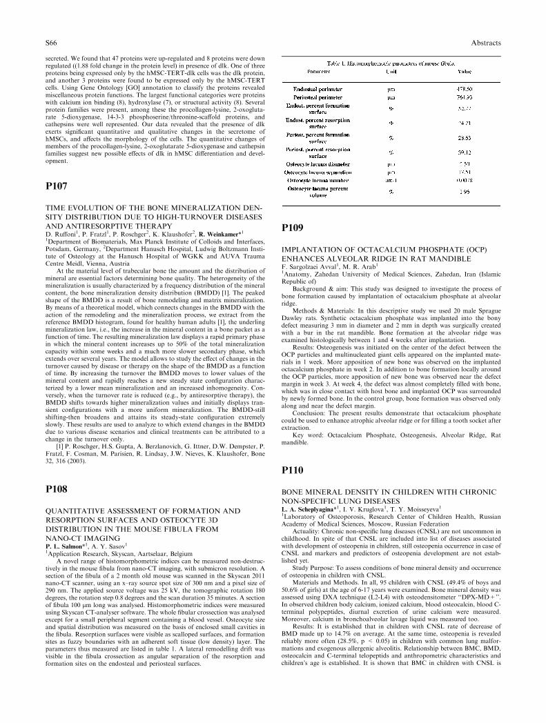
secreted. We found that 47 proteins were up-regulated and 8 proteins were downregulated ((1.88 fold change in the protein level) in presence of dlk. One of threeproteins being expressed only by the hMSC-TERT-dlk cells was the dlk protein,and another 3 proteins were found to be expressed only by the hMSC-TERTcells. Using Gene Ontology [GO] annotation to classify the proteins revealedmiscellaneous protein functions. The largest functional categories were proteinswith calcium ion binding (8), hydroxylase (7), or structural activity (8). Severalprotein families were present, among these the procollagen-lysine, 2-oxogluta-rate 5-dioxygenase, 14-3-3 phosphoserine/threonine-scaffold proteins, andcathepsins were well represented. Our data revealed that the presence of dlkexerts significant quantitative and qualitative changes in the secretome ofhMSCs, and affects the morphology of the cells. The quantitative changes ofmembers of the procollagen-lysine, 2-oxoglutarate 5-dioxygenase and cathepsinfamilies suggest new possible effects of dlk in hMSC differentiation and devel-opment.
P107
TIME EVOLUTION OF THE BONE MINERALIZATION DEN-
SITY DISTRIBUTION DUE TO HIGH-TURNOVER DISEASES
AND ANTIRESORPTIVE THERAPYD. Ruffoni1, P. Fratzl1, P. Roschger2, K. Klaushofer2, R. Weinkamer*11Department of Biomaterials, Max Planck Institute of Colloids and Interfaces,Potsdam, Germany, 2Department Hanusch Hospital, Ludwig Boltzmann Insti-tute of Osteology at the Hanusch Hospital of WGKK and AUVA TraumaCentre Meidl, Vienna, Austria
At the material level of trabecular bone the amount and the distribution ofmineral are essential factors determining bone quality. The heterogeneity of themineralization is usually characterized by a frequency distribution of the mineralcontent, the bone mineralization density distribution (BMDD) [1]. The peakedshape of the BMDD is a result of bone remodeling and matrix mineralization.By means of a theoretical model, which connects changes in the BMDD with theaction of the remodeling and the mineralization process, we extract from thereference BMDD histogram, found for healthy human adults [1], the underlingmineralization law, i.e., the increase in the mineral content in a bone packet as afunction of time. The resulting mineralization law displays a rapid primary phasein which the mineral content increases up to 50% of the total mineralizationcapacity within some weeks and a much more slower secondary phase, whichextends over several years. The model allows to study the effect of changes in theturnover caused by disease or therapy on the shape of the BMDD as a functionof time. By increasing the turnover the BMDD moves to lower values of themineral content and rapidly reaches a new steady state configuration charac-terized by a lower mean mineralization and an increased inhomogeneity. Con-versely, when the turnover rate is reduced (e.g., by antiresorptive therapy), theBMDD shifts towards higher mineralization values and initially displays tran-sient configurations with a more uniform mineralization. The BMDD-stillshifting-then broadens and attains its steady-state configuration extremelyslowly. These results are used to analyze to which extend changes in the BMDDdue to various disease scenarios and clinical treatments can be attributed to achange in the turnover only.
[1] P. Roschger, H.S. Gupta, A. Berzlanovich, G. Ittner, D.W. Dempster, P.Fratzl, F. Cosman, M. Parisien, R. Lindsay, J.W. Nieves, K. Klaushofer, Bone32, 316 (2003).
P108
QUANTITATIVE ASSESSMENT OF FORMATION AND
RESORPTION SURFACES AND OSTEOCYTE 3D
DISTRIBUTION IN THE MOUSE FIBULA FROM
NANO-CT IMAGINGP. L. Salmon*1, A. Y. Sasov11Application Research, Skyscan, Aartselaar, Belgium
A novel range of histomorphometric indices can be measured non-destruc-tively in the mouse fibula from nano-CT imaging, with submicron resolution. Asection of the fibula of a 2 month old mouse was scanned in the Skyscan 2011nano-CT scanner, using an x–ray source spot size of 300 nm and a pixel size of290 nm. The applied source voltage was 25 kV, the tomographic rotation 180degrees, the rotation step 0.8 degrees and the scan duration 35 minutes. A sectionof fibula 100 lm long was analysed. Histomorphometric indices were measuredusing Skyscan CT-analyser software. The whole fibular crossection was analysedexcept for a small peripheral segment containing a blood vessel. Osteocyte sizeand spatial distribution was measured on the basis of enclosed small cavities inthe fibula. Resorption surfaces were visible as scalloped surfaces, and formationsites as fuzzy boundaries with an adherent soft tissue (low density) layer. Theparameters thus measured are listed in table 1. A lateral remodelling drift wasvisible in the fibula crossection as angular separation of the resorption andformation sites on the endosteal and periosteal surfaces.
P109
IMPLANTATION OF OCTACALCIUM PHOSPHATE (OCP)
ENHANCES ALVEOLAR RIDGE IN RAT MANDIBLEF. Sargolzaei Avval1, M. R. Arab11Anatomy, Zahedan University of Medical Sciences, Zahedan, Iran (IslamicRepublic of)
Background & aim: This study was designed to investigate the process ofbone formation caused by implantation of octacalcium phosphate at alveolarridge.
Methods & Materials: In this descriptive study we used 20 male SpragueDawley rats. Synthetic octacalcium phosphate was implanted into the bonydefect measuring 3 mm in diameter and 2 mm in depth was surgically createdwith a bur in the rat mandible. Bone formation at the alveolar ridge wasexamined histologically between 1 and 4 weeks after implantation.
Results: Osteogenesis was initiated on the center of the defect between theOCP particles and multinucleated giant cells appeared on the implanted mate-rials in 1 week. More apposition of new bone was observed on the implantedoctacalcium phosphate in week 2. In addition to bone formation locally aroundthe OCP particles, more apposition of new bone was observed near the defectmargin in week 3. At week 4, the defect was almost completely filled with bone,which was in close contact with host bone and implanted OCP was surroundedby newly formed bone. In the control group, bone formation was observed onlyalong and near the defect margin.
Conclusion: The present results demonstrate that octacalcium phosphatecould be used to enhance atrophic alveolar ridge or for filling a tooth socket afterextraction.
Key word: Octacalcium Phosphate, Osteogenesis, Alveolar Ridge, Ratmandible.
P110
BONE MINERAL DENSITY IN CHILDREN WITH CHRONIC
NON-SPECIFIC LUNG DISEASESL. A. Scheplyagina*1, I. V. Kruglova1, T. Y. Moisseyeva11Laboratory of Osteoporosis, Research Center of Children Health, RussianAcademy of Medical Sciences, Moscow, Russian Federation
Actuality: Chronic non-specific lung diseases (CNSL) are not uncommon inchildhood. In spite of that CNSL are included into list of diseases associatedwith development of osteopenia in children, still osteopenia occurrence in case ofCNSL and markers and predictors of osteopenia development are not estab-lished yet.
Study Purpose: To assess conditions of bone mineral density and occurrenceof osteopenia in children with CNSL.
Materials and Methods. In all, 95 children with CNSL (49.4% of boys and50.6% of girls) at the age of 6-17 years were examined. Bone mineral density wasassessed using DXA technique (L2-L4) with osteodensitometer ‘‘DPX-MD+’’.In observed children body calcium, ionized calcium, blood osteocalcin, blood C-terminal polypeptides, diurnal excretion of urine calcium were measured.Moreover, calcium in bronchoalveolar lavage liquid was measured too.
Results: It is established that in children with CNSL rate of decrease ofBMD made up to 14.7% on average. At the same time, osteopenia is revealedreliably more often (28.5%, p < 0.05) in children with common lung malfor-mations and exogenous allergenic alveolitis. Relationship between BMC, BMD,osteocalcin and C-terminal telopeptids and anthropometric characteristics andchildren�s age is established. It is shown that BMC in children with CNSL is
S66 Abstracts
closely related with severity of clinical course of disease (r = 0.50; p < 0,05),duration of disease (r = 0.26; p < 0.05), degree of intensity of bronchi patency(r = 0.34; p < 0.05) and intensity of hypoxemia (r = 0.20; p < 0.05). It isshown that in 60% of examined children plasma ionized calcium is lower thanpresent standards and closely correlates with duration of disease (r = 0.32; p <0,05) and severity of clinical course of disease (r = 0.40; p < 0,05). Relationshipbetween indicators of excretion of urine calcium with bone mass levels (r = 0.34;p < 0.05) is established. Calcium in bronchoalveolar lavage liquid correlateswith BMC (r = 0.88; p < 0.05), plasma ionized calcium (r = 0.42; p < 0.05)and excretion of urine calcium (r = 0.42; p < 0.05).
Conclusion: In children with CNSL rate of decrease of bone mineral densitydepends on severity of lung affection and is underlined by hypoxemia and cal-cium metabolism disorder.
P111
CLINICAL SIGNIFICANCE OF OSTEODENSITOMETRY IN
CHILDRENL. A. Scheplyagina*1, I. V. Kruglova1, T. Y. Moisseyeva11Laboratory of Osteoporosis, Research Center of Children Health, RussianAcademy of Medical Sciences, Moscow, Russian Federation
Actuality: With the advent of X-ray densitometers opportunity to evaluateskeleton mineralization level during of child�s intensified growth periods hassignificantly increased. At that, BMD values below age-specific standard (Z-score ( -1 SD) are considered as criterion of lower content of skeleton calcium.
Accumulation of calcium in bones is a necessary condition for bonesdevelopment and formation of peak bone mass.
During last decade, it is noted that in Russia factually in all groups ofpopulation calcium intake is in quantities below age-specific standard. Severalstudies testify that deficient calcium intake negatively impacts increase of lineardimensions of body, especially during periods of intensive growth (‘‘half-growth’’ and pubertal spurt).
Study Purpose: To assess clinical significance of BMD in children usingDXA technique (L2–L4 Total body).
Materials and Methods. 238 healthy children at the age of 5-16 years (out ofthem-152 children of adolescent age), having no diseases capable to result indisorder of bone and calcium phosphorus metabolism had been examined. BMDwas evaluated using DXA technique on osteodensitometer DPX)MD+ (Lunar)equipped with special pediatric software program. Recommendations of ISCD(2003) had been taken into account. Hence, two programs were applied: lumbarspine (L2-L4) and Total body. Decrease of BMD was established using devicereference base proper at value of Z-score ( -1 SD).
Results: It is established that BMD values are lower in 38% of examinedchildren (L2-L4). Examination of children with reduced BMD values revealedhalf as great values (16%) in the case of using Total Body program. It is to benoted that out of general number of adolescents BMD decrease in L2–L4 pro-jection was established in 48% and on Total body program)23%. Almost half ofexamined children of adolescent age (48%) had decrease of BMD in both pointsof measurement.
Conclusions: In that way, children of adolescent age constitute risk groupboth on development of osteopenia and deficiency of peak bone mass duringpubertal spurt.
P112
MICROARRAY ANALYSIS OF GENE EXPRESSION IN
OSTEOBLAST CULTURES FROM PATIENTS WITH HYPER-
TROPHIC FRACTURE NON-UNIONSC. Schmid*1, A. Hofmann1, U. Ritz1, A. Ackermann1, N. Mladenovic1, M.Kuntz1, A. Meurer1, J. Heine1, P. Rommens2, J. Rompe31Orthopaedic Surgery, 2Trauma Surgery, Johannes Gutenberg University,Mainz, 3Orthopaedics, Orthotrauma Clinic, Gruenstadt, Germany
Background: Surgical treatment of fracture non-unions is still a challengingproblem, even in modern bone and joint surgery. Although, different morpho-logical non-union types and potential etiological factors have been considered,the underlying cellular and molecular regulatory mechanisms are still unknown.In the present study, Affymetrix oligonucleotide microarrays were used in orderto chart global gene expression differences in osteoblast cultures from non-un-ion-patients and controls.
Methods: Primary osteoblast (Ob) cultures were isolated from cancellousbone of seven patients undergoing reconstructive surgery for hypertrophicfracture non-unions and from five control patients. Gene expression was assayedin first passage cultures using Affymetrix U133A microarrays. Significant resultswere confirmed by qPCR.
Results: We identified 319 mRNAs that were expressed significantly differentin OB-cultures from non-union patients (p < 0.005), compared with controls. Asubset of up- and down-regulated mRNAs was able to distinguish the two
experimental groups. Among others, different transcription factors, ECM-pro-teins, proteinases, cytokins, growth factors, and their receptors were found to besignificantly altered in non-union-Ob.
Conclusion: We identified differently expressed genes in osteoblast culturesfrom hypertrophic fracture non-unions. Such markers may provide clues to thepathophysiology of hypertrophic fracture non-union and may be of predictivevalue in experimental and clinical trials.
P113
MYELOID PROGENITOR CELLS SUPPRESS OSTEOGENIC
DIFFERENTIATION OF MESENCHYMAL PROGENITOR
CELLSB. Vogel1, T. Vogel1, E. Rohde2, D. Strunk2, A. Gopferich3, M. B. Schulz*11Pharmaceutical Technology, Karl-Franzens-University, 2Division of Haema-tology and Department of Transfusion Medicine, Medical University, Graz,Austria, 3Pharmaceutical Technology, University of Regensburg, Regensburg,Germany
Aim: Primary cultures of mesenchymal progenitor cells (MPC) from bonemarrow are contaminated with adherent hematopoietic cells (HC) if tissueplastic adherence is the only enrichment criterion. While it is well known thatMSC are able to support hematopoiesis, little is known about the role of HC inmesenchymal differentiation. We therefore analyzed primary cultures of MPCfor the presence of HC, determined their phenotype and the influence of HC onosteogenic differentiation of MPC.
Materials and Methods: Rat bone marrow was isolated using a centrifuga-tional method. Cells were plated in tissue culture flasks in primary mediumcontaining 10% FBS. On day 7 after isolation, cells were detached and analyzedfor surface marker expression using flow cytometry. Contaminating HC wereimmuno-magnetically separated from MPC, using CD45 as target. Purity ofseparation was checked by flow cytometry. Cells were cultured under osteoin-ductive conditions (Dexamethasone, Vit.C, \beta-glycerophosphate). Osteogenicdifferentiation was determined by quantification of alkaline phosphatase activity(ALP), calcium deposition and quantitative RT-PCR for osteoblast-specificmarkers. Four independent experiments were performed to confirm the results.
Results: Primary cultures of rat MSC contained 10–30% CD45+ HC and noCD31+ endothelial cells. CD45-MPC expressed the typical markers also knownfor human MSC. CD45+ cells expressed CD11a, CD11b/c, CD18, CD29,CD49a, CD49e, CD54, CD90, but were negative for CD3, CD4, CD8b,CD45RA, CD161a and granulocyte marker and were therefore addressed asmyeloid cells. ALP of cells cultured without HC was significantly higher andincreased earlier than in the cultures containing HC. Confirming these results,von Kossa staining of calcium deposition on day 21 showed increased miner-alisation in the absence of HC. Furthermore, gene expression for osteogenicmarkers was strongly increased in cultures without HC. A trans-well controlindicated that the inhibition was caused by a soluble factor.
Conclusion: Myeloid progenitor cells sustain in vitro differentiation of ratMPC to osteoblasts. The mechanism of inhibition involves a soluble factor.
P114
IMMUNOLOCALIZATION OF SCLEROSTIN (SOST) IN
CARTILAGE AND BONE OF RATS TREATED WITH
INTERMITTENT PTH ADMINISTRATIONG. Silvestrini*1, P. Ballanti1, M. Leopizzi1, S. Berni1, E. Bonucci11Dept. Experimental Medicine and Pathology, La Sapienza University of Rome,Rome, Italy
Recently, the SOST gene and its protein product sclerostin were identified as apotent negative regulator of bone formation ( Winkler et al., 2003). SOST isstrongly expressed in osteocytes within the inner parts of bone; it is structurallyrelated to the BMP antagonist DAN/Cerberus family for its binding to BMPs andinhibiting bone formation. The loss of SOST in humans causes the autosomalrecessive high bone mass disorders of Van Buchem and sclerostosis, while trans-genic mice over-expressing SOST show low bone mass. Chronic elevation of PTHreduces SOST expression by osteocytes which express PTH receptors, suggestingthat SOST regulation may play a role in mediating PTH activity in bone.
Eight male Wistar rats were s.c. injected with 80 mg/Kg PTH (1-34) for threeweeks, three times a week; control rats were s.c. injected with PBS. The distalright femurs were embedded in glycolmethacrylate for bone histomorphometry,and the left femurs were paraffin embedded for SOST immunodetection. Goatanti-mouse SOST antibody (R & D Systems, Minneapolis, MN) was used.
In metaphyseal bone, histomorphometric variables of both bone formationand resorption were increased after PTH. In controls, SOST was absent in theosteocytes located near the bone surfaces, while was expressed by osteocyteslocated in the inner areas of bone trabeculae, near their residual axial cartilage.SOST-positive osteocytes were more numerous in the cortical and epiphysealtrabecular bone than in the metaphyseal trabecular bone. Osteoblasts were
Abstracts S67
negative or showed a light labeling. Chondrocytes of the growth plate and os-teoclasts were negative.
After PTH administration, the general distribution of SOST in the osteo-cytes did not change among the different skeletal districts. The growth platematurative/hypertrophic chondrocytes were labeled. Some chondroclasts wereintensely labeled. Osteoblasts showed a moderate labeling, more evident inmetaphyseal primary than in secondary trabecular bone. Unidentified, weaklylabeled cells were visible in bone marrow. Periosteal cells were moderately orstrongly stained.
In conclusion, the major number of labeled osteocytes in the cortical andepiphyseal areas appears to be in agreement with the observation that these areasare metabolically less active than metaphyseal trabeculae. After PTH, the pos-itivity of osteoblasts and maturative/hypertrophic chondrocytes may be relatedto the PTH-induced reduction of apoptosis.
P115
DIFFERENTIAL REGULATION OF MMPS IN
CHONDROCYTES AND THEIR INVOLVEMENT IN THE
DEVELOPMENT OF TIBIAL DYSCHONDROPLASIA IN
BROILERS AND TURKEYSS. Simsa*1, A. Hasdai1, E. Monsonego Ornan11Animal Seince, The Volcani Center, Bet Dagan, Israel
Tibial Dyschondroplasia (TD) is a cartilage disorder charechterised by theformation of a non-vascularized, non-minerlized area in the tibial growth plateof fast growing species such as broilers and turkeys.
The matrix metaloproteinases (MMPs) play a crucial role in growth platevascularization and ossification. These zinc dependent proteases have beenimplicated in physiological and pathological processes that involve proteolyticcleavage or remodeling of the ECM allowing endothelial cell invasion, growthfactors release and vascular growth. Thiram, a copper chelator that induce TD inbroilers within 7 days, fails to do so in turkeys. Since TD involves vasculariza-tion impairment, we sudied MMPs regulation and role in the development of TDin these species.
In primary culture of growth plate chondrocytes, retinoic acid (RA) treat-ment elevates MMP-2 and induces MMP-9 activity in broiler chondrocytes, butnot in turkey’s, where it induces a high molecular weight MMP activity. Incontrast, PMA treatment induces MMP-9 activity in turkey chondrocytes, but inbroiler chondrocytes it induces the activity of a 130kDa MMP. Thiram reducesMMP-2 and a 55kDa MMP activity in both broiler and turkey; although 10 foldhigher concentrations were required in turkey cells. The combined effect ofthiram and RA or PMA induces MMP-9 activity in turkey chondrocytes but notin broilers, while thiram alone does not induce MMP-9 activity.
These results indicates diverse development pattern of TD in broilers andturkeys, and suggest that MMP regulation differ in the growth plates of theseclosely related avian species, leading to altered matrix assembly and TD devel-opment.
P116
ENDOSTATIN RETARDS THE CARTILAGE PHASE IN THE
MODEL OF ECTOPIC OSSIFICATIONA.J. Sipola*1, J. Ilvesaro2, E. Birr3, P. Jalovaara3, F. Stenback4, T. Hautala5,J. Tuukkanen11Anatomy and Cell Biology, University of Oulu, Oulu, Finland, 2MedicineHematology and Oncology, University of Alabama at Birmingham, Birming-ham, United States, 3BTRG Department of Orthopaedic Surgery, 4Departmentof Pathology, 5Department of Internal Medicine, University of Oulu, Oulu,Finland
Background: Bone remodelling is a complex and well-coordinated series ofevents, which includes proper transport of nutrients and precursor cells via bloodvessels around the renewing bone tissue. It is well known that angiogenesis isessential for the replacement of cartilage by bone during skeletal growth andregeneration. Vascular endothelial growth factor (VEGF) is a key regulator ofangiogenesis. Growth factors including VEGF have been used in order toaccelerate fracture healing in animal models. Bone morphogenetic proteins(BMPs) are osteoinductive and they have been used to heal bone defects inexperimental animal models and clinical settings. Endostatin is a knownantagonist for VEGF-A. It inhibits endothelial proliferation and it may inhibitangiogenesis and tumor growth. Understanding of VEGF-A and endostatin inbone remodelling requires further studying.
Methods: We studied the influence of VEGF-A and endostatin on boneformation in vivo using double adenovirus gene transfer in a mouse hind leg.Ectopic bone formation was induced with BMP preparate implemented in gel-atine gel together with the viruses. BMP alone induced ectopic bone formation atthe injection site in muscle fascial tissue. The mice were sacrificed 1, 2 and 3weeks after the operation and their legs were removed. Evaluation of theinfluence of gene therapy was made from radiographs and histological sections.
Results: X-ray images taken from the dissected legs displayed significantbone formation in all treatment groups. Prior to ossification vascularization wasdemonstrated in Factor VIII stained histological sections. Ectopic new bone wasevaluated as a proportional area of cartilage/bone two and three weeks after theinjection. On two week time point there was no difference in the area cartilage/osseous nodule, but three weeks after the injection in response to endostatintreatment we could see a significant reduction in bone formation. VEGF-Astimulated and endostatin had inhibitory effect of the number of cartilage re-sorbing osteoclasts.
Conclusion: Endostatin is demonstrated to play a role in the inhibition ofcartilage resorption in noncalcifying hyaline cartilage. Our findings are inaccordance with this and indicate that endostatin retards the cartilage phase inendochondral ossification. Our data also demonstrate that dual gene therapy canbe used to modify BMP-induced ectopic bone formation.
P117
POSTNATAL CATCH-UP IN PIGLETS BONE DEVELOPMENT
AFTER PRENATAL DEXAMETHASONE TREATMENTE. Sliwa*1, M. R. Tatara1, T. Piersiak2, P. Dobrowolski2, T. Studzinski11Department of Animal Physiology, The Agricultural University of Lublin,2Department of Comperative Anatomy and Anthropolgy, Maria Curie-Sklo-dowska University, Lublin, Poland
Prenatal glucocorticoid excess slows linear growth and BMD. We adminis-tered dexamethasone (dex) to pregnant sows and/or growing piglets to suppressbone development.
The study was carried out on 8 sows and 32 their newborns. Sows weredivided into 2 groups - the control sows and experimental one treated with 3 mgdex per sow/48h during last 24 days of pregnancy. Newborn piglets of controlpregnancy were divided as well into 2 groups–the controls received i.m. saline (C)and group injected with dex i.m. at the dosage of 0.5 mg/kg BW/48h for 30 daysof postnatal life (C/D). Piglets of mothers administered with dex were dividedinto 2 groups–the controls received i.m. saline (D/C) and the group administeredwith dex (D/D) i.m. at the dosage of 0.5 mg/kg BW/48h for 30 days of postnatallife. BMD using NORLAND XR 43 apparatus of humeri and the geometric andmechanical properties using three-point bending test in INSTRON 4302 appa-ratus were evaluated.
Dex treatment decreased the weight of humerus and femur in C/D and D/Dgroups of 30 days old piglets and humerus weight in D/K group as well. Theweight of femur in D/C group reached similar value as observed in controlpiglets. Prenatal and postnatal administration with dex shortened femur andhumerus as well as postnatal treatment (C/D and D/D groups), while treatmentduring only parental time did not influence the length of both bones. Similarchanges were observed in the mean relative wall thickness (MRWT), the cross-sectional area (A) and in the second moment of inertia (Ix) of humerus andfemur in 30 days old piglets. Dex treatment during prenatal and/or postnatal lifedecreased the value of the ultimate strength (Wy) of femur. The value of theultimate strength (Wy) of humerus decreased in C/D and D/D groups. Thevalues of maximum elastic strength of humerus and femur were decreased as wellin C/D and D/D groups of piglets. BMD of femur decreased only in pigletstreated with dex during both periods, prenatal and postnatal, while BMD ofhumerus decreased in 3 groups being under the influence of dex during prenataland/or postnatal time.
After prenatal dex administration in untreated controls piglets we observed aresult described as catch-up growth. During the period of catch-up growth, inthe animals previously treated with dex, the decline of the values of the allinvestigated parameters of bones was delayed.
Study supported by grant No. 2PO6K/00828 from Polish Ministry ofEducation and Science.
P118
DEXAMETHASONE (DEX) AND ALPHA-KETOGLUTARATE
(AKG) INFLUENCE THE BONE DEVELOPMENT DURING
BOTH MATERNAL AND NEONATAL ADMINISTRATION IN
PIGSE. Sliwa*1, M. R. Tatara1, T. Studzinski11Department of Animal Physiology, The Agricultural University of Lublin,Lublin, Poland
The purpose of this study was to determine whether dexamethasone (Dex)and alfa-ketoglutarate (AKG) influence prenatal skeletal system programmingand development when administered separately or simultaneously to sows dur-ing 24 last days of pregnancy and 14 days of neonatal life.
The study was carried out on 8 sows and 48 their newborns. Sows weredivided into one control (saline group) and 3 experimental groups administeredwith Dex i.m. at the dosage of 3 mg/sow/48h or/and AKG per os 0.4 g/kg BW/day (AKG group, Dex + AKG and Dex groups). Six piglets born by controland experimental sows were euthanized within 1 h after their birth but other six
S68 Abstracts
piglets from every litter were further administered with Dex or/and AKG,according to their mothers with Dex i.m. at the dosage of 0.5 mg/kg BW/48h or/and AKG per os 0.4 g/kg BW/day, up to 14 days of neonatal life, when pigletswere euthanized. Bone mineral density (BMD) and bone mineral content (BMC)using NORLAND XR 43 apparatus of humeri and the geometric andmechanical properties using three-point bending test in INSTRON 4302 appa-ratus were evaluated.
BMD of humerus of newborns was the highest in the Dex + AKG groupand the lowest in groups administered to sows Dex and AKG separately. Sim-ilarly BMD of humerus after 14 days of neonatal continuation of experiment topiglets was the highest in Dex + AKG group, whereas the lowest in Dex group.The ultimate strength (Wy) of humerus in newborns was the highest in thecontrols and in Dex + AKG group, while the lowest in groups administeredseparately Dex and AKG. Wy of humerus in 14 days old piglets increased afterDex + AKG administration, whereas Dex treatment decreased this parameter.The cross-sectional area (A) of humerus of newborns reached the highest valuein the controls and in Dex + AKG group while lower in groups administeredseparately Dex and AKG.
Prenatal combined administration of Dex with AKG increased the Wy,BMD and BMC of humerus in comparison with groups which received thesesubstances apart. Combined neonatal administration of AKG and Dex im-proved length, weight, mechanical endurance and mineralisation in 14 days oldpiglets comparing with groups treated alone Dex. AKG administration in pre-natal and neonatal life may be applied as an effective treatment of negativeresults of Dex therapy on bone development and mineralization.
Study supported by grant No. 2PO6K/00828 from Polish Ministry ofEducation and Science.
P119
OOPHORECTOMY AND VITAMIN D3 DEFICIENCY DO
NOT AFFECT THE REPARATIVE PHASE OF FRACTURE
HEALING IN RATSL. B. Solberg*1, G. Melhus1, S. Dimmen2, J. Madsen2, L. Nordsletten2,F. P. Reinholt11Institute of Pathology, Rikshospitalet University Hospital, 2Orthopeadic cen-ter, Ulleval University Hospital, Oslo, Norway
The oophorectomized rat is an established model for postmenopausalosteoporosis (OP), but the relevance of the model for the human condition hasbeen questioned. An alternative model, combining oophorectomy and vitaminD3 deficiency (Ox-D), may appear more relevant as with this approach weak-ening of the femoral neck has been demonstrated for the first time in a rat model(1). In the current project the Ox-D model was used to study fracture healing. 50female Wistar rats were randomized into Ox-D or sham operation with sub-sequent normal rat chow (Sham). After 12 weeks, all animals underwent uni-lateral tibial midshaft fracture with intramedullary pinning. Bone mineraldensity (BMD) was measured, and histomorphometric analyses were performedafter 3 and 6 weeks. Mechanical testing was performed 6 weeks after fracture in a3-point bending test. The statistical analysis was performed using a conventionalindependent-sample T-test (two-tailed) to compare the means between thegroups. All p-values and confidence intervals were given with equal variancesassumed. A p-value of 0.05 was considered significant. The Ox-D group devel-oped OP with significant trabecular bone loss. Mechanical testing of callus in theOx-D group indicated slightly changed biomechanical properties, but the dif-ference to the Sham group did not reach statistical significance. There was nodifference in BMD in callus and no compensatory increase in the diameter or inthe cortical area of the femoral and tibial shafts. Our results show that OP in thismodel does not affect the reparative phase of healing after long bone fracture.
1.Kaastad, T.S., et al., Vitamin D deficiency and ovariectomy reduced thestrength of the femoral neck in rats. Calcified Tissue International, 2001. 69(2):p. 102–8.
P120
PRENATAL ADMINISTRATION OF 3-HYDROXY-3-METH-
YLBUTYRATE (HMB) TO PIGS INCREASES PEAK BONE
MASS IN OFFSPRINGM. R. Tatara*1, E. Sliwa1, W. Krupski2, T. Studzinski31Department of Animal Physiology, The Agricultural University of Lublin, 2IIDepartment of Radiology, 3Department of Animal Physiology, Medical Uni-versity of Lublin, Lublin, Poland
There is evidence that programming in prenatal life may have long-terminfluence on growth, health and the incidence of disease. Thus, prenatal period iscritical stage at which marked changes in nutrition, endocrine status, and envi-ronmental conditions can profoundly determine systemic development.
The aim of the study was to determine whether administration of HMB topregnant sows positively influences foetal development of the skeleton with itslong-term consequences in later life. This study was performed on pigs born to
sows that were administrated Ca(HMB)2 at the daily dose of 0.05 g/kg BW inthe experimental group during the last 2 weeks of pregnancy. Both HMB in theexperimental sows and CaCO3 (0.05 g/kg BW/day) in the control group wereadministered orally during the morning meal. After the birth, the piglets werekept under standard rearing conditions with free access to fresh water, and fed awell balanced diet. At the age of 6 months, the pigs were slaughtered and thefemur was isolated. Using quantitative computed tomography (QCT) techniqueand Somatom Emotion (Siemens) apparatus, the volumetric bone mineraldensity (vBMD) for both trabecular and cortical bone was measured.Mechanical properties of the femur were determined in INSTRON 4302 usingthe three-point bending test. Geometrical properties of femur such as cross-sectional area (A), second moment of inertia (Ix), mean relative wall thickness(MRWT) and cortical index (CI) were determined.
HMB administration to pregnant sows increased the vBMD of the trabecularand the cortical bone, as well as maximum elastic strength and ultimate strengthof femur obtained from 6 months old offspring (P ( 0.01). Moreover, HMBtreatment induced higher values of the investigated geometrical parameters offemur (P ( 0.01). In conclusion, HMB administration to sows during the last 2weeks of pregnancy positively influences skeletal development in their offspringwith its long-term consequences expressed by increased peak bone mass.
Acknowledgments: Study supported by Grant No. 2 P06K 012 29 fromPolish Ministry of Education and Science.
P121
ALPHA-KETOGLUTARATE (AKG) ADMINISTRATION
TO PREGNANT SOWS INCREASES MINERALIZATION,
GEOMETRICAL AND MECHANICAL ENDURANCE OF
SKELETON IN THEIR OFFSPRINGM. R. Tatara*1, E. Sliwa1, W. Krupski2, T. Studzinski31Department of Animal Physiology, The Agricultural University of Lublin, 2IIDepartment of Radiology, 3Department of Animal Physiology, Medical Uni-versity of Lublin, Lublin, Poland
Our recent studies showed that alpha-ketoglutarate (AKG) increases aminoacids synthesis such as proline and leucine. While proline and its metabolite–hydroksyproline contribute in two thirds to collagen structure, leucine regulatesglutamate dehydrogenaze activity which is responsible for conversion of AKGinto glutamate–main substrate for proline synthesis.
The aim of the study was to determine whether administration of AKG topregnant sows influences skeletal development in their offspring. The study wasperformed on pigs born to sows administrated AKG at the daily dose of 0.4 g/kgBW in the experimental group, during the last 2 weeks of pregnancy. Both AKGin the experimental sows and saline in the control group were administered orallyduring the morning meal. After the birth, the piglets were kept under standardrearing conditions with free access to fresh water, and fed a well balanced diet.At the age of 6 months, the pigs were slaughtered and the femurs isolated. Usingquantitative computed tomography (QCT) technique and Somatom Emotion(Siemens) apparatus, volumetric bone mineral density (vBMD) for both tra-becular and cortical bone was measured. Mechanical properties of the femurwere determined using the three-point bending test. Geometrical properties offemur such as cross-sectional area (A), second moment of inertia (Ix), meanrelative wall thickness (MRWT) and cortical index (CI) were determined.
AKG administration to pregnant sows increased vBMD of the trabecularand cortical bone (P ( 0.01), maximum elastic strength and ultimate strength(P ( 0.01) in 6 month old porkers. Furthermore, AKG-treated animals werecharacterized by higher values of the geometrical parameters of femur (P ( 0.01).Obtained results showed that AKG administration in pregnant sows may beconsidered as a factor positively influencing foetal skeletal development withlong-term consequences in later life of the offspring.
Acknowledgments: Study supported by Grant No. 2 P06K 012 29 fromPolish Ministry of Education and Science.
P122
FUNDECTOMY INDUCES OSTEOPENIA OF AXIAL
SKELETON IN PIGSM. R. Tatara*1, E. Sliwa1, W. Krupski2, T. Studzinski31Department of Animal Physiology, The Agricultural University of Lublin, 2IIDepartment of Radiology, 3Department of Animal Physiology, Medical Uni-versity of Lublin, Lublin, Poland
Total gastrectomy is known to cause osteopenia altering calcium and hor-monal status in humans and animals. However, information presenting botheffects of the surgical removing of the acid-producing part of the stomach(fundectomy) on bone tissue metabolism, and mechanisms responsible for theseeffects are strongly limited.
Thus, the aim of the study was to determine the effects of fundectomy onmineralization of axial skeleton and serum level of Insulin-like Growth Factor–1(IGF–1).
Abstracts S69
The study was performed on 12 healthy pigs of Large Polish White breed thatwere randomly divided into two weight-matched groups at the age of 40 days. Theexperimental group (n = 6) was subjected to fundectomy, which was carried outby surgical resecting the fundus of the stomach. The sham–operation of the controlgroup of animals (n = 6) was performed with a mid incision and manipulation ofthe viscera. After the surgical procedure and recovery period, all animals were keptunder standard rearing conditions with free access to fresh water and commercialfeed supplied according to their age. At the age of 9 months, blood samples werecollected and then animals were slaughtered. Three lumbar spine vertebra (L4–L6)were isolated, cleaned, weighed and frozen for further analyses. Using SomatomEmotion apparatus (Siemens, Germany) supplied with VB10B software, volu-metric bonemineral density (vBMD) of the trabecular bone of each vertebral bodywas performed. Moreover, using OSTEO software, the total amount of calciumand phosphorus as hydroxyapatites (Ca-HA) was measured. Using Volume soft-ware, the total volume of bone tissue in each vertebrae was determined. DEXAmethod (Norland 46-XR) was applied for determination of bone mineral density(BMD) and bone mineral content (BMC). Using OCTEIA IGF–1 immunoenzy-mometric assay (IDS Inc., USA), serum level of IGF–1 was determined. Obtainedresults showed decreased weight, vBMD, BMD, BMC, Ca-HA, BV and IGF-1serum level in fundectomized pigs (all p ( 0.01).
Our results suggest that fundectomy–evoked osteopenia may be mediated bydecreased IGF–1 level.
Study supported by Grant No. 2 P06K 036 29 from Polish Ministry ofEducation and Science.
P123
POST-GASTRECTOMY OSTEOPENIA OF FEMUR IN PIGSE. Sliwa*1, M. R. Tatara1, W. Krupski2, T. Studzinski31Department of Animal Physiology, The Agricultural University of Lublin, 2IIDepartment of Radiology, 3Department of Animal Physiology, Medical Uni-versity of Lublin, Lublin, Poland
Diet and metabolites of digestion are important factors influencing properfunction of skeletal system. Gastrectomy may alter calcium and bone metabo-lism, resulting in bone disorders. However, data showing effects of partialremoving of the stomach on bone tissue are limited.
Thus, the aim of the study was to determine effects of surgical removing ofthe fundus of the stomach on mineralization, geometrical properties andmechanical endurance of the femur in pigs.
The study was performed on 12 healthy pigs that were randomly divided intotwo weight-matched groups at the age of 40 days. The experimental group ofpiglets (n = 6) was subjected to fundectomy, which was carried out by surgicalresecting the fundus of the stomach. The sham–operation of the control group ofpiglets (n = 6) was performed with a mid-incision and manipulation of theviscera. After the surgical procedure and a one week recovery period, all animalswere kept under standard rearing conditions with free access to fresh water andcommercial feed supplied according to their age. All animals were slaughtered 8months after the fundectomy and right femur was isolated, weighed and mea-sured and then frozen at)25�C (wrapped in gauze soaked with saline) for furtheranalyses. Using Somatom Emotion apparatus (Siemens, Germany) supplied withVB10B software, the volumetric bone mineral density (vBMD) of trabecular andcortical bone was measured. Using OSTEO software, the total amount of cal-cium and phosphorus as hydroxyapatites (Ca-HA) was measured both for tra-becular and cortical bone. Using Bone Volume software, the volume of totalbone tissue in each vertebrae was determined. DEXA method (Norland 46-XR)was used for determination of bone mineral density (BMD) and bone mineralcontent (BMC) of the femur.
Obtained results showed the decrease of body weight of piglets in theexperimental group by approximately 200%. Moreover, bone length and weight,vBMD, and amount of hydroxyapatites of the trabecular and cortical bone,BMD, BMC and BV decreased in fundectomized pigs (all p ( 0.05).
In conclusion, fundectomy in pigs may serve as an experimental model forstudies on bone tissue metabolism.
Acknowledgments: Study supported by Grant No. 2 P06K 036 29 fromPolish Ministry of Education and Science.
P124
MORPHOLOGICAL DIVERSITY OF HYPERTROPHIC CHON-
DROCYTES IN GROWTH CARTILAGEL. Tatarczuch*1, Y. A. Ahmed1, H. M. S. Davies1, E. J. Mackie11School of Veterinary Science, University of Melbourne, Parkville, Australia
During endochondral ossification chondrocytes undergo hypertrophicdifferentiation contributing to cartilage growth and bone formation. It isusually assumed that hypertrophic chondrocytes are a homogeneous cellpopulation with discrete stages of maturation although some previous studieshave indicated their morphological heterogeneity. The aim of this study wasto determine the ultrastructural morphology of hypertrophic chondrocytes inequine articular–epiphyseal and physeal growth cartilage (AEGC and PGC,
respectively). Specimens of AEGC and PGC collected from proximal tibiaand distal femur during foetal and postnatal growth were studied by routinetransmission electron microscopy. Some specimens were stained by variousultracytochemical methods to visualise proteoglycans. Despite morphologicalvariations in ultrastructural appearance, �dark� and �light� hypertrophicchondrocytes were present at both locations. The dark chondrocytes werecharacterised by electron dense cytoplasm that contained abundant, oftendilated, rough endoplasmic reticulum, a prominent Golgi region and numer-ous secretory vesicles containing proteoglycans. This is in contrast to theelectron lucent cytoplasm of light chondrocytes that contained less developedrough endoplasmic reticulum and an inconspicuous Golgi region with sparsesecretory vesicles. Proteoglycans were observed in various membrane-boundand non-membrane bound vacuoles. Both light and dark chondrocytes wereobserved in the proliferation zone and their subsequent maturation and deathfollowed two distinctive patterns. The light chondrocytes appeared to disin-tegrate within a preserved cellular membrane whereas dark chondrocytesunderwent gradual condensation with cytoplasm extruded into the extracel-lular space. The proportion of light chondrocytes was higher in foetal thanpostnatal specimens. The presence of morphologically distinct populations ofhypertrophic chondrocytes with apparently diverse pathways for secretion ofproteoglycan in growth cartilage suggests they play specific roles duringskeletal development and growth.
P125
THE INFLUENCE OF EXTREMELY LOW FREQUENCY
MAGNETIC FIELD ON THE MINERALIZATION PROCESSG. Taton1, M. Karwala-Szytula1, T. Rok1, E. Rokita1, Z. Tabor1, A. Wrobel2,F. Beckmann3, T. Donath3, J. Fischer4, G. Taton*11Department of Biophysics, 2Institute of Physics, Jagiellonian University, Cra-cow, Poland, 3GKSS-Research Center, Geesthacht, 4Hannover Medical School,Hannover, Germany
The aim of the project is to evaluate the influence of extremely low frequencymagnetic field (ELFMF) on the bone mineralization process.
The investigation is based on the rat bone model. A group of pregnantfemale rats were treated with ELFMF. Parameters typical for magnetotherapy inhumans were utilized. The rats were also treated with their breed after the la-bour. The second group with their breeds was considered as a control. Youngtreated and control rats were killed in different ages (10, 20 and 30 d). Theirfemoral bones were dissected and investigated.
The morphometrical measurements of bones were done. Energy dispersiveX-ray fluorescence (ED-XRF) was performed in order to estimate the elementalcomposition. Moreover the bone samples were scanned with the microtomog-raphy with the use of synchrotron radiation (BW2 beamline of HASYLAB atDESY, Hamburg, Germany). Achieved image resolution was about 10 lm. A setof parameters characterizing the trabecular bone was calculated on the basis ofreconstructed three-dimensional (3D) images (e.g.: tissue volume (TV), bonevolume (BV), trabecular thickness (TT), trabecular number (TN) and trabecularseparation (TS)).
The morphometrical measurements indicated that the animals treaded withELFMF had smaller mass of femoral bones.
In the case of ED-XRF results we focused on the most important elementsfor the mineralization process i.e., Ca and P. The concentrations of both ele-ments were significantly lower in the case of animals exposed to ELFMF. Thebigger differences were observed for higher filed frequencies. We observed alsothe bigger variability of elemental concentrations in the exposed group. Thedifferences were also observed in the structural parameters. Particularly the TTwas lower in the exposed animals while the TS was lower in the control group.The values of other parameters scattered over a broad range.
The elemental composition and the structural parameters indicate that thedevelopment of the mineralization process is impaired for animals treated withELFMF. The explanation of the observed effect based on the enhanced angio-genesis is proposed.
AcknowledgementWork supported by the European Community-Research Infrastructure
Action under the FP6 ‘‘Structuring the European Research Area’’ Programmethrough the Integrated Infrastructure Initiative ‘‘Integrating Activity onSynchrotron and Free Electron Laser Science’’ Contract RII3-CT-2004-506008.
P126
THE EFFECTIVENESS OF COX-2 INHIBITIONS IN THE
PREVENTION OF HETEROTOPIC OSSIFICATION, AN
EXPERIMENTAL STUDY IN RABBITSP. G. Tsailas*1, G. C. Babis1, K. Nikolopoulos1, P. Nikiforides111st Orthopaedic Department University of Athens, KAT Hospital, Kifisia,Greece
S70 Abstracts
Backgroundaim: Although Cox-2 inhibitors have been available for sometime there has not been conclusive evidence that they can prevent heterotopicossification (HO) like other non-selective NSAIDS.
The purpose of this study was to show the effectiveness of this specificcategory of NSAIDS in an animal model, as well as to test the new injectableCOX-2 inhibitor parecoxib.
Method: 18 white mature male rabbits were used, which were divided inthree groups of 6, one control group, one group which was administeredmeloxicam SC and one which were administered paracoxib SC. For theinduction of HO the Michelson model was used which consisted of immo-bilization of the right hind knee in a cast for 5 weeks and passive mobili-zation in the full range of motion for 5 minutes every day except Sundays,during this period. The drugs were administered once daily for the durationof the 5 weeks. Afterwards the cast was removed and 5 weeks later theanimals were tested for range of motion and submitted to a lateral X-ray ofthe knee. Each X-ray was reviewed and assigned a number according toScott�s classification (0 no HO, 1 periosteal reaction, 2 HO less than half thefemur, 3 HO more than half the femur).
Results: All the rabbits in the control group presented with high degree ofstiffness of the knee and Scott�s 2 and 3 HO (mean 2.5) in the lateral X-rays,while the rabbits of the other two groups had significantly better range ofmotion and significantly less production of heterotopic bone (meloxicammean 0.83, parecoxib mean 0.83). There was no significant difference betweenthe meloxicam and parecoxib group, neither in motion nor in the radio-graphic results.
Conclusion: From this experimental study we concluded that meloxicam iscapable of preventing the development of HO and we agree with the few pub-lications that support this. We also managed to show that parecoxib and con-sequently its active substance valdecoxib can be used for the prophylaxis againstHO.
P127
EVALUATION OF PHOTOPOLYMERS, CAPABLE FOR
RAPID PROTOTYPING ON PROLIFERATION AND
MORPHOLOGY OF OSTEOBLASTSC. Turecek*1, M. Schuster2, F. Varga1, R. Liska2, B. Kaiser3, J. Stampfl3,K. Klaushofer114th Medical Department, Hanusch Hospital, Ludwig Boltzmann Institute ofOsteology, 2University of Technology, Institute of Applied Synthetic Chemistry,3University of Technology, Institute of Materials Science and Technology,Vienna, Austria
The development of bone replacement materials is a growing field of re-search for application in surgery. For bone tissue repair and substitution auto-grafts are the golden standard, because they fulfill the most importantcharacteristics of bone replacement materials for example biocompatibility andbioresorbability. Disadvantages of autografts are additional expense and traumato the patient, possibility of donor site morbidity and limited availability.Therefore, there is a critical need to develop new bone substitute materialsapproximating the properties of bone tissue. Stereophotolithography allowsconstructing scaffolds, which support bone cell adhesion and proliferation withnearly any desired structure and geometry.
Aim of the study was the synthesis and evaluation of the biocompatibilityof different types of resins made from mono-and multi-acrylated commerciallyavailable monomers capable for the rapid prototyping. Cell viability, prolif-eration and cell morphology was related to the functional groups in themonomers present e.g., oligoethyleneglycole, urethane-, hydroxy- or carboxygroups.
After sterilization of the test specimens MG63 osteoblast-like cells wereseeded at a density of 50000 cells/cm2 in culture medium (aMEM supplementedwith 5% FCS) and kept in culture for three days. For the measurements of cellviability and multiplication a quantitative colorimetric assay, based on MTT (3-(4,5-Dimethylthiazol-2-yl)-2,5-diphenyltetrazolium-bromid) was used. To assesscell morphology, cells were fixed and actin filaments stained with TRITC-Phalloidin. Finally cells were examined using a confocal laser scanning micro-scope.
Test specimens made from mono-acrylated monomers showed good celladhesion and proliferation but no significant differences. Depending on thefunctional groups of the multi-acrylated monomers significant differences inproliferation were found. One resin was superior to a commercial one, used forclinical applications. By staining actin filaments it was shown that osteoblastsformed strong stress fibers and proper contacts with the materials. Morpho-logical evaluation of the cells revealed a more fibroblastic appearance comparedto cells cultured on glass.
In conclusion new resins made from mono-and multi-acrylated monomerscapable for rapid prototyping were synthesized, which were superior to knownpolymers in supporting cell multiplication of osteoblasts.
P128
INTERCELLULAR COMMUNICATION IN OSTEOCYTES
AFTER MECHANICAL STIMULATIONA.Vatsa*1, D. Mizuno2, T. H. Smit3, C. F. Schmidt2, F. C. MacKintosh4,J. Klein-Nulend11Oral Cell Biology, ACTA-UvA and Vrije Universiteit, 2Biophysics and Com-plex Systems, Vrije Universiteit, 3Clinical Physics and Engineering, VU medischcentrum, 4Theoretical Physics, Vrije Universiteit, Amsterdam, Netherlands
Background: Osteocytes are believed to be the mechanosensors in bone (1).It is hypothesised that mechanical loading on bones causes strain-derived fluidshear stresses on osteocytes, which activates them. Activated osteocytes producesignaling molecules like nitric oxide (NO), which modulate the activity of os-teoblasts and osteoclasts thereby orchestrating bone adaptation to mechanicalloading. This single-cell level mechanosensing and intercellular signaling isessential for bone adaptation (2). We studied how a mechanically-stimulatedsingle osteocyte functions in a social context via intercellular communication, bymonitoring changes in intracellular NO production, using DAR-4M AM chro-mophore, in the surrounding osteocytes.
Methods: DAR-4M AM chromophore-loaded, surface-attached MLO-Y4osteocyte-like cells were subjected to a localized oscillatory mechanical stimulus(10-20nN) using an Eppendorf micro-needle. DAR-4M AM is membrane-per-meable and reacts with NO to form a stable, fluorescent compound intracellu-larly, which can be visualised with a rhodamine filter (excitationwavelength = 554nM; emission wavelength = 572nM). Fluorescence wasquantified by Scion Image software.
Results: Mechanical stimulation of a single osteocyte resulted in a rapidincrease of 12-24 % in fluorescence intensity in surrounding osteocytes as com-pared to their basal level intensities, indicating upregulation of NO production.
Conclusion: NO production is essential for mechanical loading-inducedbone formation in vivo (3). Hence, NO production in response to mechanicalstimulation is a meaningful parameter for measuring bone cell activation.However, its short half-life (0.1–5 s) makes its online detection difficult. Ourtechnique allows real-time monitoring of chemical signaling at the single oste-ocyte level. Here we show that a single mechanically-stimulated osteocyte acti-vates its surrounding osteocytes via direct and/or indirect intercellular signaling,as indicated by upregulation of intracellular NO production in these cells. Thispaves a path for better understanding of the dynamic processes involved in boneadaptation.
Acknowledgements: This study is supported by NOW-ALW/FOM, project# 01FB28/2.
References: 1) Burger EH, Klein-Nulend J (1999) FASEB J 13:S101; 2)Burger EH et al. (2003) J Biomech 36:1453; 3) Turner CH et al. (1997) Bone21:487, 1997.
P129
BMP-6 INDUCES NEW BONE FORMATION IN
OVARIECTOMIZED MICE AND ENHANCES
DIFFERENTIATION OF PRIMARY HUMAN OSTEOBLASTS
THROUGH ACTIVATION OF IGF-1 AND EGF PATHWAYSF. Borovecki*1, W. A. Grasser2, I. Orlic3, V. M. Paralkar2, S. Vukicevic31Center for Functional Genomics, School of Medicine, Zagreb, Croatia, 2PfizerCentral Research, Pfizer Inc., Groton, United States, 3Department of Anatomy,School of Medicine, Zagreb, Croatia
We have recently shown that human recombinant BMP-6 (rhBMP-6), givensystematically, can restore bone in animal models of osteoporosis. To furtherelucidate the underlying mechanisms of new bone formation following systemicapplication of BMPs we conducted gene expression profiling experiments usingbone samples of ovariectomized (OVX) mice treated with BMP-6. Mice wereOVX at 3 months of age and three weeks later treated for the next 4 weeks asfollows: (1) sham, (2) OVX and (3) OVX + BMP-6 (10 lg/kg i.v. 3 x weeks).Total RNA was isolated from whole femurs and genome-wide expressionanalysis was conducted using Affymetrix GeneChip MOE 430 2.0 microarrays.In an effort to test the functional correlation of gene sets that might be sys-temically altered in mice treated with BMP-6, we applied a Gene Set EnrichmentAnalysis (GSEA), which uses the principle of gene-ranking based on the corre-lation between their expression and the class distinction by applying the signal-to-noise ratio metric. GSEA revealed statistically significant enrichment ofinsulin-like growth factor-1 (IGF-1) and epidermal growth factor (EGF) relatedpathways in animals treated with BMP-6. OVX animals exhibited markedenrichment of IL-12 and IL-1 receptor pathways. QRT-PCR analysis confirmedsignificant upregulation of IGF-1 and EGF expression in BMP-6 treated mice.To develop an in vitro model for evaluation of the effects of BMP-6 on cells ofhuman origin, we cultured primary human osteoblasts (HOB). Treatment withrhBMP-6 accelerated cell differentiation as indicated by the formation of min-eralized nodules by day 18 of culture vs 28 to 30 days in vehicle treated cultures.
Abstracts S71
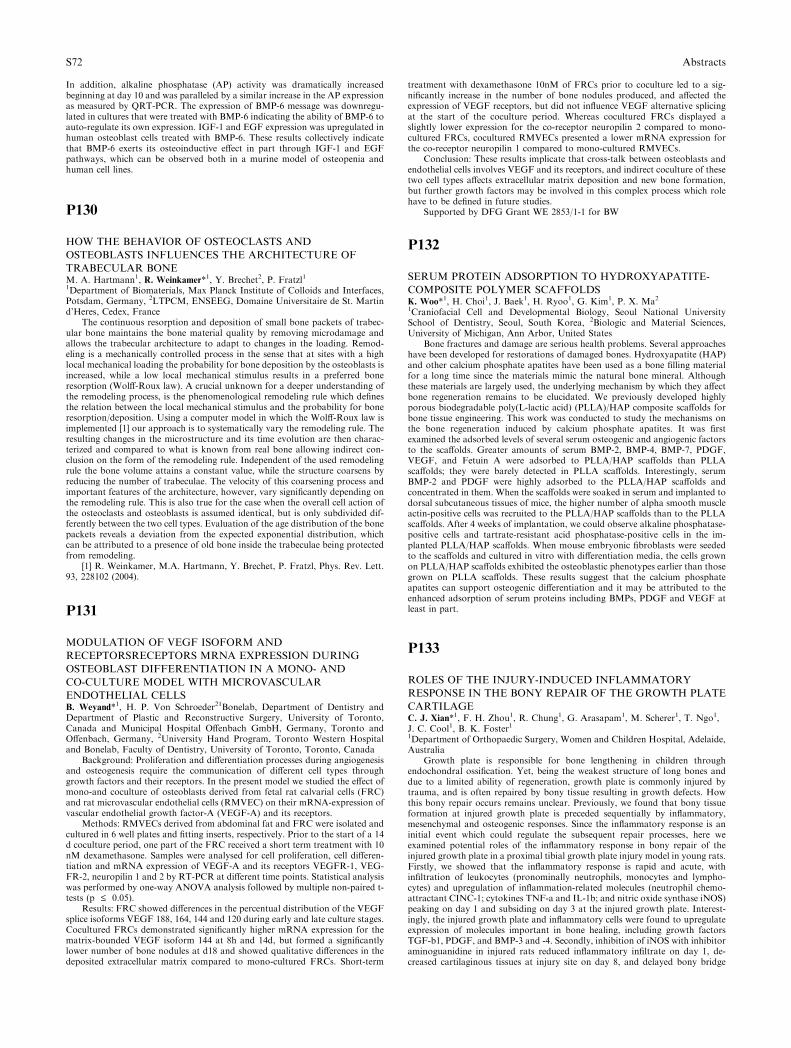
In addition, alkaline phosphatase (AP) activity was dramatically increasedbeginning at day 10 and was paralleled by a similar increase in the AP expressionas measured by QRT-PCR. The expression of BMP-6 message was downregu-lated in cultures that were treated with BMP-6 indicating the ability of BMP-6 toauto-regulate its own expression. IGF-1 and EGF expression was upregulated inhuman osteoblast cells treated with BMP-6. These results collectively indicatethat BMP-6 exerts its osteoinductive effect in part through IGF-1 and EGFpathways, which can be observed both in a murine model of osteopenia andhuman cell lines.
P130
HOW THE BEHAVIOR OF OSTEOCLASTS AND
OSTEOBLASTS INFLUENCES THE ARCHITECTURE OF
TRABECULAR BONEM. A. Hartmann1, R. Weinkamer*1, Y. Brechet2, P. Fratzl11Department of Biomaterials, Max Planck Institute of Colloids and Interfaces,Potsdam, Germany, 2LTPCM, ENSEEG, Domaine Universitaire de St. Martind’Heres, Cedex, France
The continuous resorption and deposition of small bone packets of trabec-ular bone maintains the bone material quality by removing microdamage andallows the trabecular architecture to adapt to changes in the loading. Remod-eling is a mechanically controlled process in the sense that at sites with a highlocal mechanical loading the probability for bone deposition by the osteoblasts isincreased, while a low local mechanical stimulus results in a preferred boneresorption (Wolff-Roux law). A crucial unknown for a deeper understanding ofthe remodeling process, is the phenomenological remodeling rule which definesthe relation between the local mechanical stimulus and the probability for boneresorption/deposition. Using a computer model in which the Wolff-Roux law isimplemented [1] our approach is to systematically vary the remodeling rule. Theresulting changes in the microstructure and its time evolution are then charac-terized and compared to what is known from real bone allowing indirect con-clusion on the form of the remodeling rule. Independent of the used remodelingrule the bone volume attains a constant value, while the structure coarsens byreducing the number of trabeculae. The velocity of this coarsening process andimportant features of the architecture, however, vary significantly depending onthe remodeling rule. This is also true for the case when the overall cell action ofthe osteoclasts and osteoblasts is assumed identical, but is only subdivided dif-ferently between the two cell types. Evaluation of the age distribution of the bonepackets reveals a deviation from the expected exponential distribution, whichcan be attributed to a presence of old bone inside the trabeculae being protectedfrom remodeling.
[1] R. Weinkamer, M.A. Hartmann, Y. Brechet, P. Fratzl, Phys. Rev. Lett.93, 228102 (2004).
P131
MODULATION OF VEGF ISOFORM AND
RECEPTORSRECEPTORS MRNA EXPRESSION DURING
OSTEOBLAST DIFFERENTIATION IN A MONO- AND
CO-CULTURE MODEL WITH MICROVASCULAR
ENDOTHELIAL CELLSB. Weyand*1, H. P. Von Schroeder21Bonelab, Department of Dentistry andDepartment of Plastic and Reconstructive Surgery, University of Toronto,Canada and Municipal Hospital Offenbach GmbH, Germany, Toronto andOffenbach, Germany, 2University Hand Program, Toronto Western Hospitaland Bonelab, Faculty of Dentistry, University of Toronto, Toronto, Canada
Background: Proliferation and differentiation processes during angiogenesisand osteogenesis require the communication of different cell types throughgrowth factors and their receptors. In the present model we studied the effect ofmono-and coculture of osteoblasts derived from fetal rat calvarial cells (FRC)and rat microvascular endothelial cells (RMVEC) on their mRNA-expression ofvascular endothelial growth factor-A (VEGF-A) and its receptors.
Methods: RMVECs derived from abdominal fat and FRC were isolated andcultured in 6 well plates and fitting inserts, respectively. Prior to the start of a 14d coculture period, one part of the FRC received a short term treatment with 10nM dexamethasone. Samples were analysed for cell proliferation, cell differen-tiation and mRNA expression of VEGF-A and its receptors VEGFR-1, VEG-FR-2, neuropilin 1 and 2 by RT-PCR at different time points. Statistical analysiswas performed by one-way ANOVA analysis followed by multiple non-paired t-tests (p £ 0.05).
Results: FRC showed differences in the percentual distribution of the VEGFsplice isoforms VEGF 188, 164, 144 and 120 during early and late culture stages.Cocultured FRCs demonstrated significantly higher mRNA expression for thematrix-bounded VEGF isoform 144 at 8h and 14d, but formed a significantlylower number of bone nodules at d18 and showed qualitative differences in thedeposited extracellular matrix compared to mono-cultured FRCs. Short-term
treatment with dexamethasone 10nM of FRCs prior to coculture led to a sig-nificantly increase in the number of bone nodules produced, and affected theexpression of VEGF receptors, but did not influence VEGF alternative splicingat the start of the coculture period. Whereas cocultured FRCs displayed aslightly lower expression for the co-receptor neuropilin 2 compared to mono-cultured FRCs, cocultured RMVECs presented a lower mRNA expression forthe co-receptor neuropilin 1 compared to mono-cultured RMVECs.
Conclusion: These results implicate that cross-talk between osteoblasts andendothelial cells involves VEGF and its receptors, and indirect coculture of thesetwo cell types affects extracellular matrix deposition and new bone formation,but further growth factors may be involved in this complex process which rolehave to be defined in future studies.
Supported by DFG Grant WE 2853/1-1 for BW
P132
SERUM PROTEIN ADSORPTION TO HYDROXYAPATITE-
COMPOSITE POLYMER SCAFFOLDSK. Woo*1, H. Choi1, J. Baek1, H. Ryoo1, G. Kim1, P. X. Ma21Craniofacial Cell and Developmental Biology, Seoul National UniversitySchool of Dentistry, Seoul, South Korea, 2Biologic and Material Sciences,University of Michigan, Ann Arbor, United States
Bone fractures and damage are serious health problems. Several approacheshave been developed for restorations of damaged bones. Hydroxyapatite (HAP)and other calcium phosphate apatites have been used as a bone filling materialfor a long time since the materials mimic the natural bone mineral. Althoughthese materials are largely used, the underlying mechanism by which they affectbone regeneration remains to be elucidated. We previously developed highlyporous biodegradable poly(L-lactic acid) (PLLA)/HAP composite scaffolds forbone tissue engineering. This work was conducted to study the mechanisms onthe bone regeneration induced by calcium phosphate apatites. It was firstexamined the adsorbed levels of several serum osteogenic and angiogenic factorsto the scaffolds. Greater amounts of serum BMP-2, BMP-4, BMP-7, PDGF,VEGF, and Fetuin A were adsorbed to PLLA/HAP scaffolds than PLLAscaffolds; they were barely detected in PLLA scaffolds. Interestingly, serumBMP-2 and PDGF were highly adsorbed to the PLLA/HAP scaffolds andconcentrated in them. When the scaffolds were soaked in serum and implanted todorsal subcutaneous tissues of mice, the higher number of alpha smooth muscleactin-positive cells was recruited to the PLLA/HAP scaffolds than to the PLLAscaffolds. After 4 weeks of implantation, we could observe alkaline phosphatase-positive cells and tartrate-resistant acid phosphatase-positive cells in the im-planted PLLA/HAP scaffolds. When mouse embryonic fibroblasts were seededto the scaffolds and cultured in vitro with differentiation media, the cells grownon PLLA/HAP scaffolds exhibited the osteoblastic phenotypes earlier than thosegrown on PLLA scaffolds. These results suggest that the calcium phosphateapatites can support osteogenic differentiation and it may be attributed to theenhanced adsorption of serum proteins including BMPs, PDGF and VEGF atleast in part.
P133
ROLES OF THE INJURY-INDUCED INFLAMMATORY
RESPONSE IN THE BONY REPAIR OF THE GROWTH PLATE
CARTILAGEC. J. Xian*1, F. H. Zhou1, R. Chung1, G. Arasapam1, M. Scherer1, T. Ngo1,J. C. Cool1, B. K. Foster11Department of Orthopaedic Surgery, Women and Children Hospital, Adelaide,Australia
Growth plate is responsible for bone lengthening in children throughendochondral ossification. Yet, being the weakest structure of long bones anddue to a limited ability of regeneration, growth plate is commonly injured bytrauma, and is often repaired by bony tissue resulting in growth defects. Howthis bony repair occurs remains unclear. Previously, we found that bony tissueformation at injured growth plate is preceded sequentially by inflammatory,mesenchymal and osteogenic responses. Since the inflammatory response is aninitial event which could regulate the subsequent repair processes, here weexamined potential roles of the inflammatory response in bony repair of theinjured growth plate in a proximal tibial growth plate injury model in young rats.Firstly, we showed that the inflammatory response is rapid and acute, withinfiltration of leukocytes (pronominally neutrophils, monocytes and lympho-cytes) and upregulation of inflammation-related molecules (neutrophil chemo-attractant CINC-1; cytokines TNF-a and IL-1b; and nitric oxide synthase iNOS)peaking on day 1 and subsiding on day 3 at the injured growth plate. Interest-ingly, the injured growth plate and inflammatory cells were found to upregulateexpression of molecules important in bone healing, including growth factorsTGF-b1, PDGF, and BMP-3 and -4. Secondly, inhibition of iNOS with inhibitoraminoguanidine in injured rats reduced inflammatory infiltrate on day 1, de-creased cartilaginous tissues at injury site on day 8, and delayed bony bridge
S72 Abstracts
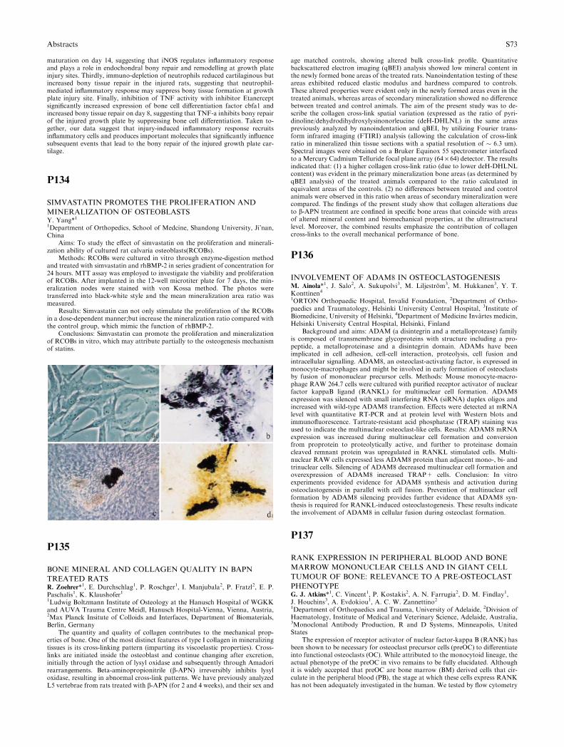
maturation on day 14, suggesting that iNOS regulates inflammatory responseand plays a role in endochondral bony repair and remodelling at growth plateinjury sites. Thirdly, immuno-depletion of neutrophils reduced cartilaginous butincreased bony tissue repair in the injured rats, suggesting that neutrophil-mediated inflammatory response may suppress bony tissue formation at growthplate injury site. Finally, inhibition of TNF activity with inhibitor Etanerceptsignificantly increased expression of bone cell differentiation factor cbfa1 andincreased bony tissue repair on day 8, suggesting that TNF-a inhibits bony repairof the injured growth plate by suppressing bone cell differentiation. Taken to-gether, our data suggest that injury-induced inflammatory response recruitsinflammatory cells and produces important molecules that significantly influencesubsequent events that lead to the bony repair of the injured growth plate car-tilage.
P134
SIMVASTATIN PROMOTES THE PROLIFERATION AND
MINERALIZATION OF OSTEOBLASTSY. Yang*11Department of Orthopedics, School of Medcine, Shandong University, Ji’nan,China
Aims: To study the effect of simvastatin on the proliferation and minerali-zation ability of cultured rat calvaria osteoblasts(RCOBs).
Methods: RCOBs were cultured in vitro through enzyme-digestion methodand treated with simvastatin and rhBMP-2 in series gradient of concentration for24 hours. MTT assay was employed to investigate the viability and proliferationof RCOBs. After implanted in the 12-well microtiter plate for 7 days, the min-eralization nodes were stained with von Kossa method. The photos weretransferred into black-white style and the mean mineralization area ratio wasmeasured.
Results: Simvastatin can not only stimulate the proliferation of the RCOBsin a dose-dependent manner;but increase the mineralization ratio compared withthe control group, which mimic the function of rhBMP-2.
Conclusions: Simvastatin can promote the proliferation and mineralizationof RCOBs in vitro, which may attribute partially to the osteogenesis mechanismof statins.
P135
BONE MINERAL AND COLLAGEN QUALITY IN BAPN
TREATED RATSR. Zoehrer*1, E. Durchschlag1, P. Roschger1, I. Manjubala2, P. Fratzl2, E. P.Paschalis1, K. Klaushofer11Ludwig Boltzmann Institute of Osteology at the Hanusch Hospital of WGKKand AUVA Trauma Centre Meidl, Hanusch Hospital-Vienna, Vienna, Austria,2Max Planck Insitute of Colloids and Interfaces, Department of Biomaterials,Berlin, Germany
The quantity and quality of collagen contributes to the mechanical prop-erties of bone. One of the most distinct features of type I collagen in mineralizingtissues is its cross-linking pattern (imparting its viscoelastic properties). Cross-links are initiated inside the osteoblast and continue changing after excretion,initially through the action of lysyl oxidase and subsequently through Amadorirearrangements. Beta-aminopropionitrile (b-APN) irreversibly inhibits lysyloxidase, resulting in abnormal cross-link patterns. We have previously analyzedL5 vertebrae from rats treated with b-APN (for 2 and 4 weeks), and their sex and
age matched controls, showing altered bulk cross-link profile. Quantitativebackscattered electron imaging (qBEI) analysis showed low mineral content inthe newly formed bone areas of the treated rats. Nanoindentation testing of theseareas exhibited reduced elastic modulus and hardness compared to controls.These altered properties were evident only in the newly formed areas even in thetreated animals, whereas areas of secondary mineralization showed no differencebetween treated and control animals. The aim of the present study was to de-scribe the collagen cross-link spatial variation (expressed as the ratio of pyri-dinoline/dehydrodihydroxylysinonorleucine (deH-DHLNL) in the same areaspreviously analyzed by nanoindentation and qBEI, by utilizing Fourier trans-form infrared imaging (FTIRI) analysis (allowing the calculation of cross-linkratio in mineralized thin tissue sections with a spatial resolution of � 6.3 um).Spectral images were obtained on a Bruker Equinox 55 spectrometer interfacedto a Mercury Cadmium Telluride focal plane array (64 · 64) detector. The resultsindicated that: (1) a higher collagen cross-link ratio (due to lower deH-DHLNLcontent) was evident in the primary mineralization bone areas (as determined byqBEI analysis) of the treated animals compared to the ratio calculated inequivalent areas of the controls. (2) no differences between treated and controlanimals were observed in this ratio when areas of secondary mineralization werecompared. The findings of the present study show that collagen alterations dueto b-APN treatment are confined in specific bone areas that coincide with areasof altered mineral content and biomechanical properties, at the ultrastructurallevel. Moreover, the combined results emphasize the contribution of collagencross-links to the overall mechanical performance of bone.
P136
INVOLVEMENT OF ADAM8 IN OSTEOCLASTOGENESISM. Ainola*1, J. Salo2, A. Sukupolvi3, M. Liljestrom3, M. Hukkanen3, Y. T.Konttinen41ORTON Orthopaedic Hospital, Invalid Foundation, 2Department of Ortho-paedics and Traumatology, Helsinki University Central Hospital, 3Institute ofBiomedicine, University of Helsinki, 4Department of Medicine Invartes medicin,Helsinki University Central Hospital, Helsinki, Finland
Background and aims: ADAM (a disintegrin and a metalloprotease) familyis composed of transmembrane glycoproteins with structure including a pro-peptide, a metalloproteinase and a disintegrin domain. ADAMs have beenimplicated in cell adhesion, cell-cell interaction, proteolysis, cell fusion andintracellular signalling. ADAM8, an osteoclast-activating factor, is expressed inmonocyte-macrophages and might be involved in early formation of osteoclastsby fusion of mononuclear precursor cells. Methods: Mouse monocyte-macro-phage RAW 264.7 cells were cultured with purified receptor activator of nuclearfactor kappaB ligand (RANKL) for multinuclear cell formation. ADAM8expression was silenced with small interfering RNA (siRNA) duplex oligos andincreased with wild-type ADAM8 transfection. Effects were detected at mRNAlevel with quantitative RT-PCR and at protein level with Western blots andimmunofluorescence. Tartrate-resistant acid phosphatase (TRAP) staining wasused to indicate the multinuclear osteoclast-like cells. Results: ADAM8 mRNAexpression was increased during multinuclear cell formation and conversionfrom proprotein to proteolytically active, and further to proteinase domaincleaved remnant protein was upregulated in RANKL stimulated cells. Multi-nuclear RAW cells expressed less ADAM8 protein than adjacent mono-, bi- andtrinuclear cells. Silencing of ADAM8 decreased multinuclear cell formation andoverexpression of ADAM8 increased TRAP+ cells. Conclusion: In vitroexperiments provided evidence for ADAM8 synthesis and activation duringosteoclastogenesis in parallel with cell fusion. Prevention of multinuclear cellformation by ADAM8 silencing provides further evidence that ADAM8 syn-thesis is required for RANKL-induced osteoclastogenesis. These results indicatethe involvement of ADAM8 in cellular fusion during osteoclast formation.
P137
RANK EXPRESSION IN PERIPHERAL BLOOD AND BONE
MARROW MONONUCLEAR CELLS AND IN GIANT CELL
TUMOUR OF BONE: RELEVANCE TO A PRE-OSTEOCLAST
PHENOTYPEG. J. Atkins*1, C. Vincent1, P. Kostakis2, A. N. Farrugia2, D. M. Findlay1,J. Houchins3, A. Evdokiou1, A. C. W. Zannettino21Department of Orthopaedics and Trauma, University of Adelaide, 2Division ofHaematology, Institute of Medical and Veterinary Science, Adelaide, Australia,3Monoclonal Antibody Production, R and D Systems, Minneapolis, UnitedStates
The expression of receptor activator of nuclear factor-kappa B (RANK) hasbeen shown to be necessary for osteoclast precursor cells (preOC) to differentiateinto functional osteoclasts (OC). While attributed to the monocytoid lineage, theactual phenotype of the preOC in vivo remains to be fully elucidated. Althoughit is widely accepted that preOC are bone marrow (BM) derived cells that cir-culate in the peripheral blood (PB), the stage at which these cells express RANKhas not been adequately investigated in the human. We tested by flow cytometry
Abstracts S73
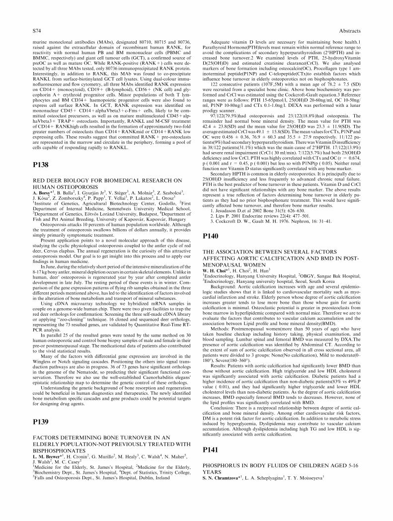
murine monoclonal antibodies (MAbs), designated 80710, 80715 and 80736,raised against the extracellular domain of recombinant human RANK, forreactivity with normal human PB and BM mononuclear cells (PBMC andBMMC, respectively) and giant cell tumour cells (GCT), a confirmed source ofpreOC as well as mature OC. While RANK-positive (RANK+) cells were de-tected by all three MAbs tested, only 80736 immunoprecipitated RANK protein.Interestingly, in addition to RANK, this MAb was found to co-precipitateRANKL from surface-biotinylated GCT cell lysates. Using dual-colour immu-nofluorescence and flow cytometry, all three MAbs identified RANK expressionon CD14+ (monocytoid), CD19+ (B-lymphoid), CD56+ (NK cell) and gly-cophorin A+ erythroid progenitor cells. Minor populations of both T lym-phocytes and BM CD34+ haemopoietic progenitor cells were also found toexpress cell surface RANK. In GCT, RANK expression was identified onmononuclear CD45+ CD14+alphaVbeta3+c-Fms+ cells, likely to be com-mitted osteoclast precursors, as well as on mature multinucleated CD45+alp-haVbeta3+ TRAP+ osteoclasts. Importantly, RANKL and M-CSF treatmentof CD14+ RANKhigh cells resulted in the formation of approximately two-foldgreater numbers of osteoclasts than CD14+RANKmid or CD14+RANK lowexpressing cells. These results suggest that committed RANK+ pre-osteoclastsare represented in the marrow and circulate in the periphery, forming a pool ofcells capable of responding rapidly to RANKL.
P138
RED DEER BIOLOGY FOR BIOMEDICAL RESEARCH ON
HUMAN OSTEOPOROSISA. Borsy*1, B. Balla2, I. Gyurjan Jr3, V. Steger3, A. Molnar3, Z. Szabolcsi3,J. Kosa2, Z. Zomborszky4, P. Papp1, T. Vellai3, P. Lakatos2, L. Orosz31Institute of Genetics, Agricultural Biotechnology Center, Godollo, 2FirstDepartment of Internal Medicine, Semmelweis University Medical School,3Department of Genetics, Eotvos Lorand University, Budapest, 4Department ofFish and Pet Animal Breeding, University of Kaposvar, Kaposvar, Hungary
Osteoporosis attacks 10 percents of human population worldwide. Althoughthe treatment of osteoporosis swallows billions of dollars annually, it providessimply primarily symptomatic treatment.
Present application points to a novel molecular approach of this disease,studying the cyclic physiological osteoporosis coupled to the antler cycle of reddeer, Cervus elaphus. The annual regeneration is the curiosity of this attractiveosteoporosis model. Our goal is to get insight into this process and to apply ourfindings in human medicine.
In June, during the relatively short period of the intensivemineralization of the8-17 kg bony antler,mineral depletionoccurs in certain skeletal elements.Unlike inhuman, deer� osteoporosis is regenerated year by year after completed antlerdevelopment in late July. The resting period of these events is in winter. Com-parison of the gene expression patterns of flying rib samples obtained in the threedifferent periods mentioned above, has led to the identification of factors involvedin the alteration of bone metabolism and transport of mineral substances.
Using cDNA microarray technology we hybridized mRNA samples incouple on a genome-wide human chip. There were two given facilities to trap thered deer orthologs for confirmation: Screening the three self-made cDNA libraryor applying ‘‘zoo-cloning’’ technique. 16 cloned and sequenced deer orthologs,representing the 73 resulted genes, are validated by Quantitative Real-Time RT-PCR analysis.
In parallel 25 of the resulted genes were tested by the same method on 30human osteoporotic and control bone biopsy samples of male and female in theirpre-or postmenopausal stage. The medicational data of patients also contributedto the vivid statistical results.
Many of the factors with differential gene expression are involved in theWingless or Notch signaling cascades. Positioning the others into signal trans-duction pathways are also in progress. 36 of 73 genes have significant orthologsin the genome of the Nematode, so predicting their significant functional con-servation. Therefore we also use the well-established Caenorhabditis elegans�epistatic relationship map to determine the genetic control of these orthologs.
Understanding the genetic background of bone resorption and regenerationcould be beneficial in human diagnostics and therapeutics. The newly identifiedbone metabolism specific cascades and gene products could be potential targetsfor designing drug agents.
P139
FACTORS DETERMINING BONE TURNOVER IN AN
ELDERLY POPULATION-NOT PREVIOUSLY TREATEDWITH
BISPHOSPHONATESL. M. Brewer*1, H. Cronin2, G. Murillo2, M. Healy3, C. Walsh4, N. Maher5,J. Walsh2, M. C. Casey21Medicine for the Elderly, St. James’s Hospital, 2Medicine for the Elderly,3Biochemistry Dept., St. James’s Hospital, 4Dept. of Statistics, Trinity College,5Falls and Osteoporosis Dept., St. James’s Hospital, Dublin, Ireland
Adequate vitamin D levels are necessary for maintaining bone health.1Parathyroid Hormone(PTH)levels must remain within normal reference range toavoid the complications of secondary hyperparathyroidism (2�HPTH) and in-creased bone turnover.2 We examined levels of PTH, 25-hydroxyVitaminD(25(OH)D) and estimated creatinine clearance(CrCl). We also analysedmarkers of bone formation including osteocalcin(OC), Procollagen type 1 am-inoterminal peptide(P1NP) and C-telopeptide(CTx)to establish factors whichinfluence bone turnover in elderly osteoporotics not on bisphosphonates.
122 consecutive patients (107F,5M) with a mean age of 78.2 ± 7.5 (SD)were recruited from a specialist bone clinic. Above bone biochemistry was per-formed and CrCl was estimated using the Cockcroft-Gault equation.3 Referenceranges were as follows: PTH 15-65pmol/l, 25(OH)D 20-60ng/ml, OC 10-50ng/ml, P1NP 10-80ng/l and CTx 0.1-1.0ng/l. DEXA was performed with a lunarprodigy scanner.
97/122(79.5%)had osteoporosis and 23/122(18.8%)had osteopenia. Theremainder had normal bone mineral density. The mean value for PTH was42.4 ± 23.8(SD) and the mean value for 25(OH)D was 23.3 ± 11.9(SD). Theaverage estimatedCrClwas 49.1 ± 15.8(SD).Themeanvalues forCTx,P1NPandOC were 0.456 ± 0.36, 76.9 ± 60.3 and 35.5 ± 27.9 respectively. 11/122 pa-tients(9%) had secondary hyperparathyroidism.TherewasVitaminD insufficiencyin 38/122 patients(31.1%) which was the main cause of 2�HPTH. 17/122(13.9%)had severe renal impairment (CrCl ( 30 ml/min). 7/122(5.7%) had both 25(OH)Ddeficiency and lowCrCl. PTHwas highly correlated with CTx andOC (r = 0.674,p ( 0.001 and r = 0.45, p ( 0.001) but less so with P1NP(p ( 0.05). Neither renalfunction nor Vitamin D status significantly correlated with any bone marker.
Secondary HPTH is common in elderly osteoporotics. It is principally due to25(OH)D insufficiency and less frequently to advanced chronic renal failure.PTH is the best predictor of bone turnover in these patients. Vitamin D and CrCldid not have significant relationships with any bone marker. The above resultsrepresent a true reflection of factors determining bone turnover in elderly pa-tients as they had no prior bisphosphonate treatment. This would have signifi-cantly affected bone turnover, and therefore bone marker results.
1. Jesudason D.et al 2002 Bone 31(5): 626–630.2. Lips P. 2001 Endocrine reviews 22(4): 477–501.3. Cockcroft D. W., Gault M. H. 1976. Nephron, 16: 31–41.
P140
THE ASSOCIATION BETWEEN SEVERAL FACTORS
AFFECTING AORTIC CALCIFICATION AND BMD IN POST-
MENOPAUSAL WOMENW. H. Choi*1, H. Choi2, H. Han31Endocrinology, Hanyang University Hospital, 2OBGY, Sangae Bak Hospital,3Endocrinology, Hanyang university hospital, Seoul, South Korea
Background: Aortic calcification increases with age and several epidemio-logic studies shows that it is linked to cardiovascular mortality such as myo-cardial infarction and stroke. Elderly person whose degree of aortic calcificationincreases greater tends to lose more bone than those whose gain for aorticcalcification is minimal. Osteoclastic potential is greater in preosteoclasts frombone marrow in hyperlipidemic compared with normal mice. Therefore we are toevaluate the factors that contributes to vascular calcium accumulation and theassociation between Lipid profile and bone mineral density(BMD).
Methods: Postmenopausal women(more than 50 years of age) who havetaken baseline checkup including history taking, physical examination, andblood sampling. Lumbar spinal and femoral BMD was measured by DXA.Thepresence of aortic calcification was identified by Abdominal CT. According tothe extent of sum of aortic calcification observed in all cross sectional area, allpatients were divided to 3 groups: None(No calcification), Mild to moderate(0–180�), Severe(180–360�).
Results: Patients with aortic calcification had significantly lower BMD thanthose without aortic calcification. High triglyceride and low HDL cholesterolwas significantly associated with aortic calcification. Diabetic patients had ahigher incidence of aortic calcification than non-diabetic patients(83% vs 49%;Pvalue ( 0.01), and they had significantly higher triglyceride and lower HDLcholesterol levels than non-diabetic patients. As the degree of aortic calcificationincreases, BMD especially femoral BMD tends to decreases. However, none ofthe lipid profiles was significantly correlated with BMD.
Conclusion: There is a reciprocal relationship between degree of aortic cal-cification and bone mineral density. Among other cardiovascular risk factors,DM is a potent risk factor for aortic calcification. In addition to metabolic stressinduced by hyperglycemia, Dyslipidemia may contribute to vascular calciumaccumulation. Although dyslipidemia including high TG and low HDL is sig-nificantly associated with aortic calcification.
P141
PHOSPHORUS IN BODY FLUIDS OF CHILDREN AGED 5-16
YEARSS. N. Chramtzova*1, L. A. Scheplyagina1, T. Y. Moisseyeva1
S74 Abstracts
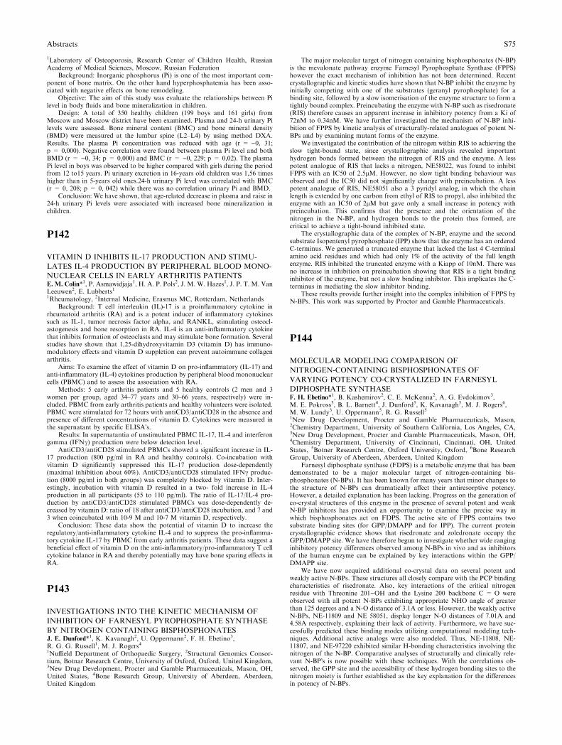
1Laboratory of Osteoporosis, Research Center of Children Health, RussianAcademy of Medical Sciences, Moscow, Russian Federation
Background: Inorganic phosphorus (Pi) is one of the most important com-ponent of bone matrix. On the other hand hyperphosphatemia has been asso-ciated with negative effects on bone remodeling.
Objective: The aim of this study was evaluate the relationships between Pilevel in body fluids and bone mineralization in children.
Design: A total of 350 healthy children (199 boys and 161 girls) fromMoscow and Moscow district have been examined. Plasma and 24-h urinary Pilevels were assessed. Bone mineral content (BMC) and bone mineral density(BMD) were measured at the lumbar spine (L2–L4) by using method DXA.Results. The plasma Pi concentration was reduced with age (r = )0, 31;p = 0,000). Negative correlation were found between plasma Pi level and bothBMD (r = )0, 34; p = 0,000) and BMC (r = )0, 229; p = 0,02). The plasmaPi level in boys was observed to be higher compared with girls during the periodfrom 12 to15 years. Pi urinary excretion in 16-years old children was 1,56 timeshigher than in 5-years old ones.24-h urinary Pi level was correlated with BMC(r = 0, 208; p = 0, 042) while there was no correlation urinary Pi and BMD.
Conclusion: We have shown, that age-related decrease in plasma and raise in24-h urinary Pi levels were associated with increased bone mineralization inchildren.
P142
VITAMIN D INHIBITS IL-17 PRODUCTION AND STIMU-
LATES IL-4 PRODUCTION BY PERIPHERAL BLOOD MONO-
NUCLEAR CELLS IN EARLY ARTHRITIS PATIENTSE. M. Colin*1, P. Asmawidjaja1, H. A. P. Pols2, J. M. W. Hazes1, J. P. T. M. VanLeeuwen2, E. Lubberts11Rheumatology, 2Internal Medicine, Erasmus MC, Rotterdam, Netherlands
Background: T cell interleukin (IL)-17 is a proinflammatory cytokine inrheumatoid arthritis (RA) and is a potent inducer of inflammatory cytokinessuch as IL-1, tumor necrosis factor alpha, and RANKL, stimulating osteocl-astogenesis and bone resorption in RA. IL-4 is an anti-inflammatory cytokinethat inhibits formation of osteoclasts and may stimulate bone formation. Severalstudies have shown that 1,25-dihydroxyvitamin D3 (vitamin D) has immuno-modulatory effects and vitamin D suppletion can prevent autoimmune collagenarthritis.
Aims: To examine the effect of vitamin D on pro-inflammatory (IL-17) andanti-inflammatory (IL-4) cytokines production by peripheral blood mononuclearcells (PBMC) and to assess the association with RA.
Methods: 5 early arthritis patients and 5 healthy controls (2 men and 3women per group, aged 34–77 years and 30–66 years, respectively) were in-cluded. PBMC from early arthritis patients and healthy volunteers were isolated.PBMC were stimulated for 72 hours with antiCD3/antiCD28 in the absence andpresence of different concentrations of vitamin D. Cytokines were measured inthe supernatant by specific ELISA�s.
Results: In supernatantia of unstimulated PBMC IL-17, IL-4 and interferongamma (IFNc) production were below detection level.
AntiCD3/antiCD28 stimulated PBMCs showed a significant increase in IL-17 production (800 pg/ml in RA and healthy controls). Co-incubation withvitamin D significantly suppressed this IL-17 production dose-dependently(maximal inhibition about 60%). AntiCD3/antiCD28 stimulated IFNc produc-tion (8000 pg/ml in both groups) was completely blocked by vitamin D. Inter-estingly, incubation with vitamin D resulted in a two- fold increase in IL-4production in all participants (55 to 110 pg/ml). The ratio of IL-17/IL-4 pro-duction by antiCD3/antiCD28 stimulated PBMCs was dose-dependently de-creased by vitamin D: ratio of 18 after antiCD3/antiCD28 incubation, and 7 and3 when coincubated with 10-9 M and 10-7 M vitamin D, respectively.
Conclusion: These data show the potential of vitamin D to increase theregulatory/anti-inflammatory cytokine IL-4 and to suppress the pro-inflamma-tory cytokine IL-17 by PBMC from early arthritis patients. These data suggest abeneficial effect of vitamin D on the anti-inflammatory/pro-inflammatory T cellcytokine balance in RA and thereby potentially may have bone sparing effects inRA.
P143
INVESTIGATIONS INTO THE KINETIC MECHANISM OF
INHIBITION OF FARNESYL PYROPHOSPHATE SYNTHASE
BY NITROGEN CONTAINING BISPHOSPHONATESJ. E. Dunford*1, K. Kavanagh2, U. Oppermann2, F. H. Ebetino3,R. G. G. Russell1, M. J. Rogers41Nuffield Department of Orthopaedic Surgery, 2Structural Genomics Consor-tium, Botnar Research Centre, University of Oxford, Oxford, United Kingdom,3New Drug Development, Procter and Gamble Pharmaceuticals, Mason, OH,United States, 4Bone Research Group, University of Aberdeen, Aberdeen,United Kingdom
The major molecular target of nitrogen containing bisphosphonates (N-BP)is the mevalonate pathway enzyme Farnesyl Pyrophosphate Synthase (FPPS)however the exact mechanism of inhibition has not been determined. Recentcrystallographic and kinetic studies have shown that N-BP inhibit the enzyme byinitially competing with one of the substrates (geranyl pyrophosphate) for abinding site, followed by a slow isomerisation of the enzyme structure to form atightly bound complex. Preincubating the enzyme with N-BP such as risedronate(RIS) therefore causes an apparent increase in inhibitory potency from a Ki of72nM to 0.34nM. We have further investigated the mechanism of N-BP inhi-bition of FPPS by kinetic analysis of structurally-related analogues of potent N-BPs and by examining mutant forms of the enzyme.
We investigated the contribution of the nitrogen within RIS to achieving theslow tight-bound state, since crystallographic analysis revealed importanthydrogen bonds formed between the nitrogen of RIS and the enzyme. A lesspotent analogue of RIS that lacks a nitrogen, NE58022, was found to inhibitFPPS with an IC50 of 2.5lM. However, no slow tight binding behaviour wasobserved and the IC50 did not significantly change with preincubation. A lesspotent analogue of RIS, NE58051 also a 3 pyridyl analog, in which the chainlength is extended by one carbon from ethyl of RIS to propyl, also inhibited theenzyme with an IC50 of 2lM but gave only a small increase in potency withpreincubation. This confirms that the presence and the orientation of thenitrogen in the N-BP, and hydrogen bonds to the protein thus formed, arecritical to achieve a tight-bound inhibited state.
The crystallographic data of the complex of N-BP, enzyme and the secondsubstrate Isopentenyl pyrophosphate (IPP) show that the enzyme has an orderedC-terminus. We generated a truncated enzyme that lacked the last 4 C-terminalamino acid residues and which had only 1% of the activity of the full lengthenzyme. RIS inhibited the truncated enzyme with a Kiapp of 10nM. There wasno increase in inhibition on preincubation showing that RIS is a tight bindinginhibitor of the enzyme, but not a slow binding inhibitor. This implicates the C-terminus in mediating the slow inhibitor binding.
These results provide further insight into the complex inhibition of FPPS byN-BPs. This work was supported by Proctor and Gamble Pharmaceuticals.
P144
MOLECULAR MODELING COMPARISON OF
NITROGEN-CONTAINING BISPHOSPHONATES OF
VARYING POTENCY CO-CRYSTALIZED IN FARNESYL
DIPHOSPHATE SYNTHASEF. H. Ebetino*1, B. Kashemirov2, C. E. McKenna2, A. G. Evdokimov3,M. E. Pokross3, B. L. Barnett4, J. Dunford5, K. Kavanagh5, M. J. Rogers6,M. W. Lundy3, U. Oppermann5, R. G. Russell51New Drug Development, Procter and Gamble Pharmaceuticals, Mason,2Chemistry Department, University of Southern California, Los Angeles, CA,3New Drug Development, Procter and Gamble Pharmaceuticals, Mason, OH,4Chemistry Department, University of Cincinnati, Cincinnati, OH, UnitedStates, 5Botner Research Centre, Oxford University, Oxford, 6Bone ResearchGroup, University of Aberdeen, Aberdeen, United Kingdom
Farnesyl diphosphate synthase (FDPS) is a metabolic enzyme that has beendemonstrated to be a major molecular target of nitrogen-containing bis-phosphonates (N-BPs). It has been known for many years that minor changes tothe structure of N-BPs can dramatically affect their antiresorptive potency.However, a detailed explanation has been lacking. Progress on the generation ofco-crystal structures of this enzyme in the presence of several potent and weakN-BP inhibitors has provided an opportunity to examine the precise way inwhich bisphosphonates act on FDPS. The active site of FPPS contains twosubstrate binding sites (for GPP/DMAPP and for IPP). The current proteincrystallographic evidence shows that risedronate and zoledronate occupy theGPP/DMAPP site. We have therefore begun to investigate whether wide ranginginhibitory potency differences observed among N-BPs in vivo and as inhibitorsof the human enzyme can be explained by key interactions within the GPP/DMAPP site.
We have now acquired additional co-crystal data on several potent andweakly active N-BPs. These structures all closely compare with the PCP bindingcharacteristics of risedronate. Also, key interactions of the critical nitrogenresidue with Threonine 201)OH and the Lysine 200 backbone C = O wereobserved with all potent N-BPs exhibiting appropriate NHO angle of greaterthan 125 degrees and a N-O distance of 3.1A or less. However, the weakly activeN-BPs, NE-11809 and NE 58051, display longer N-O distances of 7.01A and4.58A respectively, explaining their lack of activity. Furthermore, we have suc-cessfully predicted these binding modes utilizing computational modeling tech-niques. Additional active analogs were also modeled. Thus, NE-11808, NE-11807, and NE-97220 exhibited similar H-bonding characteristics involving thenitrogen of the N-BP. Comparative analyses of structurally and clinically rele-vant N-BP�s is now possible with these techniques. With the correlations ob-served, the GPP site and the accessibility of these hydrogen bonding sites to thenitrogen moiety is further established as the key explanation for the differencesin potency of N-BPs.
Abstracts S75
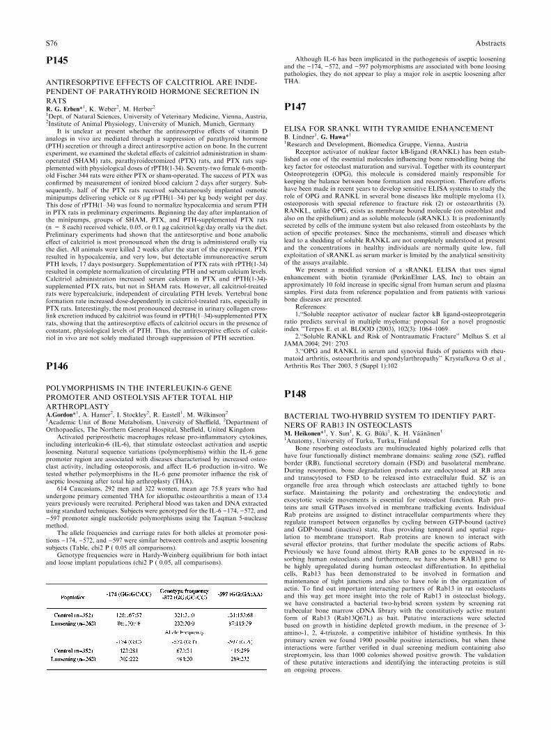
P145
ANTIRESORPTIVE EFFECTS OF CALCITRIOL ARE INDE-
PENDENT OF PARATHYROID HORMONE SECRETION IN
RATSR. G. Erben*1, K. Weber2, M. Herber21Dept. of Natural Sciences, University of Veterinary Medicine, Vienna, Austria,2Institute of Animal Physiology, University of Munich, Munich, Germany
It is unclear at present whether the antiresorptive effects of vitamin Danalogs in vivo are mediated through a suppression of parathyroid hormone(PTH) secretion or through a direct antiresorptive action on bone. In the currentexperiment, we examined the skeletal effects of calcitriol administration in sham-operated (SHAM) rats, parathyroidectomized (PTX) rats, and PTX rats sup-plemented with physiological doses of rPTH(1-34). Seventy-two female 6-month-old Fischer 344 rats were either PTX or sham-operated. The success of PTX wasconfirmed by measurement of ionized blood calcium 2 days after surgery. Sub-sequently, half of the PTX rats received subcutaneously implanted osmoticminipumps delivering vehicle or 8 lg rPTH(1–34) per kg body weight per day.This dose of rPTH(1–34) was found to normalize hypocalcemia and serum PTHin PTX rats in preliminary experiments. Beginning the day after implantation ofthe minipumps, groups of SHAM, PTX, and PTH-supplemented PTX rats(n = 8 each) received vehicle, 0.05, or 0.1 lg calcitriol/kg/day orally via the diet.Preliminary experiments had shown that the antiresorptive and bone anaboliceffect of calcitriol is most pronounced when the drug is administered orally viathe diet. All animals were killed 2 weeks after the start of the experiment. PTXresulted in hypocalcemia, and very low, but detectable immunoreactive serumPTH levels, 17 days postsurgery. Supplementation of PTX rats with rPTH(1-34)resulted in complete normalization of circulating PTH and serum calcium levels.Calcitriol administration increased serum calcium in PTX and rPTH(1-34)-supplemented PTX rats, but not in SHAM rats. However, all calcitriol-treatedrats were hypercalciuric, independent of circulating PTH levels. Vertebral boneformation rate increased dose-dependently in calcitriol-treated rats, especially inPTX rats. Interestingly, the most pronounced decrease in urinary collagen cross-link excretion induced by calcitriol was found in rPTH(1–34)-supplemented PTXrats, showing that the antiresorptive effects of calcitriol occurs in the presence ofconstant, physiological levels of PTH. Thus, the antiresorptive effects of calcit-riol in vivo are not solely mediated through suppression of PTH secretion.
P146
POLYMORPHISMS IN THE INTERLEUKIN-6 GENE
PROMOTER AND OSTEOLYSIS AFTER TOTAL HIP
ARTHROPLASTYA.Gordon*1, A. Hamer2, I. Stockley2, R. Eastell1, M. Wilkinson21Academic Unit of Bone Metabolism, University of Sheffield, 2Department ofOrthopaedics, The Northern General Hospital, Sheffield, United Kingdom
Activated periprosthetic macrophages release pro-inflammatory cytokines,including interleukin-6 (IL-6), that stimulate osteoclast activation and asepticloosening. Natural sequence variations (polymorphisms) within the IL-6 genepromoter region are associated with diseases characterised by increased osteo-clast activity, including osteoporosis, and affect IL-6 production in-vitro. Wetested whether polymorphisms in the IL-6 gene promoter influence the risk ofaseptic loosening after total hip arthroplasty (THA).
614 Caucasians, 292 men and 322 women, mean age 75.8 years who hadundergone primary cemented THA for idiopathic osteoarthritis a mean of 13.4years previously were recruited. Peripheral blood was taken and DNA extractedusing standard techniques. Subjects were genotyped for the IL-6 )174, )572, and)597 promoter single nucleotide polymorphisms using the Taqman 5-nucleasemethod.
The allele frequencies and carriage rates for both alleles at promoter posi-tions )174, )572, and )597 were similar between controls and aseptic looseningsubjects (Table, chi2 P ( 0.05 all comparisons).
Genotype frequencies were in Hardy-Weinberg equilibrium for both intactand loose implant populations (chi2 P ( 0.05, all comparisons).
Although IL-6 has been implicated in the pathogenesis of aseptic looseningand the )174, )572, and )597 polymorphisms are associated with bone loosingpathologies, they do not appear to play a major role in aseptic loosening afterTHA.
P147
ELISA FOR SRANKL WITH TYRAMIDE ENHANCEMENTB. Lindner1, G. Hawa*11Research and Development, Biomedica Gruppe, Vienna, Austria
Receptor activator of nuklear factor kB-ligand (RANKL) has been estab-lished as one of the essential molecules influencing bone remodelling being thekey factor for osteoclast maturation and survival. Together with its counterpartOsteoprotegerin (OPG), this molecule is considered mainly responsible forkeeping the balance between bone formation and resorption. Therefore effortshave been made in recent years to develop sensitive ELISA systems to study therole of OPG and RANKL in several bone diseases like multiple myeloma (1),osteoporosis with special reference to fracture risk (2) or osteoarthritis (3).RANKL, unlike OPG, exists as membrane bound molecule (on osteoblast andalso on the epithelium) and as soluble molecule (sRANKL). It is predominantlysecreted by cells of the immune system but also released from osteoblasts by theaction of specific proteases. Since the mechanisms, stimuli and diseases whichlead to a shedding of soluble RANKL are not completely understood at presentand the concentrations in healthy individuals are normally quite low, fullexploitation of sRANKL as serum marker is limited by the analytical sensitivityof the assays available.
We present a modified version of a sRANKL ELISA that uses signalenhancement with biotin tyramide (PerkinElmer LAS, Inc) to obtain anapproximately 10 fold increase in specific signal from human serum and plasmasamples. First data from reference population and from patients with variousbone diseases are presented.
References:1.‘‘Soluble receptor activator of nuclear factor kB ligand-osteoprotegerin
ratio predicts survival in multiple myeloma: proposal for a novel prognosticindex ’’Terpos E. et al. BLOOD (2003), 102(3): 1064–1069
2.‘‘Soluble RANKL and Risk of Nontraumatic Fracture’’ Melhus S. et alJAMA.2004; 291: 2703
3.‘‘OPG and RANKL in serum and synovial fluids of patients with rheu-matoid arthritis, osteoarthritis and spondylarthropathy’’ Krystufkova O et al ,Arthritis Res Ther 2003, 5 (Suppl 1):102
P148
BACTERIAL TWO-HYBRID SYSTEM TO IDENTIFY PART-
NERS OF RAB13 IN OSTEOCLASTSM. Heikonen*1, Y. Sun1, K. G. Buki1, K. H. Vaananen11Anatomy, University of Turku, Turku, Finland
Bone resorbing osteoclasts are multinucleated highly polarized cells thathave four functionally distinct membrane domains: sealing zone (SZ), ruffledborder (RB), functional secretory domain (FSD) and basolateral membrane.During resorption, bone degradation products are endocytosed at RB areaand transcytosed to FSD to be released into extracellular fluid. SZ is anorganelle free area through which osteoclasts are attached tightly to bonesurface. Maintaining the polarity and orchestrating the endocytotic andexocytotic vesicle movements is essential for osteoclast function. Rab pro-teins are small GTPases involved in membrane trafficking events. IndividualRab proteins are assigned to distinct intracellular compartments where theyregulate transport between organelles by cycling between GTP-bound (active)and GDP-bound (inactive) state, thus providing temporal and spatial regu-lation to membrane transport. Rab proteins are known to interact withseveral effector proteins, that further modulate the specific actions of Rabs.Previously we have found almost thirty RAB genes to be expressed in re-sorbing human osteoclasts and furthermore, we have shown RAB13 gene tobe highly upregulated during human osteoclast differentiation. In epithelialcells, Rab13 has been demonstrated to be involved in formation andmaintenance of tight junctions and also to have role in the organization ofactin. To find out important interacting partners of Rab13 in rat osteoclastsand this way get more insight into the role of Rab13 in osteoclast biology,we have constructed a bacterial two-hybrid screen system by screening rattrabecular bone marrow cDNA library with the constitutively active mutantform of Rab13 (Rab13Q67L) as bait. Putative interactions were selectedbased on growth in histidine depleted growth medium, in the presence of 3-amino-1, 2, 4-triazole, a competitive inhibitor of histidine synthesis. In thisprimary screen we found 1900 possible positive interactions, but when theseinteractions were further verified in dual screening medium containing alsostreptomycin, less than 1000 colonies showed positive growth. The validationof these putative interactions and identifying the interacting proteins is stillan ongoing process.
S76 Abstracts
P149
IL-6 INHIBITS RANKL-INDUCED OSTEOCLASTOGENESISL. Duplomb1, V. Trichet1, M. Baud’huin1, C. Charrier1, S. Battaglia1,D. Heymann1, F. Blanchard*11University of Nantes EA3822 INSERM ERI 7, Medicine Faculty, Nantes,France
Osteoclasts are bone-resorptive multinucleated cells which differentiate fromhematopoıetic precursors of the monocyte/macrophage lineage in the presence ofM-CSF and receptor activator of NF-kB ligand (RANKL). Previous studiesdemonstrated that IL-6 indirectly up-modulates osteoclast differentiationthrough the production of RANKL by osteoblasts. However, only few data areavailable concerning the direct effect of IL6 on osteoclast. To investigate the roleof IL-6 on osteoclast differentiation, we used the monocyte/macrophage cell lineRAW 264.7 which differentiates in osteoclast in the presence of RANKL. Weshowed that the addition of IL-6 inhibits RANKL-induced osteoclast differen-tiation of RAW 264.7 in a dose dependant way (81% inhibition at 100 ng/ml).Furthermore, this effect was irreversible as Raw 264.7 were resistant to RANKLaction after been pre-cultured for 3 days with IL-6. RT-PCR and Flow cytom-etry analysis showed that IL-6 decreased the expression of specific osteoclastsmarkers such as Tartrate Resistant Acid Phosphatase, Calcitonin Receptor andCD44. Interestingly, Raw 264.7 cultured in presence of RANKL and IL-6strongly expressed macrophage markers such as CD11b and CD16/CD32, whichwere both conversely down-regulated in Raw 264.7 cultured with RANKL only.Finally, we confirmed the IL-6 inhibitory effect on osteoclasts differentiation byusing freshly isolated mouse bone marrow precursors. The mechanisms involvedin this inhibition, such as signalling pathways, are currently under investigations.Thus, our study showed for the first time that IL6 inhibits directly RANKL-induced osteoclastogenesis by diverting cells into the macrophage lineage. Insuch context, macrophage differentiation appeared dominant on osteoclast dif-ferentiation. The understanding of the balance osteoclast/macrophage is of greatimportance for novel therapeutic approaches of pathologies associating boneremodelling and inflammatory process such as rheumatoid arthritis.
P150
CHARACTERIZATION OF OSTEOPROTEGERIN BINDING TO
GLYCOSAMINOGLYCANS BY SURFACE PLASMON RESO-
NANCE: ROLE IN THE INTERACTIONS WITH RECEPTOR
ACTIVATOR OF NUCLEAR FACTOR KB LIGAND (RANKL)
AND RANKS. Theoleyre1, S. Kwan Tat1, P. Vusio2, F. Blanchard1, J. Gallagher3, S. Ricard-Blum4, Y. Fortun1, M. Padrines1, F. Redini*1, D. Heymann11Universite de Nantes EA3822 INSERM ERI 7, Medicine Faculty, 2IFR26,Institut de Biologie, Nantes, France, 3Department of Medical Oncology, ChristieHospital NHS Trust, Manchester, United Kingdom, 4UMR 5086 CNRS UCBL,Institut de Biologie et Chimie des Proteines, Lyon, France
Osteoprotegerin (OPG) is a decoy receptor for receptor activator of nuclearfactor kB ligand (RANKL), a key inducer of osteoclastogenesis via its receptorRANK. We previously showed that RANK, RANKL and OPG are able to forma tertiary complex and that OPG must be also considered as a direct effector ofosteoclast functions. As OPG contains a heparin binding domain, the presentstudy investigated the interactions between OPG and glycosaminoglycans(GAGs) by surface plasmon resonance and their involvement in the OPGfunctions. Kinetic data confirmed that OPG binds to heparin with a high affinity(KD: 0.28 nanoM) and that the pre-incubation of OPG with heparin inhibits in adose-dependent manner the OPG binding to the complex RANK-RANKL.GAGs from different structure/origin (heparan sulfate, dermatan sulfate,chondroitin sulfate) exert similar activity on OPG binding. The contribution ofthe sulfation pattern and the size of the oligosaccharide have been determined inthis inhibitory mechanism. The results demonstrated that sulfation is essential inthe OPG-blocking function of GAGs since a totally desulfated heparin loose itscapacity to bind to block OPG-binding to RANKL. Moreover a decasaccharideis the minimal structure that totally inhibits the OPG binding to the complexRANK-RANKL. Western blot analysis performed in 293 cells surexpressingRANKL, revealed that the pre-incubation of OPG with these GAGs inhibits theOPG-induced decrease of membrane RANKL half-life, confirming the resultsobtained by surface plasmon resonance binding assays. These data support anessential function of the related glycosaminoglycans heparin and heparan sulfatein the activity of the triad RANK-RANKL-OPG.
P151
DETECTION OF CATHEPSIN K IN OSTEOCLASTS BY ELISA
AND IMMUNOHISTOCHEMISTRY IN COMMON IN VITRO
BONE RESORPTION MODELSO. Hoffmann*1, J. Nguemo1, S. Maitzen2, G. Hawa2
1Pharmacology and Toxicology, University of Vienna, 2Research and Devel-opment, Biomedica, Vienna, Austria
Cathepsin K is a member of the cysteine protease family that is highlyexpressed in osteoclasts and plays an essential role in bone resorption bydegrading the collagen matrix of bone. It is the most abundantly synthesizedprotein in active, resorbing osteoclasts and is, therefore, a candidate markerfor osteoclast bone resorption activity. We raised 3 antibodies against hu-man cathepsin K that are currently being used in ELISAs. In the currentstudy, we tested the capacity of these antibodies to detect the cellular dis-tribution of cathepsin K in mouse osteoclasts and cathepsin K proteinproduction from mouse and rabbit osteoclasts during in vitro differentiationand bone resorption. Osteoclasts were either generated by co-culture withmouse bone marrow precursor cells and osteoblasts in the presence of 1,25-dihyroxyvitamin D3 and PGE2 for 5–6 days or directly isolated from rabbitlong bones. Osteoclasts were then fixed and stained with 3 individual fluo-rochrome-conjugated antibodies to cathepsin K for immunofluorescencedetection with confocal microscopy. In addition, we measured the levels ofcathepsin K secreted during cultures in which osteoclasts are seeded ontoplastic or bone surfaces. We found that all 3 antibodies against humancathepsin K recognized mouse osteoclast cathepsin K. Cellular distributionof cathepsin K was near the cell membrane. Cathepsin K protein levelsmeasured with our novel ELISA assay were detected in culture supernatantsbeginning on culture day 2 (7.15 pM/l) and then on days 3 and 5 (8.6 and11.7pM/l, respectively). The concentration of cathepsin K peaked in day 5supernatants and appeared to correlate with increased bone resorption and ahigh number of TRAP+ cells present within the cultures. To test forchanges in cathepsin K levels upon inhibition of osteoclast differentiationand function, we added flavopiridol, a cyclin-dependent kinase 2 inhibitor, tobone cell cultures. Flavopiridol significantly reduced bone resorption andTRAP+ cells, which correlated with reduced cathepsin K concentrations inthe supernatants. Taken together, these data demonstrate that these anti-bodies are cross reactive with human, mouse, and rabbit cathepsin K andthat cathepsin K levels can be measured using our recently establishedELISA. The determination of cathepsin K levels in bone cultures is apotentially useful assay for testing the effectiveness of anti-resorptive drugson in vitro osteoclast differentiation and function.
P152
THE EVALUATION OF OSTEOPOROSIS RISK IN PATIENTS
WITH THYROID CANCERSW. Horst-Sikorska*1, W. Nosowicz1, S. Dabrowski1, L. Celczynska-Bajew1
1Family Medicine Department, University of Medical Sciences, Poznan, Po-land
Background/Aim: Malignant thyroid cancer is derived from epithelial orfollicular cells. The death rate per 100 000 people is 0,5 for men and 2 forwomen.
The standard therapy of thyroid cancer consists in total strumectomy,administration of J131 ablational doses and L-tyroksine in doses which suppressTSH secretion. The aftermath of L-tyroksine dosages is the iatrogenic hyper-thyroidism. In patients with hyperthyroidism, acceleration of bone resorptionwith relation to bone reconstruction, and the increase of number resorptionactive places leads up to low quality of bone. Thus results in higher risk of bonefractures in women and men at all age.
The aim of the study was to evaluate if existing standards in thyroid cancerstreatment include prevention of osteoporosis in proper extent.
Subjects and methods. Patients were recruited from Clinical Hospital SPSK2 AM in Poznan, Poland. The subjects were 83 patients with thyroid cancers. Allpatients were treated with L-tyroksine.
Patients were investigated with the osteoporosis questionnaire based ondiagnostic guidelines of Polish Foundation of Osteoporosis.
The measurements of bone density in hip and lumbar (L1-L4) were doneusing DEXA.
Results: A total of 71 women and 12 men were recruited. Mean age ofwomen - 48,3; men - 45,08. Mean height of women - 164,16; men - 177,42. Meanloss of height (at baseline - the start of the study) in women - 0,83; men - 1,17.Among women 27% had used HRT or contraceptive pills. About 23% of womenand 8% of men had used vitamin D3, and 45% of women and 17% of men hadused calcium as a dietary supplement. In group of women 17% had suffered fromfracture, and 42% of them were low-energy fractures. In group of men 42% hadsuffered fracture, and all of them were high-energy fractures. About 24% ofwomen and 17% of men reported that their mother had a fracture. In historytaking 34% of women and 17% of men were diagnosed with hyperthyroidism.The mean T-score (measured in L1–L4) in women: 0,19 and in men: )0,30. Themean Z-score in women: 0,17 and in men: )0,20. The mean T-score (measured inhip bone) in women: )0,21 and in men: -0,17. The mean Z-score in women: 0,23and in men: )0,02.
Conclusions. The treatment of thyroid cancer should be completed withantiresorption therapy.
Abstracts S77
P153
VASCULAR CALCIFICATION IN PATIENTS ON HEMODIAL-
YSISB. B. J. Jeren*1, B. B. R. Rozman1, P. M. P. Marusic21Hemodalysis, 2Radiology, Clinical Hosital Dubrava, Zagreb, Croatia
Uremic bone disease is syndrome that includes two entites: mineralizationdeficiency and bone loss.
Bone loss associated with alterations of bone remodelling and mineralizationinduces fractures.
Aim of the study: To correlate vascular calcification (VC) intensity andduration with duration of heamodialysis, age of patients, level of parathyroidalhormon (PTH) and product of calcium and phosphorous(CaP) in the serum.
Patients and methods: The standard roentgenograms of the chest, lumbarspine, pelvis, upper leg, lower leg, feet, upper arms, forearms and hands thepresence, site and severity of VC in twenty patients ranging from 28 to 74 yearsof age (mean age 54.5) were analysed. Average duration of haemodialysis was9.2 years (ranging from 4 to 13 years). PTH value (normal level 8–16 pg/ml) andCaP value (normal level 2.4 mmol/L) were measured in all partients. Accrodingto severity of VC patients were divided in three groups: group 1 patients withmild VC, group 2 with moderate VC and group 3 patients with severe VC.
Results: The first group included 13 patients, second 4, and the third grouphad 3 patients. Mean age in the first group was 49.9 years, in second 62.5 yearsand in third group 64 years of age. Average duration of heamodialysis in the firstgroup was 7.6 years, in second 9.0 years and in third group 9.3 years. Theaverage level of PTH in the firs group was 355.8 pg/ml, in second 637.5 pg/mland in third 693.3 pg/ml. CaP value in the first group was 4.56 mmol/L, insecond 4.64 mmol/L and 4.90mmol/L in the third group.
Conclusion: This study shows severe VC in the patients that have been onheamodialysis for a longer time and those with higher levels ofPTH and CaP inserum compared to the patients with mild and moderate VC.
P154Abstract withdrawn
P155
EFFECT OF TOPICAL ADMINISTERED CLODRONATE ON
THE BONE MINERAL DENSITY AFTER TOTAL HIP
ARTHROPLASTYM. Kellinsalmi*1, R. Ojala2, J. Risteli3, M. Hamalainen1, P. Lehenkari11Department of Surgery, 2Department of Radiology, 3Department of ClinicalChemistry, University Hospital of Oulu, Oulu, Finland
Periprosthetic bone loss is a phenomenom that exposes to periprostheticfractures and to revision arthroplasties. According to Wolf’s law the implantcarries or directs the forces that are normally taken by bone alone.The dual-energy absorptiometric (DXA) studies suggest that even 40-60 % of peri-implantbone looses in five years. Loosening of prosthesis is a result of combination ofdifferent factors including also the inflammatory process which could be acti-vated by metal or cement particles released from the implant thus activatingmacrophages and osteoclasts.
Bisphosphonates are widely used in the situations related to osteoclastmediated bone loss like osteoporosis. Main target of bisphosphonates is boneresorbing osteoclasts and from this aspect they could be the drug of choise forpatients who are going to have the implant surgery.
The patients undergoing the primary cementless total hip arthroplasty wererecruited. The study group consisted of 28 patients who were randomized anddouble-blinded to receive either the topical clodronate-rinsing to femoral bonemarrow before application of prosthesis (A) or to control patients rinsed by saline(B). Patients in the study group C received clodronate orally at dose 1600mg/dayfor three months. Patients were followed by x-ray pictures, DXA, specific bonemarkers and physical recovering was examined by modified Harris-hip-score.
At one year control point bone mineral density (BMD) decreased most in thefemoral calcar. The decreasewas equal in all groupsA,B andC; 86%, 85% and 86%of the baseline, respectively. A slight increase in BMD was seen in the area oftrochanter major (Gruen zone 1) in goups A and B. The increase was most evidentin control-group 109% of the baseline and 102% in Group B. BMD decreased to96% from the baseline inGroupC (p<0.05). Inspecting the effect of clodronate onBMD in total peri-implant area (gruen zones 1–7) no significant changes betweenthe study groups were detected (97%, 95%, 95% of the baseline, respectively).
Surprisingly a significant increase in BMD was seen after 12 months to 24months in clodronate-receiving groups. Formation of ectopic bone was the leastat clodronate rinsing-group (11%).
During the short follow-up of two years a minor increase in periprostheticbone density was observed related to bisphosphonate therapy. The longer sur-veillance is needed to find out whether the topical clodronate-rinsing facilitatesthe better survival of implants.
P156
A MOUSEMODEL FOR CRANIOMETAPHYSEAL DYSPLASIA
DEMONSTRATES OSTEOBLAST DYSREGULATION AS
CAUSE OF HYPEROSTOSISJ. Kuehnisch*1, J. Loebner1, M. Giehl2, D. Epari3, H. Thiele4, D. Felsenberg2,P. Nuernberg5, S. Mundlos1, U. Kornak11Institute for Medical Genetics, 2Centre of Muscle and Bone Research, 3Centerfor Musculoskeletal Surgery, Charite, Berlin, 4Cologne Centre for Genomics,University of Cologne, Cologne, 5Cologne Centre for Genomics, University ofCologne, Berlin, Germany
Craniometaphyseal Dysplasia (CMD) is a rare autosomal dominant humanskeletal disorder characterised by sclerosis of the skull and reduced modelling ofthe long bones. Complications include facial palsy, hearing loss or blindness.Missense mutations and amino acid deletions in the human ankylosis geneANKH were identified as the cause of CMD. ANKH encodes for the integralmembrane protein ANK, which is thought to transport pyrophosphate (PPi)from the cytosol to the extracellular space. PPi is a potent inhibitor of matrixcalcification and it is assumed that the hyperostosis in CMD is somehow causedby altered extra- or intracellular PPi levels.
ANKH is expressed in a variety of cell types, among them osteoblasts andosteoclasts, the two cell types necessary for bone remodelling. To reveal themolecular mechanisms and the cell type primarily affected in CMD we generateda transgenic mouse line (TgC1AnkF377Del) that expresses the most commonAnk mutation F377Del under the control of an osteoblast-specific collagen 1alpha 1 promoter fragment. RT-PCR revealed a moderate level of transgeneexpression. We analysed femora and vertebrae of adult wild type and transgenicmice by pQCT, lCT, and histomorphometry.
The pQCT analysis revealed a slightly increased trabecular bone mineraldensity and cross-sectional surface in TgC1AnkF377Del mice. Cortical bonemineral density was not altered. Histological and lCT examination showedsignificantly increased bone density in the femora of theTgC1AnkF377Del micedue to higher trabecular numbers and reduced trabecular separation. In con-trast, bone density in vertebrae from transgenic mice appeared decreased. Noskull hyperostosis could be found.
Our results indicate that CMD is most likely caused by osteoblast dysre-gulation. The different extent of bone density elevation detected in pQCTcompared to lCT and histomorphometry is in line with the finding that CMDpatients on average display slightly reduced bone mineral density at the spineand the femoral neck in spite of high trabecular density.
P157
TREATMENT OF YOUNG MALE MONKEYS FOR 12
MONTHS WITH A HIGHLY POTENT INHIBITOR OF
CATHEPSIN K INHIBITS BONE RESORPTION, AND IN-
CREASES BONE MINERAL DENSITY AND STRENGTHS. Kumar*1, S. Rehm1, R. Boyce1, J. Birmingham1, G. Stroup1, C. Jerome2,P. Wier11R and D, GlaxoSmithKline, Collegeville, 2R and D, MDS Pharma, Bothell,United States
Objective: Cathepsin K has been shown to be highly expressed in osteoclastsand play an important role in bone matrix degradation. SB-462795 (relacatib) isa potent inhibitor of human cathepsin K, L and V. This study investigated thepharmacological effects of relacatib in monkeys.
Methods: Four groups of eight, 3–5 year-old, male, cynomolgus monkeysreceived vehicle or relacatib at 3, 30, or 1000 mg/kg/day by daily gavage for 52weeks. Bone biomarkers and bone density were measured prior to, and peri-odically during the study; bone biomechanical testing, bone histomorphometry,gross and histopathologic evaluations were performed terminally. The com-pound was well tolerated at all doses, with no evidence of significant toxicity.
Results & Conclusions: Compared with pre-treatment values, treatment at30 or 1000 mg/kg/day had significant and comparable effects to reduce cartilageand bone resorption biomarkers. A decrease in one bone formation marker(Bone-specific Alkaline Phosphatase) was also observed but not another(Osteocalcin).
A significant increase in BMD was observed at sites rich in trabecular bone(lumbar spine, femur neck, and distal radius), and at sites rich in cortical bone(mid-femur and proximal radius). The mid-femur exhibited a dose-dependentdecrease in medullary cavity area, consistent with positive bone balance on en-docortical surfaces, and increased total area, consistent with stimulation of boneformation on the periosteal surface. Biomechanical testing demonstrated, asignificant increase in maximum load, 42% and 49%, respectively, at 30 and 1000mg/kg/day in lumbar vertebrae. Histomorphometric data (LV2 and mid-femur)showed significantly decreased activation on all endosteal surfaces, consistentwith a reduction in bone turnover in animals treated at 30 and 1000 mg/kg/day.
Thus relacatib (SB-462795) appears to increase bone strength in young malemonkeys by decreasing bone turnover and improving the balance between boneresorption and formation.
S78 Abstracts

P158
SB-462795 (RELACATIB), A CATHEPSIN INHIBITOR, PRE-
VENTS THE LOSS OF BONE MASS AT SEVERAL SITES IN
OVARIECTOMIZED CYNOMOLGUS MONKEYSS. Kumar*1, C. Jerome2, G. Stroup11R and D, GlaxoSmithKline, Collegeville, 2R and D, MDS Pharma, Bothell,United States
Cathepsin K has been shown to play an important role in bone matrixdegradation during osteoclastic bone resorption. We studied the effects of SB-462795 (relacatib) a novel, potent inhibitor of cathepsins K, L & V on boneturnover in aged ovariectomized (Ovx) female monkeys.
After collection of baseline data adult female cynomolgus monkeys wererandomized to 6 groups of 20 each, of which one was sham-Ovx (Sham) & theremainder Ovx. Ovx animals were treated with vehicle, relacatib at 1, 3, or 10mg/kg/d by oral gavage, or alendronate at 0.05 mg/kg by IV injection once every2 weeks for 9 months. All statistical comparisons are with Ovx, vehicle-treatedanimals.
Relacatib dose-dependently decreased urinary excretion of NTx with a sig-nificant effect observed earlier for relacatib versus vehicle than alendronate.Unlike alendronate which decreased bone formation markers, treatment withrelacatib at 10 mg/kg/d decreased bone formation markers to a lesser extent thanthe bone resorption markers & significantly increased serum osteocalcin. Thissuggests that relacatib may inhibit bone turnover while providing a morefavorable balance of resorption and formation compared to a bisphosphonate.
Animals treated with relacatib had dose-dependent improvement in aBMDat the lumbar spine and proximal & distal femur. At the distal femur relacatibprevented 73% of the loss in aBMD while alendronate showed a non-significant23% protection. At the proximal femur the level of protection by the two agentswere comparable. At the lumbar spine, relacatib protected 42% while alendro-nate protected 100% of the loss in aBMD. Femoral neck analysis using pQCT,showed a similar level of protection of total vBMD by relacatib (58%, p < 0.05)& alendronate (51%, p > 0.1). Both agents had a similar, positive effect ontrabecular vBMD & BMC. Treatment with relacatib at the high dose signifi-cantly increased cortical area & cortical BMC compared to vehicle. This sta-tistical significance was not achieved with alendronate.
Histomorphometry data revealed a dose-dependent reduction in both boneresorption & formation parameters at cancellous sites with both agents. How-ever, a stimulatory effect on femur periosteal bone (Ps.BFR/BV) was observedwith relacatib but not with alendronate treatment. These findings suggest thatrelacatib significantly differs from alendronate in that it may have classic anti-resorptive effects in cancellous bone, and a novel, beneficial, stimulatory effecton cortical bone.
P159
FIBROBLAST GROWTH FACTOR 8E INCREASES OSTEOCL-
ASTOGENESIS IN VITROM. Mattila1, T. Laitala-Leinonen*1, K. Buki1, K. Vaananen1, P. Harkonen11Dept. Anatomy, Inst. Biomedicine, Turku, Finland
Osteoclast formation is regulated by signals transmitted from their micro-environment. In bone marrow, mesenchymal stem cells provide for the nichewhere hematopoietic cells proliferate. Osteoblasts are mesenchymal cells thatproduce RANKL, a major regulator of osteoclast differentiation and activity.Treatment of hematopoietic cells with RANKL and M-CSF in vitro leads to theformation of multinuclear, bone-resorbing osteoclasts. We have producedFGF8e and used this purified protein to induce osteoclast formation in mousebone marrow cultures. The addition of FGF8e alone stimulated osteoclast for-mation similarly to RANKL + M-CSF treatment. The stimulatory effect ofFGF8e was almost totally inhibited by addition of OPG. These data suggest thatthe increased osteoclastogenesis with FGF8e was mediated by RANKL. Takentogether our data show that FGF8e is a powerful stimulator of osteoclast for-mation at least in mouse bone marrow cultures. The formed osteoclasts werecapable of bone resorption and exhibited characteristisks of mature osteoclasts,including polarized morphology and targeted vesicle trafficking. The effects ofFGF8e on osteoblasts are also discussed.
P160
REFINEMENTS IN THE USE OF COLUMN CHROMATOG-
RAPHY TO EXPOSE DIFFERENCES IN RELATIVE BINDING
AFFINITIES OF BISPHOSPHONATES TO HYDROXYAPATITEM. A. Lawson*1, Z. Xia1, J. T. Triffitt1, F. H. Ebetino2, R. J. Phipps2, R. G. G.Russell11Nuffield Department of Orthopaedic Surgery, University of Oxford, Oxford,United Kingdom, 2New Drug Development, Procter and Gamble Pharmaceu-ticals, Mason, OH, United States
It is becoming increasingly apparent that there are small but significantlyimportant differences among the bisphosphonates (BPs), in their affinities forbone mineral that may result in differences in their pharmacological potenciesand durations of action. To investigate differences in mineral binding that mayrelate more directly to their retention and diffusion through bone, we havedeveloped recently a novel method based on the use of hydroxyapatite crystals(BioGel� HT gel) and FPLC on glass columns (3 · 25 mm). The BPs adsorb tothe hydroxyapatite and can be eluted subsequently by using potassium phos-phate buffer gradients (1mM–1M) at pH 6.8. The specific BPs emerge as discretepeaks detectable by their UV absorbance, reaction with OPA or by mass spec-trometry. Under the particular conditions used the retention time (mins;mean ± SEM) for risedronate was 16.16 ± 1.17 compared with 22.0 ± 0.1 forzoledronate. The retention time for NE10790, a risedronate analogue in whichone of the phosphonate groups is replaced by a carboxyl group, was significantlylower at 7.33 ± 0.08. The elution characteristics were consistent and theretention times statistically significantly different (p < 0.05). Notably, it wasfound that that the sequence of chromographic elution was in the same order asthat described recently by determinations of kinetic affinity constants (KL) forbinding to HAP. By this method, which used an inhibition assay of hydroxy-apatite crystal growth, the KL values were 3.3, 2.0, 0.03 · 10-E6 L/mol forzoledronate, risedronate, and NE10790 respectively (Nancollas et al: Bone, 2005,in press). The hydroxyapatite crystal columns required repacking with newhydroxyapatite adsorbent to be prepared fresh after each run, which is incon-venient. However, by using a ceramic HAP preparation (BioRad) this problemhas been overcome as the column bed matrix is highly stable. The bindingaffinities are comparable within the two systems, but there is superior repro-ducibility with the ceramic HAP. The present results confirm that differences inthe binding affinities of BPs to hydroxyapatite are a feature of all BPs andsuggest that this is likely to be of high significance in those BPs commonly usedin clinical practice. The profiles of mineral binding abilities may help to explainthe variations in retention and the persistence of the effects of BPs observed bothin experimental animal and in clinical studies.
P161
REGULATION OF BONE BALANCE - SEQUENTIAL MICRO-
NUTRITIONM. Lugand- Bourgeois*11Research, Bioresearch and Partners, Monthey, Switzerland
Osteoporosis is becoming a real public health issue. Bioresearch & Partners�objective is to exploit a new nutritional approach:Micronutrisequences are acombination of microgramme-dosed dietary substances organized in sequence.
The micronutritional approach developed by Bioresearch & Partners is anew mode of utilizing nutrients which targets an action on the global regulationof bone balance, the nutrients have been selected to participate in a nutritionalstrategy aiming to help the restoration of bone metabolism homeostasis.
The aim of the nutritional intervention was the measurement in monother-apy in open study of bone balance regulation by the dosage of biologicalmarkers of bone formation (osteocalcin) and resorption (24-hour urine des-oxypiridonoline at the creatinine dosage), as well as the increase of bone densityobserved with bone density test in menopaused women presenting a disturbanceof bone balance markers and osteopenia or osteoporosis (BMD).
The nutritional intervention lasted two years. Were excluded of the trial thepatients who were immobilized or taking corticoids, having osteomalacia orunstable diseases: diabetes, thyroid, crohn disease, kidney or active hepaticdiseases.
Nutrisequence (capsules containing micronutrition granules) was taken threetimes a day during 3 months. Population studied: 24 women aged 64 in average.
All 24 patients are followed on bone markers balance, 10 are complemen-tarily followed in absorptiometry.
Results: Of the 17 cases out of 24 cases which were presenting at thebeginning of the trial a bone balance with an excessive increase of dpyr, 94% re-stabilized their bone balance, 82% totally normalized their balance (in 17,3months in average)Results on bone density: Of the 10 cases followed in BMD: 7patients out of 9 had osteopenia or osteoporosis and showed an increase of thetrabecular density by 7%.
Conclusion: the regulation of bone markers (82%) and the increase of bonedensity (+7%) offer a satisfactory response to the required therapeutic aims in apopulation presenting an excessively negative bone balance.
Micronutrisequence is efficient and perfectly adapted to a systematic pre-ventive action and can be associated with any other drug therapeutics.
P162
ADAMS IN THE FORMATION OF FOREIGN BODY GIANT
CELLS AND OSTEOCLASTSG. Ma*1, M. Ainola2, M. Liljestrom2, M. Hukkanen2, J. Salo3, Y. T. Konttinen41Department of Medicine, Institute of Biomeidicine, 2Anatomy, Biomedicum,3Orthopedics and Traumatology, Helsinki University Central Hospital,4Department of Medicine, Biomedicum, Helsinki, Finland
Abstracts S79
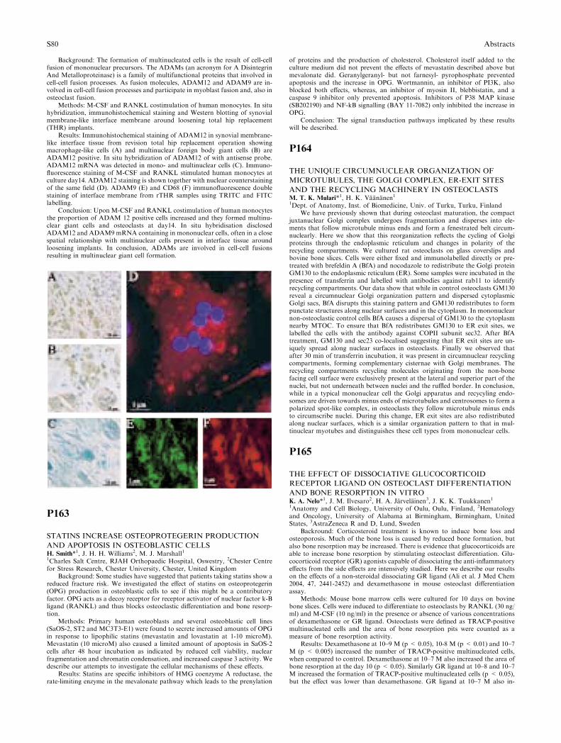
Background: The formation of multinucleated cells is the result of cell-cellfusion of mononuclear precursors. The ADAMs (an acronym for A DisintegrinAnd Metalloproteinase) is a family of multifunctional proteins that involved incell-cell fusion processes. As fusion molecules, ADAM12 and ADAM9 are in-volved in cell-cell fusion processes and participate in myoblast fusion and, also inosteoclast fusion.
Methods: M-CSF and RANKL costimulation of human monocytes. In situhybridization, immunohistochemical staining and Western blotting of synovialmembrane-like interface membrane around loosening total hip replacement(THR) implants.
Results: Immunohistochemical staining of ADAM12 in synovial membrane-like interface tissue from revision total hip replacement operation showingmacrophage-like cells (A) and multinuclear foreign body giant cells (B) areADAM12 positive. In situ hybridization of ADAM12 of with antisense probe.ADAM12 mRNA was detected in mono- and multinuclear cells (C). Immuno-fluorescence staining of M-CSF and RANKL stimulated human monocytes atculture day14. ADAM12 staining is shown together with nuclear counterstainingof the same field (D). ADAM9 (E) and CD68 (F) immunofluorescence doublestaining of interface membrane from rTHR samples using TRITC and FITClabelling.
Conclusion: Upon M-CSF and RANKL costimulation of human monocytesthe proportion of ADAM 12 positive cells increased and they formed multinu-clear giant cells and osteoclasts at day14. In situ hybridisation disclosedADAM12 and ADAM9 mRNA containing in mononuclear cells, often in a closespatial relationship with multinuclear cells present in interface tissue aroundloosening implants. In conclusion, ADAMs are involved in cell-cell fusionsresulting in multinuclear giant cell formation.
P163
STATINS INCREASE OSTEOPROTEGERIN PRODUCTION
AND APOPTOSIS IN OSTEOBLASTIC CELLSH. Smith*1, J. H. H. Williams2, M. J. Marshall11Charles Salt Centre, RJAH Orthopaedic Hospital, Oswestry, 2Chester Centrefor Stress Research, Chester University, Chester, United Kingdom
Background: Some studies have suggested that patients taking statins show areduced fracture risk. We investigated the effect of statins on osteoprotegerin(OPG) production in osteoblastic cells to see if this might be a contributoryfactor. OPG acts as a decoy receptor for receptor activator of nuclear factor k-Bligand (RANKL) and thus blocks osteoclastic differentiation and bone resorp-tion.
Methods: Primary human osteoblasts and several osteoblastic cell lines(SaOS-2, ST2 and MC3T3-E1) were found to secrete increased amounts of OPGin response to lipophilic statins (mevastatin and lovastatin at 1-10 microM).Mevastatin (10 microM) also caused a limited amount of apoptosis in SaOS-2cells after 48 hour incubation as indicated by reduced cell viability, nuclearfragmentation and chromatin condensation, and increased caspase 3 activity. Wedescribe our attempts to investigate the cellular mechanisms of these effects.
Results: Statins are specific inhibitors of HMG coenzyme A reductase, therate-limiting enzyme in the mevalonate pathway which leads to the prenylation
of proteins and the production of cholesterol. Cholesterol itself added to theculture medium did not prevent the effects of mevastatin described above butmevalonate did. Geranylgeranyl- but not farnesyl- pyrophosphate preventedapoptosis and the increase in OPG. Wortmannin, an inhibitor of PI3K, alsoblocked both effects, whereas, an inhibitor of myosin II, blebbistatin, and acaspase 9 inhibitor only prevented apoptosis. Inhibitors of P38 MAP kinase(SB202190) and NF-kB signalling (BAY 11-7082) only inhibited the increase inOPG.
Conclusion: The signal transduction pathways implicated by these resultswill be described.
P164
THE UNIQUE CIRCUMNUCLEAR ORGANIZATION OF
MICROTUBULES, THE GOLGI COMPLEX, ER-EXIT SITES
AND THE RECYCLING MACHINERY IN OSTEOCLASTSM. T. K. Mulari*1, H. K. Vaananen11Dept. of Anatomy, Inst. of Biomedicine, Univ. of Turku, Turku, Finland
We have previously shown that during osteoclast maturation, the compactjuxtanuclear Golgi complex undergoes fragmentation and disperses into ele-ments that follow microtubule minus ends and form a fenestrated belt circum-nuclearly. Here we show that this reorganization reflects the cycling of Golgiproteins through the endoplasmic reticulum and changes in polarity of therecycling compartments. We cultured rat osteoclasts on glass coverslips andbovine bone slices. Cells were either fixed and immunolabelled directly or pre-treated with brefeldin A (BfA) and nocodazole to redistribute the Golgi proteinGM130 to the endoplasmic reticulum (ER). Some samples were incubated in thepresence of transferrin and labelled with antibodies against rab11 to identifyrecycling compartments. Our data show that while in control osteoclasts GM130reveal a circumnuclear Golgi organization pattern and dispersed cytoplasmicGolgi sacs, BfA disrupts this staining pattern and GM130 redistributes to formpunctate structures along nuclear surfaces and in the cytoplasm. In mononuclearnon-osteoclastic control cells BfA causes a dispersal of GM130 to the cytoplasmnearby MTOC. To ensure that BfA redistributes GM130 to ER exit sites, welabelled the cells with the antibody against COPII subunit sec32. After BfAtreatment, GM130 and sec23 co-localised suggesting that ER exit sites are un-iquely spread along nuclear surfaces in osteoclasts. Finally we observed thatafter 30 min of transferrin incubation, it was present in circumnuclear recyclingcompartments, forming complementary cisternae with Golgi membranes. Therecycling compartments recycling molecules originating from the non-bonefacing cell surface were exclusively present at the lateral and superior part of thenuclei, but not underneath between nuclei and the ruffled border. In conclusion,while in a typical mononuclear cell the Golgi apparatus and recycyling endo-somes are driven towards minus ends of microtubules and centrosomes to form apolarized spot-like complex, in osteoclasts they follow microtubule minus endsto circumscribe nuclei. During this change, ER exit sites are also redistributedalong nuclear surfaces, which is a similar organization pattern to that in mul-tinuclear myotubes and distinguishes these cell types from mononuclear cells.
P165
THE EFFECT OF DISSOCIATIVE GLUCOCORTICOID
RECEPTOR LIGAND ON OSTEOCLAST DIFFERENTIATION
AND BONE RESORPTION IN VITROK. A. Nelo*1, J. M. Ilvesaro2, H. A. Jarvelainen3, J. K. K. Tuukkanen11Anatomy and Cell Biology, University of Oulu, Oulu, Finland, 2Hematologyand Oncology, University of Alabama at Birmingham, Birmingham, UnitedStates, 3AstraZeneca R and D, Lund, Sweden
Backround: Corticosteroid treatment is known to induce bone loss andosteoporosis. Much of the bone loss is caused by reduced bone formation, butalso bone resorption may be increased. There is evidence that glucocorticoids areable to increase bone resorption by stimulating osteoclast differentiation. Glu-cocorticoid receptor (GR) agonists capable of dissociating the anti-inflammatoryeffects from the side effects are intensively studied. Here we describe our resultson the effects of a non-steroidal dissociating GR ligand (Ali et al. J Med Chem2004, 47, 2441-2452) and dexamethasone in mouse osteoclast differentiationassay.
Methods: Mouse bone marrow cells were cultured for 10 days on bovinebone slices. Cells were induced to differentiate to osteoclasts by RANKL (30 ng/ml) and M-CSF (10 ng/ml) in the presence or absence of various concentrationsof dexamethasone or GR ligand. Osteoclasts were defined as TRACP-positivemultinucleated cells and the area of bone resorption pits were counted as ameasure of bone resorption activity.
Results: Dexamethasone at 10–9 M (p < 0.05), 10-8 M (p < 0.01) and 10–7M (p < 0.005) increased the number of TRACP-positive multinucleated cells,when compared to control. Dexamethasone at 10–7 M also increased the area ofbone resorption at the day 10 (p < 0.05). Similarly GR ligand at 10–8 and 10–7M increased the formation of TRACP-positive multinucleated cells (p < 0.05),but the effect was lower than dexamethasone. GR ligand at 10–7 M also in-
S80 Abstracts
creased bone resorption at the day 10 (p < 0.05). However, 10–7 M GR agonistreduced the resorption area (p < 0.05) when compared to 10-7 M dexametha-sone. Lower concentrations of dexamethasone and GR ligand (10–11 M and 10–10 M) had no significant effects on osteoclast activity.
Conclusion: The data indicate that dexamethasone at 10–9 M, 10–8 M and10–7 M and GR ligand at 10–8 and 10–7 M have a direct effect on osteoclastformation and bone resorption. However, the effect of GR ligand was lower thandexamethasone. This data suggests that bone side-effects may not be dissociatedby this non-steroidal dissociating GR ligand in respect to osteoclast differenti-ation.
P166
RELEASE OF CALCIUM FROM THE INORGANIC PHASE
OF BONE HAMPERS OSTEOCLAST SURVIVAL, AND
AFFECTS THE COUPLING OF BONE FORMATION TO BONE
RESORPTIONR. H. Nielsen*1, K. Henriksen1, M. G. Sorensen1, J. Gram2, S. Schaller1,J. Bollerslev3, M. A. Karsdal11Research and Development, Pharmos Bioscience, Herlev, 2Department ofMedicine, Ribe County Hospital, Esbjerg, Denmark, 3Section of Endocrinology,National University Hospital, Oslo, Norway
Osteoclasts resorb the calcified bone matrix via secretion of protons throughthe V-ATPase and chlorides through ClC-7. Inhibition of either one of thesechannels results in osteopetrosis, with low bone resorption, but normal boneformation, suggesting that the coupling between bone formation and resorptionhas been challenged.
We investigated if the release of calcium during dissolution of the inorganicphase of bone is involved in regulation of osteoclast survival, bone resorptionand the coupling with bone formation. We used calcium and the calcium channelantagonist Ryanodine, inhibitors of ClC-7 (NS5818/PB1) and V-ATPase (ba-filomycin A1) together with calcified and decalcified cortical bone slices, and invivo, the rat OVX model.
NS5818 and bafilomycin A1 dose-dependently inhibited acidification of theosteoclastic resorption lacunae, investigated by acridine orange. In alignment, adose-dependent decrease in resorption was observed, which correlated to a dose-dependent increase in the survival of osteoclasts, evaluated by counting of cal-citonin receptor positive multinuclear cells. Interestingly, the inhibition ofacidification led to increased survival only on normal bone slices, but not ondecalcified bone slices. Moreover, we observed that calcium dose-dependentlyincreased osteoclast apoptosis and that Ryanodine dose-dependently increasedosteoclast survival up to 100% at 90lM.
In the OVX model, we used a potent ClC-7 inhibitor (PB1) (oral dosing100mg/kg/rat) and we observed that resorption was restored to sham levels,whereas bone formation was unchanged compared to OVX levels, as investi-gated by CTX-I and osteocalcin.
In conclusion, the release of calcium from resorption of calcified bone de-creases osteoclastic survival, thereby controlling the resorptive activity throughan auto-regulatory loop. Inhibition of osteoclastic bone resorption in vivo byblocking acidification does not reduce bone formation. Thus, acidificationattenuated osteoclasts that are non-resorbing may still be able to generate signalsthat are essential for bone formation.
P167
ISOLATION AND COMPARISON OF THE VACUOLAR PRO-
TON PUMP ISOFORM A3-CONTAINING COMPARTMENTS
FROM HUMAN OSTEOCLASTS AND MACROPHAGESJ. K. E. Nyman*1, K. Vaananen11Department of Anatomy, Institute of Biomedicine, Turku, Finland
Osteoclasts resorb bone mineral by acidifying the extracellular resorptionlacuna. The acidification is carried out by the vacuolar type of proton pump thatmediates acidification of many intracellular compartments. This proton pump isa complex protein consisting of at least twelve different subunits, six out of whichhave been shown to possess different isoforms. The isoform composition of theosteoclastic proton pump is not known. Interestingly, mutations in the a3 iso-form of the 116 kDa subunit seem to specifically hamper bone resorption, al-though a3 mRNA is expressed in all tested cells. We aim to find out whether theosteoclastic proton pump harbours any determinant that would enable itsselective inhibition.
Human in vitro differentiated osteoclasts were cultured and the localizationof a3 was studied by immunofluorescence staining. To further study the a3-containing compartment, we isolated it as phagolysosomes from osteoclasts, andfor comparison from THP.1 macrophages. Acridine orange, a fluorescent weakbase that accumulates into acidic compartments, was used to analyse acidifi-cation of the isolated phagolysosomes.
We found that a3, which in resorbing osteoclasts is polarised towards theruffled border, localises to the intracellular lysosomal compartment in nonre-
sorbing osteoclasts. Moreover, a3 colocalises partly with cathepsin K but notwith MMP-9 nor with early endosomes. Phagocytosed latex beads entered thea3-positive compartments, and when isolated from the rest of the cellular con-tents, the latex bead containing phagosomes possessed intraphagosomal acidi-fication. This acidification was highly sensitive to inhibition by SB242784, atissue selective proton pump inhibitor that prevents bone resorption in animalmodels of osteoporosis. Taken together, our results suggest that we have isolatedthe proton pump that mediates acidification of the resorption lacuna. However,also macrophage phagolysosomes were a3-positive and equally sensitive toSB242784. We speculate that a3 is a lysosomal protein and that SB242784 islysosome selective rather than osteoclast selective.
P169
THE LENGHT OF PERIOD OF ACTIVE DISEASE AS
PREDICTOR OF DECREASING BONE MINERAL
DENSITY IN PATIENTS WITH INFLAMMATORY BOWEL
DISEASESZ. C. Orlic*1, T. Turk1, B. Sincic-Mijandrusic1, D. Jurisic-Erzen1, E. Fisic2,D. Stimac11Department of Internal Medicine, 2Laboratory Diagnostic Department, Clini-cal Hospital Center Rijeka, Croatia, Rijeka, Croatia
Introduction: The decrease in bone mineral density (BMD) is a well knownproblem in patients with inflammatory bowel diseases: Crohn’s disease andulcerative colitis
Aim: To determine the correlation between the use of corticosteroid therapy,the lenght of period of active diesease with the bone mineral density in patientswith inflammatory bowel disease
Patients and methods: In our study 92 patients were included: 60 patientswith
Crohn’s disease and 32 with ulcerative colitis. The data about diseaseduration, the lenght of periods of active disease corticosteroid therapy weretaken from the database of patients with inflammatory bowel diseases. UsingCDAI (Crohn’s disease activity index) and CAI ( Colitis activity index) wedetermined the period of active disease which was verified in 30 patients withChron’s disease and 20 patients with ulcerative colitis. Lumbar and femoralneck bone mineral densities were measured by dual energy x-ray apsorpti-ometry.
Statistical evaluation of data was performed using StatSoft, Inc.Results: Osteopenia was found in 14 (43%) patients with ulcertive colitis and
24 patients (40 %) with Crohn’s disease. Four patients (12%) with ulcerativecolitis and 7 (11%)patients with Crohn’s disease have osteoporosis. Analysis ofbone density (lumbar and hip T and Z score) by multiple regression analysisshowed a statistically significant correlation with the duration of active disease(p < 0.001 for hip T and Z score, p = 0.001 for spine T score 0.003 and 0.002for spine Z score).
Conclusion: To adjust for possible counfounding of CS therapy to thecorrelation of bone parameters with disease duration, we performed a multipleregression analysis that has showed the overall disease duration as much bettterpredictor of bone reducement in comparison of CS therapy duration.
P170
SERUM LEVELS OF RANKL DECREASE WITH AGE IN
HEALTHY WOMEN AND MENP. Pietschmann*1, J. Wendlova2, S. Kudlacek3, A. Gleiss4, W. Woloszczuk5,K. Kerschan-Schindl61Department of Pathopyhsiology, Medical University of Vienna, Vienna, Aus-tria, 2Division of Osteological Surgery, Derer’s University Hospital and Poly-clinic, Bratislava, Slovakia, 3Department of Internal Medicine, Krankenhaus derBarmherzigen Bruder, 4Core Unit for Medical Statistics and Informatics,Medical University of Vienna, 5Institute of Experimental Endocrinology, Lud-wig Boltzmann Society, 6Department of Physical Medicine and Rehabilitation,Medical University of Vienna, Vienna, Austria
Background: The receptor activator of nuclear factor kB ligand (RANKL) isan important regulator of bone metabolism. The aim of this investigation was toevaluate potential age- and gender-related changes in free RANKL and totalRANKL and to determine correlations, if any, between these parameters andbone mineral density (BMD).
Methods: Two hundred and forty-five volunteers with a median age of 48years were included in the study. Serum levels of free RANKL and totalRANKL were evaluated; BMD of the spine and the right hip were measured byDXA.
Results: A significant negative correlation between age and serum levels offree RANKL (r = )0.169; p < 0.01) and total RANKL ()0.187;p < 0.01)could be established.On average, men had a 1.53-fold higher free RANKL leveland a 2.17-fold higher free/total RANKL ratio than women after correction forpotential age differences.In women, there was a minor significant negative cor-
Abstracts S81
relation between total RANKL and the Z score of the spinal BMD as well as theZ score of the hip BMD.In men, a minor positive correlation existed betweentotal RANKL and the Z score of the spinal BMD.
Conclusion: This study showed that serum levels of free RANKL and totalRANKL decrease with age, and also revealed some gender-related differences.
P171
CD44-hyaluronic acid engagement in osteoclast-like culturesE. Pivetta*1, P. Spessotto1, M. Scapolan1, C. Danussi1, B. Wassermann1,A. Colombatti11Experimental Oncology 2, Centro di Riferimento Oncologico, Aviano, Italy
CD44 proteins are a class of membrane-bound glycoprotein receptorsencoded by a single, highly conserved gene. The amino-terminal region,present in all the variants of CD44, is a docking site for several component ofthe extracellular matrix (ECM), including hyaluronic acid (HA). It is wellknown that CD44-HA interaction leads to numerous signalling responses,including those involving mitogen-activated protein kinases, nuclear factor kB(NFkB), RAS, cytoskeletal components, phosphatidyinositol 3-kinase. Wepreviously described that in an osteoclastic-like cell line, FLG29.1, the HA-CD44 interaction resulted in hampering cell migration and caused the downregulation of metalloproteinase 9 (MMP9) (Spessotto et al., 2002). Severallines of evidence indicate the critical role of MMPs for osteoclasts (OCs)migration and function. In this study we have investigated CD44-HAengagement in human OC-like cultures, derived from peripheral bloodmononuclear cells. We observed that also in these cultures MMP9 activityand expression decreased in the presence of HA. This finding was accom-panied by an increase in TIMP1 expression, which could in part explain thereduction in MMP9 activity. In addition, on mineralised matrix, the CD44-HA interaction resulted in a decreased matrix digestion. In fact, both mor-phology, actin organisation and dimension of OCs vesicles were different inthe presence of HA and this could explain the reduced digestion. In order totry clarifying which signalling pathways are involved in the down regulationof MMP9, we have demonstrated an increase in p38 and NFkB phosphor-ylation in the presence of HA. The CD44-HA interaction may represent onone side a motility stop signal for OCs and on the other side results in areduction of the extent of bone digestion exerted by OCs: both effects couldcontribute to modulate bone remodelling under physiological and/or patho-logical condition.
P172
HIGH BONE REMODELLING ACCORDING TO VALUES OF
C-TELOPEPTIDE IN PATIENTS WITH POSTMENOPAUSAL
OSTEOPOROSISJ. Ramos*1, D. Lopez1, G. Velasquez1, G. S. Riera-Espinoza11UNILIME UC, Hospital Universitario Angel Larralde. Universidad de Cara-bobo, Valencia, Venezuela
Ovarian suppression leads to an increase of markers of bone remodelling,this increase can be variable and stable for many years, in some cases during thewhole life. High rates of bone turnover are associated to greater loss of bonemass and fractures.
C-Telopeptide is a collagen degradation product that reflects osteoclasticactivity and is a specific marker of bone resorption.
The objective of this study is to evaluate the values of C-Telopeptide inpostmenopausal osteoporotic women and comparing them with healthy pre-menopausal. C-Telopeptide (ß Cross-Laps) was determined by electrochemi-luminescence immunoassay ‘‘ECLIA’’ in a Elecsys 1010, Roche Laboratory(intra assay VC 2.6%), in 105 patients who attended for the first time to theconsultation of osteoporosis at the Clinical Research Centre UNILIME UC,from January 2004 to January 2005. They had not received previous treat-ment for osteoporosis nor hormonal replacement therapy; in addition it wasdetermined risk factors and bone mineral density at lumbar spine and femoralneck by DXA (Lunar DPX. VC 1.5%). The average age was 58.69 ± 10.84years; mean age of menopause was 48.29 ± 3.98 years. The most commonrisk factors for osteoporosis were: breast feeding: 88.6%; low dairy productsconsumption: 81%; sedentarism: 50.5% and smoking habit 26.7%. The BMDvalues were: L1–L4 0.902 gr/cm2, T score )2.31, femoral neck 0.778 gr/cm2,T score )1.74. 16.2% of patients had previous fractures. The average value ofC-Telopeptide was 0.421 ± 0.22 ng/ml, percentile 10–90:0,160–0,792. 70.5%of the patients were over the premenopausal value (0.281 ± 0.134) and 45.7%over 1SD of this premenopausal average. There was significant correlationbetween CTx and Tartrate Resistant Acid Phosphatase r:0.660 p < 0.001. Inconclusion: Mean value of C telopeptide (ß Cross Laps) in postmenopausalwomen with low BMD was 0.421 ± 0.22 ng/ml. 70.5 % of them were overthe premenopausal mean (0.281 ng/ml) obtained in healthy premenopausalVenezuelan women, with normal bone mineral density and 45.7% had valuesover 1 SD above this mean.
P173
OSTEOPROTEGERIN AND IBANDRONATE REDUCE TU-
MOUR BURDEN VIA EFFECTS ON APOPTOSIS AND PRO-
LIFERATION IN A MURINE MODEL OF BREAST CANCER
BONE METASTASISY. Zheng1, H. Zhou1, M. J. Seibel*1, C. R. Dunstan11Bone Research Programme, Anzac Research Institute, Sydney, Australia
In mouse models of breast cancer metastasis, inhibition of osteoclastic boneresorption by osteoprotegerin (OPG) or a bisphosphonate results in bone pro-tection and inhibition of tumour growth. It is unclear whether the effect ontumour growth results from a direct anti-tumour effect, or indirectly, throughinhibition of osteoclast function. To evaluate this further we have treated micewith established breast cancer xenografts, within the proximal tibia, withibandronate (kindly supplied by Roche) or OPG (kindly supplied by Amgen) ora combination of these two agents. We hypothesized that treating mice with twoagents with similar anti-resorptive effects but different mechanism of action,would reveal the presence of direct anti-tumour effects.
The bone-seeking MDA-MB-231 cell line was implanted into nude mice byintratibial injection. After tumours were evident radiologically (day 10), micewere treated with ibandronate (0.16mg/kg/d) or recombinant OPG (1mg/kg/day)or a combination of these agents for 7 days to model treatment of establishedbone metastasis. Both Ibandronate and OPG treatment completely preventedthe expansion of osteolytic lesions indicating potent anti-resorptive action. Thiswas confirmed by histomorphometry, demonstrating preservation of bonestructure. Relative to vehicle control mice, ibandronate and OPG reduced tu-mour burden significantly, and to a similar extent. Combined treatment withibandronate and OPG had similar effects on tumour growth in the bone to thatof ibandronate or OPG alone. These changes were associated with similar in-creases in cancer cell apoptosis (TUNEL) and decreased cancer cell proliferation(ki67 immunohistochemistry).
In conclusion, anti-resorptive therapy with either ibandronate or OPGinhibits osteolysis and reduces tumour burden, at least in part, through increasedcancer cell apoptosis and decreased cancer cell proliferation. These effects weresimilar for each treatment alone and the combined use of ibandronate and OPGdid not show additive effects. These results support the concept that bis-phosphonates and OPG suppress tumour growth indirectly by inhibiting boneresorption rather than via a direct anti-tumour action.
P174
EVIDENCE FOR THE INVOLVEMENT OF ERA AND ERB IN
PERIOSTEAL APPOSITION AND UNCOUPLING OF ENDO-
CORTICAL RESORPTION AND PERIOSTEAL APPOSITION IN
THE ABSENCE OF ERBY. H. Sniekers*1, H. Weinans2, J. H. Waarsing2, A. G. H. Ederveen3,J. P. T. Van Leeuwen11Department of Internal Medicine, 2Department of Orthopaedics, ErasmusMedical Centre, Rotterdam, 3Pharmacology, N.V. Organon, Oss, Netherlands
Bone strength depends not only on material properties, but also on thestructure of the bone. Change in structure is associated with postmenopausalestrogen deficiency [1]. Estrogen affects bone via estrogen receptor a (ERa) andestrogen receptor b (ERb), but the exact role of the receptors is not fullyunderstood. To study the structural changes of the bone in absence of ERa orERb, we analysed tibias of 6-month-old ERa-/- and ERb-/- mice and theirwildtype (WT) littermates, using lCT. Endocortical volume, cortical thickness,perimeter and polar moment of inertia of the cortex (MOI) were calculated forthe metaphyseal part (height:2.5 mm) and for the proximal part (height:0.8 mm)of the tibial diaphysis. The MOI is a proxy for the structural strength of thebone, in which the amount of bone and its distance to the centroidal axis isdescribed. In both humans and mice with aging the expansion of the medullarydiameter by endocortical resorption and the increase in outer perimeter byperiosteal apposition are closely correlated [1,2]. This matches the mechanicalprinciple that aims to maintain MOI constant and to preserve bone strength withaging.
In the metaphysis of ERa-/-, the endocortical volume and perimeter werehigher than in the WT (p = 0.004 and p = 0.025, resp.), whereas corticalthickness was equal in ERa-/- and WT. As a consequence, MOI was slightlyhigher in ERa-/- (p = 0.055). In the metaphysis of ERb-/-, the perimeter washigher than in WT (p = 0.016), but endocortical volume did not significantlychange. Hence, the cortical thickness increased somewhat (p = 0.055), and as aconsequence MOI was significantly increased as well in ERb-/- (p = 0.025).Similar changes were seen in the proximal diaphysis of the tibia, although thechanges were not significant in ERa-/-.
In conclusion, the current results show that lack of estradiol signalling viaeither ERa or ERb results in increased periosteal apposition, implicating aninhibitory role for estradiol in periosteal apposition in these growing animals.Comparison of ERa-/- and ERb-/- mice revealed that in ERa-/- mice the cou-
S82 Abstracts

pling between endocortical resorption and periosteal apposition is maintainedwhile in ERb-/- mice there is an uncoupling. This indicates that ERa and ERbhave a different role in controlling endocortical resorption and/or in the yetunidentified mechanism of coupling between endocortical resorption andperiosteal apposition.
1 Ahlborg et al., NEJM 20032 Somerville et al., CTI 2004
P175
APOPTOSIS AND WNT SIGNALING DURING UNLOADED
CONDITIONS IN HUMAN BONE CELLSA. Sundaresan*1, A. Dharmarajan2, N. R. Pellis31Biology, Texas Southern University, Houston, United States, 2Biology, Uni-versity of Southwestern Australia, Perth, Australia, 3Space Life Sciences, NASA,Houston, United States
Inflammatory diseases such as rheumatoid arthritis, and bone diseasessuch as osteoporosis involve increased rates of bone resorption, thereby tip-ping the bone remodeling cycle towards bone loss. Another condition whichresults in a bone loss rate of 1–2% a month is microgravity. Using the NASArotating wall vessel which mimics some aspects of microgravity (modeledmicrogravity) we have successfully cultured and characterized a threedimensional human osteoclast-osteoblast cellular model which can be used asa physiological test bed to investigate bone resorption issues and apoptosis ofbone cells under unloaded conditions. Apoptosis is a key factor regulatingboth cell types. Study of the wnt signaling pathway in bone resorption inunloaded conditions is key in unraveling the key players as well as apoptosiskinetics in bone cells. Osteoblasts express components necessary for coordi-nating mineralization of ECM and phosphate handling by the kidney. FGF-23 secreted from osteoblasts under the control of PHEX and possibly Gsa-dependent pathways has systemic actions to regulate renal phosphate han-dling as well as potential autocrine effects to regulate osteoblast-mediatedbone mineralization. The LRP-Wnt signaling pathway in osteoblasts regulatesbone mass and, in conjunction with PHEX and FGF-23, may also participatein coordinating renal phosphate conservation to meet the needs of osteoblast-mediated mineralization. Preliminary ‘‘ in vitro’’ results on the status ofsFRP4 and apoptosis in normal human osteoblasts and osteoclasts in 1g andmodeled microgravity will be presented in this study.
P176
A NOVEL METHOD FOR EFFICIENTLY TRANSFECTING
HUMAN OSTEOCLASTSA. Taylor*1, M. J. Rogers1, D. Tosh1, F. P. Coxon11Bone Research Group, University of Aberdeen, Aberdeen, United Kingdom
Osteoclasts and their precursors are notoriously difficult to transfect usingconventional lipid-based or electroporation approaches. This limitation rep-resents a major technical obstacle in the study of osteoclast biology. We haverecently developed a protocol, using Amaxa Nucleofector� technology, forefficient transfection of human M-CSF-dependent osteoclast precursors thatcan subsequently be stimulated to differentiate into mature, resorbing osteo-clasts by treatment with RANKL. Following LymphoprepTM density gradientisolation, human peripheral blood mononuclear cells are seeded into 75cm2
flasks (12–15 · 106 per flask) in aMEM (+10% FCS, 100U/ml penicillin,0.1mg/ml streptomycin, 2mM L-Glutamine, 20ng/ml recombinant human M-CSF). This medium is replenished on day 4 after 2–3 washes with PBS toremove non-adherent cells. 100ng/ml rhRANKL is added when the adherentcells reach about 80% confluence (�day 6). 24–48 hours later, the osteoclastprecursors are removed by trypsinisation and resuspended at 0.5–1 · 106 cells/100ll Mouse Macrophage Nucleofector� Solution (Amaxa). 2mug of dsDNA(eg pCMV-GFP) is added to the cell suspension prior to transferring toAmaxa electrode cuvettes and electroporation in the Amaxa NucleofectorTMIIunit. Immediately after transfection, cells are diluted to the required seedingdensity in RPMI medium containing 20% FCS and cooled on ice for 5–10minutes. The medium is then supplemented with RANKL and M-CSF, priorto seeding. The following day, medium is replaced with aMEM + 10% FCS+ RANKL/M-CSF. Multinucleated, TRAP-positive, vitronectin receptor-positive human osteoclasts form within 2 days of transfection, and resorptionof dentine is noted by day 4. Typically 40–50% of the osteoclasts that formwill express the transgene, and expression is retained for at least 7 days. Wehave begun to use this approach to study the subcellular localisation ofEGFP-tagged small GTPases in osteoclasts. For example, EGFP-tagged Rab6was clearly detected in osteoclasts and localised to the Golgi. Treatment ofthese cells with the Rab GGTase inhibitor NE10790 completely disrupted theGolgi-association due to inhibition of Rab prenylation. This novel transfec-tion method is therefore an effective and convenient new approach forexpressing DNA constructs in human osteoclasts.
P177
ACTIVE CROSSTALK BETWEEN OSTEOBLASTS AND OSTE-
OCLASTS IS DEPENDING ON DIFFERENTIATION STATUSM. Van Driel*1, M. Koedam1, C. J. Buurman1, H. Chiba2, H. A. P. Pols1,J. P. T. M. Van Leeuwen11Internal Medicine, Erasmus MC, Rotterdam, Netherlands, 2Pathology, Uni-versity of Sapporo, Sapporo, Japan
Osteoblasts play a pivotal role in bone metabolism, as they are the boneforming cells while they also control bone resorption via regulation of osteoclastformation. Still it is unknownwhether bone formation and regulation of osteoclastformation/activity by osteoblasts are functionally linked. Nor is it known whetherin return osteoclasts can modify osteoblast function. The aim of the current studywas to examine osteoclast formation in relation to osteoblast differentiation and toassess whether osteoclasts can in turn regulate osteoblast function.
Human osteoblasts that differentiate in a 3-week period forming a miner-alized extracellular matrix were co-cultured with the osteoclast precursor cell lineRAW 264.7. RAW 264.7 cells develop in one week into a mature osteoclast inthe presence of RANKL. Co-culture started at 3 stages during osteoblast dif-ferentiation: day 9 (before mineralization: BM), day 16 (early mineralization:EM) or day 23 (full mineralization: FM). Osteoclast formation (TRAP+ cells)was assessed at days 3 (pre-OC), 5, and 7 (OC) after start of co-culture.
Abundant osteoclast formation was present in the BM-phase but wasstrongly reduced in the EM-phase and virtually absent in the FM-phase. Activeproduction of factors by osteoblasts is needed for this inhibition since osteoclastformation was not affected when osteoblasts (irrespective of differentiation) werekilled (3 freeze thaw cycles) prior to co-culture. OPG is a strong candidate forthis negative regulation as expression increases during osteoblast differentiation.Interestingly, in a feedback loop OPG production by osteoblasts was inhibitedby pre-OC but not OC and most predominant in the FM-phase of osteoblasts.Alternatively, alkaline phosphatase activity was most potently inhibited by OC.Calcium content of the matrix was reduced by co-culture during EM-phase whileduring FM-phase, when osteoclast formation was inhibited, no effect was ob-served suggesting a functional link with resorption.
In conclusion, the current study demonstrates that support of osteoclast for-mation is dependent on the osteoblast differentiation and the stage of bone for-mation, i.e. mineralization. Moreover, we show a novel feedback system betweenosteoclasts and osteoblasts in which osteoclasts inhibit OPG production by os-teoblasts, thereby facilitating osteoclast formation again, and inhibit bone for-mation. Also these interactions are dependent on osteoclast differentiation stage.
P178
FIBROBLAST-LIKE CELLS IN INTERFACES OF ASEPTI-
CALLY LOOSENED ENDOPROSTHESES EXPRESS MMP-13S. Wagner*1, H. Gollwitzer2, D. Wernicke3, K. A. Siebenrock4, W. Hofstetter51Department of Orthopaedic Surgery, Klinikum Grosshadern, Ludwig-Maxi-millians-Universitat Munchen, Munchen, 2Department for Orthopaedic surgery,TU Munchen, Munich, 3Max-Delbruck-Centrum, Max-Delbruck-Centrum,Berlin, Germany, 4Clinics of Orthopaedic Surgery, 5Group for Bone Biology,Department Clinical Research, University of Berne, Berne, Switzerland
Background: Aseptic loosening of endoprostheses is the major cause of theirfailure. Loosening occurs due to a variety of factors, among them the develop-ment of a synovia-like interface tissue between prosthesis and bone, causing aweakening of the material – bone bonding. In this interface tissue, high levels ofMatrix metalloproteinase-13 (MMP-13) have been found. As MMP’s are ex-pected to cause tissue degradation and thus contribute to prosthetic loosening,we were interested in the nature of the cell releaseing MMP-13.
Aim of this study was to investigate, whether MMP-13 is produced by cellspresent in peri-implant tissue and which celltype is responsible for the produc-tion.
Methods: Tissue specimens from bone-prosthesis interface were collected atthe time of revision surgery due to clinical failure of the prostheses (n = 27).Synovial tissue from osteoarthritic patients was used as a control (n = 6). Tissuesamples were fixed in 4% PFA, decalcified with EDTA and embedded in par-affin. 4 mm sections were stained with haematoxylin/eosin and for the osteo-clastic marker enzyme tartrate resistant acid phosphatase. Monocytes/macrophages were characterized with a monoclonal antibody against CD68 andtranscripts encoding MMP-13 and Collagen type I were detected by in situhybridisation.
Results: In 70% of the interface tissues, cells containing transcripts encodingMMP-13 were found. These cells colocalized with a cell population, whichsynthesized Collagen I, and which were morphologically similar to fibroblasts.Control tissues were positive for transcripts encoding Collagen I but not forMMP-13 mRNA. No signal for transcripts encoding MMP-13 was detected inmultinuclear giant cells.
Conclusion: Fibroblast-like cells of the interface from aseptically loosenedendoprostheses express MMP-13. The activity of locally synthesized MMP-13may contribute to aseptic implant loosening.
Abstracts S83
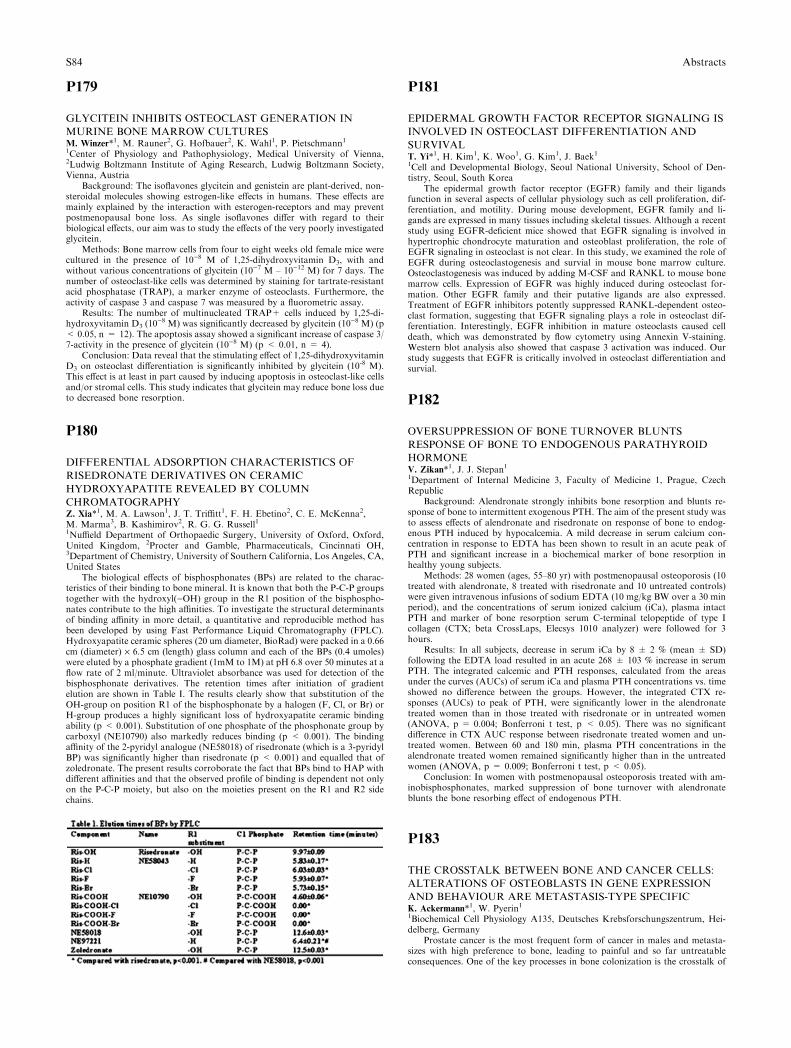
P179
GLYCITEIN INHIBITS OSTEOCLAST GENERATION IN
MURINE BONE MARROW CULTURESM. Winzer*1, M. Rauner2, G. Hofbauer2, K. Wahl1, P. Pietschmann11Center of Physiology and Pathophysiology, Medical University of Vienna,2Ludwig Boltzmann Institute of Aging Research, Ludwig Boltzmann Society,Vienna, Austria
Background: The isoflavones glycitein and genistein are plant-derived, non-steroidal molecules showing estrogen-like effects in humans. These effects aremainly explained by the interaction with esterogen-receptors and may preventpostmenopausal bone loss. As single isoflavones differ with regard to theirbiological effects, our aim was to study the effects of the very poorly investigatedglycitein.
Methods: Bone marrow cells from four to eight weeks old female mice werecultured in the presence of 10)8 M of 1,25-dihydroxyvitamin D3, with andwithout various concentrations of glycitein (10)7 M – 10)12 M) for 7 days. Thenumber of osteoclast-like cells was determined by staining for tartrate-resistantacid phosphatase (TRAP), a marker enzyme of osteoclasts. Furthermore, theactivity of caspase 3 and caspase 7 was measured by a fluorometric assay.
Results: The number of multinucleated TRAP+ cells induced by 1,25-di-hydroxyvitamin D3 (10
)8 M) was significantly decreased by glycitein (10)8 M) (p< 0.05, n = 12). The apoptosis assay showed a significant increase of caspase 3/7-activity in the presence of glycitein (10)8 M) (p < 0.01, n = 4).
Conclusion: Data reveal that the stimulating effect of 1,25-dihydroxyvitaminD3 on osteoclast differentiation is significantly inhibited by glycitein (10-8 M).This effect is at least in part caused by inducing apoptosis in osteoclast-like cellsand/or stromal cells. This study indicates that glycitein may reduce bone loss dueto decreased bone resorption.
P180
DIFFERENTIAL ADSORPTION CHARACTERISTICS OF
RISEDRONATE DERIVATIVES ON CERAMIC
HYDROXYAPATITE REVEALED BY COLUMN
CHROMATOGRAPHYZ. Xia*1, M. A. Lawson1, J. T. Triffitt1, F. H. Ebetino2, C. E. McKenna2,M. Marma3, B. Kashimirov2, R. G. G. Russell11Nuffield Department of Orthopaedic Surgery, University of Oxford, Oxford,United Kingdom, 2Procter and Gamble, Pharmaceuticals, Cincinnati OH,3Department of Chemistry, University of Southern California, Los Angeles, CA,United States
The biological effects of bisphosphonates (BPs) are related to the charac-teristics of their binding to bone mineral. It is known that both the P-C-P groupstogether with the hydroxyl()OH) group in the R1 position of the bisphospho-nates contribute to the high affinities. To investigate the structural determinantsof binding affinity in more detail, a quantitative and reproducible method hasbeen developed by using Fast Performance Liquid Chromatography (FPLC).Hydroxyapatite ceramic spheres (20 um diameter, BioRad) were packed in a 0.66cm (diameter) · 6.5 cm (length) glass column and each of the BPs (0.4 umoles)were eluted by a phosphate gradient (1mM to 1M) at pH 6.8 over 50 minutes at aflow rate of 2 ml/minute. Ultraviolet absorbance was used for detection of thebisphosphonate derivatives. The retention times after initiation of gradientelution are shown in Table I. The results clearly show that substitution of theOH-group on position R1 of the bisphosphonate by a halogen (F, Cl, or Br) orH-group produces a highly significant loss of hydroxyapatite ceramic bindingability (p < 0.001). Substitution of one phosphate of the phosphonate group bycarboxyl (NE10790) also markedly reduces binding (p < 0.001). The bindingaffinity of the 2-pyridyl analogue (NE58018) of risedronate (which is a 3-pyridylBP) was significantly higher than risedronate (p < 0.001) and equalled that ofzoledronate. The present results corroborate the fact that BPs bind to HAP withdifferent affinities and that the observed profile of binding is dependent not onlyon the P-C-P moiety, but also on the moieties present on the R1 and R2 sidechains.
P181
EPIDERMAL GROWTH FACTOR RECEPTOR SIGNALING IS
INVOLVED IN OSTEOCLAST DIFFERENTIATION AND
SURVIVALT. Yi*1, H. Kim1, K. Woo1, G. Kim1, J. Baek11Cell and Developmental Biology, Seoul National University, School of Den-tistry, Seoul, South Korea
The epidermal growth factor receptor (EGFR) family and their ligandsfunction in several aspects of cellular physiology such as cell proliferation, dif-ferentiation, and motility. During mouse development, EGFR family and li-gands are expressed in many tissues including skeletal tissues. Although a recentstudy using EGFR-deficient mice showed that EGFR signaling is involved inhypertrophic chondrocyte maturation and osteoblast proliferation, the role ofEGFR signaling in osteoclast is not clear. In this study, we examined the role ofEGFR during osteoclastogenesis and survial in mouse bone marrow culture.Osteoclastogenesis was induced by adding M-CSF and RANKL to mouse bonemarrow cells. Expression of EGFR was highly induced during osteoclast for-mation. Other EGFR family and their putative ligands are also expressed.Treatment of EGFR inhibitors potently suppressed RANKL-dependent osteo-clast formation, suggesting that EGFR signaling plays a role in osteoclast dif-ferentiation. Interestingly, EGFR inhibition in mature osteoclasts caused celldeath, which was demonstrated by flow cytometry using Annexin V-staining.Western blot analysis also showed that caspase 3 activation was induced. Ourstudy suggests that EGFR is critically involved in osteoclast differentiation andsurvial.
P182
OVERSUPPRESSION OF BONE TURNOVER BLUNTS
RESPONSE OF BONE TO ENDOGENOUS PARATHYROID
HORMONEV. Zikan*1, J. J. Stepan11Department of Internal Medicine 3, Faculty of Medicine 1, Prague, CzechRepublic
Background: Alendronate strongly inhibits bone resorption and blunts re-sponse of bone to intermittent exogenous PTH. The aim of the present study wasto assess effects of alendronate and risedronate on response of bone to endog-enous PTH induced by hypocalcemia. A mild decrease in serum calcium con-centration in response to EDTA has been shown to result in an acute peak ofPTH and significant increase in a biochemical marker of bone resorption inhealthy young subjects.
Methods: 28 women (ages, 55–80 yr) with postmenopausal osteoporosis (10treated with alendronate, 8 treated with risedronate and 10 untreated controls)were given intravenous infusions of sodium EDTA (10 mg/kg BW over a 30 minperiod), and the concentrations of serum ionized calcium (iCa), plasma intactPTH and marker of bone resorption serum C-terminal telopeptide of type Icollagen (CTX; beta CrossLaps, Elecsys 1010 analyzer) were followed for 3hours.
Results: In all subjects, decrease in serum iCa by 8 ± 2 % (mean ± SD)following the EDTA load resulted in an acute 268 ± 103 % increase in serumPTH. The integrated calcemic and PTH responses, calculated from the areasunder the curves (AUCs) of serum iCa and plasma PTH concentrations vs. timeshowed no difference between the groups. However, the integrated CTX re-sponses (AUCs) to peak of PTH, were significantly lower in the alendronatetreated women than in those treated with risedronate or in untreated women(ANOVA, p = 0.004; Bonferroni t test, p < 0.05). There was no significantdifference in CTX AUC response between risedronate treated women and un-treated women. Between 60 and 180 min, plasma PTH concentrations in thealendronate treated women remained significantly higher than in the untreatedwomen (ANOVA, p = 0.009; Bonferroni t test, p < 0.05).
Conclusion: In women with postmenopausal osteoporosis treated with am-inobisphosphonates, marked suppression of bone turnover with alendronateblunts the bone resorbing effect of endogenous PTH.
P183
THE CROSSTALK BETWEEN BONE AND CANCER CELLS:
ALTERATIONS OF OSTEOBLASTS IN GENE EXPRESSION
AND BEHAVIOUR ARE METASTASIS-TYPE SPECIFICK. Ackermann*1, W. Pyerin11Biochemical Cell Physiology A135, Deutsches Krebsforschungszentrum, Hei-delberg, Germany
Prostate cancer is the most frequent form of cancer in males and metasta-sizes with high preference to bone, leading to painful and so far untreatableconsequences. One of the key processes in bone colonization is the crosstalk of
S84 Abstracts
disseminated cancer cells with osteoblasts, a so far poorly characterized event.Using large scale transcript profiling (approx. 23,000 genes) we analyzed theeffect on the expression of genes in osteoblasts (hfOB; mpOB) caused by theircrosstalk with three different prostate cancer cell lines (LNCaP; PC3; DU145) inan in-vitro metastasis model system allowing communication via released fac-tors. The bone metastasis-originating PC3 cells, already adapted to the bonemicroenvironment, caused the smallest changes in osteoblast gene expressions,followed by those due to the lymph-node metastasis- originating LNCaP cells.The highest number of alterations were seen with DU145 prostate cancer cellsoriginating from brain metastasis. Correspondance analysis reveals a relativelyclose relationship between the osteoblast gene expression effects by PC3 andLNCaP, and a significant distance of both to DU145. The genes elevated inexpression in osteoblasts include among others genes encoding collagens andcollagen-modifying enzymes (collagen type I, lysyl oxidase, etc.), targeted traf-ficking of bone matrix proteins (VAMPs), and metallothioneins. In contrast, wefind the prostate cancer cells to repress in osteoblasts a whole set of interferon-regulated genes with links to tumor defense and apoptosis promotion. Inter-estingly, quantitative real-time PCR indicated stronger elevations in expressionof genes encoding osteogenic proteins such as collagen type I\alpha1, alkalinephosphatase, cbfa1 and osteopontin in osteoblasts due to the crosstalk withLNCaP than with PC3. As a consequence, when we stained osteoblast layerswith Alizarin red-S for the calcium mineral content we noted striking differences;the crosstalk with LNCaP but not with PC3 led to increased mineralization.Instead, the crosstalk with PC3 destroyed the regular arrangement of osteoblastcell layers. Our data indicate that alterations in gene expression and behavior inosteoblasts due to their crosstalk with prostate cancer cells have specific links tothe way metastasis occurs with consequences for their contribution to boneformation and maintenance.
P184
CASE REPORT OF A PATIENT WITH A DIAGNOSIS OF
ANKYLOSING SPONDYLITIS AND CHONDROSARCOMA OF
SACROILIAC JOINTM. Graberski Matasovic1, A. Aljinovic*1, M. Paar-Puhovski1, M. Jaksic11Department of physical medicine, rehabilitation and rheumatology, Generalhospital Sveti Duh, Zagreb, Croatia
The occurrences of malignancy in patients with rheumatic diseases have beenreported previously. However, searching the literature we have not found areport on a chondrosarcoma of the sacroiliac joint presenting in a patient withankylosing spondylits.
A 68 years old woman presented with a twelve-month history of progressivelow back and buttock pain. Nineteen years ago she was diagnosed to haveankylosing spondylitis (HLA B27 positive). She underwent intensive physio-therapy and analgesics were prescribed. However, since her symptoms were notrelieved, additional diagnostic procedures were performed. Radiographs showedsclerotic zone described as sacroiliitis condenses. Recent L5 (L3 and L4) nerveroot lesion was noted on EMNG. Ordered MRI of lumbosacral spine suggested,and subsequent abdominal CT confirmed extensive expansive-proliferativeprocess in the region of sacroiliac joint with destruction of iliac bone. Tumor waspartially resected and patohistologic analysis showed moderately differentiatedchondrosarcoma. Since further operative treatment was not recommended,radiation therapy was introduced. Patient still has pain in sacroiliac area andfaces difficulty in walking due to leg weakness.
We believe that proper diagnose in this patient was delayed because this raretumor mimicked the symptoms seen in ankylosing spondylits.
P185
CALCITROPIC HORMONES IN WOMEN WITH POLYCYSTIC
OVARY SYNDROMEM. S. Ardawi*1, A. A. Rouzi21Clinical Biochemistry, 2Obstetrics and Gynecology, Faculty of Medicine andKing Abdulaziz University Hospital, Jeddah, Saudi Arabia
Objective: To determine the serum levels of parathyroid hormone (PTH),calcitriol and calcidiol in women with and without polycystic ovary syndrome(PCOS) in relation to obesity and the hormonal and metabolic characteristics ofthe syndrome.
Subjects and Methods: This is a case-control study in which 180 selectedwomen were studied and classified as follows: 45 obese (BMI > 30 Kg/m2) withPCOS; 45 lean (BMI < 25 Kg/m2) with PCOS; 45 obese (BMI > 30 Kg/m2)without PCOS and 45 lean (BMI < 25 Kg/m2) without PCOS. Blood sampleswere collected from all women studied between 8:00–11:00 am; after an over-night fast. The serum levels of follicle-stimulating hormone (FSH), luteinizinghormone (LH), prolactin (PRL), thyroid stimulating hormone (TSH), free thy-roxine (FT4), total testosterone (T), dihydroepiandrosterone (DHEA), DHEA-sulphate (DHEAS), 17)OH-progesterone (17)OH-P), d4-androstenedione (d4-
A), sex hormone binding globulin (SHBG), insulin, intact-PTH, calcitriol, cal-cidiol and glucose were measured. Measures of insulin resistance (IR) included:fasting serum glucose, glucose-to-insulin ratio (GIR) and homeostasis modelassessment (HOMA). Linear regression (2-way ANOVA) model (LRM) wasused. Comparison of means and adjustments were performed with univariateLRM. Independent relationships were assessed by partial correlation analysis.
Results: Serum levels of intact-PTH were found to be significantly increasedin women with PCOS and in obese women without PCOS as compared with leanwomen without PCOS (P < 0.001): this difference remained significant afteradjustment for age and BMI (P < 0.001). Women with PCOS exhibited highervalues for serum calcitriol and calcidiol as compared with BMI-matched womenwithout PCOS (P < 0.05). Serum levels of intact-PTH were significantly cor-related with age, BMI, SHBG, glucose and T. The effect of PCOS on intact-PTHlevels remained significant after adjustment for BMI, but not after adjustmentfor T. Higher BMI values were negatively related to changes in serum levels ofcalcitriol and calcidiol but without association with PCOS.
Conclusion: Increased serum intact-PTH is evident in obese and lean womenwith PCOS and it is suggested that PTH may be linked to the hyperandrogen-emia of PCOS and/or other metabolic abnormalities of the syndrome.
P186
EFFECT OF CLODRONATE ON BONE MASS
MINERALIZATION IN PROSTATE CARCINOMA PATIENTST. Banek*1, L. Banek2, G. Kurnik11Department of Nuclear Medicine, Clinical Hospital Dubrava, 2Institute ofHistology and Embriology, School of Medicine, University of Zagreb, Zagreb,Croatia
Prostate carcinoma (CaP) is the second most common Ca in men older than50. CaP metastasizing to the bone is the most common form of hematogenousdissemination, characterized by osteoblastic or osteoclastic metastatic foci.Clodronate as a bisphosphonate pharmacologic agent minimizes further boneloss and inhibits osteoclast mediated bone resorption. The aim of the study wasto assess the rate of bone mass increase in patients with CaP and osteolyticskeletal lesions, treated with clodronate for one year. The study included 23 menaged 49–78 (mean 71) diagnosed with adeno-CaP. Relevant biochemical andendocrinologic parameters, and prostate-specific antigen (PSA) in serum weredetermined. Skeleton scintigraphy revealed multiple osteoblastic foci withradiographically verified osteolysis. Along with orchiectomy and pharmacologiccastration with LH-RH agonists, the majority of patients received antiandro-gens. All patients used clodronate, 1600 mg/day, for 12 months. Lumbar spine,hips and whole body measurements were performed on an Hologic-DXA den-sitometer, along with bone mineral density (BMD) and bone mineral content(BMC) determination. Basal DXA showed osteopenia in 5 and osteoporosis in12 (4 false-positive findings due to lumbar spine osteolysis) men, whereas 6 menhad normal BMD. Compared with control group, total BMC decrease by 18–53g was found in 14 men. The initial level of total serum PSA ranged from 19–125ng/ml (control values 0.3–94 ng/ml). Repeat DXA measurements of BMD andBMC after 12-month clodronate therapy revealed osteopenia in 3/5 and osteo-porosis in 9/12 patients. In 2 men, there was newly detected osteoporosis due tothe disease progression to lumbar spine. A total BMC decrease by 15–43 g wasrecorded in 10 men. Abnormality of BMD as a primary indicator of the rate ofmineralization and amount of bone tissue was recorded in patients with CaP anddisease progression to the skeletal system. A BMC decrease by 15–43 g followingclodronate therapy was considered a secondary indicator of the skeletal systemhealth improvement.
P187
TRANSCRIPTOME ANALYSIS REVEALS AN OSTEOMIMETIC
PHENOTYPE FOR HUMAN BONE METASTATIC BREAST
CANCER CELLSA. Bellahcene*1, R. Bachelier2, C. Detry1, R. Lidereau3, P. Clezardin2,V. Castronovo11Metastasis Research Laboratory, University of Liege, Liege, Belgium, 2IN-SERM, Unit Research 644, Lyon, 3INSERM Unit Research 735, Centre ReneHuguenin, St-Cloud, France
Breast cancer metastatic cells exhibit the selective ability to seed and grow inthe skeleton. In this study, gene microarraying was performed on a model ofbreast cancer bone metastasis consisting of the MDA-MB-231 cell line and itsvariant B02, which is characterized by a high affinity for bone in vivo. Analysisof B02 cells transcriptional profile revealed that 11 and 9 out of the 50 most up-and down-regulated genes, respectively, overlapped with genes expressed by cellsof the osteoblastic lineage. For example, B02 cells surexpress osteoblast specificcadherin-11, which mediates the differentiation of mesenchymal cells intoosteoblastic cells. In contrast, S100A4, recently described as a key negativeregulator of osteoblastic differentiation, was the most down-regulated gene in
Abstracts S85
B02 cells. We established a list of differentially expressed genes compatible withthe acquisition of an osteomimetic phenotype by B02 breast cancer cells. RT-PCR and western blotting experiments allowed us to validate the modulation ofseveral candidate genes. Finally, we verified the pathophysiological relevance ofour data using immunohistochemistry on human breast primary tumors andmatched liver and bone metastases. This is the first large-scale depiction of theosteomimetic phenotype adopted by bone metastatic breast cancer cells. Ourresults provide with well-built basis for functional studies aimed at the under-standing of the molecular mechanisms that govern bone metastasis development.
P188
BONE SIALOPROTEIN OVEREXPRESSION IN RESPONSE
TO SERUM DEPRIVATION AND APOPTOTIC STRESS
PROMOTES CELL SURVIVAL IN CANCER CELLSA. Bellahcene*1, F. Journe2, M. Chaplet1, N. Kheddoumi2, J. J. Body2,V. Castronovo11Metastasis Research Laboratory, University of Liege, Liege, 2Laboratory ofEndocrinology-Bone Diseases, Department of Medicine, Institut Jules BordetUniversite Libre de Bruxelles, Bruxelles, Belgium
Breast and prostate cancer cells commonly metastasize to the skeleton. Theyexpress bone-related proteins, such as bone sialoprotein (BSP) and osteopontin(OPN), that have been implicated in their osteotropic behavior. Previous studieshave shown that OPN promotes cell survival upon serum deprivation. In thisstudy, we tested the hypothesis that BSP could have the same effect. Twenty fourhours serum deprivation induced BSP overexpression at both mRNA and pro-tein levels in MCF-7 human breast tumor cells. This increased BSP expressionwas more pronounced in subconfluent and highly proliferating MCF-7 cells thanin confluent cancer cells. Moreover, exogenous recombinant BSP significantlydecreased the proportion of apoptotic MCF-7 cells induced by serum with-drawal, as assessed by annexin-V flow cytometric analysis. We also tested ourhypothesis using two PC-3 human prostate cancer subclones, characterized ei-ther by a high BSP (PC3-H) or a low BSP expression (PC3-L). Both subcloneswere incubated with zoledronic acid (ZOL) at 10–4M during 72 hours, a con-dition which has been previously shown to induce apoptosis in PC3 cancer cells.Under this treatment, PC3-L cells proved to be significantly more sensitive toZOL-induced cell growth inhibition than PC3-H cells. Ongoing experimentsusing cancer cells transfected to overexpress BSP will help elucidating the po-tential role of BSP as a mediator of cancer cell survival. Since BSP is an alphaVbeta3 integrin ligand, we will also investigate whether alphaV beta3 blockadecauses the loss of BSP-mediated prevention of apoptosis. In conclusion, cellsurvival induced by BSP in breast and prostate cancer cells is a new biologicalactivity for this bone matrix protein that is in favor of cancer cell growth andestablishment in bone.
P189
ASSOCIATION BETWEEN THE TNFRSF11B GENE
PROMOTER POLYMORPHISM 163A/G AND PAGET’S
DISEASE OF BONE IN A BELGIAN POPULATIONG. Beyens*1, F. De Freitas1, F. Vanhoenacker2, L. Verbruggen3, H. Zmierczak4,J. Devogelaer5, W. Van Hul61Department of Medical Genetics, University of Antwerp, Wilrijk, 2Departmentof Radiology, University Hospital of Antwerp, Antwerp, 3Department ofRheumatology, University Hospital of Brussels, Brussels, 4Department ofEndocrinology and Metabolic diseases, University Hospital of Ghent, Ghent,5Department of Rheumatology, Saint- Luc University Hospital, Brussels,6Department of Medical Genetics, University of Antwerp, Antwerp, Belgium
Background/aims: Paget’s Disease of Bone (PDB) is a common- late onsetmetabolic bone disorder, characterized by focal areas of increased boneresorption and disorganized formation of new bone. In the past, two SNPs in theTNFRSF11B gene have been reported to be associated with PDB. We reported apositive association between SNP 505C/T and PDB in a small cohort of BelgianPDB patients [1]. More recently, in an extended British cohort the G allele ofSNP 1181C/G was found to be associated with the disease [2]. In this study, werecruited 120 sporadic Belgian PDB patients and 170 Belgian control individualsand determined the genotype- and allele frequencies of 5 SNPs: 163A/G and950T/C located in the promoter region, 1181C/G in exon 1, 505C/T in intron 2and 6893A/G in exon 4.
Methods: Genotyping of SNPs was performed using either sequencing or asingle base primer extension technique. For each SNP both the allele- andgenotype frequencies were compared between cases and controls using chi-square tests. LD statistics were calculated based on the genotyping results of 70control individuals.
Results: For SNPs 950T/C, 1181C/G and 6893A/G no associations werefound. We did not identify an association between SNP 163A/G and PDB at thegenotype level (p = 0.063), but the G allele was significantly overrepresented in
the case group (p = 0.023, OR: 1.71, 95%CI: 1.07– 2.75). The C allele of SNP505C/T was more frequent in the case group, but the significance level of 0.05was not reached (genotypes: p = 0.4703, alleles: p = 0.056, OR 1.05, 95%CI:0.65–1.7). LD analysis indicated that SNP 163A/G is not in LD with SNPs1181C/G and 505C/T in the Belgian population. The D’ and r1=2 values betweenSNPs 1181G/C and 505C/T were 0.778 and 0.464 respectively.
Conclusion: In this study we found a significant association between the Gallele of SNP 163A/G and PDB (p = 0.023). Although the previously reportedassociations with SNPs 505C/T and 1181C/G were not replicated in this Belgianpopulation, this analysis confirms that the TNFRSF11B gene contains a sus-ceptibility variant for the development of PDB. Further analysis will indicatewhich SNP carries the functional effect in each population: 163A/G, 505C/T,1181C/G or a SNP that is in LD with one of these.
[1] Wuyts et al 2001 Bone 28(1): 104-107, [2] Daroszewska et al 2004 JBMR19(9): 1507-1511. This work was supported by a Paget Foundation researchaward.
P190
INTERACTION BETWEEN BONE MICROENVIRONMENT
AND CANCER CELLS IN ANIMAL METASTASIS MODELSS. Blouin1, N. Josselin1, M. Basle1, D. Chappard*11EMI 0335, INSERM, ANGERS, France
Introduction: Bone metastases are a major cause of mortality in humans butthe preferential proliferation of cancer cells in the bone microenvironment ispoorly characterized. Bone metastases can be predominantly osteolytic orosteoblastic or mixed. Osteolytic (Walker W256) and osteoblastic (MatLyLuMLL) metastases were induced to investigate the expression pattern of bonemarrow and others organs microenvironment in contact with the metastases areaby immunochemistry and histology.
Material and methods: Fisher rats received an intracardiac, intramuscular orsubcutaneous injection with W256 cells and were euthanized after 9 days.Copenhagen rats received an intracardiac, intramuscular, intraliver, intraspleenor subcutaneous injection with MLL cells and were euthanized after 15 days.Bone and other metastatic sites within soft tissues were analyzed by immuno-chemistry for DKK1, cathepsin K, RANKL, MCSF, c-Fms or IL6 expression inthe microenvironment of the tumors. Tartrate Resistant Acid Phosphatase(TRAcP)+ cells were also detected by a histoenzymatic technique.
Results: In bone, a huge membrane expression of MCSF was shown instromal cells of the bone marrow, in contact with metastatic foci of both W256and MLL tumors. DKK1 was also expressed at high levels by stromal cells incontact with metastatic foci (membrane and cytoplasmic expression). Manystromal cells were found RANKL+ in the vicinity of the tumors. Cellsexpressing cathepsin K were found in direct contact with trabeculae and inmedullar spaces at distance from bone surfaces. Multinucleated TRAcP+ cellswere apposed on trabeculae but were also encountered in bone marrow spaces,far from trabecular surfaces and near metastatic cells.
In extraosseous tumors, cells in contact with malignant cells did not ex-pressed DKK1, MCSF, c-Fms, cathepsin K. IL6 expression was found in theliver but did not differ from normal expression. Some RANKL+ cells werefound in the periphery of subcutaneous tumors but may represent Langerhanscells. TRAcP+ cells were never observed in the vicinity of malignant cells in anyextraosseous metastases. However, TRAcP+ cells corresponding to activatedmacrophages were encountered in invaded and non-invaded spleens.
Discussion: Interaction between stromal and cancer cells induces theexpression on the formers of characteristics leading to osteoclastogenesis only inthe bone microenvironment.
P191
BONE MORPOGENETIC PROTEINS AND THEIR RECEPTORS
IN PROSTATE CANCER PATHOGENESISD. Bobinac*1, J. Spanjol2, I. Maric1, G. Dordevic3, E. Mustac3, Z. Fuckar21Department of Anatomy, Faculty of Medicine, 2Department of Urology,Clinical Hospital, 3Department of Pathology, Faculty of Medicine, Rijeka,Croatia
Prostate cancer is highly spread malignancy in male population. Further,osteoblastic bone metastases are common in advanced prostate cancer but themechanisms by which tumor cells stimulate new bone formation remain un-clear. Bone morphogenetic proteins (BMPs) are growth factors produced byprostate cancer and stimulate osteoblast proliferation. Recent research showedthat BMPs are expressed in normal and malignant prostatic tissue. BMPssignal through an interaction with BMP membrane receptors (BMPRs) type Iand type II. It was shown that well-differentiated cancers were positive for theexpression of BMPR II, BMPR-IA and BMPR-IB in contrast to poorlydifferentiated prostate cancers which were negative for each of the threeBMPRs. So, BMPs can be potential regulators of prostate cancer cell growthand metastasis.
S86 Abstracts
We have investigated the expression of some BMPs and their membranereceptors in differently prostate cancer tissue with or without metastasis as wellas in benign prostatic hyperplasia.
Immunohistochemical analysis of BMP)2/4,)3,)5, )6, )7 and BMPR-IA,BMPR-IB and BMPR-II were carried out in 31 patients with diagnosed prostatecancer. Twenty-one patients had multiple bone metastases proven by bone scan.Three bone metastases had corresponding primary prostate cancer tissue. Other10 patients had no skeletal metastasis and prostatic tissue sections containingprostate cancer and associated benign prostatic hyperplasia (BPH).
In non-metastatic prostate carcinoma the expression of all BMPs is rathersimilar to normal prostate tissue except BMP-7 which is lower positive in car-cinoma tissue. In prostate carcinoma with bone metastases the expression ofBMP-6 and -7 was significantly less positive. In normal prostate, BMPRs werelocalized predominantly to epithelial cells. In prostate cancer samples withproved bone metastases the expression of three receptors was significantlychanged and very low. That can be seen especially for BMPR-II which expres-sion was almost negative. The bone metastatic tissue, patohistological analysisproved as osteoblastic nature, showed significantly higher expression of BMP-6,-7 and three receptors in contrast to prostate cancer tissue of the same patients.So, we can see loss of BMPRs expression in primary cancer tissue after devel-oping of bone metastases.
P192
SENSITIZATION OF OSTEOSARCOMA CELLS TO APOPTOSIS
BY ONCOSTATIN M DEPENDS ON STAT5 AND P53.C. Chipoy*1, P. Juin2, F. Redini1, D. Heymann1, F. Blanchard11University of Nantes EA3822 INSERM ERI 7, Medicine Faculty, 2U601,INSERM, Nantes, France
Oncostatin M (OSM), a cytokine of the IL-6 family, reduces the growth andinduces differentiation of osteoblasts / osteosarcoma cells into glial-like or os-teocytic cells. Given the strong interaction between differentiation and apoptosis,and their key role in regulating osteoblast / osteocyte functions, we askedwhether OSM could regulate apoptosis of normal or transformed osteoblasts.
Alone, OSM did not induce cell death, but OSM-treated osteosarcoma cellsor proliferating osteoblasts were particularly more sensitive to apoptosis inducedby different death inducers among which staurosporine (STS). Cell death in-duced by OSM+STS was associated with activation of caspase 9 and 3 andcould be prevented by the caspase inhibitor Z-VAD-FMK. OSM alone inducedactivation (phosphorylation) of p53, STAT1, 3 and 5 transcription factors, andconcomitantly enhanced expression of Bax / Bad and reduced expression of Bcl-2. Other Bcl-2 family members (Bak, Bcl-xL, Bcl-xS) were not modulated. Of alldominant negative (dn) STATs, only the dnSTAT5 was able to prevent apop-tosis induced by OSM+STS. On dnSTAT5-transfected cells, OSM was unableto reduce Bcl-2 expression, whereas Bax was still induced. Conversely, the p53inhibitor pifithrin a prevented apoptosis induced by OSM+STS as well as Baxinduction, whereas reduction of Bcl-2 was not modified. By co-immunoprecip-itation experiments, we observed a constitutive interaction between STAT5 andp53, the C-terminal trans-activation domain of STAT5 being necessary for thisinteraction.
In this study, we characterize a new signalling pathway (based on p53-STAT5 interaction) used by OSM to increase Bax and decrease Bcl-2 expression,and thus controlling the mitochondrial cell death pathway in osteoblastic cells.In parallel, strong anti-apoptotic signals are also activated by OSM, via thePKCd and PI3K/Akt pathways. In human osteosarcoma, p53 mutations arefrequent and we effectively found that the p53-STAT5 pathway is often dis-rupted. The use of kinase inhibitors such as STS in association with OSM couldtherefore represent new treatments for wild type p53 osteosarcoma. Whetherthese pathways are also deregulated in osteoarticular pathologies such as oste-oporosis or rheumatoid arthritis remains to be determined.
P193
THE DEVELOPMENT OF PAGET�S DISEASE IN PEOPLEWITH
INHERITED SQSTM1 MUTATIONST. Cundy*1, M. Bolland1, P. Tong1, K. Callon1, D. Wattie1, D. Naot11Department of Medicine, Faculty of Medical and Health Sciences, Universityof Auckland, Auckland, New Zealand
Mutations in the ubiquitin-binding domain region of the gene SQSTM1 arefound in 25-50% of patients with familial Paget�s disease (FPD), and are asso-ciated with early-onset and more extensive disease. The mutations are believed tobe disease-causing with a high degree of penetrance. We sought to determine theprevalence and phenotype of Paget�s disease in the adult offspring of affectedfamily members known to carry SQSTM1 mutations. 63 offspring from 5 fam-ilies (18 index patients) were invited to participate, and 41 subjects (12M, 29F)agreed to do so. The mean age was 44 yrs (range 30–63). In all subjects plasmaalkaline phosphatase (ALP) activity was measured (normal <120l/l), and the
ubiquitin-binding domain region of the gene was sequenced, and the presence orabsence of the known familial mutation established. Subjects with a mutationwent on to have a whole body bone scintiscan, from which the percentageskeletal involvement was calculated.
14 of the 41 subjects had inherited a familial mutation: the mean ALP was74u/l in those with a mutation compared to 81l/l in those without (p = NS). Todate 13 of 14 subjects with mutations have had scintiscans: PD was demon-strated in only 4 subjects (mean age 46, mean ALP 139u/l), and 9 subjects did nothave PD (mean age 46, mean ALP 58l/l).
In the four with PD, the skeletal involvement was substantially less than intheir affected parent. We conclude that by age 46 this genetic trait showsincomplete penetrance, with only one third of carriers demonstrating a pageticphenotype. The data are also consistent with the hypothesis that diminishedexposure to an as yet unidentified environmental factor is reducing the incidenceand severity of Paget�s disease.
P194
THE RISK OF MORPHOMETRIC VERTEBRAL FRACTURES
IN POSTMENOPAUSAL PATIENTS WITH PRIMARY
HYPERPARATHYROIDISMS. De Geronimo*1, D. Diacinti2, E. Romagnoli2, S. Piemonte2, F. Fidanza2,J. Pepe2, E. D’Erasmo2, S. Minisola21Dpt of Clinical Sciences, University of Rome La Sapienza, 2Dpt of ClinicalSciences, University of Rome ‘‘La Sapienza’’, Rome, Italy
This study aimed to evaluate the prevalence of morphometric vertebralfractures (MVF) in 98 PHPT postmenopausal women, divided into mild (M,n = 25) and non mild (NM, n = 73), according to new criteria for surgery inpatients with mild PHPT (2002). Patients were compared to 89 healthy post-menopausal subjects matched for age, years since menopause (YSM) and BMI.
We performed spine x-rays for the evaluation of MVF according to Genant’ssemiquantitative technique, and bone mineral density (BMD) measurement atthe lumbar spine (LS) and femoral sites (both neck-FN and total femur-FT) byDEXA (Hologic 4500). Volumetric BMD of the third lumbar vertebra(vBMDL3) was also calculated according to Seeman’s method, wherevBMD = BMC/V and V = A3/2; this measurement recognizes the influence ofbone size on the areal BMD assessed by DEXA.
No difference was found between these two groups and control subjects (C)in age (M = 60.84 ± 6.82; NM = 61.65 ± 8.4; C = 60.65 ± 6.92), YSM(M = 13.44 ± 10.44; NM = 13.09 ± 9.4; C = 11.15 ± 8.99),and BMI(M = 27.4 ± 4.16;NM = 25.11 ± 3.94; C = 24.8 ± 3.15). The prevalence ofMVF was significantly higher (p < 0.001) in both M (n = 11, 44%) and in NM(n = 35, 47%) PHPT patients compared to C (n = 8, 9%). However, thisprevalence did not differ between M and NM patients. On the contrary, nodifference was found between NM and C as far as the prevalence of non ver-tebral fractures (NM n = 13, 17.8% vs C n = 17, 19.1%). BMD (mg/cm2) wassignificantly lower (p < 0.001) in NM patients as compared to both C (LS:765 ± 156 vs 832 ± 140; FN: 601 ± 102 vs 687 ± 108; FT: 699 ± 126 vs785 ± 138) and M patients (LS: 912 ± 100; FN: 709 ± 92; FT: 823 ± 116).BMD at LS in M patients was also significantly higher in respect to C. Similarresults were also obtained for vBMDL3 (g/cm3)(NM: 0.21 ± 0.04 vs both C:0.23 ± 0.03 and M: 0.25 ± 0.02, p < 0,001). In M patients vBMDL3 was alsosignificantly higher compared to C. When M and NM patients were subdividedaccording to the presence or lack of MVF, no difference was found betweenfractured and unfractured patients in both BMD and vBMDL3 values.
In conclusion, the risk of MVF is increased in postmenopausal patients withmild PHPT even if BMD appears well preserved. This finding suggests that otherfactors, i.e. bone quality, seem to be relevant in determining fracture risk,especially when gonadal function is lacking. Our results indicate that the eval-uation of MVF could be proposed among the criteria for surgery on thesepatients.
P195
TUMORAL CALCINOSIS AND CALCIPHYLAXIS PRESENT-
ING IN A HEMODIALYSIS PATIENTZ. M. Dimitrijevic*1, M. M. Stojanovic1, K. R. Paunovic1, G. J. Paunovic1,S. B. Ljubenovic11Institute of nephrology and hemodialysis, Clinical Center of Nis, Serbia, Nis,Yugoslavia
Calciphylaxis is a rare, often fatal condition that occurs in patients withchronic renal failure receiving regular, long-term hemodialysis, and character-ized by calcified periarticular soft tissue masses, often of considerable size andmultiplicity. Histopathologically, it is characterized by medial calcification andintimal hyperplasia of cutaneous and subcutaneous vasculature, resulting inprogressive necrosis of subcutaneous tissue and fat. It is clinically characterizedby livedo reticularis and painful violaceous skin lesions that progress to non-healing ulcers, tissue necrosis and sepsis.
Abstracts S87
We describe the case of a 46-year-old man with end-stage renal disease dueto polycystic disease who presented with both tumoral calcinosis and calciphy-laxis and subsequently developed devastating vascular complications. Clinical,laboratory, radiography, scintigraphy and patohystologic findings are reviewed.Treatment is difficult and, as a result, calciphylaxis is associated with a highmortality rate.
Keywords: calciphylaxis, hemodialysis, tumoral calcinosis
P196
INVESTIGATION OF UBIQUITIN-ASSOCIATED DOMAIN
MUTATIONS OF SQSTM1 IN SPORADIC HUNGARIAN PA-
TIENTS WITH PAGET�S DISEASE OF BONEJ. Donath*1, P. J. R. Gergely1, A. Blazsek2, G. Poor11Ist. Departement of Rheumatology, 2Laboratory of Molecular Biology, Na-tional Institute of Rheumatology and Physiotherapy, Budapest, Hungary
Backgrounds: Paget’s disease of bone (PDB) is a common disorder char-acterised by focal and disorganized increases of bone turnover. Genetic factorsplay an important role in the pathogenesis of PDB. Mutations of sequestosome1(SQSTM1) gene were identified as responsible for most of the sporadic andfamilial forms of PDB in patients in different populations.
Methods: To verify the involvement of the SQSTM1 gene in Hungariansubject affected by PDB, we performed mutational analysis in 60 sporadic PDBcases and 60 age and sex-matched control subjects.
Results: Mutations in the SQSTM1 gene were found in 5 cases of 60 (8.3 %)patients with sporadic PDB. Three patients carried the P392L mutation; M404Vand 394X mutations were also identified in two cases. All mutations were presentin heterozygous form. No SQSTM1 mutations were found in the controls sub-jects.
Conclusion: Hungarian patients with PDB carry the same SQSTM1 muta-tions decsribed earlier in different populations. Ubiquitin-associated domainmutations of SQSTM1 may contribute significantly to the. genetic backgroundof PDB. However, the low penetrance suggests that other genetic and/or envi-ronmental factors are necessary for the development of the disease.
P197
MOLECULAR CHARACTERIZATION OF THE GENETIC DE-
FECT IN MUTANT PIGS WITH A NON-FUNCTIONING 25-
HYDROXYVITAMIN D3 1ALPHA-HYDROXYLASE (CYP27B1)S. Harabasz*1, K. Weber1, R. G. Erben21Institute of Animal Physiology, University of Munich, Munich, Germany,2Dept. of Natural Sciences, University of Veterinary Medicine, Vienna, Austria
The aim of the current study was to characterize the genetic defect in the 25-hydroxyvitamin D3 1\alpha-hydroxylase (1\alpha-OHase, CYP27B1) deficientHannover pig model of pseudo-vitamin D-dependent rickets type I (PVDDR).To localize the genomic defect, various parts of the 1\alpha-OHase gene weresequenced in wild-type and mutant pigs. In addition, the promoter region andgenomic organization of the porcine 1\alpha-OHase gene were analyzed andfound to be highly homologous compared with the human 1\alpha-OHase gene.Expression of 1\alpha-OHase was found in the kidney and several nonrenaltissues such as uterine cervix, skin, bone, lymph node, adrenal gland, and colonin wild-type and mutant pigs. We detected a point mutation in the splice donorsite of intron 6 (IVS6+1G>A) in mutant pigs. Deletion of exon 6 in homo-zygous mutant pigs at the messenger RNA level was found both in renal andextrarenal tissues, resulting in a frame shift and in a premature terminationcodon. Thus, this mutation is associated with complete lack of CYP27B1function in all tissues. The point mutation also ablates a restriction site forBpu10 I, allowing to genotype by Restriction Fragment Length Polymorphism(RFLP). The expression level of 1\alpha-OHase was determined in several tissuesby quantitative RT-PCR in wild-type and mutant animals. In the kidney,expression was significantly lower in mutant compared with wild-type pigs, butno difference was found in the skin. Activation of blood-derived macrophages byIFN-\gamma resulted in increased 1\alpha-OHase expression in wild-type, butnot in mutant pigs. We conclude that this animal model of PVDDR is aninteresting model for further investigation of the regulation of vitamin D andcalcium metabolism in the pig.
P198
THE EFFECT OF ANTIHISTAMINE AND CORTICOSTEROID
TREATMENT ON BONE FRACTURES IN POSTMENOPAUSAL
POLLEN ALLERGIC WOMENV. Ferencz*1, S. Meszaros1, E. Csupor2, E. Toth3, K. Bors4, A. Falus5,C. Horvath1
11st Department of Medicine, Semmelweis University, 2The Health Service,Budavar Local Authorities, Budapest, 3Department of Rheumatology, FerencFlor County Hospital, Kerepestarcsa, 4Regional Osteoporosis Centre Ferenc-varos, Ferencvaros Local Authorities, 5Department of Genetics, Cell and Im-munbiology, Semmelweis University, Budapest, Hungary
Our aim was to investigate whether the treatment of pollen-allergy can affectbone mass and fractures in postmenopausal women. A total of 125 postmeno-pausal pollen-allergic women (mean age 61.26yr) were split into four groups:treated neither with H1 histamine receptor (H1R) antagonist nor with inhaledcorticosteroid (n = 43), treated only with H1R antagonist (n = 53), treatedboth with H1R antagonist and inhaled corticosteroid (n = 17), treated onlywith inhaled corticosteroid (n = 12) for at least 5 years, seasonally. One-hun-dred non-allergic postmenopausal subjects matched for age, body mass indexand age at menopause served as controls. Allergic patients without treatmenthad a slightly lower bone density than their non-allergic mates. Untreatedallergic had almost triple the rate of prevalent low-energy fractures (distalforearm, hip and clinical vertebral fractures: 34.9%) compared to non-allergicwomen (13%, chi2 p = 0.003). Bone fracture occurred more often in H1R-onlytreated patients (30.19%) than in controls (chi2 p = 0.01), however, clinicalvertebral or hip fractures developed neither in those treated only with H1Rantagonist nor in those who received both H1R antagonist and inhaled corti-costeroid. Bone fractures were more frequent among patients with inhaled ste-roid treatment than among patients with a combined treatment of inhaledsteroid and antihistamine (50% vs. 29.4%). In conclusion we found a highprevalence of low-energy fractures among pollen-allergic postmenopausal wo-men. It is possible that the H1R antagonists compensate for the negative effect ofpollen-allergy and the adverse effect of inhaled corticosteroid treatment on bonefracture risk.
P199
SERUM CALCIUM CONCENTRATION IN THE ELDERLYE. Franek*1, L. Napiorkowska1, K. Safranow2, K. Stepien31Dept of Int Dis, Endocrinology and Diabetology, Central Clinical HospitalMSWiA, Warszawa, 2Dept of Clin Biochemistry, Pomeranian Medical Acad-emy, Szczecin, 3Central Laboratory Unit, Central Clinical Hospital MSWiA,Warszawa, Poland
Background: Aging causes several processes that may influence serum cal-cium concentration. In most subjects diminished kidney function and impairedvitamin D hydroxylation occur, which may decrease serum Ca. From the otherside, frequency of neoplastic disease (with possible hypercalcaemia) is increased.In most sources, however, normal serum Ca values are given as not age-dependent. From the other side, Roche Diagnostic manual for serum Caassessment gives decreasing with age range of serum Ca, with the normal valuefor subjects aged more than 50y ranging from 2,10 to 2,42 mmol/l. The purposeof the study was to assess serum Ca concentration in the population of patientsolder than 50 y.
Materials and methods: 6645 patients with serum creatinine concentrationlower than 1.5 mg/dl in whom serum Ca concentration was assessed in the years2001–2004 in Central Clinical Hospital MSWiA in Warszawa, were included tothe study. Mean serum Ca concentration was assessed for particular years anddecades of life.
Results: Weighted means (for the ± 5years span) of serum Ca concentrationfor particular age decrease slowly from 70 year of age. The ranges (± 2SD) inparticular decades of life are as follows: 50–59: 2.16–2.50; 60–69: 2.18–2.50; 70–79: 2.15–2.49; >80: 2.08–2.48 mmol/l. After 95 yaer of age the prevalence ofhypercalcaemia increases, causing increase of the mean serum Ca value, whichexceeds 2.5 mmol/l in the group of patients aged 95-105 years.
Conclusion: Frequency of hypercalcaemia after 95 year of age is high,indicating an increase of prevalence of malignancy in this population. As serumcalcium concentration in the examined population decreases slowly only after 70year of age, one should be cautious using the normal Ca value established byRoche Diagnostics for the population aged over 50.
P200
THE STX16-TARGETED MOUSE MODEL OF PSEUDOHYPO-
PARATHYROIDISM TYPE IB PROVIDES EVIDENCE FOR A
NEW INTRAGENIC TRANSCRIPTL. F. Frohlich*1, M. Bastepe2, H. S. Abu-Zahra2, H. Juppner31Institute of Pathophysiology, Veterinary University of Vienna, Vienna, Austria,2Endocrine Unit, 3Endocrine Unit and Pediatric Nephrology Unit, Massachu-setts General Hospital and Harvard Medical School, Boston, United States
Pseudohypoparathyroidism (PHP) is characterized by hypocalcemia andhyperphosphatemia due to resistance to parathyroid hormone (PTH) and caninclude or lack features of Albright�s hereditary osteodystrophy (AHO). Patientswith PHP-Ib present predominantly with renal PTH-resistance and show nosigns of AHO. Furthermore, it was shown that the defect is paternally imprinted
S88 Abstracts

and that affected individuals, but not unaffected carriers, demonstrate a loss ofmethylation at exon A/B, a differentially methylated region (GNAS-DMR) lo-cated upstream of the promoter Gsalpha encoding an alternative transcript.Recently, we identified an identical nearly 3 kb microdeletion affecting exons 4 to6 of Syntaxin-16 (STX16) in numerous unrelated kindreds for an autosomaldominant form of PHP-Ib (AD-PHP-Ib) more than 220 kb upstream of theGNAS-DMR. This deletion, which appears to be directly or indirectly involvedin the development of hormonal resistance, is thought to disrupt an imprintingcontrol element affecting methylation at exon A/B, and silencing Gsalpha;expression in the renal cortex. To further explore the mechanisms underlying theepigenotypic regulation of exon A/B and the resulting development of PTH-resistance in the proximal renal tubules, we generated mice with the equivalent ofthe 3-kb deletion identified in AD-PHP-Ib (Stx16d). For methylation analysis ofthe Gnas-DMR in the mouse (exon 1A), Southern blots were performed withgenomic tail DNA using methylation-sensitive restriction enzymes. Alterna-tively, differential methylation at exon 1A was assessed by direct restrictionenzymatic analyses and sequencing of PCR products amplified from bisulfite-treated genomic DNA. Conclusively, the parent-of-origin of exon 1A-transcriptsin the kidney of Stx16d mice was determined by RT-PCR amplification and thefollowing restriction using a strain-specific allelic polymorphism. Despite similarorganization of the GNAS/Gnas locus in human and mouse, and despite oflargely indistinguishable regions upstream of STX16/stx16, no phenotypic orepigenotypic abnormalities were detected in mice lacking one or both copies ofthe 3-kb equivalent. However, evidence for a novel intragenic transcript whichcould be amplified by RT-PCR from total RNA (which underwent DNase Itreatment before reverse transcription) of the kidney of Stx16d mice was ob-tained. This transcript may, if deleted or altered, contribute to the developmentof AD-PHP-Ib.
P201
THE CHONDROSARCOMA CELL LINE C422 UNDERGOES
APOPTOSIS UNDER THE EFFECT OF SIMVASTATINA. Arin-Martinez1, O. Hernandez-Perera1, N. E. Cabrera-Benitez1,R. G. Diaz-Penate1, R. Perez-Machin1, A. Marrero-Callico1,M. C. Garcia-Chicano1, J. M. Garcıa-Castellano*11Laboratory of Molecular Oncology, Unidad de Investigacion. Hospital de G.C. Dr. Negrin, Las Palmas de G. C., Spain
Background/Aims. The goal of this work was to study the apoptosis inducedin the chondrosarcoma cell line C422 by simvastatin.
Methods. The rat chondrosarcoma cell line (C422) was treated with thelipid-lowering agent simvastatin at different doses (vehicle, 0.1, 0.3, 1.0, 3.0 and10 microM) and during different times (24, 48 and 72 hours). Under theseconditions it was evaluated the cell viability by Trypan blue cell exclusion assayand MTT assay; the morphological changes; and the apoptotic response studiedby DAPI, and by DNA laddering. Data was presented as mean + SEM of atleast three different experiments. A rejection level of p < 0.05 will be consideredsignificant.
Results: The simvastatin induced decrease in the cell viability, which wasgreater in the chondrosarcoma cell line compared with normal chondroblasts orfibroblasts (NIH3T3). The simvastatin produced apoptotic-like changes in thecell morphology, greater than the produced by etopoxide. These morphologicalchanges were reverted by the isoprenoid lipids FPP and GGPP. The apoptosiswas time- and dose-dependent and partially reverted by FPP and GGPP. Theextent of apoptosis induced by simvastatin was 4–8 times greater than theapoptosis produced by etopoxide and 2.5-fold the induced by heat shock.
Conclusions. The lipid-lowering simvastatin affects the viability of thechondrosarcoma cell line C422 inducing an apoptotic response dependent on themevalonate pathway, greater than the produced by classical chemotherapeuticagents like etopoxide. The concomitant use of statins with chemotherapy in thetreatment of chondrosarcomas must be considered.
P202
THE STATINS INDUCE CHANGES IN THE EXPRESSION OF
TGF-BETA1, FIBRONECTIN-1 AND COLLAGEN IV IN THE
CHONDROSARCOMA CELL LINE C422 WITH A SIMULTA-
NEOUS CELL CYCLE ARRESTA. Arin-Martinez1, O. Hernandez-Perera1, N. E. Cabrera-Benitez1,R. Perez-Machin1, R. G. Diaz-Penate1, A. Marrero-Callico1,M. C. Garcia-Chicano1, J. M. Garcıa-Castellano*11Laboratory of Molecular Oncology, Unidad de Investigacion. Hospital de G.C. Dr. Negrın, Las Palmas de G. C., Spain
Background/Aims. Cell proliferation is controlled by elements of theextracellular matrix (ECM), such as TGF-beta, fibronectin and collagen IV. Theaim of this work was to study the effect of simvastatin on the expression of thesemolecules in the chondrosarcoma cell line C422 by simvastatin and how thesemodifications influence the cell cycle.
Methods. The rat chondrosarcoma cell line (C422) was treated with thelipid-lowering agent simvastatin at different doses (vehicle, 0.1, 0.3, 1.0, 3.0 and10 microM) and during different times (24, 48 and 72 hours). The mRNA wasextracted by the Trizol method and a real time RT-PCR was performed withspecific primers for these molecules. The cell cycle was analyzed by flowcytometry with propidium iodide. After protein extraction a Western blot wasdone with antibodies against p27, cyclin A and Rho A. Data was presented asmean + SEM of at least three different experiments. A rejection level of p <0.05 will be considered significant.
Results: The chondrosarcoma cell line underwent a cell cycle arrest in theG0/G1 and G2/M phases, with a concomitant increase in the p27 proteinexpression and a decrease in the cyclin A expression. This alteration of the cellcycle was associated with an accumulation of the RhoA protein due to anom-alous protein degradation because the alteration of the RhoA isoprenilation bythe lipid isoprenoids from the mevalonate pathway. Besides, the simvastatintreatment caused a decrease in the mRNA expression of the extracellular matrixcomponents TGF-beta, fibronectin-1 and collagen IV, which was reverted by theaddition of mevalonate
Conclusion. In the best of our knowledge, in this work it is shown anassociation between the cell cycle arrest, the Rho A protein expression and themodifications of some elements of the extracellular matrix in the chondrosar-coma C422 cell line after simvastatin treatment. To dilucidate the link amongthese phenomena and how they affect the chondrocytes biology needs furtherstudies.
P203
INTRANUCLEAR AND CYTOPLASMIC LOCALISATION
OF SEQUESTOSOME-1 (P62) AND PROTEASOMAL PROTEINS
IN OSTEOCLASTS IN PAGET�S DISEASE: POSSIBLE
CORRELATION WITH INCLUSION BODIES?D. Scott1, S. Yuen1, J. Greenhorn1, M. Helfrich*11Department of Medicine and Therapeutics, Institute of Medical Sciences,University of Aberdeen, Aberdeen, United Kingdom
Paracrystalline inclusions are known to occur in the nuclei and cytoplasmof osteoclasts in Paget�s disease (PDB) and in sporadic inclusion body myo-sitis (s-IBM) a degenerative muscle disease. In s-IBM inclusions have recentlybeen found to have features of aggresomes, aggregates of misfolded proteinsthat assemble when a cell�s ability to degrade misfolded proteins is over-whelmed. Aggresomes contain proteasomal subunits, gamma tubulin, heatshock proteins, ubiquitin, and the ‘‘offending’’, often mutated, misfoldedprotein. Like PDB, s-IBM has an unknown etiology. Given the ultrastructuralsimilarities of the inclusions seen in PDB and s-IBM and the frequentmutation in PDB of p62, a ubiquitin-binding protein with likely involvementin proteasomal protein degradation pathways, we investigated the expressionof p62 and proteasome-associated proteins in bone biopsies of patients withPDB using light microscopic immunohistochemistry. In PDB, osteoclasts ex-press high levels of p62, clearly higher than in non-PDB controls. P62 waspresent in distinct areas of the cytoplasm, but also intranuclear with a centrallocalisation, reminiscent of that seen for the ultrastructural inclusions. Thepattern of cytoplasmic staining for p62 was similar to that seen for the 20Sproteasomal alpha and beta subunits. In addition we found clear staining ofselective osteoclast nuclei for the 20S beta subunit. Ubiquitin staining wasseen in both the cytoplasm and in selective osteoclasts nuclei and, similar tothe 20S beta subunit, but unlike p62, this staining was throughout the wholenucleus. Systematic ultrastructural analysis revealed that in PDB osteoclastsintracytoplasmic inclusions were found more frequently than previously sus-pected and that their organisation was often identical to that of inclusionsfound in osteoclast nuclei. We suspect that cytoplasmic regions with inclu-sions may correlated with the regions containing high levels of p62 andproteasomal subunits and are currently investigating this using immuno-EMtechniques. Overexpression of p62 in HEK293 cells resulted in formation ofaggresomes, but their ultrastructural features were different from those seen inPDB osteoclasts. We conclude that there are similarities in the inclusions andtheir contents between s-IBM and PDB and suggest that defective proteindegradation may form part of the pathogenesis in PDB. Furthermore ourdata suggest that intranuclear protein degradation may occur in osteoclasts.
P204
FUNCTIONAL AND ULTRASTRUCTURAL STUDIES OF
OSTEOCLASTS FROM OSTEOPETROTIC PATIENTSF. Coxon*1, K. Mackenzie2, J. Greenhorn1, L. Van Wesenbeeck3, A. Frattinni4,.A. Pangrazio4, A. Villa4, P. Odgren5, M. Helfrich11Department of Medicine and Therapeutics, Institute of Medical Sciences,University of Aberdeen, 2Histology- EM core facility, Institute of Medical Sci-ences, University of Aberdeen, Aberdeen, United Kingdom, 3Department of
Abstracts S89
Medical Genetics, University and University Hospital Antwerp, Antwerp, Bel-gium, 4Instituto di Tecnologie Biomediche Avanzate, Consiglio Nazionale delleRicerche, Segrate, Milano, Italy, 5Department of Cell Biology, University ofMassachusetts Medical School, Worcester MA01655, United States
Osteopetrosis is a clinical phenotype characterised by increased skeletalmass. In most cases the disease is caused by loss of function mutations ingenes essential for acid production and secretion in osteoclasts, such asTCIRG1, CLCN7 and CAII. In some cases mutations in less-well charac-terised genes are responsible (OSTM1 and PLEKHM1) and in another groupof patients no genetic defect has as yet been identified. We have started toanalyse the functional phenotype of osteoclasts generated in vitro from pa-tients with osteopetrosis to help understand the cellular basis of the disease.Osteoclasts were generated from peripheral blood mononuclear cells in thepresence of RANKL and MCSF on dentine disks. Cells were examined byscanning and transmission electron microscopy (SEM and TEM) and byconfocal microscopy. So far we have examined osteoclasts from 4 differentgenetic backgrounds: dominant osteopetrosis caused by a mutation inCLCN7, recessive osteopetrosis caused by mutations in CLCN7 or PLE-KHM1 and a case with unknown genetic background. Osteoclasts developedin all cases and controls (unaffected siblings, or unrelated controls) and allcells expressed high levels of VNR. F-actin was seen in peripheral rings ofpodosomes in osteopetrotic osteoclasts, whereas proper sealing zones wereseen in all resorbing cells. By SEM osteopetrotic osteoclasts were flat, wellspread and had fewer membrane ruffles than the control resorbing cells. Therewas no sign of resorption in cultures from patients, whereas in all controlcultures large, deep (> 40 micron) resorption pits were seen made by oste-oclasts with multiple microvilli. By TEM all non-resorbing osteoclasts con-tained large numbers of electron dense secretory granules, very similar tothose seen in the osteopetrotic ia/ia rat where they have been found tocontain TrAP. Resorbing cells contained many multivesicular bodies andother non-electron dense endosomal structures and very few secretory gran-ules. These vesicular structures are currently being analysed using immuno-EM techniques. We conclude that in vitro generation of osteopetrotic oste-oclasts reproduces the in vivo phenotype accurately and may help to identifyfurther genetic causes. In addition, these studies offer the possibility to gainfurther insight in the vesicular transport routes within this highly specialisedcell type.
P205
RECEPTOR ACTIVATOR OF NF-KB- LIGAND (RANKL)
PROMOTES CELL MIGRATION OF RANK POSITIVE DU145
HUMAN PROSTATE CANCER CELLSK. Mori*1, B. Le Goff1, C. Charrier1, D. Heymann1, F. Redini11University of Nantes EA3822 INSERM ERI 7, Medicine Faculty, Nantes,France
The osteoprotegerin (OPG)/RANK/RANKL triad is the key molecular axisregulating the pathophysiologic bone remodelling. Recently, it has been shownthat the functional blockade of RANKL inhibits tumor-induced osteolysis andprevents the progression of prostate bone metastases. Understanding themolecular mechanisms underlying prostate cancer metastases to bone and elu-cidation of their interactions are therefore current objectives of prostate cancer/bone biology research. The present work analyzed RANK expression on humanprostate cancer cell lines (PC3 and DU145) at the transcript (RT-PCR) andprotein (immunohistochemistry) levels and the RANKL-induced functional ef-fects in terms of signal transduction (western blot), proliferation and cellmigration (slit assay, time lapse microscopy).
RANK is expressed by the PC3 and DU145 cell lines both at the tran-script and protein level in contrast to RANKL which is not produced bythese cells. RANKL treatment of DU145 cells led to the acute activation ofsignal transduction pathways including p38 MAPK, ERK 1/2, and NF-kB ina dose and time-dependent manner. Interestingly, RANKL treatment pro-moted DU145 cell invasion of a slit made in a DU145 confluent cell culturecompared to the control cultures without RANKL. To determine whether thisphenomenon was associated with cell proliferation and/or cell migration,proliferation assay and time lapse microscopy analyses have been performed.RANKL did not modify DU145 cell proliferation (XTT, manual counting) inlow and high level of serum conditions and did not modulate the number ofcell division at the slit margins. Thus, these data were confirmed by time lapseassay.
This study revealed that RANKL appears as a migration promoting factorfor human prostate cancer. In bone microenvironment, osteoblasts are the mainsource of RANKL which could attracts and activates RANK-positive cancercells through its soluble and membrane forms. Moreover, osteoblast-producedRANKL participates to the osteoclastic differentiation and then to the tumorosteolysis. Thus, paracrine and/or juxtacrine stimulations strengthen the viciouscycle between osteolytic process and tumor development. In these context,RANK-RANKL axis can be considered as a potential therapeutic target in bonemetastases of human prostate cancers.
P206
HUMAN OSTEOSARCOMAS EXPRESS FUNCTIONAL
RECEPTOR ACTIVATOR OF NUCLEAR FACTOR-KB (RANK)K. Mori*1, F. Gouin1, A. Riet1, N. Passuti1, M. Berreur1, F. Redini1,D. Heymann11University of Nantes EA3822 INSERM ERI 7, Medicine Faculty, Nantes,France
RANK Ligand (RANKL)/RANK/Osteoprotegerin (OPG) are the key reg-ulators of bone metabolism both in normal and pathological conditions. Oste-osarcoma is the most frequent primary malignant bone tumor. Recently, wedemonstrated that the RANKL/OPG ratio is increased in severe osteolysisincluding high-grade osteosarcoma and more recently that functional RANK isexpressed on POS-1 murine osteosarcoma cell line. These findings suggest thatthe molecular triad OPG/RANK/RANKL might play a crucial role in osteo-sarcoma. To characterize the functional involvement of RANK/RANKL axis inseveral human osteosarcoma, we analysed RANK expression (by RT-PCR andimmunohistochemistry) in human osteosarcoma cell lines (MG63, Saos2, U2OSand MNNG-HOS) and in 10 human osteosarcoma biopsies. The functionalactivity of RANK was studied by western blot. In contrast to U2OS, MG63,Saos2 and MNNG-HOS expressed RANK both at the mRNA and proteinlevels. Furthermore, histological examination confirmed the RANK positiveexpression at the protein level in 50% of human osteosarcoma specimens.RANKL increased the phosphorylation of p38 MAPK, ERK 1/2, and NF-kB ina time- and dose-dependent manner in RANK-positive osteosarcoma cell lines.Indeed, in Saos2 cells, RANKL-stimulation started after 2 min for p-ERK1/2,after 5 min for p-p38, after 10 min for p-IkB reaching a plateau after 5, 10 and 15min respectively. The highest effects were observed in the presence of 50 ng/mlRANKL. RANKL did not modulate cell proliferation, cell migration and tumorapoptosis.
This study is the first evidence of RANK expression on human osteoblasticcell types. Although all cell lines analyzed weakly or did not expressed RANKL,RANKL is present in the bone microenvironment, then can bind to RANK onosteosarcoma cells in a paracrine (soluble RANKL) and/or juxtacrine (mem-brane RANKL) manner inducing some direct biological phenomena. Thus,during the osteosarcoma development in bone, osteoclasts and tumor cells aretwo potential targets of RANKL. The function of RANK expressed on osteo-sarcoma cell line will be characterized by cDNA microarray targeting cancer-related genes using Saos-2 cells.
P207
WNT SIGNALLING IN OSTEOARTHRITIC AND OSTEOPO-
ROTIC BONEX. Hochet1, R. M. Aspden*11Department of Orthopaedics, University of Aberdeen, Aberdeen, UnitedKingdom
Wnts are secreted signalling molecules, traditionally associated with devel-opmental processes. The Wnt signalling pathway is complex and variousmembers have been implicated in osteogenic differentiation and high and lowbone mass phenotypes. In osteoarthritis (OA) there is a massive proliferation oftrabecular bone, while in osteoporosis (OP) bone loss exceeds bone formation.To analyze the role of Wnt signalling in these diseases we have profiled theexpression of Wnt pathway genes in osteoblasts from patients with either OA orOP.
Femoral heads were obtained from consenting patients undergoing a totalhip replacement for OA (N = 5, aged 55–86) or a hemiarthroplasty following afractured neck of femur for OP (N = 5, aged 68–92). Primary osteoblasts weregrown from bone chips. Total RNA was isolated from cells using Trizol (Invi-trogen) followed by RNeasy purification (Qiagen) and prepared for applicationas biotinylated cRNA to a Wnt pathway Oligo GE Array (Super Array Bio-sciences) containing oligo-probes for 128 Wnt-related and housekeeping genes.Intensities were corrected for background and normalised using the inter-quartile median. Median values of signal intensities were found for each diseasegroup. The most highly expressed 25 percentile were examined in each group andthe Log2(OA/OP) (signal log ratio, SLR) calculated.
The Wnt signalling pathway was clearly active in osteoblasts, with WISP2,GSK3A, DVL1 among the top 5 of the 31 most highly expressed genes in bothdiseases. Of this 31, 27 were common to both groups and SLR values greaterthan 0.5 or less than )0.5 were found in 8 genes. WNT1 (SLR 1.34), secretedfrizzled related proteins SFRP4 (0.84), SFRP3/FRZB (0.59) and cyclins CCND1(0.52) and CCND2 (0.76) were higher in OA than OP. The Na/H transporterregulator SLC9A3R1/EBP50 ()0.85) and transducin-like enhancer of split TLE1()0.51) and TLE3 ()0.52) were more highly expressed in OP than OA.
OA osteoblasts proliferate in culture faster than those from OP bone, pos-sibly reflected in the higher levels of cyclins. The balance in OA bone betweenenhanced levels of Wnt-1 and the SFRP inhibitors of Wnt signalling has yet tobe explored but may help explain the increased bone formation in OA if theeffects of Wnt-1 outweigh those of the SFRPs. Low levels of TLEs in OA would
S90 Abstracts
increase activation of gene transcription. Wnt signalling is clearly an importanthomeostatic mechanism in elderly bone and may be a factor in these diseases.
P208
SONOLOGICAL EVALUATION AND GRADING OF TENDO-
ACHILLIS ENTHESOPATHY IN ALKAPTONURIAI. J. Jebaraj*1 A. Rao2 N. K. Shyamkumar31Orthopaedic Surgery, Christian Medical College & Hospital, Vellore, India,2,3
Radiodiagnosis, Christian Medical College, Vellore. India, 632 004ABSTRACTThere is paucity of literature in Alkaptonuria related enthesopathy.Sono-
logical evaluation of twenty tendoachilles was performed in ten clinicallysymptomatic Alkaptonuric patients in Romanis community with ankle pain.Alkaptonuria was confirmed by urine high profile liquid chromatogra-phy(HPLC).Ultrasound was selected as the modality because of its proven highsensitivity and specificity in the evaluation of enthesopathy. We found enthes-opathy is common in these AKU patients involving tendoachilles (TA)due toochronotic pigment deposition. We propose a sonological grading in TA en-thesopathy which can also be used for enthesopathies of varied etiology.
‘‘Vellore’’ Grading of tendoachilles enthesopathy.0.Normal-A normal Achilles tendon appears as a ribbon like image that
tendon fascicles (fibers) show a fibrillary pattern with alternate hypoechoic andhyperechoic lines, thickness less than 5.2mm
1.Early enthesopathy -Loss of fibrillary pattern of the tendon (haziness).2.Late enthesopathy - Loss of fibrillary pattern with nodules,increased
thickness,calcification and intact fibers.3.Partial tear -Partial disruption of the tendon fibers.4.Complete tear -Total disruption of the tendon fibers.We recommend US study, which is sensitive to early and late changes, in TA
AKU related enthesopathy. An early detection will help in appropriate man-agement reducing the disease-related morbidity in enthesopathy.
P209
RELATIONSHIP BETWEEN BONE MINERAL DENSITY,
BONE TURNOVER MARKERS, VITAMIN D METABOLITES,
PTH, IGF-1 AND IGFBP-3 STATUS IN CHILDREN AFTER LI-
VER TRANSPLANTATIONE. Karczmarewicz1, J. Pawlowska2, E. Kryskiewicz1, E. Skorupa1, H. Matusik1,H. Ismail3, J. Ryzko2, J. Jankowska2, M. Teisseyre2, P. Kalicinski3, J. Lu-kaszkiewicz4, J. Socha2, R. S. Lorenc*11Department of Biochemistry and Experimental Medicine, 2Gastroenterology,Hepatology and Nutrition Clinic, 3Surgery and Transplantation Clinic, TheChildren’s Memorial Health Institute, 4Farmacy Faculty, Medical Academy,Warsaw, Poland
Abnormal hepatocyte function in end-stage liver diseases, disturbed vitaminD, IGF-1 and IGFBP-3 homeostasis, malnutrition and immunosuppressivetreatment are potential factors of bone pathology before and after liver trans-plantation (LTx).
The aim of the study was evaluation of hormonal factors involved in catch-up skeletal growth after LTx and bone turnover markers as predictors ofrecovery progress.
24 children aged 1.3 to5 years classified for LTx were enrolled into the study.Serum levels of bone formation marker (OC and PINP), bone resorption marker(CTx), serum level of PTH, IGF-1, IGFBP-3, 25(OH)D, 1,25(OH)2D and den-sitometric bone mineral status (total body BMD - TBBMD, and total bodyBMC - TBBMC) were analyzed before and 6, 12, 24 months after LTx.Immunosuppressive treatment was introduced with tacrolimus and prednisone.Relationship among particular parameters were identified using Spearman’srank correlation index.
Velocity of TBBMC gain (indicator of bone size changes) was positivelycorrelated with level of PTH after LTx (r = 0.700, p = 0.001258) and with levelof PINP after LTx (r = 0.667, p = 0.049867). TBBMD gain velocity after LTxwas negatively correlated with IGFBP-3 expressed as difference of level afterLTx minus level before LTx (r = )0.636, p = 0.026097). Spearman’s correla-tions show following relationships between hormonal status parameters andbone turnover markers: positive correlation between PTH level after LTx andgain of 25(OH)D, 1,25(OH)2D, and PINP expressed as difference of the serumlevel before and after LTx (r = 0.667, p = 0.049867; r = 0.833, p = 0.005266;r = 0.817, p = 0.007225, respectively). There was also positive correlation be-tween increase in serum level of IGFBP-3 and increase in serum level of CTx(r = 0.654, p = 0.028865) and OC (r = 0.845, p = 0.001045). Moreover, po-sitive correlations between markers of bone turnover was also shown (OC vs.CTx - r = 0.738, p = 0.000724 and OC vs. PINP - r = 0.717, p = 0.029818).Other relationships between analyzed parameters are not statistically significant.
Presented data indicated that in children after successful transplantationcatch-up bone size growth, measured by TBBMC, is probably dependent on
PTH anabolic action and best reflected by PINP. Moreover, areal bone mineraldensity (TBBMD) gain after transplantation is negatively correlated with IG-FBP-3 level and this may be an effect of prednisone therapy.
P210
OXANDROLONE STIMULATES ANDROGEN RECEPTOR
TRANSLOCATION AND INCREASES BIOMARKERS OF
OSTEOBLAST ACTIVITYL. X. Bi1, G. L. Klein*2, G. V. Oliveira3, E. G. Mainous1, W. L. Buford4, D. N.Herndon31Oral and Maxillofacial Surgery, 2Pediatrics, 3Surgery, University of TexasMedical Branch and Shriners Burns Hospital, 4Orthopaedic Surgery, Universityof Texas Medical Branch, Galveston, United States
Chronic oxandrolone therapy of children for 1 yr following severe burninjury results in a rise in lean body mass by 6 mo post-burn followed by anincrease in total body bone mineral content at 1 yr post-burn (1). The aim of thisstudy was to determine if there is a direct effect of oxandrolone on bone cells or ifthe observed effect in children was secondary to the skeletal loading of increasedmuscle mass. Human osteoblasts were cultured in \alphaMEM and 10% fetalbovine serum with oxandrolone(ox) concentrations of 0(control)-15 \mug/ml for24h, at least 6 wells/dose. Immunohistochemical staining using confocal laser-scanning microscopy for androgen receptor (AR) and type I collagen(col)showedincreased nuclear fluorescence for AR and increased cellular col at 24h stimu-lation, confirmed by a 22% rise in col by immunoquantitative assay with 15\mug/ml ox, while assays for alk phos showed increases vs control by 11–20% (p< 0.05)at concentrations of 5-15\mug/ml ox and osteocalcin increased 13–18%(p < 0.05) over that same range. These data suggest osteoblast incubation withox promotes AR translocation and stimulates col, alk phos, and osteocalcin incontrast to previous findings with markers quantitated from marrow stromalcells obtained from severely burned and control children in which alk phos, col,and osteocalcin were low, indicating presence of high levels of endogenousglucocorticoids (2) . Thus oxandrolone may act as a steroid antagonist and havedirect as well as indirect effects on bone in this context.
1. Murphy KD et al, Surgery 2004; 136: 219–24.2. Klein GL et al, Osteop Int 2004; 15:468–74.Grant support: NIH 1 P50 GM 60338
P211
FAS/FAS LIGAND SYSTEM IS IMPORTANT BOTH FOR
APOPTOSIS AND DIFFERENTIATION OF MURINE OSTEO-
BLASTS AND OSTEOCLASTSN. Kovacic*1, I. Lukic1, D. Grcevic2, V. Katavic1, V. Grubisic3, K. Mihovilovic3,P. Croucher4, A. Marusic11Anatomy, 2Physiology, University of Zagreb Medical School, 3Medical School,University of Zagreb, Zagreb, Croatia, 4Bone Biology Group, University ofSheffield Medical School, Sheffield, United Kingdom
Apoptosis is an important mechanism that regulates bone turnover butthe mechanisms of their effects on bone cells are still not clear. We system-atically analyzed the expression of Fas and Fas ligand on osteoblasts andosteoclasts from murine bone marrow at different stages of their in vitrodifferentiation tested the effect of Fas ligand on cellular apoptosis, and as-sessed osteoblast and osteoclast differentiation and apoptosis in mice withfunctional deficiency of Fas or Fas ligand. Fas receptor was weakly expressedon osteoblasts, and even weaker on the cells of the osteoclastic lineage. Fasligand was also weakly expressed on cells of both lineages. Addition of Fasligand to osteoblastic cells increased the number of apoptotic cells, but thisnumber was lower than the number of osteoblastic cells expressing Fas.Addition of Fas ligand to osteoclastic cells only slightly increased the numberof apoptotic cells. Furthermore, addition of Fas ligand to osteoblastic cellssignificantly lowered the number of alkaline phosphatase positive osteoblastcolonies. Mice without functional Fas (Fas knockout) or Fas ligand (gldmice) formed more osteoblast colonies compared with wild-type controls. Thiswas accompanied with an increased expression of genes characteristic forosteoblast development. In these mice, ovariectomy increased the number ofosteoblast colonies, which was unaltered in wild-type controls. The number ofosteoclasts in vitro and in vivo was higher in the Fas receptor knockout micethan in the wild-type mice. Ovariectomy increased the number osteoclasts inthe-wild type mice, but such increase was absent in the Fas receptor knockoutmice.
The role of the Fas/Fas ligand system in the control of differentiation andsurvival was more pronounced in cells of the osteoblastic lineage than in theosteoclastic cells. Action of the Fas/Fas ligand apoptotic system in osteoblastswas based not only on the induction of cell death, but also the inhibition ofcellular differentiation. In osteoclastic cells, Fas /Fas ligand action was mostlydirected towards the induction of osteoclast progenitor apoptosis. Fas receptordysfunction may have a protective role during bone loss induced by estrogendeficiency.
Abstracts S91
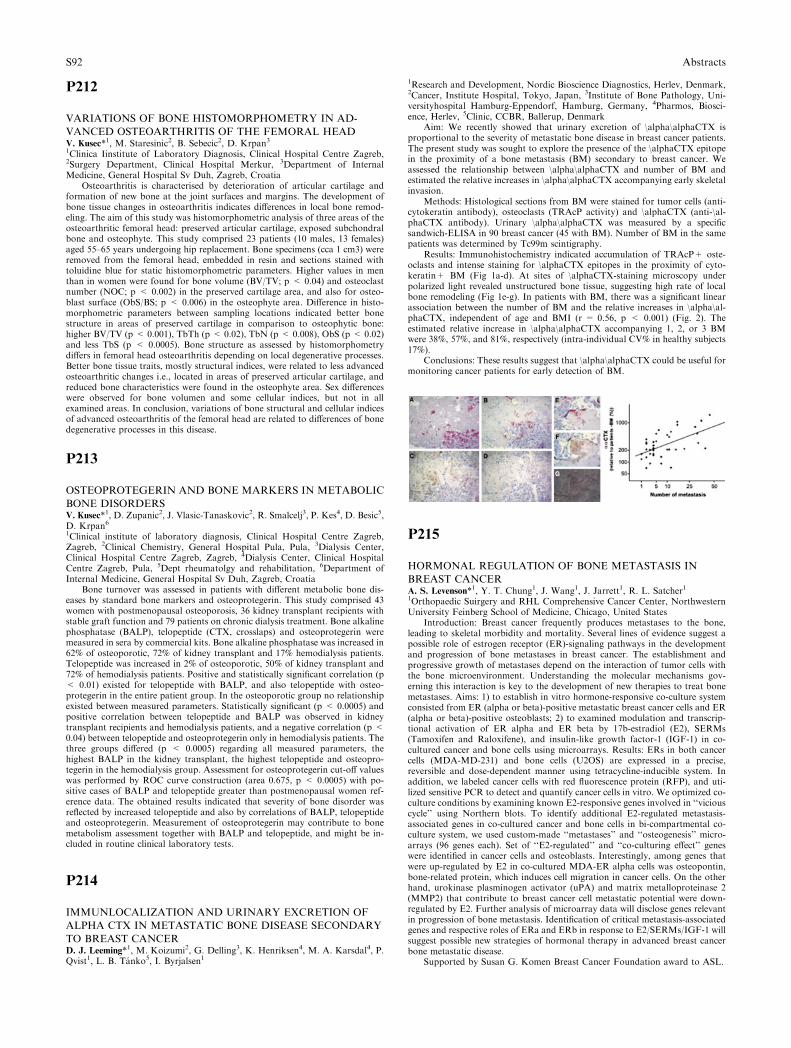
P212
VARIATIONS OF BONE HISTOMORPHOMETRY IN AD-
VANCED OSTEOARTHRITIS OF THE FEMORAL HEADV. Kusec*1, M. Staresinic2, B. Sebecic2, D. Krpan31Clinica Iinstitute of Laboratory Diagnosis, Clinical Hospital Centre Zagreb,2Surgery Department, Clinical Hospital Merkur, 3Department of InternalMedicine, General Hospital Sv Duh, Zagreb, Croatia
Osteoarthritis is characterised by deterioration of articular cartilage andformation of new bone at the joint surfaces and margins. The development ofbone tissue changes in osteoarthritis indicates differences in local bone remod-eling. The aim of this study was histomorphometric analysis of three areas of theosteoarthritic femoral head: preserved articular cartilage, exposed subchondralbone and osteophyte. This study comprised 23 patients (10 males, 13 females)aged 55–65 years undergoing hip replacement. Bone specimens (cca 1 cm3) wereremoved from the femoral head, embedded in resin and sections stained withtoluidine blue for static histomorphometric parameters. Higher values in menthan in women were found for bone volume (BV/TV; p < 0.04) and osteoclastnumber (NOC; p < 0.002) in the preserved cartilage area, and also for osteo-blast surface (ObS/BS; p < 0.006) in the osteophyte area. Difference in histo-morphometric parameters between sampling locations indicated better bonestructure in areas of preserved cartilage in comparison to osteophytic bone:higher BV/TV (p < 0.001), TbTh (p < 0.02), TbN (p < 0.008), ObS (p < 0.02)and less TbS (p < 0.0005). Bone structure as assessed by histomorphometrydiffers in femoral head osteoarthritis depending on local degenerative processes.Better bone tissue traits, mostly structural indices, were related to less advancedosteoarthritic changes i.e., located in areas of preserved articular cartilage, andreduced bone characteristics were found in the osteophyte area. Sex differenceswere observed for bone volumen and some cellular indices, but not in allexamined areas. In conclusion, variations of bone structural and cellular indicesof advanced osteoarthritis of the femoral head are related to differences of bonedegenerative processes in this disease.
P213
OSTEOPROTEGERIN AND BONE MARKERS IN METABOLIC
BONE DISORDERSV. Kusec*1, D. Zupanic2, J. Vlasic-Tanaskovic2, R. Smalcelj3, P. Kes4, D. Besic5,D. Krpan61Clinical institute of laboratory diagnosis, Clinical Hospital Centre Zagreb,Zagreb, 2Clinical Chemistry, General Hospital Pula, Pula, 3Dialysis Center,Clinical Hospital Centre Zagreb, Zagreb, 4Dialysis Center, Clinical HospitalCentre Zagreb, Pula, 5Dept rheumatolgy and rehabilitation, 6Department ofInternal Medicine, General Hospital Sv Duh, Zagreb, Croatia
Bone turnover was assessed in patients with different metabolic bone dis-eases by standard bone markers and osteoprotegerin. This study comprised 43women with postmenopausal osteoporosis, 36 kidney transplant recipients withstable graft function and 79 patients on chronic dialysis treatment. Bone alkalinephosphatase (BALP), telopeptide (CTX, crosslaps) and osteoprotegerin weremeasured in sera by commercial kits. Bone alkaline phosphatase was increased in62% of osteoporotic, 72% of kidney transplant and 17% hemodialysis patients.Telopeptide was increased in 2% of osteoporotic, 50% of kidney transplant and72% of hemodialysis patients. Positive and statistically significant correlation (p< 0.01) existed for telopeptide with BALP, and also telopeptide with osteo-protegerin in the entire patient group. In the osteoporotic group no relationshipexisted between measured parameters. Statistically significant (p < 0.0005) andpositive correlation between telopeptide and BALP was observed in kidneytransplant recipients and hemodialysis patients, and a negative correlation (p <0.04) between telopeptide and osteoprotegerin only in hemodialysis patients. Thethree groups differed (p < 0.0005) regarding all measured parameters, thehighest BALP in the kidney transplant, the highest telopeptide and osteopro-tegerin in the hemodialysis group. Assessment for osteoprotegerin cut-off valueswas performed by ROC curve construction (area 0.675, p < 0.0005) with po-sitive cases of BALP and telopeptide greater than postmenopausal women ref-erence data. The obtained results indicated that severity of bone disorder wasreflected by increased telopeptide and also by correlations of BALP, telopeptideand osteoprotegerin. Measurement of osteoprotegerin may contribute to bonemetabolism assessment together with BALP and telopeptide, and might be in-cluded in routine clinical laboratory tests.
P214
IMMUNLOCALIZATION AND URINARY EXCRETION OF
ALPHA CTX IN METASTATIC BONE DISEASE SECONDARY
TO BREAST CANCERD. J. Leeming*1, M. Koizumi2, G. Delling3, K. Henriksen4, M. A. Karsdal4, P.Qvist1, L. B. Tanko5, I. Byrjalsen1
1Research and Development, Nordic Bioscience Diagnostics, Herlev, Denmark,2Cancer, Institute Hospital, Tokyo, Japan, 3Institute of Bone Pathology, Uni-versityhospital Hamburg-Eppendorf, Hamburg, Germany, 4Pharmos, Biosci-ence, Herlev, 5Clinic, CCBR, Ballerup, Denmark
Aim: We recently showed that urinary excretion of \alpha\alphaCTX isproportional to the severity of metastatic bone disease in breast cancer patients.The present study was sought to explore the presence of the \alphaCTX epitopein the proximity of a bone metastasis (BM) secondary to breast cancer. Weassessed the relationship between \alpha\alphaCTX and number of BM andestimated the relative increases in \alpha\alphaCTX accompanying early skeletalinvasion.
Methods: Histological sections from BM were stained for tumor cells (anti-cytokeratin antibody), osteoclasts (TRAcP activity) and \alphaCTX (anti-\al-phaCTX antibody). Urinary \alpha\alphaCTX was measured by a specificsandwich-ELISA in 90 breast cancer (45 with BM). Number of BM in the samepatients was determined by Tc99m scintigraphy.
Results: Immunohistochemistry indicated accumulation of TRAcP+ oste-oclasts and intense staining for \alphaCTX epitopes in the proximity of cyto-keratin+ BM (Fig 1a-d). At sites of \alphaCTX-staining microscopy underpolarized light revealed unstructured bone tissue, suggesting high rate of localbone remodeling (Fig 1e-g). In patients with BM, there was a significant linearassociation between the number of BM and the relative increases in \alpha\al-phaCTX, independent of age and BMI (r = 0.56, p < 0.001) (Fig. 2). Theestimated relative increase in \alpha\alphaCTX accompanying 1, 2, or 3 BMwere 38%, 57%, and 81%, respectively (intra-individual CV% in healthy subjects17%).
Conclusions: These results suggest that \alpha\alphaCTX could be useful formonitoring cancer patients for early detection of BM.
P215
HORMONAL REGULATION OF BONE METASTASIS IN
BREAST CANCERA. S. Levenson*1, Y. T. Chung1, J. Wang1, J. Jarrett1, R. L. Satcher11Orthopaedic Suirgery and RHL Comprehensive Cancer Center, NorthwesternUniversity Feinberg School of Medicine, Chicago, United States
Introduction: Breast cancer frequently produces metastases to the bone,leading to skeletal morbidity and mortality. Several lines of evidence suggest apossible role of estrogen receptor (ER)-signaling pathways in the developmentand progression of bone metastases in breast cancer. The establishment andprogressive growth of metastases depend on the interaction of tumor cells withthe bone microenvironment. Understanding the molecular mechanisms gov-erning this interaction is key to the development of new therapies to treat bonemetastases. Aims: 1) to establish in vitro hormone-responsive co-culture systemconsisted from ER (alpha or beta)-positive metastatic breast cancer cells and ER(alpha or beta)-positive osteoblasts; 2) to examined modulation and transcrip-tional activation of ER alpha and ER beta by 17b-estradiol (E2), SERMs(Tamoxifen and Raloxifene), and insulin-like growth factor-1 (IGF-1) in co-cultured cancer and bone cells using microarrays. Results: ERs in both cancercells (MDA-MD-231) and bone cells (U2OS) are expressed in a precise,reversible and dose-dependent manner using tetracycline-inducible system. Inaddition, we labeled cancer cells with red fluorescence protein (RFP), and uti-lized sensitive PCR to detect and quantify cancer cells in vitro. We optimized co-culture conditions by examining known E2-responsive genes involved in ‘‘viciouscycle’’ using Northern blots. To identify additional E2-regulated metastasis-associated genes in co-cultured cancer and bone cells in bi-compartmental co-culture system, we used custom-made ‘‘metastases’’ and ‘‘osteogenesis’’ micro-arrays (96 genes each). Set of ‘‘E2-regulated’’ and ‘‘co-culturing effect’’ geneswere identified in cancer cells and osteoblasts. Interestingly, among genes thatwere up-regulated by E2 in co-cultured MDA-ER alpha cells was osteopontin,bone-related protein, which induces cell migration in cancer cells. On the otherhand, urokinase plasminogen activator (uPA) and matrix metalloproteinase 2(MMP2) that contribute to breast cancer cell metastatic potential were down-regulated by E2. Further analysis of microarray data will disclose genes relevantin progression of bone metastasis. Identification of critical metastasis-associatedgenes and respective roles of ERa and ERb in response to E2/SERMs/IGF-1 willsuggest possible new strategies of hormonal therapy in advanced breast cancerbone metastatic disease.
Supported by Susan G. Komen Breast Cancer Foundation award to ASL.
S92 Abstracts
P216
EXPRESSION AND REGULATION OF OSTEOPROTEGERIN
IN ADIPOSE TISSUE IN VITRO: OSTEOPROTEGERIN AS A
NOVEL ADIPOCYTOKINED. Kim1, J. An1, Y. Rhee1, S. Park1, W. Choi2, Y. Chung3, S. Lim*11Department of Internal Medicine, College of Medicine, Yonsei University,2Department of internal Medicine, College of Medicine, Hanyang University,Seoul, 3Department of Internal Medicine, College of Medicine, Ajou University,Suwon, South Korea
Backgrounds and aims: Osteoprotegerin (OPG), an anti-inflammatory pro-tein derived from osteoblasts, decreases bone resorption by inhibiting differen-tiation and activation of the osteoclasts. It is well known that OPG is elevated inplasma of patients with type 2 diabetes or coronary artery diseases. Becauseadipocytes and osteoblasts have their origin in common, we hypothesized thatOPG might be secreted from the adipocytes and contribute to protect the vesselfrom intimal inflammation and medial calcification.
Study design – in vitro experiments using cell culture and RT-PCRResults: Expression of OPG mRNA increased with the differentiation of
3T3L1 adipocytes and mRNA of RANKL, the counterpart of OPG in osteoclastdifferentiation, did not change significantly. The pattern of mRNA expression ofthe two molecules in subcutaneous adipose tissue was similar throughout thespecies, mouse, rat, and human. OPG mRNA expressed higher in white adiposetissue than in brown adipose tissue, and most abundantly in epididymal adiposetissue. Similarly to the in vitro experiments using vascular smooth muscle cells,whereas TNF-alpha increased the expression of OPG mRNA, rosiglitazone andinsulin reduced it dose- and time- dependently in 3T3L1 adipocyte.
Conclusions: OPG is expressed also in adipose tissue. Because adipose tissuecomprises considerable portions of the body and the amount varies in a lot ofmetabolic conditions, our results may give a reasonable explanation to theprevious clinical data by others. Although we cannot suggest whether OPG isexactly a cause or result of the metabolic derangements, we cautiously proposethat serum OPG, as one of the adipocytokines, could reflect the systemicinflammatory status and thus, be used as an inflammatory marker. Consideringits role in bone metabolism, it is speculated that OPG might have a protectiverole against inflammation and calcification of the arteries.
P217
EVIDENCE FOR AN IMPORTANT SUSCEPTIBILITY
GENE FOR FAMILIAL PAGET’S DISEASE OF BONE ON
CHROMOSOME 10P13 (PDB6) IN FAMILIES WITHOUT
SQSTM1 MUTATIONSG. J. A. Lucas*1, L. J. Hocking2, T. Cundy3, G. C. Nicholson4, J. P. Walsh5,S. H. Ralston11Bone Research Group, Molecular Medicine Centre, University of Edinburgh,Edinburgh, 2Dept. of Medicine and Therapeutics, University of Aberdeen,Aberdeen, United Kingdom, 3Dept. of Medicine, University of Auckland,Auckland, New Zealand, 4Dept of Clinical and Biomedical Sciences, Universityof Melbourne, Melbourne, 5Dept. of Endocrinology and Diabetes, Sir CharlesGairdner Hospital, Perth, Australia
Paget�s disease of bone has a strong genetic component and several sus-ceptibility loci for the disease have been identified by genome-wide scan. Wepreviously reported susceptibility loci for PDB on chromosomes 5q35 (PDB3;lodscore 3.0 at 189.63cM), 2q36 (PDB5; lodscore 2.7 at 218.24cM) and 10p13(PDB6; lodscore 2.6 at 41.43cM) in a cohort of 62 PDB kindreds of mainlyBritish descent(1). Subsequently, we identified various mutations in the SQSTM1gene as the cause of 5q35 linked PDB in 23 families from this population.Screening of the SQSTM1 gene for mutations in the remaining 39 families wasnegative. In this study we report the results of a reanalysis of our genome-widesearch data in the 39 families who did not carry SQSTM1 mutations. Analysis ofthese data using GENEHUNTER revealed significant evidence for linkage tothe PDB6 locus on chromosome 10p13 (lodscore + 4.08 at 41.43cM) close toD10S1653, whereas evidence for linkage to the 2q36 locus diminished (lodscore0.81). Heterogeneity testing using the HOMOG program supported a model oflinkage with homogeneity at marker D10S1653 (theta = 0.02; p = 0.00017), asopposed to linkage with heterogeneity or no linkage. In agreement with theseresults, conditional probability testing indicated that all families in this cohorthave a high probability of linkage to marker D10S1653. These data suggest thatthere is an important susceptibility gene for PDB on chromosome 10p13 infamilies of British descent and we are currently performing fine-mapping studiesof this locus to narrow the critical region and identify the gene responsible.
1. Hocking,L.J., Herbert,C.A., Nicholls,R.K., Williams,F., Bennett,S.T.,Cundy,T., Nicholson,G.C., Wuyts,W., Van Hul,W., and Ralston,S.H. 2001.Genomewide search in familial Paget disease of bone shows evidence of geneticheterogeneity with candidate loci on chromosomes 2q36, 10p13, and 5q35.Am.J.Hum.Genet. 69:1055–1061.
P218
OSTEOPROTEGERIN AS A MEDIATOR OF BONE PHENO-
TYPE INMURINE GENERALISED LYMPHOPROLIFERATIVE
DISORDERI. K. Lukic*1, N. Kovacic1, D. Grcevic2, V. Katavic1, S. Ivcevic3, A. Marusic11Department of Anatomy, 2Department of Physiology and Immunology,3Centre for Functional Genomics, Zagreb University School of Medicine, Za-greb, Croatia
We have previously shown that mice with generalised lymphoproliferativedisorder (gld) have increased bone mass in addition to autoimmune diseasecharacterised by the accumulation of double negative (dn) T lymphocytes (CD3+ CD4 ) CD8 ) B220+). To further explore the association of the immunedisorder with the bone phenotype of gld mice, we established parabiotic circu-lation between gld and wild-type animals (C57BL/6, B6). The mice were sacri-ficed weekly for four weeks or, in the second set of experiments, were separatedafter four weeks of parabiosis and sacrificed two weeks following the separation.One week after the surgery, the proportion of dn T lymphocytes increased inperipheral blood and lymphatic tissues of wild-type members of the B6-gldparabiotic pair and decreased in tissues of gld pair members. The mixing of cellscontinued during four weeks of parabiosis. Moreover, the proportions of dn Tcells in tissues of gld members of the B6-gld pairs after the separation becameindistinguishable to those in control B6 animals. Number of osteoclast-like(OCL) cells in bone marrow cultures from a wild-type member of B6-gldparabiotic pair at the end of the first week decreased from 266 ± 52 to 120 ± 5OCL/cm2, P < 0.05, comparable with gld mice (99 ± 21 OCL/cm2), while thenumber of osteoblast colonies did not change. After four weeks, number of OCLcells formed from the bone marrow of B6 parabiotic mice was still similar to thenumber of OCL cells in their gld counterparts (150 ± 18 and 131 ± 24 OCL/cm2, respectively). In addition, the number of osteoblast colonies in B6 membersof B6-gld parabiotic pairs increased (from 6 ± 2 to 18 ± 1 colonies/cm2, P <0.05) thus resembling the cell cultures of gld mice (18 ± 1 colonies/cm2). Thealterations of osteoclastogenic and osteoblastogenic potential of the bone mar-row of B6 members of B6-gld parabiotic pairs were paralleled by changes in theosteoprotegerin (OPG) expression. Levels of OPG in the bones of B6 mice, asassessed both by quantitative PCR and ELISA, increased during parabiosis andreached the levels found in gld animals. Taken together, these data show that thecirculation of cells, including dn T lymphocytes, established by parabiosis con-fers the osteoclast and osteoblast phenotype of gld to wild-type animals. Thosechanges are likely to be mediated through OPG, although the exact mechanismremains to be determined.
P219
PROSTATE CANCER AND SKELETALMETASTASIS: IMPACT
OF THE BONE MICROENVIRONMENT AND EXTRACELLU-
LAR CALCIUMJ. Liao1, A. Schneider2, N. Datta1, K. Pienta3, L. K. McCauley*11Periodontics and Oral Medicine, University of Michigan, Ann Arbor, 2Diag-nostic Sciences and Pathology, University of Maryland, Baltimore, 3InternalMedicine, University of Michigan, Ann Arbor, United States
Prostate cancer has a high predilection of metastasis to bone and previousstudies suggested that proteins released from the bone matrix during resorptionmay contribute to tumor localization and growth in bone. The dependency onbone remodeling for tumor localization is still controversial, but changes in bonemarrow microenvironment likely play a critical role in skeletal metastasis. Thisstudy determined the impact of extracellular calcium [Ca2+]o such as that re-leased during resorption, on prostate cancer cells. Human prostate cancer cells(PC-3, C4-2B and LNCaP) were cultured in low (0.5 mM) or high (2.5 mM)[Ca2+]o. High [Ca2+]o increased both PC-3 and C4-2B prostate cancer cellnumbers over a 6-day period. In contrast, numbers of LNCaP cells, a prostatecancer cell line of low tumorigenicity and low skeletal localization, were notaffected by high [Ca2+]o. Cell lines with increased numbers in response to high[Ca2+]o had higher levels of extracellular calcium sensing receptor (CaSR)versus nonresponders as determined by RT-PCR (CaSR mRNA) and westernblot analysis (CaSR protein). Western analysis revealed higher levels of cell cyclepromoting cyclin D1 in PC-3 cells in high versus low [Ca2+]o. Activation of theG-protein coupled CaSR in PC-3 cells by high [Ca2+]o was verified by aninhibition of PGE2 or forskolin-induced cAMP stimulation. Additionally,treatment of PC-3 cells with the CaSR agonist neomycin inhibited the cAMPincrease by PGE2 or forskolin. Cell adhesion in the presence of [Ca2+]o wasdetermined by plating PC-3/Luc+ cells, rinsing unattached cells and determi-nation of luciferase levels in the remaining adherent layer. [Ca2+]o or neomycinenhanced the attachment of PC-3 cells and cell adhesion reduced by the G-protein inhibitor pertussis toxin. Furthermore, western analysis revealed thathigh [Ca2+]o treatment of PC-3 cells lead to activation of the anti-apoptoticprotein Akt; and pharmacologic inhibition of the PI3K/Akt pathway withLY294002 reduced the [Ca2+]o and neomycin mediated increase in PC-3 cellattachment. These data suggest that high [Ca2+]o released during bone turnover
Abstracts S93
could facilitate prostate tumor localization in the skeleton via the CaSR and itsimpact on cell viability. Hence, alterations in the bone marrow microenviron-ment including changes in extracellular calcium may alter the occurrence ofprostate cancer skeletal metastasis. This project was supported by the US-PHS,NIH:NCI P01-CA093900.
P220
MUTATION ANALYSES OF 30 FAMILIES WITH
HYPOPHOSPHATASIAT. Michigami*1, K. Tachikawa1, T. Kubota1, K. Ozono21Department of Environmental Medicine, Osaka Medical Center and ResearchInstitute for Maternal and Child Health, Izumi, 2Department of Pediatrics,Osaka University Graduate School of Medicine, Suita, Japan
Hypophosphatasia is an inherited disease characterized by hypomineraliza-tion of bone associated with impaired activity of the tissue non-specific alkalinephosphatase (TNSALP). Because of the diverse clinical manifestations, hypo-phosphatasia is usually classified into five subtypes based on the age of onset andclinical features; perinatal, infantile, childhood, adult-type and odontohypo-phosphatasia. To date, more than 100 mutations in TNSALP gene have beenreported in patients with hypophosphatasia. However, the frequency of thesemutations and the correlation with phenotypes are still not fully elucidated. Herewe report the results of TNSALP gene analyses of 30 families with hypophos-phatasia and their clinical manifestations. Among the 30 families, 27 familieswere Japanese, one was German, one was Israeli and the other was Indian. In the27 Japanese families, 20 distinct mutations were identified. As we previouslyreported, 1559delT and F310L were the most common mutations in the Japanesepatients (24/54 and 5/54 alleles, respectively), although both of them were notfound in other races. 1559delT was often identified in severe forms such asperinatal and infantile type. On the other hand, F310L was associated withunique phenotype where the patients survived despite the perinatal onset, andexhibited deformity of the long bones and characteristic bone spurs withoutapparent hypomineralization, indicating the existence of the distinct subtype ofperinatal hypophosphatasia with benign prognosis. The German family withautosomal dominant hypophosphatasia was found to carry D361V mutant al-lele. The genotype of the Israeli patient diagnosed with perinatal hypophos-phatasia was E274K/G309R. The Indian patient was homozygous for a splicesite mutation. The mutations identified in the German, Israeli and Indian fam-ilies were not found in the Japanese patients. In our Japanese patients, 7mutations (A23V, T83M, A159T, R206Q, E218V, L282P, G322R) were previ-ously reported in other races. When the DNA samples of the parents wereavailable, they were always revealed to be carriers, demonstrating the quite lowrate of de novo mutation. Taken together, the racial difference in mutationssuggests founder effects in some of the mutations including 1559delT and F310L.To further understand the genotype-phenotype correlation in hypophosphatasia,we should consider the frequency of mutations in each race.
P221
ADULT PATIENTS AFFECTED BY OSTEOGENESIS IMPER-
FECTA HAVE IMPAIRMENT OF DIASTOLIC FUNCTIONS. Migliaccio*1, G. Barbaro1, R. Fornari1, G. Di Lorenzo1, S. Falcone1,S. Marzano1, E. Fabbrini1, M. Marini1, C. Lubrano1, M. Brama1, G. Prosso-mariti1, E. Greco1, F. Conti1, G. Spera11Cattedra di Medicina Interna, Dipartimento di FIsiopatologia Medica, Uni-versita La Sapienza, Roma, Italy
Osteogenesis imperfecta (OI) is a rare inherited connective disorder causingincreased bone fragility and low bone mass. OI includes severe bone fragility,impared dentinogenesis, with less common alterations in the joints, blood ves-sels, heart valves, skin. However, description of left ventricular rupture, aorticdissection and heart valves incompetence have been previously described. Thus,aim of the study has been to characterize cardiac functional parameters byechocardiography in adults with OI, to evaluate the presence of potential cardiacdisorders.
To this aim, 29 (16 females and 13 males) affected by type I, III, IV of OI (1)and 24 control subjects (12 females and 12 males) were evaluated. Patients andcontrols selected for the study underwent clinical examination, 12-lead electro-cardiogram and echocardiogram. All subjects were evaluated by two-dimen-sional echocardiography with continuous- and pulse-wave Doppler Patients andcontrols belonged to NYHA class I and no significant electrocardiographicalteration was documented in both groups. Twenty one OI patients (71%)showed valvular defects but only one control subject had valvular defect. Asregards the diastolic function parameters, in OI patients E/A ratio was reducedby 11% (95% CI: 6% to 20%; P < 0.001), Isovolumetric relaxation time (IRT)was increased by 46% (95% CI: 27% to 50%; P < 0.001) and E wave decelerationtime (DT) was increased by 18% (95% CI: 12% to 25%; P < 0.01) compared tocontrols.
In conclusion, our data indicate that adult patients affected by OI have analtered diastolic funcion. These diastolic echocardiographic parameters mayworsen over time, especially if other cardiovascular risk factors (eg, smoking,hypertension, metabolic alterations) are not carefully checked, monitored andtreated. Thus, in the context of a multidisciplinary evaluation of OI patients, ourdata suggest that a careful cardiological evaluation of these patients is indicatedbeside skeletal evaluation.
P222
SUCCESSFUL TREATMENT OF THE POST-TRAUMATIC
OSTEOLYSIS OF DISTAL CLAVICLE WITH ALENDRONATE
A CASE REPORTM. T. K. Mulari*1, G. Gu2, K. Mattila3, K. Parkkola4, H. K. Vaananen21Kiukainen health station, Joint Municipal Health Care Services of Harjavaltaregion, Panelia, 2Dept. of Anatomy, Inst. of Medicine, Univ. of Turku, 3Diag-nostic Imaging Centre, Turku University Hospital, 41Health Care Unit, Archi-pelago Sea Naval Command, Turku, Finland
We describe a 20-year-old male, whose progressing post-traumatic osteolysisof the distal clavicle was successfully treated with alendronate. Radiographsobtained immediately after the accident showed a gradus I subluxation in the leftacromioclavicular joint and 1 mm (3 and 4 mm on the right and left, respectively)asymmetric widening of the joint. During four months of follow-up and cessa-tion of any inciting activity, conventional radiographs and magnetic resonanceimages revealed progressive vanishing of the acromial extremity of the leftclavicle, and widening of the joint up to 6 mm. To slow down the pathologicalosteoclastic bone resorption, we initiated oral admininstration of alendronate(70 mg weekly) for three months. The progression of osteolysis was ceased afterone month, and the width of the joint was still 6 mm. Radiographs revealed there-construction of cortex and bone fragment ossification and fusion with theclavicle. After three months of alendronate therapy, the AC joint space haddecreased from 6 to 5 mm and the patient was painless. Healing of the joint wasevident and patient was painless in a control meeting three months after the endof alendronate therapy. Although without a concurrent control patient or groupof patients we cannot know for sure if alendronate caused these improvements,our results suggest that no matter what mediators are associated with theosteolytic process, the cellular pathway in osteolysis involves in the end theactivity of the osteoclast and as long as the pharmaceutical agent can inhibit theosteoclast function, improvement could be anticipated by inhibiting pathologi-cally increased osteoclastic bone resorption.
P223
BONE TURNOVER MARKERS IN NEWLY DIAGNOSED
BREAST CANCER PATIENTSB. F. Murray*1, D. Smith2, J. J. Brady1, M. L. Healy2, N. O’ Higgins3,E. McDermot3, M. J. McKenna4, T. J. McKenna41Metabolism, 2Endocrinology, 3Surgery, St Vincent’s University Hospital,4Endocrinology, St Vincent’s University Hospital, Dublin, Ireland
Bone Turnover markers have been studied in breast cancer patients withbone metastases, however bone turnover at the time of diagnosis has not beenexamined to the same extent. The aim of this study was to compare biochemicalmarkers of bone turnover in newly diagnosed pre menopausal, early postmenopausal and elderly breast cancer patients with healthy aged matched wo-men to ascertain bone turnover status.
Twenty-six pre menopausal, mean age 40, 43 early post menopausal, meanage 55, 25 elderly post menopausal, mean age 70, women with breast cancer wererecruited. Patients had histologically confirmed nonmetastatic breast cancer. Theabsence of bone metastases was determined by bone scintigraphy. Twenty fourpre menopausal females, mean age 27 years, 21 healthy post menopausal, meanage 55 years and 13 healthy elderly post menopausal women, mean age 72 yearswere included. Fasting blood and second void urine samples were obtained atdiagnosis for measurement of bone formation markers Procollagen Type 1 N-Propeptide (P1NP), Intact Osteocalcin (OCI), and Bone Alkaline Phosphatase(BAP) and bone resorption markers C-Terminal cross-linking Telopeptide ofType 1 Collagen (\betaCTX) ,Deoxypyridinoline Crosslinks (fDPD/Cr) and N-Terminal Telopeptide of Type 1 Collagen (NTX/Cr) respectively. Osteocalcin p< 0.0001, P1NP p < 0.02 and NTX p < 0.005 were lower in the pre meno-pausal breast cancer patients when compared to healthy pre menopausal women.In the early post menopausal patients, OCI p = 0.02, P1NP p < 0.005 and BAPp = 0.02 were lower when compared to aged matched healthy women whereasno difference was found with the resorption markers. None of the bone for-mation or resorption markers were significantly different in the elderly postmenopausal patients when compared to aged matched healthy women. T valuesfor P1NP and NTX were used to derive indices of Bone Remodelling Bal-ance(BRBI)(bone formation - resorption) and Bone Turnover(BTI)(bone for-mation + resorption) and applied to the three groups. The BRBI was
S94 Abstracts
significantly lower in the early post-menopausal (p = 0.01) and elderly patients(p < 0.05) than in aged matched healthy women. The BTI was lower in premenopausal (p < 0.001) and in early post menopausal patients (p < 0.05) thanin controls.
These results demonstrate significantly lower bone formation and low boneturnover in the pre and early post menopausal breast cancer patients comparedto healthy women.
P224
ANTI-METASTATIC AND ANTI-ANGIOGENIC ACTIVITY
OF 1,25-DIHYDROXYVITAMIN D3 IN BONE-METASTATIC
LUNG CARCINOMA CELLSK. Nakagawa*1, T. Okano11Department of Hygienic Sciences, Kobe Pharmaceutical University, Kobe,Japan
Bone is one of the most preferential target sites for cancers including breast,prostate, and lung cancers. Bone metastases can lead to pain, pathological frac-tures, andhypercalcemia. The process of bonemetastasis basically consists of threesteps including: (1) proliferation and invasion of cancer cells at primary site, (2)intravasation, migration in the circulation, and extravasation of cancer cells, and(3) arrest in bone marrow, egress from central sinus, attachment to bone surfaces,osteoclastic bone destruction, and colonization of cancer cells in bone. Currenttreatments are mainly palliative and do not provide a life-prolonging benefit tometastatic patients. There is, therefore, a need to define new therapeutic targetsthat could become useful adjunct therapies for the treatment of cancers thatmetastasize to bone. 1,25-Dihydroxyvitamin D3 [1,25-D3] exhibits potent anti-proliferative and anti-invasive effects against cancer cells in vitro and in vivo. Inthis study,we examined the anti-metastatic effects of 1,25-D3using bonemetastaticLewis lung carcinoma cells expressing green fluorescent protein (LLC-GFP cells).In the in vitro experiment, 1,25-D3 significantly inhibited the metastatic activity ofLLC-GFP cells. Moreover, 1,25-D3 significantly reduced the expression of matrixmetalloproteinase (MMP)-2,MMP-9, vascular endothelial growth factor (VEGF)and parathyroid hormone-related peptide (PTHrP), which is an important acti-vator of osteoclast activation, in LLC-GFP cells. In the angiogenesis assay, thenumber of tumor cells and the bFGF-induced angiogenesis were significantly re-duced in 1,25-D3-treated mice. In vivo study, we used the animal model in whichLLC-GFP cells injected in the left cardiac ventricle reproducibly colonize specificsites of the skeletal system of mice. Intravenous injection of LLC-GFP cells re-sulted in tumor colonies in the lung only. Left cardiac injection of LLC-GFP cellscaused bone colonization. The total number of metastases were decreased in 1,25-D3-treated mice. Bone is a dynamic organ composed of cells including hemato-poietic, stromal, endothelial, and other cell types. These data suggested that 1,25-D3 reduced metastasis through not only anti-tumor action but also the regulationof hematopoietic, stromal, endothelial, osteoblast and osteoclast cell functions.Our results indicate that 1,25-D3 analogs would be a potentially beneficial drug tobe used in the treatment of cancer metastasis.
P225
ELEVATED SERUM FGF23 PROTEIN ASSOCIATED WITH
TUMOR-INDUCED OSTEOMALACIA: A CASE REPORTK. Nawrot-Wawrzyniak*1, F. Varga1, A. Nader2, P. Roschger1, S. Sieghart3,E. Zwettler1, S. Lang4, K. Klaushofer11Ludwig Boltzmann Institute of Osteology at Hanusch Hospital of WGKK andAUVA Trauma Centre Meidling, 4th Medical Department , Hanusch Hospital,2Ludwig Boltzmann Institute of Osteology at Hanusch Hospital of WGKK andAUVA Trauma Centre Meidling, 4th Medical Department and Institute forPathology, Hanusch Hospital, 32nd Medical Department, Kaiserin ElisabethHospital, 4Clinical Institute of Clinical Pathology, University of Vienna MedicalSchool, Vienna, Austria
Tumor-induced osteomalacia (TIO) is a paraneoplastic disorder character-ized by hypophosphatemia, phosphaturia, low serum levels of 1,25-di-hydroxyvitamin D and skeletal undermineralization. It is most commonlycaused by benign mesenchymal tumors that produce, among others, fibroblastgrowth factor 23 (FGF23)[1]. Current evidence thus suggests that this proteinhas an important role in the regulation of phosphate homeostasis.
Serum, tumor tissue and iliac crest biopsy of a 68 years old male with amesenchymal tumor with a haemangiopericytoma-like morphology from thescapula were analysed. Serum concentrations of FGF23 were measured byELISA that detected both the intact or the C-terminal fragment. FGF23 mRNAexpression, was investigated by in-situ hybridization (ISH) on formaldehydefixed, paraffin embedded tumor tissue. ISH was performed with a biotin-labeledanti-sense hFGF23-transcript as probe and with the sense transcript as negativecontrol. Developing was performed with ExtrAvidin-HRP/DAB-substrate. Bonehistology was addressed by Giemsa and trichrome Goldner�s staining. Trabec-ular bone mineralization density distribution (BMDD) was determined byquantitative backscattered electron imaging (qBEI).
Clinical evaluation of the patient revealed low serum phosphate and elevatedserum levels of bone-type alkaline phosphatase and calcium. In serum intactFGF23 protein was low, whereas the C-terminal peptide was high (3330 RU/ml).After removal of the tumor, the serum parameters turned to normal. ISH oftumor-tissue showed mRNA FGF23 expression in distinct tumor cells. Scatteredbetween the spindled tumor cells, which resembled activated fibroblasts, and thinwalled blood vessels, numerous multinucleated osteoclast-like giant cells wereidentified. Analysis of the bone biopsy revealed that compared to normal theaverage mineral content of the biopsy was decreased () 6%) while the hetero-geneity of mineralization was increased (+ 39%). Trabecular architecture(parameters: BV/TV )11%, TbTh )18%) was irregular with thin, loosely con-nected trabeculae. Histopathologically, surface osteidosis without endostealfibrosis or morphological signs of hyperparathyroidism was found.
In conclusion we demonstrated FGF23 mRNA expression in tumor tissue ofa patient with hypophosphatemia and osteomalacia, which could be responsiblefor the elevated FGF23 serum levels before resection.
[1]Larsson T et al. 2003 Eur J Endocrinol 148:269
P226
BONE METABOLISM IN DIALYSED PATIENTS - ARE THERE
ANY DIFFERENCES BETWEEN HEMODIALYSIS AND PERI-
TONEAL DIALYSIS?Z. Nowak*1, M. Konieczna2, Z. Wankowicz11Department of Nephrology, 2 Center of Nuclear Medicine, Central Hospital ofMilitary Institute of Medicine, Warsaw, Poland
Bone pathology is an important problem in patients on dialysis. Noninvasive measurement of bone turnover markers has been considered as apotentially useful tool of diagnosis renal osteodystrophy (ROD) in dialysedpatients.
The purpose of the study was to determine the influence of dialysis modalityon bone turnover markers and bone mineral density.
We studied group of 143 patients (F = 61, M = 82). 69 were treated onperitoneal dialysis (PD) mean age 59 ± 17 and 74 on hemodialysis (HD) meanage 63 ± 18. The following parameters were determined in serum: iPTH, Ca, P,ALP, aminoterminal propeptide of type I procollagen - PINP, carboxyterminalpropeptide of type I procollagen - PICP as a markers of bone formation and typeI collagen crosslinked telopeptide - ICTP and tartrate resistant acid phosphataseTRAP5b as a markers of bone resorption. These parameters were measuredbefore onset of dialysotherapy and after 1st year observation. Bone mineraldensity were evaluated by DEXA ( DPX Lunar). iPTH levels were used todifferentiate types of ROD. iPTH 450pg/ml- high turnover ROD.
Patients treated with peritoneal dialysis after 1st year of dialysotherapy hassignificantly lower values of iPTH in comparison with HD patients (146 ± 89vs. 342 ± 178 pg/ml; p < 0.01). Level of iPTH was <100pg/ml -(low turnoverROD). in 59.5% PD group (41/69) and only 28% HD group (23/82). Level ofiPTH was > 450pg-(high turnover ROD) in 8.7% PD group (6/69) and 24% HDgroup (20/82). We found significant correlation only between iPTH and PINP(r = 0.7074; p < 0.001) and between iPTH and TRAP (r = 0.6938, p < 0.001).In patients with low turnover ROD significantly lower values of PINP andTRAP were found in comparison with high turnover ROD. (51 ± 23ug/lvs.283 ± 146ug/l p < 0.001 and 2.7 ± 1.5U/L vs. 7.5 ± 1.6U/L p < 0.001respectively). The difference between values of PICP, ICTP, P, Ca, bone mineraldensity in CAPD and HD group were non significant and did not correlate withvalue of iPTH.
In conclusion, in patients treated with PD predominant bone lesion seems tobe low turnover renal osteodystrophy, but in patients on HD secondaryhyperparathyroidism was more frequent. Monitoring of iPTH, PINP andTRAP5b levels might be useful in assessment of bone metabolism in patient ondialysis.
P227
ZOLEDRONIC ACID INDUCES OSTEOSARCOMA CELL
DEATH BY AIF TRANSLOCATION AND CELL CYCLE
ARREST IN S AND G2/M PHASESB. Ory*1, F. Blanchard1, S. Couillaud1, F. Gouin1, F. Redini1, D. Heymann11Universite de Nantes EA3822 INSERM ERI 7, Medicine Faculty, Nantes,France
As a vicious cycle takes place between tumor cell proliferation and boneresorption in bone tumors, the use of nitrogen-containing bisphosphonates (N-BP) is a promising approach to break this phenomenon. Indeed, Zoledronic acid(ZOL) is a N-BP of the third generation used as an osteoclast inhibitor in severalpathologies involving bone resorption from tumor origin or not. However, ZOLmolecular mechanisms remain unclear. In the present study, the in vitro effects ofZOL were compared between several osteosarcoma cell lines and primaryosteoblast cultures, in terms of cell proliferation, cell cycle analysis (flowcytometry, western blots), apoptosis (caspase 1, 3 and 8 activities, Hoechst
Abstracts S95
staining, western blots, mitochondrial permeability assay, time lapse and con-focal microscopy) and cell adherence (migration assay, integrin involvement,cytoskeleton observation by confocal microscopy).
ZOL strongly inhibited osteosarcoma cell proliferation compared to osteo-blasts. ZOL-induced inhibition of cell proliferation was due to cell cycle arrest inS and G2/M phases and is associated with an increase of Wee1 and p-cdc2 levelsand a decrease of cdc25c. No alteration of nuclear morphology can be observedin ZOL-treated osteosarcoma cell lines after Hoechst staining. In addition, ZOLdid not induce any caspase-1, 3 and 8 activation. However, trypan blue stainingand time lapse microscopy demonstrated that 10 microM ZOL selectively in-duced osteosarcoma cell death which is not inhibited by Z-Vad solution. Mor-over, ZOL increased the phosphorylation of p53, the expression of Bax, andinhibited Bcl-2 level. It also induced mitochondrial permeability associated withthe translocation of Apoptosis Inducing Factor (AIF). Although the main targetof ZOL described in the literature is the farnesyl diphosphate synthase, anotherpathway involving integrins can be suggested. Indeed, western blot analysesshowed that a 48-hour treatment with 10 microM ZOL inhibited the phos-phorylation of focal adhesion kinase and cell migration, disturbed the cyto-skeleton and adherence. Moreover, RGD peptide inhibited ZOL-inducedactivities.
These results demonstrate selective and original anti-tumor effects of ZOLon several osteosarcoma cell lines, thus allowing to consider these molecules aspotential therapeutic agents in clinical trials of tumor bone pathologies.
P228
PERCUTANEOUS BALLOON KYPHOPLASTY IN PATIENTS
WITH PATHOLOGICAL VERTEBRAL BODY FRACTURE
CAUSED BY MULTIPLE MYELOMAR. Pflugmacher*
1
1Centrum fur Muskuloskeletale Chirurgie, Charite-Universitatsmedizin Berlin,Berlin, Germany
Background/aims: A prospective study to evaluate the clinical and radio-graphic outcomes of Balloon Kyphoplasty in patients with fractures of thethoracic and lumbar spine caused by multiple myeloma.
Methods: Vertebral fractures due to multiple myeloma were treated byBalloon Kyphoplasty (20 patients, 48 vertebral bodies). Symptomatic levels wereidentified by clinical presentation, MRI, radiographs and CT. During the fol-lowing year, Visual Analog Scale (VAS) and Oswestry Disability Score weredocumented. Radiographs were performed pre- and postoperatively at 3, 6 and12 months. The vertebral height and the kyphotic deformity were measured toassess the restoration of the sagittal alignment.
Results: The median pain scores (VAS) decreased from pre- to post-treat-ment significantly as well as the Oswestry Disability Score (p < 0.05). Clinicallyasymptomatic cement leakage occurred in 5 fracture levels (10.4%). During 1year follow-up this surgical technique demonstrated restoration and stabilizationof the height of the vertebral body.
Conclusion: Balloon Kyphoplasty is an effective minimally invasive proce-dure for the stabilization of pathological vertebral fractures caused by multiplemyeloma leading to a statistically significant reduction of pain status. BalloonKyphoplasty is able to stabilize the vertebral body height and is only partiallyable to avoid further kyphotic deformities.
P229
NON-VIRAL OSTEOPROTEGERIN GENE TRANSFER
INHIBITS THE TUMOR PROGRESSION AND PROLONGS
SURVIVAL IN A MOUSE EXPERIMENTAL MODEL OF
OSTEOSARCOMAF. Lamoureux1, P. Richard2, Y. Wittrant1, V. Trichet1, B. Pitard2,D. Heymann1, F. Redini*11University of Nantes EA 3822 INSERM ERI 7, 2INSERM U 533, MedicineFaculty, Nantes cedex 1, France
Osteosarcoma is the most frequent primary bone tumor and despite recentimprovements in surgery and in multidrug chemotherapy, survival remainsaround 50% after 5 years. To break the vicious cycle that takes place betweentumor cell proliferation and bone resorption in bone tumors, the use of anti-resorptive agents is a promising approach. Osteoprotegerin (OPG) is a potentinhibitor of osteoclast differentiation and activation, but is limited as a thera-peutic agent due to its short circulating half-life. In order to overcome theselimitations, the therapeutic effects of OPG produced by gene transfer areexamined in the murine osteolytic model of osteosarcoma POS-1.
In vitro, we can efficiently transfer and express either the GFP marker or themurine OPG 1-194 genes into murine musculoskeletal C2C12 cells but notosteosarcoma POS-1 cells by using the cationic non-viral vector polyethyleni-mine. OPG administered as exogenous molecule or by transgene expression doesnot modify the cell proliferation nor the gene expression in both cell types.
In vivo, a transplantable model of osteosarcoma in C3H/He mice was used,leading to the development of a primary osteolytic tumor and pulmonarymetastases dissemination. To promote gene transfer in vivo we used formula-tions resulting from the association of plasmid DNA with the Lutrol blockcopolymer, that have been shown to increase transfection efficiency in variousorgans. Three groups of mice were assigned respectively as controls (no injec-tion), control vectors (lutrol/pcDNA3 alone) and OPG (lutrol/pcDNA3-OPG 1–194). The lutrol/DNA formulations were injected into tibial anterior musclesonce a week, beginning at 7 days before osteosarcoma implantation up to 28days post-implantation.
The osteosarcoma incidence was significantly diminished in the OPG groupas compared to control groups (respectively 33 and 83%) and the mean tumorvolume in this group was also decreased as compared to controls (respectively616 and 3060 mm3 at day 23, p < 0.01). This was accompanied by a significantincrease of animal survival in the OPG group (67% versus 16% at day 28, p <0.005). The efficacy of OPG as anti-resorptive agent was confirmed by evaluatingthe prevention of tumor-associated bone degradation, as revealed by radiographand micro-scanner analyses. Using a non-viral gene transfer method, the presentstudy demonstrates for the first time the beneficial effect of OPG as therapeuticagent in the treatment of osteosarcoma.
P230
ANTI-TUMOR EFFECTS INDUCED BY DENDRITIC CELL-
BASED IMMUNOTHERAPY AGAINST ESTABLISHED OSTE-
OSARCOMA IN RATSY. Wittrant1, C. Chauvin2, F. Lamoureux1, B. Trinite2, F. Hubert2, D. Hey-mann1, R. Josien2, F. Redini*11University of Nantes EA 3822 INSERM ERI 7, Medicine Faculty, Nantescedex 1, 2INSERM U643, ITERT, Nantes, France
Osteosarcoma is the most frequent primary bone tumor, and despite recentimprovements in surgery and the development of different regimens of multidrugchemotherapy, survival remains around 55 to 70% after 5 years. This poorprognostic warrants new therapeutic strategies to improve the overall rate ofsurvival, among them immunotherapies are very promising. A distinct subset ofrat dendritic cells (DC) exhibits cytotoxicity against tumor cells in vitro. Thisactivity is particular to the MHC II low CD103+ CD4) (referred to as CD4-DC) subset of immature DC found in spleen and lymph nodes. CD4- DC in-duced a rapid Ca2+) and caspase-independent apoptosis-like cell death in alarge number of rat, mouse and human tumor cells. An adenovirus encoding forhuman Flt3L was used to expand CD4- DC in vivo. To explore the in vivo roleof this subset of DC, a rat osteosarcoma model induced by tumor transplanta-tion at the bone site was used. CD4) DC killed the cell line derived from thistumor (OSRGa) in vitro and phagocyted efficiently their victims. These antigenloaded DC were used to vaccinate tumor bearing rats. A complete regression ofthe tumor was observed at 50 days after implantation for 5 rats out of 9, ascompared to 2/9 in animals vaccinated with irrelevant tumor antigen (Jurkat)loaded-DC. The tumor volume in osteosarcoma-bearing rats was significantlylower in animals vaccinated with OSRGa loaded DC as compared to controls orto animals vaccinated with Jurkat tumor antigen loaded-DC (respectively 8369,28562 and 21559 mm3 at 26 days post implantation). The rats with tumorregression exhibit no recurrence when re-implanted with new tumor fragment,suggesting a memory effect. As a consequence; the overall survival rate wassignificantly increased in the group that received OSRGa loaded-DC (42.8%versus 12.5% in controls and Jurkat tumor antigen loaded DC). These resultssuggest that killer DC can efficiently present tumor antigens captured from tu-mor cells.
P231
BONE METASTASIS IS INDEPENDENT OF OSTEOLYSIS IN A
NUDE MOUSE MODEL OF CANINE PROSTATE CANCER
WITH MIXED BONE METASTASESN. K. Thudi1, C. K. Martin1, M. V. P. Nadella1, W. P. Dirksen1, J. L. Werbeck1,T. J. Rosol*11Veterinary Biosciences, Ohio State University, Columbus, United States
Bone metastasis is the most common cause of mortality in patients withadvanced prostate cancer. Prostate cancer metastases to bone primarily developmixed osteoblastic and osteolytic lesions; however, the mechanisms responsiblefor inducing bone resorption and formation are unknown. We hypothesized thattumor-induced osteolysis in mixed bone metastases regulated osteoblasticactivity and tumor growth. Left ventricular injections were performed in 6-week-old nude mice with luciferase-transfected canine prostate cancer cells (ACE-1)that induce mixed bone metastases. Zoledronic acid, a potent bisphosphonatethat inhibits osteoclast-mediated bone resorption, was used to treat mice at adosage of 100lg/kg body weight subcutaneously twice a week starting a weekbefore intracardiac injections and continued over the course of the experiment (4weeks). Mice developed metastases in the metaphyses of long bones and in the
S96 Abstracts
vertebrae (up to 10 per mouse). In vivo bioluminescence imaging revealed nodifferences in the progressive growth of the metastases and the incidence ofmetastasis between control (4.3 ± 2.6) and treatment groups (5.0 ± 2.5).However, radiographic images showed a significant decrease in osteolysis inbone metastases of ZA-treated mice compared to control mice. Bone histo-morphometry demonstrated a significant (P < 0.001) decrease in tartrate-resistant acid phosphatase (TRAP)-positive osteoclasts and osteoclast perimeteralong tumor bone interfaces in ZA-treated mice (1.2 ± 0.6 Oc/mm) comparedto control mice (6.8 ± 2.9 Oc/mm). Trabecular bone volume was significantly (P< 0.05) increased in ZA-treated mice in unaffected bones (56.3 ± 3.5%) com-pared to normal nude mice (34.5 ± 8.5%). The data demonstrated that metas-tasis of prostate cancer to bone was independent of tumor-induced osteolysis.The long bone metastases in ZA-treated mice were predominantly osteoblastic innature due to the inhibition of osteoclastic bone resorption. In conclusion,osteolysis was not necessary for growth of prostate cancer mixed bone metas-tases and induction of new bone formation at metastatic sites.
P232
CARBON DIOXIDE RICH WATER BATHING INCREASES
LOCAL VEGF SECRETION IN ISCHEMIC LOWER LIMBS OF
DM, ASO PATIENTSK. Saito*1, T. Kinoshita2, T. Yamamoto2, K. Kanachi2, H. Miyazaki3, S. Na-kamura4, M. Nonomura51Department of Nephrology, Hemodialysis Center, 2Internal Medicine, 3Reha-bilitation, 4Cardiology, 5Urology, Hemodialysis Center, Kyoto Katsura Hos-pital, Kyoto, Japan
Background: Carbon dioxide rich water bathing (COWB) known to dilatelocal veins, increase local vascular blood flow, decrease local tissue pressure byincreasing NO synthesis. COWB improves numbness, edema, skin ulcer ofischemic tissues in Diabetes Mellitus(DM), and necrosis of ArteriosclerosisObliterans(ASO) patients. Through LDL-Apheresis (LDL-A) is known to in-crease vascular endothelial growth factor (VEGF) in ASO patients, we evaluateVEGF, and CD34/CD33(2-color).
Methods: About 36 degree centigrade, 1100-1200 ppm carbon dioxide en-riched water has made by CO2 diffusion method using follow fiber, which CO2gas inside, and circulating warm water outside.
In 16 DM or ASO patients, we executes COWB of lower limbs for 15minutes. We collect blood samples from the local vein where COWB executed,and evaluate plasma VEGF and populations of CD34+CD33+ endothelialprogenitor cells.
Results: After once COBW, VEGF are significantly increased (1.6-folds; p< 0.05) and daily COBW cause dose-dependent increment of VEGF (3.1-foldsat day 14; p < 0.05). CD34+ population seems to become decrease according toages. Correspondingly CD34+CD33+ endothelial progenitor cells increased.Patients who resist COWB therapy, VEGF and populations of CD34+CD33+endothelial progenitor cells are low.
Conclusion: COWB, which increase local VEGF secretion andCD34+CD33+ endothelial progenitor cells, is suggested to improve clinicalsymptoms of DM, ASO.
P233
ACTIVIN A CIRCULATING LEVELS IN PATIENTS WITH
PRIMARY OSTEOPOROSIS OR BONE METASTASES FROM
BREAST CANCERC. Sferrazza*1, L. Incorvaia2, G. Di Fede2, G. Badalamenti2, G. Rotolo1, S.Fricano2, G. Di Lorenzo1, G. Leto2, N. Gebbia2, G. Rini11Department of Internal Medicine, 2Department of Surgery and Oncology,University of Palermo, Palermo, Italy
Backgroud: Activin A is a member of the transforming growth factor-b(TGF- b) superfamily which appears to be actively involved in bone remodelingprocesses.Several in vitro studies have highlighted that this cytokine stimulatesthe proliferation of osteoblasts,enhances matrix secretion by these cells and, inconcert with RANKL, stimulates osteoclast differentiation. Interestingly, recentclinical observations have reported that the expression levels of Activin A maybe altered in a number of malignant and non malignant pathological conditionsassociated with an active bone tissue remodeling processes. These data suggestthat Activin A may be implicated in bone metastasis formation. Therefore thisgrowth factor may be potentially useful as biochemical marker for the thera-peutic monitoring and follow up of patients with metastatic bone disease.
Materials and methods: To test this hypothesis we have determined, by acommercially available ELISA kit (Serotec, Oxford Bio-Innovation LTD, UK),the concentrations of Activin A in the serum of patients with primary breastcancer or metastatic only to the bone. These levels were then compared to thosemeasured in patients with primary osteoporosis on in healthy blood donors(control group) and correlated with some clinicobiological parameters of breastcancer progression. RESULTS: Mean Activin A levels resulted slightly lower in
osteoporosis (370pg/ml ± 90) as compared to healthy subjects (430pg/ml ± 170). Conversely, these levels were significantly more elevated in breastcancer patients (930pg/ml ± 950) as compared to heathy subjects (430pg/ml ± 170)(p < 0.0001) or patients with primary osteoporosis (p = 0.0001).Moreover, patients with bone metastasis had mean Activin A concentrationssignificantly higher than those determined in patients with localized disease(1399.3pg/ml ± 1369.3 vs 604.1pg/ml ± 217.7; p = 0.0007). A significantcorrelation was highlighted between number of bone metastases and Activin Aserum levels (p = 0.026). On the other hand, no correlation was highlightedbetween Activin A serum levels and some clinical and biological parameters ofmetastatic breast cancer i.e., tumor size, tumor grade and CA15.3 serum levels.CONCLUSIONS. These results indicate that Activin A may be considered a newpotential therapeutic target in the treatment of metastatic bone disease and maybe useful as additional biochemical marker for the therapeutic monitoring andfollow up of patients with bone metastasis.
P234
EXPRESSION AND REGULATION OF TOLL-LIKE RECEPTOR
2 BY IL-1B IN HUMAN ARTICULAR CHONDROCYTESS. L. Su*11Medical Affairs Bureau, Ministry of National Defense, Taipei, Taiwan
Objective: The objective of this study was to examine expression and regu-lation of Toll-like receptor 2 (TLR2) in human articular chondrocytes.
Methods: Human articular chondrocytes were enzymatically isolated fromnormal and osteoarthritic knee cartilage. Immunohistochemistry, Westernblotting, and reverse transcriptase-polymerase chain reaction (RT-PCR) wereused to assess the expression of toll-like receptors. Following stimulation ofchondrocytes in vitro by IL-1bb and fibronectin proteolytic fragments, the rel-ative levels of mRNA for TLR2 were determined by quantitative real-time PCR.MyD88 activation and nuclear factor-kB (NF-kB) translocation were evaluatedby immunoprecipitation and electrophoretic mobility shift assay respectively.
Results: Human articular chondrocytes mainly expressed TLR1, 2, 5 by RT-PCR. Protein expression of TLR2 was also identified in adult human articularcartilage. TLR2 was upregulated following IL-1bb and fibronectin proteolyticfragments stimulation in primary cultures of osteoarthritic articular chondro-cytes. Fibronectin proteolytic fragments induced TLR2 upregulation involved anIL-1b autocrine/paracrine pathway.
Conclusions: TLR2 is expressed in human articular cartilage and is upreg-ulated by proarthritic agents including IL-1b and fibronectin fragments. Sig-naling through TLR is a novel pro-inflammatory mechanism in osteoarthritisand targeting of these signaling pathways may be of value in treatment ofdegenerative joint disease.
P235
TISSUE-SPECIFIC PARTIAL VITAMIN D AGONISM/ANTAG-
ONISM IN CALCIUM AND BONE HOMEOSTASIS BY THE
NOVEL VITAMIN D ANALOG ZK191784B. C. J. Van der Eerden*1, T. Nijenhuis2, J. G. J. Hoenderop2, H. A. P. Pols1,H. Weinans3, R. J. M. Bindels2, J. P. T. M. Van Leeuwen11Internal Medicine, Erasmus MC, Rotterdam, 2Cell Physiology, NCMLS,Radboud University medical Centre, Nijmegen, 3Orthopedics, Erasmus MC,Rotterdam, Netherlands
Genetic ablation of the epithelial Ca2+ channel TRPV5 in TRPV5 knockout(TRPV5)/)) mice results in hypercalciuria, intestinal Ca2+ hyperabsorption,hypervitaminosis D and reduced trabecular and cortical bone thickness. Weexplored the role of the high vitamin D levels in TRPV5-/- mice using a novelvitamin D analogue ZK191784, which was described as a vitamin D antagonist1.
Wildtype and TRPV5)/) mice received daily peritoneal injections withvehicle or 50 mg/kg ZK191784 during 4 weeks. INTESTINE: TRPV5)/) miceshowed a significant Ca2+ hyperabsorption, which was abolished by ZK191784treatment, whereas Ca2+ absorption was not affected in wild-type mice. In linewith these functional data, ZK191784 blocked the markedly upregulated intes-tinal expression of TRPV6 and calbindin-D9K in TRPV5)/) mice, but did notalter it in wild-type mice. KIDNEY: In wild-type mice, ZK191784 increasedTRPV5 and calbindin-D28K expression, while the significantly reduced calbin-din-D28K expression in TRPV5)/) mice was unaffected. BONE: TRPV6expression was increased in ZK191784-treated mice, irrespective of genotype.However, using muCT, ZK191784 caused no changes in the micro architectureof the femurs.
In conclusion: 1) These data showed that upregulation of intestinal Ca2+
transport proteins due to hypervitaminosis D mediates Ca2+ hyperabsorption inTRPV5)/) mice. ZK191784 displays competitive vitamin D receptor antagonismin the presence of high but not at normal vitamin D levels. 2) The stimulatoryaction of ZK191784 on renal Ca2+ reabsorption and TRPV6 expression in bonesuggests that this vitamin D analogue displays tissue-specific antagonism(intestine) as well as partial agonism (kidney and bone).
Abstracts S97
Reference:1) Zugel U et al., J. Invest. Dermatol. 119, 1434–1441 (2002)
P236
ZOLEDRONIC ACID CAN NORMALIZE BONE MASS
DENSITY IN THE REGION OF OSTEOBLASTIC AND
OSTEOLITIC METASTASISK. F. Vartanian*11Radiology, Russian Academy for Advanced Medical Studies, Moscow, RussianFederation
Zoledronic acid can normalize bone mass density in the region of osteoblasticand osteoclastic metastasis. Background: Prostate cancer is a common disease. InRussian Federation in males over 50, the incidence of prostate cancer within alltypes of cancers is 5.36%. The objective of this study was to investigate the effectof zoledronic acid on bone mass density (BMD) in the region of bone metastasissecondary to prostate cancer. Materials and Methods: 60 patient (mean age 52 +9 years) with prostate cancer. All patients underwented Tc-99m bone scintigraphyfor possible detection of bone metastasis. All 60 patients were reported with bonemetastasis in different regions of interest. All diagnosis were histologically con-firmed45 patients observed osteolitic metastasis and 15 osteoblastic metastasis.Prostate Serum Antigen was recorded in all patients very high. BMD wasdetermined in all regions of interest by dual-energy X-ray absortiometry. Results:In all 45 patients with osteoclastic metastasis BMD was below the normal range.17 patients were diagnosed as osteopenic (T )1.77+ 0.38) and 28 were diagnosedas osteoporotic (T )2.68+0.24), in the group of patients with osteoblasticmetastasis BMD was abnormal high in regions of interest (T + 4.57+ 0.35). Allpatients in the group of osteoclastic metastasis showed an increase of BMD 3–4%,in the group with osteoblastic metastasis was observed a decrease of BMD up to2–3%. Conclusion: Bone metastasis showed a good response to zoledronic acid.The preliminary results of this study suggest that zoledronic acid can inhibit bothosteoblastic and osteoclastic metastasis and is a potent method for the trearmentof skeletal dissemination induced by prostate cancer.
P237
IDENTIFICATION AND FUNCTIONAL
CHARACTERIZATION OF A NOVEL MUTATION IN THE
CALCIUM-SENSING RECEPTOR GENE IN FAMILIAL
HYPOCALCIURIC HYPERCALCEMIAK. Zajickova*1, J. Vrbikova2, D. Goltzman1, G. N. Hendy11Calcium Research Laboratory, Royal Victoria Hospital, Montreal, Canada,2Clinical Endocrinology, Institute of Endocrinology, Prague, Czech Republic
Familial hypocalciuric hypercalcemia (FHH) is a benign condition associ-ated with heterogeneous inactivating mutations in the calcium-sensing receptor(CASR) gene. FHH is characterized by modest elevation of the serum calciumconcentration, relative hypocalciuria, and parathyroid hormone (PTH) levelsthat are inappropriately normal. We report a Czech kindred with FHH, in whichthe proband (a 34-yr-old male) was initially diagnosed with primary hyper-parathyroidism due to frankly elevated serum PTH levels. At that time a familyhistory of hypercalcemia was not known. However, the low calcium/creatinineratio (<1%) and low 25-hydroxyvitamin D levels indicative of vitamin D defi-ciency that would promote hyperparathyroidism led to the consideration ofFHH. A novel heterozygous mutation (F180C, TTC->TGC) in the extracellulardomain of the CASR encoded by gene exon 4 was identified. The proband�smother who was also hypercalcemic (but not his younger sister) also harboredthe heterozygous mutation. A functional analysis was made by transientlytransfecting cDNAS encoding wild-type and/or mutant CASRs tagged with a c-Myc epitope into human embryonic kidney (HEK293) cells. The mutantreceptor was expressed at a similar level to that of the wild-type (as assessed byimmunoblots probed with c-Myc antibody) and exhibited the normal comple-ment of molecular species comprising monomers (immature and mature gly-cosylated forms) and dimers indicating that trafficking of the mutant receptor tothe plasma membrane was unimpaired. However, the mutant was virtuallywithout mitogen-activated protein kinase (MAPK) responsiveness (assessed byimmunoblot analysis of phosphorylation of ERK1/2) to increasing concentra-tions of extracellular calcium. When cotransfected with wild-type CASR tomimic the heterozygous state, the curve was right-shifted (EC50 = 7.0 mM)relative to wild-type (EC50 = 3.8 mM). This defect in cell signaling most likelyreflects an alteration in ligand interaction with and/or activation of the mutatedCASR.
In summary, the identification and functional evaluation of this novelmutation in the CASR gene established the basis of the hypercalcemia in thekindred. Consequently, the proband and other family members carrying themutation would not be at risk of undergoing unnecessary parathyroidectomy.The present case emphasizes that concomitant vitamin D deficiency may mod-ulate the severity of the clinical presentation of FHH.
P238
ETHNIC DIFFERENCES IN THE EFFECTS OF SUBCUTANE-
OUS AND VISCERAL FAT ON BONE MINERAL CONTENT IN
PRE-PUBERTAL CHILDRENA. Afghani*1, M. I. Goran21College of Health Sciences, Touro University International, Cypress,2Departments of Preventive Medicine, Physiology and Biophysics, University ofSouthern California, Los Angeles, United States
Total fat mass plays a significant role in determining bone mass, but thespecific role of central adiposity independent of total fat mass has not beenwidely studied. 181 pre-pubertal (Tanner 1) children (65 boys, 116 girls,7.8 ± 1.5 years), including 99 Caucasians and 82 African-Americans (AA) fromBirmingham, Alabama participated in this study. Body composition, includingtotal body and trunk fat mass, and bone mineral content (BMC) were measuredusing dual energy x-ray absorptiometry. Subcutaneous abdominal adipose tissue(SAAT) and intra-abdominal adipose tissue (IAAT) were determined by singleslice computed tomography (CT). After adjusting for gender, age, total fat andlean mass, trunk weight was inversely correlated with BMC in Caucasians(r = )0.59, p < 0.0001) and in African-Americans (r = )0.26, p < 0.05). InCaucasians, independent of gender, age, total fat and lean mass, there was aninverse correlation between SAAT and BMC (r = )0.65, p < 0.0001), but nosignificant association between IAAT and BMC. In contrast, SAAT and BMCwere not significantly correlated in African-Americans. However, partial corre-lations controlling for gender, age, SAAT, total fat and lean mass revealed aninverse association between IAAT and BMC in African-Americans (r = )0.45,p < 0.0001). These findings suggest that, in general, total abdominal weight isnegatively associated with bone mass, but there appear to be ethnic differenceswith regards to the contributions of subcutaneous and visceral fat to BMC inpre-pubertal children.
P239
INVERSE ASSOCIATION BETWEEN SYSTOLIC BLOOD
PRESSURE AND BONE MINERAL CONTENT AND DENSITY
IN OVERWEIGHT LATINO CHILDREN WITH A FAMILY
HISTORY OF TYPE 2 DIABETESA. Afghani*1, M. I. Goran21College of Health Sciences, Touro University International, Cypress,2Departments of Preventive Medicine, Physiology and Biophysics, University ofSouthern California, Los Angeles, United States
In adults, hypertension has been shown to be inversely correlated withbone mineral content (BMC) and bone mineral density (BMD); however, theassociation between blood pressure and BMC and BMD has not been pre-viously studied in the pediatric population. Total body BMC and BMD of187 overweight (mean BMI = 28.7 kg/m2) Latino children (mean age = 11.2years) with a family history of type 2 diabetes were measured using dual-energy x-ray absorptiometry. Seated systolic (SBP) and diastolic (DBP) bloodpressure were measured using a standard sphygmomanometer. Partial corre-lations controlling for gender, age, weight, height, and Tanner stage revealedan inverse association between SBP and BMC (r = )0.16, p < 0.05) andBMD (r = )0.22, p < 0.01); there were no significant correlations betweenDBP and BMC or BMD. When the data was analyzed separately by genderand was controlled for age, weight, height, and Tanner stage, in boys(n = 105), the inverse correlations persisted and became stronger (r = )0.24for SBP and BMC; r = )0.25 for SBP and BMD), while the results werenon-significant in girls (n = 82). When the analysis was restricted to post-pubertal children (Tanner stage 4–5; n = 48) and gender, age, weight, andheight were adjusted for, the inverse correlations became much stronger(r = )0.40 for SBP and BMC; r = )0.39 for SBP and BMD); results werenon-significant in pre-pubertal and pubertal children (Tanner stage 1–3;n = 139). Our findings reveal that SBP is partially and inversely correlatedwith BMC and BMD in overweight Latino children with a family history oftype 2 diabetes; these associations were exacerbated in boys and in the moremature children. Hypertension may influence peak bone mass and may be anindependent risk factor for osteoporosis.
P240
OSTEOPOROSIS, BONE TURNOVER MARKERS AND BONE
MINERAL DENSITY IN PATIENTS WITH THALASSEMIA
MAJORM. S. Ardawi*1, M. H. Qari21Clinical Biochemistry, 2Hematology, Faculty of Medicine and King AbdulazizUniversity Hospital, Jeddah, Saudi Arabia
S98 Abstracts

Objectives: To examine the contribution of osteoprotegerin (OPG) andreceptor activator of nuclear factor-kappa B ligand (RANKL) among othercellular factors to the pathogenesis of thalassemia-induced osteoporosis in pa-tients with thalassemia major TM.
Subjects and Methods: A total of 31 patients with TM were studied andcompared with age- and sex-matched healthy controls (n = 31). TM patientswere maintained on a regular transfusion program since early childhood ,subjected to Fe-Chelation with Desferrioxamine mini-pump and monitoredaccording to a standardized protocol. Healthy subjects were free of diseasesor drug use affecting bone metabolism. Blood and urine samples were col-lected from subjects after an overnight fast. The serum concentrations ofOPG, RANKL,total testosterone (total-T), bioavailable testosterone (bio-T),4-androstenedione (4-A), total estradiol (total-E2), bioavailable estradiol (bio-E2), and dihydroepiandrosterone-sulphate (DHEAS), sex-hormone bindingglobulin (SHBG) were measured together with bone turnover markersincluding (formation: sOC, sBAP, sPICP; and resorption: sCTX, uCTX,uNTX, uDPYR.BMD of the spine (L2–L4) and neck femur were determinedusing DXA technique and subjects were classified with osteoporosis accordingto the WHO criteria. ANOVA was used to examine the differences among thegroups studied. Correlations were carried out using multiple linear regressionanalysis.
Results: Patients with TM showed markedly lower BMD values at theskeletal sites examined as compared with corresponding healthy controls(P < 0.000). Patients with TM exhibited increased levels of bone markers sCTX(P < 0.000); uCTX (P < 0.001); uNTX (P < 0.000); uDPYR (P < 0.007); sOC(P < 0.007) and BALP (P < 0.001) as compared with healthy controls,respectively. Serum RANKL levels were increased (P < 0.000); whereas, thatOPG were unchanged: this was associated with a significant increase inRANKL/OPG ratio (P < 0.000) in patients with TM as compared with healthycontrols.
Conclusions: Patients with TM exhibited an unbalanced bone turnover witha higher degree of bone resorption accompanied by a moderate change in boneformation; thus, uncoupling of bone turnover. The changes in the OPG-RANKL system may indicate a role played by this mediator system in devel-oping bone turnover in patients with TM.
P241
SERUM OSTEOPROTEGERIN AND BONE MINERAL
DENSITY IN POSTMENOPAUSAL WOMEN WITH OR
WITHOUT OSTEOPOROSISM. S. Ardawi*1, H. A. Nasrat2, T. M. Bahksh3, A. A. Maimany4, A. Y. Syamik51Clinical Biochemistry, 2Obstetrics and Gynecology, 3Surgery, 4Radiology,5Physiology, Faculty of Medicine and King Abdulaziz University Hospital,Jeddah, Saudi Arabia
Objectives: To study the relationship between osteoprotegerin (OPG),receptor activator of nuclear factor-kappa B ligand (RANKL), bone turnovermarkers and bone mineral density (BMD) in Saudi post-menopausal womenwith and without osteoporosis.
Subjects and Methods: A prospective study was conducted on randomlyselected Saudi postmenopausal women (age > 50 years) with osteoporosis(n = 172), or with normal BMD values (n = 146) who were living in theJeddah area. The serum concentrations of OPG, RANKL together with thatof total testosterone (total-T), bioavailable testosterone (bio-T), ;4-andro-stenedione (4-A), total estradiol (total-E2), bioavailable estradiol (bio-E2),dihydroepiandrosterone-sulphate (DHEAS)and of sex-hormone binding glob-ulin (SHBG) were measured together with biochemical bone turnover markers(formation: sOC, sBAP, sPICP; and resorption: sCTX, uCTX, uNTX, uD-PYR). BMD of the spine (L2–L4) and neck femur were determined usingDXA technique and women were classified with osteoporosis according to theWHO criteria. ANOVA was used to examine the differences among thegroups according to BMD status. Correlations were carried out using mul-tiple linear regression analysis.
Results: Women with osteoporosis exhibited markedly lower BMD values atvarious skeletal sites examined as compared with that of the correspondingcontrols. Women with osteoporosis exhibited significant decreases in the serumlevels of OPG with no changes in that of RANKL but with moderate increase inthe RANKL/OPG ratio values. Serum OPG levels were markedly correlatedwith age (r = 0.169; P < 0.013); years since menopause (YSM) (r = 0.185; P <0.002) and BMD [spine (L2–L4)-T score; r = 0.291; P < 0.001; neck femur-Tscore; r = 0.203; P < 0.001]. In age- and BMI-adjusted postmenopausal wo-men, the BMD values of the spine (L2–L4) correlated positively with total-T,free-T, bio-T, total E2, bio-E2, DHEAS and SHBG. Using logistic regressionanalysis, correction for age, YSM, BMI and other confounders, OPG was foundto be independently associated with osteoporosis (OR: 3.1; 1.5 ) 6.3) in thepostmenopausal women examined.
Conclusions: Serum OPG levels were independently related to BMDvalues and osteoporosis in post-menopausal women with and without osteo-porosis.
P242
OSTEOPROTEGERIN, RANKL, BIOCHEMICALMARKERS OF
BONE TURNOVER AND BONE MINERAL DENSITY IN PRE-
AND POST-MENOPAUSAL SAUDI WOMENM. S. M. Ardawi*1, H. A. N. Nasrat2, T. M. Bahksh3, A. A. Maimany4, A. Y.Syamik51Clinical Biochemistry, Faculty of Medicine and King Abdulaziz UniversityHospital,King Abdulaziz University, 2Obstetrics and Gynecology, Faculty ofMedicine and King Abdulaziz University Hospital, King, 3Surgery, Faculty ofMedicine and King Adulaziz University Hospital, King A, 4Radiology, 5Physi-ology, Faculty of Medicine and King Abdulaziz University Hospital, King,Jeddah, Saudi Arabia
Objectives: To assess the relationships between osteoprotegerin (OPG) andreceptor activator of nuclear factor-kappa B ligand (RANKL) with biochemicalmarkers of turnover and bone mineral density (BMD) in pre- and post-meno-pausal Saudi women.
Subjects and Methods: A total of 545 Saudi women (pre-menopausaln = 244; post-menopausal n = 301) were included in the present study andwere stratified for menopause and age. The serum levels of OPG and RANKLwere measured together with that of bone turnover markers including (forma-tion: sOC, sBAP, sPICP; and resorption: sCTX, uCTX, uNTX, uDPYR). Serumlevels of intact-PTH, Ca, PO4 and Mg were also measured. BMD values of thespine (L2–L4) and neck femur were determined using DXA technique and BMDvalues were classified according to the WHO criteria. ANOVA was used toexamine the differences among the groups according to menopausal status.Correlations were carried out using multiple linear regression analysis.
Results: Postmenopausal women showed markedly higher levels of serumOPG and lower values for RANKL and RANKL/OPG ratio as compared withthat of pre-menopausal women. Serum OPG (P < 0.001) and RANKL (P <0.001) levels were positively and negatively related to age, respectively. Aftercontrolling for age, BMI and other confounding variables, OPG showed weakpositive relationship with BMD values (P < 0.062). The serum levels of OPGwere positively related to bone resorption markers uNTX (P < 0.05), uDPYR (P< 0.01) and uCTX (P < 0.02). Serum levels of RANKL and RANKL/OPGratio were negatively related to sOC and sPICP (P < 0.05). In multipleregression analysis, age, menopause and bone turnover markers contributed tothe variance of OPG-RANKL system (range of R2 values = 0.123 – 0.261).
Conclusions: Although, the changes in serum levels of OPG were weaklyrelated to BMD values; the age-, menopausal- and bone marker-dependentvariations of serum OPG might be considered to be a protective mechanismagainst age-related bone loss in postmenopausal women.
P243
BONE MINERAL DENSITY AND SERUM LIPIDS IN SAUDI
POSTMENOPAUSAL WOMENM. S. Ardawi*1, A. A. Rouzi21Clinical Biochemistry, 2Obstetrics and Gynecology, Faculty of Medicine andKing Abdulaziz University Hospital, Jeddah, Saudi Arabia
Objectives: To evaluate the relationship between bone mineral density(BMD) and plasma lipids in Saudi postmenopausal women.
Subjects and Methods: A total of 436 Saudi postmenopausal women livingin the Jeddah area were studied. The inclusion criteria were: menopausal status(defined as amenorrhea for a minimum of 12 months), age 45–65 years, serumtriglycerides (TG) < 3.50 mmol/L, absence, for at least 6 months before clinicalassessment, of treatment with hormone replacement therapy (HRT) or withdrugs affecting bone and/or lipid metabolism. The serum concentrations of totalcholesterol (c), low-density lipoprotein-c (LDL-c), high density lipoprotein-c(HDL-c) and TG were measured together with biochemical bone turnovermarkers including (formation: sOC, sBAP, sPICP; and resorption: sCTX,uCTX, uNTX, and uDPYR. BMD of the spine (L2–L4) and neck femur weredetermined using dual-energy X-ray absorptiometry (DXA) technique and wo-men were classified with normal BMD according to WHO criteria. The rela-tionship between BMD and lipid variables were examined using univariate
Abstracts S99
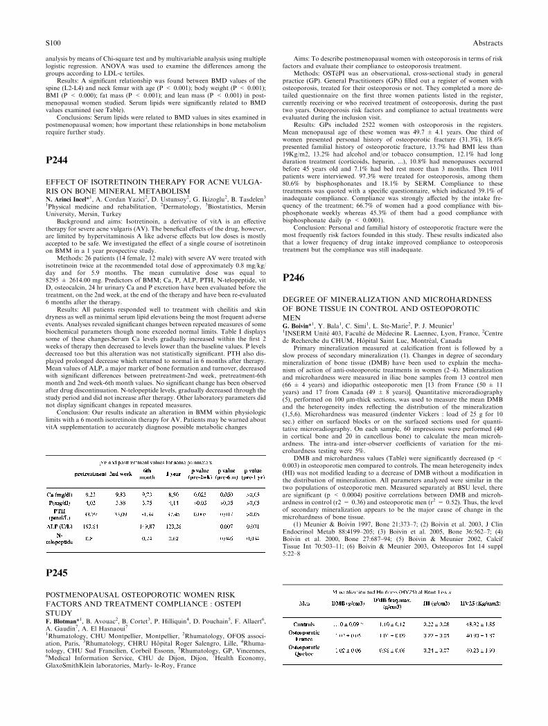
analysis by means of Chi-square test and by multivariable analysis using multiplelogistic regression. ANOVA was used to examine the differences among thegroups according to LDL-c tertiles.
Results: A significant relationship was found between BMD values of thespine (L2-L4) and neck femur with age (P < 0.001); body weight (P < 0.001);BMI (P < 0.000); fat mass (P < 0.001); and lean mass (P < 0.001) in post-menopausal women studied. Serum lipids were significantly related to BMDvalues examined (see Table).
Conclusions: Serum lipids were related to BMD values in sites examined inpostmenopausal women; how important these relationships in bone metabolismrequire further study.
P244
EFFECT OF ISOTRETINOIN THERAPY FOR ACNE VULGA-
RIS ON BONE MINERAL METABOLISMN. Arinci Incel*1, A. Cordan Yazici2, D. Ustunsoy2, G. Ikizoglu2, B. Tasdelen31Physical medicine and rehabilitation, 2Dermatology, 3Biostatistics, MersinUniversity, Mersin, Turkey
Background and aims: Isotretinoin, a derivative of vitA is an effectivetherapy for severe acne vulgaris (AV). The benefical effects of the drug, however,are limited by hypervitaminosis A like adverse effects but low doses is mostlyaccepted to be safe. We investigated the effect of a single course of isotretinoinon BMM in a 1 year prospective study.
Methods: 26 patients (14 female, 12 male) with severe AV were treated withisotretinoin twice at the recommended total dose of approximately 0.8 mg/kg/day and for 5.9 months. The mean cumulative dose was equal to8295 ± 2614.00 mg. Predictors of BMM; Ca, P, ALP, PTH, N-telopeptide, vitD, osteocalcin, 24 hr urinary Ca and P excretion have been evaluated before thetreatment, on the 2nd week, at the end of the therapy and have been re-evaluated6 months after the therapy.
Results: All patients responded well to treatment with cheilitis and skindryness as well as minimal serum lipid elevations being the most frequent adverseevents. Analyses revealed significant changes between repeated measures of somebiochemical parameters though none exceeded normal limits. Table I displayssome of these changes.Serum Ca levels gradually increased within the first 2weeks of therapy then decreased to levels lower than the baseline values. P levelsdecreased too but this alteration was not statistically significant. PTH also dis-played prolonged decrease which returned to normal in 6 months after therapy.Mean values of ALP, a major marker of bone formation and turnover, decreasedwith significant differences between pretreatment-2nd week, pretreatment-6thmonth and 2nd week-6th month values. No significant change has been observedafter drug discontinuation. N-telopeptide levels, gradually decreased through thestudy period and did not increase after therapy. Other laboratory parameters didnot display significant changes in repeated measures.
Conclusion: Our results indicate an alteration in BMM within physiologiclimits with a 6 month isotretinoin therapy for AV. Patients may be warned aboutvitA supplementation to accurately diagnose possible metabolic changes
P245
POSTMENOPAUSAL OSTEOPOROTIC WOMEN RISK
FACTORS AND TREATMENT COMPLIANCE : OSTEPI
STUDYF. Blotman*1, B. Avouac2, B. Cortet3, P. Hilliquin4, D. Pouchain5, F. Allaert6,A. Gaudin7, A. El Hasnaoui71Rhumatology, CHU Montpellier, Montpellier, 2Rhumatology, OFOS associ-ation, Paris, 3Rhumatology, CHRU Hopital Roger Salengro, Lille, 4Rhuma-tology, CHU Sud Francilien, Corbeil Essonn, 5Rhumatology, GP, Vincennes,6Medical Information Service, CHU de Dijon, Dijon, 7Health Economy,GlaxoSmithKlein laboratories, Marly- le-Roy, France
Aims: To describe postmenopausal women with osteoporosis in terms of riskfactors and evaluate their compliance to osteoporosis treatment.
Methods: OSTePI was an observational, cross-sectional study in generalpractice (GP). General Practitioners (GPs) filled out a register of women withosteoporosis, treated for their osteoporosis or not. They completed a more de-tailed questionnaire on the first three women patients listed in the register,currently receiving or who received treatment of osteoporosis, during the pasttwo years. Osteoporosis risk factors and compliance to actual treatments wereevaluated during the inclusion visit.
Results: GPs included 2522 women with osteoporosis in the registers.Mean menopausal age of these women was 49.7 ± 4.1 years. One third ofwomen presented personal history of osteoporotic fracture (31.3%), 18.6%presented familial history of osteoporotic fracture, 13.7% had BMI less than19Kg/m2, 13.2% had alcohol and/or tobacco consumption, 12.1% had longduration treatment (corticoids, heparin, ...), 10.8% had menopauses occurredbefore 45 years old and 7.1% had bed rest more than 3 months. Then 1011patients were interviewed. 97.3% were treated for osteoporosis, among them80.6% by bisphosphonates and 18.1% by SERM. Compliance to thesetreatments was quoted with a specific questionnaire, which indicated 39.1% ofinadequate compliance. Compliance was strongly affected by the intake fre-quency of the treatment; 66.7% of women had a good compliance with bis-phosphonate weekly whereas 45.3% of them had a good compliance withbisphosphonate daily (p < 0.0001).
Conclusion: Personal and familial history of osteoporotic fracture were themost frequently risk factors founded in this study. These results indicated alsothat a lower frequency of drug intake improved compliance to osteoporosistreatment but the compliance was still inadequate.
P246
DEGREE OF MINERALIZATION AND MICROHARDNESS
OF BONE TISSUE IN CONTROL AND OSTEOPOROTIC
MENG. Boivin*1, Y. Bala1, C. Simi1, L. Ste-Marie2, P. J. Meunier11INSERM Unite 403, Faculte de Medecine R. Laennec, Lyon, France, 2Centrede Recherche du CHUM, Hopital Saint Luc, Montreal, Canada
Primary mineralization measured at calcification front is followed by aslow process of secondary mineralization (1). Changes in degree of secondarymineralization of bone tissue (DMB) have been used to explain the mecha-nism of action of anti-osteoporotic treatments in women (2–4). Mineralizationand microhardness were measured in iliac bone samples from 13 control men(66 ± 4 years) and idiopathic osteoporotic men [13 from France (50 ± 11years) and 17 from Canada (49 ± 8 years)]. Quantitative microradiography(5), performed on 100 lm-thick sections, was used to measure the mean DMBand the heterogeneity index reflecting the distribution of the mineralization(1,5,6). Microhardness was measured (indenter Vickers : load of 25 g for 10sec.) either on surfaced blocks or on the surfaced sections used for quanti-tative microradiography. On each sample, 60 impressions were performed (40in cortical bone and 20 in cancellous bone) to calculate the mean microh-ardness. The intra-and inter-observer coefficients of variation for the mi-crohardness testing were 5%.
DMB and microhardness values (Table) were significantly decreased (p <0.003) in osteoporotic men compared to controls. The mean heterogeneity index(HI) was not modified leading to a decrease of DMB without a modification inthe distribution of mineralization. All parameters analyzed were similar in thetwo populations of osteoporotic men. Measured separately at BSU level, thereare significant (p < 0.0004) positive correlations between DMB and microh-ardness in control (r2 = 0.36) and osteoporotic men (r2 = 0.52). Thus, the levelof secondary mineralization appears to be the major cause of change in themicrohardness of bone tissue.
(1) Meunier & Boivin 1997, Bone 21:373–7; (2) Boivin et al. 2003, J ClinEndocrinol Metab 88:4199–205; (3) Boivin et al. 2005, Bone 36:562–7; (4)Boivin et al. 2000, Bone 27:687–94; (5) Boivin & Meunier 2002, CalcifTissue Int 70:503–11; (6) Boivin & Meunier 2003, Osteoporos Int 14 suppl5:22–8
S100 Abstracts
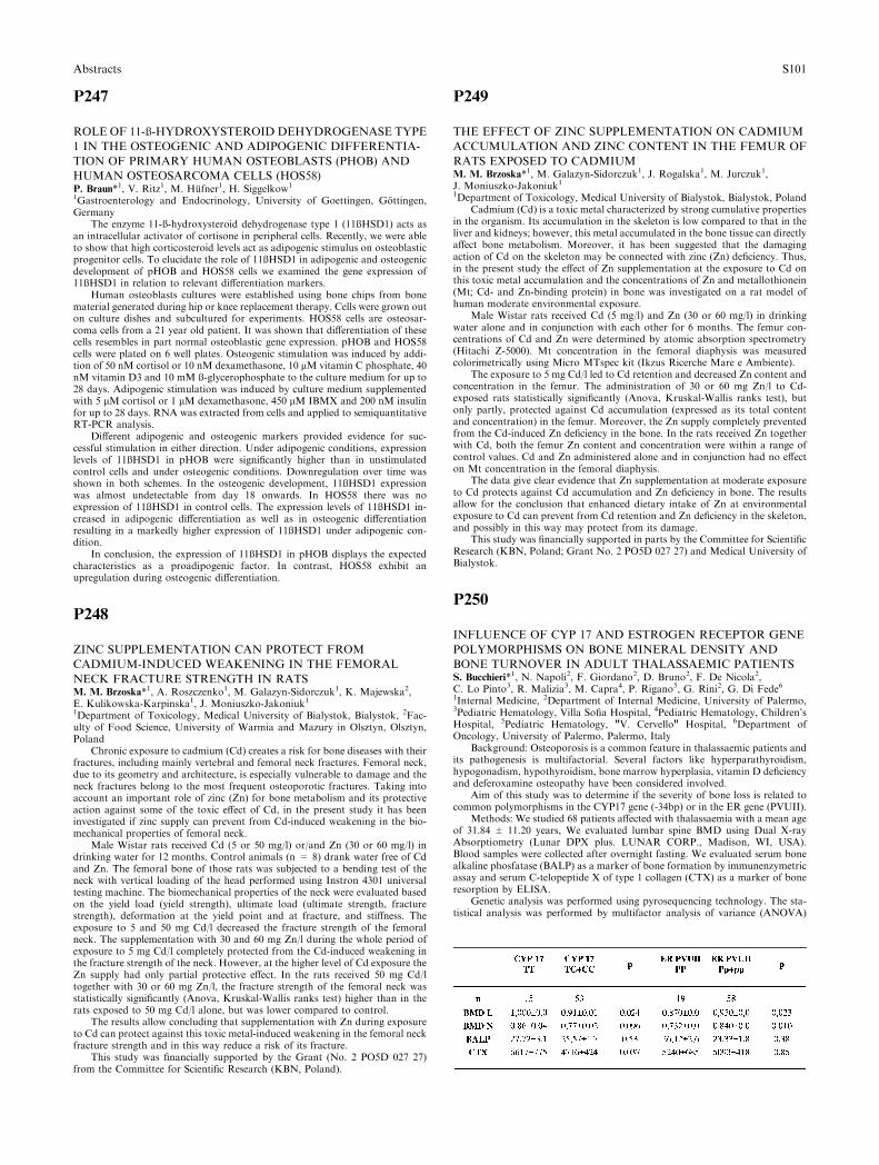
P247
ROLE OF 11-ß-HYDROXYSTEROID DEHYDROGENASE TYPE
1 IN THE OSTEOGENIC AND ADIPOGENIC DIFFERENTIA-
TION OF PRIMARY HUMAN OSTEOBLASTS (PHOB) AND
HUMAN OSTEOSARCOMA CELLS (HOS58)P. Braun*1, V. Ritz1, M. Hufner1, H. Siggelkow1
1Gastroenterology and Endocrinology, University of Goettingen, Gottingen,Germany
The enzyme 11-ß-hydroxysteroid dehydrogenase type 1 (11ßHSD1) acts asan intracellular activator of cortisone in peripheral cells. Recently, we were ableto show that high corticosteroid levels act as adipogenic stimulus on osteoblasticprogenitor cells. To elucidate the role of 11ßHSD1 in adipogenic and osteogenicdevelopment of pHOB and HOS58 cells we examined the gene expression of11ßHSD1 in relation to relevant differentiation markers.
Human osteoblasts cultures were established using bone chips from bonematerial generated during hip or knee replacement therapy. Cells were grown outon culture dishes and subcultured for experiments. HOS58 cells are osteosar-coma cells from a 21 year old patient. It was shown that differentiation of thesecells resembles in part normal osteoblastic gene expression. pHOB and HOS58cells were plated on 6 well plates. Osteogenic stimulation was induced by addi-tion of 50 nM cortisol or 10 nM dexamethasone, 10 lM vitamin C phosphate, 40nM vitamin D3 and 10 mM ß-glycerophosphate to the culture medium for up to28 days. Adipogenic stimulation was induced by culture medium supplementedwith 5 lM cortisol or 1 lM dexamethasone, 450 lM IBMX and 200 nM insulinfor up to 28 days. RNA was extracted from cells and applied to semiquantitativeRT-PCR analysis.
Different adipogenic and osteogenic markers provided evidence for suc-cessful stimulation in either direction. Under adipogenic conditions, expressionlevels of 11ßHSD1 in pHOB were significantly higher than in unstimulatedcontrol cells and under osteogenic conditions. Downregulation over time wasshown in both schemes. In the osteogenic development, 11ßHSD1 expressionwas almost undetectable from day 18 onwards. In HOS58 there was noexpression of 11ßHSD1 in control cells. The expression levels of 11ßHSD1 in-creased in adipogenic differentiation as well as in osteogenic differentiationresulting in a markedly higher expression of 11ßHSD1 under adipogenic con-dition.
In conclusion, the expression of 11ßHSD1 in pHOB displays the expectedcharacteristics as a proadipogenic factor. In contrast, HOS58 exhibit anupregulation during osteogenic differentiation.
P248
ZINC SUPPLEMENTATION CAN PROTECT FROM
CADMIUM-INDUCED WEAKENING IN THE FEMORAL
NECK FRACTURE STRENGTH IN RATSM. M. Brzoska*1, A. Roszczenko1, M. Galazyn-Sidorczuk1, K. Majewska2,E. Kulikowska-Karpinska1, J. Moniuszko-Jakoniuk11Department of Toxicology, Medical University of Bialystok, Bialystok, 2Fac-ulty of Food Science, University of Warmia and Mazury in Olsztyn, Olsztyn,Poland
Chronic exposure to cadmium (Cd) creates a risk for bone diseases with theirfractures, including mainly vertebral and femoral neck fractures. Femoral neck,due to its geometry and architecture, is especially vulnerable to damage and theneck fractures belong to the most frequent osteoporotic fractures. Taking intoaccount an important role of zinc (Zn) for bone metabolism and its protectiveaction against some of the toxic effect of Cd, in the present study it has beeninvestigated if zinc supply can prevent from Cd-induced weakening in the bio-mechanical properties of femoral neck.
Male Wistar rats received Cd (5 or 50 mg/l) or/and Zn (30 or 60 mg/l) indrinking water for 12 months. Control animals (n = 8) drank water free of Cdand Zn. The femoral bone of those rats was subjected to a bending test of theneck with vertical loading of the head performed using Instron 4301 universaltesting machine. The biomechanical properties of the neck were evaluated basedon the yield load (yield strength), ultimate load (ultimate strength, fracturestrength), deformation at the yield point and at fracture, and stiffness. Theexposure to 5 and 50 mg Cd/l decreased the fracture strength of the femoralneck. The supplementation with 30 and 60 mg Zn/l during the whole period ofexposure to 5 mg Cd/l completely protected from the Cd-induced weakening inthe fracture strength of the neck. However, at the higher level of Cd exposure theZn supply had only partial protective effect. In the rats received 50 mg Cd/ltogether with 30 or 60 mg Zn/l, the fracture strength of the femoral neck wasstatistically significantly (Anova, Kruskal-Wallis ranks test) higher than in therats exposed to 50 mg Cd/l alone, but was lower compared to control.
The results allow concluding that supplementation with Zn during exposureto Cd can protect against this toxic metal-induced weakening in the femoral neckfracture strength and in this way reduce a risk of its fracture.
This study was financially supported by the Grant (No. 2 PO5D 027 27)from the Committee for Scientific Research (KBN, Poland).
P249
THE EFFECT OF ZINC SUPPLEMENTATION ON CADMIUM
ACCUMULATION AND ZINC CONTENT IN THE FEMUR OF
RATS EXPOSED TO CADMIUMM. M. Brzoska*1, M. Galazyn-Sidorczuk1, J. Rogalska1, M. Jurczuk1,J. Moniuszko-Jakoniuk11Department of Toxicology, Medical University of Bialystok, Bialystok, Poland
Cadmium (Cd) is a toxic metal characterized by strong cumulative propertiesin the organism. Its accumulation in the skeleton is low compared to that in theliver and kidneys; however, this metal accumulated in the bone tissue can directlyaffect bone metabolism. Moreover, it has been suggested that the damagingaction of Cd on the skeleton may be connected with zinc (Zn) deficiency. Thus,in the present study the effect of Zn supplementation at the exposure to Cd onthis toxic metal accumulation and the concentrations of Zn and metallothionein(Mt; Cd- and Zn-binding protein) in bone was investigated on a rat model ofhuman moderate environmental exposure.
Male Wistar rats received Cd (5 mg/l) and Zn (30 or 60 mg/l) in drinkingwater alone and in conjunction with each other for 6 months. The femur con-centrations of Cd and Zn were determined by atomic absorption spectrometry(Hitachi Z-5000). Mt concentration in the femoral diaphysis was measuredcolorimetrically using Micro MTspec kit (Ikzus Ricerche Mare e Ambiente).
The exposure to 5 mg Cd/l led to Cd retention and decreased Zn content andconcentration in the femur. The administration of 30 or 60 mg Zn/l to Cd-exposed rats statistically significantly (Anova, Kruskal-Wallis ranks test), butonly partly, protected against Cd accumulation (expressed as its total contentand concentration) in the femur. Moreover, the Zn supply completely preventedfrom the Cd-induced Zn deficiency in the bone. In the rats received Zn togetherwith Cd, both the femur Zn content and concentration were within a range ofcontrol values. Cd and Zn administered alone and in conjunction had no effecton Mt concentration in the femoral diaphysis.
The data give clear evidence that Zn supplementation at moderate exposureto Cd protects against Cd accumulation and Zn deficiency in bone. The resultsallow for the conclusion that enhanced dietary intake of Zn at environmentalexposure to Cd can prevent from Cd retention and Zn deficiency in the skeleton,and possibly in this way may protect from its damage.
This study was financially supported in parts by the Committee for ScientificResearch (KBN, Poland; Grant No. 2 PO5D 027 27) and Medical University ofBialystok.
P250
INFLUENCE OF CYP 17 AND ESTROGEN RECEPTOR GENE
POLYMORPHISMS ON BONE MINERAL DENSITY AND
BONE TURNOVER IN ADULT THALASSAEMIC PATIENTSS. Bucchieri*1, N. Napoli2, F. Giordano2, D. Bruno2, F. De Nicola2,C. Lo Pinto3, R. Malizia3, M. Capra4, P. Rigano5, G. Rini2, G. Di Fede61Internal Medicine, 2Department of Internal Medicine, University of Palermo,3Pediatric Hematology, Villa Sofia Hospital, 4Pediatric Hematology, Children’sHospital, 5Pediatric Hematology, "V. Cervello" Hospital, 6Department ofOncology, University of Palermo, Palermo, Italy
Background: Osteoporosis is a common feature in thalassaemic patients andits pathogenesis is multifactorial. Several factors like hyperparathyroidism,hypogonadism, hypothyroidism, bone marrow hyperplasia, vitamin D deficiencyand deferoxamine osteopathy have been considered involved.
Aim of this study was to determine if the severity of bone loss is related tocommon polymorphisms in the CYP17 gene (-34bp) or in the ER gene (PVUII).
Methods: We studied 68 patients affected with thalassaemia with a mean ageof 31.84 ± 11.20 years, We evaluated lumbar spine BMD using Dual X-rayAbsorptiometry (Lunar DPX plus. LUNAR CORP., Madison, WI, USA).Blood samples were collected after overnight fasting. We evaluated serum bonealkaline phosfatase (BALP) as a marker of bone formation by immunenzymetricassay and serum C-telopeptide X of type 1 collagen (CTX) as a marker of boneresorption by ELISA.
Genetic analysis was performed using pyrosequencing technology. The sta-tistical analysis was performed by multifactor analysis of variance (ANOVA)
Abstracts S101

using BMI and age as covariates. All the analysis was performed by StatgraphicPlus 5.0 software (Manugistic, Inc., Rockville, MD, U.S.A.).
Results: Mean age of the patients was 31.84 ± 11.2 All of them were onbimonthly transfusion regimen and were on desferoxamine treatment. Com-pared to TT subjects, those carrying the C allele had lower BMD at lumbar andat the femoral neck also if in this site this difference didn�t reach the full sta-tistical significance. Those patients had also significantly higher CTX, comparedto the TT ones. When the ER PVUII polymorhisms were evaluated, PP subjectshad lower BMD than those with Pp+pp, but without significant changes in bonemarkers (TABLE).
Conclusions: These data show that, as already shown in postmenopausalhealthy women, young thalassemic patients with C allele at the )34 bp of theCYP17 gene or PP allele of the ER gene have a high bone turn over that in turnreflects a lower bone density. These polymorphisms may be additional factors tothe pathogenesis of bone impairment in thalassaemia.
P251
REGULATION OF CRYSTALLINITY IN HIP FRACTURE: AN
INITIAL STUDY WITH FTIRA. Caballero-Alias*1, D. Faibish2, N. Loveridge1, S. K. Kaptoge1, A. Lyon1, J.Reeve1, A. L. Boskey21Medicine, University of Cambridge, Cambridge, United Kingdom, 2Minera-lised Tissue Laboratory, Hospital for Special Surgery, New York, NY, UnitedStates
Background Whether osteoporotic bone suffers deterioration of its materialproperties that make it brittle is unclear. Our aim was to see if these propertieseither differed generally between hip fracture cases and controls or whetherosteoporotic bone was potentially ‘‘flawed’’ through having severe microscopicabnormalities promoting failure.
Methods We studied femoral neck cortices from female hip fracture cases(n = 5; 79–86 years), and post-mortem controls (n = 6; 78–82 years) usingFourier transform infrared imaging (FTIRI). FTIRI provides quantitative dataon crystal size and perfection, and on matrix structure and composition at aspatial resolution of 6-7lm. The spectra were acquired from several 400 · 400lm fields (6–8/biopsy) in each of four quadrants (anterior, inferior, posterior,superior). Parameters measured were: crystallinity (crystal size and perfection);collagen cross link maturity; and mineral:matrix ratio.
Results Distributions across fields were mostly positively skewed, particu-larly for crystallinity and maturity. First, outliers were excluded (censored) bymodelling data within the interquartile range (IQR) ± 3 IQRs. There was ageneralised 5% increase in crystallinity in the cases (p < 0.04), independent ofquadrant, but no significant differences in collagen cross-linking or min-eral:matrix ratio. In the controls only, crystallinity was negatively associatedwith bone resorption (adj r2 < 0.18; p < 0.027) and positively related to theratio of forming to resorbing canals (adj r2 < 0.2; p < 0.019). Quantile-quantileplots showed a clear excess of very high (previously censored) crystallinity pixelsin the fracture cases, with between 69% and 94% of these in the anterior andposterior (but not the inferior) quadrants. Interestingly, in some fields thesevalues were anatomically clustered.
Conclusions This study suggests that the bone mineral in the femoral neckfrom the fracture cases has an increased crystal size and/or perfection. Addi-tionally, locally clustered micro-areas with excessively high crystallinity wereseen in the cases. There are only 2 previous reports from 20+ years ago oncrystal size in hip fracture, both supportive. The hypothesis that a local loss ofcontrol of crystallinity or the interface between the mineral and the matrix cansometimes result in localised flaws in the bone composite appears viable. Loss ofcontrol of crystallinity requires further investigation in the aetiology of hipfracture.
P252
THE INFLUENCE OF LIFE-STYLE AND SECONDARY CAU-
SES OF OSTEOPOROSIS IN DOBRUGEA REGION IN ROMA-
NIAE. G. Circo*1, S. E. Circo1, I. S. Chirca11Endocrinology, Ovidius University of Constanta, Constanta, Romania
AIms: The aim of this study was to evaluate the influence of life–style andsecondary causes of osteoporosis in Dobrugea region in Romania.
Methods: We surveyed 1032 caucasian postmenopausal women by usingdual-energy X-ray absorbtiometry (DXA) in accordance to Z and T scores (byWHO criteria). The following criteria were considered: cronological age, age atfirst menses, age at which menopause was installed,particularities of menstruallife. Life-style habits included: the consumption of milk products, calciumpreparates, physical acticity, voluntary sun exposure, educational level, access toinformation and access to medical investigations.
Results: Osteoporosis was diagnosed in 503or 48.7% of postmenopausalwomen, age range 61.5 ± 8.2 yrs, first menses in 12.4 ± 1.3 yrs, menopause wasinstalled on 44.1 ± 1.8 yrs. Early menopause was noted in 9.1% mostly due to
ovarectomy, 13.4 were nulliparous. Life-style habits: milk products were takenregularly by 14 %; 5.4% received calcium preparates recently and 6.8% interm-itently. Sun exposure was poor only 18% claimed regular summer vacations;access to medical information 60%, access to medical investigations. 18.5% ofthe patients had superior studies, 49.8 finished high school and 31.7 didn�tmanage to reach high school.
Conclusions: Prevalence of osteoporosis is high in Dobrugea region inRomania. Life-style habits showed to have a determining influence on meno-pause installation and as consequence on osteoporosis itself. Low educationallevel, decreased access to medical information and lack of interest on routinemedical investigations, associating menopause installation, allows us to considerthem as secondary factors that influence indirectly osteoporosis.
P253
MALE IDIOPATHIC OSTEOPOROSIS. ILLIAC BONE BIOPSY
STUDY BY MICRO-TCM. Ciria*1, J. Blanch1, L. Perez-Edo1, M. Marinoso2, D. Chappard3, S. Ser-rano2, A. Diez-Perez4, J. Carbonell11Rheumatology, 2Pathology, Institut Municipal d’Asist, Barcelona, Spain,3Rheumatology, University of Angers, Angers, France, 4Internal Medicine,Institut Municipal d’Asist, Barcelona, Spain
There are few histomorphometric studys about male idiopathic osteoporosis(MIO). Our group found a decrease of osteoblasts and trabecular number inclassic histomorphometric studies of illiac bone biopsies, showing a decreasedformation levels and a normal resorption levels.
The aim of the present study is to examine the microarchitecture of can-cellous bone with micro-TC technics.
We included 16 of 23 illiac bone biopsies performed in male with normo-calciuric idiopathic osteoporosis, diagnosed by densitometric study. The controlgroup included 11 age-mached, healthy men. Micro-TC study was performedwith Microtomographie X-Skyscan. We studied bone volume / total volume(BV/TV), bone surface / bone volume (BS/BV), trabecular thickness (Tb.Th),trabecular separation (Tb.Sp), and trabecular number (Tb.N).
Results: Bone volume / total volume, trabecular number and trabecularseparation was lower in osteoporotic group than in control group. Tb.Th wassimilar in both groups (Table 1).
Conclusions: In our sample, male normocalciuric idiopathic osteoporosisshowed a low trabecular number and a high trabecular separation, with amaintenance of trabecular thickness. This findings differ of the postmenopausicosteoporosis.
P254
PARATHYROID HORMONE (1-84) IS EFFECTIVE IN PA-
TIENTSWITH AWIDE RANGE OF CLINICAL RISK FACTORSJ. Compston*1, S. Silverman2, A. Mathisen3, W. Gallwitz3, S. Morris31Cambridge School of Clinical Medicine, University of Cambridge, Cambridge,United Kingdom, 2Cedars Sinai Medical Center, David Geffen School ofMedicine UCLA, Beverly Hills, 3Nycomed Group, Nps Pharmaceuticals, SaltLake City, United States
Although bone mineral density (BMD) measurement is important in theassessment of fracture risk in postmenopausal women, the addition of inde-pendent clinical risk factors improves risk prediction and identifies some womenwho would not be considered at risk on the basis of BMD criteria alone. In thisexploratory analysis, new vertebral fracture incidence as a function of clinicalrisk factors was determined in the Treatment of Osteoporosis with PTH (TOP)clinical study, designed to determine the effectiveness of PTH (100 mcg daily) inpreventing vertebral fractures in osteoporotic women during 18 months oftreatment. A total of 2532 subjects were enrolled, 1246 of whom received placeboand 1286 received PTH (100 mcg daily). Clinical risk factors captured at baselinewere as follows: femoral neck BMD T-score <)3.0, age prior fracture, currentsmoking, daily alcohol intake, family history of osteoporosis and current glu-cocorticoid use.
At baseline the distribution of clinical risk factors was similar for the PTH-treated and placebo groups. 8% percent of the baseline population had noclinical risk factors, 28% had 1, 31% had 2, 20% had 3, 9% had 4, 3% had 5, and1% of the subjects had 6 clinical risk factors. In the ITT population, PTH
S102 Abstracts
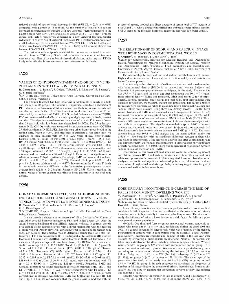
reduced the risk of new vertebral fractures by 61% (95% CI, + 22% to + 69%)compared with placebo at 18 months. As the number of clinical risk factorsincreased, the percentage of subjects with new vertebral fractures increased in theplacebo group with 1.1%, 3.8% and 8.3% of women with 0–1, 2–3 and 4 or moreclinical risk factors respectively experiencing a new vertebral fracture. Similarreductions in relative risk of vertebral fractures in PTH-treated women were seenin all 3 subgroups, (0–1 clinical risk factors 59% (95% CI, )112% to +92%), 2–3clinical risk factors 68% (95% CI, + 31% to + 86%) and 4 or more clinical riskfactors, 48% (95% CI, )26% to + 79%)
Conclusion: A wide range of clinical risk factors was encountered in womenrecruited into the TOP study. Similar risk reductions in new vertebral fractureswere seen regardless of the number of clinical risk factors, indicating that PTH islikely to be effective in women selected for treatment on this basis.
P255
VALUES OF 25-HYDROXYVITAMIN D (25-OH D3) IN VENE-
ZUELAN MEN WITH LOW BONE MINERAL DENSITYR. Constantino*1, J. Ramos1, J. Cedeno-Taborda1, L. Marcano1, E. Belzares1,G. S. Riera-Espinoza11UNILIME UC, Hospital Universitario Angel Larralde. Universidad de Cara-bobo, Valencia, Venezuela
The vitamin D deficit has been observed in adolescents as much as adultsand, mainly, in old people. The vitamin D supplements produce a reduction ofPTH, diminish the bone remodelation and increase the bone mineral density andare able to diminish the risk of fractures. Corporal reserves of vitamin D areestimated by serum levels of 25(OH) D3. So called ‘‘reference values of 25(OH)D3’’ are controversial and affected mainly by sunlight exposure, latitude, seasonsand diet. The objective is to determine the values of vitamin D in men of morethan 30 years old with low bone mass determined by DXA. The 25-hydroxyvi-tamin D (25-OH D) levels were measured by enzymeinmmunoassay (OCTEIA25-Hydroxyvitamin D. IDS.UK). Samples were taken from venous blood in thefasting state, frozen at )70�C and measured in duplicate at the same time. Weanalyzed 45 male patients with 60.18 ± 12.45 years of age, Bone MineralDensity (BMD) at Total Hip was 0.901 ± 0.123 g/cm2, T-score )1.35 ± 0.99;BMD Femoral Neck 0.836 ± 0.106 g/cm3 T-score )1.7 ± 0.82; BMD L1–L41.044 ± 0.149 T-score )1.4 ± 1.24; the serum calcium level was 8.88 ± 0.39mg/dl; Range 1 ± SD 8.49 – 9.27 with minimum values and maximum 8.50 and9.90 mg/dl; vitamin D 52.062 ± 15.04 ng/dl. Range 1 ± SD 37.02 – 67.10 withminimal and maximal values 80 and 250 ng/dl. There were no significant cor-relations between 25-hydroxyvitamin D and age, BMD and serum calcium level.(Edad p = 0.381; Total Hip p = 0.678; Femoral Neck p = 0.922; L1–L4p = 0.613; Serum calcium level p = 0.477). In conclusion the Vitamin D levelsin our sample were not different to the Venezuelan premenopausal womenaverage levels (52.86 ± 24.28ng/ml. Range ± SD 28.58–77.14), regarding thenormal values of serum calcium regardless of being patients with low level bonedensity.
P256
GONADAL HORMONES LEVEL, SEXUAL HORMONE BIND-
ING GLOBULIN LEVEL AND GONADOTROPIN LEVEL IN
VENEZUELAN MEN WITH LOW BONE MINERAL DENSITYR. Constantino*1, J. Cedeno-Taborda1, L. Marcano1, J. Ramos1,G. S. Riera-Espinoza11UNILIME UC, Hospital Universitario Angel Larralde. Universidad de Cara-bobo, Valencia, Venezuela
In men there is a decrease in testosterone of 1% to 2%/year after 30 year ofage, other gonadal hormone increase as FSH and in less degree LH. Also thesexual hormone binding globulin (SHBG) increases with age. In contrast there islittle change within Estradiol levels; with a direct relationship with the decreaseof Bone Mineral Density (BMD) at cortical (2% per decade) and trabecular bone(12% per decade). The objective was to determine serum levels of Total Tes-tosterone (TT) Free testosterone (FT) Biodisponible Testosterone (BT) SexualHormone Globulin Level (SHBG), Lutein Hormone (LH) and Estradiol (E) inmen over 30 years of age with low bone density by DEXA. 64 patients werestudied mean age 58.69 ± 12.91 BMD Total Hip (TH) 0.911 ± 0.12 g/cm2 T-Score )1.2 ± 0.99; Femoral Neck (FC) 0.842 ± 0.10 g/cm2 T-score)1.7 ± 0.79; Trocanter (T) 0.854 g/cm2 T-score )1.66 ± 0.76; L1–L41.057 ± 0.14 g/cm3 T-score )1.34 ± 1.25. TT levels 4.81 ± 1.87 ng/ml; TL0.282 ± 0.105 nmol/L; BT 7.12 ± 4.03 nmol/L; SHBG 47.89 ± 24.03 nmol/L;LH 6.44 ± 4.50 mUI/ml; E 30.74 ± 9.72 pg/ml. Age was co-related with FT(p = 0.01), SHBG (p = 0.000) and LH (p = 0.001). Regarding BMD andGonadal Hormone we found significant co-relation between BMD TH, T andL1–L4 with TT (P < 0.007, < 0.05, < 0.004 respectively); with FT and L1–L4p = 0.04 and with SHBG TH p = 0.002, FN p = 0.01, T p = 0.006, of thesecorrelations the strongest was between BMD and SHBG; not like with BT, LHand E (p > 0.05). We can conclude that the gonadal axis is modified with the
process of ageing, producing a direct decrease of serum level of TT increase ofSHBG and LH, with a decrease in cortical and trabecular bone mineral density,SHBG seems to be the main hormonal maker in men with low bone density.
P257
THE RELATIONSHIP OF SODIUM AND CALCIUM INTAKE
WITH BONE MASS IN POSTMENOPAUSAL WOMENS. Cvijetic*1, M. Blanusa2, I. Colic Baric3, J. Ilich41Center for Osteoporosis, Institute for Medical Research and OccupationalHealth, 2Department for Mineral Metabolism, Institute for Medical researchand Occupational Health, 3Faculty of Food Technology and Biotechnology,University of Zagreb, Zagreb, Croatia, 4School of Allied Health, University ofConnecticut, Storrs, United States
The relationship between calcium and sodium metabolism is well known.High sodium intake can accelerate calcium excretion and hypercalciuria is riskfactor for osteoporosis.
Aim: to analyze the relationship of sodium and calcium intake and excretionwith bone mineral density (BMD) in postmenopausal women. Subjects andMethods: 120 postmenopausal women participated in the study. The mean agewas 59.9 + 7.2 years and the mean age after menopause was 10.8 + 7.4 years.Bone mineral density (BMD) was measured, using dual energy X-ray absorpti-ometry, in spine, proximal femur, radius and total body. Spot urine samples wereanalyzed for calcium, magnesium, sodium and potassium. The values obtainedfor metals were expressed as ratios to creatinine (mg/g creatinine). Calcium andsodium intake were assessed using three-day dietary records. Results: Mostwomen had normal BMD in all measured sites. Osteoporosis (T score < )2.5)was most common in radius (cortical bone) (15.9%) and in spine (14.2%), whilethe greatest number of women had normal BMD in total body (73.2%). Therewere no differences in obtained concentrations of elements between subjects withand without osteoporosis. The significant correlation (p < 0.0001) betweencalcium and sodium in urine was determined. Subjects with osteoporosis hadsignificant correlation between urinary calcium and BMD (p < 0.05). The meancalcium intake was 809.8 + 348.5 mg/day and the mean sodium intake was5335.6 + 1650.6 mg/day, with no significant difference between subjects withand without osteoporosis. Using regression analysis and after controlling for ageand anthropometry, we founded that potassium in urine was the only significantpredictor of bone mass (p < 0.05). There was no significant relationship betweensodium intake or excretion and BMD.
Conclusions: in this cross-sectional study we could not confirm significantcorrelation between BMD and sodium intake or excretion. We also could notrelate osteoporosis to the amount of calcium ingested. However, based on urineanalyses, we confirmed significant relationship between calcium and sodiummetabolism. Longitudinal analysis is probably necessary to get better insight ofcalcium and sodium influence on bone.
P258
DOES URINARY INCONTINENCE INCREASE THE RISK OF
FALLS IN COMMUNITY DWELLING WOMEN?Y. Dionyssiotis*1, G. Trovas1, A. Galanos1, E. Papakitsou1, S. Tournis1,A. Katsalira1, D. Economopoulos1, B. Samdanis1, G. P. Lyritis11Laboratory for Research Musculoskeletal System, University of Athens,KATHospital, Kifissia, Greece
Aims: Urinary incontinence is a common problem and a chronic medicalcondition.A little importance has been attributed to the association of urinaryincontinence and falls, especially in community dwelling women. The aim was tostudy the influence of urinary incontinence as a risk factor for falls in a post-menopausal women population.
Methods: One thousand forty seven postmenopausal women, randomly se-lected, with mean age 64.72 ± 8.9 (SD), participated during the years 2004 and2005, in a control program for osteoporosis which was organised by the HellenicFoundation of Osteoporosis in cooperation with the Hellenic Patient Osteopo-rosis Society. Incontinence episodes and number of falls in the last year wereassessed by answering a questionnaire by interview. None of the women wastaken any antiosteoporotic drug including calcium supplementation. Womenwere separated in group A:329 women with incontinence and in group B:718women without incontinence episodes. Women were also separated in subgroupsaccording to the number of falls(nf) during the last year (nf:0,1,2 or more) asfollow: Subgroup 1 {nf:0, n = 776 (74.1%)}, subgroup 2 {nf:1,n = 120(11.5%)}, subgroup 3 {nf:2 or more,n = 151 (14.4%)}.The mean age of theparticipants included in the study was 64.8 ± 8.8 (SD) in group A and63.8 ± 8.9(SD) in group B. In each subgroup we calculated the frequency ofnumber of falls according to the presence of urinary incontinence. Pearsons chi-square test was used to estimate the association between urinary incontinenceand number of falls.
Results: According to the number of falls in groups A and B respectively, 0:65.3% vs. 78.1%,1:13.4% vs. 10.6% and 2 or more: 21.3% vs. 11.3% (p <
Abstracts S103
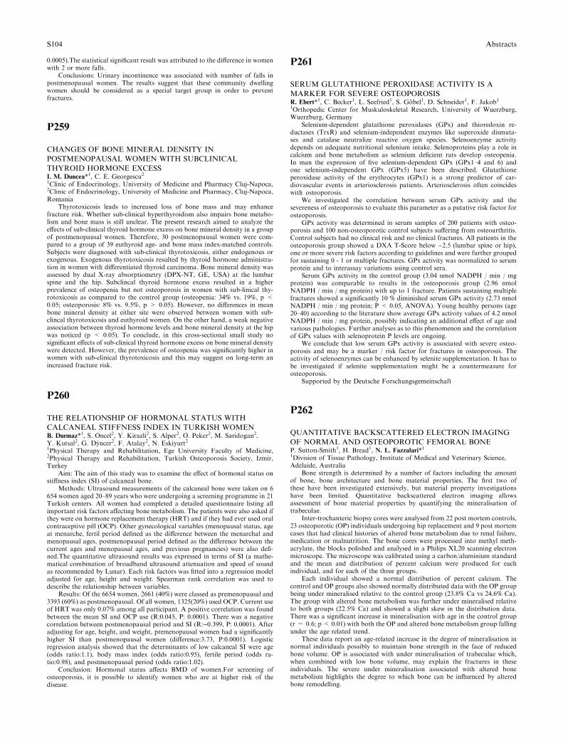
0.0005).The statistical significant result was attributed to the difference in womenwith 2 or more falls.
Conclusions: Urinary incontinence was associated with number of falls inpostmenopausal women. The results suggest that these community dwellingwomen should be considered as a special target group in order to preventfractures.
P259
CHANGES OF BONE MINERAL DENSITY IN
POSTMENOPAUSAL WOMEN WITH SUBCLINICAL
THYROID HORMONE EXCESSI. M. Duncea*1, C. E. Georgescu21Clinic of Endocrinology, University of Medicine and Pharmacy Cluj-Napoca,2Clinic of Endocrinology, University of Medicine and Pharmacy, Cluj-Napoca,Romania
Thyrotoxicosis leads to increased loss of bone mass and may enhancefracture risk. Whether sub-clinical hyperthyroidism also impairs bone metabo-lism and bone mass is still unclear. The present research aimed to analyze theeffects of sub-clinical thyroid hormone excess on bone mineral density in a groupof postmenopausal women. Therefore, 30 postmenopausal women were com-pared to a group of 39 euthyroid age- and bone mass index-matched controls.Subjects were diagnosed with sub-clinical thyrotoxicosis, either endogenous orexogenous. Exogenous thyrotoxicosis resulted by thyroid hormone administra-tion in women with differentiated thyroid carcinoma. Bone mineral density wasassessed by dual X-ray absorptiometry (DPX-NT, GE, USA) at the lumbarspine and the hip. Subclincal thyroid hormone excess resulted in a higherprevalence of osteopenia but not osteoporosis in women with sub-linical thy-rotoxicosis as compared to the control group (osteopenia: 34% vs. 19%, p <0.05; osteoporosis: 8% vs. 9.5%, p > 0.05). However, no differences in meanbone mineral density at either site were observed between women with sub-clincal thyrotoxicosis and euthyroid women. On the other hand, a weak negativeassociation between thyroid hormone levels and bone mineral density at the hipwas noticed (p < 0.05). To conclude, in this cross-sectional small study nosignificant effects of sub-clinical thyroid hormone excess on bone mineral densitywere detected. However, the prevalence of osteopenia was significantly higher inwomen with sub-clinical thyrotoxicosis and this may suggest on long-term anincreased fracture risk.
P260
THE RELATIONSHIP OF HORMONAL STATUS WITH
CALCANEAL STIFFNESS INDEX IN TURKISH WOMENB. Durmaz*1, S. Oncel2, Y. Kirazli2, S. Alper2, O. Peker2, M. Saridogan2,Y. Kutsal2, G. Dyncer2, F. Atalay2, N. Eskiyurt21Physical Therapy and Rehabilitation, Ege University Faculty of Medicine,2Physical Therapy and Rehabilitation, Turkish Osteoporosis Society, Izmir,Turkey
Aim: The aim of this study was to examine the effect of hormonal status onstiffness index (SI) of calcaneal bone.
Methods: Ultrasound measurements of the calcaneal bone were taken on 6654 women aged 20–89 years who were undergoing a screening programme in 21Turkish centers. All women had completed a detailed questionnaire listing allimportant risk factors affecting bone metabolism. The patients were also asked ifthey were on hormone replacement therapy (HRT) and if they had ever used oralcontraceptive pill (OCP). Other gynecological variables (menopausal status, ageat menarche, fertil period defined as the difference between the menarchal andmenopausal ages, postmenopausal period defined as the difference between thecurrent ages and menopausal ages, and previous pregnancies) were also defi-ned.The quantitative ultrasound results was expressed in terms of SI (a mathe-matical combination of broadband ultrasound attenuation and speed of soundas recommended by Lunar). Each risk factors was fitted into a regression modeladjusted for age, height and weight. Spearman rank correlation was used todescribe the relationship between variables.
Results: Of the 6654 women, 2661 (40%) were classed as premenopausal and3393 (60%) as postmenopausal. Of all women, 1325(20%) used OCP. Current useof HRT was only 0.07% among all participant. A positive correlation was foundbetween the mean SI and OCP use (R:0.045, P: 0.0001). There was a negativecorrelation between postmenopausal period and SI (R:)0.399, P: 0.0001). Afteradjusting for age, height, and weight, premenopausal women had a significantlyhigher SI than postmenopausal women (difference:3.73, P:0.0001). Logisticregression analysis showed that the determinants of low calcaneal SI were age(odds ratio:1.1), body mass index (odds ratio:0.95), fertile period (odds ra-tio:0.98), and postmenopausal period (odds ratio:1.02).
Conclusion: Hormonal status affects BMD of women.For screening ofosteoporosis, it is possible to identify women who are at higher risk of thedisease.
P261
SERUM GLUTATHIONE PEROXIDASE ACTIVITY IS A
MARKER FOR SEVERE OSTEOPOROSISR. Ebert*1, C. Becker1, L. Seefried1, S. Gobel1, D. Schneider1, F. Jakob11Orthopedic Center for Muskuloskeletal Research, University of Wuerzburg,Wuerzburg, Germany
Selenium-dependent glutathione peroxidases (GPx) and thioredoxin re-ductases (TrxR) and selenium-independent enzymes like superoxide dismuta-ses and catalase neutralize reactive oxygen species. Selenoenzyme activitydepends on adequate nutritional selenium intake. Selenoproteins play a role incalcium and bone metabolism as selenium deficient rats develop osteopenia.In man the expression of five selenium-dependent GPx (GPx1–4 and 6) andone selenium-independent GPx (GPx5) have been described. Glutathioneperoxidase activity of the erythrocytes (GPx1) is a strong predictor of car-diovascular events in arteriosclerosis patients. Arteriosclerosis often coincideswith osteoporosis.
We investigated the correlation between serum GPx activity and thesevereness of osteoporosis to evaluate this parameter as a putative risk factor forosteoporosis.
GPx activity was determined in serum samples of 200 patients with osteo-porosis and 100 non-osteoporotic control subjects suffering from osteoarthritis.Control subjects had no clinical risk and no clinical fractures. All patients in theosteoporosis group showed a DXA T-Score below )2.5 (lumbar spine or hip),one or more severe risk factors according to guidelines and were further groupedfor sustaining 0 - 1 or multiple fractures. GPx activity was normalized to serumprotein and to interassay variations using control sera.
Serum GPx activity in the control group (3.04 nmol NADPH / min / mgprotein) was comparable to results in the osteoporosis group (2.96 nmolNADPH / min / mg protein) with up to 1 fracture. Patients sustaining multiplefractures showed a significantly 10 % diminished serum GPx activity (2.73 nmolNADPH / min / mg protein; P < 0.05, ANOVA). Young healthy persons (age20–40) according to the literature show average GPx activity values of 4.2 nmolNADPH / min / mg protein, possibly indicating an additional effect of age andvarious pathologies. Further analyses as to this phenomenon and the correlationof GPx values with selenoprotein P levels are ongoing.
We conclude that low serum GPx activity is associated with severe osteo-porosis and may be a marker / risk factor for fractures in osteoporosis. Theactivity of selenoenzymes can be enhanced by selenite supplementation. It has tobe investigated if selenite supplementation might be a countermeasure forosteoporosis.
Supported by the Deutsche Forschungsgemeinschaft
P262
QUANTITATIVE BACKSCATTERED ELECTRON IMAGING
OF NORMAL AND OSTEOPOROTIC FEMORAL BONEP. Sutton-Smith1, H. Bread1, N. L. Fazzalari*11Division of Tissue Pathology, Institute of Medical and Veterinary Science,Adelaide, Australia
Bone strength is determined by a number of factors including the amountof bone, bone architecture and bone material properties. The first two ofthese have been investigated extensively, but material property investigationshave been limited. Quantitative backscattered electron imaging allowsassessment of bone material properties by quantifying the mineralisation oftrabeculae.
Inter-trochanteric biopsy cores were analysed from 22 post mortem controls,23 osteoporotic (OP) individuals undergoing hip replacement and 9 post mortemcases that had clinical histories of altered bone metabolism due to renal failure,medication or malnutrition. The bone cores were processed into methyl meth-acrylate, the blocks polished and analysed in a Philips XL20 scanning electronmicroscope. The microscope was calibrated using a carbon/aluminium standardand the mean and distribution of percent calcium were produced for eachindividual, and for each of the three groups.
Each individual showed a normal distribution of percent calcium. Thecontrol and OP groups also showed normally distributed data with the OP groupbeing under mineralised relative to the control group (23.8% Ca vs 24.6% Ca).The group with altered bone metabolism was further under mineralised relativeto both groups (22.5% Ca) and showed a slight skew in the distribution data.There was a significant increase in mineralisation with age in the control group(r = 0.6; p < 0.01) with both the OP and altered bone metabolism group fallingunder the age related trend.
These data report an age-related increase in the degree of mineralisation innormal individuals possibly to maintain bone strength in the face of reducedbone volume. OP is associated with under mineralisation of trabeculae which,when combined with low bone volume, may explain the fractures in theseindividuals. The severe under mineralisation associated with altered bonemetabolism highlights the degree to which bone can be influenced by alteredbone remodelling.
S104 Abstracts
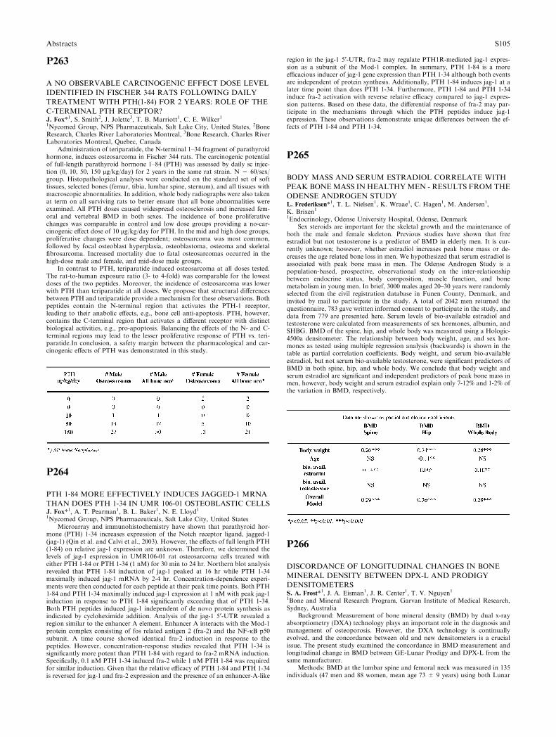
P263
A NO OBSERVABLE CARCINOGENIC EFFECT DOSE LEVEL
IDENTIFIED IN FISCHER 344 RATS FOLLOWING DAILY
TREATMENT WITH PTH(1-84) FOR 2 YEARS: ROLE OF THE
C-TERMINAL PTH RECEPTOR?J. Fox*1, S. Smith2, J. Jolette3, T. B. Marriott1, C. E. Wilker11Nycomed Group, NPS Pharmaceuticals, Salt Lake City, United States, 2BoneResearch, Charles River Laboratories Montreal, 3Bone Research, Charles RiverLaboratories Montreal, Quebec, Canada
Administration of teriparatide, the N-terminal 1–34 fragment of parathyroidhormone, induces osteosarcoma in Fischer 344 rats. The carcinogenic potentialof full-length parathyroid hormone 1–84 (PTH) was assessed by daily sc injec-tion (0, 10, 50, 150 lg/kg/day) for 2 years in the same rat strain. N = 60/sex/group. Histopathological analyses were conducted on the standard set of softtissues, selected bones (femur, tibia, lumbar spine, sternum), and all tissues withmacroscopic abnormalities. In addition, whole body radiographs were also takenat term on all surviving rats to better ensure that all bone abnormalities wereexamined. All PTH doses caused widespread osteosclerosis and increased fem-oral and vertebral BMD in both sexes. The incidence of bone proliferativechanges was comparable in control and low dose groups providing a no-car-cinogenic effect dose of 10 lg/kg/day for PTH. In the mid and high dose groups,proliferative changes were dose dependent; osteosarcoma was most common,followed by focal osteoblast hyperplasia, osteoblastoma, osteoma and skeletalfibrosarcoma. Increased mortality due to fatal osteosarcomas occurred in thehigh-dose male and female, and mid-dose male groups.
In contrast to PTH, teriparatide induced osteosarcoma at all doses tested.The rat-to-human exposure ratio (3- to 4-fold) was comparable for the lowestdoses of the two peptides. Moreover, the incidence of osteosarcoma was lowerwith PTH than teriparatide at all doses. We propose that structural differencesbetween PTH and teriparatide provide a mechanism for these observations. Bothpeptides contain the N-terminal region that activates the PTH-1 receptor,leading to their anabolic effects, e.g., bone cell anti-apoptosis. PTH, however,contains the C-terminal region that activates a different receptor with distinctbiological activities, e.g., pro-apoptosis. Balancing the effects of the N- and C-terminal regions may lead to the lesser proliferative response of PTH vs. teri-paratide.In conclusion, a safety margin between the pharmacological and car-cinogenic effects of PTH was demonstrated in this study.
P264
PTH 1-84 MORE EFFECTIVELY INDUCES JAGGED-1 MRNA
THAN DOES PTH 1-34 IN UMR 106-01 OSTEOBLASTIC CELLSJ. Fox*1, A. T. Pearman1, B. L. Baker1, N. E. Lloyd11Nycomed Group, NPS Pharmaceuticals, Salt Lake City, United States
Microarray and immunohistochemistry have shown that parathyroid hor-mone (PTH) 1-34 increases expression of the Notch receptor ligand, jagged-1(jag-1) (Qin et al. and Calvi et al., 2003). However, the effects of full length PTH(1-84) on relative jag-1 expression are unknown. Therefore, we determined thelevels of jag-1 expression in UMR106-01 rat osteosarcoma cells treated witheither PTH 1-84 or PTH 1-34 (1 nM) for 30 min to 24 hr. Northern blot analysisrevealed that PTH 1-84 induction of jag-1 peaked at 16 hr while PTH 1-34maximally induced jag-1 mRNA by 2-4 hr. Concentration-dependence experi-ments were then conducted for each peptide at their peak time points. Both PTH1-84 and PTH 1-34 maximally induced jag-1 expression at 1 nM with peak jag-1induction in response to PTH 1-84 significantly exceeding that of PTH 1-34.Both PTH peptides induced jag-1 independent of de novo protein synthesis asindicated by cycloheximide addition. Analysis of the jag-1 5¢-UTR revealed aregion similar to the enhancer A element. Enhancer A interacts with the Mod-1protein complex consisting of fos related antigen 2 (fra-2) and the NF-jB p50subunit. A time course showed identical fra-2 induction in response to thepeptides. However, concentration-response studies revealed that PTH 1-34 issignificantly more potent than PTH 1-84 with regard to fra-2 mRNA induction.Specifically, 0.1 nM PTH 1-34 induced fra-2 while 1 nM PTH 1-84 was requiredfor similar induction. Given that the relative efficacy of PTH 1-84 and PTH 1-34is reversed for jag-1 and fra-2 expression and the presence of an enhancer-A-like
region in the jag-1 5¢-UTR, fra-2 may regulate PTH1R-mediated jag-1 expres-sion as a subunit of the Mod-1 complex. In summary, PTH 1-84 is a moreefficacious inducer of jag-1 gene expression than PTH 1-34 although both eventsare independent of protein synthesis. Additionally, PTH 1-84 induces jag-1 at alater time point than does PTH 1-34. Furthermore, PTH 1-84 and PTH 1-34induce fra-2 activation with reverse relative efficacy compared to jag-1 expres-sion patterns. Based on these data, the differential response of fra-2 may par-ticipate in the mechanisms through which the PTH peptides induce jag-1expression. These observations demonstrate unique differences between the ef-fects of PTH 1-84 and PTH 1-34.
P265
BODY MASS AND SERUM ESTRADIOL CORRELATE WITH
PEAK BONEMASS IN HEALTHYMEN - RESULTS FROM THE
ODENSE ANDROGEN STUDYL. Frederiksen*1, T. L. Nielsen1, K. Wraae1, C. Hagen1, M. Andersen1,K. Brixen11Endocrinology, Odense University Hospital, Odense, Denmark
Sex steroids are important for the skeletal growth and the maintenance ofboth the male and female skeleton. Previous studies have shown that freeestradiol but not testosterone is a predictor of BMD in elderly men. It is cur-rently unknown; however, whether estradiol increases peak bone mass or de-creases the age related bone loss in men. We hypothesized that serum estradiol isassociated with peak bone mass in men. The Odense Androgen Study is apopulation-based, prospective, observational study on the inter-relationshipbetween endocrine status, body composition, muscle function, and bonemetabolism in young men. In brief, 3000 males aged 20–30 years were randomlyselected from the civil registration database in Funen County, Denmark, andinvited by mail to participate in the study. A total of 2042 men returned thequestionnaire, 783 gave written informed consent to participate in the study, anddata from 779 are presented here. Serum levels of bio-available estradiol andtestosterone were calculated from measurements of sex hormones, albumin, andSHBG. BMD of the spine, hip, and whole body was measured using a Hologic-4500a densitometer. The relationship between body weight, age, and sex hor-mones as tested using multiple regression analysis (backwards) is shown in thetable as partial correlation coefficients. Body weight, and serum bio-availableestradiol, but not serum bio-available testosterone, were significant predictors ofBMD in both spine, hip, and whole body. We conclude that body weight andserum estradiol are significant and independent predictors of peak bone mass inmen, however, body weight and serum estradiol explain only 7-12% and 1-2% ofthe variation in BMD, respectively.
P266
DISCORDANCE OF LONGITUDINAL CHANGES IN BONE
MINERAL DENSITY BETWEEN DPX-L AND PRODIGY
DENSITOMETERSS. A. Frost*1, J. A. Eisman1, J. R. Center1, T. V. Nguyen11Bone and Mineral Research Program, Garvan Institute of Medical Research,Sydney, Australia
Background: Measurement of bone mineral density (BMD) by dual x-rayabsorptiometry (DXA) technology plays an important role in the diagnosis andmanagement of osteoporosis. However, the DXA technology is continuallyevolved, and the concordance between old and new densitometers is a crucialissue. The present study examined the concordance in BMD measurement andlongitudinal change in BMD between GE-Lunar Prodigy and DPX-L from thesame manufacturer.
Methods: BMD at the lumbar spine and femoral neck was measured in 135individuals (47 men and 88 women, mean age 73 ± 9 years) using both Lunar
Abstracts S105
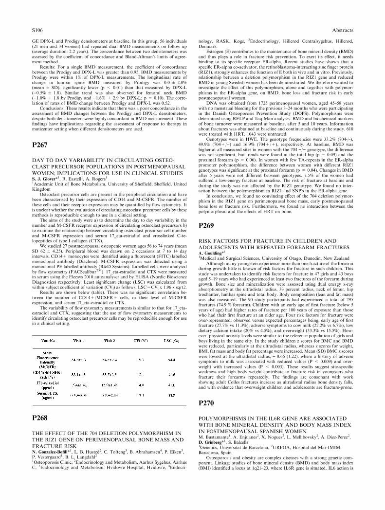
GE DPX-L and Prodigy densitometers at baseline. In this group, 56 individuals(21 men and 34 women) had repeated dual BMD measurements on follow up(average duration: 2.2 years). The concordance between two densitometers wasassessed by the coefficient of concordance and Bland-Altman�s limits of agree-ment method.
Results: For a single BMD measurement, the coefficient of concordancebetween the Prodigy and DPX-L was greater than 0.95. BMD measurements byProdigy were within 1% of DPX-L measurements. The longitudinal rate ofchange in lumbar spine BMD measured by Prodigy was 0.0 ± 2.0%(mean ± SD), significantly lower (p < 0.01) than that measured by DPX-L()0.5% ± 1.8). Similar trend was also observed for femoral neck BMD()1.0% ± 1.8 by Prodigy and )1.6% ± 2.9 by DPX-L; p = 0.10). The corre-lation of rates of BMD change between Prodigy and DPX-L was 0.52.
Conclusions: These results indicate that there was a poor concordance in theassessment of BMD changes between the Prodigy and DPX-L densitometers,despite both densitometers were highly concordant in BMD measurement. Thesefindings have implications regarding the assessment of response to therapy inmuticenter setting when different densitometers are used.
P267
DAY TO DAY VARIABILITY IN CIRCULATING OSTEO-
CLAST PRECURSOR POPULATIONS IN POSTMENOPAUSAL
WOMEN; IMPLICATIONS FOR USE IN CLINICAL STUDIESS. J. Glover*1, R. Eastell1, A. Rogers11Academic Unit of Bone Metabolism, University of Sheffield, Sheffield, UnitedKingdom
Osteoclast precursor cells are present in the peripheral circulation and havebeen characterised by their expression of CD14 and M-CSFR. The number ofthese cells and their receptor expression may be quantified by flow cytometry. Itis unclear whether the evaluation of circulating osteoclast precursor cells by thesemethods is reproducible enough to use in a clinical setting.
The aims of the study were a) to determine the day to day variability in thenumber and M-CSFR receptor expression of circulating osteoclast precursors b)to examine the relationship between circulating osteoclast precursor cell numberand M-CSFR expression and serum 17_eta-estradiol and crosslinked C-te-lopeptides of type I collagen (CTX).
We studied 27 postmenopausal osteopenic women ages 56 to 74 years (meanSD 62 ± 4.25). Peripheral blood was drawn on 2 occasions at 7 to 14 dayintervals. CD14+ monocytes were identified using a fluorescent (FITC) labelledmonoclonal antibody (Diaclone). M-CSFR expression was detected using amonoclonal PE labelled antibody (R&D Systems). Labelled cells were analysedby flow cytometry (FACScaliburTM). 17_eta-estradiol and CTX were measuredin serum using the Elecsys 2010 autoanalyser and by ELISA (Nordic BioscienceDiagnostics) respectively. Least significant change (LSC) was calculated fromwithin subject coefficient of variation (CVi) as follows; LSC=CVi x 1.96 x sqrt2.
Results are shown below (table). There was no significant correlation be-tween the number of CD14+/MCSFR+ cells, or their level of M-CSFRexpression, and serum 17_eta-estradiol or CTX.
The variability of flow cytometry measurements is similar to that for 17_eta-estradiol and CTX, suggesting that the use of flow cytometry measurements toidentify circulating osteoclast precursor cells may be reproducible enough for usein a clinical setting.
P268
THE EFFECT OF THE 704 DELETION POLYMORPHISM IN
THE RIZ1 GENE ON PERIMENOPAUSAL BONE MASS AND
FRACTURE RISKN. Gonzalez-Bofill*1, L. B. Husted2, C. Tofteng3, B. Abrahamsen4, P. Eiken5,P. Vestergaard1, B. L. Langdahl21Osteoporosis Clinic, 2Endocrinology and Metabolism, Aarhus Sygehus, AarhusC, 3Endocrinology and Metabolism, Hvidovre Hospital, Hvidovre, 4Endocri-
nology, RASK, Køge, 5Endocrinology, Hillerød Centralsygehus, Hillerød,Denmark
Estrogen (E) contributes to the maintenance of bone mineral density (BMD)and thus plays a role in fracture risk prevention. To exert its effect, it needsbinding to its specific receptor ER-alpha. Recent studies have shown that aspecific ER-alpha co-activator, the retinoblastoma-interacting zinc finger protein(RIZ1), strongly enhances the function of E both in vivo and in vitro. Previously,relationship between a deletion polymorphism in the RIZ1 gene and reducedBMD in young Swedish women has been demonstrated. We therefore wanted toinvestigate the effect of this polymorphism, alone and together with polymor-phisms in the ER-alpha gene, on BMD, bone loss and fracture risk in earlypostmenopausal women.
DNA was obtained from 1725 perimenopausal women, aged 45–58 yearswith no menstrual bleeding for the previous 3–24 months who were participatingin the Danish Osteoporosis Prevention Study (DOPS). Polymorphisms weredetermined using RFLP and Taq-Man analyses. BMD and biochemical markersof bone turnover were measured at baseline, after 5 and 10 years. Informationabout fractures was obtained at baseline and continuously during the study. 610were treated with HRT, 1043 were untreated.
Genotypes were in HWE. The genotype frequencies were 33.2% (704-/-),49.9% (704+/)) and 16.9% (704+/+), respectively. At baseline, BMD washigher at all measured sites in women with the 704 )/) genotype, the differencewas not significant, but trends were found at the total hip (p = 0.09) and theproximal forearm (p = 0.06). In women with few TA-repeats in the ER-alphapromoter polymorphism, the difference between women with different RIZ1genotypes was significant at the proximal forearm (p = 0.04). Changes in BMDafter 5 years were not different between genotypes. 7.5% of the women hadsuffered a low-energy fracture at baseline. The risk of fracture at baseline andduring the study was not affected by the RIZ1 genotype. We found no inter-action between the polymorphism in RIZ1 and SNP�s in the ER-alpha gene.
In conclusion, we found no convincing effect of the 704 deletion polymor-phism in the RIZ1 gene on perimenopausal bone mass, early postmenopausalbone loss or fracture risk. Furthermore, we found no interaction between thepolymorphism and the effects of HRT on bone.
P269
RISK FACTORS FOR FRACTURE IN CHILDREN AND
ADOLESCENTS WITH REPEATED FOREARM FRACTURESA. Goulding*11Medical and Surgical Sciences, University of Otago, Dunedin, New Zealand
Although many youngsters experience more than one fracture of the forearmduring growth little is known of risk factors for fracture in such children. Thisstudy was undertaken to identify risk factors for fracture in 47 girls and 43 boysaged 5–19 years who had experienced at least two fractures of the forearm duringgrowth. Bone size and mineralization were assessed using dual energy x-rayabsorptiometry at the ultradistal radius, 33 percent radius, neck of femur, hiptrochanter, lumbar spine and total body. Body composition (lean and fat mass)was also measured. The 90 study participants had experienced a total of 295fractures (74.9 % forearm). Children with an early age of first fracture (below 5years of age) had higher rates of fracture per 100 years of exposure than thosewho had their first fracture at an older age. Four risk factors for fracture wereover-represented: observed versus expected percentages being; early age of firstfracture (27.7% vs 11.3%), adverse symptoms to cow milk (22.2% vs 6.7%), lowdietary calcium intake (20% vs 4.5%), and overweight (33.3% vs 15.5%). How-ever, physical activity levels were similar to the reference population of girls andboys living in the same city. In the study children z scores for BMC and BMDwere reduced, particularly at the ultradistal radius, whereas z scores for weight,BMI, fat mass and body fat percentage were increased. Mean (SD) BMC z scoreswere lowest at the ultradistal radius, ) 0.66 (1.22), where a history of adversesymptoms to milk was associated with reduced values (P < 0.009) and over-weight with increased values (P < 0.003). These results suggest site-specificweakness and high body weight contribute to fracture risk in youngsters whofracture their forearms repeatedly. The findings are consonant with workshowing adult Colles fractures increase as ultradistal radius bone density falls,and with evidence that overweight children and adolescents are fracture-prone.
P270
POLYMORPHISMS IN THE IL6R GENE ARE ASSOCIATED
WITH BONE MINERAL DENSITY AND BODY MASS INDEX
IN POSTMENOPAUSAL SPANISH WOMENM. Bustamante1, A. Enjuanes2, X. Nogues2, L. Mellibovsky2, A. Diez-Perez2,D. Grinberg*1, S. Balcells11Genetics, Universitat de Barcelona, 2URFOA, Hospital del Mar-IMIM,Barcelona, Spain
Osteoporosis and obesity are complex diseases with a strong genetic com-ponent. Linkage studies of bone mineral density (BMD) and body mass index(BMI) identified a locus at 1q21–23, where IL6R gene is situated. IL6 action is
S106 Abstracts
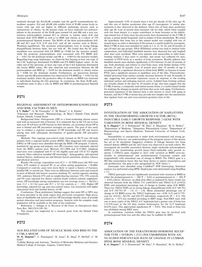
mediated through the IL6-IL6R complex and the gp130 transmembrane ho-modimer receptor. IL6 and sIL6R (the soluble form of IL6R) serum levels in-crease with age and are higher in different pathologic process such asosteoporosis or obesity. In the present study we have analyzed two polymor-phisms in the promoter of the IL6R gene (named 6A and 6B) and a non syn-onimous polymorphism (named 6C) in relation to lumbar spine (LS) andfenomal neck (FN) BMD as well as to BMI and obesity in a cohort of 570postmenopausal Spanish women. The MAFs of the three polymorphisms (6A,6B and 6C) were 0.14, 0.38 and 0.43, and all the variants were in Hardy-Weinberg equilibrium. The promoter polymorphisms were in strong linkagedisequilibrium between them, but not with 6C. We found that the 6C poly-morphism was associated with LS BMD (p=0.040 for the recessive model),while the promoter polymorphisms were associated with FN BMD (6A:p = 0.011 for the dominant model; and 6B: p = 0.064 for the recessive model).Regarding long range haplotypes, we observed that bearing at least one copy ofthe CAC haplotype determined LS BMD and FN BMD highest values. At theother end of the spectrum, at least one copy of the CAA haplotype determinedthe lowest BMD values. We also observed and association between the 6Bpolymorphism and BMI (p = 0.039 for the dominant model) or obesity status(p = 0.006 for the dominant model). Furthermore, an interaction betweenobesity and the 6B polymorphism was observed for FN BMD (p = 0.021 for thedominant model), where the protective effect of obesity on BMD was restrictedto individuals bearing the GG genotype. In conclusion, the three IL6R poly-morphisms seem to play a role on BMD and BMI in postmenopausal Spanishwomen.
P271
REGIONAL ASSESSMENT OF OSTEOPOROSIS KNOWLEDGE
AND RISK FACTORS IN MENI. V. Haller*1, A. M. Fernandez2, C. M. Renier1, J. A. Palcher11Education and Research, 2Rheumatology, St Mary’s Duluth Clinic HealthSystem, Duluth, United States
Background/Aims: Osteoporosis (OP) is a bone-weakening disease associ-ated with an increased risk of fracture. It is estimated that in the US one in fourmen over 50 will have an OP fracture in their lifetime, yet awareness about OP inmen is limited and OP prevention strategies are sparse. The goal of this studywas to conduct a regional assessment of OP knowledge and OP risk factorsamong men with subsequent development of gender-specific OP preventivestrategies.
Methods: This ongoing case-control study focused on 40-79 year old malepatients from a regional health system (RHS). Men diagnosed with osteopenia(OPA) or OP (cases) were identified through the RHS’s OP program. Controls,matched by age group and primary care (PC) location, were randomly selectedfrom the RHS’s patient pool. Between 10/1/2004 and 9/15/2005, telephoneinterviews were completed with 97 of 142 eligible cases and 134 of 313 eligiblecontrols. Data collection included OP knowledge1, demographics, self-reportedmedical history, medication use and lifestyle factors (nutrition, alcohol, tobaccoand physical activity).
Results: On average, respondents were 62.5 ± 10.7(SD) years old; 98% werewhite; 82% resided or received PC in an urban setting (population >20,000).Compared to controls, cases were more likely to report a history of fractures,familial OP, or steroid use. There were no differences between cases and controlsin most of lifestyle risk factors: excessive drinking 7%, current cigarette smoking14%, sedentary lifestyle 47% and no weight-bearing exercises 75%; 55% controlsand 9% cases reported low dietary calcium intake without calcium supplemen-tation. OP knowledge among respondents was low (average scores (± Std.Err.)were 11.2 ± 0.3 for cases and 10.7 ± 0.3 for controls, p = .310). Higher OPknowledge, adjusted for age and case-control status, was associated with highereducational level and familial history of OP.
Conclusions: These preliminary findings suggest that male OP or OPA maybe more prevalent in the study region than previously recognized. High preva-lence of modifiable risk factors and low OP knowledge identify areas of possiblepatient education and intervention programs. Analysis with the complete studypopulation will be available at the time of the conference.
References: 1. Ailinger et al. Revision of the Facts on Osteoporosis Quiz.Nurs Res. 2003;52(3):198–201.
This project was supported by a research grant from the Duluth ClinicFoundation.
P272
AGE-RELATED LOSS OF MUSCLE MASS AND BMD IN THE
C57BL/6 MOUSEM. W. Hamrick*1, C. Pennington1, H. Jones1, K. Ding2, P. McNeil1, C. M.Isales21Cellular Biology and Anatomy, 2Institute of Molecular Medicine and Genetics,Medical College of Georgia, Augusta, United States
Approximately 5-8% of muscle mass is lost per decade of life after age 30,and this rate of decline accelerates after age 65 (sarcopenia). A similar phe-nomenon is also observed in the skeleton, where loss of bone mass acceleratesafter age 40. Loss of muscle mass increases risk for falls, which when coupledwith low bone density is a major contributor to bone fractures in the elderly.Age-related loss of bone mass has previously been documented in the C57BL/6mouse, a mouse model frequently used in studies of bone metabolism. We testedthe hypothesis that bone loss in this animal model was coupled with loss ofmuscle mass, in order to determine if this mouse is a useful model for sarcopenia.Male C57BL/6 mice were included for study at 3, 6, 12, 18, 24, and 29 months ofage (10 mice per age group). DXA (PIXImus) system was used to analyze bodycomposition, and individual hindlimb muscles were dissected free and weighedafter mice were sacrificed. Mice were injected with tetracycline to label bone-forming surfaces 5 days prior to sacrifice, and serum was collected for mea-surement of PYD levels as a marker of bone resorption. Results indicate thathindlimb muscle mass declines significantly (15%) between 18 and 24 months ofage, both absolutely and relative to total body mass, with further decline (15%)between 24 and 29 months. Loss of muscle mass after 18 months of age isaccompanied by a significant decrease in whole-body BMD, an increase in serumPYD, and a significant increase in medullary area of the tibia. Fluorochrome-labeled periosteal bone surface actually increases between 18 and 24 months ofage, suggesting that periosteal expansion occurs in response to the erodingendosteal surface and enlarging marrow cavity. Labeled bone forming surfacesare virtually absent in the mice by 29 months of age. Our data suggest that theC57BL/6 mouse, a commonly used inbred mouse strain, is a useful animal modelfor studying the changes in muscle and bone that occur with aging. Furthermore,periosteal expansion of the femoral neck is also known to occur with aging inhumans, and the C57BL/6 mouse may provide new insights into the mechanismsthat regulate bone size and cross-sectional geometry later in life.
P273
INVESTIGATION OF THE ASSOCIATION OF HAPLOTYPES
IN THE TRANSFORMING GROWTH FACTOR-{BETA}-
INDUCIBLE EARLY GROWTH RESPONSE 2 GENE WITH
VARIATION IN BONE MINERAL DENSITYK. G. Hegarty*1, F. J. Drummond1, M. Daly1, F. Shanahan2, M. G. Molloy31Department of Medicine, 2Alimentary Pharmabiotic Center, 3Department ofRheumatology and Sports Medicine, National University of Ireland, Cork,Ireland
Susceptibility to osteoporosis is under both environmental and strong ge-netic control, however, our understanding of the polygenic nature of this diseasehas yet to be elucidated. Suggestive evidence of linkage between low bonemineral density (BMD) and the 2p25 locus was observed in an Irish cohort. Weinvestigated the possible association between single nucleotide polymorphisms(SNPs) in the transforming growth factor-{beta}-inducible early growth re-sponse 2 (TIEG2) gene, mapped to 2p25, with variation in BMD cross-sec-tionally in premenopausal and postmenopausal Caucasian women andlongitudinally with annualised rate of change in BMD. The TIEG2 gene is aSP1-like transcription factor that has been shown to repress transcription andcell proliferation, this gene is also upregulated by TGF-{beta}-1.
Genotypes were identified using CodeRed� SNP Genotyping. Statisticalanalysis was performed using SPSS. Haplotypes were determined using HapMapv 3.2.
TIEG2 genotypes were not significantly associated with variation in BMD ineither the premenopausal (n = 180; P > 0.02) or postmenopausal (n = 380; P> 0.02) cohorts. However, an allele dose effect as indicated by linear trends wasobserved between both the TIEG2 T/C (rs6432053) and TIEG2 G/A (rs7632)SNPs and annualised percentage rate of change in lumbar spine (LS) BMD.These two TIEG2 SNPs are in strong linkage disequilibrium (LD) (A-C (49.3%)and G-T (50.7%) D¢ = 1.0, r2 = 1.0). Mean annualised percentage rate ofchange in LS BMD across the TIEG2 haplotypes was )0.08, 0.22 and 0.77%/year for AA-CC, AG-TC and GG-TT individuals, respectively (P = 0.1). Thecohort (n = 257) was stratified according to HRT usage. Non-HRT users withone or more copies of the TIEG2 A-C haplotype had a greater rate of bone loss()0.55%/year) at the LS than women homozygous for the G-T haplotype(0.63%/year). This approached significance (P = 0.03). This trend in bone losswas abolished among HRT users.
In conclusion, variation within the TIEG2 gene may be involved withpostmenopausal bone loss and this effect may be nullified by HRT.
P274
ASSOCIATION OF THE PARATHYROID HORMONE RECEP-
TOR TYPE 1 INTRON 1 (T/C) POLYMORPHISM WITH AN-
NUALISED PERCENTAGE RATE OF CHANGE IN LUMBAR
SPINE BONE MINERAL DENSITYK. G. Hegarty*1, F. J. Drummond1, M. Daly1, F. Shanahan2, M. G. Molloy3
Abstracts S107
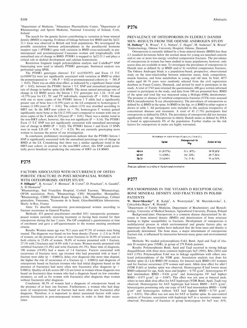
1Department of Medicine, 2Alimentary Pharmabiotic Center, 3Department ofRheumatology and Sports Medicine, National University of Ireland, Cork,Ireland
The search for the genetic factors contributing to variation in bone mineraldensity (BMD) is ongoing. Evidence of linkage between low BMD and the 3p22-21.1 locus was observed in British and Irish populations. We investigated thepossible association between polymorphisms in the parathyroid hormonereceptor type 1 (PTHR1) gene with variation in BMD cross-sectionally in pre-menopausal and postmenopausal Caucasian women and longitudinally withannualised rate of change in BMD. PTHR1 is mapped to 3p22-21.1 and plays acritical role in skeletal development and calcium homeostasis.
Restriction fragment length polymorphism analysis and CodeRed� SNPGenotyping were used to identify PTHR1 genotypes. Statistical analysis wasperformed using SPSS.
The PTHR1 genotypes (Intron1 T/C (rs11926707) and Exon 13 T/C(rs1869872)) were not significantly associated with variation in BMD in eitherthe premenopausal (n = 180; P > 0.02) or postmenopausal cohorts (n = 380; P> 0.02). There was an allele dose effect, as indicated by a significant linear trend(P < 0.02), between the PTHR1 Intron 1 T/C SNP and annualised percentagerate of change in lumbar spine (LS) BMD. The mean annual percentage rate ofchange in LS BMD across the Intron 1 T/C genotypes was 1.24, )0.16 and)0.27%/year for CC, TC and TT individuals, respectively (P < 0.01). Womenwith one or more copies of the PTHR1 Intron 1 T allele had a significantlygreater rate of bone loss ()0.19%/year) at the LS compared to homozygous Cwomen (1.24%/year) (P < 0.01). The cohort (155) was stratified according toHRT use. In the HRT-user cohort (n = 70), homozygous C women gainedsignificantly more bone at the LS (2.18%/year) compared to women with one ormore copies of the T allele (0.32%year) (P < 0.01). There was a similar trend inthe non-HRT cohort, however, this was not significant (P = 0.16). The PTHR1Exon 13 T/C SNP was not significantly associated with annualised percentagerate of change in BMD (P > 0.02). The PTHR1 Intron 1 and Exon 13 SNPswere in weak LD (D¢ = 0.54, r2 = 0.23). We are currently genotyping morewomen to increase the power of our investigation.
In conclusion, preliminary investigations indicate that the PTHR1 Intron 1SNP is significantly associated with the annualised percentage rate of change inBMD at the LS. Considering that there was a similar significant trend in theHRT-user cohort, in contrast to the non-HRT cohort, this SNP could poten-tially prove beneficial in predicting the outcome of HRT on LS BMD.
P275
FACTORS ASSOCIATED WITH OCCURRENCE OF OSTEO-
POROTIC FRACTURE IN POST-MENOPAUSAL WOMEN
WITH OSTEOPOROSIS: OSTEPI STUDYP. Hilliquin*1, B. Avouac2, F. Blotman3, B. Cortet4, D. Pouchain5, A. Gaudin6,A. El Hasnaoui61Rhumatology, Sud Francilien Hospital, Corbeil Essonne, 2Rhumatology,OFOS association, Paris, 3Rhumatology, CHU Montpellier, Montpellier,4Rhumatology, CHRU Hopital Roger Salengro, Lille, 5Rhumatology, Cabinetgeneraliste, Vincennes, 6Economie de la Sante, GlaxoSmithKlein laboratories,Marly- le-Roy, France
Aims: To describe osteoporotic post-menopausal women according tofracture occurrence in General Practice.
Methods: 453 general practitioners enrolled 1011 osteoporotic postmeno-pausal women currently receiving treatment or having been treated for theirosteoporosis during the last 2 years. The GP completed a questionnaire at timeof enrolment and gave a self-questionnaire to the women meeting enrolmentcriteria.
Results: Women mean age was 70.3 years and 97.3% of women were beingtreated. The diagnosis was based on low bone density (Tscore £ -2.5) in 39.8%of women, on the presence of one or more fractures in 36.9% of women and onboth criteria in 23.4% of women. 58.0% of women presented with 1 fracture,27.1% with 2 fractures and 14.9% with 3 or more. Women mostly presented withvertebral fractures (51.4%) and wrist fractures (41.3%). Since time of diagnosis,190 women (19.0%) had a mean of 1.4 fractures. Factors associated withoccurrence of fractures were: age (women who had presented with at least 1fracture were older (p < 0.0001)), delay over diagnosis (the more time elapsed,the higher the risk of occurrence of a fracture (p < 0.0001)) and diagnosis ofosteoporosis based on fracture (women who had initial diagnosis based on thepresence of fracture(s) had more often new fracture(s) after diagnosis (p <0.0001)). Quality-of-Life scores (SF-12) are lower in women whose diagnosis wasbased on fracture(s) than women who had a diagnosis based on low osteoden-sitometry, as well as for women with fracture(s) after diagnosis than womenwithout fracture(s) after diagnosis.
Conclusion: 60.3% of women had a diagnosis of osteoporosis based onthe presence of at least one fracture. Furthermore, a woman who had diag-nosis of osteoporosis based on fracture had more often new fracture(s) afterdiagnosis. Therefore, it is important to detect associated factors with osteo-porotic fracture(s) in post-menopausal women in order to limit their occur-rence.
P276
PREVALENCE OF OSTEOPOROSIS IN ELDERLY DANISH
MEN -RESULTS FROM THE ODENSE ANDROGEN STUDYM. Høiberg*1, K. Wraae1, T. L. Nielsen1, C. Hagen1, M. Andersen1, K. Brixen11Endocrinology, Odense University Hospital, Odense, Denmark
Osteoporosis is currently defined by a bone mineral density (BMD) less than2.5 standard deviations below the normal mean for young sex-matched controlsand/or one or more low-energy vertebral compression fractures. The prevalenceof osteoporosis in women has been studied in many populations; however, onlyscarce data are available in men. To investigate the prevalence of osteoporosis inDanish men as defined by a) BMD and/or b) vertebral compression fractures.The Odense Androgen Study is a population-based, prospective, observationalstudy on the inter-relationship between endocrine status, body composition,muscle function, and bone metabolism in young and old men. In brief, 4875males aged 60–74 years were randomly selected from the civil registrationdatabase in Funen County, Denmark, and invited by mail to participate in thestudy. A total of 3743 men returned the questionnaire, 600 gave written informedconsent to participate in the study, and data from 590 are presented here. BMDof the spine and total hip was measured using a Hologic-4500a densitometer.The presence or absence of vertebral compression fractures (VTX) were assessedMXA (morphometric X-ray absorptiometry). The prevalence of osteoporosis asdefined by a) BMD in the spine, b) BMD in the hip, or c) BMD in either region isshown in table 1. All participants were included in this analysis irrespective ofconcomitant diseases and treatment. A total of 45 (7.7%) participants hadosteoporosis as defined by DXA. The prevalence of osteoporosis did not increasesignificantly with age. Osteoporosis in elderly Danish males as defined by DXAis found in approximately 8% of the population. Further studies on the riskfactors for osteoporosis in males are highly warranted.
P277
POLYMORPHISMS IN THE VITAMIN D RECEPTOR GENE,
BONE MINERAL DENSITY AND FRACTURES IN POLISH
PATIENTSW. Horst-Sikorska*1, R. Kalak1, A. Wawrzyniak1, M. Marcinkowska1, L.Celczynska-Bajew1, R. Slomski11Department of Family Medicine, Department of Biochemistry and Biotech-nology, University of Medical Sciences, Agricultural University, Poznan, Poland
Background/aims: Osteoporosis is a common disease characterized by de-crease in bone mineral density (BMD) and deterioration of bone structureleading to higher susceptibility to fractures. Development of osteoporosis ismultifactorial process in which environmental and genetic factors play animportant role. Recent studies have indicated that the bone mass and density isgenetically determined. The bone mass, a major determinant of osteoporosisfracture risk, is influenced by interaction between many genes and environmentalfactors.
Methods: We studied polymorphisms FokI, BsmI, ApaI and TaqI of vita-min D receptor gene (VDR), in group of 279 Polish patients.
Results: Polymorphisms BsmI, ApaI and TaqI occurred in strong linkagedisequilibrium with three most common haplotypes baT (50%), BAt (36%) andbAT (13%). Polymorphism FokI was in linkage equilibrium with others ana-lyzed polymorphisms of the VDR gene. Association analysis was done forlumbar spine (L1-L4) BMD (99 women), for femoral neck BMD (163 women)and for fracture occurrence (279 women and men). Allele dose effect for allel f(VDR FokI) at lumbar spine was observed. Homozygotes ff had lowest meanBMD (adjusted for age, body mass and height) ) 0.792 g/cm2, heterozygotes Ffhad intermediate BMD ) 0.826 g/cm2 and homozygotes FF had highestBMD ) 0.875 g/cm2, (p = 0.047). This effect was not seen at femoral neck,where in turn allele dose effect for bAT haplotype (VDR BsmI, ApaI, TaqI) wasobserved. Homozygotes for bAT haplotype had lowest BMD - 0.671 g/cm2,heterozygotes possessing only one copy of bAT had intermediate BMD ) 0.686g/cm2 and homozygotes without bAT had highest BMD ) 0.719 g/cm2,(p = 0.031). This effect was not seen for bAT at lumbar spine. In case-controlanalysis of fracture, association with haplotype baT in a recessive manner wasobserved. Prevalence of fractures in group homozygous for baT was: 41%,
S108 Abstracts
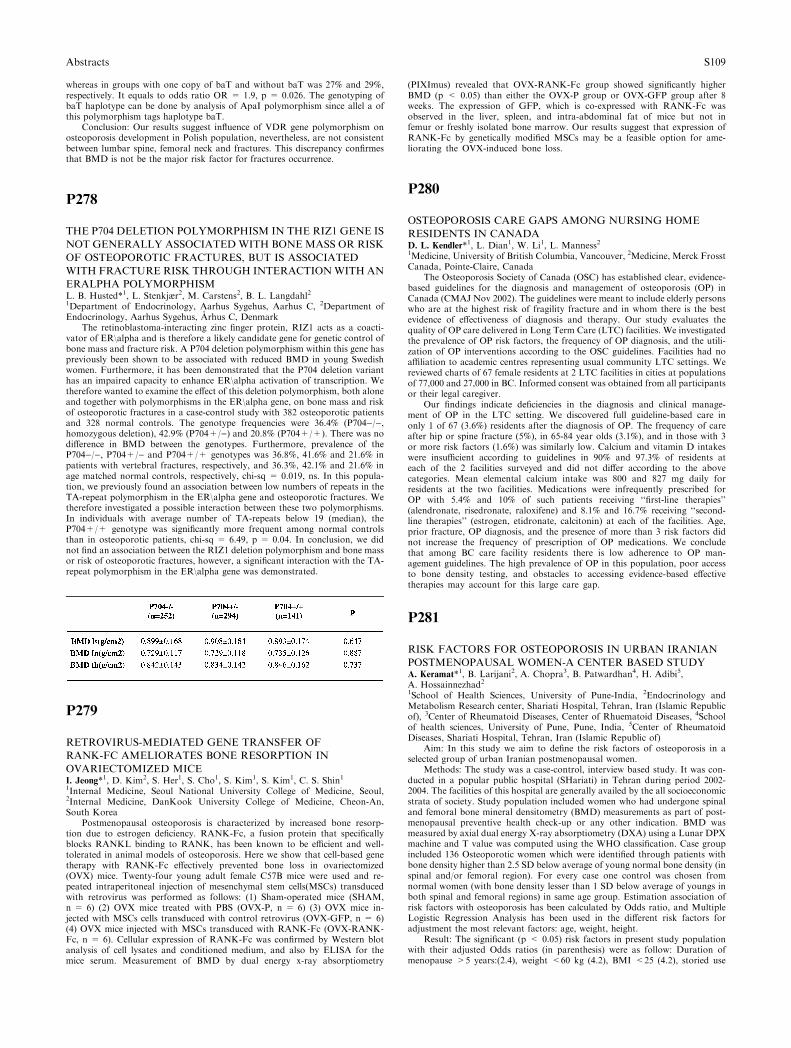
whereas in groups with one copy of baT and without baT was 27% and 29%,respectively. It equals to odds ratio OR = 1.9, p = 0.026. The genotyping ofbaT haplotype can be done by analysis of ApaI polymorphism since allel a ofthis polymorphism tags haplotype baT.
Conclusion: Our results suggest influence of VDR gene polymorphism onosteoporosis development in Polish population, nevertheless, are not consistentbetween lumbar spine, femoral neck and fractures. This discrepancy confirmesthat BMD is not be the major risk factor for fractures occurrence.
P278
THE P704 DELETION POLYMORPHISM IN THE RIZ1 GENE IS
NOT GENERALLY ASSOCIATED WITH BONEMASS OR RISK
OF OSTEOPOROTIC FRACTURES, BUT IS ASSOCIATED
WITH FRACTURE RISK THROUGH INTERACTIONWITH AN
ERALPHA POLYMORPHISML. B. Husted*1, L. Stenkjær2, M. Carstens2, B. L. Langdahl21Department of Endocrinology, Aarhus Sygehus, Aarhus C, 2Department ofEndocrinology, Aarhus Sygehus, Arhus C, Denmark
The retinoblastoma-interacting zinc finger protein, RIZ1 acts as a coacti-vator of ER\alpha and is therefore a likely candidate gene for genetic control ofbone mass and fracture risk. A P704 deletion polymorphism within this gene haspreviously been shown to be associated with reduced BMD in young Swedishwomen. Furthermore, it has been demonstrated that the P704 deletion varianthas an impaired capacity to enhance ER\alpha activation of transcription. Wetherefore wanted to examine the effect of this deletion polymorphism, both aloneand together with polymorphisms in the ER\alpha gene, on bone mass and riskof osteoporotic fractures in a case-control study with 382 osteoporotic patientsand 328 normal controls. The genotype frequencies were 36.4% (P704)/),homozygous deletion), 42.9% (P704+/)) and 20.8% (P704+/+). There was nodifference in BMD between the genotypes. Furthermore, prevalence of theP704)/), P704+/) and P704+/+ genotypes was 36.8%, 41.6% and 21.6% inpatients with vertebral fractures, respectively, and 36.3%, 42.1% and 21.6% inage matched normal controls, respectively, chi-sq = 0.019, ns. In this popula-tion, we previously found an association between low numbers of repeats in theTA-repeat polymorphism in the ER\alpha gene and osteoporotic fractures. Wetherefore investigated a possible interaction between these two polymorphisms.In individuals with average number of TA-repeats below 19 (median), theP704+/+ genotype was significantly more frequent among normal controlsthan in osteoporotic patients, chi-sq = 6.49, p = 0.04. In conclusion, we didnot find an association between the RIZ1 deletion polymorphism and bone massor risk of osteoporotic fractures, however, a significant interaction with the TA-repeat polymorphism in the ER\alpha gene was demonstrated.
P279
RETROVIRUS-MEDIATED GENE TRANSFER OF
RANK-FC AMELIORATES BONE RESORPTION IN
OVARIECTOMIZED MICEI. Jeong*1, D. Kim2, S. Her1, S. Cho1, S. Kim1, S. Kim1, C. S. Shin11Internal Medicine, Seoul National University College of Medicine, Seoul,2Internal Medicine, DanKook University College of Medicine, Cheon-An,South Korea
Postmenopausal osteoporosis is characterized by increased bone resorp-tion due to estrogen deficiency. RANK-Fc, a fusion protein that specificallyblocks RANKL binding to RANK, has been known to be efficient and well-tolerated in animal models of osteoporosis. Here we show that cell-based genetherapy with RANK-Fc effectively prevented bone loss in ovariectomized(OVX) mice. Twenty-four young adult female C57B mice were used and re-peated intraperitoneal injection of mesenchymal stem cells(MSCs) transducedwith retrovirus was performed as follows: (1) Sham-operated mice (SHAM,n = 6) (2) OVX mice treated with PBS (OVX-P, n = 6) (3) OVX mice in-jected with MSCs cells transduced with control retrovirus (OVX-GFP, n = 6)(4) OVX mice injected with MSCs transduced with RANK-Fc (OVX-RANK-Fc, n = 6). Cellular expression of RANK-Fc was confirmed by Western blotanalysis of cell lysates and conditioned medium, and also by ELISA for themice serum. Measurement of BMD by dual energy x-ray absorptiometry
(PIXImus) revealed that OVX-RANK-Fc group showed significantly higherBMD (p < 0.05) than either the OVX-P group or OVX-GFP group after 8weeks. The expression of GFP, which is co-expressed with RANK-Fc wasobserved in the liver, spleen, and intra-abdominal fat of mice but not infemur or freshly isolated bone marrow. Our results suggest that expression ofRANK-Fc by genetically modified MSCs may be a feasible option for ame-liorating the OVX-induced bone loss.
P280
OSTEOPOROSIS CARE GAPS AMONG NURSING HOME
RESIDENTS IN CANADAD. L. Kendler*1, L. Dian1, W. Li1, L. Manness21Medicine, University of British Columbia, Vancouver, 2Medicine, Merck FrosstCanada, Pointe-Claire, Canada
The Osteoporosis Society of Canada (OSC) has established clear, evidence-based guidelines for the diagnosis and management of osteoporosis (OP) inCanada (CMAJ Nov 2002). The guidelines were meant to include elderly personswho are at the highest risk of fragility fracture and in whom there is the bestevidence of effectiveness of diagnosis and therapy. Our study evaluates thequality of OP care delivered in Long Term Care (LTC) facilities. We investigatedthe prevalence of OP risk factors, the frequency of OP diagnosis, and the utili-zation of OP interventions according to the OSC guidelines. Facilities had noaffiliation to academic centres representing usual community LTC settings. Wereviewed charts of 67 female residents at 2 LTC facilities in cities at populationsof 77,000 and 27,000 in BC. Informed consent was obtained from all participantsor their legal caregiver.
Our findings indicate deficiencies in the diagnosis and clinical manage-ment of OP in the LTC setting. We discovered full guideline-based care inonly 1 of 67 (3.6%) residents after the diagnosis of OP. The frequency of careafter hip or spine fracture (5%), in 65-84 year olds (3.1%), and in those with 3or more risk factors (1.6%) was similarly low. Calcium and vitamin D intakeswere insufficient according to guidelines in 90% and 97.3% of residents ateach of the 2 facilities surveyed and did not differ according to the abovecategories. Mean elemental calcium intake was 800 and 827 mg daily forresidents at the two facilities. Medications were infrequently prescribed forOP with 5.4% and 10% of such patients receiving ‘‘first-line therapies’’(alendronate, risedronate, raloxifene) and 8.1% and 16.7% receiving ‘‘second-line therapies’’ (estrogen, etidronate, calcitonin) at each of the facilities. Age,prior fracture, OP diagnosis, and the presence of more than 3 risk factors didnot increase the frequency of prescription of OP medications. We concludethat among BC care facility residents there is low adherence to OP man-agement guidelines. The high prevalence of OP in this population, poor accessto bone density testing, and obstacles to accessing evidence-based effectivetherapies may account for this large care gap.
P281
RISK FACTORS FOR OSTEOPOROSIS IN URBAN IRANIAN
POSTMENOPAUSAL WOMEN-A CENTER BASED STUDYA. Keramat*1, B. Larijani2, A. Chopra3, B. Patwardhan4, H. Adibi5,A. Hossainnezhad21School of Health Sciences, University of Pune-India, 2Endocrinology andMetabolism Research center, Shariati Hospital, Tehran, Iran (Islamic Republicof), 3Center of Rheumatoid Diseases, Center of Rhuematoid Diseases, 4Schoolof health sciences, University of Pune, Pune, India, 5Center of RheumatoidDiseases, Shariati Hospital, Tehran, Iran (Islamic Republic of)
Aim: In this study we aim to define the risk factors of osteoporosis in aselected group of urban Iranian postmenopausal women.
Methods: The study was a case-control, interview based study. It was con-ducted in a popular public hospital (SHariati) in Tehran during period 2002-2004. The facilities of this hospital are generally availed by the all socioeconomicstrata of society. Study population included women who had undergone spinaland femoral bone mineral densitometry (BMD) measurements as part of post-menopausal preventive health check-up or any other indication. BMD wasmeasured by axial dual energy X-ray absorptiometry (DXA) using a Lunar DPXmachine and T value was computed using the WHO classification. Case groupincluded 136 Osteoporotic women which were identified through patients withbone density higher than 2.5 SD below average of young normal bone density (inspinal and/or femoral region). For every case one control was chosen fromnormal women (with bone density lesser than 1 SD below average of youngs inboth spinal and femoral regions) in same age group. Estimation association ofrisk factors with osteoporosis has been calculated by Odds ratio, and MultipleLogistic Regression Analysis has been used in the different risk factors foradjustment the most relevant factors: age, weight, height.
Result: The significant (p < 0.05) risk factors in present study populationwith their adjusted Odds ratios (in parenthesis) were as follow: Duration ofmenopause >5 years:(2.4), weight <60 kg (4.2), BMI <25 (4.2), storied use
Abstracts S109
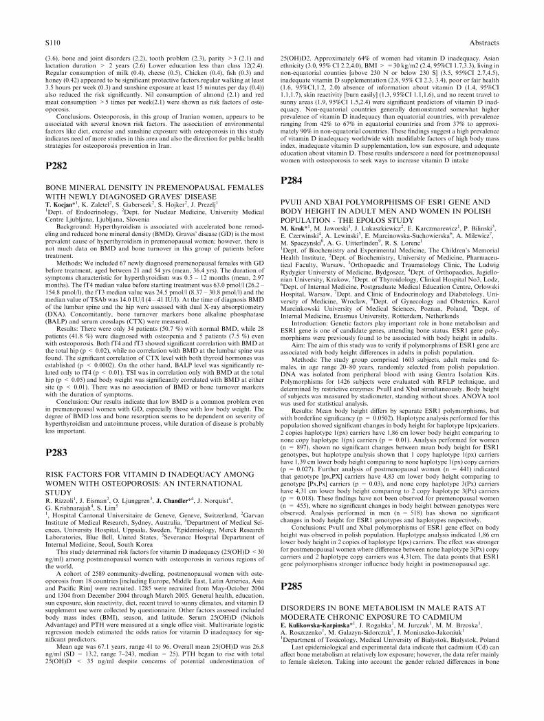
(3.6), bone and joint disorders (2.2), tooth problem (2.3), parity >3 (2.1) andlactation duration > 2 years (2.6) Lower education less than class 12(2.4).Regular consumption of milk (0.4), cheese (0.5), Chicken (0.4), fish (0.3) andhoney (0.42) appeared to be significant protective factors.regular walking at least3.5 hours per week (0.3) and sunshine exposure at least 15 minutes per day (0.4))also reduced the risk significantly. Nil consumption of almond (2.1) and redmeat consumption >5 times per week(2.1) were shown as risk factors of oste-oporosis.
Conclusions. Osteoporosis, in this group of Iranian women, appears to beassociated with several known risk factors. The association of environmentalfactors like diet, exercise and sunshine exposure with osteoporosis in this studyindicates need of more studies in this area and also the direction for public healthstrategies for osteoporosis prevention in Iran.
P282
BONE MINERAL DENSITY IN PREMENOPAUSAL FEMALES
WITH NEWLY DIAGNOSED GRAVES’ DISEASET. Kocjan*1, K. Zaletel2, S. Gaberscek2, S. Hojker2, J. Prezelj11Dept. of Endocrinology, 2Dept. for Nuclear Medicine, University MedicalCentre Ljubljana, Ljubljana, Slovenia
Background: Hyperthyroidism is associated with accelerated bone remod-eling and reduced bone mineral density (BMD). Graves� disease (GD) is the mostprevalent cause of hyperthyroidism in premenopausal women; however, there isnot much data on BMD and bone turnover in this group of patients beforetreatment.
Methods: We included 67 newly diagnosed premenopausal females with GDbefore treatment, aged between 21 and 54 yrs (mean, 36.4 yrs). The duration ofsymptoms characteristic for hyperthyroidism was 0.5 – 12 months (mean, 2.97months). The fT4 median value before starting treatment was 63.0 pmol/l (26.2 –154.8 pmol/l), the fT3 median value was 24.5 pmol/l (8.37 – 30.8 pmol/l) and themedian value of TSAb was 14.0 IU/l (4 – 41 IU/l). At the time of diagnosis BMDof the lumbar spine and the hip were assessed with dual X-ray absorptiometry(DXA). Concomitantly, bone turnover markers bone alkaline phosphatase(BALP) and serum crosslaps (CTX) were measured.
Results: There were only 34 patients (50.7 %) with normal BMD, while 28patients (41.8 %) were diagnosed with osteopenia and 5 patients (7.5 %) evenwith osteoporosis. Both fT4 and fT3 showed significant correlation with BMD atthe total hip (p < 0.02), while no correlation with BMD at the lumbar spine wasfound. The significant correlation of CTX level with both thyroid hormones wasestablished (p < 0.0002). On the other hand, BALP level was significantly re-lated only to fT4 (p < 0.01). TSI was in correlation only with BMD at the totalhip (p < 0.05) and body weight was significantly correlated with BMD at eithersite (p < 0.01). There was no association of BMD or bone turnover markerswith the duration of symptoms.
Conclusion: Our results indicate that low BMD is a common problem evenin premenopausal women with GD, especially those with low body weight. Thedegree of BMD loss and bone resorption seems to be dependent on severity ofhyperthyroidism and autoimmune process, while duration of disease is probablyless important.
P283
RISK FACTORS FOR VITAMIN D INADEQUACY AMONG
WOMEN WITH OSTEOPOROSIS: AN INTERNATIONAL
STUDYR. Rizzoli1, J. Eisman2, O. Ljunggren3, J. Chandler*4, J. Norquist4,G. Krishnarajah4, S. Lim5
1, Hospital Cantonal Universitaire de Geneve, Geneve, Switzerland, 2GarvanInstitute of Medical Research, Sydney, Australia, 3Department of Medical Sci-ences, University Hospital, Uppsala, Sweden, 4Epidemiology, Merck ResearchLaboratories, Blue Bell, United States, 5Severance Hospital Department ofInternal Medicine, Seoul, South Korea
This study determined risk factors for vitamin D inadequacy (25(OH)D<30ng/ml) among postmenopausal women with osteoporosis in various regions ofthe world.
A cohort of 2589 community-dwelling, postmenopausal women with oste-oporosis from 18 countries [including Europe, Middle East, Latin America, Asiaand Pacific Rim] were recruited. 1285 were recruited from May-October 2004and 1304 from December 2004 through March 2005. General health, education,sun exposure, skin reactivity, diet, recent travel to sunny climates, and vitamin Dsupplement use were collected by questionnaire. Other factors assessed includedbody mass index (BMI), season, and latitude. Serum 25(OH)D (NicholsAdvantage) and PTH were measured at a single office visit. Multivariate logisticregression models estimated the odds ratios for vitamin D inadequacy for sig-nificant predictors.
Mean age was 67.1 years, range 41 to 96. Overall mean 25(OH)D was 26.8ng/ml (SD = 13.2, range 7–243, median = 25). PTH began to rise with total25(OH)D < 35 ng/ml despite concerns of potential underestimation of
25(OH)D2. Approximately 64% of women had vitamin D inadequacy. Asianethnicity (3.0, 95% CI 2.2,4.0), BMI >=30 kg/m2 (2.4, 95%CI 1.7,3.3), living innon-equatorial counties [above 230 N or below 230 S] (3.5, 95%CI 2.7,4.5),inadequate vitamin D supplementation (2.8, 95% CI 2.3, 3.4), poor or fair health(1.6, 95%CI,1.2, 2.0) absence of information about vitamin D (1.4, 95%CI1.1,1.7), skin reactivity [burn easily] (1.3, 95%CI 1.1,1.6), and no recent travel tosunny areas (1.9, 95%CI 1.5,2.4) were significant predictors of vitamin D inad-equacy. Non-equatorial countries generally demonstrated somewhat higherprevalence of vitamin D inadequacy than equatorial countries, with prevalenceranging from 42% to 67% in equatorial countries and from 37% to approxi-mately 90% in non-equatorial countries. These findings suggest a high prevalenceof vitamin D inadequacy worldwide with modifiable factors of high body massindex, inadequate vitamin D supplementation, low sun exposure, and adequateeducation about vitamin D. These results underscore a need for postmenopausalwomen with osteoporosis to seek ways to increase vitamin D intake
P284
PVUII AND XBAI POLYMORPHISMS OF ESR1 GENE AND
BODY HEIGHT IN ADULT MEN AND WOMEN IN POLISH
POPULATION - THE EPOLOS STUDYM. Kruk*1, M. Jaworski1, J. Lukaszkiewicz2, E. Karczmarewicz1, P. Bilinski3,E. Czerwinski4, A. Lewinski5, E. Marcinowska-Suchowierska6, A. Milewicz7,M. Spaczynski8, A. G. Uitterlinden9, R. S. Lorenc11Dept. of Biochemistry and Experimental Medicine, The Children’s MemorialHealth Institute, 2Dept. of Biochemistry, University of Medicine, Pharmaceu-tical Faculty, Warsaw, 3Orthopaedic and Traumatology Clinic, The LudwigRydygier University of Medicine, Bydgoszcz, 4Dept. of Orthopaedics, Jagiello-nian University, Krakow, 5Dept. of Thyroidology, Clinical Hospital No3, Lodz,6Dept. of Internal Medicine, Postgraduate Medical Education Centre, OrlowskiHospital, Warsaw, 7Dept. and Clinic of Endocrinology and Diabetology, Uni-versity of Medicine, Wroclaw, 8Dept. of Gynecology and Obstetrics, KarolMarcinkowski University of Medical Sciences, Poznan, Poland, 9Dept. ofInternal Medicine, Erasmus University, Rotterdam, Netherlands
Introduction: Genetic factors play important role in bone metabolism andESR1 gene is one of candidate genes, attending bone status. ESR1 gene poly-morphisms were previously found to be associated with body height in adults.
Aim: The aim of this study was to verify if polymorphisms of ESR1 gene areassociated with body height differences in adults in polish population.
Methods: The study group comprised 1603 subjects, adult males and fe-males, in age range 20–80 years, randomly selected from polish population.DNA was isolated from peripheral blood with using Gentra Isolation Kits.Polymorphisms for 1426 subjects were evaluated with RFLP technique, anddetermined by restrictive enzymes: PvuII and XbaI simultaneously. Body heightof subjects was measured by stadiometer, standing without shoes. ANOVA toolwas used for statistical analysis.
Results: Mean body height differs by separate ESR1 polymorphisms, butwith borderline significancy (p = 0.0502). Haplotype analysis performed for thispopulation showed significant changes in body height for haplotype 1(px)cariers.2 copies haplotype 1(px) carriers have 1,86 cm lower body height comparing tonone copy haplotype 1(px) carriers (p = 0.01). Analysis performed for women(n = 897), shown no significant changes between mean body height for ESR1genotypes, but haplotype analysis shown that 1 copy haplotype 1(px) carriershave 1,39 cm lower body height comparing to none haplotype 1(px) copy carriers(p = 0.027). Further analysis of postmenopausal women (n = 441) indicatedthat genotype [px,PX] carriers have 4,83 cm lower body height comparing togenotype [Px,Px] carriers (p = 0.03), and none copy haplotype 3(Px) carriershave 4,31 cm lower body height comparing to 2 copy haplotype 3(Px) carriers(p = 0.018). These findings have not been observed for premenopausal women(n = 455), where no significant changes in body height between genotypes wereobserved. Analysis performed in men (n = 518) has shown no significantchanges in body height for ESR1 genotypes and haplotypes respectively.
Conclusions: PvuII and XbaI polymorphisms of ESR1 gene effect on bodyheight was observed in polish population. Haplotype analysis indicated 1,86 cmlower body height in 2 copies of haplotype 1(px) carriers. The effect was strongerfor postmenopausal women where difference between none haplotype 3(Px) copycarriers and 2 haplotype copy carriers was 4,31cm. The data points that ESR1gene polymorphisms stronger influence body height in postmenopausal age.
P285
DISORDERS IN BONE METABOLISM IN MALE RATS AT
MODERATE CHRONIC EXPOSURE TO CADMIUME. Kulikowska-Karpinska*1, J. Rogalska1, M. Jurczuk1, M. M. Brzoska1,A. Roszczenko1, M. Galazyn-Sidorczuk1, J. Moniuszko-Jakoniuk11Department of Toxicology, Medical University of Bialystok, Bialystok, Poland
Last epidemiological and experimental data indicate that cadmium (Cd) canaffect bone metabolism at relatively low exposure; however, the data refer mainlyto female skeleton. Taking into account the gender related differences in bone
S110 Abstracts

vulnerability to various damaging factors, in the present study the effect ofmoderate chronic exposure to Cd on bone metabolism was investigated on amale rat model of human environmental exposure to this toxic metal.
Male Wistar rats (n = 8) were exposed to Cd in drinking water at theconcentration of 5 mg/l for 6 months. Control rats (n = 8) drank water free ofCd. Bone mineral density (BMD) and bone mineral content (BMC) at the femurand the fourth lumbar vertebral body (L4) were measured densitometrically(Prodigy, GE, USA). Serum concentrations of osteocalcin (OC) as a marker ofbone formation and carboxy-terminal cross-linking telopeptides of type I col-lagen (CTX) as a marker of bone resorption were determined using ELISA kits(Nordic Bioscience Diagnostics, Denmark). Data were analysed by Anova(Kruskal-Wallis ranks test).
The exposure to Cd affected bone metabolism as it was reflected in disturbedbone mineralization and changes in biochemical markers of bone turnover. TheBMD and BMC at the femur and L4 were decreased compared to control. Theserum concentration of OC decreased, whereas the concentration of CTX in-creased.
The results allow for the conclusion that moderate exposure to Cd affects theskeletal mineralization via decreasing bone formation and increasing itsresorption. The findings of this study confirm the hypothesis that moderateenvironmental exposure to Cd may create a risk for bone damage.
This study was financially supported by the Grant (No. 2 PO5D 027 27)from the Committee for Scientific Research (KBN, Poland).
P286
BONE MINERAL DENSITY IN LUMBAR SPINE AND FEMUR
IN A RURAL POPULATION IN GREECE. AN EPIDEMIO-
LOGICAL STUDY IN THE ATHENS METROPOLITAN AREA,
GREECEI. N. Legakis*1, V. Papadopoulos2, V. Tataridas2, K. Strigaris21Endocrinology, 2Radiology, Henry Dunant Hospital, Athens, Greece
In order to estimate the prevalence of osteoporosis in a general populationof a rural area, we evaluated the dual-energy X-ray absorptiometry measure-ments, using LUNAR�S DRX-L densitometer, first of the lumbar spine in 6920females and in 181 males and second of the femur in 3222 females and in 123males, aged 29 to 89 years .The data were collected in a 4 year period (2001–2005) from the Radiology Department of the Henry Dunant Hospital inAthens, Greece. Bone mineral density (BMD) measurements and the corre-sponding T-scores were analysed using multivariate regression models. Sepa-rate analyses for the spine and femur (Neck, Ward�s, Trochanter) wereperformed. The significance of somatometric characteristics such as weight,height, and body mass index (BMI) was also investigated. According to ourdata, the prevalence rate of osteoporosis, based to the WHO criteria, for thelumbar spine in women was 15.3% and for osteopenia was 36.5%. In men, theprevalence rate of osteoporosis for the lumbar spine was 13.8% and of oste-openia was 37.5%. In females osteoporosis rate for the femur was 17.7% forthe neck, 30.65% for the ward�s triangle and 1.86% for the tronchater. Oste-openic rates at the femur for females were 53.3% for the neck, 44.7% for theward�s triangle and 32.09% for the trochanter. In males, osteoporosis rate forthe femur was 36.5% for the neck, 50.4% for the ward�s triangle and 15.45%for the tronchater. Osteopenic rates at the femur in males were 44.4% for theneck, 28.8% for the ward�s triangle and 40.65% for the tronchater. Addition-ally, initial exploratory analysis revealed that BMD and T-score changes byage were not linear across the chronological range of our sample (20 to 90years). For that reason, a polynomial cubic model was performed for agewhich showed good fit for the observed data, allowing also the estimation ofage related slopes to T-scores values. The steepest decline was observed at theage of 54 years (0.0106, 95% CI) for the spine BMD and at the age of 62 yearsfor the femur BMD measurements.
Although ,as expected, BMI had a significant correlation with BMD and T-scores ,weight and height showed a better fit to the observed data than BMI in allcase models.
P287
VITAMIN D STATUS IN FRENCH POSTMENOPAUSAL
WOMEN WITH DENSITOMETRIC OSTEOPOROSISF. E. Levy-weil*1, D. Khalfon2, L. Chauvelot-Moachon3, J. C. Souberbielle4,S. Djennane1, J. L. Feldmann11Department of Rheumatology, 2Biochemistry Laboratory, Centre HospitalierVictor Dupouy, Argenteuil, 3Center of Pharmacovigilance, Hopital Cochin,4Physiology Laboratory, Hopital Necker, Paris, France
Background: Vitamin D plays an important role in bone health. Deficiencyin 25-hydroxyvitamin D (25OHD) leads to bone loss and increases fracture risk.Such a deficiency may be difficult to detect, as, in most cases, there is not dis-order of phosphorus/calcium metabolism.
The aim of this study was to investigate the frequency of vitamin D inade-quacy in postmenopausal women with densitometric osteoporosis but no bonefracture.
Methods: We studied 62 postmenopausal women over the age of 45 yearswith densitometric osteoporosis (T-score <)2.5DS) detected at Argenteuil’sHospital. We excluded a history of fracture or secondary osteoporosis. All pa-tients underwent assessment of phosphorus/calcium metabolism. Serum 25OHDconcentration was determined by DiaSorin LIAISON automated immunoassay.We used 30 ng/ml (=75 nmol/l) as the cutoff point defining vitamin D inadequacyas recently recommended. Vitamin D status was categorised in four groups:normal (25OHD>30 ng/ml), moderate insufficiency (20 ng/ml<25OHD<30 ng/ml), severe insufficiency (10 ng/ml<25OHD<20 ng/ml) and deficiency(25OHD<10 ng/ml). Bone mineral density (BMD) was measured with an Ho-logic. Mean age was 62 years with 64% of the patients being less than 65 years old.
Results: Vitamin D inadequacy was detected in 79% of the population: 28%had moderate vitamin D insufficiency, 36% had severe vitamin D insufficiencyand 15% had vitamin D deficiency. The distribution of severity was similar in thetwo age groups considered (< and > 65 years). Women with vitamin D defi-ciency tended to have a lower BMD than women with normal vitamin D status.
Conclusion: Vitamin D inadequacy was common in this French populationof postmenopausal women with densitometric osteoporosis. These results sug-gest that vitamin D inadequacy should be sought in all osteoporotic women evenwithout any fracture. Adequate vitamin D supplementation can both preventbone loss and optimise the effects of action of antiosteoporotic treatment whenintroduced.
P288
VITAMIN D STATUS AND THE THRESHOLD FOR THE IN-
CREASE OF SERUM PTH IN ADULT MEN FROM DIFFER-
ENT EUROPEAN COUNTRIES: THE NEMO STUDYP. Lips*1, N. M. Van Schoor2, P. Szulc3, J. M. Kaufman4, S. Gonnelli5,J. Stepan6, S. Boonen7, M. T. E. Puts2, M. A. Blankenstein81Endocrinology, 2EMGO, VU University Medical Center, Amsterdam, Neth-erlands, 3INSERM, Claude Bernard University, Lyon, France, 4Endocrinology,Ghent University, Ghent, Belgium, 5Endocrinology, University of Siena, Siena,Italy, 6Internal Medicine, University of Prague, Prague, Czech Republic,7Geriatrics, Leuven University, Leuven, Belgium, 8Clinical Chemistry, VUUniversity Medical Center, Amsterdam, Netherlands
Vitamin D deficiency and insufficiency are common in adults and cause anincrease of the serum parathyroid hormone (PTH) concentration. The thresholdfor the increase of serum PTH has been used as the cut-off between vitamin Dinsufficiency and sufficiency. It has been reported between 50 and 80 nmol/l. Theaim of our study was to assess the threshold for the increase of serum PTH inhealthy men from different European countries. The study was done within thecontext of the Network in Europe on Male Osteoporosis (NEMO). Bloodsamples were obtained from men between 25 and 88 years from France (MINOSstudy, n = 987), Ghent (n = 407), Siena (n = 29), Prague (n = 42), Leuven(n = 25) and Amsterdam (LASA study, n = 643). Serum 25-hydroxyvitamin D(25(OH)D) and PTH were measured by radioimmunological methods.
Serum 25(OH)D values were cross-calibrated by remeasuring about 25samples from each center in Amsterdam. Correction factors were calculatedranging from 0.89 to 1.23. All data were corrected to the Diasorin assay values,currently in use in Amsterdam.
The percentage of participants with serum 25(OH)D lower than 50 nmol/l, aconservative estimate for vitamin D insufficiency, ranged from 20 to 68 %. SerumPTH was significantly higher in persons having a low serum 25(OH)D in mostdatasets. Data from all centers showed the expected negative relationship be-tween serum PTH and serum 25(OH)D. The threshold serum 25(OH)D wasabove 75 nmol/l in most centers. The large interlaboratory variation can explaindifferences in reported threshold values for serum 25(OH)D.
In conclusion, vitamin D insuffiency was common in adult men in differentEuropean countries and the threshold serum 25(OH)D for the increase of serumPTH was higher than expected.
P289
SHWACHMAN-DIAMOND SYNDROME IS ASSOCIATED
WITH OSTEOPOROSISS. Toiviainen-Salo1, M. Mayranpaa1, P. Durie2, L. Ellis2, J. Rommens2,E. Savilahti1, O. Makitie*11Metabolic Bone Clinic, Hospital for Children and Adolescents, Helsinki Uni-versity Hospital, Helsinki, Finland, 2Hospital for Sick Children, University ofToronto, Toronto, Canada
Shwachman–Diamond Syndrome (SDS) is an autosomal recessive disordercharacterized by exocrine pancreatic insufficiency and bone marrow dysfunction.These result in malabsorption and variable neutropenia. The associated skeletal
Abstracts S111
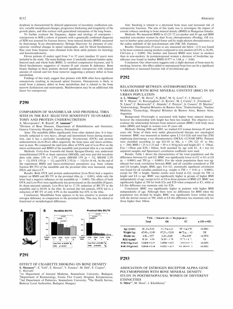
dysplasia in characterized by delayed appearance of secondary ossification cen-ters, variable metaphyseal changes, progressive thickening and irregularity of thegrowth plates, and thin cortices with generalized osteopenia of the long bones.
To further evaluate the frequency, degree and etiology of osteopenia /osteoporosis in SDS a cohort of patients with a genetically confirmed diagnosisof SDS was assessed for fracture history, bone mineral density (BMD; measuredby dual-energy X-ray absorptiometry, Hologic Discovery A), presence of oste-oporotic vertebral changes in spinal radiographs, and for blood biochemistry.Iliac crest bone biopsies were obtained from three adult patients for histologyand histomorphometry.
Eleven patients (8 males) aged from 5 to 32 years (median 18 years) wereincluded in the study. The main findings were 1) markedly reduced lumbar spine,femoral neck and whole body BMD, 2) vertebral compression fractures, and 3)blood biochemistry suggestive of vitamin D and vitamin K deficiency. Bonebiopsy findings in three patients showed significant osteoporosis with reducedamount of osteoid and low bone turnover suggesting a primary defect in bonemetabolism.
Findings of this study suggest that patients with SDS often have significantosteoporosis resulting in increased spinal fractures. Osteoporosis is likely toresult from a primary defect in bone metabolism that is related to the bonemarrow dysfunction and neutropenia. Malabsorption may be an additional riskfactor for osteoporosis.
P290
COMPARISON OF MANDIBULAR AND PROXIMAL TIBIA
SITES IN THE RAT: SELECTIVE SENSITIVITY TO OVARIEC-
TOMY AND PROTEIN UNDERNUTRITIONA. Mavropoulos1, R. Rizzoli1, P. Ammann*11Division of Bone Diseases, Department of Rehabilitation and Geriatrics,Geneva University Hospital, Geneva, Switzerland
Aims: The mandible differs significantly from other skeletal sites. It is func-tionally subjected to very heavy, abrupt and intermittent forces during mastica-tion, and it has a particular morphology. Ovariectomy (OVX) and proteinundernutrition (Low-Prot) affect negatively the bone mass and micro-architec-ture in man. We compared the mid-term effect of OVX and of Low-Prot on themicro-architecture and BMD of the mandible and proximal tibia in a rat model.
Methods: Forty-four 6-month-old female Sprague-Dawley rats underwenttransabdominal OVX or sham operation (SHAM), and were pair-fed isocaloricdiets with either 15% or 2.5% casein (SHAM 15% [n = 11], SHAM 2.5%[n = 11], OVX 15% [n = 11], and OVX 2.5% [n = 11]) for 16 wk. At the end ofthe experiment BMD and micro-architecture parameters (e.g. bone volumefraction BV/TV) of the mandible and the proximal tibia were measured, usingDXA and micro-CT.
Results: Both OVX and protein undernutrition (Low-Prot) had a negativeimpact on BMD and BV/TV in the proximal tibia (p < 0.001), while only thelatter had a negative influence on the mandible (p < 0.005). The effects of bothOVX and Low-Prot were significantly less pronounced on the mandible (Figure).In sham-operated animals, Low-Prot led to 17.3% reduction of BV/TV in themandible and to 84.6% in the tibia. In normal diet fed animals, OVX led to areduction of BV/TV of only 4.9% in the mandible but 82% in the tibia.
Conclusions: Mandibular bone appears to be less sensitive to protein andestrogen deficiency in comparison to the proximal tibia. This may be related tofunctional or morphological differences.
P291
EFFECT OF CIGARETTE SMOKING ON BONE DENSITYS. Meszaros*1, E. Toth2, E. Hosszu3, V. Ferencz1, M. Deli1, E. Csupor4,C. Horvath111st Department of Internal Medicine, Semmelweis University, Budapest,2Department of Reumatology, Ferenc Flor County Hospital, Kerepestarcsa,32nd Department of Pediatrics, Semmelweis University, 4The Health Service,Budavar Local Authorities, Budapest, Hungary
Aim: Smoking is related to a decreased bone mass and increased risk ofosteoporotic fractures. The aim of this study was to investigate the impact ofcurrent tobacco smoking to bone mineral density (BMD) in Hungarian females.
Methods: We measured BMD in 43 (25–72 ys) smoker and 43 age and BMImatched non-smoker women by dual X-ray absorptiometry (Prodigy, GE Lu-nar) in lumbar spine and proximal femur and by single photon absorptiometry atthe radius midshaft of the non-dominant side (NK 364, Gamma).
Results: Osteoporosis (T-score at any measured site below )2,5) was foundto be more common among smokers compared to non smokers (63.6% vs 36.4%,Chi2-test p = 0.009). The lumbar and femoral BMD were lower in smokersthan in non-smokers. In postmenopausal women a decrease of borderline sig-nificance was found in lumbar BMD (0.977 vs 1.04; p = 0,06).
Conclusion: Our observation suggests only a slight decrease of bone mass bysmoking, however, this effect added to menopausal bone loss can be a significantcontribution to increased fracture risk of involutional age.
P292
RELATIONSHIP BETWEEN ANTHROPOMETRICS
VARIABLES WITH BONE MINERAL CONTENT (BMC) IN AN
URBAN POPULATIONA. L. Misiunas*1, M. Hevia2, N. Rella2, M. G. Loto2, C. J. Herrera2,M. V. Moran2, G. Roccatagliata2, G. Rovira2, M. I. Curria2, C. Fitzsimons3,N. Llacer4, C. Baistrocchi4, C. Almeida3, C. Pereyra3, A. Casares3, D. Martino31Endocrinology, Hospital Britanico de Buenos Aires, 2Endocrinology, 3NuclearMedicine, 4Gynecology, Hospital Britanico de Buenos Aires, Buenos Aires,Argentina
Background: Overweight is associated with higher bone mineral density,however the relationship with height has been less studied. The objective is toevaluate the relationship between bone mineral content (BMC) with body massindex (BMI) and height in women over 45 years old.
Methods: During 2004 and 2005, we studied 633 women between 45 and 84years old. None of them were under glucocorticoid therapy nor oncologicaltreatment. BMC was measured at lumbar spine L2–L4 (LS) and total hip (TH)employing dual-energy x-ray absorptiometry (DEXA; LUNAR DPX). Patientswere classified according to age (G1:45 to 60y, n = 339; G2: 61 to 84y;n = 294), BMI (<25 vs ‡ 25 and <30 vs ‡ 30 kg/m2) and height (E1: £ 150cm,E2a:>150cm and E2b:>160cm, both matched by age with E1. A t test forunpaired samples and Spearman�s correlation coefficient were used.
Results: Table 1 shows the characteristics of the total population and thedifferences between G1 and G2. BMC was significantly lower in G2 vs G1 at LS(p < 0.0001) and TH (p < 0.0001). For the whole population there was sig-nificant but weak correlation between BMC and all variables considered at THand LS (weight, height, BMI, age). For G1, statistically significant correlationwas found for all variables analized (e.g.: weight vs TH: r = 0.41; p < 0.0001),except for TH vs height. Similar results were found in G2, except for TH vsheight and LS vs age. BMC was significantly higher in groups of higher BMIindependently of age, except in G1 at LS in those patients of BMI ‡25. BMC wassignificantly higher at TH for both E2a and E2b when compared to E1, while atLS this difference was maintain only for E2b.
Conclusion: BMC was significantly higher in patients with higher BMIindependently of age. Although there were no differences for BMI when thepopulation was divided by height, BMC was significantly lower within thosewith the shorter stature at TH, while at LS this difference was maintain only forthose higher than 160cm.
P293
ASSOCIATION OF ESTROGEN RECEPTOR ALPHA GENE
POLYMORPHISMS WITH BONE MINERAL DENSITY:
STUDY IN POSTMENOPAUSAL WOMEN OF DIFFERENT
ETHNICITIESS. Mitra*1, M. Desai1, I. Khatkhatay1
S112 Abstracts
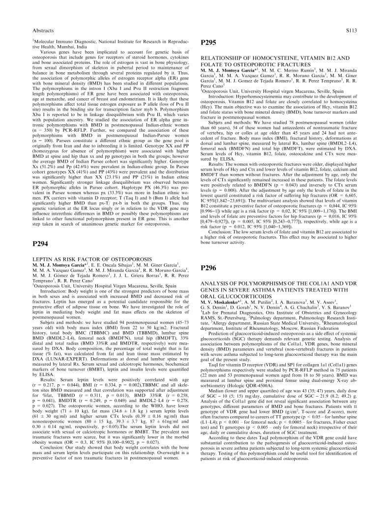
1Molecular Immuno Diagnostic, National Institute for Research in Reproduc-tive Health, Mumbai, India
Various genes have been implicated to account for genetic basis ofosteoporosis that include genes for receptors of steroid hormones, cytokinesand bone associated proteins. The role of estrogen is vast in bone physiology,from sexual dimorphism of skeleton in pubertal period to maintenance ofbalance in bone metabolism through several proteins regulated by it. Thus,the association of polymorphic alleles of estrogen receptor alpha (ER) genewith bone mineral density (BMD) has been studied in different populations.The polymorphisms in the intron I (Xba I and Pvu II restriction fragmentlength polymorphisms) of ER gene have been associated with osteoporosis,age at menarche, and cancer of breast and endometrium. It is likely that thesepolymorphisms affect total tissue estrogen exposure as P allele (loss of Pvu IIsite) results in the binding site for transcription factor myb b. PolymorphismXba I is reported to be in linkage disequilibrium with Pvu II, which varieswith population ancestry. We studied the association of ER alpha gene in-tronic polymorphisms with BMD in postmenopausal Indian-ethnic women(n = 350) by PCR-RFLP. Further, we compared the association of thesepolymorphisms with BMD in postmenopausal Indian-Parsee women(n = 100). Parsees constitute a different ethnic group as the gene pool isoriginally from Iran and due to inbreeding it is limited. Genotype XX and PP(homozygous for absence of polymorphism) were associated with higherBMD at spine and hip than xx and pp genotypes in both the groups; howeverthe average BMD of Indian Parsee cohort was significantly higher. GenotypeXx (51.2%) and Pp (42.6%) were prevalent in Indian-ethnic group. In Parseecohort genotypes XX (41%) and PP (43%) were prevalent and the distributionwas significantly higher than XX (23.1%) and PP (21%) in Indian ethnicwomen. Significantly stronger linkage disequilibrium was observed betweenER polymorphic alleles in Parsee cohort. Haplotype PX (46.3%) was pre-valent in Parsee women whereas px (33.3%) was more in Indian ethnic wo-men. PX carriers with vitamin D receptor; T (Taq I) and b (Bsm I) allele hadsignificantly higher BMD than px-T/ px-b in both the groups. Thus, thegenetic variation at the ER locus singly or in relation to the VDR gene mayinfluence interethnic differences in BMD or possibly these polymorphisms arelinked to other functional polymorphism present in ER gene. This is anotherstep taken in search of unanimous genetic marker for osteoporosis.
P294
LEPTIN AS RISK FACTOR OF OSTEOPOROSISM. M. J. Montoya Garcia*1, E. E. Oncala Sibajas1, M. M. Giner Garcia1,M. M. A. Vazquez Gamez1, M. M. J. Miranda Garcia1, R. R. Moruno Garcia1,M. M. J. Gomez de Tejada Romero1, J. J. L. Griera Borras1, R. R. PerezTemprano1, R. R. Perez Cano11Osteoporosis Unit, University Hospital Virgen Macarena, Seville, Spain
Introduction: Body weight is one of the strongest predictors of bone massin both sexes and is associated with increased BMD and decreased risk offractures. Leptin has emerged as a potential candidate responsible for theprotective effect of adipose tissue on bone. We have investigated the role ofleptin in mediating body weight and fat mass effects on the skeleton ofpostmenopausal women.
Subjets and methods: we have studied 94 postmenopausal women (45–73years old) with body mass index (BMI) from 22 to 50 kg/m2. Fracturalhistory, total body BMC (TBBMC) and BMD (TBBMD), lumbar spineBMD (BMDL2–L4), femoral neck (BMDFN), total hip (BMDFT), 33%distal and total radius (BMD 33%R and BMDTR, respectively) were mea-sured by DXA. Body composition, the percentage of total weight that is fattissue (% fat), was calculated from fat and lean tissue mass estimated byDXA (LUNAR-EXPERT). Deformations at dorsal and lumbar spine weremeasured by lateral Rx. Serum sexual and calciotropic hormones, biochemicalmarkers of bone turnover (BMBT), leptin and insulin levels were quantifiedby ELISA.
Results: Serum leptin levels were positively correlated with age(r = 0.217, p = 0.044), BMI (r = 0.334, p = 0.002),TBBMC and all skele-ton sites BMD measured and that correlation was supported after adjustmentfor %fat, TBBMD (r = 0.311, p = 0.013), BMD 33%R (r = 0.258,p = 0.041), BMDTR (r = 0.249, p = 0.049) and BMDL2–L4 (r = 0.278,p = 0.027). The osteoporotic women, according to the WHO, have lowerbody weight (71 ± 10 kg), fat mass (34.6 ± 1.8 kg ) serum leptin levels(61 ± 30 ng/ml) and higher serum CTx levels (0.39 ± 0.16 ng/ml) thannonosteoporotic women (80 ± 15 kg, 39.3 ± 3.7 kg, 87 ± 61ng/ml and0.30 ± 0.14 ng/ml, respectively, p<0.05).The serum leptin levels did notassociate with sexual or calciotropic hormones or BMBT. The prevalent nontraumatic fractures were scarce, but it was significantly lower in the morbidobesity women (OR = 0.3, IC 95% [0.100)0.902], p = 0.027).
Conclusion: Our study showed that body weight correlates with the bonemass and serum leptin levels participate on this relationship. Overweight is apreventive factor of non traumatic fractures in postmenopausal women.
P295
RELATIONSHIP OF HOMOCYSTEINE, VITAMIN B12 AND
FOLATE TO OSTEOPOROTIC FRACTURESM. M. J. Montoya Garcia*1, M. M. C. Merino Rumın1, M. M. J. MirandaGarcia1, M. M. A. Vazquez Gamez1, R. R. Moruno Garcia1, M. M. GinerGarcia1, M. M. J. Gomez de Tejada Romero1, R. R. Perez Temprano1, R. R.Perez Cano11Osteoporosis Unit, University Hospital virgen Macarena, Seville, Spain
Introduction: Hyperhomocysteinemia may contribute to the development ofosteoporosis. Vitamin B12 and folate are closely correlated to homocysteina(Hcy). The main objective was to examine the association of Hcy, vitamin B12and folate status with bone mineral density (BMD), bone turnover markers andfracture in postmenopausal women.
Subjets and methods: We have studied 78 postmenopausal women (olderthan 60 years), 54 of these women had antecedents of nontraumatic fractureof vertebra, hip or colles at age older than 45 years and 24 had not ante-cedent of fracture. Body mass index (BMI), fractural history, deformations atdorsal and lumbar spine, measured by lateral Rx, lumbar spine (BMDL2–L4),femoral neck (BMDFN) and total hip (BMDFT), were estimated by DXA.Serum levels of Hcy, vitamin B12, folate, osteocalcine and CTx were mea-sured by ELISA.
Results: The women with osteoporotic fractures were older, displayed higherserum levels of Hcy and Ctx and lower levels of vitamin B12, folate, calcium andBMDFT than women without fractures. After the adjustment by age, only thelevels of CTx significantly remained increased in these patients. The folate levelswere positively related to BMDFN (p = 0.043) and inversely to CTx serumlevels (p = 0.008). After the adjustment by age only the levels of folate in thelowest quartil constituted a risk factor of suffering hip fractures (OR = 6,605,IC 95%[1,842)23,691]). The multivariant analysis showed that levels of vitaminB12 constitute a preventive factor of osteoporotic fractures (p = 0,044, IC 95%[0.996)1]) while age is a risk factor (p = 0,02, IC 95% [1,009)1,176]). The BMIand levels of folate are preventive factors for hip fractures (p = 0,016, IC 95%[0,479)0,927]), (p = 0.005, IC 95% [0,243)0,777]), respectively, while age is arisk factor (p = 0.012, IC 95% [1,040)1,369]).
Conclusion: The low serum levels of folate and vitamin B12 are associated toa higher risk of osteoporotic fractures. This effect may be associated to higherbone turnover activity.
P296
ANALYSIS OF POLYMORPHISMS OF THE COL1A1 AND VDR
GENES IN SEVERE ASTHMA PATIENTS TREATED WITH
ORAL GLUCOCORTICOIDSM. V. Moskalenko*1, A. M. Putilin2, I. A. Baranova3, M. V. Aseev1,G. S. Demin1, D. Gorab1, N. V. Demin4, A. G. Chuchalin2, V. S. Baranov11Lab for Prenatal Diagnostics, Otts Institute of Obstetrics and GynecologyRAMS, St.-Petersburg, 2Pulmology department, Pulmonology Research Insti-tute, 3Allergy department, Russian State Medical University, 4Rheumatologicaldepartment, Institute of Rheumatology, Moscow, Russian Federation
Prediction of glucocorticoid-induced osteoporosis as a side effect of systemicglucocorticoids (SGC) therapy demands relevant genetic testing. Analysis ofassociation between polymorphisms of the Col1a1, VDR genes, bone mineraldensity (BMD) parameters and vertebral (non-vertebral) fractures in patientswith severe asthma subjected to long-term glucocorticoid therapy was the maingoal of the present study.
TaqI for vitamin D receptor (VDR) and SP1 for collagen 1a1 (Col1a1) genespolymorphisms respectively were studied by PCR-RFLP method in 75 patients(22 men and 53 premenopausal women aged from 18 to 50 years). BMD wasmeasured at lumbar spine and proximal femur using dual-energy X-ray ab-sorbtiometry (Hologic QDR-4500A).
Median (lower and upper quartiles) of age was 43 (35; 47) years, daily doseof SGC ) 10 (5; 15) mg/day, cumulative dose of SGC ) 21.9 (8.2; 49.2) g.Analysis of the Col1a1 gene did not reveal significant association between anygenotypes, different parameters of BMD and bone fractures. Patients with ttgenotype of VDR gene had lower BMD (g/cm2, T-score and Z-score), moreoften fractures compared to careers of TT genotype (p < 0.05 – for lumbar spine(L1–L4); p < 0.001 – for femoral neck; p < 0.0005 – for fractures, Fisher exacttest) and Tt genotypes (p < 0.005 – only for femoral neck) irrespective of theirage, daily or cumulative doses, duration of SGC treatment.
According to these dates TaqI polymorphism of the VDR gene could havesubstantial contribution to the pathogenesis of glucocorticoid-induced osteo-porosis in severe asthma patients subjected to long-term systemic glucocorticoidtherapy. Testing of this polymorphism could be useful tool for identification ofpatients at risk of glucocorticoid-induced osteoporosis.
Abstracts S113
P297
THE ROLE OF OBESITY IN THE IMPACT OF MENOPAUSE
ON BONE MINERAL DENSITYA. Mylonakis*1, I. I. Androulakis1, M. Peppa1, A. Papaefstathiou1,D. Hadjidakis1, S. A. Raptis112nd Department of Internal Medicine-Propaedeutic, Research Institute, Atti-kon and Evgenidion University Hospitals, Athens University, Athens, Greece
Numerous studies have demonstrated the negative effect of menopause onbone mineral density [BMD]. Aims: To investigate the impact of obesity on theobserved reduction of BMD in the early postmenopausal phase. Methods: In 163postmenopausal [PMP] women (age 50.7 ± 1.1 years, age at menopause47.6 ± 1.2 years) [64 normal weight ([NW], BMI <25), 41 overweight ([OW], 25<30)] and 190 premenopausal [PRE] women (age 49.8 ± 1.3) (99 NW, 47 OWand 44 OB) BMD measurements were performed in the L1–L4 vertebrae andfemoral neck [FN] by DXA. The age of all studied women ranged from 46 to 52years. In all postmenopausal women an interval of at least 3 years had elapsedsince menopause. All women were healthy and did not report any disease ormedication in the past with a known effect on bone metabolism. Results: Inregard to L1–L4 vertebral BMD, PMP-OB presented significantly higher ver-tebral BMD values compared to PMP-NW and PMP-OW (p <0.05). PRE-OWwomen did not significantly differ from PRE-OB, but presented significantlyhigher values compared to PRE-NW (p < 0.05). PMP-NW and PMP-OWpresented significantly lower vertebral BMD values compared to PRE-NW andPRE-OW respectively (p < 0.05), but this did not occur between PMP-OB andPRE-OB. Regarding FN BMD, PMP-OB presented significantly higher valuescompared to PMP-NW and PMP-OW (p < 0.05). PRE-OW presented highervalues compared to PRE-NW (p < 0.01). No significant difference existed be-tween PMP-NW and PRE-NW, whilst PMP-OB presented significantly highervalues compared to PRE-OB (p < 0.05). In all PMP and PRE women a sig-nificant positive correlation was observed between either vertebral or FN BMDand BMI values (r = 0.34)0.47, p < 0.01). Conclusions: Increased body massseems to lead both trabecular and mixed bone mineral density to overcome thedisadvantage of the postmenopausal state. In both premenopausal and post-menopausal women increased body mass seems to favor both the trabecular andmixed bone mineral density. Mixed bone does not seem to be remarkably af-fected at the early menopausal period.
P298
CHROMOSOME13 LOCUS FOR PEAK FEMORAL BONE
MINERAL DENSITY MODULATES BONE FORMATION IN
MICER. Nakanishi*1, M. Shimizu2, M. Mori3, B. Otsuki1, S. Okudaira4, K. Higuchi3,T. Nakamura1, T. Tsuboyama51Orthopedic surgery,Graduate school of medicine, Kyoto University, 2Depart-ment of Rehabilitation, Utano National Hospital, Kyoto, 3Department of agingbiology,Institute on aging and adaptation, Shinsyu University Graduate Schoolof Medicine, Matsumoto, 4Orthopedic surgery, Kyoto City Hospital, 5School ofhealth sciences,Fuculty of medicine, Kyoto University, Kyoto, Japan
In the previous genome wide linkage study, we identified two significantQTLs (quantative trait loci) for femoral bone mineral density (BMD) on Chr(chromosome) 11 and 13 in mice.In this study, we used F2 intercrosses of twomouse strains, SAMP2 and SAMP6, the latter exhibiting a significantly lowerpeak bone density than the former. In the next step, we constructed a congenicstrain P6.P2-Pbd , which carried a 15cM SAMP2 interval of Chr13 QTL on theSAMP6 derived osteoporotic background. Consequently, it was certificated thatthis Pbd2 locus raised peak femoral BMD by 15%, using MD, DXA, and pQCT.The aim of the present study was to further narrow the interval carrying the QTLand to characterize the effect of the candidate gene on bone.
For the genetic decomposition of Pbd2 locus, we mated SAMP6 and P6.P2-Pbd2 and thus developed sub-lines which have different segregated QTL regions.Then the bone fraction area (BV/TV%) at the femoral mid-shaft of 4-months-oldmale mice was measured in sublines using micro-CT, and we constructed acongenic mouse (P6-P2-13) which has the shortest QTL interval and a relativelyhigher BV/TV compared to other developed sub-lines. Using P6.P2-13 we couldinvestigate the effects of this locus on morphology by using micro-CT, and onbone formation ability by using dynamic histomorphometry. Furthermore, weexamined osteoblastic activity by using primarily cultured cells that were har-vested from SAMP6 and P6.P2-13 calvaria.
P6.P2-13 had a segregated 2.4Mb QTL region (located on 17.46�19.86Mbgenetic distance from centlomere of Chr13). In the morphometrical analysis,P6.P2-13 showed increased bone area fraction (BV/TV) by 6.6% at the dia-physial cortex, and increased trabecular bone volume (BV/TV) by 54.2% atmetaphysis when compared to SAMP6. In the dynamic histomorphometricalstudy at 2 months of age, P6.P2-13 exhibited a 40% higher bone formation rate,and a 40% enlarged mineralizing surface in the periosteal surface of the femoralmid-shaft than that of SAMP6. The significantly elevated serum osteocalcin levelof P6.P2-13, also supported its relatively higher total bone formation ability. In
addition, primarily cultured osteoblasts harvested from P6.P2-13 showed ahigher osteoblastic activity than those from SAMP6, in the condition withoutsystemic factors.
We narrowed Chr 13 QTL to the 2.4Mb interval. This locus directly stim-ulates Bone formation and consequently, affects peak femoral BMD.
P299
RELATION BETWEEN OSTEOID THICKNESS AND SERUM
25-HYDROXYVITAMIN D IN AMBULANT PATIENTSA. G. Need*1, R. J. Moore2, H. A. Morris3, M. Horowitz4, C. Nordin11Clinical Biochemistry, 2Tissue Pathology, Institute of Medical and VeterinaryScience, 3Bone Research, Hanson Centre, 4Medicine, Royal Adelaide Hospital,Adelaide, Australia
It is 30 years since a seasonal variation in osteoid surfaces and calcificationfronts was noted in bone biopsies from hip fracture patients in Leeds andattributed to vitamin D status [1]. It was suggested at that time that mild vitaminD deficiency might cause osteoporosis from malabsorption of calcium and moresevere deficiency osteomalacia but little has been published on this subject since.We have now re-examined bone biopsies, calcium absorption data and vitaminD metabolite levels in 111 patients attending our osteoporosis clinics to seewhether we can shed further light on this issue. Biopsies were collected from theanterior iliac crest with a Jamshidi needle after two oral doses of tetracycline 10days apart. They were processed into plastic without demineralisation and allparameters measured by point counting using a Weibel II graticule. Calciumabsorption (alpha) was measured from a single blood sample using 5lCi of 45Cain 250 ml of water with 20 mg of calcium carrier. 25(OH)D was measured byradioimmunoassay and 1,25(OH)2D by radioimmunoassay after HPLC.25(OH)D was lower in the winter months, July to September, (46(4.0SE) nmol/L) than in the summer months, January to March, (62(6.4))(P = 0.04). Osteoidthickness (O.Th.) tended to be greater in the winter than in the summer months(8.4(0.4SE) vs 6.6(0.6) l( P = 0.054) as did mineralisation lag time (MLT)(11.4(1.0) vs 9.3(0.9) days (P = 0.20). No other seasonal change was observed.O.Th. was inversely related to serum 25(OH)D (P = 0.024) but not to serum1,25(OH)2D. Calcium absorption was positively related to serum 1,25(OH)2D(P < 0.001) but not to serum 25(OH)D (TABLE). Thus serum 1,25(OH)2D hada major effect on calcium absorption but no significant effect on O.Th. whereasserum 25(OH)D exerted a significant influence on O.Th. but no effect on calciumabsorption. 25(OH)D may exert its influence on bone at the cellular level and notthrough the circulating 1,25(OH)2D. 1. Aaron JE et al, Lancet 1974;2:84–85
P300
HIP AXIS LENGTH PREDICTS BONE STRENGTH IN THE
PROXIMAL FEMUR INDEPENDENT OF BMD - A HUMAN
AUTOPSY STUDYN. Nissen*1, E. Hauge2, F. Melsen2, B. Abrahamsen3, K. Brixen1, J. Jensen41Endocrinology, University of Southern Denmark, Odense University hospital,Odense, 2Rheumatology, Aarhus Sygehus, Aarhus University Hospital, Aarhus,3Endocrinology, Roskilde Amts Sygehus, Koge, 4Endocrinology, HvidovreUniversity Hospital, Hvidovre, Denmark
Background: Bone mineral density (BMD) predicts the risk of fracture of theproximal hip, however, clinical studies have suggested that geometrical param-eters of the hip such as Hip Axis Length (HAL)) may predict the risk of hipfractures independent of BMD. Few experimental data, however, has beenpublished to substantiate this.
Aim: The aim of this study was to investigate the relationship betweenBMD, geometrical parameters of the hip, and the maximal bone strength in thehuman proximal femur in vitro.
Methods: 37 proximal femurs without any bone diseases (3 female, 34 male)from forensic autopsies (aged 30–68 years) were studied. BMD of the femoralneck was measured using Dual-energy X-ray absorptiometry (DXA) and HAL,neck-width (NW), neck-shaft-angle (NSA), and femoral head-radius (HR) weremeasured on DXA screen images acquired with a Hologic� QDR-2000 densi-tometer. We measured maximal strength of the bone in a position simulating asideways fall by help of Instron test machine with a load-speed of 2 mm/min andan increasing load.
S114 Abstracts
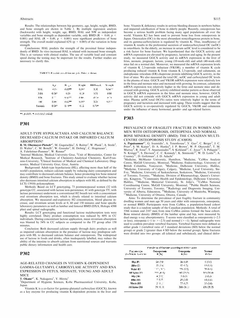
Results: The relationships between hip geometry, age, height, weight, BMD,and bone strength are shown in Table 1. By multiple regression analysis(backwards) with height, weight, age, BMD, HAL and NW as independentvariables and bone strength as dependent variable, only BMD (R = 0.68, p <0.001) and HAL (R = 0.49, p < 0.001) were significant predictors of bonestrength. The overall model explained 62% (p < 0.005) of the variation in bonestrength
Conclusions: HAL predicts the strength of the proximal femur indepen-dently of BMD. In vitro increased HAL is related with increased bone strength.This is at variance with clinical studies. The use of variable load and constantspeed during the testing may be important for the results. Further studies arenecessary to clarify this.
P301
ADULT-TYPE HYPOLACTASIA AND CALCIUM BALANCE:
DECREASED CALCIUM INTAKE OR IMPAIRED CALCIUM
ABSORPTION?B. M. Obermayer-Pietsch*1, M. Gugatschka1, S. Reitter1, W. Plank1, A. Strele2,D. Walter1, C. M. Bonelli1, W. Goessler3, H. Dobnig1, C. Hogenauer1,A. Fahrleitner-Pammer1, W. Renner41Internal Medicine, Medical University Graz, 2Unit of Biostatistics, Center forMedical Research, 3Institute of Chemistry-Analytical Chemistry, Karl-Fran-zens-University, 4Clinical Institute of Medical and Chemical Laboratory Diag-nostics, Medical University Graz, Graz, Austria
Background: Adult-type hypolactasia (HL), affecting more than half of theworld�s population, reduces calcium supply by reducing dairy consumption andmay contribute to decreased calcium balance, hence promoting low bone mineraldensity (BMD) and bone fractures. This study aimed to evaluate whether lactosealso decreases intestinal calcium absorption in subjects with HL as defined by theLCT(-13910) polymorphism.
Methods: Based on LCT genotyping, 73 postmenopausal women (32 withgenotype CC, associated with lactase non-persistence, 41 with genotype TT, withlactase persistence) underwent a conventional H2 breath test with a concomitantoral strontium absorption test, which is closely related to intestinal calciumabsorption. We measured end-expiratory H2 concentration, blood glucose in-crease, and strontium serum levels at 0, 60 and 150 minutes and bone specificlaboratory parameters as well as lumbar and femoral BMD (DXA, Hologic 4500plus) and spinal radiographs.
Results: LCT genotyping and functional lactose malabsorption tests werehighly correlated. Dairy product consumption was reduced by 80% in CCindividuals. During concomitant lactose application, mean strontium absorptionwas blunted by 54% in CC subjects as compared to the TT group after 150minutes.
Conclusion: Both decreased calcium supply through dairy products as wellas impaired calcium absorption in the presence of lactose may predispose sub-jects with HL to decreased calcium balance and osteoporosis. The widespreaduse of lactose in foods and drinks, often inadequately labelled, may reduce theability of the intestine to absorb calcium from nutritional sources and warrantspublic dietary information and health care.
P302
AGE-RELATED CHANGES IN VITAMIN K-DEPENDENT
GAMMA-GLUTAMYL CARBOXYLASE ACTIVITY AND RNA
EXPRESSION IN FETUS, NEONATE, YOUNG AND ADULT
MICET. Okano*1, K. Nakagawa1, Y. Hirota11Department of Hygienic Sciences, Kobe Pharmaceutical University, Kobe,Japan
Vitamin K is a co-factor for gamma-glutamyl carboxylase (GGCX), knownas a key enzyme for producing vitamin K-dependent proteins in the liver and
bone. Vitamin K deficiency results in serious bleeding diseases in newborn babiesand impaired calcification of bone in elderly people. Recently, osteoporosis hasbecome a serious health problem facing many aged populations all over theworld. Vitamin K2 has been used to prevent bone loss from osteoporosis inJapan. Osteocalcin (OC) is the most aboundant noncollagenous protein in bone,and its gamma-carboxylation is regulated by vitamin K. Thus, insufficiency ofvitamin K results in the preferential secretion of undercarboxylated OC (ucOC)in osteoblasts. In the elderly, an increase in serum ucOC level is considered to bea risk factor for bone fracture. We hypothesized that the GGCX activity andmRNA expression are elevated by pregnancy, lactation and aging. In the presentstudy, we measured GGCX activity and its mRNA expression in the liver offetus, neonate, pregnant, lactate, young (10-week-old) and adult (40-week-old)mice fed on a normal diet. Moreover, we measured the mRNA expression levelsof vitamin K 2,3-epoxide reductase (VKOR), a member of vitamin K cycleproducing reduced vitamin K from vitamin K 2,3-epoxide, and calumenin, anendoplasmic reticulum (ER) chaperone protein inhibiting GGCX activity, in theliver of mice. We also measured the total OC, ucOC and carboxylated OC levelsin the plasma of mice. GGCX and VKOR mRNA expression were relatively lowin the fetus and neonate mice and increased with growing. In contrast, calumeninmRNA expression was relatively higher in the fetus and neonate mice and de-creased with growing. GGCX activity exhibited similar pattern as those observedin GGCX mRNA expression in the fetus and neonate mice, however, GGCXactivity did not coincide with GGCX mRNA expression in young and adultmice. Plasma ucOC/total OC(%) ratios were relatively higher in the terms ofpregnancy and lactation and increased with aging. These results suggest that theGGCX activity is co-operatively regulated by GGCX, VKOR and calumeninunder the influence of many hormonal, gender- and age-related factors.
P303
PREVALENCE OF FRAGILITY FRACTURE IN WOMEN AND
MEN WITH OSTEOPOROSIS, OSTEOPENIA AND NORMAL
BONE MINERAL DENSITY (BMD): THE CANADIAN MULTI-
CENTRE OSTEOPOROSIS STUDY (CAMOS)A. Papaioannou*1, G. Ioannidis1, A. Tenenhouse2, Y. Gao3, C. Berger3, J. C.Prior4, S. M. Kaiser5, D. A. Hanley6, J. P. Brown7, W. P. Olszynski8, T. M.Murray9, R. Josse9, T. Anastassiades10, S. Kirkland11, C. Joyce12, S. Poliquin13,N. Kreiger14, S. Davison8, C. C. Kennedy1, K. Siminoski15, W. D. Leslie16, A.Cranney17, D. Goltzman2, J. D. Adachi11Medicine, McMaster University, Hamilton, 2Medicine, 3CaMos AnalysisCentre, McGill University, Montreal, 4Medicine, Endocrinology, University ofBritish Columbia, Vancouver, 5Medicine, Dalhousie University, Halifax,6Medicine, University of Calgary, Calgary, 7Medicine, Laval University, Ste-Foy, 8Medicine, University of Saskatchewan, Saskatoon, 9Medicine, Universityof Toronto, Toronto, 10Medicine, Division of Rheumatology, Queen’s Univer-sity, Kingston, 11Community Health and Epidemiology, Dalhousie University,Halifax, 12Medicine, Memorial University, St. John’s, 13CaMos NationalCoordinating Centre, McGill University, Montreal, 14Public Health Sciences,University of Toronto, Toronto, 15Radiology and Diagnostic Imaging, Uni-versity of Alberta, Edmonton, 16Medicine, University of Manitoba, Winnipeg,17Medicine, University of Ottawa, Ottawa, Canada
Aims: To determine the prevalence of past fragility fracture in communitydwelling women and men age 50 years and older with osteoporosis, osteopenia,or normal BMD. Participants were from CaMos, a population-based cohortstudy that is a random sample of the Canadian population. Methods: A total of5566 women and 2187 men from nine CaMos centres formed the base cohort.Bone mineral density (BMD) of the lumbar spine and hip, were measured bydual energy x-ray absorptiometry. T-scores were classified as osteoporotic ()2.5or less), osteopenic ()1 to >)2.5) and normal (>)1). Spinal radiographs wereused to confirm prevalent vertebral fractures. Vertebral fractures were defined aseither grade 1 (vertebral ratio of 3 standard deviations (SD) below the normalgroup) or grade 2 (greater than 4 SD below the normal group). Spine fractureswere divided into two groups: all (clinical and subclinical), and clinical defor-
Abstracts S115
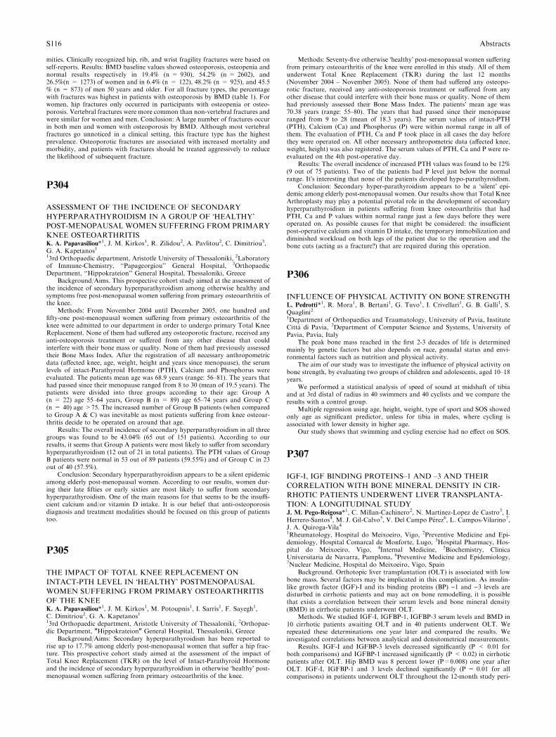
mities. Clinically recognized hip, rib, and wrist fragility fractures were based onself-reports. Results: BMD baseline values showed osteoporosis, osteopenia andnormal results respectively in 19.4% (n = 930), 54.2% (n = 2602), and26.5%(n = 1273) of women and in 6.4% (n = 122), 48.2% (n = 925), and 45.5% (n = 873) of men 50 years and older. For all fracture types, the percentagewith fractures was highest in patients with osteoporosis by BMD (table 1). Forwomen, hip fractures only occurred in participants with osteopenia or osteo-porosis. Vertebral fractures were more common than non-vertebral fractures andwere similar for women and men. Conclusion: A large number of fractures occurin both men and women with osteoporosis by BMD. Although most vertebralfractures go unnoticed in a clinical setting, this fracture type has the highestprevalence. Osteoporotic fractures are associated with increased mortality andmorbidity, and patients with fractures should be treated aggressively to reducethe likelihood of subsequent fracture.
P304
ASSESSMENT OF THE INCIDENCE OF SECONDARY
HYPERPARATHYROIDISM IN A GROUP OF �HEALTHY�POST-MENOPAUSAL WOMEN SUFFERING FROM PRIMARY
KNEE OSTEOARTHRITISK. A. Papavasiliou*1, J. M. Kirkos1, R. Zilidou2, A. Pavlitou2, C. Dimitriou3,G. A. Kapetanos113rd Orthopaedic department, Aristotle University of Thessaloniki, 2Laboratoryof Immune-Chemistry, ‘‘Papageorgiou’’ General Hospital, 3OrthopaedicDepartment, ‘‘Hippokrateion’’ General Hospital, Thessaloniki, Greece
Background/Aims. This prospective cohort study aimed at the assessment ofthe incidence of secondary hyperparathyroidism among otherwise healthy andsymptoms free post-menopausal women suffering from primary osteoarthritis ofthe knee.
Methods: From November 2004 until December 2005, one hundred andfifty-one post-menopausal women suffering from primary osteoarthritis of theknee were admitted to our department in order to undergo primary Total KneeReplacement. None of them had suffered any osteoporotic fracture, received anyanti-osteoporosis treatment or suffered from any other disease that couldinterfere with their bone mass or quality. None of them had previously assessedtheir Bone Mass Index. After the registration of all necessary anthropometricdata (affected knee, age, weight, height and years since menopause), the serumlevels of intact-Parathyroid Hormone (PTH), Calcium and Phosphorus wereevaluated. The patients mean age was 68.9 years (range: 56–81). The years thathad passed since their menopause ranged from 8 to 30 (mean of 19.5 years). Thepatients were divided into three groups according to their age: Group A(n = 22) age 55–64 years, Group B (n = 89) age 65–74 years and Group C(n = 40) age >75. The increased number of Group B patients (when comparedto Group A & C) was inevitable as most patients suffering from knee osteoar-thritis decide to be operated on around that age.
Results: The overall incidence of secondary hyperparathyroidism in all threegroups was found to be 43.04% (65 out of 151 patients). According to ourresults, it seems that Group A patients were most likely to suffer from secondaryhyperparathyroidism (12 out of 21 in total patients). The PTH values of GroupB patients were normal in 53 out of 89 patients (59.55%) and of Group C in 23out of 40 (57.5%).
Conclusion: Secondary hyperparathyroidism appears to be a silent epidemicamong elderly post-menopausal women. According to our results, women dur-ing their late fifties or early sixties are most likely to suffer from secondaryhyperparathyroidism. One of the main reasons for that seems to be the insuffi-cient calcium and/or vitamin D intake. It is our belief that anti-osteoporosisdiagnosis and treatment modalities should be focused on this group of patientstoo.
P305
THE IMPACT OF TOTAL KNEE REPLACEMENT ON
INTACT-PTH LEVEL IN �HEALTHY� POSTMENOPAUSAL
WOMEN SUFFERING FROM PRIMARY OSTEOARTHRITIS
OF THE KNEEK. A. Papavasiliou*1, J. M. Kirkos1, M. Potoupnis1, I. Sarris1, F. Sayegh1,C. Dimitriou2, G. A. Kapetanos113rd Orthopaedic department, Aristotle University of Thessaloniki, 2Orthopae-dic Department, "Hippokrateion" General Hospital, Thessaloniki, Greece
Background/Aims: Secondary hyperparathyroidism has been reported torise up to 17.7% among elderly post-menopausal women that suffer a hip frac-ture. This prospective cohort study aimed at the assessment of the impact ofTotal Knee Replacement (TKR) on the level of Intact-Parathyroid Hormoneand the incidence of secondary hyperparathyroidism in otherwise �healthy� post-menopausal women suffering from primary osteoarthritis of the knee.
Methods: Seventy-five otherwise �healthy� post-menopausal women sufferingfrom primary osteoarthritis of the knee were enrolled in this study. All of themunderwent Total Knee Replacement (TKR) during the last 12 months(November 2004 – November 2005). None of them had suffered any osteopo-rotic fracture, received any anti-osteoporosis treatment or suffered from anyother disease that could interfere with their bone mass or quality. None of themhad previously assessed their Bone Mass Index. The patients� mean age was70.38 years (range: 55–80). The years that had passed since their menopauseranged from 9 to 28 (mean of 18.3 years). The serum values of intact-PTH(PTH), Calcium (Ca) and Phosphorus (P) were within normal range in all ofthem. The evaluation of PTH, Ca and P took place in all cases the day beforethey were operated on. All other necessary anthropometric data (affected knee,weight, height) was also registered. The serum values of PTH, Ca and P were re-evaluated on the 4th post-operative day.
Results: The overall incidence of increased PTH values was found to be 12%(9 out of 75 patients). Two of the patients had P level just below the normalrange. It�s interesting that none of the patients developed hypo-parathyroidism.
Conclusion: Secondary hyper-parathyroidism appears to be a �silent� epi-demic among elderly post-menopausal women. Our results show that Total KneeArthroplasty may play a potential pivotal role in the development of secondaryhyperparathyroidism in patients suffering from knee osteoarthritis that hadPTH, Ca and P values within normal range just a few days before they wereoperated on. As possible causes for that might be considered: the insufficientpost-operative calcium and vitamin D intake, the temporary immobilization anddiminished workload on both legs of the patient due to the operation and thebone cuts (acting as a fracture?) that are required during this operation.
P306
INFLUENCE OF PHYSICAL ACTIVITY ON BONE STRENGTHL. Pedrotti*1, R. Mora1, B. Bertani1, G. Tuvo1, I. Crivellari1, G. B. Galli1, S.Quaglini21Department of Orthopaedics and Traumatology, University of Pavia, InstituteCitta di Pavia, 2Department of Computer Science and Systems, University ofPavia, Pavia, Italy
The peak bone mass reached in the first 2-3 decades of life is determinedmainly by genetic factors but also depends on race, gonadal status and envi-ronmental factors such as nutrition and physical activity.
The aim of our study was to investigate the influence of physical activity onbone strength, by evaluating two groups of children and adolescents, aged 10–18years.
We performed a statistical analysis of speed of sound at midshaft of tibiaand at 3rd distal of radius in 40 swimmers and 40 cyclists and we compare theresults with a control group.
Multiple regression using age, height, weight, type of sport and SOS showedonly age as significant predictor, unless for tibia in males, where cycling isassociated with lower density in higher age.
Our study shows that swimming and cycling exercise had no effect on SOS.
P307
IGF-I, IGF BINDING PROTEINS–1 AND –3 AND THEIR
CORRELATION WITH BONE MINERAL DENSITY IN CIR-
RHOTIC PATIENTS UNDERWENT LIVER TRANSPLANTA-
TION: A LONGITUDINAL STUDYJ. M. Pego-Reigosa*1, C. Millan-Cachinero2, N. Martinez-Lopez de Castro3, I.Herrero-Santos4, M. J. Gil-Calvo5, V. Del Campo Perez6, L. Campos-Vilarino7,J. A. Quiroga-Vila41Rheumatology, Hospital do Meixoeiro, Vigo, 2Preventive Medicine and Epi-demiology, Hospital Comarcal de Monforte, Lugo, 3Hospital Pharmacy, Hos-pital do Meixoeiro, Vigo, 4Internal Medicine, 5Biochemistry, ClinicaUniversitaria de Navarra, Pamplona, 6Preventive Medicine and Epidemiology,7Nuclear Medicine, Hospital do Meixoeiro, Vigo, Spain
Background. Orthotopic liver transplantation (OLT) is associated with lowbone mass. Several factors may be implicated in this complication. As insulin-like growth factor (IGF)-I and its binding proteins (BP) )1 and )3 levels aredisturbed in cirrhotic patients and may act on bone remodelling, it is possiblethat exists a correlation between their serum levels and bone mineral density(BMD) in cirrhotic patients underwent OLT.
Methods. We studied IGF-I, IGFBP-1, IGFBP-3 serum levels and BMD in10 cirrhotic patients awaiting OLT and in 40 patients underwent OLT. Werepeated these determinations one year later and compared the results. Weinvestigated correlations between analytical and densitometrical measurements.
Results. IGF-I and IGFBP-3 levels decreased significantly (P < 0.01 forboth comparisons) and IGFBP-1 increased significantly (P < 0.02) in cirrhoticpatients after OLT. Hip BMD was 8 percent lower (P=0.008) one year afterOLT. IGF-I, IGFBP-1 and 3 levels declined significantly (P = 0.01 for allcomparisons) in patients underwent OLT throughout the 12-month study peri-
S116 Abstracts
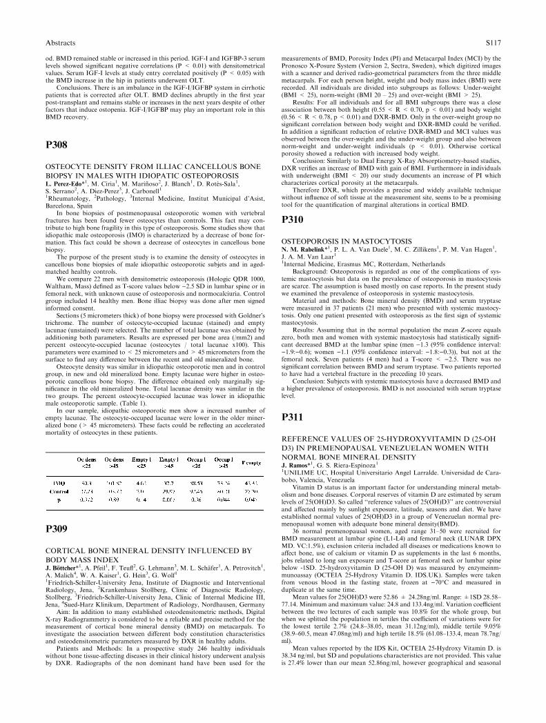
od. BMD remained stable or increased in this period. IGF-I and IGFBP-3 serumlevels showed significant negative correlations (P < 0.01) with densitometricalvalues. Serum IGF-I levels at study entry correlated positively (P < 0.05) withthe BMD increase in the hip in patients underwent OLT.
Conclusions. There is an imbalance in the IGF-I/IGFBP system in cirrhoticpatients that is corrected after OLT. BMD declines abruptly in the first yearpost-transplant and remains stable or increases in the next years despite of otherfactors that induce ostopenia. IGF-I/IGFBP may play an important role in thisBMD recovery.
P308
OSTEOCYTE DENSITY FROM ILLIAC CANCELLOUS BONE
BIOPSY IN MALES WITH IDIOPATIC OSTEOPOROSISL. Perez-Edo*1, M. Ciria1, M. Marinoso2, J. Blanch1, D. Rotes-Sala1,S. Serrano2, A. Diez-Perez3, J. Carbonell11Rheumatology, 2Pathology, 3Internal Medicine, Institut Municipal d’Asist,Barcelona, Spain
In bone biopsies of postmenopausal osteoporotic women with vertebralfractures has been found fewer osteocytes than controls. This fact may con-tribute to high bone fragility in this type of osteoporosis. Some studies show thatidiopathic male osteoporosis (IMO) is characterized by a decrease of bone for-mation. This fact could be shown a decrease of osteocytes in cancellous bonebiopsy.
The purpose of the present study is to examine the density of osteocytes incancellous bone biopsies of male idiopathic osteoporotic subjets and in aged-matched healthy controls.
We compare 22 men with densitometric osteoporosis (Hologic QDR 1000,Waltham, Mass) defined as T-score values below )2.5 SD in lumbar spine or infemoral neck, with unknown cause of osteoporosis and normocalciuria. Controlgroup included 14 healthy men. Bone illiac biopsy was done after men signedinformed consent.
Sections (5 micrometers thick) of bone biopsy were processed with Goldner�strichrome. The number of osteocyte-occupied lacunae (stained) and emptylacunae (unstained) were selected. The number of total lacunae was obtained byadditioning both parameters. Results are expressed per bone area (/mm2) andpercent osteocyte-occupied lacunae (osteocytes / total lacunae x100). Thisparameters were examined to < 25 micrometers and > 45 micrometers from thesurface to find any difference between the recent and old mineralized bone.
Osteocyte density was similar in idiopathic osteoporotic men and in controlgroup, in new and old mineralized bone. Empty lacunae were higher in osteo-porotic cancellous bone biopsy. The difference obtained only marginally sig-nificance in the old mineralized bone. Total lacunae density was similar in thetwo groups. The percent osteocyte-occupied lacunae was lower in idiopathicmale osteoporotic sample. (Table 1).
In our sample, idiopathic osteoporotic men show a increased number ofempty lacunae. The osteocyte-occuped lacunae were lower in the older miner-alized bone (> 45 micrometers). These facts could be reflecting an acceleratedmortality of osteocytes in these patients.
P309
CORTICAL BONE MINERAL DENSITY INFLUENCED BY
BODY MASS INDEXJ. Bottcher*1, A. Pfeil1, F. Teufl2, G. Lehmann3, M. L. Schafer1, A. Petrovitch1,A. Malich4, W. A. Kaiser1, G. Hein3, G. Wolf11Friedrich-Schiller-University Jena, Institute of Diagnostic and InterventionalRadiology, Jena, 2Krankenhaus Stollberg, Clinic of Diagnostic Radiology,Stollberg, 3Friedrich-Schiller-University Jena, Clinic of Internal Medicine III,Jena, 4Sued-Harz Klinikum, Department of Radiology, Nordhausen, Germany
Aim: In addition to many established osteodensitometric methods, DigitalX-ray Radiogrammetry is considered to be a reliable and precise method for themeasurement of cortical bone mineral density (BMD) on metacarpals. Toinvestigate the association between different body constitution characteristicsand osteodensitometric parameters measured by DXR in healthy adults.
Patients and Methods: In a prospective study 246 healthy individualswithout bone tissue-affecting diseases in their clinical history underwent analysisby DXR. Radiographs of the non dominant hand have been used for the
measurements of BMD, Porosity Index (PI) and Metacarpal Index (MCI) by thePronosco X-Posure System (Version 2, Sectra, Sweden), which digitized imageswith a scanner and derived radio-geometrical parameters from the three middlemetacarpals. For each person height, weight and body mass index (BMI) wererecorded. All individuals are divided into subgroups as follows: Under-weight(BMI < 25), norm-weight (BMI 20 – 25) and over-weight (BMI > 25).
Results: For all individuals and for all BMI subgroups there was a closeassociation between both height (0.55 < R < 0.70, p < 0.01) and body weight(0.56 < R < 0.78, p < 0.01) and DXR-BMD. Only in the over-weight group nosignificant correlation between body weight and DXR-BMD could be verified.In addition a significant reduction of relative DXR-BMD and MCI values wasobserved between the over-weight and the under-weight group and also betweennorm-weight and under-weight individuals (p < 0.01). Otherwise corticalporosity showed a reduction with increased body weight.
Conclusion: Similarly to Dual Energy X-Ray Absorptiometry-based studies,DXR verifies an increase of BMD with gain of BMI. Furthermore in individualswith underweight (BMI < 20) our study documents an increase of PI whichcharacterizes cortical porosity at the metacarpals.
Therefore DXR, which provides a precise and widely available techniquewithout influence of soft tissue at the measurement site, seems to be a promisingtool for the quantification of marginal alterations in cortical BMD.
P310
OSTEOPOROSIS IN MASTOCYTOSISN. M. Rabelink*1, P. L. A. Van Daele1, M. C. Zillikens1, P. M. Van Hagen1,J. A. M. Van Laar11Internal Medicine, Erasmus MC, Rotterdam, Netherlands
Background: Osteoporosis is regarded as one of the complications of sys-temic mastocytosis but data on the prevalence of osteoporosis in mastocytosisare scarce. The assumption is based mostly on case reports. In the present studywe examined the prevalence of osteoporosis in systemic mastocytosis.
Material and methods: Bone mineral density (BMD) and serum tryptasewere measured in 37 patients (21 men) who presented with systemic mastocy-tosis. Only one patient presented with osteoporosis as the first sign of systemicmastocytosis.
Results: Assuming that in the normal population the mean Z-score equalszero, both men and women with systemic mastocytosis had statistically signifi-cant decreased BMD at the lumbar spine (men )1.3 (95% confidence interval:)1.9:)0.6); women )1.1 (95% confidence interval: )1.8:)0.3)), but not at thefemoral neck. Seven patients (4 men) had a T-score < )2.5. There was nosignificant correlation between BMD and serum tryptase. Two patients reportedto have had a vertebral fracture in the preceding 10 years.
Conclusion: Subjects with systemic mastocytosis have a decreased BMD anda higher prevalence of osteoporosis. BMD is not associated with serum tryptaselevel.
P311
REFERENCE VALUES OF 25-HYDROXYVITAMIN D (25-OH
D3) IN PREMENOPAUSAL VENEZUELAN WOMEN WITH
NORMAL BONE MINERAL DENSITYJ. Ramos*1, G. S. Riera-Espinoza11UNILIME UC, Hospital Universitario Angel Larralde. Universidad de Cara-bobo, Valencia, Venezuela
Vitamin D status is an important factor for understanding mineral metab-olism and bone diseases. Corporal reserves of vitamin D are estimated by serumlevels of 25(OH)D3. So called ‘‘reference values of 25(OH)D3’’ are controversialand affected mainly by sunlight exposure, latitude, seasons and diet. We haveestablished normal values of 25(OH)D3 in a group of Venezuelan normal pre-menopausal women with adequate bone mineral density(BMD).
36 normal premenopausal women, aged range 31–50 were recruited forBMD measurement at lumbar spine (L1-L4) and femoral neck (LUNAR DPXMD. VC:1.5%), exclusion criteria included all diseases or medications known toaffect bone, use of calcium or vitamin D as supplements in the last 6 months,jobs related to long sun exposure and T-score at femoral neck or lumbar spinebelow -1SD. 25-hydroxyvitamin D (25-OH D) was measured by enzymeinm-munoassay (OCTEIA 25-Hydroxy Vitamin D. IDS.UK). Samples were takenfrom venous blood in the fasting state, frozen at )70�C and measured induplicate at the same time.
Mean values for 25(OH)D3 were 52.86 ± 24.28ng/ml. Range: ±1SD 28.58–77.14. Minimum and maximum value: 24.8 and 133.4ng/ml. Variation coefficientbetween the two lectures of each sample was 10.8% for the whole group, butwhen we splitted the population in tertiles the coefficient of variations were forthe lowest tertile 2.7% (24.8–38.05, mean 31.12ng/ml), middle tertile 9.05%(38.9–60.5, mean 47.08ng/ml) and high tertile 18.5% (61.08–133.4, mean 78.7ng/ml).
Mean values reported by the IDS Kit, OCTEIA 25-Hydroxy Vitamin D. is38.34 ng/ml, but SD and populations characteristics are not provided. This valueis 27.4% lower than our mean 52.86ng/ml, however geographical and seasonal
Abstracts S117

variations have been widely reported. Venezuela is a tropical country with plentyof sunshine during almost the whole year.
In conclusion, reference values of 25-hydroxyvitamin D (25-OH D3) inpremenopausal Venezuelan women with normal bone mineral density measuredby enzymeinmmunoassay (OCTEIA 25-Hydroxy Vitamin D. IDS.UK) are:mean 52.86 ± 24.28ng/ml. Range: mean ± 1SD 28.58–77.14. Minimum andmaximum value: 24.8 and 133.4ng/ml
P312
NO EFFECT OF VITAMIN K1 INTAKE ON BONE MINERAL
DENSITY AND FRACTURE RISK IN PERIMENOPAUSAL
WOMENL. Rejnmark*1, P. Vestergaard1, P. Charles2, P. Hermann3, C. Brot4, P. Eiken5,L. Mosekilde21The Osteoporosis Clinic, 2Dept. of Endocrinology and Metabolism C, Uni-versity Hospital of Aarhus, Aarhus, 32 Dept. of Endocrinology, Odense Uni-versity Hospital, Odense, 4The Osteoporosis Research Centre, HvidovreHospital, Hvidovre, 5Dept. of Endocrinology and Clinical Physiology and Nu-clear Medicine, Hilleroed Hospital, Hilleroed, Denmark
Background: Vitamin K functions as a cofactor in the posttranslationalcarboxylation of several bone proteins including osteocalcin. In recent studiesfrom the USA, a high vitamin K1 intake has been associated an increased bonemineral density (BMD) and a decreased fracture risk. However, in the popula-tions previously studied, the averages vitamin K1 intake estimated by the foodfrequency method was approximately three times the recommended daily intake.
Aim: We studied relations between vitamin K1 intake and BMD and frac-ture risk in a Danish population.
Methods: Within the Danish Osteoporosis Prevention Study (DOPS),including a population-based cohort of 2016 perimenopausal women, associa-tions between vitamin K1 intake and BMD was assessed at baseline and after 5-years of follow-up (cross sectional design). Moreover, associations betweenvitamin K1 intake and 5- and 10-years changes in BMD were studied (follow updesign). Finally, fracture risk was assessed in relation to vitamin K1 intake(Nested case control design).
Results: In our cohort, dietary vitamin K1 intake (0.6 lg/day) was close tothe daily intake recommended by the FAO, but substantively lower than theintake reported in previously studied populations. Cross-sectional and longitu-dinal analyses showed no associations between intake of vitamin K1 and BMDof the femoral neck or lumbar spine. Neither did BMD differ between those 5%who had the highest- and those 5% who had the lowest vitamin K1 intake.During the 10-years of follow-up, 360 subjects sustained a fracture (cases).Compared to 1440 controls, logistic regression analyses revealed no difference invitamin K1 intake between cases and controls.
Conclusion: In a Danish population, with an average vitamin K1 intakeclose to the recommended daily intake, vitamin K1 intake is not associated witheffects on BMD or fracture risk.
P313
OSTEOPOROSIS RISK FACTORS IN PATIENTS WITH
ESTABLISHED AND SEVERE OSTEOPOROSIS; A MATCHED
PAIRS ANALYSIS FROM THE OSSO SPANISH COHORTM. Rentero*1, E. Martin Mola2, P. Aguado2, A. Oteo3, J. Zubieta41Research Center, Lilly, 2S. Reumatologia, H. La Paz, 3S. Traumatologia,H. Moratalaz, Madrid, 4S. Reumatologia, H. Toledo, Toledo, Spain
The Observational Study on Severe Osteoporosis (OSSO) is a 12-monthprospective, European study with a special cohort in Spain.
Objetive: A follow-up of for 12 months the patients with the severe form anda established control group to describe changes in HRQoL and detect differencesbetween groups.
Method: Women with established osteoporosis were recruited from 64Spanish sites. Demographic data and osteoporosis risk factors were collected.Increased risks for fractures were defined: a) history of 2 or more fragilityfractures and/or b) inadequate osteoporosis drugs response. 104 pairs were ob-tained; one, with 1 or more conditions associated with increased fracture riskand a control group without any said conditions. Matching variables were age,years since menopause, and years on antiresortive treatment. Matched groupswere compared for osteoporosis risk factors and quality of life and followed-upfor 12 months
Results: More than 90% of patients were taking one or more OP medicationsat follow-up for OP The majority were taking calcium and Vit D. Alendronate,Risedronate and Raloxifene were most commonly used antiresorptive therapiesin both cohorts (54.6%, 23.5 and 18.8% respectively). 63.1% in established cohortwas taking one or more medications related to the risk of OP and falls at baselineand 54.7% in the severe cohort. Antihypertensives were most commonly in bothcohorts 54%.Antidepressants and benzodiazepines were the next in the estab-
lished cohort 32.3% and 26.2%. Severe cohort benzodiazepines were taking in48.1% and antidepressants in 21.2%.
There no statistically significant difference in fracture status between thematched pairs. Arthritis rheumatoid was the most frequently reported followedby chronic pulmonary obstructive disease and urolithiasis (28.5%, 21.1% and17.1% respectively). EuroQoL self-care domain was the only statistically sig-nificant difference.
Qualeffo: Established group had statistically significant difference betterQoL outcome in pain (10.3 p = 0.01), physical function (8.3 p < 0.01) ,totalQualeffo score (6.9 p = 0.01) and general health perception domains (9.71 p <0.01).
Conclusions: In spite of the majority are with treatment of OP. The com-orbility is very important in theses groups and they are taking several drugsrelated to the risk of OP and falls that increase the risk of OP. The quality of lifeis better in the established group. These aspects could have influence in the costsand the resources utilisation.
P314
THE INFLUENCE OF KLOTHO GENE ON FEMALE SKELE-
TAL AGINGJ. A. Riancho*1, C. Valero1, J. L. Hernandez1, N. Pena2, A. L. Zarrabeitia1, C.Viadero2, F. Ortiz1, J. Gonzalez-Macias1, M. T. Zarrabeitia31Internal Medicine, Hospital UM Valdecilla, University Cantabria, 2Geriatrics,Centro Mayores Cueto, 3Legal Medicine, University Cantabria, Santander,Spain
Klotho gene codes for a protein with glucuronidase activity and it is thoughtto be related to bone and vascular homeostasis. In fact, klotho knock-out micehave an accelerated aging phenotype, with abnormal bones, atherosclerosis, andectopic calcifications. The objective of this study was to analyze the relationshipof a common T/G polymorphism of the klotho gene, resulting in a phenylalanineto valine substitution at aminoacid position 352, with bone mineral density(BMD) and osteoporotic fractures.
The study group comprised 861women, including control subjects, patientswith osteoporotic vertebral fractures and patients with hip fractures. BMD wasmeasured by DEXA. DNA was isolated from peripheral blood and klotho alleleswere characterised by PCR with TaqMan probes.The T allele was more frequentthan the G allele (86% and 14%, respectively). In comparison with the mostcommon TT genotype, the TG genotype was associated with higher hip BMD inpostmenopausal women (femoral neck: 0.644 ± 0.006 vs 0.675 ± 0.012 g/cm2;p = 0.02; total hip:0.774 ± 0.008 vs 0.809 ± 0.014 g/cm2; p = 0.03). Womenwith different klotho genotypes did not differ regarding body weight and height,BMI, calcium intake, age at menarche, age at menopause or serum estradiollevels. Klotho alleles explained about 1.5% of BMD variance, but were notassociated to the risk of osteoporotic spine or hip fractures. By contrast topostmenopausal women, in premenopausal women there were no klotho-relateddifferences in BMD.
In conclusion, the F352V klotho polymorphism seems to influence skeletalaging in women and is associated to hip BMD. However, its effect is quantita-tively small and it does not appear to be related to fracture risk.
Supported by grants from FPMV-IFIMAV and FIS 02-0063 and 04-0028.
P315
AGE, BUT NOT SEX, INFLUENCE AROMATASE GENE
EXPRESSION BY HUMAN BONE CELLSJ. A. Riancho*1, C. Sanudo1, V. Mijares1, M. Perez-Nunez2, M. A.Alonso21Internal Medicine, 2Traumatology, Hospital UM Valdecilla, UniversityCantabria, Santander, Spain
In men and in postmenopausal women most estrogens derive from non-gonadal sources, where androgenic precursors are aromatised into estrogens. Anumber of tissues, including fat and bone, have been shown to express thearomatase gene. It may be important in modulating estrogen levels both at thesystemic and the local levels, which, in turn, play a critical role in bonehomeostasis. The objective of this study was to determine the influence of sexand age on the expression of aromatase gene by human osteoblasts.Osteoblastswere grown from explants of femoral neck fragments obtained from patientssuffering a hip fracture (17 women, 6 men). Age range was 58-93 yr (median 75 inboth sexes). After confluency was attained, the cells were plated in the presenceor the absence of serum for 48 hours. Total RNA was extracted and theexpression of aromatase was determined by real time-PCR after cDNA syn-thesis, using TBP as an endogenous control gene. The results were expressed asarbitary units (AU) in comparison with a commercially-available standard RNAisolated from several human cell lines (Stratagene).
The expression of aromatase by cultured osteoblasts showed wide interin-dividual variation and was much higher in the absence of serum. There were nosex-related differences (41.9 ± 65.5 vs 39.3 ± 67.0 AU, in women and men,respectively, p > 0.8). However, in serum-free cultures aromatase RNA levels
S118 Abstracts
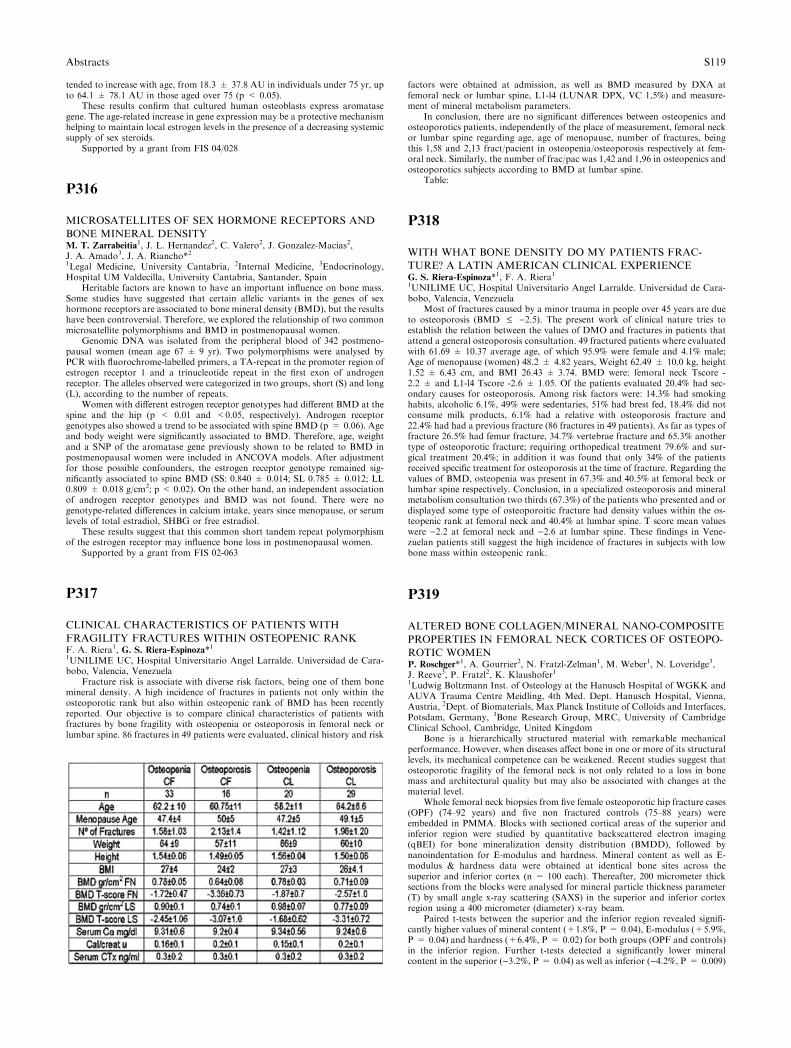
tended to increase with age, from 18.3 ± 37.8 AU in individuals under 75 yr, upto 64.1 ± 78.1 AU in those aged over 75 (p < 0.05).
These results confirm that cultured human osteoblasts express aromatasegene. The age-related increase in gene expression may be a protective mechanismhelping to maintain local estrogen levels in the presence of a decreasing systemicsupply of sex steroids.
Supported by a grant from FIS 04/028
P316
MICROSATELLITES OF SEX HORMONE RECEPTORS AND
BONE MINERAL DENSITYM. T. Zarrabeitia
1, J. L. Hernandez2, C. Valero2, J. Gonzalez-Macias2,J. A. Amado3, J. A. Riancho*21Legal Medicine, University Cantabria, 2Internal Medicine, 3Endocrinology,Hospital UM Valdecilla, University Cantabria, Santander, Spain
Heritable factors are known to have an important influence on bone mass.Some studies have suggested that certain allelic variants in the genes of sexhormone receptors are associated to bone mineral density (BMD), but the resultshave been controversial. Therefore, we explored the relationship of two commonmicrosatellite polymorphisms and BMD in postmenopausal women.
Genomic DNA was isolated from the peripheral blood of 342 postmeno-pausal women (mean age 67 ± 9 yr). Two polymorphisms were analysed byPCR with fluorochrome-labelled primers, a TA-repeat in the promoter region ofestrogen receptor 1 and a trinucleotide repeat in the first exon of androgenreceptor. The alleles observed were categorized in two groups, short (S) and long(L), according to the number of repeats.
Women with different estrogen receptor genotypes had different BMD at thespine and the hip (p < 0.01 and <0.05, respectively). Androgen receptorgenotypes also showed a trend to be associated with spine BMD (p = 0.06). Ageand body weight were significantly associated to BMD. Therefore, age, weightand a SNP of the aromatase gene previously shown to be related to BMD inpostmenopausal women were included in ANCOVA models. After adjustmentfor those possible confounders, the estrogen receptor genotype remained sig-nificantly associated to spine BMD (SS: 0.840 ± 0.014; SL 0.785 ± 0.012; LL0.809 ± 0.018 g/cm2; p < 0.02). On the other hand, an independent associationof androgen receptor genotypes and BMD was not found. There were nogenotype-related differences in calcium intake, years since menopause, or serumlevels of total estradiol, SHBG or free estradiol.
These results suggest that this common short tandem repeat polymorphismof the estrogen receptor may influence bone loss in postmenopausal women.
Supported by a grant from FIS 02-063
P317
CLINICAL CHARACTERISTICS OF PATIENTS WITH
FRAGILITY FRACTURES WITHIN OSTEOPENIC RANKF. A. Riera1, G. S. Riera-Espinoza*11UNILIME UC, Hospital Universitario Angel Larralde. Universidad de Cara-bobo, Valencia, Venezuela
Fracture risk is associate with diverse risk factors, being one of them bonemineral density. A high incidence of fractures in patients not only within theosteoporotic rank but also within osteopenic rank of BMD has been recentlyreported. Our objective is to compare clinical characteristics of patients withfractures by bone fragility with osteopenia or osteoporosis in femoral neck orlumbar spine. 86 fractures in 49 patients were evaluated, clinical history and risk
factors were obtained at admission, as well as BMD measured by DXA atfemoral neck or lumbar spine, L1-l4 (LUNAR DPX, VC 1,5%) and measure-ment of mineral metabolism parameters.
In conclusion, there are no significant differences between osteopenics andosteoporotics patients, independently of the place of measurement, femoral neckor lumbar spine regarding age, age of menopause, number of fractures, beingthis 1,58 and 2,13 fract/pacient in osteopenia/osteoporosis respectively at fem-oral neck. Similarly, the number of frac/pac was 1,42 and 1,96 in osteopenics andosteoporotics subjects according to BMD at lumbar spine.
Table:
P318
WITH WHAT BONE DENSITY DO MY PATIENTS FRAC-
TURE? A LATIN AMERICAN CLINICAL EXPERIENCEG. S. Riera-Espinoza*1, F. A. Riera11UNILIME UC, Hospital Universitario Angel Larralde. Universidad de Cara-bobo, Valencia, Venezuela
Most of fractures caused by a minor trauma in people over 45 years are dueto osteoporosis (BMD £ )2.5). The present work of clinical nature tries toestablish the relation between the values of DMO and fractures in patients thatattend a general osteoporosis consultation. 49 fractured patients where evaluatedwith 61.69 ± 10.37 average age, of which 95.9% were female and 4.1% male;Age of menopause (women) 48.2 ± 4.82 years, Weight 62.49 ± 10.0 kg, height1.52 ± 6.43 cm, and BMI 26.43 ± 3.74. BMD were: femoral neck Tscore -2.2 ± and L1-l4 Tscore -2.6 ± 1.05. Of the patients evaluated 20.4% had sec-ondary causes for osteoporosis. Among risk factors were: 14.3% had smokinghabits, alcoholic 6.1%, 49% were sedentaries, 51% had brest fed, 18.4% did notconsume milk products, 6.1% had a relative with osteoporosis fracture and22.4% had had a previous fracture (86 fractures in 49 patients). As far as types offracture 26.5% had femur fracture, 34.7% vertebrae fracture and 65.3% anothertype of osteoporotic fracture; requiring orthopedical treatment 79.6% and sur-gical treatment 20.4%; in addition it was found that only 34% of the patientsreceived specific treatment for osteoporosis at the time of fracture. Regarding thevalues of BMD, osteopenia was present in 67.3% and 40.5% at femoral beck orlumbar spine respectively. Conclusion, in a specialized osteoporosis and mineralmetabolism consultation two thirds (67.3%) of the patients who presented and ordisplayed some type of osteoporoitic fracture had density values within the os-teopenic rank at femoral neck and 40.4% at lumbar spine. T score mean valueswere )2.2 at femoral neck and )2.6 at lumbar spine. These findings in Vene-zuelan patients still suggest the high incidence of fractures in subjects with lowbone mass within osteopenic rank.
P319
ALTERED BONE COLLAGEN/MINERAL NANO-COMPOSITE
PROPERTIES IN FEMORAL NECK CORTICES OF OSTEOPO-
ROTIC WOMENP. Roschger*1, A. Gourrier2, N. Fratzl-Zelman1, M. Weber1, N. Loveridge3,J. Reeve3, P. Fratzl2, K. Klaushofer11Ludwig Boltzmann Inst. of Osteology at the Hanusch Hospital of WGKK andAUVA Trauma Centre Meidling, 4th Med. Dept. Hanusch Hospital, Vienna,Austria, 2Dept. of Biomaterials, Max Planck Institute of Colloids and Interfaces,Potsdam, Germany, 3Bone Research Group, MRC, University of CambridgeClinical School, Cambridge, United Kingdom
Bone is a hierarchically structured material with remarkable mechanicalperformance. However, when diseases affect bone in one or more of its structurallevels, its mechanical competence can be weakened. Recent studies suggest thatosteoporotic fragility of the femoral neck is not only related to a loss in bonemass and architectural quality but may also be associated with changes at thematerial level.
Whole femoral neck biopsies from five female osteoporotic hip fracture cases(OPF) (74–92 years) and five non fractured controls (75–88 years) wereembedded in PMMA. Blocks with sectioned cortical areas of the superior andinferior region were studied by quantitative backscattered electron imaging(qBEI) for bone mineralization density distribution (BMDD), followed bynanoindentation for E-modulus and hardness. Mineral content as well as E-modulus & hardness data were obtained at identical bone sites across thesuperior and inferior cortex (n = 100 each). Thereafter, 200 micrometer thicksections from the blocks were analysed for mineral particle thickness parameter(T) by small angle x-ray scattering (SAXS) in the superior and inferior cortexregion using a 400 micrometer (diameter) x-ray beam.
Paired t-tests between the superior and the inferior region revealed signifi-cantly higher values of mineral content (+1.8%, P = 0.04), E-modulus (+5.9%,P = 0.04) and hardness (+6.4%, P = 0.02) for both groups (OPF and controls)in the inferior region. Further t-tests detected a significantly lower mineralcontent in the superior ()3.2%, P = 0.04) as well as inferior ()4.2%, P = 0.009)
Abstracts S119
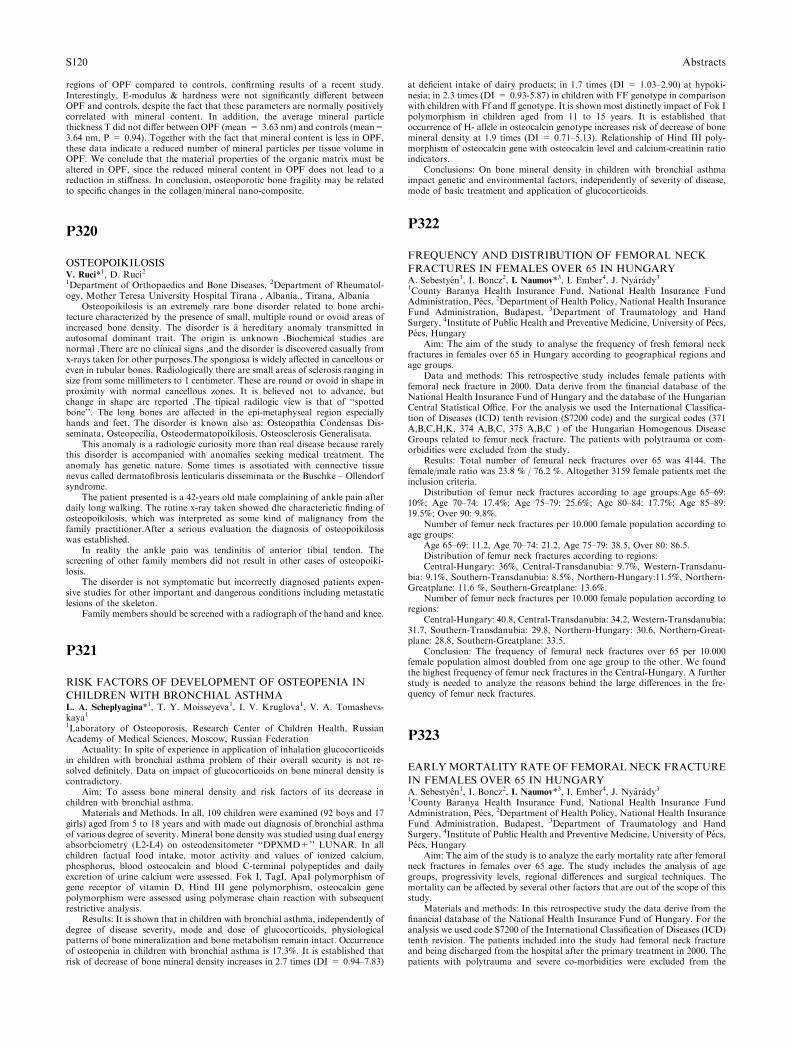
regions of OPF compared to controls, confirming results of a recent study.Interestingly, E-modulus & hardness were not significantly different betweenOPF and controls, despite the fact that these parameters are normally positivelycorrelated with mineral content. In addition, the average mineral particlethickness T did not differ between OPF (mean = 3.63 nm) and controls (mean=3.64 nm, P = 0.94). Together with the fact that mineral content is less in OPF,these data indicate a reduced number of mineral particles per tissue volume inOPF. We conclude that the material properties of the organic matrix must bealtered in OPF, since the reduced mineral content in OPF does not lead to areduction in stiffness. In conclusion, osteoporotic bone fragility may be relatedto specific changes in the collagen/mineral nano-composite.
P320
OSTEOPOIKILOSISV. Ruci*1, D. Ruci21Department of Orthopaedics and Bone Diseases, 2Department of Rheumatol-ogy, Mother Teresa University Hospital Tirana , Albania., Tirana, Albania
Osteopoikilosis is an extremely rare bone disorder related to bone archi-tecture characterized by the presence of small, multiple round or ovoid areas ofincreased bone density. The disorder is a hereditary anomaly transmitted inautosomal dominant trait. The origin is unknown .Biochemical studies arenormal .There are no clinical signs ,and the disorder is discovered casually fromx-rays taken for other purposes.The spongiosa is widely affected in cancellous oreven in tubular bones. Radiologically there are small areas of sclerosis ranging insize from some millimeters to 1 centimeter. These are round or ovoid in shape inproximity with normal cancellous zones. It is believed not to advance, butchange in shape are reported .The tipical radilogic view is that of ‘‘spottedbone’’. The long bones are affected in the epi-metaphyseal region especiallyhands and feet. The disorder is known also as: Osteopathia Condensas Dis-seminata, Osteopecilia, Osteodermatopoikilosis, Osteosclerosis Generalisata.
This anomaly is a radiologic curiosity more than real disease because rarelythis disorder is accompanied with anomalies seeking medical treatment. Theanomaly has genetic nature. Some times is assotiated with connective tissuenevus called dermatofibrosis lenticularis disseminata or the Buschke – Ollendorfsyndrome.
The patient presented is a 42-years old male complaining of ankle pain afterdaily long walking. The rutine x-ray taken showed dhe characterictic finding ofosteopoikilosis, which was interpreted as some kind of malignancy from thefamily practitioner.After a serious evaluation the diagnosis of osteopoikilosiswas established.
In reality the ankle pain was tendinitis of anterior tibial tendon. Thescreening of other family members did not result in other cases of osteopoiki-losis.
The disorder is not symptomatic but incorrectly diagnosed patients expen-sive studies for other important and dangerous conditions including metastaticlesions of the skeleton.
Family members should be screened with a radiograph of the hand and knee.
P321
RISK FACTORS OF DEVELOPMENT OF OSTEOPENIA IN
CHILDREN WITH BRONCHIAL ASTHMAL. A. Scheplyagina*1, T. Y. Moisseyeva1, I. V. Kruglova1, V. A. Tomashevs-kaya11Laboratory of Osteoporosis, Research Center of Children Health, RussianAcademy of Medical Sciences, Moscow, Russian Federation
Actuality: In spite of experience in application of inhalation glucocorticoidsin children with bronchial asthma problem of their overall security is not re-solved definitely. Data on impact of glucocorticoids on bone mineral density iscontradictory.
Aim: To assess bone mineral density and risk factors of its decrease inchildren with bronchial asthma.
Materials and Methods. In all, 109 children were examined (92 boys and 17girls) aged from 5 to 18 years and with made out diagnosis of bronchial asthmaof various degree of severity. Mineral bone density was studied using dual energyabsorbciometry (L2-L4) on osteodensitometer ‘‘DPXMD+’’ LUNAR. In allchildren factual food intake, motor activity and values of ionized calcium,phosphorus, blood osteocalcin and blood C-terminal polypeptides and dailyexcretion of urine calcium were assessed. Fok I, TagI, ApaI polymorphism ofgene receptor of vitamin D, Hind III gene polymorphism, osteocalcin genepolymorphism were assessed using polymerase chain reaction with subsequentrestrictive analysis.
Results: It is shown that in children with bronchial asthma, independently ofdegree of disease severity, mode and dose of glucocorticoids, physiologicalpatterns of bone mineralization and bone metabolism remain intact. Occurrenceof osteopenia in children with bronchial asthma is 17.3%. It is established thatrisk of decrease of bone mineral density increases in 2.7 times (DI = 0.94–7.83)
at deficient intake of dairy products; in 1.7 times (DI = 1.03–2.90) at hypoki-nesia; in 2.3 times (DI = 0.93-5.87) in children with FF genotype in comparisonwith children with Ff and ff genotype. It is shown most distinctly impact of Fok Ipolymorphism in children aged from 11 to 15 years. It is established thatoccurrence of H- allele in osteocalcin genotype increases risk of decrease of bonemineral density at 1.9 times (DI = 0.71–5.13). Relationship of Hind III poly-morphism of osteocalcin gene with osteocalcin level and calcium-creatinin ratioindicators.
Conclusions: On bone mineral density in children with bronchial asthmaimpact genetic and environmental factors, independently of severity of disease,mode of basic treatment and application of glucocorticoids.
P322
FREQUENCY AND DISTRIBUTION OF FEMORAL NECK
FRACTURES IN FEMALES OVER 65 IN HUNGARYA. Sebestyen1, I. Boncz2, I. Naumov*3, I. Ember4, J. Nyarady31County Baranya Health Insurance Fund, National Health Insurance FundAdministration, Pecs, 2Department of Health Policy, National Health InsuranceFund Administration, Budapest, 3Department of Traumatology and HandSurgery, 4Institute of Public Health and Preventive Medicine, University of Pecs,Pecs, Hungary
Aim: The aim of the study to analyse the frequency of fresh femoral neckfractures in females over 65 in Hungary according to geographical regions andage groups.
Data and methods: This retrospective study includes female patients withfemoral neck fracture in 2000. Data derive from the financial database of theNational Health Insurance Fund of Hungary and the database of the HungarianCentral Statistical Office. For the analysis we used the International Classifica-tion of Diseases (ICD) tenth revision (S7200 code) and the surgical codes (371A,B,C,H,K, 374 A,B,C, 375 A,B,C ) of the Hungarian Homogenous DiseaseGroups related to femur neck fracture. The patients with polytrauma or com-orbidities were excluded from the study.
Results: Total number of femural neck fractures over 65 was 4144. Thefemale/male ratio was 23.8 % / 76.2 %. Altogether 3159 female patients met theinclusion criteria.
Distribution of femur neck fractures according to age groups:Age 65–69:10%; Age 70–74: 17.4%; Age 75–79: 25.6%; Age 80–84: 17.7%; Age 85–89:19.5%; Over 90: 9.8%.
Number of femur neck fractures per 10.000 female population according toage groups:
Age 65–69: 11.2, Age 70–74: 21.2, Age 75–79: 38.5, Over 80: 86.5.Distribution of femur neck fractures according to regions:Central-Hungary: 36%, Central-Transdanubia: 9.7%, Western-Transdanu-
bia: 9.1%, Southern-Transdanubia: 8.5%, Northern-Hungary:11.5%, Northern-Greatplane: 11.6 %, Southern-Greatplane: 13.6%.
Number of femur neck fractures per 10.000 female population according toregions:
Central-Hungary: 40.8, Central-Transdanubia: 34.2, Western-Transdanubia:31.7, Southern-Transdanubia: 29.8, Northern-Hungary: 30.6, Northern-Great-plane: 28.8, Southern-Greatplane: 33.5.
Conclusion: The frequency of femural neck fractures over 65 per 10.000female population almost doubled from one age group to the other. We foundthe highest frequency of femur neck fractures in the Central-Hungary. A furtherstudy is needed to analyze the reasons behind the large differences in the fre-quency of femur neck fractures.
P323
EARLYMORTALITY RATE OF FEMORAL NECK FRACTURE
IN FEMALES OVER 65 IN HUNGARYA. Sebestyen1, I. Boncz2, I. Naumov*3, I. Ember4, J. Nyarady31County Baranya Health Insurance Fund, National Health Insurance FundAdministration, Pecs, 2Department of Health Policy, National Health InsuranceFund Administration, Budapest, 3Department of Traumatology and HandSurgery, 4Institute of Public Health and Preventive Medicine, University of Pecs,Pecs, Hungary
Aim: The aim of the study is to analyze the early mortality rate after femoralneck fractures in females over 65 age. The study includes the analysis of agegroups, progressivity levels, regional differences and surgical techniques. Themortality can be affected by several other factors that are out of the scope of thisstudy.
Materials and methods: In this retrospective study the data derive from thefinancial database of the National Health Insurance Fund of Hungary. For theanalysis we used code S7200 of the International Classification of Diseases (ICD)tenth revision. The patients included into the study had femoral neck fractureand being discharged from the hospital after the primary treatment in 2000. Thepatients with polytrauma and severe co-morbidities were excluded from the
S120 Abstracts
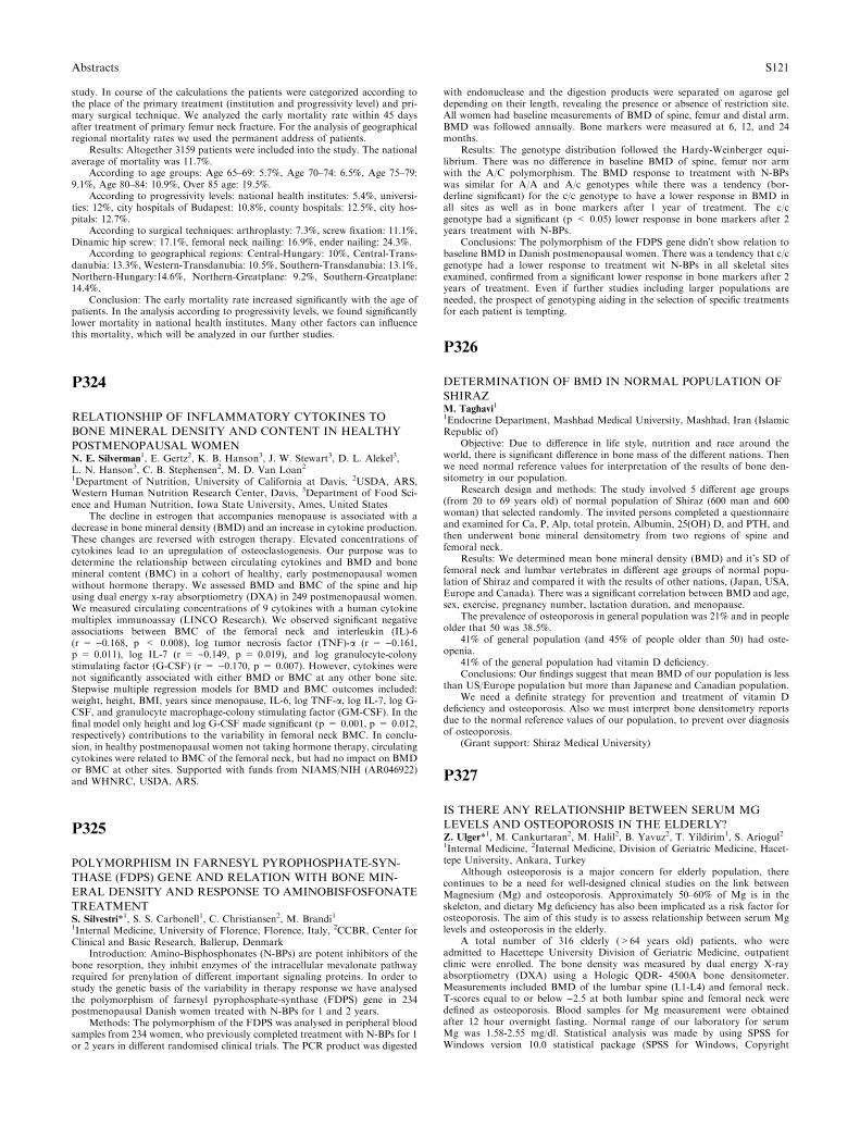
study. In course of the calculations the patients were categorized according tothe place of the primary treatment (institution and progressivity level) and pri-mary surgical technique. We analyzed the early mortality rate within 45 daysafter treatment of primary femur neck fracture. For the analysis of geographicalregional mortality rates we used the permanent address of patients.
Results: Altogether 3159 patients were included into the study. The nationalaverage of mortality was 11.7%.
According to age groups: Age 65–69: 5.7%, Age 70–74: 6.5%, Age 75–79:9.1%, Age 80–84: 10.9%, Over 85 age: 19.5%.
According to progressivity levels: national health institutes: 5.4%, universi-ties: 12%, city hospitals of Budapest: 10.8%, county hospitals: 12.5%, city hos-pitals: 12.7%.
According to surgical techniques: arthroplasty: 7.3%, screw fixation: 11.1%,Dinamic hip screw: 17.1%, femoral neck nailing: 16.9%, ender nailing: 24.3%.
According to geographical regions: Central-Hungary: 10%, Central-Trans-danubia: 13.3%, Western-Transdanubia: 10.5%, Southern-Transdanubia: 13.1%,Northern-Hungary:14.6%, Northern-Greatplane: 9.2%, Southern-Greatplane:14.4%.
Conclusion: The early mortality rate increased significantly with the age ofpatients. In the analysis according to progressivity levels, we found significantlylower mortality in national health institutes. Many other factors can influencethis mortality, which will be analyzed in our further studies.
P324
RELATIONSHIP OF INFLAMMATORY CYTOKINES TO
BONE MINERAL DENSITY AND CONTENT IN HEALTHY
POSTMENOPAUSAL WOMENN. E. Silverman1, E. Gertz2, K. B. Hanson3, J. W. Stewart3, D. L. Alekel3,L. N. Hanson3, C. B. Stephensen2, M. D. Van Loan21Department of Nutrition, University of California at Davis, 2USDA, ARS,Western Human Nutrition Research Center, Davis, 3Department of Food Sci-ence and Human Nutrition, Iowa State University, Ames, United States
The decline in estrogen that accompanies menopause is associated with adecrease in bone mineral density (BMD) and an increase in cytokine production.These changes are reversed with estrogen therapy. Elevated concentrations ofcytokines lead to an upregulation of osteoclastogenesis. Our purpose was todetermine the relationship between circulating cytokines and BMD and bonemineral content (BMC) in a cohort of healthy, early postmenopausal womenwithout hormone therapy. We assessed BMD and BMC of the spine and hipusing dual energy x-ray absorptiometry (DXA) in 249 postmenopausal women.We measured circulating concentrations of 9 cytokines with a human cytokinemultiplex immunoassay (LINCO Research). We observed significant negativeassociations between BMC of the femoral neck and interleukin (IL)-6(r = )0.168, p < 0.008), log tumor necrosis factor (TNF)-a (r = )0.161,p = 0.011), log IL-7 (r = )0.149, p = 0.019), and log granulocyte-colonystimulating factor (G-CSF) (r = )0.170, p = 0.007). However, cytokines werenot significantly associated with either BMD or BMC at any other bone site.Stepwise multiple regression models for BMD and BMC outcomes included:weight, height, BMI, years since menopause, IL-6, log TNF-a, log IL-7, log G-CSF, and granulocyte macrophage-colony stimulating factor (GM-CSF). In thefinal model only height and log G-CSF made significant (p = 0.001, p = 0.012,respectively) contributions to the variability in femoral neck BMC. In conclu-sion, in healthy postmenopausal women not taking hormone therapy, circulatingcytokines were related to BMC of the femoral neck, but had no impact on BMDor BMC at other sites. Supported with funds from NIAMS/NIH (AR046922)and WHNRC, USDA, ARS.
P325
POLYMORPHISM IN FARNESYL PYROPHOSPHATE-SYN-
THASE (FDPS) GENE AND RELATION WITH BONE MIN-
ERAL DENSITY AND RESPONSE TO AMINOBISFOSFONATE
TREATMENTS. Silvestri*1, S. S. Carbonell1, C. Christiansen2, M. Brandi11Internal Medicine, University of Florence, Florence, Italy, 2CCBR, Center forClinical and Basic Research, Ballerup, Denmark
Introduction: Amino-Bisphosphonates (N-BPs) are potent inhibitors of thebone resorption, they inhibit enzymes of the intracellular mevalonate pathwayrequired for prenylation of different important signaling proteins. In order tostudy the genetic basis of the variability in therapy response we have analysedthe polymorphism of farnesyl pyrophosphate-synthase (FDPS) gene in 234postmenopausal Danish women treated with N-BPs for 1 and 2 years.
Methods: The polymorphism of the FDPS was analysed in peripheral bloodsamples from 234 women, who previously completed treatment with N-BPs for 1or 2 years in different randomised clinical trials. The PCR product was digested
with endonuclease and the digestion products were separated on agarose geldepending on their length, revealing the presence or absence of restriction site.All women had baseline measurements of BMD of spine, femur and distal arm.BMD was followed annually. Bone markers were measured at 6, 12, and 24months.
Results: The genotype distribution followed the Hardy-Weinberger equi-librium. There was no difference in baseline BMD of spine, femur nor armwith the A/C polymorphism. The BMD response to treatment with N-BPswas similar for A/A and A/c genotypes while there was a tendency (bor-derline significant) for the c/c genotype to have a lower response in BMD inall sites as well as in bone markers after 1 year of treatment. The c/cgenotype had a significant (p < 0.05) lower response in bone markers after 2years treatment with N-BPs.
Conclusions: The polymorphism of the FDPS gene didn�t show relation tobaseline BMD in Danish postmenopausal women. There was a tendency that c/cgenotype had a lower response to treatment wit N-BPs in all skeletal sitesexamined, confirmed from a significant lower response in bone markers after 2years of treatment. Even if further studies including larger populations areneeded, the prospect of genotyping aiding in the selection of specific treatmentsfor each patient is tempting.
P326
DETERMINATION OF BMD IN NORMAL POPULATION OF
SHIRAZM. Taghavi
1
1Endocrine Department, Mashhad Medical University, Mashhad, Iran (IslamicRepublic of)
Objective: Due to difference in life style, nutrition and race around theworld, there is significant difference in bone mass of the different nations. Thenwe need normal reference values for interpretation of the results of bone den-sitometry in our population.
Research design and methods: The study involved 5 different age groups(from 20 to 69 years old) of normal population of Shiraz (600 man and 600woman) that selected randomly. The invited persons completed a questionnaireand examined for Ca, P, Alp, total protein, Albumin, 25(OH) D, and PTH, andthen underwent bone mineral densitometry from two regions of spine andfemoral neck.
Results: We determined mean bone mineral density (BMD) and it’s SD offemoral neck and lumbar vertebrates in different age groups of normal popu-lation of Shiraz and compared it with the results of other nations, (Japan, USA,Europe and Canada). There was a significant correlation between BMD and age,sex, exercise, pregnancy number, lactation duration, and menopause.
The prevalence of osteoporosis in general population was 21% and in peopleolder that 50 was 38.5%.
41% of general population (and 45% of people older than 50) had oste-openia.
41% of the general population had vitamin D deficiency.Conclusions: Our findings suggest that mean BMD of our population is less
than US/Europe population but more than Japanese and Canadian population.We need a definite strategy for prevention and treatment of vitamin D
deficiency and osteoporosis. Also we must interpret bone densitometry reportsdue to the normal reference values of our population, to prevent over diagnosisof osteoporosis.
(Grant support: Shiraz Medical University)
P327
IS THERE ANY RELATIONSHIP BETWEEN SERUM MG
LEVELS AND OSTEOPOROSIS IN THE ELDERLY?Z. Ulger*1, M. Cankurtaran2, M. Halil2, B. Yavuz2, T. Yildirim1, S. Ariogul21Internal Medicine, 2Internal Medicine, Division of Geriatric Medicine, Hacet-tepe University, Ankara, Turkey
Although osteoporosis is a major concern for elderly population, therecontinues to be a need for well-designed clinical studies on the link betweenMagnesium (Mg) and osteoporosis. Approximately 50–60% of Mg is in theskeleton, and dietary Mg deficiency has also been implicated as a risk factor forosteoporosis. The aim of this study is to assess relationship between serum Mglevels and osteoporosis in the elderly.
A total number of 316 elderly (>64 years old) patients, who wereadmitted to Hacettepe University Division of Geriatric Medicine, outpatientclinic were enrolled. The bone density was measured by dual energy X-rayabsorptiometry (DXA) using a Hologic QDR- 4500A bone densitometer.Measurements included BMD of the lumbar spine (L1-L4) and femoral neck.T-scores equal to or below )2.5 at both lumbar spine and femoral neck weredefined as osteoporosis. Blood samples for Mg measurement were obtainedafter 12 hour overnight fasting. Normal range of our laboratory for serumMg was 1.58-2.55 mg/dl. Statistical analysis was made by using SPSS forWindows version 10.0 statistical package (SPSS for Windows, Copyright
Abstracts S121
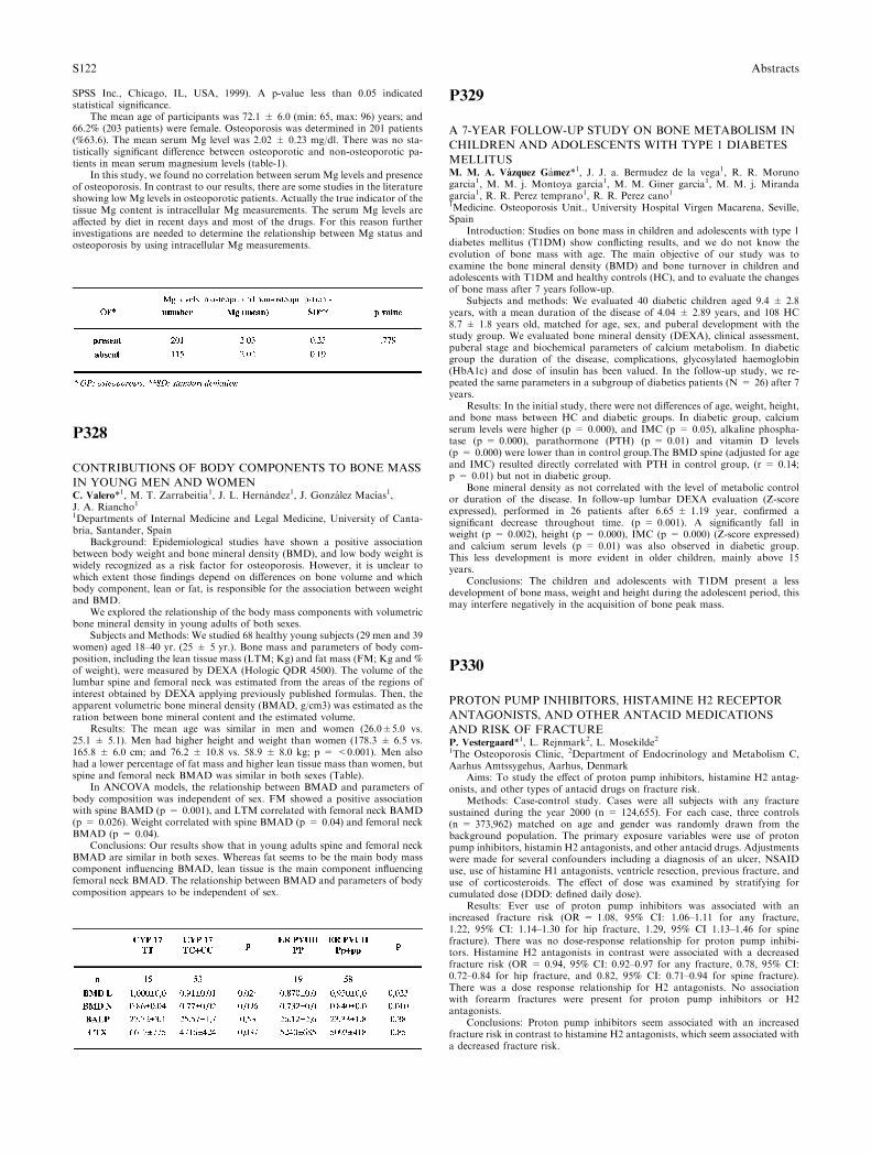
SPSS Inc., Chicago, IL, USA, 1999). A p-value less than 0.05 indicatedstatistical significance.
The mean age of participants was 72.1 ± 6.0 (min: 65, max: 96) years; and66.2% (203 patients) were female. Osteoporosis was determined in 201 patients(%63.6). The mean serum Mg level was 2.02 ± 0.23 mg/dl. There was no sta-tistically significant difference between osteoporotic and non-osteoporotic pa-tients in mean serum magnesium levels (table-1).
In this study, we found no correlation between serum Mg levels and presenceof osteoporosis. In contrast to our results, there are some studies in the literatureshowing low Mg levels in osteoporotic patients. Actually the true indicator of thetissue Mg content is intracellular Mg measurements. The serum Mg levels areaffected by diet in recent days and most of the drugs. For this reason furtherinvestigations are needed to determine the relationship between Mg status andosteoporosis by using intracellular Mg measurements.
P328
CONTRIBUTIONS OF BODY COMPONENTS TO BONE MASS
IN YOUNG MEN AND WOMENC. Valero*1, M. T. Zarrabeitia1, J. L. Hernandez1, J. Gonzalez Macıas1,J. A. Riancho11Departments of Internal Medicine and Legal Medicine, University of Canta-bria, Santander, Spain
Background: Epidemiological studies have shown a positive associationbetween body weight and bone mineral density (BMD), and low body weight iswidely recognized as a risk factor for osteoporosis. However, it is unclear towhich extent those findings depend on differences on bone volume and whichbody component, lean or fat, is responsible for the association between weightand BMD.
We explored the relationship of the body mass components with volumetricbone mineral density in young adults of both sexes.
Subjects and Methods: We studied 68 healthy young subjects (29 men and 39women) aged 18–40 yr. (25 ± 5 yr.). Bone mass and parameters of body com-position, including the lean tissue mass (LTM; Kg) and fat mass (FM; Kg and %of weight), were measured by DEXA (Hologic QDR 4500). The volume of thelumbar spine and femoral neck was estimated from the areas of the regions ofinterest obtained by DEXA applying previously published formulas. Then, theapparent volumetric bone mineral density (BMAD, g/cm3) was estimated as theration between bone mineral content and the estimated volume.
Results: The mean age was similar in men and women (26.0±5.0 vs.25.1 ± 5.1). Men had higher height and weight than women (178.3 ± 6.5 vs.165.8 ± 6.0 cm; and 76.2 ± 10.8 vs. 58.9 ± 8.0 kg; p = <0.001). Men alsohad a lower percentage of fat mass and higher lean tissue mass than women, butspine and femoral neck BMAD was similar in both sexes (Table).
In ANCOVA models, the relationship between BMAD and parameters ofbody composition was independent of sex. FM showed a positive associationwith spine BAMD (p = 0.001), and LTM correlated with femoral neck BAMD(p = 0.026). Weight correlated with spine BMAD (p = 0.04) and femoral neckBMAD (p = 0.04).
Conclusions: Our results show that in young adults spine and femoral neckBMAD are similar in both sexes. Whereas fat seems to be the main body masscomponent influencing BMAD, lean tissue is the main component influencingfemoral neck BMAD. The relationship between BMAD and parameters of bodycomposition appears to be independent of sex.
P329
A 7-YEAR FOLLOW-UP STUDY ON BONE METABOLISM IN
CHILDREN AND ADOLESCENTS WITH TYPE 1 DIABETES
MELLITUSM. M. A. Vazquez Gamez*1, J. J. a. Bermudez de la vega1, R. R. Morunogarcia1, M. M. j. Montoya garcia1, M. M. Giner garcia1, M. M. j. Mirandagarcia1, R. R. Perez temprano1, R. R. Perez cano11Medicine. Osteoporosis Unit., University Hospital Virgen Macarena, Seville,Spain
Introduction: Studies on bone mass in children and adolescents with type 1diabetes mellitus (T1DM) show conflicting results, and we do not know theevolution of bone mass with age. The main objective of our study was toexamine the bone mineral density (BMD) and bone turnover in children andadolescents with T1DM and healthy controls (HC), and to evaluate the changesof bone mass after 7 years follow-up.
Subjects and methods: We evaluated 40 diabetic children aged 9.4 ± 2.8years, with a mean duration of the disease of 4.04 ± 2.89 years, and 108 HC8.7 ± 1.8 years old, matched for age, sex, and puberal development with thestudy group. We evaluated bone mineral density (DEXA), clinical assessment,puberal stage and biochemical parameters of calcium metabolism. In diabeticgroup the duration of the disease, complications, glycosylated haemoglobin(HbA1c) and dose of insulin has been valued. In the follow-up study, we re-peated the same parameters in a subgroup of diabetics patients (N = 26) after 7years.
Results: In the initial study, there were not differences of age, weight, height,and bone mass between HC and diabetic groups. In diabetic group, calciumserum levels were higher (p = 0.000), and IMC (p = 0.05), alkaline phospha-tase (p = 0.000), parathormone (PTH) (p = 0.01) and vitamin D levels(p = 0.000) were lower than in control group.The BMD spine (adjusted for ageand IMC) resulted directly correlated with PTH in control group, (r = 0.14;p = 0.01) but not in diabetic group.
Bone mineral density as not correlated with the level of metabolic controlor duration of the disease. In follow-up lumbar DEXA evaluation (Z-scoreexpressed), performed in 26 patients after 6.65 ± 1.19 year, confirmed asignificant decrease throughout time. (p = 0.001). A significantly fall inweight (p = 0.002), height (p = 0.000), IMC (p = 0.000) (Z-score expressed)and calcium serum levels (p = 0.01) was also observed in diabetic group.This less development is more evident in older children, mainly above 15years.
Conclusions: The children and adolescents with T1DM present a lessdevelopment of bone mass, weight and height during the adolescent period, thismay interfere negatively in the acquisition of bone peak mass.
P330
PROTON PUMP INHIBITORS, HISTAMINE H2 RECEPTOR
ANTAGONISTS, AND OTHER ANTACID MEDICATIONS
AND RISK OF FRACTUREP. Vestergaard*1, L. Rejnmark2, L. Mosekilde21The Osteoporosis Clinic, 2Department of Endocrinology and Metabolism C,Aarhus Amtssygehus, Aarhus, Denmark
Aims: To study the effect of proton pump inhibitors, histamine H2 antag-onists, and other types of antacid drugs on fracture risk.
Methods: Case-control study. Cases were all subjects with any fracturesustained during the year 2000 (n = 124,655). For each case, three controls(n = 373,962) matched on age and gender was randomly drawn from thebackground population. The primary exposure variables were use of protonpump inhibitors, histamin H2 antagonists, and other antacid drugs. Adjustmentswere made for several confounders including a diagnosis of an ulcer, NSAIDuse, use of histamine H1 antagonists, ventricle resection, previous fracture, anduse of corticosteroids. The effect of dose was examined by stratifying forcumulated dose (DDD: defined daily dose).
Results: Ever use of proton pump inhibitors was associated with anincreased fracture risk (OR = 1.08, 95% CI: 1.06–1.11 for any fracture,1.22, 95% CI: 1.14–1.30 for hip fracture, 1.29, 95% CI 1.13–1.46 for spinefracture). There was no dose-response relationship for proton pump inhibi-tors. Histamine H2 antagonists in contrast were associated with a decreasedfracture risk (OR = 0.94, 95% CI: 0.92–0.97 for any fracture, 0.78, 95% CI:0.72–0.84 for hip fracture, and 0.82, 95% CI: 0.71–0.94 for spine fracture).There was a dose response relationship for H2 antagonists. No associationwith forearm fractures were present for proton pump inhibitors or H2antagonists.
Conclusions: Proton pump inhibitors seem associated with an increasedfracture risk in contrast to histamine H2 antagonists, which seem associated witha decreased fracture risk.
S122 Abstracts
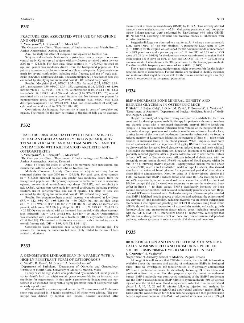
P331
FRACTURE RISK ASSOCIATED WITH USE OF MORPHINE
AND OPIATESP. Vestergaard*1, L. Rejnmark2, L. Mosekilde21The Osteoporosis Clinic, 2Department of Endocrinology and Metabolism C,Aarhus Amtssygehus, Aarhus, Denmark
Aim: To study the effect of morphine and opiates on fracture risk.Subjects and methods: Nationwide register based study designed as a case-
control study. Cases were all subjects with any fracture sustained during the year2000 (n = 124,655). For each case, three controls (n = 373,962) matched onage and gender was randomly drawn from the background population. Theprimary exposure variables were use of morphine and opiates. Adjustments weremade for several confounders including prior fracture, and use of weak anal-gesics (NSAIDs, acetylsalicylic acid, and acetaminiphene). The effect of dose wasexamined by stratifying for cumulated dose (DDD: defined daily dose).
Results: Morphine (1.47, 95%CI 1.37–1.58), fentanyl (2.23, 95%CI 1.89–2.64), methadone (1.39, 95%CI 1.05–1.83), oxycodone (1.36, 95%CI 1.08–1.69),nicomorphine (1.57, 95%CI 1.38–1.78), ketobemidone (1.07, 95%CI 1.02–1.13),tramadol (1.54, 95%CI 1.49–1.58), and codeine (1.16, 95%CI 1.12–1.20) were allassociated with an increase in overall fracture risk. No increase was present forbuprenorphine (0.86, 95%CI 0.79–0.95), pethidine (0.98, 95%CI 0.89–1.08),dextropropoxiphene (1.02, 95%CI 0.90–1.16), and combinations of acetylsali-cylic acid and codeine (0.94, 95%CI 0.88–1.01).
Conclusions: An increased fracture risk is seen in users of morphine andopiates. The reason for this may be related to the risk of falls due to dizziness.
P332
FRACTURE RISK ASSOCIATED WITH USE OF NON-STE-
ROIDAL ANTI-INFLAMMATORY DRUGS (NSAID), ACE-
TYLSALICYLIC ACID, AND ACETAMINOPHENE, AND THE
INTERACTION WITH RHEUMATOID ARTHRITIS AND
OSTEOARTHRITISP. Vestergaard*1, L. Rejnmark2, L. Mosekilde21The Osteoporosis Clinic, 2Department of Endocrinology and Metabolism C,Aarhus Amtssygehus, Aarhus, Denmark
Aims: To study the effect of various non-morphine pain medication, andrheumatoid arthritis and osteoarthritis on fracture risk.
Methods: Case-control study. Cases were all subjects with any fracturesustained during the year 2000 (n = 124,655). For each case, three controls(n = 373,962) matched on age and gender was randomly drawn from thebackground population. The primary exposure variables were use of acetamin-ophene, non-steroidal anti-inflammatory drugs (NSAID), and acetylsalicylicacid (ASA). Adjustments were made for several confounders including previousfracture, use of corticosteroids, and use of opiates. The effect of dose wasexamined by stratifying for cumulated dose (DDD: defined daily dose).
Results: For acetaminophene a small increase was observed at low doses(RR = 1.12, 95% CI: 1.08–1.16) for <50 DDD) but not at high doses(RR = 1.03, 95% CI: 0.99–1.06 for >=300 DDD). For ASA no increase waspresent, while some NSAIDs (e.g. ibuprofen RR = 1.26, 95% CI 1.23–1.29 for<20 DDD) were associated with an increased fracture risk while others were not(e.g., celecoxib: RR = 0.84, 95%CI 0.67–1.04 for <20 DDD). Osteoarthrosiswas associated with a decreased risk of fractures (OR for any fracture: 0.78, 95%CI: 0.76–0.81). Rheumatoid arthritis was associated with a limited increase inoverall fracture risk (OR = 1.09, 95% CI: 1.04–1.15).
Conclusions: Weak analgesics have varying effects on fracture risk. Thereasons for this may be numerous but most likely related to the risk of fallsrelated to dizziness.
P333
A GENOMEWIDE LINKAGE SCAN IN A FAMILY WITH A
HIGHLY PENETRANT FORM OF OSTEOPOROSISC. Vidal*1, R. Galea2, M. Brincat2, A. Xuereb-Anastasi31Department of Pathology, 2Department of Obstetrics and Gynaecology,3Institute of Health Care, University of Malta, G’Mangia, Malta
Family based linkage studies were performed by a number of investigators totry to identify loci that might contain genes responsible for an increased sus-ceptibility for osteoporosis. In this study a genomewide linkage scan was per-formed in an extended family with a highly penetrant form of osteoporosis withan early age of onset.
400 microsatellite markers spread across the 22 autosomes and X chromo-some were analysed in 18 family members, 9 of whom were affected. The phe-notype was defined by lumbar and femoral z-scores calculated after
measurement of bone mineral density (BMD) by DEXA. Two severely affectedmembers were males (z-scores <)2.0). Multipoint parametric and non-para-metric linkage analyses were performed by EasyLinkage v4.0 using GENE-HUNTER v2.1, assuming dominant and recessive modes of inheritance withvariable penetrance.
Suggestive linkage was observed to a marker at 5p14 where a non-parametricLOD score (NPL) of 4.86 was obtained. A parametric LOD score of 2.09(p = 0.0156) for this region was obtained for the dominant mode of inheritancewith 90% penetrance and a phenocopy rate of 1%. An NPL of 5.75 and a LODscore of 1.21 (p = 0.0156) for the dominant model was observed to region 11p13while region 15q15 gave an NPL of 3.65 and LOD of 1.01 (p = 0.0172) for arecessive mode of inheritance with 30% penetrance for the homozygous mutant.The disease allele frequency was assumed to be 0.001 (0.10%).
These results suggest that multiple genes might be responsible for this highlypenetrant form of osteoporosis. Further studies are required to identify the genesand mutations that might be responsible for the disease and that might also playa role in osteoporosis in the general population.
P334
BMP-6 INCREASES BONE MINERAL DENSITY AND
REDUCES GLYCEMIA IN OSTEOPENIC MICEP. Simic*1, J. Buljan Culej1, I. Orlic1, M. Zuvic2, F. Borovecki1, S. Vukicevic11Department of Anatomy, 2Department of Nuclear Medicine, School of Medi-cine, Zagreb, Croatia
Despite the variety of drugs for treating osteoporosis and diabetes, there is agreat need for developing new anabolic therapy for patients with severe bone lossand diabetic drugs with a prolonged therapeutic interval. BMP-6 knock-out(Bmp-6 -/-) mice have reduced trabecular bone volume of the peripheral skele-ton, under developed pancreas and a reduction in the size of stomach and spleen,causing fusion of the liver and duodenum. Immunohistochemically we found areduced number of Langerhans islands in the pancreas of Bmp-6 -/- mice whichresulted in increased levels of the blood glucose. When Bmp-6 -/- mice weretreated systemically with i.v. injection of 10 lg/kg BMP-6 to restore lost bone,we discovered that increased blood glucose was reduced to normal levels within 2h following the protein administration. Single i.v. injection of 60 lg/kg BMP-6gradually reduced glycemia after a glucose tolerance test to near-normal valuesin both WT and in Bmp-6 -/- mice. Alloxan induced diabetic rats, with nodetectable serum insulin showed 57-63% reduction of blood glucose within 30min to 48 h following BMP-6 injection. Hyperglycemic, insulin free non obesediabetic (NOD) mice, a well established model of type I diabetes, also showedsignificant reduction of blood glucose for a prolonged period of time after asingle BMP-6 administration. Next, by using 18 F-deoxy-labeled glucose (18FDG) we found that BMP-6 reduced blood and urine 18 FDG levels up to 44%and 47%, respectively, in both normal and diabetic mice. In parallel, six weeks ofBMP-6 therapy (10 lg/ 5· week) was necessary to restore the trabecular bonedefect in Bmp-6 -/- to sham values. BMP-6 significantly increased the bonevolume, trabecular number, thickness and connectivity parameters in both Bmp-6 -/- and WT ovariectomized mice. Molecular analyses of tissue samples revealedthat BMP-6 inhibited hepatic glucose production and activated the expression ofkey enzymes of lipid metabolism, reducing glycemia via an insulin independentmechanism. Gene expression profiling and RT-PCR analyzes using total femurmRNA showed increased expression of extracellular matrix, cell cycle, growthfactor/cytokine and transcription factor related genes, including procollagentype IX, IGF-1, EGF, FGF, interleukins 12 and 17, respectively. We suggest thatBMP-6 has a strong anabolic effect on bone and, via an insulin independentpathway, reduces glycemia in rodent models of diabetes and bone loss.
P335
BIODISTRIBUTION AND IN VIVO EFFICACY OF SYSTEMI-
CALLY ADMINISTERED AND FROM URINE PURIFIED
SOLUBLE BMP-7/BMP-6 HYBRID MOLECULE IN RATSL. Grgurevic*1, S. Vukicevic11Department of Anatomy, School of Medicine, Zagreb, Croatia
Although it is well known that TGF-ß circulates, there is little informationavailable about the presence and activity of endogenous BMPs in biologicalfluids. Here we investigated the biodistribution of systemically administeredBMP with particular reference to its activity following 24 h secretion andpurification from the urine. For this purpose a specific dimeric recombinanthuman BMP-6 molecule was constructed consisting of the BMP-7 prodomainand the BMP-6 mature domain. BMP-7/BMP-6 hybrid molecule (300 lg/kg) wasinjected into the rat tail vein. Blood samples were collected from the rat orbitalplexus 1, 5, 10, 15, 20 and 30 minutes following injection and analyzed byimmunoprecipitation with protein G and a polyclonal antibody against BMP-6.Urine samples were collected for 24 h in metabolic cages and then purified usingheparin sepharose columns. SDS-PAGE of purified urine was run on a 10% gel
Abstracts S123
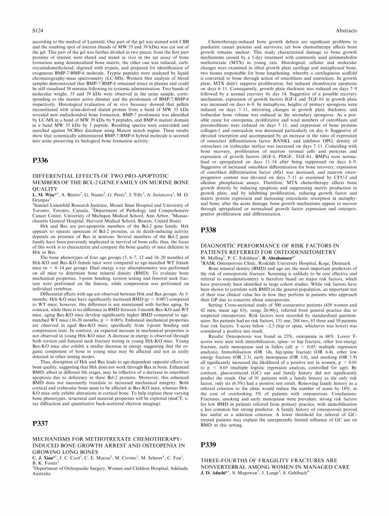
according to the method of Laemmli. One part of the gel was stained with CBBand the resulting spot of interest (bands of MW 35 and 39 kDa) was cut out ofthe gel. This part of the gel was further divided in two pieces: from the first partproteins of interest were eluted and tested in vivo in the rat assay of boneformation using demineralized bone matrix; the other one was reduced, carb-oxyamidomethylated, digested with trypsin, and prepared for identification ofexogenous BMP-7/BMP-6 molecule. Tryptic peptides were analyzed by liquidchromatography-mass spectrometry (LC-MS). Western blot analysis of bloodsamples demonstrated that BMP-7/BMP-6 remained intact in plasma and couldbe still visualized 30 minutes following its systemic administration. Two bands ofmolecular weight, 35 and 39 kDa were observed in the urine sample, corre-sponding to the mature active dimmer and the prodomain of BMP-7/BMP-6respectively. Histological evaluation of in vivo bioassay showed that pelletsreconstituted with urine-derived eluted protein from a band of MW 35 kDarevealed new endochondral bone formation. BMP-7 prodomain was identifiedby LC-MS in a band of MW 39 kDa by 9 peptides, and BMP-6 mature domainin a band MW 35 kDa by 1 peptide. Resulting spectra were centroided andsearched against NCBInr database using Mascot search engine. These resultsshow that systemically administered BMP-7/BMP-6 hybrid molecule is secretedinto urine preserving its biological bone formation activity.
P336
DIFFERENTIAL EFFECTS OF TWO PRO-APOPTOTIC
MEMBERS OF THE BCL-2 GENE FAMILY ON MURINE BONE
QUALITYL. M. Wise*1, A. Benito2, G. Nunez2, G. Perez3, J. Tilly3, A. Jurisicova1, M. D.Grynpas11Samuel Lunenfeld Research Institute, Mount Sinai Hospital and University ofToronto, Toronto, Canada, 2Department of Pathology and ComprehensiveCancer Center, University of Michigan Medical School, Ann Arbor, 3Massas-chusetts General Hosptial, Harvard Medical School, Boston, United States
Hrk and Bax are pro-apoptotic members of the Bcl-2 gene family. Hrkappears to operate upstream of Bcl-2 proteins, as its death-inducing activitydepends on presence of Bax in neurons. Several members of the Bcl-2 genefamily have been previously implicated in survival of bone cells; thus, the focusof this work is to characterize and compare the bone quality of mice deficient inHrk or Bax.
The bone phenotypes of four age groups (3, 6–7, 12 and 16–20 months) ofHrk-KO and Bax-KO female mice were compared to age-matched WT femalemice (n = 8–14 per group). Dual energy x-ray absorptiometry was performedon all mice to determine bone mineral density (BMD). To evaluate bonemechanical properties, 3-point bending, torsion testing and femoral neck frac-ture were performed on the femora, while compression was performed onindividual vertebrae.
Differential effects with age are observed between Hrk and Bax groups. At 3-months, Hrk-KO mice have significantly increased BMD (p = 0.007) comparedto WT mice; however, this difference is not maintained with further aging. Incontrast, while there is no difference in BMD between 3-month Bax-KO and WTmice, aging Bax-KO mice develop significantly higher BMD compared to age-matched WT mice (16-20 months; p = 0.005). Enhanced mechanical propertiesare observed in aged Bax-KO mice, specifically from 3-point bending andcompression tests. In contrast, an expected increase in mechanical properties isnot observed in young Hrk-KO mice. A decrease in energy is observed throughboth torsion and femoral neck fracture testing in young Hrk-KO mice. YoungBax-KO mice also exhibit a similar decrease in energy suggesting that the or-ganic component of bone in young mice may be affected and not as easilydetected in other testing modes.
Thus, disruption of Hrk and Bax leads to age-dependent opposite effects onbone quality, suggesting that Hrk does not work through Bax in bone. EnhancedBMD, albeit at different life stages, may be reflective of a decrease in osteoblastapoptosis due to deficiency in these Bcl-2 proteins. Moreover, this enhancedBMD does not necessarily translate to increased mechanical integrity. Bothcortical and trabecular bone seem to be affected in Bax-KO mice, whereas Hrk-KO mice only exhibit alterations in cortical bone. To help explain these varyingbone phenotypes, structural and material properties will be explored (muCT, x-ray diffraction and quantitative back-scattered electron imaging).
P337
MECHANISMS FOR METHOTREXATE CHEMOTHERAPY-
INDUCED BONE GROWTH ARREST AND OSTEOPENIA IN
GROWING LONG BONESC. J. Xian*1, J. C. Cool1, C. E. Macsai1, M. Covino1, M. Scherer1, C. Fan1,B. K. Foster11Department of Orthopaedic Surgery, Women and Children Hospital, Adelaide,Australia
Chemotherapy-induced bone growth defects are significant problems inpaediatric cancer patients and survivors; yet how chemotherapy affects bonegrowth remains unclear. This study characterised damage to bone growthmechanisms caused by a 5-day treatment with commonly used antimetabolitemethotrexate (MTX) in young rats. Histological, cellular and molecularchanges were examined in tibial growth plate cartilage and metaphyseal bone,two tissues responsible for bone lengthening, whereby a cartilaginous scaffoldis converted to bone through action of osteoblasts and osteoclasts. In growthplate, MTX didn�t suppress proliferation, but induced chondrocyte apoptosison days 6–11. Consequently, growth plate thickness was reduced on days 7–9followed by a normal recovery by day 14. Suggestive of a possible recoverymechanism, expression of growth factors IGF-I and TGF-b1 in growth platewas increased on days 6–9. In metaphysis, heights of primary spongiosa werereduced on days 7–11, mirroring changes in growth plate thickness; andtrabecular bone volume was reduced in the secondary spongiosa. As a pos-sible cause for osteopenia, proliferative and total numbers of osteoblasts andpreosteoblasts were reduced on days 7–11, and expression of bone proteinscollagen-1 and osteocalcin was decreased particularly on day 6. Suggestive ofelevated resorption and accompanied by an increase in the ratio of expressionof osteoclast differentiation factor RANKL and inhibitor OPG, density ofosteoclasts on trabecular surface was increased on days 7–11. Coinciding withbone recovery, proliferation of marrow stromal cells and pericytes andexpression of growth factors (IGF-I, PDGF, TGF-b1, BMPs) were norma-lised or upregulated on days 11–14 after being suppressed on days 6–9.Suggestive of increased osteoblast differentiation for bone recovery, expressionof osteoblast differentiation factor cbfa1 was increased, and marrow osteo-progenitor content was elevated on days 7–11 as examined by CFU-f andalkaline phosphatase assays. Therefore, MTX chemotherapy affects bonegrowth directly by inducing apoptosis and suppressing matrix production ingrowth plate, and by inhibiting proliferation, reducing growth factor andmatrix protein expression and increasing osteoclastic resorption in metaphy-seal bone; after the acute damage, bone growth mechanisms appear to recoverthrough upregulated or normalised growth factor expression and osteopro-genitor proliferation and differentiation.
P338
DIAGNOSTIC PERFORMANCE OF RISK FACTORS IN
PATIENTS REFERRED FOR OSTEODENSITOMETRYM. Malling1, P. C. Eskildsen1, B. Abrahamsen*11RASK Osteoporosis Clinic, Roskilde University Hospital, Koge, Denmark
Bone mineral density (BMD) and age are the most important predictors ofthe risk of osteoporotic fracture. Screening is unlikely to be cost effective andreferral to osteodensitometry is therefore based on major risk factors, whichhave previously been identified in large cohort studies. While risk factors havebeen shown to correlate with BMD in the general population, an important testof their true clinical value lies in how they perform in patients who approachtheir GP due to concerns about osteoporosis.
Setting: Cross-sectional study of 500 consecutive patients (458 women and42 men, mean age 63y, range 26-90y), referred from general practice due tosuspected osteoporosis. Risk factors were recorded by standardised question-naire. Six patients had no risk factors, 131 one, 268 two, 85 three and 10 patientsfour risk factors. T-score below )2.5 (hip or spine, whichever was lower) wasconsidered a positive test result.
Results: Osteoporosis was found in 23%, osteopenia in 46%. Lower T-scores were seen with immobilisation, spine- or hip fracture, other low-energyfracture, early menopause and in fallers (all: p < 0.05, multiple regressionanalysis). Immobilisation (OR 14), hip/spine fracture (OR 6.4), other lowenergy fracture (OR 2.3), early menopause (OR 3.0), and smoking (OR 1.9)all significantly increased the likelihood of a positive test in women, p < 0.01to p < 0.05 (multiple logistic regression analysis, controlled for age). Bycontrast, glucocorticoid (GC) use and family history did not significantlypredict the result. Out of 91 patients with a family history as the only riskfactor, only six (6.5%) had a positive test result. Removing family history as areferral criterion to the clinic would reduce the number of scans by 18%, atthe cost of overlooking 5% of patients with osteoporosis. Conclusions:Fractures, smoking and early menopause were prevalent, strong risk factorsfor low BMD in patients referred from primary practice, with immobilisationa less common but strong predictor. A family history of osteoporosis provedless useful as a selection criterion. A lower threshold for referral of GC-treated patients may explain the unexpectedly limited influence of GC use onBMD in this setting.
P339
THREE-FOURTHS OF FRAGILITY FRACTURES ARE
NONVERTEBRAL AMONG WOMEN IN MANAGED CAREJ. D. Adachi*1, S. Magowan2, J. Lange3, S. Gehlbach4
S124 Abstracts

1Saint Joseph’s Hospital, McMaster University, Hamilton, Canada, 2Medicaland Technical Affairs, 3Epidemiology and PharmacoEconomics, Procter andGamble Pharmaceuticals, Mason, 4Epidemiology, University of Massachusetts,Amherst, United States
Background: Clinical trials have demonstrated that different osteoporotictherapies have different efficacies at vertebral and nonvertebral sites.
Objective: To estimate the proportion of clinical fragility fractures that arevertebral and nonvertebral occurring in managed care among women aged ‡50years.
Methods: Inpatient and outpatient medical claims for vertebral (n = 4,710)and nonvertebral fractures (n = 16,530) among women between ages 40 and 89years (n = 1,430,008) enrolled in managed care programs during the year 2003were investigated using Medstat Marketscan, a database comprised of approx-imately 45 private and public US health plans. Fracture rates by 10-year agegroups were calculated for each of the anatomical sites (wrist, leg, arm, hip,pelvis, clavicle, spine). Fragility fractures were defined by the fracture rate forwomen aged ‡50 years minus the fracture rate of the 40–49 year age group(assumed to be the baseline traumatic fracture rate). This calculation removedfractures not likely related to osteoporosis.
Results: Nonvertebral fractures represented 73% of all fragility fracturesamong women aged ‡50 years enrolled in managed care. By decade age group,nonvertebral fractures represented 86% of fractures among women between theages 50 to 59 years, and lower percentages in older age groups (table).
Discussion: It is essential that an osteoporotic therapy demonstrates provenefficacy beyond the spine to be clinically beneficial and cost-effective for man-aged care.
P340
GREATER THAN 90% OF MANAGED CARE COSTS FOR
FRAGILITY FRACTURES ARE NONVERTEBRAL IN
NATURE AMONG WOMENJ. Adachi*1, S. Magowan2, N. Borisov3, J. Lange4, R. Ohsfeldt51St. Joseph’s Hospital, McMaster University, Hamilton, Canada, 2Medical andTechnical Affairs, 3Epidemiology and PharmacoEconomics, 4Epidemiology,Procter and Gamble Pharmaceuticals, Mason, 5School of Rural Public Health,Texas AM, Bryan, United States
Background: Approximately three-fourths of clinical fragility fracturesamong women over 50 years of age in managed care populations are nonver-tebral.
Objective: To determine the annual costs by age and fragility fracture type(nonvertebral & vertebral) among women aged >50 years enrolled in managedcare programs.
Methods: The data source for this analysis was Medstat Marketscan which iscomprised of approximately 45 private and public health plans in the US.Inpatient and outpatient medical claims for fractures were analyzed amongwomen between ages 50 and 89 years (n = 1,009,270) enrolled in managed careprograms in the year 2003. Fracture rates by 10-year age groups were calculatedfor each anatomical site (wrist, leg, arm, hip, pelvis, clavicle, spine). Fragilityfractures were estimated by the fracture rate for women 50 years and over minusthe fracture rate of the 40–49 year age group (assumed to be the baseline trau-matic fracture rate). These fragility fracture rates were multiplied by the fracture-related direct medical costs, based upon the 2003 Medicare fee schedule, over thefirst year after fracture.
Results: Nonvertebral fractures were responsible for more than 90% oftotal annual fragility fracture costs across 4 age decades of women aged > 50years.
Conclusion: Although nonvertebral fractures account for three-fourths offracture volume, they are responsible for over 90% of direct medical costsrelated to fragility fractures. It is therefore imperative that osteoporosistherapy demonstrate nonvertebral efficacy to be cost-effective in the managedcare setting.
P341
RENAL TOLERABILITY PROFILE OF INTERMITTENT
INTRAVENOUS IBANDRONATE INJECTIONS IN
POSTMENOPAUSAL WOMEN WITH OSTEOPOROSIS:
DIVA 2-YEAR ANALYSISS. Adami*1, C. Benhamou2, V. Vyskocil3, C. Leigh4, F. Sedarati4, B. Minic4,D. M. Reid51University of Verona, Verona, Italy, 2IPROS-CHR, Orleans, France, 3Uni-versity Hospital, Plzen, Czech Republic, 4F. Hoffmann-La Roche Ltd, Basel,Switzerland, 5University of Aberdeen, Aberdeen, United Kingdom
Background. Intravenous (i.v.) bisphosphonates are administered as pro-longed infusions, primarily to avoid renal adverse events. Ibandronate, a potentnitrogen-containing bisphosphonate, can be administered as a rapid (15-30seconds) i.v. injection in women with postmenopausal osteoporosis.1-3 The renalsafety profile of two i.v. ibandronate injection regimens (2mg every 2 months[q2mo] or 3mg every 3 months [q3mo]) was compared with daily oral ibandro-nate (2.5 mg) in the DIVA study.
Methods. A total of 1,395 women with postmenopausal osteoporosis wererandomised into this double-blind and double-dummy study. Women withbaseline creatinine >2.4mg/dL were excluded. Renal safety was assessed byadverse events monitoring, measurements of serum creatinine and calculatedcreatinine clearance (Cockroft-Gault equation). Values of serum creatinine wereconsidered clinically relevant if they doubled, elevated >=0.5mg/dL in patientswith baseline creatinine <1.4mg/dL or elevated >=1mg/dL in those withbaseline creatinine >=1.4mg/dL.
Results. At 2 years, 53 (3.8%) of 1,382 participants included in the safetyanalysis experienced renal adverse events. Events were similarly distributedacross the treatment arms (daily: 3.9%, q2mo: 4.5%, q3mo: 3.2%). There were nocases of acute renal failure. Half of all participants had estimated creatinineclearance <60mL/min at baseline (<30mL/min in 0.5%, 30-<60mL/min in 50%and 60-<90mL/min in 44.5%, >=90mL/min in 5%). Over 2 years of treatment,12 participants (0.9%) had clinically relevant changes in serum creatinine (daily:0.6%, q2mo: 1.3%, q3mo: 0.6%). During the same period, the mean annualchange in creatinine clearance was between -0.9 and -1.6mL/min, which corre-sponds to the natural decrease seen with age (approximately 1mL/min/year), andwas comparable in all treatment groups.
Conclusion. Ibandronate has a favourable renal safety profile whenadministered orally or by i.v. injection. The ability to administer ibandronate asa rapid (15–30 seconds) injection without compromising renal function may havepractical advantages in the treatment of postmenopausal osteoporosis.
1. Adami S, et al. Bone 2004;34:881-9.2. Recker RR, et al. Arthritis Rheum 2004;50(Suppl.):4095.3. Emkey R, et al. ACR 2005.
P342
AUTOMATIC ASSESSMENT OF VERTEBRAL SHAPE USING
ACTIVE APPEARANCE MODELSJ. E. Adams*1, M. G. Roberts1, T. Cootes11Imaging Science and Biomedical Engineering, University of Manchester,Manchester, United Kingdom
Background: Vertebral fractures are important features of osteoporosis andcan be identified on lateral DXA images. We have applied active appearancemodels to identify vertebral shapes automatically located on DXA images of thespine, including fractured vertebrae. We have assessed the accuracy androbustness of the techniques applied.
Methods: 202 DXA images of the spine were obtained, containing 173fractures. The vertebral body outlines were annotated manually from L4 to T7,using 40 points for each vertebra. Statistical models of vertebral shape andtexture were derived, with the spine modelled by a sequence of overlappingvertebral triplets. A provisional texture model was derived on the best 120images, and the extracted texture on the remaining images was constrained byfirst fitting it to the provisional model. This reduced the number of clutter modesin the texture model, and improved overall robustness. The search algorithmfitted the best sequence of models to an unseen image, using an ActiveAppearance Model for each vertebral triplet. Multiple triplet candidates aretried, but the best fitting one only is imposed at each iteration. Thus the orderingof the sequence is dynamically adjusted to defer poorly fitting regions until theyare constrained by neighbours. Miss-N out tests were run to obtain estimates ofaccuracy, comparing the automatic and manual shapes.
Results: there was a mean point-to-line accuracy of 0.7 mm in normal ver-tebrae (well within the limits of manual precision). Over 95% of points in normalvertebrae were located with an accuracy better than 2mm. The correspondingaccuracies for fractured vertebrae were: mean = 1.2 mm, median = 0.7 mm,and 84% of points better than 2 mm. Significant accuracy improvements weremade by using the restricted texture model training, and by dynamicallyadjusting the order of the fit. However the models did not always match the more
Abstracts S125
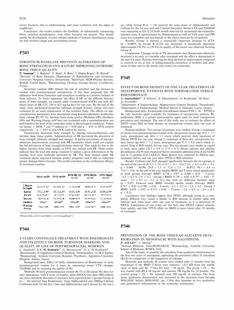
severe fractures, due to undertraining, and some confusion with the edges ofneighbours.
Conclusion: the results confirm the feasibility of substantially automatingDXA vertebral morphometry, even when fractures are present. This shouldenable the development of more reliable methods of fracture classification usingthe full vertebral shape and surrounding texture.
P343
STRONTIUM RANELATE PREVENTS ALTERATION OF
BONE STRENGTH IN OVX RATS BY IMPROVING INTRINSIC
BONE TISSUE QUALITYP. Ammann*1, I. Badoud1, V. Shen2, S. Bain2, I. Dupin-Roger3, R. Rizzoli11Division of Bone Diseases, Department of Rehabilitation and Geriatrics,University Hospital, Geneva, Switzerland, 2SkeleTech, MDS Pharma Services,Bothell, United States, 3Rheumatology Division, Groupe Servier, Courbevoie,France
Strontium ranelate (SR) reduces the risk of vertebral and hip fracture inwomen with postmenopausal osteoporosis. It has been proposed that SRinfluences both bone formation and resorption resulting in a positive balance ofbone turnover. To better appreciate the effect of SR on the different determi-nants of bone strength, we treated adult ovariectomized (OVX) rats with dif-ferent doses of SR (125, 250 or 625 mg/kg.day) for one year. By the end of thestudy, bone mechanical properties (ultimate strength N/mm2, energy mJ) wereevaluated by an axial compression of the vertebral body. The effects on mic-roarchitecture parameters were determined using histomorphometry (trabecularbone volume BV/TV %). Intrinsic bone tissue quality (Modulus GPa, HardnessGPa and Working Energy mN*nm) was evaluated with a nanoindentation testperformed at the level of the trabecular nodes in physiological conditions. Valuesare means ± SEM, * and ** indicate p < 0.05 and p < 0.01 vs OVX controlrespectively, � p < 0.05 vs non OVX control by Anova.
Ovariectomy decreased bone strength by altering microarchitecture andintrinsic bone tissue quality. Administration of SR prevented the alteration ofbone strength (ultimate strength and energy). A partial prevention of microar-chitecture deteriorations occurred with SR treatments which could not explainthe full prevention of bone strength decrease observed. This might be due to thehigher intrinsic bone tissue quality in OVX rats treated with SR. These resultsindicate that the load and energy required to induce a given deformation at thelamellar level were markedly increased by SR. The bone formed under SRtreatment shows improved intrinsic quality properties and is able to withstandgreater damage before fracture. This could contribute to the antifracture efficacyof strontium ranelate.
P344
4 YEARS CONTINUOUS TREATMENT WITH RISEDRONATE
AND ITS EFFECT ON BONE TURNOVER MARKERS AND
QUALITY OF LIFE OF POSTMENOPAUSAL WOMWENL. Anadiotis1, I. C. H. Koulouris*2, G. Skarantavos3, G. C. H. Koulouris41Biochemistry, Evangelismos General Hospital, 2Orthopaedics, 1st IKA Egaleo,3Reumatology, Atttikon University Hospital, 4Psychiatry, Aeginition UniversityHospital, Athens, Greece
Background/aims: Effect of dailly administration of Risedronate to earlypostmenopausal women for 4 years by measuring serum CTX changes,25(OH)D and by assessing the quality of life.
Methods: 40 early postmenopausal women 48–53 ys old (mean 50), 6mo-1yrafter menopause, with T score on lumbar spine (DEXA) less than 2SD,withoutany prior metabolic disorders or fractures were separated into 2 groups:Group A(n = 30) received 5mg Risedronate, 1mcg Alphacalcidol and 1000mg CalciumCarbonate daily for the first 12mo and Alphacalcidol and Calcium for the rest 3
yrs, while Group B (n = 10) received the same doses of Alphacalcidol andCalcium for the 1st year and only Calcium thereafter. Serum CTX and 25(OH)Dwere measured at 0,6,12,24,36,48 month intervals by automated elecrochemilu-minence assay.A questionnaire by Zimmermann as well as VAS score and PPIscore was compiled and reevaluated at the above intervals by all patients.
Results: Group A showed a statistically important decrease in sCTX(p = 0.0005). In Group B sCTX was increased. A statistically importantimprovement (70.2%) vs (29.8%) in quality of life scores was observed favoringGroup A.
Conclusions: Changes in the sCTX demonstrate that Risedronate effectivelydecreases it as early as 6 months after treatment and the effect is maintained atthe end of 4 years. Patients receiving the drug showed an improvement comparedto controls as far as fear of falling,insecurity,restriction of mobility and alter-ation of their role in the family and society are concerned.
P345
EFFECT ON BONE DENSITY OF ONE YEAR TREATMENT OF
OSTEOPOROTIC PATIENTS WITH TERIPARATIDE VERSUS
RISENDRONATEA. Anastasilakis*1, S. Polyzos1, Z. Efstathiadou1, G. Koukoulis2, M. Kita1,A. Avramidis11Department of Endocrinology, Hippocration General Hospital, Thessaloniki,2Department of Endocrinology, Medical School of Thessalia, Larisa, Greece
Background/aims. Teriparatide (recombinant human PTH 1–34 – TPTD) isthe only anabolic agent available for treatment of osteoporotic patients. Ri-sendronate (RIS) is a potent antiresorptive agent used for both osteoporosisprevention and treatment. The aim of this study was to evaluate the effects ofTPTD versus RIS on bone density of osteoporotic women after one year oftreatment.
Patients/methods. Two groups of patients were studied. Group 1 comprisedof twenty-four postmenopausal women with osteoporosis (mean age 65.8 ± 1.7years, menopausal age 44.4 ± 1.3 years) which received daily injections of20mug TPTD for one year. Group 2 comprised of thirty-five osteoporotic wo-men (mean age 64.8 ± 1.2 years, menopausal age 47.5 ± 0.8 years) which re-ceived 35mg of RIS weekly for one year. The two groups were similar in regardto body mass index (28.5 ± 0.9 vs 27.9 ± 0.7). Serum calcium and alkalinephosphatase (ALP) were measured before treatment administration (Ca0-ALP0)and at 12 months (Ca12-ALP12). Bone density was estimated by DEXA mea-surements before and one year after TPTD or RIS initiation.
Results. Calcium and ALP changed significantly between the two groups atthe end of the year (Ca0: 9.3 ± 0.1 vs 9.7 ± 0.1 – Ca12: 9.8 ± 0.1 vs 9.6 ± 0.1,p = 0.008 – ALP0: 59.9 ± 2.3 vs 72.8 ± 5.3 – ALP12: 98.1 ± 6.6 vs73.8 ± 5.0). At the end of the year lumbar BMD and T-score raised significantlyin both groups (Group1 BMD: 0.758 ± 0.03 vs 0.800 ± 0.03 – T-score:)3.2 ± 0.2 vs -2.8 ± 0.2 – Group 2 BMD: 0.759 ± 0.02 vs 0.797 ± 0.02 – T-score: )2.5 ± 0.1 vs )2.2 vs 0.1) but with no difference between them(p = 0.97). Femoral neck BMD revealed similar results (Group1 BMD:0.717 ± 0.02 vs 0.708 ± 0.02 – T-score: )2.2 ± 0.2 vs )2.4 ± 0.2 – Group 2BMD: 0.670 ± 0.02 vs 0.712 ± 0.02 – T-score: )2.8 ± 0.3 vs )2.6 ± 0.1,p = 0.2).
Conclusion. Our findings suggest that TPTD, although acting in a com-pletely different way, causes a similar to RIS increase in lumbar spine andfemoral neck bone mass after one year of treatment, as it is measured byDEXA. Limitations of our study are the facts that DEXA cannot estimatebone quality and that TPTD effect on BMD is more intent after 18 monthsof treatment.
P346
PREVENTION OF THE BONE TISSUE QUALITATIVE DETE-
RIORATION IN MENOPAUSE WITH RALOXIFENEP. ASTAZI*1, L. Mirone21Internal Medicine, OsteoRESEARCH, 2Reumatology, Catholic UniversitySchool of Medicine, ROMA, Italy
Aim of the study: to quantify the cancellous bone qualitative deterioration inthe first two years of menopause, appraising the preventive effect of raloxifene(RLX) in comparison to the integration of calcium.
Materials and methods: 60 women were studied after 12 months from themenopause if the BMD T-Score were inclusive ±0.5 SD from the middlevalue of peak and the 17-beta-E2 were <30 pg/ml. The group 1 (N = 30)was treated with RLX 60 mg/die and calcium 500 mg/die for 24 months. Thecontrol group 2 (N = 30) assumed only 500 mg/die of calcium. The bonetissue qualitative deterioration was measured at the calcaneus bone throughHOLOGIC Sahara (HOLOGIC, inc., USA) that furnishes in vivo qualitativeand quantitative informations of the trabecular architecture.
S126 Abstracts
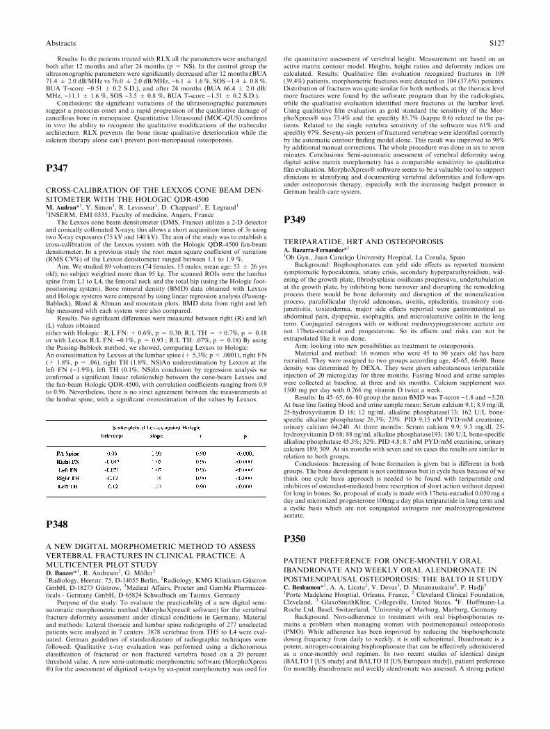
Results: In the patients treated with RLX all the parameters were unchangedboth after 12 months and after 24 months (p = NS). In the control group theultrasonographic parameters were significantly decreased after 12 months:(BUA71.4 ± 2.0 dB/MHz vs 76.0 ± 2.0 dB/MHz, )6.1 ± 1.6 %, SOS )1.4 ± 0.8 %,BUA T-score )0.51 ± 0.2 S.D.), and after 24 months (BUA 66.4 ± 2.0 dB/MHz, )11.1 ± 1.6 %, SOS )3.5 ± 0.8 %, BUA T-score )1.51 ± 0.2 S.D.).
Conclusions: the significant variations of the ultrasonographic parameterssuggest a precocius onset and a rapid progression of the qualitative damage ofcancellous bone in menopause. Quantitative Ultrasound (MOC-QUS) confirmsin vivo the ability to recognize the qualitative modifications of the trabeculararchitecture. RLX prevents the bone tissue qualitative deterioration while thecalcium therapy alone can�t prevent post-menopausal osteoporosis.
P347
CROSS-CALIBRATION OF THE LEXXOS CONE BEAM DEN-
SITOMETER WITH THE HOLOGIC QDR-4500M. Audran*1, Y. Simon1, R. Levasseur1, D. Chappard1, E. Legrand11INSERM, EMI 0335, Faculty of medicine, Angers, France
The Lexxos cone beam densitometer (DMS, France) utilizes a 2-D detectorand conically collimated X-rays; this allows a short acquisition times of 3s usingtwo X-ray exposures (75 kV and 140 kV). The aim of the study was to establish across-calibration of the Lexxos system with the Hologic QDR-4500 fan-beamdensitometer. In a previous study the root mean square coefficient of variation(RMS CV%) of the Lexxos densitometer ranged between 1.1 to 1.9 %.
Aim. We studied 89 volunteers (74 females, 15 males; mean age: 53 ± 26 yrsold); no subject weighted more than 95 kg. The scanned ROIs were the lumbarspine from L1 to L4, the femoral neck and the total hip (using the Hologic foot-positioning system). Bone mineral density (BMD) data obtained with Lexxosand Hologic systems were compared by using linear regression analysis (Passing-Bablock), Bland & Altman and mountain plots. BMD data from right and lefthip measured with each system were also compared.
Results. No significant differences were measured between right (R) and left(L) values obtainedeither with Hologic : R/L FN: + 0.6%, p = 0.30; R/L TH = +0.7%, p = 0.18or with Lexxos R/L FN: )0.1%, p = 0.93 ; R/L TH: .07%, p = 0.18) By usingthe Passing-Bablock method, we showed, comparing Lexxos to Hologic:An overestimation by Lexxos at the lumbar spine (+ 5.3%; p < .0001), right FN(+ 1.8%, p = .06), right TH (1.8%, NS)An underestimation by Lexxos at theleft FN ()1.9%), left TH (0.1%, NS)In conclusion by regression analysis weconfirmed a significant linear relationship between the cone-beam Lexxos andthe fan-beam Hologic QDR-4500, with correlation coefficients ranging from 0.9to 0.96. Nevertheless, there is no strict agreement between the measurements atthe lumbar spine, with a significant overestimation of the values by Lexxos.
P348
A NEW DIGITAL MORPHOMETRIC METHOD TO ASSESS
VERTEBRAL FRACTURES IN CLINICAL PRACTICE: A
MULTICENTER PILOT STUDYD. Banzer*1, R. Andresen2, G. Moller31Radiology, Heerstr. 75, D-14055 Berlin, 2Radiology, KMG Klinikum GustrowGmbH, D-18273 Gustrow, 3Medical Affairs, Procter and Gamble Pharmaceu-ticals - Germany GmbH, D-65824 Schwalbach am Taunus, Germany
Purpose of the study: To evaluate the practicability of a new digital semi-automatic morphometric method (MorphoXpress� software) for the vertebralfracture deformity assessment under clinical conditions in Germany. Materialand methods: Lateral thoracic and lumbar spine radiographs of 277 unselectedpatients were analyzed in 7 centers. 3878 vertebrae from TH5 to L4 were eval-uated. German guidelines of standardization of radiographic techniques werefollowed. Qualitative x-ray evaluation was performed using a dichotomousclassification of fractured or non fractured vertebra based on a 20 percentthreshold value. A new semi-automatic morphometric software (MorphoXpress�) for the assessment of digitized x-rays by six-point morphometry was used for
the quantitative assessment of vertebral height. Measurement are based on anactive matrix contour model. Heights, height ratios and deformity indices arecalculated. Results: Qualitative film evaluation recognized fractures in 109(39.4%) patients, morphometric fractures were detected in 104 (37.6%) patients.Distribution of fractures was quite similar for both methods, at the thoracic levelmore fractures were found by the software program than by the radiologists,while the qualitative evaluation identified more fractures at the lumbar level.Using qualitative film evaluation as gold standard the sensitivity of the Mor-phoXpress� was 73.4% and the specifity 85.7% (kappa 0.6) related to the pa-tients. Related to the single vertebra sensitivity of the software was 61% andspecifity 97%. Seventy-six percent of fractured vertebrae were identified correctlyby the automatic contour finding model alone. This result was improved to 98%by additional manual corrections. The whole procedure was done in six to sevenminutes. Conclusions: Semi-automatic assessment of vertebral deformity usingdigital active matrix morphometry has a comparable sensitivity to qualitativefilm evaluation. MorphoXpress� software seems to be a valuable tool to supportclinicians in identifying and documenting vertebral deformities and follow-upsunder osteoporosis therapy, especially with the increasing budget pressure inGerman health care system.
P349
TERIPARATIDE, HRT AND OSTEOPOROSISA. Bazarra-Fernandez*11Ob Gyn., Juan Canalejo University Hospital, La Coruna, Spain
Background: Bisphosphonates can yeld side effects as reported transientsymptomatic hypocalcemia, tetany crisis, secondary hyperparathyroidism, wid-ening of the growth plate, fibrodysplasia ossificans progressiva, undertubulationat the growth plate, by inhibiting bone turnover and disrupting the remodelingprocess there would be bone deformity and disruption of the mineralizationprocess, parafollicular thyroid adenomas, uveitis, episcleritis, transitory con-junctivitis, toxicoderma, major side effects reported were gastrointestinal asabdominal pain, dyspepsia, esophagitis, and microulcerative colitis in the longterm. Conjugated estrogens with or without medroxyprogesterone acetate arenot 17beta-estradiol and progesterone. So its effects and risks can not beextrapolated like it was done.
Aim: looking into new possibilities as treatment to osteoporosis.Material and method: 16 women who were 45 to 80 years old has been
recruited. They were assigned to two groups according age, 45-65, 66-80. Bonedensity was determined by DEXA. They were given subcutaneous teriparatideinjection of 20 microg/day for three months. Fasting blood and urine sampleswere collected at baseline, at three and six months. Calcium supplement was1500 mg per day with 0.266 mg vitamin D twice a week.
Results: In 45–65, 66–80 group the mean BMD was T-score )1.8 and )3.20.At base line fasting blood and urine sample mean: Serum calcium 9.1; 8.9 mg/dl,25-hydroxyvitamin D 16; 12 ng/ml, alkaline phosphatase173; 162 U/L bone-specific alkaline phosphatase 26.3%; 23%. PID 9;15 nM PYD/mM creatinine,urinary calcium 64;240. At three months: Serum calcium 9.9; 9.3 mg/dl, 25-hydroxyvitamin D 68; 88 ng/ml, alkaline phosphatase193; 180 U/L bone-specificalkaline phosphatase 45.3%; 32%. PID 4.8; 8.7 nM PYD/mM creatinine, urinarycalcium 189; 309. At six months with seven and six cases the results are similar inrelation to both groups.
Conclusions: Increasing of bone formation is given but is different in bothgroups. The bone development is not continuous but in cycle basis because of wethink one cycle basis approach is needed to be found with teriparatide andinhibitors of osteoclast-mediated bone resorption of short action without depositfor long in bones. So, proposal of study is made with 17beta-estradiol 0.050 mg aday and micronized progesterone 100mg a day plus teriparatide in long term anda cyclic basis which are not conjugated estrogens nor medroxyprogesteroneacetate.
P350
PATIENT PREFERENCE FOR ONCE-MONTHLY ORAL
IBANDRONATE AND WEEKLY ORAL ALENDRONATE IN
POSTMENOPAUSAL OSTEOPOROSIS: THE BALTO II STUDYC. Benhamou*1, A. A. Licata2, V. Devas3, D. Masanauskaite4, P. Hadji51Porte Madeleine Hosptial, Orleans, France, 2 Cleveland Clinical Foundation,Cleveland, 3 GlaxoSmithKline, Collegeville, United States, 4F. Hoffmann-LaRoche Ltd, Basel, Switzerland, 5University of Marburg, Marburg, Germany
Background. Non-adherence to treatment with oral bisphosphonates re-mains a problem when managing women with postmenopausal osteoporosis(PMO). While adherence has been improved by reducing the bisphosphonatedosing frequency from daily to weekly, it is still suboptimal. Ibandronate is apotent, nitrogen-containing bisphosphonate that can be effectively administeredas a once-monthly oral regimen. In two recent studies of identical design(BALTO I [US study] and BALTO II [US/European study]), patient preferencefor monthly ibandronate and weekly alendronate was assessed. A strong patient
Abstracts S127

preference for monthly dosing was seen in BALTO I. Here, we report resultsfrom BALTO II.
Methods. This 6-month, two-period, crossover, open-label study enrolled350 women with PMO and randomised them to two different sequences oftreatment. In sequence A (n = 176), monthly ibandronate was followed byweekly alendronate, while this was reversed in sequence B (n = 174). Once-monthly ibandronate (150 mg) was given for 3 calendar months and weeklyalendronate (70 mg) for 12 weeks. A questionnaire was used at the final studyvisit to assess patient preference. Women who received at least one dose of eachtrial medication and completed the questionnaire were defined as the modifiedintent-to-treat (mITT) population.
Results. The majority of mITT patients expressed a preference (n = 299[93.1%]), and a greater number preferred monthly ibandronate (n = 211, 70.6%)over weekly alendronate (n = 88, 29.4%). The preference rate for monthlyibandronate was statistically significant (p < 0.0001). The monthly regimen wasconsidered to facilitate long-term adherence by 81.5% and to achieve a betterlifestyle fit by 75.4% of women. In addition, of the 273 women (85.0%) whoexpressed an opinion on convenience, a greater number favoured monthlyibandronate (n = 209, 76.6%) over weekly alendronate (n = 64, 23.4%). Theconvenience rate for monthly ibandronate was statistically significant (p <0.0001). The two treatment regimens showed a similar safety profile.
Conclusions. Once-monthly oral ibandronate was considered preferable to,and more convenient than, weekly oral alendronate. This outcome reinforces theresults from the US BALTO I study, and suggests that once-monthly bis-phosphonate dosing has the potential to improve adherence.
P351
IMPACT OF PRIOR BISPHOSPHONATE THERAPY ON THE
ANABOLIC EFFECTS OF TERIPARATIDE: RATIONALE AND
DESIGN OF THE OPTAMISE STUDYJ. P. Bilezikian*1, N. B. Watts2, P. D. Delmas3, P. Miller41Professor, Medicine and Pharmacology, College of Physicians and Surgeons,Columbia University, New York, NY, 2Professor of Medicine, Director, Oste-oporosis and Bone Health Program, University of Cincinnati College of Medi-cine, Cincinnati, OH, United States, 3Professor, Medicine and Rheumatology,Director, Inserm Research Unit, Inserm Research Unit 403 and Claude BernardUniversity of Lyon, Lyon, France, 4Medical Director, Colorado Center for BoneResearch, Lakewood, CO, United States
ablBackground/aims: It is not known if the extent to which subsequentresponsiveness to teriparatide (TPTD) after bisphosphonate (BP) therapy differsbetween alendronate (ALN) and risedronate (RIS). Some observations suggestthat the anabolic response to TPTD, as measured by changes in biochemicalmarkers of bone formation and BMD, may be delayed in patients previouslytreated with alendronate (ALN) [Ettinger J Bone Miner Res. 2004;19:745.] whileother studies do not [Cosman N Eng J Med 2005 2005;353:566].
Methods: The Open-label Study to Determine How Prior Therapy withAlendronate or Risedronate in PostMenopausal Women with OsteoporosisInfluences the Clinical EffectivenesS of TeriparatidE (OPTAMISE) is a multi-national, parallel-group study designed to compare differences in anabolic re-sponse to TPTD in postmenopausal women previously treated with either ALNor RIS. Key entry criteria include lumbar spine (LS) or total hip BMD T-score£ )2.0 with a fragility fracture or T-score <)2.5, vitamin D replete (‡ 16 ng/mL), uninterrupted treatment with ALN or RIS for ‡2 years, and urinary NTX<50 nmol/mmol creatinine (to insure compliance with previous BP therapy).Subjects (n = 290) are being stratified and paired based on prior BP use andduration of BP treatment. After enrollment, subjects discontinue BP treatmentand receive daily TPTD injections (20 mg) for 12 months. Baseline and posttreatment measures of bone formation (N-terminal propeptide of type I collagen[P1NP], bone-specific alkaline phosphatase, and osteocalcin) and bone resorp-tion (urinary N-telopeptide and serum C-telopeptide) are obtained throughoutthe study, with particular interest in the early anabolic response to TPTD at 3and 6 months. The effects of TPTD on BMD (measured by DXA) and measuresof bone strength (determined by QCT and MRI) are also being assessed.
Conclusions: OPTAMISE is the first study to examine prospectively andcompare the anabolic effects of TPTD after ALN or RIS therapy, and addressesthe question of whether the initial choice of BP therapy influences the anabolicresponse of TPTD.
P352
WHAT IS HIGH BONE TURNOVER?J. A. Blazquez*1, L. Navarro2, F. Mateos1, M. Chafer2, E. Almar3, J. Del Pino41Internal Medicine, 2Clinical Chemistry, University Hospital, 3Epidemyology,Health Department, Albacete, 4Rheumatology, University Hospital, Salamanca,Spain
Background and objective. There are different definitions of high turnover inpostmenopausal women, that include 1 SD above the mean of premenopausal
women, 2 SD of the same population, and the highest quartile of the post-menopausal women. The aim of this study was to explore the influence of the schosen criteria on the prevalence of high turnover.
Methods. 197 early postmenopausal women (mean 52.7 ± 1.6 yr ) wererandomly selected in the province of Albacete (Spain). At the same time 34normal premenopausal women were selected as the reference population(39 ± 5.4 yr). To assess bone turnover, BAP, DPD, NTx and Ca/Cr weremeasured . The prevalence of high turnover was calculated by means of threedifferent criteria: + 2 SD, + 2.5 SD and + 3 SD above the mean of thepremenopausal normal reference population.
Results. Statistics of premenopausal and postmenopausal women are writtenin another communication.
Prevalence of high turnover depends on the chosen criterion. For BAP, +2SD coincide with the highest tertiles (T3) of the postmenopausal women, and+2,5 SD coincide with the highest quartiles (Q4). For DPD and NTx, +2,5 SDcoincide with the highest tertiles and +3 SD coincide with the highest quartiles.For Ca/Cr, +1,5 SD (0,201 mg/mg) is slightly above the highest tertiles and + 2SD is slightly above the highest quartiles (table ).
Conclusion. Prevalence of high turnover in early postmenopausal womendepends on the selected criterion. According to our results we propose the fol-lowing criteria: +2 SD above the mean of premenopausal women for boneformation markers, +2,5 SD for bone resorption markers and +1,5 SD for Ca/Cr.
Supported by grants from FIS 99/07059, JCCM98196 FISCAM, Diputacionof Albacete.
P353
MUSCULOSKELETAL ADVERSE EFFECTS OF ORAL
TREATMENT WITH ONCE WEEKLY ALENDRONATE AND
RISEDRONATE IN PATIENTS WITH OSTEOPOROSISO. Bock*1, H. Borst1, M. Stephan-Oelkers1, C. Degner1, D. Felsenberg11Centre for Muscle and Bone Research, Charite - Universitatsmedizin Berlin,Campus Benjamin Franklin, Berlin, Germany
Objective: To examine in a major cohort of patients whether or not mus-culoskeletal side effects, similar to those seen in intravenous bisphosphonates(BP), might occur also in high dosage oral treatment regimens with alendronate(ALN) and risedronate (RSN).
Patients and methods: 612 consecutive patients (mean age 68.2 ± 9.7 years;527 females, 85 males) treated in the osteoporosis outpatient clinic at Charite,Campus Benjamin Franklin, between July 2002 and October 2003 with oralALN or RSN, were examined and followed up for musculoskeletal adverseevents (MAEs).
Results: The overall incidence of any severe MAEs in our patients was low(5.6 %), but relatively high in the group of patients primarily treated with onceweekly ALN or RSN (21.0 %). All severe MAEs occurred in primarily onceweekly treated patients: 27 in ALN 70 mg once weekly (27/134 = 20.1%) and 7in RSN 35 mg once weekly (7/28 = 25.0%), with no significant difference be-tween those groups. The most frequently reported MAE was acute arthralgia in12.6 %, followed by acute back pain in 9.1 % of all primarily once weekly treatedcases. None of the 302 patients initially treated with daily BP reported anyMAEs when later switching to once weekly administration (218 patients to ALN70 mg once weekly and 84 patients to RSN 35 mg once weekly). With referenceto recently published data, the phenomenon is probably related to dose depen-dent cd T cell activation by accumulation of isopentenyl pyrophosphate (IPP)due to inhibition of the mevalonate pathway by nitrogen containing bis-phosphonates (nBP).
Conclusions: MAEs in oral BP are in general less common and severe thanin intravenous BP. They were observed exclusively in patients starting ALN orRSN treatment with once weekly dosage regimens. In order to avoid this phe-nomenon, it is suggested to start ALN or RSN treatment with the lower dailydosages of ALN 10 mg or RSN 5 mg for about two weeks before switching tothe overall more convenient once weekly dose regimen.
S128 Abstracts
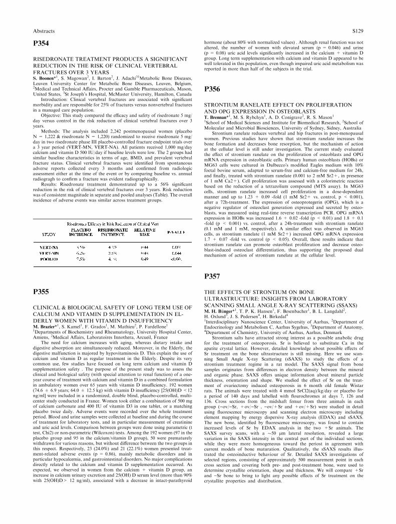
P354
RISEDRONATE TREATMENT PRODUCES A SIGNIFICANT
REDUCTION IN THE RISK OF CLINICAL VERTEBRAL
FRACTURES OVER 3 YEARSS. Boonen*1, S. Magowan2, I. Barton2, J. Adachi31Metabolic Bone Diseases,Leuven University Center for Metabolic Bone Diseases, Leuven, Belgium,2Medical and Technical Affairs, Procter and Gamble Pharmaceuticals, Mason,United States, 3St Joseph’s Hospital, McMaster University, Hamilton, Canada
Introduction: Clinical vertebral fractures are associated with significantmorbidity and are responsible for 25% of fractures versus nonvertebral fracturesin a managed care population.
Objective: This study compared the efficacy and safety of risedronate 5 mg/day versus control in the risk reduction of clinical vertebral fractures over 3years.
Methods: The analysis included 2,242 postmenopausal women (placeboN = 1,222 & risedronate N = 1,220) randomized to receive risedronate 5 mg/day in two risedronate phase III placebo-controlled fracture endpoint trials overa 3 year period (VERT-MN, VERT-NA). All patients received 1,000 mg/daycalcium and vitamin D 500 IU/day if baseline levels were low. The 2 groups hadsimilar baseline characteristics in terms of age, BMD, and prevalent vertebralfracture status. Clinical vertebral fractures were identified from spontaneousadverse reports collected every 3 months and confirmed from radiologicassessment either at the time of the event or by comparing baseline vs. annualradiogragh to confirm a fracture was evident radiographically.
Results: Risedronate treatment demonstrated up to a 56% significantreduction in the risk of clinical vertebral fractures over 3 years. Risk reductionwas of consistent magnitude in separate and pooled analyses (Table). The overallincidence of adverse events was similar across treatment groups.
P355
CLINICAL & BIOLOGICAL SAFETY OF LONG TERM USE OF
CALCIUM AND VITAMIN D SUPPLEMENTATION IN EL-
DERLY WOMEN WITH VITAMIN D INSUFFICIENCYM. Brazier*1, S. Kamel1, F. Grados1, M. Mathieu2, P. Fardellone11Departments of Biochemistry and Rheumatology, University Hospital Center,Amiens, 2Medical Affairs, Laboratoires Innothera, Arcueil, France
The need for calcium increases with aging, whereas dietary intake anddigestive absorption are simultaneously reduced. Moreover, in the Elderly, thedigestive malfunction is majored by hypovitaminosis D. This explain the use ofcalcium and vitamin D as regular treatment in the Elderly. Despite its verycommon use, few studies have focused on long term calcium and vitamin Dsupplementation safety . The purpose of the present study was to assess theclinical and biological safety (with special attention to renal function) of a one-year course of treatment with calcium and vitamin D in a combined formulationin ambulatory women over 65 years with vitamin D insufficiency. 192 women(74.6 ± 6.9 years; 64.0 ± 12.5 kg) with vitamin D insufficiency [25(OH)D <12ng/ml] were included in a randomized, double blind, placebo-controlled, multi-center study conducted in France. Women took either a combination of 500 mgof calcium carbonate and 400 IU of vitamin D3 in one tablet, or a matchingplacebo twice daily. Adverse events were recorded over the whole treatmentperiod. Blood and urine samples were collected at baseline and during the courseof treatment for laboratory tests, and in particular measurement of creatinineand uric acid levels. Comparison between groups were done using parametric (ttest, Chi2) or non-parametric (Wilcoxon) tests. Among the 192 women (97 in theplacebo group and 95 in the calcium/vitamin D group), 50 were prematurelywithdrawn for various reasons, but without difference between the two groups inthis respect. Respectively, 23 (24.0%) and 21 (22.1%) women presented treat-ment-related adverse events (p = 0.86), mainly metabolic disorders and inparticular hypocalcemia, and gastrointestinal disorders. No major complicationsdirectly related to the calcium and vitamin D supplementation occurred. Asexpected, we observed in women from the calcium + vitamin D group, anincrease in calcium urinary excretion and 25(OH) D serum level (more than 90%with 25(OH)D> 12 ng/ml), associated with a decrease in intact-parathyroid
hormone (about 80% with normalized values) . Although renal function was notaltered, the number of women with elevated serum (p = 0.046) and urine(p = 0.08) uric acid levels significantly increased in the calcium + vitamin Dgroup. Long term supplementation with calcium and vitamin D appeared to bewell tolerated in this population, even though impaired uric acid metabolism wasreported in more than half of the subjects in the trial.
P356
STRONTIUM RANELATE EFFECT ON PROLIFERATION
AND OPG EXPRESSION IN OSTEOBLASTST. Brennan*1, M. S. Rybchyn1, A. D. Conigrave2, R. S. Mason11School of Medical Sciences and Institute for Biomedical Research, 2School ofMolecular and Microbial Biosciences, University of Sydney, Sidney, Australia
Strontium ranelate reduces vertebral and hip fractures in post-menopausalwomen. Previous studies have shown that strontium ranelate increases thebone formation and decreases bone resorption, but the mechanism of actionat the cellular level is still under investigation. The current study evaluatedthe effect of strontium ranelate on the proliferation of osteoblasts and OPGmRNA expression in osteoblastic cells. Primary human osteoblasts (HOBs) orMG63 cells were cultured in Dulbecco�s modified Eagles medium with 10%foetal bovine serum, adapted to serum-free and calcium-free medium for 24h,and finally, treated with strontium ranelate (0.001 to 2 mM Sr2+, in presenceof 1 mM Ca2+). Cell proliferation was assessed with a colorimetric reactionbased on the reduction of a tetrazolium compound (MTS assay). In MG63cells, strontium ranelate increased cell proliferation in a dose-dependentmanner and up to 1.23 + 0.09 -fold (1 mM Sr2+ vs. control, p < 0.001),after a 72h-treatment. The expression of osteoprotegerin (OPG), which is anegative regulator of osteoclast generation expressed and secreted by osteo-blasts, was measured using real-time reverse transcription PCR. OPG mRNAexpression in HOBs was increased 1.6 + 0.02 -fold (p < 0.01) and 1.8 + 0.1-fold (p < 0.001) vs. control, after a 24h-treatment with strontium ranelate(0.1 mM and 1 mM, respectively). A similar effect was observed in MG63cells, as strontium ranelate (1 mM Sr2+) increased OPG mRNA expression1.7 + 0.07 -fold vs. control (p < 0.05). Overall, these results indicate thatstrontium ranelate can promote osteoblast proliferation and decrease osteo-blast-induced osteoclast differentiation, thus supporting the proposed dualmechanism of action of strontium ranelate at the cellular level.
P357
THE EFFECTS OF STRONTIUM ON BONE
ULTRASTRUCTURE: INSIGHTS FROM LABORATORY
SCANNING SMALL ANGLE X-RAY SCATTERING (SSAXS)M. H. Bunger*1, T. P. K. Hansen1, F. Besenbacher1, B. L. Langdahl2,H. Oxlund3, J. S. Pedersen4, H. Birkedal41Interdisciplinary Nanoscience Center, University of Aarhus, 2Department ofEndocrinology and Metabolism C, Aarhus Sygehus, 3Department of Anatomy,4Department of Chemistry, University of Aarhus, Aarhus, Denmark
Strontium salts have attracted strong interest as a possible anabolic drugfor the treatment of osteoporosis. Sr is believed to substitute Ca in theapatite crystal lattice. However, detailed knowledge about possible effects ofSr treatment on the bone ultrastructure is still missing. Here we use scan-ning Small Angle X-ray Scattering (sSAXS) to study the effects of astrontium treatment regime in a rat model. The SAXS signal from bonesamples originates from differences in electron density between the mineraland organic phase. SAXS offers unique information about mineral particlethickness, orientation and shape. We studied the effect of Sr on the treat-ment of ovariectomy induced osteoporosis in 6 month old female Wistarrats. The animals were treated with 4 mmol SrCl2(aq)/kg/day or placebo fora period of 140 days and labelled with flourochromes at days 7, 126 and136. Cross sections from the midshaft femur from three animals in eachgroup ()ov/)Sr, +ov/)Sr , )ov/+Sr and +ov/+Sr) were studied in detailusing fluorescence microscopy and scanning electron microscopy includingelement mapping by energy dispersive X-ray analysis (EDAX) and sSAXS.The new bone, identified by fluorescence microscopy, was found to containincreased levels of Sr by EDAX analysis in the two +Sr animals. TheSAXS survey scans, with a �50 lm lateral resolution, revealed a largevariation in the SAXS intensity in the central part of the individual sections,while they were more homogeneous toward the periost in agreement withcurrent models of bone maturation. Qualitatively, the sSAXS results illus-trated the osteoinductive behaviour of Sr. Detailed SAXS investigations ofselected regions, consisting of approximately 500 measurement point in eachcross section and covering both pre- and post-treatment bone, were used todetermine crystallite orientation, shape and thickness. We will compare +Srand )Sr bone to bring to light any possible effects of Sr treatment on thecrystallite properties and distribution.
Abstracts S129

P358
GRANULOCYTE-MACROPHAGE COLONY-STIMULATING
FACTOR PREVENTS DISORDERS OF BONE REMODELING
INDUCED BY LOW-DOSE METHOTREXATE IN RATSU. Cegiela1, W. Janiec*11Department of Pharmacology, Medical University of Silesia, Sosnowiec,Poland
The immunosuppressive therapy with low-dose methotrexate (MTX) isconsidered effective in patients with rheumatoid arthritis. However, it can impairbone formation in a population already predisposed to osteoporosis.Granulo-cyte-macrophage colony-stimulating factor (GM-CSF) is a cytokine producedby T cells following activation and by most myeloid cells, such as macrophagesand granulocytes. Recent in vitro data suggest that GM-CSF is a stronginhibitor of osteoclast differentiation and may inhibit not only the physiologicalbone resorption stimulated by RANKL, but also the pathological boneresorption stimulated by TNF-a.
The aim of the present study was to investigate in vivo the possibility ofprevention of the effects of low-dose MTX on the rat osseous tissue by admin-istration of GM-CSF.
The experiments were carried out on 9–10-week-old male Wistar rats. MTXwas administered in a dose of 0.25 mg/kg p.o. daily for 4 weeks, and GM-CSF(rat recombinant) was administered in a dose of 0.003 mg/kg s.c. daily for thefirst 5 days. The effects of the treatments on bone mass, bone mineral andcalcium content, histomorphometric parameters of the tibia and femur, andmechanical properties of the femur were studied.
MTX in a dose of 0.25 mg/kg p.o. caused disorders of bone remodeling,mainly inhibition of synthesis and mineralization of the osseous tissue, withoutthe significant effect on bone resorption. MTX also worsened mechanicalproperties of the femur (decreases in extrinsic stiffness, ultimate and breakingload, an increase in the bone deformation caused by the breaking load wereobserved).
After administration of GM-CSF (0.003 mg/kg s.c. daily) for 5 days togetherwith MTX (0.25 mg/kg p.o. daily) for 4 weeks, in comparison with the groupreceiving MTX only, statistically significant increases in the mass of the tibia andfemur, the transverse cross-section of the cortical bone, width of periostealosteoid, periosteal transverse growth in the tibia, and increases in the width oftrabeculae in the femur were observed. GM-CSF also improved mechanicalproperties of the femur (increases in the extrinsic stiffness, ultimate load andbreaking load).
In conclusion, GM-CSF prevented the unfavorable effects of MTX on therat osseous tissue.
Acknowledgement: This study was supported by grant No 2P05D00326from The Ministry of Science and Informatization, Poland.
P359
ANALGESIC ACTIVITY OF DIFFERENT ANTI-OSTEOPO-
ROTIC DRUGSR. Y. Chechurin*11Internal Medicine, B&C, Moscow, Russian Federation
The present work aimed at evaluating analgesic activities of some of themost popular drugs in the treatment of osteoporosis seen in an out-patientsetting.
Data of an osteoporosis diagnostic and consulting centre spanning 5 years(2000 – 2005) were analyzed. Three hundred and forty-four subjects were in-cluded, 302 females and 42 males (mean age, 66.1 ± 6.5 years). Intensity of pain(thoracic, lumbar, and/or in the limbs) was evaluated using a 4-grade scale, priorto and 2 months into treatment (as a monotherapy or with vitamin D3 and/orcalcium added). The patients were on (n/%): alfacalcidol (121/35%), etidronicacid (102/30%), synthetic salmon calcitonin (62/18%), Osteopan (21/6%), a-lendronic acid (20/6%), Osteogenon (10/3%), sodium fluoride (8/2%).
The results showed improvements in the pain (that is, at least 1 grade lessthan before treatment) for 47% of the calcitonin cases (n+/n=29/62), 24% ofthe Osteopan subjects (5/21), 20% of the Osteogenon patients (2/10), 20% ofthose on alendronic acid (4/20), 17% of the alfacalcidol cases (21/121), and for9% of the etidronic acid patients (9/102); none of the 8 subjects on sodiumfluoride experienced any improvement over the 2 months of treatment as far aspain was concerned. Calcitonin was the drug responsible for the greatest mag-nitude of improvement (0.82 on the average in the whole group). Worsening painwas most frequently observed in the alfacalcidol group (joint pain in the limbs) –10% (in 12 patients).
It is therefore concluded that rapid analgesic effects of many popular anti-osteoporotic drugs have not much to do with their impact on bone mineraldensity or bone quality. We observed such effects for different drugs includingones for which analgesic activity has not been described. More studies are nec-essary to clarify the mechanisms of this activity and to substantiate combinedtreatment regimens including anti-osteoporotic drugs and analgesics.
P360
TREATMENT RESULTS FOR POSTMENOPAUSAL OSTEO-
POROSIS IN MOSCOW OUT-PATIENTSR. Y. Chechurin*1, M. P. Rubin21Internal Medicine, BC-med, 2Diagnostics, City Clinical Hospital 23, Moscow,Russian Federation
This work evaluated the outcomes of the treatment of postmenopausalosteoporosis in female patients visiting Moscow out-patient institutions.
Postmenopausal women (n = 447) aged between 50 and 70 were included.Dual-energy x-ray absorptiometry (DXA) was performed in all patients prior totreatment and at follow-up visits. Follow-up was done with one-year intervals.Bone mineral density was recorded for the L2 – L4 and femur neck. Diagnosticconclusions were made according to the WHO guidelines. Patients with normalT-scores ()1.0 or higher) were not included.
There were 117 women with osteoporosis and 330 subjects with osteopenia.The treatment results in women diagnosed with osteoporosis were better, 45% ofthe patients (n = 52) showing significant increments in bone mineral density (atleast 2% over 1 yr), while 34% (40 women) had no significant changes comparedto baseline. Decreases in bone mineral density were seen in the remaining 25patients (21%). Out of all osteopenic subjects, significant improvements wereobserved in 48 people (15%). Non-significance of the changes was recorded in162 patients (49%), whereas pronounced decreases in bone mineral density wereseen in 120 subjects (36%).
We also analyzed data of the second DXA follow-up performed in 198women (80 of whom initially had osteoporosis, and 118 were osteopenic) in 2years. These results were similar to those at the first follow-up. Forty-six oste-oporotic women (57%) had significant increases in bone mineral density (1% ormore over 2 yr). Non-significance of the changes was seen in 11 patients (14%),and 23 subjects (29%) had significantly lower bone density values compared tobaseline. In osteopenic patients, 43 women (36%) showed a significantly in-creased bone mineral density at the 2-year follow-up, insignificant changes wereobserved in 15 patients (13%), whereas 60 subjects (51%) had pronounced bonemineral density decreases.
We conclude that physicians tend to focus their attention on osteoporosisproper, postmenopausal osteoporosis studied in this work being responsible forthe majority of osteoporotic cases. At the same time, osteopenia, which is easierto manage, has not thus far drawn due attention among practitioners. As aresult, osteopenia can freely progress to osteoporosis as the number of patientssuffering from it continuously increases.
P361
POBLATIONAL SCREENING WITH ACCUDEXA AND
FRACTURE INDEX. RELATIONSHIP WITH HIP FRACTURE
INCIDENCEM. Ciria*1, M. Coll2, I. Gonzalez3, J. Fernandez4, L. Perez-Edo1, J. Blanch1,X. Nogues5, P. Benito11Rheumatology, Institut Municipal d’Asistencia Sanitaria, 2Rheumatology,Hospital de Mataro, 3Primary Care Unit, CAP Maragall, 4Primary Care Unit,CAP Besos, 5Internal Medicine, Institut Municipal d’Asistencia Sanitaria, Bar-celona, Spain
To assess the changes of incidence of hip fracture in a population screenedthrough a pheripherical densitometrical equipment on medium phallanx (Ac-cudexa) and FRACTURE questionnaire, compared with a non-screened pop-ulation.
We performed a randomized, poblational-based prospective study. Theuniverse were women higher than 65 years-old from an urban primary care unit.A randomized screening of half of the population was done, randomized by theirfamily physician in the primary care unit. A structurated questionnaire wasdone, including anthropometrical data, calcium intake, tobacco and alcoholhabit, osteopenic drugs, age of menarchia and menopausia, and previous use ofantiresorptive drugs. The FRACTURE questionnaire was calculated, withoutbone mineral density. A densitometrical study of the second phallanx of the non-dominating hand was done. Women with high risk of hip fracture were thosewith an Accudexa T-score < )2.5 SD or T-score < )1.6 SD and FRACTUREhigher than 4 units. All women with high risk of hip fracture started therapy withrisedronate once weekly. Patients with hip fractures being admitted in the ref-erence hospital were compiled. Statistical analysis with Chi-square was done.
Results: screenig group: 1.650 women, screenend 789. Denied to participatein the study: 7%. Control group: 1990. Basal characteristics of both groups wassimilar. Number of women identified as hip fracture high risk: 284 women. Hipfracture in first year: 14 (3 in screened group and 11 in control group,p = 0.072). Hip fracture in second year: 13 (5 in screened group and 8 in controlgroup, p = 0.62). Four of the 8 women with hip fracture in screened groupdeclined his participation in the study and was not screened.
Conclusions: The incidence of hip fracture in the screened population in thefirst year of following tends to be lower than the control group. This phenomenondisappears in the second year. Possibly, the change of incidence in the second year
S130 Abstracts
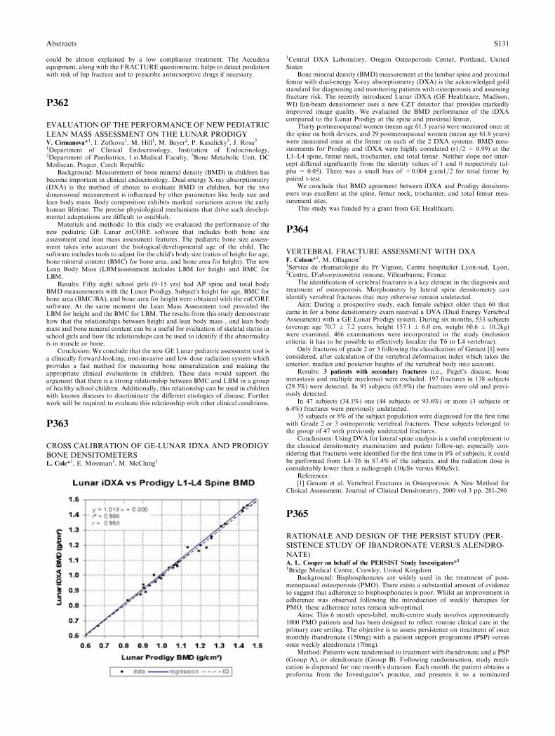
could be almost explained by a low compliance treatment. The Accudexaequipment, along with the FRACTURE questionnaire, helps to detect poulationwith risk of hip fracture and to prescribe antiresorptive drugs if necessary.
P362
EVALUATION OF THE PERFORMANCE OF NEW PEDIATRIC
LEAN MASS ASSESSMENT ON THE LUNAR PRODIGYV. Cirmanova*1, I. Zofkova1, M. Hill1, M. Bayer2, P. Kasalicky3, J. Rosa31Department of Clinical Endocrinology, Institution of Endocrinology,2Department of Paediatrics, 1.st.Medical Faculty, 3Bone Metabolic Unit, DCMediscan, Prague, Czech Republic
Background: Measurement of bone mineral density (BMD) in children hasbecome important in clinical endocrinology. Dual-energy X-ray absorptiometry(DXA) is the method of choice to evaluate BMD in children, but the twodimensional measurement is influenced by other parameters like body size andlean body mass. Body composition exhibits marked variations across the earlyhuman lifetime. The precise physiological mechanisms that drive such develop-mental adaptations are difficult to establish.
Materials and methods: In this study we evaluated the performance of thenew pediatric GE Lunar enCORE software that includes both bone sizeassessment and lean mass assessment features. The pediatric bone size assess-ment takes into account the biological/developmental age of the child. Thesoftware includes tools to adjust for the child�s body size (ratios of height for age,bone mineral content (BMC) for bone area, and bone area for height). The newLean Body Mass (LBM)assessment includes LBM for height and BMC forLBM.
Results: Fifty eight school girls (9–15 yrs) had AP spine and total bodyBMD measurements with the Lunar Prodigy. Subject�s height for age, BMC forbone area (BMC/BA), and bone area for height were obtained with the enCOREsoftware. At the same moment the Lean Mass Assessment tool provided theLBM for height and the BMC for LBM. The results from this study demonstratehow that the relationships between height and lean body mass , and lean bodymass and bone mineral content can be a useful for evaluation of skeletal status inschool girls and how the relationships can be used to identify if the abnormalityis in muscle or bone.
Conclusion: We conclude that the new GE Lunar pediatric assessment tool isa clinically forward-looking, non-invasive and low dose radiation system whichprovides a fast method for measuring bone mineralization and making theappropriate clinical evaluations in children. These data would support theargument that there is a strong relationship between BMC and LBM in a groupof healthy school children. Additionally, this relationship can be used in childrenwith known diseases to discriminate the different etiologies of disease. Furtherwork will be required to evaluate this relationship with other clinical conditions.
P363
CROSS CALIBRATION OF GE-LUNAR IDXA AND PRODIGY
BONE DENSITOMETERSL. Cole*1, E. Mossman1, M. McClung1
1Central DXA Laboratory, Oregon Osteoporosis Center, Portland, UnitedStates
Bone mineral density (BMD) measurement at the lumbar spine and proximalfemur with dual-energy X-ray absorptiometry (DXA) is the acknowledged goldstandard for diagnosing and monitoring patients with osteoporosis and assessingfracture risk. The recently introduced Lunar iDXA (GE Healthcare, Madison,WI) fan-beam densitometer uses a new CZT detector that provides markedlyimproved image quality. We evaluated the BMD performance of the iDXAcompared to the Lunar Prodigy at the spine and proximal femur.
Thirty postmenopausal women (mean age 61.3 years) were measured once atthe spine on both devices, and 29 postmenopausal women (mean age 61.8 years)were measured once at the femur on each of the 2 DXA systems. BMD mea-surements for Prodigy and iDXA were highly correlated (r1=2 = 0.99) at theL1–L4 spine, femur neck, trochanter, and total femur. Neither slope nor inter-cept differed significantly from the identity values of 1 and 0 respectively (al-pha = 0.05). There was a small bias of +0.004 g/cm1=2 for total femur bypaired t-test.
We conclude that BMD agreement between iDXA and Prodigy densitom-eters was excellent at the spine, femur neck, trochanter, and total femur mea-surement sites.
This study was funded by a grant from GE Healthcare.
P364
VERTEBRAL FRACTURE ASSESSMENT WITH DXAF. Colson*1, M. Ollagnon21Service de rhumatologie du Pr Vignon, Centre hospitalier Lyon-sud, Lyon,2Centre, D�absorptiometrie osseuse, Villeurbanne, France
The identification of vertebral fractures is a key element in the diagnosis andtreatment of osteoporosis. Morphometry by lateral spine densitometry canidentify vertebral fractures that may otherwise remain undetected.
Aim: During a prospective study, each female subject older than 60 thatcame in for a bone densitometry exam received a DVA (Dual Energy VertebralAssessment) with a GE Lunar Prodigy system. During six months, 533 subjects(average age 70.7 ± 7.2 years, height 157.1 ± 6.0 cm, weight 60.6 ± 10.2kg)were examined. 466 examinations were incorporated in the study (inclusioncriteria: it has to be possible to effectively localize the T6 to L4 vertebrae).
Only fractures of grade 2 or 3 following the classification of Genant [1] wereconsidered, after calculation of the vertebral deformation index which takes theanterior, median and posterior heights of the vertebral body into account.
Results: 3 patients with secondary fractures (i.e., Paget’s disease, bonemetastasis and multiple myeloma) were excluded. 197 fractures in 138 subjects(29.3%) were detected. In 91 subjects (65.9%) the fractures were old and previ-ously detected.
In 47 subjects (34.1%) one (44 subjects or 93.6%) or more (3 subjects or6.4%) fractures were previously undetected.
35 subjects or 8% of the subject population were diagnosed for the first timewith Grade 2 or 3 osteoporotic vertebral fractures. These subjects belonged tothe group of 47 with previously undetected fractures.
Conclusions: Using DVA for lateral spine analysis is a useful complement tothe classical densitometry examination and patient follow-up, especially con-sidering that fractures were identified for the first time in 8% of subjects, it couldbe performed from L4–T6 in 87.4% of the subjects, and the radiation dose isconsiderably lower than a radiograph (10lSv versus 800lSv).
References:[1] Genant et al. Vertebral Fractures in Osteoporosis: A New Method for
Clinical Assessment. Journal of Clinical Densitometry, 2000 vol 3 pp. 281-290
P365
RATIONALE AND DESIGN OF THE PERSIST STUDY (PER-
SISTENCE STUDY OF IBANDRONATE VERSUS ALENDRO-
NATE)A. L. Cooper on behalf of the PERSIST Study Investigators*
1
1Bridge Medical Centre, Crawley, United KingdomBackground: Bisphosphonates are widely used in the treatment of post-
menopausal osteoporosis (PMO). There exists a substantial amount of evidenceto suggest that adherence to bisphosphonates is poor. Whilst an improvement inadherence was observed following the introduction of weekly therapies forPMO, these adherence rates remain sub-optimal.
Aims: This 6 month open-label, multi-centre study involves approximately1000 PMO patients and has been designed to reflect routine clinical care in theprimary care setting. The objective is to assess persistence on treatment of oncemonthly ibandronate (150mg) with a patient support programme (PSP) versusonce weekly alendronate (70mg).
Method: Patients were randomised to treatment with ibandronate and a PSP(Group A), or alendronate (Group B). Following randomisation, study medi-cation is dispensed for one month�s duration. Each month the patient obtains aproforma from the Investigator�s practice, and presents it to a nominated
Abstracts S131
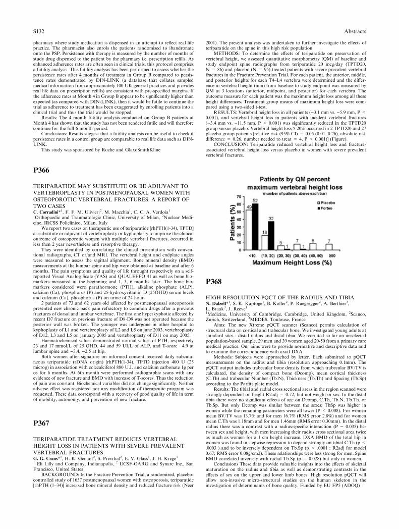
pharmacy where study medication is dispensed in an attempt to reflect real lifepractice. The pharmacist also enrols the patients randomised to ibandronateonto the PSP. Persistence with therapy is measured by the number of months ofstudy drug dispensed to the patient by the pharmacy i.e. prescription refills. Asenhanced adherence rates are often seen in clinical trials, this protocol comprisesa futility analysis. This futility analysis has been performed to assess whether thepersistence rates after 4 months of treatment in Group B compared to persis-tence rates demonstrated by DIN-LINK (a database that collates sampledmedical information from approximately 100 UK general practices and providesreal life data on prescription refills) are consistent with pre-specified margins. Ifthe adherence rates at Month 4 in Group B appear to be significantly higher thanexpected (as compared with DIN-LINK), then it would be futile to continue thetrial as adherence to treatment has been exaggerated by enrolling patients into aclinical trial and thus the trial would be stopped.
Results: The 4 month futility analysis conducted on Group B patients atMonth 4 has shown that the study has not been rendered futile and will thereforecontinue for the full 6 month period.
Conclusions: Results suggest that a futility analysis can be useful to check ifpersistence rates in a control group are comparable to real life data such as DIN-LINK.
This study was sponsored by Roche and GlaxoSmithKline
P366
TERIPARATIDE MAY SUBSTITUTE OR BE ADIUVANT TO
VERTEBROPLASTY IN POSTMENOPAUSAL WOMEN WITH
OSTEOPOROTIC VERTEBRAL FRACTURES: A REPORT OF
TWO CASESC. Corradini*1, F. F. M. Ulivieri2, M. Macchia1, C. C. A. Verdoia11Orthopaedic and Traumatologic Clinic, University of Milan, 2Nuclear Medi-cine, IRCSS Policlinico, Milan, Italy
We report two cases on therapeutic use of teriparatide [rhPTH(1-34), TPTD]as substitute or adjuvant of vertebroplasty or kyphoplasty to improve the clinicaloutcome of osteoporotic women with multiple vertebral fractures, occurred inless then 2 year nevertheless anti resorptive therapy.
They were identified by correlating the clinical presentation with conven-tional radiographs, CT or/and MRI. The vertebral height and endplate angleswere measured to assess the sagittal alignment. Bone mineral density (BMD)measurements at the lumbar spine and hip were obtained at baseline and after 6months. The pain symptoms and quality of life throught respectively on a self-reported Visual Analog Scale (VAS) and QUALEFFO 41 as well as bone bio-markers measured at the beginning and 1, 3, 6 months later. The bone bio-markers considered were parathormone (PTH), alkaline phosphate (ALP),calcium (Ca), phosphorus (P) and 25-hydroxyvitamin D (25OHD) serum levelsand calcium (Ca), phosphorus (P) on urine of 24 hours.
2 patients of 73 and 62 years old affected by postmenopausal osteoporosispresented new chronic back pain refractory to common drugs after a previousfractures of dorsal and lumbar vertebrae. The first one hyperkyphotic affected byrecent D7 fracture on previous fracture of D8-D9 was not operated because theposterior wall was broken. The younger was undergone in other hospital tokyphoplasty of L1 and vertebroplasty of L2 and L5 on june 2003, vertebroplastyof D12, L3 and L5 on january 2005 and vertebroplasty of D11 on may 2005.
Haematochemical values demonstrated normal values of PTH, respectively23 and 17 mmol/L of 25 OHD, 44 and 59 UI/L of ALP, and T-score )4.9 atlumbar spine and )3.4, )2.5 at hip.
Both women after signature on informed consent received daily subcuta-neous teriparatide (rDNA origin) [rhPTH(1-34), TPTD injection 400 U (25microg) in association with colecalciferol 880 U.I. and calcium carbonate 1g peros for 6 months. At 6th month were performed radiographic scans with anyevidence of new fracture and BMD with increase of T-scores. Thus the reductionof pain was constant. Biochemical variables did not change significantly. Neitheradverse effect was registered nor any modification of therapeutic program wasrequested. These data correspond with a recovery of good quality of life in termof mobility, autonomy, and prevention of new fracture.
P367
TERIPARATIDE TREATMENT REDUCES VERTEBRAL
HEIGHT LOSS IN PATIENTS WITH SEVERE PREVALENT
VERTEBRAL FRACTURESG. G. Crans*1, H. K. Genant2, S. Prevrhal2, E. V. Glass1, J. H. Krege11 Eli Lilly and Company, Indianapolis, 2 UCSF-OARG and Synarc Inc., SanFrancisco, United States
BACKGROUND: In the Fracture Prevention Trial, a randomized, placebo-controlled study of 1637 postmenopausal women with osteoporosis, teriparatide[rhPTH (1–34)] increased bone mineral density and reduced fracture risk (Neer
2001). The present analysis was undertaken to further investigate the effects ofteriparatide on the spine in this high risk population.
METHODS: To determine the effects of teriparatide on preservation ofvertebral height, we assessed quantitative morphometry (QM) of baseline andstudy endpoint spine radiographs from teriparatide 20 mcg/day (TPTD20,N = 86) and placebo (N = 95) treated patients with severe prevalent vertebralfractures in the Fracture Prevention Trial. For each patient, the anterior, middle,and posterior heights for each T4–L4 vertebra were determined and the differ-ence in vertebral height (mm) from baseline to study endpoint was measured byQM at 3 locations (anterior, midpoint, and posterior) for each vertebra. Theoutcome measure for each patient was the maximum height loss among all theseheight differences. Treatment group means of maximum height loss were com-pared using a two-sided t-test.
RESULTS: Vertebral height loss in all patients ()3.1 mm vs. )5.9 mm, P <0.001), and vertebral height loss in patients with incident vertebral fractures()3.4 mm vs. )11.5 mm, P < 0.001) was significantly reduced in the TPTD20group versus placebo. Vertebral height loss ‡ 20% occurred in 2 TPTD20 and 27placebo group patients [relative risk (95% CI) = 0.05 (0.01, 0.26), absolute riskdifference = 0.26, number needed to treat = 4, P < 0.001)] (Figure).
CONCLUSION: Teriparatide reduced vertebral height loss and fracture-associated vertebral height loss versus placebo in women with severe prevalentvertebral fractures.
P368
HIGH RESOLUTION PQCT OF THE RADIUS AND TIBIAN. Dalzell*1, S. K. Kaptoge1, B. Koller2, P. Ruegsegger2, A. Berthier3,L. Braak3, J. Reeve11Medicine, University of Cambridge, Cambridge, United Kingdom, 2Scanco,Zurich, Switzerland, 3MEDES, Toulouse, France
Aims: The new Xtreme pQCT scanner (Scanco) permits calculation ofstructural data on cortical and trabecular bone. We investigated young adults atstandard sites - distal radius and distal tibia. We recruited so far an unselectedpopulation-based sample, 29 men and 39 women aged 20-50 from a primary caremedical practice. Our aims were to provide normative and descriptive data andto examine the correspondence with axial DXA.
Methods: Subjects were approached by letter. Each submitted to pQCTmeasurements on the radius and tibia (resolution approaching 0.1mm). ThepQCT output includes trabecular bone density from which trabecular BV/TV iscalculated, the density of compact bone (Dcomp), mean cortical thickness(C.Th) and trabecular Number (Tb.N), Thickness (Tb.Th) and Spacing (Tb.Sp)according to the Parfitt plate model.
Results: The tibial and radial cross sectional areas in the region scanned werestrongly dependent on height R2adj = 0.72, but not weight or sex. In the distaltibia there were no significant effects of age on Dcomp, C.Th, Tb.N, Tb.Th, orTb.Sp. But only Dcomp was similar between the sexes; TbSp was higher inwomen while the remaining parameters were all lower (P < 0.008). For womenmean BV/TV was 13.7% and for men 16.7% (RMS error 2.8%) and for womenmean C.Th was 1.18mm and for men 1.46mm (RMS error 0.30mm). In the distalradius there was a contrast with a radius-specific interaction (P = 0.035) be-tween sex and height, with men increasing their radius cross sectional area twiceas much as women for a 1 cm height increase. DXA BMD of the total hip inwomen was found in stepwise regression to depend strongly on tibial C.Th (p <.0003 ) and to be inversely dependent on Tb.Sp (p < .0001 ; R2adj for model0.67; RMS error 0.08g/cm2). These relationships were less strong for men. SpineBMD correlated inversely with radial Tb.Sp (p = 0.028) but only in women.
Conclusions These data provide valuable insights into the effects of skeletalmaturation on the radius and tibia as well as demonstrating contrasts in theeffects of sex on the upper and lower limb bones. High resolution pQCT willallow non-invasive micro-structural studies on the human skeleton in theinvestigation of determinants of bone quality. Funded by EU FP5 (ADOQ)
S132 Abstracts
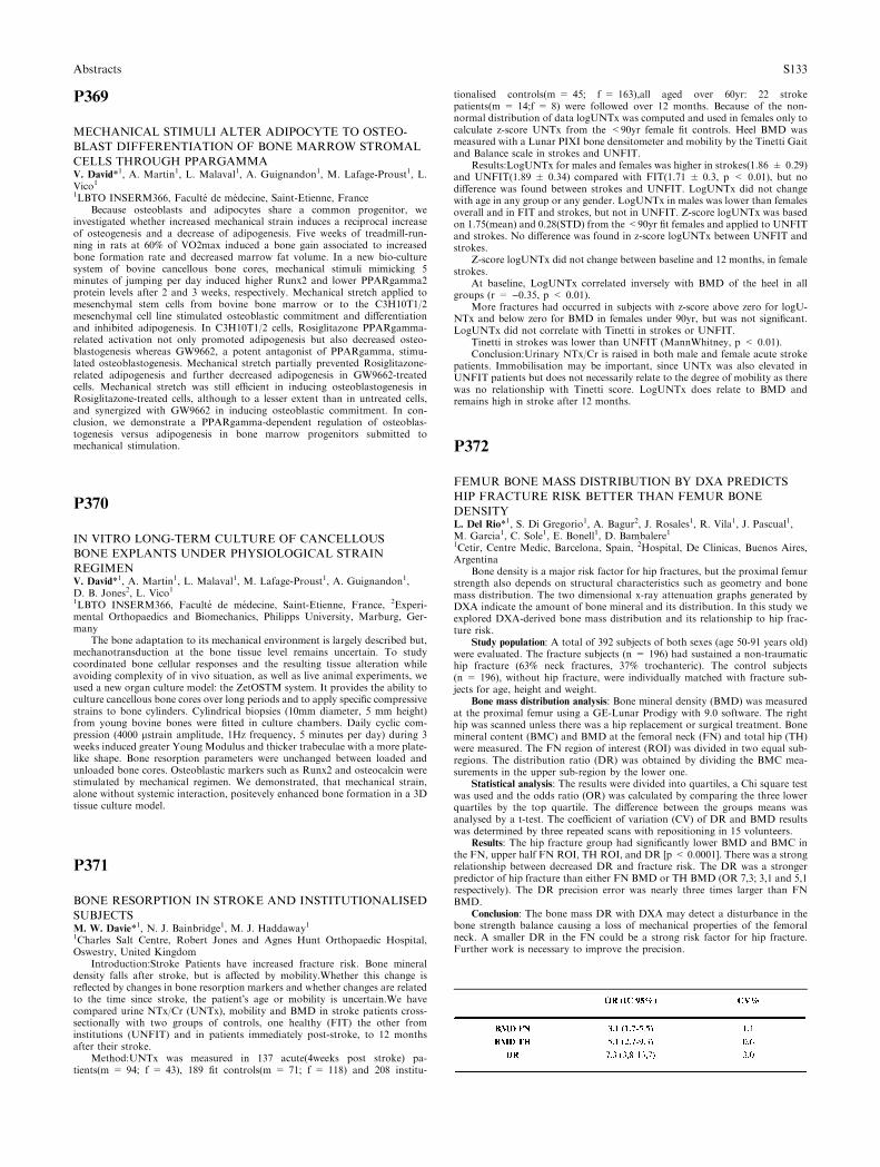
P369
MECHANICAL STIMULI ALTER ADIPOCYTE TO OSTEO-
BLAST DIFFERENTIATION OF BONE MARROW STROMAL
CELLS THROUGH PPARGAMMAV. David*1, A. Martin1, L. Malaval1, A. Guignandon1, M. Lafage-Proust1, L.Vico11LBTO INSERM366, Faculte de medecine, Saint-Etienne, France
Because osteoblasts and adipocytes share a common progenitor, weinvestigated whether increased mechanical strain induces a reciprocal increaseof osteogenesis and a decrease of adipogenesis. Five weeks of treadmill-run-ning in rats at 60% of VO2max induced a bone gain associated to increasedbone formation rate and decreased marrow fat volume. In a new bio-culturesystem of bovine cancellous bone cores, mechanical stimuli mimicking 5minutes of jumping per day induced higher Runx2 and lower PPARgamma2protein levels after 2 and 3 weeks, respectively. Mechanical stretch applied tomesenchymal stem cells from bovine bone marrow or to the C3H10T1/2mesenchymal cell line stimulated osteoblastic commitment and differentiationand inhibited adipogenesis. In C3H10T1/2 cells, Rosiglitazone PPARgamma-related activation not only promoted adipogenesis but also decreased osteo-blastogenesis whereas GW9662, a potent antagonist of PPARgamma, stimu-lated osteoblastogenesis. Mechanical stretch partially prevented Rosiglitazone-related adipogenesis and further decreased adipogenesis in GW9662-treatedcells. Mechanical stretch was still efficient in inducing osteoblastogenesis inRosiglitazone-treated cells, although to a lesser extent than in untreated cells,and synergized with GW9662 in inducing osteoblastic commitment. In con-clusion, we demonstrate a PPARgamma-dependent regulation of osteoblas-togenesis versus adipogenesis in bone marrow progenitors submitted tomechanical stimulation.
P370
IN VITRO LONG-TERM CULTURE OF CANCELLOUS
BONE EXPLANTS UNDER PHYSIOLOGICAL STRAIN
REGIMENV. David*1, A. Martin1, L. Malaval1, M. Lafage-Proust1, A. Guignandon1,D. B. Jones2, L. Vico11LBTO INSERM366, Faculte de medecine, Saint-Etienne, France, 2Experi-mental Orthopaedics and Biomechanics, Philipps University, Marburg, Ger-many
The bone adaptation to its mechanical environment is largely described but,mechanotransduction at the bone tissue level remains uncertain. To studycoordinated bone cellular responses and the resulting tissue alteration whileavoiding complexity of in vivo situation, as well as live animal experiments, weused a new organ culture model: the ZetOSTM system. It provides the ability toculture cancellous bone cores over long periods and to apply specific compressivestrains to bone cylinders. Cylindrical biopsies (10mm diameter, 5 mm height)from young bovine bones were fitted in culture chambers. Daily cyclic com-pression (4000 lstrain amplitude, 1Hz frequency, 5 minutes per day) during 3weeks induced greater Young Modulus and thicker trabeculae with a more plate-like shape. Bone resorption parameters were unchanged between loaded andunloaded bone cores. Osteoblastic markers such as Runx2 and osteocalcin werestimulated by mechanical regimen. We demonstrated, that mechanical strain,alone without systemic interaction, positevely enhanced bone formation in a 3Dtissue culture model.
P371
BONE RESORPTION IN STROKE AND INSTITUTIONALISED
SUBJECTSM. W. Davie*1, N. J. Bainbridge1, M. J. Haddaway11Charles Salt Centre, Robert Jones and Agnes Hunt Orthopaedic Hospital,Oswestry, United Kingdom
Introduction:Stroke Patients have increased fracture risk. Bone mineraldensity falls after stroke, but is affected by mobility.Whether this change isreflected by changes in bone resorption markers and whether changes are relatedto the time since stroke, the patient�s age or mobility is uncertain.We havecompared urine NTx/Cr (UNTx), mobility and BMD in stroke patients cross-sectionally with two groups of controls, one healthy (FIT) the other frominstitutions (UNFIT) and in patients immediately post-stroke, to 12 monthsafter their stroke.
Method:UNTx was measured in 137 acute(4weeks post stroke) pa-tients(m = 94; f = 43), 189 fit controls(m = 71; f = 118) and 208 institu-
tionalised controls(m = 45; f = 163),all aged over 60yr: 22 strokepatients(m = 14;f = 8) were followed over 12 months. Because of the non-normal distribution of data logUNTx was computed and used in females only tocalculate z-score UNTx from the <90yr female fit controls. Heel BMD wasmeasured with a Lunar PIXI bone densitometer and mobility by the Tinetti Gaitand Balance scale in strokes and UNFIT.
Results:LogUNTx for males and females was higher in strokes(1.86 ± 0.29)and UNFIT(1.89 ± 0.34) compared with FIT(1.71 ± 0.3, p < 0.01), but nodifference was found between strokes and UNFIT. LogUNTx did not changewith age in any group or any gender. LogUNTx in males was lower than femalesoverall and in FIT and strokes, but not in UNFIT. Z-score logUNTx was basedon 1.75(mean) and 0.28(STD) from the <90yr fit females and applied to UNFITand strokes. No difference was found in z-score logUNTx between UNFIT andstrokes.
Z-score logUNTx did not change between baseline and 12 months, in femalestrokes.
At baseline, LogUNTx correlated inversely with BMD of the heel in allgroups (r = )0.35, p < 0.01).
More fractures had occurred in subjects with z-score above zero for logU-NTx and below zero for BMD in females under 90yr, but was not significant.LogUNTx did not correlate with Tinetti in strokes or UNFIT.
Tinetti in strokes was lower than UNFIT (MannWhitney, p < 0.01).Conclusion:Urinary NTx/Cr is raised in both male and female acute stroke
patients. Immobilisation may be important, since UNTx was also elevated inUNFIT patients but does not necessarily relate to the degree of mobility as therewas no relationship with Tinetti score. LogUNTx does relate to BMD andremains high in stroke after 12 months.
P372
FEMUR BONE MASS DISTRIBUTION BY DXA PREDICTS
HIP FRACTURE RISK BETTER THAN FEMUR BONE
DENSITYL. Del Rio*1, S. Di Gregorio1, A. Bagur2, J. Rosales1, R. Vila1, J. Pascual1,M. Garcia1, C. Sole1, E. Bonell1, D. Bambalere11Cetir, Centre Medic, Barcelona, Spain, 2Hospital, De Clinicas, Buenos Aires,Argentina
Bone density is a major risk factor for hip fractures, but the proximal femurstrength also depends on structural characteristics such as geometry and bonemass distribution. The two dimensional x-ray attenuation graphs generated byDXA indicate the amount of bone mineral and its distribution. In this study weexplored DXA-derived bone mass distribution and its relationship to hip frac-ture risk.
Study population: A total of 392 subjects of both sexes (age 50-91 years old)were evaluated. The fracture subjects (n = 196) had sustained a non-traumatichip fracture (63% neck fractures, 37% trochanteric). The control subjects(n = 196), without hip fracture, were individually matched with fracture sub-jects for age, height and weight.
Bone mass distribution analysis: Bone mineral density (BMD) was measuredat the proximal femur using a GE-Lunar Prodigy with 9.0 software. The righthip was scanned unless there was a hip replacement or surgical treatment. Bonemineral content (BMC) and BMD at the femoral neck (FN) and total hip (TH)were measured. The FN region of interest (ROI) was divided in two equal sub-regions. The distribution ratio (DR) was obtained by dividing the BMC mea-surements in the upper sub-region by the lower one.
Statistical analysis: The results were divided into quartiles, a Chi square testwas used and the odds ratio (OR) was calculated by comparing the three lowerquartiles by the top quartile. The difference between the groups means wasanalysed by a t-test. The coefficient of variation (CV) of DR and BMD resultswas determined by three repeated scans with repositioning in 15 volunteers.
Results: The hip fracture group had significantly lower BMD and BMC inthe FN, upper half FN ROI, TH ROI, and DR [p < 0.0001]. There was a strongrelationship between decreased DR and fracture risk. The DR was a strongerpredictor of hip fracture than either FN BMD or TH BMD (OR 7,3; 3,1 and 5,1respectively). The DR precision error was nearly three times larger than FNBMD.
Conclusion: The bone mass DR with DXA may detect a disturbance in thebone strength balance causing a loss of mechanical properties of the femoralneck. A smaller DR in the FN could be a strong risk factor for hip fracture.Further work is necessary to improve the precision.
Abstracts S133
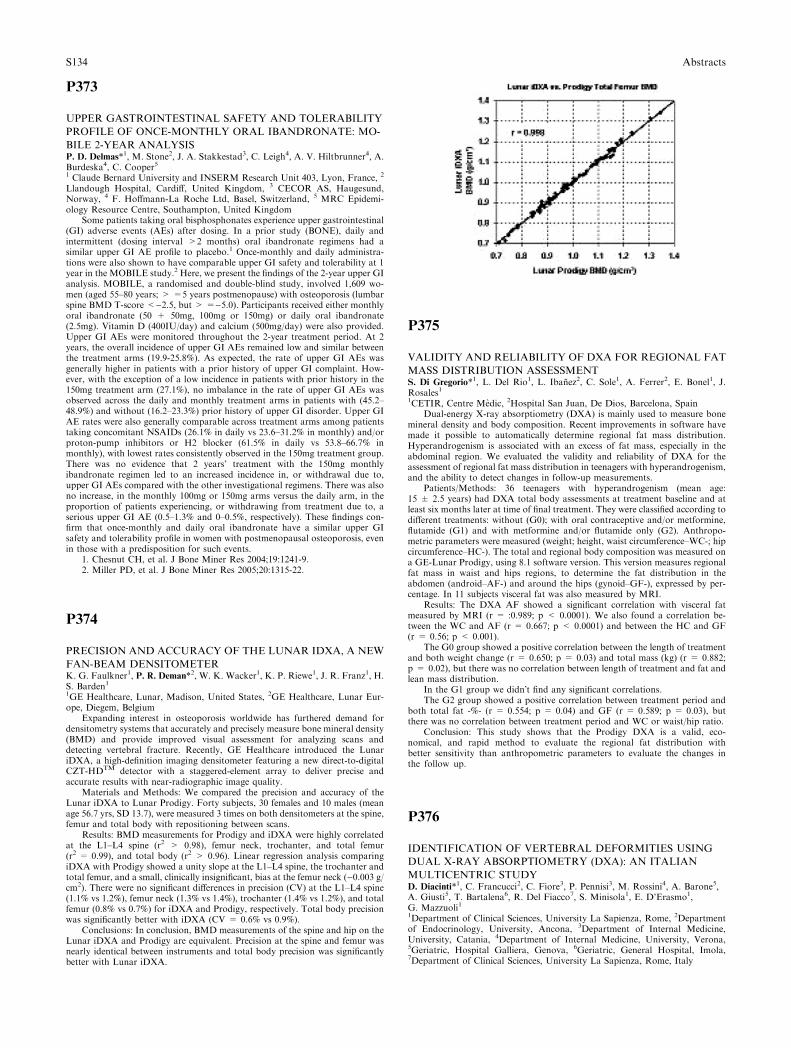
P373
UPPER GASTROINTESTINAL SAFETY AND TOLERABILITY
PROFILE OF ONCE-MONTHLY ORAL IBANDRONATE: MO-
BILE 2-YEAR ANALYSISP. D. Delmas*1, M. Stone2, J. A. Stakkestad3, C. Leigh4, A. V. Hiltbrunner4, A.Burdeska4, C. Cooper51 Claude Bernard University and INSERM Research Unit 403, Lyon, France, 2
Llandough Hospital, Cardiff, United Kingdom, 3 CECOR AS, Haugesund,Norway, 4 F. Hoffmann-La Roche Ltd, Basel, Switzerland, 5 MRC Epidemi-ology Resource Centre, Southampton, United Kingdom
Some patients taking oral bisphosphonates experience upper gastrointestinal(GI) adverse events (AEs) after dosing. In a prior study (BONE), daily andintermittent (dosing interval >2 months) oral ibandronate regimens had asimilar upper GI AE profile to placebo.1 Once-monthly and daily administra-tions were also shown to have comparable upper GI safety and tolerability at 1year in the MOBILE study.2 Here, we present the findings of the 2-year upper GIanalysis. MOBILE, a randomised and double-blind study, involved 1,609 wo-men (aged 55–80 years; > =5 years postmenopause) with osteoporosis (lumbarspine BMD T-score <)2.5, but > =)5.0). Participants received either monthlyoral ibandronate (50 + 50mg, 100mg or 150mg) or daily oral ibandronate(2.5mg). Vitamin D (400IU/day) and calcium (500mg/day) were also provided.Upper GI AEs were monitored throughout the 2-year treatment period. At 2years, the overall incidence of upper GI AEs remained low and similar betweenthe treatment arms (19.9-25.8%). As expected, the rate of upper GI AEs wasgenerally higher in patients with a prior history of upper GI complaint. How-ever, with the exception of a low incidence in patients with prior history in the150mg treatment arm (27.1%), no imbalance in the rate of upper GI AEs wasobserved across the daily and monthly treatment arms in patients with (45.2–48.9%) and without (16.2–23.3%) prior history of upper GI disorder. Upper GIAE rates were also generally comparable across treatment arms among patientstaking concomitant NSAIDs (26.1% in daily vs 23.6–31.2% in monthly) and/orproton-pump inhibitors or H2 blocker (61.5% in daily vs 53.8–66.7% inmonthly), with lowest rates consistently observed in the 150mg treatment group.There was no evidence that 2 years’ treatment with the 150mg monthlyibandronate regimen led to an increased incidence in, or withdrawal due to,upper GI AEs compared with the other investigational regimens. There was alsono increase, in the monthly 100mg or 150mg arms versus the daily arm, in theproportion of patients experiencing, or withdrawing from treatment due to, aserious upper GI AE (0.5–1.3% and 0–0.5%, respectively). These findings con-firm that once-monthly and daily oral ibandronate have a similar upper GIsafety and tolerability profile in women with postmenopausal osteoporosis, evenin those with a predisposition for such events.
1. Chesnut CH, et al. J Bone Miner Res 2004;19:1241-9.2. Miller PD, et al. J Bone Miner Res 2005;20:1315-22.
P374
PRECISION AND ACCURACY OF THE LUNAR IDXA, A NEW
FAN-BEAM DENSITOMETERK. G. Faulkner1, P. R. Deman*2, W. K. Wacker1, K. P. Riewe1, J. R. Franz1, H.S. Barden11GE Healthcare, Lunar, Madison, United States, 2GE Healthcare, Lunar Eur-ope, Diegem, Belgium
Expanding interest in osteoporosis worldwide has furthered demand fordensitometry systems that accurately and precisely measure bone mineral density(BMD) and provide improved visual assessment for analyzing scans anddetecting vertebral fracture. Recently, GE Healthcare introduced the LunariDXA, a high-definition imaging densitometer featuring a new direct-to-digitalCZT-HDTM detector with a staggered-element array to deliver precise andaccurate results with near-radiographic image quality.
Materials and Methods: We compared the precision and accuracy of theLunar iDXA to Lunar Prodigy. Forty subjects, 30 females and 10 males (meanage 56.7 yrs, SD 13.7), were measured 3 times on both densitometers at the spine,femur and total body with repositioning between scans.
Results: BMD measurements for Prodigy and iDXA were highly correlatedat the L1–L4 spine (r2 > 0.98), femur neck, trochanter, and total femur(r2 = 0.99), and total body (r2 > 0.96). Linear regression analysis comparingiDXA with Prodigy showed a unity slope at the L1–L4 spine, the trochanter andtotal femur, and a small, clinically insignificant, bias at the femur neck ()0.003 g/cm2). There were no significant differences in precision (CV) at the L1–L4 spine(1.1% vs 1.2%), femur neck (1.3% vs 1.4%), trochanter (1.4% vs 1.2%), and totalfemur (0.8% vs 0.7%) for iDXA and Prodigy, respectively. Total body precisionwas significantly better with iDXA (CV = 0.6% vs 0.9%).
Conclusions: In conclusion, BMD measurements of the spine and hip on theLunar iDXA and Prodigy are equivalent. Precision at the spine and femur wasnearly identical between instruments and total body precision was significantlybetter with Lunar iDXA.
P375
VALIDITY AND RELIABILITY OF DXA FOR REGIONAL FAT
MASS DISTRIBUTION ASSESSMENTS. Di Gregorio*1, L. Del Rio1, L. Ibanez2, C. Sole1, A. Ferrer2, E. Bonel1, J.Rosales11CETIR, Centre Medic, 2Hospital San Juan, De Dios, Barcelona, Spain
Dual-energy X-ray absorptiometry (DXA) is mainly used to measure bonemineral density and body composition. Recent improvements in software havemade it possible to automatically determine regional fat mass distribution.Hyperandrogenism is associated with an excess of fat mass, especially in theabdominal region. We evaluated the validity and reliability of DXA for theassessment of regional fat mass distribution in teenagers with hyperandrogenism,and the ability to detect changes in follow-up measurements.
Patients/Methods: 36 teenagers with hyperandrogenism (mean age:15 ± 2.5 years) had DXA total body assessments at treatment baseline and atleast six months later at time of final treatment. They were classified according todifferent treatments: without (G0); with oral contraceptive and/or metformine,flutamide (G1) and with metformine and/or flutamide only (G2). Anthropo-metric parameters were measured (weight; height, waist circumference–WC-; hipcircumference–HC-). The total and regional body composition was measured ona GE-Lunar Prodigy, using 8.1 software version. This version measures regionalfat mass in waist and hips regions, to determine the fat distribution in theabdomen (android–AF-) and around the hips (gynoid–GF-), expressed by per-centage. In 11 subjects visceral fat was also measured by MRI.
Results: The DXA AF showed a significant correlation with visceral fatmeasured by MRI (r = :0.989; p < 0.0001). We also found a correlation be-tween the WC and AF (r = 0.667; p < 0.0001) and between the HC and GF(r = 0.56; p < 0.001).
The G0 group showed a positive correlation between the length of treatmentand both weight change (r = 0.650; p = 0.03) and total mass (kg) (r = 0.882;p = 0.02), but there was no correlation between length of treatment and fat andlean mass distribution.
In the G1 group we didn�t find any significant correlations.The G2 group showed a positive correlation between treatment period and
both total fat -%- (r = 0.554; p = 0.04) and GF (r = 0.589; p = 0.03), butthere was no correlation between treatment period and WC or waist/hip ratio.
Conclusion: This study shows that the Prodigy DXA is a valid, eco-nomical, and rapid method to evaluate the regional fat distribution withbetter sensitivity than anthropometric parameters to evaluate the changes inthe follow up.
P376
IDENTIFICATION OF VERTEBRAL DEFORMITIES USING
DUAL X-RAY ABSORPTIOMETRY (DXA): AN ITALIAN
MULTICENTRIC STUDYD. Diacinti*1, C. Francucci2, C. Fiore3, P. Pennisi3, M. Rossini4, A. Barone5,A. Giusti5, T. Bartalena6, R. Del Fiacco7, S. Minisola1, E. D’Erasmo1,G. Mazzuoli11Department of Clinical Sciences, University La Sapienza, Rome, 2Departmentof Endocrinology, University, Ancona, 3Department of Internal Medicine,University, Catania, 4Department of Internal Medicine, University, Verona,5Geriatric, Hospital Galliera, Genova, 6Geriatric, General Hospital, Imola,7Department of Clinical Sciences, University La Sapienza, Rome, Italy
S134 Abstracts
Aim: to evaluate in a sample of Italian population the prevalence ofasymptomatic vertebral deformities using visual assessment (VFA) of DXAimages . Methods : the 6 centres participating in the study (Ancona, Ca-tania, Genova, Imola, Roma, Verona) enrolled 1320 postmenopausal women,aged 45–82 years (65 ± 9yrs) consecutively referred to bone densitometry ofeach Centre. All women had spine DXA scans images, two centres using theHologic QDR4500A, four centres using the GE Lunar Prodigy. Both sys-tems acquired spine images with dual-energy (high-definition).Subjects wereincluded if at least 12 out of 13 vertebrae from T4 to L4 could be suffi-ciently visualized. For VFA, DXA images was centrally analyzed by anexperienced radiologist according to the Genant�s visual semiquantitativemethod1. Vertebral deformities were graded as mild, moderate, or severebased on the degree of reduction of anterior/posterior (ha/hp=wedging) ormiddle/posterior (hm/hp=biconcavity) heights ratios or reduction of globalvertebral height (crush).Results: Intra-operator precision was found to besimilar on the two systems, Hologic and GE Lunar: CV% was 1.7% for theposterior height and 2.2% for the anterior height.159 (12%) of 1320 totalwomen enrolled had vertebral deformities, 105/159 (66%) had at least onemoderate or severe deformity. Prevalence of vertebral deformity increasedwith aging, until to 35% in women over 75 yrs. Of all vertebrae imaged byDXA only 2% resulted unanalyzable because of poor image quality in theupper thoracic spine. A total of 300 vertebral deformities were identified,occurring most commonly at the T12–L1 level.100 deformities were classifiedas mild, 135 as moderate, and 65 were severe. Conclusion: this multicentricstudy demonstrated that visual assessment of spine DXA images was able toidentificate vertebral deformities; the majority of the asymptomatic vertebraldeformities were moderate or severe with increased risk of future frac-tures that may be symptomatic; therefore the use of VFA of DXA imagesbecause of rapid performance, convenience, and minimal radiation exposureshould be encouraged both in clinical practice and in epidemiological stud-ies.
1 Genant HK et al. J Bone Miner Res 1996; 11: 984-996.
P377
EFFECTS OF 12 MONTHS GROWTH HORMONE
ADMINISTRATION ON MARKERS OF BONE TURNOVER
AND PHOSPHOCALCIUM METABOLSIM IN WOMEN WITH
ESTABLISHED OSTEOPOROSISB. H. Durham*1, F. Joseph2, A. M. Ahmed2, H. D. White2, A. Joshi2,J. P. Vora2, W. D. Fraser11Clinical Biochemistry and Metabolic Medicine, 2Diabetes and Endocrinolgy,Royal Liverpool University Hospital, Liverpool, United Kingdom
The combination of increased bone resorption and decreased bone forma-tion results in the development of osteoporosis in postmenopausal women. It hasbeen reported that the concomitant increase in bone resorption markers andformation markers demonstrated in short term growth hormone [GH] admin-istration in women with osteoporosis resulted in minimal or no increase in BMD.We investigated the effects of 12 months of GH administration on bone for-mation and resorption markers, PTH sensitivity and phosphocalcium metabo-lism in aging osteoporotic women.
Fourteen postmenopausal women age 63.4 ± 2.1 yr [mean ± SEM], range52–79 yr; mean BMD T score ± SEM at lumbar spine (L2–L4) was )3.3 ± 0.2and at femoral neck )2.0 ± 0.2 were commenced on 0.2mg of GH/day. Patientswere hospitalised for 24 hours before then 1,3,6 and 12 months following GHR.Half-hourly blood and 3-hourly urine samples were collected for PTH, calcium,phosphate,NcAMP [marker of renal PTH activity]. CTX [bone resorptionmarker], PINP and osteocalcin [bone formation markers] and 1,25(OH)VitDwere measured on 0700h fasting samples.
IGF-1 concentrations increased significantly [p < 0.001]. CTX increasedprogressively from 0.7 ± 0.08mcg/L at baseline to 0.8 ± 0.07mcg/L at 1m, [p< 0.01] and 1.1 ± 0.09mcg/L at 3m, [p < 0.01] with no further significantincrease at 6 or 12 m. PINP concentration increased progressively from56 ± 6mcg/L at baseline to 126 ± 10mcg/L at 6m [p < 0.001] with nofurther significant increase at 12 m. Osteocalcin showed a similar pattern ofincrease, 34.1 ± 3.3mcg/L at baseline to 61.2 ± 4.1mcg/L at 6m [p < 0.001].The percentage increase in PINP was significantly higher than that for CTXfollowing 6 and 12 months of GH as was the percentage increase in osteo-calcin. These changes were accompanied by a decrease in 24-h mean PTHconcentration and an increase in NcAMP, adjusted serum calcium, phosphateand 1,25(OH)VitD.
GH administration to osteoporotic women results in relatively greater in-creases in bone formation than resorption. Improved target organ sensitivity toPTH and improved phosphocalcium metabolism contribute to this beneficialeffect of GH. These changes may explain the previously demonstrated increase inBMD following long term [> 12 m] administration of GH in women withosteoporosis.
P378
PREVALENCE OF VITAMIN D INSUFFICIENCY IN DANISH
ADULTS WITH FRAGILITY HIP FRACTURESM. Bogh1, P. Eiken*21Orthopaedic Surgery, 2Endocrinology, Hillerod Hospital, Hillerod, Denmark
Vitamin D insufficiency is defined as values below 50 nmol/l. Fracture riskand vitamin D insufficiency increases with age and is among elderly living in thecommunity a major public health problem. A number of studies have reported arelationship between vitamin D insufficiency and hip fractures. During the lastyears, the recommended intake of vitamin D has increased in Denmark (now 7.5lg/day among people > 60 years, 20 lg/day among institutionalised persons),and vitamin D has received more focus. Food is not fortified in Denmark.
Results of serum 25-hydroxyvitamin D analysis were reviewed for 50 con-secutive patients (median age 77 years, range 50 to 96 years) presented to ourdepartment with low energy hip fractures. 68% had 25-hydroxyvitamin D below50 nmol/l and all patients had 25-hydroxyvitamin D below 90 nmol/l. 24% hadsecondary hyperparathyroidism. Only 24% of the patients had received supple-mentary calcium and vitamin D (>10 lg/day) within the last year or morebefore admission. Among these patients, 42% had vitamin D insufficiency. 76%did not receive any supplementary calcium and vitamin D.
It can be concluded that vitamin D insufficiency is extremely commonamong patients with hip fractures. Only one out of four patients are recievingsupplementary calcium and vitamin D even though vitamin D has received moreattention recently. Intake of 10 lg/day for more than one year is not enough toincrease 25-hydroxyvitamin D sufficiently in a population of patients with hipfractures. Awareness of the efficacy of calcium and vitamin D is still low, andfurther work need to be done to increase awareness among patients, physicians,and people at risk.
P379
COMPARISON OF SERUM TARTRATE-RESISTANT ACID
PHOSPHATASE ASSAYS FOR MONITORING ALENDRO-
NATE TREATMENTK. M. Fagerlund*1, A. J. Janckila2, H. Ylipahkala1, S. L. Tiitinen3, L. T. Yam2,H. K. Vaananen1, J. M. Halleen41Department of Anatomy, University of Turku, Institute of Biomedicine, Turku,Finland, 2Special Hematology Laboratory, Veterans Affairs Medical Center,Louisville, United States, 3Finnish Red Cross, Blood Service, Helsinki, 4Phar-matest Services Ltd, Turku, Finland
Two forms of tartrate-resistant acid phosphatase (TRACP) circulate inhuman blood, TRACP 5a derived from inflammatory macrophages and TRACP5b derived from bone-resorbing osteoclasts. We studied the clinical performanceof six different serum TRACP assays for monitoring alendronate treatment, andcompared them with other commonly used markers of bone turnover, includingserum CTX, PINP, BAP, osteocalcin and urinary DPD. This double-blindedstudy included 137 healthy postmenopausal women that were randomly assignedinto two groups, one receiving 5 mg alendronate daily (n = 70), and the otherreceiving placebo (n = 67). The TRACP assays were as follows: 1) A pH-selective immunoassay measuring TRACP 5b activity (BoneTRAP�, SBA-Sci-ences, Oulu, Finland); 2) An immunoassay measuring total TRACP activity(TRAP5, Biovendor, Brno, Czech Republic); 3) An immunoassay using naph-thol-ASBI-phosphate as a selective substrate for TRACP 5b (ASBI); 4) Animmunoassay measuring TRACP 5a activity (5a-Act); 5) An immunoassaymeasuring total TRACP protein amount (Tot-Prot); 6) An immunoassay mea-suring TRACP 5a protein amount (5a-Prot). All bone markers were assessed atbaseline and at 3 months after start of treatment.
Based on clinical sensitivity, area under ROC curve (AUC) and signal-to-noise ratio, the BoneTRAP� assay, CTX and PINP were the best markers formonitoring alendronate treatment. Alendronate decreased serum TRACPmeasured by BoneTRAP�, TRAP5 and ASBI, best performance being observedwith BoneTRAP�. Tot-Prot, 5a-Act and 5a-Prot were not affected by alendr-onate treatment. These results demonstrate that serum TRACP 5b is an excellentmarker for monitoring alendronate treatment, serum TRACP 5a is not associ-ated with bone turnover, and TRACP 5b specific methods should be used inassessing bone resorption.
Abstracts S135
P380
VITAMIN D INADEQUACY IN PATIENTES WHO ARE
SCREENED FOR OSTEOPOROSISD. Fernandez-Garcıa*1, R. Reyes-Garcıa1, G. Alonso1, P. Rozas1, A. Sebastian-Ochoa1, M. Ruiz-Requena2, M. Munoz-Torres11Bone Metabolic Unit. Endocrinology Division, 2Biochemistry Department,University Hospital San Cecilio., Granada, Spain
Low concentrations of vitamin D leads to secondary hiperparathyroidism,bone loss, and an increase of osteoporotic fractures in populations at risk.Adequate vitamin D and calcium intake is considered an essential component ofpostmenopausal osteoporosis management. Several epidemiological studies haveassessed the prevalence of low serum vitamin D concentrations, indicating thatvitamin D inadequacy (<30 ng/ml) is an usual problem world-wide.
AIMS: To evaluate vitamin D inadequacy at baseline in patients who arescreened for osteoporosis, and in postmenopausal women with osteoporosisafter one year of treatment.
PATIENTS AND METHODS: In 126 postmenopausal women (mean age63 ± 7 years) who were evaluated for osteoporosis at the Bone Metabolic Unitwe determined: BMD by DXA (Hologic QDR 4500w) at lumbar spine, femoralneck and total hip, basic anthropometric parameters, biochemical markers ofbone turnover, PTH and 25(OH) vitamin D. 76% of the women were diagnosedof osteoporosis (T-score )2.5 SD) and all of them started treatment with ant-irresoptives, calcium and vitamin D ( 1200 mg and 800 UI daily).
RESULTS: At baseline 90% of the women had serum levels of 25 (OH)vitamin D less than 30 ng/ml, and 42% less than 15 ng/ml. There was no cor-relation between low vitamin D levels and age. There was a significative negativecorrelation between serum PTH concentrations and vitamin D levels (r )0.251, p< 0.05). After one year of treatment, serum 25(OH) vitamin D was less than 30ng/ml in 68 % of patients, and less than 15 ng/ml in 12 %. There was nocorrelation between vitamin D levels and BMD changes after treatment.
CONCLUSIONS: There is a high prevalence of vitamin D inadequacyamong women screened for osteoporosis. A significative percentage of osteo-porotic patients treated during one year including calcium and vitamin D sup-plementation remains with inadequate levels of serum vitamin D.
P381
VOLUMETRIC BONEMINERAL DENSITY DISTRIBUTION IN
LONG BONES AFFECTED OR NOT BY GRAVITY IN 250
NORMAL MEN AND PRE- AND POST-MENOPAUSAL
WOMEN. A PERIPHERAL QUANTITATIVE COMPUTED
TOMOGRAPHY STUDYG. R. Cointry1, R. F. Capozza1, S. Feldman1, S. E. Ferretti1, M. V. Ferretti1,G. Marchetti1, J. L. Ferretti*21Center for P-Ca Metabolism Studies, Natl. Univ . of Rosario, 2Center for P-CaMetabolism Studies, Natl. Univ. of Rosario, Rosario, Argentina
Background/aims. This study aimed to analyze the gender-related variationof the distribution of vBMD of mineralized tissue within the radial and tibialcortex (linearly related to its elastic modulus) as an indicator of bone materialquality in healthy adults.
Methods. pQCT scans were taken at points situated 66% of the fibula ortibial length proximal to the wrist or ankle joints, respectively, in the left limbs of250 normal adults (50 men, 80 pre- and 120 post-MP women) aged 25-85 years.Specific ROIs were previously defined with high or low values of diaphysealvBMD (voxels with attenuation coefficient values >1.0 cm)1, HD, and 0.4-1.0cm)1, LD, respectively).
Results. Data for tibiae and radii were generally coherent. The distributionof percentual HD and LD voxel areas was similar in men and pre-MP women.The %HD area was lower and the %LD area was higher in post-MP than pre-MP women. The HD area decreased proportionally to the years elapsed sinceMP (YSMP). A single, negative-exponential relationship between the percentualHD (y) and LD (x) areas of all the studied bones showed characteristic distri-bution zones, with decaying values of the HD/LD relationship in the order: men> pre-MP women > post-MP women with up to 7–9 YSMP > post-MPwomen with more YSMP. The proportion between the percentual HD and LDareas, similar in males and fertile females, decayed in the post-MP women withYSMP (tibiae, r = )0.384, p < 0.001). The loss of HD area (representing therelative amount of the stiffest and strongest cortical tissue) after MP determinedalso a geometrical discontinuity of the respective ROI in the cross section, whichmay have severe mechanical consequences.
Conclusions. Congruence of results in tibiae and radii suggest little or noinfluence of gravity on this aspect of skeletal physiology. The interdependencebetween the percentual HD and LD areas was reflected by the negative rela-tionships observed between those variables in both forearms and legs when allbones were studied together. These curves provide reference charts suitable forevaluating the relative deterioration of the structure and mechanical propertiesof cortical bone (shifts toward the lower-right region of the graphs) in men and
women in clinical studies. This will allow assessing cortical bone materialproperties (which could show similar features in any other skeletal region) incloser connection with fracture risk than the standard DXA determinations ofBMC and areal BMD.
P382
ALENDRONATE INHIBITS THE DEVELOPMENT OF
OSTEOPENIA INDUCED BY CYCLOSPORINE OR
TACROLIMUS ADMINISTRATION IN RATSU. Cegiela1, M. Pytlik1, J. Folwarczna*1, I. Kaczmarczyk-Sedlak1,B. Nowinska1, L. Sliwinski1, W. Janiec11Department of Pharmacology, Medical University of Silesia, Sosnowiec,Poland
Cyclosporine (CsA) and tacrolimus (TRL) are effective immunosuppressivedrugs used to prevent organ transplant rejection and in the treatment of auto-immune diseases. However, they cause numerous unwanted effects, includingnephropathy and dose-dependent disorders of bone metabolism. Experimentscarried out on rats confirmed that therapy with CsA or TRL leads to increasedbone turnover and development of osteopenia.
The aim of the present study was to investigate the possibility of preventionof the effects of CsA or TRL on the rat osseous tissue by administration ofalendronate sodium (ALN).
The experiments were carried out on 8–9-week-old male Wistar rats. CsAwas administered in doses of 5 or 10 mg/kg p.o. and TRL was administered indoses of 0.3 or 3 mg/kg p.o. ALN was given in a dose of 3 mg/kg p.o. The drugswere administered once daily for 4 weeks. The effects of the treatments on bonemass, bone mineral and calcium content, histomorphometric parameters of thetibia and femur, and mechanical properties of the whole femur and the femoralneck were studied.
CsA in a dose of 5 mg/kg p.o. and TRL in a dose of 0.3 mg/kg p.o. causedslight disturbances of bone turnover, which were prevented by coadministrationof ALN (3 mg/kg p.o.).
The investigated immunosuppressive drugs caused significant changes in therat bone system when administered in the higher doses. In comparison with thecontrol group, CsA (10 mg/kg p.o.) and TRL (3 mg/kg p.o.) caused decreases inthe bone mass and bone mineral and calcium content, disorders of hispomor-phometric parameters of the tibia (decreases in the periosteal and endostealtranverse growth and osteoid width and increases in the transverse cross-sectionarea of the marrow cavity) and the femur (decreases in the trabeculae width and,after CsA administration, a decrease in the width of epiphyseal cartilage).Mechanical properties of the whole femur also worsened. Within the elasticdeformation region, decreases in the extrinsic stiffness were observed. Within theplastic deformation region, the ultimate load and the breaking load were de-creased. The load causing fracture of the femoral neck was also decreased. ALN(3 mg/kg p.o.) administered with CsA (10 mg/kg p.o.) or TRL (3 mg/kg p.o.)reduced the unfavorable effects of the immunosuppressive drugs on the ratskeletal system.
Acknowledgement: This study was supported by grant No 2P05D00326from The Ministry of Science and Informatization, Poland.
P383
THE EFFECTS OF RALOXIFENE AND METHOTREXATE ON
THE MECHANICAL PROPERTIES OF THE FEMUR IN MALE
RATSB. Nowinska1, U. Cegiela1, J. Folwarczna*1, M. Pytlik1, I. Kaczmarczyk-Sed-lak1, L. Sliwinski1, W. Janiec11Department of Pharmacology, Medical University of Silesia, Sosnowiec,Poland
Methotrexate, a cytostatic and immunosuppresive drug, has been reportedto deteriorate the osseous system. Raloxifene, a SERM, is used in the preventionand treatment of postmenopausal osteoporosis. Its use in male subjects is alsoconsidered. There is the lack of data concerning the possible ways of preventingthe unwanted skeletal effects of prolonged immunosuppressive drug therapy.The aim of the present study was to investigate the effect of raloxifene onmechanical properties of the femur in male rats administered methotrexate.
The experiments were carried out on 4-month-old male Wistar rats, whichwere divided into 6 groups: C - the controls, R - rats administered raloxifenehydrochloride at 5 mg/kg p.o., MTX p.o. - rats administered methotrexate at 0.5mg/kg p.o., R+MTX p.o. - rats administered raloxifene hydrochloride at 5 mg/kg p.o. and methotrexate at 0.5 mg/kg p.o., MTX i.m. - rats administeredmethotrexate at 0.5 mg/kg i.m., R+MTX i.m. - rats administered raloxifenehydrochloride at 5 mg/kg p.o. and methotrexate at 0.5 mg/kg i.m. Methoth-rexate was administered for the first 10 days of the experiment and raloxifenewas administered for 28 days. After 28 days of drug administration, the fol-lowing mechanical parameters of the whole femur were determined in each of the
S136 Abstracts

above groups: the extrinsic stiffness, the ultimate load and the breaking load, aswell as deformation caused by the ultimate and breaking loads, and the loadcausing fracture of the femoral neck. Additionally, the mass of isolated femursand their mineral and calcium contents were determined.
Intragastrically or intramuscularly administered methotrexate impaired theendurance of the tested bones. Administration of raloxifene alone had no sig-nificant effect on the determined mechanical parameters of the femur in malerats. Raloxifene administration resulted in a reduction of the adverse changesinduced by methotrexate.
Acknowledgement: This study was supported by grant No 2P05D00326from The Ministry of Science and Informatization, Poland.
P384
EFFECT OF CYCLOSPORINE ON BONE REMODELING IN
RATSM. Pytlik1, U. Cegiela1, J. Folwarczna*1, L. Sliwinski1, B. Nowinska1,I. Kaczmarczyk-Sedlak1, W. Janiec11Department of Pharmacology, Medical University of Silesia, Sosnowiec,Poland
Cyclosporine is an immunosuppressive drug, used at a dose of 5 mg/kg in thetreatment of autoimmune diseases and at 10–15 mg/kg in transplantology, afterheart, liver, lung or kidney transplants. The effect of cyclosporine on boneremodeling is controversial.
The aim of the present study was to investigate effects of cyclosporine (5 and10 mg/kg p.o.) administered for 28 days on bone remodeling in rats. Theexperiments were carried out on mature male Wistar rats, divided into 3 groups:I – control group, II – group receiving cyclosporine at a dose of 5 mg/kg p.o.daily, III – group receiving cyclosporine at a dose of 10 mg/kg p.o. daily. Bonemass, mineral and calcium content, macrometric and histomorphometricparameters, and mechanical properties of the whole femur and the femoral neckwere examined.
Cyclosporine at a dose of 5 mg/kg p.o. slightly intensified the processes ofcortical and trabecular bone formation (increases in transverse growth andtranverse cross-section area of the cortical bone, and a decrease in the ratio oftranverse cross-section area of the marrow cavity to the tranverse cross-sectionarea of the diaphysis in the tibia, as well as increases in the width of trabeculae inthe femoral epiphysis and metaphysis were observed).
Cyclosporine at a dose of 10 mg/kg p.o. intensified the processes of boneresorption in the cortical bone (increasing the ratio of tranverse cross-sectionarea of the marrow cavity to the tranverse cross-section area of the diaphysis inthe tibia) and trabecular bone (decreasing width of trabeculae in the femoralepiphysis and metaphysis). Worsening of mechanical properties of the wholefemur and the femoral neck confirmed the deleterious effect of cyclosporine at adose of 10 mg/kg p.o. on the bone structure.
In conclusion, the effect of the lower dose of cyclosporine (5 mg/kg p.o.daily) on the rat skeletal system differred from that of the higher dose ofcyclosporine (10 mg/kg p.o. daily).
Acknowledgement: This study was supported by grant No 2P05D00326from The Ministry of Science and Informatization, Poland.
P385
EFFECT OF RALOXIFENE AND LOW-DOSE TACROLIMUS
ON BONE HISTOMORPHOMETRIC PARAMETERS IN MALE
RATSI. Kaczmarczyk-Sedlak1, J. Folwarczna*1, U. Cegiela1, B. Nowinska1,M. Pytlik1, W. Janiec1, L. Sliwinski11Department of Pharmacology, Medical University of Silesia, Sosnowiec,Poland
Raloxifene is a selective estrogen receptor modulator. It reduces bone lossand prevents fractures in postmenopausal women. Tacrolimus, an immuno-suppressant, is used to prevent organ transplant rejection. The effect of ra-loxifene and tacrolimus on the osseous tissue in men has not been exhaustivelystudied. To study the effects of raloxifene, tacrolimus and concurrent adminis-tration of raloxifene and tacrolimus on the osseous tissue in male rats, a pre-liminary assessment of the drug action on histomorphometric parameters of ratbones was made.
The experiments were carried out on mature male Wistar rats. The animalswere divided into six groups (n = 7): I – control rats; II – rats which wereadministered raloxifene hydrochloride (5 mg/kg p.o. daily); III – rats which wereadministered tacrolimus (0.3 mg/kg p.o. daily); IV – rats which were adminis-tered tacrolimus (0.6 mg/kg p.o. daily); V – rats which were administered ra-loxifene hydrochloride (5 mg/kg p.o. daily) and tacrolimus (0.3 mg/kg p.o.daily); VI – rats which were administered raloxifene hydrochloride (5 mg/kg p.o.daily) and tacrolimus (0.6 mg/kg p.o. daily). The drugs were administered for 4weeks.
Body mass, macrometric parameters of the tibia, femur and L-4 vertebra,histomorphometric parameters of the tibia (transverse growth, width of osteoid,area of the transverse cross-section of bone marrow cavity and cortical bone),and the femur (width of trabeculae, width of epiphyseal cartilage) were exam-ined.
Raloxifene increased width of osteoid. Increased transverse growth of bone,width of osteoid, transverse cross-section of the cortical bone and the marrowcavity and increased width of trabeculae were observed in rats receiving ta-crolimus at 0.3 mg/kg. The results obtained in this group are indicative ofintensified bone remodeling processes (with prevalence of bone formation).Administration of tacrolimus at 0.6 mg/kg resulted in increased transversegrowth of bone and increased width of osteoid, whereas the width of trabeculaeremained unaffected. The results obtained in the rats administered concurrentlyraloxifene and tacrolimus (at 0.3 mg/kg or at 0.6 mg/kg) were similar to thoseobtained in the group of rats receiving tacrolimus at 0.3 mg/kg.
Acknowledgement: This study was supported by grant No 2P05D00326from The Ministry of Science and Informatization, Poland.
P386
EFFECTS OF RALOXIFENE ON THE OSSEOUS SYSTEM OF
MALE RATSJ. Folwarczna*1, L. Sliwinski1, U. Cegiela1, M. Pytlik1, I. Kaczmarczyk-Sedlak1,B. Nowinska1, W. Janiec11Department of Pharmacology, Medical University of Silesia, Sosnowiec,Poland
Raloxifene, a selective estrogen receptor modulator, is used for preventionand treatment of osteoporosis in postmenopausal women. Its use in male sub-jects is increasingly considered and a few clinical studies of its effect on boneturnover have already been performed. There are no experimental reports on theeffect of raloxifene on bones of male animals. The aim of the present study wasto investigate the effects of raloxifene on the osseous system of mature male rats.
The experiments were performed onmature male Wistar rats, divided into thecontrol group and group receiving raloxifene hydrochloride (5 mg/kg po daily)for 28 days. Bone mass, mineral and calcium content, macrometric and histo-morphometric parameters (endosteal and periosteal transverse growth, width ofendosteal and periosteal osteoid, transverse cross-section area of the cortical bonein the diaphysis and of the marrow cavity in the tibia, width of epiphyseal car-tilage, width of trabeculae in the epiphysis and metaphysis in the femur), as wellas mechanical properties of the whole femur (extrinsic stiffness, ultimate andbreaking load, deformation caused by the applied load) and the femoral neck(load at fracture) were examined. For comparison, we also studied the effects ofraloxifene on the osseous system of mature ovariectomized female rats.
In male rats, raloxifene administration caused statistically significant in-creases in the bone mass/body mass ratio, bone mineral content/body mass ratioand bone mineral content/bone mass ratio in comparison with the control group.Most of histomorphometric parameters remained unchanged. Raloxifene didnot significantly affect bone mechanical properties.
In ovariectomized female rats, raloxifene administration caused statisticallysignificant increases in the bone mass/body mass ratio, bone mineral content/body mass ratio and bone mineral content/bone mass ratio in comparison withthe ovariectomized control group, to the level of sham-operated control group.Raloxifene also counteracted the changes in histomorphometric parameterscaused by ovariectomy in female rats and did not significantly affect bonemechanical properties.
In conclusion, the changes induced by raloxifene in bones of male rats weresimilar to those induced by the drug in ovariectomized female rats.
Acknowledgement: This study was supported by grant No 2P05D00326from The Ministry of Science and Informatization, Poland.
P387
EFFECT OF CONCURRENT ADMINISTRATION OF
CYCLOSPORINE AND RALOXIFENE ON THE SKELETAL
SYSTEM OF MALE RATSM. Pytlik1, B. Nowinska1, J. Folwarczna*1, U. Cegiela1,I. Kaczmarczyk-Sedlak1, L. Sliwinski1, W. Janiec11Department of Pharmacology, Medical University of Silesia, Sosnowiec,Poland
Raloxifene acts as an estrogen receptor agonist in bone cells. The effect ofraloxifene on bone remodeling in male rats has not been studied so far. Cyclo-sporine is an immunosuppressive drug, used in the treatment of autoimmunediseases and in transplantology. Cyclosporine treatment can lead to developmentof osteoporosis. The methods of counteracting the development of boneremodeling disorders induced by immunosuppressive drugs has not beenestablished so far.
The aim of the present study was to investigate the effect of concurrentadministration of cyclosporine and raloxifene on the mechanical properties ofthe femur in male rats.
Abstracts S137
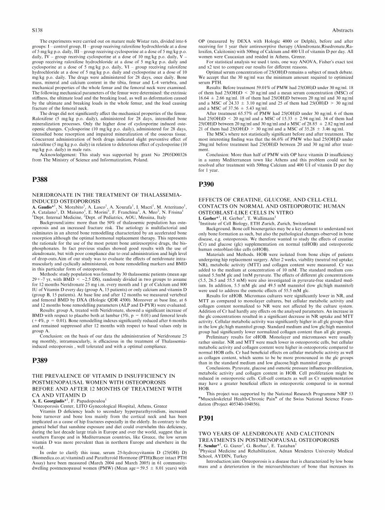
The experiments were carried out on mature male Wistar rats, divided into 6groups: I – control group, II – group receiving raloxifene hydrochloride at a doseof 5 mg/kg p.o. daily, III – group receiving cyclosporine at a dose of 5 mg/kg p.o.daily, IV – group receiving cyclosporine at a dose of 10 mg/kg p.o. daily, V –group receiving raloxifene hydrochloride at a dose of 5 mg/kg p.o. daily andcyclosporine at a dose of 5 mg/kg p.o. daily, VI – group receiving raloxifenehydrochloride at a dose of 5 mg/kg p.o. daily and cyclosporine at a dose of 10mg/kg p.o. daily. The drugs were administered for 28 days, once daily. Bonemass, mineral and calcium content in the tibia, femur and L-4 vertebra, andmechanical properties of the whole femur and the femoral neck were examined.The following mechanical parameters of the femur were determined: the extrinsicstiffness, the ultimate load and the breaking load, as well as deformation causedby the ultimate and breaking loads in the whole femur, and the load causingfracture of the femoral neck.
The drugs did not significantly affect the mechanical properties of the femur.Raloxifene (5 mg/kg p.o. daily), administered for 28 days, intensified bonemineralization processes. Only the higher dose of cyclosporine induced oste-openic changes. Cyclosporine (10 mg/kg p.o. daily), administered for 28 days,intensified bone resorption and impaired mineralization of the osseous tissue.Concurrent administration of both drugs indicated slight preventive effect ofraloxifene (5 mg/kg p.o. daily) in relation to deleterious effect of cyclosporine (10mg/kg p.o. daily) in male rats.
Acknowledgement: This study was supported by grant No 2P05D00326from The Ministry of Science and Informatization, Poland.
P388
NERIDRONATE IN THE TREATMENT OF THALASSEMIA-
INDUCED OSTEOPOROSISA. Gaudio*1, N. Morabito1, A. Lasco1, A. Xourafa1, I. Macrı1, M. Atteritano1,A. Catalano1, D. Maisano1, E. Morini1, F. Franchina1, A. Meo2, N. Frisina11Dept. Internal Medicine, 2Dept. of Pediatrics, AOU, Messina, Italy
Background/aims: more than the 50% of thalassemic population has oste-oporosis and an increased fracture risk. The aetiology is multifactorial andculminates in an altered bone remodelling characterized by an accelerated boneresorption although the optimal hormonal replacement therapy. This representsthe rationale for the use of the most potent bone antiresorptive drugs, the bis-phosphonates. In fact previous studies showed good results with the use ofalendronate, but with poor compliance due to oral administration and high levelof drop-outs.Aim of our study was to evaluate the effects of neridronate intra-muscularly and cyclically administered, on bone remodelling markers and BMDin this particular form of osteoporosis.
Methods: study population was formed by 30 thalassemic patients (mean age29+-7 yr, with BMD < )2.5 DS), randomly divided in two groups to assumefor 12 months Neridronate 25 mg i.m. every month and 1 gr of Calcium and 800IU of Vitamin D every day (group A, 15 patients) or only calcium and vitamin D(group B, 15 patients). At base line and after 12 months we measured vertebraland femoral BMD by DXA (Hologic QDR 4500). Moreover at base line, at 6and 12 months bone remodelling parameters (ALP and D-PYR) were evaluated.
Results: group A, treated with Neridronate, showed a significant increase ofBMD with respect to placebo both at lumbar (5%, p = 0.01) and femoral levels(+4%, p = 0.01). Bone remodelling indexes significantly reduced after 6 monthsand remained suppressed after 12 months with respect to basal values only ingroup A.
Conclusion: on the basis of our data the administration of Neridronate 25mg monthly, intramuscularly, is efficacious in the treatment of Thalassemia-induced osteoporosis , well tolerated and with a optimal compliance.
P389
THE PREVALENCE OF VITAMIN D INSUFFICIENCY IN
POSTMENOPAUSAL WOMEN WITH OSTEOPOROSIS
BEFORE AND AFTER 12 MONTHS OF TREATMENT WITH
CA AND VITAMIN DA. E. Georgiadis*1, F. Papadopoulou11Osteoporosis Center, LITO Gynecological Hospital, Athens, Greece
Vitamin D deficiency leads to secondary hyperparathyroidism, increasedbone turnover and bone loss mainly from the cortical neck and has beenimplicated as a cause of hip fractures especially in the elderly. In contrary to thegeneral belief that sunshine exposure and diet could overwhelm this deficiency,during the last decade large trials in Europe and over the world, suggest that insouthern Europe and in Mediterranean countries, like Greece, the low serumvitamin D was more prevalent than in northern Europe and elsewhere in theworld.
In order to clarify this issue, serum 25-hydroxyvitamin D (25(OH) D)(Biomedica.co.at/vitamind) and Parathyroid Hormone (PTH)(Bayer intact PTHAssay) have been measured (March 2004 and March 2005) in 61 community-dwelling postmenopausal women (PMW) (Mean age=59.5 ± 8.01 years) with
OP (measured by DEXA with Hologic 4000 or Delphi), before and afterreceiving for 1 year their antiresorptive therapy (Alendronate,Risedronate,Ra-loxifen, Calcitonin) with 500mg of Calcium and 400 UI of vitamin D per day. Allwomen were Caucasian and resided in Athens, Greece.
For statistical analysis we used t tests, one way ANOVA, Fisher�s exact testand x2 test to compare our results for different reasons.
Optimal serum concentration of 25(OH)D remains a subject of much debate.We accept that the 30 ng/ml was the minimum amount required to optimizedserum PTH.
Results: Before treatment 59.01% of PMW had 25(OH)D under 30 ng/ml. 18of them had 25(OH)D < 20 ng/ml and a mean serum concentration (MSC) of16.64 ± 2.66 ng/ml. 18 of them had 25(OH)D between 20 ng/ml and 30 ng/mland a MSC of 24.33 ± 3.10 ng/ml and 25 of them had 25(OH)D > 30 ng/mland a MSC of 37.56 ± 5.43 ng/ml.
After treatment 65.57% of PMW had 25(OH)D under 30 ng/ml. 6 of themhad 25(OH)D < 20 ng/ml and a MSC of 15.33 ± 2.94 ng/ml. 34 of them had25(OH)D between 20 ng/ml and 30 ng/ml and a MSC of 28.85 ± 2.82 ng/ml and21 of them had 25(OH)D > 30 ng/ml and a MSC of 35.28 ± 3.46 ng/ml.
The MSCs where not statistically significant before and after treatment. Themost interesting finding was that the 66.6% of PMW who had 25(OH)D under20ng/ml before treatment had 25(OH)D between 20 and 30 ng/ml after treat-ment.
Conclusion: More than half of PMW with OP have vitamin D insufficiencyin a sunny Mediterranean town like Athens and this problem could not beresolved after treatment with 500mg Calcium and 400 UI of vitamin D per dayfor 1 year.
P390
EFFECTS OF CREATINE, GLUCOSE, AND CELL-CELL
CONTACTS ON NORMAL AND OSTEOPOROTIC HUMAN
OSTEOBLAST-LIKE CELLS IN VITROI. Gerber*1, H. Gerber1, T. Wallimann11Institute of Cell Biology, ETH Zurich, Zurich, Switzerland
Background. Bone cell bioenergetics may be a key element to understand notonly bone formation as such, but also the pathological changes observed in bonedisease, e.g. osteoporosis. We therefore wanted to study the effects of creatine(Cr) and glucose (glc) supplementation on normal (nHOB) and osteoporotichuman osteoblast-like cells (oHOB).
Materials and Methods. HOB were isolated from bone chips of patientsundergoing hip replacement surgery. After 2 weeks, viability (neutral red uptake;NR), metabolic activity (MTT) and collagen content were measured. Cr wasadded to the medium at concentration of 10 mM. The standard medium con-tained 5.5mM glc and 1mM pyruvate. The effects of different glc concentrations(5.5, 26.5 and 55.5 mM) were also investigated in pyruvate-free standard med-ium. In addition, 5.5 mM glc and 49.5 mM mannitol (low glc/high mannitol)were used to address the osmotic effects of 55.5 mM glc.
Results for nHOB. Micromass cultures were significantly lower in NR, andMTT as compared to monolayer cultures, but cellular metabolic activity andcollagen content normalized to NR were not affected by the culture system.Addition of Cr had hardly any effects on the analysed parameters. An increase inthe glc concentrations resulted in a significant decrease in NR uptake and MTTactivity. Cellular metabolic activity was significantly higher in all glc groups thanin the low glc/high mannitol group. Standard medium and low glc/high mannitolgroup had significantly lower normalized collagen content than all glc groups.
Preliminary results for oHOB. Monolayer and micromasses were usuallyrather similar. NR and MTT were much lower in osteoporotic cells, but cellularmetabolic activity and collagen content were higher in osteoporotic compared tonormal HOB cells. Cr had beneficial effects on cellular metabolic activity as wellas collagen content, which seems to be be more pronounced in the glc groupsthan in the standard medium and low glucose/high mannitol group.
Conclusions. Pyruvate, glucose and osmotic pressure influence proliferation,metabolic activity and collagen content in HOB. Cell proliferation might bereduced in osteoporotic cells. Cell-cell contacts as well as Cr supplementationmay have a greater beneficial effects in osteoporotic compared to in normalHOB.
This project was supported by the National Research Programme NRP 53"Musculoskeletal Health-Chronic Pain" of the Swiss National Science Foun-dation (Project 405340-104856).
P391
TWO YEARS OF ALENDRONATE AND CALCITONIN
TREATMENTS IN POSTMENOPAUSAL OSTEOPOROSISF. Sendur*1, G. Gurer1, G. Bozbas1, E. Tastaban11Physical Medicine and Rehabilitation, Adnan Menderes University MedicalSchool, AYDIN, Turkey
Introduction/aim: Osteoporosis is a disease that is characterized by low bonemass and a deterioration in the microarchitecture of bone that increases its
S138 Abstracts
susceptibility to fracture. Randomized clinical studies indicate that prolongedadministration of salmon calcitonin prevents bone loss, increases spine bonemass, and decreases fracture risk. In randomized clinical trials,alendronatesubstantially reduced the risk of vertebral fractures.In another trial,alendronatereduced the risk of hip fractures in women with vertebral fractures and a lowBMD.
This study was to compare and research the effects of alendronate andcalcitonin treatments with 2 years duration in women with postmenopausalosteoporosis.
Methods: One hundred and ten women with postmenopausal osteoporosis(calcitonin group:55 subjects, alendronate group:55 subjects) were enrolled inthis study. Patients were divided into two groups randomly. While alendronateat one dose of 70 mgr weekly was administered to one group, calcitonin at onedose of 200IU daily was gived to another group. Bone mineral density (BMD)was measured at the proximal femur and lumbar spine three times as beforetreatment, first and second year.
Results: Significant improvement was found at the end of treatment ingroups itself by lomber spine and femur BMD. A significant more improvementwas determined at the alendronate group than calcitonin group (p=0.001) in thelomber spine. Statistically a difference was not found in other comparisonsamong these groups.
In the conclusion, alendronate and calcitonin in the treatment of postmenopausal osteoporosis by 2 years produced increasing in BMD of the tra-becular and cortical bone. However, alendronate treatment was found moreeffective than calcitonin treatment in the trabecular bone.
P392
EFFECTS OF RALOXIFENE AND CALCITONIN
TREATMENTS ON BONE MINERAL DENSITY IN
POSTMENOPAUSAL OSTEOPOROSIS AFTER ONE YEARG. Gurer1, F. Sendur*1, A. Aydeniz1, H. Aydemir11Physical Medicine and Rehabilitation, Adnan Menderes University MedicalSchool, AYDIN, Turkey
Introduction: The risk of bone fractures is higher in elderly people, partic-ularly in postmenopausal women, which is associated with the development ofosteoporosis.
Raloxifene is the only selective estrogen receptor modulator that has beenapproved for the prevention and treatment of postmenopausal osteoporosis.Calcitonin inhibits bone resorption and is recommended for use in women withosteoporosis
Objective: This study was performed to compare and research the effects ofraloxifene and calcitonin treatments in postmenopausal osteoporosis.
Methods: Forty four women with postmenopausal osteoporosis (raloxifenegroup: 22 subjects, calcitonin group: 22 subjects) were enrolled in this study.Patients were divided into two groups randomly. BMD in the lumbar spine andleft proximal femur was measured by dual energy X-ray absorptiometry twice asbefore treatment and first year. While raloxifene at one dose of 60 mg daily wasadministered to one group, calcitonin at one dose of 200IU daily was given toanother group.
Results: Although statistically significant improvement was found in thelumbar spine (p < 0.05), any recovery was not determined in femur BMD (p >0.05) at the end of first year with this treatments.
In conclusion, raloxifene and calcitonin provided significantly increase inBMD of the trabecular bone, but were not effective in cortical bone.
P393
EVALUATION OF DXA SPINE, HIP AND DUAL HIP
PRECISION USING THE PRODIGYP. Hadji*1, V. Ziller1, M. Bauer1, M. Gottschalk1, A. Kauka11Gyn. Endocrinology, Reproductive Medicine and Osteoporosis, UniversityHospital of Giessen and Marburg, Campus Marburg, Marburg, Germany
Introduction: BMD measurements are of major importance in the detectionand follow-up of patients with osteoporosis. Hereby, the precision of the mea-surement device is a key factor in determining wether a measured change in apatients DXA result represents a clinically significant change. Differences inprecision are attributed to different technologies, manufactures and technicandependent factors. Although results on the precision of lumbar spine and singlehip measurements have been reported, data on dual hip measurements are rarelyavailable. The aim of our study was to evaluate the short term precision at allrelevant sites using the Prodigy.
Methods: 40 healthy women without a history of osteoporosis or relatedfracture were included.BMD was measured by DXA (Prodigy, GE Lunar) at thespine and hip. Each woman was measured in accordance to the followingstandard protocol: spine, left hip, right hip, dual femur, standing position beforerepositioning and repeated measurement at all sights.
Results: We found a significant correlation of the absolut values and the T-score between both measurements at the spine (L1-L4) of R2 = 0.99 and
R2 = 0.99 representing a CV (%) of 1.00 and a LSC 2.76 (%).The absolute meanvalues of the repeated measurements at the left total femur, femoral neck, wardtriangle and trochanteric region was 0.97, 0.94, 0.75 and 0.79 representing a CV(%) of 0.49, 1.07, 1.14 and 0.72 and a LSC (%) of 1.36, 2.98, 3.16, and 2.0.Thecorrelation of the absolute values of the repeated measurements at the right totalfemur was R2 = 0.94 and R2 = 0.94.The absolute mean values of the righttotal femur, femoral neck, ward triangle and trochanteric region was 0.97, 0.95,0.75 and 0.78 representing a CV (%) of 0.52, 1.82, 2.15 and 1.03 and a LSC (%)of 1.43, 5.05, 5.97 and 2.86.The correlation of the absolute values of the repeatedmeasurements at the dual total femur was R2 = 0.86 and R2 = 0.90.Theabsolute mean values of the dual total femur, dual femoral neck, dual wardtriangle and dual trochanteric region was 0.97, 0.94, 0.75 and 0.79 representing aCV (%) of 0.31, 0.91, 1.20 and 0.65 and a LSC of 0.87, 2.51, 3.33 and 1.80,respectively.
Conclusion: Our results underline the high correlation between repeatedmeasurements at all relevant measurement sights including the dual femur usingthe Prodigy (GE Lunar).This low precision error is of upmost importance in thecalculation of the least significant change and in the clinical decision making.
P394
ARE TROCHANTERIC BMD MEASUREMENTS CAPABLE OF
REPLACING THE VERTEBRAL ONES ?D. Hadjidakis*1, A. Mylonakis1, I. I. Androulakis1, A. Papaefstathiou1,S. A. Raptis112nd Department of Internal Medicine Propaedeutic, Research Institute, Atti-kon and Evgenidion University Hospitals, Athens University, Athens, Greece
Vertebral and femoral neck [FN] bone mineral density [BMD] measurementsby DXA are often used for the evaluation of bone mass status. Trochanter major[TRO] consists mostly of trabecular bone but in contrast to vertebrae it is notloaded. Aims: To investigate the behavior of TRO T-scores in cases of majordiscrepancies between vertebral and FN T-scores. Methods: In 785 healthypostmenopausal [PMP] women BMD measurements were performed by DXA inL1–L4 vertebrae [VER], FN and TRO. All women were divided into 3 groupsaccording to the difference between VER and FN T-scores [group A, (VER-FN>1), group B (VER-FN<1) and group C (VER-FN<)1)]. Separate eval-uations were performed in women aged 45-65 years (middle-aged, [MID]) and65–80 years [OLD]. Six groups (130 A-MID, 47 A-OLD, 409 B-MID, 99 B-OLD, 75 C-MID and 25 C-OLD) were constructed. Women with radiologicallyobvious vertebral degenerative lesions were excluded. All data was analyzedusing T-scores. Results: In the whole 785 women population there was no sig-nificant difference between mean VER, FN and TRO T-scores. Regarding thesame population, in 277 (35.3%) the difference VER-FN T-scores exceeded 1. Inall MID women this difference was observed in 33.4%, whilst in all OLD in 42%.In 39.2% of all women the difference FN-TRO was less than 20% from FN-AP.The same proportion was 36.9% for all MID women and 46.8% for all OLDwomen. In all groups, the correlations between VER, FN and TRO T-scoreswere always significantly positive, but those between FN and TRO were alwaysstronger than between VER and either FN or TRO. In all groups a negativecorrelation was observed between VER-TRO and FN-TRO (r = )0.30 to)0.42, p < 0.01). Conclusions: In a remarkable proportion of middle-agedpostmenopausal women, major discrepancies exist between the trabecular ver-tebral and mixed femoral neck bone mineral density values without obviousradiological reason. In such cases, the trochanteric bone density behavior seemsto resemble that of femoral neck. At any rate, the recommendation that bothvertebral and hip DXA measurements be performed in all women is enforced.
P395
ROLE OF PROSTAGLANDINS IN THE ANABOLIC EFFECTS
OF STRONTIUM RANELATES. Choudhary*1, P. Halbout2, C. Alander1, L. Raisz1, C. Pilbeam1
1UConn Center for Osteoporosis, University of Connecticut Health, Farming-ton, United States, 2Rheumatology Division, Groupe Servier, Courbevoie,France
Strontium ranelate is a new agent for the treatment of post-menopausalosteoporosis that increases bone formation and decreases bone resorption. Al-though its anti-fracture efficacy has been demonstrated, its mechanism of actionat the cellular level is still under investigation. We have assessed the involvementof prostaglandins (PGE2) in the effects of strontium ranelate on osteoblasts.Osteoblastic precursors from bone marrow (MSC) or primary osteoblasts iso-lated from murine calvariae (POB) were cultured for 7 to 14 days, and treatedwith strontium ranelate (1 and 3 mM strontium, Sr2+) for various times. Effectson proliferation, alkaline phosphatase (ALP) activity, PGE2 production andmineralization were assessed. Strontium ranelate (1 mM and 3 mM Sr2+) in-creased the proliferation of POB 1.53 fold (p < 0.01) and 1.35 fold (p < 0.01),respectively, after a 7-day treatment. ALP activity of POB assessed at the end ofa 14-d culture was significantly increased by strontium ranelate (3 mM Sr2+)after a 0–3 d (186%, p < 0.01), 0–7 d (95%, p < 0.05) and 0-14 d (92%, p <0.05) treatment. Similarly, ALP activity was increased in MSC by 92% (p <
Abstracts S139

0.01) and 63% (p < 0.01) after a continuous treatment with strontium ranelate(3 mM Sr2+) for 10 and 14 days, respectively. This effect was completelyblocked by NS-398, a selective cyclooxygenase (COX)-2 inhibitor. A 14-dayculture was used to assess the strontium ranelate effects on PGE2 production byMSC. After 7-days of treatment with strontium ranelate (1 mM and 3 mMSr2+), cumulative PGE2 levels were increased 2.4 fold (p < 0.01) and 11.0 fold(p < 0.01), respectively. COX-2 was involved in the strontium ranelate-inducedPGE2 production, as demonstrated by the inhibition of this effect by NS-398.Osteoblasts differentiated in presence of strontium ranelate were functional asillustrated by the increased mineralization observed when MSC were continu-ously treated for 14 days with strontium ranelate (3 mM Sr2+). These resultsconfirm the stimulatory effects of strontium ranelate on proliferation, differen-tiation and mineralization of murine osteoblasts. These effects, which werecoupled to a COX-2 dependent increase in PGE2 production, are consistent witha possible involvement of prostaglandins in the anabolic effect of strontiumranelate.
P396
BONEMINERAL DENSITY AND HEALT RELATED QUALITY
OF LIFE IN YOUNG ADULTS WITH JUVENILE IDIOPATHIC
ARTHRITISH. Hamalainen*1, M. Arkela-Kautainen1, J. Haapasaari1, H. Kautiainen1,A. Kotaniemi1, M. Leirisalo-Repo21Rheumatology, Rheumatism Foundation Hospital, Heinola, 2Rheumatology,Helsinki University Hospital, Helsinki, Finland
Aims: Juvenile idiopathic arthritis (JIA) is a heterogeneous disease emergingin childhood. Diminished bone mineral density (BMD) in JIA patients have beenconnected with high inflammatory activity and long duration of JIA, delay ofpubertal maturation and treatment with corticosteroids. RAND-36 is a measureof health related quality of life containing 36 items on eight scales within twocomponents: physical component (physical functioning, bodily pain, role limi-tation due to physical health problems, general health) and mental one (energy,role limitation due to personal emotional problems, emotional wellbeing, andsocial functioning). Could RAND-36 questionnaire tell us whom to target inrelation to bone mass development among a cohort of 116 young adults diag-nosed to have JIA during the years 1976-1995?
Methods: BMD measurement was performed at the lumbar spine andfemoral neck by dual-energy X-ray absorptiometry. JIA patients were dividedinto two groups according to the T score: T score > )1.0 (normal, N = 76) andT score < )1.0 (abnormal, N = 40).
Results: Age at follow up was 23 (21–26) years, in both groups. There was nostatistical difference between both groups in following items: age at onset, timefrom diagnosis to follow up, number of swollen and tender joints, ESR, CRP,patients receiving DMARDs, height, number of females. There was a statisticaldifference between groups in weight 69 (SD16) kg in group T score > )1.0 and62 (SD12) kg in group T score < )1.0 (p = 0.026). Prednisolone received 20patients in T score < )1.0 group and 3 patients in > )1.0 group (p = 0.003). InRAND-36 both groups did not differ from each other at mental componentscales scores being 49 (SD1) in T score < )1.0 group and 50 (SD 1) in T score >)1.0 group (p = 0.21), but group T score < )1.0 differed at physical compo-nent scales from group T score > )1 scores being 47 (SD 10) and 51 (SD 8),respectively (p = 0.02).
Conclusions: JIA patients thriving better on RAND-36 questionnaire werethriving better also on BMD scale. This may have clinical significance amongpatients with JIA.
P397
RELATIONSHIPS BETWEEN BONE ANABOLIC, PROTEOAN-
ABOLIC AND LIPOLYTIC EFFECTS OF GROWTH HORMONE
(GH) REPLACEMENT IN GH DEFICIENT ADULTSV. Hana1, J. J. Stepan*113rd Department of Internal Medicine, Charles University Faculty of Medicine,Prague, Czech Republic
Growth hormone deficiency (GHD) in adulthood has been associated withincreased central adiposity, reduced lean body mass, increased cardiovascularrisk, reduced bone mineral density with increased fracture risk and decreasedquality of life. GH replacement might improve these alterations. The aim of thisstudy was to evaluate the long-term metabolic effects of GH replacement onbone metabolism and body composition. The study involved 24 patients withGHD (20 males, mean age 38 ± 11 years, 20 adult onsets). Serum IGF-I andbone mineral density (BMD, Hologic QDR 4500) was determined at the lumbarspine, proximal femur and total body at baseline and yearly for up to five years.Mean GH dose of 0.4 ± 0.2 mg/day resulted in significant increase and nor-malization of IGF-I serum concentrations. A significant increase in the leanbody mass and decrease in body fat mass was observed after one year of GHtreatment, both 3% of body weight (p < 0.05). These positive effects persisted
over the whole treatment period. As compared with baseline values, after 2 yearsof treatment the lumbar spine and total femur BMD increased (mean ± SEM)by 4.9 ± 1.1 %, and 3.4 ± 0.8%, respectively (p < 0.05). After 5 years thelumbar spine and femur BMD increased by 6.1 ± 3.2% and 6.3 ± 1.1%,respectively (p < 0.05). Whole body BMD significantly increased after 3 yearsby 2.6 ± 0.8%, and by 4.2 ± 2.0% after 5 years. Significant negative correlationwas observed between increase in total body BMD and total body fat tissue(r = 0.55, p < 0.001). A positive trend was observed between total body BMDand total body lean mass (r = 0.18). In conclusion, the long-term GHreplacement in adults with GHD is effective in restoring IGF-I levels, favorablychanges body composition and increases bone mineral density. These metaboliceffects last after five years of treatment.
P398
APPLICATION OF A TRIAGE APPROACH FOR THE DIAG-
NOSIS OF OSTEOPOROSIS: RISK INDICES AND PERIPHERAL
BONE DENSITOMETRYE. J. Harrison*1, J. E. Adams11Clinical Radiology, The University of Manchester, Manchester, United King-dom
In some countries availability of central dual energy X-ray absorptiometry(DXA) is limited. There is therefore interest in the use of peripheral scannerswhich are portable and less expensive. Despite their potential there is uncertaintyon how to utilise, and interpret results from, peripheral scanners. A methodrecently proffered for the interpretation of results from peripheral DXA is theapplication of a triage approach to stratify patients into one of three categories;i) treatment recommended ii) refer for central DXA or iii) no further actionrequired.
In this study the triage approach was applied to peripheral bone densi-tometry measurements and results from four risk indices calculated fromdemographic, treatment and fracture histories. 207 post-menopausal women hadmeasurements performed at the calcaneus by peripheral DXA (GE Lunar PIXI)and quantitative ultrasound (QUS) (McCue CubaClinical, GE Lunar Achilles).The risk indices calculated were the osteoporosis self assessment tool (OST),osteoporosis risk assessment instrument (ORAI), osteoporosis indices of risk(OSIRIS) and the simple calculated osteoporosis risk estimation (SCORE).Threshold values of )1.6 and )3.0 Achilles, )1.02 and )2.27 CubaClinical, )0.4and )1.7 PIXI , 9 and 17 ORAI, )3 and )1 OST, )3 and 1 OSIRIS and 7 and 15SCORE were used to stratify women into categories.
The area under the receiver operating curves (ROC) were 0.77 Achilles, 0.75CubaClinical, 0.80 PIXI, 0.67 SCORE, 0.67 ORAI, 0.70 OSIRIS and 0.69 OST.The percentage of women stratified into category (ii) refer for central DXA was62% ORAI, 53% OSIRIS, 68% SCORE, 50% OST, 49% Achilles, 57% Cuba-Clinical and 39% PIXI.
The triage approach was an effective tool to correctly identify women at riskof osteoporosis, particularly when using peripheral bone densitometry scanners.If the triage approach is applied to measurements from peripheral scanners thenumber of post-menopausal women requiring central DXA could be reduced.
P399
RESULTS FROM A 3-YEAR STUDY DESIGNED TO
EVALUATE THE EFFECT OF SODIUM ON BONE MINERAL
DENSITY IN POSTMENOPAUSAL WOMENJ. Z. Ilich*1, R. A. Brownbill2, A. H. N. Cillessen31Nutrition, Food and Exercise Sciences, Florida State University, Tallahassee,2Nutritional Sciences, 3Psychology, University of Connecticut, Storrs, UnitedStates
It is speculated that sodium (Na) might negatively impact bone due to itscalciuric effect, however scientific evidence regarding the relationship betweenNa and bone mineral density (BMD) is sparse and inconclusive. We designeda study to monitor the long-term effect of different dietary Na levels on BMDin over 100 generally healthy, Caucasian postmenopausal women. After thebaseline screening, half of the subjects were instructed to reduce their Naintake to �1500 mg/day. The other half remained on a habitual Na intake of�3000 mg/day. All subjects were given Ca (630 mg/day) and vitamin D (400IU/day) supplements to bring their total Ca intake to �1300 mg/day. Theaverage age at enrollment was 68.6 ± 7.1y and subjects� anthropometries,bone mass measurement, 24-h urine analyses and dietary and activity recordswere assessed every 6 months, totaling 7 evaluations over a 3-year period.Data were analyzed using the Growth Curve Multilevel Modeling to examinethe changes over-time in bone variables of interest and their predictors. Thefollowing covariates were controlled for in all analyses: age, body mass index,and/or total body fat and lean tissue, systolic blood pressure, physicalactivity, alcohol and caffeine consumption, as well as calcium, energy, protein,and vitamin D intake. Na intake (expressed as urinary Na/Creatinine) at
S140 Abstracts
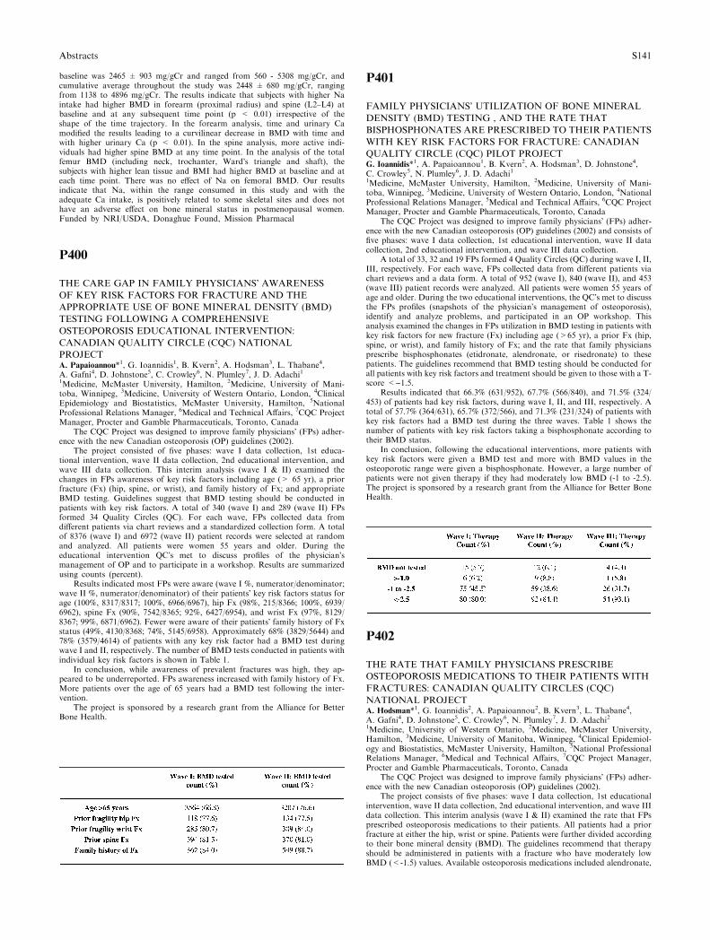
baseline was 2465 ± 903 mg/gCr and ranged from 560 - 5308 mg/gCr, andcumulative average throughout the study was 2448 ± 680 mg/gCr, rangingfrom 1138 to 4896 mg/gCr. The results indicate that subjects with higher Naintake had higher BMD in forearm (proximal radius) and spine (L2–L4) atbaseline and at any subsequent time point (p < 0.01) irrespective of theshape of the time trajectory. In the forearm analysis, time and urinary Camodified the results leading to a curvilinear decrease in BMD with time andwith higher urinary Ca (p < 0.01). In the spine analysis, more active indi-viduals had higher spine BMD at any time point. In the analysis of the totalfemur BMD (including neck, trochanter, Ward�s triangle and shaft), thesubjects with higher lean tissue and BMI had higher BMD at baseline and ateach time point. There was no effect of Na on femoral BMD. Our resultsindicate that Na, within the range consumed in this study and with theadequate Ca intake, is positively related to some skeletal sites and does nothave an adverse effect on bone mineral status in postmenopausal women.Funded by NRI/USDA, Donaghue Found, Mission Pharmacal
P400
THE CARE GAP IN FAMILY PHYSICIANS� AWARENESS
OF KEY RISK FACTORS FOR FRACTURE AND THE
APPROPRIATE USE OF BONE MINERAL DENSITY (BMD)
TESTING FOLLOWING A COMPREHENSIVE
OSTEOPOROSIS EDUCATIONAL INTERVENTION:
CANADIAN QUALITY CIRCLE (CQC) NATIONAL
PROJECTA. Papaioannou*1, G. Ioannidis1, B. Kvern2, A. Hodsman3, L. Thabane4,A. Gafni4, D. Johnstone5, C. Crowley6, N. Plumley7, J. D. Adachi11Medicine, McMaster University, Hamilton, 2Medicine, University of Mani-toba, Winnipeg, 3Medicine, University of Western Ontario, London, 4ClinicalEpidemiology and Biostatistics, McMaster University, Hamilton, 5NationalProfessional Relations Manager, 6Medical and Technical Affairs, 7CQC ProjectManager, Procter and Gamble Pharmaceuticals, Toronto, Canada
The CQC Project was designed to improve family physicians� (FPs) adher-ence with the new Canadian osteoporosis (OP) guidelines (2002).
The project consisted of five phases: wave I data collection, 1st educa-tional intervention, wave II data collection, 2nd educational intervention, andwave III data collection. This interim analysis (wave I & II) examined thechanges in FPs awareness of key risk factors including age (> 65 yr), a priorfracture (Fx) (hip, spine, or wrist), and family history of Fx; and appropriateBMD testing. Guidelines suggest that BMD testing should be conducted inpatients with key risk factors. A total of 340 (wave I) and 289 (wave II) FPsformed 34 Quality Circles (QC). For each wave, FPs collected data fromdifferent patients via chart reviews and a standardized collection form. A totalof 8376 (wave I) and 6972 (wave II) patient records were selected at randomand analyzed. All patients were women 55 years and older. During theeducational intervention QC�s met to discuss profiles of the physician�smanagement of OP and to participate in a workshop. Results are summarizedusing counts (percent).
Results indicated most FPs were aware (wave I %, numerator/denominator;wave II %, numerator/denominator) of their patients� key risk factors status forage (100%, 8317/8317; 100%, 6966/6967), hip Fx (98%, 215/8366; 100%, 6939/6962), spine Fx (90%, 7542/8365; 92%, 6427/6954), and wrist Fx (97%, 8129/8367; 99%, 6871/6962). Fewer were aware of their patients� family history of Fxstatus (49%, 4130/8368; 74%, 5145/6958). Approximately 68% (3829/5644) and78% (3579/4614) of patients with any key risk factor had a BMD test duringwave I and II, respectively. The number of BMD tests conducted in patients withindividual key risk factors is shown in Table 1.
In conclusion, while awareness of prevalent fractures was high, they ap-peared to be underreported. FPs awareness increased with family history of Fx.More patients over the age of 65 years had a BMD test following the inter-vention.
The project is sponsored by a research grant from the Alliance for BetterBone Health.
P401
FAMILY PHYSICIANS� UTILIZATION OF BONE MINERAL
DENSITY (BMD) TESTING , AND THE RATE THAT
BISPHOSPHONATES ARE PRESCRIBED TO THEIR PATIENTS
WITH KEY RISK FACTORS FOR FRACTURE: CANADIAN
QUALITY CIRCLE (CQC) PILOT PROJECTG. Ioannidis*1, A. Papaioannou1, B. Kvern2, A. Hodsman3, D. Johnstone4,C. Crowley5, N. Plumley6, J. D. Adachi11Medicine, McMaster University, Hamilton, 2Medicine, University of Mani-toba, Winnipeg, 3Medicine, University of Western Ontario, London, 4NationalProfessional Relations Manager, 5Medical and Technical Affairs, 6CQC ProjectManager, Procter and Gamble Pharmaceuticals, Toronto, Canada
The CQC Project was designed to improve family physicians� (FPs) adher-ence with the new Canadian osteoporosis (OP) guidelines (2002) and consists offive phases: wave I data collection, 1st educational intervention, wave II datacollection, 2nd educational intervention, and wave III data collection.
A total of 33, 32 and 19 FPs formed 4 Quality Circles (QC) during wave I, II,III, respectively. For each wave, FPs collected data from different patients viachart reviews and a data form. A total of 952 (wave I), 840 (wave II), and 453(wave III) patient records were analyzed. All patients were women 55 years ofage and older. During the two educational interventions, the QC�s met to discussthe FPs profiles (snapshots of the physician�s management of osteoporosis),identify and analyze problems, and participated in an OP workshop. Thisanalysis examined the changes in FPs utilization in BMD testing in patients withkey risk factors for new fracture (Fx) including age (>65 yr), a prior Fx (hip,spine, or wrist), and family history of Fx; and the rate that family physiciansprescribe bisphosphonates (etidronate, alendronate, or risedronate) to thesepatients. The guidelines recommend that BMD testing should be conducted forall patients with key risk factors and treatment should be given to those with a T-score <)1.5.
Results indicated that 66.3% (631/952), 67.7% (566/840), and 71.5% (324/453) of patients had key risk factors, during wave I, II, and III, respectively. Atotal of 57.7% (364/631), 65.7% (372/566), and 71.3% (231/324) of patients withkey risk factors had a BMD test during the three waves. Table 1 shows thenumber of patients with key risk factors taking a bisphosphonate according totheir BMD status.
In conclusion, following the educational interventions, more patients withkey risk factors were given a BMD test and more with BMD values in theosteoporotic range were given a bisphosphonate. However, a large number ofpatients were not given therapy if they had moderately low BMD (-1 to -2.5).The project is sponsored by a research grant from the Alliance for Better BoneHealth.
P402
THE RATE THAT FAMILY PHYSICIANS PRESCRIBE
OSTEOPOROSIS MEDICATIONS TO THEIR PATIENTS WITH
FRACTURES: CANADIAN QUALITY CIRCLES (CQC)
NATIONAL PROJECTA. Hodsman*1, G. Ioannidis2, A. Papaioannou2, B. Kvern3, L. Thabane4,A. Gafni4, D. Johnstone5, C. Crowley6, N. Plumley7, J. D. Adachi21Medicine, University of Western Ontario, 2Medicine, McMaster University,Hamilton, 3Medicine, University of Manitoba, Winnipeg, 4Clinical Epidemiol-ogy and Biostatistics, McMaster University, Hamilton, 5National ProfessionalRelations Manager, 6Medical and Technical Affairs, 7CQC Project Manager,Procter and Gamble Pharmaceuticals, Toronto, Canada
The CQC Project was designed to improve family physicians� (FPs) adher-ence with the new Canadian osteoporosis (OP) guidelines (2002).
The project consists of five phases: wave I data collection, 1st educationalintervention, wave II data collection, 2nd educational intervention, and wave IIIdata collection. This interim analysis (wave I & II) examined the rate that FPsprescribed osteoporosis medications to their patients. All patients had a priorfracture at either the hip, wrist or spine. Patients were further divided accordingto their bone mineral density (BMD). The guidelines recommend that therapyshould be administered in patients with a fracture who have moderately lowBMD (<-1.5) values. Available osteoporosis medications included alendronate,
Abstracts S141
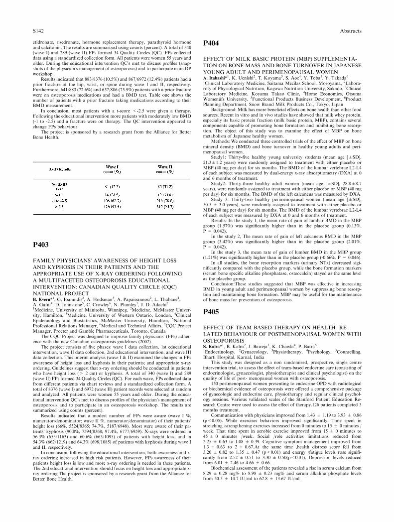
etidronate, risedronate, hormone replacement therapy, parathyroid hormoneand calcitonin. The results are summarized using counts (percent). A total of 340(wave I) and 289 (wave II) FPs formed 34 Quality Circles (QC). FPs collecteddata using a standardized collection form. All patients were women 55 years andolder. During the educational intervention QC�s met to discuss profiles (snap-shots of the physician�s management of osteoporosis) and to participate in an OPworkshop.
Results indicated that 883/8376 (10.5%) and 867/6972 (12.4%) patients had aprior fracture at the hip, wrist, or spine during wave I and II, respectively.Furthermore, 641/883 (72.6%) and 657/886 (75.9%) patients with a prior fracturewere on osteoporosis medications and had a BMD test. Table one shows thenumber of patients with a prior fracture taking medications according to theirBMD measurement.
In conclusion, most patients with a t-score <-2.5 were given a therapy.Following the educational intervention more patients with moderately low BMD(-1 to -2.5) and a fracture were on therapy. The QC intervention appeared tochange FPs behaviour.
The project is sponsored by a research grant from the Alliance for BetterBone Health.
P403
FAMILY PHYSICIANS� AWARENESS OF HEIGHT LOSS
AND KYPHOSIS IN THEIR PATIENTS AND THE
APPROPRIATE USE OF X-RAY ORDERING FOLLOWING
A MULTIFACETED OSTEOPOROSIS EDUCATIONAL
INTERVENTION: CANADIAN QUALITY CIRCLE (CQC)
NATIONAL PROJECTB. Kvern*1, G. Ioannidis2, A. Hodsman3, A. Papaioannou2, L. Thabane4,A. Gafni4, D. Johnstone5, C. Crowley6, N. Plumley7, J. D. Adachi21Medicine, University of Manitoba, Winnipeg, 2Medicine, McMaster Univer-sity, Hamilton, 3Medicine, University of Western Ontario, London, 4ClinicalEpidemiology and Biostatistics, McMaster University, Hamilton, 5NationalProfessional Relations Manager, 6Medical and Technical Affairs, 7CQC ProjectManager, Procter and Gamble Pharmaceuticals, Toronto, Canada
The CQC Project was designed to improve family physicians� (FPs) adher-ence with the new Canadian osteoporosis guidelines (2002).
The project consists of five phases: wave I data collection, 1st educationalintervention, wave II data collection, 2nd educational intervention, and wave IIIdata collection. This interim analysis (wave I & II) examined the changes in FPsawareness of height loss and kyphosis in their patients; and appropriate x-rayordering. Guidelines suggest that x-ray ordering should be conducted in patientswho have height loss (> 2 cm) or kyphosis. A total of 340 (wave I) and 289(wave II) FPs formed 34 Quality Circles (QC). For each wave, FPs collected datafrom different patients via chart reviews and a standardized collection form. Atotal of 8376 (wave I) and 6972 (wave II) patient records were selected at randomand analyzed. All patients were women 55 years and older. During the educa-tional intervention QC�s met to discuss profiles of the physician�s management ofosteoporosis and to participate in an osteoporosis workshop. The results aresummarized using counts (percent).
Results indicated that a modest number of FPs were aware (wave I %,numerator/denominator; wave II %, numerator/denominator) of their patients�height loss (66%, 5524/8365; 74.7%, 5187/6948). Most were aware of their pa-tients� kyphosis (90.8%, 7594/8368; 97.4%, 6777/6959). X-rays were ordered in56.3% (655/1163) and 60.6% (663/1095) of patients with height loss, and in54.3% (662/1219) and 64.3% (698/1085) of patients with kyphosis during wave Iand II, respectively.
In conclusion, following the educational intervention, both awareness and x-ray ordering increased in high risk patients. However, FPs awareness of theirpatients height loss is low and more x-ray ordering is needed in these patients.The 2nd educational intervention should focus on height loss and appropriate x-ray ordering.The project is sponsored by a research grant from the Alliance forBetter Bone Health.
P404
EFFECT OF MILK BASIC PROTEIN (MBP) SUPPLEMENTA-
TION ON BONE MASS AND BONE TURNOVER IN JAPANESE
YOUNG ADULT AND PERIMENOPAUSAL WOMENA. Itabashi*1, K. Uenishi2, T. Koyama3, S. Aoe4, Y. Toba5, Y. Takada61Clinical Laboratory Medicine, Saitama Mecilas School, Moroyama, 2Labora-roty of Physiological Nutrition, Kagawa Nutrition University, Sakado, 3ClinicalLaboratory Medicine, Koyama Takao Clinic, 4Home Economics, OtsumaWomenufs University, 5Functional Products Business Development, 6ProductPlanning Department, Snow Brand Milk Products Co., Tokyo, Japan
Background: Milk has more beneficial effects on bone health than other foodsources. Recent in vitro and in vivo studies have showed that milk whey protein,especially its basic protein fraction (milk basic protein, MBP), contains severalcomponents capable of promoting bone formation and inhibiting bone resorp-tion. The object of this study was to examine the effect of MBP on bonemetabolism of Japanese healthy women.
Methods: We conducted three controlled trials of the effect of MBP on bonemineral density (BMD) and bone turnover in healthy young adults and peri-menopausal women.
Study1: Thirty-five healthy young university students (mean age [±SD],21.3±1.2 years) were randomly assigned to treatment with either placebo orMBP (40 mg per day) for six months. The BMD of the lumbar vertebrae L2-L4of each subject was measured by dual-energy x-ray absorptiometry (DXA) at 0and 6 months of treatment.
Study2: Thirty-three healthy adult women (mean age [±SD], 28.8±8.7years), were randomly assigned to treatment with either placebo or MBP (40 mgper day) for six months. The BMD of the left calcaneus was measured by DXA.
Study 3: Thirty-two healthy perimenopausal women (mean age [±SD],50.5 ± 3.0 years), were randomly assigned to treatment with either placebo orMBP (40 mg per day) for six months. The BMD of the lumbar vertebrae L2-L4of each subject was measured by DXA at 0 and 6 months of treatment.
Results: In the study 1, the mean rate of gain of lumbar BMD in the MBPgroup (1.57%) was significantly higher than in the placebo group (0.13%,P = 0.042).
In the study 2, The mean rate of gain of left calcaneus BMD in the MBPgroup (3.42%) was significantly higher than in the placebo group (2.01%,P = 0.042).
In the study 3, the mean rate of gain of lumber BMD in the MBP group(1.21%) was significantly higher than in the placebo group (-0.66%, P = 0.046).
In all studies, the bone resorption markers (urinary NTx) decreased sigi-nificantly compared with the placebo group, while the bone formation markers(serum bone specific alkaline phosphatase, osteocalcin) stayed as the same levelas the placebo group.
Conclusion:These studies suggested that MBP was effective in increasingBMD in young adult and perimenopausal women by suppressing bone resorp-tion and maintaining bone formation. MBP may be useful for the maintenanceof bone mass for prevention of osteoporosis.
P405
EFFECT OF TEAM-BASED THERAPY ON HEALTH -RE-
LATED BEHAVIOUR OF POSTMENOPAUSAL WOMEN WITH
OSTEOPOROSISS. Kalra*1, B. Kalra2, J. Baweja3, K. Chawla4, P. Batra51Endocrinology, 2Gynaecology, 3Physiotherapy, 4Psychology, 5Counselling,Bharti Hospital, Karnal, India
This study was designed as a non randomized, prospective, single centreintervention trial, to assess the effect of team-based endocrine care (consisting ofendocrinologist, gynaecologist, physiotherapist and clinical psychologist) on thequality of life of post- menopausal women with osteoporosis.
150 postmenopausal women presenting to endocrine OPD with radiologicalor biochemical evidence of osteoporosis were offered a comprehensive packageof gynecologic and endocrine care, physiotherapy and regular clinical psychol-ogy sessions. Various validated scales of the Stanford Patient Education Re-search Centre were used to assess the effect of therapy.126 patients completed 3months treatment.
Communication with physicians improved from 1.43 ± 1.19 to 3.93 ± 0.86(p<0.05). While exercises behaviors improved significantly. Time spent instretching /strengthening exercises increased from 0 minutes to 15 ± 0 minutes /week. That time spent in aerobic exercise improved from 15 ± 0 minutes to45 ± 0 minutes /week. Social /role activities limitations reduced from2.25 ± 0.63 to 1.08 ± 0.39. Cognitive symptom management improved from1.3 ± 0.63 to 2 ± 0.67.At the same time ,health distress score fell from3.20 ± 0.82 to 1.35 ± 0.47 (p<0.01) and energy /fatigue levels rose signifi-cantly from 2.52 ± 0.51 to 3.30 ± 0.50(p<0.01). Depression levels reducedfrom 6.01 ± 2.46 to 4.66 ± 0.66. .
Biochemical assessment of the patients revealed a rise in serum calcium from8.29 ± 0.28 mg% to 8.98 ± 0.23 mg% and serum alkaline phosphate levelsfrom 50.5 ± 14.7 IU/ml to 62.8 ± 13.67 IU/ml.
S142 Abstracts
This study demonstrates the multiple beneficial effects of team based therapyon various health parameters in post menopausal women with osteoporosis.
P406
PHYSIOTHERAPY IN OSTEOPOROSIS PATIENTS:
UTILIZATION AND BENEFITSS. Kalra*1, J. Baweja2, B. Kalra31Endocrinology, 2Physiotherapy, 3Gynaecology, Bharti Hospital, Karnal, India
This study was a prospective analysis designed to assess the frequency ofmusculoskeletal complaints, and the improvement noted with physiotherapeutictherapies in osteoporosis patients attending an endocrine clinic. Benefits werenoted by using previously validated scales of the Stanford Patient EducationCentre.
247 patients (121 male, 126 female) were analyzed. 34.89% were aged 51-60years while 28.83% were 61-70 years. The commonest presentations were neu-ropathic pain (43.39%), cervical symptoms (13.21%), low-backache (11.05%),and knee pain (7.55%). 4.58% persons requested advice for weight and waist loss.
Neuropathy was commoner in males (49.72% vs. 37.50%), while femalesexperienced more cervical spondylosis (18.75% vs. 7.26%), low-backache(13.54% vs. 8.38%) and frozen shoulder (5.73% vs. 1.68%). The physiothera-peutic interventions used most often were TENS/ IFT (43.39%), ultrasonictherapy (16.97%), and short-wave diathermy (13.05%).
Physician communication improved from 1.43 ± 1.19 to 3.93 ± 0.86 overtwo months of therapy. Time spent in stretching /strengthening exercises in-creased from 0 ± 0 to 15 ± 0 while aerobic exercise improved from 15 ± 0 to45 ± 0 minutes /week. Social /role activities limitations reduced from2.25 ± 0.63 to 1.08 ± 0.39. Cognitive symptom management improved from1.3 ± 0.63 to 2 ± 0.67. Health distress score fell from 3.20 ± 0.82 to1.35 ± 0.47 and energy /fatigue levels rose from 2.52 ± 0.51 to 3.30 ± 0.50.Pain scores reduced from 6.01 ± 2.46 to 4.66 ± 0.66.
This study demonstrates the multiple beneficial effects of physiotherapyinterventions in patients of osteoporosis.
P407
AGE RELATED CHANGES IN BONE MINERAL DENSITY IN
HEALTHY INDIAN ROLE OF SEX STEROIDSI. M. Khatkhatay*1, K. K. V. Venkat1, M. P. Desai11Division of Molecular Immunodiagnostics, Natinal Institute for Research inReproductive Health, Mumbai, India
Osteoporosis is set to occur if there is decrease in bone mass greater thanexpected for person�s age, sex and race. It is considered as common consequences ofold age.Althoughosteoporosis is seen less often inmen, yet 30%of the reported hipfractures occur in men and the mortality attributed to hip fractures is twice as highas in women. Thus, osteoporosis in men is recognized as increasing importanthealth issue. It is well establish that hormonal factors a play important role inpatho-physiology of osteoporosis in women.However, influence of sex steroids onmale skeleton is poorly understood. The aim of the study was to measure bonemineral density (BMD) in healthy men and examine the influence of age and sexsteroids onBMD.Two hundred and forty two army personnel were recruited fromArmed Forces Medical College, Pune, India following well-defined inclusion cri-teria. The BMD was measured at spine (L1-L4) and femoral neck by DEXA.Levels of serum testosterone, estradiol, sex hormone binding globulin and para-thyroid hormones were measured by ELISA. The BMD data revealed that peakbone mass is reached at 30-35 yrs of age and thereafter a gradual decrease in BMDlevels was observed with 6.01% fall in spinal BMD (r=-0.940) and 6.12% (r=)0.916) fall in femoral BMD per decade. The prevalence of osteoporosis in armyrecruit was found to be 13.9% of spine and 2.2% of femur using the westernnormative data. When ethnic based t-score was applied, the prevalence of osteo-porosis dropped down to 3.2% and 0% for spine and femur respectively. Fur-thermore, BMD showed a stronger positive association with height and weight(r=0.64). The bioavailable testosterone levels in serum decreased with advancingage (r=)0.57, P<0.001) while total testosterone and estradiol remain unchangedindicating that the decrease in serum free testosterone is partially associated withage related decline in BMD. The study recommends establishment of ethnic basedT-score and concludes that the age related decline in BMD is multi-factorial andalso resultant of hormonal milieu.
P408
TUMORAL CALCINOSIS OF THE HANDH. Kim*1, M. Joo1, S. Chang1, J. Kwak1, J. Suh2, Y. Kim3
1Pathology, 2Orthopaedic surgery, 3Radiology, Inje University Ilsan Paik Hos-pital, Koyang, South Korea
Tumoral calcinosis is characterized by calcifying masses in large joints.Three cases of tumoral calcinosis occurred in the hand of 22, 44 and 29 years-oldwomen. Laboratory tests were unremarkable. Radiologic examination showspara-articular calcific masses. During operation, the masses were found in col-lateral ligaments and milky and chalky fluid was released. Microscopically, themasses had fibrous capsule and the inner small cystic spaces contain granularcalcified material, capillaries, inflammatory cells. During follow up periods, norecurrence was found.
P409
INSULIN RESISTANCE IN POSTMENOPAUSAL WOMEN:
THE BENEFIT OF VITAMIN D SUPPLEMENTATIONZ. Krivosikova*1, V. Spustova1, K. Stefikova1, R. Dzurik11Department of Clinical and Experimental Pharmacotherapy, Slovak MedicalUniversity, Bratislava, Slovakia
Aims: Vitamin D concentrations may be inversly related to the prevalence ofdiabetes, to the concentration of glucose and to insulin resistance. Hypovita-minosis D has been suspected as a risk factor for the metabolic syndrome, ahighly prevalent condition among postmenopausal women. The aim of the studywas to test the influence of the vitamin D supplementation on insulin resistancein postmenopausal women.
Methods: The effect of a daily 6 month course of 20 000 IU of cholecalciferolon circulating 25(OH)D, iPTH, CRP, lipid parameters, glucose, insulin andQUICKI (Quantitative Insulin Sensitivity Check Index) were studied in 88postmenopausal women with densitometrically confirmed osteopenia/osteopo-rosis. 34 women were insulin sensitive (IS) and 54 were insulin resistent (IR).Probands were divided into 2 groups according their BMI (group1.:BMI £ 25,group2.: BMI>25).
Results: At the begining of the study, there were no differences in age,mineral metabolism parameters, 25(OH)D and iPTH concentrations in bothgroups. But in group 2, there were significantly higher glucose, insulin, andtriglycerides concentrations and lower QUICKI and HDL-cholesterol. Average25(OH)D concentration was<30 ng/ml in both groups. After vitamin D sup-plementation, the significant increase in 25(OH)D (p<0.001) in both groups andthe significant decrease in iPTH in group 1 (p<0.01) were observed. In group 1,glucose and insulin concentrations lowerd (p<0.01, p<0.05 respectively) andQUICKI increased (p<0.01). No differences were observed in lipid parameters.In group 2, only glucose and triglyceride concentrations significantly decreased(p<0.01). Insulin resistance was changed to insulin sensitivity at 11 women (20.4%); 2 in group 1 and 9 in group 2 (p<0.05).
Conclusions: These data show that vitamin D supplementation significantlyimproves insulin sensitivity in postmenopausal women independently on BMI.Further research is needed to determine possible mechanisms of vitamin Dpreventive effect against insulin resistance.
This work was supported by Science and Technology Assistance Agencyunder the contract No. APVT–21–019702.
Abstracts S143
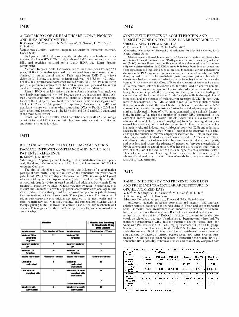
P410
A COMPARISON OF GE HEALTHCARE LUNAR PRODIGY
AND IDXA DENSITOMETERSD. Krueger*1, M. Checovich1, N. Vallarta-Ast1, D. Gemar1, R. Clodfelter1,N. Binkley11Osteoporosis Clinical Research Program, University of Wisconsin, Madison,United States
Background: GE Healthcare recently introduced a new fan-beam densi-tometer, the Lunar iDXA. This study evaluated BMD measurement compara-bility and precision obtained on a Lunar iDXA and Lunar Prodigydensitometer.
Methods: In 245 subjects, 153 women and 91 men, age (mean [range]) 52.8years, [20.0 - 91.5] lumbar spine and dual proximal femur measurements wereobtained in routine clinical manner. Their mean lowest BMD T-score fromeither the L1-4 spine, total femur or femur neck was – 0.8 [3.4 to – 4.3]. Addi-tionally, in 30 postmenopausal women age 69.8 years, [61.7-78.8] from the abovegroup, a precision assessment of the lumbar spine and proximal femur wasconducted using each instrument following ISCD recommendations.
Results: BMD at the L1-4 spine, mean total femur and mean femur neck wasvery highly correlated (r2 >= .98) between these two instruments. Bland-Alt-man analyses confirmed the absence of clinically significant bias. Specifically,biases at the L1-4 spine, mean total femur and mean femoral neck regions were0.011, - 0.002 and - 0.008 grams/cm2 respectively. Moreover, the BMD leastsignificant change was similar when comparing iDXA to Prodigy (0.041 and0.044 grams/cm2 respectively at the L1-4 spine and 0.013 and 0.010 grams/cm2at the mean total femur).
Conclusion: There is excellent BMD correlation between iDXA and Prodigydensitometers and BMD precision with these two instruments at the L1-4 spineand femur is virtually identical.
P411
RISEDRONATE 35 MG PLUS CALCIUM COMBINATION
PACKAGE IMPROVES COMPLIANCE AND INFLUENCES
PATIENTS PREFERENCEH. Kruse*1, J. D. Ringe21Abteilung fur Nephrologie und Osteologie, Universitats-Krankenhaus Eppen-dorf, Hamburg, 2Medizinische Klinik IV, Klinikum Leverkusen, D-51375 Le-verkusen, Germany
The aim of this pilot study was to test the influence of a combinationpackage of risedronate 35 mg plus calcium on the compliance and preference ofpatients with PMO. We investigated 18 women with PMO (mean age 67.1 years)who were taking an oral bisphosphonate (daily or weekly, n=13) or anotherosteoporosis drug (n=5) for at least 3 months and calcium and/or vitamin D. Atbaseline all patients were asked. Patients were then switched to risedronate pluscalcium and 3 months after switching, patients were interviewed once again. Theresults (table) show a strong trend for improved compliance in patients takingthe combination package of risedronate 35 mg plus calcium. The procedure oftaking bisphosphonate plus calcium was reported to be much easier and tointerfere markedly less with daily routine. The combination package with atherapy-guiding blister, improves the correct I use of the bisphosphonate andcalcium. This suggests that the overall therapeutic results can be improved withco-packaging.
P412
SYNERGISTIC EFFECTS OF AGOUTI PROTEIN AND
ROSIGLITAZONE ON BONE LOSS IN A MURINE MODEL OF
OBESITY AND TYPE 2 DIABETESO. P. Lazarenko1, L. J. Suva2, B. Lecka-Czernik*11Geriatrics, 2Orthopedics, University of Arkansas for Medical Sciences, LittleRock, United States
Mechanistically, thazolidinediones (TZDs) such as rosiglitazone (R) sensitizecells to insulin via the activation of PPAR-gamma. In murine mesenchymal stemcell (MSC) cultures R treatment inhibits osteoblast differentiation and promotesadipocyte differentiation. In C57BL/6 mice R induces bone loss by decreasingbone formation and increasing bone resorption. In humans, certain polymorphicchanges in the PPAR-gamma gene locus impact bone mineral density, and TZDtherapies lead to the bone loss in diabetic post-menopausal patients. In order todetermine whether diabetes and obesity are confounding factors that sensitizebone to R, we compared the effects of R on the skeletons of obese and diabeticAvy/a mice, which ectopically express agouti protein, and on control non-dia-betic a/a mice. Agouti antagonizes leptin-controlled alpha-melanocyte stimu-lating hormone (alpha-MSH) signaling in the hypothalamus leading todevelopment of obesity and diabetes. A role for alpha-MSH in the regulation ofbone mass and the presence of melanocortin receptors (MCRs) in bone wererecently demonstrated. The BMD of adult (4 mo) Avy/a mice is slightly higherthan a/a animals, despite the 3-fold higher number of adipocytes in the Avy/amarrow. Consistently, the expression of osteoblast- and adipocyte-specific genemarkers is elevated in bone of Avy/a mice, compared to a/a animals. Interest-ingly, in adult Avy/a mice the number of marrow MSC committed to theosteoblast lineage was significantly (10-fold) lower than in a/a marrow. Theadministration of R for 8 wks (20 mg/kg/day) to Avy/a mice significantly in-creased body weights, normalized glucose and insulin levels, increased circulat-ing leptin levels, and induced severe trabecular bone loss (52%) and a dramaticdecrease in bone strength (35%). None of these changes occurred in a/a mice,although the number of marrow adipocytes increased by 3-fold in these mice,while only a modest 0.5-fold increased was observed in Avy/a animals. Thesedata demonstrate a lack of association between the extent of marrow adiposityand bone loss, and suggest the existence of interactions between the activities ofPPAR-gamma and the agouti protein. Whether this dialog occurs directly at thelevel of MSCs, or at the level of the CNS and hypothalamus, remains unclear.These observations have clinical significance since diabetic patients, many ofwhom suffer altered hypothalamic control of metabolism, may be at risk of boneloss due to TZD therapies.
P413
RANKL INHIBITION BY OPG PREVENTS BONE LOSS
AND PRESERVES TRABECULAR ARCHITECTURE IN
ORCHIECTOMIZED RATSX. Li*1, M. S. Ominsky1, F. Asuncion1, M. Grisanti1, H. L. Tan1,K. S. Warmington1, P. J. Kostenuik11Metabolic Disorders, Amgen Inc., Thousand Oaks, United States
Androgens maintain trabecular bone mass and integrity, and androgenablation results in decreased bone mineral density (BMD) and loss of trabecularbone. Trabecular bone architecture is an important determinant of vertebralfracture risk in men with osteoporosis. RANKL is an essential mediator of boneresorption, but the ability of RANKL inhibitors to prevent trabecular oste-openia associated with androgen ablation has not been previously described. Wetherefore orchiectomized (ORX) rats at 3 months of age and treated them for 6weeks with PBS or human OPG-Fc (10 mg/kg, twice/week SC, n=10-11/group).Sham-operated control rats were treated with PBS. Treatments began immedi-ately after surgery. Distal left femurs and lumbar vertebrae (L5) were harvestedand analyzed by micro-CT (GEHC eXplore Locus SP). After 6 weeks, PBS-treated ORX rats had significant reductions in trabecular bone volume (BV/TV),volumetric BMD (vBMD), trabecular number and connectivity compared with
S144 Abstracts
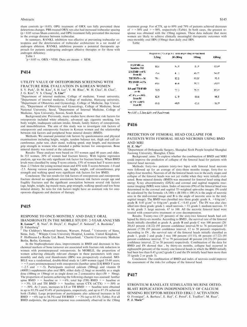
sham controls (p<0.05). OPG treatment of ORX rats fully prevented thesedeleterious changes. PBS-treated ORX rats also had increased trabecular spacing(p<0.05 versus Sham controls), and OPG treatment fully prevented this increasein the average distance between trabeculae.
In summary, RANKL inhibition was effective at preventing trabecular os-teopenia and the deterioration of trabecular architecture in a rat model ofandrogen ablation. RANKL inhibition presents a potential therapeutic ap-proach for patients undergoing androgen ablative therapies or for those withandrogen deficiency.
Table 1*p<0.05 vs. ORX+VEH. Data are means ± SEM.
P414
UTILITY VALUE OF OSTEOPOROSIS SCREENING WITH
FRACTURE RISK EVALUATION IN KOREAN WOMENS. Y. Park1, D. M. Kim1, S. H. Lee1, Y. M. Rhee1, W. H. Choi2, H. Choi3,J. G. Kim4, Y. S. Chung5, S. Lim*11Department of internal medicine, College of medicine, Yonsei university,2Department of internal medicine, College of medicine, Hanyang university,3Department of Obstetrics and Gynecology, College of Medicine, Inje Univer-sity, 4Department of Obstetrics and Gynecology, College of Medicine, SeoulNational University, Seoul, 5Department of Internal Medicine, College ofMedicine, Ajou University, Suwon, South Korea
Background/aim: Previously, many studies have shown that risk factors forosteoporosis included white ethnicity, advanced age, cigarette smoking, lowbody weight, inadequate calcium intake, female, family history of osteoporosis,and immobilization. The aim of this study was to investigate risk factors ofosteoporosis and osteoporotic fracture in Korean women and the relationshipbetween risk factors and peripheral bone mineral density (BMD).
Methods: We assessed potential risk factors by questionnaires and physicalexaminations including height, weight, tandem balance test, thigh and calf cir-cumference, pulse rate, chair stand, walking speed, step length, and maximumgrip strength in women who attended a public lecture for osteoporosis. Bonemineral density was tested at the calcaneus.
Results: These analyses were based on 353 women aged 40 years and older.In 353 women, 20% of women had fracture history. With logistic regressionanalysis, age was the only significant risk factor for fracture history. When BMDlevels were classified by using T-score criteria, 13% of women had T-scores morethan -2.5 below the young normal mean and 45% with T-scores between -1.0 and-2.5. Among various parameters, age, height, weight, calf circumference, gripstrength and walking speed were significant risk factors for low BMD.
Conclusion: The test results for risk factors of osteoporosis and osteoporoticfracture showed no significant association between risk factors except age andfragility fractures and the significant association between several risk factors(age, height, weight, leg muscle mass, grip strength, walking speed) and low bonemineral density. So tests for risk factors might have an assistant role for oste-oporosis diagnosis and decision of therapy.
P415
RESPONSE TO ONCE-MONTHLY AND DAILY ORAL
IBANDRONATE IN THE MOBILE STUDY: 2-YEAR ANALYSISR. Lorenc*1, R. Nuti2, D. Doyle3, C. Hughes4, F. Sedarati4, A. Burdeska4,D. Felsenberg51 The Children’s Memorial Institute, Warsaw, Poland, 2 University of Siena,Siena, Italy, 3 Whipps Cross University Hospital, London, United Kingdom, 4
F. Hoffmann-La Roche Ltd, Basel, Switzerland, 5 Charite-University MedicineBerlin, Berlin, Germany
In the bisphosphonate class, improvements in BMD and decreases in bio-chemical markers of bone turnover are associated with fracture risk reduction inwomen with postmenopausal osteoporosis. In MOBILE, the proportion ofwomen achieving clinically relevant changes in these parameters with once-monthly and daily oral ibandronate (IBN) was prospectively evaluated. MO-BILE was a randomised, double-blind study in 1,609 women (aged 55-80 years;>=5 years postmenopause) with osteoporosis (lumbar spine [LS] BMD T-score<-2.5 and >=-5). Participants received calcium (500mg) and vitamin D(400IU) supplements plus oral IBN, either daily (2.5mg) or monthly as a singledose (100mg or 150mg) or as single doses on 2 consecutive days (50 + 50mg).The proportion of patients achieving the following changes was assessed: lumbarspine BMD >= baseline or >=6%; total hip (TH) BMD >= baseline or>=3%; LS and TH BMD >= baseline; serum CTX (sCTX) >=-30% or>=-50%. At 2 years, increases in LS or TH BMD >= baseline were obtainedin up to 93.5% and 93.4% of participants, respectively, and up to 87.1% achievedboth (Table). Large proportions of participants also achieved increases in LSBMD >=6% (up to 54.3%) and TH BMD>=3% (up to 65.1%; Table). For allBMD endpoints, the greatest response was consistently observed in the 150mg
treatment group. For sCTX, up to 89% and 79% of patients achieved decreasesof >=30% and >=50%, respectively (Table). In both cases, the greatest re-sponse was obtained with the 150mg regimen. These data indicate that morewomen are likely to achieve clinically meaningful therapeutic outcomes withonce-monthly oral IBN (150mg) than daily oral IBN.
Table:
P416
PREDICTION OF FEMORAL HEAD COLLAPSE FOR
PATIENTS WITH FEMORAL HEAD NECROSIS USING BMD
AND MRIH. Z. Ma*11Department of Orthopaedic Surgery, Shanghai Sixth People hospital ShanghaiJiaotong University, Shanghai, China
Background /aims: To explore whether the combination of BMD and MRIcould improve the prediction of collapse of the femoral head for patients withfemoral head necrosis.
Methods: forty-two patients (sixty-two hips) with femoral head necrosiswere followed up for an average of sixty-three months (range, forty-five toeighty-four months). Necrosis of all the femoral heads was in the early stages andcollapse of the femoral heads was not yet visible when they were initially eval-uated. Bone mineral density (BMD) was measured for femoral head using dualenergy X-ray absorptiometry (DXA) and coronal and sagittal magnetic reso-nance imaging (MRI) were taken. Index of necrosis (IN) of the femoral head wasdetermined in the coronal and sagittal T1-weighted spin-echo images. IN couldbe calculated by the formula: (A/180) · (B/180) · 100 (A is the angle of necroticarea in the mid-coronal image and B is the angle of necrotic area in the mid-sagittal image). The BMD was classified into three grade: grade A. >0.6g/cm2;grade B. 0.45 g/cm2 to 0.6g/cm2; grade C.<0.45 g/cm2. The IN was also clas-sified into three grade: grade 1. small necrosis.<33; grade 2. medium necrosis. 33to 66; grade 3. large necrosis. 67 to 100. All the necrotic femoral head weretreated with conservative treatment or core decompression.
Results: Twenty-two (35 percent) of the sixty-two femoral heads had col-lapsed by thirty-six months. According to BMD , the survival rate of the femoralheads initially classified as grade A, grade B and grade C was 100 percent (20/20),63 percent (14/22) (95 percent confidence interval,50 to 76 percent)and 35percent (7/20) (95 percent confidence interval, 12 to 58 percent) respectively.According to IN , the survival rate of the femoral heads initially classified asgrade 1, grade 2 and grade 3 was 100 percent (15/15), 68 percent (17/22) (95percent confidence interval, 57 to 79 percent)and 40 percent (10/25) (95 percentconfidence interval, 22 to 58 percent) respectively. Combination of the data forBMD and IN showed that , by thirty-six months, collapse had occurred ineighteen(86 percent) of the twenty-one femoral heads in which the BMD initiallyhad been less than 0.45 g/cm2 (grade C) and the IN initially head been more than33 (grade 2 or grade 3).
Conclusion: The combination of BMD and index of necrosis could improvethe assessment of the risks for collapse of the femoral head.
P417
STRONTIUM RANELATE STIMULATES MURINE OSTEO-
BLAST REPLICATION INDEPENDENTLY OF CALCIUM
SENSING RECEPTOR-MEDIATED ERK1/2 ACTIVATIONO. Fromigue1, A. Barbara1, E. Hay1, C. Petrel2, E. Traiffort2, M. Ruat2,P. J. Marie*1
Abstracts S145
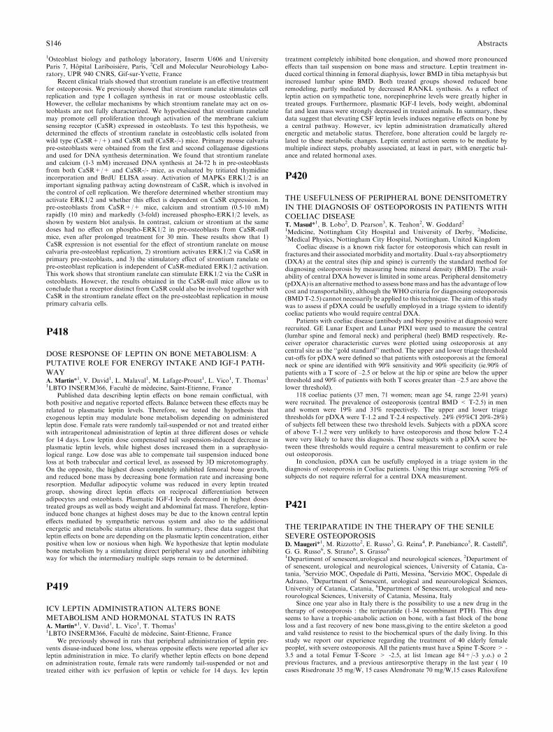
1Osteoblast biology and pathology laboratory, Inserm U606 and UniversityParis 7, Hopital Lariboisiere, Paris, 2Cell and Molecular Neurobiology Labo-ratory, UPR 940 CNRS, Gif-sur-Yvette, France
Recent clinical trials showed that strontium ranelate is an effective treatmentfor osteoporosis. We previously showed that strontium ranelate stimulates cellreplication and type I collagen synthesis in rat or mouse osteoblastic cells.However, the cellular mechanisms by which strontium ranelate may act on os-teoblasts are not fully characterized. We hypothesized that strontium ranelatemay promote cell proliferation through activation of the membrane calciumsensing receptor (CaSR) expressed in osteoblasts. To test this hypothesis, wedetermined the effects of strontium ranelate in osteoblastic cells isolated fromwild type (CaSR+/+) and CaSR null (CaSR-/-) mice. Primary mouse calvariapre-osteoblasts were obtained from the first and second collagenase digestionsand used for DNA synthesis determination. We found that strontium ranelateand calcium (1-3 mM) increased DNA synthesis at 24-72 h in pre-osteoblastsfrom both CaSR+/+ and CaSR-/- mice, as evaluated by tritiated thymidineincorporation and BrdU ELISA assay. Activation of MAPKs ERK1/2 is animportant signaling pathway acting downstream of CaSR, which is involved inthe control of cell replication. We therefore determined whether strontium mayactivate ERK1/2 and whether this effect is dependent on CaSR expression. Inpre-osteoblasts from CaSR+/+ mice, calcium and strontium (0.5-10 mM)rapidly (10 min) and markedly (3-fold) increased phospho-ERK1/2 levels, asshown by western blot analysis. In contrast, calcium or strontium at the samedoses had no effect on phospho-ERK1/2 in pre-osteoblasts from CaSR-nullmice, even after prolonged treatment for 30 min. These results show that 1)CaSR expression is not essential for the effect of strontium ranelate on mousecalvaria pre-osteoblast replication, 2) strontium activates ERK1/2 via CaSR inprimary pre-osteoblasts, and 3) the stimulatory effect of strontium ranelate onpre-osteoblast replication is independent of CaSR-mediated ERK1/2 activation.This work shows that strontium ranelate can stimulate ERK1/2 via the CaSR inosteoblasts. However, the results obtained in the CaSR-null mice allow us toconclude that a receptor distinct from CaSR could also be involved together withCaSR in the strontium ranelate effect on the pre-osteoblast replication in mouseprimary calvaria cells.
P418
DOSE RESPONSE OF LEPTIN ON BONE METABOLISM: A
PUTATIVE ROLE FOR ENERGY INTAKE AND IGF-I PATH-
WAYA. Martin*1, V. David1, L. Malaval1, M. Lafage-Proust1, L. Vico1, T. Thomas11LBTO INSERM366, Faculte de medecine, Saint-Etienne, France
Published data describing leptin effects on bone remain conflictual, withboth positive and negative reported effects. Balance between these effects may berelated to plasmatic leptin levels. Therefore, we tested the hypothesis thatexogenous leptin may modulate bone metabolism depending on administeredleptin dose. Female rats were randomly tail-suspended or not and treated eitherwith intraperitoneal administration of leptin at three different doses or vehiclefor 14 days. Low leptin dose compensated tail suspension-induced decrease inplasmatic leptin levels, while highest doses increased them in a supraphysio-logical range. Low dose was able to compensate tail suspension induced boneloss at both trabecular and cortical level, as assessed by 3D microtomography.On the opposite, the highest doses completely inhibited femoral bone growth,and reduced bone mass by decreasing bone formation rate and increasing boneresorption. Medullar adipocytic volume was reduced in every leptin treatedgroup, showing direct leptin effects on reciprocal differentiation betweenadipocytes and osteoblasts. Plasmatic IGF-I levels decreased in highest dosestreated groups as well as body weight and abdominal fat mass. Therefore, leptin-induced bone changes at highest doses may be due to the known central leptineffects mediated by sympathetic nervous system and also to the additionalenergetic and metabolic status alterations. In summary, these data suggest thatleptin effects on bone are depending on the plasmatic leptin concentration, eitherpositive when low or noxious when high. We hypothesize that leptin modulatebone metabolism by a stimulating direct peripheral way and another inhibitingway for which the intermediary multiple steps remain to be determined.
P419
ICV LEPTIN ADMINISTRATION ALTERS BONE
METABOLISM AND HORMONAL STATUS IN RATSA. Martin*1, V. David1, L. Vico1, T. Thomas11LBTO INSERM366, Faculte de medecine, Saint-Etienne, France
We previously showed in rats that peripheral administration of leptin pre-vents disuse-induced bone loss, whereas opposite effects were reported after icvleptin administration in mice. To clarify whether leptin effects on bone dependon administration route, female rats were randomly tail-suspended or not andtreated either with icv perfusion of leptin or vehicle for 14 days. Icv leptin
treatment completely inhibited bone elongation, and showed more pronouncedeffects than tail suspension on bone mass and structure. Leptin treatment in-duced cortical thinning in femoral diaphysis, lower BMD in tibia metaphysis butincreased lumbar spine BMD. Both treated groups showed reduced boneremodeling, partly mediated by decreased RANKL synthesis. As a reflect ofleptin action on sympathetic tone, norepinephrine levels were greatly higher intreated groups. Furthermore, plasmatic IGF-I levels, body weight, abdominalfat and lean mass were strongly decreased in treated animals. In summary, thesedata suggest that elevating CSF leptin levels induces negative effects on bone bya central pathway. However, icv leptin administration dramatically alteredenergetic and metabolic status. Therefore, bone alteration could be largely re-lated to these metabolic changes. Leptin central action seems to be mediate bymultiple indirect steps, probably associated, at least in part, with energetic bal-ance and related hormonal axes.
P420
THE USEFULNESS OF PERIPHERAL BONE DENSITOMETRY
IN THE DIAGNOSIS OF OSTEOPOROSIS IN PATIENTS WITH
COELIAC DISEASET. Masud*1, B. Lobo2, D. Pearson3, K. Teahon2, W. Goddard21Medicine, Nottingham City Hospital and University of Derby, 2Medicine,3Medical Physics, Nottingham City Hospital, Nottingham, United Kingdom
Coeliac disease is a known risk factor for osteoporosis which can result infractures and their associatedmorbidity andmortality. Dual x-ray absorptiometry(DXA) at the central sites (hip and spine) is currently the standard method fordiagnosing osteoporosis by measuring bone mineral density (BMD). The avail-ability of central DXA however is limited in some areas. Peripheral densitometry(pDXA) is an alternativemethod to assess bonemass and has the advantage of lowcost and transportability, although the WHO criteria for diagnosing osteoporosis(BMDT-2.5) cannot necessarily be applied to this technique. The aim of this studywas to assess if pDXA could be usefully employed in a triage system to identifycoeliac patients who would require central DXA.
Patients with coeliac disease (antibody and biopsy positive at diagnosis) wererecruited. GE Lunar Expert and Lunar PIXI were used to measure the central(lumbar spine and femoral neck) and peripheral (heel) BMD respectively. Re-ceiver operator characteristic curves were plotted using osteoporosis at anycentral site as the ‘‘gold standard’’ method. The upper and lower triage thresholdcut-offs for pDXA were defined so that patients with osteoporosis at the femoralneck or spine are identified with 90% sensitivity and 90% specificity (ie.90% ofpatients with a T score of –2.5 or below at the hip or spine are below the upperthreshold and 90% of patients with both T scores greater than –2.5 are above thelower threshold).
118 coeliac patients (37 men, 71 women; mean age 54, range 22-91 years)were recruited. The prevalence of osteoporosis (central BMD < T-2.5) in menand women were 19% and 31% respectively. The upper and lower triagethresholds for pDXA were T-1.2 and T-2.4 respectively. 24% (95%CI 20%-28%)of subjects fell between these two threshold levels. Subjects with a pDXA scoreof above T-1.2 were very unlikely to have osteoporosis and those below T-2.4were very likely to have this diagnosis. Those subjects with a pDXA score be-tween these thresholds would require a central measurement to confirm or ruleout osteoporosis.
In conclusion, pDXA can be usefully employed in a triage system in thediagnosis of osteoporosis in Coeliac patients. Using this triage screening 76% ofsubjects do not require referral for a central DXA measurement.
P421
THE TERIPARATIDE IN THE THERAPY OF THE SENILE
SEVERE OSTEOPOROSISD. Maugeri*1, M. Rizzotto2, E. Russo3, G. Reina4, P. Panebianco5, R. Castelli6,G. G. Russo6, S. Strano6, S. Grasso61Departiment of senescent,urological and neurological sciences, 2Department ofof senescent, urological and neurological sciences, University of Catania, Ca-tania, 3Servizio MOC, Ospedale di Patti, Messina, 4Servizio MOC, Ospedale diAdrano, 5Department of Senescent, urological and neurourological Sciences,University of Catania, Catania, 6Department of Senescent, urological and neu-rourological Sciences, University of Catania, Messina, Italy
Since one year also in Italy there is the possibility to use a new drug in thetherapy of osteoporosis : the teriparatide (1-34 recombinant PTH). This drugseems to have a trophic-anabolic action on bone, with a fast block of the boneloss and a fast recovery of new bone mass,giving to the entire skeleton a goodand valid resistence to resist to the biochemical spurs of the daily living. In thisstudy we report our experience regarding the treatment of 40 elderly femalepeople(, with severe osteoporosis. All the patients must have a Spine T-Score > -3.5 and a total Femur T-Score > -2.5, at list 1mean age 84+/-3 y.o.) o 2previous fractures, and a previous antiresorptive therapy in the last year ( 10cases Risedronate 35 mg/W, 15 cases Alendronate 70 mg/W,15 cases Raloxifene
S146 Abstracts
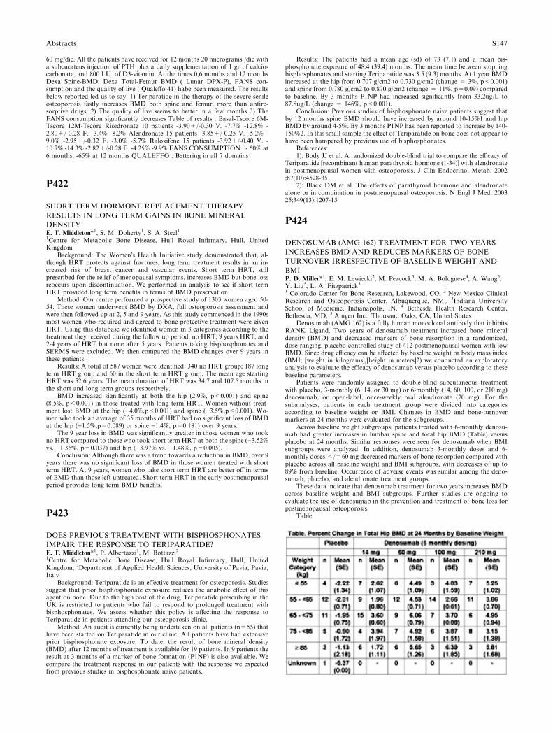
60 mg/die. All the patients have received for 12 months 20 micrograms /die witha subcucateus injection of PTH plus a daily supplementation of 1 gr of calcio-carbonate, and 800 I.U. of D3-vitamin. At the times 0,6 months and 12 monthsDexa Spine-BMD, Dexa Total-Femur BMD ( Lunar DPX-P), FANS con-sumption and the quality of live ( Qualeffo 41) habe been measured. The resultsbelow reported led us to say: 1) Teriparatide in the therapy of the severe senileosteoporosis fastly increases BMD both spine and femur, more than antire-sorptive drugs. 2) The quality of live seems to better in a few months 3) TheFANS consumption significantly decreases Table of results : Basal-Tscore 6M-Tscore 12M-Tscore Risedronate 10 patients -3.90+/-0.30 V. -7.7% -12.8% -2.80+/-0.28 F. -3.4% -8.2% Alendronate 15 patients -3.85+/-0.25 V. -5.2% -9.0% -2.95+/-0.32 F. -3.0% -5.7% Raloxifene 15 patients -3.92+/-0.40 V. -10.7% -14.3% -2.82+/-0.28 F. -4.25% -9.9% FANS CONSUMPTION : - 50% at6 months, -65% at 12 months QUALEFFO : Bettering in all 7 domains
P422
SHORT TERM HORMONE REPLACEMENT THERAPY
RESULTS IN LONG TERM GAINS IN BONE MINERAL
DENSITYE. T. Middleton*1, S. M. Doherty1, S. A. Steel11Centre for Metabolic Bone Disease, Hull Royal Infirmary, Hull, UnitedKingdom
Background: The Women�s Health Initiative study demonstrated that, al-though HRT protects against fractures, long term treatment results in an in-creased risk of breast cancer and vascular events. Short term HRT, stillprescribed for the relief of menopausal symptoms, increases BMD but bone lossreoccurs upon discontinuation. We performed an analysis to see if short termHRT provided long term benefits in terms of BMD preservation.
Method: Our centre performed a prospective study of 1303 women aged 50-54. These women underwent BMD by DXA, full osteoporosis assessment andwere then followed up at 2, 5 and 9 years. As this study commenced in the 1990smost women who required and agreed to bone protective treatment were givenHRT. Using this database we identified women in 3 categories according to thetreatment they received during the follow up period: no HRT; 9 years HRT; and2-4 years of HRT but none after 5 years. Patients taking bisphosphonates andSERMS were excluded. We then compared the BMD changes over 9 years inthese patients.
Results: A total of 587 women were identified: 340 no HRT group; 187 longterm HRT group and 60 in the short term HRT group. The mean age startingHRT was 52.6 years. The mean duration of HRT was 34.7 and 107.5 months inthe short and long term groups respectively.
BMD increased significantly at both the hip (2.9%, p<0.001) and spine(8.5%, p<0.001) in those treated with long term HRT. Women without treat-ment lost BMD at the hip ()4.0%,p<0.001) and spine ()3.5%,p<0.001). Wo-men who took an average of 35 months of HRT had no significant loss of BMDat the hip ()1.5%,p=0.089) or spine )1.4%, p=0.181) over 9 years.
The 9 year loss in BMD was significantly greater in those women who tookno HRT compared to those who took short term HRT at both the spine ()3.52%vs. )1.36%, p=0.037) and hip ()3.97% vs. )1.48%, p=0.005).
Conclusion: Although there was a trend towards a reduction in BMD, over 9years there was no significant loss of BMD in those women treated with shortterm HRT. At 9 years, women who take short term HRT are better off in termsof BMD than those left untreated. Short term HRT in the early postmenopausalperiod provides long term BMD benefits.
P423
DOES PREVIOUS TREATMENT WITH BISPHOSPHONATES
IMPAIR THE RESPONSE TO TERIPARATIDE?E. T. Middleton*1, P. Albertazzi1, M. Bottazzi21Centre for Metabolic Bone Disease, Hull Royal Infirmary, Hull, UnitedKingdom, 2Department of Applied Health Sciences, University of Pavia, Pavia,Italy
Background: Teriparatide is an effective treatment for osteoporosis. Studiessuggest that prior bisphosphonate exposure reduces the anabolic effect of thisagent on bone. Due to the high cost of the drug, Teriparatide prescribing in theUK is restricted to patients who fail to respond to prolonged treatment withbisphosphonates. We assess whether this policy is affecting the response toTeriparatide in patients attending our osteoporosis clinic.
Method: An audit is currently being undertaken on all patients (n=55) thathave been started on Teriparatide in our clinic. All patients have had extensiveprior bisphosphonate exposure. To date, the result of bone mineral density(BMD) after 12 months of treatment is available for 19 patients. In 9 patients theresult at 3 months of a marker of bone formation (P1NP) is also available. Wecompare the treatment response in our patients with the response we expectedfrom previous studies in bisphosphonate naive patients.
Results: The patients had a mean age (sd) of 73 (7.1) and a mean bis-phosphonate exposure of 48.4 (39.4) months. The mean time between stoppingbisphosphonates and starting Teriparatide was 3.5 (9.3) months. At 1 year BMDincreased at the hip from 0.707 g/cm2 to 0.730 g/cm2 (change = 3%, p<0.001)and spine from 0.780 g/cm2 to 0.870 g/cm2 (change = 11%, p=0.09) comparedto baseline. By 3 months P1NP had increased significantly from 33.2ug/L to87.8ug/L (change = 146%, p<0.001).
Conclusion: Previous studies of bisphosphonate naive patients suggest thatby 12 months spine BMD should have increased by around 10-15%1 and hipBMD by around 4-5%. By 3 months P1NP has been reported to increase by 140-150%2. In this small sample the effect of Teriparatide on bone does not appear tohave been hampered by previous use of bisphosphonates.
References:1): Body JJ et al. A randomized double-blind trial to compare the efficacy of
Teriparatide [recombinant human parathyroid hormone (1-34)] with alendronatein postmenopausal women with osteoporosis. J Clin Endocrinol Metab. 2002;87(10):4528-35
2): Black DM et al. The effects of parathyroid hormone and alendronatealone or in combination in postmenopausal osteoporosis. N Engl J Med. 200325;349(13):1207-15
P424
DENOSUMAB (AMG 162) TREATMENT FOR TWO YEARS
INCREASES BMD AND REDUCES MARKERS OF BONE
TURNOVER IRRESPECTIVE OF BASELINE WEIGHT AND
BMIP. D. Miller*1, E. M. Lewiecki2, M. Peacock3, M. A. Bolognese4, A. Wang5,Y. Liu5, L. A. Fitzpatrick51 Colorado Center for Bone Research, Lakewood, CO, 2 New Mexico ClinicalResearch and Osteoporosis Center, Albuquerque, NM,, 3Indiana UniversitySchool of Medicine, Indianapolis, IN, 4 Bethesda Health Research Center,Bethesda, MD, 5 Amgen Inc., Thousand Oaks, CA, United States
Denosumab (AMG 162) is a fully human monoclonal antibody that inhibitsRANK Ligand. Two years of denosumab treatment increased bone mineraldensity (BMD) and decreased markers of bone resorption in a randomized,dose-ranging, placebo-controlled study of 412 postmenopausal women with lowBMD. Since drug efficacy can be affected by baseline weight or body mass index(BMI; [weight in kilograms]/[height in meters]2) we conducted an exploratoryanalysis to evaluate the efficacy of denosumab versus placebo according to thesebaseline parameters.
Patients were randomly assigned to double-blind subcutaneous treatmentwith placebo, 3-monthly (6, 14, or 30 mg) or 6-monthly (14, 60, 100, or 210 mg)denosumab, or open-label, once-weekly oral alendronate (70 mg). For thesubanalyses, patients in each treatment group were divided into categoriesaccording to baseline weight or BMI. Changes in BMD and bone-turnovermarkers at 24 months were evaluated for the subgroups.
Across baseline weight subgroups, patients treated with 6-monthly denosu-mab had greater increases in lumbar spine and total hip BMD (Table) versusplacebo at 24 months. Similar responses were seen for denosumab when BMIsubgroups were analyzed. In addition, denosumab 3-monthly doses and 6-monthly doses </=60 mg decreased markers of bone resorption compared withplacebo across all baseline weight and BMI subgroups, with decreases of up to89% from baseline. Occurrence of adverse events was similar among the deno-sumab, placebo, and alendronate treatment groups.
These data indicate that denosumab treatment for two years increases BMDacross baseline weight and BMI subgroups. Further studies are ongoing toevaluate the use of denosumab in the prevention and treatment of bone loss forpostmenopausal osteoporosis.
Table
Abstracts S147
P425
PTH(1-84) PREVENTS VERTEBRAL FRACTURES IN POST
MENOPAUSAL WOMEN WITH HIGHER FRACTURE RISK:
RESULTS FROM THE TOP STUDYP. Miller*1, S. L. Greenspan21Colorado Center for Bone Research, University of Colorado Health SciencesCenter, Denver, 2Osteoporosis Center, University of Pittsburgh, Pittsburg,United States
Introduction: The ‘‘TOP’’ Study was an 18-month, multinational, random-ized, double-blind, placebo-controlled study including 2532 postmenopausalosteoporotic women. In the per protocol population (n = 1870), there was a66% relative risk reduction (RRR) in vertebral fractures in subjects receivingPTH (P = .002). In subjects with and without prevalent fractures, PTH therapycaused a RRR in vertebral fractures of 69% (P = 0.025) and 63% (P = .028),respectively. We report the effectiveness of PTH(1-84) in reducing vertebralfracture in 2 subgroups of high fracture risk subjects: women with advanced ageor low BMD.
Methods: women 55 years or above with spine, femoral neck, or total hip T-score equal to or <)2.5 (or equal to or <)2.0 + prevalent vertebral fracture),or 45–54 years of age with a T-score equal to or <)3.0 (or equal to or <)2.5 +fracture). All received 700 mg calcium + 400 IU vitamin D daily and wererandomized to placebo/ PTH 100 lg daily. This subanalysis of the intention totreat (ITT) group included women with advanced age (>60) or with low BMD(lumbar spine T-score <)3.0).
Results: In the ITT group, 1797 women were > 60. PTH treatment resultedin a 64% RRR in vertebral fracture (1.5% vs 4.2%, respectively, P = .001).When stratified according to vertebral fracture, those with prevalent fracture(s)receiving PTH experienced a 58% RRR in vertebral fracture compared withplacebo (4.4% vs 10.3%, respectively; P = .027). In the group without prevalentfracture, PTH therapy resulted in a RRR of 67% compared with placebo (2.5%vs .8%, respectively; P = .012). When the TOP ITT population were analyzedwith regard to low BMD (n = 1149), PTH reduced vertebral fracture by 74%(RRR) compared with placebo (1.3% vs 5.1%, respectively; P < .001). In thissame group, PTH treatment resulted in a RRR in incident vertebral fracture of59% compared to placebo in those with prevalent vertebral fracture(s) (5.5% vs13.6%, respectively; P = .040), and an RRR of 85% compared with placebo thegroup without prevalent vertebral fracture (2.8% vs 0.41%; P = .003).Therewere no substantial difference in the adverse event profile and discontinuationcauses between subgroups and the TOP population. Conclusion: As in theoverall TOP population. PTH (1-84) substantially reduces the incidence ofvertebral fracture in subgroups with high fracture risk.
P426
NOVEL FLUOROCHROME MARKERS TO DETECT
MICRODAMAGE IN BONES. Mohsin*1, . T. C. Lee1, . R. Parkesh2, T. Gunnlaugsson21Anatomy, Royal College of Surgeons in Ireland, 2Chemistry, Trinity College,Dublin, Ireland
Fluorochrome agents are used to study the dynamics of bone remodellingand as markers of microdamage detection in bone. Bone microdamage accu-mulation has been implicated in increasing skeletal fragility, in the pathome-chanics of osteoporosis and in stress fractures. Microcracks are discontinuities inthe calcium rich bone matrix and agents which bind calcium can selectively labelthem.
This study was designed to investigate fluorescents agents which work onphotoinduced electron transfer mechanism and show fluorescence only whenchelate to calcium ions hence are site-specific. Fluorescent agents of differentcolours were selected which can give better contrast against the autofluorescenceof bone, are pH independent in the physiological range, sufficiently soluble inwater and show good penetration in the bone tissue.
Beam-shaped bone samples were prepared from ten different bovine tibiaeusing a band saw and polished with emery paper (grade 400). The bone sampleswere scratched using a compass to mimic a microcrack. Three fluorescent agentsfluo-3, calcium orange and calcium crimson were purchased from MolecularProbes Ltd., Eugene, Oregon. A 10-4 M solution of each dye was prepared at pH7.4. Bone specimens were dipped in each of the solutions at various time inter-vals and examined using epifluorescence microscopy. Images were analysedusing Lucia software and chelation time, sequence and ability to penetrate bonewere assessed. The optimal chelation time found to label surface scratches wasfifteen minutes after which it leached into the surrounding matrix. The optimalsequence of application of fluorescent agents was calcium crimson, calcium or-ange and fluo-3. Using this sequence, it was possible to differentiate each agentfrom the other and from the surrounding bone matrix. These fluorescent agentsare site specific and fluoresced only when bound to calcium ions. All of thesefluorescent agents showed good contrast and penetration of bone tissue. Fluo-rescent agents were able to label bone surfaces, Haversian canals, lacunae andresorption cavities.
These fluorescent agents can be used to label changes occurring during boneremodelling. These can be used in sequence to monitor microcrack growth andto detect microdamage in vitro. However there is a need to detect microdamagein vivo using radiopaque agents.
P427
EFFECTS OF INTRAVENOUS PAMIDRONATE ON BONE
LOSS AFTER LIVER TRANSPLANTATIONA. Monegal*1, M. Navasa2, M. Suarez3, S. Suarez Lopez4, F. Garcia Gonzalez5,T. Serrano6, G. Clemente7, F. Casafont8, M. De la Mata9, C. Barrios10, P. Peris1,N. Guanabens11Rheumatology Service, 2Liver Unit, Hospital Clinic Barcelona, Barcelona,3Transplantation Unit, Hospital Cruces, Bilbao, 4Transplantation Unit, Hos-pital Juan Canalejo, La Coruna, 5Transplantation Unit, Hospital Ramon yCajal, Madrid, 6Transplantation Unit, Hospital Lozano Blesa, Zaragoza,7Transplantation Unit, Hospital Gregorio Maranon, Madrid, 8TransplantationUnit, Hospital Marques de Valdecilla, Santander, 9Transplantation Unit, Hos-pital Reina Sofia, Cordoba, 10Transplantation Unit, Hospital Puerta de Hierro,Madrid, Spain
Background: Rapid bone loss and high fracture rates, particularly during thefirst 6 to 12 months, occur following liver transplantation. Aim: To analyse theeffect of intravenous pamidronate on bone loss after liver transplantation. Pa-tients and Methods: A randomized, double-masked, placebo-controlled studywas performed at 9 Spanish centers. 79 patients were randomized to two groupsof treatment: Group 1 (n=38): Pamidronate 90 mg/IV in the first week and 3months after transplantation (30 m/8 f; age mean ± SD: 53 ± 11 years); Group2 (n=41): placebo in the same periods (31 m/10 f; age, mean ± SD: 53 ± 9years). All patients received calcium and vitamin D. In all patients, bone mineraldensity (BMD) (lumbar spine and proximal femur) (DXA) and spinal X-rayswere performed before, and at 6 and 12 months after liver transplantation.Biochemical and hormonal determinations including serum 25-OH-D and PTHwere measured before transplantation, 24h pre and post-treatment, as well as atand 6 and 12 months after liver transplantation. Results: No differences werefound in baseline characteristics between the two treatment groups. At 6 and 12months after transplantation there were significant differences in lumbar BMDchanges in men (6 months G1: +1.72 ± 5.8% vs G2: )1.47 ± 3.7%, p<0.05; 1year G1 +4.1 ± 7.7% vs G2 –1.98 ± 4.5%, p<0.01). However, there were nodifferences in the changes of lumbar BMD in women . Changes were similar atthe femoral neck and total hip in the groups receiving pamidronate or placebo.Moreover, no changes in fracture rates (G1 18%; G2 7%: p=ns), serum PTHand 25-OH-D values between groups 1 and 2 were found. Pamidronate did notincrease the risk of adverse effects. Conclusions: The results of this study showthat 90 mg of intravenous Pamidronate in the first week and at 3 months fol-lowing liver transplantation preserve lumbar bone mass in men, without sig-nificant adverse effects. This study was supported by Novartis Pharmaceuticals.
P428
PREVALENCE OF VITAMIN D INADEQUACY IN OSTEOPO-
ROTIC HIP FRACTURE PATIENTS IN S.E. LONDONC. Moniz*1, T. Dew1, T. Dixon21Clinical Biochemistry, Kings College Hospital, London, 2JB Medical, Sudbury,Suffolk, United Kingdom
The elderly with serum vitamin D levels either lower than those of controlpatients of similar age are likely to have fragility fractures consequent on acombination of altered bone turnover and or falls. In a retrospective patientaudit at Kings College Hospital, SE London, carried out over a 17-month period(September 2003 to January 2005), patient records were searched for hip fractureadmissions and cross matched with vitamin D analysis carried out within 3 daysof the hip fracture admission. The resulting records were hand searched to ex-clude patients with a hip fracture resulting from high impact/trauma. There weredata for 103 hip fracture patients, 79.6% of the patients were women (n=82).The mean age at the time of fracture was 73.4 years, 100% were aged 60 years orover and 41% were aged 75 years or over. Around 20% of the patients werereceiving supplementation with calcium and/or vitamin D and were not excludedfrom the analysis. The mean vitamin D level was 32.1 nmol/L (12.9 ug/ L),SD=19.4 (7.8), however, it is likely that the true mean is lower since inapproximately 15% of cases vitamin D levels were reported as <12.5 nmol/L,the detection limit. 81.6% had levels <50 nmol/L, a target level which maintainsnormocalcaemia and normo-parathyroid levels. There were no significant dif-ferences by patient age or sex, however there were significant seasonal differencesin vitamin D. In the year from September 2003 to August 2004, 82.8% ofsummer admissions had vitamin D levels <70 nmol/L compared with 98.0% inwinter (p=0.04). Mean vitamin D levels in the 30 patients with parathyroidhormone (PTH) levels above the reference range were significantly lower thanlevels in the 71 patients within the range: mean 19.9 nmol/L, SD=16.2 versus
S148 Abstracts

mean 37.5 nmol/L, SD=18.5 (p<0.0001). Furthermore, 50% of the patientswith PTH levels above the reference range had vitamin D levels <12.5 nmol/Lreflecting extremely low levels of vitamin D. This study confirms almost uni-versal vitamin D inadequacy among 103 patients admitted to hospital with hipfracture in SE London indicating a major clinical problem that could be reducedby proactive supplementation in high risk elderly subjects.
P429
BONE TURNOVER INCREASES IN EARLY POSTMENO-
PAUSAL COMPARE TO PREMENOPAUSAL WOMENL. Navarro*1, J. Blazquez2, M. Chafer1, F. Mateos2, E. Almar3, J. Del Pino41Clinical Chemistry, 2Internal Medicine, University Hospital, 3Epidemiology,Health Department, Albacete, 4Rheumatology, University Hospital, Salamanca,Spain
Objective. The aim of this study was to know how much increases the boneturnover in early postmenopausal with regard to premenopausal women.Methods. 197 women aged 50 to 55 years (mean 52,7 ± 1,6 yr ) were randomlyselected in the province of Albacete (Spain). All women were postmenopausal,from 6 to 36 months. At the same time 34 normal premenopausal women wereselected as the reference population (39 ± 5,4 yr). To assess bone turnover, bonephosphatase alkaline (BAP) was measured in serum and deoxypyridinoline(DPD), NTx, and calcium (Ca/Cr) were measured in a fasting 2 h morning urine.Comparison between premenopausal and postmenopausal women was assessedby the percent difference and by the t score (t score (t score = [chi-M]/SD),where chi is the marker level of an individual postmenopausal women, M andSD are the mean and the standard deviation of the premenopausal normalreference population. Results. Table. Conclusions. Early postmenopausal wo-men had a significant increase of all markers. Bone resorption markers increasedmore than BAP. NTx had the highest percent increase, bigger than in otherstudies, but the t-score was similar the DPD t-score. Ca/Cr was also quite in-creased but less than the others markers.
Supported by grants from FIS 99/07059, JCCM98196 FISCAM, Diputacionof Albacete
Table:
P430
INDIRECT COMPARISON OF ANTI-VERTEBRAL FRACTURE
EFFICACY AMONG AVAILABLE THERAPIES: A BAYESIAN
ANALYSISN. D. Nguyen*1, J. A. Eisman1, T. V. Nguyen11Bone and Mineral Research Program, Garvan Institute of Medical Research,Sydney, Australia
Background. Clinicians sometimes have to make decision on treatmentselection based on efficacy and cost consideration. Several anti-fracture drugswith different magnitudes of treatment effect are currently available for thetreatment of osteoporosis. In the absence of a comparative head-to-head clinicaltrial, the selection of an optimal treatment is difficult. The present analysis wasundertaken to indirectly compare the anti-vertebral fracture efficacies amongtherapies by using a Bayesian approach.
Methods. Published data from placebo-controlled clinical trials (RCT)evaluating the anti-fracture efficacy of alendronate (n=4 trials), residronate(n=5), calcitonin (n=8), calcium (n=3), fluoride (n=6), raloxifene, (n=4) andHRT (n=13) were included in the analysis. A random-effects Bayesian meta-analysis model was utilized to estimate the effect size (log relative risk) and itsvariance for an individual therapy. The index of superiority was defined as theprobability that a drug�s effect size is 20% greater than another drug. Thissuperiority index was computed by the Bayesian hierachical models usingMarkov chain Monte Carlo implemented in WinBUGS.
Results. When compared to placebo, most active therapies significantly re-duced the risk of vertebral fracture, with variable magnitudes: alendronate (RR;95%CI: 0.52; 0.34-0.79), risedronate (0.59; 0.40-0.71), raloxifene (0.60; 0.37-0.99)and HRT (0.60; 0.37-0.99). However, the evidence of effect was uncertain forcalcitonin (0.79; 0.53-1.21), calcium (0.71; 0.35-1.39) and fluoride (0.72; 0.44-1.36). More importantly, pairwise comparison of active therapies by the index ofsuperiority revealed no strong evidence of superiority among the drugs. Theprobabilities that alendronate is superior to risedronate was 0.42, to HRT: 0.47,raloxifene: 0.48, calcitonin: 0.82, fluoride: 0.68, and calcium: 0.65.
Conclusion. These results indicate that while alendronate, risedronate, ra-loxifene and HRT were efficacious in reducing vertebral fracture risk, their effectsizes were comparable. There is no evidence for the superiority of one drug toanother among the currently available drugs.
P431
ON THE ASSOCIATION BETWEEN STATIN AND FRACTURE:
A BAYESIAN CONSIDERATIONC. Y. Wang1, N. D. Nguyen*1, J. A. Eisman1, T. V. Nguyen11Bone and Mineral Research Program, Garvan Institute of Medical Research,Sydney, Australia
Background. Despite observational studies have suggested that statin usemay be associated with reduced fracture risk, the association is controversial,due to conflicting findings from published studies, probably due to publicationbias. This study utilized the Bayesian approach to combine existing evidence andupdate the association with consideration of potential publication bias.
Methods. Data on the association between statin use and fracture incidencefrom 11 observational studies and 3 secondary analyses of randomized clinicaltrials (RCTs) were synthesized by both Empirical Bayesian analysis and fullyBayesian random-effects meta-analysis models. Sensitivity analyses were carriedout to estimate the effect of potential bias on the observed association. Theheterogeneity of effect sizes was assessed by the coefficient of inconsistency.
Results. Empirical Bayesian analysis showed that statin use was associatedwith a reduction in hip fracture risk with odds ratio (OR) being 0.56 and 95%credible interval (CrI) ranging from 0.44 to 0.70. The OR for non-vertebralfracture was 0.62 (95% CrI, 0.56-0.68). These results were comparable with thefully Bayesian random-effects meta-analysis�. The probability that statin usereduces fracture risk by at least 20% was 0.99 for hip fracture and 0.85 for non-vertebral fracture. Smaller studies tended to produce larger effect sizes, raisingthe possibility of publication bias. Under the assumption that the bias over-estimates the true OR by 10%, there is still a probability of 0.975 that statin usereduces hip fracture risk; however, the effect on non-vertebral fracture was lessrobust with a probability of 0.86. If bias over-estimates the true OR by 30%, theprobability that statin is associated with reduced hip fracture and vertebralfracture is 0.95 and 0.45, respectively.
Conclusion. These findings suggest statin use can reduce hip fracture risk;however, there is considerable uncertainty on the association between statin useand vertebral fracture. The Bayesian approach presented here can helpresearchers update existing evidence when new data becomes available and takeinto account potential bias.
P432
A GROUP-BASED EDUCATION PROGRAMME INCREASES
KNOWLEDGE ON OSTEOPOROSIS AND ADHERENCE TO
PHARMACOLOGICAL TREATMENT. INTERIM ANALYSIS
FROM A RANDOMISED TRIAL.D. Nielsen*1, W. Nielsen2, B. Knold1, N. Nissen1, J. Ryg1, A. R. Madsen1,K. Brixen11Department of Endocrinology, Odense University Hospital,, 2Department ofPhysiotherapy, Odense University Hospital, Odense, Denmark
Background: Modification of individual life style factors and fall preventionprogrammes may have significant positive effects on fracture incidence. Also, alarge number of studies have demonstrated that pharmacological therapy ofosteoporosis is effective; however non-adherence to such therapy is a well rec-ognized problem. Few studies, however, have examined the effect of particularpatient education programmes on knowledge and adherence to therapy.
Aim: We hypothesised that a group-based, multi-disciplinary, educationprogramme increases the patients� knowledge on osteoporosis and adherencewith pharmacological therapy.
Participants and design: A total of 300 patients, recently diagnosed withosteoporosis and started on specific treatment, were randomised to either the’’school’’ or ’’control’’ group. In the school-group, patients attended four classeswith 8-12 participants during four weeks (a total of 12 hours). Teaching wasperformed by nurses, physiotherapists, dieticians, and doctors and was based ondialogs and situated learning. The classes covered ’’facts on osteoporosis’’,’’fractures and pain’’, ’’diet’’, ’’preventive measures’’, ’’balance and exercise’’,and ’’medical treatment’’. Teaching was designed to increase empowerment. In
Abstracts S149
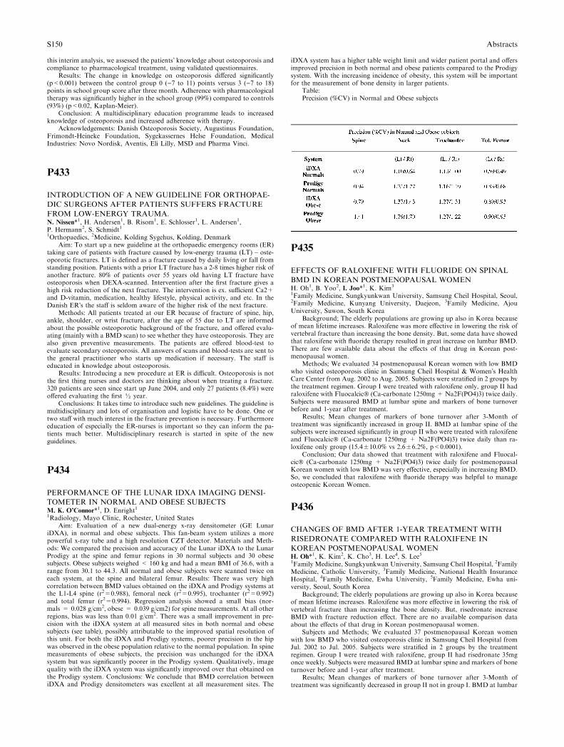
this interim analysis, we assessed the patients� knowledge about osteoporosis andcompliance to pharmacological treatment, using validated questionnaires.
Results: The change in knowledge on osteoporosis differed significantly(p<0.001) between the control group 0 ()7 to 11) points versus 3 ()7 to 18)points in school group score after three month. Adherence with pharmacologicaltherapy was significantly higher in the school group (99%) compared to controls(93%) (p<0.02, Kaplan-Meier).
Conclusion: A multidisciplinary education programme leads to increasedknowledge of osteoporosis and increased adherence with therapy.
Acknowledgements: Danish Osteoporosis Society, Augustinus Foundation,Frimondt-Heineke Foundation, Sygekassernes Helse Foundation, MedicalIndustries: Novo Nordisk, Aventis, Eli Lilly, MSD and Pharma Vinci.
P433
INTRODUCTION OF A NEW GUIDELINE FOR ORTHOPAE-
DIC SURGEONS AFTER PATIENTS SUFFERS FRACTURE
FROM LOW-ENERGY TRAUMA.N. Nissen*1, H. Andersen1, B. Risom1, E. Schlosser1, L. Andersen1,P. Hermann2, S. Schmidt11Orthopaedics, 2Medicine, Kolding Sygehus, Kolding, Denmark
Aim: To start up a new guideline at the orthopaedic emergency rooms (ER)taking care of patients with fracture caused by low-energy trauma (LT) – oste-oporotic fractures. LT is defined as a fracture caused by daily living or fall fromstanding position. Patients with a prior LT fracture has a 2-8 times higher risk ofanother fracture. 80% of patients over 55 years old having LT fracture haveosteoporosis when DEXA-scanned. Intervention after the first fracture gives ahigh risk reduction of the next fracture. The intervention is ex. sufficient Ca2+and D-vitamin, medication, healthy lifestyle, physical activity, and etc. In theDanish ER�s the staff is seldom aware of the higher risk of the next fracture.
Methods: All patients treated at our ER because of fracture of spine, hip,ankle, shoulder, or wrist fracture, after the age of 55 due to LT are informedabout the possible osteoporotic background of the fracture, and offered evalu-ating (mainly with a BMD scan) to see whether they have osteoporosis. They arealso given preventive measurements. The patients are offered blood-test toevaluate secondary osteoporosis. All answers of scans and blood-tests are sent tothe general practitioner who starts up medication if necessary. The staff iseducated in knowledge about osteoporosis.
Results: Introducing a new procedure at ER is difficult. Osteoporosis is notthe first thing nurses and doctors are thinking about when treating a fracture.320 patients are seen since start up June 2004, and only 27 patients (8.4%) wereoffered evaluating the first ½ year.
Conclusions: It takes time to introduce such new guidelines. The guideline ismultidisciplinary and lots of organisation and logistic have to be done. One ortwo staff with much interest in the fracture prevention is necessary. Furthermoreeducation of especially the ER-nurses is important so they can inform the pa-tients much better. Multidisciplinary research is started in spite of the newguidelines.
P434
PERFORMANCE OF THE LUNAR IDXA IMAGING DENSI-
TOMETER IN NORMAL AND OBESE SUBJECTSM. K. O’Connor*1, D. Enright11Radiology, Mayo Clinic, Rochester, United States
Aim: Evaluation of a new dual-energy x-ray densitometer (GE LunariDXA), in normal and obese subjects. This fan-beam system utilizes a morepowerful x-ray tube and a high resolution CZT detector. Materials and Meth-ods: We compared the precision and accuracy of the Lunar iDXA to the LunarProdigy at the spine and femur regions in 30 normal subjects and 30 obesesubjects. Obese subjects weighed < 160 kg and had a mean BMI of 36.6, with arange from 30.1 to 44.3. All normal and obese subjects were scanned twice oneach system, at the spine and bilateral femur. Results: There was very highcorrelation between BMD values obtained on the iDXA and Prodigy systems atthe L1-L4 spine (r2=0.988), femoral neck (r2=0.995), trochanter (r2=0.992)and total femur (r2=0.994). Regression analysis showed a small bias (nor-mals = 0.028 g/cm2, obese = 0.039 g/cm2) for spine measurements. At all otherregions, bias was less than 0.01 g/cm2. There was a small improvement in pre-cision with the iDXA system at all measured sites in both normal and obesesubjects (see table), possibly attributable to the improved spatial resolution ofthis unit. For both the iDXA and Prodigy systems, poorer precision in the hipwas observed in the obese population relative to the normal population. In spinemeasurements of obese subjects, the precision was unchanged for the iDXAsystem but was significantly poorer in the Prodigy system. Qualitatively, imagequality with the iDXA system was significantly improved over that obtained onthe Prodigy system. Conclusions: We conclude that BMD correlation betweeniDXA and Prodigy densitometers was excellent at all measurement sites. The
iDXA system has a higher table weight limit and wider patient portal and offersimproved precision in both normal and obese patients compared to the Prodigysystem. With the increasing incidence of obesity, this system will be importantfor the measurement of bone density in larger patients.
Table:Precision (%CV) in Normal and Obese subjects
P435
EFFECTS OF RALOXIFENE WITH FLUORIDE ON SPINAL
BMD IN KOREAN POSTMENOPAUSAL WOMENH. Oh1, B. Yoo2, I. Joo*1, K. Kim3
1Family Medicine, Sungkyunkwan University, Samsung Cheil Hospital, Seoul,2Family Medicine, Kunyang University, Daejeon, 3Family Medicine, AjouUniversity, Suwon, South Korea
Background; The elderly populations are growing up also in Korea becauseof mean lifetime increases. Raloxifene was more effective in lowering the risk ofvertebral fracture than increasing the bone density. But, some data have showedthat raloxifene with fluoride therapy resulted in great increase on lumbar BMD.There are few available data about the effects of that drug in Korean post-menopausal women.
Methods; We evaluated 34 postmenopausal Korean women with low BMDwho visited osteoporosis clinic in Samsung Cheil Hospital & Women�s HealthCare Center from Aug. 2002 to Aug. 2005. Subjects were stratified in 2 groups bythe treatment regimen. Group I were treated with raloxifene only, group II hadraloxifene with Fluocalcic� (Ca-carbonate 1250mg + Na2F(PO4)3) twice daily.Subjects were measured BMD at lumbar spine and markers of bone turnoverbefore and 1-year after treatment.
Results; Mean changes of markers of bone turnover after 3-Month oftreatment was significantly increased in group II. BMD at lumbar spine of thesubjects were increased significantly in group II who were treated with raloxifeneand Fluocalcic� (Ca-carbonate 1250mg + Na2F(PO4)3) twice daily than ra-loxifene only group (15.4±10.0% vs 2.6±6.2%, p<0.0001).
Conclusion; Our data showed that treatment with raloxifene and Fluocal-cic� (Ca-carbonate 1250mg + Na2F(PO4)3) twice daily for postmenopausalKorean women with low BMD was very effective, especially in increasing BMD.So, we concluded that raloxifene with fluoride therapy was helpful to manageosteopenic Korean Women.
P436
CHANGES OF BMD AFTER 1-YEAR TREATMENT WITH
RISEDRONATE COMPARED WITH RALOXIFENE IN
KOREAN POSTMENOPAUSAL WOMENH. Oh*1, K. Kim2, K. Cho3, H. Lee4, S. Lee51Family Medicine, Sungkyunkwan University, Samsung Cheil Hospital, 2FamilyMedicine, Catholic University, 3Family Medicine, National Health InsuranceHospital, 4Family Medicine, Ewha University, 5Family Medicine, Ewha uni-versity, Seoul, South Korea
Background; The elderly populations are growing up also in Korea becauseof mean lifetime increases. Raloxifene was more effective in lowering the risk ofvertebral fracture than increasing the bone density. But, risedronate increaseBMD with fracture reduction effect. There are no available comparison dataabout the effects of that drug in Korean postmenopausal women.
Subjects and Methods; We evaluated 37 postmenopausal Korean womenwith low BMD who visited osteoporosis clinic in Samsung Cheil Hospital fromJul. 2002 to Jul. 2005. Subjects were stratified in 2 groups by the treatmentregimen. Group I were treated with raloxifene, group II had risedronate 35mgonce weekly. Subjects were measured BMD at lumbar spine and markers of boneturnover before and 1-year after treatment.
Results; Mean changes of markers of bone turnover after 3-Month oftreatment was significantly decreased in group II not in group I. BMD at lumbar
S150 Abstracts
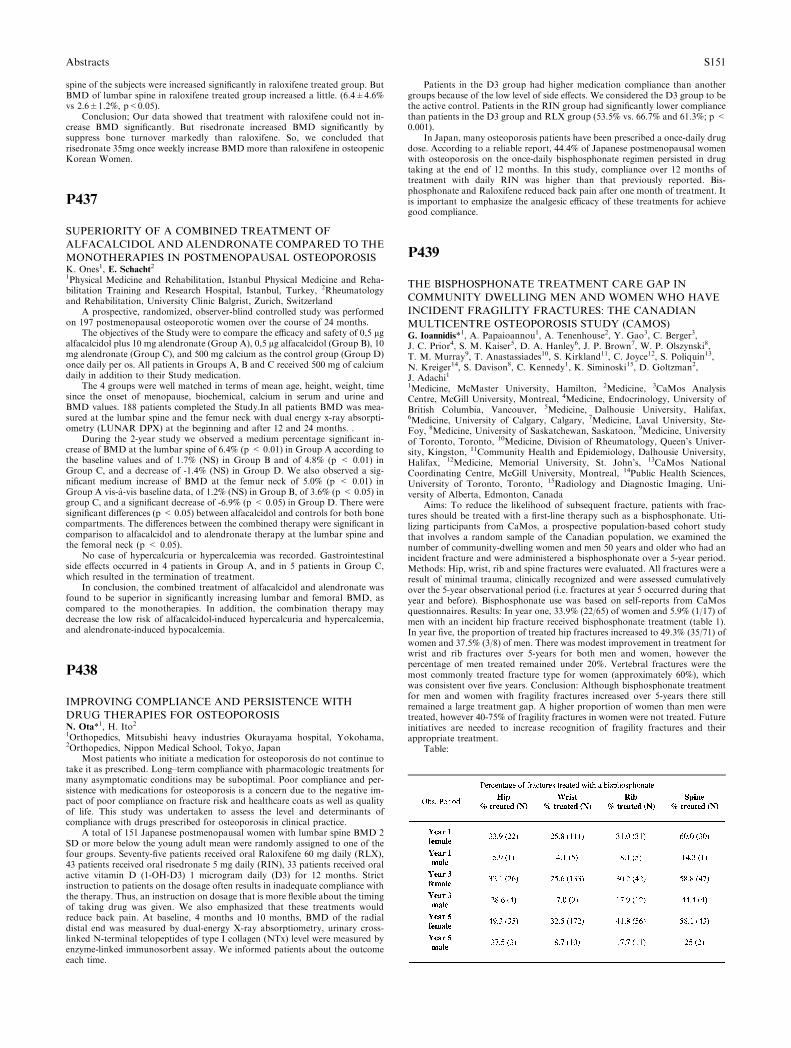
spine of the subjects were increased significantly in raloxifene treated group. ButBMD of lumbar spine in raloxifene treated group increased a little. (6.4±4.6%vs 2.6±1.2%, p<0.05).
Conclusion; Our data showed that treatment with raloxifene could not in-crease BMD significantly. But risedronate increased BMD significantly bysuppress bone turnover markedly than raloxifene. So, we concluded thatrisedronate 35mg once weekly increase BMD more than raloxifene in osteopenicKorean Women.
P437
SUPERIORITY OF A COMBINED TREATMENT OF
ALFACALCIDOL AND ALENDRONATE COMPARED TO THE
MONOTHERAPIES IN POSTMENOPAUSAL OSTEOPOROSISK. Ones1, E. Schacht21Physical Medicine and Rehabilitation, Istanbul Physical Medicine and Reha-bilitation Training and Research Hospital, Istanbul, Turkey, 2Rheumatologyand Rehabilitation, University Clinic Balgrist, Zurich, Switzerland
A prospective, randomized, observer-blind controlled study was performedon 197 postmenopausal osteoporotic women over the course of 24 months.
The objectives of the Study were to compare the efficacy and safety of 0,5 lgalfacalcidol plus 10 mg alendronate (Group A), 0,5 lg alfacalcidol (Group B), 10mg alendronate (Group C), and 500 mg calcium as the control group (Group D)once daily per os. All patients in Groups A, B and C received 500 mg of calciumdaily in addition to their Study medication.
The 4 groups were well matched in terms of mean age, height, weight, timesince the onset of menopause, biochemical, calcium in serum and urine andBMD values. 188 patients completed the Study.In all patients BMD was mea-sured at the lumbar spine and the femur neck with dual energy x-ray absorpti-ometry (LUNAR DPX) at the beginning and after 12 and 24 months. .
During the 2-year study we observed a medium percentage significant in-crease of BMD at the lumbar spine of 6.4% (p < 0.01) in Group A according tothe baseline values and of 1.7% (NS) in Group B and of 4.8% (p < 0.01) inGroup C, and a decrease of -1.4% (NS) in Group D. We also observed a sig-nificant medium increase of BMD at the femur neck of 5.0% (p < 0.01) inGroup A vis-a-vis baseline data, of 1.2% (NS) in Group B, of 3.6% (p < 0.05) ingroup C, and a significant decrease of -6.9% (p < 0.05) in Group D. There weresignificant differences (p < 0.05) between alfacalcidol and controls for both bonecompartments. The differences between the combined therapy were significant incomparison to alfacalcidol and to alendronate therapy at the lumbar spine andthe femoral neck (p < 0.05).
No case of hypercalcuria or hypercalcemia was recorded. Gastrointestinalside effects occurred in 4 patients in Group A, and in 5 patients in Group C,which resulted in the termination of treatment.
In conclusion, the combined treatment of alfacalcidol and alendronate wasfound to be superior in significantly increasing lumbar and femoral BMD, ascompared to the monotherapies. In addition, the combination therapy maydecrease the low risk of alfacalcidol-induced hypercalcuria and hypercalcemia,and alendronate-induced hypocalcemia.
P438
IMPROVING COMPLIANCE AND PERSISTENCE WITH
DRUG THERAPIES FOR OSTEOPOROSISN. Ota*1, H. Ito21Orthopedics, Mitsubishi heavy industries Okurayama hospital, Yokohama,2Orthopedics, Nippon Medical School, Tokyo, Japan
Most patients who initiate a medication for osteoporosis do not continue totake it as prescribed. Long–term compliance with pharmacologic treatments formany asymptomatic conditions may be suboptimal. Poor compliance and per-sistence with medications for osteoporosis is a concern due to the negative im-pact of poor compliance on fracture risk and healthcare coats as well as qualityof life. This study was undertaken to assess the level and determinants ofcompliance with drugs prescribed for osteoporosis in clinical practice.
A total of 151 Japanese postmenopausal women with lumbar spine BMD 2SD or more below the young adult mean were randomly assigned to one of thefour groups. Seventy-five patients received oral Raloxifene 60 mg daily (RLX),43 patients received oral risedronate 5 mg daily (RIN), 33 patients received oralactive vitamin D (1-OH-D3) 1 microgram daily (D3) for 12 months. Strictinstruction to patients on the dosage often results in inadequate compliance withthe therapy. Thus, an instruction on dosage that is more flexible about the timingof taking drug was given. We also emphasized that these treatments wouldreduce back pain. At baseline, 4 months and 10 months, BMD of the radialdistal end was measured by dual-energy X-ray absorptiometry, urinary cross-linked N-terminal telopeptides of type I collagen (NTx) level were measured byenzyme-linked immunosorbent assay. We informed patients about the outcomeeach time.
Patients in the D3 group had higher medication compliance than anothergroups because of the low level of side effects. We considered the D3 group to bethe active control. Patients in the RIN group had significantly lower compliancethan patients in the D3 group and RLX group (53.5% vs. 66.7% and 61.3%; p <0.001).
In Japan, many osteoporosis patients have been prescribed a once-daily drugdose. According to a reliable report, 44.4% of Japanese postmenopausal womenwith osteoporosis on the once-daily bisphosphonate regimen persisted in drugtaking at the end of 12 months. In this study, compliance over 12 months oftreatment with daily RIN was higher than that previously reported. Bis-phosphonate and Raloxifene reduced back pain after one month of treatment. Itis important to emphasize the analgesic efficacy of these treatments for achievegood compliance.
P439
THE BISPHOSPHONATE TREATMENT CARE GAP IN
COMMUNITY DWELLING MEN AND WOMEN WHO HAVE
INCIDENT FRAGILITY FRACTURES: THE CANADIAN
MULTICENTRE OSTEOPOROSIS STUDY (CAMOS)G. Ioannidis*1, A. Papaioannou1, A. Tenenhouse2, Y. Gao3, C. Berger3,J. C. Prior4, S. M. Kaiser5, D. A. Hanley6, J. P. Brown7, W. P. Olszynski8,T. M. Murray9, T. Anastassiades10, S. Kirkland11, C. Joyce12, S. Poliquin13,N. Kreiger14, S. Davison8, C. Kennedy1, K. Siminoski15, D. Goltzman2,J. Adachi11Medicine, McMaster University, Hamilton, 2Medicine, 3CaMos AnalysisCentre, McGill University, Montreal, 4Medicine, Endocrinology, University ofBritish Columbia, Vancouver, 5Medicine, Dalhousie University, Halifax,6Medicine, University of Calgary, Calgary, 7Medicine, Laval University, Ste-Foy, 8Medicine, University of Saskatchewan, Saskatoon, 9Medicine, Universityof Toronto, Toronto, 10Medicine, Division of Rheumatology, Queen’s Univer-sity, Kingston, 11Community Health and Epidemiology, Dalhousie University,Halifax, 12Medicine, Memorial University, St. John’s, 13CaMos NationalCoordinating Centre, McGill University, Montreal, 14Public Health Sciences,University of Toronto, Toronto, 15Radiology and Diagnostic Imaging, Uni-versity of Alberta, Edmonton, Canada
Aims: To reduce the likelihood of subsequent fracture, patients with frac-tures should be treated with a first-line therapy such as a bisphosphonate. Uti-lizing participants from CaMos, a prospective population-based cohort studythat involves a random sample of the Canadian population, we examined thenumber of community-dwelling women and men 50 years and older who had anincident fracture and were administered a bisphosphonate over a 5-year period.Methods: Hip, wrist, rib and spine fractures were evaluated. All fractures were aresult of minimal trauma, clinically recognized and were assessed cumulativelyover the 5-year observational period (i.e. fractures at year 5 occurred during thatyear and before). Bisphosphonate use was based on self-reports from CaMosquestionnaires. Results: In year one, 33.9% (22/65) of women and 5.9% (1/17) ofmen with an incident hip fracture received bisphosphonate treatment (table 1).In year five, the proportion of treated hip fractures increased to 49.3% (35/71) ofwomen and 37.5% (3/8) of men. There was modest improvement in treatment forwrist and rib fractures over 5-years for both men and women, however thepercentage of men treated remained under 20%. Vertebral fractures were themost commonly treated fracture type for women (approximately 60%), whichwas consistent over five years. Conclusion: Although bisphosphonate treatmentfor men and women with fragility fractures increased over 5-years there stillremained a large treatment gap. A higher proportion of women than men weretreated, however 40-75% of fragility fractures in women were not treated. Futureinitiatives are needed to increase recognition of fragility fractures and theirappropriate treatment.
Table:
Abstracts S151
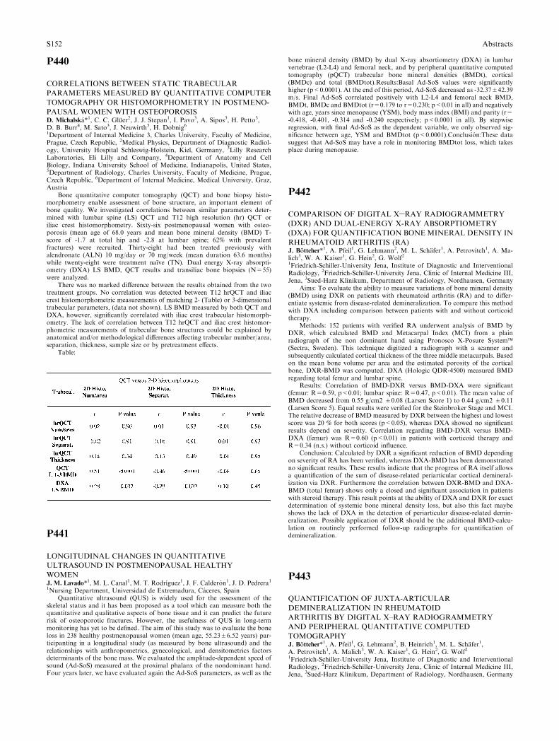
P440
CORRELATIONS BETWEEN STATIC TRABECULAR
PARAMETERS MEASURED BY QUANTITATIVE COMPUTER
TOMOGRAPHY OR HISTOMORPHOMETRY IN POSTMENO-
PAUSAL WOMEN WITH OSTEOPOROSISD. Michalska*1, C. C. Gluer2, J. J. Stepan1, I. Pavo3, A. Sipos3, H. Petto3,D. B. Burr4, M. Sato3, J. Neuwirth5, H. Dobnig61Department of Internal Medicine 3, Charles University, Faculty of Medicine,Prague, Czech Republic, 2Medical Physics, Department of Diagnostic Radiol-ogy, University Hospital Schleswig-Holstein, Kiel, Germany, 3Lilly ResearchLaboratories, Eli Lilly and Company, 4Department of Anatomy and CellBiology, Indiana University School of Medicine, Indianapolis, United States,5Department of Radiology, Charles University, Faculty of Medicine, Prague,Czech Republic, 6Department of Internal Medicine, Medical University, Graz,Austria
Bone quantitative computer tomography (QCT) and bone biopsy histo-morphometry enable assessment of bone structure, an important element ofbone quality. We investigated correlations between similar parameters deter-mined with lumbar spine (LS) QCT and T12 high resolution (hr) QCT oriliac crest histomorphometry. Sixty-six postmenopausal women with osteo-porosis (mean age of 68.0 years and mean bone mineral density (BMD) T-score of -1.7 at total hip and -2.8 at lumbar spine; 62% with prevalentfractures) were recruited. Thirty-eight had been treated previously withalendronate (ALN) 10 mg/day or 70 mg/week (mean duration 63.6 months)while twenty-eight were treatment naıve (TN). Dual energy X-ray absorpti-ometry (DXA) LS BMD, QCT results and transiliac bone biopsies (N=55)were analyzed.
There was no marked difference between the results obtained from the twotreatment groups. No correlation was detected between T12 hrQCT and iliaccrest histomorphometric measurements of matching 2- (Table) or 3-dimensionaltrabecular parameters, (data not shown). LS BMD measured by both QCT andDXA, however, significantly correlated with iliac crest trabecular histomorph-ometry. The lack of correlation between T12 hrQCT and iliac crest histomor-phometric measurements of trabecular bone structures could be explained byanatomical and/or methodological differences affecting trabecular number/area,separation, thickness, sample size or by pretreatment effects.
Table:
P441
LONGITUDINAL CHANGES IN QUANTITATIVE
ULTRASOUND IN POSTMENOPAUSAL HEALTHY
WOMENJ. M. Lavado*1, M. L. Canal1, M. T. Rodrıguez1, J. F. Calderon1, J. D. Pedrera11Nursing Department, Universidad de Extremadura, Caceres, Spain
Quantitative ultrasound (QUS) is widely used for the assessment of theskeletal status and it has been proposed as a tool which can measure both thequantitative and qualitative aspects of bone tissue and it can predict the futurerisk of osteoporotic fractures. However, the usefulness of QUS in long-termmonitoring has yet to be defined. The aim of this study was to evaluate the boneloss in 238 healthy postmenopausal women (mean age, 55.23±6.52 years) par-ticipanting in a longitudinal study (as measured by bone ultrasound) and therelationships with anthropometrics, gynecological, and densitometrics factorsdeterminants of the bone mass. We evaluated the amplitude-dependent speed ofsound (Ad-SoS) measured at the proximal phalanx of the nondominant hand.Four years later, we have evaluated again the Ad-SoS parameters, as well as the
bone mineral density (BMD) by dual X-ray absortiometry (DXA) in lumbarvertebrae (L2-L4) and femoral neck, and by peripheral quantitative computedtomography (pQCT) trabecular bone mineral densities (BMDt), cortical(BMDc) and total (BMDtot).Results:Basal Ad-SoS values were significantlyhigher (p<0.0001). At the end of this period, Ad-SoS decreased as -32.37±42.39m/s. Final Ad-SoS correlated positively with L2-L4 and femoral neck BMD,BMDt, BMDc and BMDtot (r=0.179 to r=0.230; p<0.01 in all) and negativelywith age, years since menopause (YSM), body mass index (BMI) and parity (r=-0.418, -0.401, -0.314 and -0.240 respectively; p<0.0001 in all). By stepwiseregression, with final Ad-SoS as the dependent variable, we only observed sig-nificance between age, YSM and BMDtot (p<0.0001).Conclusion:These datasuggest that Ad-SoS may have a role in monitoring BMDtot loss, which takesplace during menopause.
P442
COMPARISON OF DIGITAL X–RAY RADIOGRAMMETRY
(DXR) AND DUAL-ENERGY X-RAY ABSORPTIOMETRY
(DXA) FOR QUANTIFICATION BONEMINERAL DENSITY IN
RHEUMATOID ARTHRITIS (RA)J. Bottcher*1, A. Pfeil1, G. Lehmann2, M. L. Schafer1, A. Petrovitch1, A. Ma-lich3, W. A. Kaiser1, G. Hein2, G. Wolf21Friedrich-Schiller-University Jena, Institute of Diagnostic and InterventionalRadiology, 2Friedrich-Schiller-University Jena, Clinic of Internal Medicine III,Jena, 3Sued-Harz Klinikum, Department of Radiology, Nordhausen, Germany
Aims: To evaluate the ability to measure variations of bone mineral density(BMD) using DXR on patients with rheumatoid arthritis (RA) and to differ-entiate systemic from disease-related demineralization. To compare this methodwith DXA including comparison between patients with and without corticoidtherapy.
Methods: 152 patients with verified RA underwent analysis of BMD byDXR, which calculated BMD and Metacarpal Index (MCI) from a plainradiograph of the non dominant hand using Pronosco X-Posure System�(Sectra, Sweden). This technique digitized a radiograph with a scanner andsubsequently calculated cortical thickness of the three middle metacarpals. Basedon the mean bone volume per area and the estimated porosity of the corticalbone, DXR-BMD was computed. DXA (Hologic QDR-4500) measured BMDregarding total femur and lumbar spine.
Results: Correlation of BMD-DXR versus BMD-DXA were significant(femur: R=0.59, p<0.01; lumbar spine: R=0.47, p<0.01). The mean value ofBMD decreased from 0.55 g/cm2 ±0.08 (Larsen Score 1) to 0.44 g/cm2 ±0.11(Larsen Score 5). Equal results were verified for the Steinbroker Stage and MCI.The relative decrease of BMD measured by DXR between the highest and lowestscore was 20 % for both scores (p<0.05), whereas DXA showed no significantresults depend on severity. Correlation regarding BMD-DXR versus BMD-DXA (femur) was R=0.60 (p<0.01) in patients with corticoid therapy andR=0.34 (n.s.) without corticoid influence.
Conclusion: Calculated by DXR a significant reduction of BMD dependingon severity of RA has been verified, whereas DXA-BMD has been demonstratedno significant results. These results indicate that the progress of RA itself allowsa quantification of the sum of disease-related periarticular cortical demineral-ization via DXR. Furthermore the correlation between DXR-BMD and DXA-BMD (total femur) shows only a closed and significant association in patientswith steroid therapy. This result points at the ability of DXA and DXR for exactdetermination of systemic bone mineral density loss, but also this fact maybeshows the lack of DXA in the detection of periarticular disease-related demin-eralization. Possible application of DXR should be the additional BMD-calcu-lation on routinely performed follow-up radiographs for quantification ofdemineralization.
P443
QUANTIFICATION OF JUXTA-ARTICULAR
DEMINERALIZATION IN RHEUMATOID
ARTHRITIS BY DIGITAL X–RAY RADIOGRAMMETRY
AND PERIPHERAL QUANTITATIVE COMPUTED
TOMOGRAPHYJ. Bottcher*1, A. Pfeil1, G. Lehmann2, B. Heinrich1, M. L. Schafer1,A. Petrovitch1, A. Malich3, W. A. Kaiser1, G. Hein2, G. Wolf21Friedrich-Schiller-University Jena, Institute of Diagnostic and InterventionalRadiology, 2Friedrich-Schiller-University Jena, Clinic of Internal Medicine III,Jena, 3Sued-Harz Klinikum, Department of Radiology, Nordhausen, Germany
S152 Abstracts
Aims:As a generalized inflammatory disease rheumatoid arthritis involvesseveral joints as well as synovial sheaths of tendons and bursae. Both,rheumatoid arthritis and its extended treatment (i.e. cortison therapy) cause asignificant systemic bone loss in a high number of patients and additionally aperiarticular disease-related osteoporosis, especially at the hands and feet inan early stage of the disease. To evaluate changes of bone mineral density(BMD) using a radiogrammetrically based bone densitometric technology onpatients suffering from rheumatoid arthritis with and without corticoid ther-apy. To compare this method with peripheral Quantitative ComputedTomography.
Methods:90 patients underwent a prospective analysis of BMD via DXRand pQCT. Radiographs have been subjected to DXR for estimating BMD froma plain radiograph of the non dominant hand using Pronosco X-Posure System(Sectra, Sweden), which digitizes a radiograph with a scanner and subsequentlyderives a BMD of the three middle metacarpals; pQCT calculated BMD (total,trabecular, cortical) regarding distal radius.All patients were divided into asubgroup (n=52) with (5 mg cortison /d for 6 months) and without corticoidtherapy (n=38).
Results:The mean value of DXR-BMD decreased from 0.57 g/cm1=2±0.08 (Larsen Score 1) to 0.45 g/cm1=2 ±0.11 (Larsen Score 5). The relativedecrease of BMD measured by DXR between the highest and lowest scorewas 20% (p<0.05). The relative decrease of BMD (pQCT) from Larsen-Score1 to Score 5 showed a significant result regarding pQCT-BMD (trabecular, asmost metabolic active bone tissue) with 16% (p<0.05). No significantdemineralization confirmed for pQCT-BMD (total) with 12% and pQCT-BMD (cortical) with 2%.
Correlation between DXR-BMD and pQCT-BMD (total and trabecular)demonstrated a significant result (R=0,53 versus 0,55; p<0,01) for the cortison-subgroup. Correlation of DXR-BMD with pQCT-BMD (cortical) was lower(R=0.37, p<0.05). Equal results were verified for patients without corticoidtherapy.
Conclusion:The digital radiogrammetry can exactly measure cortical differ-ences of bone mineralization on patients suffering from rheumatoid arthritis andseems to be able to quantify disease-related periarticular loss of bone mineraldensity depend on severity and independent from corticoid influence.
P444
EVALUATION OF DISEASE ACTIVITY DEPENDENT
CORTICAL BONE LOSS IN RHEUMATOID
ARTHRITISA. Pfeil*1, J. Bottcher1, H. J. Mentzel1, M. L. Schafer1, G. Lehmann2,M. Schmidt2, A. Petrovitch1, A. Malich3, W. A. Kaiser1, G. Wolf4, G. Hein21Friedrich-Schiller-University Jena, Institute of Diagnostic and InterventionalRadiology, 2Friedrich-Schiller-University Jena, Clinic of Internal Medicine III,Jena, 3Sued-Harz Klinikum, Department of Radiology, Nordhausen, 4Friedrich-Schiller-University Jena, Department of Radiology, Jena, Germany
Aims: To differentiate the influence of inflammation and soft tissue swellingon the measurements of peripheral bone status in patients with rheumatoidarthritis (RA) using multi-site Quantitative Ultrasound (QUS) compared toDigital X-ray Radiogrammetry (DXR).
Methods: 53 patients with verified RA underwent measurements of QUS(Sunlight multi-site Omnisense 7000, Sunlight Medical Ltd., Tel Aviv, Israel)with estimation of the Speed of Sound (QUS-SOS) at the distal radius and at thephalanx III. Also Bone Mineral Density (DXR-BMD) and Metacarpal Index(DXR-MCI) were estimated on the metacarpals II-IV using DXR-technology(Pronosco X-Posure System� Version 2.0, Pronosco/Sectra ASS, Denmark).Disease activity of RA was estimated by Erythrocyte Sedimentation Rate (ESR;in mm/1st hour).
Results: For the group with minor disease activity (ESR £ 20 mm/1st hour)QUS-SOS (phalanx) was significantly associated to DXR-BMD (R=0.67,p<0.01) and DXR-MCI (R=0.54, p<0.05). Lower correlation coefficients wererevealed between QUS-SOS (radius) and DXR-parameters (DXR-BMD:R=0.39, p<0.05; DXR-MCI: 0.49, p<0.05). In the case of high disease activity(ESR>20 mm/1st hour) QUS-SOS of the radius showed a closed correlation toDXR-BMD (R=0.74; p<0.01) and DXR-MCI (R=0.89; p<0.01), whichsurpassed the correlation coefficients compared to patients with minor diseaseactivity of RA. For QUS-SOS (phalanx) no significant correlation to DXR-parameters could be observed in patients with high disease activity.
Conclusion: Multi-site QUS and in a significant extent DXR can alsodistinguish between patients with minor disease activity and patients withactive RA which demonstrates accelerated bone loss characterized by de-creased values of DXR- and QUS-SOS parameters. In this study the multi-site QUS-device is surpassed by DXR. In patients with accentuated diseaseactivity no significant association among the QUS-parameters themselves andbetween QUS-SOS (phalanx) and the DXR-parameters exists. These dataseem to indicate a significant influence of soft tissue swelling caused by activeRA on QUS estimates at the phalanx as measurement site. In patients withincreased disease activity QUS-SOS measurements at the radius should beconsistently prefered.
P445
PRECISION OF DIGITAL X-RAY RADIOGRAMMETRY (DXR)A. Pfeil*1, J. Bottcher2, G. Lehmann3, M. L. Schafer2, A. Petrovitch2, W. Linß4,A. Malich5, W. A. Kaiser2, G. Wolf3, G. Hein31Friedrich-Schiller-University, 2Friedrich-Schiller-University Jena, Institute ofDiagnostic and Interventional Radiology, 3Friedrich-Schiller-University Jena,Clinic of Internal Medicine III, 4Friedrich-Schiller-University Jena, Institute ofAnatomy I, 5Sued-Harz Klinikum, Department of Radiology, Jena, Germany
Aims: The potential of Digital X-ray Radiogrammetry for estimating cor-tical bone loss seems to be promising, in particular because of the high precision.To evaluate the inter– and intra-radiograph reproducibility of Digital X-rayRadiogrammetry (DXR) under consideration of both conventional and digitalperformed radiographs.
Methods: Radiographs of the non-dominant hand from two deceased maleswere performed according to a predefined study protocol. For conventionalradiographs (Philips Super 80 CP) our study considered the following stan-dardized parameters: 42kV, 6mAs, Kodak T-MAT-Plus 200 or Agfa Curix 200film, FFD 100cm. For digital print-outs (Diagnost Philips Optimus and SiemensPolydoros SX 80) the constant image capturing conditions were as follows:42kV, 4mAs, FFD 100cm.
Inter-radiograph-reproducibility of Bone mineral Density (BMD) and Me-taxarpal Index (MCI) were evaluated by acquiring ten repeated radiographs withre-positioning under standard settings.
To verify intra-radiograph reproducibility, a single conventional image anda single print-out underwent 10 repeated DXR analyses (42kV, 6mAs, FFD100cm) with the Pronosco-X posure System (Version 2, Sectra, Sweden).
Results: The inter-radiograph reproducibility of DXR-BMD using conven-tional images under standardised conditions was calculated to be CV=0.49% forAgfa Curix film and CV=0.33% for Kodak T-MAT-Plus film, whereas repro-ducibility error using digital images ranged from CV=0.57% (Philips, digitalprint-outs) to CV=1.18% (Siemens, digital print-outs). The intra-radiographreproducibility error was observed to be CV=0.13% and CV=0.26% (conven-tional, Kodak and Agfa film) versus CV=0.27% and CV= 0.05% (digital print-outs, Philips and Siemens). No significant differences in the sensitivity towardsimage capturing parameters were observed between the BMD and MCI.
Conclusion: DXR shows a high inter- and intra – radiograph reproducibilityfor conventional as well as digital images and seems to be a reliable osteoden-sitometric technique for evaluation of the peripheral cortical bone which ap-prove the quantification of minor disease-related alterations of the cortical bonepartition.
P446
INITIAL RESULTS OF REPRODUCIBILITY FOR DUAL
X-RAY AND LASER (DXL)A. Pfeil*1, J. Bottcher2, G. Lehmann3, M. L. Schafer1, A. Kramer1,A. Petrovitch1, M. Schmidt1, A. Malich4, W. A. Kaiser1, G. Wolf3, G. Hein31Friedrich-Schiller-University Jena, Institute of Diagnostic and InterventionalRadiology, 2Friedrich-Schiller-University Jena, Friedrich-Schiller-UniversityJena, 3Friedrich-Schiller-University Jena, Clinic of Internal Medicine III, Jena,4Sued-Harz Klinikum, Department of Radiology, Nordhausen, Germany
Aims: Dual Energy X-ray Absorptiometry (DXA) is a widely availabletechnique for the assessment of osteoporosis. The usual DXA has an accuracybetween 2% and 20%. But a high precision are necessary for diagnosis andmonitoring of minor changes in the course of osteoporosis. Dual X-Ray andLaser (DXL) is a new technique based on DXA technology combined with aLaser for measurements peripheral Bone Mineral Density (BMD) at the calca-neus. The purpose of this study is to evaluate reproducibility of DXL dependenton body fat and textile material in the measurement area.
Methods: Reproducibility of DXL was measured on five healthy volunteers.The DXL (Demetech AB, Solna, Sweden) technique uses two X-ray energies incombination with laser measurement of the object thickness in order to deter-mine all three tissue components (bone mineral, lean soft tissues, and fatty tis-sues). For each subjects measurements of DXL-BMD on the right and leftcalcaneus was performed. Data of height, weight and BMI (Body Mass Index)were acquired and the study group was subdivided into individuals withunderweight (BMI<25), normal-weight (BMI 20-25) and overweight(BMI>25). The influence of textile material was evaluated by five measurementswith a textile sock (material: 100% cotton, material thickness: 1.3 mm).
Results: Reproducibility of the DXL-BMD was CV=1.75% (left) andCV=1.05% (right) for the underweight group. The over-weight group showedhigher coefficients of variation (left: CV=2.51%, right: 2.23%) compared to thenorm-weight group (left: CV=3.08%, right 1.10%). The data showed the highestcoefficient of variation for under-weight individuals CV=3.30% regardingreproducibility influenced by textile material. Improved results were observed forthe norm-weighted group (CV=2.97%) as well as over-weight (CV=3.04%).
Conclusion: The DXL shows a remarkable high reproducibility. Howeverour data showed an influence by body mass and body fat. Based on this results a
Abstracts S153

accurate and precise measurement of BMD by this new technique is available. Inaddition DXL seems to be a promising diagnostic tool in the assessment ofosteoporosis at peripheral measurements.
P447
EVALUATION OF IMAGE CAPTURING PARAMETERS ON
PRECISION OF DIGITAL X-RAY RADIOGRAMMETRY (DXR)J. Bottcher*1, A. Pfeil1, G. Lehmann2, M. L. Schafer1, A. Petrovitch1, A. Ma-lich3, W. Linß4, W. A. Kaiser1, G. Wolf2, G. Hein21Friedrich-Schiller-University Jena, Institute of Diagnostic and InterventionalRadiology, 2Friedrich-Schiller-University Jena, Clinic of Internal Medicine III,Jena, 3Sued-Harz Klinikum, Department of Radiology, Nordhausen, 4Friedrich-Schiller-University Jena, Institute of Anatomy I, Jena, Germany
Aims: To evaluate the importance of different image capturing conditions,which may influence the characteristics of radiographs and consequently mayimpact calculation of bone mineral density (BMD) using Digital X-ray Radi-ogrammetry (DXR). The investigated parameters include film-focus distance(FFD), film quality (sensitivity of 200 versus 400), film brand (Kodak T-MAT-Plus, Agfa Curix), exposure level (mAs) and tube voltage (kV). Furthermore theimpact of the imaging technology used in terms of conventional radiographsversus digital x-ray images is considered (Siemens Polydoros SX 80, DiagnostPhilips Optimus), either in the original digital format or through digitization ofprint-outs. The reproducibility of DXR-BMD and Metacarpal Index (MCI) arecompared across these different image modalities.
Methods: Radiographs of the left hands of deceased males were acquiredthree times using systematically varied parameters: 4-8 mAs, 40-52kV, FFD: 90-130cm, film sensitivity: 200/400 and image modality/source (conventional versusoriginal digital radiographs as well as digital print-outs). All BMD and MCImeasurements were obtained with the DXR-technology (Pronosco-Xposuresystem, Sectra Pronosco A/S, Denmark), which is originally calibrated foranalysis of conventional radiographs.
Results: BMD-calculation was not noticeably affected by changes of FFD(conventional: CV=0.98%), exposure level (conventional: CV=0.49%; digital:CV=0.35%), film sensitivity/film brand (conventional: CV=0.33% / 0.49%), butwas influenced by tube voltage (conventional: CV=1.32% for Agfa andCV=0.99% for Kodak; digital: CV=0.57% for Philips print-outs andCV=0.50% with original digital images versus CV=2.05% for Siemens print-outs and 1.35% with original digital images). No significant differences in thesensitivity towards image capturing parameters were observed between BMDand MCI.
Conclusion: Digital X-ray Radiogrammetry provides measurements of MCIand BMD with high precision. The measurements are unaffected by all testedimage capturing conditions with the exception of tube voltage. Regarding con-ventional and digital radiographs, especially digital print-outs, our data revealeda significant influence of varied tube voltage on DXR-BMD and MCI mea-surements. In addition an influence of the applied image modality is detectedbetween the original digital images and their corresponding printed versions.
P448
REPRODUCIBILITY OF EXA-3000 AS A NEW DUAL ENERGY
X-RAY ABSORPTIOMETRY DEVICE FOR MEASUREMENT
BONE MINERAL DENSITY ON RADIUS AND CALCANEUSA. Pfeil*1, J. Bottcher1, M. L. Schafer1, G. Lehmann2, M. Schmidt1, A. Kra-mer1, A. Petrovitch1, A. Malich3, W. A. Kaiser1, G. Hein2, G. Wolf21Friedrich-Schiller-University Jena, Institute of Diagnostic and InterventionalRadiology, 2Friedrich-Schiller-University Jena, Clinic of Internal Medicine III,Jena, 3Sued-Harz Klinikum, Department of Radiology, Nordhausen, Germany
Aims: The Dual Energy X-ray Absorptiometry (DXA) is worldwide used indiagnosis and monitoring of osteoporosis. In the following study reproducibilityof a new peripheral DXA-System EXA-3000 was verified dependent on BodyMass Index (BMI) and reposition.
Methods: DXA-Reproducibility was measured on healthy volunteers. Foreach subject a measurement of peripheral DXA-BMD (EXA-3000, OsteoSys CoLtd, Seoul, Korea) on the left radius and calcaneus was performed. Data ofheight, weight and BMI (Body Mass Index) were acquired and the study groupwas subdivided into individuals with underweight (BMI<25), normal-weight(BMI 20-25) and overweight (BMI>25). To verify the impact of reposition onDXA-measurements five measurements under reposition were performed. Theinfluence of textile material was evaluated by five measurements with a textilesock at the left foot (material: 100% cotton, material thickness: 1,3 mm).
Results: For healthy controls the reproducibility for the BMD (radius) wasCV=1.20% (under-weight), CV=2.21 % (norm-weight) and CV=2.29% (over-weight). Regarding reposition our data showed CV=1.12% (over-weight indi-viduals) and CV=1.29% (under- and norm-weight group). Improved resultswere observed for the calcaneus: CV=1.60% (under-weight group), CV=1.00%(norm-weight group) as well as CV=1.58% (over-weight group). Reproduc-
ibility presented major CV-values for the calcaneus under reposition:CV=1.52% (under-weight group), CV=2.89% (norm-weight group) andCV=2.06% (over-weight group). Regarding textile sock at the foot no increasedcoefficients of variation with CV=1.93% (under-weight group), CV=1.92%(norm-weight group) and CV=1.61% (over-weight group) were observed.
Conclusion: The DXA shows a high reproducibility in comparison to a usualDXA-System. In this study an impact of body fat, reposition and textile materialon BMD measurements by EXA-3000 could not be clarified. EXA-3000 seems tobe promising technique for quantification peripheral bone mineral density.
P449
REPRODUCIBILITY OF MULTI-SITE QUANTITATIVE
ULTRASOUND FOR HEALTHY INDIVIDUALS AND
PATIENTS SUFFERING FROM RHEUMATOID ARTHRITISA. Pfeil*1, J. Bottcher1, H. J. Mentzel1, G. Lehmann2, M. L. Schafer1,A. Kramer1, A. Petrovitch1, A. Malich3, W. A. Kaiser1, G. Wolf2, G. Hein21Friedrich-Schiller-University, Institute of Diagnostic and Interventional Radi-ology, 2Friedrich-Schiller-University, Clinic of Internal Medicine III, Jena,3Sued-Harz Klinikum, Department of Radiology, Nordhausen, Germany
Aim: To evaluate inter- and intraobserver-reproducibility of multi-site QUSconsidering the effects of Body Mass Index (BMI) for healthy individuals andquantifying the impact of disease activity in patients suffering from RA.
Methods: For the measurement of the SOS on phalanx III and radius amulti-site QUS device (Sunlight multi-site Omnisense 7000, Sunlight MedicalLtd., Tel Aviv, Israel) was used. Inter-observer and intra-observer reproduc-ibility were measured including three healthy volunteers from each BMI-group(BMI £ 25, mean:21.7) and over-weight controls (BMI>25, mean:32.6), andalso six RA patients with advanced disease activity (n=3, CRP>25mg/l) as wellas low disease activity (n=3, CRP £ 25mg/l). Inter-observer reproducibility ofQUS-SOS was evaluated by three trained observers with ten repeated mea-surements at the distal radius and third phalanx. To verify intra-observerreproducibility a single observer performed 10 repeated QUS-SOS estimates forall 12 individuals.
Results: For healthy controls the intra-observer reproducibility for the QUS-SOS ranged between CV=0.90% (norm-weight, radius) and CV=2.55% (over-weight, phalanx). The over-weight group showed higher CV-values (phalanxCV=2.55%, radius 1.43%) compared to the norm-weight group (phalanxCV=1.05%, radius 0.90%). The intra-observer reproducibility of measurementstaken from patients with advanced RA were limited (phalanx CV=1.88%, ra-dius 1.03%). Inter-observer reproducibility for healthy individuals showed thehighest CV for over-weight individuals regarding QUS-SOS at the distal radius(CV=1.79%) and QUS-SOS at the phalanx (CV=2.17%). Improved resultswere observed for the BMI group<25 (radius: CV=1.66%, phalanx:CV=1.40%). Inter-observer reproducibility showed minor CV-values for theadvanced disease activity group (CV=0.94%) compared to patients with low RAactivity (CV=0.65%) measured by QUS-SOS (radius). Regarding QUS-SOS(phalanx), increased CV-values with CV=1.81% (low disease activity) versusCV=1.89% (advanced disease activity) were observed.
Conclusion: QUS-SOS is characterized by a reduced short-term precision,which is significantly more noticeable in the healthy individuals with pronouncedBMI and patients with advanced RA. It may be concluded that the multi-siteQUS is of limited priority for patients with active inflammation of the soft tissuein RA and with an elevated BMI, using measurements at the phalanx.
P450
DIGITAL X-RAY RADIOGRAMMETRY - IMPLEMENTATION
OF CORRECTION FACTORS OF CORTICAL THICKNESS,
METACARPAL INDEX AND BONE MINERAL DENSITYJ. Bottcher*1, A. Pfeil1, M. L. Schafer1, G. Lehmann2, A. Petrovitch1,A. Malich3, W. Linß4, W. A. Kaiser1, G. Wolf2, G. Hein21Friedrich-Schiller-University Jena, Institute of Diagnostic and InterventionalRadiology, 2Friedrich-Schiller-University Jena, Clinic of Internal Medicine III,Jena, 3Sued-Harz Klinikum, Department of Radiology, Nordhausen, 4Friedrich-Schiller-University Jena, Institute of Anatomy I, Jena, Germany
Aims: In many studies DXR seems to be a precise method for evaluation ofcortical bone partition. Unfortunately a majority of these studies are presumablybased on an extensive variety of different image capturing conditions which caninfluence the reliability of the DXR results. In particular varied tube voltageduring conventional and digital acquisition of radiographs affects the short termprecision of DXR. Aim of this study is to implement correction factors forCortical Thickness (DXR-CT), Metacarpal Index (MCI) and Bone MineralDensity (DXR-BMD) in case of varied tube voltage during image capturingcaused by different X-ray settings.
Methods: Radiographs of the left hands of two deceased males were ac-quired three times using systematically varied tube voltage (40-52kV), both for
S154 Abstracts
conventional X-rays (Kodak T-MAT-Plus film and Agfa Curix film with PhilipsSuper 80 CP device) and digital print-outs (Siemens Polydoros SX 80 and Di-agnost Philips Optimus devices). All CT, MCI and BMD measurements wereestimated by the DXR-technique (Pronosco X-Posure System;Version 2, Sectra,Sweden), which is originally calibrated for analysis of conventional radiographs.
Results: The following average correction factors could be calculated for onestep of kV (± 1 kV) based on the to our part recommended tube voltage of 40kV:
For DXR-BMD: -0.48% (conventional, Kodak), -0.75% (conventional,Agfa), +0.11% (digital print-outs, Philips) and -0.92% (digital print-outs, Sie-mens). For MCI: -0.35% (conventional, Kodak), -0.74% (conventional, Agfa),+0.04% (digital print-outs, Philips) and –1.28% (digital print-outs, Siemens).Finally for DXR-CT: -0.57% (conventional, Kodak), -0.94% (conventional,Agfa), +0.47% (digital print-outs, Philips) and –1,18% (digital print-outs, Sie-mens).
Conclusion: The calculated corrections factors could facilitate and alsocompensate differences of DXR results caused by varied tube voltage duringimage capturing. These correction factors support the extended availability ofDXR based on a better comparability of different DXR-data.
P451
VERTEBROPLASTY AND KYPHOPLASTY IN
OSTEOPOROTIC FRACTURES OF VERTEBRAL BODIESR. Pflugmacher*11Centrum fur Muskuloskeletale Chirurgie, Charite-Universitatsmedizin Berlin,Berlin, Germany
Background/aims: Kyphoplasty and vertebroplasty offer two minimalinvasive operative stabilisation procedures for osteoporotic vertebral compres-sion fractures. The purpose of this prospective study was to investigate wetherboth procedures are able to reduce pain and to preserve postoperative vertebralheight during a 1 year follow-up.
Methods: Osteoporotic vertebral fractures were treated in 42 cases, 20 pa-tients (15 female, 5 male) underwent vertebroplasty, 22 patients (14 female, 8male) underwent kyphoplasty. 32 vertebral fractures were treated with verteb-roplasty and 35 vertebral fractures were treated with kyphoplasty. Symptomaticlevels were identified by correlating the clinical presentation with conventionalradiographs, CT and / or MRI. During the follow-up reduction of pain wasdetermined. Radiographic scans were performed pre- and postoperatively andafter 3, 6 and 12 months. The vertebral height and endplate angles were mea-sured to assess the restoration of the sagittal alignment. The effects on painsymptoms were measured on a self-reported Visual Analog Scale (VAS) and theOswestry score was documented.
Results: The median pain scores (VAS) decreased for kyphoplasty andvertebroplasty from pre- to post-treatment significantly as well as the Oswestryscore (p < 0.05). No significant differences could be found between both groupsfor the median pain score (VAS) and the Oswestry score. Kyphoplasty led to asignificant restoration of the vertebral height and reduction of kyphosis (p <0.05). During the 1 year follow-up both operation techniques were able to sta-bilize the height of the vertebral body.
Conclusion: Kyphoplasty and vertebroplasty are effective minimal invasiveprocedures for the stabilization of osteoporotic vertebral fractures leading to astatistically significant reduction of pain. Kyphoplasty restores significantlyvertebral body height in acute fractures. The restoration of vertebral height andreduction of kyphosis may have an influence on the long term clinical outcome.This has to be evaluated in a long term prospective study.
P452
RALOXIFENE EFFECTS ON BONE MARKERS, BONE
MINERAL DENSITY, LIPID PROFILE AND BODY
COMPOSITION IN POSTMENOPAUSAL WOMENC. Poiana*1, L. Stoian21Endocrinology, Carol Davila University of Medicine and Pharmacy, Bucharest,Romania, 2Endocrinology, C.I.Parhon Institute of Endocrinology, Bucharest,Romania
Raloxifene, a non-steroidal benzothiophene, is a second generation selectiveestrogen receptor modulator, with estrogen agonist effects on bone and car-diovascular system and estrogen antagonist effects on endometrium and breast.
The aim of our study was to evaluate the effects of raloxifene therapy (60mg/day) on biochemical markers of bone turnover, bone mineral density (BMD),body composition and lipid profile in postmenopausal osteoporotic women.
We investigated 29 postmenopausal white women with osteoporosis aged56.9 ± 7.8 years (mean ± SD) and in addition a control group of 27 pre-menopausal white women with a mean age of 35 ± 8.1 years. The diagnosis ofosteoporosis was established by dual-energy x-ray absorptiometry, measuringBMD (Kg/sqm) at lumbar spine using a GE (Lunar DPX-L) device. Exclusion
criteria: previous treatments for osteopororsis, diseases or medication whichinterfere with bone metabolism, history of thrombembolism.
In both groups body mass composition was performed using a body com-position analyzer TBF 310 GS (Tanita Corporation). We determined also thelipid profile - cholesterol (mg/dL), HDL-cholesterol (mg/dL), LDL-cholesterol(mg/dL), triglycerides (mg/dL) - and the biochemical markers of bone turnover:osteocalcin (ng/mL) and beta crosslaps (ng/mL).
We found statistically significant increased values of bone markers in post-menopausal women with osteoporosis vs premenopausal women: 27.94 ± 12.1vs 17,30 ± 8.9 ng/mL for osteocalcin and 0.48 ± 0.2 vs 0.23 ± 0.1 ng/mL forbeta crosslaps, p < 0.001. After 3, 6 and 12 months of therapy with raloxifenethe bone markers were statistically significant reduced: )21.2%, )20.4% andrespectively )30.6% for osteocalcin and )25%, )39.6% and respectively )50%for beta crosslaps (p < 0.01). BMD was increased with 3.7% after 6 months andwith 8.6% after 12 months of treatment. Total cholesterol and LDL-C weresignificantly reduced after 12 months (p = 0.03), with no increase in triglyce-rides and in the same time body composition was unchanged.
Our results suggest that raloxifene reduces as early as three months the boneturnover, especially the bone resorbtion (beta crosslaps) in postmenopausalosteoporosis, with favourable effects on BMD and lipid profile.
P453
BONE LOSS IN WOMEN OF DIFFERENT AGE WITH
RHEUMATOID ARTHRITISV. V. Povoroznjuk*11Physiology and Pathology of Locomotor Apparatus, Institute of Gerontology,Kiev, Ukraine
Background. Osteoporosis is recognized as a complication of rheumatoidarthritis. Several studies have explored the relation between osteoporosis anddisease related variables. Use of corticosteroids has been found to be related toosteoporosis in RA, and several studies have also indicated that the developmentof bone loss and osteoporosis is related to disease course, especially at the earlystages.
Methods. 30 female patients with a mean age of 50.2 years with RA andduration of disease more 6 months were examined. All patients had clinicalexaminations and bone mass measurement by dualenergy X-ray absorptiometry:anterior-posterior spine L1-L4, total hip, and femoral neck, distal and ultradistalradius. The following demographic variables were assessed: age, disease dura-tion, body weight and height, co-morbidities, menopause status. The activenature of disease was assessed by the following variables: acute phase reactantsby ESR and CRP, the number of deformed joints, and functional health statusby the Health Assessment Questionnaire. All women never use the corticoster-oids or take it in dose less than 5 mg per day, equivalent to prednisolon, and lessthan 3 weeks.Our aim was to estimate the influence of active nature of disease,duration, presence of deformed joints, on bone mineral density and to determinethe patterns of bone loss in female patients of different age.
Results. We found out that osteoporosis and low bone mass in RA are moreconsistently associated with age, menopause status, BMI than disease variables.There was no correlation of bone loss and active nature of disease, results ofHAQ, presence of deformed joints.
Postmenopausal women featured bone loss at femur neck and total body,whereas young women had osteoporosis at the anterior-posterior spine L1-L4,compared with reference values. The relation between bone loss at distal andultradistal radius and other parameters was not found.
Conclusion. We�ve concluded that loss of bone mass is heterochronous, andit�s necessary to monitor it and use prevention and treatment of osteoporosis inpatients with rheumatoid arthritis.
P454
ORAL IBANDRONATE REDUCES ACTIVATION
FREQUENCY TO HEALTHY PREMENOPAUSAL LEVELS IN
WOMEN WITH POSTMENOPAUSAL OSTEOPOROSISR. R. Recker*1, K. M. Davis1, M. J. Barger-Lux1, R. P. Heaney1, J. M. Lappe11Osteoporosis Research Center, Creighton University, Omaha, United States
Oversuppression of bone remodelling with oral bisphosphonates is a hypo-thetical concern in postmenopausal osteoporosis (PMO). In the BONE study oforal ibandronate (IBN), significant antifracture efficacy was observed in osteo-porotic women receiving a daily regimen or novel intermittent (int.) regimen(dosing interval >2 months). To establish the extent of bone remodelling sup-pression provided by these regimens, we compared the activation frequency(Ac.f) observed in BONE with that obtained in studies involving healthy andosteoporotic women. BONE was a randomised, double-blind, placebo-con-trolled study in postmenopausal (PM) women (aged 55–80 years, >=5 yearsmenopausal) with osteoporosis (1–4 prevalent vertebral fractures, BMD T-scoreof )2 to )5 in >=1 lumbar vertebra). All participants received 3-years treat-
Abstracts S155
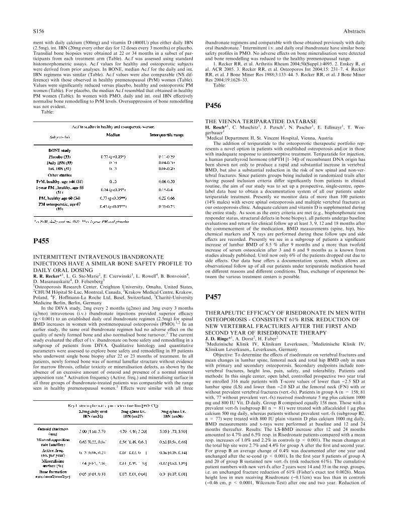
ment with daily calcium (500mg) and vitamin D (400IU) plus either daily IBN(2.5mg), int. IBN (20mg every other day for 12 doses every 3 months) or placebo.Transilial bone biopsies were obtained at 22 or 34 months in a subset of par-ticipants from each treatment arm (Table). Ac.f was assessed using standardhistomorphometric assays. Ac.f values for healthy and osteoporotic subjectswere derived from prior analyses. In BONE, median Ac.f for the daily and int.IBN regimens was similar (Table). Ac.f values were also comparable (NS dif-ference) with those observed in healthy premenopausal (PrM) women (Table).Values were significantly reduced versus placebo, healthy and osteoporotic PMwomen (Table). For placebo, the median Ac.f resembled that obtained in healthyPM women (Table). In women with PMO, daily and int. oral IBN effectivelynormalise bone remodelling to PrM levels. Oversuppression of bone remodellingwas not evident.
Table:
P455
INTERMITTENT INTRAVENOUS IBANDRONATE
INJECTIONS HAVE A SIMILAR BONE SAFETY PROFILE TO
DAILY ORAL DOSINGR. R. Recker*1, L. G. Ste-Marie2, E. Czerwinski3, L. Rowell4, B. Bonvoisin4,D. Masanauskaite4, D. Felsenberg51Osteoporosis Research Center, Creighton University, Omaha, United States,2CHUMHopital St-Luc, Montreal, Canada, 3Krakow Medical Centre, Krakow,Poland, 4F. Hoffmann-La Roche Ltd, Basel, Switzerland, 5Charite-UniversityMedicine Berlin, Berlin, Germany
In the DIVA study, 2mg every 2 months (q2mo) and 3mg every 3 months(q3mo) intravenous (i.v.) ibandronate injections provided superior efficacy(p<0.001) to an established daily oral ibandronate regimen (2.5mg) for spinalBMD increases in women with postmenopausal osteoporosis (PMO).1,2 In anearlier study, the same oral ibandronate regimen had no adverse effect on thequality of newly formed bone and also normalised bone turnover.3 The currentstudy evaluated the effect of i.v. ibandronate on bone safety and remodelling in asubgroup of patients from DIVA. Qualitative histology and quantitativeparameters were assessed to explore bone safety and remodelling in 89 patientswho underwent single bone biopsy after 22 or 23 months of treatment. In allpatients, newly formed bone was of normal lamellar structure without evidencefor marrow fibrosis, cellular toxicity or mineralisation defects, as shown by theabsence of an excessive amount of osteoid and presence of a normal mineralapposition rate.4 Activation frequency (Active. freq.) and mineralising surface inall three groups of ibandronate-treated patients was comparable with the rangeseen in healthy premenopausal women.5 Effects were similar with all three
ibandronate regimens and comparable with those obtained previously with dailyoral ibandronate.2 Intermittent i.v. and daily oral ibandronate have similar bonesafety profiles in PMO. No adverse effects on bone mineralisation were detectedand bone remodelling was reduced to the healthy premenopausal range.
1. Recker RR, et al. Arthritis Rheum 2004;50(Suppl.):4095. 2. Emkey R, etal. ACR 2005. 3. Recker RR, et al. Osteoporos Int 2004;15: 231–7. 4. ReckerRR, et al. J Bone Miner Res 1988;3:133–44. 5. Recker RR, et al. J Bone MinerRes 2004;19:1628–33.
Table:
P456
THE VIENNA TERIPARATIDE DATABASEH. Resch*1, C. Muschitz1, J. Patsch1, N. Pascher1, E. Edlmayr1, T. Woe-gerbauer11Medical Department II, St. Vincent Hospital, Vienna, Austria
The addition of teriparatide to the osteoporotic therapeutic portfolio rep-resents a novel option in patients with established osteoporosis and/or in thosewith inadequate response to antiresorptive treatment. Teriparatide for injection,a human parathyroid hormone (rhPTH [1–34]) of recombinant DNA origin hasbeen shown not only to produce a rapid and substantial increase in vertebralBMD, but also a substantial reduction in the risk of new spinal and non-ver-tebral fractures. Since patients groups being included in randomized trails afterhaving passed inclusion criteria differ significantly from patients in clinicalroutine, the aim of our study was to set up a prospective, single-centre, open-label data base to obtain a documentation system of all our patients underteriparatide treatment. Presently we monitor data of more than 100 patients(14% males) with severe spinal osteoporosis and multiple vertebral fractures atour osteoporosis clinic. Adequate calcium and vitamin D is supplemented duringthe entire study. As soon as the entry criteria are met (e.g., bisphosphonate nonresponder status, structural defects in bone biopsy), all patients undergo baselineevaluations and return for clinical follow up at least 3, 9, 12 and 18 months afterthe commencement of the medication. BMD measurements (spine, hip), bio-chemical markers and X rays are performed during these follow ups and sideeffects are recorded. Presently we see in a subgroup of patients a significantincrease of lumbar BMD of 8.5 % after 9 months and a more than twofoldincrease of serum osteocalcin after 3 and 6 and 9 months as is known fromstudies already published. Until now only 6% of the patients dropped out due toside effects. Our data base offers a documentation system, which allows anobservational follow up of all our patients under teriparatide medication basedon different reasons and different conditions. Thus, exchange of experience be-tween the various treatment centers is possible.
P457
THERAPEUTIC EFFICACY OF RISEDRONATE IN MENWITH
OSTEOPOROSIS - CONSISTENT 61% RISK REDUCTION OF
NEW VERTEBRAL FRACTURES AFTER THE FIRST AND
SECOND YEAR OF RISEDRONATE THERAPYJ. D. Ringe*1, A. Dorst2, H. Faber21Medizinische Klinik IV, Klinikum Leverkusen, 2Medizinische Klinik IV,Klinikum Leverkusen,, Leverkusen, Germany
Objective: To determine the effects of risedronate on vertebral fractures andmean changes in lumbar spine, femoral neck and total hip BMD only in menwith primary and secondary osteoporosis. Secondary endpoints include non-vertebral fractures, height loss, pain, safety, and tolerability. Patients andmethods: In this single center, open label, controlled prospective two year trialwe enrolled 316 male patients with T-score values of lower than )2.5 SD atlumbar spine (LS) and lower than )2.0 SD at the femoral neck (FN) with orwithout prevalent vertebral fractures (vert.-fx). Patients in group A (n = 158; 81with, 77 without prevalent vert.-fx) received risedronate 5 mg plus calcium 1000mg and 800 IU Vit. D daily. Group B comprised equally 158 men. Those with aprevalent vert-fx (subgroup B1 n = 81) were treated with alfacalcidol 1 lg pluscalcium 500 mg daily, whereas patients without prevalent vert.-fx (subgroup B2,n = 77) were treated with 800 IU plain vitamin D plus calcium 1000 mg daily.BMD measurements and x-rays were performed at baseline and 12 and 24months thereafter. Results: The LS-BMD increase after 12 and 24 monthsamounted to 4.7% and 6.5% resp. in Risedronate patients compared with a meanresp. increases of 1.0% and 2.2% in controls (p < 0.001). The mean changes atthe total hip site were 2.7% and 4.4% for group A after the first and second year.For group B an average change of 0.4% was documented after one year andunchanged after the se-cond (p < 0.001). In the first year 8 patients of group Aand 20 of group B sustained new vert.-fx (risk reduction 61%). The cumulativepatient numbers with new vert-fx after 2 years were 14 and 35 in the resp. groups,i.e. an unchanged fracture reduction of 61% (Fisher�s exact test 0.0026). Meanheight loss in men receiving Risedronate ()0.11cm) was less than in controls()0.46 cm, p < 0.0001, Wilcoxon-Test) after one and two year. Reduction of
S156 Abstracts
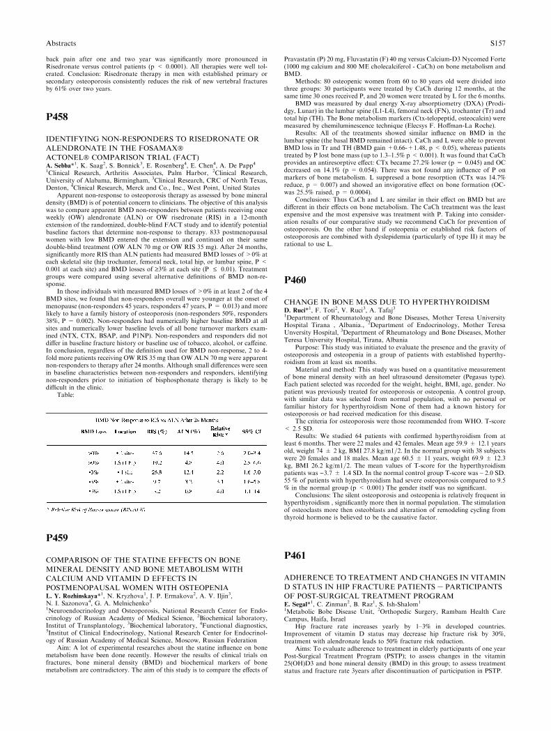
back pain after one and two year was significantly more pronounced inRisedronate versus control patients (p < 0.0001). All therapies were well tol-erated. Conclusion: Risedronate therapy in men with established primary orsecondary osteoporosis consistently reduces the risk of new vertebral fracturesby 61% over two years.
P458
IDENTIFYING NON-RESPONDERS TO RISEDRONATE OR
ALENDRONATE IN THE FOSAMAX�ACTONEL� COMPARISON TRIAL (FACT)A. Sebba*1, K. Saag2, S. Bonnick3, E. Rosenberg4, E. Chen4, A. De Papp41Clinical Research, Arthritis Associates, Palm Harbor, 2Clinical Research,University of Alabama, Birmingham, 3Clinical Research, CRC of North Texas,Denton, 4Clinical Research, Merck and Co., Inc., West Point, United States
Apparent non-response to osteoporosis therapy as assessed by bone mineraldensity (BMD) is of potential concern to clinicians. The objective of this analysiswas to compare apparent BMD non-responders between patients receiving onceweekly (OW) alendronate (ALN) or OW risedronate (RIS) in a 12-monthextension of the randomized, double-blind FACT study and to identify potentialbaseline factors that determine non-response to therapy. 833 postmenopausalwomen with low BMD entered the extension and continued on their samedouble-blind treatment (OW ALN 70 mg or OW RIS 35 mg). After 24 months,significantly more RIS than ALN patients had measured BMD losses of >0% ateach skeletal site (hip trochanter, femoral neck, total hip, or lumbar spine, P <0.001 at each site) and BMD losses of ‡3% at each site (P £ 0.01). Treatmentgroups were compared using several alternative definitions of BMD non-re-sponse.
In those individuals with measured BMD losses of >0% in at least 2 of the 4BMD sites, we found that non-responders overall were younger at the onset ofmenopause (non-responders 45 years, responders 47 years, P = 0.013) and morelikely to have a family history of osteoporosis (non-responders 50%, responders38%, P = 0.002). Non-responders had numerically higher baseline BMD at allsites and numerically lower baseline levels of all bone turnover markers exam-ined (NTX, CTX, BSAP, and P1NP). Non-responders and responders did notdiffer in baseline fracture history or baseline use of tobacco, alcohol, or caffeine.In conclusion, regardless of the definition used for BMD non-response, 2 to 4-fold more patients receiving OW RIS 35 mg than OW ALN 70 mg were apparentnon-responders to therapy after 24 months. Although small differences were seenin baseline characteristics between non-responders and responders, identifyingnon-responders prior to initiation of bisphosphonate therapy is likely to bedifficult in the clinic.
Table:
P459
COMPARISON OF THE STATINE EFFECTS ON BONE
MINERAL DENSITY AND BONE METABOLISM WITH
CALCIUM AND VITAMIN D EFFECTS IN
POSTMENOPAUSAL WOMEN WITH OSTEOPENIAL. Y. Rozhinskaya*1, N. Kryzhova1, I. P. Ermakova2, A. V. Iljin3,N. I. Sazonova4, G. A. Melnichenko51Neuroendocrinology and Osteoporosis, National Research Center for Endo-crinology of Russian Academy of Medical Science, 2Biochemical laboratory,Institut of Transplantology, 3Biochemical laboratory, 4Functional diagnostics,5Institut of Clinical Endocrinology, National Research Center for Endocrinol-ogy of Russian Academy of Medical Science, Moscow, Russian Federation
Aim: A lot of experimental researches about the statine influence on bonemetabolism have been done recently. However the results of clinical trials onfractures, bone mineral density (BMD) and biochemical markers of bonemetabolism are contradictory. The aim of this study is to compare the effects of
Pravastatin (P) 20 mg, Fluvastatin (F) 40 mg versus Calcium-D3 Nycomed Forte(1000 mg calcium and 800 ME cholecalciferol - CaCh) on bone metabolism andBMD.
Methods: 80 osteopenic women from 60 to 80 years old were divided intothree groups: 30 participants were treated by CaCh during 12 months, at thesame time 30 ones received P, and 20 women were treated by L for the 6 months.
BMD was measured by dual energy X-ray absorptiometry (DXA) (Prodi-dgy, Lunar) in the lumbar spine (L1-L4), femoral neck (FN), trochanter (Tr) andtotal hip (TH). The Bone metabolism markers (Ctx-telopeptid, osteocalcin) weremeasured by chemiluminescence technique (Elecsys F. Hoffman-La Roche).
Results: All of the treatments showed similar influence on BMD in thelumbar spine (the basal BMD remained intact). CaCh and L were able to preventBMD loss in Tr and TH (BMD gain +0.66-+1.48, p < 0.05), whereas patientstreated by P lost bone mass (up to 1.3–1.5% p < 0.001). It was found that CaChprovides an antiresorptive effect: CTx became 27.2% lower (p = 0.045) and OCdecreased on 14.1% (p = 0.054). There was not found any influence of P onmarkers of bone metabolism. L suppresed a bone resorption (CTx was 14.7%reduce, p = 0.007) and showed an invigorative effect on bone formation (OC-was 25.5% raised, p = 0.0004).
Conclusions: Thus CaCh and L are similar in their effect on BMD but aredifferent in their effects on bone metabolism. The CaCh treatment was the leastexpensive and the most expensive was treatment with P. Taking into consider-ation results of our comparative study we recommend CaCh for prevention ofosteoporosis. On the other hand if osteopenia or established risk factors ofosteoporosis are combined with dyslepidemia (particularly of type II) it may berational to use L.
P460
CHANGE IN BONE MASS DUE TO HYPERTHYROIDISMD. Ruci*1, F. Toti2, V. Ruci3, A. Tafaj31Department of Rheumatology and Bone Diseases, Mother Teresa UniversityHospital Tirana , Albania., 2Department of Endocrinology, Mother TeresaUnversity Hospital, 3Department of Rheumatology and Bone Diseases, MotherTeresa University Hospital, Tirana, Albania
Purpose: This study was initiated to evaluate the presence and the gravity ofosteoporosis and osteopenia in a group of patients with established hyperthy-roidism from at least six months.
Material and method: This study was based on a quantitative measurementof bone mineral density with an heel ultrasound densitometer (Pegasus type).Each patient selected was recorded for the weight, height, BMI, age, gender. Nopatient was previously treated for osteoporosis or osteopenia. A control group,with similar data was selected from normal population, with no personal orfamiliar history for hyperthyroidism None of them had a known history forosteoporosis or had received medication for this disease.
The criteria for osteoporosis were those recommended from WHO. T-score< 2.5 SD.
Results: We studied 64 patients with confirmed hyperthyroidism from atleast 6 months. Ther were 22 males and 42 females. Mean age 59.9 ± 12.1 yearsold, weight 74 ± 2 kg, BMI 27.8 kg/m1=2. In the normal group with 38 subjectswere 20 females and 18 males. Mean age 60.5 ± 11 years, weight 69.9 ± 12.3kg, BMI 26.2 kg/m1=2. The mean values of T-score for the hyperthyroidismpatients was )3.7 ± 1.4 SD. In the normal control group T-score was – 2.0 SD.55 % of patients with hyperthyroidism had severe osteoporosis compared to 9.5% in the normal group (p < 0.001) The gender itself was no significant.
Conclusions: The silent osteoporosis and osteopenia is relatively frequent inhyperthyroidism , significantly more then in normal population. The stimulationof osteoclasts more then osteoblasts and alteration of remodeling cycling fromthyroid hormone is believed to be the causative factor.
P461
ADHERENCE TO TREATMENT AND CHANGES IN VITAMIN
D STATUS IN HIP FRACTURE PATIENTS – PARTICIPANTS
OF POST-SURGICAL TREATMENT PROGRAME. Segal*1, C. Zinman2, B. Raz1, S. Ish-Shalom1
1Metabolic Bobe Disease Unit, 2Orthopedic Surgery, Rambam Health CareCampus, Haifa, Israel
Hip fracture rate increases yearly by 1–3% in developed countries.Improvement of vitamin D status may decrease hip fracture risk by 30%,treatment with alendronate leads to 50% fracture risk reduction.
Aims: To evaluate adherence to treatment in elderly participants of one yearPost-Surgical Treatment Program (PSTP); to assess changes in the vitamin25(OH)D3 and bone mineral density (BMD) in this group; to assess treatmentstatus and fracture rate 3years after discontinuation of participation in PSTP.
Abstracts S157
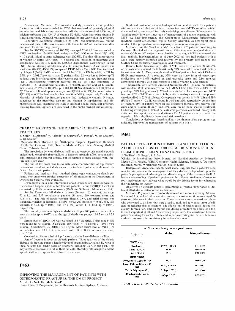
Patients and Methods: 125 consecutive elderly patients after surgical hipfracture correction were enrolled in PTSP that consisted of quarterly physicalexamination and laboratory evaluation. All the patients received 1500 mg ofcalcium carbonate and 800 IU of vitamin D3 daily. After improving vitamin Dstatus alendronate 70 mg/wk was administered for one year within the program.
Laboratory evaluation: 25(OH)D3 by 125I- radioimmunoassay, routinebiochemical tests. BMD was assessed with Lunar DEXA at baseline and afterone year of antiresorbing therapy.
Results: 91(73%) women and 34(27%) men aged 72.68±9.5 were enrolled inPSTP. At baseline 124(99%) had inadequate 25(OH)D3 serum level; 27(21.6%)<10 ng/ml. Initial patients� adherence was 29(23%). The time till improvementof vitamin D status (25(OH)D3 >18 ng/ml) and initiation of treatment withalendronate was 18 ± 6 months. 65(52%) discontinued participation in thePSTP before starting alendronate due to noncompliance. Alendronate wasstarted in 54(43%); 46(36.8%) completed PTSP. Mean 25(OH)D3 level increasedby 5.6ng/ml (34%). BMD increased in LS by 5.2%, in FN by 4.8%, in TH by2.7%, p < 0.001.Three years later 22 patients died, 32 were lost to follow up;71patients were interviewed about their current treatment and new fractures sincePSTP. Antiresorbing treatment received 28(78%) of PTSP completed vs7(20%)of PTSP discontinied patients, p < 0.0001, calcium and vit D supple-ments took 27(75%) vs 18(51%), p < 0.0001,DEXA elaluation had 18(50%) vs2(5.6%),were followed up in specialty clinic 9(25%) vs 4(11%),had new fractures4(12.5%) vs 6(17%), had new hip fractures 0 vs 3(8%), respectively.Conclusion:Majority of elderly hip fracture patients had inadequate vitamin D status;adherence to the prescribed calcium and vitamin D supplements and bis-phosphonates was unsatisfactory even in hospital bound outpatient program.Existing treatment options are underused in the public medical service setting.
P462
CHARACTERISTICS OF THE DIABETIC PATIENTS WITH HIP
FRACTURESE. Segal*1, C. Zinman2, V. Raichlin2, B. Gurevich1, A. Psavke1, M. Ish-Shalom3,S. Ish-Shalom1
1Metabolic Bobe Disease Unit, 2Orthopedic Surgery Department, RambamHealth Care Campus, Haifa, 3Internal Medicine Department, Sorasky MedicalCenter, Tel-Aviv, Israel
The association between diabetes mellitus and osteoporosis remains poorlyunderstood. Metabolic abnormalities of diabetes probably affect bone metabo-lism, structure and mineral density, but association of these changes with frac-ture risk is not clear.
The aim of this work was to evaluate some characteristics of hip fracturepatients with and without diabetes and try to detect potential additional riskfactors for diabetic patients.
Patients and methods: Four hundred ninety eight consecutive elderly pa-tients, who underwent surgical correction of hip fracture in the Department ofOrthopedic Surgery, were evaluated.
Demographic data and information about concomitant diseases were re-trieved from hospital charts of hip fracture patients. Serum 25(OH)D3 level wasevaluated by 125I- radioimmunoassay (DiaSorin, Stillwater, Minnesota, USA).
Results: There were 141 diabetic patients (36 men, 105 women); mean age75 ± 8.5, and 357 non-diabetic patients (88 men, 269 women); mean age77.8 ± 9.1. The rate of cardio-vascular disease, CVA and renal disease wassignificantly higher in diabetics: 117(83%) versus 207 (58%), p = 0.01), 30 (21%)versus16 (4.5%), (p = 0.047) and 17 (12%) versus 13 (3.6%), (p = 0.034),respectively.
The mortality rate was higher in diabetics: 18 per 100 patients, versus 8 innon- diabetics (p = 0.037), and the age of death was younger: 80.5 versus 82.9years.
Serum level of 25(OH)D3 was evaluated in 87 diabetics. Thirty-nine (48%)were found to be vitamin D deficient, 25(OH)D3 < 10 ng/ml; 27 (28%) werevitamin D insufficient, 25(OH)D3 < 15 ng/ml. Mean serum level of 25(OH)D3in diabetics was 13.8 ± 7, compared with 18 ± 34.25 in non- diabetics,p = 0.051.
Conclusion: About third of hip fracture patients have diabetes mellitus.Age of fracture is lower in diabetic patients. Three quarters of the elderly
diabetic hip fracture patients had low level of serum hydroxyvitamin D. Most ofthese patients had cardio-vascular disorders, including CVA in the past. Thismay increase propensity to fall in these patients. Mortality rate is higher, and theage of death after hip fracture is lower in diabetics.
P463
IMPROVING THE MANAGEMENT OF PATIENTS WITH
OSTEOPOROTIC FRACTURES: THE OMEN PROJECTA. Lih1, C. Nickolls1, M. J. Seibel*11Bone Research Programme, Anzac Research Institute, Sydney, Australia
Worldwide, osteoporosis is underdiagnosed and undertreated. Even patientswith recurrent and obvious minimal trauma fractures (MTF) are often neitherdiagnosed with, nor treated for their underlying bone disease. Subsequent to a�baseline study� into the status quo of management of patients presenting withMTF, we have implemented the �Osteoporosis Management Enhancement(OMEN) Project� at Concord Hospital, Sydney, Australia. We here report initialresults of this prospective interdisciplinary continuum-of-care program.
Methods: For the �baseline study�, data from 537 patients presenting toConcord Hospital with a diagnostic code of fracture were analysed via chartreview. Of those, 302 subjects were classified as having a MTF and entered thefinal analysis. Implementation: as of June 2005, all non-frail patients with aMTF were actively identified and referred by the primary care team to theOMEN Clinic for further investigation and treatment.
Results: In the �baseline study�, 70% of MTF occurred in women. While 65%of patients were assessed for falls risk, only 20% were asked about risk factorsfor osteoporosis. An X-ray was obtained in 97% of patients, but only 4.1% had aBMD measurement. At discharge, 35% were on some form of osteotropicmedication; only 8.6% received an anti-resorptive agent and 2.1% receivedcombination therapy with anti-resorptive agents, vitamin D and calcium.
�Implementation�: Between June and November 2005, 130 non-frail patientswith incident MTF were referred to the OMEN Clinic (80% female, 84% < 80yrs of age, 98% living at home). 27% of patients had at least one previous MTFfracture. 92% of MTF were due to falls, while secondary causes of osteoporosiswere identified in 22% of subjects. Osteopaenia was present in 44% (LS) and 51%(FN); a T-score < -2.5SD was found in 30% and 22%, respectively. At the timeof fracture, 15% of patients were on anti-resorptive therapy, 30% received cal-cium and/or vitamin D only, and 54% were on no bone specific treatment.Following investigation, 74% of patients were placed on combined therapy withanti-resorptive agent, calcium and vitamin D. All patients received advice inregards to life style, dietary factors and risk avoidance.
Conclusion: A dedicated interdisciplinary continuum-of-care program sig-nificantly improves the management of patients with MTF.
P464
PATIENTS’ PERCEPTION OF IMPORTANCE OF DIFFERENT
ATTRIBUTES OF OSTEOPOROSIS MEDICATION: RESULTS
FROM THE PREFER-INTERNATIONAL STUDYJ. Walliser*1, S. Bolge2, S. S. Sen31Clinical de Metabolismo Oseo, Mineral del Hospital Angeles del Pedregal,Mexico City, Mexico, 2CHS, Consumer Health Sciences, Princeton, 3OutcomesResearch, Merck, Whitehouse Station, United States
Background: Anderson’s health belief model suggests that a patient’s deci-sion to take action in the management of their disease is dependent upon thepatient’s perception of advantages and disadvantages of the treatment itself. Abetter understanding of patients’ preference for different attributes of osteopo-rosis medication may indicate what would be the driving factor for enhancingtheir adherence to therapy.
Objective: To evaluate patients’ perceptions of relative importance of dif-ferent attributes of osteoporosis medication.
Methods: Physicians were randomly selected in France, Germany, Mexico,Spain and UK and asked to recruit consecutive 4 osteoporotic women aged 50years or older seen in their practices. These patients were contacted and thosewho consented to an interview were asked to rank and rate importance of effi-cacy in reducing risk of fracture, side effects, out-of-pocket costs, dosing fre-quency, formulation, time on market and dosing procedure on a scale of 1 to 5(1=not important at all and 5=extremely important). The correlation betweenpatient’s ranking for each attribute and importance rating for that attribute wasevaluated to assess the consistency in patients� response.
S158 Abstracts
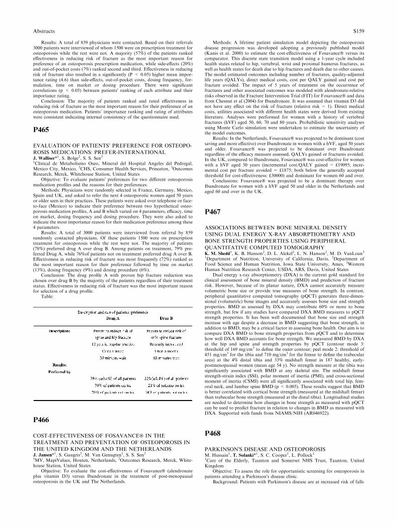
Results: A total of 859 physicians were contacted. Based on their referrals3000 patients were interviewed of whom 1500 were on prescription treatment forosteoporosis while the rest were not. A majority (57%) of the patients rankedeffectiveness in reducing risk of fracture as the most important reason forpreference of an osteoporosis prescription medication, while side-effects (20%)and out-of-pocket costs (7%) ranked second and third. Effectiveness in reducingrisk of fracture also resulted in a significantly (P < 0.05) higher mean impor-tance rating (4.6) than side-effects, out-of-pocket costs, dosing frequency, for-mulation, time on market or dosing procedure. There were significantcorrelations (p < 0.05) between patients� ranking of each attribute and theirimportance rating.
Conclusion: The majority of patients ranked and rated effectiveness inreducing risk of fracture as the most important reason for their preference of anosteoporosis medication. Patients’ importance ranking and rating of attributeswere consistent indicating internal consistency of the questionnaire used.
P465
EVALUATION OF PATIENTS’ PREFERENCE FOR OSTEOPO-
ROSIS MEDICATIONS: PREFER-INTERNATIONALJ. Walliser*1, S. Bolge2, S. S. Sen31Clinical de Metabolismo Oseo, Mineral del Hospital Angeles del Pedregal,Mexico City, Mexico, 2CHS, Consumer Health Services, Princeton, 3OutcomesResearch, Merck, Whitehouse Station, United States
Objective: To evaluate patients� preferences for two different osteoporosismedication profiles and the reasons for their preferences.
Methods: Physicians were randomly selected in France, Germany, Mexico,Spain and UK, and asked to refer the next 4 osteoporotic women aged 50 yearsor older seen in their practices. These patients were asked over telephone or face-to-face (Mexico) to indicate their preference between two hypothetical osteo-porosis medication profiles, A and B which varied on 4 parameters, efficacy, timeon market, dosing frequency and dosing procedure. They were also asked toindicate the most importance reason for their medication preference among these4 parameters.
Results: A total of 3000 patients were interviewed from referral by 859randomly contacted physicians. Of these patients 1500 were on prescriptiontreatment for osteoporosis while the rest were not. The majority of patients(78%) preferred drug A over drug B. Among patients on treatment, 79% pre-ferred Drug A, while 76%of patients not on treatment preferred drug A over B.Effectiveness in reducing risk of fracture was most frequently (72%) ranked asthe most important reason for their preference followed by time on market(13%), dosing frequency (9%) and dosing procedure (6%).
Conclusion: The drug profile A with proven hip fracture reduction waschosen over drug B by the majority of the patients regardless of their treatmentstatus. Effectiveness in reducing risk of fracture was the most important reasonfor selection of a drug profile.
Table:
P466
COST-EFFECTIVENESS OF FOSAVANCE� IN THE
TREATMENT AND PREVENTATION OF OSTEOPOROSIS IN
THE UNITED KINGDOM AND THE NETHERLANDSJ. Jansen*1, S. Gaugris2, M. Van Genugten1, S. S. Sen21MV, MapiValues, Houten, Netherlands, 2Outcomes Research, Merck, White-house Station, United States
Objective: To evaluate the cost-effectiveness of Fosavance� (alendronateplus vitamin D3) versus Ibandronate in the treatment of post-menopausalosteoporosis in the UK and The Netherlands.
Methods: A lifetime patient simulation model depicting the osteoporosisdisease progression was developed adopting a previously published model(Kanis et al. 2000) to estimate the cost-effectiveness of Fosavance� versus itscomparator. This discrete state transition model using a 1-year cycle includedhealth states related to hip, vertebral, wrist and proximal humerus fractures, aswell as health states for death due to hip fractures and death due to other causes.The model estimated outcomes including number of fractures, quality-adjustedlife years (QALYs), direct medical costs, cost per QALY gained and cost perfracture avoided. The impact of 5 years of treatment on the occurrence offractures and other associated outcomes was modeled with alendronate-relativerisks observed in the Fracture Intervention Trial (FIT) for Fosavance� and datafrom Chesnut et al (2004) for Ibandronate. It was assumed that vitamin D3 didnot have any effect on the risk of fracture (relative risk = 1). Direct medicalcosts, utilities associated with different health states were derived from existingliterature. Analyses were performed for women with a history of vertebralfractures (hVF) aged 50, 60, 70 and 80 years. Probabilistic sensitivity analysesusing Monte Carlo simulation were undertaken to estimate the uncertainty ofthe model outcomes.
Results: In the Netherlands, Fosavance� was projected to be dominant (costsaving and more effective) over Ibandronate in women with a hVF, aged 50 yearsand older. Fosavance� was projected to be dominant over Ibandronateregardless of the efficacy measure assessed, QALYs gained or fractures avoided.In the UK, compared to Ibandronate, Fosavance� was cost-effective for womenwith a hVF aged 50 years (incremental cost/QALY gained = £19095; incre-mental cost per fracture avoided = £1875; both below the generally acceptedthreshold for cost-effectiveness: £30000) and dominant for women 60 and over.
Conclusions: Fosavance� was projected to be a dominant therapy overIbandronate for women with a hVF aged 50 and older in the Netherlands andaged 60 and over in the UK.
P467
ASSOCIATIONS BETWEEN BONE MINERAL DENSITY
USING DUAL ENERGY X-RAY ABSORPTIOMETRY AND
BONE STRENGTH PROPERTIES USING PERIPHERAL
QUANTITATIVE COMPUTED TOMOGRAPHYK. M. Shedd1, K. B. Hanson2, D. L. Alekel2, L. N. Hanson2, M. D. VanLoan31Department of Nutrition, University of California, Davis, 2Department ofFood Science and Human Nutrition, Iowa State University, Ames, 3WesternHuman Nutrition Research Center, USDA, ARS, Davis, United States
Dual energy x-ray absorptiometry (DXA) is the current gold standard forclinical assessment of bone mineral density (BMD) and prediction of fracturerisk. However, because of its planar nature, DXA cannot accurately measurevolumetric bone size or provide true measures of bone strength. In contrast,peripheral quantitative computed tomography (pQCT) generates three-dimen-sional (volumetric) bone images and accurately assesses bone size and strengthproperties. BMD as assessed by DXA may contribute 60% or more to bonestrength, but few if any studies have compared DXA BMD measures to pQCTstrength properties. It has been well documented that bone size and strengthincrease with age despite a decrease in BMD suggesting that bone strength, inaddition to BMD, may be a critical factor in assessing bone health. Our aim is tocompare DXA BMD to bone strength properties from pQCT and to determinehow well DXA BMD accounts for bone strength. We measured BMD by DXAat the hip and spine and strength properties by pQCT (contour mode 3:threshold of 169 mg/cm3 to define the outer contour; peel mode 2: threshold of451 mg/cm3 for the tibia and 710 mg/cm3 for the femur to define the trabeculararea) at the 4% distal tibia and 33% midshaft femur in 187 healthy, early-postmenopausal women (mean age 54 y). No strength measure at the tibia wassignificantly associated with BMD at any skeletal site. The midshaft femurstrength-strain index (SSI), polar moment of inertia (PMI), and cross-sectionalmoment of inertia (CSMI) were all significantly associated with total hip, fem-oral neck, and lumbar spine BMD (p < 0.005). These results suggest that BMDis better correlated with cortical bone strength (measured at the midshaft femur)than trabecular bone strength (measured at the distal tibia). Longitudinal studiesare needed to determine how changes in bone strength as measured with pQCTcan be used to predict fracture in relation to changes in BMD as measured withDXA. Supported with funds from NIAMS/NIH (AR046922).
P468
PARKINSON�S DISEASE AND OSTEOPOROSISM. Hussain1, T. Solanki*1, S. C. Cooper1, L. Pollock11Care of the Elderly, Taunton and Somerset NHS Trust, Taunton, UnitedKingdom
Objective: To assess the role for opportunistic screening for osteoporosis inpatients attending a Parkinson�s disease clinic.
Background: Patients with Parkinson�s disease are at increased risk of falls
Abstracts S159
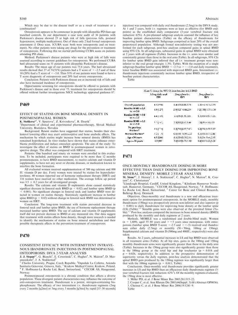
Which may be due to the disease itself or as a result of treatment or acombination?
Osteoporosis appears to be commoner in people with idiopathic PD than agematched controls. In our department a case note audit of 36 patients withParkinson�s disease showed 23 at high risk of falls (previous falls, posturalhypotension, multiple medications). Out of these only 2 had formal osteoporosisassessment (1 Dexa scan, 1CUBA scan: both were osteoporotic and on treat-ment). No other patients were taking any drugs for the prevention or treatmentof osteoporosis. Following this we started performing CUBA scans on patientsattending PD clinic.
Method: Parkinson�s disease patients who were at high risk of falls wereassessed according to current guidelines for osteoporosis. We performed CUBAheel ultrasound scans on 51 patients with idiopathic Parkinson�s disease.
Results: The mean age of our patients was 75.6 years. The heel ultrasoundresults showed that 27 of 51(51%) patients had a T score of <)2.5 and of these10 (20%) had a T score of <)3.0. Thus 51% of our patients were found to have aT score diagnostic of osteoporosis and 20% had severe osteoporosis.
Conclusion: Patients with Parkinson disease are at increased risk of falls andalso have increased incidence of osteoporosis.
Opportunistic screening for osteoporosis should be offered to patients withParkinson�s disease and in those over 75, treatment for osteoporosis should beoffered without further investigations NICE technology appraisal guidance 87.
P469
EFFECT OF STATINS ON BONE MINERAL DENSITY IN
POSTMENOPAUSAL WOMENK. Stefikova*1, V. Spustova1, Z. Krivosikova1, R. Dzurik11Department of clinical and experimental pharmacotherapy, Slovak MedicalUniversity, Bratislava, Slovakia
Background: Resent studies have suggested that statins, besides their cho-lesterol lowering effect may exert antiresorptive and bone anabolic effects. Themechanism by which statins might increase bone mineral density (BMD) hasremained hypothetical. In vitro studies have shown that statins enhance osteo-blastic proliferation and induce osteoclast apoptosis. The aim of the study: Toinvestigate the effect of statins on BMD in postmenopausal women in retro-spective design. The effect was compared with HRT treatment.
Patients: One hundred and ninety six women were enrolled in this evalua-tion. To be included, participants were required to be more then 12 monthspostmenopause, to have BMD measurement, to receive calcium and vitamin Dsupplements, to have not any sever or chronic diseases, or medication known tointerfere the bone formation.
All evaluated women received supplementation of 500 mg calcium and 800IU vitamin D per day. Forty women were treated by statins for hypercholes-terolemy, 40 women reported use of hormone replacement therapy (HRT) and116 women have received no other medication. The average follow up periodwas 3.5 ± 0.2 years for all investigated groups.
Results: The calcium and vitamin D suplements alone caused statisticalysignificat decrease in femoral neck BMD (p < 0.02) and lumbar spine BMD (p< 0.001). No significant changes in femoral neck and lumbar BMD were de-tected in women on statins therapy. Statisticaly significant increase in lumbarspine BMD (p < 0.02) without change in femoral neck BMD was determined inwomen on HRT.
Conclusion: The long-term treatment with statins prevented decrease infemoral neck and lumbar spine BMD, the use of hormone replacement therapyincreased lumbar spine BMD. The use of calcium and vitamin D supplementsitself did not prevent decrease in BMD at any measured site. Our data suggestthat treatment with statins affects bone density, though more research is neededto identify the mechanisms of statins on bone mineral metabolism and theirpossible therapeutic effects in the prevention/treatment of osteoporosis.
P470
CONSISTENT EFFICACY WITH INTERMITTENT INTRAVE-
NOUS IBANDRONATE INJECTIONS IN POSTMENOPAUSAL
OSTEOPOROSIS: DIVA 2-YEAR ANALYSISJ. J. Stepan*1, G. Bianchi2, E. Czwerinski3, C. Hughes4, N. Mairon4, D. Mas-anauskaite4, J. A. Stakkestad51 Charles University, Prague, Czech Republic, 2Ospedale La Colletta, AziendaSanitaria Genovese, Genova, Italy, 3 Krakow Medical Centre, Krakow, Poland,4 F. Hoffmann-La Roche Ltd, Basel, Switzerland, 5 CECOR AS, Haugesund,Norway
Postmenopausal osteoporosis is a chronic condition that affects a diversepopulation. These divergent patient characteristics may influence the outcome ofanti-osteoporosis treatment. Ibandronate is a potent, nitrogen-containing bis-phosphonate. The efficacy of two intermittent i.v. ibandronate regimens (2mgevery 2 months [q2mo] or 3mg every 3 months [q3mo] by rapid [15–30 seconds]
injection) was compared with daily oral ibandronate (2.5mg) in the DIVA study.At 1 and 2 years, both i.v. regimens were at least as effective (for BMD end-points) as the established daily comparator (3-year vertebral fracture riskreduction: 62%). A pre-planned subgroup analysis assessed the influence of keybaseline patient characteristics (Table) on the efficacy of ibandronate forimproving lumbar spine BMD. Subgroups comprised at least 20% of the overallperprotocol population. Although formal non-inferiority testing was not per-formed for each subgroup, post-hoc analyses compared gains in spinal BMDusing 95% CIs. In all subgroups, substantial gains in spinal BMD were obtainedat 2 years with all regimens (Table). Increases in the i.v. arms were similar andconsistently greater than those in the oral arm (Table). In all subgroups, 95% CIsfor lumbar spine BMD gain inferred that all i.v. treatment groups were non-inferior to the oral group (margin: 1.3%; Table). With the exception of a singlesubgroup (baseline lumbar spine BMD <)2.5 to >=)3.0 and daily treatment),lower baseline BMD was associated with improved BMD gain. Intermittent i.v.ibandronate injections consistently increase lumbar spine BMD, irrespective ofbaseline patient characteristics.
Table:
P471
ONCE-MONTHLY IBANDRONATE DOSING IS MORE
EFFECTIVE THAN DAILY DOSING FOR IMPROVING BONE
MINERAL DENSITY: MOBILE 2-YEAR ANALYSISM. Stone*1, J. Hensen2, J. A. Stakkestad3, C. Hughes4, N. Mairon4, K. Cou-tant4, C. Christiansen51Llandough Hosptial, Cardiff, United Kingdom, 2 Klinikum Hannover Nords-tadt, Hannover, Germany, 3 CECOR AS, Haugesund, Norway, 4 F. Hoffmann-La Roche Ltd, Basel, Switzerland, 5 Center for Basic and Clinical Research,Ballerup Byvej, Denmark
Objectives. Once-monthly oral ibandronate is a new and efficacious treat-ment option for postmenopausal osteoporosis. In the MOBILE study, monthlyibandronate (150mg) was prospectively proven non-inferior and also superior (p< 0.001) to daily ibandronate for improving bone density at the lumbar spine(LS) (Table).1,2 Sizeable gains were also observed at the proximal femur (Ta-ble).1,2 Post-hoc analyses compared the increases in bone mineral density (BMD)produced by the monthly and daily regimens at 2 years.
Methods. MOBILE was a randomised and double-blind study. Women(n = 1,609), aged 55–80 years and >=5 years since menopause, with osteo-porosis (lumbar spine BMD T-score <-2.5 and >=-5) received oral ibandro-nate either daily (2.5mg) or monthly (50+50mg, 100mg or 150mg).Supplemental calcium and vitamin D (500mg and 400IU, respectively) were alsotaken.
Results. At 2 years, substantial increases in LS and hip BMD were observedin all treatment arms (Table). At all hip sites, gains in the 100mg and 150mgmonthly ibandronate arms were significantly greater than those in the daily arm(Table). Increases in the 150mg group were also significantly greater than thosein the 100mg group at the total hip and hip trochanter (p = 0.014 andp = 0.026, respectively). In addition to the prospective demonstration ofsuperiority versus the daily regimen, post-hoc analysis demonstrated that thespinal BMD gain produced by the 150mg regimen was significantly larger thanthat with the 100mg regimen (p = 0.011; Table).
Conclusions. Once-monthly oral ibandronate provides significantly greaterincreases in LS and hip BMD than an efficacious daily ibandronate regimen (3-year vertebral fracture risk reduction: 62%3). Of the monthly regimens evaluated,the 150mg dose is most effective.
1. Miller PD, et al. J Bone Miner Res 2005;20:1315–22.2. Cooper C, et al. Ann Rheum Dis 2005;64(Suppl. 3):68 (Abstract OP0036).3. Chesnut C, et al. J Bone Miner Res 2004;19:1241–9.Table:
S160 Abstracts

P472
ESTIMATING STRUCTURAL PROPERTIES OF TRABECULAR
BONE FROM GRAY-LEVEL LOW-RESOLUTION IMAGESZ. Tabor*1, E. Rokita1, P. Gluszko21Department of Biophysics, 2Department of Rheumatology, Collegium Medi-cum Jagiellonian University, Cracow, Poland
In this study the problem is addressed, which structural parameters (otherthan bone density) calculated for low-resolution (LR) images improve estimationof mechanical properties of trabecular bone. Typically multivariate regressionmodels combining some measure of trabecular bone density with other three- ormore frequently two-dimensional parameters are proposed to explain variationof the trabecular bone apparent elastic constants found in mechanical tests. Thereported results are however often controversial. In this study a novel approachto the structural analysis of LR data is proposed. First ‘‘gold standard’’ analysisof 3D microCT images of distal radius trabecular bone is performed. Next thedeveloped ‘‘optimal paths’’ analysis is applied to gray-level LR (MRI) images ofthe same samples. Fifteen radius bone cubes from the distal radius were obtainedfrom 15 individuals. The samples were scanned with a microCT-20 scanner(Scanco Medical, Zurich, Switzerland) with a voxel size of 34micrometers in allthree spatial dimensions. Next MRI images of the samples were acquired with areceive-only wrist coil (Medical Advances, Milwaukee, WI) on a GE SIGNA 1.5Tesla echo-speed system (General Electric Medical Systems, Waukesha, WI)with pixel size of 156micrometers and slice thickness of 300micrometers.Young’s modulus for the cranio-caudal direction, bone volume fraction, tra-becular thickness, separation, length and anisotropy were calculated for mi-croCT and MRI images. Optimal path analysis [1] was also performed. LRhistomorphometric parameters did not significantly improve estimation ofelastic modulus, comparing to bone volume fraction App.BV/TV estimated fromMRI data. Multivariate linear model combining optimal path parameter andApp.BV/TV explains 88% of the variation of the apparent Young�s modulus.The performance of such model is thus better than of any other one combiningApp.BV/TV with histomorphometric parameters.
[1] Z.Tabor, E.Rokita, Detection of trabecular discontinuities in the CTimages of trabecular bone, 2nd Joint Meeting of ECTS and IBMS, 25-29 June2005, Geneva, Switzerland, Bone 36 (supp. 2), S326, 2005.
P473
DETERMINING OPTIMAL CUT OF TRABECULAR BONEZ. Tabor*11Department of Biophysics, Collegium Medicum Jagiellonian University,Cracow, Poland
It has been shown recently, that frequently not a whole trabecular sample isdamaged, when the sample is mechanically tested [1,2]. The failure can belocalized in some oblique plane or surface. It was also found that BV/TV of thefailure region was smaller than BV/TV of the whole sample [2]. These results leadto a conjecture that localized failure is related to presence of a surface of minimalcut - a surface separating the analyzed trabecular sample into two disjoint partsin such a way, that the separation requires removal of minimal possible amountof bone mass. An exact solution to the minimal cut problem can not be found for3D structures. Proposed in the present study approximate solution of thisproblem in 3D involves following steps: (1) Digital distance transform of thebinary image of a trabecular bone sample; (2) Skeletonization of the binaryimage of a trabecular bone sample; (3) Converting the calculated skeleton to agraph; (4) Spectral analysis of the graph; (5) Monte Carlo optimization of thespectral solution. Fifteen radius bone cubes from the distal radius were obtainedfrom 15 individuals. The samples were scanned with a microCT-20 scanner(Scanco Medical, Zurich, Switzerland) with a voxel size of 34microns in all threespatial dimensions. The microCT images were binarized. The surface of minimalcut was found for every sample. Both minimal cut area AC and the number NCof trabeculae in the minimal cut decrease with BV/TV, correlation coefficientbeing equal to 0.85 and 0.75, respectively. These quantities correlate also withYoung�s modulus calculated for the cranio-caudal direction (correlation coeffi-cient equal to 0.84 and 0.77 for AC and NC, respectively). Comparing to BV/TV
however, neither AC nor NC provides significant, new information about theYoung�s modulus of the samples. This result can be explained by the complicatedgeometry of the minimal cuts, extending over substantial part of the samples. Infact, it means that the structures are optimal, without unhomogeneities,decreasing their strength.
[1] R. Muller, ‘‘Anatomical and functional CT imaging of bone,’’ Bone 36,Supp. 2, S105 (2005).
[2] E. Perilli, F. Baruffaldi, M. Baleani, R. Fognani, M. Visentin, S. Stea andF. Traina, ‘‘Trabecular bone of proximal femur:dependence of mechanicalcompressive strength on local variations in bone morphometry,’’ Bone 36, Supp.2, S191–S192 (2005).
P474
URINARY OSTEOCALCIN AND OTHER MARKERS OF BONE
METABOLISM: EFFECT OF RISEDRONATE THERAPYA. Tamm*1, J. Kumm1, K. Rohtla1, K. K. Ivaska21Laboratory Medicine, University of ’Tartu, Tartu, Estonia, 2Institute of Clin-ical Sciences, University of Lund, Malmo, Sweden
For monitoring response to osteoporosis therapy, biochemical markers ofbone remodeling play an important role. The short-term changes in boneresorption in response to therapy are considered to have the predictive value ofpositive treatment effect. Up to now there is limited information about urinaryosteocalcin (U-OC).
Aim: To examine the behaviour of urinary osteocalcin in comparison withother bone turnover markers in patients with postmenopausal osteoporosistreated with Risedronate.
Methods. The study group consisted of 29 patients with postmenopausalosteoporosis (age 48-65, mean 56 years). 17 of them had received Risedronatetherapy for 6 months. 19 healthy women, aged 36–45, served as the controls. U-OC was measured by two-site immunoassay specific for the U-OC mid-fragment(U-MidOC) and expressed per mmol creatinine. Bone resorption was also as-sessed by collagen type I C-terminal telopeptide in serum, S-CTx-I, bone for-mation - by serum levels of procollagen type 1 amino-terminal propeptide, S-P1NP and osteocalcin, S-OC (Roche, Elecsys).
Results. Output of U-OC decreased markedly (> 1 T-score) during the firstmonth of therapy in 10 patients (‘‘quick response’’) and in 4 more patientsduring the next 3–6 months (‘‘slower response’’). In two cases, with low basalvalues, it did not change (non-responders). At the same time the response of S-CTx-I occurred in 12 (quick), 3 and 2 cases, respectively. S-P1NP levels de-creased mainly by the 3rd month.
Before the therapy U-MidOC values correlated with CTx-1 (rho = 0.519,p = 0.004) and S-OC (0.435, p = 0.018) but not with P1NP. The changes of theT scores of the U-OC during the therapy correlated with those of S-CTx-1(rho = 0.634, n = 39, p < 0.0001), but non-significantly with changes of S-OCand S-P1NP.
Conclusions1. U-MidOC responds to the Risedronate therapy similarily to that of S-CTx
– mainly within the first month.2. Strong correlation between U-MidOC and S-CTx - at the same time
without correlation with collagen type I synthesis marker P1NP - confirmsearlier results regarding the origin of urinary fragments of OC from boneresorption.
3. Use of bone resorption markers like S-CTx-1 and U-MidOC may allowmonitoring the effectiveness of antiresorptive therapy in individual patients.
P475
A MONTHLY DOSING REGIMEN OF RISEDRONATE
REDUCES BONE TURNOVER SIMILAR TO DAILY
ADMINISTRATIONG. A. Thompson*1, D. J. Schnell1, D. A. Russell1, D. E. Burgio1, L. A. Sun11Drug Development, Procter and Gamble Pharmaceuticals, Mason, UnitedStates
This was a randomized, multiple oral dose, parallel group study conductedin 58 postmenopausal women. Subjects were randomly assigned in a 1:1 ratio toreceive risedronate 150 mg monthly (75 mg on 2 consecutive days per month [2CD/M]) or risedronate 5 mg daily for four-months. Urine samples for uNTxdetermination were collected at baseline and on days 1, 4, 7, 14 and 30 duringMonth 1 and Month 4. Of the 58 subjects enrolled, 51 completed the study.Overall risedronate was shown to be well tolerated in both dose groups. uNTxresults are summarized in the Table 1.
There was no difference between the risedronate 150 mg/monthly and the 5mg daily groups for uNTx reduction at the end of Month 1 or Month 4 (Day 30).Although, the area under the effect curve (AUEC) over 30 days (both Month 1and 4) showed a greater reduction for the 150 mg/monthly dose compared to the5 mg daily dose; and the median Emax (the maximum observed effect for uNTx)
Abstracts S161
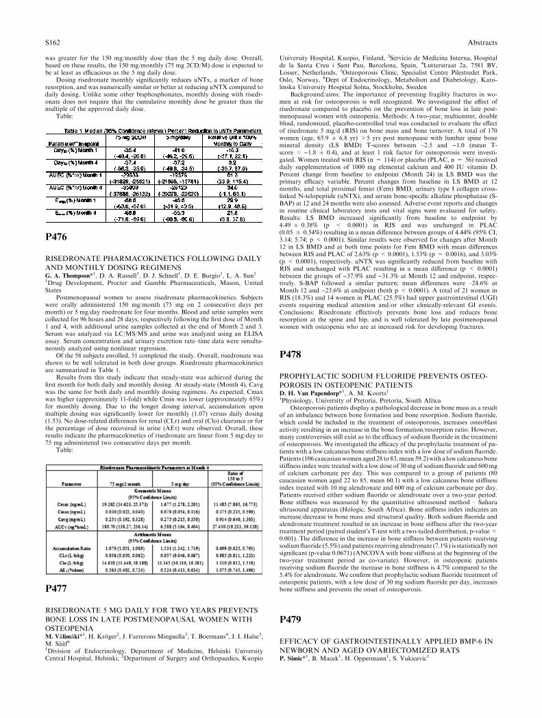
was greater for the 150 mg/monthly dose than the 5 mg daily dose. Overall,based on these results, the 150 mg/monthly (75 mg 2CD/M) dose is expected tobe at least as efficacious as the 5 mg daily dose.
Dosing risedronate monthly significantly reduces uNTx, a marker of boneresorption, and was numerically similar or better at reducing uNTX compared todaily dosing. Unlike some other bisphosphonates, monthly dosing with risedr-onate does not require that the cumulative monthly dose be greater than themultiple of the approved daily dose.
Table:
P476
RISEDRONATE PHARMACOKINETICS FOLLOWING DAILY
AND MONTHLY DOSING REGIMENSG. A. Thompson*1, D. A. Russell1, D. J. Schnell1, D. E. Burgio1, L. A. Sun11Drug Development, Procter and Gamble Pharmaceuticals, Mason, UnitedStates
Postmenopausal women to assess risedronate pharmacokinetics. Subjectswere orally administered 150 mg/month (75 mg on 2 consecutive days permonth) or 5 mg/day risedronate for four months. Blood and urine samples werecollected for 96 hours and 28 days, respectively following the first dose of Month1 and 4, with additional urine samples collected at the end of Month 2 and 3.Serum was analyzed via LC/MS/MS and urine was analyzed using an ELISAassay. Serum concentration and urinary excretion rate–time data were simulta-neously analyzed using nonlinear regression.
Of the 58 subjects enrolled, 51 completed the study. Overall, risedronate wasshown to be well tolerated in both dose groups. Risedronate pharmacokineticsare summarized in Table 1.
Results from this study indicate that steady-state was achieved during thefirst month for both daily and monthly dosing. At steady-state (Month 4), Cavgwas the same for both daily and monthly dosing regimens. As expected, Cmaxwas higher (approximately 11-fold) while Cmin was lower (approximately 65%)for monthly dosing. Due to the longer dosing interval, accumulation uponmultiple dosing was significantly lower for monthly (1.07) versus daily dosing(1.53). No dose-related differences for renal (CLr) and oral (Clo) clearance or forthe percentage of dose recovered in urine (AEs) were observed. Overall, theseresults indicate the pharmacokinetics of risedronate are linear from 5 mg/day to75 mg administered two consecutive days per month.
Table:
P477
RISEDRONATE 5 MG DAILY FOR TWO YEARS PREVENTS
BONE LOSS IN LATE POSTMENOPAUSAL WOMEN WITH
OSTEOPENIAM. Valimaki*1, H. Kroger2, J. Farrerons Minguella3, T. Boermans4, J. I. Halse5,M. Saaf61Division of Endocrinology, Department of Medicine, Helsinki UniversityCentral Hospital, Helsinki, 2Department of Surgery and Orthopaedics, Kuopio
University Hospital, Kuopio, Finland, 3Servicio de Medicina Interna, Hospitalde la Santa Creu i Sant Pau, Barcelona, Spain, 4Lutterstraat 2a, 7581 BV,Losser, Netherlands, 5Osteoporosis Clinic, Specialist Centre Pilestredet Park,Oslo, Norway, 6Dept of Endocrinology, Metabolism and Diabetology, Karo-linska University Hospital Solna, Stockholm, Sweden
Background/aims: The importance of preventing fragility fractures in wo-men at risk for osteoporosis is well recognized. We investigated the effect ofrisedronate compared to placebo on the prevention of bone loss in late post-menopausal women with osteopenia. Methods: A two-year, multicenter, doubleblind, randomized, placebo-controlled trial was conducted to evaluate the effectof risedronate 5 mg/d (RIS) on bone mass and bone turnover. A total of 170women (age, 65.9 ± 6.8 yr) >5 yrs post menopause with lumbar spine bonemineral density (LS BMD) T-scores between –2.5 and )1.0 (mean T-score = )1.8 ± 0.4), and at least 1 risk factor for osteoporosis were investi-gated. Women treated with RIS (n = 114) or placebo (PLAC, n = 56) receiveddaily supplementation of 1000 mg elemental calcium and 400 IU vitamin D.Percent change from baseline to endpoint (Month 24) in LS BMD was theprimary efficacy variable. Percent changes from baseline in LS BMD at 12months, and total proximal femur (Fem) BMD, urinary type I collagen cross-linked N-telopeptide (uNTX), and serum bone-specific alkaline phosphatase (S-BAP) at 12 and 24 months were also assessed. Adverse event reports and changesin routine clinical laboratory tests and vital signs were evaluated for safety.Results: LS BMD increased significantly from baseline to endpoint by4.49 ± 0.38% (p < 0.0001) in RIS and was unchanged in PLAC(0.05 ± 0.54%) resulting in a mean difference between groups of 4.44% (95% CI,3.14; 5.74; p < 0.0001). Similar results were observed for changes after Month12 in LS BMD and at both time points for Fem BMD with mean differencesbetween RIS and PLAC of 2.63% (p < 0.0001), 1.53% (p = 0.0016), and 3.03%(p < 0.0001), respectively. uNTX was significantly reduced from baseline withRIS and unchanged with PLAC resulting in a mean difference (p < 0.0001)between the groups of )37.9% and )31.3% at Month 12 and endpoint, respec-tively. S-BAP followed a similar pattern; mean differences were –24.6% atMonth 12 and )23.6% at endpoint (both p < 0.0001). A total of 21 women inRIS (18.3%) and 14 women in PLAC (25.5%) had upper gastrointestinal (UGI)events requiring medical attention and/or other clinically-relevant GI events.Conclusions: Risedronate effectively prevents bone loss and reduces boneresorption at the spine and hip, and is well tolerated by late postmenopausalwomen with osteopenia who are at increased risk for developing fractures.
P478
PROPHYLACTIC SODIUM FLUORIDE PREVENTS OSTEO-
POROSIS IN OSTEOPENIC PATIENTSD. H. Van Papendorp*1, A. M. Koorts11Physiology, University of Pretoria, Pretoria, South Africa
Osteoporosis patients display a pathological decrease in bone mass as a resultof an imbalance between bone formation and bone resorption. Sodium fluoride,which could be included in the treatment of osteoporosis, increases osteoblastactivity resulting in an increase in the bone formation/resorption ratio. However,many controversies still exist as to the efficacy of sodium fluoride in the treatmentof osteoporosis. We investigated the efficacy of the prophylactic treatment of pa-tients with a low calcaneus bone stiffness index with a low dose of sodium fluoride.Patients (106 caucasianwomen aged 28 to83,mean 59.2)with a lowcalcaneus bonestiffness index were treated with a low dose of 30mg of sodium fluoride and 600mgof calcium carbonate per day. This was compared to a group of patients (80caucasian women aged 22 to 85, mean 60.1) with a low calcaneus bone stiffnessindex treated with 10 mg alendronate and 600 mg of calcium carbonate per day.Patients received either sodium fluoride or alendronate over a two-year period.Bone stiffness was measured by the quantitative ultrasound method – Saharaultrasound apparatus (Hologic, South Africa). Bone stiffness index indicates anincrease/decrease in bone mass and structural quality. Both sodium fluoride andalendronate treatment resulted in an increase in bone stiffness after the two-yeartreatment period (paired student�s T-test with a two-tailed distribution, p-value<0.001). The difference in the increase in bone stiffness between patients receivingsodiumfluoride (5.5%) andpatients receiving alendronate (7.1%) is statistically notsignificant (p-value 0.0671) (ANCOVA with bone stiffness at the beginning of thetwo-year treatment period as co-variate). However, in osteopenic patientsreceiving sodium fluoride the increase in bone stiffness is 4.7% compared to the5.4% for alendronate. We confirm that prophylactic sodium fluoride treatment ofosteopenic patients, with a low dose of 30 mg sodium fluoride per day, increasesbone stiffness and prevents the onset of osteoporosis.
P479
EFFICACY OF GASTROINTESTINALLY APPLIED BMP-6 IN
NEWBORN AND AGED OVARIECTOMIZED RATSP. Simic*1, B. Macek1, H. Oppermann1, S. Vukicevic1
S162 Abstracts
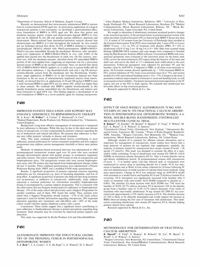
1Department of Anatomy, School of Medicine, Zagreb, CroatiaRecently we demonstrated that intravenously administered BMP-6 restores
bone in ovariectomized (OVX) aged rats. As intravenous application of a drug inosteoporotic patients is not convenient, we developed a gastrointestinal effica-cious formulation of BMP-6 in OVX aged rats. We show that gastric andduodenal enzymes, pepsin, trypsin and chymotrypsin degrade BMP-6 in vitroand can be inhibited by low pH and specific enzyme inhibitors, pepstatin andchymostatin. Matrix-assisted laser desorption/ionization analysis showed thatchymotrypsin cuts BMP-6 at 8 sites throughout the molecule. In vitro evertedgut sac technique showed that about 18-32% of BMP-6 chelated to mercapto-acetyltriglycine (MAG3) labeled with 99mTc-pertechnetate (BMP-6-MAG3-99mTc) can pass unmodified through the duodenum. However, after intraduo-denal (i.d.) application of BMP-6-MAG3-99mTc in vivo, only 0.04% of theapplied dose passed into the bloodstream of adult rats. On the contrary, new-born rats, with less duodenal enzymes, absorbed about 9% unmodified BMP-6protein of the total applied dose, suggesting an important role for a previouslyshowed excess of BMPs in the mother�s colostrum. Next, we showed that about35% of BMP-6-MAG3-99mTc applied i.d. to adult rats in acetate buffer pH 4.0and absorption enhancers, 1 mg thaurodeoxicolic acid and 1 mg DL-lauroyl-carnitin-chloride, passed from the duodenum into the bloodstream. Further-more, single application of BMP-6 i.d. in this formulation induced new boneformation in the rat assay of subcutaneously implanted inactive bone matrix.Finally, we showed that five i.d. applications of 10 and 100 lg/kg of BMP-6 oncea week increased BMD in aged OVX rats for about 10%, which was higher thanin sham animals. We conclude that BMP-6 applied intraduodenally in thisspecific formulation passes unmodified into the bloodstream and induces newbone formation in aged OVX rats. This finding supports a development of anoral formulation of BMP-6 as a novel anabolic agent for treating osteoporosis.
P480
IMPROVED PATIENT EDUCATION AND SUPPORT MAY
ENHANCE ADHERENCE TO BISPHOSPHONATE THERAPYM. A. Koay1, M. Walker*1, A. Corner1, F. Hammond2, A. Carr21Medical Department, Roche Products Ltd, Welwyn Garden City, 2 Clinimatrix,United Kingdom
Background: Long-term effective management of osteoporosis is hamperedby poor adherence to therapies. While this may be due to the silent and chronicnature of osteoporosis, it is also compounded by patients� concerns regarding theuse of medications and related side-effects. We propose that adherence to ther-apy may reflect patients� attitudes to disease and treatment.
Aims: This study examined patient-related factors contributing to pooradherence, patient preferences for various treatment modalities and how supportprogrammes may address current management shortfalls to better meet patientneeds.
Methods: A telephone-based structured interview was administered to 1083postmenopausal osteoporotic women aged over 50 years who were recruitedthrough advertisements placed with patient groups; in community magazinesand radio stations. The cohort comprised 550 women at-risk of osteoporosis andbisphosphonate naıve, 334 osteoporotic women who were current bisphospho-nate users; and 199 women who had lapsed from bisphosphonate therapy withinthe last 12 months. Three validated questionnaires were administered: a PatientPreference Rating Scale, PHIT and a modified EVOS study questionnaire.
Results: A significant proportion of patients expressed concerns regardingmedication use for osteoporosis e.g. fears of becoming dependent, and loss ofdrug effect. A significant proportion of patients also believed that drug treatmentwas un-necessary or ineffective in osteoporosis. Additionally, study subjectsexpressed a notional preference for intermittent (monthly) bisphosphonatedosing if accompanied by a patient support programme. This is consistent withthe observation that less frequent dosing improves adherence to bisphosphonatetherapy. 15% of the study cohort had previously used a Support Programme foranother condition, and of these, 94% found this useful. Thus, patients wereasked what a Support Programme should contain. �27% of the study cohortexpressed a need for further information regarding osteoporosis, 20% requestededucation regarding new treatments and side-effects and �26% of the studycohort would welcome regular telephone contact with a nurse.
Conclusions: These results suggest that a significant factor contributing topoor adherence to bisphosphonate therapy is patient perceptions regarding drugtreatment. Such obstacles may be overcome by improved patient support andeducation.
This study was supported by Roche Products Ltd and GlaxoSmithKline.
P481
ALENDRONATE IMPROVES THE STRUCTURAL GEOME-
TRY OF THE PROXIMAL FEMUR IN POSTMENOPAUSAL
OSTEOPOROTIC WOMENT. J. Beck*1, J. A. Cauley2, A. E. De Papp3, L. E. Wehren4, D. T. Baran5
1 Johns Hopkins Medical Institutions, Baltimore, MD, 2 University of Pitts-burgh, Pittsburgh, PA, 3 Merck Research Laboratories, Horsham, PA, 4MedicalCommunications, Merck Research Laboratories, Rahway, NJ, 5 Merck Re-search Laboratories, Upper Gwynned, PA, United States
We sought to determine if alendronate treatment produced positive changesin the structural geometry of the proximal femur in postmenopausal women witheither prevalent vertebral fractures (VF) or femoral neck BMD T-Scores less than-2.5. A subset of 319 women from the University of Pittsburgh clinic enrolled inthe Fracture Intervention Trial (FIT) were evaluated after 3 years (VF) or 4 years(BMD T-score <2.5; no VF) of treatment with placebo (PBO, N=155) oralendronate (ALN (5 mg/2 yrs; 10 mg/3-4 y) N=164). Hips were scanned usingHologic QDR2000 DXA scanners and scan images were compared to baselineusing the Hip Structure Analysis (HSA) software. Conventional BMD and cross-sectional geometries were evaluated at the narrowest point on the femur neck(NN), across the intertrochanteric (IT) region along the bisector of the neck andshaft axes and across the shaft at 1.5 x minimum neck width distal to the axesintersection. Follow-up parameters were adjusted for prevalent VF and thebaseline value by ANCOVA. Compared to PBO, at all three cross-sectional re-gions, ALN-treated women showed significantly (p < 0.05) increased BMD (4-6%), cortical thickness (4-7%), bone cross-sectional areas (4 to 7%), and sectionmoduli (4 to 8%) and reduced buckling ratios ()4 to )7%). Changes in the latter 3parameters indicate improvement in resistance to axial and bending loads, as wellas increased cortical stability. Treatment increased periosteal apposition at the ITregion, but not at the NN or shaft regions. We conclude that alendronate hasfavorable effects on hip structural geometry.
Research supported by Merck & Co., Inc.
P482
EFFECT OF ONCE-WEEKLY ALENDRONATE 70 MG AND
VITAMIN D3 2800 IU ON FRACTIONAL CALCIUM ABSORP-
TION IN POSTMENOPAUSAL OSTEOPOROTIC WOMEN: A 4-
WEEK, DOUBLE-BLIND, RANDOMIZED, CONTROLLED
MULTI-CENTER CLINICAL TRIALR. Robson*1, D. Kendler2, M. Handel3, S. Shapses4, Z. Yang5, T. Wilson5, M.Liu5, A. Mantz5, L. E. Wehren6, A. Santora51Christchurch Clinical Trials, Christchurch, New Zealand, 2 Osteoporosis Re-search Centre, Vancouver, BC, Canada, 3 Prince of Wales Hospital, Randwick,NSW, Australia, 4 Rutgers University, New Brunswick, NJ, 5 6Medical Com-munications, Merck Research Laboratories, Rahway, NJ, United States
Although vitamin D is essential for efficient calcium absorption and isimportant for management of osteoporosis, recent studies have shown that alarge minority of patients do not regularly take supplements; similarly, themajority of patients treated for osteoporosis have serum vitamin D levels <30ng/mL (75 nmol/L). This study was designed to examine the effect of a once-weekly tablet containing 70 mg of alendronate and vitamin D3 2800 IU (ALND2800) on fractional calcium absorption (FCA). After a 4-week placebo run-inand dietary stabilization period, 56 postmenopausal women with osteoporosis(T-score £ )2 at lumbar spine, total hip, femoral neck, or trochanter) wererandomized to receive drug or matching placebo for 4 weeks. FCA was mea-sured at baseline and at Week 4 from urines collected for 10 hours following IVand oral administration of stable isotopes of calcium (dual tracer method), usingmass spectrometry. Change in FCA was analyzed using an ANCOVA modelwith treatment as a model factor and baseline FCA and 25-hydroxyvitamin D ascovariates. FCA absorption was significantly increased from baseline after 4weeks of treatment with once-weekly ALN D2800 compared to placebo (p <0.001). The absolute LS mean increase with ALN D2800 was 7.0% from thebaseline of 30.8% (22.7% relative increase); FCA decreased 1.6% in the placebogroup from a baseline value of 31.6% (5.1% relative decrease). Four weeks oftreatment with once-weekly alendronate 70 mg vitamin D3 2800 IU was asso-ciated with approximately a 23% relative increase in FCA. The absolute increaseof 7.0% is likely to be clinically meaningful and would support the increase inBMD observed during the first year of treatment with alendronate. This medi-cation containing alendronate and vitamin D3 improves FCA, thereby helpingpatients absorb more calcium.
Research supported by Merck & Co., Inc.
P483
METHODOLOGY FOR DETERMINATION OF FRACTIONAL
CALCIUM ABSORPTIONR. Sherrell*1, P. Field1, S. Shapses1, R. Robson2, M. Liu3, W. Bayne3, E.Woolf3, L. E. Wehren4, A. Santora31 Rutgers University, New Brunswick, NJ, United States, 2 Christchurch ClinicalTrials, Christchurch, New Zealand, 34Medical Communications, Merck ResearchLaboratories, Rahway, NJ, United States
Abstracts S163
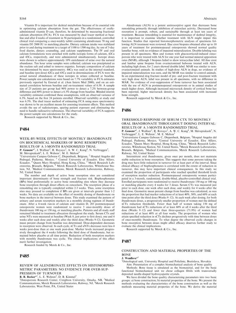
Vitamin D is important for skeletal metabolism because of its essential rolein optimizing calcium absorption from the gut. The effectiveness of orally-administered vitamin D can, therefore, be determined by measuring fractionalcalcium absorption (FCA). FCA was measured by dual tracer method at base-line and after 4 weeks of treatment in 56 participants in a randomized, controlledclinical trial comparing a once-weekly tablet containing alendronate 70 mg andvitamin D3 2800 IU to matching placebo. Dietary calcium intake was stabilizedprior to and during treatment to a target of 1100 to 1300 mg/day, by use of 3-dayfood diaries, dietary counseling, and calcium supplements. The IV and oralcalcium formulations were enriched with stable isotopic forms of calcium, 42Caand 43Ca, respectively, and were administered simultaneously. Isotope doseswere chosen to achieve approximately 10% enrichment of urine over the naturalabundance. Ten hour urine samples were collected; calcium was precipitated asthe oxalate salt and ashed to remove organics. Isotopic composition of calciumwas determined by mass spectrometry. 44Ca was used as the internal standard,and baseline (pre-dose) 42Ca and 43Ca used in determinations of FCA were theactual natural abundances of these isotopes in urines collected at baseline.Power/sample size calculations were based on 7.7% variability in FCA estimatespreviously reported by Ensrud et al. (Ann Intern Med 2000), and on an esti-mated correlation between baseline and on-treatment values of 0.50. A samplesize of 25 patients per group had 90% power to detect a 7.2% between-groupdifference and 80% power to detect a 6.3% change from baseline. Blinded interimvariability estimates confirmed these assumptions, with an observed variance of6.3%, based on the first 24 patients enrolled. Observed variability at study endwas 6.5%. The dual tracer method of estimating FCA using mass spectrometrywas shown to be an excellent means for assessing treatment effects. This methodavoids the use of radioisotopes, sparing patient exposure and eliminating theneed to handle radioactive substances. The observed variability of FCA supportsthe power/sample size calculations for the study.
Research supported by Merck & Co., Inc.
P484
WEEK-BY-WEEK EFFECTS OF MONTHLY IBANDRONATE
ON BIOCHEMICAL MARKERS OF BONE RESORPTION:
RESULTS OF A 3-MONTH RANDOMIZED TRIALP. Geusens*1, J. Walliser2, R. Rovayo3, A. W. C. Kung4, N. Verbruggen5,M. Shivaprakash6, L. E. Wehren7, M. E. Melton61Universitaire Campus Gebouw C, Diepenbeek, Belgium, 2 Hospial Angeles delPedregal, Padierna, Mexico, 3 Central University of Ecuador, Eloy Alfaro,Ecuador, 4 Queen Mary Hospital, Hong Kong, China, 5 Merck Research Lab-oratories, Brussels, Belgium, 6 Merck Research Laboratories, Whitehouse Sta-tion, NJ, 7Medical Communications, Merck Research Laboratories, Rahway,NJ, United States
The number and depth of active bone resorption sites are consideredimportant determinants of bone strength and fracture risk. Bisphosphonates(BPs) bind preferentially to active bone remodeling sites where they decreasebone resorption through direct effects on osteoclasts. The resorption phase of aremodeling site is typically completed within 2-3 weeks. Thus, some resorptionsites may proceed to completion if BPs are given at intervals greater than 1-2weeks. No data are available on the effect of monthly dosing of ibandronate onbone markers in the interval between doses. This study examined the pattern ofurinary and serum resorption markers in a monthly dosing regimen of ibandr-onate. After a 4-week run-in of calcium and vitamin D, 203 postmenopausalosteoporotic women were randomized to receive 3 once-monthly doses ofibandronate 100 mg or 150 mg, or matching placebo. Patients and all study staffremained blinded to treatment allocation throughout the study. Serum CTx andurine NTx were measured at baseline (Week 0, just prior to first dose), one and 4weeks after each dose and weekly after the third dose (Weeks 9-12). Geometricmean percent change from baseline was determined. Dose-dependent decreasesin resorption were observed. In each cycle, sCTx and uNTx decreases were less 4weeks post-dose than at one week post-dose. Marker levels increased progres-sively throughout the 4 weeks following the third dose of ibandronate, but re-mained below placebo at all time points. Reduction of both resorption markerswith monthly ibandronate was cyclic. The clinical implications of this effectmerit further investigation.
Research funded by Merck & Co., Inc.
P485
REVIEW OF ALENDRONATE EFFECTS ON HISTOMORPHO-
METRIC PARAMETERS: NO EVIDENCE FOR OVER-SUP-
PRESSION OF TURNOVERR. R. Recker*1, L. E. Wehren2, D. B. Kimmel31Osteoporosis Research Center, Creighton University, Omaha, NB, 2MedicalCommunications, Merck Research Laboratories, Rahway, NJ, 3Merck ResearchLaboratories, West Point, PA, United States
Alendronate (ALN) is a potent antiresorptive agent that decreases boneremodeling primarily through inhibition of osteoclast activity. Decreased boneresorption is prompt, robust, and sustainable through at least ten years oftreatment. Because remodeling is essential for maintenance of skeletal integrity,it is important to examine whether treatment with ALN might impair thisfunction. Studies that reported histomorphometric analyses of specimens fol-lowing ALN treatment were reviewed. Iliac crest biopsies obtained after up to 10years of treatment for postmenopausal osteoporosis showed normal qualitylamellar bone, with no evidence of impaired mineralization. Double-labeling waspresent in all specimens. Men and women with glucocorticoid-induced osteo-porosis who were treated with ALN for one year had normal mineral appositionrates (MAR), although 3 biopsies failed to show tetracycline label. All iliac crestand lumbar spine biopsies from ovariectomized baboons treated with ALN,including high doses, for 2 years showed double label. MAR was similar to non-ovariectomized animals. In studies of high-dose ALN in dogs, no evidence ofimpaired mineralization was seen, and the MAR was similar to control animals.In an experimental dog fracture model of pre- and post-fracture treatment withvery high dose ALN, label was present in all specimens, with no difference inMAR. No evidence of over-suppression of bone turnover has been associatedwith the use of ALN in postmenopausal osteoporosis or in animal studies ofmuch higher doses. Although increased microcrack density of cortical bone hasbeen reported, higher microcrack density has been associated with increasedbone strength.
Research supported by Merck & Co., Inc.
P486
THRESHOLD RESPONSE OF SERUM CTX TO MONTHLY
ORAL IBANDRONATE THROUGHOUT DOSING INTERVAL:
RESULTS OF A 3-MONTH RANDOMIZED TRIALP. Geusens*1, J. Walliser2, R. Rovayo3, A. W. C. Kung4, M. Shivaprakash5, N.Verbruggen6, L. E. Wehren7, M. E. Melton51Universitaire Campus Gebouw C, Diepenbeek, Belgium, 2Hospial Angeles delPedregal, Padierna, Mexico, 3Central University of Ecuador, Eloy Alfaro,Ecuador, 4Queen Mary Hospital, Hong Kong, China, 5Merck Research Labo-ratories, Whitehouse Station, NJ, United States, 6Merck Research Laboratories,Brussels, Belgium, 7Medical Communications, Merck Research Laboratories,Rahway, NJ, United States
Intermittent oral ibandronate, given once per month, does not maintainstable reduction in bone resorption. This suggests that some persons taking thedrug may have little reduction in turnover for at least part of the interval. Sinceclinical efficacy of bisphosphonates is correlated with the extent of reduction inturnover, this may have important clinical consequences. In this study weexamined the proportion of participants who reached specified threshold levelsof resorption marker reduction. Postmenopausal osteoporotic women partici-pated in a 3-month, randomized, double-blind, placebo-controlled clinical trial.Enrolled participants were randomized to receive ibandronate 150 mg or 100 mgor matching placebo every 4 weeks for 3 doses. Serum CTx was measured justprior to each dose, one week after each dose, and weekly for 4 weeks after thefinal dose. Geometric mean percent change from baseline was calculated, as wasthe proportion that had marker reductions of at least 40%, 50%, and 60% at eachtime point after the third dose (Weeks 9–12). At each week after dosing, for bothibandronate doses, a progressively smaller proportion of women met the definedsCTx reduction thresholds. Fewer than half of women taking 150 mg ofibandronate had sCTx reductions of at least 60% at all 4 weeks after the thirddose (Weeks 9–12) and fewer than three-quarters (71.9%) of women hadreductions of at least 40% at all four weeks. The proportion of women whoattain specified reduction in sCTx declines progressively with time between dosesof once-monthly ibandronate. To what degree the observed cyclic changes inbone resorption between doses might affect efficacy deserves further study toevaluate the clinical implications.
Research supported by Merck & Co., Inc.
P487
CONSTRUCTION AND MATERIAL PROPERTIES OF THE
BONEJ. Wendlova*11Osteological unit, University Hospital and Policlinic, Bratislava, Slovakia
Aim: Presentation of a complex biomechanical analysis of bone qualityMethods: Bone tissue is simulated as the biomaterial, and for the basic
functional biomechanical unit we chose collagen fibrils with transversallydeposited needle-shaped hydroxyapatite crystals.
We have divided the bone quality characterizing parameters into two basicgroups: a) bone construction; b) material properties of the bone. We present themethods evaluating the characteristics of the bone construction as well as themethods measuring material properties of the bone. We derive the material
S164 Abstracts
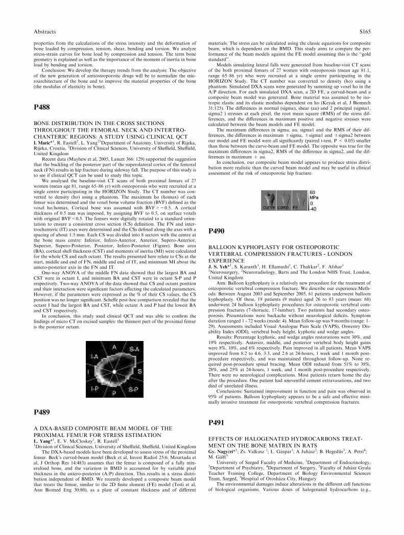
properties from the calculations of the stress intensity and the deformation ofbone loaded by compression, tension, shear, bending and torsion. We analyzestress-strain curves for bone load by compression and tension. The term bonegeometry is explained as well as the importance of the moment of inertia in boneload by bending and torsion.
Conclusion: We develop the therapy trends from the analysis: The objectiveof the new generation of antiosteoporotic drugs will be to normalize the mic-roarchitecture of the bone and to improve the material properties of the bone(the modulus of elasticity in bone).
P488
BONE DISTRIBUTION IN THE CROSS SECTIONS
THROUGHOUT THE FEMORAL NECK AND INTERTRO-
CHANTERIC REGIONS: A STUDY USING CLINICAL QCTI. Maric*1, R. Eastell2, L. Yang21Department of Anatomy, University of Rijeka,Rijeka, Croatia, 2Division of Clinical Sciences, University of Sheffield, Sheffield,United Kingdom
Recent data (Mayhew et al, 2005, Lancet 366: 129) supported the suggestionthat the buckling of the posterior part of the superolateral cortex of the femoralneck (FN) results in hip fracture during sideway fall. The purpose of this study isto see if clinical QCT can be used to study this topic.
We analysed the baseline-visit CT scans of both proximal femurs of 27women (mean age 81, range 65–86 yr) with osteoporosis who were recruited at asingle centre participating in the HORIZON Study. The CT number was con-verted to density (ho) using a phantom. The maximum ho (homax) of eachfemur was determined and the voxel bone volume fraction (BVF) defined as thevoxel ho/homax. Cortical bone was assumed with BVF>=0.5. A corticalthickness of 0.5 mm was imposed, by assigning BVF to 0.5, on surface voxelswith original BVF<0.5. The femurs were digitally rotated to a standard orien-tation to ensure a consistent cross section (CS) definition. The FN and inter-trochanteric (IT) axes were determined and the CSs defined along the axes with aspacing of about 1.5 mm. Each CS was divided into 8 sectors with the centre atthe bone mass centre: Inferior, Infero-Anterior, Anterior, Supero-Anterior,Superior, Supero-Posterior, Posterior, Infero-Posterior (Figure). Bone area(BA), cortical shell thickness (CST) and moments of inertia (MI) were calculatedfor the whole CS and each octant. The results presented here relate to CSs at thestart, middle and end of FN, middle and end of IT, and minimum MI about theantero-posterior axis in the FN and IT.
One-way ANOVA of the middle FN data showed that the largest BA andCST were in octant I, and minimum BA and CST were in octant S-P and Prespectively. Two-way ANOVA of the data showed that CS and octant positionand their interaction were significant factors affecting the calculated parameters.However, if the parameters were expressed as the % of their CS values, the CSposition was no longer significant. Scheffe post-hoc comparison revealed that theoctant I had the largest BA and CST, while octant A and P had the lowest BAand CST respectively.
In conclusion, this study used clinical QCT and was able to confirm thefindings of micro CT on excised samples: the thinnest part of the proximal femuris the posterior octant.
P489
A DXA-BASED COMPOSITE BEAM MODEL OF THE
PROXIMAL FEMUR FOR STRESS ESTIMATIONL. Yang*1, E. V. McCloskey1, R. Eastell11Division of Clinical Sciences, University of Sheffield, Sheffield, United Kingdom
The DXA-based models have been developed to assess stress of the proximalfemur. Beck’s curved-beam model (Beck et al, Invest Radiol 25:6; Mourtada etal, J Orthop Res 14:483) assumes that the femur is composed of a fully min-eralised bone, and the variation in BMD is accounted for by variable pixelthickness in the antero-posterior (A/P) direction. This results in a stress distri-bution independent of BMD. We recently developed a composite beam modelthat treats the femur, similar to the 2D finite element (FE) model (Testi et al,Ann Biomed Eng 30:80), as a plate of constant thickness and of different
materials. The stress can be calculated using the classic equations for compositebeam, which is dependent on the BMD. This study aims to compare the per-formance of the beam models against the FE model assuming this is the ‘‘goldstandard’’.
Models simulating lateral falls were generated from baseline-visit CT scansof the both proximal femurs of 27 women with osteoporosis (mean age 81.1,range 65–86 yr) who were recruited at a single centre participating in theHORIZON Study. The CT number was converted to density (ho) using aphantom. Simulated DXA scans were generated by summing up voxel ho in theA/P direction. For each simulated DXA scan, a 2D FE, a curved-beam and acomposite beam model was generated. Bone material was assumed to be iso-tropic elastic and its elastic modulus dependent on ho (Keyak et al, J Biomech31:125). The differences in normal (sigma), shear (au) and 2 principal (sigma1,sigma2 ) stresses at each pixel, the root mean square (RMS) of the stress dif-ferences, and the differences in maximum positive and negative stresses werecalculated between the beam models and FE model.
The maximum differences in sigma, au, sigma1 and the RMS of their dif-ferences, the differences in maximum ±sigma, ±sigma1 and ±sigma2 betweenour model and FE model were all significantly (paired t-test, P < 0.05) smallerthan those between the curve-beam and FE model. The opposite was true for themaximum differences in sigma2, RMS of the difference in sigma2, and the dif-ferences in maximum ± au.
In conclusion, our composite beam model appears to produce stress distri-bution more realistic than the curved beam model and may be useful in clinicalassessment of the risk of osteoporotic hip fracture.
P490
BALLOON KYPHOPLASTY FOR OSTEOPOROTIC
VERTEBRAL COMPRESSION FRACTURES - LONDON
EXPERIENCEJ. S. Yeh*1, S. Karanth1, H. Ellamushi1, C. Thakkar2, F. Afshar11Neurosurgery, 2Neuroradiology, Barts and The London NHS Trust, London,United Kingdom
Aim: Balloon kyphoplasty is a relatively new procedure for the treatment ofosteoporotic vertebral compression fracture. We describe our experience.Meth-ods: Between August 2003 and September 2005, 61 patients underwent balloonkyphoplasty. Of these, 19 patients (9 males) aged 26 to 83 years (mean: 68)underwent 24 balloon kyphoplasty procedures for osteoporotic vertebral com-pression fractures (7-thoracic, 17-lumbar). Two patients had secondary osteo-porosis. Presentations were backache without neurological deficits. Symptomduration ranged 1 - 72 weeks (mode: 4). Mean follow-up was 9 months (range: 1–29). Assessments included Visual Analogue Pain Scale (VAPS), Oswestry Dis-ability Index (ODI), vertebral body height, kyphotic and wedge angles.
Results: Percentage kyphotic, and wedge angles restorations were 30%, and19% respectively. Anterior, middle, and posterior vertebral body height gainswere 8%, 10%, and 6% respectively. Pain improved in all patients. Mean VAPSimproved from 8.2 to 4.6, 3.3, and 2.6 at 24-hours, 1 week and 1 month post-procedure respectively, and was maintained throughout follow-up. None re-quired post-procedure spinal bracing. Mean ODI reduced from 51% to 38%,28%, and 25% at 24-hours, 1 week, and 1 month post-procedure respectively.There were no neurological complications. Most patients return home the dayafter the procedure. One patient had uneventful cement extravasations, and twodied of unrelated illness.
Conclusions: Sustained improvement in function and pain was observed in95% of patients. Balloon kyphoplasty appears to be a safe and effective mini-mally invasive treatment for osteoporotic vertebral compression fractures.
P491
EFFECTS OF HALOGENATED HYDROCARBONS TREAT-
MENT ON THE BONE MATRIX IN RATSGy. Nagyeri*1; Zs. Valkusz 1; L. Gaspar1; A Juhasz2; B. Heged}us3, A. Petri4;M. Galfi5
University of Szeged Faculty of Medicine, 1Department of Endocrinology,2Department of Psychiatry, 4Department of Surgery, 5Faculty of Juhasz GyulaTeacher Training College, Department of Biology Environmental SciencesTeam, Szeged, 3Hospital of Oroshaza City, Hungary
The environmental damages induce alterations in the different cell functionsof biological organisms. Various doses of halogenated hydrocarbons (e.g.,
Abstracts S165
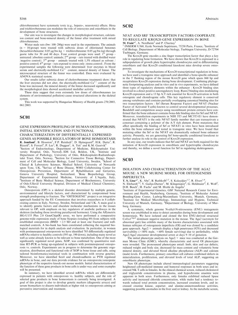
chlorobenzenes) have systematic toxic (e.g., hepato-, neurotoxic) effects. Hexaand trichlorobenzenes can modulate the role of osteocytes and osteoblasts in thedevelopment of bone structures.
Our aim was to investigate the changes in morphological structure, calcium-ion content and bone-mineral density of the femur after treatment with mixedchlorobenzenes.
Wistar male rats (bw: 100–280g) were used in the experiments. The animals(n = 10/group) were treated with subtoxic doses of chlorinated benzenes(hexachlorobenzene: 0.05 lg/bw.kg + trichlorobenzene: 0.05 lg/bw.kg) throughgasric tube for 30 and 60 days. Four control groups were used: 1st group -untreated /absolute control/ animals; 2nd group – rats treated with distilled water/negative control/; 3rd group – animals treated with 1,5% ethanol as solvent /positive control; 4th group – rats exposed to stress only /stress control/. From theexperimental samples the following were determined: liver enzymes in bloodserum and electrically-mobilized Ca2+ content, mineral density of femur. Themicroscopical structure of the femur was controlled. Data were evaluated byANOVA statistical system.
Our results (after subtoxic doses of cholorobenzenes treatment) show: thatthe liver enzymes did not alter, the electrically-mobilized Ca2+ content of thebone was increased, the mineral density of the femur decreased significantly andthe morphological data showed accelerated medullar activity.
These data suggest that even extremely low doses of chlorobenzenes (aselements of environmental pollution) cause structural alterations in the skeletalsystem.
This work was supported by Hungarian Ministry of Health grants 270/2003,061/2003.
SC01
GENE EXPRESSION PROFILINGOFHUMANOSTEOPOROSIS;
INITIAL IDENTIFICATION AND FUNCTIONAL
CHARACTERIZATION OF DIFFERENTIALLY EXPRESSED
GENES AS POSSIBLE REGULATORS OF BONE HOMEOSTASISR. Jemtland1, B. Houston2, F.P. Reinholt3, D. Heinegaard4, H.K. Datta5, R.Rizzoli6, S. Ferrari6, P. Lee2, R. Duggan2, A. Teti7 and K.M. Gautvik81Section of Endocrinology, Department of Medicine, Rikshospitalet Uni-versity Hospital, Oslo, Norway,2 IDS Ltd, Boldon, UK, 3Institute andDepartment of Pathology, University of Oslo, Rikshospitalet-Radiumhospi-talet Trust, Oslo, Norway, 4Section for Connective Tissue Biology, Depart-ment of Cell and Molecular Biology, Lund University, Sweden, 5School ofClinical & Laboratory Sciences, Medical School, University of Newcastleupon Tyne, UK, 6Service of Bone Disease, WHO Collaborating Center forOsteoporosis Prevention, Department of Rehabilitation and Geriatrics,Geneva University Hospital, Switzerland, 7Bone Biopathology Group,Department of Experimental Medicine, University of L’Aquila, Italy,8Institute of Basic Medical Science, Department of Biochemistry, Universityof Oslo/Ulleval University Hospital, Division of Medical Clinical Chemistry,Oslo, Norway.
Osteoporosis (OP) is a skeletal disorder determined by multiple genetic,environmental and lifestyle factors, and characterized by compromised bonestrength predisposing to fracture. The ‘‘OSTEOGENE’’ project is an integrativeapproach funded by the EU Commission that involves researchers in 8 collab-orating centres in Italy, Norway, Sweden, Switzerland and UK. A main goal isto identify genetic factors and elucidate molecular mechanisms in the tissuesrelevant to OP, with emphasis on key regulators of anabolic pathways in theskeleton (including those involving parathyroid hormone). Using the AffymetrixHG-U133 Plus 2.0 GeneChip(R) array, we have performed a comparativegenome-wide expression study of bone biopsies (totaling 89) from subjects withestablished osteoporosis (BMD <2.5 SD and at least one fracture) and well-matched controls. A bio-bank has been constituted collecting the required bio-logical materials for in depth analysis and evaluation. In particular, in womenwith postmenopausal osteoporosis we have identified 785 differentially regulatedmRNAs relative to healthy controls (595 up, 190 down), including many novel aswell as some already known to be relevant to bone metabolism. One of the mostsignificantly regulated novel genes, XOP, was confirmed by quantitative real-time RT-PCR as being up-regulated in subjects with postmenopausal osteopo-rosis vs. controls. Experiments are in progress to determine the genomic orga-nization, distribution and functional role of XOP in bone tissue and cells, usingtailored bioinformatics/statistical tools in combination with reverse genetics.Moreover, we have identified Sox4 and chondroadherin as PTH regulatedmRNAs in bone, and our data provide evidence for an osteoporotic/osteopenicphenotype of the respective knock-out mouse models. Initial characterization ofthe function of these gene products in bone cells is under way and some exampleswill be presented.
In summary, we have identified several mRNAs which are differentiallyexpressed in patients with osteoporosis vs. healthy subjects, and the role ofselected gene products in bone homeostasis is currently being investigated. Agoal of this project is also to develop genetic markers (diagnostic arrays) andserum biomarkers to discern individuals at higher risk to osteoporosis aiming atpreventive intervention and treatment.
SC02
NFAT AND SRF TRANSCRIPTION FACTORS COOPERATE
TO REGULATE KROX20 GENE EXPRESSION IN BONEM. Frain*1, A. Nordheim2 and P. Charnay11 INSERM U368, Ecole Normale Superieure, 75230 Paris, France, 2Institute ofCell Biology, Department of Molecular biology, Tuebingen University, D-72704Tuebingen, Germany.
The Krox20 gene encodes a zinc finger transcription factor that plays a keyrole in regulating bone formation. We have shown that Krox20 is expressed in asubpopulation of growth plate hypertrophic chondrocytes and in differentiatingosteoblasts and that Krox20 conditional knockout mice develop severe osteo-porosis.
To investigate the mechanisms of Krox20 transcriptional regulation in bone,we have used a transgenic mice approach and identified a bone-specific enhancerin the 5� flanking region of the mouse Krox20 gene which spans 860 bp andrecapitulates Krox20 expression during bone development. Combining phyloge-netic footprinting analyses and in vitro and in vivo experiments, we have definedthree types of regulatory elements within the enhancer : Krox20 binding sitesinvolved in a direct positive autoregulatory loop, Runx2 binding sites modulatingKrox20 expression and a 13 bp A/T rich essential for Krox20 activation in bothosteogenic and chondrogenic cells. This key regulatory element is conservedamong vertebrates and contains partially overlapping canonical binding sites fortwo transcription factors : Srf (Serum Response Factor) and NFAT (NuclearFactor of Activated T-cells) known to control several developmental processes.Bandshift and competition assays using recombinant protein extracts have con-firmed that the bone enhancer contains bona-fide binding sites for Srf and NFAT.Moreover, transfection experiments in NIH 3T3 and MC3T3-E1 have demon-strated that NFAT1 is the only NFAT family member that can transactivate aconstruct containing a polymer of the A/T rich element. Point mutations thataffect specifically the binding of Srf or NFAT1 in vitro have been introducedwithin the bone enhancer and tested in transgenic mice. We have found thatmutating either the Srf or the NFAT site dramatically reduced bone enhanceractivity. Presently, we are generating Srf bone-specific conditional knockout tofurther analyse its role in the control of Krox20 expression.
In any case, our results indicate that Srf and NFAT are both required for theinitiation of Krox20 expression in osteoblasts and hypertrophic chondrocytesand thereby, we define a novel function for Srf in regulating skeletogenesis.
SC03
ISOLATION AND CHARACTERIZATION OF THE AGA2
MOUSE: A NEW MURINE MODEL FOR OSTEOGENESIS
IMPERFECTAT. S. Lisse*1, K. Abe1, B. Rathkolb1,2, S. Kalaydjiev1,3, R. Elvert1,4,N. Ehrhardt1,4, V. Gailus-Durner1, M. Klingenspor4, G. Heldmaier4, E. Wolf2,D.H. Busch3, H. Fuchs1 and M. Hrabe de Angelis11Institute of Experimental Genetics, GSF National Research Center for Envi-ronment and Health, Neuherberg, Germany, 2Institute of Molecular AnimalBreeding/Gene Center, Ludwig-Maximilians University, Munich, Germany,3Institute for Medical Microbiology, Immunology and Hygiene, TechnicalUniversity of Munich, Germany, 4Department of Biology, University of Mar-burg, Germany.
A systematic, whole genome N-ethyl-N-nitrosourea (ENU) mutagenesisscreen was established in mice to detect anomalies during bone development andhomeostasis. We have isolated and cloned the first ENU-derived structuralCol1a1Aga2 dominant negative mutation in the mouse. The Aga2 (acronym forabnormal gait) line exhibits many of the known characteristics associated withhuman subtypes of osteogenesis imperfecta (OI) and was cloned by a candidategene approach. Aga2/+ animals display a high penetrance (92%) and decreasedsurvivability (<80% male, <60% female surviving) due to perilethality, whileAga2/Aga2 encounter developmental arrest at days 9–10 of gestation.
The initial phenotype analysis on Aga2/+ mice was conducted at the Ger-man Mouse Clinic (GMC), whereby characteristic and novel OI phenotypeswere revealed. The pronounced phenotypes entail: limb, skin and eye defects,reduced weight and body size, decreased fat mass content and volumetric bonemineral density, and elevated blood alkaline phosphatase (ALP) and calcium(male) levels. Furthermore, primary osteoblast cell cultures confirmed defects inmineralization, proliferation, and elevated levels of total ALP, suggesting anosteoblastic phenotype.
New OI phenotypes include altered immunological parameters suggestingheightened cell-mediated immune and humoral responses in both sexes and in-creased NK T cells in females. In the clinical chemical screen, reduced cholesteroland triglyceride concentrations in plasma, and hypochromic anaemia wereapparent in both sexes. Furthermore, only females exhibited reduced lipaseactivity and increased chloride concentration, while males had a tendency to-wards reduced total protein concentration, increased creatinin levels, and in-creased creatine kinase, aspartat- and alanine-aminotransferase activities,suggesting possible anomalies in the liver and/or kidney. In the metabolic screen,
S166 Abstracts

Aga2/+ animals displayed a metabolic phenotype in which mutant femalesshowed the highest energetic demand in relation to body weight. Female Aga2/+ animals were succumbed to a lowered body temperature, and higher energyuptake and metabolized energy values. It is proposed that these phenotypes maybe secondarily linked to the mutation, for example, during altered leukocytedevelopment in the bone marrow due to the bone defect cascade. Nevertheless,these results provide new insights into affected biological systems in OI.
It is surmised that the Col1a1Aga2 mutation affects procollagen processing,propagation and stability, and may interfere in concert with intermolecularcollagen–collagen and collagen–extracellular matrix protein interactions, therebyleading to the observed systemic structural and physiological clinical outcomes.The Aga2 OI line provides a unique opportunity in understanding one form ofhuman OI pathophysiology and phenotypic variability. We envision Aga2 as aviable OI line to apply therapeutic strategies such as antisense and/or cellreplacement approaches to provide insights toward management of a bonedisorder.
SC04
GENETIC DETERMINANTS OF OSTEOPOROSIS IN MENH.K. Datta1, S. Ferrari2, MC de Vernejoul3, M. Cohen-Solal3, R.M. Francis4,F.P. Reinholt5, R. Jemtland6, R. Duggan7, P.D.K Lee7, B. Houston7, A. Teti8,K.M. Gautvik9, R. Rizzoli21School of Clinical & Laboratory Sciences, Medical School, University ofNewcastle upon Tyne, UK, 2Service of Bone Disease, WHO CollaboratingCenter for Osteoporosis Prevention, Department of Rehabilitation and Geriat-rics, Geneva University Hospital, Switzerland, 3INSERM 349, Paris, France,4School of Clinical Medical Sciences, Medical School, University of Newcastleupon Tyne, UK, 5Institute and Department of Pathology, University of Oslo,Rikshospitalet-Radiumhospitalet Trust, Oslo, Norway, 6Section of Endocri-nology, Department of Medicine, Rikshospitalet University Hospital, Oslo,Norway, 7IDS Ltd, Boldon, UK, 8Bone Biopathology Group, Department ofExperimental Medicine, University of L’Aquila, Italy, 9Institute of Basic Med-ical Science, Department of Biochemistry, University of Oslo / Ulleval Univer-sity Hospital, Division of Medical Clinical Chemistry, Oslo, Norway,9
Though less frequent than in women, osteoporosis is a recognized prob-lem in men. The lower fracture risk in men is related to a greater peak bonemass, a lack of accelerated bone loss at middle age, and a shorter lifeexpectancy. Peak bone mass has polygenic determinants, with distinct multi-ple chromosomal loci that influence skeletal development. In a pilot study, anassociation of BMD and plasma vitamin D binding protein (DBP) with mi-cro-satellite markers ((TAAAn)-Alu repeats) in the downstream of intron 8 ofDBP gene was shown. We present results of a larger study on 177 men,comprising healthy male subjects (62.4 ± 10.4yr; n = 121) and men withsymptomatic vertebral fractures (59.6 ± 13.6yr; n = 56), confirm these re-sults, showing relationship between certain DBP-Alu genotypes and reducedBMD and vertebral fractures. In the present study, we also found that thecommonest genotype in men with vertebral fractures was 8/10, while inhealthy subjects it was 10/10; and allele *10 was protective (odds ratio 0.39;95% CI 0.25-.64, p < 0.005).
LRP5 is a member of the low-density lipoprotein (LDL) receptor-relatedfamily that mediates Wnt signaling, and loss-of-function mutations in LRP5 areresponsible for autosomal recessive osteoporosis pseudoglioma syndrome. A
population-based study of five LRP5 polymorphisms with allele frequencies>2% found that a missense substitution in exon 9 (c.2047G>A, p.V667M) andhaplotypes based on exon 9 and exon 18 (c.4037C>T, p.A1330V) alleles weresignificantly associated with bone mass and projected area of vertebrae at thelumbar spine in adult males, but not females, accounting for up to 15% of thepopulation variance for these traits in men. Moreover, 1-year changes in lumbarspine bone mass and size in pre-pubertal boys were also significantly associatedwith these LRP5 variants, suggesting that LRP5 polymorphisms influence ver-tebral bone growth during childhood. To investigate whether LRP5 variants areimplicated in idiopathic male osteoporosis, we studied 78 men with low BMD(mean ± SD: 50 ± 16 yrs) and 86 controls (51 ± 10yrs). Osteoporosis wassignificantly associated with LRP5 haplotypes (p = 0.0036) independently ofage, weight, calcium intake, alcohol and tobacco consumption. Odds ratio forosteoporosis was 3.8 (95% CI 1.3–11.3, p < 0.001) in male carriers of haplotype3 (c.2047A-4037T, n = 20 cases and 12 controls) versus homozygous carriers ofhaplotype 1 (c.2047G-4037C, n = 42 cases and 61 controls). These data indicatea contribution LRP5 to peak bone mass in men and to a role of LRP5 variantson idiopathic osteoporosis in males.
SC05
20 NOVEL SKELETOGENESISRELATEDGENES DETECTED BY
COMPREHENSIVE ANALYSIS OF A RUNX2-/- MOUSE MODELJ. Hecht 1, V. Seitz *1, M. Urban *1, F Wagner 2, P.N. Robinson 3, A. Stiege 1,C. Dieterich 1, N. Brieske 1, C. Zwingmann 1, S. Stricker 1, U. Kornak
1,3
S. Mundlos1,31Max Planck Institute for Molecular Genetics, Ihnestr. 73 14195 Berlin, Ger-many, 2RZPD Deutsches Ressourcenzentrum fur Genomforschung GmbHHeubnerweg 6 D-14059 Berlin Germany, 3Institute for Medical Genetics, Cha-rite University Hospital, Humboldt University, Augustenburger Platz 1, 13353Berlin, Germany; *authors contributed equally to this work
Runx2 is an essential factor for skeletogenesis and heterozygous mutationsin the human RUNX2 gene are the cause for the skeletal malformation syn-drome cleidocranial dysplasia. Runx2-deficient mice lack hypertrophic cartilageand bone. We reasoned that comparison of the expression profiles of wildtypeand Runx2-/- mice should reveal new transcripts involved in cartilage and bonedevelopment. We isolated RNA from wildtype and Runx2-/- humeri from day14.5 embryos and compared their gene expression profiles. 71 differentially ex-pressed genes were identified by two independent oligonucleotide-microarrayhybridizations and quantitative RT-PCR experiments. Gene-Ontology analysisdemonstrated an enrichment of the differentially regulated genes in annotationsto terms such as extracellular, skeletal development, and ossification, providingevidence for the biological relevance for bone development of the genes identifiedby the present study.
In situ hybridization revealed co-expression with Runx2 for 44 differentiallyexpressed genes. Evolutionarily conserved Runx transcription factor bindingsites were detected in promoter regions of five genes not previously described asdirect Runx2 targets, in addition to already known Runx2 target genes. While 40of the 71 differentially expressed genes have a known role in bone and cartilage,we identified 18 known genes that have not yet been implicated in skeletaldevelopment and 13 entirely new transcripts. Most of these potentially novelskeletogenesis-genes showed skeletal expression, which makes a role in skeletaldevelopment and disease likely.
Abstracts S167