a possible molecular ancestor for mollusk apgwamide, insect adipokinetic hormone, and crustacean red...
TRANSCRIPT
A Possible Molecular Ancestor for Mollusk APGWamide, Insect AdipokineticHormone, and Crustacean Red Pigment Concentrating Hormone
Francisco Martınez-Perez,1 Arturo Becerra,1 Jesus Valdes,2 Samuel Zinker,3 Hugo Arechiga4
1 Posgrado en Ciencias Biologicas, Facultad de Ciencias, Universidad Nacional Autonoma de Mexico, Mexico, D.F., Mexico2 Departamento de Bioquımica, Centro de Investigacion y de Estudios Avanzados del IPN, Mexico, D.F., Mexico3 Departamento de Genetica y Biologıa Molecular, Centro de Investigacion y de Estudios Avanzados del IPN, Mexico, D.F., Mexico4 Division de Estudios de Posgrado e Investigacion, Facultad de Medicina, Universidad Nacional Autonoma de Mexico, Mexico, D.F., Mexico
Received: 9 February 2001 / Accepted: 3 October 2001
Abstract. Precursor structures of various members ofthe neuropeptide family adipokinetic hormone/red pig-ment concentrating hormone (AKH/RPCH) of mandibu-lar arthropods and the APGWamide family of molluskswere compared. Amino acid alignments showed a com-mon overall architecture (signal peptide, active peptide,related peptide), with a similar � helix–random coil sec-ondary structure. DNA sequence alignments revealedclose similarities between the genes encoding for thepeptides of the two families. The APGWamide genes arelarger than the AKH/RPCH genes. The sequence envi-ronment occupied by introns is similar in AKH/RPCHand APGWamide genes. Such similarities suggest thatthese peptide families might have been originated bygene rearrangements from a common ancestor havingeither an AKH/RPCH/APGWamide-like structure orboth an AKH/RPCH-like and an APGWamide-likestructures. In the former model, DNA fragments couldhave been gained when the ancestor evolved to mollusksand it could have lost nucleotides when the progressionto mandibular arthropods took place. In the secondmodel, AKH/RPCH-like structures could have beenfused during evolution toward mandibular arthropods,whereas in mollusks they could have been lost with thepossible amplification of the APGWamide-like structure.Loss of domains in exon 1 may have originated the sig-
nal peptide and the first codon of the active RPCH. Inexon 2, loss of domains possibly determined the junc-tions of codons 2 to 5 with the loss of a APGWamidecopy; exon 3 underwent fewer variations. The similarityof the mollusk APGWamide precursors is closer to thatof the RPCH family than the insect AKH family, indi-cating an earlier evolutionary departure.
Key words: Adipokinetic hormone — APGWamide— Crustaceans — Peptide evolution — Insects — Mol-lusks — Neuropeptides — Red pigment concentratinghormone
Introduction
Elucidation of the chemical structure of bioactive pep-tides disclosed the existence of structurally related fami-lies, some members of which are present in a wide va-riety of zoological groups while other members appear tobe unique to a given group. For instance, while peptidessuch as enkephalins have been identified in many spe-cies, from unicellulars to humans (Le Roith et al. 1982;O’Neill et al. 1988), the crustacean hyperglycemic hor-mone retains this denomination because it has beenfound only in crustaceans (Lacombe et al. 1999). Char-acterization of the peptide precursors, and more recentlycloning of the genes encoding them, allows us to exploresome molecular mechanisms underlying evolutionarytrends (Cerff 1995).
Correspondence to: Hugo Arechiga, Ciudad Universitaria, Mexico,D.F. 04510, Mexico; email: [email protected]
J Mol Evol (2002) 54:703–714DOI: 10.1007/s00239-001-0036-7
© Springer-Verlag New York Inc. 2002

Fig
.1.
Alig
nmen
tsof
the
amin
oac
idse
quen
ces
ofth
epr
ecur
sors
ofth
eA
KH
/RPC
Han
dA
PGW
amid
efa
mili
es.A
The
four
regi
ons
are
indi
cate
dby
line
sw
ith
diam
onds
(bot
tom
).T
hedi
ffer
ent
mot
ifs
ofea
chpr
ecur
sor
are
indi
cate
dby
line
sw
ith
circ
les
(top
).A
llac
tive
pept
ides
are
indi
cate
din
bold
face
.Se
quen
ces
corr
espo
ndin
gto
the
sign
alpe
ptid
ear
ere
sidu
es1–
85in
AK
H/R
PCH
and
resi
dues
1–54
inA
PGW
amid
e.T
heba
sic
amin
oac
ids
that
serv
eas
prot
eoly
ticsi
tes
are
unde
rlin
ed.
The
Cys
part
icip
atin
gin
prec
urso
rdi
mer
izat
ion
isco
mm
onto
all
spec
ies
(arr
owat
resi
due)
(226
)and
may
form
disu
lfid
ebo
nds
with
othe
rcys
tein
esin
the
rela
ted
pept
ide
orin
the
sign
alpe
ptid
e(i
tali
cs,b
oldf
ace)
(und
erli
ned)
.BA
llpr
ecur
sors
show
sim
ilar
poss
ible
seco
ndar
yst
ruct
ures
,with
an�
helix
(h)
(reg
ion
1)fo
llow
edby
ara
ndom
coil
(c)
(reg
ions
2–3)
upto
the
end
ofth
ere
late
dpe
ptid
e,w
hich
show
san
�he
lixfo
llow
edby
ara
ndom
coil
inth
eca
rbox
ylte
rmin
us(r
egio
n4)
(e,
exte
nded
stra
nd).
Sgr,
Schi
stoc
erca
greg
aria
;Si
n,Sc
hist
o-ce
rca
nita
ns;
Lm
i,L
ocus
tam
igra
tori
a;B
di,
Bla
beru
sdi
scoi
dali
s;D
me,
Dro
soph
ila
mel
ano-
gast
er;
Cm
a,C
arci
nus
mae
nas;
Csa
,Cal
line
ctes
sapi
dus;
Mse
,Man
duca
sext
a;A
ca,A
plys
iaca
lifo
rnic
a;L
st,
Lym
nea
stag
nali
s;M
ed,
Myt
ilus
edul
is.
704

705

In the present work, we have taken as a model aneuropeptide family to date ascribed only to mandibulararthropods, particularly crustaceans and insects, withsome differential features in each group. The crustaceanmember of the family is the red pigment concentratinghormone (RPCH), an octapeptide with the sequencepGlu–Leu–Asn–Phe–Ser–Pro–Gly–Trp–NH2 (Fernlundand Josefsson 1972). This structure is common to allcrustacean species in which it has been identified (Gausset al. 1990). The initial physiologic effect it was knownto exert, as indicated by its name, is the aggregation ofpigment granules in the tegumentary erythrophores, thusregulating their color intensity. Other physiologic func-tions for RPCH have been described, such as the aggre-gation of pigments in retinal cells, thus participating inthe control of photon flow to the retinal photoreceptors(Garfias et al. 1995), as well as in direct influences on theexcitability of central neurons and the control of motorpatterns (Swensen and Marder 2000).
In insects, a very similar peptide has been amply char-acterized, i.e., adipokinetic hormone (AKH). It has a pri-mary structure varying from 8 to 10 amino acid residues,depending on the species. The most commonly identifiedphysiologic effect of AKH is the control of lipid andcarbohydrate metabolism, although it has some otherfunctions (Gade et al. 1997). The first described structure(AKH I) is pGlu–Leu–Asn–Phe–Thr–Pro–Asn–Trp–Gly–Thr–NH2 (Stone et al. 1976), thus having the firstfour and the last amino acids in common with RPCH. Upto two other distinct sequences have been found in asingle species, for example, AKH II (pGlu–Leu–Asn–Phe–Ser–Ala–Gly–Trp–NH2) and AKH III (pGlu–Leu–Asn–Phe–Thr–Pro–Trp–Trp–NH2) from Locusta migra-toria. At present, the primary structures of 34 membersof the family have been elucidated in 75 species. Thirty-three AKHs of the three varieties have been identified ininsects, and only one RPCH structure in crustaceans(Gade et al. 1997; Lee et al. 2000). Of particular interestis that biological cross-reactivity has been demonstratedbetween members of these two groups; thus, AKH mayinduce pigment aggregation in crustaceans and RPCHelicits adipokinetic effects in insects (Mordue and Stone1977). These structural and functional similarities be-tween RPCH and AKH led to the notion of an RPCH/AKH family (Gade et al. 1997). Not all members appearto raise blood lipid levels in their native species; others,called hypertrehalosemic hormones (HTHs), stimulatethe synthesis of trehalose, the main blood carbohydratein many insect species. An example from the cockroachis a decapeptide with the following structure: pGlu–Val–Asp–Phe–Ser–Pro–Gly–Trp–Gly–Thr–NH2 (Hayes et al.1986). Other varieties have been identified (Gade et al.1997).
Study of the primary structure of peptides has provenuseful for disclosing structural–functional relationshipsand phylogenetic analyses of peptide hormones. Among
other spectroscopical techniques, circular dichroism(Paolillo et al. 1992; Brakch et al. 1993; Goldsworthy1994; Goldsworthy et al. 1997) has been used to deter-mine the amino acid secondary structure exposed to thesurface of prohormones, such as �-turn promoting se-quences (Rholam et al. 1986, 1990; Paolillo et al. 1992;Brakch et al. 1993) and � loops (Leszczynsky and Rose1986; Bek and Berry 1990; Rayne and O’Shea 1993).Comparisons of peptide hormone secondary structureamong distant zoological groups have been used in at-tempts to disclose similar functional activity (Golds-worthy 1994) or the ultimate localization of signal pep-tide amino acid sequences in Escherichia coli (Sjostromet al. 1987). Mathematical models have been put forwardto predict the relationships between hormone structuresand their potencies (Lee et al. 2000). Physiological andbiochemical assays include trehalose mobilization (Mi-chalik et al. 1998), activation of glycogen phosphorylasein Manduca (Ziegler et al. 1998) lipid mobilization(Gade 1993), and acetate uptake into fat bodies in locusts(Lee and Goldworthy 1995). Finally, comparison of pep-tide hormone primary and secondary structures amongdistant zoological groups has provided information forconstructing phylogenetic trees (Gade et al. 1994; Bo-gerd et al. 1995) and on evolutionary trends (Hoyle1999).
A tetrapeptide has been identified in various mollus-can species with the structure APGWamide, that is, withits last three residues in common with those in the Cterminus of RPCH (Kuroki et al. 1990; Croll et al. 1991).It appears to play an important role in reproductive be-havior. Its precursor has been sequenced (Smit et al.1992; Favrel and Mathieu 1996; Fan et al. 1997) andbears interesting similarities to those of RPCH and AKH,which are discussed later.
In the search for a possible common ancestor of theAKH/RPCH and APGWamide peptide families, aminoacid primary and secondary structures and the DNA se-quences of the genes coding for these peptides werecompared. Whether the sequence environments occupiedby introns in the AKH genes were conserved in theRPCH and in the APGWamide genes was considered asan additional comparative criterion.
The structure of the precursors of these peptides hasrevealed a common overall architecture, with an initialsignal peptide followed by the sequence of the activepeptide and ending in the related peptide (see Fig. 1).However, a great diversity of structures was apparent,and between them and the RPCH precursors, thus sug-gesting a great genetic diversity (Linck et al. 1993).Some of the genes encoding for the precursors of thethree AKHs have been cloned, and their architecture isquite similar (Bogerd et al. 1995).
The AKH/RPCH and APGWamide families are there-fore a good model system to pose questions such as, Arethese genes only present in arthropods or are there anysimilar genes in other zoological groups? In particular, it
706

is tempting to search for the possible existence of a com-mon ancestor within earlier phylogenetic groups. Thispossibility has been suggested (Noyes and Schaffer1993), as well as the likelihood that the two families havebeen derived from separate but convergent evolutionarylines (Smit et al. 1992).
In this paper, we present a comparative analysis of thestructure of the precursors of the members of these threepeptide families and their similarities and propose pos-sible routes for their diversification.
Materials and Methods
For structural analysis comparisons were made for the cDNA se-quences of AKH I, II, and III of Locusta migratoria (Bogerd et al.1995), AKH I and II of Schistocerca gregaria (Schulz-Allen et al.1989; Fisher-Lougheed et al. 1993), AKH I and II of Schistocercanitans (Noyes and Schaffer 1990), and HTH from Blaberus discoidalis(Lewis et al. 1997). Comparisons were made to the only availablecDNA sequence of RPCH precursors, Carcinus maenas (Linck et al.1993) and that of Callinectes sapidus (Klein et al. 1995). Sequencescontaining APGWamide were from Limnea stagnalis (Smit et al.1992), Mytilus edulis (Favrel and Mathieu 1996), and Aplysia califor-nica (Fan et al. 1997). The AKH gene structures considered were thosefrom Manduca sexta (Bradfield and Keeley 1989), Schistocerca nitans(Noyes and Schaffer 1993), and Drosophila melanogaster (Noyes et al.1995).
The alignments of the precursors were carried out using the pro-gram CLUSTAL W (Thompson et al. 1994) with a 0.05 gap station andgap window p 9. Phylogenetic trees were made with the average dis-tances tree, using PID (from CLUSTAL W). The secondary structurewas determined following the methods devised by Gibrat et al. (1987)and Geourjon and Deleage (1994).
Results and Discussion
Alignment of the Amino Acid Structures of AKH/RPCHand APGWamide Precursors
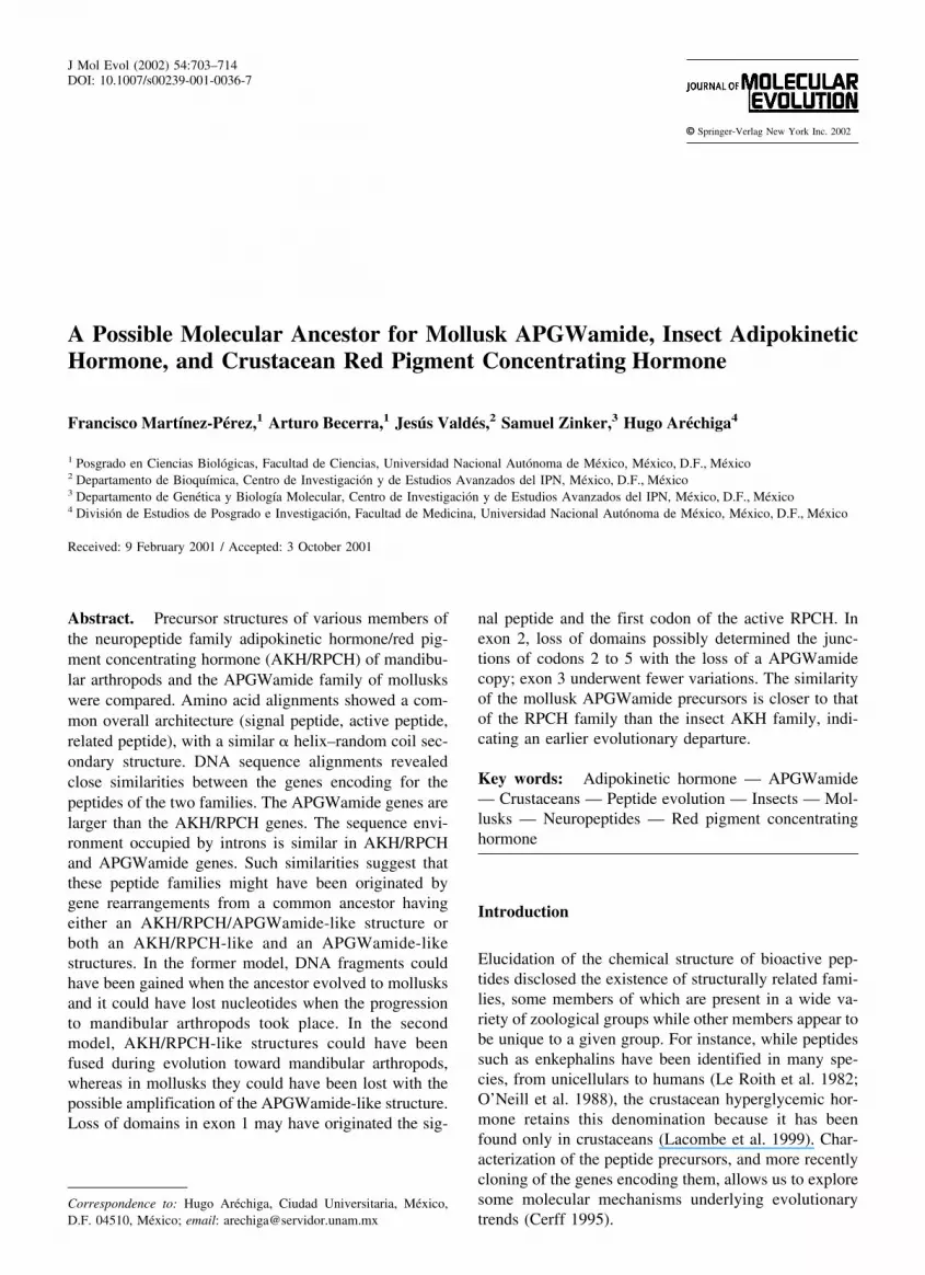
As shown in Fig. 1A, the alignment shows the existenceof the following four domains: the signal peptide (region1, residues 1–85), the active peptide (region 2, residues86–120), the APGWamide repeats (region 3, residues121–205), and the carboxyl terminus (region 4).
In mollusks, the region of the signal peptide includesthe first copy of the APGWamide; it is 43 to 57 residueslonger than those of insects and crustaceans, which aremore alike in this regard.
The second region contains the sequence coding forthe active peptide of AKH and RPCH and the secondAPGWamide copy (residues 86–94). This is followed bythe basic amino acids (residues 95–96 and 109–110),which are the cleavage sites for the processing of theprohormones. In addition, it contains 7–9 residues of theAKH-related peptide and the first 24 residues of theRPCH-related peptide. Among the AKH precursors, thisregion shows the greatest variations in size. While in D.melanogaster it is made up of 31 residues (including the
active peptide), in other insects it contains only 18–23residues. In this regard, crustaceans are more similar tomollusks, with 31–35 residues.
The third region shows the greatest differences amongthe three groups. While it contains most of the copies ofthe APGWamide stretch, only 20 amino acids corre-spond to the RPCH-related peptide in crustaceans andthe whole region is absent in insects.
The fourth region contains the last amino acids of theAKH and RPCH-related peptides, and a similar domainis present in APGWamide. It contains the cysteine (resi-due 226), which has been shown to be necessary forAKH–AKH dimerization, a prerequisite for the process-ing of the pro-AKH in L. migratoria (Fisher-Lougheed etal. 1993). Additionally, Cys 226 may form a disulfidebond with another Cys in the third domain of crustaceansand mollusks (residue 201) and in the AKHs of M. sexta,D. melanogaster, and B. discoidalis and the AKH III ofL. migratoria (residues 113 and 118, respectively).
To validate the aforementioned domains, an analysiswas made of the possible secondary structure of the pre-cursors of the AKH/RPCH family, following the meth-ods devised by Geourjon and Deleage (1994) and Gibratet al. (1987); both methods yielded the same results. Asshown in Fig. 1B, the structure of all precursors is quitesimilar in all the species analyzed. Actually, the commonstructure is an � helix comprising region 1; however, the� helix in the mollusk is interrupted by a random coilstructure (residues 20–44 and 54–74 in A. californica,residues 31–77 in L. stagnalis, and residues 15–28 and44–65 in M. edulis). Region 2 forms a random coil in allspecies but in mollusks the structure is interrupted by an� helix formed by residues 99–111. The random coilstructure is maintained in region 3 in crustaceans but atthe beginning of the region in mollusks an � helix exists(residues 121–134 in A. californica and L. stagnalis andresidues 141–154 in M. edulis). Finally, region 4 presentsone � helix and ends with a random coil structure in allprecursors.
As reported previously (Kuroki et al. 1990; Croll et al.1991), both analyses showed good primary and second-ary structural conservation of the signal peptide, activepeptide, and C-terminus regions. In particular, the lastthree amino acids of the physiologically active AKH/RPCH peptide are well conserved with the second repeatof the APGWamide peptides in all species studied (Figs.1A and B). For this reason, it was investigated whetherthis conservation is maintained at gene level.
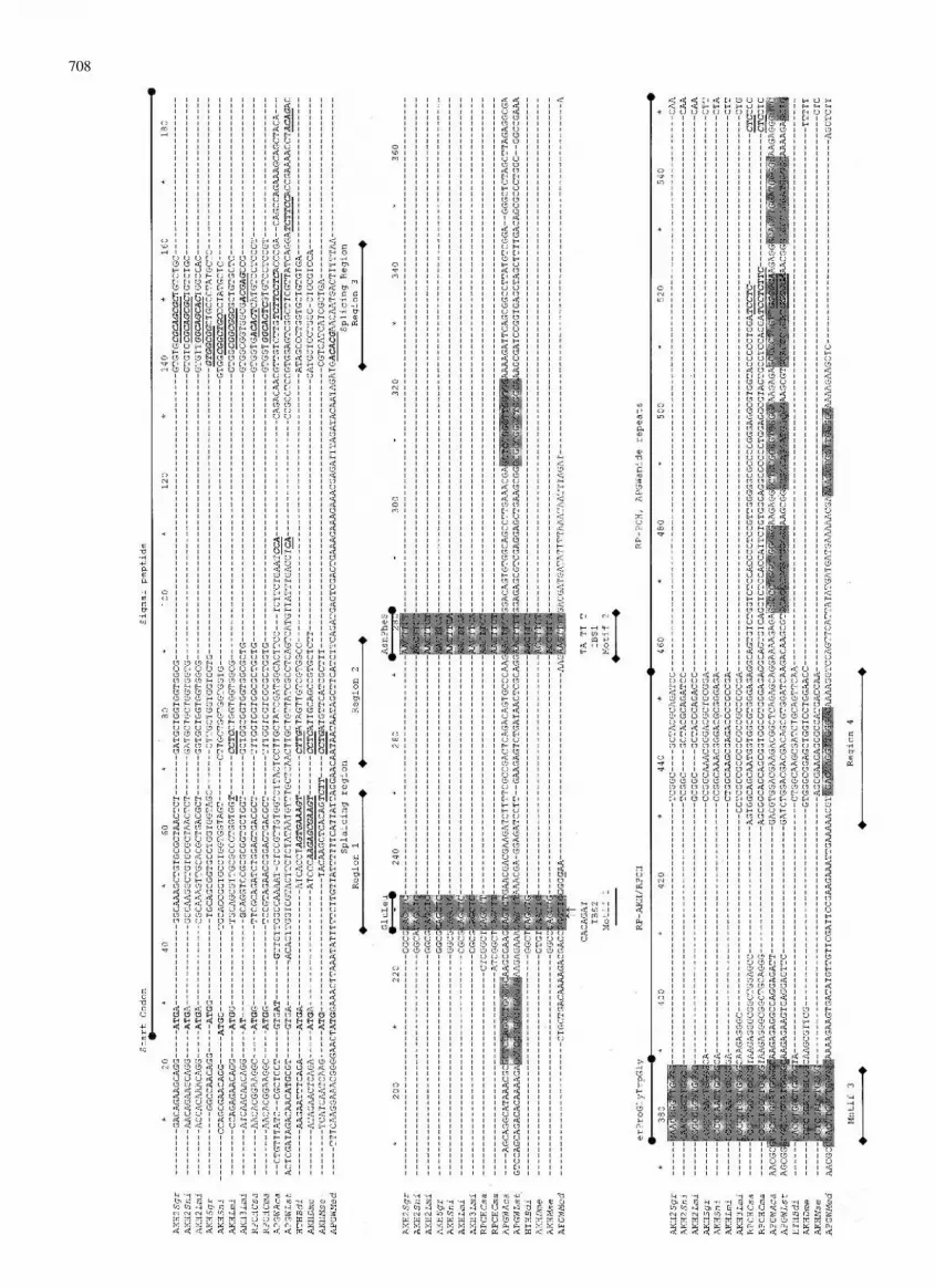
DNA Sequence Alignments of the Precursors
Figure 2 shows that the codons for the signal peptideconsist of three regions [nucleotides (nt), 1–223]: an im-mediate one, with those encoding for the last codon ofthe signal peptide, and the first two amino acids of AKHand RPCH (nt 224–232). This region is also present in
707
708

Fig
.2.
Alig
nmen
tof
the
nucl
eotid
ese
quen
cepr
ecur
sors
ofth
eA
KH
/RPC
Han
dA
PGW
amid
efa
mili
es.
The
hom
olog
ous
regi
ons
amon
gho
rmon
esA
KH
,R
PCH
,an
dA
PGW
amid
ear
ein
dica
ted
belo
wth
eal
ignm
ents
(lin
esw
ith
fill
eddi
amon
ds).
Not
eth
atm
otif
1co
rres
pond
sto
the
firs
ttw
oco
dons
ofA
KH
and
RPC
H,w
hile
mot
if2
cont
ains
codo
ns3
and
4an
dth
efi
rst
nucl
eotid
eof
codo
n5.
Fina
lly,
mot
if3
cont
ains
the
rem
aini
ngtw
onu
cleo
tides
ofco
don
5an
dco
dons
6–8
ofA
KH
,R
PCH
,an
dA
PGW
amid
e(b
oxed
).M
otif
s1
to3
are
indi
cate
dby
open
diam
onds
.T
hear
row
sun
dern
eath
indi
cate
the
nucl
eotid
esin
whi
chin
tron
sar
elo
caliz
edin
the
AK
Hge
nes
(ast
eris
ks
indi
cate
the
repo
rted
sequ
ence
sfo
rpr
ecur
sor
gene
s).
Imm
edia
tely
upst
ream
ofth
ese
exon
–exo
nju
nc-
tions
are
loca
ted
the
poss
ible
SRpr
otei
n-bi
ndin
gan
dsp
licin
g-en
hanc
erm
otif
s(i
tali
cs,
bold
face
,an
dun
derl
ined
).M
otif
s1
and
2al
soco
rres
pond
totw
opo
ssib
leco
nsen
sus
sequ
ence
sIB
S2an
dIB
S1fo
rin
sert
ion
orel
imin
atio
nof
grou
pII
self
-spl
icin
gin
tron
s.C
odon
sfo
rth
eac
tive
pept
ides
are
boxe
d.T
heC
ysco
don
for
prec
urso
rdi
mer
izat
ion
(nuc
leot
ides
730–
732)
isw
ell
cons
erve
d.Sp
ecie
sab
brev
iatio
nsar
eas
indi
cate
din
the
lege
ndto
Fig.
1.
709

the precursor of the three molluscan species (motif 1)and its consensus is CAGBTB (B � no adenine). Thecodons for amino acids 3 and 4 and the first nucleotide ofcodon 5 in AKH and RPCH (nt 274–280) are separatedby 45 nt in the precursors of A. californica and M. edulis.In this region, the consensus in the precursors of mol-lusks, crustaceans, and insects is AACTTCW (W �A/T) (motif 2). Finally, the codons for amino acids 6–8(nt 375–387) are separated by 94 nt in A. californica andL. stagnalis. This region contains the two nucleotides ofamino acid 6 and the first two nucleotides coding foramino acid 9 of AKH and RPCH (motif 3). The wholemotif has the consensus CVCCBDRNTGGGGN (R �A/T; N � any; B, D, and V � not A, C, and T, respec-tively) and includes the first copy of APGWamide of M.edulis and the third copy of APGWamide of A. califor-nica and L. stagnalis. After motif 3, there are two ho-mologous regions (nt 429–455 and 621–662) coding forthe RP-AKH/RPCH amino acids and most of the repeatsof APGWamide. The final portion of the nucleotide se-quence of the precursors is a region encoding for thetermination site, which show close similarities among allspecies. Regions 6, 7, and 8 (nt 680–689, 701–719, and737–776, respectively) show the greatest homology atthe 3� end of the precursor, in particular, region 6, whichcontains the intron of AKH I and II of S. nitans and the
codon for the Cys (nt 730) which participates in peptidedimerization.
The analysis we have presented thus far raises theissue of the origin of these genomic structures. At leasttwo possibilities can be considered: (1) a common an-cestor that could have had an AKH/RPCH/APGWamide-like structure, which possibly could have gained DNAfragments in the evolution to the molluscan lineage andcould have lost nucleotides when the progression to in-sects and crustaceans occurred; and (2) a common an-cestor that possibly had separate APGWamide-like andAKH/RPCH-like structures, which possibly could havefused during the evolutionary steps toward insects andcrustaceans, whereas in mollusks the former might havebeen amplified, losing the majority of the AKH/RPCH-like fragments.
These possibilities are further supported when joiningthe three motifs obtained from the alignment of thecDNAs of the various AKH, RPCH, and APGWamides(nt 227–232, 274–280, and 374–387 in Fig. 2). Figure 3shows the conceptual translation of this union, in whichthe APGWamide precursors in A. californica and L.stagnalis initiate the AKH/RPCH sequence. Two differ-ences were observed: (1) codon 2 is occupied by Phe inA. californica, while in L. stagnalis, crustaceans, andinsects a Val or Leu is substituted instead; and (2) in A.
Fig. 3. Sequence alignment of the regionsencoding for AKH and RPCH with theprecursors of the APGWamide. When linkingthe three motifs of nucleic acids conservedbetween the AKH/RPCH family andAPGWamide (indicated by arrows), it can beseen that the three mollusk species (boldface)contain the sequenced encoding AKH andRPCH. The conceptual translation of theAPGWamide of A. californica and L.stagnalis (in boldface) shows 91% identitywith RPCH and AKH. The only difference isin codon 2 of A. californica, in which acytosine substitutes for a thymine (see Fig.2). In the APGWamide precursor of M.edulis, the union of the three motifs resultsin a stop codon TGA (codon 5, labeled ***).Species abbreviations are as indicated in thelegend to Fig. 1.
710

californica and L. stagnalis, codon 5 is occupied by theTCG codon for Ser. A similar result can be obtained byrepeating the operation for M. edulis. The sequence for L.stagnalis is obtained, except for codon 1 (Gly) and theinterruption of the open reading frame by a TGA stopcodon in position 5.
The phylogenetic tree obtained from the amino acidprimary sequences of the precursors of the APGWamideand RPCH/AKH families agrees with our results andprevious findings (Gade et al. 1994; Bogerd et al. 1995).The tree shows that the APGWamide precursor of mol-lusks, although similar, appeared earlier than those ofcrustacean RPCH and insect AKH precursors (Fig. 4).
A second feature that allows us to think about DNArearrangements for the AKH/RPCH family and theAPGWamide precursors of any possible ancestor is theposition of the introns in the AKH gene. In D. melano-gaster, the gene has an intron between the first andthe second codons of the active peptide, while in theAKH genes of S. nitans, the intron is between codon20 and codon 21 (AKH I) and between codon 52 andcodon 53 (AKH II) of the AKH-related peptide (Noyesand Schaffer 1993); the AKH gene of M. sexta has nointrons. Furthermore, AKH and APGWamide precursoralignments showed that the nucleotide environment oc-cupied by introns is preserved in mollusks and crusta-ceans (Fig. 2).
Intron insertion might have occurred by reverse splic-ing of an excised intron (within a nonhomologousmRNA) followed by reverse transcription and homolo-gous recombination; in addition, it could have occurredby invasion of self-splicing Group II introns (from or-
ganelles) into the nuclear genome followed by muta-tion, transforming intron II into a nuclear intron. A sin-gle mutation of the sequence flanking Group II introns(U/CA. . .GU) is required to produce the canonic se-quence of nuclear introns (Rzhetsky et al. 1997).
A remarkable feature of the arrangement revealed bythe nucleotide alignments is that the regions containingthe AKH genes in D. melanogaster and S. nitans share anidentity with the IBS2 motif (nt 225–231 and 681–689 inFig. 2), which appears to be a region for insertion orelimination of self-splicing Group II introns (Morl andSchmelzer 1990; Yang et al. 1996). By assuming such afunction for these regions, and the fact that they havebecome nuclear introns, it is possible that even when theexon–exon junction site does not show 100% identitywith the IBS2 motif, regions sharing similarities may beconsidered echoes of the insertion or elimination of bothintrons in the precursors of APGWamide, AKH, andRPCH.
According to this assumption, the exons of AKHgenes lacking one or both introns might have containedsites for splicing enhancers and/or binding domains forsplicing auxiliary factors, which may have participatedin the processing of the pre-mRNA of the ancestralmolecule. Although such motifs no longer participate ina splicing process due to the lack of an intron, theirpresence could reflect indirect evidence of their role inthe processing of the eliminated intron. In this regard,all possible exons have the consensus proposed by Liu etal. (1998) and by Schaal and Maniatis (1999). The se-quence AGAGC (nt 56–60) is present in the first exon (nt9–229) of D. melanogaster. This is similar to the binding
Fig. 4. Phylogenetic tree of the AKH/RPCH and APGWamide families. The amino acid phylogenetic tree shows that molluscan APGWamideprecursors are closer to the possible ancestor than crustacean RPCH and insect AKH.
711

motif for SRp40 proteins (sequence consensus ACDGS,where S � G/C and D � not T) and a class II motif F(nt 65–76), characteristic of pyrimidine-rich enhancers(TCCTC). This motif is also in the same position in theAKH of L. migratoria, whereas M. sexta has a class IImotif E (TCTTC) in the same position, but the gene lacksexons (regions 1 and 2 in Fig. 2). Near the 3� end of thefirst exon in the D. melanogaster AKH precursor (region3), the S. nitans gene, which lacks an intron, has themotif SRSASGA (nt 142–148), similar to the bindingsite for SF2/AFS protein. This motif is also present inregion 3 of the precursors of S. gregaria, L. migratoria,and C. maenas. In this region, precursors of C. sapidus,L. stagnalis, M. edulis, and AKH III of L. migratoriahave motifs similar to those recognized by SRp40 pro-teins. A. californica and M. edulis precursors have se-quences resembling those of splicing enhancers, that is,class II motif E (Fig. 2).
In a similar fashion, at the 3� end of the second exonof AKH I and II of S. nitans (nt 683–684), what could bea class II motif F (nt 629–656) precedes the intron. Thismotif is also present in AKH I and II of S. gregaria andL. migratoria.
In region 5, motifs similar to the binding site ofSRp40 and class II motif D (TCTCC) are present in theprecursors of M. sexta and D. melanogaster, respec-tively. Neither species has introns in this position. Re-gion 5 bears homology with the AKH precursors of B.discoidalis, AKH III of L. migratoria, the RPCH precur-sors of C. sapidus and C. maenas, and the APGWamideprecursors of A. californica and L. stagnalis, in which apossible SRp40 motif is also present. Additionally, in M.edulis and in AKH I and II of S. gregaria and L. migra-toria, possible class I (GGGGA) and class II A motifs arepresent, respectively (Fig. 2).
Although no motif mentioned thus far has been testedexperimentally, these features might suggest that theORF in the ancestral gene could possibly have been con-stituted by three exons separated by two introns. If so,the first exon could have contained part of the 5�UTRend, the start codon, the nucleotides encoding the signalpeptide, and the first codon for RPCH. The second exoncould have encoded the APGWamide copies. It mighthave been localized between the nucleotides coding foramino acid residues 2–4 of RPCH, preceding one of theAPGWamide copies. Finally, the third exon might havecontained the last codons of the related peptide and the3�UTR.
From our analyses, we propose that the AKH/RPCHfamily could have originated from rearrangements of oneancestral gene (AKH/RPC/APGWamide-like) or by re-combination of two ancestral genes (AKH/RPCH-likeand APGWamide-like). In any event, loss of domains inthe first exon might have formed what later became thesignal peptide and the first codon for the Glu residue inRPCH. In the second exon, this loss of domains could
result in the union of the nucleotides forming amino acidresidues 2–5 with the subsequent copy of APGWamide,from which the amino acids forming RPCH are derived.The third exon possibly underwent fewer variations in allmolecular species considered in this study. Selection pres-sure acted to maintain the peptidic domain conservingthe required Cys for AKH dimerization and for the pro-cessing of the prohormone (Fischer-Lougheed et al. 1993).
Once the RPCH precursor was formed, the gene wasinherited in crustaceans and preserved in insects, inwhich more variations have appeared; in some species,an intron was lost, as is the case for D. melanogaster andS. nitans, while in others, such as M. sexta, both intronswere lost to form AKH. Future cloning of RPCH andAPGWamide genes will allow a more precise under-standing of intron movements in the AKH/RPCH family.
The search for the ancestral gene of AKH/RPCH andAPGWamide may be conducted in other mollusks, aswell as in other invertebrates. In the nematode Panagrel-lus redvivus, a peptide has been identified with physi-ological activity similar to that of the AKH/RPCH pep-tides (Davenport et al. 1991); however, its structure isstill unknown. To interpret these physiologic similarities,one must bear in mind that, although orthologous struc-tures or sequences may correspond to homogeneousmolecules stemming from a common molecular ances-tor, they do not necessarily retain the original func-tion(s) (Fitch 1970; Goldsworthy 1994). The neuropep-tides AKH, RPCH, and APGWamide are a goodexample of this functional diversification, because whileAPGWamide’s main known function appears to be theregulation of reproductive behavior and muscle control(Favrel and Mathieu 1996; Smit et al. 1992; Fan et al.1997), RPCH regulates pigment position and neuronalactivity (Garfias et al. 1995; Swensen and Marder 2000)and AKH controls lipid and carbohydrate metabolism(Stone et al. 1976). This functional diversity suggests anindependent evolution of the receptors to these peptidesand has been documented (Goldsworthy 1994; Hoyle1999): the same bioactive molecule carries out entirelydifferent functions in various phyla. The evidence of bio-logical cross-reactivity between these peptides suggeststhat some molecular similarities may be found amongreceptors (Mordue and Stone 1976, 1977; Dallman et al.1981). Some structure–function correlations have beenreported for members of the AKH/RPCH family (Lee etal. 2000), but to date sufficient information on receptorstructure is still lacking.
Given the fast rate at which information on the ge-nomic structure of various species is being accrued, ourscope for future comparisons will widen and more de-tailed results will be produced.
Acknowledgments. We thank Ma. Teresa Pacheco Reyes for her ex-pert secretarial assistance. This work was supported by Grants 30595-N(to J.V.) and 28089-N from the Consejo Nacional de Ciencia y Tec-nologıa, Mexico.
712

References
Bek E, Berry R (1990) Prohormonal cleavage sites are associated with� loops. Biochemistry 29:178–183
Bogerd J, Kooiman FP, Pijnenburg MAP, Hekking LHP, OudejansRCHM, Van der Horst DJ (1995) Molecular cloning of the threedistinct cDNAs, each encoding a different adipokinetic hormoneprecursor of the migratory locust, Locusta migratoria. J Biol Chem270:23038–23043
Bradfield JY, Keeley LL (1989) Adipokinetic hormone gene sequencefrom Manduca sexta. J Biol Chem 264:12791–12793
Brakch N, Rholam M, Boussetta H, Cohen P (1993) Role of �-turn inproteolytic processing of peptide hormone precursors at dibasicsites. Biochemistry 32:4925–4930
Cerff R (1995) In: Go M, Schimmel P (eds) Tracing biological evolu-tion in protein and gene structures. Elsevier, New York, pp 205–227
Croll RP, Van Minnen J, Smit AB, Kits KS (1991) APGWamide:Molecular, histological and physiological examination of a novelneuropeptide. In: Kitts KS, Boer HH, Joose J (eds). Molluscanneurobiology. North-Holland, Amsterdam, pp 248–254
Dallmann SH, Herman WS, Carlsen J, Josefsson L (1981) Adipokineticactivity of shrimp and locust peptide hormones in butterflies. GeneComp Endocrinol 43:256–258
Davenport TRB, Isaac RE, Lee DL (1991) The presence of peptidesrelated to the adipokinetic hormone/red pigment-concentrating hor-mone family in the nematode, Panagrellus redivivus. Gen CompEndocrinol 81:419–425
Fan X, Croll RP, Wu B, Fang L, Shen Q, Painter SD, Nagle GT (1997)Molecular cloning of a cDNA encoding the neuropeptidesAPGWamide and cerebral peptide 1: Localization of APGWamide-like immunoreactivity in the central nervous system and male re-productive organs of Aplysia. J Comp Neurol 387:53–62
Favrel P, Mathieu M (1996) Molecular cloning of a cDNA encodingthe precursor of Ala-Pro-Gly-Trp amide-related neuropeptides fromthe bivalve mollusk Mytilus edilus. Neurosci Lett 205:210–214
Fernlund P, Josefsson L (1972) Crustacean color change hormone:Amino acid sequence and chemical synthesis. Science 177:173–175
Fischer-Lougheed J, O’Shea M, Cornish I, Losberger C, Roluet E,Schulz-Allen MF (1993) AKH biosynthesis: Transcriptional andtranslational control of two co-localized prohormones. J Exp Biol177:223–241
Fitch WM (1970) Distinguishing homologous from analogous proteins.Syst Zool 19:99–113
Gade G (1993) Structure-activity-relationships for the lipid-mobiliza-tion action of further bioanalogs of the adipokinetic hormone redpigment-concentrating hormone family of peptides. J Insect Physiol39:375–383
Gade G, Reynolds SE, Beeching JR (1994) Molecular evolution ofpeptides of the AKH/RPCH family. In: Davey KG, Peter RE, TobeSS (eds). Perspectives in comparative endocrinology. National Re-search of Canada, Ottawa, pp 486–492
Gade G, Hoffmann KH, Spring JH (1997) Hormonal regulation ininsects: Facts, gaps, and future directions. Physiol Rev 77:963–1032
Garfias A, Rodrıguez-Sosa L, Arechiga H (1995) Modulation of cray-fish retinal function by red pigment concentrating hormone. J ExpBiol 198:1447–1454
Gauss G, Kleinholz LH, Kegel G, Keller R (1990) Isolation and char-acterization of red pigment-concentrating hormone (RPCH) fromsix crustacean species. J Comp Physiol 160B:373–379
Geourjon C, Deleage G (1994) SPOM: A self-optimized method forprotein secondary structure prediction. Protein Eng 7:157–164
Gibrat JF, Garnier J, Robson B (1987) Further developments of proteinsecondary structure prediction using information theory. J Mol Biol198:425–443
Goldsworthy GJ (1994) Adipokinetic hormones of insects: Are theyinsect glucagons? In: Davey KG, Peter RE, Tobe SS (eds). Per-spectives in comparative endocrinology. National Research ofCanada, Ottawa, pp 486–492
Goldsworthy GJ, Lee MJ, Luswata R, Drake AF, Hyde D (1997) Struc-tures, assays and receptors for Locust adipokinetic hormones.Comp Biochem Physiol 117:483–496
Hayes TK, Keeley LL, Knight DW (1986) Insect hypertrehalosemichormone: Isolation and primary structure from Blaberus discoidaliscockroaches. Biochem Biophys Res Commun 140:674–678
Hoyle CH (1999) Neuropeptide families and their receptors: Evolu-tionary perspectives. Brain Res 848:1–25
Klein JM, Mohrher CJ, Sleutels F, Janeke N, Riehm JP, Rao R (1995)A highly conserved red pigment concentrating hormone precursorin the blue crab Callinectes sapidus. Biochem Biophys Res Com-mun 212:151–158
Kuroki Y, Kanda T, Kubota I, Fujisawa Y, Ikeda T, Miura A, Mina-mitake Y, Muneoka YA (1990) Molluscan neuropeptide related tothe crustacean hormone RPCH. Biochem Biophys Res Commun167:273–279
Lacombe C, Greve P, Martin G (1999) Overview on the sub-groupingto the crustacean hyperglycemic hormone family. Neuropeptides33:71–80
Lee MJ, Goldsworthy GJ (1995) Acetate uptake assay: The basis for arapid method for determining potencies of adipokinetic peptides forstructure-activity studies. J Insect Physiol 41:163–170
Lee MJ, de Jong S, Gade G, Poulus C, Goldsworthy GJ (2000) Math-ematical modeling of insect neuropeptides potencies: Are quanti-tatively predictive models possible? Insect Biochem Mol Biol 30:899–907
Le Roith D, Shiloach J, Roth J (1982) Is there an earlier phylogeneticprecursor that is common to both the nervous and endocrine sys-tems? Peptides 3:211–215
Leszczynsky JF, Rose GD (1986) Loops in globular proteins: A novelcategory of secondary structure. Science 234:849–855
Lewis DK, Jezierski MK, Keeley LL, Bradfield JY (1997) Hypertre-halosemic hormone in a cockroach: Molecular cloning and expres-sion. Mol Cell Endocrinol 130:101–108
Linck B, Kelin JM, Mangerich S, Keller R, Weidemann WM (1993)Molecular cloning of crustacean red pigment concentrating hor-mone precursor. Biochem Biophys Res Commun 195:807–813
Liu HX, Zhang M, Krainer A (1998) Identification of functional exonicsplicing enhancer motifs recognized by individual SR proteins.Genes Dev 12:1998–2012
Michalik J, Szolajska E, Lombarska-Sliwinska D, Rosinski G, Ko-nopinnska D (1998) Hypertrehalosemic insect peptide periplanetinCC-2 and its analogues: Synthesis and biological evaluation. Eur JEntomol 95:1–7
Mordue W, Stone JV (1976) Comparison of the biological activities ofan insect and crustacean neurohormone that are structurally similar.Nature 264:287–289
Mordue W, Stone JV (1977) Relative potencies of locust adipokinetichormone and prawn red pigment-concentrating hormone in insectand crustacean systems. Gen Comp Endocrinol 33:103–108
Morl M, Schmelzer C (1990) Integration of group II intron bl1 into aforeign RNA by reversal of the self-splicing reaction in vitro. Cell60:629–636
Noyes BE, Schaffer MH (1990) The structurally similar neuropeptidesadipokinetic hormone I and II are derived from similar, very smallmRNAs. J Biol Chem 265:483–489
Noyes BE, Schaffer MH (1993) The closely related neuropeptide genesencoding adipokinetic hormones I and II have very different 5�-flanking regions. DNA Cell Biol 12:509–516
Noyes BE, Katz FN, Schaffer MH (1995) Identification and expressionof the Drosophila adipokinetic hormone gene. Mol Cell Endocrinol109:133–141
O’Neill JB, Pert CB, Ruff MR, Smith CC, Higgins WJ, Zipser B (1988)
713
Identification and characterization of the opiate receptor in the cili-ated protozoan, Tetrahymena. Brain Res 450:303–315
Paolillo L, Simonetti M, Brakch N, D’Auria G, Saviano M, Dettin M,Rholam M, Scatturin A, Di Bello C, Cohen P (1992) Evidence forthe presence of a secondary structure at the dibasic processing siteof prohormone: The pro-oxytocin model. EMBO J 11:2399–2405
Rayne RC, O’Shea M (1993) Structural requirements of pro-adipokinetic hormone I. FEBS Lett 217:905–911
Rholam M, Nicolas P, Cohen P (1986) Precursors for peptide hormonesshare common secondary structures forming features at the proteo-lytic processing sites. FEBS Lett 207:1–6
Rholam M, Cohen P, Brakch N, Paolillo L, Scatturin A, Di Bello C(1990) Evidence for �-turn structure in model peptides reproducingpro-oxytocin/neurophysin proteolytic processing site. BiochemBiophys Res Commun 168:1066–1073
Rzhetsky A, Ayala FJ, Hsu LC, Chang C, Yoshida A (1997) Exon/intron structure of aldehyde dehydrogenase genes supports the “in-trons-late” theory. Proc Natl Acad Sci USA 94:6820–6825
Schaal TM, Maniatis T (1999) Selection and characterization of pre-mRNA splicing enhancers: Identification of novel SR protein-specific enhancers sequences. Mol Cell Biol 19:1705–1719
Schulz-Allen MF, Roulet E, Fischer-Lougheed J, O’Shea M (1989)Synthesis of a homodimer neurohormone precursor of locus adipo-kinetic hormone studied by in vitro translation and cDNA cloning.Neuron 2:1369–1373
Sjostrom M, Wold S, Wieslander A, Rilfors L (1987) Signal peptideamino acid sequences in Escherichia coli contain information re-lated to final protein localization. A multivariate data analysis.EMBO J 6:823–831
Smit AB, Jimenez CR, Dirks RW, Croll RP, Geraerts PM (1992) Char-acterization of a cDNA clone encoding multiple copies of the neu-ropeptide APGWamide in the mollusk Lymnaea stagnalis. J Neu-rosci 12:1709–1715
Stone JV, Mordue W, Batley KE, Morris HR (1976) Structure of locustadipokinetic hormone, a neurohormone that regulates lipid utiliza-tion during flight. Nature 263:207–211
Swensen A, Marder E (2000) Multiple peptides converge to activate thesame voltage-dependent current in a central pattern generating cir-cuit. J Neurosci 20:6752–6759
Thompson JD, Higgins DG, Gibson TJ (1994) CLUSTAL W: Improv-ing the sensitivity of progressive multiple sequence alignmentthrough sequence weighting, position specific gap penalties andweighting matrix choice. Nucleic Acids Res 22:4673–4680
Yang J, Zimmerly S, Perlman PS, Lambowitz AM (1996) Efficientintegration of an intron RNA into double-stranded DNA by reversesplicing. Nature 381:332–335
Ziegler R, Cushing AS, Walpole P, Janensky RD, Morimoto H (1998)Analogs of Manduca adipokinetic hormone tested in bioassay andin a receptor-binding assay. Peptides 19:481–486
714